
Defective Neurogenesis in Citron Kinase Knockout Mice by Altered Cytokinesis and Massive Apoptosis
13
Neuron, Vol. 28, 115–127, October, 2000, Copyright 2000 by Cell Press Defective Neurogenesis in Citron Kinase Knockout Mice by Altered Cytokinesis and Massive Apoptosis growth, axon pathfinding, and synaptogenesis. A great number of in vitro studies have shown that small GTPases of the Rho family are crucial regulators of the above-mentioned processes (Narumiya, 1996; Luo et al., 1997; Van Aelst and D’Souza-Schorey, 1997; Hall, Ferdinando Di Cunto,*# Sara Imarisio,* Emilio Hirsch,* Vania Broccoli, Alessandro Bulfone, Antonio Migheli, ‡ Cristiana Atzori, ‡ Emilia Turco,* Roberta Triolo, § Gian Paolo Dotto, k 1998). However, their functions in the development and Lorenzo Silengo,* and Fiorella Altruda* physiology of intact animals are still largely unknown. * Department of Genetics, Biology and Biochemistry The investigation of Rho GTPase actions during devel- University of Torino opment of the mammalian CNS by loss-of-function mu- Via Santena 5 bis, 10126, Torino tants is complicated by two major obstacles: the possi- Italy ble functional redundancy between different family Telethon Institute for Genetics and Medicine members (Valencia et al., 1991; Van Aelst and D’Souza- HSR Biomedical Science Park Schorey, 1997) and the potential pleiotropic effects Via Olgettina 58, Milano of such mutations. Indeed, genetic targeting of Rho Italy GTPases and their upstream regulators in various model ‡ Department of Neuroscience organisms resulted in multiple developmental pheno- Laboratory of Neuropathology types, affecting gastrulation (Barrett et al., 1997; Sugi- University of Torino hara et al., 1998), early morphogenesis (Magie et al., Via Cherasco 15, Torino 1999; Wunnenberg-Stapleton et al., 1999), myoblast fu- Italy sion (Luo et al., 1994), neuronal proliferation (Lee et § Dipartimento di Scienze Biomediche al., 2000) and migration (Steven et al., 1998), dendrite e Oncologia Umana outgrowth (Luo et al., 1996; Steven et al., 1998; Lee et Via Santena 7, Torino al., 2000), axon outgrowth, and guidance (Luo et al., Italy 1994; Steven et al., 1998). k Cutaneous Biology Research Center Rho GTPases exert their complex functions through Massachusetts General Hospital and a network of effector proteins, which physically interact Harvard Medical School with the GTP-bound conformation and change their bio- Charlestown, Massachusetts 02129 logic activity upon binding (Narumiya et al., 1997; Hall, 1998). These molecules could work as integration points in Rho-dependent signal transduction pathways and play more restricted roles in the regulation of cytoskele- Summary tal dynamics (Van Aelst and D’Souza-Schorey, 1997). Henceforth, inactivation of specific effectors in mam- Citron-kinase (Citron-K) has been proposed by in vitro mals could be expected to be even more informative studies as a crucial effector of Rho in regulation of than targeting of any given GTPase. cytokinesis. To further investigate in vivo its biologic Citron-N and Citron-K are two target molecules for functions, we have inactivated Citron-K gene in mice activated Rho, produced by the same transcription unit by homologous recombination. Citron-K 2/2 mice grow (Di Cunto et al., 1998; Madaule et al., 1998). Citron-N at slower rates, are severely ataxic, and die before was first identified for its ability to interact with GTP- adulthood as a consequence of fatal seizures. Their bound Rho and Rac (Madaule et al., 1995). It is specifi- brains display defective neurogenesis, with depletion cally expressed in the nervous system and is localized to of specific neuronal populations. These abnormalities postsynaptic densities, where it forms a stable complex arise during development of the central nervous sys- with the membrane-associated guanylate kinase PSD- tem due to altered cytokinesis and massive apoptosis. 95 (Furuyashiki et al., 1999; Zhang et al., 1999). The Our results indicate that Citron-K is essential for cyto- functions of Citron-N are unknown, although it has been kinesis in vivo but only in specific neuronal precursors. hypothesized that it may link the Rho signaling cascades Moreover, they suggest a novel molecular mechanism to NMDA receptor complexes (Furuyashiki et al., 1999; for a subset of human malformative syndromes of Zhang et al., 1999). the CNS. Citron-K (also referred to as CRIK) is a longer variant of Citron-N, comprising an amino-terminal serine/threo- Introduction nine kinase domain (Di Cunto et al., 1998; Madaule et al., 1998). It shares a high degree of structural homology Development of the mammalian central nervous system with the Rho-kinases (ROCKs) (Leung et al., 1996; Matsui (CNS) requires a tightly regulated sequence of cellular et al., 1996; Nakagawa et al., 1996), and its activity is events involving the dynamic rearrangement of actin- stimulated by activated Rho-A (Di Cunto et al., 1998). rich cytoskeletal structures. These include cell prolifera- ROCKs and Citron-K have been implicated in control of tion, migration, and polarization, axon and dendrite out- cytokinesis downstream of Rho. Indeed, immunolocali- zation experiments have shown that they associate with the cleavage furrow and the midbody (Madaule et al., # To whom correspondence should be addressed (e-mail: dicunto@ molinette.unito.it). 1998; Kosako et al., 1999). Moreover, ROCKs appear
Transcript of Defective Neurogenesis in Citron Kinase Knockout Mice by Altered Cytokinesis and Massive Apoptosis
Neuron, Vol. 28, 115–127, October, 2000, Copyright 2000 by Cell Press
Defective Neurogenesis in Citron KinaseKnockout Mice by AlteredCytokinesis and Massive Apoptosis
growth, axon pathfinding, and synaptogenesis. A greatnumber of in vitro studies have shown that smallGTPases of the Rho family are crucial regulators of theabove-mentioned processes (Narumiya, 1996; Luo etal., 1997; Van Aelst and D’Souza-Schorey, 1997; Hall,
Ferdinando Di Cunto,*# Sara Imarisio,*Emilio Hirsch,* Vania Broccoli,†Alessandro Bulfone,† Antonio Migheli,‡Cristiana Atzori,‡ Emilia Turco,*Roberta Triolo,§ Gian Paolo Dotto,‖
1998). However, their functions in the development andLorenzo Silengo,* and Fiorella Altruda*physiology of intact animals are still largely unknown.*Department of Genetics, Biology and Biochemistry
The investigation of Rho GTPase actions during devel-University of Torinoopment of the mammalian CNS by loss-of-function mu-Via Santena 5 bis, 10126, Torinotants is complicated by two major obstacles: the possi-Italyble functional redundancy between different family†Telethon Institute for Genetics and Medicinemembers (Valencia et al., 1991; Van Aelst and D’Souza-HSR Biomedical Science ParkSchorey, 1997) and the potential pleiotropic effectsVia Olgettina 58, Milanoof such mutations. Indeed, genetic targeting of RhoItalyGTPases and their upstream regulators in various model‡Department of Neuroscienceorganisms resulted in multiple developmental pheno-Laboratory of Neuropathologytypes, affecting gastrulation (Barrett et al., 1997; Sugi-University of Torinohara et al., 1998), early morphogenesis (Magie et al.,Via Cherasco 15, Torino1999; Wunnenberg-Stapleton et al., 1999), myoblast fu-Italysion (Luo et al., 1994), neuronal proliferation (Lee et§Dipartimento di Scienze Biomedicheal., 2000) and migration (Steven et al., 1998), dendritee Oncologia Umanaoutgrowth (Luo et al., 1996; Steven et al., 1998; Lee etVia Santena 7, Torinoal., 2000), axon outgrowth, and guidance (Luo et al.,Italy1994; Steven et al., 1998).‖Cutaneous Biology Research Center
Rho GTPases exert their complex functions throughMassachusetts General Hospital anda network of effector proteins, which physically interactHarvard Medical Schoolwith the GTP-bound conformation and change their bio-Charlestown, Massachusetts 02129logic activity upon binding (Narumiya et al., 1997; Hall,1998). These molecules could work as integration pointsin Rho-dependent signal transduction pathways andplay more restricted roles in the regulation of cytoskele-Summarytal dynamics (Van Aelst and D’Souza-Schorey, 1997).Henceforth, inactivation of specific effectors in mam-Citron-kinase (Citron-K) has been proposed by in vitromals could be expected to be even more informativestudies as a crucial effector of Rho in regulation ofthan targeting of any given GTPase.cytokinesis. To further investigate in vivo its biologic
Citron-N and Citron-K are two target molecules forfunctions, we have inactivated Citron-K gene in miceactivated Rho, produced by the same transcription unitby homologous recombination. Citron-K2/2 mice grow(Di Cunto et al., 1998; Madaule et al., 1998). Citron-Nat slower rates, are severely ataxic, and die beforewas first identified for its ability to interact with GTP-adulthood as a consequence of fatal seizures. Theirbound Rho and Rac (Madaule et al., 1995). It is specifi-brains display defective neurogenesis, with depletioncally expressed in the nervous system and is localized to
of specific neuronal populations. These abnormalitiespostsynaptic densities, where it forms a stable complex
arise during development of the central nervous sys- with the membrane-associated guanylate kinase PSD-tem due to altered cytokinesis and massive apoptosis. 95 (Furuyashiki et al., 1999; Zhang et al., 1999). TheOur results indicate that Citron-K is essential for cyto- functions of Citron-N are unknown, although it has beenkinesis in vivo but only in specific neuronal precursors. hypothesized that it may link the Rho signaling cascadesMoreover, they suggest a novel molecular mechanism to NMDA receptor complexes (Furuyashiki et al., 1999;for a subset of human malformative syndromes of Zhang et al., 1999).the CNS. Citron-K (also referred to as CRIK) is a longer variant
of Citron-N, comprising an amino-terminal serine/threo-Introduction nine kinase domain (Di Cunto et al., 1998; Madaule et
al., 1998). It shares a high degree of structural homologyDevelopment of the mammalian central nervous system with the Rho-kinases (ROCKs) (Leung et al., 1996; Matsui(CNS) requires a tightly regulated sequence of cellular et al., 1996; Nakagawa et al., 1996), and its activity isevents involving the dynamic rearrangement of actin- stimulated by activated Rho-A (Di Cunto et al., 1998).rich cytoskeletal structures. These include cell prolifera- ROCKs and Citron-K have been implicated in control oftion, migration, and polarization, axon and dendrite out- cytokinesis downstream of Rho. Indeed, immunolocali-
zation experiments have shown that they associate withthe cleavage furrow and the midbody (Madaule et al.,# To whom correspondence should be addressed (e-mail: dicunto@
molinette.unito.it). 1998; Kosako et al., 1999). Moreover, ROCKs appear
Neuron116
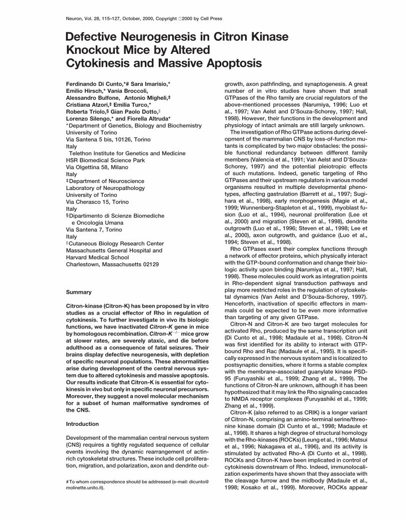
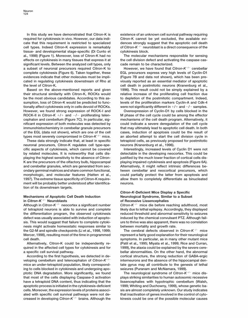
Figure 1. Expression of Citron-K andCitron-N during Development of Wild-TypeMouse CNS
(A) Lateral view of whole-mount ISH per-formed with probe K on an E11.5 mouse.(B) Radioactive ISH performed with probe Kon E14.5 sagittal section of telencephalon.Citron-K mRNA (pink) is restricted to the ac-tively proliferating regions.(C) Western blotting with anti-Citron antibod-ies, recognizing both Citron-K (CK) and Cit-ron-N (CN). Samples were 30 mg of total pro-teins from embryonic telencephalon at theindicated stages and 5 mg from adult brain(P30). After higher exposure of the blot, CKwas detected also in the P30 sample.(D) Radioactive ISH performed with probe Kon P12 cerebellum.(E) High power field of (D) displays thatCitron-K expression is restricted to the exter-nal part of the EGL.(F) Radioactive ISH performed with probe Con P12 cerebellum. Besides labeling of theEGL, strong expression is detected in the IGL.(G) Radioactive ISH performed with probe Kon P12 telencephalon.ge, ganglionic eminence; gl, germinal layer;cp, cortical plate; ob, olfactory bulb; egl,external germinal layer; e, external part; i,internal part; ml, molecular layer; igl, internalgranular layer; cx, cerebral cortex; sms, sub-ventricular migratory stream. Scale bars: 250mm (B, D, F, and G); 50 mm (E).
to phosphorylate in a cytokinesis-dependent way the cytokinesis. Finally, our findings indicate a novel poten-tial molecular basis for human brain malformative syn-myosin binding subunit (MBS) of myosin light chain
phosphatase (Kawano et al., 1999) and at least some dromes characterized by microcephaly and epilepsy.classes of intermediate filaments (Kosako et al., 1997;Goto et al., 1998; Yasui et al., 1998). Nevertheless, func-
Resultstional in vitro studies, performed by overexpression ofmutants and pharmacological inhibition in HeLa cells,
In the Developing CNS, Citron-K Is Expressedsuggested that only Citron-K is necessary for cytokine-by Proliferating Precursors, Citron-Nsis (Madaule et al., 1998).by Postmitotic CellsIn this report we have investigated the in vivo rolePrevious studies have revealed that, in the adult CNS,played by Citron-K in mouse development. We haveCitron-N is abundantly expressed by many neuronalfound that, during embryogenesis, it is abundantly ex-populations, while Citron-K is expressed at much lowerpressed throughout the neuraxis, with a remarkable spa-levels (Di Cunto et al., 1998; Furuyashiki et al., 1999).tial restriction to proliferating areas. Moreover, whenHowever, the expression pattern of the two differentneuroblasts leave the cell cycle, Citron-K is downmodu-isoforms during development of the CNS was not re-lated, while Citron-N becomes strongly expressed.ported. For this reason, we performed RNA in situ hy-Mutant mice completely lacking Citron-K expression,bridization (ISH) on mouse embryos and postnatalbut still producing normal levels of Citron-N, are affectedbrains using two different probes. The first (K) was de-by a complex neurological syndrome, characterized byrived from the kinase domain-coding sequence and spe-severe ataxia and epilepsy, which bring them to deathcifically hybridizes with Citron-K transcript. The secondwithin the first 3 postnatal weeks. Their brains display(C) corresponds to a fragment of the coiled-coil regiondramatic neuronal depletion in the cerebral cortex andof Citron-K and reacts with both Citron-N and Citron-Kin the granular layers of cerebellum, hippocampus, andmRNAs.olfactory bulb. Abnormal cytokinesis and massive apo-
A clear signal was detected with both probes as earlyptosis were detected in the same brain regions duringas embryonic day (E) 10.5. From this stage to E16.5, thedevelopment of Citron-K2/2 mice.highest expression levels were observed in the devel-Our results demonstrate that Citron-K is indeed re-oping CNS (Figure 1A), with a noticeable spatial restric-quired for cytokinesis in vivo, but only in specific prolifer-tion to the proliferating areas of the entire neural tubeating neuroblasts. Moreover, they strongly suggest that(Figure 1B). Strong expression was also observed inthe induction of programmed cell death in Citron-K2/2
neuroblasts is a direct consequence of the abnormal limb buds and retina (Figure 1A). Lower expression was
Knockout of Citron-Kinase117
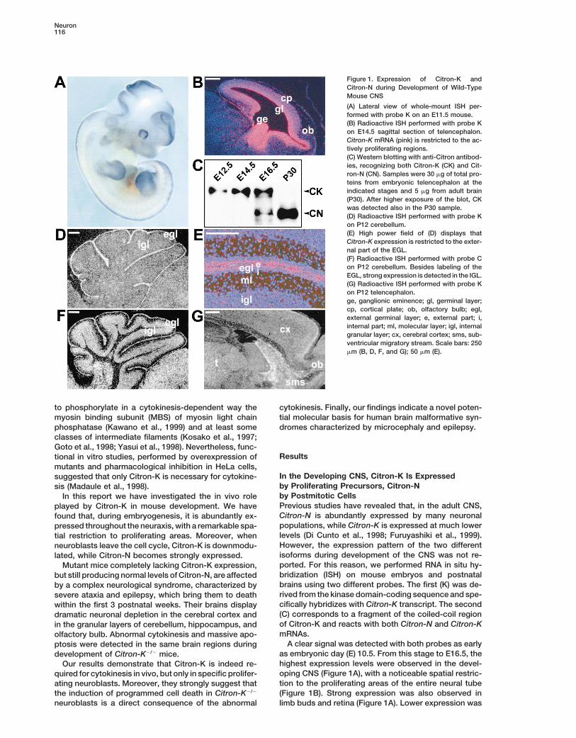
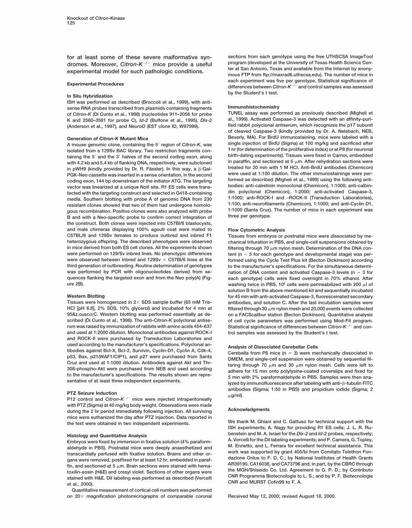
detected in the other somatic organs already known to For this reason, using homologous recombination inES cells, we inserted a b-gal-Neo selection cassetteexpress Citron-K in the adult (Di Cunto et al., 1998; andwithin the second coding exon of mouse Citron-K (Fig-data not shown).ure 2B). Since the insertion point is far from the putativeBefore E16.5, the hybridization pattern obtained withtranscriptional initiation site of Citron-N, this mutationboth probes was essentially identical (data not shown),would be predicted to cause an early stop of Citron-Kthus suggesting that the predominant isoform producedtranscription and not to disturb Citron-N expression.in the mouse brain during neurogenesis is Citron-K. ThisTwo independent ES cell clones were derived harboringwas confirmed by analysis of Citron-N and Citron-K pro-the correct homologous recombination event (Figuretein levels in brains at different developmental stages.2C). Heterozygous (1/2) mice obtained from germlineWestern blotting with antibodies raised against a com-transmission of both ES clones were intercrossed tomon epitope revealed that before E16.5 the only isoformgenerate homozygous mutant (2/2) progeny.produced is Citron-K, at E16.5 they are both present, and
Frequency at birth of the different genotypes obtainedin the adult brain Citron-N is by far the predominatingfrom 1/2 intercrossing was consistent with the ex-isoform (Figure 1C).pected Mendelian ratio (PCR analysis of a representa-In the first two postnatal weeks, robust expression oftive litter is shown in Figure 2D).Citron-K was still detected by probe K in the external
Western blotting with the above-mentioned antiserumgerminal layer (EGL) of the cerebellum (Figure 1D). Dur-confirmed complete loss of Citron-K expression in everying this period, cells of the cerebellar internal granularanalyzed tissue of 2/2 mice (Figure 2E). By contrast,layer (IGL) are generated by proliferation and migrationcompared to wild-type (1/1) littermate controls, Cit-of precursor cells of the EGL (Goldowitz and Hamre,ron-N protein levels of Citron-K2/2 brains were not de-1998). Strikingly, Citron-K was expressed only in thecreased (Figure 2E), further confirming the existence ofexternal part of the EGL, which contains the proliferatinga second transcriptional initiation site.precursors (Figures 1E). It was not detectable in the
IGL and in the internal part of the EGL, composed ofCitron-K2/2 Mice Are Affected by a Complexpostmitotic cells (Figures 1D and 1E). By contrast, probeNeurological Syndrome and Display ReducedC strongly labeled both the EGL and the IGL (Figure 1F),Life Span for Lethal Epilepsythus indicating high expression of Citron-N in differenti-Average weight and growth rate of Citron-K2/2 mice didated cerebellar granules.not differ significantly from 1/2 and 1/1 littermatesIn the same period, relatively strong expression ofin the first postnatal week. However, failure to thriveCitron-K was also detected in the subventricular migra-became apparent around P10 in Citron-K2/2 animals (attory stream (SMS; Figure 1G). This structure containsP12 the difference of body weight of 2/2 versus 1/1the precursors of olfactory bulb granules, which are stillwas 20%).mitotically active while migrating toward their final loca-
After P8 Citron-K2/2 mice started to present an overttion (Hinds, 1968).neurological phenotype, characterized by tremor andTaken together, these data indicate that, within thesevere ataxia. At P12 Citron-K2/2 mice were unable toCNS, Citron-K is specifically expressed by proliferatingstand for .2 s on a narrow (3 cm wide) platform, whereasneuronal precursors, while postmitotic neuroblasts andlittermate controls stayed balanced for .2 min. Theirdifferentiated neurons mainly express Citron-N.gait was wide and uncoordinated, and they were fre-quently falling on their backs while walking. Around thesame age, Citron-K2/2 mice became more irritable andGeneration of Citron-K-Deficient Micestarted to present lethal seizures, characterized byTo directly address in vivo the specific role of Citron-myoclonic jerks followed by loss of posture, hyperexten-K, we devised a gene targeting approach that wouldsion of trunk and hindlimbs, and respiratory arrest. Oc-
produce a null allele for this particular isoform and sparecasionally, they were able to recover from crisis, but
Citron-N expression.none of them survived beyond P21, with a peak of mor-
mRNAs coding for both molecules are transcribed tality observed at P14. No correlation was detected be-from the same gene (Di Cunto et al., 1998; Madaule et tween body weight and mortality (data not shown).al., 1998), but so far it has not been defined if they To confirm the epileptic nature of crisis, we tested ifare produced by alternative transcriptional initiation or Citron-K2/2 mice had a lower threshold for seizures us-alternative splicing. To clarify this point, we compared ing the chemical convulsant pentylenetetrazol (PTZ)the available sequences of Citron-K and Citron-N (Chae et al., 1997). P12 1/1 (n 5 15) and 1/2 (n 5 12)59 untranslated regions (UTRs) and found no significant mice, injected intraperitoneally with PTZ, did not showoverlapping. Moreover, we assembled the complete seizure activity during the observation period. By con-genomic sequence of human Citron-K from three clones trast, 89% (n 5 9) of Citron-K2/2 mice displayed one oravailable in the public database (R. H. Waterston, more seizures indistinguishable from those occurringGenBank accession numbers AC004813, AC004811, spontaneously, with an average latency of 20 min fromAC002563; Figure 2A) and compared it with mouse and injection, and 78% died by the end of the observationrat Citron-N 59 UTRs. This analysis revealed that all of period (p , 0.001, chi-square test).the identified Citron-N 59 noncoding exons map down-stream of the kinase domain-coding region. Brains of Citron-K2/2 Mice Display Dramatic
The above data argue against the alternative splicing Depletion of Microneurons in the Olfactorymechanism and would be consistent with a second tran- Bulb, Hippocampus, and Cerebellumscriptional initiation site for Citron-N downstream of the Autopsy of Citron-K2/2 mice did not reveal evidence of
cerebral tumors, hemorrhage, or obvious disorders inkinase domain-coding region.
Neuron118
Figure 2. Gene-Targeting Strategy for theSpecific Inactivation of Citron-K
(A) Schematic representation of the humanCitron-K gene. S1 and S2 indicate the puta-tive transcriptional initiation sites for Citron-Kand Citron-N mRNAs, respectively. Underlin-ing indicates the region homologous to themouse sequence used for constructing thetargeting vector.(B) Structure of the wild-type mouse Citron-Klocus, of the targeting construct, and of themutated allele. Exons 1 and 2 are the first twocoding exons of Citron-K. Lines P1 and P2indicate the position of the 59 and 39 probes,respectively, used for Southern screening ofES cells and knockout mice. Arrows O1, O2,and O3 indicate the positions of primers usedfor routine genotyping of mice. K, KpnI; S,SmaI; E, EcoRI; N, NotI.(C) Southern blotting with probe P1 of KpnI-digested genomic DNA from ES cell clones.Clones 2 and 3 presented the correct homolo-gous recombination event.(D) PCR performed with primers O1, O2, andO3 on a representative litter of Citron-K1/2
mice, detecting wild-type (wt) and mutated(m) alleles.(E) Western blotting with anti-Citron antibod-ies, performed on 30 mg of total proteins ex-tracted from P5 cerebellum (Cb), spleen (Sp),and thymus (Th) of 1/1 and 2/2 mice.
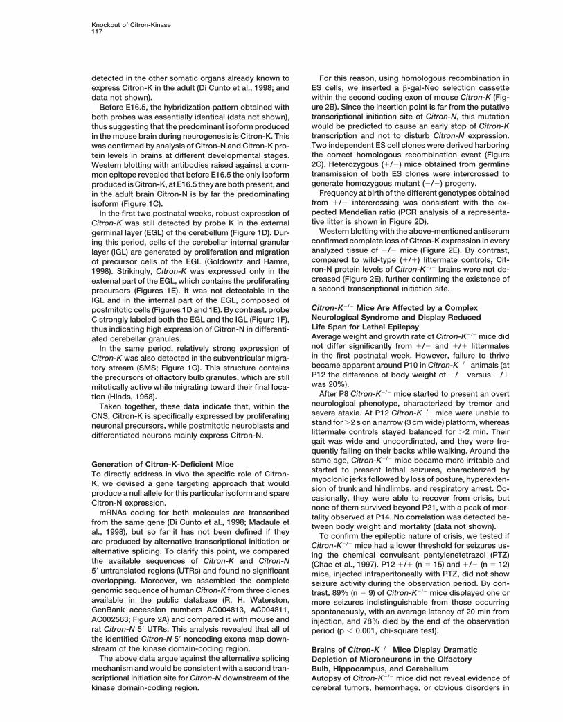
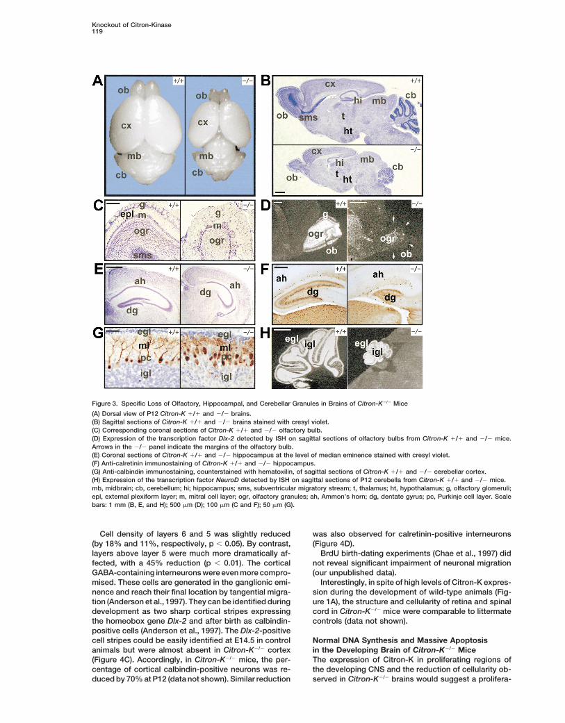
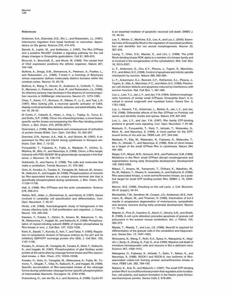
somatic tissues. However, macroscopic inspection of In the cerebellum, all major lobules were recognizable,although they were strikingly smaller than in controlstheir brain showed a striking size reduction compared
to control littermates (Figure 3A). At P14, the overall (Figure 3B). The number of Purkinje cells was not re-duced, but their monolayer organization was disruptedaverage weight of Citron-K2/2 brains was reduced by
50% (p , 0.01). Dissection of principal brain areas re- and their dendritic harborization severely underdevel-oped, as revealed by anti-calbindin immunostainingvealed an average reduction of z20% in diencephalon
and midbrain, 50% in cerebral hemispheres, and 70% (Figure 3G). By contrast, the granular cell layer was dra-matically reduced (Figures 3B and 3G). However, estab-in cerebellum and olfactory bulbs. Histological examina-
tion showed that the structure of thalamus, hypothala- lishment of the cerebellar granule cell fate and differenti-ation was apparently conserved, as suggested bymus, basal ganglia, and midbrain was preserved to a
large extent, even though their cellularity was slightly normal expression in the remaining granules of the tran-scription factor NeuroD (Miyata et al., 1999) (Figure 3H).reduced (Figure 3B and data not shown). Brain regions
displaying the most abnormal structure in Citron-K2/2
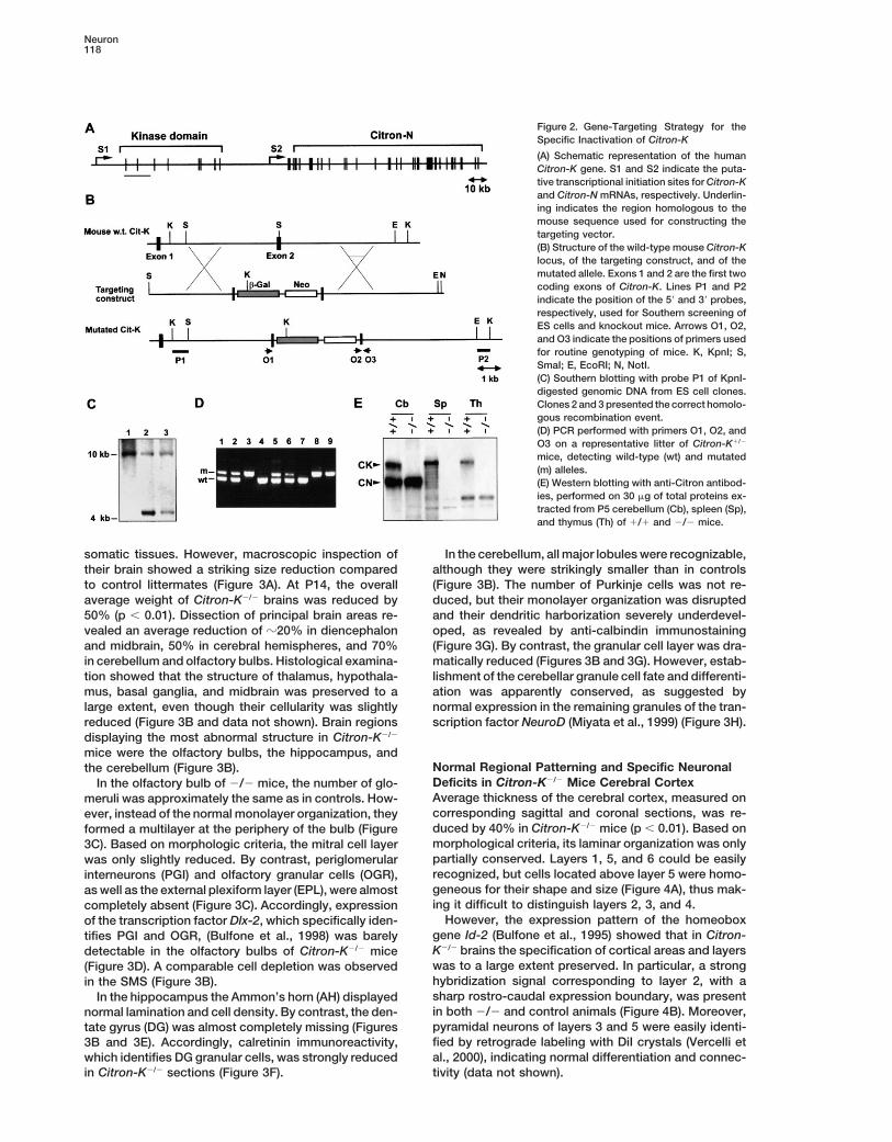
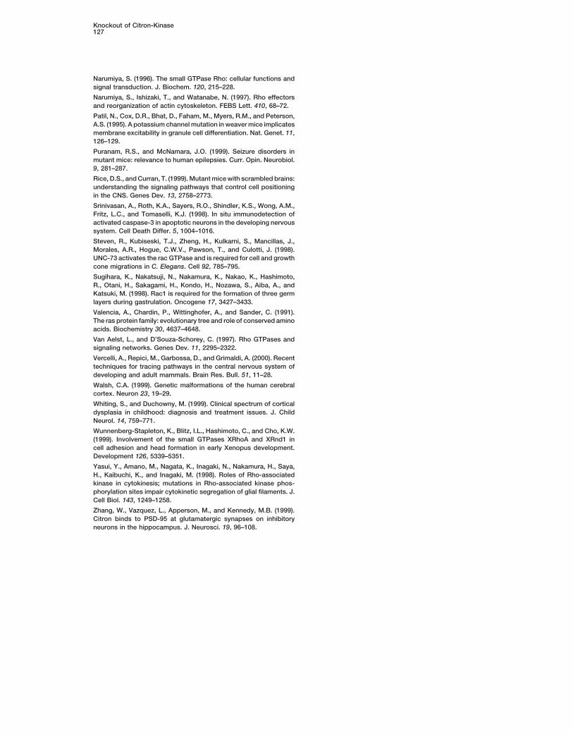
mice were the olfactory bulbs, the hippocampus, andNormal Regional Patterning and Specific Neuronalthe cerebellum (Figure 3B).Deficits in Citron-K2/2 Mice Cerebral CortexIn the olfactory bulb of 2/2 mice, the number of glo-Average thickness of the cerebral cortex, measured onmeruli was approximately the same as in controls. How-corresponding sagittal and coronal sections, was re-ever, instead of the normal monolayer organization, theyduced by 40% in Citron-K2/2 mice (p , 0.01). Based onformed a multilayer at the periphery of the bulb (Figuremorphological criteria, its laminar organization was only3C). Based on morphologic criteria, the mitral cell layerpartially conserved. Layers 1, 5, and 6 could be easilywas only slightly reduced. By contrast, periglomerularrecognized, but cells located above layer 5 were homo-interneurons (PGI) and olfactory granular cells (OGR),geneous for their shape and size (Figure 4A), thus mak-as well as the external plexiform layer (EPL), were almosting it difficult to distinguish layers 2, 3, and 4.completely absent (Figure 3C). Accordingly, expression
However, the expression pattern of the homeoboxof the transcription factor Dlx-2, which specifically iden-gene Id-2 (Bulfone et al., 1995) showed that in Citron-tifies PGI and OGR, (Bulfone et al., 1998) was barelyK2/2 brains the specification of cortical areas and layersdetectable in the olfactory bulbs of Citron-K2/2 micewas to a large extent preserved. In particular, a strong(Figure 3D). A comparable cell depletion was observedhybridization signal corresponding to layer 2, with ain the SMS (Figure 3B).sharp rostro-caudal expression boundary, was presentIn the hippocampus the Ammon’s horn (AH) displayedin both 2/2 and control animals (Figure 4B). Moreover,normal lamination and cell density. By contrast, the den-pyramidal neurons of layers 3 and 5 were easily identi-tate gyrus (DG) was almost completely missing (Figuresfied by retrograde labeling with DiI crystals (Vercelli et3B and 3E). Accordingly, calretinin immunoreactivity,al., 2000), indicating normal differentiation and connec-which identifies DG granular cells, was strongly reduced
in Citron-K2/2 sections (Figure 3F). tivity (data not shown).
Knockout of Citron-Kinase119
Figure 3. Specific Loss of Olfactory, Hippocampal, and Cerebellar Granules in Brains of Citron-K2/2 Mice
(A) Dorsal view of P12 Citron-K 1/1 and 2/2 brains.(B) Sagittal sections of Citron-K 1/1 and 2/2 brains stained with cresyl violet.(C) Corresponding coronal sections of Citron-K 1/1 and 2/2 olfactory bulb.(D) Expression of the transcription factor Dlx-2 detected by ISH on sagittal sections of olfactory bulbs from Citron-K 1/1 and 2/2 mice.Arrows in the 2/2 panel indicate the margins of the olfactory bulb.(E) Coronal sections of Citron-K 1/1 and 2/2 hippocampus at the level of median eminence stained with cresyl violet.(F) Anti-calretinin immunostaining of Citron-K 1/1 and 2/2 hippocampus.(G) Anti-calbindin immunostaining, counterstained with hematoxilin, of sagittal sections of Citron-K 1/1 and 2/2 cerebellar cortex.(H) Expression of the transcription factor NeuroD detected by ISH on sagittal sections of P12 cerebella from Citron-K 1/1 and 2/2 mice.mb, midbrain; cb, cerebellum; hi; hippocampus; sms, subventricular migratory stream; t, thalamus; ht, hypothalamus; g, olfactory glomeruli;epl, external plexiform layer; m, mitral cell layer; ogr, olfactory granules; ah, Ammon’s horn; dg, dentate gyrus; pc, Purkinje cell layer. Scalebars: 1 mm (B, E, and H); 500 mm (D); 100 mm (C and F); 50 mm (G).
Cell density of layers 6 and 5 was slightly reduced was also observed for calretinin-positive interneurons(Figure 4D).(by 18% and 11%, respectively, p , 0.05). By contrast,
layers above layer 5 were much more dramatically af- BrdU birth-dating experiments (Chae et al., 1997) didnot reveal significant impairment of neuronal migrationfected, with a 45% reduction (p , 0.01). The cortical
GABA-containing interneurons were even more compro- (our unpublished data).Interestingly, in spite of high levels of Citron-K expres-mised. These cells are generated in the ganglionic emi-
nence and reach their final location by tangential migra- sion during the development of wild-type animals (Fig-ure 1A), the structure and cellularity of retina and spinaltion (Anderson et al., 1997). They can be identified during
development as two sharp cortical stripes expressing cord in Citron-K2/2 mice were comparable to littermatecontrols (data not shown).the homeobox gene Dlx-2 and after birth as calbindin-
positive cells (Anderson et al., 1997). The Dlx-2-positivecell stripes could be easily identified at E14.5 in control Normal DNA Synthesis and Massive Apoptosis
in the Developing Brain of Citron-K2/2 Miceanimals but were almost absent in Citron-K2/2 cortex(Figure 4C). Accordingly, in Citron-K2/2 mice, the per- The expression of Citron-K in proliferating regions of
the developing CNS and the reduction of cellularity ob-centage of cortical calbindin-positive neurons was re-duced by 70% at P12 (data not shown). Similar reduction served in Citron-K2/2 brains would suggest a prolifera-

Neuron120
Figure 4. Structural Abnormalities of Cere-bral Cortex in Citron-K2/2 Mice
(A) Corresponding sagittal sections ofCitron-K 1/1 and 2/2 neocortex stainedwith cresyl violet. Numbers indicate the posi-tion of presumptive cortical layers.(B) Expression of the transcription factor Id-2detected by ISH on sagittal sections of P12brains from Citron-K 1/1 and 2/2 mice.Arrows indicate the rostro-caudal expressionboundary.(C) Expression of the transcription factorDlx-2 detected by ISH on sagittal sectionsof E14.5 brains from Citron-K 1/1 and 2/2mice. Arrows in the 1/1 panel indicate thepositions of the positive cell stripes.(D) Anti-calretinin immunostaining of coronalsections of Citron-K 1/1 and 2/2 cerebralcortex. Scale bars: 1 mm (B and C); 250 mm(A); 100 mm (D).
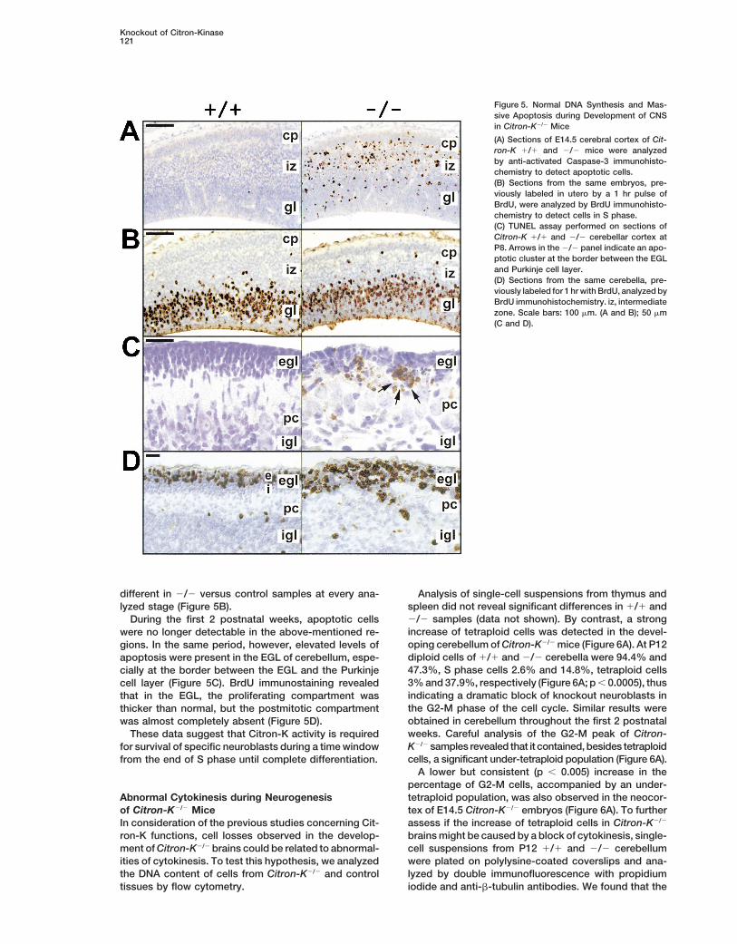
tion deficit in neuroblast precursors. Alternatively, or in by TUNEL assay and activated Caspase-3 immunostain-ing (Srinivasan et al., 1998), indicating a massive apo-association, increased cell death could be involved.
To discriminate between these possibilities, serial ptotic process (Figure 5A). At E14.5 apoptotic cells wereparticularly abundant in the ganglionic eminence and insections from 1/1 and 2/2 mice, at different stages of
embryonic and postnatal development, were analyzed the germinal and intermediate zones of the neocortexbut were also detected in the cortical plate. At E16.5to reveal cells undergoing DNA synthesis or apoptotic
cell death. apoptotic cells were identified in close proximity to thegerminal regions of dentate gyrus and olfactory bulbBefore E11.5, no differences were observed between
controls and Citron-K2/2 animals. However, after this granular layer (data not shown). Significant apoptosiswas also detected in midbrain, pons, and medulla ofstage, a large number of pyknotic and fragmented nu-
clei, isolated or in clusters, were detected throughout E12.5–E16.5 animals. By contrast, the percentage anddistribution of BrdU-positive cells was not significantlythe brain of 2/2 mice. These cells were strongly positive
Knockout of Citron-Kinase121
Figure 5. Normal DNA Synthesis and Mas-sive Apoptosis during Development of CNSin Citron-K2/2 Mice
(A) Sections of E14.5 cerebral cortex of Cit-ron-K 1/1 and 2/2 mice were analyzedby anti-activated Caspase-3 immunohisto-chemistry to detect apoptotic cells.(B) Sections from the same embryos, pre-viously labeled in utero by a 1 hr pulse ofBrdU, were analyzed by BrdU immunohisto-chemistry to detect cells in S phase.(C) TUNEL assay performed on sections ofCitron-K 1/1 and 2/2 cerebellar cortex atP8. Arrows in the 2/2 panel indicate an apo-ptotic cluster at the border between the EGLand Purkinje cell layer.(D) Sections from the same cerebella, pre-viously labeled for 1 hr with BrdU, analyzed byBrdU immunohistochemistry. iz, intermediatezone. Scale bars: 100 mm. (A and B); 50 mm(C and D).
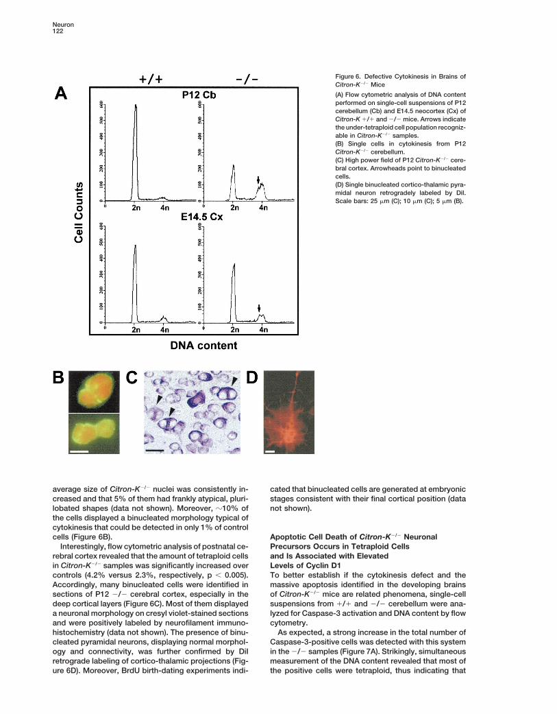
different in 2/2 versus control samples at every ana- Analysis of single-cell suspensions from thymus andspleen did not reveal significant differences in 1/1 andlyzed stage (Figure 5B).2/2 samples (data not shown). By contrast, a strongDuring the first 2 postnatal weeks, apoptotic cellsincrease of tetraploid cells was detected in the devel-were no longer detectable in the above-mentioned re-oping cerebellum of Citron-K2/2 mice (Figure 6A). At P12gions. In the same period, however, elevated levels ofdiploid cells of 1/1 and 2/2 cerebella were 94.4% andapoptosis were present in the EGL of cerebellum, espe-47.3%, S phase cells 2.6% and 14.8%, tetraploid cellscially at the border between the EGL and the Purkinje3% and 37.9%, respectively (Figure 6A; p , 0.0005), thuscell layer (Figure 5C). BrdU immunostaining revealedindicating a dramatic block of knockout neuroblasts inthat in the EGL, the proliferating compartment wasthe G2-M phase of the cell cycle. Similar results werethicker than normal, but the postmitotic compartmentobtained in cerebellum throughout the first 2 postnatalwas almost completely absent (Figure 5D).weeks. Careful analysis of the G2-M peak of Citron-These data suggest that Citron-K activity is requiredK2/2 samples revealed that it contained, besides tetraploidfor survival of specific neuroblasts during a time windowcells, a significant under-tetraploid population (Figure 6A).from the end of S phase until complete differentiation.
A lower but consistent (p , 0.005) increase in thepercentage of G2-M cells, accompanied by an under-tetraploid population, was also observed in the neocor-Abnormal Cytokinesis during Neurogenesis
of Citron-K2/2 Mice tex of E14.5 Citron-K2/2 embryos (Figure 6A). To furtherassess if the increase of tetraploid cells in Citron-K2/2In consideration of the previous studies concerning Cit-
ron-K functions, cell losses observed in the develop- brains might be caused by a block of cytokinesis, single-cell suspensions from P12 1/1 and 2/2 cerebellumment of Citron-K2/2 brains could be related to abnormal-
ities of cytokinesis. To test this hypothesis, we analyzed were plated on polylysine-coated coverslips and ana-lyzed by double immunofluorescence with propidiumthe DNA content of cells from Citron-K2/2 and control
tissues by flow cytometry. iodide and anti-b-tubulin antibodies. We found that the
Neuron122
Figure 6. Defective Cytokinesis in Brains ofCitron-K2/2 Mice
(A) Flow cytometric analysis of DNA contentperformed on single-cell suspensions of P12cerebellum (Cb) and E14.5 neocortex (Cx) ofCitron-K 1/1 and 2/2 mice. Arrows indicatethe under-tetraploid cell population recogniz-able in Citron-K2/2 samples.(B) Single cells in cytokinesis from P12Citron-K2/2 cerebellum.(C) High power field of P12 Citron-K2/2 cere-bral cortex. Arrowheads point to binucleatedcells.(D) Single binucleated cortico-thalamic pyra-midal neuron retrogradely labeled by DiI.Scale bars: 25 mm (C); 10 mm (C); 5 mm (B).
average size of Citron-K2/2 nuclei was consistently in- cated that binucleated cells are generated at embryonicstages consistent with their final cortical position (datacreased and that 5% of them had frankly atypical, pluri-
lobated shapes (data not shown). Moreover, z10% of not shown).the cells displayed a binucleated morphology typical ofcytokinesis that could be detected in only 1% of controlcells (Figure 6B). Apoptotic Cell Death of Citron-K2/2 Neuronal
Precursors Occurs in Tetraploid CellsInterestingly, flow cytometric analysis of postnatal ce-rebral cortex revealed that the amount of tetraploid cells and Is Associated with Elevated
Levels of Cyclin D1in Citron-K2/2 samples was significantly increased overcontrols (4.2% versus 2.3%, respectively, p , 0.005). To better establish if the cytokinesis defect and the
massive apoptosis identified in the developing brainsAccordingly, many binucleated cells were identified insections of P12 2/2 cerebral cortex, especially in the of Citron-K2/2 mice are related phenomena, single-cell
suspensions from 1/1 and 2/2 cerebellum were ana-deep cortical layers (Figure 6C). Most of them displayeda neuronal morphology on cresyl violet-stained sections lyzed for Caspase-3 activation and DNA content by flow
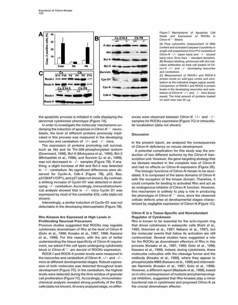
cytometry.and were positively labeled by neurofilament immuno-histochemistry (data not shown). The presence of binu- As expected, a strong increase in the total number of
Caspase-3-positive cells was detected with this systemcleated pyramidal neurons, displaying normal morphol-ogy and connectivity, was further confirmed by DiI in the 2/2 samples (Figure 7A). Strikingly, simultaneous
measurement of the DNA content revealed that most ofretrograde labeling of cortico-thalamic projections (Fig-ure 6D). Moreover, BrdU birth-dating experiments indi- the positive cells were tetraploid, thus indicating that
Knockout of Citron-Kinase123
Figure 7. Mechanisms of Apoptotic CellDeath and Expression of ROCKs inCitron-K2/2 Brains
(A) Flow cytometric measurement of DNAcontent and activated Caspase-3 positivity insingle-cell suspensions from P12 cerebella ofCitron-K 1/1 (open bars) and 2/2 (closedbars) mice. Error bars 5 standard deviation.(B) Western blotting, performed with the indi-cated antibodies on total cell lysates of Cit-ron-K 1/1 and 2/2 developing neocortexand cerebellum.(C) Measurement of ROCK-I and ROCK-IIprotein levels on wild-type cortex and cere-bellum at the indicated stages (upper panel).Comparison of ROCK-I and ROCK-II proteinlevels in the developing neocortex and cere-bellum of Citron-K 1/1 and 2/2 mice (lowerpanel). The total amount of proteins loadedon each lane was 30 mg.
the apoptotic process is initiated in cells displaying the ences were observed between Citron-K 1/1 and 2/2samples for ROCKs expression (Figure 7C) or intracellu-abnormal cytokinesis phenotype (Figure 7A).
In order to investigate the molecular mechanisms un- lar localization (data not shown).derlying the induction of apoptosis in Citron-K2/2 neuro-blasts, the level of different proteins previously impli- Discussioncated in this process was measured in the developingneocortex and cerebellum of 1/1 and 2/2 mice. In the present report, we analyzed the consequences
The expression of proteins promoting cell survival, of Citron-K deficiency on mouse development.such as Akt and its Thr-308-phosphorylated isoform A potential complication for this study was the pro-(Downward, 1998), Bcl-X (Motoyama et al., 1995), Bcl-2 duction of two different isoforms by the Citron-K tran-(Michaelidis et al., 1996), and Survivin (Li et al., 1999), scription unit. However, the gene-targeting strategy thatwas not decreased in 2/2 samples (Figure 7B). If any- we devised resulted in the complete loss of Citron-Kthing, a slight increase of Akt and Bcl-2 was detected and had no effects on Citron-N expression (Figure 2E).in 2/2 cerebellum. No significant differences were ob- The biologic functions of Citron-N remain to be eluci-served for Cyclin-A, Cdk-4 (Figure 7B), p53, Bax, dated. It is composed of the same domains of Citron-Kp21(WAF1/CIP1), and p27 (data not shown). By contrast, with the exception of the kinase domain. Therefore, ita striking increase of Cyclin-D1 was detected in devel- could compete for binding to activated Rho and act asoping 2/2 cerebellum. Accordingly, immunohistochem- an endogenous inhibitor of Citron-K function. However,ical analysis showed that in 2/2 mice Cyclin D1 was this mechanism is unlikely to play a role in producingexpressed by most of the cerebellar EGL cells (data not the phenotype of Citron-K2/2 mice, since the observedshown). cellular defects arise at developmental stages charac-
Interestingly, a similar induction of Cyclin-D1 was not terized by negligible expression of Citron-N (Figure 1C).detectable in the developing telencephalon (Figure 7B).
Citron-K Is a Tissue-Specific and NonredundantRegulator of CytokinesisRho-Kinases Are Expressed at High Levels in
Proliferating Neuronal Precursors Rho is known to be essential for the acto-myosin ringthat drives cytokinesis in eukaryotic cells (Kishi et al.,Previous studies suggested that ROCKs may regulate
cytokinesis downstream of Rho at the level of Citron-K 1993; Drechsel et al., 1997; Nakano et al., 1997), butthe molecular events that follow its activation are still(Goto et al., 1998; Kosako et al., 1997, 1999; Kawano
et al., 1999). For this reason, with the aim of better controversial. Several studies have suggested a rolefor the ROCKs as downstream effectors of Rho in thisunderstanding the tissue specificity of Citron-K require-
ment, we asked if the cell types undergoing cytokinesis process (Kosako et al., 1997, 1999; Goto et al., 1998;Kawano et al., 1999). Indeed, during cytokinesis, theseblock in Citron-K2/2 are devoid of ROCKs expression.
ROCK-I and ROCK-II protein levels were measured in molecules colocalize with the cleavage furrow and themidbody (Kosako et al., 1999), where they appear tothe neocortex and cerebellum of Citron-K 1/1 and 2/2
mice at different developmental stages. Robust expres- phosphorylate MBS (Kawano et al., 1999) and intermedi-ate filaments (Kosako et al., 1997; Goto et al., 1998).sion of both molecules was detected throughout brain
development (Figure 7C). In the cerebellum, the highest However, a different report (Madaule et al., 1998), basedon in vitro overexpression of mutants and pharmacologi-levels were detected during the time window of granular
cell proliferation (Figure 7C). Accordingly, immunohisto- cal inhibition, suggested that Rho-kinases play a minorfunctional role in cytokinesis and proposed Citron-K aschemical analysis revealed strong positivity of the EGL
cells (data not shown). At every analyzed stage, no differ- the crucial downstream effector.
Neuron124
In this study we have demonstrated that Citron-K is existence of an unknown cell survival pathway requiringCitron-K cannot be yet excluded, the available evi-required for cytokinesis in vivo. However, our data indi-
cate that this requirement is restricted to specialized dences strongly suggest that the apoptotic cell deathof Citron-K2/2 neuroblast is a direct consequence of thecell types. Indeed Citron-K expression is remarkably
tissue- and developmental stage-specific (Di Cunto et cytokinesis block.The molecular mechanisms responsible for sensingal., 1998) (Figure 1). Moreover, loss of Citron-K had no
effects on cytokinesis in many tissues that express it at the cell division defect and activating the caspase cas-cade remain to be characterized.significant levels. Between the analyzed cell types, only
a subset of neuronal precursors required Citron-K to However, we have found that Citron-K2/2 cerebellarEGL precursors express very high levels of Cyclin-D1complete cytokinesis (Figure 6). Taken together, these
evidences indicate that other molecules must be impli- (Figure 7B and data not shown), which has been pre-viously reported as an essential mediator of apoptoticcated in regulating cytokinesis downstream of Rho at
the level of Citron-K. cell death in postmitotic neurons (Kranenburg et al.,1996). This result could not be simply explained by aBased on the above-mentioned reports and given
their structural similarity with Citron-K, ROCKs would relative increase of the proliferating cell fraction dueto depletion of the postmitotic compartment. Indeed,be the most obvious candidates. According to this as-
sumption, loss of Citron-K would be predicted to func- levels of the proliferation markers Cyclin-A and Cdk-4were not significantly different in 1/1 and 2/2 samples.tionally affect cytokinesis only in cells devoid of ROCKs.
However, we found strong expression of ROCK-I and Overexpression of Cyclin-D1 by cells blocked in theM phase of the cell cycle could be among the effectorROCK-II in Citron-K 1/1 and 2/2 proliferating telen-
cephalon and cerebellum (Figure 7C). In particular, sig- mechanisms of the cell death program. Alternatively, itcould indicate a severe deregulation of the cell cyclenificant expression of both molecules was detected by
immunohistochemistry in cerebellar granule precursors that may ultimately lead to apoptotic cell death. In bothcases, induction of apoptosis could be the result ofof the EGL (data not shown), which are one of the cell
types most severely compromised in Citron-K2/2 mice. an aborted attempt to start the cell division cycle intetraploid cells, as previously proposed for postmitoticThese data strongly indicate that, at least in specific
neuronal precursors, Citron-K regulates cell type–spe- neurons (Kranenburg et al., 1996).Interestingly, increased levels of Cyclin D1 were notcific aspects of cytokinesis, which cannot be covered
by related molecules. It is noteworthy that cells dis- detectable in the developing neocortex. This could bejustified by the much lower fraction of cortical cells dis-playing the highest sensitivity to the absence of Citron-
K are the precursors of the olfactory bulb, hippocampal playing impaired cytokinesis and apoptosis (Figure 6A).Alternatively, it might reflect intrinsic differences be-and cerebellar granules, which are generated from sec-
ondary germinal matrices and share common functional, tween cerebellar and neocortical precursors, whichcould partially protect the latter from apoptosis andmorphologic, and molecular features (Hatten et al.,
1997). The extreme tissue specificity of Citron-K require- allow them to completely differentiate as binucleatedneurons.ment will be probably better understood after identifica-
tion of its downstream targets.Citron-K-Deficient Mice Display a SpecificNeurological Syndrome, Similar to a SubsetMechanisms of Apoptotic Cell Death Induction
in Citron-K2/2 Neuroblasts of Recessive LissencephaliesCitron-K2/2 mice die before reaching adulthood, mostAlthough in Citron-K2/2 neocortex a significant number
of tetraploid neurons is able to survive and complete likely due to lethal epilepsy. Accordingly, they displayedreduced threshold and abnormal sensitivity to seizuresthe differentiation program, the observed cytokinesis
defect was usually associated with induction of apopto- induced by the chemical convulsant PTZ. Although fail-ure to thrive was also apparent, there was no correlationsis. This would suggest that failure to complete cytoki-
nesis might activate homeostatic responses similar to between mortality and growth rate.The cerebral defects observed in Citron-K2/2 micethe G2-M and spindle checkpoints (Li et al., 1998, 1999;
Mercer, 1998), resulting most of the time in programmed represent a fairly good explanation for their neurologicalsymptoms. In particular, as in many other mutant micecell death.
Alternatively, Citron-K could be independently re- (Patil et al., 1995; Miyata et al., 1999; Rice and Curran,1999), the ataxia could be explained by the severe cere-quired in the affected cell types for cytokinesis and for
a specific cell survival pathway. bellar abnormalities. On the other hand, the abnormalcortical structure, the strong reduction of GABA-ergicAccording to the first hypothesis, we detected in de-
veloping cerebellum and telencephalon of Citron-K2/2 interneurons and the absence of the hippocampal den-tate gyrus may all contribute to the genesis of lethalmice an under-tetraploid population, likely correspond-
ing to cells blocked in cytokinesis and undergoing apo- seizures (Puranam and McNamara, 1999).The neurological syndrome of Citron-K2/2 mice dis-ptotic DNA degradation. More significantly, we found
that most of the cells displaying Caspase-3 activation plays striking similarities to human autosomic recessivelissencephalies with hypotrophic cerebellum (Walsh,have a tetraploid DNA content, thus indicating that the
apoptotic process is initiated in the cytokinesis-deficient 1999; Whiting and Duchowny, 1999), whose genetic ba-sis are almost completely unknown. Our study indicatescells. Moreover, the expression levels of proteins associ-
ated with specific cell survival pathways were not de- that inactivation of genes involved in the control of cyto-kinesis could be one of the possible molecular causescreased in developing Citron-K2/2 brains. Although the
Knockout of Citron-Kinase125
sections from each genotype using the free UTHSCSA ImageToolfor at least some of these severe malformative syn-program (developed at the University of Texas Health Science Cen-dromes. Moreover, Citron-K2/2 mice provide a usefulter at San Antonio, Texas and available from the Internet by anony-experimental model for such pathologic conditions.mous FTP from ftp://maxrad6.uthscsa.edu). The number of mice ineach experiment was five per genotype. Statistical significance of
Experimental Proceduresdifferences between Citron-K2/2 and control samples was assessedby the Student’s t test.
In Situ HybridizationISH was performed as described (Broccoli et al., 1999), with anti-
Immunohistochemistrysense RNA probes transcribed from plasmids containing fragmentsTUNEL assay was performed as previously described (Migheli etof Citron-K (Di Cunto et al., 1998) (nucleotides 911–2056 for probeal., 1999). Activated Caspase-3 was detected with an affinity-puri-K and 2560–3581 for probe C), Id-2 (Bulfone et al., 1995), Dlx-2fied rabbit polyclonal antiserum, which recognizes the p17 subunit(Anderson et al., 1997), and NeuroD (EST clone ID, W97999).of cleaved Caspase-3 (kindly provided by Dr. A. Nelsbach, NEB,Beverly, MA). For BrdU immunostaining, mice were labeled with aGeneration of Citron-K Mutant Micesingle injection of BrdU (Sigma) at 100 mg/kg and sacrificed afterA mouse genomic clone, containing the 59 region of Citron-K, was1 hr (for determination of the proliferative index) or at P8 (for neuronalisolated from a 129Sv BAC library. Two restriction fragments con-birth-dating experiments). Tissues were fixed in Carnoi, embeddedtaining the 59 and the 39 halves of the second coding exon, alongin paraffin, and sectioned at 5 mm. After rehydration sections werewith 4.2 kb and 5.4 kb of flanking DNA, respectively, were subclonedtreated for 20 min with 1 M HCl. Anti-BrdU antibodies (Chemicon)in pWH9 (kindly provided by Dr. R. Fassler). In this way, a b-Gal-were used at 1:100 dilution. The other immunostainings were per-PGK-Neo cassette was inserted in a sense orientation, in the secondformed as described (Migheli et al., 1999) using the following anti-coding exon, 144 bp downstream of the initiator ATG. The targetingbodies: anti-calretinin monoclonal (Chemicon), 1:1000; anti-calbin-vector was linearized at a unique NotI site. R1 ES cells were trans-din polyclonal (Chemicon), 1:2000; anti-activated Caspase-3,fected with the targeting construct and selected in G418-containing1:1000; anti-ROCK-I and -ROCK-II (Transduction Laboratories),media. Southern blotting with probe A of genomic DNA from 2301:100; anti-neurofilaments (Chemicon), 1:1000; and anti-Cyclin D1,resistant clones showed that two of them had undergone homolo-1:1000 (Santa Cruz). The number of mice in each experiment wasgous recombination. Positive clones were also analyzed with probethree per genotype.B and with a Neo-specific probe to confirm correct integration of
the construct. Both clones were injected into C57Bl/6 blastocysts,and male chimeras displaying 100% agouti coat were mated to Flow Cytometric AnalysisC57BL/6 and 129Sv females to produce outbred and inbred F1 Tissues from embryos or postnatal mice were dissociated by me-heterozygous offspring. The described phenotypes were observed chanical trituration in PBS, and single-cell suspensions obtained byin mice derived from both ES cell clones. All the experiments shown filtering through 70 mm nylon mesh. Determination of the DNA con-were performed on 129/Sv inbred lines. No phenotypic differences tent (n 5 5 for each genotype and developmental stage) was per-were observed between inbred and 129Sv 3 C57Bl/6 lines at the formed using the Cycle Test Plus kit (Becton Dickinson) accordingthird generation of outbreeding. Routine determination of genotypes to the manufacturer’s specifications. For the simultaneous determi-was performed by PCR with oligonucleotides derived from se- nation of DNA content and activated Caspase-3 levels (n 5 3 forquences flanking the targeted exon and from the Neo poly(A) (Fig- each genotype) cells were fixed overnight in 70% ethanol. Afterure 2B). washing twice in PBS, 106 cells were permeabilized with 200 ml of
solution B from the above-mentioned kit and sequentially incubatedWestern Blotting for 45 min with anti-activated Caspase-3, fluoresceinated secondaryTissues were homogenized in 23 SDS sample buffer (65 mM Tris- antibodies, and solution C. After the last incubation samples wereHCl [pH 6.8], 2% SDS, 10% glycerol) and incubated for 4 min at filtered through 30 mm nylon mesh and 20,000 events were collected95&z.ousco;C. Western blotting was performed essentially as de- on a FACScalibur station (Becton Dickinson). Quantitative analysisscribed (Di Cunto et al., 1998). The anti-Citron-K polyclonal antise- of cell cycle parameters was performed using Mod-Fit program.rum was raised by immunization of rabbits with amino acids 454–637 Statistical significance of differences between Citron-K2/2 and con-and used at 1:2000 dilution. Monoclonal antibodies against ROCK-I trol samples was assessed by the Student’s t test.and ROCK-II were purchased by Transduction Laboratories andused according to the manufacturer’s specifications. Polyclonal an- Analysis of Dissociated Cerebellar Cellstibodies against Bcl-X, Bcl-2, Survivin, Cyclin-D1, Cyclin A, Cdk-4, Cerebella from P8 mice (n 5 3) were mechanically dissociated inp53, Bax, p21(WAF1/CIP1), and p27 were purchased from Santa DMEM, and single-cell suspension were obtained by sequential fil-Cruz and used at 1:1000 dilution. Antibodies against Akt and Thr- tering through 70 mm and 30 mm nylon mesh. Cells were left to308-phospho-Akt were purchased from NEB and used according adhere for 15 min onto polylysine-coated coverslips and fixed forto the manufacturer’s specifications. The results shown are repre- 5 min with 2% paraformaldehyde in PBS. Samples were then ana-sentative of at least three independent experiments. lyzed by immunofluorescence after labeling with anti-b-tubulin FITC
antibodies (Sigma; 1:50 in PBS) and propidium iodide (Sigma; 2PTZ Seizure Induction mg/ml).P12 control and Citron-K2/2 mice were injected intraperitoneallywith PTZ (Sigma) at 40 mg/kg body weight. Observations were made
Acknowledgmentsduring the 2 hr period immediately following injection. All survivingmice were euthanized the day after PTZ injection. Data reported in
We thank M. Ghiani and C. Gattuso for technical support with thethe text were obtained in two independent experiments.ISH experiments; A. Nagy for providing R1 ES cells; J. L. R. Ru-benstein and M. A. Israel for the Dlx-2 and Id-2 probes, respectively;Histology and Quantitative AnalysisA. Vercelli for the DiI labeling experiments; and P. Camera, G. Topley,Embryos were fixed by immersion in fixative solution (4% paraform-M. Enrietto, and L. Ferrara for excellent technical assistance. Thisaldehyde in PBS). Postnatal mice were deeply anaesthetized andwork was supported by grant 405/bi from Comitato Telethon Fon-transcardially perfused with fixative solution. Brains and other or-dazione Onlus to F. D. C.; by National Institutes of Health Grantsgans were removed, postfixed for at least 12 hr, embedded in paraf-AR39190, CA16038, and CA73796 and, in part, by the CBRC throughfin, and sectioned at 5 mm. Brain sections were stained with hema-the MGH/Shiseido Co. Ltd. Agreement to G. P. D.; by Contributotoxilin-eosin (H&E) and cresyl violet. Sections of other organs wereCNR Programma Biotecnologie to L. S.; and by P. F. Biotecnologiestained with H&E. DiI labeling was performed as described (VercelliCNR and MURST Cofin99 to F. A.et al., 2000).
Quantitative measurement of cortical cell numbers was performedon 203 magnification photomicrographs of comparable coronal Received May 12, 2000; revised August 18, 2000.
Neuron126
References is an essential mediator of apoptotic neuronal cell death. EMBO J.15, 46–54.
Anderson, S.A., Eisenstat, D.D., Shi, L., and Rubenstein, J.L. (1997). Lee, T., Winter, C., Marticke, S.S., Lee, A., and Luo, L. (2000). Essen-Interneuron migration from basal forebrain to neocortex: depen- tial roles of Drosophila RhoA in the regulation of neuroblast prolifera-dence on Dlx genes. Science 278, 474–476. tion and dendritic but not axonal morphogenesis. Neuron 25,Barrett, K., Leptin, M., and Settleman, J. (1997). The Rho GTPase 307–316.and a putative RhoGEF mediate a signaling pathway for the cell Leung, T., Chen, X.Q., Manser, E., and Lim, L. (1996). The p160shape changes in Drosophila gastrulation. Cell 91, 905–915. RhoA-binding kinase ROK alpha is a member of a kinase family andBroccoli, V., Boncinelli, E., and Wurst, W. (1999). The caudal limit is involved in the reorganization of the cytoskeleton. Mol. Cell. Biol.of Otx2 expression positions the isthmic organizer. Nature 401, 16, 5313–5327.164–168. Li, F., Ambrosini, G., Chu, E.Y., Plescia, J., Tognin, S., Marchisio,Bulfone, A., Smiga, S.M., Shimamura, K., Peterson, A., Puelles, L., P.C., and Altieri, D.C. (1998). Control of apoptosis and mitotic spindleand Rubenstein, J.L. (1995). T-brain-1: a homolog of Brachyury checkpoint by survivin. Nature 396, 580–584.whose expression defines molecularly distinct domains within the
Li, F., Ackermann, E.J., Bennett, C.F., Rothermel, A.L., Plescia, J.,cerebral cortex. Neuron 15, 63–78.
Tognin, S., Villa, A., Marchisio, P.C., and Altieri, D.C. (1999). Pleiotro-Bulfone, A., Wang, F., Hevner, R., Anderson, S., Cutforth, T., Chen, pic cell-division defects and apoptosis induced by interference withS., Meneses, J., Pedersen, R., Axel, R., and Rubenstein, J.L. (1998). survivin function. Nat. Cell Biol. 1, 461–466.An olfactory sensory map develops in the absence of normal projec-
Luo, L., Liao, Y.J., Jan, L.Y., and Jan, Y.N. (1994). Distinct morphoge-tion neurons or GABAergic interneurons. Neuron 21, 1273–1282.netic functions of similar small GTPases: Drosophila Drac1 is in-
Chae, T., Kwon, Y.T., Bronson, R., Dikkes, P., Li, E., and Tsai, L.H. volved in axonal outgrowth and myoblast fusion. Genes Dev. 8,(1997). Mice lacking p35, a neuronal specific activator of Cdk5, 1787–1802.display cortical lamination defects, seizures, and adult lethality. Neu-
Luo, L., Hensch, T.K., Ackerman, L., Barbel, S., Jan, L.Y., and Jan,ron 18, 29–42.Y.N. (1996). Differential effects of the Rac GTPase on Purkinje cell
Di Cunto, F., Calautti, E., Hsiao, J., Ong, L., Topley, G., Turco, E., axons and dendritic trunks and spines. Nature 379, 837–840.and Dotto, G.P. (1998). Citron rho-interacting kinase, a novel tissue-
Luo, L., Jan, L.Y., and Jan, Y.N. (1997). Rho family GTP-bindingspecific ser/thr kinase encompassing the Rho-Rac-binding proteinproteins in growth cone signaling. Curr. Opin. Neurobiol. 7, 81–86.Citron. J. Biol. Chem. 273, 29706–29711.Madaule, P., Furuyashiki, T., Reid, T., Ishizaki, T., Watanabe, G.,Downward, J. (1998). Mechanisms and consequences of activationMorii, N., and Narumiya, S. (1995). A novel partner for the GTP-of protein kinase B/Akt. Curr. Opin. Cell Biol. 10, 262–267.bound forms of rho and rac. FEBS Lett. 377, 243–248.
Drechsel, D.N., Hyman, A.A., Hall, A., and Glotzer, M. (1997). A re-Madaule, P., Eda, M., Watanabe, N., Fujisawa, K., Matsuoka, T.,quirement for Rho and Cdc42 during cytokinesis in Xenopus em-Bito, H., Ishizaki, T., and Narumiya, S. (1998). Role of citron kinasebryos. Curr. Biol. 7, 12–23.as a target of the small GTPase Rho in cytokinesis. Nature 394,Furuyashiki, T., Fujisawa, K., Fujita, A., Madaule, P., Uchino, S.,491–494.Mishina, M., Bito, H., and Narumiya, S. (1999). Citron, a Rho-target,Magie, C.R., Meyer, M.R., Gorsuch, M.S., and Parkhurst, S.M. (1999).interacts with PSD-95/SAP-90 at glutamatergic synapses in the thal-Mutations in the Rho1 small GTPase disrupt morphogenesis andamus. J. Neurosci. 19, 109–118.segmentation during early Drosophila development. DevelopmentGoldowitz, D., and Hamre, K. (1998). The cells and molecules that126, 5353–5364.make a cerebellum. Trends Neurosci. 21, 375–382.Matsui, T., Amano, M., Yamamoto, T., Chihara, K., Nakafuku, M.,Goto, H., Kosako, H., Tanabe, K., Yanagida, M., Sakurai, M., Amano,Ito, M., Nakano, T., Okawa, K., Iwamatsu, A., and Kaibuchi, K. (1996).M., Kaibuchi, K., and Inagaki, M. (1998). Phosphorylation of vimentinRho-associated kinase, a novel serine/threonine kinase, as a puta-by Rho-associated kinase at a unique amino-terminal site that istive target for small GTP binding protein Rho. EMBO J. 15, 2208–specifically phosphorylated during cytokinesis. J. Biol. Chem. 273,2216.11728–11736.Mercer, W.E. (1998). Checking on the cell cycle. J. Cell. Biochem.Hall, A. (1998). Rho GTPases and the actin cytoskeleton. Science30–31 (suppl.), 50–54.279, 509–514.Michaelidis, T.M., Sendtner, M., Cooper, J.D., Airaksinen, M.S., Holt-Hatten, M.E., Alder, J., Zimmerman, K., and Heintz, N. (1997). Genesmann, B., Meyer, M., and Thoenen, H. (1996). Inactivation of bcl-2involved in cerebellar cell specification and differentiation. Curr.results in progressive degeneration of motoneurons, sympatheticOpin. Neurobiol. 7, 40–47.and sensory neurons during early postnatal development. NeuronHinds, J.W. (1968). Autoradiographic study of histogenesis in the17, 75–89.mouse olfactory bulb. II. Cell proliferation and migration. J. Comp.Migheli, A., Piva, R., Casolino, S., Atzori, C., Dlouhy, S.R., and Ghetti,Neurol. 134, 305–322.B. (1999). A cell cycle alteration precedes apoptosis of granule cellKawano, Y., Fukata, Y., Oshiro, N., Amano, M., Nakamura, T., Ito,precursors in the weaver mouse cerebellum. Am. J. Pathol. 155,M., Matsumura, F., Inagaki, M., and Kaibuchi, K. (1999). Phosphory-365–373.lation of myosin-binding subunit (MBS) of myosin phosphatase by
Rho-kinase in vivo. J. Cell Biol. 147, 1023–1038. Miyata, T., Maeda, T., and Lee, J.E. (1999). NeuroD is required fordifferentiation of the granule cells in the cerebellum and hippocam-Kishi, K., Sasaki, T., Kuroda, S., Itoh, T., and Takai, Y. (1993). Regula-pus. Genes Dev. 13, 1647–1652.tion of cytoplasmic division of Xenopus embryo by rho p21 and its
inhibitory GDP/GTP exchange protein (rho GDI). J. Cell Biol. 120, Motoyama, N., Wang, F., Roth, K.A., Sawa, H., Nakayama, K., Negi-1187–1195. shi, I., Senju, S., Zhang, Q., Fujii, S., et al. (1995). Massive cell death of
immature hematopoietic cells and neurons in Bcl-x-deficient mice.Kosako, H., Amano, M., Yanagida, M., Tanabe, K., Nishi, Y., Kaibuchi,Science 267, 1506–1510.K., and Inagaki, M. (1997). Phosphorylation of glial fibrillary acidic
protein at the same sites by cleavage furrow kinase and Rho-associ- Nakagawa, O., Fujisawa, K., Ishizaki, T., Saito, Y., Nakao, K., andated kinase. J. Biol. Chem. 272, 10333–10336. Narumiya, S. (1996). ROCK-I and ROCK-II, two isoforms of Rho-
associated coiled-coil forming protein serine/threonine kinase inKosako, H., Goto, H., Yanagida, M., Matsuzawa, K., Fujita, M., To-mice. FEBS Lett. 392, 189–193.mono, Y., Okigaki, T., Odai, H., Kaibuchi, K., and Inagaki, M. (1999).
Specific accumulation of Rho-associated kinase at the cleavage Nakano, K., Arai, R., and Mabuchi, I. (1997). The small GTP-bindingfurrow during cytokinesis: cleavage furrow-specific phosphorylation protein Rho1 is a multifunctional protein that regulates actin localiza-of intermediate filaments. Oncogene 18, 2783–2788. tion, cell polarity, and septum formation in the fission yeast Schizo-
saccharomyces pombe. Genes Cells 2, 679–694.Kranenburg, O., van der Eb, A.J., and Zantema, A. (1996). Cyclin D1
Knockout of Citron-Kinase127
Narumiya, S. (1996). The small GTPase Rho: cellular functions andsignal transduction. J. Biochem. 120, 215–228.
Narumiya, S., Ishizaki, T., and Watanabe, N. (1997). Rho effectorsand reorganization of actin cytoskeleton. FEBS Lett. 410, 68–72.
Patil, N., Cox, D.R., Bhat, D., Faham, M., Myers, R.M., and Peterson,A.S. (1995). A potassium channel mutation in weaver mice implicatesmembrane excitability in granule cell differentiation. Nat. Genet. 11,126–129.
Puranam, R.S., and McNamara, J.O. (1999). Seizure disorders inmutant mice: relevance to human epilepsies. Curr. Opin. Neurobiol.9, 281–287.
Rice, D.S., and Curran, T. (1999). Mutant mice with scrambled brains:understanding the signaling pathways that control cell positioningin the CNS. Genes Dev. 13, 2758–2773.
Srinivasan, A., Roth, K.A., Sayers, R.O., Shindler, K.S., Wong, A.M.,Fritz, L.C., and Tomaselli, K.J. (1998). In situ immunodetection ofactivated caspase-3 in apoptotic neurons in the developing nervoussystem. Cell Death Differ. 5, 1004–1016.
Steven, R., Kubiseski, T.J., Zheng, H., Kulkarni, S., Mancillas, J.,Morales, A.R., Hogue, C.W.V., Pawson, T., and Culotti, J. (1998).UNC-73 activates the rac GTPase and is required for cell and growthcone migrations in C. Elegans. Cell 92, 785–795.
Sugihara, K., Nakatsuji, N., Nakamura, K., Nakao, K., Hashimoto,R., Otani, H., Sakagami, H., Kondo, H., Nozawa, S., Aiba, A., andKatsuki, M. (1998). Rac1 is required for the formation of three germlayers during gastrulation. Oncogene 17, 3427–3433.
Valencia, A., Chardin, P., Wittinghofer, A., and Sander, C. (1991).The ras protein family: evolutionary tree and role of conserved aminoacids. Biochemistry 30, 4637–4648.
Van Aelst, L., and D’Souza-Schorey, C. (1997). Rho GTPases andsignaling networks. Genes Dev. 11, 2295–2322.
Vercelli, A., Repici, M., Garbossa, D., and Grimaldi, A. (2000). Recenttechniques for tracing pathways in the central nervous system ofdeveloping and adult mammals. Brain Res. Bull. 51, 11–28.
Walsh, C.A. (1999). Genetic malformations of the human cerebralcortex. Neuron 23, 19–29.
Whiting, S., and Duchowny, M. (1999). Clinical spectrum of corticaldysplasia in childhood: diagnosis and treatment issues. J. ChildNeurol. 14, 759–771.
Wunnenberg-Stapleton, K., Blitz, I.L., Hashimoto, C., and Cho, K.W.(1999). Involvement of the small GTPases XRhoA and XRnd1 incell adhesion and head formation in early Xenopus development.Development 126, 5339–5351.
Yasui, Y., Amano, M., Nagata, K., Inagaki, N., Nakamura, H., Saya,H., Kaibuchi, K., and Inagaki, M. (1998). Roles of Rho-associatedkinase in cytokinesis; mutations in Rho-associated kinase phos-phorylation sites impair cytokinetic segregation of glial filaments. J.Cell Biol. 143, 1249–1258.
Zhang, W., Vazquez, L., Apperson, M., and Kennedy, M.B. (1999).Citron binds to PSD-95 at glutamatergic synapses on inhibitoryneurons in the hippocampus. J. Neurosci. 19, 96–108.