Iqg1p links spatial and secretion landmarks to polarity and cytokinesis
11
The Rockefeller University Press, 0021-9525/2002/11/601/11 $5.00 The Journal of Cell Biology, Volume 159, Number 4, November 25, 2002 601–611 http://www.jcb.org/cgi/doi/10.1083/jcb.200205084 JCB Article 601 Iqg1p links spatial and secretion landmarks to polarity and cytokinesis Mahasin A. Osman, 1 James B. Konopka, 2 and Richard A. Cerione 1 1 Department of Molecular Medicine, College of Veterinary Medicine, Cornell University, Ithaca, NY 14853 2 Department of Molecular Genetics and Microbiology, State University of New York, Stony Brook, NY 11794 ytokinesis requires the polarization of the actin cyto- skeleton, the secretion machinery, and the correct positioning of the division axis. Budding yeast cells commit to their cytokinesis plane by choosing a bud site and polarizing their growth. Iqg1p (Cyk1p) was previously implicated in cytokinesis (Epp and Chant, 1997; Lippincott and Li, 1998; Osman and Cerione, 1998), as well as in the establishment of polarity and protein trafficking (Osman and Cerione, 1998). To better understand how Iqg1p influences these processes, we performed a two-hybrid screen and identified the spatial landmark Bud4p as a binding partner. C Iqg1p can be coimmunoprecipitated with Bud4p, and Bud4p requires Iqg1p for its proper localization. Iqg1p also appears to specify axial bud-site selection and mediates the proper localization of the septin, Cdc12p, as well as binds and helps localize the secretion landmark, Sec3p. The double mutants iqg1sec3 and bud4sec3 display defects in polarity, budding pattern and cytokinesis, and electron microscopic studies reveal that these cells have aberrant septal deposition. Taken together, these findings suggest that Iqg1p recruits landmark proteins to form a targeting patch that coordinates axial budding with cytokinesis. Introduction A critical step in eukaryotic cell division involves the correct positioning of the division plane, which in turn determines the segregation of one intact genome into each cell progeny. Eukaryotic organisms utilize diverse mechanisms to define their division planes. The budding yeast, Saccharomyces cerevisiae, uses cortical cues to mark the division site by initiating a bud early in G1, thereby committing to its division axis (Chant, 1996). Haploid yeast divide in such a manner that a bud is formed next to the previous division site, thus resulting in an axial budding pattern (Chant and Herskowitz, 1991). It is believed that a septin ring marks the division site at G1 and persists to assemble other axial markers as well as proteins involved in cytokinesis (for review see Chant, 1996, 1999). Genetic analyses have identified Bud3p, Bud4p, Axl1, and the transmembrane protein Bud10p as axial markers involved in bud-site selection (for reviews see Sanders and Fields, 1995; Chant, 1996, 1999; Madden and Snyder, 1998). The molecular mechanism by which the septin ring localizes to the bud site to position these bud site selection markers (future division-site) is poorly understood. Once a bud site is selected, polarized secretion is directed to that site (Lew and Reed, 1995). Fusion of the secretory vesicles at the target site requires a protein complex, the exocyst (Finger et al., 1998), composed of the Sec3, -5, -6, -8, -10, and -15 proteins. Some of the exocyst members, such as Sec3p and Sec4p, localize to the bud tip (Bowser et al., 1992; TerBush and Novick, 1995). After nuclear division, the exocyst reorients to the mother bud neck to promote cytokinesis (Finger et al., 1998). Thus, budding and cytokinesis both involve directed secretion. Furthermore, some sec3 mutants exhibit a random budding pattern, suggesting the involve- ment of the complex in bud-site selection (Finger et al., 1998). Nevertheless, no direct molecular link between the exocyst and the bud site selection proteins has yet been reported. Sec3p localizes to growth sites independent of the other exocyst components, actin, or septins, indicating that Sec3p works as a landmark for secretion (Finger et al., 1998). The polarized localization of Sec3p has been suggested to require the kinase Cdc28p (Finger et al., 1998), as well as the small GTPases Rho1 (Guo et al., 2001) and Cdc42p (Zhang et al., 2001). It is also believed that the positional signal imposed by the septin and bud site selection proteins is interpreted by Cdc42p and other polarity establishment proteins to polarize the actin cytoskeleton (Johnson and Pringle, 1990) and the secretory pathway (Finger et al., 1998). The Cdc42 effector(s) that mediates these functions and the mechanism by which it is achieved remain important questions. However, one intriguing Address correspondence to Mahasin A. Osman, Department of Molecular Medicine, College of Veterinary Medicine, Cornell University, Ithaca, NY 14853-6401. Tel.: (607) 253-3883. Fax: (607) 253-3659. E-mail: [email protected] *Abbreviation used in this paper: IBID, Iqg1-Bud4 interacting domain. Key words: Iqg1; Bud4; Sec3; polarity; cytokinesis on July 12, 2015 jcb.rupress.org Downloaded from Published November 25, 2002
Transcript of Iqg1p links spatial and secretion landmarks to polarity and cytokinesis
The Rockefeller University Press, 0021-9525/2002/11/601/11 $5.00The Journal of Cell Biology, Volume 159, Number 4, November 25, 2002 601–611http://www.jcb.org/cgi/doi/10.1083/jcb.200205084
JCB
Article
601
Iqg1p links spatial and secretion landmarks to polarity and cytokinesis
Mahasin A. Osman,
1
James B. Konopka,
2
and Richard A. Cerione
1
1
Department of Molecular Medicine, College of Veterinary Medicine, Cornell University, Ithaca, NY 14853
2
Department of Molecular Genetics and Microbiology, State University of New York, Stony Brook, NY 11794
ytokinesis requires the polarization of the actin cyto-skeleton, the secretion machinery, and the correctpositioning of the division axis. Budding yeast cells
commit to their cytokinesis plane by choosing a bud siteand polarizing their growth. Iqg1p (Cyk1p) was previouslyimplicated in cytokinesis (Epp and Chant, 1997; Lippincottand Li, 1998; Osman and Cerione, 1998), as well as in theestablishment of polarity and protein trafficking (Osman andCerione, 1998). To better understand how Iqg1p influencesthese processes, we performed a two-hybrid screen andidentified the spatial landmark Bud4p as a binding partner.
C
Iqg1p can be coimmunoprecipitated with Bud4p, andBud4p requires Iqg1p for its proper localization. Iqg1p alsoappears to specify axial bud-site selection and mediates theproper localization of the septin, Cdc12p, as well as bindsand helps localize the secretion landmark, Sec3p. The double
mutants
iqg1
�
sec3
�
and
bud4
�
sec3
�
display defects inpolarity, budding pattern and cytokinesis, and electronmicroscopic studies reveal that these cells have aberrantseptal deposition. Taken together, these findings suggest thatIqg1p recruits landmark proteins to form a targeting patchthat coordinates axial budding with cytokinesis.
Introduction
A critical step in eukaryotic cell division involves the correctpositioning of the division plane, which in turn determines thesegregation of one intact genome into each cell progeny.Eukaryotic organisms utilize diverse mechanisms to define
their division planes. The budding yeast,
Saccharomycescerevisiae
, uses cortical cues to mark the division site byinitiating a bud early in G1, thereby committing to its divisionaxis (Chant, 1996). Haploid yeast divide in such a manner thata bud is formed next to the previous division site, thus resultingin an axial budding pattern (Chant and Herskowitz, 1991). It isbelieved that a septin ring marks the division site at G1 andpersists to assemble other axial markers as well as proteinsinvolved in cytokinesis (for review see Chant, 1996, 1999).Genetic analyses have identified Bud3p, Bud4p, Axl1, andthe transmembrane protein Bud10p as axial markers involvedin bud-site selection (for reviews see Sanders and Fields,1995; Chant, 1996, 1999; Madden and Snyder, 1998). Themolecular mechanism by which the septin ring localizes tothe bud site to position these bud site selection markers (futuredivision-site) is poorly understood.
Once a bud site is selected, polarized secretion is directedto that site (Lew and Reed, 1995). Fusion of the secretoryvesicles at the target site requires a protein complex, the exocyst(Finger et al., 1998), composed of the Sec3, -5, -6, -8, -10,and -15 proteins. Some of the exocyst members, such asSec3p and Sec4p, localize to the bud tip (Bowser et al., 1992;TerBush and Novick, 1995). After nuclear division, the exocystreorients to the mother bud neck to promote cytokinesis(Finger et al., 1998). Thus, budding and cytokinesis bothinvolve directed secretion. Furthermore, some
sec3
mutantsexhibit a random budding pattern, suggesting the involve-ment of the complex in bud-site selection (Finger et al., 1998).Nevertheless, no direct molecular link between the exocyst andthe bud site selection proteins has yet been reported.
Sec3p localizes to growth sites independent of the otherexocyst components, actin, or septins, indicating that Sec3pworks as a landmark for secretion (Finger et al., 1998). Thepolarized localization of Sec3p has been suggested to requirethe kinase Cdc28p (Finger et al., 1998), as well as the smallGTPases Rho1 (Guo et al., 2001) and Cdc42p (Zhang et al.,2001). It is also believed that the positional signal imposed bythe septin and bud site selection proteins is interpreted byCdc42p and other polarity establishment proteins to polarizethe actin cytoskeleton (Johnson and Pringle, 1990) and thesecretory pathway (Finger et al., 1998). The Cdc42 effector(s)that mediates these functions and the mechanism by which it isachieved remain important questions. However, one intriguing
Address correspondence to Mahasin A. Osman, Department of MolecularMedicine, College of Veterinary Medicine, Cornell University, Ithaca,NY 14853-6401. Tel.: (607) 253-3883. Fax: (607) 253-3659. E-mail:[email protected]
*Abbreviation used in this paper: IBID, Iqg1-Bud4 interacting domain.Key words: Iqg1; Bud4; Sec3; polarity; cytokinesis
on July 12, 2015jcb.rupress.org
Dow
nloaded from
Published November 25, 2002
602 The Journal of Cell Biology
|
Volume 159, Number 4, 2002
possibility for the Cdc42 effector is a member of the familyof IQGAPs.
We and others isolated the mammalian IQGAPs-1 and -2as putative target/effectors for Cdc42p (Hart et al., 1996;McCallum et al., 1996; Erickson et al., 1997). The mamma-lian IQGAPs were localized to cell–cell junctions (Hart et al.,1996; Kuroda et al., 1996; Bashour et al., 1997), as well as toGolgi membranes (McCallum et al., 1998), with the latterfinding suggesting their possible involvement in protein traf-ficking events. To better understand the cellular functions ofthe IQGAP family of proteins, we isolated and characterizedthe yeast homologue, Iqg1p, and found that the
IQG1
-nullstrain produced phenotypes consistent with the involvementof Iqg1p in both polarity and cytokinesis (Osman and Ceri-one, 1998). Other groups have shown Iqg1 (Cyk1p) to beessential (Epp and Chant, 1997; Lippincott and Li, 1998)and to participate in an actomyosin-based contractile ringfunction. However, because the actomyosin ring was foundto be dispensable for both cytokinesis and yeast cell growth(Bi et al., 1998), Iqg1p must play additional roles in the cell.Here we report that Iqg1p determines the axial budding pat-tern, interacts with and promotes the localization of the axialmarkers Bud4p and Cdc12p, and functionally interacts withthe secretion marker, Sec3p. Overall, these findings raise theinteresting possibility that the Cdc42p target, Iqg1p, serves
to interface proteins involved in a key polarity-dependentprocess (axial budding) with proteins involved in exocytosis/secretion and cytokinesis.
Results
Identification of Bud4p as an Iqg1p binding partner
To investigate the role of Iqg1p in polarity and cytokinesis,we used the full-length
IQG1
gene in a two-hybrid system(James et al., 1996) to isolate candidate binding partners. Inthis study, we focus on one clone we identified as
BUD4
.The region of Bud4p that spans amino acids 769–880 ap-
Figure 1. Iqg1p binds Bud4p. (A) Schematic representation of Bud4p. IBID, represents the Iqg1p-Bud4 Interacting Domain, amino acids 769–880. The RGD motif (a specific cell binding tri-peptide Arg-Gly-Asp) at position 819 is similar to that found in mammalian extracellular matrix proteins. GTP, stands for the GTP-binding and hydrolysis domain, amino acids 1175–1403 (Sanders and Herskowitz, 1996). PH, is a Pleckstrin Homology motif at amino acids 1304–1407. (B) Domains of IQG1 cloned into the two-hybrid plasmid pGBD-C2. (a) Schematic representation of Iqg1p domains. (b) The NH2 terminus (NG). (c) NH2 terminus (CG) lacking the Calponin Homology domain (CHD). (d) The COOH terminus of Iqg1p that includes the RasGAP-like domain. (C) Iqg1p coimmunoprecipitates (coIP) with Bud4p. (Top) MO3 cells were cotransformed with HA-IQG1 and GAL4IBID (the fragment containing the Iqg1p-binding domain) or, for control, with the Gal4-binding domain plasmid (empty vector) and the HA-IQG1 plasmid. Cells were grown to saturation in cm-leu-trp. The total cell lysate was used for coIP with the �-Gal4 binding domain antibody. Western blot analysis was performed using an �-HA antibody to detect HA-tagged Iqg1p. (Bottom) MO3 cells were transformed with HA-IQG1 plasmid or with empty vector as a control. Total cell lysate was used for coIP with �-HA antibodies and Western blot analysis was performed using affinity-purified �-Bud4p antibodies.
Table I.
Interaction of Bud4p with different domains of Iqg1p
Iqg1p domain fusion pGADC 2-BUD4 PGAD-C2
Miller units
PGBDC2-Iqg1p (1–1495) 558.67
�
0.3 0.2
�
0.3PGBDC2-Iqg1p (1–823) 502.93
�
0.5 0.14
�
0.5PGBDC2-Iqg1p (295–823) 401.16
�
0.4 0.2
�
0.3PGBDC2-Iqg1p (787–1495) 25.1
�
0.4 0.2
�
0.3pGBDC2 0.22
�
0.2 0.1
�
0.3
�
-Galactosidase activity (Miller units) was calculated from five independenttransformants. The unrelated protein lamin on a binding domain plasmidwas used as a negative control and shows no binding.
on July 12, 2015jcb.rupress.org
Dow
nloaded from
Published November 25, 2002
Iqg1p links polarity establishment with axial tags |
Osman et al. 603
pears to be the interacting domain for Iqg1p (Iqg1-Bud4 in-teracting domain [IBID];* Fig. 1 A). This region was iso-lated four times from the two-hybrid library and does notinteract with control proteins such as lamin or with theGal4-binding domain alone. Bud4p was previously identi-fied as an essential protein for axial budding (Chant andHerskowitz, 1991; Chant and Pringle, 1995; Sanders andHerskowitz, 1996). It contains a putative GTP binding mo-tif at its COOH terminus overlapped by a pleckstrin homol-ogy domain. At position 819, and overlapped by the IBID,Bud4p has an RGD motif (Fig. 1 A), denoting a specific cellbinding tripeptide motif found on adhesive proteins such asfibronectin. Different domains of Iqg1p (Fig. 1 B) were as-sayed for their ability to interact with Bud4p, and the resultsindicated that the NH
2
-terminal half of Iqg1p (residues1–823) contains the Bud4-binding site (Table I).
Iqg1p coimmunoprecipitates with Bud4p
To establish whether Iqg1p binds Bud4p in cells, we trans-formed a high-copy HA-tagged
IQG1
plasmid into the yeaststrain (MO3) lacking the chromosomal copy of the
IQG1
gene (see Materials and methods). We also cotransformedthe same strain with both the HA-tagged
IQG1
plasmid andthe Gal4-binding domain-tagged
BUD4
plasmid (encodingthe IBID) that was isolated from the two-hybrid screen de-scribed above. As a control, MO3 cells were also trans-formed with the vector encoding HA alone, or with both theHA-encoding vector and the Gal4-tagged
BUD4
plasmidcontaining the IBID domain (Fig. 1 A). The total proteinextract was used to isolate immune complexes of eitherHA-tagged Iqg1p or Gal4-IBID using a monoclonal
�
-HAantibody (BAbCO) or an
�
-Gal4 antibody (Santa Cruz Bio-technology, Inc.), respectively. Western blot analysis wasperformed to detect the presence of Bud4p and HA-taggedIqg1p in the immune complexes. Fig. 1 C (top) shows thatHA-tagged Iqg1p was efficiently coimmunoprecipitatedwith Gal4-IBID (lane 2), whereas Iqg1p was not coimmu-noprecipitated with the Gal4 binding domain alone (lane 5).
The bottom panel shows that the endogenous Bud4p coim-munoprecipitated with full-length HA-tagged Iqg1p (lane2) but not with HA alone (lane 4). As additional controls,we also used other antibodies, such as
�
-Intersectin and
�
-GFP,and found that these were unable to coimmunoprecipitateeither HA-Iqg1 or Bud4p (unpublished data).
Iqg1p is required for axial budding
To assess whether Iqg1p, like Bud4p, influences the bud-ding pattern, we compared the pattern of the bud scars onthe surfaces of haploid and homozygous diploid cells lackingthe
IQG1
gene with their isogenic wild-type counterparts.Chitin rings were visualized using Calcofluor as described inthe Material and methods. Interestingly, the majority of thehaploid cells lacking Iqg1p (
�
60%,
n
�
400) exhibited abipolar budding pattern (Fig. 2 A, A–F) similar to cells lack-ing
BUD4
(Fig. 2 A, top right). This budding pattern wasnot observed in the isogenic wild-type cells which showedaxial budding (Fig. 2 A, top left), but it appears similar topatterns observed in cells defective in
BUD3
or
BUD4
(Chant and Herskowitz, 1991). Approximately 30% of the
iqg1
�
cells exhibited a semirandom pattern of budding suchthat the scars were clustered but not located at the oppositepoles (Fig. 2 A, C and D). Another subset of these cells (Fig.2 A, E and F) showed a distribution of bud scars to the op-posite poles but these scars were scattered at the pole and notlined up as in the wild-type cells. Homozygous diploidstrains lacking
IQG1
showed the expected bipolar pattern ofdiploid budding (unpublished data).
Bud4p requires Iqg1p for localization
To determine whether Iqg1p is required for the proper lo-calization of Bud4p to growth sites, we used an affinity-puri-fied Bud4p antibody, a gift from Dr. S. Sanders (Massachu-setts Institute of Technology, Boston, MA) (Sanders andHerskowitz, 1996) to compare the localization of Bud4p inwild-type and
iqg1
�
cells. As expected, in 75% of wild-typecells (
n
�
400) Bud4p localized in two rings at the mother–
Figure 2. Iqg1p specifies axial budding and localizes Bud4p. (A) Iqg1p selects the axial bud-site: wild-type, bud4�, and iqg1� cells were grown in YEPD at 26�C, incubated with Calcofluor as described in Materials and methods, and photographed under identical conditions. (A–H) MO3 (iqg1�) cells. (B) Subcellular localization of Bud4p in wild-type and iqg1� cells: The subcellular localization of Bud4p was examined in wild-type IQG1 cells (MO5) and in iqg1� cells (MO3) as indicated in the panels. Cultures were grown at 23�C overnight, shifted to 30�C for 2 h, fixed with 3.7% formaldehyde and processed for indirect immunofluorescence using an affinity purified anti-Bud4p antibody as described in the Materials and methods.
on July 12, 2015jcb.rupress.org
Dow
nloaded from
Published November 25, 2002
604 The Journal of Cell Biology
|
Volume 159, Number 4, 2002
daughter junction and as a ring in unbudded cells (Fig. 2 B,top left; Sanders and Herskowitz, 1996). In 65% of the
iqg1
�
cells (
n
�
400), Bud4p localization at the neck was ei-ther totally abolished or significantly diminished (Fig. 2 B,iqg1-labeled panels). In
�
4% of the cells, we detected onlyone faint ring in the mother cell but not in the daughter cell.In a few cases, we observed a diffuse pattern throughout thecell. However, in no case did we observe a wild-type doublering. These results suggest that Iqg1p is needed to recruitBud4p to the growth sites.
Efficient septin localization requires Iqg1p
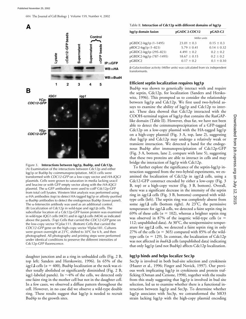
Bud4p was shown to genetically interact with and requirethe septin, Cdc12p, for localization (Sanders and Hersko-witz, 1996). This prompted us to consider the relationshipbetween Iqg1p and Cdc12p. We first used two-hybrid as-says to examine the ability of Iqg1p and Cdc12p to inter-act. These data showed that Cdc12p interacted with theCOOH-terminal region of Iqg1p that contains the RasGAP-like domain (Table II). However, thus far, we have not beenable to detect the coimmunoprecipitation of a GFP-taggedCdc12p on a low-copy plasmid with the HA-tagged Iqg1pon a high-copy plasmid (Fig. 3 A, top, lane 2), suggestingthat Iqg1p and Cdc12p may undergo a relatively weak ortransient interaction. We detected a band for the endoge-nous Bud4p after immunoprecipitation of Cdc12p-GFP(Fig. 3 A, bottom, lane 2, compare with lane 5), suggestingthat these two proteins are able to interact in cells and maybridge the interaction of Iqg1p with Cdc12p.
To further explore the significance of the septin-Iqg1p in-teraction suggested from the two-hybrid experiments, we ex-amined the localization of Cdc12p in
iqg1
�
cells, using aCdc12-GFP construct encoded by either a low-copy (Fig. 3B, top) or a high-copy vector (Fig. 3 B, bottom). Overall,there was a significant decrease in the intensity of the septinring in
iqg1
�
cells (Fig. 3 B, bottoms) compared with wild-type cells (left). The septin ring was completely absent fromsome
iqg1
�
cells (bottom right). At 23
�
C, the permissivetemperature for
iqg1
�
cells, we detected a faint septin ring in69% of these cells (
n
�
102), whereas a brighter septin ringwas observed in 87% of the isogenic wild-type cells (
n
�
112; unpublished data). At 30
�
C, the semipermissive temper-ature for
iqg1
�
cells, we detected a faint septin ring in only27% of the cells (
n
�
365) compared with 85% of the wild-type cells (
n
�
129). In contrast, the localization of Cdc12pwas not affected in
bud4
�
cells (unpublished data) indicatingthat only Iqg1p (and not Bud4p) affects Cdc12p localization.
Iqg1p binds and helps localize Sec3p
Sec3p is involved in both bud-site selection and cytokinesis(Haarer et al., 1996; Finger and Novick, 1997). Our previ-ous work implicating Iqg1p in cytokinesis and protein traf-ficking (Osman and Cerione, 1998), together with the resultsfrom this study suggesting that Iqg1p is involved in bud siteselection, led us to examine whether there is a functional in-teraction between Iqg1p and Sec3p. To determine whetherIqg1p associates with Sec3p, we cotransformed the MO3strain lacking Iqg1p with the high-copy plasmid encoding
Figure 3. Interactions between Iqg1p, Bud4p, and Cdc12p. (A) Examination of the interactions between Cdc12p and either Iqg1p or Bud4p by coimmunoprecipitation. MO3 cells were transformed with CDC12-GFP on a low-copy vector and HA-IQG1 plasmids. Cells were grown to saturation in media lacking uracil and leucine or with GFP empty vector along with the HA-IQG1 plasmid. The �-GFP antibodies were used to coIP Cdc12p-GFP from total cell lysates. Western blot analysis was performed using �-HA antibodies (top) to detect HA-tagged Iqg1p or affinity-purified �-Bud4p antibodies to detect the endogenous Bud4p (lower panel). The �-Intersectin antibody was used as an additional control. (B) Localization of Cdc12p in wild-type and iqg1� cells. The subcellular location of a Cdc12p-GFP fusion protein was examined in wild-type IQG1 cells (MO1) and in iqg1� cells (MO4) as indicated above the panels. (Top) Cells that carried the CDC12-GFP gene on the low-copy vector YCplac111. (Bottom) Cells that carried the CDC12-GFP gene on the high-copy vector YEplac181. Cultures were grown overnight at 23�C, shifted to 30�C for 6 h, and then photographed. All photography and printing steps were performed under identical conditions to preserve the different intensities of Cdc12p-GFP fluorescence.
Table II. Interaction of Cdc12p with different domains of Iqg1p
Iqg1p domain fusion pGADC 2-CDC12 pGAD-C2
Miller units
pGBDC2-Iqg1p (1–1495) 23.01 � 0.3 0.15 � 0.3pBDC2-Iqg1p (1–823) 5.79 � 0.41 0.14 � 0.32pGBDC2-Iqg1p (295–823) 4.491 � 0.2 0.2 � 0.2pGBDC2-Iqg1p (787–1495) 18.67 � 0.15 0.2 � 0.2pGBDC2 0.17 � 0.2 0.1 � 0.10
�-Galactosidase activity (Miller units) was calculated from six independenttransformants.
on July 12, 2015jcb.rupress.org
Dow
nloaded from
Published November 25, 2002
Iqg1p links polarity establishment with axial tags | Osman et al. 605
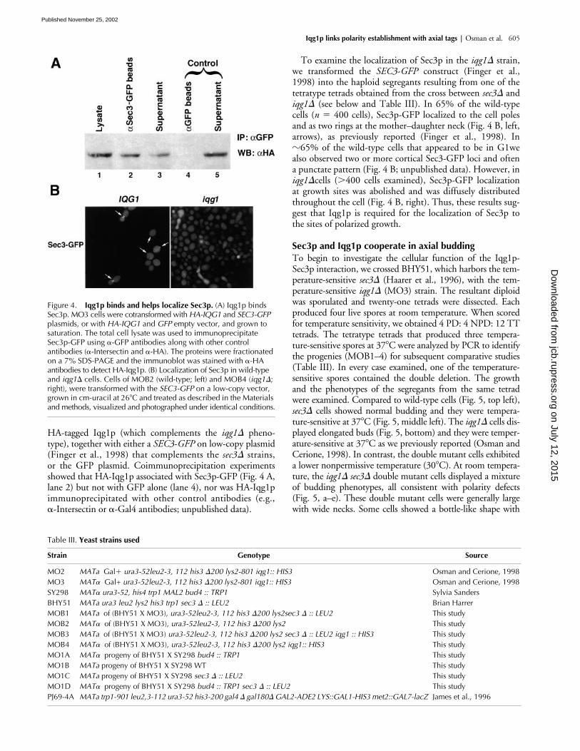
HA-tagged Iqg1p (which complements the iqg1� pheno-type), together with either a SEC3-GFP on low-copy plasmid(Finger et al., 1998) that complements the sec3� strains,or the GFP plasmid. Coimmunoprecipitation experimentsshowed that HA-Iqg1p associated with Sec3p-GFP (Fig. 4 A,lane 2) but not with GFP alone (lane 4), nor was HA-Iqg1pimmunoprecipitated with other control antibodies (e.g.,�-Intersectin or �-Gal4 antibodies; unpublished data).
To examine the localization of Sec3p in the iqg1� strain,we transformed the SEC3-GFP construct (Finger et al.,1998) into the haploid segregants resulting from one of thetetratype tetrads obtained from the cross between sec3� andiqg1� (see below and Table III). In 65% of the wild-typecells (n � 400 cells), Sec3p-GFP localized to the cell polesand as two rings at the mother–daughter neck (Fig. 4 B, left,arrows), as previously reported (Finger et al., 1998). In�65% of the wild-type cells that appeared to be in G1wealso observed two or more cortical Sec3-GFP loci and oftena punctate pattern (Fig. 4 B; unpublished data). However, iniqg1�cells (�400 cells examined), Sec3p-GFP localizationat growth sites was abolished and was diffusely distributedthroughout the cell (Fig. 4 B, right). Thus, these results sug-gest that Iqg1p is required for the localization of Sec3p tothe sites of polarized growth.
Sec3p and Iqg1p cooperate in axial buddingTo begin to investigate the cellular function of the Iqg1p-Sec3p interaction, we crossed BHY51, which harbors the tem-perature-sensitive sec3� (Haarer et al., 1996), with the tem-perature-sensitive iqg1� (MO3) strain. The resultant diploidwas sporulated and twenty-one tetrads were dissected. Eachproduced four live spores at room temperature. When scoredfor temperature sensitivity, we obtained 4 PD: 4 NPD: 12 TTtetrads. The tetratype tetrads that produced three tempera-ture-sensitive spores at 37�C were analyzed by PCR to identifythe progenies (MOB1–4) for subsequent comparative studies(Table III). In every case examined, one of the temperature-sensitive spores contained the double deletion. The growthand the phenotypes of the segregants from the same tetradwere examined. Compared to wild-type cells (Fig. 5, top left),sec3� cells showed normal budding and they were tempera-ture-sensitive at 37�C (Fig. 5, middle left). The iqg1� cells dis-played elongated buds (Fig. 5, bottom) and they were temper-ature-sensitive at 37�C as we previously reported (Osman andCerione, 1998). In contrast, the double mutant cells exhibiteda lower nonpermissive temperature (30�C). At room tempera-ture, the iqg1� sec3� double mutant cells displayed a mixtureof budding phenotypes, all consistent with polarity defects(Fig. 5, a–e). These double mutant cells were generally largewith wide necks. Some cells showed a bottle-like shape with
Figure 4. Iqg1p binds and helps localize Sec3p. (A) Iqg1p binds Sec3p. MO3 cells were cotransformed with HA-IQG1 and SEC3-GFP plasmids, or with HA-IQG1 and GFP empty vector, and grown to saturation. The total cell lysate was used to immunoprecipitate Sec3p-GFP using �-GFP antibodies along with other control antibodies (�-Intersectin and �-HA). The proteins were fractionated on a 7% SDS-PAGE and the immunoblot was stained with �-HA antibodies to detect HA-Iqg1p. (B) Localization of Sec3p in wild-type and iqg1� cells. Cells of MOB2 (wild-type; left) and MOB4 (iqg1�; right), were transformed with the SEC3-GFP on a low-copy vector, grown in cm-uracil at 26�C and treated as described in the Materials and methods, visualized and photographed under identical conditions.
Table III. Yeast strains used
Strain Genotype Source
MO2 MATa Gal ura3-52leu2-3, 112 his3 �200 lys2-801 iqg1:: HIS3 Osman and Cerione, 1998MO3 MAT� Gal+ ura3-52leu2-3, 112 his3 �200 lys2-801 iqg1:: HIS3 Osman and Cerione, 1998SY298 MAT� ura3-52, his4 trp1 MAL2 bud4 :: TRP1 Sylvia SandersBHY51 MATa ura3 leu2 lys2 his3 trp1 sec3 � :: LEU2 Brian HarrerMOB1 MATa of (BHY51 X MO3), ura3-52leu2-3, 112 his3 �200 lys2sec3 � :: LEU2 This studyMOB2 MAT� of (BHY51 X MO3), ura3-52leu2-3, 112 his3 �200 lys2 This studyMOB3 MATa of (BHY51 X MO3) ura3-52leu2-3, 112 his3 �200 lys2 sec3 � :: LEU2 iqg1 :: HIS3 This studyMOB4 MAT� of (BHY51 X MO3), ura3-52leu2-3, 112 his3 �200 lys2 iqg1:: HIS3 This studyMO1A MAT� progeny of BHY51 X SY298 bud4 :: TRP1 This studyMO1B MATa progeny of BHY51 X SY298 WT This studyMO1C MATa progeny of BHY51 X SY298 sec3 � :: LEU2 This studyMO1D MAT� progeny of BHY51 X SY298 bud4 :: TRP1 sec3 � :: LEU2 This studyPJ69-4A MATa trp1-901 leu2,3-112 ura3-52 his3-200 gal4 � gal180� GAL2-ADE2 LYS::GAL1-HIS3 met2::GAL7-lacZ James et al., 1996
on July 12, 2015jcb.rupress.org
Dow
nloaded from
Published November 25, 2002
606 The Journal of Cell Biology | Volume 159, Number 4, 2002
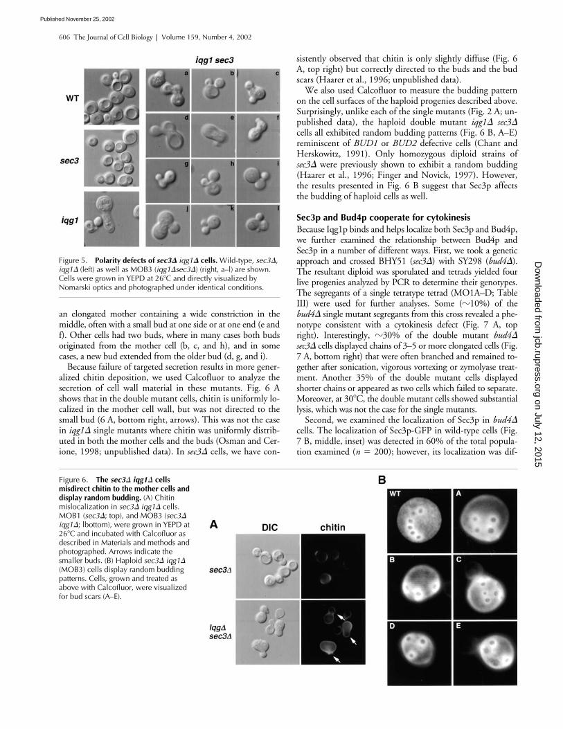
sistently observed that chitin is only slightly diffuse (Fig. 6A, top right) but correctly directed to the buds and the budscars (Haarer et al., 1996; unpublished data).
We also used Calcofluor to measure the budding patternon the cell surfaces of the haploid progenies described above.Surprisingly, unlike each of the single mutants (Fig. 2 A; un-published data), the haploid double mutant iqg1� sec3�cells all exhibited random budding patterns (Fig. 6 B, A–E)reminiscent of BUD1 or BUD2 defective cells (Chant andHerskowitz, 1991). Only homozygous diploid strains ofsec3� were previously shown to exhibit a random budding(Haarer et al., 1996; Finger and Novick, 1997). However,the results presented in Fig. 6 B suggest that Sec3p affectsthe budding of haploid cells as well.
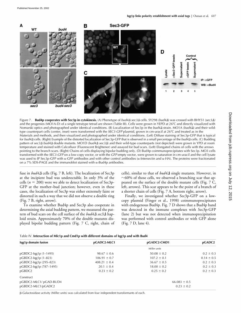
Sec3p and Bud4p cooperate for cytokinesisBecause Iqg1p binds and helps localize both Sec3p and Bud4p,we further examined the relationship between Bud4p andSec3p in a number of different ways. First, we took a geneticapproach and crossed BHY51 (sec3�) with SY298 (bud4�).The resultant diploid was sporulated and tetrads yielded fourlive progenies analyzed by PCR to determine their genotypes.The segregants of a single tetratype tetrad (MO1A–D; TableIII) were used for further analyses. Some (�10%) of thebud4� single mutant segregants from this cross revealed a phe-notype consistent with a cytokinesis defect (Fig. 7 A, topright). Interestingly, �30% of the double mutant bud4�sec3� cells displayed chains of 3–5 or more elongated cells (Fig.7 A, bottom right) that were often branched and remained to-gether after sonication, vigorous vortexing or zymolyase treat-ment. Another 35% of the double mutant cells displayedshorter chains or appeared as two cells which failed to separate.Moreover, at 30�C, the double mutant cells showed substantiallysis, which was not the case for the single mutants.
Second, we examined the localization of Sec3p in bud4�cells. The localization of Sec3p-GFP in wild-type cells (Fig.7 B, middle, inset) was detected in 60% of the total popula-tion examined (n � 200); however, its localization was dif-
Figure 6. The sec3� iqg1� cells misdirect chitin to the mother cells and display random budding. (A) Chitin mislocalization in sec3� iqg1� cells. MOB1 (sec3�; top), and MOB3 (sec3� iqg1�; lbottom), were grown in YEPD at 26�C and incubated with Calcofluor as described in Materials and methods and photographed. Arrows indicate the smaller buds. (B) Haploid sec3� iqg1� (MOB3) cells display random budding patterns. Cells, grown and treated as above with Calcofluor, were visualized for bud scars (A–E).
an elongated mother containing a wide constriction in themiddle, often with a small bud at one side or at one end (e andf). Other cells had two buds, where in many cases both budsoriginated from the mother cell (b, c, and h), and in somecases, a new bud extended from the older bud (d, g, and i).
Because failure of targeted secretion results in more gener-alized chitin deposition, we used Calcofluor to analyze thesecretion of cell wall material in these mutants. Fig. 6 Ashows that in the double mutant cells, chitin is uniformly lo-calized in the mother cell wall, but was not directed to thesmall bud (6 A, bottom right, arrows). This was not the casein iqg1� single mutants where chitin was uniformly distrib-uted in both the mother cells and the buds (Osman and Cer-ione, 1998; unpublished data). In sec3� cells, we have con-
Figure 5. Polarity defects of sec3� iqg1� cells. Wild-type, sec3�, iqg1� (left) as well as MOB3 (iqg1�sec3�) (right, a–l) are shown. Cells were grown in YEPD at 26�C and directly visualized by Nomarski optics and photographed under identical conditions.
on July 12, 2015jcb.rupress.org
Dow
nloaded from
Published November 25, 2002
Iqg1p links polarity establishment with axial tags | Osman et al. 607
fuse in bud4� cells (Fig. 7 B, left). The localization of Sec3pat the incipient bud was undetectable. In only 3% of thecells (n � 200) were we able to detect localization of Sec3p-GFP at the mother–bud junction; however, even in thesecases, the localization of Sec3p was either extremely faint ordistorted in such a way that we did not observe a double ring(Fig. 7 B, right, arrow).
To examine whether Bud4p and Sec3p also cooperate indetermining the axial budding pattern, we measured the pat-tern of bud scars on the cell surface of the bud4� sec3� hap-loid strain. Approximately 70% of the double mutants dis-played bipolar budding pattern (Fig. 7 C, right, chain of
cells), similar to that of bud4� single mutants. However, in�60% of those cells, we observed a branching scar that ap-peared on the surface of the double mutant cells (Fig. 7 C,left, arrows). This scar appears to be the point of a branch ofa shorter chain of cells (Fig. 7 A, bottom right, arrow).
Finally, we investigated whether Sec3p-GFP on a low-copy plasmid (Finger et al., 1998) coimmunoprecipitateswith endogenous Bud4p. Fig. 7 D shows that a Bud4p bandwas detected in the immune complexes with Sec3p-GFP(lane 2) but was not detected when immunoprecipitationwas performed with control antibodies or with GFP alone(Fig. 7 D, lane 4).
Figure 7. Bud4p cooperates with Sec3p in cytokinesis. (A) Phenotype of bud4� sec3� cells. SY298 (bud4�) was crossed with BHY51 (sec3�) and the progenies (MO1A-D) of a single tetratype tetrad are shown (Table III). Cells were grown in YEPD at 26�C and directly visualized with Nomarski optics and photographed under identical conditions. (B) Localization of Sec3p in the bud4� strain. MO1A (bud4�) and their wild-type counterpart cells (center, inset) were transformed with the SEC3-GFP plasmid, grown in cm-uracil at 26�C and treated as in the Materials and methods, and then visualized and photographed under identical conditions. (Left) Diffuse staining of Sec3p-GFP that is typical for bud4� cells. (Right) Example of the distorted localization of Sec3p-GFP that is observed in a small percentage of the bud4� cells. (C) Budding pattern of sec3� bud4� double mutants. MO1D (bud4� sec3�) and their wild-type counterparts (not depicted) were grown in YPD at room temperature and stained with Calcofluor (Fluorescent Brightener) and assayed for bud scars. (Left) Elongated chains of cells with the arrows pointing to the branch scars. (Right) Chains of cells displaying bipolar budding only. (D) Bud4p coimmunoprecipitates with Sec3p. MO3 cells transformed with the SEC3-GFP on a low-copy vector, or with the GFP empty vector, were grown to saturation in cm-uracil and the cell lysate was used to IP Sec3p-GFP with �-GFP antibodies and with other control antibodies (�-Intersectin and �-HA). The proteins were fractionated on a 7% SDS-PAGE and the immunoblot stained with �-Bud4p antibodies.
Table IV. Interaction of Mlc1p and Cmd1p with different domains of Iqg1p and with Bud4
Iqg1p domain fusion pGADC2-MLC1 pGADC2-CMD1 pGADC2
Miller units
pGBDC2-Iqg1p (1–1495) 98.67 � 0.6 50.08 � 0.2 0.2 � 0.3pGBDC2-Iqg1p (1–823) 106.91 � 0.7 107.2 � 0.1 0.14 � 0.5pGBDC2-Iqg1p (295–823) 408.21 � 0.4 36.67 � 0.5 0.2 � 0.3pGBDC2-Iqg1p (787–1495) 20.1 � 0.4 18.00 � 0.2 0.2 � 0.3pGBDC2 0.23 � 0.2 0.25 � 0.2 0.2 � 0.3
ConstructpGBDC2-MLC1/ pGAD-BUD4 66.083 � 0.5pGBDC2-MLC1/pGADC2 0.23 � 0.2
�-Galactosidase activity (Miller units) was calculated from four independent transformants of each.
on July 12, 2015jcb.rupress.org
Dow
nloaded from
Published November 25, 2002
608 The Journal of Cell Biology | Volume 159, Number 4, 2002
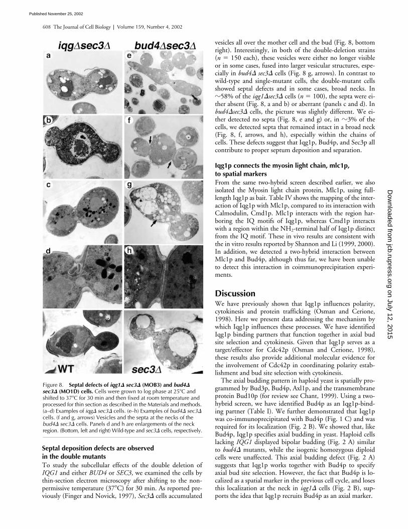
Septal deposition defects are observed in the double mutantsTo study the subcellular effects of the double deletion ofIQG1 and either BUD4 or SEC3, we examined the cells bythin-section electron microscopy after shifting to the non-permissive temperature (37�C) for 30 min. As reported pre-viously (Finger and Novick, 1997), Sec3� cells accumulated
vesicles all over the mother cell and the bud (Fig. 8, bottomright). Interestingly, in both of the double-deletion strains(n � 150 each), these vesicles were either no longer visibleor in some cases, fused into larger vesicular structures, espe-cially in bud4� sec3� cells (Fig. 8 g, arrows). In contrast towild-type and single-mutant cells, the double-mutant cellsshowed septal defects and in some cases, broad necks. In�58% of the iqg1�sec3� cells (n � 100), the septa were ei-ther absent (Fig. 8, a and b) or aberrant (panels c and d). Inbud4�sec3� cells, the picture was slightly different. We ei-ther detected no septa (Fig. 8, e and g) or, in �3% of thecells, we detected septa that remained intact in a broad neck(Fig. 8, f, arrows, and h), especially within the chains ofcells. These defects suggest that Iqg1p, Bud4p, and Sec3p allcontribute to proper septum deposition and separation.
Iqg1p connects the myosin light chain, mlc1p, to spatial markersFrom the same two-hybrid screen described earlier, we alsoisolated the Myosin light chain protein, Mlc1p, using full-length Iqg1p as bait. Table IV shows the mapping of the inter-action of Iqg1p with Mlc1p, compared to its interaction withCalmodulin, Cmd1p. Mlc1p interacts with the region har-boring the IQ motifs of Iqg1p, whereas Cmd1p interactswith a region within the NH2-terminal half of Iqg1p distinctfrom the IQ motif. These in vivo results are consistent withthe in vitro results reported by Shannon and Li (1999, 2000).In addition, we detected a two-hybrid interaction betweenMlc1p and Bud4p, although thus far, we have been unableto detect this interaction in coimmunoprecipitation experi-ments.
DiscussionWe have previously shown that Iqg1p influences polarity,cytokinesis and protein trafficking (Osman and Cerione,1998). Here we present data addressing the mechanism bywhich Iqg1p influences these processes. We have identifiedIqg1p binding partners that function together in axial budsite selection and cytokinesis. Given that Iqg1p serves as atarget/effector for Cdc42p (Osman and Cerione, 1998),these results also provide additional molecular evidence forthe involvement of Cdc42p in coordinating polarity estab-lishment and bud site selection with cytokinesis.
The axial budding pattern in haploid yeast is spatially pro-grammed by Bud3p, Bud4p, Axl1p, and the transmembraneprotein Bud10p (for review see Chant, 1999). Using a two-hybrid screen, we have identified Bud4p as an Iqg1p-bind-ing partner (Table I). We further demonstrated that Iqg1pwas co-immunoprecipitated with Bud4p (Fig. 1 C) and wasrequired for its localization (Fig. 2 B). We showed that, likeBud4p, Iqg1p specifies axial budding in yeast. Haploid cellslacking IQG1 displayed bipolar budding (Fig. 2 A) similarto bud4� mutants, while the isogenic homozygous diploidcells were unaffected. This axial budding defect (Fig. 2 A)suggests that Iqg1p works together with Bud4p to specifyaxial bud site selection. However, the fact that Bud4p is lo-calized as a spatial marker in the previous cell cycle, and losesthis localization at the neck in iqg1� cells (Fig. 2 B), sup-ports the idea that Iqg1p recruits Bud4p as an axial marker.
Figure 8. Septal defects of iqg1� sec3� (MOB3) and bud4� sec3� (MO1D) cells. Cells were grown to log phase at 25�C and shifted to 37�C for 30 min and then fixed at room temperature and processed for thin section as described in the Materials and methods. (a–d) Examples of iqg� sec3� cells. (e–h) Examples of bud4� sec3� cells. (f and g, arrows) Vesicles and the septa at the necks of the bud4� sec3� cells. Panels d and h are enlargements of the neck region. (Bottom, left and right) Wild-type and sec3� cells, respectively.
on July 12, 2015jcb.rupress.org
Dow
nloaded from
Published November 25, 2002
Iqg1p links polarity establishment with axial tags | Osman et al. 609
Iqg1p also appears to interact with and helps localize theseptin Cdc12p (Table II; Fig. 3 B), which is necessary forboth cytokinesis and axial budding (for review see Chant,1996, 1999; Madden and Snyder, 1998). Bud4p may medi-ate the interaction between Iqg1p and Cdc12p, as we havedetected the coimmunoprecipitation of Cdc12p and Bud4p,but have not yet been able to detect an interaction betweenCdc12p and Iqg1p in coimmunoprecipitation experiments(Fig. 3 A). However, we have found that Iqg1p, and notBud4p, affects the efficient localization of the septin Cdc12pto the neck (Fig. 3 B; unpublished data), further support-ing the idea that Iqg1p assembles the proteins required foraxial budding. In addition, the interaction between Iqg1p,Bud4p, and Cdc12p may involve a cooperation in some as-pect of cytokinesis, as both Iqg1p and septins have been pre-viously implicated in this process.
Several lines of evidence presented in this study indicatethat Iqg1p and Sec3p work together to influence cell polar-ity, axial budding and cytokinesis in yeast. We have pro-vided evidence that Iqg1p coimmunoprecipitates with (Fig.4 A) and is required for the localization of Sec3p (Fig. 4 B).The iqg1�sec3� double mutants displayed a lower restrictivetemperature and exhibited phenotypes consistent with a po-larity defect (Fig. 5) that resulted in random budding (Fig. 6B). These double mutants also failed to direct growth mate-rial to the new bud (Fig. 6 A) and to form correct septa atthe mother-daughter junctions (Fig. 8).
Together, these results suggest a role for Sec3p in axialbudding. Although earlier studies have suggested that Sec3pis primarily involved in diploid budding (Haarer et al.,1996; Finger and Novick, 1997), our findings are consistentwith the idea that targeted secretion is required for buddingin both cell types (for review see Finger and Novick, 1998).Our data also suggest that Iqg1p works with Sec3p to main-tain cell polarity. Figs. 5 and 6 B show that budding in theiqg1� sec3� double mutant strain was initiated normally butwas not maintained such that another bud originated eitherfrom the mother, the bud (Fig. 5) and/or at a random loca-tion (Fig. 6 B). This may reflect the actions of polarity estab-
lishment proteins such as Cdc42p and Bud1p. Iqg1p maymediate the interplay between the bud site selection func-tion of the Bud1 GTPase and the maintenance of polarityby Cdc42p (Fig. 9). Three independent pieces of evidenceseem to support this view. First, the random budding phe-notype of the iqg1� sec3� double mutant cells (Fig. 6 B) issimilar to that of bud1� or bud2� mutant cells (Chant andHerskowitz, 1991). Second, analyses of bud1� cdc24� cellsrevealed that the essential role of the polarity establishmentmolecules in morphogenesis is to stabilize the axis of polar-ity, and that in their absence, this axis wanders (Nern andArkowitz, 2000). Third, a recent study demonstrated directbinding between Sec3p and Cdc42p (Zhang et al., 2001)and showed that Sec3p was mislocalized in cells expressingcertain mutations of Cdc42p. Together, these data supportthe idea that the Cdc42p target Iqg1p coordinates the posi-tional signal for budding and secretion with the signal tomaintain polarity and promote cytokinesis.
There also appears to be a functional interplay betweenBud4p and Sec3p in budding and cytokinesis. Bud4p can becoimmunoprecipitated with Sec3p (Fig. 7 D) and the properlocalization of Sec3p is nearly abolished in the absence ofBud4p (Fig. 7 B). Furthermore, the bud4�sec3� double mu-tants displayed a lower nonpermissive temperature and ex-hibited budding defects (Fig. 7 C). In addition, the pheno-type of bud4�sec3� cells highlights a possible role for Bud4pin cytokinesis (Fig. 7 A), apparently, in the final stages of cellseparation (Fig. 8). Iqg1, Bud4p and Sec3p may work to-gether to ensure proper septum formation or deposition,thus explaining the wide and aberrant neck formation re-ported in some sec3� cells (Finger and Novick, 1997), aswell as that observed in sec3� bud4� cells, or the absence ofsepta in iqg1� sec3� cells (Fig. 8). Overall, our findings ap-pear to be consistent with the “cytokinesis tag” model for ax-ial budding. This model predicts that components involvedin cytokinesis are also involved in selecting the future bud-site. The subsequent activation of the Bud1 GTPase then di-rects the assembly and the initiation of the new bud at thatsite (for review see Madden and Snyder, 1998).
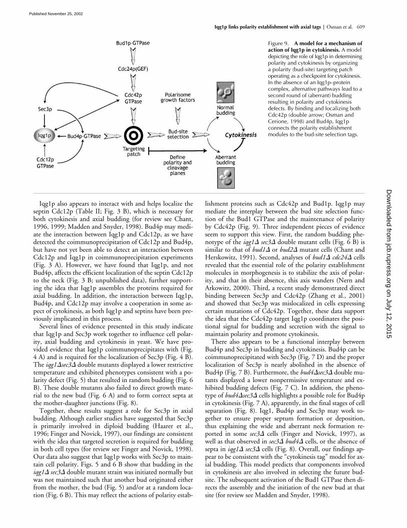
Figure 9. A model for a mechanism of action of Iqg1p in cytokinesis. A model depicting the role of Iqg1p in determining polarity and cytokinesis by organizing a polarity (bud-site) targeting patch operating as a checkpoint for cytokinesis. In the absence of an Iqg1p–protein complex, alternative pathways lead to a second round of (aberrant) budding resulting in polarity and cytokinesis defects. By binding and localizing both Cdc42p (double arrow; Osman and Cerione, 1998) and Bud4p, Iqg1p connects the polarity establishment modules to the bud-site selection tags.
on July 12, 2015jcb.rupress.org
Dow
nloaded from
Published November 25, 2002
610 The Journal of Cell Biology | Volume 159, Number 4, 2002
In conclusion, we propose that Iqg1p forms a targetingpatch that includes Bud4p, Cdc12p, and Sec3p (Fig. 9). Thisprotein complex may play a primary role in cytokinesis by firstdetermining, and then maintaining, the axis of polarity bychoosing a bud site. The complex would recruit the exocystfor targeted secretion and other morphogenetic factors such asMlc1p and Cmd1p (Table IV) to promote bud growth andeventually lead to septum deposition and separation. In thisway, the Iqg1 complex would in effect serve as a checkpointfor cytokinesis by preventing another round of budding untilcytokinesis is complete (Fig. 9). In the absence of the Iqg1p-targeting complex, alternative pathways for budding can ap-parently bypass the checkpoint leading to rounds of buddingand chains of cells observed in the double mutants.
Materials and methodsIsolation of Bud4p as an Iqg1p binding partnerThe PCR-amplified IQG1 gene was cloned in frame into pGBD-C2 to pro-duce the Gal4 binding domain Iqg1p fusion protein. This plasmid wasused to screen three yeast genomic libraries, YL2H-C1-3, using the yeaststrain PJ694A (James et al., 1996). The interacting library plasmid was sub-sequently transformed into Escherichia coli, purified, and sequenced toidentify Iqg1p binding partners. The Iqg1p domain deletions shown on Fig.1 B were generated by PCR and cloned into pGBD-C2 to further delineatethe binding domain for Bud4p, Cdc12p, Mlc1p, and Cmd1p.
Coimmunoprecipitation experimentsFor coimmunoprecipitation experiments, tagged plasmids were cotrans-formed into a strain lacking the chromosomal copy of IQG1. To examinethe coimmunoprecipitation of Iqg1p and Bud4p, cells were grown in syn-thetic complete media (cm) lacking tryptophan and leucine after transfor-mation with HA-tagged Iqg1p on a high copy plasmid (pA1; Osman andCerione, 1998) and Gal4-IBID (amino acids 769–880 from Bud4p, Fig. 1 A).Cells harboring HA-Iqg1p (pA1) alone were grown in cm-leucine. For con-trol experiments, cells were transformed with parent plasmids lacking therelevant gene. When examining the interaction between Iqg1p and Cdc12,MO3 cells were transformed with CDC12-GFP on a low copy plasmid (de-scribed below) and HA-IQG1, and grown on cm-uracil and leucine. Whenexamining the coimmunoprecipitation of Iqg1p and Sec3p, MO3 cellstransformed with SEC3-GFP on a low-copy plasmid (Finger et al., 1998) andHA-IQG1 were grown in cm media lacking uracil and leucine. In all cases,the cell cultures were grown to saturation and the cell pellets washed withand suspended in cold IP-buffer (50 mM Tris, pH 7.5, 1 mM EDTA, 5%glycerol, 0.1% Triton, 1 mM PMSF and fungal protease inhibitor cocktail[Sigma-Aldrich]). Acid-washed glass beads (Sigma-Aldrich) were added tobreak the cells by vortexing and the cell lysates were collected after centrif-ugation at 20,000 rpm at 4�C. Two mL of the total cell lysate (scaled up forSec3p-Iqg1p experiments) was incubated with antibodies (a control anti-body was also included) for 3 h with gentle rocking at 4�C. 4 mg of IPbuffer-washed protein A Sepharose (Sigma-Aldrich) was added to the im-mune complexes and incubated overnight at 4�C with gentle rocking. Thereaction mix was centrifuged at 10,000 rpm for 30 s and the bead portionwas washed four times with ice-cold IP buffer, suspended in 30 L SDS-loading buffer, boiled at 100�C and separated by 7-10% SDS-PAGE. West-ern blot analyses were performed using standard methods.
Fluorescence microscopyTo visualize chitin deposition and bud scars on the yeast cell wall, cellswere collected at log phase and stained with Calcofluor (FluorescentBrightener; Sigma-Aldrich) as described (Pringle, 1991). Cells with three ormore bud scars were scored for budding pattern. For Cdc12p localization,a CDC12-GFP fusion gene was constructed by using PCR to fuse an en-hanced version of the GFP (Cormack et al., 1997) to the carboxyl terminusof Cdc12p. The CDC12-GFP gene was cloned into the low-copy vectorYCplac111 and also into the high-copy vector YEplac181 (Gietz and Su-gino, 1988). Wild-type (MO5) and iqg1� cells (MO2) carrying the indicatedCDC12-GFP plasmid, or a control vector, were grown overnight at 23�Cand then adjusted to 106 cells/mL and shifted to 30�C for 6 h. The cellswere then examined using an Olympus BH2 fluorescence microscope,photographed with Kodak TMAX400 film, and then the negatives were
scanned into digital images under identical conditions. By comparing dif-ferent exposures, we found that it requires at least a fourfold longer expo-sure to see similar neck staining in MO2 than in MO5. CDC12-GFP fluo-rescence was better at 23�C in the MO2 strain, but was still weaker thanthe wild-type strain.
Strains resulting from single tetratype tetrads of the cross between eitherSY298 (bud4�) or MO3 (iqg1�) with sec3� were transformed with a SEC3-GFP plasmid (Finger et al., 1998) which encodes GFP fused to the COOHterminus of Sec3p. Log phase cells growing in cm-uracil liquid media wereprocessed as described in (Finger et al., 1998) and visualized under 100�using a Zeiss fluorescence microscope. Images were collected using Axio-vision software under identical conditions.
Thin-section electron microscopyLog phase overnight cultures of wild-type (MOB2 or MO1B), sec3�(MOB1), iqg1� (MOB4), bud4� (MO1A), iqg�sec3� (MOB3), andbud4�sec3� (MO1D) strains growing at room temperature were adjustedto 0.3 A600nm units/mL in YEPD and incubated at 37�C for 30 min. Cul-tures were directly fixed in 0.1 M cacodylate containing 2.5% glutaralde-hyde and 2.5% paraformaldehyde for 2 h. Cells were treated with 0.2 mg/mL zymolyase 100T in 0.1 M KPi, pH 7.5. The cell pellets were incubatedwith ice-cold OsO4 in 0.1 M cacodylate for 1 h, washed twice with waterand incubated for 1 h in 1.5 mL of filtered 2% uranyl acetate at room tem-perature. A series of ethanol concentrations (50, 70, 90, and 100%) wereused to dehydrate the cells followed by acetone treatment. The cells werethen incubated for a few hours in 50% acetone/50% SPURR (Electron Mi-croscopy Sciences), changed to 100% SPURR, and incubated overnight atroom temperature. After changing to SPURR 2�, cells were baked at 80�Cfor 24 h. This and the rest of the sectioning and processing were carriedout at the Cornell Integrated Microscopy Center (Ithaca, NY).
We thank Drs. Sylvia Sanders for Bud4 strains and antibody, Brian Haarerfor strains and plasmids, and Peter Novick for the SEC3-GFP construct. Wealso thank Drs. Juan Lucas and Sigrid Holmgren for helping with tetrad dis-section, Dr. David Holowka for critical reading of the manuscript andhelpful comments, and Drs. David Pruyne for helpful discussion, and Jian-bin Wang for the �-Intersectin antibody used for controls. Thanks to CindyWestmiller for expert technical assistance. This work was supported byNIH grant RO1 GM47458. MAO was supported by an NIH Training GrantT32 CA09682.
Submitted: 16 May 2002Revised: 16 October 2002Accepted: 21 October 2002
ReferencesBashour, A.-M., A.T. Fullerton, M.J, Hart, and G.S. Bloom. 1997. IQGAP1, a
Rac- and Cdc42-binding protein, directly binds and cross links microfila-ments. J. Cell Biol. 137:1555–1566.
Bi, E., P. Maddox, D.J. Lew, E.D. Salmon, J.N. McMillan, E. Yeh, and J.R. Prin-gle. 1998. Involvement of an actomyosin ring in Saccharomyces cerevisiae. J.Cell Biol. 142:1301–1312.
Bowser, R., H. Muller, B. Govidan, and P. Novick. 1992. Sec8p and Sec15p arecomponents of a plasma membrane-associated 19.5 S particle that may func-tion downstream of Sec4p to control exocytosis. J. Cell Biol. 118:1041–1056.
Chant, J. 1996. Septin scaffolds and cleavage planes in Saccharomyces. Cell. 84:187–190.
Chant, J. 1999. Cell Polarity in yeast. Annu. Rev. Cell Dev. Biol. 15:365–391.Chant, J., and I. Herskowitz. 1991. Genetic control of bud site selection in yeast by
a set of gene products that comprise a morphogenetic pathway. Cell. 65:1203–1212.
Chant, J., and J.R. Pringle. 1995. Patterns of bud-site selection in the yeast Saccha-romyces cerevisiae. J. Cell Biol. 129:751–765.
Cormack, B.P., G. Bertram, M. Egerton, N.A. Gow, S. Falkow, and A.J. Brown.1997. Yeast-enhanced green fluorescent protein (yEGFP) a reporter of geneexpression in Candida albicans. Microbiology. 143:303–311.
Epp, A.J., and J. Chant. 1997. An IQGAP-related protein controls actin-ring for-mation and cytokinesis in yeast. Curr. Biol. 7:921–929.
Erickson, J.W., R.A. Cerione, and M.J. Hart. 1997. Identification of an actin cy-toskeleton complex that includes IQGAP and the Cdc42 GTPase. J. Biol.Chem. 272:24443–24447.
Finger, F., and P. Novick. 1997. Sec3 is involved in secretion and morphogenesisin. Saccharomyces cerevisiae. Mol. Biol. Cell. 8:647–662.
on July 12, 2015jcb.rupress.org
Dow
nloaded from
Published November 25, 2002
Iqg1p links polarity establishment with axial tags | Osman et al. 611
Finger, F., and P. Novick. 1998. Spatial regulation of exocyst: lessons from yeast. J.Cell Biol. 142:609–612.
Finger, F.P., T.E. Hughes, and P. Novick. 1998. Sec3p Is a spatial landmark forpolarized secretion in budding yeast. Cell. 92:559–571.
Gietz, R.D., and A. Sugino. 1988. New yeast-Escherichia coli shuttle vectors con-structed with in vitro mutagenized yeast genes lacking six-base pair restric-tion sites. Gene. 30:527–534.
Guo, W., F. Tamanoi, and P. Novick. 2001. Spatial regulation of the exocyst com-plex by Rho1 GTPase. Nat. Cell Biol. 3:353–360.
Haarer, B.K., A. Corbett, Y. Kweon, A.S. Petzold, P. Silver, and S.S. Brown. 1996.SEC3 Mutations are synthetically lethal with profilin mutations and causedefects in diploid-specific bud selection. Genetics. 144:495–510.
Hart, M.J., M.G. Callow, B. Souza, and P. Polakis. 1996. IQGAP1, a calmodulin-binding protein with a RasGAP-related domain, is a potential effector forCdc42Hs. EMBO J. 15:2997–3005.
James, P., J. Halladay, and E.A. Craig. 1996. Genomic libraries and a host straindesigned for highly efficient two-hybrid selection in yeast. Genetics. 144:1425–1436.
Johnson, D.I., and J.R. Pringle. 1990. Molecular characterization of CDC42, aSaccharomyces cerevisiae gene involved in the development of cell polarity. J.Cell Biol. 111:143–152.
Kuroda, S., M. Fukata, K. Kobayashi, M. Nakafuku, N. Nomura, A. Iwamatsu,and K. Kaibuchi. 1996. Identification of IQGAP as a putative target for thesmall GTPase, Cdc42 and Rac1. J. Biol. Chem. 271:23363–23367.
Lew, D.J., and S.I. Reed. 1995. Cell cycle control of morphogenesis in buddingyeast. Curr. Opin. Genet. Dev. 5:17–23.
Lippincott, J., and R. Li. 1998. Sequential assembly of myosin II, an IQGAP-likeprotein, and filamentous actin to a ring structure involved in budding yeastcytokinesis. J. Cell Biol. 140:355–366.
Madden, K., and M. Snyder. 1998. Cell polarity and morphogenesis in buddingyeast. Annu. Rev. Microbiol. 52:687–744.
McCallum, S.J., W.J. Wu, and R.A. Cerione. 1996. Identification of a putative ef-fector for Cdc42Hs with high sequence similarity to the RasGAP-relatedprotein IQGAP1 and a Cdc42Hs binding partner IQGAP2. J. Biol. Chem.271:21732–21737.
McCallum, S.J., J.W. Erickson, and R.A. Cerione. 1998. Characterization of theAssociation of the actin-binding protein IQGAP, and activated Cdc42 withGolgi membrane. J. Biol. Chem. 273:22537–22544.
Nern, A., and R.A. Arkowitz. 2000. G Proteins mediate changes in cell shape bystabilizing the axis of polarity. Mol. Cell. 5:853–864.
Osman, M., and R.A. Cerione. 1998. Iqg1p, a yeast homologue of the mammalianIQGAPs, mediates Cdc42p effects on the actin cytoskeleton. J. Cell Biol.142:443–455.
Pringle, J.R. 1991. Staining of bud scars and mother cell wall chitin with Calco-fluor. Methods Enzymol. 194:732–735.
Sanders, S.L., and C. Fields. 1995. Bud-site selection is only skin deep. Curr. Biol.5:1213–1215.
Sanders, S.L., and I. Herskowitz. 1996. The BUD4 protein of yeast, required foraxial budding, is localized to the mother/BUD neck in a cell cycle-depen-dent manner. J. Cell Biol. 134:413–427.
Shannon, K.B., and R. Li. 1999. The multiple roles of Cyk1p in the assembly andfunction of the actomyosin ring in budding yeast. Mol. Biol. Cell. 10:283–296.
Shannon, K.B., and R.A. Li. 2000. Myosin light chain mediates the localization ofthe budding yeast IQGAP-like protein during contractile ring formation.Curr. Biol. 10:727–730.
TerBush, D.R., and P. Novick. 1995. Sec6, Sec8, and Sec15 are components of amultisubunit complex which localizes to small bud tips in Saccharomycescerevisiae. J. Cell Biol. 130:299–312.
Zhang, X., E. Bi, P. Novick, L. Du, K.G. Kozminski, J.H. Lipschutz, and W. Guo.2001. Cdc42 interacts with the exocyst and regulates polarized secretion. J.Biol. Chem. 276:46745–46750.
on July 12, 2015jcb.rupress.org
Dow
nloaded from
Published November 25, 2002