Vibrational Spectra and Electronic Structural Studies of Some Coumarins
Upload
independentCategory
view
0download
0
Polarity-Sensitive Coumarins Tailored to Live Cell Imaging
Giovanni Signore,*,† Riccardo Nifosı,‡ Lorenzo Albertazzi,†,‡ Barbara Storti,†,‡ andRanieri Bizzarri*,†,‡
NEST, CNR-INFM, p.za San SilVestro 12, 56127 Pisa, Italy, NEST, Scuola Normale Superiore, andIIT@NEST, Center for Nanotechnology InnoVation, p.za San SilVestro 12, I-56127 Pisa, Italy
Received June 19, 2009; E-mail: [email protected]; [email protected]
Abstract: Polarity-dependent fluorescent probes are recently attracting interest for high-resolution cellimaging. Following a stepwise rational approach, we prepared and tested a toolbox of new coumarinderivatives tailored to in vivo imaging applications. Our compounds are characterized by a donor-(coumarincore)-acceptor molecular structure, where the electron donor is represented by alkylether or naphthylgroups, and the electron acceptor is represented by benzothiazene and cyano groups. Prior to synthesis,the substitution patterns were screened by computational methods to provide functional fluorescentderivatives easy to synthesize, and with excitation in the visible region of spectrum. We set up a robustsynthetic procedure tunable on the substitution patterns to achieve. These coumarins possess excellentfluorescence quantum yields (up to 0.95), high molar extinction coefficients (up to 46,000 M-1 cm-1), andlarge Stokes shifts. Furthermore, they display strong solvatochromism, being almost non-emissive in waterand very fluorescent in less polar media (up to 780-fold enhancement in brightness). The solvatochromismof these compounds can be accounted for by a photophysical method encompassing two communicatingexcited states. When tested on cultured cells, the results showed that the developed coumarins were notharmful and their photophysical properties were unchanged compared to free solution. According to thedetermined solvatochromic properties, the coumarin fluorescence was detected only in the most lipophilicenvironments of the cell. The prepared compounds represent remarkable tools to investigate subtlebiochemical processes in the cell environment after appropriate conjugation to biomolecules, and at thesame time constitute the basis for further engineering of a new generation of biosensors.
1. Introduction
Cell behavior is regulated by transient activation of proteinactivities in specific subcellular regions. The recent developmentof highly fluorescent probes tailored to the cellular environmentand of high-resolution imaging techniques allows monitoringprotein activity in living cells by direct optical methods.1
Members of the green fluorescent protein family (GFPs) areparticularly impressive nanoprobes for in ViVo studies, owingto their genetically encoded fluorescence.2,3 Effective chemicalmethods were developed to link functional organic dyes, oftenmore fluorescent and resistant to photodegradation than GFPs,to proteins in ViVo.4 In most cases, however, genetic modificationof the target protein is needed for the probe binding; the leastinvasive methods involve adding non-native peptide sequencesof 6-20 amino acids to the target protein. Thus, the detectionof protein activity in the true native state or after post-translational modification is intrinsically impossible. The useof multicomponent molecular species penetrating into the celland capable of recognizing selectively a certain state of the targetprotein (universal biosensors) could overcome this problem.Peptide sequences able to convey into the cell a tailored
molecular cargo and/or to bind to a specific protein configurationare now widely investigated for this purpose.5 How to signalthe protein recognition event, however, is still unclear. Anelegant answer to this question considers organic dyes whoseoptical properties are sensitiVe to the surrounding environment(solvatochromic probes).6-19 According to this approach, the
† IIT@NEST, Center for Nanotechnology Innovation, I-56127 Pisa, Italy.‡ NEST, Scuola Normale Superiore and CNR-INFM, I-56127 Pisa, Italy.
(1) Petty, H. R. Microsc. Res. Tech. 2007, 70, 687–709.(2) Miyawaki, A. Neuron 2005, 48, 189–199.(3) Bizzarri, R.; Arcangeli, C.; Arosio, D.; Ricci, F.; Faraci, P.; Cardarelli,
F.; Beltram, F. Biophys. J. 2006, 90, 3300–3314.(4) Prescher, J. A.; Bertozzi, C. R. Nat. Chem. Biol. 2005, 1, 13–21.
(5) Cardarelli, F.; Serresi, M.; Bizzarri, R.; Beltram, F. Traffic 2008, 9,528–539.
(6) Toutchkine, A.; Kraynov, V.; Hahn, K. M. J. Am. Chem. Soc. 2003,125, 4132–4145.
(7) Nalbant, P.; Hodgson, L.; Kraynov, V.; Toutchkine, A.; Hahn, K. M.Science 2004, 305, 1615–1619.
(8) Cohen, B. E.; Pralle, A.; Yao, X.; Swaminath, G.; Gandhi, C. S.; Jan,Y. N.; Kobilka, B. K.; Isacoff, E. Y.; Jan, L. Y. Proc. Natl. Acad.Sci. U.S.A. 2005, 102, 965–970.
(9) Kobiilka, B. K.; Gether, U. Methods Enzymol. 2002, 343, 170–182.(10) Mannuzzu, L. M.; Moronne, M. M.; Isacoff, E. Y. Science (Wash-
ington, DC, U.S.) 1996, 271, 213–216.(11) Hanouni, P.; Gryczynski, Z.; Steenhuis, J.; Lee, T. W.; Farrens, D. L.;
Lakowicz, J. R.; Kobilka, B. K. J. Biol. Chem. 2001, 276, 24433–24436.
(12) Cohen, B. E.; McAnaney, T. B.; Park, E. S.; Jan, Y. N.; Boxer, S. G.;Jan, L. Y. Science 2002, 296, 1700–1703.
(13) Lavis, L. D.; Raines, R. T. ACS Chem. Biol. 2008, 3, 142–155.(14) Venkatraman, P.; Nguyen, T. T.; Sainlos, M.; Bilsel, O.; Chitta, S.;
Imperiali, B.; Stern, L. Nat. Chem. Biol. 2007, 3, 222–228.(15) Fulop, A.; Arian, D.; Lysenko, A.; Mokhir, A. Bioorg. Med. Chem.
Lett. 2009, 19, 3104–3107.(16) Sainlos, M.; Iskenderian, W. S.; Imperiali, B. J. Am. Chem. Soc. 2009,
131, 6680–6682.(17) Dai, Z.; Dulyaninova, N. G.; Kumar, S.; Bresnick, A. R.; Lawrence,
D. S. Chem. Biol. 2007, 1254–1260.
Published on Web 01/05/2010
10.1021/ja9050444 2010 American Chemical Society1276 9 J. AM. CHEM. SOC. 2010, 132, 1276–1288

recognition signal comes from a solvatochromic prosthetic groupable to sense the polarity change associated with binding.Reasonably, it should be located at a site close enough to thebinding region of the biosensor (where changes in polaritynecessarily occur) but not too close, so that it does not hamperthe recognition and binding process.
In past years, solvatochromic dyes have been extensivelyemployed in selective staining of many subcellular domains.Notably, solvatochromic FM dyes are widely employed asmembrane-staining agents, since they localize preferentially oncell membrane, where their fluorescence is enhanced.20 How-ever, the development of solvatochromic dyes as polarity probesto investigate protein binding, membrane rearrangement, andother biological processes has not evolved to its full potential.
Fluorescent coumarins are attractive fluorophores, as they arecharacterized by high quantum yields (up to 0.90),21 highextinction coefficients (10,000-40,000),22,23 large Stokes shifts(up to 160 nm),24 and can be engineered to respond to theirenvironment polarity (solvatochromic probes).25-27 These prop-erties promoted their use as dyes,28,21,29 sensors for metalcations,30-34 and dopants for OLEDs.35,36 The well-knowncommercial fluorophores Alexa 350 and Alexa 430 are basedon a coumarin core.37 Recently, coumarins were thoroughlyinvestigated in view of synthesizing new probes for biologicalapplications, in particular for the imaging of living cells.38-48
In spite of their relevance as biosensors, however, no studies
describing solvatochromic coumarins tailored to probe thedifferent polarity properties of biological environments incultured cells have been yet reported.
A powerful method to confer solvatochromic properties toaromatic fluorophores involves the concomitant functionalizationwith electron-donor (D) and electron-acceptor (A) groups. Thiseffect is related to the large dipole moment present in suchstructures and its significant variations between the ground andthe excited state. In such a case, the excited state is often referredto as intramolecular charge transfer (ICT). The change insolvation energy between polar and apolar environments ac-counts for the shift in intensity and/or wavelength in theirabsorption and fluorescence spectra.49 Alternatively, the ICTstate can evolve along the energy landscape to excited states ofdifferent nature following a process that is strongly influencedby the surrounding polarity.
The aromatic core of coumarin is particularly suitable forthe introduction of D and A groups, as the most commonsynthetic pathway involves the use of hydroxy aldehydes and2-aryl substituted acetates. Both of these aromatic subunits canbe easily functionalized with A or D groups. This work willpresent the synthesis and spectroscopic analysis of new solva-tochromic coumarins for intracellular use bearing one A groupin position 3, and D groups in positions 5, 6, 7 and/or 8.
Concerning position 3, our choice fell on a benzothiazenegroup, owing to its electron-withdrawing aromatic systemcapable also to extend electron conjugation. Amino substituentsare excellent D groups, and would represent a good choice forthe electron-rich part of the fluorophore. These substituents,however, bring about significant drawbacks. Indeed, aromaticamino groups are quite reactive and may undergo chemicalprocesses that hamper their further functionalization and/orstrongly affect the solvatochromic outcome (e.g., protonationof the amino group strongly decreases its electron-donatingcapability). Our selection for the positions 5-8 fell on oxygen-based D groups, which essentially overcome these limitationswhile retaining most of the electron-donating properties of aminosubstituents. Additionally, we explored the possibility toincorporate a further aromatic ring to increase electronicconjugation. In the following, we shall show that these newfluorophores display high sensitivity to the environmentalpolarity combined with good-to-excellent brightness and areindeed suitable for intracellular use.
2. Results
2.1. Computational Screening of Coumarin Compounds.Preliminarily to any experimental step, a computational analysisby density functional theory (DFT/B3LYP) was performed toidentify interesting substitution donor-(coumarin core)-acceptor
(18) Loving, G.; Imperiali, B. J. Am. Chem. Soc. 2008, 130, 13630–13638.(19) Volodymyr, S. V.; Klymchenko, A. S.; de Rocquigny, H.; Mely, Y.
Nucleic Acids Res. 2009, 37, e25.(20) Bolte, S.; Talbot, C.; Boutte, Y.; Catrice, O.; Read, N. D.; Satiat-
Jeunemaitre, B. J. Microsc. 2004, 214, 159–173.(21) Turki, H.; Abid, S.; Fery-Forgues, S.; El Gharbi, R. Dyes Pigm. 2007,
73, 311–316.(22) Christie, R. M.; Lui, C.-H. Dyes Pigm. 2000, 47, 79–89.(23) Wolfbeis, O. S.; Koller, E.; Hochmuth, P. Bull. Chem. Soc. Jpn. 1985,
58, 731–734.(24) Elangovan, A.; Lin, J.-H.; Yang, S.-W.; Hsu, H.-Y.; Ho, T.-I. J. Org.
Chem. 2004, 69, 8086–8092.(25) Hirano, T.; Hiromoto, K.; Kagechika, H. Org. Lett. 2007, 9, 1315–
1318.(26) Turki, H.; Abid, S.; El Gharbi, R.; Fery-Forgues, S. C. R. Chimie
2006, 9, 1252–1259.(27) Uchiyama, S.; Takehira, K.; Yoshihara, T.; Tobita, S.; Ohwada, T.
Org. Lett. 2006, 8, 5869–5872.(28) Christie, R. M.; Morgan, K. M.; Islam, M. S. Dyes Pigm. 2008, 76,
741–747.(29) Luo, X.; Naiyun, X.; Cheng, L.; Huang, D. Dyes Pigm. 2001, 51,
153–159.(30) Peng, M.-S.; Cai, J. Dyes Pigm. 2008, 79, 270–272.(31) Wang, J.; Qian, X.; Cui, J. J. Org. Chem. 2006, 71, 4308–4311.(32) Lin, W.; Yuan, L.; Feng, J.; Cao, X. Eur. J. Org. Chem. 2008, 73,
2689–2692.(33) Lim, N. C.; Schuster, J. V.; Porto, M. C.; Tanudra, M. A.; Yao, L.;
Freake, H. C.; Bruckner, C. Inorg. Chem. 2005, 44, 2018–2030.(34) Ma, Y.; Luo, W.; Quinn, P. J.; Liu, Z.; Hider, R. C. J. Med. Chem.
2004, 47, 6349–6362.(35) Lee, M.-T.; Yen, C.-K.; Yang, W.-P.; Chen, H.-H.; Liao, C.-H.; Tsai,
C.-H.; Chen, C. H. Org. Lett. 2004, 6, 1241–1244.(36) Swanson, S. A.; Wallraff, G. M.; Chen, J. P.; Zhang, W.; Bozano,
L. D.; Carter, K. R.; Salem, J. R.; Villa, R.; Campbell, S. Chem. Mater.2003, 15, 2305–2312.
(37) Panchuk-Voloshina, N.; Haugland, R. P.; Bishop-Stewart, J.; Bhalgat,M. K.; Millard, P. J.; Mao, F.; Leung, W.-L.; Haugland, R. P.J. Histochem. Cytochem. 1999, 47, 1179–1188.
(38) Brun, M.-P.; Bischoff, L.; Garbay, C. Angew. Chem., Int. Ed. 2004,43, 3432–3436.
(39) Zhao, Y.; Zheng, Q.; Dakin, K.; Xu, K.; Martinez, M. L.; Li, W.-H.J. Am. Chem. Soc. 2004, 126, 4653–4663.
(40) Eckardt, T.; Hagen, V.; Schade, B.; Schmidt, R.; Schweitzer, C.;Bendig, J. J. Org. Chem. 2002, 67, 703–710.
(41) Coleman, R. S.; Berg, M. A.; Murphy, C. J. Tetrahedron 2007, 63,3450–3456.
(42) Kim, H. M.; Fang, X. Z.; Yang, P. R.; Yi, J.-S.; Ko, Y. G.; Piao,M. J.; Chung, Y. D.; Park, Y. W.; Jeon, S.-J.; Cho, B. R. TetrahedronLett. 2007, 48, 2791–2795.
(43) Dillingham, M. S.; Tibbles, K. L.; Hunter, J. L.; Bell, J. C.;Kowalczykowski, S. C.; Webb, M. R. Biophys. J. 2008, 95, 3330–3339.
(44) Webb, M. R.; Corrie, J. E. T. Biophys. J. 2001, 81, 1562–1569.(45) Katritzky, A. R.; Cusido, J.; Narindoshvili, T. Bioconjugate Chem.
2008, 19, 1471–1475.(46) Komatsu, K.; Urano, Y.; Kojima, H.; Nagano, T. J. Am. Chem. Soc.
2007, 129, 13447–13454.(47) Komatsu, H.; Miki, T.; Citterio, D.; Kubota, T.; Shindo, Y.; Kitamura,
Y.; Oka, K.; Suzuki, K. J. Am. Chem. Soc. 2005, 127, 10798–10799.(48) Webb, W. R.; Corrie, J. E. T. Biophys. J. 2001, 81, 1562–1569.(49) Suppan, P.; Ghoneim, N. SolVatochromism; The Royal Society of
Chemistry, Cambridge, U.K., 1997; ISBN 0-85404-419-1.
J. AM. CHEM. SOC. 9 VOL. 132, NO. 4, 2010 1277
Live Cell Imaging with Solvatochromic Coumarins A R T I C L E S

patterns onto the basic coumarin rings as well as to put inevidence the differences in the spectroscopic properties of thesestructures. The performance of DFT methods on coumarins hasbeen already tested.50,51 For structure optimization we alsocompared the DFT results with those obtained by the Hartree-Fock method, because DFT has been reported to artificiallypredict planar structures in molecules where the rotational energybarriers are low.52 All calculations were performed with theGaussian 03 package.53
The whole set of screened structures is reported in Scheme1 in Supporting Information.
Our attention was eventually attracted by seven differentcompounds carrying dialkyl or naphthyl groups in position 6-8,benzothiazene in position 3, and cyano groups in position 4 ofthe coumarin ring. Indeed, the benzothiazenyl group waspredicted to generate a large red-shift of the excitation wave-length, owing to both the extension of the electronic conjugationand the electron-withdrawing effect of the added aromatic ring.Additionally, disubstituted coumarins seem very promising interms of further functionalization to yield activated “probes”for conjugation to proteins and other biomolecules.
Both DFT/B3LYP and Hartree-Fock revealed that all non-cyano compounds share a planar conformation where the sulfurof benzothiazene and the ring carboxyl group of coumarin arearranged on the same side (the other conformation, where they
(50) Preat, J.; Jacquemin, D.; Perpete, E. A. Chem. Phys. Lett. 2005, 415,20–24.
(51) Xu, B.; Yang, J.; Jiang, X.; Wang, Y.; Sun, H.; Yin, J. J. Mol. Struct.2009, 917, 15–20.
(52) Zhao, K.; Ferrighi, L.; Frediani, L.; Wang, C.-K.; Luo, Y. J. Chem.Phys. 2007, 126, 204509–204515.
(53) Frisch, M. J. et al. Gaussian 03, Revision B.03; Gaussian, Inc.:Wallingford, CT, 2004.
Table 1. Calculated and Measured Maximum Absorption Wavelengths of 1a-g
a Planar geometry, calculated by time-dependent DFT (B3LYP/6-31G*>), PCM acetonitrile. Values in parentheses indicate the oscillator strength;b Twisted geometry, optimized by Hartree-Fock method, optical values calculated by time-dependent DFT (B3LYP/6-31G*). Values in parenthesesindicate the oscillator strength. c Experimental, solvent: acetonitrile.
1278 J. AM. CHEM. SOC. 9 VOL. 132, NO. 4, 2010
A R T I C L E S Signore et al.

are opposite, is 4-5 kcal/mol higher in energy). In contrast,Hartree-Fock, but not B3LYP, predicted a twisted geometrywith an angle of 20-30° between the planes of the coumarinand the benzothiazene rings for the cyano compounds.
The excitation wavelengths and the oscillator strengthscalculated by time-dependent DFT (B3LYP/6-31G*) for theplanar and the twisted geometries (the latter case only for cyanocoumarins) are reported in Table 1. The values are calculatedincluding implicit solvent effects via the polarizable continuumfield method (PCM)54 using acetonitrile as reference solventwith high dielectric constant. The DFT analysis shows a quitestrong transition, mainly of π-π* character, for all compounds.Excitation wavelengths range in the 390-420 nm and 460-470nm regions for compounds without and with cyano groups,respectively. Hence, the presence of electron-donating/with-drawing groups red-shifts the π-π* transition by 70-180 nmcompared to the basic coumarin ring (310 nm). For 1g DFTpredicts additional peaks in the >400 nm region due to excitationto higher excited states.
Apparently, the substitution regioselectivity plays a minor rolein determining the transition wavelength. A small red-shift isalso associated with the addition of a further aromatic ringcompared to alkylether/hydroxyl groups (Table 1, 1a and 1d).Notably, the twisted geometry leads to higher transition energies(i.e., shorter wavelengths). This trend was verified also for largerangles (50-90°) between the two aromatic rings, although thesewere predicted to be higher in energy (∼3 kcal/mol).
2.2. Synthesis of Coumarin Compounds. The first step in oursynthetic strategy concerned the synthesis of ethyl 2-(benzo-[d]thiazol-2-yl)-acetate 2 starting from the o-aminothiophenoland ethyl cyanoacetate. The reaction was carried out undersolvent-free conditions at 120 °C to ensure complete conversionto the desired product.55 2 played the role of the first reactantin the piperidine-catalyzed Knoevenagel condensation that wedesigned and accomplished to obtain the final coumarins(Scheme 2).56
The other reactants were substituted o-hydroxy aldehydesbearing different functional groups (3a,b′,c,d). Notably, the twononcommercial hydroxy aldehydes 3a and 3c were synthesizedthrough a Vilsmeier formylation reaction and an alkylation byethyl 2,3-dibromopropionate under basic conditions (Scheme1), respectively. The Knoevenagel condensation was found toproceed smoothly, and the products (1a,c,d, and 4) wererecovered in excellent yields by simple filtration of the reactionmixture.
4 was further chemically modified in order to complete thetoolbox of coumarin chromophores. 1b was prepared by directmethylation of 4 in nearly quantitative yield. Finally, a cyanogroup was introduced in position 4 of coumarin 1a-c by anaddition/elimination reaction similar to that described in ref 23(Scheme 2). The latter synthetic path led to the cyano coumarins1e-g with yields around 80%. Coumarins 1a-g were analyti-cally pure (>99%), as evidenced by HPLC analyses.
2.3. General Optical Properties of the Coumarin Compounds.Absorption and fluorescence spectra in acetonitrile contain allthe principal optical features of the prepared coumarins foundalso in other solvents and need to be described first. Furthermore,acetonitrile data allow for a comparison with spectroscopiccharacteristics determined by DFT. The absorption and excita-tion/emission spectra of representative compounds 1a, 1c, 1dand 1e are shown in Figure 1, whereas quantitative data can befound in Table 2.
The absorption maxima of coumarins devoid of the cyanogroup were found to be in excellent agreement with thetheoretical values (Table 1). These compounds showed largeabsorption bands (in some cases also retaining vibrationalsubstructure) peaking in the 380-410 nm interval (Figure1a-c). Furthermore, these compounds were found to possessbroad fluorescence emission spectra spanning 400-600 nm(Figure 1), associated with remarkably high fluorescencequantum yields (Table 2). Excitation spectra were superimpos-able on absorption spectra, indicating that the quantum yield isindependent of the excitation wavelength (data not shown).
Comparison of 1a and 1d (Figure 1a,c) suggests that twomethoxy groups (1a) or a further aromatic ring (1d) haveapproximately the same electronic effect on both the funda-mental and excited states on account of a substantial constancyof absorption and emission wavelengths. Instead, the transfer ofa methoxy group from position 6 to 8 on the coumarin ring leadsto 10-20 nm blue-shifts of absorption and emission maxima (Table2, compare 1a and 1b). Substitution of two methoxy groups withone bidentate alkyl ether amenable to further functionalizationaffects neither the absorption nor the emission properties of themolecule (Table 2, compare 1b and 1c).
(54) Cossi, M.; Barone, V. J. Chem. Phys. 2001, 115, 4708–4718.(55) Abbotto, A.; Bradamante, S.; Facchetti, A.; Pagani, G. A. J. Org.
Chem. 2002, 67, 5753–5772.(56) Lee, S.; Sivakumar, K.; Shin, W.-S.; Xie, F.; Wang, Q. Bioorg. Med.
Chem. Lett. 2006, 16, 4596–4599.
Scheme 1. Synthesis of 3c Scheme 2. Synthesis of Coumarins; Substituents: R1 ) R4 ) H, R2) R3 ) OMe (a,e); R1 ) R2 ) H, R3 ) R4 ) OMe (b,f); R1 ) R2 )H, R3,R4 ) -O-CH2-CH(COOEt)-O-(c,g); R1,R2 ) fused phenylring, R3 ) R4 ) H (d); R1 ) R2 ) H, R3 ) R4 ) OH (4)
J. AM. CHEM. SOC. 9 VOL. 132, NO. 4, 2010 1279
Live Cell Imaging with Solvatochromic Coumarins A R T I C L E S
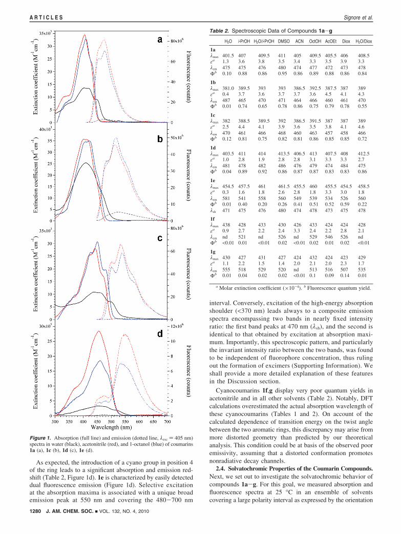
As expected, the introduction of a cyano group in position 4of the ring leads to a significant absorption and emission red-shift (Table 2, Figure 1d). 1e is characterized by easily detecteddual fluorescence emission (Figure 1d). Selective excitationat the absorption maxima is associated with a unique broademission peak at 550 nm and covering the 480-700 nm
interval. Conversely, excitation of the high-energy absorptionshoulder (<370 nm) leads always to a composite emissionspectra encompassing two bands in nearly fixed intensityratio: the first band peaks at 470 nm (λsh), and the second isidentical to that obtained by excitation at absorption maxi-mum. Importantly, this spectroscopic pattern, and particularlythe invariant intensity ratio between the two bands, was foundto be independent of fluorophore concentration, thus rulingout the formation of excimers (Supporting Information). Weshall provide a more detailed explanation of these featuresin the Discussion section.
Cyanocoumarins 1f,g display very poor quantum yields inacetonitrile and in all other solvents (Table 2). Notably, DFTcalculations overestimated the actual absorption wavelength ofthese cyanocoumarins (Tables 1 and 2). On account of thecalculated dependence of transition energy on the twist anglebetween the two aromatic rings, this discrepancy may arise frommore distorted geometry than predicted by our theoreticalanalysis. This condition could be at basis of the observed pooremissivity, assuming that a distorted conformation promotesnonradiative decay channels.
2.4. Solvatochromic Properties of the Coumarin Compounds.Next, we set out to investigate the solvatochromic behavior ofcompounds 1a-g. For this goal, we measured absorption andfluorescence spectra at 25 °C in an ensemble of solventscovering a large polarity interval as expressed by the orientation
Figure 1. Absorption (full line) and emission (dotted line, λexc ) 405 nm)spectra in water (black), acetonitrile (red), and 1-octanol (blue) of coumarins1a (a), 1c (b), 1d (c), 1e (d).
Table 2. Spectroscopic Data of Compounds 1a-g
H2O i-PrOH H2O/i-PrOH DMSO ACN OctOH AcOEt Diox H2O/Diox
1aλmax 401.5 407 409.5 411 405 409.5 405.5 406 408.5εa 1.3 3.6 3.8 3.5 3.4 3.3 3.5 3.9 3.3λem 475 475 476 480 474 477 472 473 478Φb 0.10 0.88 0.86 0.95 0.86 0.89 0.88 0.86 0.84
1bλmax 381.0 389.5 393 393 386.5 392.5 387.5 387 389εa 0.4 3.7 3.6 3.7 3.7 3.6 4.5 4.1 4.3λem 487 465 470 471 464 466 460 461 470Φb 0.01 0.74 0.65 0.78 0.86 0.75 0.79 0.78 0.55
1cλmax 382 388.5 389.5 392 386.5 391.5 387 387 389εa 2.5 4.4 4.1 3.9 3.6 3.5 3.8 4.1 4.6λem 470 461 466 468 460 463 457 458 466Φb 0.12 0.81 0.75 0.82 0.81 0.86 0.85 0.85 0.72
1dλmax 403.5 411 414 413.5 406.5 413 407.5 408 412.5εa 1.0 2.8 1.9 2.8 2.8 3.1 3.3 3.3 2.7λem 481 478 482 486 476 479 474 484 475Φb 0.04 0.89 0.92 0.86 0.87 0.87 0.83 0.83 0.86
1eλmax 454.5 457.5 461 461.5 455.5 460 455.5 454.5 458.5εa 0.3 1.6 1.8 2.6 2.8 1.8 3.3 3.0 1.8λem 581 541 558 560 549 539 534 526 560Φb 0.01 0.40 0.20 0.26 0.41 0.51 0.52 0.59 0.22λsh 471 475 476 480 474 478 473 475 478
1fλmax 438 428 433 430 426 433 424 424 428εa 0.9 2.7 2.2 2.4 3.3 2.4 2.2 2.8 2.1λem nd 521 nd 526 nd 529 546 526 ndΦb <0.01 0.01 <0.01 0.02 <0.01 0.02 0.01 0.02 <0.01
1gλmax 430 427 431 427 424 432 424 423 429εa 1.1 2.2 1.5 1.4 2.0 2.1 2.0 2.3 1.7λem 555 518 529 520 nd 513 516 507 535Φb 0.01 0.04 0.02 0.02 <0.01 0.1 0.09 0.14 0.01
a Molar extinction coefficient (×10-4). b Fluorescence quantum yield.
1280 J. AM. CHEM. SOC. 9 VOL. 132, NO. 4, 2010
A R T I C L E S Signore et al.

polarizability ∆f:57,58 water (symbol: W, ∆f ) 0.32), acetonitrile(ACN, ∆f ) 0.306), isopropanol (i-PrOH, ∆f ) 0.273),dimethylsulfoxide (DMSO, ∆f ) 0.265), ethyl acetate (EtOAc,∆f ) 0.199), n-octanol (OctOH, ∆f ) 0.103), and 1,4-dioxane(Diox, ∆f ) 0.021). 50/50 vol % mixtures of W/i-PrOH (∆f )0.306) and W/Diox (∆f ) 0.295) were also added to providemedia with intermediate hydrogen-bonding capabilities. Veryapolar solvents such as n-hexane were avoided, as the coumarinsbecame rather insoluble. Spectroscopic data for the coumarinsin all solvents are reported in Table 2.
On one hand, all coumarins display high extinction coef-ficients and quantum yields in organic solvents or water/solventmixtures, resulting in bright fluorescence (Table 2, Figure 1).
On the other hand, fluorescence emission of all compounds isnearly abolished in water, as witnessed by the very low quantumyields and lower extinction coefficients measured in this medium(Table 2, Figure 1). Remarkably, quantum yields of non-cyanocoumarins 1a-d do not correlate with the polarity function(∆f), being rather constant in organic or water/organic mediaand much lower in water (Figure 2a, red). A somewhat strongercorrelation between quantum yield and ∆f is observable for thefluorescent cyanocoumarin 1e (Figure 2a, blue). All coumarins,however, displayed no correlation at all between absorption (oremission) wavelength and ∆f. As such a correlation would beexpected by the classical Lippert-Mataga’s theory of solvato-chromism, these findings suggest a specific interaction betweenwater molecules and the coumarin structures that stronglyperturbs the photophysical behavior of the latter ones. The bandbroadening of the absorption spectra in water is a further supportto this hypothesis.
(57) Suppan, P. J. Chem. Soc. A 1968, 3125–3133.(58) Weast, R. C. Handbook of Chemistry and Physics; CRC Press: Boca
Raton, FL, 1980.
Figure 2. (a) Polarity dependence of emission quantum yield for 1a (red) and 1e (blue). (b) Relative brightness (absorption × quantum yield) enhancement(Water ) 1) for coumarins 1a-e. (c) Brightness enhancement for 1a (bottom) and 1e (top) in water/i-PrOH (red) and water/Dioxane (blue) mixtures. (d)Fluorescence emission of 1e upon excitation at 405 nm in different mixtures water/i-PrOH (v/v percentages of isopropanol are indicated in the legend). (e)Fluorescence lifetimes of 1a (λexc: 403 nm) and 1e (λexc: 468 nm) in water, i-PrOH, and acetonitrile. (f) Polarity-dependence of fluorescence lifetimes of 1eupon excitations at 403 nm (blue) and 468 nm (orange).
J. AM. CHEM. SOC. 9 VOL. 132, NO. 4, 2010 1281
Live Cell Imaging with Solvatochromic Coumarins A R T I C L E S

The fluorophore brightness (i.e., the product of extinctioncoefficient and quantum yield) is a parameter immediatelyassociated with the fluorescence emission performance inenvironments of different polarity at constant excitation power,as typically occurring in cell imaging. Accordingly, we com-puted the absolute brightness (Supporting Information, TableS1) and water-relative brightness (Figure 2b) of our coumarins.Among non-cyanocoumarins, 1b displays an astonishing >350-fold increment of brightness going from water to water/organicor organic media. Nonetheless, 1a is associated with the highestabsolute brightness in non-water solvents (>28,000, SupportingInformation, Table S1). Cyanocoumarin 1e shows a quitevariable water-relative brightness (between 170-790), in keep-ing with its more solvent-modulated extinction coefficient andquantum yield.
To determine the pattern of change in brightness betweenwater and organic media, we measured the water-relativebrightness of 1a and 1e in W/i-PrOH and W/Diox mixturescharacterized by different ∆f values (Figure 2c). For bothmixtures, the relative brightness of 1a displays a rapid increasefor ∆f above ∼0.31, whereas it follows a slower linear trendfor ∆f < ∼0.31. More precisely, the linear phase begins below∆f ) 0.312 (10% mol) for W/i-PrOH or ∆f ) 0.309 (7%mol) for W/Diox. Thermodynamic studies on W/IpOH orW/Diox mixtures have shown that at low molar content oforganic solvent (5-10% in mol), water undergoes a transitionto structure II “clathrates”, which are characterized by anice-like arrangement of 136 H2O molecules to give 24cavities. Eight of these cavities are large enough to accom-modate guest molecules such as the organic solvent, whereasthe other are smaller and should contain “monomeric” H2Omolecules (i.e., not hydrogen-bonded with the infinite net-work).59 At higher i-PrOH or Diox content, small clusters oforganic solvent and water molecules are formed.60 Thesefindings prompt us to attribute the sharp variation of brightnessof 1a to the structural rearrangement of water that leadspresumably to the loss of several H-bonds established withsurrounding H2O molecules. The linear trend at higher organiccontent may be related to a more “classical” solvatochromiceffect.
Significantly, in both W/i-PrOH and W/Diox compound 1eshows low and constant brightness in the same ∆f range whereits non-cyano counterpart 1a experiences a sharp increase ofemission. Below this region, brightness of 1e is linearly relatedto polarity. This trend suggests that 1e cannot fluoresce in thewhole region of clathrate stability, becoming emissive only whenthe water molecules become more “monomeric” and surroundedby their organic counterparts. 1e is associated with a furtherpeculiar photophysical property: its dual fluorescence upon high-energy excitation (below 405 nm) appears in all organic solvents,and for water/organic mixtures, it is dependent on the organiccontent (Figure 2d). In fact, both the high-energy (470 nm) andthe low-energy (540 nm) emission bands increase as the watercontent of the mixture is decreased. The low-energy band (λ )550 nm) is more sensitive to the polarity of the mixture thanits high-energy companion (λsh ) 470 nm). The latter one neverdisappears, even in pure water. Thus, the ratio between the twobands changes with the polarity of the mixture. This behavior
supports the use of 1e as a ratiometric (i.e., concentrationindependent)61 sensor of medium polarity.
2.5. Fluorescence Lifetime Measurements. On account oftheir non-negligible fluorescence in water, coumarin 1a,c wereselected to test the effect of solvent nature on the fluorescentlifetime (τ). 1e was added to this pool to compare thephotophysical characteristics after the incorporation of the cyanogroup. Actually, as already mentioned, 1e displays dim butdetectable fluorescence in water when excited at 405 nm.
Remarkably, 1a,c,e are all characterized by monoexponentialdecays when excited near their absorption maximum (403 nmfor 1a,c, 468 nm for 1e) regardless of the solvent (Figure 2e).Lifetime values range from 2.8 to 5.24 ns (Table 3). The actuallifetimes can be combined with the quantum yields to give thecorresponding radiative lifetimes (τr, Table 3).
For 1a,c, the position and nature of the functional groupsattached to the coumarin ring modulate the detected lifetime.Interestingly, for both coumarins τ is quite constant in the threetested solvents, whereas τr becomes extremely large (>30 ns)in water. The theoretical τr in water calculated by theStrickler-Berg equation from the absorption and fluorescencespectra is around 3 ns, an order of magnitude lower than theexperimental value. Instead, the theoretical τr in i-PrOH andACN are in good agreement with the experimental value (Table3). The Strickler-Berg equation fails to yield reasonable valuesof τr only when a strong interaction with the solvent takes placeand/or there is a change in the excited-state geometry. Thesedata suggest that the low quantum yields of 1a,c in water arenot the result of some quenching process but rather of differentproperties of the excited state. A photophysical model account-ing for this phenomenology will be described in the Discussionsection.
The cyano group in 1e leads to significantly longer τ and τr
(Table 3). Here, τr is significantly larger than the theoreticalvalue in all media, suggesting efficient interactions between themolecule and the selected solvents. We further examined theeffect of medium polarity on the lifetime of 1e by collectingmeasurements in W/i-PrOH mixtures at different ∆f (Figure 2f,orange trace). Notably, the τ values show remarkable linearitywith ∆f up to 0.315, in good agreement with the brightnessbehavior in the W/i-PrOH media (Figure 2c). These data confirmthat the fluorescence of 1e is regulated by a “classical”solvatochromic behavior once the water content falls below athreshold value (around 90% in mol). We also determined theτ values of 1e upon 403-nm excitation in the same W/i-PrOHmixtures in order to check the effect of the dual fluorescenceemission. Indeed, we always collected biexponential fluores-cence decay curves except for the pure water case. To determinethe actual τ values of the high-energy emission component (τ470),
(59) Jerie, K.; Baranowski, A.; Koziol, S.; Glinski, J.; Burakowski, A.Chem. Phys. 2005, 309, 277–282.
(60) Østergaard, K. K.; Tohidi, B.; Anderson, R.; Todd, A. C.; Danesh, A.Ind. Eng. Chem. Res. 2002, 41, 2064–2068.
(61) Bizzarri, R.; Serresi, M.; Luin, S.; Beltram, F. Anal. Bioanal. Chem.2009, 393, 1107–1122.
(62) Valeur, B. Molecular Fluorescence; Wiley-VCH Verlag GmbH:Weinheim (DE), 2002.
Table 3. Fluorescence Lifetimes of 1a,c,e in Water, Isopropanol,and Acetonitrile; Intrinsic Lifetime Values Calculated from theStrickler-Berg Relation62 are Reported in Parentheses
fluorescence lifetime (ns) radiative lifetime (ns)
coumarin H2O i-PrOH CAN H2O i-PrOH ACN
1a 3.28 3.18 3.28 32.8 (6.62) 3.6 (3.13) 3.8 (3.19)1c 2.44 2.69 2.65 20.3 (4.03) 3.32 (2.52) 3.27 (3.03)1e 3.23 4.85 5.24 323.0 (36.2) 12.1 (8.28) 12.8 (5.29)
1282 J. AM. CHEM. SOC. 9 VOL. 132, NO. 4, 2010
A R T I C L E S Signore et al.

we fitted the biexponential decays imposing one lifetime equalto that recovered by excitation at 468 nm. Importantly, we foundthat λ470 is almost invariant with solvent polarity (Figure 2f,blue trace) and its value (3.30 ns) is very close to its non-cyanocounterpart 1a.
2.6. Photophysical Properties in Cultured Cells. In order toassess the photophysical behavior of the prepared coumarinsin biological environments, compounds 1a-e were examinedin living cells. The dyes were externally administered at lowconcentration (0.5-1 µM) to Chinese Hamster Ovary (CHO)cell cultures. After 15-30 min of exposition, cells wereimaged by a confocal microscope without removing theexternal buffer by using 403 nm and/or 458 nm excitationlaser source (Figure 3a,b).
CHO cells remained viable for at least 2-3 h after additionof the coumarins. No fluorescence was detected in the externalbuffer, or in the nucleus, while collected images showed thatthe coumarins are emissive in selected lipophilic subcellularcompartments. These findings are in agreement with the
negligible fluorescence of 1a and 1e in water, as well as therather lipophilic properties of the dyes.
Remarkably, the emission profiles of the coumarins in thesubcellular region where they are active (Figure 3c) resembleclosely those determined during in Vitro experiments, indicatingthat the biological environment does not affect the fluorescentoptical characteristics of the coumarins. The conserved dualemission of 1e upon high energy (403 nm) excitation (Figure3c) is particularly relevant in view of the use of this coumarinas ratiometric emission indicator of polarity, working in thevisible region of the spectrum. A ratiometric plot of polarityby 1e is reported in Figure 3b (right) and compared with thelifetime map (Figure 3e) to which it is strictly related (seebelow).
The lifetimes of 1a and 1e were analyzed in cultured cellsby confocal FLIM. 1a was found to possess a quite uniformmonoexponential lifetime decay cell-wide (Figure 3d,f), inexcellent agreement with its constant lifetime observed inmedium of different polarity in Vitro. Also the intracellular
Figure 3. (a) Confocal microscopy images of 1a in CHO cells (λexc: 403 nm). (b) (Left) Confocal microscopy image of 1e in CHO cells (λexc ) 403 nm,λem ) 545-580 nm). (Right) Ratio of images collected at 545-580 nm and 440-480 nm (λexc ) 403 nm). (c) Fluorescence spectra of 1a and 1e in CHOcells. (d) Lifetime-intensity color-coded map of 1a in CHO cells (λexc: 403 nm). (e) Color-coded lifetime map of the image reported in panel b (λexc ) 468nm). (f) Histogram of lifetime distribution for 1a and 1e in CHO cells.
J. AM. CHEM. SOC. 9 VOL. 132, NO. 4, 2010 1283
Live Cell Imaging with Solvatochromic Coumarins A R T I C L E S

lifetime behavior of 1e was in keeping with the propertiesmeasured in Vitro. Upon 468 nm excitation, we detectedmonoexponential decays with τ fairly dependent on the intra-cellular location. Careful analysis showed that longer lifetimeswere associated with 1e localized in cell membranes or theperinuclear region, owing to the more lipophilic character ofthese regions (Figure 3e,f). Additionally, the lifetime map of1e is very similar to its emission ratiometric map (Figure 3b,e),i.e. there is a strict correspondence between longer fluorescencelifetimes and increased low-/high-energy band ratio, as previ-ously found in Vitro. These data highlight the possibility of using1e as a gradual polarity indicator in bioenvironments by bothemission ratiometry and lifetime imaging.
Finally, the photostability of 1a and 1e, measured at highlaser power in living cells, was quite comparable with that ofYFP. Indeed, we measured fluorescence decrease with τ ) 39.5( 2 s (93.1 kW/cm2), 13.7 ( 0.5 s (135 kW/cm2), and 30.3 (0.8 s (106 kW/cm2) for YFP, 1a, and 1e, respectively.
2.7. Cellular Localization of Coumarins. To gain furtherinsight into the partitioning of 1a and 1e within differentsubcellular domains, we performed some colocalization experi-ments with standard organelle markers, in particular DiIC18(5)-DS (membrane marker), BODIPY TR glibenclamide (ERmarker), and Lysotracker (lysosome marker). We found similar,although not superimposable, localizations of 1a and 1e,according to the different physicochemical properties of our
probes. 1a extensively accumulated in ER (Figure 4b), whereasits localization in the cell membrane (Figure 4a), lysosomes(Figure 4c), and Golgi apparatus (not shown) was rather limited.1e accumulated in the cell membrane (Figure 4d) and ER (Figure4e). Also, this coumarin is extensively sequestered in lysosomes(Figure 4f), giving rise to punctate staining (see Figure 3b forcomparison). Cell localization differences can be clearly evi-denced by a ratio plot of 1a and 1e fluorescence taken in cellsexposed to both coumarins (Figure 5a-c). These findingssuggest a preferential albeit different localization of 1a and 1ein lipophilic compartments, in agreement with the hydrophobiccoumarin structure. Consistently, we measured log P values of3.6 for 1a and 2.9 for 1e,63 substantiating the hypothesis of asignificant accumulation of the dye in the most apolar domainsof the cell but with different affinity. The presence of thefluorophore in the cytosol could not be evidenced directly, dueto the nonemissivity of the solvatochromic probes in aqueousenvironment. However, by means of simple fluorescencerecovery after photobleaching (FRAP) experiments, we foundthat the coumarins have remarkably high diffusivity throughoutthe cell (τ ) 3.1 ( 0.6 s, τ ) 5.6 ( 0.5 s for 1a and 1e,
(63) Takacs-Novak, K.; Avdeef, A. J. Pharm. Biomed. Anal. 1996, 14,1405–1413.
(64) Fay, F. S.; Taneja, K. L.; Shenoy, S.; Lifshitz, L.; Singer, R. H. Exp.Cell Res. 1997, 231, 27–37.
Figure 4. Colocalization of standard organelle markers (left, red channel) and 1a or 1e (right, green channel, and inset) in CHO cells. White spots representa selection of colocalized points (intensity ratio interval: 0.85-1.17). Colocalization results, obtained by a colocalization P-test according to Fay’smethod64 are reported in parenteses. (a) 1a (λexc: 403 nm, λem ) 420-600 nm) and membrane marker DiIC18(5)-DS (λexc: 633 nm, λem ) 650-750nm) (poor colocalization, P ) 0.46); (b) 1a (λexc: 458 nm, λem ) 470-550 nm) and ER marker BODIPY TR glibenclamide (λexc: 561 nm, λem )570-680 nm) (strong colocalization, P > 0.95); (c) 1a (λexc: 403 nm, λem ) 410-535 nm) and lysosome marker Lysotracker (λexc: 561 nm, λem )570-700 nm) (moderate colocalization, P ) 0.68); (d) 1e (λexc: 458 nm, λem ) 470-600 nm) and membrane marker DiIC18(5)-DS (λexc: 633 nm, λem
) 650-750 nm) (strong colocalization, P > 0.95); (e) 1e (λexc: 458 nm, λem ) 470-600 nm) and ER marker BODIPY TR glibenclamide (λexc: 561nm, λem ) 570-680 nm) (strong colocalization, P > 0.95); (f) 1e (λexc: 458 nm, λem ) 470-600 nm) and lysosome marker Lysotracker (λexc: 561 nm,λem ) 570-700 nm) (strong colocalization, P > 0.95). Note that in experiments involving 1e the two dyes were imaged sequentially to avoid extensivecross-talk.
1284 J. AM. CHEM. SOC. 9 VOL. 132, NO. 4, 2010
A R T I C L E S Signore et al.

respectively),5 thus suggesting a partition equilibrium of the dyesbetween the lipophilic compartments and the cytosol.
3. Discussion
Environmentally sensitive fluorescent molecules appear ex-tremely useful for in ViVo imaging applications. They can beused as sensors to report on polarity changes in the surroundingmedium associated with relevant biochemical events such asprotein-protein recognition, membrane lipid restructuring, andbiomolecular self-assembly processes. Note that compared tothe more popular FRET-based sensors, solvatochromic probesdo not require two partners in close proximity and in well-defined geometrical relationships to provide a signal whichmonitors the target process at nanoscale. Hence, we recentlydeveloped a renewed interest in molecular moieties opticallyinfluenced by the polarity of their environment.
The solvatochromic behavior usually stems from largevariations of the molecular dipole upon photon absorption,leading to different stabilization energies of the ground andexcited states by the solvent shell around the molecule. Theconcomitant incorporation of electron-donating and electron-
withdrawing groups onto aromatic rings is a common way topromote solvatochromism. In such a case, the electronictransition is often accompanied by a large transfer of electroniccharge from the electron donor toward the electron acceptor, aphenomenon referred to as intramolecular charge transfer (ICT).The significant electron redistribution between ground andexcited state upon photoexcitation results in a sudden changeof molecular dipole moment and therefore of stabilization bythe solvent. Nonetheless, a powerful solvatochromic effect isnot necessarily translated into a bright and polarity-dependentfluorescence. Indeed, the addition of donating or withdrawinggroup to otherwise fluorescent aromatic molecules may forbidor severely hamper the fluorescence emission of the targetcompound.
The coumarin aromatic structure is known to allow theincorporation of electron donors and acceptors while retainingmost of its excellent fluorescent properties such as high quantumyield and large Stokes’ shift. In this perspective, we targetedthe engineering of a novel toolbox of coumarins bearing differentfunctional groups in a donor-(coumarin core)-acceptor con-figuration. The attention was mainly directed to alkylethergroups as good electron-donating functional units, since theyare amenable to further modification to yield chemical “hands”for biomolecule binding. Additionally, alkylether groups preventthe chemical reactivity problems (e.g., protonation, low yieldof further functionalization) that are often encountered whenthe more popular electron-donating amino group is utilized. Wefocused on benzothiazene and cyano functional units as acceptorgroups on account of their reported electron-withdrawing effecton the physicochemical properties of conjugated aromaticcompounds.29 It is worth noting that all these groups weresingularly described to red-shift the basic coumarin spectra23
owing to a larger delocalization of the π orbital.Our approach to the development of new solvatochromic
fluorescent probes followed a sequential scheme: (1) in silicoscreening to identify interesting substitution patterns of thecoumarin structure from an optical viewpoint, (2) establishmentof a reliable and efficient synthetic procedure common for allthe target compounds (modular synthesis), (3) thorough in Vitroanalysis of the photophysical properties of the synthesizedprobes, and (4) evaluation of their photophysical behavior in abiological (e.g., cultured cell) environment. Overall, we soughtto set up a general and flexible scientific “platform” to engineerprobes tailored to different in ViVo applications.
The computational DFT screening of coumarin structures wascarried out with the clear-cut goal of identifying donor-(coumarincore)-acceptor patterns able to red-shift the absorption of purecoumarin (310 nm) to the visible region of the electromagneticspectrum, while retaining a molecular structure amenable tobiochemical functionalization. Indeed, the visible region is byfar the preferred wavelength interval for the analysis ofbiological specimens owing to the good compromise of highspatial resolution and low photodamage to cellular structures.Accordingly, most microscope apparatus for in ViVo imagingare supplied with excitation sources emitting above 400 nm.DFT/B3LYP analysis allowed for the identification of seveninteresting structures red-shifted of 70-180 nm compared tobase coumarin (Table 1, 1a-g). These structures are character-ized by the presence of two alkylether- or one naphthyl-donorgroup in positions 6-8, one benzothiazene group in position 3,and for a subset, also by a cyano group in position 4. We noticedthat the nature of alkyl ether and the substitution regioselectivityat positions 6-8 have little effect on absorption wavelength
Figure 5. (a,b) Confocal microscopy image of 1a (panel a, λexc ) 405nm, λem ) 420-470 nm) and 1e (panel b, λexc ) 458 nm, λem ) 500-600nm), concomitantly administered to CHO cells; (c) logarithm of the ratioof images a and b to unveil different intracellular distributions of 1a and1b; note that the log form was adopted to display spatial enrichment ordepletion in linear color-coded scale.
J. AM. CHEM. SOC. 9 VOL. 132, NO. 4, 2010 1285
Live Cell Imaging with Solvatochromic Coumarins A R T I C L E S

(Table 1, 1a-c, 1e-g). Furthermore, DFT highlighted that theconcomitant incorporation of both benzothiazene and cyanogroups yields a synergistic red-shift effect on absorption (Table1, 1e-g). These findings appeared promising in light of ourmodular approach to probes tailored for specific applicationsand prompted for the synthesis of 1a-g. Note that compound1c represents already a “functional” evolution of 1b, as it canbe easily converted into a reactant for protein conjugation (weshall describe this in a forthcoming paper).
Concerning the synthetic step, we set up a general procedurebased on a piperidine-catalyzed Knoevenagel reaction involvingsubstituted o-hydroxy benzaldehydes (Schemes 1-2, 3a,b′,c,d)and a benzothiazene acetate derivative (Scheme 2, 2). The cyanogroup was then added by a simple addition/elimination reaction.Overall, the synthetic procedure allows obtaining the desiredcoumarin in high yields just by selecting and preparing theappropriate o-hydroxy benzaldehyde reactant. Note that the here-adopted Knoevenagel reaction is known to be unaffected bymost functional groups that may be present on the reactants.
The absorption properties of the non-cyanocoumarins inacetonitrile closely resemble those calculated by DFT. A goodagreement is seen also for cyano 1e, whereas DFT overestimatedthe wavelengths of absorption peaks of 1f,g. We attribute thelatter discrepancy to larger distortion of the angle between thetwo aromatic rings than predicted by the theoretical method.Indeed, our calculation clearly correlates the twist angle withthe excitation energy. The absorption bands are in all cases broadand often rich in vibrational substructure. Non-cyanocoumarinscan be excited efficiently by light sources in the 380-430 nmrange (Figure 1a-c); cyanocoumarins allow for excitationwavelengths as long as 500 nm (Figure 1d). With the exceptionof 1f-g, the prepared coumarins in acetonitrile showed broadfluorescence emission characterized by large Stokes’ shifts(Figure 1a-d) and high quantum yields (Table 2). Bothproperties are extremely relevant in view of a future use of thesecompounds for high-resolution imaging as they are at the basisof large signal-to-noise ratio. 1e displays an interesting property:when excited on the high-energy absorption shoulder below 400nm it adds a minor emission band peaked at 470 nm to itsconventional band peaked at 570 nm. This dual fluorescence isassociated with a nearly constant band ratio for any excitationbelow 400 nm (Supporting Information). We demonstrated,however, that this effect is not the result of excimer formationand must be related to the photophysics of the excited state.
From a solvatochromic viewpoint, we can split the fluorescentcoumarins into two subsets, one encompassing the non-cyanoderivatives, 1a-d, and the other cyano compound, 1e. Theanalysis in solvents, or water/solvent mixtures, of differentpolarities (measured by the orientation polarizability function∆f) showed clearly that 1a-d are poorly or not emissive inpure water, whereas they maintain approximately the samequantum yield once the polarity falls below a certain value (∆f≈ 0.31) no matter whether that happens in water/organic solventmixtures or pure protic/nonprotic organic solvents (Figure 2a,b).Brightness enhancements ranged from 15- to 400-fold withrespect to pure water (Figure 2b). The sharp increase influorescence is clearly related to a rearrangement of waterstructure and the concomitant loss of strong H-bonding interac-tions with the solute probe (Figure 2c).
The H-bonding model helps explain also the different QY inwater of 1b and 1c in spite of the similar substitution pattern.Indeed, we performed molecular dynamics simulations of thesetwo coumarins embedded in explicit water molecules. The
simulations describe how water molecules interact with thesolute, during its ground-state dynamics, predicting the forma-tion and stability of hydrogen bonds. By comparing the twocoumarins, we observed that only the oxygen atom at position8 (O8) changes significantly its H-bonding degree between thetwo molecules. In 1b we found ∼60% of full H-bondingoccupation (i.e., ∼60% of the molecular dynamics snapshotsshow a configuration where a water oxygen is at a distance <3.5Å and the O8-hydrogen-water oxygen angle is >150°),whereas in 1c this value drops to ∼20%. Although these valuesare just estimates of H-bonding capabilities, the comparisonbetween these two figures clearly indicates that O8 of 1bestablishes much stronger H-bonding interactions with watermolecules than O8 of 1c.
To further investigate the photophysical behavior of thesecoumarins we determined the fluorescence lifetimes in water,i-PrOH, and ACN of 1a and c (1b,d are practically nonfluo-rescent in water, and reliable fluorescence decays could not beobtained). Surprisingly, the lifetime is very similar in these threesolvents (Table 3). The radiative lifetime in water is more than30 ns, absurdly 1 order of magnitude larger than that predictedon pure theoretical basis from absorption/emission spectra. Thesefindings imply that the low quantum yield in water cannot berelated to an additional nonradiative decay channel from thelowest vibrational level of the excited state (quenching mech-anism). This means that, after the fast vertical transition due toexcitation, the coumarin can evolve along two different energylandscapes: one is the usual internal conversion down to thelowest vibrational level of the excited state, the other engagesa fast (ps) transition to an excited state of different nature(possibly after some internal conversion) that provides a veryeffective de-excitation pathway down to ground state (Figure6). On account of recent results described for similar coumarinstructures,65 we are tempted to identify this second excited stateto a twisted intramolecular charge transfer (TICT) configuration.DFT calculations show the electronic transition of 1a to beassociated with a significant rearrangement of electronic charge,
(65) Satpati, A. K.; Kumbhakar, M.; Nath, S.; Pal, H. Photochem. Photobiol.2009, 85, 119–129.
Figure 6. Proposed model of electronic transitions in coumarins 1a-d (a)and 1e (b).
1286 J. AM. CHEM. SOC. 9 VOL. 132, NO. 4, 2010
A R T I C L E S Signore et al.
in a typical ICT fashion. Usually ICT may evolve to TICT ifthe interactions with the solvent molecules are favorable to thelatter configuration. Indeed, there is evidence for similarcoumarin systems in which a TICT state can be stabilized bythe establishment of strong H-bonding interactions with watermolecules. This would explain the high sensitivity of fluores-cence emission of our compounds from the H-bonding capabilityof the surrounding medium.
The solvatochromic behavior of 1e is richer. First of all, thequantum yield of emission shows good correlation with solventpolarity (Figure 2a), and therefore, the brightness enhancementrelative to water is solvent dependent (Figure 2b). Thiscorrelation is lost for pure water. More, 1e has constantly pooremission in all of the polarity range where its non-cyanocounterpart 1a shows a sudden increase of brightness (Figure2c). Below a certain polarity value (∆f ≈ 0.31), however, thebrightness of 1e is linearly related to ∆f, following a“Lippert-Mataga” behavior. These findings suggest that 1e hasa solvent-dependent emission related to a differential stabiliza-tion of ground and excited states below a certain polarity level,and it is dark otherwise. Accordingly, the lifetime of 1e in water/i-PrOH mixtures is strictly linear with ∆f except for pure water(τ550, Figure 2f). The dual fluorescence of 1e upon <400 nmexcitation is conserved in all solvents. Nonetheless, the relativeratio between the two emission bands is polarity-dependent(Figure 2d). This behavior is particularly evident for water/organic solvent mixtures and supports the use of 1e as aratiometric probe of polarity. Note that in water only the high-energy emission band is present (τsh ≈ 470 nm, Figure 2d).The dual fluorescence is reflected also in a biexponential (i.e.,bicomponent) lifetime decay. The deconvolution of the decaycurves yielded the lifetime associated with the high-energyemission band (τ470). Importantly, τ470 resulted independent ofmedium polarity including also water. Thus, the high-energyband of 1e looks very similar in wavelength and lifetimebehavior to the emission of its non-cyano parent 1a. These datasupport a photophysical model encompassing two communicat-ing excited states similar to that described for non-cyanocoumarins (Figure 6). The major difference, however, residesin the existence of radiative channels from both excited states.Additionally, the efficiency of the transition from the low-energyexcited state is polarity-dependent. Although we may be temptedto attribute the two excited states to ICT and TICT in analogywith non-cyanocoumarins, the complexity of the photophysicalpattern requires further investigation to provide a reliablehypothesis, and this topic will be the subject of a forthcomingpaper.
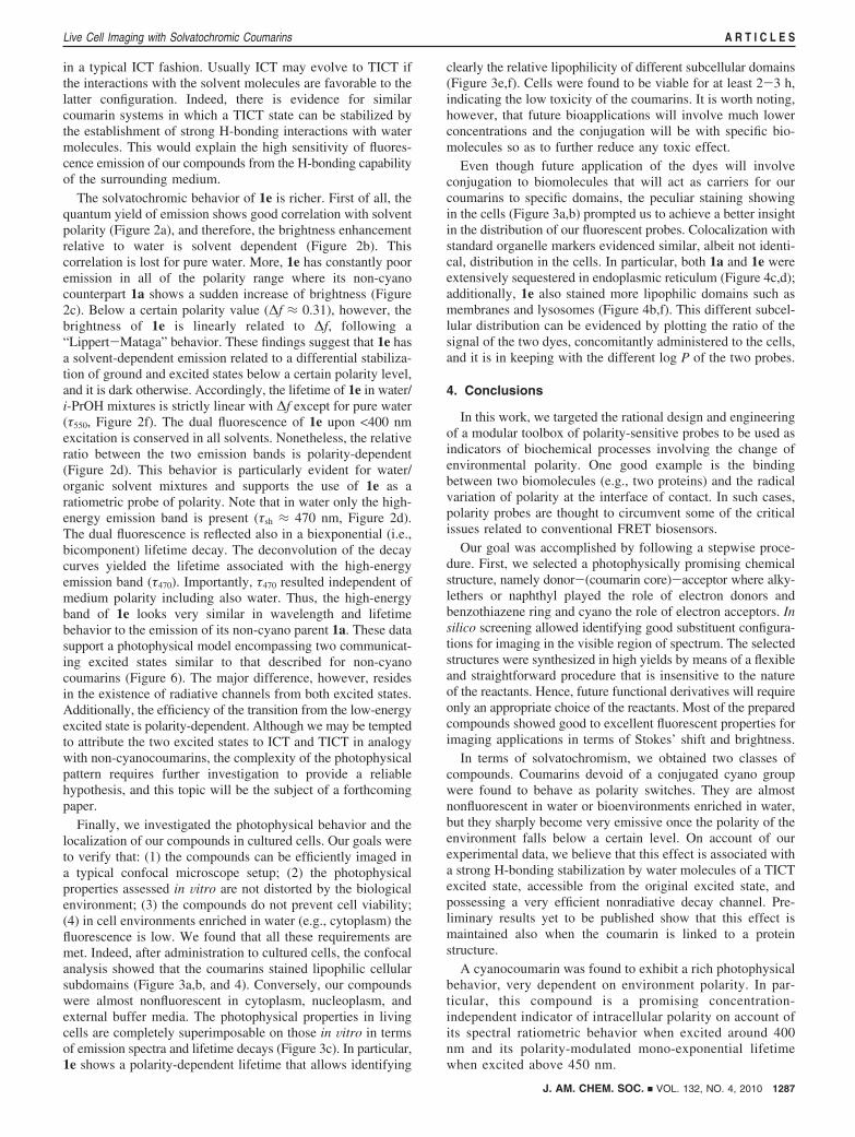
Finally, we investigated the photophysical behavior and thelocalization of our compounds in cultured cells. Our goals wereto verify that: (1) the compounds can be efficiently imaged ina typical confocal microscope setup; (2) the photophysicalproperties assessed in Vitro are not distorted by the biologicalenvironment; (3) the compounds do not prevent cell viability;(4) in cell environments enriched in water (e.g., cytoplasm) thefluorescence is low. We found that all these requirements aremet. Indeed, after administration to cultured cells, the confocalanalysis showed that the coumarins stained lipophilic cellularsubdomains (Figure 3a,b, and 4). Conversely, our compoundswere almost nonfluorescent in cytoplasm, nucleoplasm, andexternal buffer media. The photophysical properties in livingcells are completely superimposable on those in Vitro in termsof emission spectra and lifetime decays (Figure 3c). In particular,1e shows a polarity-dependent lifetime that allows identifying
clearly the relative lipophilicity of different subcellular domains(Figure 3e,f). Cells were found to be viable for at least 2-3 h,indicating the low toxicity of the coumarins. It is worth noting,however, that future bioapplications will involve much lowerconcentrations and the conjugation will be with specific bio-molecules so as to further reduce any toxic effect.
Even though future application of the dyes will involveconjugation to biomolecules that will act as carriers for ourcoumarins to specific domains, the peculiar staining showingin the cells (Figure 3a,b) prompted us to achieve a better insightin the distribution of our fluorescent probes. Colocalization withstandard organelle markers evidenced similar, albeit not identi-cal, distribution in the cells. In particular, both 1a and 1e wereextensively sequestered in endoplasmic reticulum (Figure 4c,d);additionally, 1e also stained more lipophilic domains such asmembranes and lysosomes (Figure 4b,f). This different subcel-lular distribution can be evidenced by plotting the ratio of thesignal of the two dyes, concomitantly administered to the cells,and it is in keeping with the different log P of the two probes.
4. Conclusions
In this work, we targeted the rational design and engineeringof a modular toolbox of polarity-sensitive probes to be used asindicators of biochemical processes involving the change ofenvironmental polarity. One good example is the bindingbetween two biomolecules (e.g., two proteins) and the radicalvariation of polarity at the interface of contact. In such cases,polarity probes are thought to circumvent some of the criticalissues related to conventional FRET biosensors.
Our goal was accomplished by following a stepwise proce-dure. First, we selected a photophysically promising chemicalstructure, namely donor-(coumarin core)-acceptor where alky-lethers or naphthyl played the role of electron donors andbenzothiazene ring and cyano the role of electron acceptors. Insilico screening allowed identifying good substituent configura-tions for imaging in the visible region of spectrum. The selectedstructures were synthesized in high yields by means of a flexibleand straightforward procedure that is insensitive to the natureof the reactants. Hence, future functional derivatives will requireonly an appropriate choice of the reactants. Most of the preparedcompounds showed good to excellent fluorescent properties forimaging applications in terms of Stokes’ shift and brightness.
In terms of solvatochromism, we obtained two classes ofcompounds. Coumarins devoid of a conjugated cyano groupwere found to behave as polarity switches. They are almostnonfluorescent in water or bioenvironments enriched in water,but they sharply become very emissive once the polarity of theenvironment falls below a certain level. On account of ourexperimental data, we believe that this effect is associated witha strong H-bonding stabilization by water molecules of a TICTexcited state, accessible from the original excited state, andpossessing a very efficient nonradiative decay channel. Pre-liminary results yet to be published show that this effect ismaintained also when the coumarin is linked to a proteinstructure.
A cyanocoumarin was found to exhibit a rich photophysicalbehavior, very dependent on environment polarity. In par-ticular, this compound is a promising concentration-independent indicator of intracellular polarity on account ofits spectral ratiometric behavior when excited around 400nm and its polarity-modulated mono-exponential lifetimewhen excited above 450 nm.
J. AM. CHEM. SOC. 9 VOL. 132, NO. 4, 2010 1287
Live Cell Imaging with Solvatochromic Coumarins A R T I C L E S

Finally, tests of our compounds in cultured cells revealed thatthey are very suitable for in ViVo high-resolution microscopeimaging in both spectral and lifetime modes. We believe thatthese compounds, after appropriate functionalization and con-jugation to target biomolecules, will represent remarkable toolsto investigate subtle biochemical processes in the cell environ-ment. Furthermore, the photophysical comprehension of theenvironmental sensitivity displayed by our coumarins will allowfor the engineering of further compounds tailored to specificapplications. Studies in both directions are currently under way.
Acknowledgment. We gratefully acknowledge Prof. FabioBeltram for precious scientific suggestions during the research workand in preparing this manuscript and Dr. Paolo Facci for stimulating
discussions. This work was supported by INFM-CNR under theframework of Seed Project “Solvatochromic fluorescent dyes forbulk and single-molecule detection of intracellular native proteins”.The partial support of the Italian Ministry for University andResearch (MiUR) under the framework of the FIRB projectRBLA03ER38 is also acknowledged.
Supporting Information Available: Detailed synthetic pro-cedures; 1H and 13C NMR spectra for all compounds; spectro-scopic data and high-resolution mass spectra for all compounds;complete ref 53. This material is available free of charge viathe Internet at http://pubs.acs.org.
JA9050444
1288 J. AM. CHEM. SOC. 9 VOL. 132, NO. 4, 2010
A R T I C L E S Signore et al.
Copyright © 2022 FDOKUMEN