Polarity, planes of cell division, and the evolution of plant ...
15
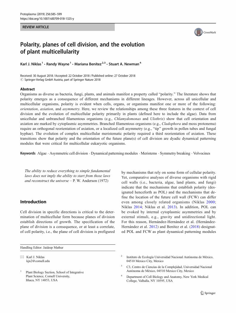
REVIEW ARTICLE Karl J. Niklas 1 & Randy Wayne 1 & Mariana Benítez 2,3 & Stuart A. Newman 4 Received: 30 August 2018 /Accepted: 22 October 2018 /Published online: 27 October 2018 # Springer-Verlag GmbH Austria, part of Springer Nature 2018 Abstract Organisms as diverse as bacteria, fungi, plants, and animals manifest a property called Bpolarity.^ The literature shows that polarity emerges as a consequence of different mechanisms in different lineages. However, across all unicellular and multicellular organisms, polarity is evident when cells, organs, or organisms manifest one or more of the following: orientation, axiation, and asymmetry . Here, we review the relationships among these three features in the context of cell division and the evolution of multicellular polarity primarily in plants (defined here to include the algae). Data from unicellular and unbranched filamentous organisms (e.g., Chlamydomonas and Ulothrix) show that cell orientation and axiation are marked by cytoplasmic asymmetries. Branched filamentous organisms (e.g., Cladophora and moss protonema) require an orthogonal reorientation of axiation, or a localized cell asymmetry (e.g., Btip^ growth in pollen tubes and fungal hyphae). The evolution of complex multicellular meristematic polarity required a third reorientation of axiation. These transitions show that polarity and the orientation of the future plane(s) of cell division are dyadic dynamical patterning modules that were critical for multicellular eukaryotic organisms. Keywords Algae . Asymmetric cell division . Dynamical patterning modules . Meristems . Symmetry breaking . Volvocines The ability to reduce everything to simple fundamental laws does not imply the ability to start from those laws and reconstruct the universe – P. W. Anderson (1972) Introduction Cell division in specific directions is critical to the deter- mination of multicellular form because planes of division establish directions of growth. The specification of the plane of division is a consequence, or at least a correlate, of cell polarity, i.e., the plane of cell division is prefigured by mechanisms that rely on some form of cellular polarity. Yet, comparative analyses of diverse organisms with rigid cell walls (i.e., bacteria, algae, land plants, and fungi) indicate that the mechanisms that establish polarity (des- ignated henceforth as POL) and the mechanisms that de- fine the location of the future cell wall (FCW) can differ even among closely related organisms (Niklas 2000; Niklas 2014; Niklas et al. 2013). In addition, POL can be evoked by internal cytoplasmic asymmetries and by external stimuli, e.g., gravity and unidirectional light. For this reason, Hernández-Hernández et al. (Hernández- Hernández et al. 2012) and Benítez et al. (2018) designat- ed POL and FCW as plant dynamical patterning modules Handling Editor: Jaideep Mathur * Karl J. Niklas [email protected] 1 Plant Biology Section, School of Integrative Plant Science, Cornell University, Ithaca, NY 14853, USA 2 Instituto de Ecología Universidad Nacional Autónoma de México, 04510 Mexico City, Mexico 3 C3, Centro de Ciencias de la Complejidad, Universidad Nacional Autónoma de México, 04510 Mexico City, Mexico 4 Department of Cell Biology and Anatomy, New York Medical College, Valhalla, NY 10595, USA Protoplasma (2019) 256:585–599 https://doi.org/10.1007/s00709-018-1325-y Polarity, planes of cell division, and the evolution of plant multicellularity
-
Upload
khangminh22 -
Category
Documents
-
view
0 -
download
0
Transcript of Polarity, planes of cell division, and the evolution of plant ...
REVIEW ARTICLE
Karl J. Niklas1 & Randy Wayne1& Mariana Benítez2,3 & Stuart A. Newman4
Received: 30 August 2018 /Accepted: 22 October 2018 /Published online: 27 October 2018# Springer-Verlag GmbH Austria, part of Springer Nature 2018
AbstractOrganisms as diverse as bacteria, fungi, plants, and animals manifest a property called Bpolarity.^ The literature shows thatpolarity emerges as a consequence of different mechanisms in different lineages. However, across all unicellular andmulticellular organisms, polarity is evident when cells, organs, or organisms manifest one or more of the following:orientation, axiation, and asymmetry. Here, we review the relationships among these three features in the context of celldivision and the evolution of multicellular polarity primarily in plants (defined here to include the algae). Data fromunicellular and unbranched filamentous organisms (e.g., Chlamydomonas and Ulothrix) show that cell orientation andaxiation are marked by cytoplasmic asymmetries. Branched filamentous organisms (e.g., Cladophora and moss protonema)require an orthogonal reorientation of axiation, or a localized cell asymmetry (e.g., Btip^ growth in pollen tubes and fungalhyphae). The evolution of complex multicellular meristematic polarity required a third reorientation of axiation. Thesetransitions show that polarity and the orientation of the future plane(s) of cell division are dyadic dynamical patterningmodules that were critical for multicellular eukaryotic organisms.
Keywords Algae . Asymmetric cell division . Dynamical patterningmodules . Meristems . Symmetry breaking . Volvocines
The ability to reduce everything to simple fundamentallaws does not imply the ability to start from those lawsand reconstruct the universe – P. W. Anderson (1972)
Introduction
Cell division in specific directions is critical to the deter-mination of multicellular form because planes of divisionestablish directions of growth. The specification of theplane of division is a consequence, or at least a correlate,of cell polarity, i.e., the plane of cell division is prefigured
by mechanisms that rely on some form of cellular polarity.Yet, comparative analyses of diverse organisms with rigidcell walls (i.e., bacteria, algae, land plants, and fungi)indicate that the mechanisms that establish polarity (des-ignated henceforth as POL) and the mechanisms that de-fine the location of the future cell wall (FCW) can differeven among closely related organisms (Niklas 2000;Niklas 2014; Niklas et al. 2013). In addition, POL canbe evoked by internal cytoplasmic asymmetries and byexternal stimuli, e.g., gravity and unidirectional light.For this reason, Hernández-Hernández et al. (Hernández-Hernández et al. 2012) and Benítez et al. (2018) designat-ed POL and FCW as plant dynamical patterning modules
Handling Editor: Jaideep Mathur
* Karl J. [email protected]
1 Plant Biology Section, School of IntegrativePlant Science, Cornell University,Ithaca, NY 14853, USA
2 Instituto de Ecología Universidad Nacional Autónoma de México,04510 Mexico City, Mexico
3 C3, Centro de Ciencias de la Complejidad, Universidad NacionalAutónoma de México, 04510 Mexico City, Mexico
4 Department of Cell Biology and Anatomy, New York MedicalCollege, Valhalla, NY 10595, USA
Protoplasma (2019) 256:585–599https://doi.org/10.1007/s00709-018-1325-y
Polarity, planes of cell division, and the evolutionof plant multicellularity
(DPMs), which are defined as sets of conserved geneproducts and molecular networks that operate in conjunc-tion with the physical morphogenetic and patterning pro-cesses they mobilize (Hernández- Hernández et al. 2012;Newman and Bhat 2009; Newman et al. 2009). Normalcell division requires that POL and FCW operate in acoordinated manner, wherein POL establishes a spatialreference system in which FCW reliably operates.Among multicellular organisms, orderly cell division typ-ically takes place in one or more directions with respect tothe body axis. Therefore, POL must establish differentspatial reference systems, even if the mechanism respon-sible for POL is invariant at the cellular level. A pro-foundly central (but as yet not fully answered) questionis, How is POL achieved in unicellular organisms andhow did it evolve in conjunction with the emergence ofmulticellular organisms?
Our goal is to address this question by focusing onpolyphyletic photosynthetic eukaryotes (i.e., algae andland plants). This focus is justified because (1) a broadphylogenetic survey is required if our hypothesis is cor-rect (viz POL and FCW are achieved in different ways bydifferent lineages), (2) most photosynthetic eukaryotesproduce rigid cell walls whose size, shape, and geometrycan be used to infer POL and FCW growth patterns, and(3) all algal lineages contain unicellular species, whichprovide an opportunity to examine the unicellular-to-multicellular evolutionary transformation. In addition,among these multicellular organisms, POL is typicallyestablished by meristems consisting of one or many cells(e.g., moss gametophores and flowering plants, respec-tively) (Fig. 1). Finally, phylogenetic analyses of the landplants and their algal relatives (collectively called thestreptophytes) provide reliable contexts to trace evolution-ary trends, from unicellular to complex multicellular taxa(Li et al. 2018; Palmer et al. 2004). Thus, photosyntheticeukaryotes provide useful diverse clades with which topursue our objectives.
In the following, we review data showing that POL andFCW are established by different mechanisms, therebyreaffirming that they are distinct DPMs. We then reviewwhat is currently known about the evolution of POL andFCW and show that changes in intracellular gradients tendto be the first discernable manifestation of POL. We alsocompare POL in plants and fungi with POL in metazoansand prokaryotes. We conclude with speculations on howPOL contributed to the evolution of multicellularity andapical meristems. Throughout, we draw a sharp distinctionbetween the manifestations of POL and the mechanismsresponsible for it because one challenge in identifying themechanisms evoking POL is determining whether the phe-nomenology observed around or within cells is the result ofPOL or its cause.
POL and its ambiguities
The word Bpolarity^ was first used in biology by G. J. Allmanwhen discussing hydroid regeneration (Allman 1864).Although the word is now widely used, its meaning iscontext-driven and sometimes ambiguous. Consider two fila-mentous algae, Spirogyra and Ulothrix. In both, FCW is in-variably transverse giving rise to an unbranched filament withtwo ends or Bpoles.^ However, in contrast to Spirogyra fila-ments,Ulothrix filaments are tethered to a substrate by a hold-fast cell formed when a zoospore attaches to a substrate andundergoes asymmetric cell division. Here, the polarity of thefilament as a whole is the result of two different expressions ofpolarity (the asymmetry of zoospore division and the subse-quent symmetry of vegetative cell division). AlthoughSpirogyra and Ulothrix are both unbranched filamentous or-ganisms, their POL is not equivalent, e.g., Spirogyra’s polarityresults from a single plane of division, whereas Ulothrix’spolarity includes asymmetric ends. For similar reasons, theword polarity is commonly used descriptively to specify ori-entation, or the direction of a process or activity. In doing so,polarity, direction, orientation, and even symmetry, are nearlyinterchangeable, and thus easily conflated.
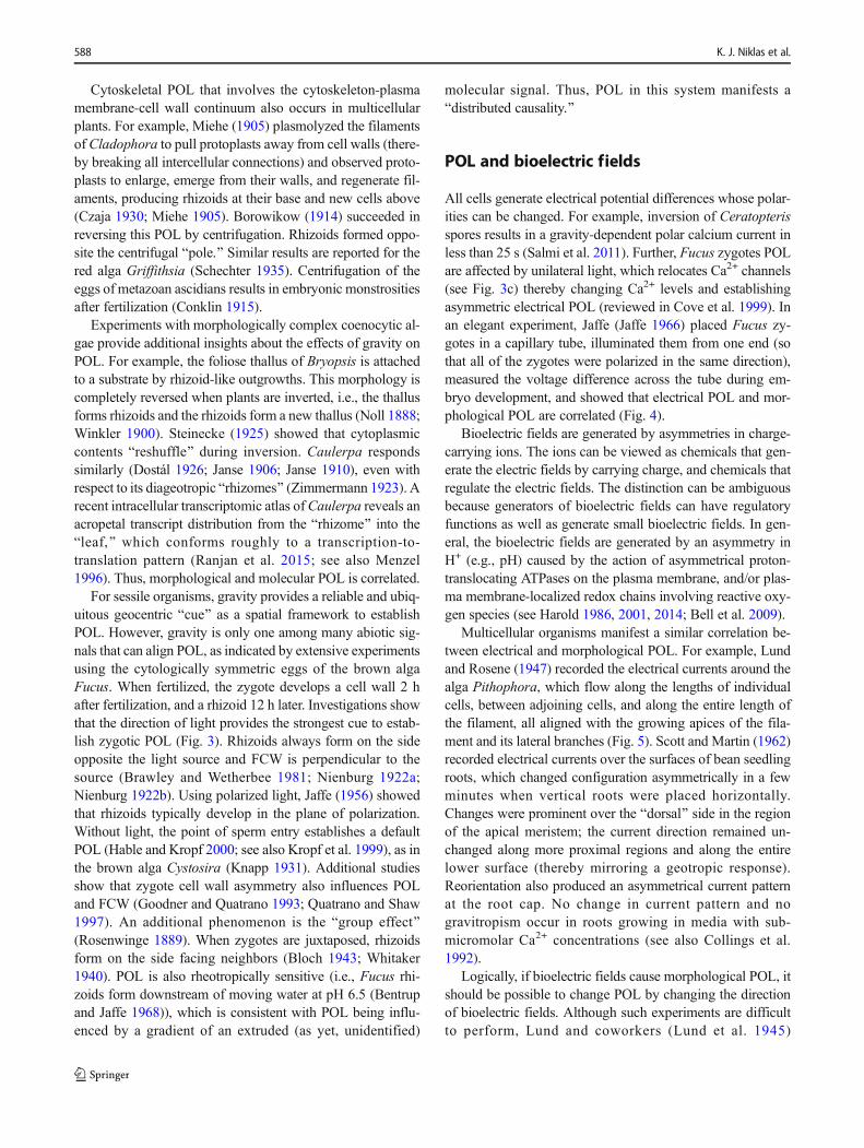
Other difficulties occur because POL can be weak orstrong, and sometimes reversible. For example, POL is notdiscernable in coccoid bacteria or in the eggs of many algae,whereas experiments using the asexual propagules of liver-worts (gemmae) show that the capacity to form rhizoids oneither side decreases over time (Haberlandt 1914). Similarly,cuttings of moss gametophores can generate rhizoids at theirbase and protonema at their top, but POL is reversed byinverting their orientation (Fitting 1938; Westerdijk 1906), aphenomenology observed for stem cuttings of vascular plants(Vöchting 1878; Wulff 1910; Zimmermann 1923), but not forroots (Warmke and Warmke 1950). Experiments using ferngametophytes show that POL during regeneration is associat-ed with physiological gradients, particularly differences in os-motic concentrations (e.g., Gratzy-Wardengg 1929 andAlbaum 1938), and when fern gametophytes are sliced trans-versely, regeneration POL is invariably apical (Fig. 2).
Despite its spatiotemporal complexity, the literature showsthat POL becomes morphologically evident when one or moreof the three features occurs: (1) orientation with respect to aspatial frame of reference (e.g., gravity or light), (2) axiationin a direction relative to orientation (e.g., cell elongation orexpansion), and (3) asymmetry with respect to orientation oraxiation (e.g., oblique cell division).
POL at the cellular level
The relationship between POL and FCW in unicellular organ-isms is illustrated with Chlamydomonas and Polytoma,
586 K. J. Niklas et al.
wherein the FCW is typically longitudinal with respect to theanterior-posterior cell axis, i.e., the cell is symmetricallybisected. However, the spindle apparatus sometimes rotatesthrough a right angle before or during division such that theFCW is transverse to the original cell axis (e.g., Mosbacher1929; see also Fritsch 1965). Cytoplasmic asymmetries occur
also as a consequence of gravity. For example, proplastidsoccupy the upper half of Equisetum spores and the vegetativecells of the lycopod Isoetes and the alga Enteromorpha(Müller-Stoll 1952; Nienburg 1924; Stewart 1948). InEquisetum, the FCW bisects the spore beneath the proplastids(Nienburg 1924).
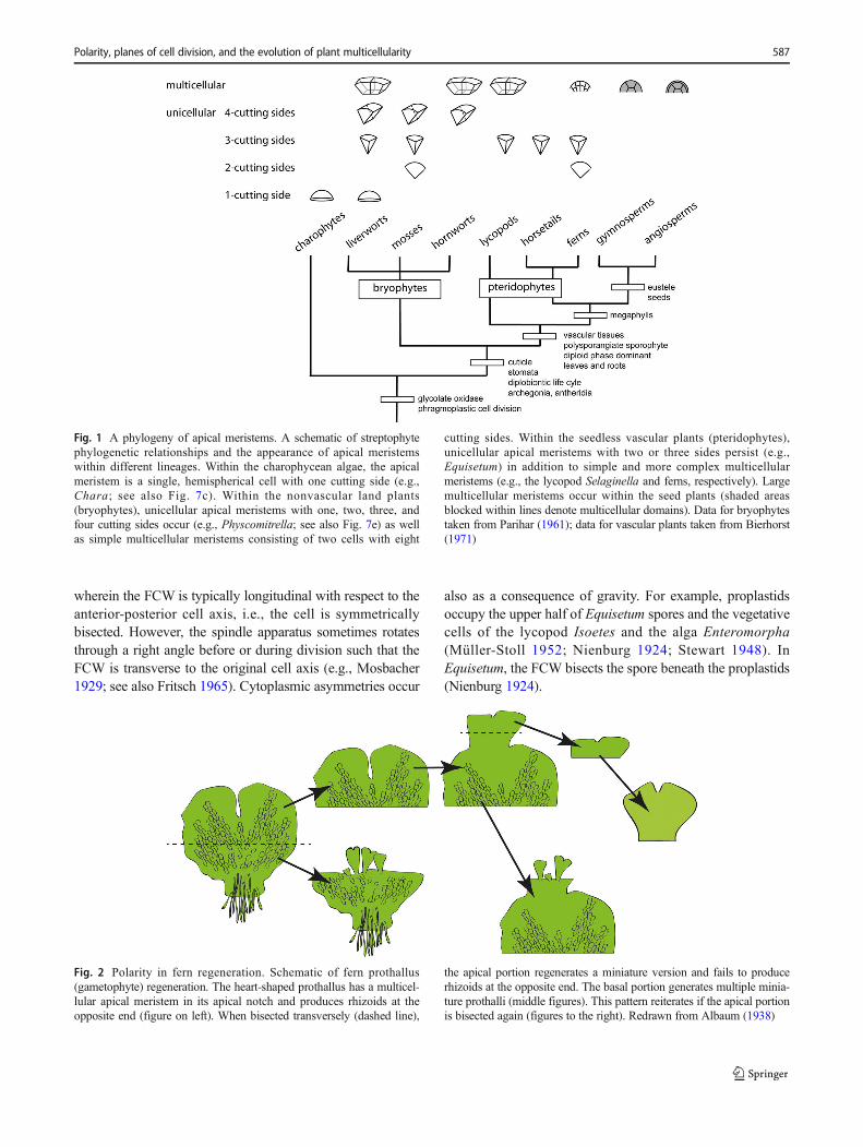
Fig. 1 A phylogeny of apical meristems. A schematic of streptophytephylogenetic relationships and the appearance of apical meristemswithin different lineages. Within the charophycean algae, the apicalmeristem is a single, hemispherical cell with one cutting side (e.g.,Chara; see also Fig. 7c). Within the nonvascular land plants(bryophytes), unicellular apical meristems with one, two, three, andfour cutting sides occur (e.g., Physcomitrella; see also Fig. 7e) as wellas simple multicellular meristems consisting of two cells with eight
cutting sides. Within the seedless vascular plants (pteridophytes),unicellular apical meristems with two or three sides persist (e.g.,Equisetum) in addition to simple and more complex multicellularmeristems (e.g., the lycopod Selaginella and ferns, respectively). Largemulticellular meristems occur within the seed plants (shaded areasblocked within lines denote multicellular domains). Data for bryophytestaken from Parihar (1961); data for vascular plants taken from Bierhorst(1971)
Fig. 2 Polarity in fern regeneration. Schematic of fern prothallus(gametophyte) regeneration. The heart-shaped prothallus has a multicel-lular apical meristem in its apical notch and produces rhizoids at theopposite end (figure on left). When bisected transversely (dashed line),
the apical portion regenerates a miniature version and fails to producerhizoids at the opposite end. The basal portion generates multiple minia-ture prothalli (middle figures). This pattern reiterates if the apical portionis bisected again (figures to the right). Redrawn from Albaum (1938)
Polarity, planes of cell division, and the evolution of plant multicellularity 587
Cytoskeletal POL that involves the cytoskeleton-plasmamembrane-cell wall continuum also occurs in multicellularplants. For example, Miehe (1905) plasmolyzed the filamentsofCladophora to pull protoplasts away from cell walls (there-by breaking all intercellular connections) and observed proto-plasts to enlarge, emerge from their walls, and regenerate fil-aments, producing rhizoids at their base and new cells above(Czaja 1930; Miehe 1905). Borowikow (1914) succeeded inreversing this POL by centrifugation. Rhizoids formed oppo-site the centrifugal Bpole.^ Similar results are reported for thered alga Griffithsia (Schechter 1935). Centrifugation of theeggs of metazoan ascidians results in embryonic monstrositiesafter fertilization (Conklin 1915).
Experiments with morphologically complex coenocytic al-gae provide additional insights about the effects of gravity onPOL. For example, the foliose thallus of Bryopsis is attachedto a substrate by rhizoid-like outgrowths. This morphology iscompletely reversed when plants are inverted, i.e., the thallusforms rhizoids and the rhizoids form a new thallus (Noll 1888;Winkler 1900). Steinecke (1925) showed that cytoplasmiccontents Breshuffle^ during inversion. Caulerpa respondssimilarly (Dostál 1926; Janse 1906; Janse 1910), even withrespect to its diageotropic Brhizomes^ (Zimmermann 1923). Arecent intracellular transcriptomic atlas ofCaulerpa reveals anacropetal transcript distribution from the Brhizome^ into theBleaf,^ which conforms roughly to a transcription-to-translation pattern (Ranjan et al. 2015; see also Menzel1996). Thus, morphological and molecular POL is correlated.
For sessile organisms, gravity provides a reliable and ubiq-uitous geocentric Bcue^ as a spatial framework to establishPOL. However, gravity is only one among many abiotic sig-nals that can align POL, as indicated by extensive experimentsusing the cytologically symmetric eggs of the brown algaFucus. When fertilized, the zygote develops a cell wall 2 hafter fertilization, and a rhizoid 12 h later. Investigations showthat the direction of light provides the strongest cue to estab-lish zygotic POL (Fig. 3). Rhizoids always form on the sideopposite the light source and FCW is perpendicular to thesource (Brawley and Wetherbee 1981; Nienburg 1922a;Nienburg 1922b). Using polarized light, Jaffe (1956) showedthat rhizoids typically develop in the plane of polarization.Without light, the point of sperm entry establishes a defaultPOL (Hable and Kropf 2000; see also Kropf et al. 1999), as inthe brown alga Cystosira (Knapp 1931). Additional studiesshow that zygote cell wall asymmetry also influences POLand FCW (Goodner and Quatrano 1993; Quatrano and Shaw1997). An additional phenomenon is the Bgroup effect^(Rosenwinge 1889). When zygotes are juxtaposed, rhizoidsform on the side facing neighbors (Bloch 1943; Whitaker1940). POL is also rheotropically sensitive (i.e., Fucus rhi-zoids form downstream of moving water at pH 6.5 (Bentrupand Jaffe 1968)), which is consistent with POL being influ-enced by a gradient of an extruded (as yet, unidentified)
molecular signal. Thus, POL in this system manifests aBdistributed causality.^
POL and bioelectric fields
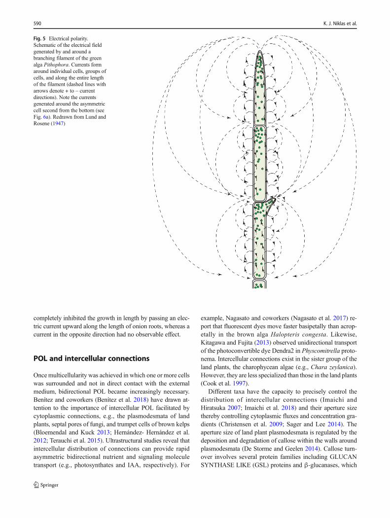
All cells generate electrical potential differences whose polar-ities can be changed. For example, inversion of Ceratopterisspores results in a gravity-dependent polar calcium current inless than 25 s (Salmi et al. 2011). Further, Fucus zygotes POLare affected by unilateral light, which relocates Ca2+ channels(see Fig. 3c) thereby changing Ca2+ levels and establishingasymmetric electrical POL (reviewed in Cove et al. 1999). Inan elegant experiment, Jaffe (Jaffe 1966) placed Fucus zy-gotes in a capillary tube, illuminated them from one end (sothat all of the zygotes were polarized in the same direction),measured the voltage difference across the tube during em-bryo development, and showed that electrical POL and mor-phological POL are correlated (Fig. 4).
Bioelectric fields are generated by asymmetries in charge-carrying ions. The ions can be viewed as chemicals that gen-erate the electric fields by carrying charge, and chemicals thatregulate the electric fields. The distinction can be ambiguousbecause generators of bioelectric fields can have regulatoryfunctions as well as generate small bioelectric fields. In gen-eral, the bioelectric fields are generated by an asymmetry inH+ (e.g., pH) caused by the action of asymmetrical proton-translocating ATPases on the plasma membrane, and/or plas-ma membrane-localized redox chains involving reactive oxy-gen species (see Harold 1986, 2001, 2014; Bell et al. 2009).
Multicellular organisms manifest a similar correlation be-tween electrical and morphological POL. For example, Lundand Rosene (1947) recorded the electrical currents around thealga Pithophora, which flow along the lengths of individualcells, between adjoining cells, and along the entire length ofthe filament, all aligned with the growing apices of the fila-ment and its lateral branches (Fig. 5). Scott and Martin (1962)recorded electrical currents over the surfaces of bean seedlingroots, which changed configuration asymmetrically in a fewminutes when vertical roots were placed horizontally.Changes were prominent over the Bdorsal^ side in the regionof the apical meristem; the current direction remained un-changed along more proximal regions and along the entirelower surface (thereby mirroring a geotropic response).Reorientation also produced an asymmetrical current patternat the root cap. No change in current pattern and nogravitropism occur in roots growing in media with sub-micromolar Ca2+ concentrations (see also Collings et al.1992).
Logically, if bioelectric fields cause morphological POL, itshould be possible to change POL by changing the directionof bioelectric fields. Although such experiments are difficultto perform, Lund and coworkers (Lund et al. 1945)
588 K. J. Niklas et al.
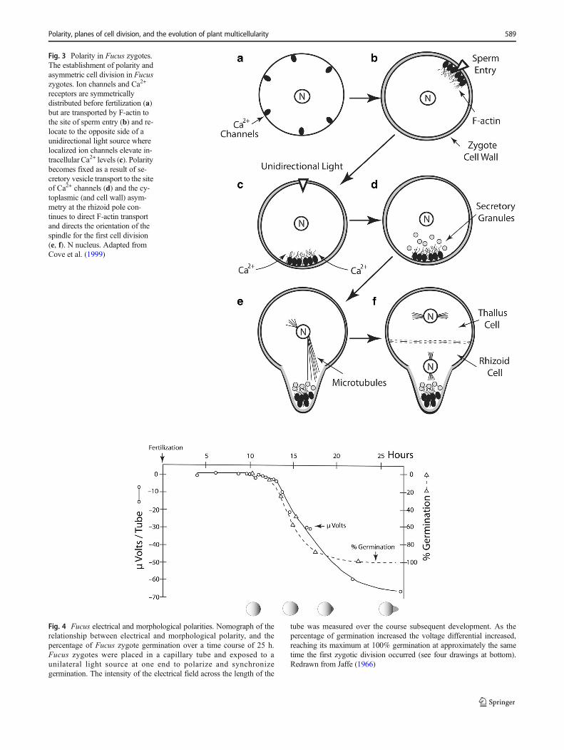
Fig. 3 Polarity in Fucus zygotes.The establishment of polarity andasymmetric cell division in Fucuszygotes. Ion channels and Ca2+
receptors are symmetricallydistributed before fertilization (a)but are transported by F-actin tothe site of sperm entry (b) and re-locate to the opposite side of aunidirectional light source wherelocalized ion channels elevate in-tracellular Ca2+ levels (c). Polaritybecomes fixed as a result of se-cretory vesicle transport to the siteof Ca2+ channels (d) and the cy-toplasmic (and cell wall) asym-metry at the rhizoid pole con-tinues to direct F-actin transportand directs the orientation of thespindle for the first cell division(e, f). N nucleus. Adapted fromCove et al. (1999)
Fig. 4 Fucus electrical and morphological polarities. Nomograph of therelationship between electrical and morphological polarity, and thepercentage of Fucus zygote germination over a time course of 25 h.Fucus zygotes were placed in a capillary tube and exposed to aunilateral light source at one end to polarize and synchronizegermination. The intensity of the electrical field across the length of the
tube was measured over the course subsequent development. As thepercentage of germination increased the voltage differential increased,reaching its maximum at 100% germination at approximately the sametime the first zygotic division occurred (see four drawings at bottom).Redrawn from Jaffe (1966)
Polarity, planes of cell division, and the evolution of plant multicellularity 589
completely inhibited the growth in length by passing an elec-tric current upward along the length of onion roots, whereas acurrent in the opposite direction had no observable effect.
POL and intercellular connections
Once multicellularity was achieved in which one or more cellswas surrounded and not in direct contact with the externalmedium, bidirectional POL became increasingly necessary.Benítez and coworkers (Benítez et al. 2018) have drawn at-tention to the importance of intercellular POL facilitated bycytoplasmic connections, e.g., the plasmodesmata of landplants, septal pores of fungi, and trumpet cells of brown kelps(Bloemendal and Kuck 2013; Hernández- Hernández et al.2012; Terauchi et al. 2015). Ultrastructural studies reveal thatintercellular distribution of connections can provide rapidasymmetric bidirectional nutrient and signaling moleculetransport (e.g., photosynthates and IAA, respectively). For
example, Nagasato and coworkers (Nagasato et al. 2017) re-port that fluorescent dyes move faster basipetally than acrop-etally in the brown alga Halopteris congesta. Likewise,Kitagawa and Fujita (2013) observed unidirectional transportof the photoconvertible dye Dendra2 in Physcomitrella proto-nema. Intercellular connections exist in the sister group of theland plants, the charophycean algae (e.g., Chara zeylanica).However, they are less specialized than those in the land plants(Cook et al. 1997).
Different taxa have the capacity to precisely control thedistribution of intercellular connections (Imaichi andHiratsuka 2007; Imaichi et al. 2018) and their aperture sizethereby controlling cytoplasmic fluxes and concentration gra-dients (Christensen et al. 2009; Sager and Lee 2014). Theaperture size of land plant plasmodesmata is regulated by thedeposition and degradation of callose within the walls aroundplasmodesmata (De Storme and Geelen 2014). Callose turn-over involves several protein families including GLUCANSYNTHASE LIKE (GSL) proteins and β-glucanases, which
Fig. 5 Electrical polarity.Schematic of the electrical fieldgenerated by and around abranching filament of the greenalga Pithophora. Currents formaround individual cells, groups ofcells, and along the entire lengthof the filament (dashed lines witharrows denote + to – currentdirections). Note the currentsgenerated around the asymmetriccell second from the bottom (seeFig. 6a). Redrawn from Lund andRosene (1947)
590 K. J. Niklas et al.
respectively synthesize and degrade callose (De Storme andGeelen 2014; Guseman et al. 2010; Ruan et al. 2004; see alsoBenítez et al. 2018). The amount of callose around plasmo-desmata is correlated with the genetic expression of GSLs andβ-glucanases, and the intercellular migration of signalingmol-ecules (Benitez-Alfonso et al. 2013; Guseman et al. 2010;Ruan et al. 2004; Vatén et al. 2011). For example, using aninducible knockdown mutation of GSL8, Han and coworkersreported that the reduced callose deposition at plasmodesmatain Arabidopsis hypocotyls enhances IAA diffusion. The lossof an asymmetric IAA distribution prevented differential cellelongation between the shaded and unshaded sides of the hy-pocotyl required for a normal phototropic response (Han et al.2014). These and other observations (e.g., Brunkard andZambryski 2017) indicate that the regulation of plasmodesma-ta apertures affects IAA transport, the establishment of IAAconcentration gradients, and thus POL at the organismic level.However, this is still an open avenue of research because theprobe used by Han and collaborators (2014) is blind to anyIAA within membraneous compartments and previous workshows that cell-cell auxin transport is not always inhibitedwhen plasmodesmata are severed (Cande and Ray 1976;Drake et al. 1978). Moreover, there are other molecules in-volved in PD integrity and gating, such as myosins and actincytoskeleton (Reichelt et al. 1999; Volkmann et al. 2003),which could be interacting with callose in complex ways.
POL and intracellular IAA gradients
Auxins, particular indole-3-acetic acid (IAA), are crucial inland plant development (Zažímalová et al. 2014). IAA mobil-ity is driven mainly by active transport into and out of cells,but auxin influx can also result from the passive diffusion ofthe protonated form of IAA across the plasma membrane.Active influx is mediated by the transport of the dissociated,anionic form (IAA−) by a permease 2H+-IAA co-transporter(e.g., AUX1), whereas active efflux of IAA requires auxinanion efflux carriers. Several protein families are known totransport IAA in Arabidopsis thaliana, but the PIN-FORMED (PIN) constitutes the best studied efflux carrierfamily. Arabidopsis has eight PIN transporters, most of whichlocalize at the plasma membrane (Feraru and Friml 2008).These proteins are actively sorted to specific domains of theplasma membrane and can thus anisotropically direct auxineffluxes (Adamowski and Friml 2015; Blilou et al. 2005;Feraru and Friml 2008; Wisniewska et al. 2006). PIN polarlocalization within cells gives rise to polar cell-to-cell trans-port, which can scale up to tissue- and organ-level patterning.Indeed, pin mutations alter IAA patterning and have signifi-cant phenotypic effects, e.g., changes in the morphology ofshoot and root apical meristems (Blilou et al. 2005; Okadaet al. 1991).
The study of PIN intracellular dynamics has uncoveredsome of the biochemical and physical regulators of IAA polartransport. Interestingly, evidence suggests that PINs are im-portant to regulate their own POL, possibly via coupled feed-backs (Geldner 2009; Hernández- Hernández et al. 2018;Niklas and Kutschera 2012). Mechanical forces acting at thetissue and plasma membrane level also seem to regulate PINlocalization, and therefore IAA polarization patterns.Specifically, there is evidence that the localization of the auxintransporter PIN1 and microtubule array orientation respond toa shared upstream biomechanical regulator as evidenced bymathematical modeling consistent with a biophysical cou-pling feedback loop between auxin transport in shoot apicalmeristems, i.e., the orientation of the microtubule cytoskele-ton, which is correlated with PIN1 orientation, is affected bybiomechanical stress fields (Heisler et al. 2010).
However, IAA also acts to loosen the cell-wall and tomodify localized responses to mechanical forces(Braybrook and Peaucelle 2013; Feraru et al. 2011;Heisler et al. 2010; Hernández- Hernández et al. 2018;Zwiewka et al. 2015). Further, PINs appear to directly orindirectly regulate proteins involved in endocytosis andvesicle formation, and therefore PIN intracellular trans-port and polar localization in the membrane. Some of themolecules potentially involved in this second feedbackloop are GNOM (a vesicle transport regulator), PINOID(a AGC-kinase that regulates PIN polarity), and rho-GTPases affecting endocytosis and actin filamentassembly. (Benjamins et al. 2001; Geldner 2009;Kleine-Vehn et al. 2008; Lehman et al. 2017; Lin et al.2013; Lin et al. 2012; Reinhardt et al. 2003; Sukumaret al. 2009). Collectively, our current knowledge suggeststhat mechanical and chemical factors act as a physico-genetic module to initiate, amplify, and maintain the po-larization patterns of the PIN auxin efflux carriers(Geldner 2009; Hernández- Hernández et al. 2018).
Although there are clear differences between the moleculesinvolved in plant and animal POL, some common elementsand dynamical motifs are recognizable in these two indepen-dently evolved systems. For example, rho-GTPases are keyfactors in the polarization processes in plants, yeast, and ani-mals, wherein they help organize actin filaments guiding moreactivated GTPases (Geldner 2009; Lin et al. 2013; Lin et al.2012). As further discussed below, the Rho family is a centralplayer in the PAR (partitioning defective complex) cytoskele-tal scaffolding apparatus underlying planar cell polarity inanimals. However, the main pattern emerging from the com-parison of intracellular-to-intercellular polarity in different lin-eages is that plant and animal systems rely on the spontaneousgeneration of POL from the amplification of small spatialdifferences, possibly initiated by diffusion and amplified byphysico-genetic modules of diverse molecular identities(Geldner 2009).
Polarity, planes of cell division, and the evolution of plant multicellularity 591
FCW and POL
A general theory predicting FCW remains elusive. Hofmeister(1863) noted that cell growth precedes division and that FCWnormally forms at 90° angle to the longitudinal axis of divid-ing cells. This observation is consistent with how cells dividein Spirogyra,Ulothrix, and the red alga Bangia. However, cellgrowth need not precede FCW (e.g., palintomy in Volvox) andBHofmeister’s rule^ is violated frequently (e.g., oblique wallsin moss protonema). Plateau (1873) predicted that FCW min-imizes the new wall’s surface area, an idea promulgated byBerthold (1886) and Errera (1888). Although Sachs (1878)agreed that new walls often meet old walls at 90° angles, evenwhen requiring curved walls, he rejected the minimum surfacearea hypothesis, which was nevertheless recapitulated byD’Arcy Thompson (Thompson and Whyte 1942).
The idea that FCW is influenced by mechanical forcescan be traced to Kny (1902), who applied unidirectionalpressure on dividing cells and observed mitotic figures torotate 90° such that FCW was parallel to the direction ofthe applied force. More recent experiments confirm thatexternally applied compression affects FCW (e.g.,Lintilhac and Vesecky 1984). However, it is not clearwhether endogenous mechanical forces generate FCWpatterns, and, it is difficult to conceive of how mechanicalforces per se dictate FCW in unicellular organisms, al-though they do affect FCW patterns in tissues (e.g.,Beauzamy et al. 2015; Louveaux et al. 2016).
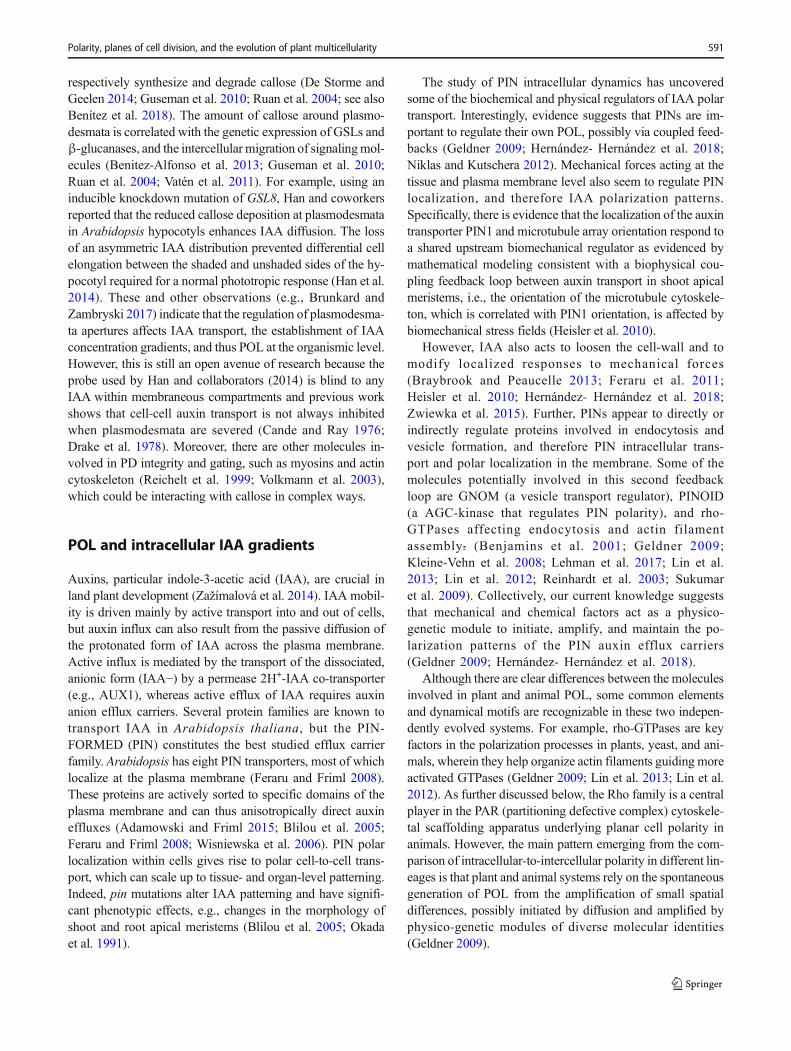
Regardless of how the FCW is prefigured, changes in POLare required to produce even simple body plans such as abranched filament. For example, in the green algaCladophora, branching is achieved by the formation of a lo-calized outgrowth in the cell wall, which subsequently forms anew cell by forming an oblique cell wall at its base (Fig. 6a;see also Fig. 5). Using the coenocytic yellow-green algaVaucheria, Kataoka (1975) found that blue light stimulatedlocalized outgrowth attended by an outward electrical current(lasting 1 hour) that reversed direction and continued as thenew branch developed. Physcomitrella protonema branchsimilarly (Fig. 6b, c), and produce a unicellular apical meri-stem initiated by a subsequent oblique division forming apyramidal cell with three Bcutting faces^ (see Fig. 7e). In eachcase, POLmust change within a cell. It is worth noting that theformation of branches in Cladophora, Vaucheria, andPhyscomitrella protonema differs from that of how transversewalls normally form because it requires a localized reductionin the yield stress of a previously formed wall and the local-ized delivery of new wall materials to the extruding tip. Bothprocesses occur in other algae, fungal hyphae, and in numer-ous specialized biological systems, e.g., the formation ofSpirogyra conjugation tubes and the Btip growth^ of pollengrains, branched unicellular trichomes, and root hairs (seeGeitmann and Ortega 2009, and Majda et al. 2017).
FCW, POL, and meristems
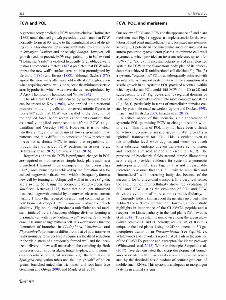
Our review of POL and FCWand the appearance of land plantmeristems (see Fig. 1) suggests a simple scenario for the evo-lution of land plant multicellularity and localized meristematicactivity: (1) polarity in the unicellular ancestor involved anantero-posterior cytoskeleton-plasma membrane-cell wallasymmetry, which provided an invariant reference system forFCW (Fig. 7a), (2) this ancestral polarity served as a referencesystem for FCW in the filamentous body plan of its descen-dants that achieved ID unidirectional cell division (Fig. 7b), (3)a systemic Borganismic^ POLwas subsequently achieved withan intercellular transport system, (4) with the acquisition of asessile growth habit, systemic POL provided a context withinwhich cytoskeletal POL could shift FCW from 1D to 2D andsubsequently to 3D (Fig. 7c–e), and (5) regional domains ofPOL and FCWactivity evolved into more complex meristems(Fig. 7e, f), particularly in terms of intercellular domains cre-ated by plasmodesmatal networks (Ligrone and Duckett 1998;Imaichi and Hiratsuka 2007; Imaichi et al. 2018).
A critical aspect of this scenario is the appearance ofsystemic POL permitting FCW to change direction with-in a cell. This form of POL may not have been difficultto achieve because a sessile growth habit provides aBglobal^ framework for POL. This is evident even atthe unicellular level when zygotes and zoospores attachto a substrate, undergo uneven transverse cell division,and produce a rhizoid at one end (see Fig. 3e, f). Thepresence of bioelectric fields around simple filamentoussessile algae provides evidence for systemic asymmetricantero-posterior POL (see Fig. 5). It is not unreasonabletherefore to assume that this POL will be amplified andBinternalized^ with increasing body size because of thenecessity for bi-directional transport. In a very real sense,the evolution of multicellularity drove the evolution ofPOL and FCW just as the evolution of POL and FCWdrove the evolution of more complex multicellularity.
Currently, little is known about the genetics involved in the1D-to-2D or a 2D-to-3D transition. However, a recent studyhighlights in importance of the CLAVATA peptide and areceptor-like kinase pathway in the land plants (Whitewoodset al. 2018). This system is unknown among the green algae(which achieve 1D and 2D polarity; see Fig. 7b, c). It is thusunique to the land plants. Using the 2D protonema-to-3D ga-metophore transition in Physcomitrella (see Fig. 7d, e),Whitewoods and coworkers report that 3D fails in the absenceof the CLAVATA peptide and a receptor-like kinase pathway(Whitewoods et al. 2018). While on this topic, Skopelitis et al.(2017) have demonstrated that sharp developmental bound-aries associated with foliar leaf dorsiventrality can be gener-ated by the threshold-based readout of counter-gradients ofmobile small RNAs. This system is analogous to morphogensystems in animal systems.
592 K. J. Niklas et al.
Plasticity of POL in volvocine algae
Unlike most multicellular algae and land plants, volvocine algaecan undergo morphogenetic movements that are similar to gas-trulation in metazoans (Höhn and Hallmann 2011; Höhn andHallmann 2016; Matt and Umen 2016), because even whenattached to each other, their cells are not cemented by rigid ma-terials. Initially cells are attached by numerous cytoplasmic brid-ges (CBs), the result of incomplete cytokinesis, which appear tobe functionally analogous, but not homologous to plasmodesma-ta. This flexible association (different from the transient cadherin-based attachments of embryonic animal cells) permits the cellsheets to undergo inversion (Höhn and Hallmann 2011; Hoopset al. 2005). In addition to the uncharacteristic (for plants) cell-sheet flexibility, the individual cells of volvocines (in contrast totheir unicellular relative, Chlamydomonas) lack rigid cell wallsbut have a flexible glycocalyx, enabling active cell shape changesthat accompany and apparently promote inversion. This plasticityof orientation, axiation, and asymmetry in cells that start out withintrinsic polarity permits these organisms to employ POL in wayakin to that in animal embryos.
As in metazoan embryos, folding or bending of thevolvocine cell sheet is achieved by changes in cell shape andconcerted movements of cells with respect to the CB system.But unlike most gastrulating animal embryos, cell division iscompleted before the beginning of inversion. Furthermore, thecells, being directly connected to one another, do not changetheir position relative to their neighbors, so migration or inter-calation is not involved. This makes the reorientation of POL,accompanied by changes in cell size and shape, the basis ofmorphogenesis in these algae.
A nonrigid extracellular matrix appears after inversion, butits absence beforehand means that volvocine cells, unlike
those of typical plants, can readily change their shape.During inversion, V. carteri cells are spindle-shaped, flask-shaped, or columnar, whereas those of V. globator are spin-dle-shaped, teardrop-shaped, paddle-shaped, or pencil-shapedin different regions at different stages. In all cases, the cells areintrinsically polarized, much the same as the unicellularChlamydomonas, with the nucleus located at the apical end,below the site of the incipient flagella, with the chloroplast atthe basal end. The position of the CB system changes relativeto these components during inversion of the cell sheets, mov-ing from a mid-cellular location to the basal portion of thecoupled cells in V. globator (Höhn and Hallmann 2011).
A physical model presented by Höhn et al. (2015) highlightsthe Bgeneric^ similarities (in the sense of being predictable fromthe physics of nonliving materials (Newman and Comper 1990))of these POL-dependent morphogenetic movements to those ofanimal systems, notwithstanding the added constraint in thevolvocines of conservation of nearest-neighbor relationships.
Comparison of POL in metazoansand prokaryotes
Although this Review focuses on POL in photosynthetic eu-karyotes, comparison with the POL DPM in animal and cer-tain bacterial systems helps to specify the particularities of ourmain subjects. Animal cells exhibit two distinct kinds of po-larity, apicobasal (A/B) and planar cell polarity (PCP). Thefirst serves to make the cell surface nonuniform by insertingdifferent sets of integral membrane proteins on different por-tions (e.g., apical, basolateral) of the plasma membrane. Thesecond changes the shape of the cell or the orientation of asurface appendage (e.g., a bristle, a cilium). In the
Fig. 6 Asymmetric cell divisions. Asymmetric cell divisions in thefilaments of the green alga Cladophora (a) and moss protonema (b, c).a Arrowheads indicate where asymmetric division is beginning (upperarrow) and where it has occurred (lower arrow). The oblique wall at thebase of the lateral filament (bottom arrow) differs in orientation from the
transverse wall just above it. b, c As in the case of Cladophora,asymmetric division in the moss protonema begins as a lateral bulge (b)that extends in length before a wall is formed at the base of the branch (seeFig. 7d)
Polarity, planes of cell division, and the evolution of plant multicellularity 593
multicellular context these are manifested as two differentPOL-type DPMs (Newman and Bhat 2009), POLa, whichleads, for example, to the formation of lumens in clusters ofcell (Bedzhov and Zernicka-Goetz 2014), and POLp, whichby enabling cell-cell intercalation, leads to the tissue reshapingphenomenon known as convergent extension (Skoglund andKeller 2010).
Both A/B and PCP are elicited in animal cells by themetazoan-specific short-range morphogen Wnt (Karner et al.
2006a; Karner et al. 2006b), via a common cytoskeletal scaf-folding complex termed PAR (Lang andMunro 2017), thoughusing some different adaptor molecules. Polarity is stabilizedin animal tissues by interactions of cells with their neighborsand extracellular matrices via transmembrane cadherin andintegrin proteins (Allam et al. 2018). Plant cells lack thePAR complex and these surface molecules. However, theirPINs serve analogous polarity-inducing functions.Interestingly, however, two of the key components of PAR,
Fig. 7 Evolution of POL and FCW. Schematics of unicellular andmulticellular plants arranged to illustrate a scenario for the evolution ofland plant multicellularity and apical meristems with increasingcomplexity. Centered dashed lines and arrows denote planes of division(1D, 2D, and 3D). The organisms depicted here are not intended tosuggest phylogenetic relationships. a POL and FCW in unicellularalgae are established by an asymmetric cytoskeleton-plasma membrane-cell wall continuum (e.g., Chlamydomonas). FCW occurs in only oneplane. b POL and FCW in a simple unbranched alga are also establishedby asymmetric cytoskeletons and cell-to-cell end wall. FCW occurs in
only one plane. c, d Among sessile plants, POL becomes systemic at theorganismic level by means of localized apical growth (denoted by *) andintercellular transport. Adjustment of POL at the cellular level can reori-ent FCW to achieve cell division in two directions (e.g., 2D in Chara,Cladophora, and moss protonema) (see Fig. 6). e, f POL in apical cells(denoted by *) reposition FCW to achieve two or more Bcutting sides^(indicated by numbers). Systemic POL and FCW are coordinated withinthe plant body by intercellular symplastic transport and signaling systems(e.g., plasmodesmata and IAA, respectively). V vacuole
594 K. J. Niklas et al.
the Rho family kinase Cdc42, a cell cycle regular, and thecalcium-dependent scaffolding protein MO25 (also calledCab29) are both present in yeast, where they are similarlyinvolved in polarity generation (Halatek et al. 2018;Mendoza et al. 2005). Other enzymes conserved betweenyeast and metazoans are also suspected of participating inestablishing polarity (Lee et al. 2018), suggesting deep diver-gence between cell polarization in plants, on the one hand, andopisthokonts, the group comprising fungi, metazoans, andunicellular holozoans, on the other.
Polarity in prokaryotes has been well-studied (Bowmanet al. 2011; Dworkin 2009). One illuminating comparison toplants involves the myxobacteria, which exhibit changes inpolarity at the single-cell level, changing reversibly from around, symmetrical form, the spore, to a rod-shaped vegeta-tive form (LaRossa et al. 1983). These cells also undergomulticellular morphogenesis (swarming and fruiting body for-mation) (Hartzell 2016; Kaiser 2008; Whitworth andAmerican Society for Microbiology. 2008; Yang and Higgs2014), exhibiting therefore the POL DPM as well. A cell wallstabilizes the shape asymmetry of the developmentally com-petent bacteria (as in most plant cells, but unlike the cells ofvolvocine algae and animals during their developmentalstages). Unlike multicellular plants, however, the cell walldoes not cement the cells together, but allows for gliding, withnearest neighbors continually changing.
InMyxococcus xanthus, a model myxobacteria, the molec-ular components associated with cellular differentiation andcommunication are integrated in a regulatory network thatincludes transcription factors, membrane proteins, protein ki-nases, scaffold proteins, and enzymes (Arias Del Angel et al.2018). POL in myxobacteria is more like that of animal em-bryos, where the cells are also independently mobile, than thevolvocines, where the cells are harnessed together by systemsof cytoplasmic bridges. As we have seen, the uses of polarityin typical multicellular plants are unlike any of these.
Conclusions: POL, ion channels,and symmetry breaking
Following the advice of George Gore (1878), our review ofthe literature leads us to conclude that asymmetry in cytosolicion concentrations resulting from changes in ion channel andpump location, activity, or type is an ancient phylogeneticallycommon denominator with which to achieve POL and thusorient FCW. If correct, the Bdefault^ ancestral condition was aunicellular organism lacking a structurally polarized cytoskel-eton enveloped by amore or less uniform cell membrane and amore or less uniform cell wall (e.g., coccoidal prokaryotes,Fucus eggs). This cytoskeleton-plasma membrane-cell wallsymmetry was broken evolutionarily in different ways toachieve more complex POL and FCW. In some lineages,
asymmetry relied on external cues (e.g., gravity or light). Inothers, asymmetry evolved as a consequence of morphologi-cal and physiological specializations, such as flagella at theanterior end of the cell (e.g., Chlamydomonas). With the ad-vent of simple multicellularity POL and FCW involved inter-as well as intracellular gradients. Complex multicellularityand meristematic growth were achieved by the ability to man-ifest 1D to 2D and then 3D POL and FCW.
Symmetry breaking in the streptophytes likely involvedCa2+ channels and pumps. Ca2+ fluxes are associated withevery example of POL thus far reported in this clade. Theregulation of cytoplasmic Ca2+ is a major link between bio-electrical signaling and cellular activity. However, calcium is acytotoxin for organisms relying on phosphate metabolism forenergy because it binds with phosphate to form hydroxyapatite(Wayne 2009). Therefore, phosphate-dependent organismshave to prevent high concentrations of calcium in extracellularspaces frommixing with cytosolic phosphate. This is achievedby Ca2+ pumps that maintain low cytosolic Ca2+ levels. Withthe evolution of Ca2+ channels, calcium could serve as anefficient signaling molecule because of its potential toxicityand low intracellular concentration. Only three componentsare necessary: (1) gated calcium channels for the rapid intra-and extracellular transport of messenger ions, (2) Ca2+ pumpsand sequestering proteins to restore the resting level of Ca2+
and regulate signal durations, and (3) regulatory moleculesable to sense intracellular Ca2+ concentrations. Ionic channelsthat extrude Ca2+ and deliver external K+ exist in prokaryotes(Hille 1992). Consequently, given their ancestral capacities toredistribute proteins, the first eukaryotes had only to changemembrane proteins to control cytoplasmic Ca2+.
Because any mechanical, electrical, or chemical change inthe plasma membrane is, in theory, capable of opening Ca2+
channels, calcium can serve as a Bsecondary^ messenger in avast constellation of metabolic processes (Weber 1976).Extensive research on plants shows that Ca2+ channels be-come activated (followed by dramatic increases in cytosolicCa2+) by mechanical perturbation (e.g., vibration and touch),growth-altering substances (e.g., brassinosteroids andjasmonates), and abiotic factors (e.g., light and gravity).Thus, Ca2+ channels permit diverse kinds of stimuli to evokesimilar or identical responses.
What is unclear is how cells discriminate among differentstimuli. Theoretically, we expect organisms to evolve signal-specific sensory systems permitting them to respond in adap-tively different ways to different environmental cues. Thismay help to explain why there are many different kinds ofCa2+ channels that may trigger different subcellular systems.Each channel is a protein, made up of one or more polypep-tides forming a hydrophilic pore in a membrane. Althougheach allows Ca2+ to pass relatively unimpeded at a rate ofabout 106 s−1 or more (Wayne 2009), channels can be eithernonselective, or highly selective for Ca2+ depending on their
Polarity, planes of cell division, and the evolution of plant multicellularity 595
pore size and the charge density of their binding sites—prop-erties that depend on protein structure, which can vary amongchannels. Thus, many types of Ca2+ channels can occupy thesame membrane. This variation in protein structure and sizeprovides a mechanism whereby different signals can be per-ceived and redirected differently.
Finally, because the origins of POL and FCWare polyphy-letic and have been achieved in different ways in differentlineages, each is a true dynamical patterning module. Sincedifferential gene expression within a cell is incapable ofachieving either and since it is obvious that both occur inunicellular organisms, changes in either or both modules mustrely on changes in gene expression patterns using internal orexternal cues. One example of the latter is the blue light recep-tor aureochrome (AUREO) found in photosyntheticstramenopiles (see Takahashi 2016). Within this clade,AUREO participates in rhizoid formation (e.g., Fucus), cellshape regulation (e.g., Vaucheria), and chloroplast movement(e.g., Vaucheria). Takahashi and coworkers reported thatAUREO1 has a bZIP domain and a light-oxygen-voltage do-main that operate as a transcription factor, binding to a specificcis element consensus sequence TGACGTwhen cells are irra-diated with blue light (Takahashi et al. 2007). It was furthershown that under oxidative conditions AUREO1 forms a di-mer at its bZIP region by means of disulfide bonds (Hisatomiet al. 2014). Although the genes that AUREO activates orsilences are currently unknown, it is clear that AUREO is in-volved in a diverse range of morphogenetic responses affectingPOL and FCW in a large clade of eukaryotic organisms.
BEvery event has many surrounding antecedents, andthey may be divided into those which are separable fromthe event, and those which are not; and the cause of anevent is always to be found amongst the inseparableones only. In ordinary language, the most probablecause of an event is, a priori, that circumstance which,in the greatest number of cases, immediately precedes oraccompanies it.^ –– Gore (1878)
Acknowledgements The authors thank Dr. Thomas Owens (CornellUniversity) for stimulating and constructive discussions and three anon-ymous reviewers who provided invaluable feedback.
Author contributions KJN, RW, MB, and SAN conceived and wrote thepaper. KJN provided the graphics in Figs. 1–7.
Funding information This study received funding from the College ofAgriculture and Life Sciences (Cornell University).
Compliance with ethical standards
Conflict of interest The authors declare that they have no conflicts ofinterest.
References
Adamowski M, Friml J (2015) PIN-dependent auxin transport: action,regulation, and evolution. Plant Cell 27:20–32. https://doi.org/10.1105/tpc.114.134874
Albaum HG (1938) Normal growth, regeneration and adventitious out-growth formation in fern prothallia. Am J Bot 25:37–44
Allam AH, Charnley M, Russell SM (2018) Context-specific mecha-nisms of cell polarity regulation. J Mol Biol 430:3457–3471
Allman GJ (1864) Report on the present state of our knowledge of thereproductive system in Hydroida. Rept British Assoc Adv Sci 1863:351–426
Arias Del Angel JA, Escalante AE, Martinez-Castilla LP, Benitez M(2018) Cell-fate determination in Myxococcus xanthus develop-ment: network dynamics and novel predictions. Develop GrowthDiffer 60:121–129. https://doi.org/10.1111/dgd.12424
Beauzamy L, Louveaux M, Hamant O, Boudaoud A (2015)Mechanically, the shoot apical meristem ofArabidopsis behaves likea shell inflated by a pressure of about 1MPa. Front Plant Sci 6:1038.https://doi.org/10.3389/fpls.2015.01038
Bedzhov I, Zernicka-Goetz M (2014) Self-organizing properties ofmouse pluripotent cells initiate morphogenesis upon implantation.Cell 156:1032–1044. https://doi.org/10.1016/j.cell.2014.01.023
Bell E, Takeda S, Dolan L (2009) Reactive oxygen species in growth anddevelopment. In del Río LA,Puppo A (eds) Reactive oxygen speciesin plant signaling. Springer, Berlin pp 43–55
Benítez M, Hernández- Hernández V, Newman SA, Niklas KJ (2018)Dynamical patterning modules, biogeneric materials, and the evolu-tion of multicellularity in plants. Frontiers in Plant Science. https://doi.org/10.3389/fpls.2018.00
Benitez-Alfonso Y, Faulkner C, Pendle A, Miyashima S, Helariutta Y,Maule A (2013) Symplastic intercellular connectivity regulates lat-eral root patterning. Dev Cell 26:136–147. https://doi.org/10.1016/j.devcel.2013.06.010
Benjamins R, Quint A, Weijers D, Hooykaas P, Offringa R (2001) ThePINOID protein kinase regulates organ development in Arabidopsisby enhancing polar auxin transport. Development 128:4057–4067
Bentrup FW, Jaffe LF (1968) Analyzing the "group effect": rheotropicresponses of developing Fucus eggs. Protoplasma 65:25–35
Berthold GDW (1886) Studien über protoplasmamechanik. A. Felix,Leipzig
Bierhorst DW (1971) Morphology of vascular plants. In: The Macmillanbiology series. Macmillan, New York
Blilou I, Xu J, Wildwater M, Willemsen V, Paponov I, Friml J, HeidstraR, Aida M, Palme K, Scheres B (2005) The PIN auxin efflux facil-itator network controls growth and patterning in Arabidopsis roots.Nature 433:39–44. https://doi.org/10.1038/nature03184
Bloch R (1943) Polarity in plants. Bot Rev 9:261–310Bloemendal S, Kuck U (2013) Cell-to-cell communication in plants, an-
imals, and fungi: a comparative review. Naturwissenschaften 100:3–19. https://doi.org/10.1007/s00114-012-0988-z
Borowikow GA (1914) La polarité renverse le Cladophora glomerate.Bull Jard Bot Pierre Grand 14:475–481
Bowman GR, Lyuksyutova AI, Shapiro L (2011) Bacterial polarity. CurrOpin Cell Biol 23:71–77. https://doi.org/10.1016/j.ceb.2010.10.013
Brawley SH, Wetherbee R (1981) The biology of seaweeds. In: LobbanCS, Wynne MJ (eds) Cytology and ultrastructure. Blackwell,Oxford, pp 248–299
Braybrook SA, Peaucelle A (2013) Mechano-chemical aspects of organformation in Arabidopsis thaliana: the relationship between auxinand pectin. PLoS One 8:e57813. https://doi.org/10.1371/journal.pone.0057813
Brunkard JO, Zambryski PC (2017) Plasmodesmata enable multicellular-ity: new insights into their evolution, biogenesis, and functions in
596 K. J. Niklas et al.
development and immunity. Curr Opin Plant Biol 35:76–83. https://doi.org/10.1016/j.pbi.2016.11.007
Cande WZ, Ray PI (1976) Nature of cell-to-cell transfer of auxin in polartransport. Planta 129:43–52
Christensen NM, Faulkner C, Oparka K (2009) Evidence for unidirec-tional flow through plasmodesmata. Plant Physiol 150:96–104.https://doi.org/10.1104/pp.109.137083
Collings DA, White RG, Overall RL (1992) Ionic current changes asso-ciated with the gravity-induced bending response in roots of Zeamays. L Plant Physiol 100:1417–1426
Conklin EG (1915) Heredity and environment in the development ofmen. Norman W Harris lectures for 1914 at NorthwesternUniversity. Princeton University Press, Princeton
CookM, Graham L, Botha C, Lavin C (1997) Comparative ultrastructureof plasmodesmata of Chara and selected bryophytes: toward anelucidation of the evolutionary origin of plant plasmodesmata. AmJ Bot 84:1169–1178
Cove DJ, Hope IA, Quatrano RS (1999) Polarity in biological systems.In: Russo VEA, Cove DJ, Edgar LG, Jaenisch R, Salamini F (eds)Development. Genetics, epigenetics and environmental regulation.Springer-Verlag, Berlin, pp 507–524
Czaja AT (1930) Zellphysiologische untersuchungen an Cladophoraglomerate Isolierung, regeneration und polarität. Protoplasma 11:197–220
De Storme N, Geelen D (2014) Callose homeostasis at plasmodesmata:molecular regulators and developmental relevance. Front Plant Sci5:138. https://doi.org/10.3389/fpls.2014.00138
Dostál R (1926) Zur Kenntnis der inneren Gestaltungsfaktoren beiCaulerpa prolifera. Ber Deutsch Bot Ges 44:56–66
Drake GA, Carr DJ, Anderson WP (1978) Plasmolysis, plasmodesmata,and the electrical coupling of oat coleoptile cells. J Exp Bot 29:1205–1214
Dworkin J (2009) Cellular polarity in prokaryotic organisms. Cold SpringHarb Perspect Biol 1:a003368. https://doi.org/10.1101/cshperspect.a003368
Errera L (1888) Über Zellformen und Seifenblasen. Bot Centralbl 34:395–398
Feraru E, Friml J (2008) PIN polar targeting. Plant Physiol 147:1553–1559. https://doi.org/10.1104/pp.108.121756
Feraru E, FeraruMI, Kleine-Vehn J,Martiniére A,Mouille G, Vanneste S,Vernhettes S, Runions J, Friml J (2011) PIN polarity maintenance bythe cell wall in Arabidopsis. Curr Biol 21:338–343. https://doi.org/10.1016/j.cub.2011.01.036
Fitting H (1938) Die Umkehrbarkeit der durch Aussenfaktoreninduzierten Dorsiventralität. Jahrb Wiss Bot 86:107–227
Fritsch FE (1965) The structure and reproduction of the algae vol I.Cambridge University Press, Cambridge
Geitmann A, Ortega JK (2009) Mechanics and modeling of plant cellgrowth. Trends Plant Sci 14:467–478. https://doi.org/10.1016/j.tplants.2009.07.006
Geldner N (2009) Cell polarity in plants: a PARspective on PINs. CurrOpin Plant Biol 12:42–48. https://doi.org/10.1016/j.pbi.2008.09.009
Goodner B, Quatrano RS (1993) Fucus embryogenesis: a model to studythe establishment of polarity. Plant Cell 5:1471–1481. https://doi.org/10.1105/tpc.5.10.1471
Gore G (1878) The art of scientific discovery. Longmans, Green, and Co.,London
Gratzy-Wardengg SAE (1929) Osmotische Untersuchungen anFarnprothallien. Planta 7:307–339
Guseman JM, Lee JS, Bogenschutz NL, Peterson KM, Virata RE, Xie B,Kanaoka MM, Hong Z, Torii KU (2010) Dysregulation of cell-to-cell connectivity and stomatal patterning by loss-of-function muta-tion in Arabidopsis chorus (glucan synthase-like 8). Development137:1731–1741. https://doi.org/10.1242/dev.049197
Haberlandt G (1914) Zur Entwicklungsphysiologie der Rhizoiden. SitzAkad Wiss Physik-Math Cl S.:384–401
Hable WE, Kropf DL (2000) Sperm entry induces polarity in fucoidzygotes. Development 127:493–501
Halatek J, Brauns F, Frey E (2018) Self-organization principles of intra-cellular pattern formation. Philos Trans R Soc Lond Ser B Biol Sci373:20170107. https://doi.org/10.1098/rstb.2017.0107
Han X, Hyun TK, Zhang M, Kumar R, Koh EJ, Kang BH, Lucas WJ,Kim JY (2014) Auxin-callose-mediated plasmodesmal gating is es-sential for tropic auxin gradient formation and signaling. Dev Cell28:132–146. https://doi.org/10.1016/j.devcel.2013.12.008
Harold FM (1986) The vital force: a study of bioenergetics. Freeman,New York
Harold FM (2001) The way of the cell: Molecules, organisms, and theorder of life. Oxford University Press, Oxford
Harold FM (2014) In search of cell history: the evolution of life’s buildingblocks. University of Chicago Press, Chicago
Hartzell T (2016) Myxobacteria. In: eLS. https://doi.org/10.1002/9780470015902.a0020391.pub2
Heisler MG, Hamant O, Krupinski U, Ohno C, Jönsson H, Traas,Meyerwitz EM (2010) Alignment between PIN1 polarity and mi-crotubule orientation in the shoot apical meristem reveals a tightcoupling between morphogenesis and auxin transport. PLoS Biol8:e1000516. https://doi.org/10.1371/journal.pbio.1000516
Hernández- Hernández V, Niklas KJ, Newman SA, Benitez M (2012)Dynamical patterning modules in plant development and evolution.Int J Dev Biol 56:661–674. https://doi.org/10.1387/ijdb.120027mb
Hernández- Hernández V, Barrio RA, Benitez M, Nakayama N, Romero-Arias JR, Villarreal C (2018) A physico-genetic module for thepolarisation of auxin efflux carriers PIN-FORMED (PIN). PhysBiol 15:036002. https://doi.org/10.1088/1478-3975/aaac99
Hille B (1992) Ionic channels of excitable membranes, 2nd edn. SinauerAssociates, Sunderland
Hisatomi O, Nakatani Y, Takeuchi K, Takahashi F, Kataoka H (2014)Blue light-induced dimerization of monomeric aureochrome-1 en-hances its affinity for the target sequence. J Biol Chem 289:17379–17391. https://doi.org/10.1074/jbc.M114.554618
Höhn S, Hallmann A (2011) There is more than one way to turn a spher-ical cellular monolayer inside out: type B embryo inversion inVolvox globator. BMC Biol 9:89. https://doi.org/10.1186/1741-7007-9-89
Höhn S, Hallmann A (2016) Distinct shape-shifting regimes of bowl-shaped cell sheets - embryonic inversion in the multicellular greenalga Pleodorina. BMC Dev Biol 16:35. https://doi.org/10.1186/s12861-016-0134-9
Höhn S, Honerkamp-Smith AR, Haas PA, Trong PK, Goldstein, RE(2015) Dynamics of a Volvox embryo turning itself inside out.Phys Rev Lett 114:178101
Hoops HJ, Nishii I, Kirk DL (2005) Cytoplasmic bridges in Volvox andits relatives. In: Baluska F, Volkmann D, Barlow PW (eds) Cell-cellchannels. Eurekah.com, Georgetown, pp 1–20
Imaichi R, Hiratsuka R (2007) Evolution of shoot apical meristem struc-tures in vascular plants with respect to plasmodesmatal network. AmJ Bot 94:1911–1921
Imaichi R, Moritoki N, Solvang HK (2018) Evolution of root apicalmeristem structure in vascular plants: plasmodesmatal networks.Am J Bot 105:1453–1468
Jaffe L (1956) Effect of polarized light on polarity of Fucus. Science 123:1081–1082. https://doi.org/10.1126/science.123.3207.1081
Jaffe LF (1966) Electrical currents through the developing Fucus egg.Proc Natl Acad Sci U S A 56:1102–1109
Janse JM (1906) Polarität und Organbildung bei Caulerpa prolifera.Jahrb Wiss Bot 42:394–460
Janse JM (1910) Über Organveränderung bei Caulerpa prolifera. JahrbWiss Bot 48:73–110
Polarity, planes of cell division, and the evolution of plant multicellularity 597
Kaiser D (2008) Myxococcus-from single-cell polarity to complex mul-ticellular patterns. Annu Rev Genet 42:109–130. https://doi.org/10.1146/annurev.genet.42.110807.091615
Karner C, Wharton KA, Carroll TJ (2006a) Apical-basal polarity, Wntsignaling and vertebrate organogenesis. Semin Cell Dev Biol 17:214–222
Karner C, Wharton KA Jr, Carroll TJ (2006b) Planar cell polarity andvertebrate organogenesis. Semin Cell Dev Biol 17:194–203
Kataoka H (1975) Phototropism in Vaucheria geminata. II. The mecha-nism of bending and branching. Plant Cell Physiol 16:439–448
Kitagawa M, Fujita T (2013) Quantitative imaging of directional trans-port through plasmodesmata in moss protonemata via single-cellphotoconversion of Dendra2. J Plant Res 126:577–585. https://doi.org/10.1007/s10265-013-0547-5
Kleine-Vehn J, Dhonukshe P, Sauer M, Brewer PB, Wiśniewska J,Paciorek T, Benková E, Friml J (2008) ARF GEF-dependenttranscytosis and polar delivery of PIN auxin carriers inArabidopsis. Curr Biol 18:526–531. https://doi.org/10.1016/j.cub.2008.03.021
Knapp E (1931) Entwicklungsphysiologische Untersuchungen anFucaceen-Eiern. I. Zur Kenntnis der Polarität der Eier vonCystosira barbata. Planta 14:731–751
Kny L (1902) Über den Einfluss von Zug und Druck auf die Richtung derScheidewände in sich theilenden Pflanzenzellen. (ZweiteMittheilung) Jahrb Wiss Bot 37:55–98
Kropf DL, Bisgrove SR, Hable WE (1999) Establishing a growth axis infucoid algae. Trends Plant Sci 4:490–494
Lang CF, Munro E (2017) The PAR proteins: from molecular circuits todynamic self-stabilizing cell polarity. Development 144:3405–3416.https://doi.org/10.1242/dev.139063
LaRossa R, Kuner J, Hagen D, Manoil C, Kaiser D (1983)Developmental cell interactions of Myxococcus xanthus: analysisof mutants. J Bacteriol 153:1394–1404
Lee ME, Rusin SF, Jenkins N, Kettenbach AN, Moseley JB (2018)Mechanisms connecting the conserved protein kinases Ssp1, Kin1,and Pom1 in fission yeast cell polarity and pivision. Curr Biol 28:84–92 e84. https://doi.org/10.1016/j.cub.2017.11.034
Lehman TA, Smertenko A, Sanguinet KA (2017) Auxin, microtubules,and vesicle trafficking: conspirators behind the cell wall. J Exp Bot68:3321–3329. https://doi.org/10.1093/jxb/erx205
Li FW, Brouwer P, Carretero-Paulet L, Cheng S, de Vries J, Delaux PM,Eily A, Koppers N, Kuo LY, Li Z, Simenc M, Small I, Wafula E,Angarita S, Barker MS, Bräutigam A, dePamphilis C, Gould S,Hosmani PS, Huang YM, Huettel B, Kato Y, Liu X, Maere S,McDowell R, Mueller LA, Nierop KGJ, Rensing SA, Robison T,Rothfels CJ, Sigel EM, Song Y, Timilsena PR, van de Peer Y, WangH, Wilhelmsson PKI, Wolf PG, Xu X, der JP, Schluepmann H,Wong GKS, Pryer KM (2018) Fern genomes elucidate land plantevolution and cyanobacterial symbioses. Nat Plants 4:460–472.https://doi.org/10.1038/s41477-018-0188-8
Ligrone R, Duckett JG (1998) Development of the leafy shoot inSphagnum (Bryophyta) involves the activity of both apical and sub-apical meristem. New Phytol 140:581–595
Lin D, Nagawa S, Chen J, Cao L, Chen X, Xu T, Li H, Dhonukshe P,Yamamuro C, Friml J, Scheres B, Fu Y, Yang Z (2012) A ROPGTPase-dependent auxin signaling pathway regulates the subcellu-lar distribution of PIN2 in Arabidopsis roots. Curr Biol 22:1319–1325. https://doi.org/10.1016/j.cub.2012.05.019
Lin D, Cao L, Zhou Z, Zhu L, Ehrhardt D, Yang Z, Fu Y (2013) RhoGTPase signaling activates microtubule severing to promote micro-tubule ordering in Arabidopsis. Curr Biol 23:290–297. https://doi.org/10.1016/j.cub.2013.01.022
Lintilhac PM, Vesecky TB (1984) Stress-induced alignment of divisionplane in plant tissues grown in vitro. Nature 307:363–364
Louveaux M, Julien JD, Mirabet V, Boudaoud A, Hamant O (2016) Celldivision plane orientation based on tensile stress in Arabidopsis
thaliana. Proc Natl Acad Sci U S A 113:E4294–E4303. https://doi.org/10.1073/pnas.1600677113
Lund EJ, Rosene HF (1947) Bioelectric fields and growth. Univ. of TexasPress, Austin
Lund EJ, Mahan RI, Hanszen AH (1945) Electric control of polar growthin roots of Allium cepa. Proc Soc Exp Biol Med 60:326–327
MajdaM, Grones P, Sintorn IM, Vain T, Milani P, Krupinski P, Zagórska-Marek B, Viotti C, Jönsson H,Mellerowicz EJ, Hamant O, Robert S(2017) Mechanochemical polarization of contiguous cell wallsshapes plant pavement cells. Dev Cell 43:290–304 e294. https://doi.org/10.1016/j.devcel.2017.10.017
Matt G, Umen J (2016) Volvox: a simple algal model for embryogenesis,morphogenesis and cellular differentiation. Dev Biol 419:99–113.https://doi.org/10.1016/j.ydbio.2016.07.014
Mendoza M, Redemann S, Brunner D (2005) The fission yeast MO25protein functions in polar growth and cell separation. Eur J Cell Biol84:915–926. https://doi.org/10.1016/j.ejcb.2005.09.013
Menzel D (1996) The role of the cytoskeleton in polarity and morpho-genesis of algal cells. Curr Opin Cell Biol 8:38–42
Miehe H (1905) Wachstum, Regeneration und Polarität isolierter Zellen.Ber Deutsch Bot Ges 23:257–264
Mosbacher R (1929) Sur le mode de scission de Polytoma uvella et sesrapports avec la division des Flagellés et le clivage cellulaire.Comptes Rendus des Séances de la Société de Biologie et des sesFiliales (Paris) 93:278–281
Müller-Stoll WR (1952) Über Regeneration und Polarität beiEnteromorpha. Flora 139:148–180
Nagasato C, Tanaka A, Ito T, Katsaros C, Motomura T (2017)Intercellular translocation of molecules via plasmodesmata in themultiseriate filamentous brown alga, Halopteris congesta(Sphacelariales, Phaeophyceae). J Phycol 53:333–341. https://doi.org/10.1111/jpy.12498
Newman SA, Bhat R (2009) Dynamical patterning modules: a "patternlanguage" for development and evolution of multicellular form. Int JDev Biol 53:693–705. https://doi.org/10.1387/ijdb.072481sn
Newman SA, ComperWD (1990) Generic' physical mechanisms of mor-phogenesis and pattern formation. Development 110:1–18
Newman SA, Bhat R,Mezentseva NV (2009) Cell state switching factorsand dynamical patterning modules: complementary mediators ofplasticity in development and evolution. J Biosci 34:553–572
Nienburg W (1922a) Die Keimungsrichtung von Fucuseiern und dieTheorie der Lichtperzeption. (Vorlaufige Mitteilung) Ber DeutschBot Ges 40:38–40
Nienburg W (1922b) Die Polarisation der Fucus-Eier durch das Licht.Wiss Meeresunters Abt Helgoland 15, N F, Abhandl 7
Nienburg W (1924) Die Wirkung des Licht auf die Keimung derEquisetum spore. Ber Deutsch Bot Ges 42:95–99
Niklas KJ (2000) The evolution of plant body plans –– a biomechanicalperspective. Ann Bot 85:411–438
Niklas KJ (2014) The evolutionary-developmental origins of multicellu-larity. Am J Bot 101:6–25. https://doi.org/10.3732/ajb.1300314
Niklas KJ, Kutschera U (2012) Plant development, auxin, and the sub-system incompleteness theorem. Front Plant Sci 3:37. https://doi.org/10.3389/fpls.2012.00037
Niklas KJ, Cobb ED, Crawford DR (2013) The evo-devo of multinucle-ate cells, tissues, and organisms, and an alternative route to multi-cellularity. Evol Dev 15:466–474. https://doi.org/10.1111/ede.12055
Noll F (1888) Über den Einfluss der Lage auf die morphologischeAusbildung einiger Siphoneen. Arb Bot Inst Würzburg 3:466–476
Okada K, Ueda J, Komaki MK, Bell CJ, Shimura Y (1991) Requirementof the auxin polar transport system in early stages of Arabidopsisfloral bud formation. Plant Cell 3:677–684. https://doi.org/10.1105/tpc.3.7.677
598 K. J. Niklas et al.
Palmer JD, Soltis DE, Chase MW (2004) The plant tree of life: an over-view and some points of view. Am J Bot 91:1437–1445. https://doi.org/10.3732/ajb.91.10.1437
Parihar NS (1961) An introduction to embryophyta. In: Bryophyta, vol 1,4th edn. Central Book Depot, Allahabad
Plateau JAF (1873) Statique expérimentale et theéorique des liquidessoumis aux seules forces moleéculaires. Gauthier-Villars, Paris
Quatrano RS, Shaw SL (1997) Role of the cell wall in the determinationof two-celled Fucus embryos. Dev Biol 30:209–213
Ranjan A, Townsley BT, Ichihashi Y, Sinha NR, Chitwood DH (2015)An intracellular transcriptomic atlas of the giant coenocyteCaulerpataxifolia. PLoS Genet 11:e1004900. https://doi.org/10.1371/journal.pgen.1004900
Reichelt S, Knight AE, Hodge TP, Baluska F, Samaj J, Volkmann D,Kendrick-Jones J (1999) Characterization of the unconventionalmyosin VIII in plant cells and its localization at the post-cytokinetic cell wall. Plant J 19:555–567
Reinhardt D, Pesce ER, Stieger P,Mandel T, Baltensperger K, Bennett M,Traas J, Friml J, Kuhlemeier C (2003) Regulation of phyllotaxis bypolar auxin transport. Nature 426:255–260. https://doi.org/10.1038/nature02081
Rosenwinge LK (1889) Influence des agents extérieus sur l’organisationpolaire et dorsiventrale des plants. Rev Gén Bot 1:53–62
Ruan YL, Xu SM, White R, Furbank RT (2004) Genotypic and develop-mental evidence for the role of plasmodesmatal regulation in cottonfiber elongation mediated by callose turnover. Plant Physiol 136:4104–4113. https://doi.org/10.1104/pp.104.051540
Sachs J (1878) Über die anordnung der Zellen in jüngstenPflanzentheilen. Arb Bot Inst Würzburg 2:46–104
Sager R, Lee JY (2014) Plasmodesmata in integrated cell signalling:insights from development and environmental signals and stresses.J Exp Bot 65:6337–6358. https://doi.org/10.1093/jxb/eru365
Salmi ML, ul Haque A, Bushart TJ, Stout SC, Roux SJ, Porterfield DM(2011) Changes in gravity rapidly alter the magnitude and directionof a cellular calcium current. Planta 233:911–920. https://doi.org/10.1007/s00425-010-1343-2
Schechter V (1935) The effect of centrifuging on the polarity of an alga,Griffithsia bornetiana. Biol Bull 68:172–179
Scott BIH, Martin DW (1962) Bioelectric fields of bean roots and theirrelation to salt accumulations. Aust J Biol Sci 15:83–100
Skoglund P, Keller R (2010) Integration of planar cell polarity and ECMsignaling in elongation of the vertebrate body plan. Curr Opin CellBiol 22:589–596. https://doi.org/10.1016/j.ceb.2010.07.012
Skopelitis DS, Benkovics AH, Husbands AY, Timmermans MCP (2017)Boundary formation through a direct threshold-based readout ofmobile small RNA gradients. Dev Cell 43 (3):265–273
Steinecke F (1925) Zur polarität von Bryopsis. Bot Arch 12:97–118Stewart WN (1948) A study of the plastids in the cells of the mature
sporophyte of Isoetes. Bot Gaz 110:281–300Sukumar P, Edwards KS, Rahman A, Delong A, Muday GK (2009)
PINOID kinase regulates root gravitropism through modulation ofPIN2-dependent basipetal auxin transport in Arabidopsis. PlantPhysiol 150:722–735. https://doi.org/10.1104/pp.108.131607
Takahashi F (2016) Blue-light-regulated transcription factor,Aurechrome, in photosynthetic stramenopiles. J Plant Res 129:189–197
Takahashi F, YamagataD, IshikawaM, FukamatsuY,OguraY,KasaharaM,Kiyosue T, Kikuyama M, Wada M, Kataoka H (2007)AUREOCHROME, a photoreceptor required for photomorphogenesisin stramenopiles. Proc Natl Acad Sci U S A 104:19625–19630. https://doi.org/10.1073/pnas.0707692104
Terauchi M, Nagasato C, Motomura T (2015) Plasmodesmata of brownalgae. J Plant Res 128:7–15. https://doi.org/10.1007/s10265-014-0677-4
Thompson DAW, Whyte LL (1942) On growth and form, A new edn.The University Press, Cambridge
VaténA,Dettmer J,Wu S, Stierhof YD,Miyashima S, Yadav SR, RobertsCJ, Campilho A, Bulone V, Lichtenberger R, Lehesranta S,Mähönen AP, Kim JY, Jokitalo E, Sauer N, Scheres B, NakajimaK, Carlsbecker A, Gallagher KL, Helariutta Y (2011) Callose bio-synthesis regulates symplastic trafficking during root development.Dev Cell 21:1144–1155. https://doi.org/10.1016/j.devcel.2011.10.006
Vöchting H (1878) Über Organbildung im Pflanzenreich. Max Cohen,Bonn
Volkmann D,Mori T, Tirlapur UK, König K, Fujiwara T, Kendrick-JonesJ, Baluška F (2003) Unconventional myosins of the plant-specificclass VIII: endocytosis, cytokinesis, plasmodesmata/pit-fields, andcell-to-cell coupling. Cell Biol Int 27:289–291
Warmke HE, Warmke GL (1950) The role of auxin in the differentiationof root and shoot primordia from root cuttings of Taraxacum andChchorium. Am J Bot 37:272–280
Wayne R (2009) Plant cell biology. Elsevier, AmsterdamWeber A (1976) Synopsis of the presentations. Symp Soc Exp Biol 30:
445–456Westerdijk J (1906) Zur Regeneration der Laubmoose. Rec Trav Bot
Néerl 3:1–66Whitaker DM (1940) Physical factors of growth. Growth 4(Suppl):75–90Whitewoods CD, Cammarata J, Venza Z, Sang S, Crook AD, Aoyama T,
Wang XY, Waller M, Kamisugi Y, Cuming AC, et al. (2018).CLAVATAwas a genetic novelty for the morphological innovationof 3D growth in land plants. Cur Bio (in press)
Whitworth DE, American Society for Microbiology (2008)Myxobacteria : multicellularity and differentiation. ASM Press,Washington
Winkler H (1900) Über Polarität, regeneration und Heteromorphose beiBryopsis. Jahrb Wiss Bot 35:449–469
Wisniewska J et al (2006) Polar PIN localization directs auxin flow inplants. Science 312:883. https://doi.org/10.1126/science.1121356
Wulff E (1910) Über Heteromorphose bei Dasycladus clavaeformis. BerDeutsch Bot Ges 28:264–268
Yang Z, Higgs PI (2014) Myxobacteria : genomics, cellular and molecu-lar biology. Caister Academic Press, Norfolk
Zažímalová E, Petrášek J, Benková E (2014) Auxin and its role in plantdevelopment. Springer. In: Wien
ZimmermannW (1923) Zytologische Untersuchungen der Zelle ZeitschrBot 15:113–175
ZwiewkaM, Nodzynski T, Robert S, Vanneste S, Friml J (2015) Osmoticstress modulates the balance between exocytosis and clathrin-mediated endocytosis in Arabidopsis thaliana. Mol Plant 8:1175–1187. https://doi.org/10.1016/j.molp.2015.03.007
Polarity, planes of cell division, and the evolution of plant multicellularity 599