Functional characterization of the cell polarity proteins ...
161
Functional characterization of the cell polarity proteins Bazooka and LKB1 DISSERTATION ZUR ERLANGUNG DES DOKTORGRADES DER NATURWISSENSCHAFTEN (DR.RER. NAT.) DER FAKULTÄT FÜR BIOLOGIE UND VORKLINISCHE MEDIZIN DER UNIVERSITÄT REGENSBURG vorgelegt von Lars Kullmann aus Flensburg im Jahr 2017
-
Upload
khangminh22 -
Category
Documents
-
view
0 -
download
0
Transcript of Functional characterization of the cell polarity proteins ...
Functional characterization of the cell
polarity proteins Bazooka and LKB1
DISSERTATION ZUR ERLANGUNG DES
DOKTORGRADES DER NATURWISSENSCHAFTEN
(DR.RER. NAT.) DER FAKULTÄT FÜR BIOLOGIE UND
VORKLINISCHE MEDIZIN DER UNIVERSITÄT
REGENSBURG
vorgelegt von
Lars Kullmann
aus
Flensburg
im Jahr 2017

Das Promotionsgesuch wurde eingereicht am:
16.11.2017
Die Arbeit wurde angeleitet von:
Prof. Dr.vet. med. Dr. rer. nat. Michael Krahn
Unterschrift:
Declaration of publications
1
Declaration of publications
Parts of this dissertation were published in the following articles:
1. Giada Dogliotti#, Lars Kullmann#, Pratibha Dhumale, Christian Thiele, Olga Panichkina,
Gudrun Mendl, Roland Houben, Sebastian Haferkamp, Andraes W. Püschel and Michael P.
Krahn (2017). Membrane-binding and activation of LKB1 by phosphatidic acid is essential
for development and tumour suppression. Nature Communications 8:15747 DOI:
10.1038/ncomms15747 (# indicates equal contribution)
2. Lars Kullmann and Michael P. Krahn. Controlling the master – upstream regulation of the
tumour suppressor LKB1. Oncogene 37, 3045 – 3057, DOI: 10.1007/s00018-018-2792-1
3. Lars Kullmann and Michael P. Krahn. Redundant regulation of localization and protein
stability of DmPar3. Cellular and Molecular Life Science, Volume 75, Issue 17, 3269 – 3282,
DOI: 10.1038/s41388-018-0145-z
4. Fabian A. Renschler, Susanne R. Bruekner, Paulin L. Salomon, Amrita Mukherjee, Lars
Kullmann, Mira C. Schütz-Stoffregen, Christine Henzler, Tony Pawson, Michael P. Krahn
and Silke Wiesner. Structural basis for the interaction between the cell polarity proteins Par3
and Par6. Science Signalig, Volume 11, Issue 517, DOI: 10.1126/scisignal.aam9899
Table of contents
2
Table of contents
Declaration of publications ........................................................................................................ 1
Table of contents ........................................................................................................................ 2
Acknowledgement ...................................................................................................................... 4
Summary .................................................................................................................................... 4
Introduction ................................................................................................................................ 6
Cell polarity ............................................................................................................................ 6
The Par Complex .................................................................................................................... 7
The scaffold protein Bazooka/Par3 ...................................................................................... 11
The Liver Kinase B1 ............................................................................................................. 13
The LKB1/AMPK signalling pathway ................................................................................. 14
The role of LKB1 in human pathologies .............................................................................. 17
Aims of the study ..................................................................................................................... 19
Chapter 1: Membrane-binding and activation of LKB1 .......................................................... 20
Introduction .......................................................................................................................... 21
Methods ................................................................................................................................ 23
Results .................................................................................................................................. 29
LKB1 localizes to the cortex of epithelial cells and neuroblasts ...................................... 29
Farnesylation of LKB1 is not essential for its localization ............................................... 31
LKB1 directly binds to phospholipids .............................................................................. 31
Membrane-bound LKB1 is crucial for Drosophila development ..................................... 34
The function of human LKB1 depends on membrane binding ......................................... 37
Overexpression of PLD2 enhances LKB1 activity ........................................................... 37
Membrane-binding-deficient LKB1 fails to induce multiple axons ................................. 38
Expression of LKB1 is downregulated in malignant melanoma ...................................... 39
Discussion ............................................................................................................................. 41
Supplemental figures ............................................................................................................ 44
Chapter 2: Upstream regulation of LKB1 ................................................................................ 48
Introduction .......................................................................................................................... 49
Non-covalent regulation by interaction partners .................................................................. 50
Posttranslational protein modifications ................................................................................ 54
Concluding remarks .............................................................................................................. 62
Chapter 3: Oligomerization and lipid-binding stabilize Bazooka ............................................ 64
Introduction .......................................................................................................................... 65
Material and Methods ........................................................................................................... 68
Results .................................................................................................................................. 71

Table of contents
3
Oligomerization and lipid binding promote Baz localization redundantly ....................... 71
Binding to phospholipids is not sufficient for the function of Baz ................................... 74
Baz∆OD∆LB fails to polarize the epithelium of the embryonic epidermis ...................... 74
Apical-basal polarity of embryonic NBs is disrupted in Baz∆OD∆LB embryos ............. 79
Baz∆OD∆LB is still recruited to the cortex by transmembrane proteins ......................... 80
Oligomerization protects Baz from degradation ............................................................... 82
Discussion ............................................................................................................................. 84
Supplemental figures ............................................................................................................ 86
Chapter 4: Structural analysis of the Par3 - Par6 interaction ................................................... 89
Introduction .......................................................................................................................... 90
Methods ................................................................................................................................ 92
Results .................................................................................................................................. 97
Par6 contains a PBM that associates with the Par3 PDZ1 domain ................................... 97
The Par6 PBM is important for Par3 interaction in vitro and in cell culture .................... 99
The PBM functions in redundancy with the PDZ domain in Par6 localization in vivo .. 101
Structural analysis of the Drosophila Par3 PDZ1:Par6 PBM complex .......................... 103
The Par3 PDZ1 and PDZ3 domains both recognize the Par6 PBM ............................... 105
Par3 can interact with two Par6 proteins simultaneously in vitro .................................. 106
The Par6 PBM can compete with the PBM of E-cadherin for Par3 binding .................. 108
The PDZ:PBM interaction is conserved in the human Par3 and Par6 proteins .............. 110
Discussion ........................................................................................................................... 112
The individual PDZ binding preferences allow for multivalent Par3 interactions ......... 113
Concluding remarks ........................................................................................................ 113
Supplemental figures .......................................................................................................... 115
Discussion .............................................................................................................................. 130
The membrane association of LKB1 .................................................................................. 131
The PA mediated activation of LKB1 ................................................................................ 133
The functional redundancy of the OD and LB motif .......................................................... 134
Oligomerization promotes the stability of Baz ................................................................... 135
Novel insights into the structural organization of the Par complex ................................... 136
References .............................................................................................................................. 138

Acknowledgement
4
Acknowledgement
Ich möchte mich zuerst herzlich bei meinem Betreuer Prof. Dr. Dr. Michael Krahn für die
Möglichkeit dieser Dissertation bedanken. Darüber hinaus möchte ich mich für die gute
Anleitung meiner Projekte, aber auch die Chance eigene Projekte zu entwickeln und
durchzuführen bedanken.
Ich möchte mich zudem bei Prof. Dr. Ralph Witzgall für die Rückmeldungen zu meinen
Projekten bedanke. Darüber hinaus gilt mein Dank seinem gesamten Lehrstuhl, insbesondere
während des letzten halben Jahres meiner Arbeit.
An dieser Stelle möchte ich mich bei Prof. Dr. Stephan Schneuwly und Dr. Juan Antonio
Navarro für die Möglichkeit bedanken in den letzten Monaten meiner Arbeit Versuche bei
ihnen im Labor durchführen zu dürfen.
Mein ganz besonderer Dank gilt meinen ehemaligen und aktuellen Kollegen, mit denen ich
die letzten drei Jahre teilen durfte. Liebe Sabine, Rui, Olga, Giada, Ina, Barbara, Daniela,
Vroni, Lucia, Arnab, Florian und Gudrun unsere gemeinsame Zeit war einfach klasse und ich
werde euch nie vergessen!
Vielen Dank auch an meine Eltern und meinen Bruder, die mich immer unterstützt haben und
halfen wo sie nur konnten. Bei meinem Bruder Björn möchte ich mich zudem für das
Korrekturlesen dieser Arbeit bedanken.
Ramona, bei dir möchte ich mich für deine ständige Unterstützung und dein Verständnis
bedanken.
Summary
5
Summary
Cell polarity is a highly conserved characteristic of epithelia. Baz and LKB1 are two key
players in the establishment of a cellular polarity and the role of their membrane localization
and organization were the subjects of this study.
The liver kinase B1 (LKB1) is an essential kinase that regulates various cellular processes by
modulating the activity of several downstream kinases. Its loss or inactivation is closely
linked to tumorigenesis. Understanding the regulation of this tumour suppressor will help to
better understand the process of tumorigenesis itself. The C-terminus of LKB1 beard a
polybasic motif (lysine and arginine residues), which mediates its membrane localization
together with a farnesylation site. The polybasic motif acts as a lipid binding (LB) domain
that directly binds to phosphoinsotides. Deletions of the LB domain and farnesylation site
cause embryonic lethality and phenocopies a null allele. LKB1 preferentially binds to
phosphatidic acid (PA), which in turn significantly enhances its catalytic activity.
Furthermore, also human LKB1 (hLKB1) localizes at the plasmamembrane of epithelial cells,
which requires its conserved LB domain. Lipid binding is important for hLKB1 to counteract
mTORC1 in order to suppress cell proliferation in human cancer cell lines. Finally, in
specimen of melanoma patients, an association of downregulated LKB1 with elevated levels
of PLD2 (which produces PA that activates LKB1 and mTOR) and activated mTORC1
targets has been found. Taken together, membrane binding of LKB1 is essential to fully
activate it in order to counteract cell proliferation and promote development.
The oligomerization domain (OD) of Bazooka (Baz) has been reported to be important for
viability of the Drosophila embryo and the cortical localization of the protein. This study
found a functional redundancy of the OD and the lipid binding (LB) motif that is essential for
the cortical localization of Baz. Thus, it has been confirmed that the OD contributes to the
membrane localization of Baz. Nevertheless, in contrast to previous studies oligomerization
was dispensable for viability of embryos, but a yet undescribed function of the OD to stabilize
Baz has been observed.
In the end, a novel basis for the interaction between Baz and Par6 has been determined. A so
far unrecognized PDZ binding motif (PBM) at the C-terminus of Par6 directly binds to the
first and the third PDZ domains of Baz. The previously assumed PDZ-PDZ domain
interaction could not be confirmed in vitro. However, in vivo a functional redundancy of the
PDZ domain and the PBM of Par6 promotes its apical localization in the epithelium of
Drosophila embryos.

Introduction
6
Introduction
Cell polarity
Many cell types, such as epithelial cells and stem cells, have a characteristic apical-basal or
planar polarization. A polarization is established by the asymmetric distribution of cellular
components, such as proteins or lipids. Polarization might either be a transient or permanent
feature of cells. Migrating cells are transiently polarized along their migratory direction,
whereas the oocytes of the worm Caenorhabditis elegans and the fruit fly Drosophila
melanogaster are polarized from their anterior to posterior pole. Epithelial cells display a
permanent apical-basal polarization. The apical region of these cells faces towards the outer
environment or a lumen and the basal region contacts the basement membrane (Wodarz,
2002). The apical-basal axis is further separated into subdomains with specific functions.
These subdomains are characterized by different protein complexes that surround the apical
domain. In Drosophila, the most apical region is the subapical region (SAR), which includes
the Crumbs (Crb) and Par complexes. The Crb complex consists of the transmembrane
protein Crb, the scaffold protein Stardust (Sdt), the adaptor protein Lin7 and the scaffold
protein Pals1 associated tight junction protein (PATJ). Beneath the Crb complex localizes the
ternary Par complex, which is formed by the scaffold protein Bazooka (Baz), the atypical
protein kinase C (aPKC) and the scaffold protein Par6 (Tepass, 2012).
Fig. 1.1: Simplified scheme of the apical-basal polarity of epithelial cells in Drosophila
and mammals. The different functional domains are highlighted in different colors and
characteristic proteins are mentioned.
Introduction
7
The adherens junctions (AJ), which are essential to mediate adhesive cell-cell contacts, are the
subjacent region. The AJ are mainly characterized by the transmembrane protein DE-
Cadherin (DE-Cad), which forms extracellular homophilic interactions and thereby generates
cellular adhesion. Moreover, DE-Cad connects the cell cortex with the Actin cytoskeleton via
p120-Catenin, Armadillo (Arm) and α-Catenin (Harris, 2012).
In the baso-lateral domain localize the septate junctions (SJ), which act as a para cellular
diffusion barrier. The diffusion barrier is mediated among others by Yurt (Yrt), Coracle (Cor),
Neurexin IV (NrxIV), Neuroglian (Nrg) and Na+/K+-ATPase (Fehon et al., 1994;
Baumgartner et al., 1996; Genova and Fehon, 2003; Laprise et al., 2009). In addition, the
Scribble (Scrib) complex, which contains the proteins Scrib, Lethal 2 giant larvae (Lgl) and
Disc large (Dlg) is required to establish the basolateral identity by antagonizing the formation
of apically localized protein complexes (Bilder et al., 2000; Bilder et al., 2003).
Although the epithelial cell polarity is highly conserved among animals and most proteins are
conserved from fly to human, its molecular organization varies. In mammals, the most apical
region is characterized by the tight junctions (TJ). The TJ are the functional equivalent to the
SJ in Drosophila and act as a para cellular diffusion barrier. Within the apical TJ localize the
Crb and Par complexes similar to invertebrates. Moreover, the TJ harbor additional proteins,
such as Occludin and Claudins, that mediate the barrier function (Anderson and van Itallie,
2009). The AJs are comparable within animals, but the vertebrate baso-lateral domain is
characterized by desmosomes that connect the intermediate filaments with the plasma-
membrane and form cell-cell contacts with neighboring cells (Fig. 1.1) (Getsios et al., 2004).
The Par Complex
The Par complex is a ternary complex that is formed by the scaffolding proteins Baz/Par3,
Par6 and the kinase aPKC (Suzuki and Ohno, 2006). The name Par originates from an initial
studies in C. elegans, where “partitioning-defective” genes were identified that affect the
anterior posterior polarity of the early embryo (Kemphues et al., 1988; Watts et al., 1996). Six
Par proteins have been identified: Par1/MARK, Par2 (C. elegans specific), Par3/Baz,
Par4/LKB1, Par5/14-3-3 and Par6. The Par complex itself and its functions in cell polarity are
highly conserved among animals (Goldstein and Macara, 2007; Salinas-Saavedra et al.,
2015).In Drosophila, the Par complex localizes in the SAR and in mammalian cells at the TJ
(Izumi et al., 1998; Lin et al., 1999; Suzuki et al., 2001; Suzuki and Ohno, 2006; Goldstein
and Macara, 2007). The Par complex is a key player in the establishment of an apical-basal
cell polarity in epithelial cells and loss of either of the Par complex proteins results in severe
Introduction
8
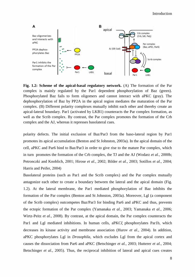
Fig. 1.2: Scheme of the apical-basal regulatory network. (A) The formation of the Par
complex is mainly regulated by the Par1 dependent phosphorylation of Baz (green).
Phosphorylated Baz fails to form oligomers and cannot interact with aPKC (gray). The
dephosphorylation of Baz by PP2A in the apical region mediates the maturation of the Par
complex. (B) Different polarity complexes mutually inhibit each other and thereby create an
apical-lateral boundary. Par1 (activated by LKB1) counteracts the Par complex formation, as
well as the Scrib complex. By contrast, the Par complex promotes the formation of the Crb
complex and the AJ, whereas it represses basolateral cues.
polarity defects. The initial exclusion of Baz/Par3 from the baso-lateral region by Par1
promotes its apical accumulation (Benton and St Johnston, 2003a). In the apical domain of the
cell, aPKC and Par6 bind to Baz/Par3 in order to give rise to the mature Par complex, which
in turn promotes the formation of the Crb complex, the TJ and the AJ (Wodarz et al., 2000b;
Petronczki and Knoblich, 2001; Hirose et al., 2002; Bilder et al., 2003; Sotillos et al., 2004;
Harris and Peifer, 2004)
Basolateral proteins (such as Par1 and the Scrib complex) and the Par complex mutually
antagonize each other to create a boundary between the lateral and the apical domain (Fig.
1.2). At the lateral membrane, the Par1 mediated phosphorylation of Baz inhibits the
formation of the Par complex (Benton and St Johnston, 2003a). Moreover, Lgl (a component
of the Scrib complex) outcompetes Baz/Par3 for binding Par6 and aPKC and thus, prevents
the ectopic formation of the Par complex (Yamanaka et al., 2003; Yamanaka et al., 2006;
Wirtz-Peitz et al., 2008). By contrast, at the apical domain, the Par complex counteracts the
Par1 and Lgl mediated inhibitions. In human cells, aPKCζ phosphorylates Par1b, which
decreases its kinase activity and membrane association (Hurov et al., 2004). In addition,
aPKC phosphorylates Lgl in Drosophila, which excludes Lgl from the apical cortex and
causes the dissociation from Par6 and aPKC (Betschinger et al., 2003; Hutterer et al., 2004;
Betschinger et al., 2005). Thus, the reciprocal inhibition of lateral and apical cues creates

Introduction
9
distinct domains along the apical-basal axis (Fig. 1.2B). Thereupon, these domains further
specify their identity and form a functional epithelium.
Beyond the polarization of epithelial cells the Par complex is also involved in the polarization
of neurons. In mammalian neurons the Par complex displays a polarized distribution at the tip
of the future axon and mediates the axon formation (Shi et al., 2003; Insolera et al., 2011),
whereas the Par complex is not required for axon and dendrite specification in Drosophila
(Rolls and Doe, 2004). Hence, although the Par complex and its general functions in polarized
epithelial cells are conserved among animals, the process of axon specification might underlie
fundamental different processes in Drosophila and mammals. However, in contrast to adult
neurons in Drosophila neuroblasts (NBs), which are neuronal stem cells, the Par complex is
required for their apical-basal polarization. NBs have a characteristic asymmetric cell division
where the Par complex defines the apical cortex that maintains its stem cell identity after cell
division. In response to aPKC phosphorylation of Miranda and Numb, both proteins become
restricted to the basal domain of the NB and recruit the cell fate determinants Prospero and
Brat. The basal domain will give rise to a ganglion mother cell, which gives rise to either glia
cells or neurons (Homem and Knoblich, 2012).
The assembly of the Par complex is mediated by multiple interactions of different functional
protein domains among Par complex members. The scaffolding protein Baz/Par3 has three
Fig. 1.3: Scheme of the molecular interactions among the members of the Par complex.
The assembly of the Par complex is mediated by several protein interaction modules. Par6
and aPKC bind each other via their N-terminal PB1 domains. Moreover, aPKC bids directly
to the PDZ domains two and three or to the aPKC binding region of Baz. Par6 also binds
directly to the first PDZ domain of Baz and interacts with the GTPase CDC42 via its CRIB
domain. Finally, Baz self-associates with its N-terminal oligomerization domain. Modified
from McCaffrey and Macara (2012).
Introduction
10
PDZ (Postsynaptic density protein 95, Disc large, Zonula occludens 1) domains and a C-
terminal aPKC binding domain (Kuchinke et al., 1998b; Morais-de-Sá et al., 2010). Par6 also
bears a single PDZ domain that binds to the first PDZ domain of Baz/Par3 (Lin et al., 2000;
Joberty et al., 2000). Moreover, Par 6 binds with its N-terminal PB1 (Phox and Bem1)
domain the PB1 domain of aPKC (Noda et al., 2003; Hirano et al., 2005). In addition, aPKC
binds directly to the PDZ domains two and three as well as the aPKC binding region of
Baz/Par3 (Wodarz et al., 2000b; Morais-de-Sá et al., 2010). Taken together, the assembly of
the Par complex depends on the interaction of multiple protein interaction domains of the
complex members (Fig. 1.3).
The activity of the Par complex is regulated by itself, because Par6 inhibits the kinase activity
of aPKC. This inhibition is reversed upon binding of the GTPase CDC42 to the CRIM
domain of Par6, which induces a conformational change of Par6 and consequently activates
aPKC (Yamanaka et al., 2001). In contrast, more recent studies demonstrate that Par6
activates aPKC by replacing its intramolecular pseudosubstrate (Graybill et al., 2012),
whereas the aPKC binding domain of Baz inhibits the kinase activity of aPKC (Soriano et al.,
2016). Thus, Baz and Par6 act as regulators of the aPKC kinase activity.
Nevertheless, the Par complex is not a permanent complex, but rather dissociates or
rearranges during development. In Drosophila, Baz localizes beneath Par6 and aPKC at the
AJ (Nam and Choi, 2003; Vogelmann and Nelson, 2004; Harris and Peifer, 2005; Martin-
Belmonte et al., 2007; Morais-de-Sá et al., 2010; Doerflinger et al., 2010). Baz has been
reported to interact with aPKC and Par6 (Wodarz et al., 2000b; Petronczki and Knoblich,
2001; Hutterer et al., 2004), however in polarized epithelial cells it seems that the Par
complex assembles only transiently and afterwards Baz segregates towards the AJ (Harris and
Peifer, 2005). The aPKC dependent phosphorylation of Baz at S980 induces the dissociation
of the Par6 and aPKC from Baz (Morais-de-Sá et al., 2010). Par6 and aPKC might either be
retained in the SAR in complex with the Crb-Sdt complex or with CDC42, which have been
reported to bind Par6 and aPKC and mediate their cortical localization (Lin et al., 2000;
Joberty et al., 2000; Hurd et al., 2003b; Wang et al., 2004; Kempkens et al., 2006; Atwood et
al., 2007; Fletcher et al., 2012; Whitney et al., 2016). Moreover, Baz forms an additional
complex with Sdt, which binds to the aPKC binding region of Baz with its PDZ domain.
Upon S980 phosphorylation of Baz by aPKC, Baz and Sdt dissociate to give rise to the Crb-
Sdt complex (Krahn et al., 2010a). In summary, the phosphorylation of S980 of Baz by aPKC
initiates the segregation of Baz to the AJ, the disassembly of the Par complex and the
formation of the Crb complex, most likely together with Par6 and aPKC.
Introduction
11
The scaffold protein Bazooka/Par3
The scaffold protein Baz/Par3 is a key player in the establishment of cell polarity. Loss of baz
prevents the formation of polarized epithelia and causes embryonic lethality. The embryonic
epidermis of baz mutants displays holes, from which its name originates. Likewise, mice that
are homozygous mutant for the mammalian orthologue Par3 die during early embryogenesis
(around E12.5) (Hirose et al., 2006). In Drosophila, Baz is already maternally provided to
initiate the early epithelial polarization. During cellularization (the formation of the cellular
blastoderm) Baz is transported in a Dynein-dependent manner to the apical region (Harris and
Peifer, 2005). To ensure the lateral inhibition of Baz the serine/threonine kinase Par1
phosphorylates Baz at two conserved residues (S151 and S1085) (Benton and St Johnston,
2003a). Binding of 14-3-3 proteins to theses phosphorylated residues prevents Baz from
oligomerization and binding to aPKC (Benton and St Johnston, 2003a; Hurd et al., 2003a). In
the apical domain of the cell the protein phosphatase PP2A antagonizes the Par1 mediated
phosphorylation to promote the maturation of the Par complex (Fig. 1.2A) (Krahn et al.,
2009). By contrast, in Drosophila follicle cells (the epithelium around the egg chamber, FCs)
Baz is dispensable for the formation of a polarized epithelium (Shahab et al., 2015). Thus, in
FCs redundant mechanisms contribute to the cellular polarization.
In mice, Par3 has been reported to act as an exocyst receptor at the AJs (Ahmed and Macara,
2017). The exocyst is an octameric protein complex that mediates directed vesicle trafficking
to deliver proteins (e.g. E-Cadherin) to the plasmamembrane. However, in Drosophila NBs
the exocyst complex is dispensable for cell apical-basal polarity (Halbsgut et al., 2011), thus
Baz’s function as an exocyst receptor might be restricted to epithelial cells.
In mouse mammary glands, Par3 antagonizes tumorigenesis by counteracting metastasis
formation by inhibiting aPKC (McCaffrey et al., 2012; Guyer and Macara, 2015).
Nevertheless, loss of Par3 alone does not promote the formation of tumors, however, loss of
Par3 associates with a decreased survival rate of breast cancer patients (McCaffrey et al.,
2012). Similar, in baz mutant clones of the Drosophila wing disc the loss of Baz does not
enhace the levels of Cycline-E (personal unpublished observation).
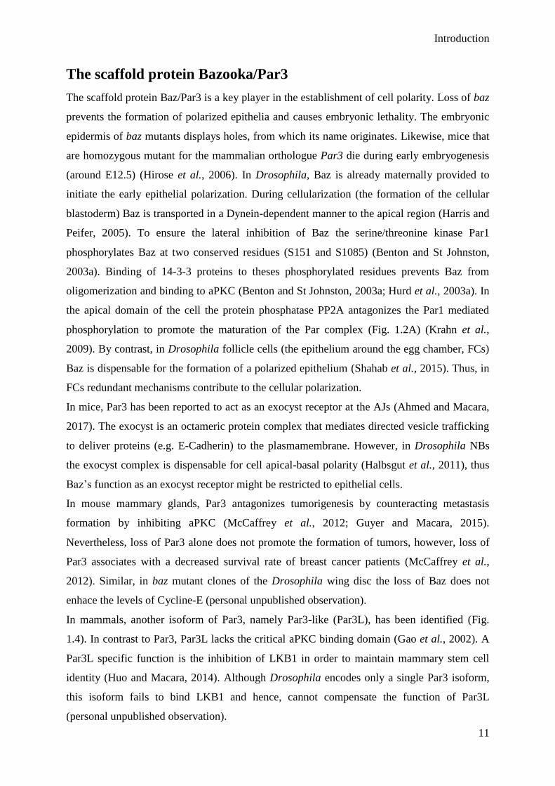
In mammals, another isoform of Par3, namely Par3-like (Par3L), has been identified (Fig.
1.4). In contrast to Par3, Par3L lacks the critical aPKC binding domain (Gao et al., 2002). A
Par3L specific function is the inhibition of LKB1 in order to maintain mammary stem cell
identity (Huo and Macara, 2014). Although Drosophila encodes only a single Par3 isoform,
this isoform fails to bind LKB1 and hence, cannot compensate the function of Par3L
(personal unpublished observation).

Introduction
12
Fig. 1.4: Comparison of Drosophila and human Par3 homologs. The N-terminal
oligomerization domain (blue) of Baz is followed by three PDZ domains (orange) that
mediate protein-protein interactions. At the C-terminus Baz has a lipid binding domain (red).
The two Par1 phosphorylation sites (S151 and S 1085, purple) are important for its lateral
exclusion. The aPKC phosphorylation site (S980 in Drosophila and S827 in human) is
essential for the interaction of Baz and aPKC. The numbers represent amino acid positions.
At the structural level, Baz/Par3 has a conserved N-terminal oligomerization domain (OD)
that mediates self-association of Baz monomers in a front-to-back manner, which are further
assembled into a filament-like oligomer (Mizuno et al., 2003; Benton and St. Johnston,
2003b; Feng et al., 2007; Zhang et al., 2013a). Additionally, Baz contains three PDZ
domains, which mediate protein interaction (Fig. 1.4). By interacting with the cell adhesion
molecule Echinoid, the tight-junction-associated protein junctional adhesion molecule (JAM)
and Armadillo (Arm)/ß-Catenin redundant mechanisms target Baz to the apical junctions
(Itoh et al., 2001; Ebnet et al., 2001; Takekuni et al., 2003; Wei et al., 2005). In Drosophila
photoreceptor cells, the phosphorylation of Arm by the P21-activated kinase (Pak4) is
required to retain Baz at the AJs (Walther et al., 2016). The conserved region 3 (CR3) around
Ser980 binds aPKC and negatively regulates its kinase activity. However, upon
phosphorylation of Ser980 by aPKC the two proteins dissociate and Baz becomes excluded
from the SAR and accumulates at AJs. A C-terminal lipid binding motif (K1173-74) directly
binds to PtdIns(4,5)P2 (PIP2) and PtdIns(3,4,5)P3 (PIP3) to target Baz to the plasmamembrane
(Krahn et al., 2010b). Furthermore, the second PDZ domain of rat Par3 has been reported to
bind directly to phosphatidylinositol lipids (Wu et al., 2007) and the three Drosophila Baz
PDZ domains bind in vitro to phosphatidic acid (Yu and Harris, 2012). Hence, several
different direct interactions between Par3 and the plasmamembrane contribute to its cortical
localization.
Introduction
13
The Liver Kinase B1
The liver kinase B1 (LKB1 or STK11) has initially been identified in patients suffering from
Peutz-Jeghers syndrome (PJS) (Hemminki et al., 1998). PJS is a cancer prone rare genetic
disorder and will be described later. LKB1 is a highly conserved serine/threonine kinase that
is involved in many different cellular processes, such as cell metabolism, cell proliferation
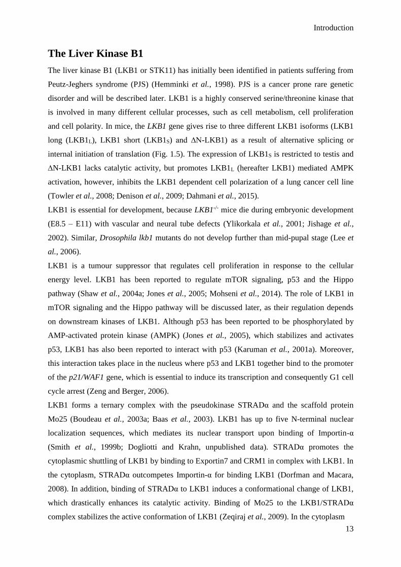
and cell polarity. In mice, the LKB1 gene gives rise to three different LKB1 isoforms (LKB1
long (LKB1L), LKB1 short (LKB1S) and ∆N-LKB1) as a result of alternative splicing or
internal initiation of translation (Fig. 1.5). The expression of LKB1S is restricted to testis and
∆N-LKB1 lacks catalytic activity, but promotes LKB1L (hereafter LKB1) mediated AMPK
activation, however, inhibits the LKB1 dependent cell polarization of a lung cancer cell line
(Towler et al., 2008; Denison et al., 2009; Dahmani et al., 2015).
LKB1 is essential for development, because LKB1-/- mice die during embryonic development
(E8.5 – E11) with vascular and neural tube defects (Ylikorkala et al., 2001; Jishage et al.,
2002). Similar, Drosophila lkb1 mutants do not develop further than mid-pupal stage (Lee et
al., 2006).
LKB1 is a tumour suppressor that regulates cell proliferation in response to the cellular
energy level. LKB1 has been reported to regulate mTOR signaling, p53 and the Hippo
pathway (Shaw et al., 2004a; Jones et al., 2005; Mohseni et al., 2014). The role of LKB1 in
mTOR signaling and the Hippo pathway will be discussed later, as their regulation depends
on downstream kinases of LKB1. Although p53 has been reported to be phosphorylated by
AMP-activated protein kinase (AMPK) (Jones et al., 2005), which stabilizes and activates
p53, LKB1 has also been reported to interact with p53 (Karuman et al., 2001a). Moreover,
this interaction takes place in the nucleus where p53 and LKB1 together bind to the promoter
of the p21/WAF1 gene, which is essential to induce its transcription and consequently G1 cell
cycle arrest (Zeng and Berger, 2006).
LKB1 forms a ternary complex with the pseudokinase STRADα and the scaffold protein
Mo25 (Boudeau et al., 2003a; Baas et al., 2003). LKB1 has up to five N-terminal nuclear
localization sequences, which mediates its nuclear transport upon binding of Importin-α
(Smith et al., 1999b; Dogliotti and Krahn, unpublished data). STRADα promotes the
cytoplasmic shuttling of LKB1 by binding to Exportin7 and CRM1 in complex with LKB1. In
the cytoplasm, STRADα outcompetes Importin-α for binding LKB1 (Dorfman and Macara,
2008). In addition, binding of STRADα to LKB1 induces a conformational change of LKB1,
which drastically enhances its catalytic activity. Binding of Mo25 to the LKB1/STRADα
complex stabilizes the active conformation of LKB1 (Zeqiraj et al., 2009). In the cytoplasm
Introduction
14
Fig. 1.5: Scheme of the Drosophila and human LKB1 variants. The large N-terminal
domain of the Drosophila LKB1 is not conserved in the human proteins. The two human
LKB1 variants LKB1-long (LKB1L) and LKB1-short (LKB1S) result from alternative
splincing and are identical with the exception of their C-terminus. An alternative translation
initiation gives rise to the human ∆N-LKB1 variant. Conserved domains are indicated and the
numbers correspond to amino acid positions.
the active LKB1/STRADα/Mo25 complex baers the potential to localize at the cell cortex as
well (Collins et al., 2000; Sapkota et al., 2001; Sebbagh et al., 2009). Thus, the diversity of
LKB1’s cellular localizations highlights its potential to regulate cellular processes on many
different levels.
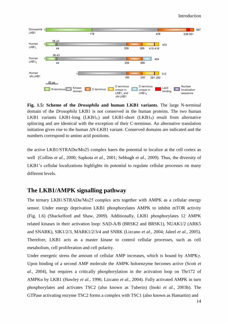
The LKB1/AMPK signalling pathway
The ternary LKB1/STRADα/Mo25 complex acts together with AMPK as a cellular energy
sensor. Under energy deprivation LKB1 phosphorylates AMPK to inhibit mTOR activity
(Fig. 1.6) (Shackelford and Shaw, 2009). Additionally, LKB1 phosphorylates 12 AMPK
related kinases in their activation loop: SAD-A/B (BRSK2 and BRSK1), NUAK1/2 (ARK5
and SNARK), SIK1/2/3, MARK1/2/3/4 and SNRK (Lizcano et al., 2004; Jaleel et al., 2005).
Therefore, LKB1 acts as a master kinase to control cellular processes, such as cell
metabolism, cell proliferation and cell polarity.
Under energetic stress the amount of cellular AMP increases, which is bound by AMPKγ.
Upon binding of a second AMP molecule the AMPK holoenzyme becomes active (Scott et
al., 2004), but requires a critically phosphorylation in the activation loop on Thr172 of
AMPKα by LKB1 (Hawley et al., 1996; Lizcano et al., 2004). Fully activated AMPK in turn
phosphorylates and activates TSC2 (also known as Tuberin) (Inoki et al., 2003b). The
GTPase activating enzyme TSC2 forms a complex with TSC1 (also known as Hamartin) and

Introduction
15
Fig. 1.6: Scheme of the interplay between the PI3K/Akt/mTOR and LKB1/AMPK
signalling pathways. The stimulation of growth factors leads to a receptor mediated
activation of PI3K, which in turn phosphorylates PIP2 to produce PIP3. PIP3 is bound by
PDK1 and Akt causing the phosphorylation an activation of Akt by PDK1. Activated Akt
phosphorylates and inhibits the GTPase activating enzyme TSC2. The GTPase Rheb
stimulates in its GTP bound active form mTORC1, which promotes protein translation by
activating S6K and eIF4E-BP. Upon low energy conditions, AMPK detects the increasing
amount of AMP molecules and binds to LKB1, which triggers its phosphorylation and
activation. AMPK phosphorylates TSC2, such as Akt, however, thereby activating it.
Stimulating the GTPase activity of Rheb inactivates it and consequently decreases the activity
mTOR. Moreover, AMPK phosphorylates Raptor to create a binding site for 14-3-3 proteins
to prevent Raptor from joining the mTORC1. In addition, AMPK inactivates Acety-CoA
carboxylase (ACC) and HMG-CoA-Reductase (HMGCR) to inhibit lipid synthesis. Green
arrows indicate activation and red arrows inhibition.
stimulates the GTPase activity of Rheb, which becomes inactive after GTP hydrolysis (Inoki
et al., 2003a). In contrast, active Rheb promotes the activity of the kinase mTOR (Tee et al.,
2003). The activation of mTOR causes an elevated protein translation, which is mediated by
phosphorylation of eIF4E-BP and S6K by mTOR (Carrera, 2004). The mTOR complex1
(mTORC1) requires binding of the scaffold protein Raptor, which recruits eIF4E-BP and S6K
for phosphorylation (Kim et al., 2002; Hara et al., 2002). However, phosphorylation of
Raptor (on Ser722 and Ser792 in humans) by AMPK causes its sequestration by 14-3-3

Introduction
16
proteins and consequently an inhibition of mTORC1 (Gwinn et al., 2008). Thus, LKB1
mediates the metabolic switch from anabolism towards catabolism under energetic stress.
Therefore, AMPK also inhibits Acety-CoA carboxylase and HMG-CoA-Reductase to
antagonize lipid synthesis (Fig. 1.6) (Carling et al., 1987; Winder and Hardie, 1996).
In addition to the well-studied activation of the LKB1/AMPK pathway by AMP, recently the
a fructose-1,6-bisphosphate (FBP) dependent mechanism to activate the LKB1/AMPK
pathway has been described. Under energy deprivation the amount of FBP decreases and
unoccupied Aldolase, which splits FBP into dihydroxyacetone phosphate (DHAP) and
glyceraldehyde 3-phosphate (G3P), promotes the formation of a complex consisting of LKB1,
AMPK, v-ATPase, Ragulator and AXIN (Zhang et al., 2017a). This complex localizes on
endosomes and is essential to activate the LKB1-AMPK pathway. The knockout of either
AXIN or LAMTOR1 (a component of the Ragulator complex) in mice prevents the LKB1
dependent activation of AMPK (Zhang et al., 2013b; Zhang et al., 2014).
Moreover, the LKB1/AMPK signaling pathway is implicated in cell polarity. In C. elegens
and Drosophila, LKB1 is required for the anterior-posterior polarization of the embryo and
oocyte, respectively (Watts et al., 2000; Martin and St Johnston, 2003). In Drosophila,
AMPK has been reported to phosphorylate Myosin regulatory light chain (MRLC), which is
essential for proper apical-basal polarization of the embryonic epithelium. Loss of either
LKB1 or AMPK results in severe polarity defects (Lee et al., 2007). However, the
phosphorylation of MRLC is not due to AMPK, but rather to other kinases, that act
downstream of AMPK (Bultot et al., 2009). In contrast to Drosophila, LKB1 is dispensable
for the early polarization of the eight-cell stage mouse embryo (Krawchuk et al., 2015). In
addition to AMPK, LKB1 phosphorylates and activates the AMPK-related kinase Par1
(MARK1-4 in vertebrates), which regulates microtubule dynamics and counteracts the
formation of the Par complex (as previously described) (Benton and St Johnston, 2003a;
Lizcano et al., 2004; Wang et al., 2007; Granot et al., 2009; Amin et al., 2009).
Taken together, the regulation of cell proliferation, metabolism and cell polarity are tightly
connected to the LKB1/AMPK signaling axis. Loss of either LKB1 or AMPK causes severe
defects that result in embryonic lethality. The significance of the LKB1 dependent regulations
with respect to human pathologies will be described in the next section.
Introduction
17
The role of LKB1 in human pathologies
The regulation of cell proliferation, energy metabolism and cell polarity are essential
processes that have to be precisely controlled. In response to energy deprivation or ionizing
radiation LKB1 becomes active to induce a metabolic switch from anabolism towards
catabolism. Germline mutations in the STK11 gene (encoding LKB1), which localizes on
chromosome 19p13.3 cause the Peutz-Jeghers syndrome (PJS). The PJS is a rare autosomal
dominant genetic disease with an incident varying from 1:50.000 to 1:200.000 individuals.
The PJS is characterized by cutaneous mispigmentation, gastro intestinal polyps and a high
risk of cancer (Jansen et al., 2009). The risk of PJS patients to develop cancer is highest in the
gastro intestinal tract, however female patients bear also an increased risk to develop breast
cancer (Hearle et al., 2006). In mice, loss of heterozygosity (LOH) of LKB1 has been reported
by several groups to induce polyposis as well, suggesting that polyps develop due to a LKB1
haploinsufficiency (Bardeesy et al., 2002; Miyoshi et al., 2002; Jishage et al., 2002).
Mutations or loss of LKB1 have also been found in cancer patients without PJS, such as non-
small cell lung cancer (NSCLC), cervical cancer, ovarian cancer, breast cancer, pancreatic
cancer and malignant melanoma (Rowan et al., 1999; Guldberg et al., 1999; Sanchez-
Cespedes et al., 2002; Carretero et al., 2004; Matsumoto et al., 2007; Wingo et al., 2009;
Morton et al., 2010; Gill et al., 2011; Tanwar et al., 2014; George et al., 2016). Moreover,
loss of LKB1 is associated with a poor survival rate of breast cancer patients (Sengupta et al.,
2017). The majority of mutations that have been reported from PJS and cancer patients affect
the catalytic domain, but some affect the less characterized C-terminus as well (Boudeau et
al., 2003c).
Lung cancer is worldwide the most diagnosed cancer with 1.82 million new cases and 1.59
million deaths in 2012 (Ferlay et al., 2015). In lung adenocarcinoma, LKB1 is the third most
frequently mutated gene (Ding et al., 2008) and 60 – 70 % of lung adenocarcinoma are
affected by LOH of LKB1 (Sanchez-Cespedes et al., 2002; Gill et al., 2011). Generally in
NSCLC, which are with 85 % the most common lung cancer, 39 - 41 % of the tumours
display genomic alterations of the LKB1 locus (Matsumoto et al., 2007; Gill et al., 2011;
Calles et al., 2015). In lung adenocarcinoma, LKB1 counteracts enhanced cell migration
independent of AMPK. Thereby LKB1 activates MARK1/4, which in turn phosphorylates the
scaffold protein DIXDC1, which leads to a decreased activation of the FAK/MEK/ERK
mediated Snail expression. DIXDC1 is frequently mutated in various tumours and thus, fails
to convey the LKB1/MARK mediated tumour suppression (Goodwin et al., 2014). Similar,
LKB1 has also been reported to repress Snail expression by activating Salt-inducible kinase 1

Introduction
18
(SIK1) to maintain E-Cadherin levels (Eneling et al., 2012).
Furthermore, LKB1 suppresses cell proliferation also by counteracting the activity of the co-
transcriptional activator YAP. The phosphorylation of MARK1/4 by LKB1 recruits the
SCRIB complex to the lateral plasmamembrane of epithelial cells, which is essential for the
core Hippo kinases MST1/2 and LATS1/2 to phosphorylate and inhibit YAP (Mohseni et al.,
2014). Taken together, beyond the canonical LKB1/AMPK tumour suppressor pathway a
redundant LKB1/MARK axis antagonizes tumorigenesis.
In addition to mutations and LOH, the promotor of the LKB1 gene is targeted by an aberrant
methylation status in some types of cancer, such as colorectal cancer, melanoma and
adenocarcinoma (Trojan et al., 2000; Sanchez-Cespedes et al., 2002; Zhang et al., 2017b).
The methylation of CpG-islands in the promotor region of LKB1 reduces its gene expression
and therefore reduces the tumour suppressive capacity of the affected cell. Nevertheless, CpG
hypermethylation is not a frequent aberration and affects only a minor number of tumours.
Until now there are no pharmaceuticals that directly target LKB1 in order to elevate its
activity or expression. However, with respect to data from cell culture and mice the
polyphenol Honokiol (HNK) from Magnolia grandiflora enhances the protein level and
activity of LKB1 (Nagalingam et al., 2012; Avtanski et al., 2015; Seo et al., 2015; Sengupta
et al., 2017). A xenograft breast cancer mouse model that is treated with HNK displays a
reduced tumour growth depending on LKB1 (Sengupta et al., 2017). The activation of LKB1
results from the HNK mediated activation of the deacetylase SIRT1, which deacetylates and
thereby activates LKB1 (Nagalingam et al., 2012; Avtanski et al., 2015; Seo et al., 2015).
Although HNK treatment might become a promising anti-cancer therapy, the challenge will
be to target cancers that are deprived of LKB1. The former anti-diabetic drug Phenformin
bears the potential to specifically target tumours that lack LKB1 (Shackelford et al., 2013).
Phenformin inhibits the mitochondrial complex I, causing energetic stress. Treatment of a
LKB1-/- NSCLC mouse model with Phenformin specifically induces apoptosis in LKB1 null
tumours and prolongs survival (Shackelford et al., 2013).
In summary, the loss of active LKB1 leads to the PJS and is a predisposition for cancer.
Under energetic stress LKB1 is essential to promote a catabolic switch. Nevertheless, with
respect to a potential treatment of cancer patients with Phenformin the loss of LKB1 might
bear the advantage that cells cannot respond via the LKB1/AMPK axis and undergo
apoptosis.
Aims of the study
19
Aims of the study
For development and homeostasis the polarization of epithelia is an essential process. The
serine/threonine kinase LKB1 and the scaffold protein Bazooka (Baz) are key mediators of a
celllar polarity. However, the regulation of both proteins is only poorly understood in the
epithelium.
LKB1 is an important regulator of cell proliferation, metabolism and cell polarity. Mutations
or loss of LKB1 have frequently been reported from various tumours. Therefore, it is essential
to better understand the regulation of this enzyme. The subcellular localization of LKB1
varies strongly from nuclear to cytoplasmic and even cortical. Several functions of the nuclear
and cytoplasmic LKB1 have already been described, however, the function and the
mechanism by which LKB1 is targeted to the plasmamembrane remain elusive. Thus, this
study aims to analyze the cortical localization of LKB1 and its function during development
and tumour suppression.
Moreover, in Drosophila, the scaffold protein Baz is an essential cue for the establishment of
an apical-basal cell polarity in epithelial cells and neuroblasts (NBs). Baz is the core
component of the heterotrimeric Par complex, which also includes scaffold protein Par6 and
the atypical protein kinase C (aPKC). The N-terminal oligomerization domain (OD) and the
C-terminal lipid binding motif of Baz are involved in the membrane localization of Baz.
Nevertheless, the function of the Baz OD beyond its contribution to the cortical localization is
not clear, yet. Given that the OD largely contributes to the viability of embryos, it is another
objective of this study to further characterize the role of the OD during the development of
Drosophila.
Finally, although the formation and function of the Par complex in epithelial polarization are
a dogma, the in vivo relevance of the Baz-Par6 interaction is unclear. Both proteins have in
vitro been reported to interact via their PDZ domains, this is why this study clarifies the
molecular basis of this interaction using in vitro structural approaches and Drosophila as an in
vivo model. The goal of these experiments will be to understand the relevance of the in vitro
obtained results.
Chapter 1: Membrane-binding and activation of LKB1
20
Chapter 1: Membrane-binding and activation of LKB1
Membrane-binding and activation of LKB1 by phosphatidic acid
is essential for development and tumour suppression
Authors: Giada Dogliotti#, Lars Kullmann#, Pratibha Dhumale, Christian Thiele, Olga
Panichkina, Gudrun Mendl, Roland Houben, Sebastian Haferkamp, Andraes W. Püschel and
Michael P. Krahn (# indicates equal contribution)
Personal contributions: preparation and analysis of some Drosophila embryos and S2R cells
by immunofluorescence, liposome flotation assays, some Western blots of embryonic lysates,
in vitro kinase assays, data analysis and discussion of results
The serine/threonine kinase LKB1 regulates various cellular processes such as cell
proliferation, energy homeostasis and cell polarity and is frequently downregulated in various
tumours. Many downstream pathways controlled by LKB1 have been described but little is
known about the upstream regulatory mechanisms. Here we show that targeting of the kinase
to the membrane by a direct binding of LKB1 to phosphatidic acid is essential to fully activate
its kinase activity. Consequently, LKB1 mutants that are deficient for membrane binding fail
to activate the downstream target AMPK to control mTOR signalling. Furthermore, the in
vivo function of LKB1 during development of Drosophila depends on its capacity to associate
with membranes. Strikingly, we find LKB1 to be downregulated in malignant melanoma,
which exhibit aberrant activation of Akt and overexpress phosphatidic acid generating
Phospholipase D. These results provide evidence for a fundamental mechanism of LKB1
activation and its implication in vivo and during carcinogenesis.
Chapter 1: Membrane-binding and activation of LKB1
21
Introduction
The serine/threonine kinase LKB1 is ubiquitously expressed and highly conserved throughout
evolution. It has been demonstrated to function as a ‘master kinase’ potentially activating
several downstream kinases (Lizcano et al., 2004). Apart from its implication in
carcinogenesis LKB1 plays a role in various cellular signalling pathways such as Wnt-,
TGFβ-signalling or the mTOR-pathway reviewed by Vaahtomeri and Makela (Vaahtomeri
and Mäkelä, 2011). The latter one is controlled by LKB1-mediated activation of AMP-
dependent kinase (AMPK), which is essential for cell survival and polarity in Drosophila and
vertebrates, in particular under energetic stress (Hawley et al., 2003; Woods et al., 2003;
Shaw et al., 2004b; Lee et al., 2007; van der Velden et al., 2011). Mechanistically, AMPK
phosphorylates (among others) Raptor (a core component of mTOR complex 1) and TSC2 (an
mTOR inhibitor), resulting in reduced mTOR activity (Arsham et al., 2003; Kimura et al.,
2003; Inoki et al., 2003b; Corradetti et al., 2004; Shaw et al., 2004a; Shaw et al., 2005;
Gwinn et al., 2008; Shackelford et al., 2009). Consequently, the LKB1-AMPK-axis is
believed to be a key modulator in carcinogenesis and cell polarity (Hardie and Alessi, 2013).
Apart from its function in cell proliferation and tumour suppression, LKB1 is directly
implicated in the establishment and maintenance of cell polarity in different cell types and
organisms (Nakano and Takashima, 2012).
Although many downstream pathways mediating the function of LKB1 have been described,
little is known about the upstream mechanisms regulating LKB1 activity. Two pseudokinases
tightly control the localization and kinase activity of LKB1: STRADα (Ste20-like kinase
(Stlk) in Drosophila) and Mo25 form a stable ternary complex with LKB1 (Boudeau et al.,
2003a). Both proteins enhance the export of LKB1 from the nucleus into the cytoplasm and
increase its kinase activity (Boudeau et al., 2003a; Dorfman and Macara, 2008). Secondly, the
conserved C-terminus of LKB1 is farnesylated in vivo and thereby might directly interact with
the plasma membrane to attach the protein to the cell cortex. Although the majority of the
protein accumulates at the plasma membrane of polarized (epithelial) cells, farnesylation has
been reported to be not essential for the (tumour suppressor) function of LKB1 in mammalian
cells or mice but might be essential for oogenesis in Drosophila (Sapkota et al., 2001; Martin
and St Johnston, 2003; Houde et al., 2014). Therefore the question remains, whether
membrane association of LKB1 is essential for the kinase activity and function of the protein
during development and tumour suppression.

Chapter 1: Membrane-binding and activation of LKB1
22
Here we report that binding of LKB1 to membranes by direct interaction with phospholipids,
in particular to phosphatidic acid, is essential for its function in vivo during development of
Drosophila. Membrane association of LKB1 is required for its kinase activity and for efficient
activation of AMPK in cultured mammalian cells, thus contributing to the tumour suppressor
function of LKB1. Strikingly, we reveal a strong correlation between overexpression of
Phospholipase D (PLD), which increases cellular levels of phosphatidic acid, downregulation
of LKB1 and enhanced activity of mTOR in malignant melanoma, thus likely contributing to
the pathogenesis of malignant melanoma.

Chapter 1: Membrane-binding and activation of LKB1
23
Methods
Fly stocks and genetics
UASt::GFP-LKB1 and lkb1::GFP-LKB1 transgenes were generated using phiC31-mediated
germ line transformation on attp40. For rescue experiments of different LKB1 variants, we
used the lkb1x5 null allele. Lethality tests were performed in three independent experiments
with n=100 in each experiment. For rescue experiments using the UAS/GAL4 system, we
used UAS::GFP-LKB1 and actin5C::GAL4 instead of lkb1::GFP-LKB1.
DNA and constructs
Cloning of the cDNA of wild-type LKB1 into pENTR was performed using standard PCR on
a full length EST clone (Drosophila Genomics Resources Center, DGRC) as template using
the following primers: LKB1-F: 5′- CACCATGCAATGTTCTAGCTCTCGG-3′, LKB1-R:
5′-CTACGAAGTTCGGCAGTGG-3′. Similar, truncated fragments of LKB1 were cloned
with the following oligonucleotides: LKB1512-F: 5′-CACCATGCACACCTACGAACCGCC-
3′, LKB1536-F: 5′-CACCATGGCGCCCGTCAAGAAG-3′, LKB1552 -F: 5′-
CACCATGCTGACGTCCTGCATCTCCG-3′. For expression of LKB1 from its endogenous
promoter we inserted a genomic fragment (from 2.8 kbp upstream of the translation start to
1 kbp downstream of the stop codon) into pENTR using the following primers: LKB1gen-F:
5′- CACC CACTAGCGTAATTTGACGG-3′, LKB1gen-R: 5′- CTC GAG
CAGCAGTACGGTCATCTC-3′. An XbaI-cutting site was introduced replacing the start
codon using mutagenesis PCR and the following primer: LKB1gen-XbaI-F: 5′-
GGCTCCGCGGAGGTTTTCTAGACAATGTTCTAGCTCTC-3′. Subsequently, GFP was
inserted into the XbaI site by PCR and standard ligation. Mutagenesis PCR was used to
generate defined point mutations with full length or genomic LKB1 cDNA in pENTR as
template. The following oligonucleotides were used for mutagenesis (mutation underlined):
LKB1ΔLB: Combination of 1. LKB1K546A R547A R548A K550A K551A-F:
5′-TCGGCACTGGCGGCGGCCGCCGCGGCGCTGACGTCCTGC-3′ and 2.LKB1K539A
K540A K541A-F: 5′-GAGGAGGCGCCCGTCGCCGCGGCGGGATCGGCACTG-3′,
LKB1C564A-F: 5′-GTGCGCAAGCTTAGCCACGCCCGAACTTCGTAG, LKB1D317A-F: 5′-
CAAACGCTGAAGATTTCCGCCTTCGGTGTGGCG.
To express LKB1ΔLBC564A PH(PLD) and LKB1ΔLBC564A PH(Akt), the PH domain of PLCδ and Akt
was amplified by PCR and ligated via an endogenous Bpu1101-I site into lkb1::LKB1
pENTR using the following primers:
Chapter 1: Membrane-binding and activation of LKB1
24
PH(PLCδ)-F: 5′- GCTGAGCCACGCCCGAACT TCGgatgaggatctacaggcgct-3′,
PH(PLCδ)-R: 5′- GCTGAGCTAGATCTTGTGCAGCCCCAG-3′,
PH(Akt)-F: 5′-GCTGAGCCACGCCCGAACTTCGGTCGTAAAGGAGGGGTGG-3′ and
PH(Akt)-R: 5′- GCTGAGCTTATATGAGCCGGCTGGATAC-3′.
For expression of hLKB1, the open reading frame of hLKB1 was cloned into pENTR using
the following nucleotides: hLKB1-F: 5′- CACC ATGGAGGTGGTGGACCC-3′ and hLKB1-
R: 5′- TCACTGCTGCTTGCAGG-3′. For mutation of the farnesylation, lipid binding motif
and construction of the kinase dead version, the following nucleotides were used in
mutagenesis PCRs:
hLKB1C430A-F: 5′- CGCCGGCTGTCGGCCGCTAAGCAGCAGTGAAAGGGT-3′,
hLKB1R415AK416A-F: 5′- GCCCCCAACCCTGCCGCCGCGGCCTGCTCCGCCAGC-3′ and
hLKB1D194A: 5′- ACCCTCAAAATCTCCGCCCTTGGCGTGGCCGAGGCA-3′.
Constructs were recloned into GFP-tagged destination vectors containing a One-Strep-Tag
fused to the N terminus of GFP (USGW, modified TGW, Murphy lab, DGRC, expression in
S2R cells) or into a modified EGFP-C1 vector (CGW, expression in mammalian cells)
containing a gateway cassette using the gateway technology (Life technology).
Antibodies
Antisera directed against full length LKB1 were raised by injection of a fusion protein of
LKB1 and MBP into two guinea pigs (Amsbio, Abingdon, UK).
Cell culture and cell viability assay
HeLa, IMR90, IGR37 and MDCK cells were obtained from ATCC. All cells were maintained
in Dulbecco’s modified Eagle’s medium supplemented with 10% fetal calf serum, 2 mM
Glutamine at 37 °C in a 5% CO2 atmosphere and negatively tested for mycoplasma
contamination by PCR. Cells were transfected using FUGENE (Promega) according to the
manufacturer’s instructions. For evaluation of cell viability upon energetic stress, 50 × 103
cells/well were transiently transfected in a 24well plate with GFP-hLKB1+STRADα
constructs and treated for 12 h with 2.5 mM AICAR (Santa Cruz Inc.). GFP+STRADα were
used as negative control. Cell viability was assessed in triplicates using MTT assay according
to the manufacturer’s instructions (SIGMA).

Chapter 1: Membrane-binding and activation of LKB1
25
Western blotting and coimmunoprecipitation
Western blotting was done as previously described (Cidlinsky et al., 2016). For mammalian
cell culture experiments, HeLa or IGR37 cells were transiently transfected with the indicated
constructs. 48 hours after transfection, cells were incubated with 2 mM AICAR for 1 h and
harvested in lysis buffer (1% Triton X-100, 150 mM NaCl, 1 mM CaCl2, 1 mM MgCl2,
50 mM TRIS-HCl pH 7.5) supplemented with protease- and phosphatase inhibitors. For
embryonic lysates, lkb1::GFP-LKB1 expressing embryos were collected from overnight
plates. Coimmunoprecipitation using GFP-Trap (ChromoTek) of GFP-LKB1 with HA-tagged
DmSTRADα (Stlk) and myc-tagged DmMo25 was done in embryonic lysates, which
ubiquitously expressed the proteins using arm::GAL4. Primary antibodies used for western
blotting were as follows: rabbit anti-Actin (1:1,000, sc-47778, Santa Cruz), mouse anti-S6K
(1:500, sc-8418, Santa Cruz ), rabbit phosphoT389-S6K (1:500, sc-11759, Santa Cruz), rabbit
anti-phospho-T172-AMPK (1:200, sc-33524, Santa Cruz), rabbit anti-phospho-MARK1/2/3
(1:500, PA5-17495, Thermo Scientific), mouse anti-myc (1:100, 9E10, DSHB), rabbit anti-
AMPK (1:400, sc-25792, Santa Cruz), guinea pig anti LKB1 (1:500, this study), mouse anti-
GFP (1:500, sc-9996, Santa Cruz), mouse anti-HA (1:500, #11583816001, Roche), rabbit
anti-GST (1:5,000, #G7781, SIGMA). For statistical analysis, three independent experiments
were scored. Intensity of the bands was quantified by ImageJ.
Immunohistochemistry
Drosophila embryos were fixed in 4% formaldehyde, phosphate buffer pH 7.4 as described
before (Sen et al., 2012). Primary antibodies used for indirect immunofluorescence were as
follows: guinea pig anti LKB1 (1:250, this study), rabbit anti Baz (1:2,000, kindly provided
by A. Wodarz), mouse anti α-spectrin (3A9, 1:50, DSHB), mouse anti Dlg (4F10, 1:50,
DSHB), rat anti DE-Cad (DCAD2, 1:25, DSHB), rabbit anti GFP (1:400, sc-8334, Santa Cruz
Inc), guinea-pig anti Miranda (1:1,000, kindly provided by A. Wodarz). HeLa cells were fixed
with 4% PFA in PBS and stained with the rabbit anti-phospho-Sad antibody (1:250) and a
mouse anti-LKB1 antibody (1:200, sc-32245, Santa Cruz) in 10% goat serum.
Secondary antibodies conjugated with Alexa 488, Alexa 568 and Alexa 647 (Life
Technology) were used at 1:400. Images were taken on a Zeiss LSM 710 Meta confocal
microscope and processed using Adobe Photoshop.
For immunohistochemical staining of healthy skin, nevi and melanoma, a tissue micro array
(TMA) of paraffin-embedded healthy skin, nevi and melanoma primary tumours was
analysed. Paraffin sections were deparaffinized for 30 min at 72 °C, washed two times for
Chapter 1: Membrane-binding and activation of LKB1
26
7 min in Xylol and subsequently re-watered in a descending sequence of ethanol/water
mixture. Prior to staining sections were subjected for 5 min to heat-induced epitope-retrieval
(HIER) using 1 mM Tris-EDTA-buffer (pH 8.5) at 120 °C. Sections were blocked in
peroxidase-blocking solution (Dako, #S2023) for 5 min at room-temperature and washed
5 min with wash buffer (Dako, #S3006) prior to incubation with primary antibody diluted in
antibody diluent (Dako, #S2022) for 30 min. Primary antibodies were as follows: rabbit anti-
LKB1 (D60C5F10, 1:200, Cell Signaling), rabbit anti-phospho-Akt (1:20, Cell Signaling
#4060), rabbit anti-PLD2 (1:1,000, Cell Signaling #13891) and rabbit anti-S6K-phospho-
T389 S6K (1:50, Cell Signaling #9206). Subsequently, sections were washed with wash
buffer and incubated for 30 min with HRP-coupled secondary antibody (Dako EnVision, #
K5007). Stainings were developed after washing using DAB/chromogen solution (Dako,
#K5007). To visualize cellular structures, stained sections were counterstained with
hematoxylin (Merk, #10517505000) for 1 min and dehydrated in ethanol/xylol before
embedding. The staining intensity was determined blinded for all tissue samples as followed:
negative-0, weak-+, moderate-++ and strong-+++. LKB1 exhibited a strong expression in
melanocytes in situ, so weak or negative intensity was scored as downregulation of the
protein, whereas moderate staining was classified as slightly downregulated. Activated Akt
was negative in healthy skin biopsies, so weak, moderate and strong staining was classified as
upregulated. PLD2 expression was negative or weak in melanocytes in situ, thus moderate
and strong staining was scored as upregulated. Phospho-S6K was strongly expressed in the
nucleus but not in the cytoplasm of melanocytes but cytoplasmic staining occurred only in
malignant tumours. Consequently, weak to strong cytoplasmic staining of phospho-S6K was
classified as upregulated.
Lipid binding assays
Fusion proteins of the C terminus of LKB1 (aa 353–567) with MBP were expressed in E.coli
and affinity purified. Lipid strips (Echelon) were incubated over night with purified MBP-
LKB1 fusion proteins at 0.5 μg ml−1 in TBST containing 3% BSA, washed and probed with
antibodies against MBP (1:1,000, Santa Cruz sc-73416) as described above.
For membrane floatation and in vitro kinase assays, lipids were obtained from Avanti Polar
Lipids (Egg-PA, #840101, Egg-PC #840051, Brain PtdIns(4,5)P2 #840046) and Echelon
(PtdIns(3,4,5)P3 #P-3916). Liposomes (10 mM total lipid concentration, either PC alone or
PC:PA/PtdIns(3,4,5)P3/PtdIns(4,5)P2 in a 9:1 molar ratio) were prepared in LB buffer
(30 mM Tris, 4 mM EGTA, pH 8.0) by extrusion through a 0.1 μm polycarbonate membrane
Chapter 1: Membrane-binding and activation of LKB1
27
using a Mini-Extruder (Avanti Polar Lipids). The membrane floatation was performed as
follows (Krahn et al., 2010b): liposomes (100 μl of 10 mM total lipid concentration) were
incubated on ice for 30 min with 1 μg recombinant protein. LB buffer (30 mM Tris, 4 mM
EGTA, 2 M sucrose (pH 8.0)) was added to the incubation reaction to bring the final sucrose
concentration to 1.6 M, and this mixture was overlaid with cushions containing 1.4 M, 0.4 M,
and 0.25 M sucrose in the same buffer in a TLA-55 tube. After centrifugation at 186,000g
(4 °C) for 45 min in a TLA-55 rotor (Beckman), the 0.25/0.4 M interphase (top fraction, T)
and the loading fraction (bottom fraction, B) were collected and analysed by SDS-PAGE and
western blot. For statistical analysis, three independent experiments were scored. Intensity of
the bands was quantified by ImageJ.
In vitro kinase assay
OneStrep-GFP-LKB1 plus OneStrep-GFP-Stlk were precipitated from transfected S2R cells
(2 mg total protein lysates) using Streptactin beads (IBA, Goettingen, Germany). The beads
were washed five times in harsh washing buffer (50 mM TRIS pH 7.5, 500 mM NaCl) and
one time in LKB1 kinase buffer (50 mM TRIS pH 7.5, 10 mM MgCl2, 10 mM MnCl2, 1 mM
DTT, 100 μM ATP, phosphatase- and protease inhibitors). Immunoprecipitated proteins were
then incubated with 2 μg of recombinant GST-DmAMPKα108-280 (=aa 108–280, containing the
T-loop with the LKB1-phosphorylation site) and 0.3 μCi[γ-32ATP] in kinase buffer for 1 h at
30 °C. The reaction was terminated by addition of SDS sample buffer and samples were
subjected to SDS-PAGE. Phosphorylation was detected by exposure to X-ray films and
quantified with Aida 2D Densitometry software.
For assays with human recombinant kinase complex (Supplemental Fig. 2.3b,c), 1 μg of a
complex of recombinant hLKB1, STRADα and Mo25 (SIGMA) was used as described above
replacing immunoprecipitated LKB1 protein. In this experiment, GFP-AMPKα1 from
transfected HeLa cells (2 mg total protein lysate) was used as substrate as a recombinant
fragment would have interfered with the autophosphorylation bands. After isolation of
precipitated OneStrep-GFP-AMPK, recombinant hLKB1/STRADα/Mo25 was added and
incubated as described above. Subsequently, OneStrep-GFP-AMPK was purified from the
reaction mixture using Streptactin beads.
For kinase assays with addition of lipids, Liposomes were prepared as described above and
added at 10 mM final concentration.
For statistical analysis, three independent experiments were scored. Intensity of the bands was
quantified by ImageJ.

Chapter 1: Membrane-binding and activation of LKB1
28
Transfection and analysis of neurons
Hippocampal neurons were isolated from the brains of E18 rat embryos and cultured as
described previously (Yang et al., 2014). Dissociated hippocampal neurons were plated at
70,000 cells per well in a 24 well plate and transfected 3 h after plating by calcium phosphate
co-precipitation. Neurons were fixed at 3 DIV and permeabilized with 0.1% Triton X-100,
0.1% sodium citrate in PBS for 3 min on ice. Neurons were stained with the Tau-1 antibody
as axonal marker (Chemicon, MAB3420; 1:300) and Alexa-Fluor-conjugated secondary
antibodies (Molecular Probes; 1:300). The stage of neuronal differentiation and axon
formation was determined according to published criteria (Schwamborn and Püschel, 2004).
For each LKB1 variant, 3 different experiments with 100 analysed neurons in each were
scored.
Statistics
All experiments were performed in triplicates. Error bars represent s.d. and statistical
significance was determined using ANOVA: P<0.0001, ****P<0.001, ***P<0.01, **P<0.05,
*P>0.05, not significant (NS).
Chapter 1: Membrane-binding and activation of LKB1
29
Results
LKB1 localizes to the cortex of epithelial cells and neuroblasts
The activity of kinases can be modulated for instance by directly influencing their enzymatic
activity (e.g., by a conformational change via phosphorylation) or by targeting the protein to
different subcellular compartments. In cultured mammalian cells, LKB1 accumulates in the
nucleus in many cell lines and only a minor fraction of the protein is found in the cytoplasm
or at the cytocortex (Smith et al., 1999a; Tiainen et al., 1999; Song et al., 2008). LKB1 has
been demonstrated to shuttle from the cytoplasm into the nucleus and back, with its two co-
factors, STRADα and Mo25 enhancing cytoplasmic localization and activating the kinase
(Baas et al., 2003; Boudeau et al., 2003a; Dorfman and Macara, 2008). Whereas endogenous
LKB1 accumulates predominately in the nucleus of non-transformed fibroblasts (Fig. 2.1A),
epithelial cells (Madin Darby Canine Kidney, MDCK) exhibit a staining of endogenous
LKB1 exclusively at the cell-cell contacts, partly overlapping with the AJ- and TJ markers E-
Cadherin and ZO-1 (Fig. 2.1B) (Sebbagh et al., 2009). Similar, endogenous LKB1 localizes
to the (lateral) membrane in epithelial cells in vivo (colon or salivary glands) (Fig. 2.1C,D).
Remarkably, polarized epithelial cells in culture (MDCK) and in situ (colon and salivary
gland) do not exhibit nuclear staining of LKB1. In Drosophila cells endogenous LKB1 is also
not found in the nucleus but localizes at the cortex of female germ line cells as well as at the
lateral membrane in epithelial cells (Martin and St Johnston, 2003), whereas it shows a
diffuse cytoplasmic pattern in neural stem cells of Drosophila larval neuroblasts (NBs)
(Bonaccorsi et al., 2007). Similar, the Caenorhabditis elegans orthologue, PAR-4, regulating
asymmetric cell division of the zygote, localizes to the entire cell cortex (Watts et al., 2000).
To test which mechanisms target LKB1 to the cortex, we raised an antibody against LKB1
and confirmed that in epithelial cells of the embryonic epidermis, endogenous LKB1 is
localized laterally, co-staining with α-spectrin (Fig. 2.1E), but also overlapping with the
zonula adherens (ZA), marked by Drosophila E-Cadherin (DE-Cad) (Fig. 2.1E). Notably, in
embryonic NBs, we detect a clear cortical LKB1 staining in interphase as well as during
mitosis (Fig. 2.1F–H). However, in contrast to key regulators of asymmetric cell division like
the apical localized Bazooka (Baz) protein and the basally segregated adaptor protein Miranda
(Mir), LKB1 does not show a polarized distribution during mitosis (Fig. 2.1F–H). Thus,
LKB1 localizes predominately to the (lateral) plasma membrane in polarized epithelial cells
and NBs in Drosophila and mammals.

Chapter 1: Membrane-binding and activation of LKB1
30
Fig. 2.1: LKB1 localizes to the plasma membrane in polarized epithelial cells and
neuroblasts. (a) In non-transformed mammalian fibroblasts (IMR90 cells), endogenous
LKB1 accumulates mostly in the nucleus. (b) In polarized epithelial cells (MDCK), LKB1 is
targeted to the cell-cell contacts, partly colocalizing with E-Cadherin (E-Cad) and Zonula
occludens protein 1 (ZO-1). (c,d) Sections of paraffin-embedded colon (c) and salivary gland
tissues (d) show a localization of LKB1 at the lateral plasma membrane in situ (arrows). (e–h)
LKB1 localizes to the lateral plasma membrane in epithelial cells of the Drosophila
embryonic epidermis (e) and to the cortex of neuroblasts (f–h). (i–n) Wild-type GFP-LKB1 as
well as farnesylation deficient LKB1 (LKB1C564A) expressed from its endogenous promoter
localize correctly to the (lateral) membrane of epithelial cells of the embryonic epidermis (i,j),
of the follicular epithelium (m,n) and of neuroblasts (k,l). (o) Lethality tests of LKB1 variants
as described in the methods section. Scale bars are 20 μm in a–d, 5 μm in e–n. Experiments
were performed in triplicates. Error bars represent s.d. and statistical significance was
determined using ANOVA: P<0.0001, ***P<0.01, *P>0.05, not significant (NS).
a
LKB1 DAPI merge
IMR90
e
DE-Cad LKB1 -spectrinMerge + DAPI
f
Baz LKB1 MirMerge + DAPI
g
Baz LKB1 Mir
Merge + DAPI
h
Baz LKB1 MirMerge + DAPI
b
LKB1 E-Cad ZO-1Merge + DAPI
c
LKB1
colond
LKB1
i
GFP-LKB1 DE-Cad DlgMerge + DAPI
j
GFP-LKB1C564A DE-Cad DlgMerge + DAPI
k
GFP-LKB1 Baz MirMerge + DAPI
l
GFP-LKB1C564A Baz DlgMerge + DAPI
mGFP-LKB1 DE-Cad Dlg
Merge + DAPI
nGFP-LKB1C564A DE-Cad Dlg
Merge + DAPI
o
lkb1X5
lkb1::GFP-LKB1; lkb1X5
lkb1::GFP-LKB1C564A; lkb1X5
lkb1::GFP-LKB1LB; lkb1X5
lkb1::GFP-LKB1LB C564A; lkb1X5
lkb1::GFP-LKB1LB C564A-PH(PLCδ); lkb1X5
lkb1::GFP-LKB1LB C564A-PH(Akt); lkb1X5
% lethality/survival
salivary
MDCK
gland
0
10
20
30
40
50
60
70
80
90
100
L1 & L2 lethal L3 lethal Pupal lethal Survivors
****
**
Embryonic L1 & L2 Lethal L3 Lethal Pupal Lethal Survivors
lethal

Chapter 1: Membrane-binding and activation of LKB1
31
Farnesylation of LKB1 is not essential for its localization
The C-terminus of LKB1 can be farnesylated (Collins et al., 2000; Houde et al., 2014),
establishing a putative membrane targeting domain—however, this modification does not
seem to be important for its tumour suppressor function in mammals (Sapkota et al., 2001)
and for viability of mice (Houde et al., 2014). In the Drosophila germ line, a block of
farnesylation leads to a weaker cortical association and disturbed oocyte polarity (Martin and
St Johnston, 2003). To test whether this is also true for epithelial cells and NBs, we used the
endogenous LKB1 promoter to express a GFP-tagged LKB1 wild-type protein or an LKB1
version with a Cys564-Ala substitution in the fly (farnesylation-deficient LKB1, lkb1::GFP-
LKB1C564A). Wild-type GFP-LKB1 is expressed at similar protein level as endogenous LKB1
(Supplemental Fig. 2.1A) and localizes to the lateral cortex of epithelial cells and the cortical
membrane in NBs indistinguishable from the endogenous protein (Fig. 2.1I,K). Surprisingly,
in the embryonic epidermis and embryonic NBs, GFP-LKB1C564A shows the same subcellular
localization as its wild-type counterpart (Fig. 2.1J,L). The farnesylation-deficient protein
shows a more cytosolic distribution only in epithelial cells surrounding the oocyte (follicular
epithelium) although a substantial fraction of the protein is still associated with the lateral
membrane (Fig. 2.1N compared to wild-type GFP-LKB1 in Fig. 2.1M).
Notably, LKB1C564A expressed from its endogenous promoter is able to rescue an lkb1-null
allele (lkb1X5) to a large extent (52% surviving flies in comparison to 69% for wild-type
LKB1, Fig. 2.1O), indicating that farnesylation of LKB1 is not essential for the function of
the protein in vivo.
LKB1 directly binds to phospholipids
In order to further elucidate the targeting of LKB1 to the plasma membrane, we used
Schneider R+ (SR+) cells as they do not exhibit an intrinsic polarity and do not express
transmembrane proteins like DE-Cad, Crumbs or Echinoid, qualifying them as a model for
the analysis of direct plasma membrane targeting. As expected, GFP-LKB1 localizes to the
plasma membrane in transfected S2R+ cells (Fig. 2.2C). In contrast, the farnesylation motif
alone (GFP fused to the last 15 amino acids of LKB1, GFP-LKB1552-C) is not sufficient to
target the protein to the cortex (Fig. 2.2D), which is in line with the hypothesis that stable
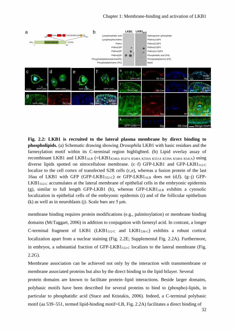
Chapter 1: Membrane-binding and activation of LKB1
32
Fig. 2.2: LKB1 is recruited to the lateral plasma membrane by direct binding to
phospholipids. (a) Schematic drawing showing Drosophila LKB1 with basic residues and the
farnesylation motif within its C-terminal region highlighted. (b) Lipid overlay assay of
recombinant LKB1 and LKB1ΔLB (=LKB1K546A R547A R548A K550A K551A K539A K540A K541A) using
diverse lipids spotted on nitrocellulose membrane. (c–f) GFP-LKB1 and GFP-LKB1512-C
localize to the cell cortex of transfected S2R cells (c,e), whereas a fusion protein of the last
16aa of LKB1 with GFP (GFP-LKB1552-C) or GFP-LKB1ΔLB does not (d,f). (g–j) GFP-
LKB1512-C accumulates at the lateral membrane of epithelial cells in the embryonic epidermis
(g), similar to full length GFP-LKB1 (h), whereas GFP-LKB1ΔLB exhibits a cytosolic
localization in epithelial cells of the embryonic epidermis (i) and of the follicular epithelium
(k) as well as in neuroblasts (j). Scale bars are 5 μm.
membrane binding requires protein modifications (e.g., palmitoylation) or membrane binding
domains (McTaggart, 2006) in addition to conjugation with farnesyl acid. In contrast, a longer
C-terminal fragment of LKB1 (LKB1512-C and LKB1536-C) exhibits a robust cortical
localization apart from a nuclear staining (Fig. 2.2E; Supplemental Fig. 2.2A). Furthermore,
in embryos, a substantial fraction of GFP-LKB1512-C localizes to the lateral membrane (Fig.
2.2G).
Membrane association can be achieved not only by the interaction with transmembrane or
membrane associated proteins but also by the direct binding to the lipid bilayer. Several
protein domains are known to facilitate protein–lipid interactions. Beside larger domains,
polybasic motifs have been described for several proteins to bind to (phospho)-lipids, in
particular to phosphatidic acid (Stace and Ktistakis, 2006). Indeed, a C-terminal polybasic
motif (aa 539–551, termed lipid-binding motif=LB, Fig. 2.2A) facilitates a direct binding of
GFP-LKB1
c
GFP-LKB1C564AGFP-LKB1552-C
d
GFP-LKB1LB
f
g
GFP-LKB1512-C DE-Cad Dlg
Merge
+ DAPI
NH2 - - COOH
VKKKGSALKRRAKKLTSCISVRKLSHCRTS538 567
5671
kinase domain
aLysophosphatic acid
Lysophosphocholine
PtdIns
PtdIns(3)P
PtdIns(4)P
PtdIns(5)P
Phosphatidylethanolamine(PE)
Phosphatidylcholine (PC)
Sphingosine-1phosphate
PtdIns(3,4)P2
PtdIns(3,5)P2
PtdIns(4,5)P2
PtdIns(3,4,5)P3
Phosphatidic acid (PA)
Phosphatidylserine (PS)
blank
LKB1 LKB1LBb
i
GFP-LKB1LB DE-Cad Dlg
Merge
+ DAPI
GFP-LKB1LB Baz Mir
Merge
+ DAPI
j
k
GFP-LKB1512-C
e h
GFP-LKB1 DE-Cad Dlg
Merge
+ DAPI

Chapter 1: Membrane-binding and activation of LKB1
33
Fig. 2.3: Membrane-association of LKB1 is essential for its kinase activity and function
in vivo. (a–c) Immunostainings of lkb1-mutant ovaries (b) exhibit a disturbed localization of
Staufen, which is rescued by expression of GFP-LKB1 (a) but not of GFP-LKB1ΔLB C564A (c).
(d) Immunoblotting of LKB1 variants expressed in embryos demonstrating that the protein
stability of GFP-LKB1ΔLB C564A is strongly reduced. (e) Overexpression of LKB1, but not of a
kinase-dead version of LKB1 (LKB1D317A=LKB1KD) or GFP-LKB1ΔLB C564A results in an
impaired eye morphology (‘rough eye’ phenotype). (f,g) In vitro kinase assays of GFP-
LKB1/GFP-STRAD purified from transfected S2R cells demonstrate a strong decrease in the
kinase activity of LKB1ΔLB C564A measured by autophosphorylation (32P GFP-LKB1) and
phosphorylation of recombinant AMPK (32P GST-AMPK108-280) (f). The addition of PA-
enriched liposomes (as described in materials and methods) increased the kinase activity of
wild type but not of membrane-binding deficient LKB1 (g). Activity of GFP-LKB1 was set as
100%. Inputs were visualized by Western Blot against GFP (GFP, GFP-Stlk and GFP-LKB1)
and by Coomassie Brilliant Blue staining (CCB, GST-AMPK108-280) Scale bars are 5 μm in c–
k and m, 100 μm in o and p, 10 μm in l and n. Experiments were performed in triplicates.
Error bars represent s.d. and statistical significance was determined using ANOVA: P<0.001,
***P<0.01, **P<0.05, not significant (NS).
Gurken StaufenMerge +
DAPI
Gurken StaufenMerge +
DAPI
lkb1X5
Gurken StaufenMerge +
DAPI
GFP-LKB1LB C564A;
lkb1X5
GFP-LKB1; lkb1X5
a
b
c
LKB1 LKB1KDLKB1LB C564A
wt
e
embryonic lysates
GFP - 100
Actin- 40
d
32P GFP-LKB1- 100
- 130
kDa
S2R+ STRAD +f
CCB GST-AMPK108-280
- 55
GFP-
STRADα
GFP-LKB1
GFP
- 70
- 55
- 40
- 35
- 100
- 130
32P GST-AMPK108-280
- 55
GFP-
STRADα
GFP-LKB1
GFP
- 70
- 55
- 40
- 35
- 100
- 130
kDa
S2R+ STRAD +g
32P GFP-LKB1- 100
- 130
%
0
50
100
150
200LKB1 auto-p
**
n.s.
0
50
100
150
200
250
300
%
AMPK-p
n.s.
***
020406080
100120
%
LKB1 auto-p
***
***
***
020406080
100120 AMPK-p
%
*** ******
CCB
GST-AMPK
- 55
32P GST-AMPK108-280
- 55

Chapter 1: Membrane-binding and activation of LKB1
34
DmLKB1 to PA, PtdIns(5)P, PtdIns(3,4,5)P3 and PtdIns(4,5)P2 as determined by a lipid
overlay assay (Fig. 2.2B). A strong binding to PA (and to some extent to PtdIns(3,4,5)P3 and
PtdIns(4,5)P2) was confirmed in liposome flotation assays (Supplemental Fig. 2.2B).
Mutation of this motif (mutation of Arg and Lys in LKB1-LB to Ala, LKB1ΔLB) abolished
liposome association in vitro (Supplemental Fig. 2.2B) as well as membrane association in
cultured cells (Fig. 2.2F) and in Drosophila epithelia and neural stem cells (Fig. 2.2H–K).
Membrane-bound LKB1 is crucial for Drosophila development
LKB1 has been implicated in various cellular functions, among them cell proliferation
control, suppression of tumour growth and regulation of cell polarity in various cell types
(Vaahtomeri and Mäkelä, 2011; Nakano and Takashima, 2012). To address the question
whether membrane targeting of LKB1 is crucial for its physiological function, we performed
rescue experiments with GFP-LKB1 expressed from its endogenous promoter. As indicated
above, not only wild-type LKB1 but also LKB1C564A can rescue the embryonic lethality of
maternal and zygotic mutant flies to a substantial extent, indicating that wild-type and
farnesylation-deficient GFP-LKB1 can substitute endogenous LKB1 in all tissues (Fig. 2.1O).
Notably, GFP-DmLKB1ΔLB still exhibits a residual rescue capacity (8% of surviving flies in
contrast to 69% for wild-type DmLKB1, (Fig. 2.1O), which might be due to a transient or
weak membrane binding mediated by the farnesylation anchor. Indeed, removal of the
farnesylation motif in GFP-LKB1ΔLB (GFP-LKB1ΔLB C564A) totally abolishes the rescue
capacity of the mutant protein and all mutant flies die during embryonic stages like the null
allele lkb1X5 (Fig. 2.1O). Moreover, lkb1-mutant flies expressing GFP-LKB1ΔLB C564A exhibit
the same polarity defects as the null allele alone (e.g., disruption of the anterior–posterior
polarity of ovaries, Fig. 2.3C, compared with wild-type LKB1 rescue in A and lkb1-mutant
phenotype in B).
Western blot analyses of lysates from embryos expressing LKB1 variants from the
endogenous promoter reveal that GFP-LKB1ΔLB C564A is unstable or rapidly degraded (Fig.
2.3D), indicating that the association of LKB1 with membranes is essential for protein
stability. In contrast, transient membrane association of LKB1ΔLB, which is probably mediated
by the farnesylation anchor, is sufficient for protein stabilization, as no differences in protein
levels are detectable between wild-type LKB1 and lipid-binding-deficient LKB1 (Fig. 2.3D).

Chapter 1: Membrane-binding and activation of LKB1
35
However, overexpression of GFP-DmLKB1ΔLB C564A in lkb1-mutant flies using the
UAS/GAL4 system instead of the endogenous lkb1-promoter does not result in detectable
rescue capacity (Supplemental Fig. 2.3A). Similar, the overexpression phenotype (rough eye
formation) of LKB1 is abolished in GFP-DmLKB1ΔLB C564A similar to a kinase dead version
(Fig. 2.3E). Thus, we assume that it is very unlikely that impaired functionality of GFP-
DmLKB1ΔLB C564A is only due to the instability of the protein.
To further substantiate this assumption, we fused two different heterologous membrane-
binding domains to the C terminus of GFP-LKB1ΔLB C564A: The PH-domain of human
phospholipase Cδ (Várnai and Balla, 1998) that preferentially binds to PtdIns(4,5)P2
(resulting in GFP-LKB1ΔLB C564A-PH(PLCδ)), but is also capable of binding to PA with a high
affinity (Pawelczyk and Matecki, 1999), and the PH-domain of human Akt1, (GFP-LKB1ΔLB
C564A-PH(Akt1)) with a high affinity to PtdIns(3,4,5)P3 (Klippel et al., 1997). Similar to PLCδ,
Akt1 has recently been demonstrated to bind PA in vitro (Bruntz et al., 2014). Strikingly, both
chimeric proteins stabilize the protein expression of GFP-LKB1ΔLB C564A (Fig. 2.3D), localize
at least partly to the lateral plasma membrane (Supplemental Fig. 2.4A,B) and are capable of
rescuing the lkb1 null allele to a large extent (56 and 48% respectively, Fig. 2.1O). These
results confirm our hypothesis that membrane association of LKB1 is crucial for its function
in vivo.
To test whether membrane association is essential for the kinase function of LKB1, we
performed in vitro kinase assays using the GFP-DmLKB1/STRADα complex purified from
transfected S2R cells. Indeed GFP-DmLKB1ΔLB C564A exhibits a strongly decreased kinase
activity (46% in autophosphorylation and 30% in AMPK-phosphorylation relative to wild-
type GFP-LKB1, Fig. 2.3F). Furthermore, addition of PA-enriched liposomes to GFP-
DmLKB1/STRADα strongly increases the kinase activity of the LKB1 complex
(autophosphorylation 1.6-fold and AMPK-phosphorylation 2.4-fold Fig. 2.3G).
Reduced kinase activity of LKB1ΔLB C654A is neither due to impaired association with the
canonical binding partners STRADα or Mo25, which enhance LKB1 activity, nor due to
decreased substrate binding (Supplemental Fig. 2.3B). Therefore, these data suggest that the
lipid binding capacity of DmLKB1 is essential for its kinase function.
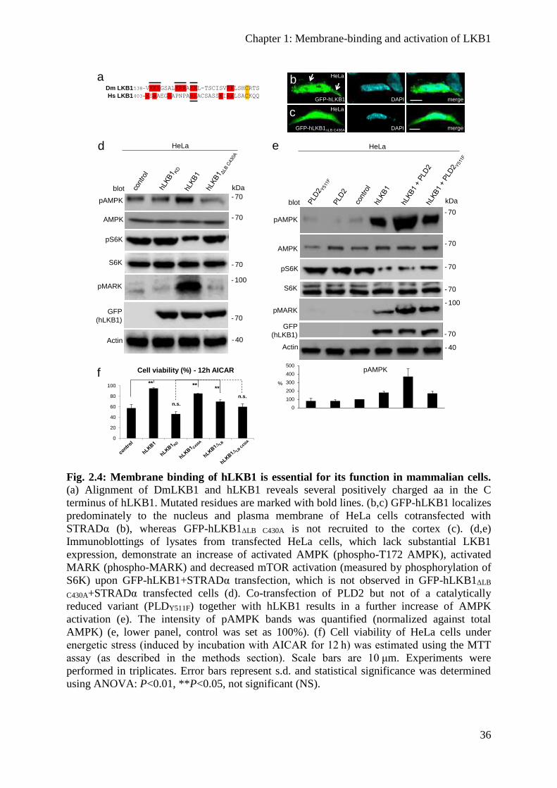
Chapter 1: Membrane-binding and activation of LKB1
36
Fig. 2.4: Membrane binding of hLKB1 is essential for its function in mammalian cells. (a) Alignment of DmLKB1 and hLKB1 reveals several positively charged aa in the C
terminus of hLKB1. Mutated residues are marked with bold lines. (b,c) GFP-hLKB1 localizes
predominately to the nucleus and plasma membrane of HeLa cells cotransfected with
STRADα (b), whereas GFP-hLKB1ΔLB C430A is not recruited to the cortex (c). (d,e)
Immunoblottings of lysates from transfected HeLa cells, which lack substantial LKB1
expression, demonstrate an increase of activated AMPK (phospho-T172 AMPK), activated
MARK (phospho-MARK) and decreased mTOR activation (measured by phosphorylation of
S6K) upon GFP-hLKB1+STRADα transfection, which is not observed in GFP-hLKB1ΔLB
C430A+STRADα transfected cells (d). Co-transfection of PLD2 but not of a catalytically
reduced variant (PLDY511F) together with hLKB1 results in a further increase of AMPK
activation (e). The intensity of pAMPK bands was quantified (normalized against total
AMPK) (e, lower panel, control was set as 100%). (f) Cell viability of HeLa cells under
energetic stress (induced by incubation with AICAR for 12 h) was estimated using the MTT
assay (as described in the methods section). Scale bars are 10 μm. Experiments were
performed in triplicates. Error bars represent s.d. and statistical significance was determined
using ANOVA: P<0.01, **P<0.05, not significant (NS).
0
20
40
60
80
100
a538-VKKKGSALKRRAKKL-TSCISVRKLSHCRTS
403-KSRAEGRAPNPARKACSASSKIRRLSACKQQ
Dm LKB1
Hs LKB1
f Cell viability (%) - 12h AICAR
**
n.s.
n.s.
****
cGFP-hLKB1LB C430A DAPI merge
HeLa
b
GFP-hLKB1 DAPI merge
HeLa
d
- 70pAMPK
blot
HeLa
kDa
- 70AMPK
pS6K
- 40Actin
- 70S6K
- 70GFP
(hLKB1)
- 100pMARK
- 70pAMPK
blot
HeLa
kDa
- 70AMPK
- 70pS6K
- 40Actin
- 70S6K
- 70GFP
(hLKB1)
- 100pMARK
e
0
100
200
300
400
500pAMPK
%
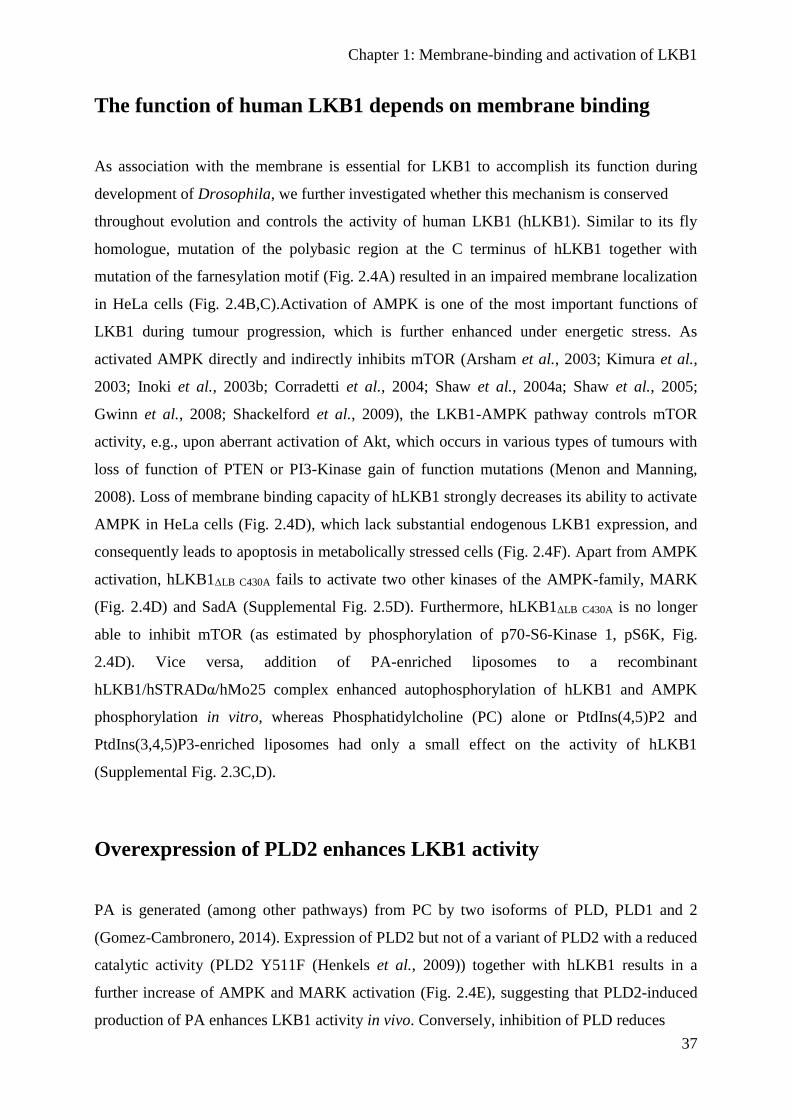
Chapter 1: Membrane-binding and activation of LKB1
37
The function of human LKB1 depends on membrane binding
As association with the membrane is essential for LKB1 to accomplish its function during
development of Drosophila, we further investigated whether this mechanism is conserved
throughout evolution and controls the activity of human LKB1 (hLKB1). Similar to its fly
homologue, mutation of the polybasic region at the C terminus of hLKB1 together with
mutation of the farnesylation motif (Fig. 2.4A) resulted in an impaired membrane localization
in HeLa cells (Fig. 2.4B,C).Activation of AMPK is one of the most important functions of
LKB1 during tumour progression, which is further enhanced under energetic stress. As
activated AMPK directly and indirectly inhibits mTOR (Arsham et al., 2003; Kimura et al.,
2003; Inoki et al., 2003b; Corradetti et al., 2004; Shaw et al., 2004a; Shaw et al., 2005;
Gwinn et al., 2008; Shackelford et al., 2009), the LKB1-AMPK pathway controls mTOR
activity, e.g., upon aberrant activation of Akt, which occurs in various types of tumours with
loss of function of PTEN or PI3-Kinase gain of function mutations (Menon and Manning,
2008). Loss of membrane binding capacity of hLKB1 strongly decreases its ability to activate
AMPK in HeLa cells (Fig. 2.4D), which lack substantial endogenous LKB1 expression, and
consequently leads to apoptosis in metabolically stressed cells (Fig. 2.4F). Apart from AMPK
activation, hLKB1ΔLB C430A fails to activate two other kinases of the AMPK-family, MARK
(Fig. 2.4D) and SadA (Supplemental Fig. 2.5D). Furthermore, hLKB1ΔLB C430A is no longer
able to inhibit mTOR (as estimated by phosphorylation of p70-S6-Kinase 1, pS6K, Fig.
2.4D). Vice versa, addition of PA-enriched liposomes to a recombinant
hLKB1/hSTRADα/hMo25 complex enhanced autophosphorylation of hLKB1 and AMPK
phosphorylation in vitro, whereas Phosphatidylcholine (PC) alone or PtdIns(4,5)P2 and
PtdIns(3,4,5)P3-enriched liposomes had only a small effect on the activity of hLKB1
(Supplemental Fig. 2.3C,D).
Overexpression of PLD2 enhances LKB1 activity
PA is generated (among other pathways) from PC by two isoforms of PLD, PLD1 and 2
(Gomez-Cambronero, 2014). Expression of PLD2 but not of a variant of PLD2 with a reduced
catalytic activity (PLD2 Y511F (Henkels et al., 2009)) together with hLKB1 results in a
further increase of AMPK and MARK activation (Fig. 2.4E), suggesting that PLD2-induced
production of PA enhances LKB1 activity in vivo. Conversely, inhibition of PLD reduces

Chapter 1: Membrane-binding and activation of LKB1
38
Fig. 2.5: Induction of multiple axons in primary rat hippocampal neurons depends on
membrane binding of hLKB1. (a–f) Hippocampal neurons from E18 rat embryos were
transfected at 0 d.i.v. with vectors for GFP (control), GFP-LKB1 or GFP fusion proteins for
the indicated LKB1 mutants and STRAD and analysed at 3 d.i.v by staining with the Tau-1
antibody (axonal marker). Representative images of transfected neurons are shown. The scale
bar is 20 μm. (f) The development of neuronal polarity was analysed by counting the number
of neurons without an axon (0, black), with a single axon (1, gray) or with multiple axons (>1,
white). Most primary rat hippocampal neurons transfected with GFP alone (control) develop
only a single axon (a, quantified in f). In contrast, neurons overexpressing GFP-
hLKB1+STRADα or GFP-hLKB1C430A+STRADα (b,c,f) frequently establish two or more
axons, whereas hLKB1 variants, which are deficient in membrane binding, fail to induce a
multiple axon phenotype (d–f). Experiments were performed in triplicates. Error bars
represent s.d. and statistical significance was determined using ANOVA: P<0.0001,
****P<0.01, **P>0.05, not significant (NS).
LKB1’s capacity to activate AMPK (Supplemental Fig. 2.5B). However, basal levels of
activated AMPK are maintained, indicating that other enzymes (e.g., Diacylglycerol kinase)
can compensate loss of PLD activity. This is in line with the observation that C. elegans,
Drosophila and mouse mutants lacking PLD enzymes do not exhibit as dramatic phenotypes
as LKB1ΔLB C564A (LaLonde et al., 2005; Raghu et al., 2009; Sato et al., 2013).
Membrane-binding-deficient LKB1 fails to induce multiple axons
Overexpression of LKB1 induces the extension of multiple axons (Shelly et al., 2007; Barnes
et al., 2007; Veleva-Rotse et al., 2014). Consistently, overexpression of wild-type GFP-
hLKB1
control hLKB1 hLKB1C430A
hLKB1LB hLKB1LB C430A
a b c
d e
f

Chapter 1: Membrane-binding and activation of LKB1
39
together with STRADα results in the formation of multiple axons by the majority of
transfected rat hippocampal neurons in culture (Fig. 2.5B,F). Remarkably, expression of
farnesylation-deficient hLKB1C430A induces this phenotype as efficiently as wild-type LKB1
(Fig. 2.5C,F). By contrast, mutation of the C-terminal basic region (hLKB1ΔLB) almost
abolished the effect on axon formation (Fig. 2.5D,F). Inactivation of the farnesylation signal
in lipid-binding-deficient hLKB1 (hLKB1ΔLB C430A) did result in further reduction of LKB1
activity in this assay (Fig. 2.5D,F). These results confirm the importance of the C-terminal
membrane binding domain for the function of LKB1 in mammalian cells.
Expression of LKB1 is downregulated in malignant melanoma
As we have demonstrated that overexpression of PLD2 in cell culture results in increased
activity of LKB1 (reflected by enhanced AMPK activation, Fig. 2.4E), we next assessed the
pathophysiological relevance of our findings regarding tumour formation in vivo.
Interestingly, expression of PLD2 is upregulated in several types of cancer, including
melanoma, and high PLD2 expression correlates with poor survival rates of patients (Saito et
al., 2007; Henkels et al., 2013). In cultured melanoma cells lacking detectable LKB1
expression (IGR37), membrane binding of LKB1 is essential for efficient AMPK (and
MARK) activation, suppression of mTOR and cell survival under energetic stress
(Supplemental Fig. 2.6).
Our data suggest that an aberrant increase in PLD expression in cancer cells will lead to
increased PA levels and subsequent mTOR activation (Fang et al., 2001; Foster et al., 2014;
Yoon et al., 2015) only if in addition the expression of LKB1 is reduced, because PA-
mediated LKB1/AMPK activation normally counteracts mTOR activity. To test this
hypothesis, we investigated whether increased expression of PLD, aberrant activation of Akt
and decreased LKB1 expression correlate with enhanced mTOR activity in melanoma.
Indeed, biopsies of melanoma primary tumours show a strong correlation between these four
parameters. In 81% of all analyzed tumours PLD2 expression as well as phospho-Akt and
mTOR activity were elevated while the expression of LKB1 was decreased (Fig. 2.6A–E).
Only 12% of the samples showed mTOR activation in the presence of stimulating phospho-
Akt and PLD2-overexpression although LKB1 staining appeared normal, which might be
explained by inactivating mutations of LKB1, which do not affect protein expression or
stability. In contrast, melanocytes in biopsies of healthy skin exhibited a strong LKB1
Chapter 1: Membrane-binding and activation of LKB1
40
Fig. 2.6: Downregulation of hLKB1 correlates with increased mTOR signalling in
malignant melanoma overexpressing PLD2. Immunostainings of paraffin-embedded tissue
sections of normal skin (a–d), neavi (a′–d′) and primary malignant melanoma tumours (a′′–
d′′). (a) hLKB1 is highly expressed in melanocytes in the stratum basale of the epidermis in
situ (a, arrows), and still detectable at substantial levels (a′) whereas it is almost undetectable
in the majority of malignant melanoma (a′′). (b) PLD2 is not detectable in melanocytes of
healthy skin sections (b, arrows) but becomes overexpressed in naevi (b′) and primary
tumours (b′′). (c) Akt is activated (indicated by staining of phospho Akt, pAkt) in naevi (c′)
and malignant melanoma (c′′), whereas it is not detectable in melanocytes of healthy skin
samples (c). (d) Melanocytes in healthy skin biopsies (d) show only faint nuclear staining for
phospho S6K (pS6K), which was used as to evaluate mTOR activation. Naevi (d′) exhibit
only very few pS6K positive cells, whereas malignant melanoma show strong cytoplasmic
and nuclear staining of pS6K (d′′). (e) Quantification of melanoma biopsies scored for the
indicated correlations. ‘Others’ are tumours, which do not exhibit increased phospho-Akt or
PLD2 staining.
expression, as well as low level of PLD2, phospho-Akt and mTOR activation (Fig. 2.6A–D,
arrows). Melanocytic nevi, a benign proliferation of melanocytes, which can give birth to
malignant melanoma, show in many cases elevated PLD2 expression as well as activation of
Akt, whereas LKB1 expression is still high in most cases, suppressing aberrant mTOR
activation, which occurred only in 25% of the analyzed specimen (Fig. 2.6A’–D’). Thus in
melanoma, loss of LKB1 in PLD2 overexpressing tumours may further contribute to mTOR
activation, whereas in benign neoplasia higher LKB1 level counterbalance aberrant PLD2
activation.
hLKB1 PLD2
Melanoma
Neavus
pAkt pS6K
a' b‘ c‘ d‘
a‘‘ b‘‘ c‘‘ H
Healthy
skin
a b c d
d‘‘
0
10
20
30
40
50
60
70
80
90
LKB1 low LKB1 high other
%
hLKB1
PLD2
pAkt
pS6K
Melanoma
(n = 67)
e
hLKB1
PLD2
pAkt
pS6K
others

Chapter 1: Membrane-binding and activation of LKB1
41
Discussion
In this study, we have elucidated a conserved mechanism regulating LKB1 activity and
function in vivo and during tumour suppression (Fig. 2.7). We have further established the
first link between PLD-mediated production of PA and activation of LKB1. Our data suggest
that membrane targeting of LKB1 by direct binding to PA is essential for the function of
LKB1 during Drosophila development and for activation of AMPK and suppression of
mTOR activity in cultured mammalian cells. Furthermore, downregulation of LKB1 in
malignant melanoma specimen, which exhibit activated Akt and overexpression of PLD2,
correlates with enhanced mTOR activity, indicating that the mechanism described in this
study contributes to pathogenesis of malignant melanoma. In contrast to previous studies
investigating the role of LKB1 farnesylation in the oocyte of Drosophila (Martin and St
Johnston, 2003), we found farnesylation of LKB1 to be dispensable for the correct protein
localization in the embryonic epidermis and NBs and for the function of LKB1 in Drosophila
development. This observation is in line with previous findings from cultured mammalian
cells and mice. In malignant melanoma cells, farnesylation-deficient LKB1 suppresses colony
formation at similar degree as the wild-type kinase (Sapkota et al., 2001). Furthermore,
studies from knock-in mice show that mice homozygous for farnesylation deficient LKB1 are
viable and do not exhibit any obvious phenotypes (Houde et al., 2014). However, in these
mice AMPK is less phosphorylated, whereas in cell culture experiments, no difference was
found between wild-type and farnesylation deficient LKB1 (Denison et al., 2009). Notably,
several C-terminally truncated hLKB1 variants (due to nucleotide deletions or point mutations
in STK11) with an intact kinase domain have been reported (Boudeau et al., 2003d) and
OMIM to be associated with Peutz-Jeghers syndrome, which might be due to the function of
the C-terminal region in PA-mediated activation of LKB1. Apart from the association of
LKB1 with STRADα/Mo25, membrane binding and PA-mediated activation of LKB1 is a
newly described upstream mechanism, which controls LKB1 kinase activity and function in
vitro and in vivo. The regulation of LKB1 function by membrane recruitment raises the
question whether this depends primarily on membrane localization or whether lipid binding
itself stimulates LKB1 activity. Several (phospho)-lipids have been demonstrated to activate
or increase the activity of various serine/threonine kinases, including Phosphoinositide-
dependent kinase 1 (PDK1, activated by PtdIns(3,4,5)P3) or PKCζ, Raf1, and mTOR (all
activated by PA (Limatola et al., 1994; Ghosh et al., 1996; Fang et al., 2001; Toschi et al.,
2009)). Fusion of GFP-LKB1ΔLB C564A to lipid-binding domains (PH(PLCδ) and PH(Akt1))

Chapter 1: Membrane-binding and activation of LKB1
42
Fig. 2.7: Simplified model of LKB1 activation by PLD2-produced PA and its
downstream effects on AMPK activation and mTOR inhibition. Under physiological
conditions in non-tumourous cells, PA is produced by PLD (and other enzymes), resulting in
an activation of mTOR and LKB1. Due to the inhibition of mTOR by LKB1-activated
AMPK, the final result of increased PA level is a balanced cell proliferation and -growth. In
contrast, in malignant melanoma, increased PI3K/Akt-signalling and enhanced production of
PA by overexpression of PLD results in aberrant mTOR activation which is not
counterbalanced by LKB1/AMPK and thus leads to increased cell proliferation and -growth
and impaired tumour suppression.
resulted in a re-activation of GFP-LKB1ΔLB C564A (Fig. 2.1O; Supplemental Fig. 2.4).
Furthermore, the activity of LKB1ΔLB C564A is strongly reduced in in vitro kinase assays (Fig.
2.3F,G). These findings could substantiate a hypothesis, in which the membrane functions as
a scaffold for bringing together LKB1 and its cofactors (STRADα and Mo25) or its
substrates. However, membrane binding deficient LKB1 robustly associates with its cofactors
and one of its substrates (AMPK, Supplemental Fig. 2.3B). Moreover, addition of PA (but not
of PtdIns(4,5)P2 or PtdIns(3,4,5)P3) is capable to substantially enhance the kinase activity of
LKB1 in vitro (Fig. 2.3G; Supplemental Fig. 2.3C,D). Thus it is more likely, that in addition
to membrane recruitment, PA functions to induce a conformational change of LKB1 thus
enhancing its kinase activity. However we cannot exclude that in our in vitro kinase assays
using immunoprecipitated proteins from transfected cells wild-type LKB1 might have co-
immunoprecipitated with PA-enriched micelles, which enhanced its kinase activity in this
experiment. Interestingly, PLD-produced PA activates both, LKB1 (this study) and mTOR
(Fang et al., 2001; Toschi et al., 2009; Foster et al., 2014; Yoon et al., 2015). Up to now,
PLD
PIP3
AktP
AMPK
P
PA PC
AMP
LKB1
mTORP
P
Balanced cell proliferation and cell growth
Tumor suppression
Cell proliferation
Cell growth
Tumor progression
AMPK
PC
LKB1
malignant melanoma
PIP3
AktP
PP
PLD
PA
mTOR
PP P
healthy cells
Chapter 1: Membrane-binding and activation of LKB1
43
overexpression of PLD and increased levels of PA have been only assigned to pro-oncogenic
pathways, in particular mTOR and Raf signalling (Ghosh et al., 1996; Rizzo et al., 1999;
Zheng et al., 2006; Shi et al., 2007; Toschi et al., 2009). Our data thus demonstrate the first
anti-oncogenic mechanism of PLD function, which might counterbalance its pro-oncogenic
effects as long as the LKB1-AMPK signalling axis is intact (Fig. 2.7). Notably,
Mukhopadhyay et al. showed recently, that in a mesenchymal-like breast cancer cell line
(MDA-MB-231), which expresses robust levels of endogenous LKB1 (in contrast to the
majority of breast cancer metastases), activated PLD induces a reduction in phospho-AMPK
(Mukhopadhyay et al., 2015). Thus, PLD-mediated activation of LKB1 might be either cell
type specific or depends on the basal activation of both signalling cascades, the LKB1-AMPK
axis and the PI3K-AKT pathway.
The LKB1-AMPK signalling pathway is well described to play a crucial role for cell polarity,
energy homeostasis and cell proliferation and is frequently disturbed in various types of
cancer (Nakano and Takashima, 2012; Hardie and Alessi, 2013). As mentioned above, one
particular function of AMPK is to counterbalance the pro-proliferative activity of mTOR
(Arsham et al., 2003; Kimura et al., 2003; Inoki et al., 2003b; Corradetti et al., 2004; Shaw et
al., 2004a; Shaw et al., 2005; Gwinn et al., 2008; Shackelford et al., 2009). Defects in the
LKB1-AMPK signalling pathway (resulting in impaired inhibition of mTOR) have been
already demonstrated to contribute to BRAF- and KRAS-induced melanoma formation in
mouse models (Zheng et al., 2009; Esteve-Puig et al., 2009; Liu et al., 2012b; Damsky et al.,
2015). Remarkably, somatic mutations in human STK11 (encoding LKB1) are rather rare in
malignant melanoma (Rowan et al., 1999; Guldberg et al., 1999). However, our data
demonstrate that downregulation of LKB1 protein expression can be observed in a majority of
malignant melanoma samples and therefore most likely contributes to the pathogenesis of this
type of skin cancer.
Our study provides strong evidences for an important and conserved regulatory mechanism,
which facilitates membrane recruitment of LKB1 by PA and is essential for the kinase activity
and function of LKB1 during development and tumour suppression. The dissection of
upstream regulatory mechanisms of LKB1 contributes to a better understanding of the
physiological regulation of LKB1 and their misregulation in tumours, which might be useful
for the development of therapeutic approaches in the future.

Chapter 1: Membrane-binding and activation of LKB1
44
Supplemental figures
Supplemental Fig. 2.1: Expression of LKB1 by its endogenous promoter. Western blot
analysis Immunoblotting of embryonic lysates from w1118 flies and homozygous mutants for
lkb1X5, an lkb1 loss of function allele (Lee et al., 2006), that express GFP-LKB1 from the
endogenous lkb1 promoter shows comparable protein levels for endogenous and exogenous
protein.
Supplemental Fig. 2.2: LKB1 binds to phosphatidic acid in liposome flotation assays. (a)
GFP-LKB1 536-C localizes to the cell cortex of transfected S2R cells. (b) Recombinant GST-
LKB1 or GST-LKB1 LB was incubated with liposomes (see Methods section for further
details), overlaid with a sucrose cushion and subjected to ultracentrifugation. Liposomes were
harvested and loaded as top fraction (T), whereas unbound protein remained in the bottom
fraction (B). n = 3, significance (wild type LKB1 versus LKB1 LB): p < 0.005 for PA, n.s
for PtdIns(4,5)P2 and p < 0.05 for PtdIns(3,4,5)P3.
GST-LKB1
PA
B T B T B T
GST-LKB1∆LB
0
20
40
60
80
100
PA PIP2 PIP3
% P
rote
in
0
20
40
60
80
100
PA PIP2 PIP3
% P
rote
in
top fraction bottom fraction
PA
PA PtdIns
(4,5)P2
PtdIns
(3,4,5)P3
GFP-LKB1536-C
a b
PtdIns
(4,5)P2
PtdIns
(3,4,5)P3
PtdIns
(4,5)P2
PtdIns
(3,4,5)P3
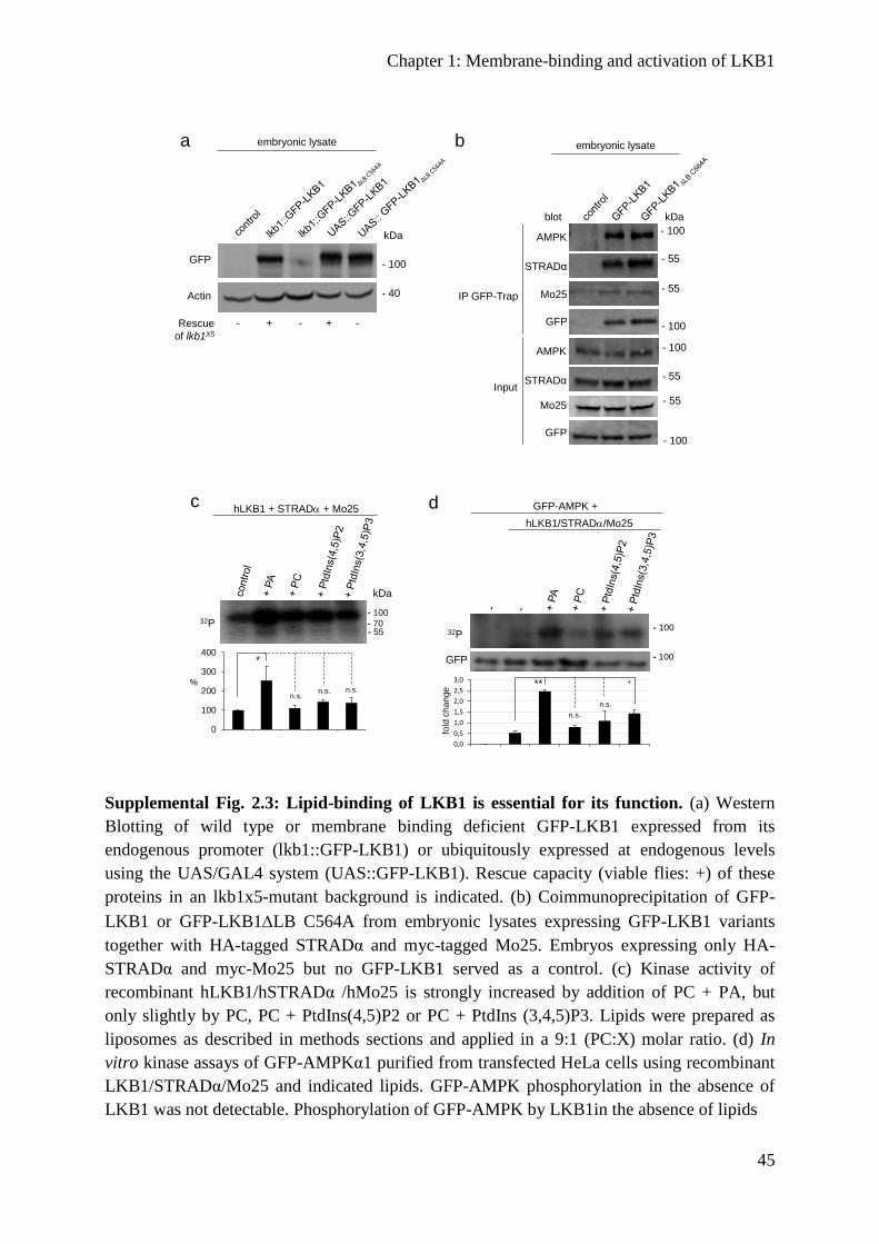
Chapter 1: Membrane-binding and activation of LKB1
45
Supplemental Fig. 2.3: Lipid-binding of LKB1 is essential for its function. (a) Western
Blotting of wild type or membrane binding deficient GFP-LKB1 expressed from its
endogenous promoter (lkb1::GFP-LKB1) or ubiquitously expressed at endogenous levels
using the UAS/GAL4 system (UAS::GFP-LKB1). Rescue capacity (viable flies: +) of these
proteins in an lkb1x5-mutant background is indicated. (b) Coimmunoprecipitation of GFP-
LKB1 or GFP-LKB1LB C564A from embryonic lysates expressing GFP-LKB1 variants
together with HA-tagged STRADα and myc-tagged Mo25. Embryos expressing only HA-
STRADα and myc-Mo25 but no GFP-LKB1 served as a control. (c) Kinase activity of
recombinant hLKB1/hSTRADα /hMo25 is strongly increased by addition of PC + PA, but
only slightly by PC, PC + PtdIns(4,5)P2 or PC + PtdIns (3,4,5)P3. Lipids were prepared as
liposomes as described in methods sections and applied in a 9:1 (PC:X) molar ratio. (d) In
vitro kinase assays of GFP-AMPKα1 purified from transfected HeLa cells using recombinant
LKB1/STRADα/Mo25 and indicated lipids. GFP-AMPK phosphorylation in the absence of
LKB1 was not detectable. Phosphorylation of GFP-AMPK by LKB1in the absence of lipids
- 100 GFP
- 40Actin
embryonic lysate
kDa
a
Rescue
of lkb1X5
- + - + -
0,0
0,5
1,0
1,5
2,0
2,5
3,0
AMPK +LKB1 + PA + PC + PIP2 + PIP3
hLKB1/STRAD/Mo25
- 10032P
d
fold
ch
an
ge
- 100GFP
GFP-AMPK +
**
n.s.
n.s.
*
- 100
- 55
AMPK
embryonic lysate
IP GFP-Trap
Input
kDablot
- 55
- 55
STRADα
b
- 100
- 55
- 100 GFP
Mo25
AMPK
STRADα
GFP
Mo25
- 100
kDa
hLKB1 + STRAD + Mo25
- 100
- 7032P
c
- 55
0
100
200
300
400
LKB1 LKB1 + PA LKB1 + PC LKB1 + PC +PIP 2
LKB1 + PC +PIP 3
%
*
n.s.n.s. n.s.

Chapter 1: Membrane-binding and activation of LKB1
46
was set as 100%. Experiments were performed in triplicates. Error bars represent standard
deviation and statistical significance was determined using ANOVA: p < 0.0001 ****, p
<0.001***, p < 0.01**, p < 0.05*, p > 0.05 not significant (n.s.).
Supplemental Fig. 2.4: Localization of fusion proteins of GFP-LKB1LB C564A with
heterologous membrane-binding domains. (a and b) Immunostainings of embryos with
GFP and α-spectrin reveal a substantial cortical localization of GFP-LKB1LB C564A fused
to the PH domain of Phospholipase C (PH(PLCδ), a) or to the PH domain of Akt1 (PH(Akt),
b). Scale bars are 5μm.
Supplemental Fig. 2.5: Activation of AMPK and AMPK-related kinases by LKB1. (a)
HeLa cells were transfected with plasmids for GFP-SadA and GFP alone (a) or the indicated
GFP-hLKB1 variants (a’-a’’) and stained with antibodies specific for LKB1 (red) and
activated Sad kinases (phosphorylated at S175 of SadA, pSAD, white). (b) Inhibition of PLD
by 100nM 4-Fluoro-N-(2-(4-(5-fluoro-1H-indol-1-yl)piperidin-1-yl)ethyl)benzamide (FIPI)
for 1h decreases the activation of AMPK by LKB1 in transfected HeLa cells. Scale bars are
5μm.
a
GFP-LKB1LB PH(Akt) SpectrinMerge
+ DAPI
b
GFP-LKB1LB PH(PLCδ)Spectrin
Merge
+ DAPI
b
- 70pAMPK
blot
HeLa
kDa
- 70AMPK
- 40Actin
- 70GFP
(hLKB1)
a
GFPLKB1
Merge
+ DAPIpSAD
GFP-hLKB1LKB1
Merge
+ DAPIpSAD
GFP-hLKB1LB C430A
LKB1Merge
+ DAPIpSADGFP-SAD-A
GFP-SAD-A
GFP-SAD-A
a‘
a‘‘
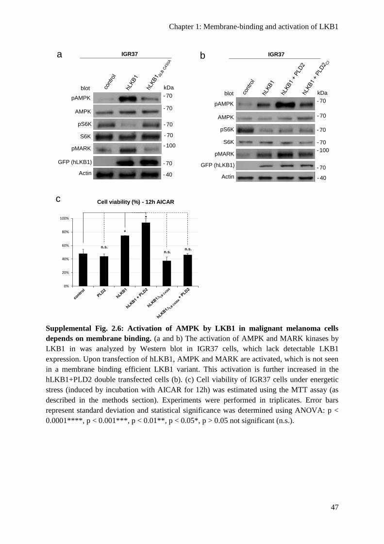
Chapter 1: Membrane-binding and activation of LKB1
47
Supplemental Fig. 2.6: Activation of AMPK by LKB1 in malignant melanoma cells
depends on membrane binding. (a and b) The activation of AMPK and MARK kinases by
LKB1 in was analyzed by Western blot in IGR37 cells, which lack detectable LKB1
expression. Upon transfection of hLKB1, AMPK and MARK are activated, which is not seen
in a membrane binding efficient LKB1 variant. This activation is further increased in the
hLKB1+PLD2 double transfected cells (b). (c) Cell viability of IGR37 cells under energetic
stress (induced by incubation with AICAR for 12h) was estimated using the MTT assay (as
described in the methods section). Experiments were performed in triplicates. Error bars
represent standard deviation and statistical significance was determined using ANOVA: p <
0.0001****, p < 0.001***, p < 0.01**, p < 0.05*, p > 0.05 not significant (n.s.).
a
- 70pAMPK
blot
IGR37
kDa
- 70AMPK
pS6K
- 40Actin
- 70
S6K
- 70GFP (hLKB1)
- 100pMARK
0%
20%
40%
60%
80%
100%
IGR PLD LKB1 LKB1 PLD LKB1 DeltaCA
LKB1 DeltaCA PLD
c Cell viability (%) - 12h AICAR
*
n.s.
n.s.
*
n.s.
b
- 70pAMPK
blot
IGR37
kDa
- 70AMPK
pS6K
- 40Actin
- 70S6K
- 70GFP (hLKB1)
- 100pMARK
- 70- 70

Chapter 2: Upstream regulation of LKB1
48
Chapter 2: Upstream regulation of LKB1
Controlling the master – upstream regulation of the tumour
suppressor LKB1
Authors: Lars Kullmann and Michael P. Krahn
Personal contributions: Wrote the manuscript and prepared the figures and table.
The tumour suppressor LKB1 is an essential serine/threonine kinase, which regulates various
cellular processes such as cell metabolism, cell proliferation, cell polarity and cell migration.
Germline mutations in the lkb1 gene are the cause of the Peutz-Jeghers syndrome, which is
characterized by benign polyps in the intestine and a higher risk for the patients to develop
intestinal and extraintestinal tumours. Moreover, mutations and misregulation of LKB1 have
been reported to occur in most types of tumours and are among the most common aberrations
in lung cancer. LKB1 activates several downstream kinases of the AMPK family by direct
phosphorylation in the T-loop. In particular the activation of AMPK upon energetic stress has
been intensively analysed in various diseases, including cancer to induce a metabolic switch
from anabolism towards catabolism to regulate energy homeostasis and cell survival.
In contrast, the regulation of LKB1 itself has long been only poorly understood. Only in the
last years, several proteins and posttranslational modifications of LKB1 have been analysed to
control its localization, activity and recognition of substrates. Here, we summarize the current
knowledge about the upstream regulation of LKB1, which is important for the understanding
of the pathogenesis of many types of tumours.
Chapter 2: Upstream regulation of LKB1
49
Introduction
The regulation of cell proliferation, energy metabolism, cell polarity and - migration are
essential processes that have to be strictly controlled. The Liver Kinase B1 (LKB1, encoded
by STK11) is a highly conserved serine/threonine kinase that functions as a tumour
suppressor. Germline mutations in STK11 cause the Peutz-Jeghers syndrome (PJS), an
autosomal dominant disorder, which is characterized by cutaneous mispigmentations, benign
gastrointestinal polyps and a high risk for the patients to develop tumours (Hemminki et al.,
1998; Mehenni et al., 1998; Jansen et al., 2009). The risk of PJS patients to develop cancer is
highest in the gastrointestinal tract, however female patients bear also an increased risk to
develop breast cancer (Hearle et al., 2006). Somatic mutations or loss of LKB1 are also
found in several other types of cancer, such as non-small cell lung cancer (NSCLC), cervical
cancer, ovarian cancer, breast cancer, pancreatic cancer and malignant melanoma (Rowan et
al., 1999; Guldberg et al., 1999; Sanchez-Cespedes et al., 2002; Carretero et al., 2004;
Matsumoto et al., 2007; Wingo et al., 2009; Morton et al., 2010; Gill et al., 2011; Tanwar et
al., 2014; George et al., 2016; Dogliotti et al., 2017). Only in the pathogenesis of salivary
gland tumours, LKB1 seems to play no role (Cidlinsky et al., 2016). In NSCLC, genetic
alterations of the STK11 gene are among the most common mutations in these tumours.
Moreover, loss of LKB1 is associated with a poor survival rate of breast cancer patients (Shen
et al., 2002; Sengupta et al., 2017).
LKB1 forms a ternary complex with the pseudokinase STRADα and the scaffolding protein
MO25, which promote nuclear export of the complex and enhance the kinase activity of
LKB1. Together with the AMP-activated protein kinase (AMPK) LKB1 acts as a cellular
energy sensor. Under energy deprivation LKB1 phosphorylates AMPK to inhibit mTORC1,
Acety-CoA carboxylase and HMG-CoA-Reductase, thus inhibiting anabolic processes
(Woods et al., 2003). Additionally, LKB1 phosphorylates 12 AMPK related kinases in their
activation loop: SAD-A/B (BRSK2 and BRSK1), NUAK1/2 (ARK5 and SNARK), SIK1/2/3,
MARK1/2/3/4 and SNRK (Lizcano et al., 2004; Jaleel et al., 2005). Given that LKB1
regulates such a broad number of other kinases it could be considered as a “master kinase”.
Beyond the role of LKB1 in metabolism and cell proliferation it is involved in the
establishment of cell polarity by activating MARKs, which in turn regulate the formation of
the Par complex (Benton and St Johnston, 2003a; Lizcano et al., 2004; Granot et al., 2009;
Amin et al., 2009). In Drosophila, LKB1 has been described to regulate cell polarity by
activation of AMPK, which in turn indirectly enhances the phosphorylation non-muscle
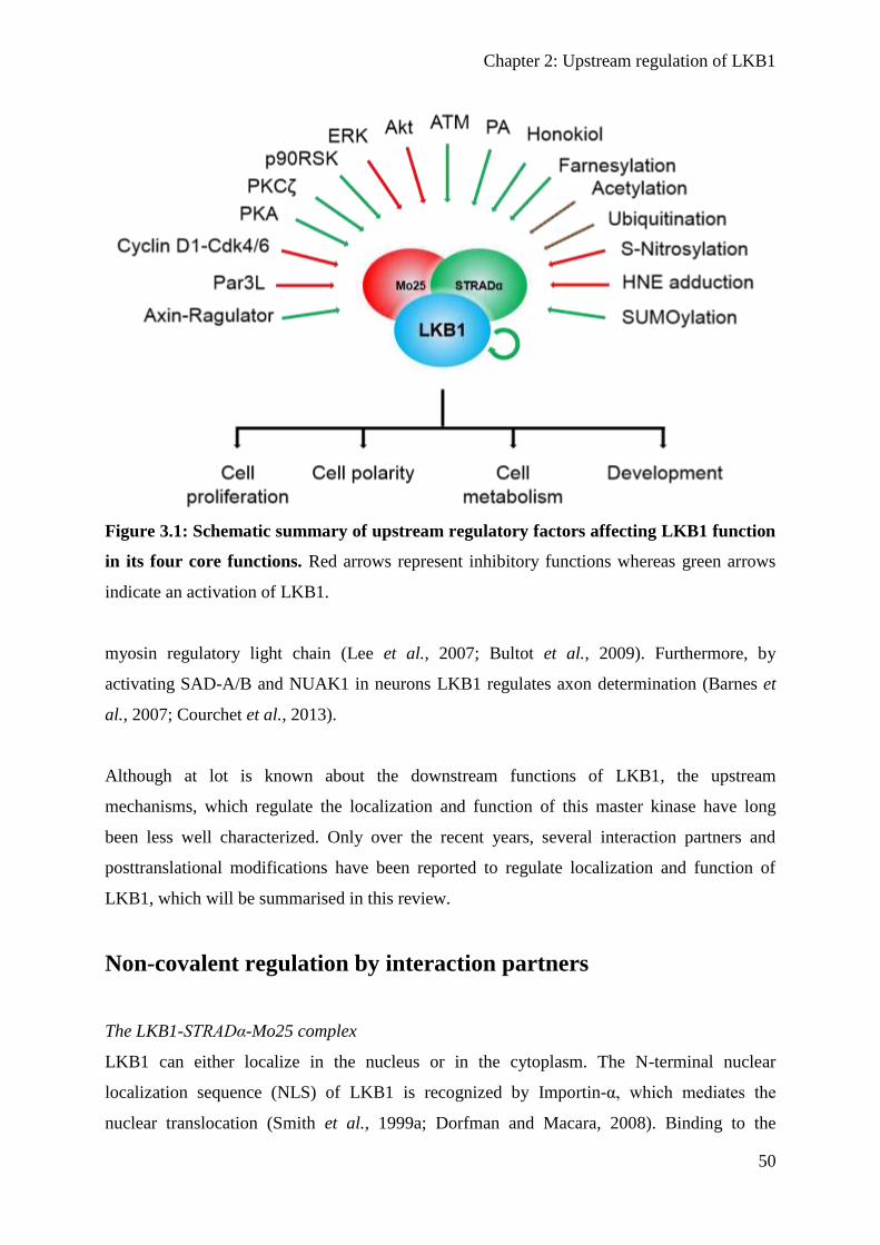
Chapter 2: Upstream regulation of LKB1
50
Figure 3.1: Schematic summary of upstream regulatory factors affecting LKB1 function
in its four core functions. Red arrows represent inhibitory functions whereas green arrows
indicate an activation of LKB1.
myosin regulatory light chain (Lee et al., 2007; Bultot et al., 2009). Furthermore, by
activating SAD-A/B and NUAK1 in neurons LKB1 regulates axon determination (Barnes et
al., 2007; Courchet et al., 2013).
Although at lot is known about the downstream functions of LKB1, the upstream
mechanisms, which regulate the localization and function of this master kinase have long
been less well characterized. Only over the recent years, several interaction partners and
posttranslational modifications have been reported to regulate localization and function of
LKB1, which will be summarised in this review.
Non-covalent regulation by interaction partners
The LKB1-STRADα-Mo25 complex
LKB1 can either localize in the nucleus or in the cytoplasm. The N-terminal nuclear
localization sequence (NLS) of LKB1 is recognized by Importin-α, which mediates the
nuclear translocation (Smith et al., 1999a; Dorfman and Macara, 2008). Binding to the

Chapter 2: Upstream regulation of LKB1
51
pseudokinase STRADα (STe20 Related ADaptor) and the scaffolding protein MO25 (mouse
protein 25) enhances nuclear export (Boudeau et al., 2003a; Baas et al., 2003; Dorfman and
Macara, 2008). STRADα translocates LKB1 into the cytoplasm by binding to the export
factors CRM1 or Exportin7 and maintains LKB1 in the cytoplasm by outcompeting Importin-
α for binding to the NLS of LKB1 (Dorfman and Macara, 2008). Moreover, STRADα and
MO25 drastically enhance the kinase activity of LKB1 (∼9 fold) (Boudeau et al., 2003a). In
line with these results, the LKB1 mutant SL26 in PJS, which was lacks the amino acids 303 –
306 cannot bind to STRADα and accumulates in the nucleus, which probably mediates its
pathogenicity as the SL26 mutant is still catalytically active (Nezu et al., 1999; Baas et al.,
2003; Boudeau et al., 2004). The crystal structure of the LKB1/-STRADα-MO25 complex
provides further insights into the formation of the active complex (Zeqiraj et al., 2009):
STRADα binds LKB1 as a pseudosubstrate and mediates its conversion in an active
conformation, whereas Mo25 is required to stabilize the active conformation of the LKB1
activation loop (Zeqiraj et al., 2009). However, no structural data are available for the N- and
C-terminus of LKB1, as in this study, only the kinase domain of LKB1 (aa 43 – 347) and the
pseudokinase domain of STRADα (aa 59 – 431) have been analysed. This might be of
particular interest, as the C-terminus contains several important phosphorylation sites as well
as a farnesylation motif and a lipid-binding domain (Collins et al., 2000; Sapkota et al., 2001;
Sapkota et al., 2002a; Sapkota et al., 2002b; Baas et al., 2003; Xie et al., 2006; Bai et al.,
2012; Dogliotti et al., 2017).
The scaffolding protein AXIN
One of the major targets of LKB1 in order to regulate metabolism and proliferation under
energetic stress is AMPK. AMPK is sensitized for an increase in the AMP/ATP ratio: A
single AMP molecule binds to the γ-subunit of the heterotrimeric AMPK, which enhances the
affinity to a second AMP molecule. Upon AMP binding the catalytic domain in the α-subunit
becomes exposed, which is then phosphorylated and activated at a conserved residue (T172)
within the activation loop (T-loop) by LKB1. The activated kinase in turn phosphorylates
Tuberin (TSC2) and Raptor to antagonize mTORC1 and thereby blocks the energy consuming
protein synthesis.
Recent studies demonstrated a more complex mechanisms of the LKB1 dependent regulation
of AMPK (Zhang et al., 2013b; Zhang et al., 2014). In hepatocytes, AXIN functions as a
scaffold by binding directly to LKB1 and AMPK and promotes the phosphorylation of
AMPK in an AMP dependent manner. Consequently, knock down of AXIN in mice causes an
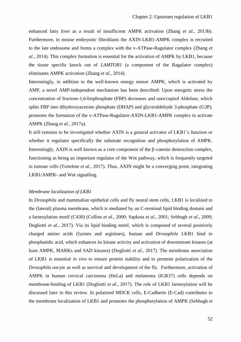
Chapter 2: Upstream regulation of LKB1
52
enhanced fatty liver as a result of insufficient AMPK activation (Zhang et al., 2013b).
Furthermore, in mouse embryonic fibroblasts the AXIN-LKB1-AMPK complex is recruited
to the late endosome and forms a complex with the v-ATPase-Ragulator complex (Zhang et
al., 2014). This complex formation is essential for the activation of AMPK by LKB1, because
the tissue specific knock out of LAMTOR1 (a component of the Ragulator complex)
eliminates AMPK activation (Zhang et al., 2014).
Interestingly, in addition to the well-known energy sensor AMPK, which is activated by
AMP, a novel AMP-independent mechanism has been described: Upon energetic stress the
concentration of fructose-1,6-bisphosphate (FBP) decreases and unoccupied Aldolase, which
splits FBP into dihydroxyacetone phosphate (DHAP) and glyceraldehyde 3-phosphate (G3P),
promotes the formation of the v-ATPase-Ragulator-AXIN-LKB1-AMPK complex to activate
AMPK (Zhang et al., 2017a).
It still remains to be investigated whether AXIN is a general activator of LKB1’s function or
whether it regulates specifically the substrate recognition and phosphorylation of AMPK.
Interestingly, AXIN is well known as a core component of the -catenin destruction complex,
functioning as being an important regulator of the Wnt pathway, which is frequently targeted
in tumour cells (Tortelote et al., 2017). Thus, AXIN might be a converging point, integrating
LKB1/AMPK- and Wnt signalling.
Membrane localization of LKB1
In Drosophila and mammalian epithelial cells and fly neural stem cells, LKB1 is localized to
the (lateral) plasma membrane, which is mediated by an C-terminal lipid binding domain and
a farnesylation motif (C430) (Collins et al., 2000; Sapkota et al., 2001; Sebbagh et al., 2009;
Dogliotti et al., 2017). Via its lipid binding motif, which is composed of several positively
charged amino acids (lysines and arginines), human and Drosophila LKB1 bind to
phosphatidic acid, which enhances its kinase activity and activation of downstream kinases (at
least AMPK, MARKs and SAD kinases) (Dogliotti et al., 2017). The membrane association
of LKB1 is essential in vivo to ensure protein stability and to promote polarization of the
Drosophila oocyte as well as survival and development of the fly. Furthermore, activation of
AMPK in human cervical carcinoma (HeLa) and melanoma (IGR37) cells depends on
membrane-binding of LKB1 (Dogliotti et al., 2017). The role of LKB1 farnesylation will be
discussed later in this review. In polarized MDCK cells, E-Cadherin (E-Cad) contributes to
the membrane localization of LKB1 and promotes the phosphorylation of AMPK (Sebbagh et
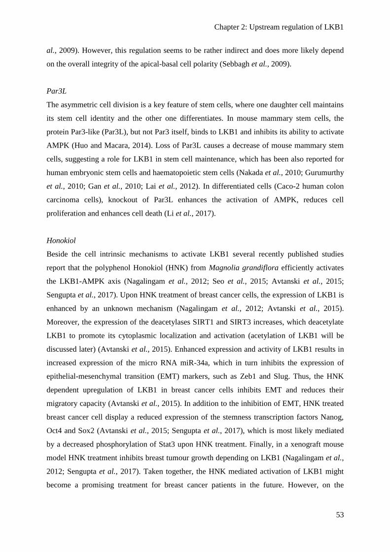
Chapter 2: Upstream regulation of LKB1
53
al., 2009). However, this regulation seems to be rather indirect and does more likely depend
on the overall integrity of the apical-basal cell polarity (Sebbagh et al., 2009).
Par3L
The asymmetric cell division is a key feature of stem cells, where one daughter cell maintains
its stem cell identity and the other one differentiates. In mouse mammary stem cells, the
protein Par3-like (Par3L), but not Par3 itself, binds to LKB1 and inhibits its ability to activate
AMPK (Huo and Macara, 2014). Loss of Par3L causes a decrease of mouse mammary stem
cells, suggesting a role for LKB1 in stem cell maintenance, which has been also reported for
human embryonic stem cells and haematopoietic stem cells (Nakada et al., 2010; Gurumurthy
et al., 2010; Gan et al., 2010; Lai et al., 2012). In differentiated cells (Caco-2 human colon
carcinoma cells), knockout of Par3L enhances the activation of AMPK, reduces cell
proliferation and enhances cell death (Li et al., 2017).
Honokiol
Beside the cell intrinsic mechanisms to activate LKB1 several recently published studies
report that the polyphenol Honokiol (HNK) from Magnolia grandiflora efficiently activates
the LKB1-AMPK axis (Nagalingam et al., 2012; Seo et al., 2015; Avtanski et al., 2015;
Sengupta et al., 2017). Upon HNK treatment of breast cancer cells, the expression of LKB1 is
enhanced by an unknown mechanism (Nagalingam et al., 2012; Avtanski et al., 2015).
Moreover, the expression of the deacetylases SIRT1 and SIRT3 increases, which deacetylate
LKB1 to promote its cytoplasmic localization and activation (acetylation of LKB1 will be
discussed later) (Avtanski et al., 2015). Enhanced expression and activity of LKB1 results in
increased expression of the micro RNA miR-34a, which in turn inhibits the expression of
epithelial-mesenchymal transition (EMT) markers, such as Zeb1 and Slug. Thus, the HNK
dependent upregulation of LKB1 in breast cancer cells inhibits EMT and reduces their
migratory capacity (Avtanski et al., 2015). In addition to the inhibition of EMT, HNK treated
breast cancer cell display a reduced expression of the stemness transcription factors Nanog,
Oct4 and Sox2 (Avtanski et al., 2015; Sengupta et al., 2017), which is most likely mediated
by a decreased phosphorylation of Stat3 upon HNK treatment. Finally, in a xenograft mouse
model HNK treatment inhibits breast tumour growth depending on LKB1 (Nagalingam et al.,
2012; Sengupta et al., 2017). Taken together, the HNK mediated activation of LKB1 might
become a promising treatment for breast cancer patients in the future. However, on the
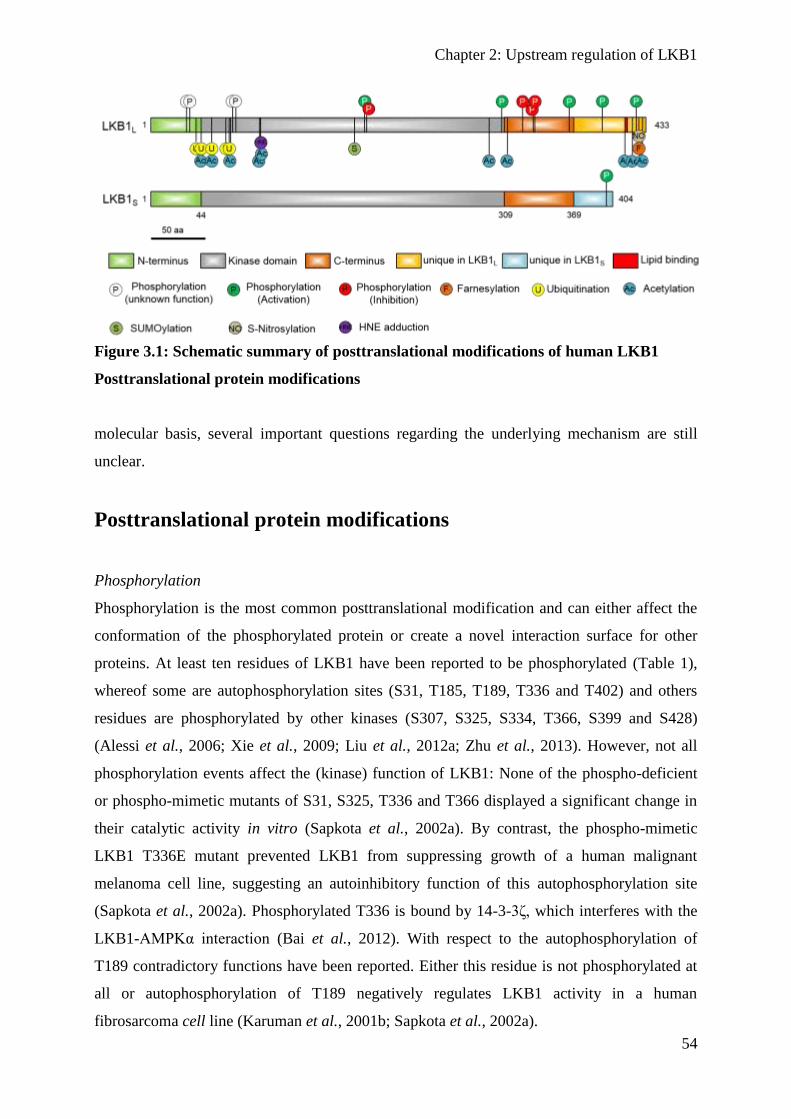
Chapter 2: Upstream regulation of LKB1
54
Figure 3.1: Schematic summary of posttranslational modifications of human LKB1
Posttranslational protein modifications
molecular basis, several important questions regarding the underlying mechanism are still
unclear.
Posttranslational protein modifications
Phosphorylation
Phosphorylation is the most common posttranslational modification and can either affect the
conformation of the phosphorylated protein or create a novel interaction surface for other
proteins. At least ten residues of LKB1 have been reported to be phosphorylated (Table 1),
whereof some are autophosphorylation sites (S31, T185, T189, T336 and T402) and others
residues are phosphorylated by other kinases (S307, S325, S334, T366, S399 and S428)
(Alessi et al., 2006; Xie et al., 2009; Liu et al., 2012a; Zhu et al., 2013). However, not all
phosphorylation events affect the (kinase) function of LKB1: None of the phospho-deficient
or phospho-mimetic mutants of S31, S325, T336 and T366 displayed a significant change in
their catalytic activity in vitro (Sapkota et al., 2002a). By contrast, the phospho-mimetic
LKB1 T336E mutant prevented LKB1 from suppressing growth of a human malignant
melanoma cell line, suggesting an autoinhibitory function of this autophosphorylation site
(Sapkota et al., 2002a). Phosphorylated T336 is bound by 14-3-3ζ, which interferes with the
LKB1-AMPKα interaction (Bai et al., 2012). With respect to the autophosphorylation of
T189 contradictory functions have been reported. Either this residue is not phosphorylated at
all or autophosphorylation of T189 negatively regulates LKB1 activity in a human
fibrosarcoma cell line (Karuman et al., 2001b; Sapkota et al., 2002a).

Chapter 2: Upstream regulation of LKB1
55
In addition to its autophosphorylation, LKB1 is phosphorylated by PKCζ (Xie et al., 2006;
Xie et al., 2008; Xie et al., 2009). Activation of PKCζ by either peroxynitrite or metformin in
endothelial cells induces the phosphorylation of LKB1 at S428, which enhances the nuclear
export of LKB1 to mediate its association with AMPK in order to activate it (Xie et al., 2006;
Xie et al., 2008). In addition to the activation of AMPK in response to S428 phosphorylation,
LKB1 phosphorylates and activates the lipid phosphatase PTEN to inhibit Akt signaling
(Song et al., 2008). Moreover, PKCζ phosphorylates LKB1 at S307 (Xie et al., 2009). This
phosphorylation is also important to enhance the nucleo-cytoplasmic shuttling of LKB1 in
peroxynitrite stimulated cells to activate AMPK. Loss of the phosphorylation sites S307 and
S428 attenuates the association of LKB1 with STRADα and the export factor CRM1 and thus
decreases the cytoplasmic accumulation of LKB1 in an adenocarcinoma cell line (Xie et al.,
2009). Consequentially, the phosphorylation-deficient LKB1 S307A mutant has a reduced
capacity to suppress angiogenesis and cannot efficiently protect cells from apoptosis (Xie et
al., 2009).
Apart from PKCζ, S428 is also a phosphorylation site for p90 ribosomal S6 kinase (p90-RSK)
and cAMP-dependent protein kinase (PKA) (Collins et al., 2000; Sapkota et al., 2001; Martin
and St Johnston, 2003). The p90-RSK mediated phosphorylation of S428 depends on ERK
stimulation, whereas PKA phosphorylates S428 in response to adenylyl cyclase activation
(Sapkota et al., 2001). In melanoma cells, phosphorylation of S428 is essential to suppress
cell growth and does not affect the farnesylation at C433 (Sapkota et al., 2001). The
PKC/PKA/p90-RSK mediated phosphorylation of LKB1 at S428 is crucial for axon
differentiation: Only wild type LKB1, but not the S428A mutant, is capable of
phosphorylating SAD-A/B in order to promote axon differentiation (Barnes et al., 2007;
Shelly et al., 2007). Furthermore, phosphorylation of LKB1 S428 by PKA is essential for
LKB1 localization and the myelination of Schwann cells (Shen et al., 2014). Notably, other
studies reports that S428 phosphorylation of LKB1 is not essential to suppress cell growth of
melanoma cells and to activate AMPK and SAD-A/B in vitro and in vivo (Bright et al., 2008;
Fogarty and Hardie, 2009; Houde et al., 2014).
The pleiotropy of studies with partly contradicting results indicates that LKB1 S428
phosphorylation might be cell-type specific (maybe even compartment specific) and probably
depends on the cellular context regarding cell proliferation, cell cycle, migration and
metabolism.
In mammals, two splice variants of LKB1 are expressed: LKB1L (long) and LKB1S (short),
which are both widely expressed (Towler et al., 2008; Denison et al., 2009). The two protein
Chapter 2: Upstream regulation of LKB1
56
variants differ only in their last 63 amino acids, hence, LKB1S lacks the critical residues S428
and C430. LKB1S is essential for spermatogenesis, since male lkb1S null mice are sterile
(Towler et al., 2008). However, despite the lack of S428 in LKB1S it is still phosphorylated
by PKCζ at its C-terminus at S399, which is unique in LKB1S (Zhu et al., 2013).
Phosphorylation of S399 in LKB1S promotes its nuclear export followed by the activation of
AMPK and is therefore thought to be the functional equivalent to S428 in LKB1L (Zhu et al.,
2013).
Another phosphorylation site in LKB1 is S325 (Sapkota et al., 2002a). Subsequent studies
managed to identify ERK and Cyclin D1-Cdk4/6 as the responsible kinases for the
phosphorylation of LKB1 at this residue (Zheng et al., 2009; Casimiro et al., 2017). In
melanoma cells, which express the oncogenic B-RafV600E variant, constitutively active ERK
phosphorylates LKB1 at this residue. This phosphorylation decreases the affinity of LKB1 to
AMPK and consequently leads to a reduction of AMPK activation. The authors confirmed
their cell culture data in tissue samples of human melanoma, where the phosphorylation and
activation of ERK associates with inactive AMPK (Zheng et al., 2009). In addition to ERK,
LKB1 is also phosphorylated at S325 by Cyclin D1-Cdk4/6 (Casimiro et al., 2017). Like
phosphorylation by ERK, Cyclin D1-Cdk4/6 mediated LKB1 S325 phosphorylation also
suppresses AMPK activation and the stress-induced activation of AMPK is enhanced in cyclin
D1-/- cells. Likewise, in mammary gland tissue of conditional cyclin D1-/- mice, the
phosphorylation of AMPK and the level of autophagy are enhanced. (Casimiro et al., 2017)
A previous study reported that mutations of S325 to alanine or glutamic acid did not affect the
catalytic activity of LKB1 (Sapkota et al., 2002a). However, this inhibitory modification of
LKB1 affects its substrate recognition rather than its kinase activity (Zheng et al., 2009). In
the context of tumour progression the misregulation of B-Raf and Cyclin D1 is a common
variation (Bartkova et al., 1994; Davies et al., 2002). Hence, affected tumours antagonize the
tumour suppressor function of the LKB1-AMPK axis, which enhances their malignancy.
The kinases Akt and LKB1 display antagonistic functions to control cell proliferation: Akt
activates mTOR signalling, whereas LKB1 inhibits it via AMPK activation (Inoki et al.,
2003b; Shaw et al., 2004a). However, Akt act as an upstream regulator of LKB1, as it directly
phosphorylates S334 of LKB1. Upon phosphorylation, 14-3-3 binds to LKB1 and antagonizes
the interaction with STARDα. Consequentially, LKB1 is sequestered in the nucleus as fails to
inhibit cell proliferation (Liu et al., 2012a).
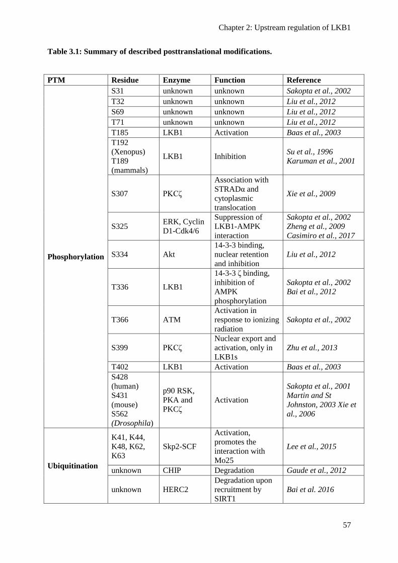
Chapter 2: Upstream regulation of LKB1
57
Table 3.1: Summary of described posttranslational modifications.
PTM Residue Enzyme Function Reference
Phosphorylation
S31 unknown unknown Sakopta et al., 2002
T32 unknown unknown Liu et al., 2012
S69 unknown unknown Liu et al., 2012
T71 unknown unknown Liu et al., 2012
T185 LKB1 Activation Baas et al., 2003
T192
(Xenopus)
T189
(mammals)
LKB1 Inhibition Su et al., 1996
Karuman et al., 2001
S307 PKCζ
Association with
STRADα and
cytoplasmic
translocation
Xie et al., 2009
S325 ERK, Cyclin
D1-Cdk4/6
Suppression of
LKB1-AMPK
interaction
Sakopta et al., 2002
Zheng et al., 2009
Casimiro et al., 2017
S334 Akt
14-3-3 binding,
nuclear retention
and inhibition
Liu et al., 2012
T336 LKB1
14-3-3 ζ binding,
inhibition of
AMPK
phosphorylation
Sakopta et al., 2002
Bai et al., 2012
T366 ATM
Activation in
response to ionizing
radiation
Sakopta et al., 2002
S399 PKCζ
Nuclear export and
activation, only in
LKB1s
Zhu et al., 2013
T402 LKB1 Activation Baas et al., 2003
S428
(human)
S431
(mouse)
S562
(Drosophila)
p90 RSK,
PKA and
PKCζ
Activation
Sakopta et al., 2001
Martin and St
Johnston, 2003 Xie et
al., 2006
Ubiquitination
K41, K44,
K48, K62,
K63
Skp2-SCF
Activation,
promotes the
interaction with
Mo25
Lee et al., 2015
unknown CHIP Degradation Gaude et al., 2012
unknown HERC2
Degradation upon
recruitment by
SIRT1
Bai et al. 2016
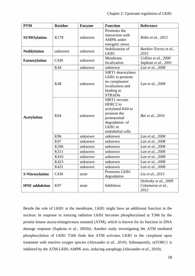
Chapter 2: Upstream regulation of LKB1
58
PTM Residue Enzyme Function Reference
SUMOylation K178 unknown
Promotes the
interaction with
AMPK under
energetic stress
Ritho et al., 2015
Neddylation unknown unknown Stabilization of
LKB1
Barbier-Torres et al.,
2015
Farnesylation C430 unknown Membrane
localization
Collins et al., 2000
Sapkota et al., 2001
Acetylation
K44 unknown unknown Lan et al., 2008
K48 unknown
SIRT1 deacetylates
LKB1 to promote
its cytoplasmic
localization and
binding to
STRADα
Lan et al., 2008
K64 unknown
SIRT1 recruits
HERC2 to
acetylated K64 to
promote the
proteasomal
degradation of
LKB1 in
endothelial cells
Bai et al., 2016
K96 unknown unknown Lan et al., 2008
K97 unknown unknown Lan et al., 2008
K296 unknown unknown Lan et al., 2008
K311 unknown unknown Lan et al., 2008
K416 unknown unknown Lan et al., 2008
K423 unknown unknown Lan et al., 2008
K431 unknown unknown Lan et al., 2008
S-Nitrosylation C430 none Promotes LKB1
degradation Liu et al., 2015
HNE adduktion K97 none Inhibition
Dolinsky et al., 2009
Calamaras et al.,
2012
Beside the role of LKB1 at the membrane, LKB1 might have an additional function in the
nucleus: In response to ionizing radiation LKB1 becomes phosphorylated at T366 by the
protein kinase ataxia-telangiectasia mutated (ATM), which is known for its function in DNA
damage response (Sapkota et al., 2002b). Another study investigating the ATM mediated
phosphorylation of LKB1 T366 finds that ATM activates LKB1 in the cytoplasm upon
treatment with reactive oxygen species (Alexander et al., 2010). Subsequently, mTORC1 is
inhibited by the ATM-LKB1-AMPK axis, inducing autophagy (Alexander et al., 2010).

Chapter 2: Upstream regulation of LKB1
59
Ubiquitination
The ubiquitination of proteins is a posttranslational modification that is involved in many
different cellular processes in addition to its well-known function during protein degradation.
LKB1 associates with the chaperon heat-shock protein 90 (Hsp90) and the co-chaperon
Cdc37, which is essential to stabilize LKB1 by protecting it from ubiquitin dependent
degradation (Nony et al., 2003; Boudeau et al., 2003b). Hsp90 binds to the kinase domain of
LKB1 and pharmacological inhibition of Hsp90 causes an enhanced LKB1 degradation
(Boudeau et al., 2003b; Nony et al., 2003). Moreover, the G163D mutation of LKB1, which
has been found in a testicular tumour, has an impaired capacity to interact with Hsp90/Cdc37.
Since LKB1 G163D still has some catalytic activity, the loss of the Hsp90/Cdc37 mediated
stabilization might the reason of its pathogeny (Ylikorkala et al., 1999; Nony et al., 2003).
The LKB1-Hsp90-Cdc37 complex does not include STRADα and in contrast to the LKB1-
STRADα-Mo25 complex lacks a catalytic activity (Gaude et al., 2012). The degradation of
LKB1 upon Hsp90 inhibition depends on the binding of Hsp/Hsc70 to the C-terminus of
LKB1. Hsp/Hsc70 then recruits the carboxyl terminus of Hsc70-interacting protein (CHIP),
which is an E3-ubiquitin ligase that drives the degradation of LKB1. Thus, the two chaperone
complexes Hsp90-Cdc37 and Hsp/Hsc70-CHIP regulate the stability of LKB1 (Gaude et al.,
2012).
Beyond protein degradation, ubiquitination can have the opposite effect on proteins. LKB1 is
also polyubiquitinated by the Skp2-SCF ubiquitin ligase, which activates the LKB1-AMPK
axis (Lee et al., 2015a). The polyubiquitination of LKB1 takes place on five lysine residues
(K41, K44, K48, K62 and K63) at the N-terminus of LKB1. Moreover, LKB1 is modified by
a Lys-63-linked polyubiquitination, which has previously been described to activate other
kinases (Yang et al., 2010; Lee et al., 2015a). Mechanistically, the polyubiquitination of
LKB1 is crucial for the interaction with Mo25 and thus, for the integrity of the LKB1-
STRADα-Mo25 complex. Surprisingly, oncogenic H-Ras promotes the polyubiquitination
and activation of LKB1. In specimen of hepatocellular carcinoma both, LKB1 and Skp2 are
upregulated and associate with a poor survival rate (Lee et al., 2015a). Although LKB1 is
classified as a tumour suppressor, its activation by Skp2 in response to oncogenic H-Ras
suggest that it also exhibits pro-oncogenic potential. Furthermore, these data highlight again
cell type specific functions of LKB1 and an ambivalent role in tumorigenesis, which should
be explored in more detail in the future.
Chapter 2: Upstream regulation of LKB1
60
SUMOylation
The SUMOylation of proteins is a posttranslational modification that involves the covalent
attachment of a SUMO (Small Ubiquitin-like Modifier) protein to a substrate. Similar to
ubiquitination SUMOylation requires an activating enzyme (E1), a conjugating enzyme (E2)
and a ligating enzyme (E3) (Geiss-Friedlander and Melchior, 2007).
Energetic stress promotes the SUMO1 modification of LKB1 on lysine 178 (Ritho et al.,
2015). Moreover, LKB1 seems to be SUMOylated at other lysine residues under normal
conditions as well. The SUMO1 modification on K178 during energy deprivation promotes
the interaction with AMPK and its activation, whereas LKB1 K178R fails to activate AMPK.
AMPKα bears a SUMO-interacting motif (SIM), which promotes the association with LKB1
and mutating the SIM decreases this interaction and the activation of AMPK. Cells that
express the LKB1 K178R mutant do not display an increased level of mitophagy under
energetic stress, which would be essential for their fitness (Ritho et al., 2015). Notably, only a
marginal proportion of LKB1 protein seems to be SUMOylated, suggesting that only a small
pool of the kinase might be capable of AMPK activation. Future studies must address the
questions about the role of LKB1 SUMOylation in vivo and whether the phosphorylation of
other substrates, in particular members of the AMPK-family kinases depends on LKB1
SUMOylation, too.
Farnesylation
Farnesylation is a posttranslational modification of proteins that can mediate a transient
membrane association. LKB1 is farnesylated at the C-terminus on the conserved cysteine 430.
Nevertheless, the LKB1 localization is not restricted to the plasma membrane, but rather
occurs in the nucleus and the cytoplasm as well, depending on the cell type and -status
(Collins et al., 2000; Sapkota et al., 2001; Dogliotti et al., 2017). A farnesylation-deficient
LKB1 has been reported to disable its membrane localization (Collins et al., 2000), however
recent studies show that although the membrane localization is affected, a substantial amount
of LKB1 still localizes at the plasma membrane (Houde et al., 2014; Dogliotti et al., 2017).
The farnesylation of LKB1 has been reported to regulate the mesenchymal polarization and
localizes LKB1 to the leading edge of motile cells (Konen et al., 2016; Wilkinson et al.,
2017). Nonetheless, transgenic flies and mice that cannot be farnesylated at C433 (or C564 in
Drosophila) are viable without obvious defects (Houde et al., 2014; Dogliotti et al., 2017).
Similar, LKB1 C433A retained its ability to suppress cell growth (Sapkota et al., 2001). In
Drosophila, the farnesylation-deficient LKB1 C564A has a slightly decreased capacity to
Chapter 2: Upstream regulation of LKB1
61
rescue an lkb1 null allele and in mice loss of the LKB1 farnesylation results in a decreased
activation of AMPK (Houde et al., 2014; Dogliotti et al., 2017). Given that the loss of LKB1
farnesylation does not affect its catalytic activity (Sapkota et al., 2001), LKB1 farnesylation is
likely to be a fine tuning mechanism to target the kinase to the plasma membrane during
specific cellular processes, such as cell migration or under non-physiological energetic stress.
Acetylation
LKB1 is strongly acetylated and at least ten lysine residues have been identified as potential
modification sites (K44, 48, 64, 96, 97, 296, 311, 416, 423 and 431) (Lan et al., 2008; Bai et
al., 2016). The protein deacetylase SIRT1 specifically deacetylates LKB1 on K48, which
enhances the interaction with STRADα, LKB1’s cytoplasmic localization and consequently
the activation of AMPK (Lan et al., 2008). In line with these results, the hepatic
overexpression of SIRT1 leads to an elevated activation of AMPK (Hou et al., 2008). Similar,
SIRT1 is activated by the polyphenol Resveratrol, which in turn activates AMPK in mouse
skeletal muscles through LKB1 deacetylation (Price et al., 2012). In contrast to the LKB1
promoting function of SIRT1 in the liver, in endothelial cells, SIRT1 targets LKB1 for
proteasomal degradation. The endothelial degradation of LKB1 promotes proliferation and
inhibits endothelial senescence (Zu et al., 2010). The SIRT1 mediated degradation does not
depend on its deacetylase activity, but rather requires a scaffolding function. SIRT1 binds to
acetylated K64 and subsequently recruits the E3-ubiquitin ligase HERC2. HERC2 then
ubiquitinates LKB1 in the nucleus to downregulate the amount of acetylated LKB1 (Bai et al.,
2016).
Neddylation
Neddylation is a posttranslational modification that relies on the attachment of NEDD8 to
target proteins. Analogous to ubiquitination and SUMOylation, neddylation requires E1, E2
and E3 enzymes. The level of neddylation is enhanced in hepatocellular carcinoma (HCC)
and LKB1 is a neddylation target. The stability of LKB1 seems to depend on its neddylation,
because upon the pharmacologic inhibition of NEDD8-activating enzymes the amount of
LKB1 decreases in HCC. Furthermore, the decrease of LKB1 promotes the metabolic switch
from oxidative phosphorylation towards glycolysis. Inhibition of neddylation enhanced
apoptosis in HCC, which is reversible by LKB1 overexpression (Barbier-Torres et al., 2015).
Taken together, the neddylation mediated stabilization of LKB1 seems to be disadvantageous
in HCC.

Chapter 2: Upstream regulation of LKB1
62
S-Nitrosylation
S-Nitrosylation is a posttranslational protein modification, where nitric oxide (NO) is attached
to a cysteine residue. In response to stimulation of macrophages with lipopolysaccharides
(LPS) the inducible NO-synthase (iNOS) generates NO as an innate immune response
(MacMicking et al., 1995). However, a side effect of the increased NO synthesis might be the
S-Nitrosylation of proteins. In LPS-challenged macrophages, LKB1 is S-Nitosylated on
C430, which causes an enhanced degradation by the ubiquitin-proteasome system. Inhibition
of iNOS after LSP challenge reduces the mortality of mice and enhances the LKB1 protein
level (Liu et al., 2015). Thus, the S-Nitrosylation of LKB1 in challenged macrophages seems
to further challenge the cells.
HNE adduktion
Under oxidative stress the electrophilic aldehyde 4-Hydroxynonenal (HNE) is produced as a
result of an enhanced lipid peroxidation. HNE can bind to proteins and diminish the catalytic
activity of enzymes and an increase of HNE is found in patients with cardiomyopathy
(Schaur, 2003; Mali and Palaniyandi, 2014). In cardiomyocytes, LKB1 is affected by a HNE
modification (Dolinsky et al., 2009; Calamaras et al., 2012, 2015). The HNE adduction takes
place on K97 and HNE-modified LKB1 is inactive and fails to activate AMPK, resulting in an
elevated mTORC1 activity and consequently an enhanced protein synthesis (Dolinsky et al.,
2009; Calamaras et al., 2012, 2015). Activation of the LKB1-AMPK axis with Resveratrol
reduces the HNE modification of LKB1 and reverses the mTORC1 activation. Furthermore,
in a mouse model for hypertension and left ventricular hypertrophy Resveratrol treated
animals have a reduced cardiac hypertrophy (Dolinsky et al., 2009). Thus, an elevated
production of HNE in cardiomyocytes promotes the progression of cardiomyopathies by
inhibiting LKB1, which results in an enhanced mTORC1 dependent anabolism.
Concluding remarks
An increasing number of publications demonstrate that the master kinase LKB1 is regulated
by various inputs – binding partners and targeting to subcellular compartments as well as
posttranslational modifications. However, not for all of them an in vivo relevance is not yet
clear. Besides its known deletion/mutation and downregulation of transcription, regulation of
the protein stability by posttranslational modifications and/or interaction partners has emerged
as an important mechanism to reduce active LKB1 levels, which certainly contributes to the
Chapter 2: Upstream regulation of LKB1
63
pathogenesis of tumours and might be a promising approach for the development of cancer
therapies.
Although LKB1 is capable of phosphorylating and thereby activating several kinases (mostly
of the AMPK-family), most studies focus on AMPK as this is supposed to be a key enzyme in
metabolism and tumour suppression. However, whether other LKB1 substrates are affected
by the upstream regulatory mechanisms or whether it establishes a substrate specificity of
LKB1 often remains to be investigated in the future. Furthermore, a possible interplay
between the distinct posttranslational modifications and between these and described
interaction partners is still poorly understood and might contribute to the complexity of
LKB1’s regulation.
Chapter 3: Oligomerization and lipid-binding stabilize Bazooka
64
Chapter 3: Oligomerization and lipid-binding stabilize Bazooka
Oligomerization and lipid-binding of Bazooka function
redundantly to promote protein stability and ensure epithelial
polarization in Drosophila
Authors: Lars Kullmann and Michael P. Krahn
Personal contributions: Performed all experiments, data analysis, discussion of results and
writing most of the manuscript.
Apical-basal polarity is an important characteristic of epithelia and Drosophila neural stem
cells. The conserved Par-complex, which consists of the atypical protein kinase C and the
scaffold proteins Baz and Par6, is a key player in the establishment of apical-basal cell
polarity. Membrane recruitment of Baz has been described to be accomplished by several
mechanisms, which might function in redundancy, to ensure the correct localization of the
complex. Here, we dissected the role of the oligomerization domain and the lipid-binding
motif of Baz in vivo in the Drosophila embryo. We found that these domains function in
redundancy to promote the apical junctional localization of Baz. However, inactivation of
only one domain is not sufficient to disrupt the function of Baz during apical-basal
polarization of epithelial cells and neural stem cells. Moreover, oligomerization stabilizes Baz
by protecting it from degradation. Notably, we found that the mutation of both domains
results in a strongly impaired protein stability and a phenotype characterized by embryonic
lethality and an impaired apical-basal polarity in the embryonic epithelium and neural stem
cells, resembling a baz-loss of function allele. Strikingly, the binding of Baz to the
transmembrane proteins E-Cadherin, Echinoid and Starry Night was not affected in this
mutant protein. Our findings reveal a redundant function of the oligomerization and the lipid-
binding domain, which is required for protein stability, correct subcellular localization and
apical-basal cell polarization.

Chapter 3: Oligomerization and lipid-binding stabilize Bazooka
65
Introduction
Apical-basal polarity is a hallmark of epithelial tissues and is essential during development
and tissue homeostasis. In monolayered epithelial cells, the apical plasma membrane domain
faces towards the outer environment or a lumen and the basal domain contacts the basement
membrane. This polarity is achieved by the distinct position of conserved protein complexes
along the apical-basal axis: The Crumbs complex (consisting of the transmembrane protein
Crumbs and the adaptor proteins PATJ and Stardust) and the Par-complex determine the
apical domain identity, whereas the Scribble-Lethal-Giant-Larvae and Discs large complexes
counterbalance their activity at the basolateral domain (Goldstein and Macara, 2007; Pocha
and Knust, 2013). Both apical junctional complexes, the Crumbs and the Par-complex overlap
in the so called subapical region and at the adherens junctions (AJs) (Harris and Peifer, 2005;
Krahn et al., 2010a; Sen et al., 2015; Koch et al., 2016).
Bazooka (Baz), the Drosophila homolog of C. elegans and vertebrate Par3, is a scaffold
protein and forms together with Par6 and aPKC the ternary Par complex (Wodarz et al.,
2000a; Petronczki and Knoblich, 2001; Suzuki and Ohno, 2006). Par3/Baz acts as an apical
cue to establish the AJ by positioning Drosophila E-Cadherin (DE-Cad) and mediates the
formation of the tight junctions in cultured mammalian cells (Hirose et al., 2002; Harris and
Peifer, 2004; Chen and Macara, 2005; Horikoshi et al., 2009). Furthermore, Par3 acts as an
exocyst receptor to regulate the delivery of membrane proteins (Lalli, 2009; Ahmed and
Macara, 2017).
In addition, Baz is required to establish apical-basal polarity and correct spindle orientation of
Drosophila neural stem cells (neuroblasts, NBs), which is essential for their asymmetric cell
division (Kuchinke et al., 1998a; Wodarz et al., 1999; Schober et al., 1999). NBs originate
from the embryonic neuroectoderm and initially inherit their apical-basal polarization. During
their asymmetric cell divisions, the Par-complex localizes to the apical cortex of the NB,
which maintains its stem cell identity after asymmetric division, whereas proteins of the basal
domain are segregated into the second daughter cell, the ganglion mother cell, which further
differentiates into two neurons or glia cells (Wodarz, 2005; Zhong and Chia, 2008; Knoblich,
2008).
In epithelial cells, the kinase Par1 inhibits the formation of the Par complex in the baso-lateral
region by phosphorylating Baz at two conserved residues (Ser151 and Ser1085). Binding of
14-3-3 proteins to these phosphorylated residues prevents the oligomerization and association
with aPKC (Hurd et al., 2003a; Benton and St Johnston, 2003a). In NBs, the protein
Chapter 3: Oligomerization and lipid-binding stabilize Bazooka
66
phosphatase PP2A counteracts the Par1 phosphorylation and thereby promotes the maturation
of the Par complex (Krahn et al., 2009). Vice versa, at least in mammalian cells, Par1 is
excluded at the apical domain by aPKC-mediated phosphorylation (Hurov et al., 2004).
Furthermore, aPKC phosphorylates and inhibits Lgl at the apical cortex of NBs, which leads
to a release of Par6 and aPKC from Lgl to promote the formation of the mature Par complex
and the asymmetric localization of Numb and Miranda to the basal region (Betschinger et al.,
2003; Wirtz-Peitz et al., 2008). The mutual antagonism of basal and apical protein complexes
maintains a border between both regions in epithelia and neural stem cells.
Within the ternary Par complex, Par6 activates aPKC by replacing its pseudosubstrate domain
(Graybill et al., 2012), whereas the aPKC binding region of Baz inhibits aPKC kinase activity
(Soriano et al., 2016). The phosphorylation of Ser980 of Baz by aPKC leads to the
dissociation of Baz/PAR-3 and aPKC whereupon Crumbs outcompetes phosphorylated Baz
for binding to aPKC (Nagai-Tamai et al., 2002; Harris and Peifer, 2005; Morais-de-Sá et al.,
2010). Furthermore, Baz recruits the Crumbs adaptor Stardust during early embryogenesis,
which is released upon aPKC-mediated phosphorylation of Baz (Krahn et al., 2010a; Sen et
al., 2015). Thus, Baz/Par3 functions as an important polarity cue, recruiting the Par-complex
to the apical junctions.
Notably, how exactly Baz/Par3 itself localizes to the plasma membrane is still not fully
clarified: Baz/PAR-3 contains an oligomerization domain (OD) at its N-terminus (Mizuno et
al., 2003; Benton and St. Johnston, 2003b; Feng et al., 2007; Li et al., 2010a; Zhang et al.,
2013a) and three PDZ (PSD-95, Disc Large, ZO-1) domains, which interact with the cell
adhesion molecule Echinoid (Ed) and Armadillo (Arm), the Drosophila homologue of ß-
catenin, which in turn stabilizes DE-Cad and thereby localizes Baz to the AJs (Wei et al.,
2005). In mammalian Par3, PDZ2 was suggested to directly bind to phospholipids of the
plasma membrane (Wu et al., 2007) and we have demonstrated previously, that a C-terminal
phosphoinositide lipid binding (LB) domain of Baz directly binds to PtdIns(4,5)P2 (PIP2) and
PtdIns(3,4,5)P3 (PIP3) to tether Baz to the cell cortex (Krahn et al., 2010b), which was
confirmed for mammalian Par3, too (Horikoshi et al., 2011). However, deletion of any of
these domains in Baz on its own disrupts the localization of the protein (Krahn et al., 2010b;
Krahn et al., 2010a; McKinley et al., 2012).
In this study we report that impaired oligomerization of Baz enhances degradation of the
protein, but affects only mildly the rescue capacity of the mutant protein. We confirmed that
the OD and LB motifs mediate the correct localization of the protein in redundancy.

Chapter 3: Oligomerization and lipid-binding stabilize Bazooka
67
Consequently, loss of both domains results in Baz degradation, which leads to the disruption
of apical-basal cell polarity in epithelial cells of the embryonic epidermis and embryonic NBs
and consequently embryonic lethality.

Chapter 3: Oligomerization and lipid-binding stabilize Bazooka
68
Material and Methods
DNA and constructs
Cloning of Baz pENTR was described before (Krahn et al., 2009). For expression plasmids,
we recloned Baz pENTR variants into UGW, UWS and PWG vectors (modified from UGW,
UWG and PWG, which were obtained from the Drosophila Genomic Resource Center as
described before (Sen et al., 2012) using the gateway technology (Life Technologies). The
following primer were used to introduce the mutants in Baz pENTR:
Baz1-968: 5’-ACAAACTCGGGCTGAGGATCCGGAGGTCACGCCTCCAAGGTG–3’
BazV14D-F: 5’ - GGCGACGTTCGCATTCTG GAT CCCTGTGGTTCCGGC - 3’
BazD68K-F: 5’ - GTCCGCGACGTGGCC AAA GATCGGGAGCAGATATTG - 3’
BazK1173K1174A-F: 5’ - AAGTCGTCGCGGGCCGCGGCGCCAAGCATACTGCGC - 3’
Fly stocks and genetics
In all experiments, we used the baz815-8 allele, which is a null allele. baz germline clones were
generated with baz815-8 FRT19A using the dominant female sterile technique (Chou and
Perrimon, 1996). Homozygous mutant embryos were identified by loss of mCherry signal
(from FM7-sqh::mCherry) in Western Blots. For immunofluorescence of germline clones,
male embryos were selected by the absence of Sxl staining.
Ubi::GFP-Baz, Ubi::Baz-OneStrep and UASp::Baz-GFP transgenes were generated using
phiC31-mediated germline transformation and attP40 was used as landing site (Groth et al.,
2004). For overexpression of Baz during early embryogenesis, we used mat-tub::GAL4
(#6356) (obtained from the Bloomington Drosophila Stock Center).
Lethality test
To test the lethality of embryos, 100 embryos of each genotype were tested in three biological
replicates. Embryos derived from germline clones were selected against mCherry. The
embryos were kept at 25°C on apple juice agarose plates and the amount of dead embryos,
larval stage 1/2 (L1/2), larval stage 3 (L3), pupae and survivors was counted.
Cuticle preparation
Cuticle preparations were done as described previously (Wieschaus and Nusslein-Volhard,
1986). The cuticle phenotypes were classified into the four categories wild type, shrunken
with holes, holes and cuticle rest.

Chapter 3: Oligomerization and lipid-binding stabilize Bazooka
69
Real time PCR Analysis
Embryos from overnight apple juice agar plates were used to isolate total RNA with TRIzol
(Life Technologies) according to the manufactures instructions. To convert the RNA into
cDNA, 1µg total RNA was used for reverse transcription with the qScript cDNA Synthesis
Kit (Quantabio). Real time PCR was performed using the SensiFAST™ SYBR No-ROX Kit
(Bioline) and the LightCycler 480 II (Roche). Relative expression levels of genes of interest
were calculated as ΔCt values normalized to the rp49 control. The following primers were
used: Baz qPCR F 5‘-GTCCGTTTGTGACGCAGGTG-3’, Baz qPCR R 5‘-
GGTCGGCGCGCCCACCCTTC-3’, rp49 F 5’-GCGGGTGCGCTTGTTCGATCC-3‘ rp49
R 5’-CCAAGGACTTCATCCGCCACC-3‘.
Cell culture
Drosophila S2R cells were kept at 25 °C in Drosophila Schneider medium supplemented with
10% FCS and 1% penicillin and streptomycin and passaged every three to four days.
Cells were transfected with FuGene HD (Promega) according to the manufactures instructions
and allowed to grow for additional three to four days after transfection.
Antibody production
To produce sera against the N-terminus of Baz, a rabbit and a guinea pig were immunized
with the recombinant GST-Baz1-318 (Eurogentec Inc.).
Immunofluorescence
Embryos were fixed in 4% formaldehyde, phosphate buffer pH 7.4 as previously described
(Krahn et al., 2009). The following primary antibodies were used for immunofluorescence:
chicken anti GFP (1:2.000, Aves Labs Inc., #GFP-1020), mouse anti Dlg (1:25, DSHB,
#4F3), rat anti DE-Cad (1:5, DSHB, #DCAD2), rabbit anti Baz (1:1.000, this study), Gp anti
Baz (1:500, this study), rat anti Mir (1:1.000, gift from A. Wodarz), rabbit anti aPKC (1:500,
Santa Cruz, #sc-216), mouse anti Sxl (1:25, DSHB, #M114). Secondary antibodies
conjugated with Alexa 488, Alexa 568 and Alexa 647 (Life Technologies) were used at 1:400.
Images were taken with a Zeiss LSM710 and processed with ImageJ.
Embryonic lysates, immunoprecipitation and Western Blotting
For embryonic lysates, embryos from overnight apple juice agar plates were collected and
dechorionated in 50% bleach. The embryos were lysed in lysis buffer (1% Triton X-100, 150
Chapter 3: Oligomerization and lipid-binding stabilize Bazooka
70
mM NaCl, 1 mM CaCl2, 1 mM MgCl2, and 50 mM Tris-HCl, pH 7.5) supplemented with
protease inhibitors. After incubation for 20 min at 4°C the lysates were centrifuged and SDS
sample buffer was added before boiling at 95°C for 5 min.
For immunoprecipitation, transfected S2R+ cells were lysed in lysis buffer supplemented with
protease inhibitors. After centrifugation, cell lysates were added to StrepTactin beads for
precipitation of OneStrep tagged Baz proteins for 45 min at 4°C. The beads were washed
three times in lysis buffer and 2 x SDS sample buffer was added before boiling at 95°C for 5
min. followed by Western blotting. Western blotting was performed according to standard
protocols. The following primary antibodies were used in this study: mouse anti GFP (1:500,
Santa Cruz #sc-9996), rabbit anti GST (1:5.000, Sigma #G7781), mouse anti c-Myc (1:100,
DSHB, #9E10), mouse anti Actin (1:1.000, Santa Cruz #sc-47778), rabbit anti Baz (1:1.000, a
gift from A. Wodarz).
In vitro crosslinking
The first 81 amino acids of wt or BazV14D D68K were fused to GST and purified from E.coli
(strain BL21*) using glutathione beads (Macherey-Nagel). For in vitro crosslinking
experiments, 5 µM recombinant protein was incubated in PBS for 1h on ice. Subsequently
formaldehyde was added to a final concentration of 2% and incubated for 10 min at room
temperature. The reaction was quenched by adding Tris to a final concentration of 250 mM.
Then 5 x SDS sample buffer was added before boiling at 65°C for 5 min. The crosslinked
proteins were analyzed by Western Blotting.
Statistical analysis
Data were analyzed using one-way ANOVA followed by Turkey’s post hoc test with
Graphpad Prism 6. All plots are expressed as the mean ± standard deviation (s.d.).
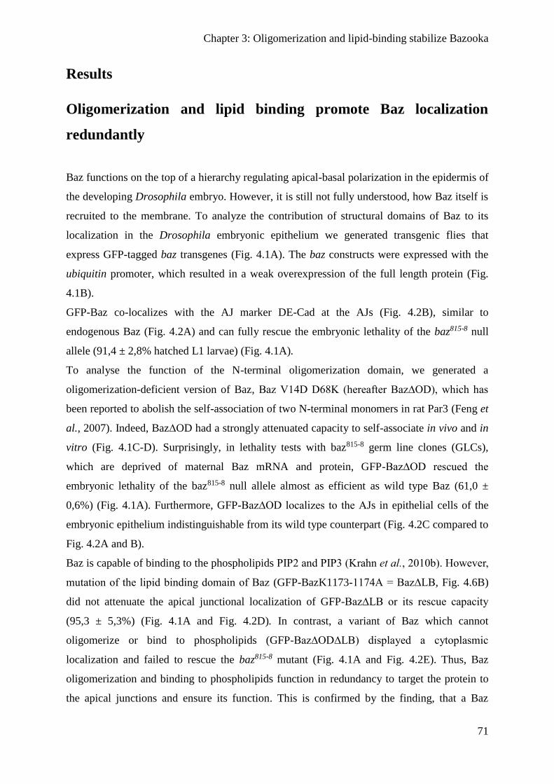
Chapter 3: Oligomerization and lipid-binding stabilize Bazooka
71
Results
Oligomerization and lipid binding promote Baz localization
redundantly
Baz functions on the top of a hierarchy regulating apical-basal polarization in the epidermis of
the developing Drosophila embryo. However, it is still not fully understood, how Baz itself is
recruited to the membrane. To analyze the contribution of structural domains of Baz to its
localization in the Drosophila embryonic epithelium we generated transgenic flies that
express GFP-tagged baz transgenes (Fig. 4.1A). The baz constructs were expressed with the
ubiquitin promoter, which resulted in a weak overexpression of the full length protein (Fig.
4.1B).
GFP-Baz co-localizes with the AJ marker DE-Cad at the AJs (Fig. 4.2B), similar to
endogenous Baz (Fig. 4.2A) and can fully rescue the embryonic lethality of the baz815-8 null
allele (91,4 ± 2,8% hatched L1 larvae) (Fig. 4.1A).
To analyse the function of the N-terminal oligomerization domain, we generated a
oligomerization-deficient version of Baz, Baz V14D D68K (hereafter Baz∆OD), which has
been reported to abolish the self-association of two N-terminal monomers in rat Par3 (Feng et
al., 2007). Indeed, Baz∆OD had a strongly attenuated capacity to self-associate in vivo and in
vitro (Fig. 4.1C-D). Surprisingly, in lethality tests with baz815-8 germ line clones (GLCs),
which are deprived of maternal Baz mRNA and protein, GFP-Baz∆OD rescued the
embryonic lethality of the baz815-8 null allele almost as efficient as wild type Baz (61,0 ±
0,6%) (Fig. 4.1A). Furthermore, GFP-Baz∆OD localizes to the AJs in epithelial cells of the
embryonic epithelium indistinguishable from its wild type counterpart (Fig. 4.2C compared to
Fig. 4.2A and B).
Baz is capable of binding to the phospholipids PIP2 and PIP3 (Krahn et al., 2010b). However,
mutation of the lipid binding domain of Baz (GFP-BazK1173-1174A = Baz∆LB, Fig. 4.6B)
did not attenuate the apical junctional localization of GFP-Baz∆LB or its rescue capacity
(95,3 ± 5,3%) (Fig. 4.1A and Fig. 4.2D). In contrast, a variant of Baz which cannot
oligomerize or bind to phospholipids (GFP-Baz∆OD∆LB) displayed a cytoplasmic
localization and failed to rescue the baz815-8 mutant (Fig. 4.1A and Fig. 4.2E). Thus, Baz
oligomerization and binding to phospholipids function in redundancy to target the protein to
the apical junctions and ensure its function. This is confirmed by the finding, that a Baz
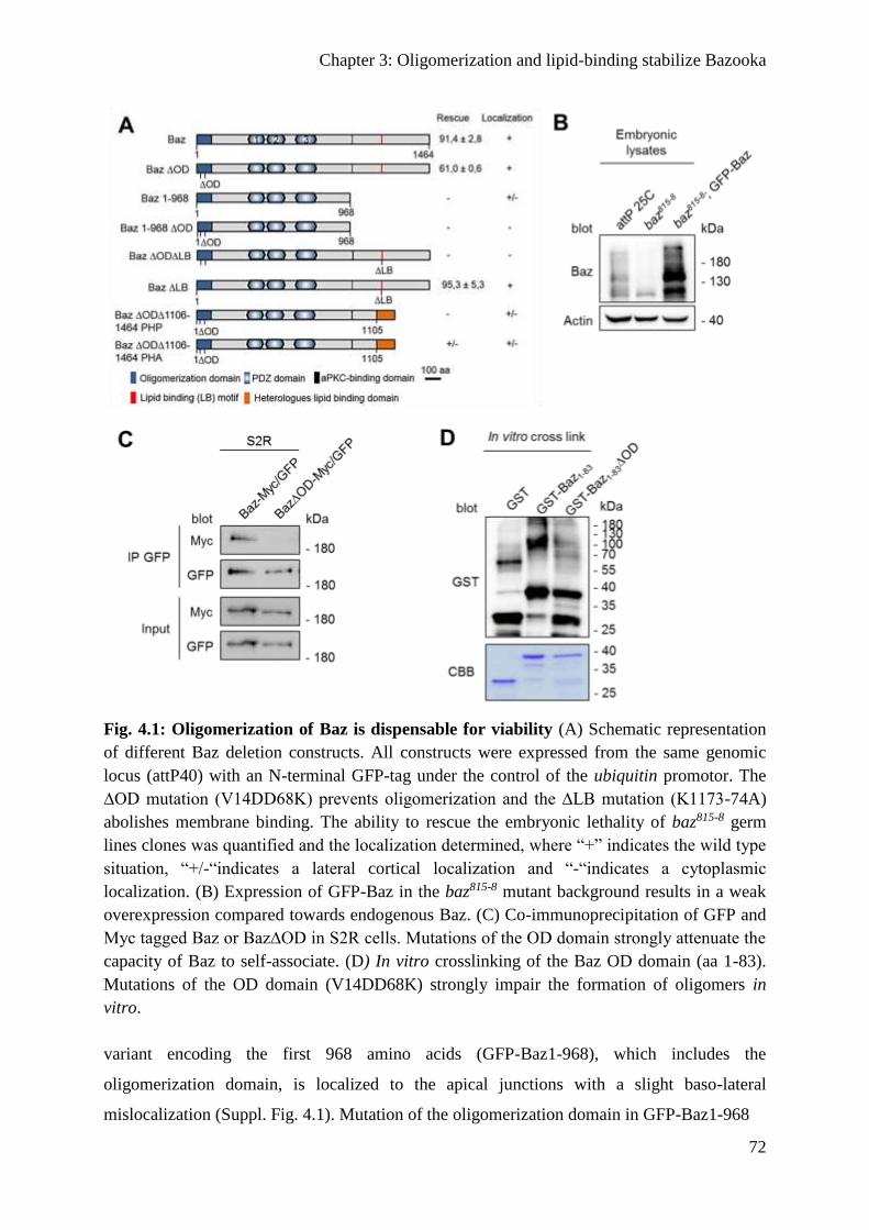
Chapter 3: Oligomerization and lipid-binding stabilize Bazooka
72
Fig. 4.1: Oligomerization of Baz is dispensable for viability (A) Schematic representation
of different Baz deletion constructs. All constructs were expressed from the same genomic
locus (attP40) with an N-terminal GFP-tag under the control of the ubiquitin promotor. The
∆OD mutation (V14DD68K) prevents oligomerization and the ∆LB mutation (K1173-74A)
abolishes membrane binding. The ability to rescue the embryonic lethality of baz815-8 germ
lines clones was quantified and the localization determined, where “+” indicates the wild type
situation, “+/-“indicates a lateral cortical localization and “-“indicates a cytoplasmic
localization. (B) Expression of GFP-Baz in the baz815-8 mutant background results in a weak
overexpression compared towards endogenous Baz. (C) Co-immunoprecipitation of GFP and
Myc tagged Baz or Baz∆OD in S2R cells. Mutations of the OD domain strongly attenuate the
capacity of Baz to self-associate. (D) In vitro crosslinking of the Baz OD domain (aa 1-83).
Mutations of the OD domain (V14DD68K) strongly impair the formation of oligomers in
vitro.
variant encoding the first 968 amino acids (GFP-Baz1-968), which includes the
oligomerization domain, is localized to the apical junctions with a slight baso-lateral
mislocalization (Suppl. Fig. 4.1). Mutation of the oligomerization domain in GFP-Baz1-968
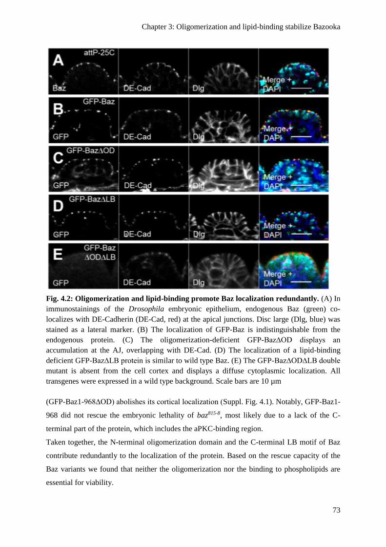
Chapter 3: Oligomerization and lipid-binding stabilize Bazooka
73
Fig. 4.2: Oligomerization and lipid-binding promote Baz localization redundantly. (A) In
immunostainings of the Drosophila embryonic epithelium, endogenous Baz (green) co-
localizes with DE-Cadherin (DE-Cad, red) at the apical junctions. Disc large (Dlg, blue) was
stained as a lateral marker. (B) The localization of GFP-Baz is indistinguishable from the
endogenous protein. (C) The oligomerization-deficient GFP-Baz∆OD displays an
accumulation at the AJ, overlapping with DE-Cad. (D) The localization of a lipid-binding
deficient GFP-Baz∆LB protein is similar to wild type Baz. (E) The GFP-Baz∆OD∆LB double
mutant is absent from the cell cortex and displays a diffuse cytoplasmic localization. All
transgenes were expressed in a wild type background. Scale bars are 10 µm
(GFP-Baz1-968∆OD) abolishes its cortical localization (Suppl. Fig. 4.1). Notably, GFP-Baz1-
968 did not rescue the embryonic lethality of baz815-8, most likely due to a lack of the C-
terminal part of the protein, which includes the aPKC-binding region.
Taken together, the N-terminal oligomerization domain and the C-terminal LB motif of Baz
contribute redundantly to the localization of the protein. Based on the rescue capacity of the
Baz variants we found that neither the oligomerization nor the binding to phospholipids are
essential for viability.

Chapter 3: Oligomerization and lipid-binding stabilize Bazooka
74
Binding to phospholipids is not sufficient for the function of Baz
To further investigate the role of the LB motif regarding the localization and function of Baz
we substituted the C-terminus including the intrinsic LB motif of GFP-Baz∆OD by the
Pleckstrin homology (PH) domains of either PLCδ (Baz∆OD∆1106-1464-PHP) or Akt1
(Baz∆OD∆1106-1464-PHA) (Fig. 4.1A). The PH domain of PLCδ specifically binds to
PtdIns(4,5)P2 (PIP2) (Várnai and Balla, 1998), whereas the PH domain of Akt1 binds to
PtdIns(3,4,5)P3 (PIP3) (James et al., 1996). Baz itself binds both, PIP2 and PIP3 in vitro (Krahn
et al., 2010b).
In the epithelium of transgenic embryos, both GFP-Baz∆OD∆1106-1464-PHP and GFP-
Baz∆OD∆1106-1464-PHA had a cortical localization and punctual enrichments at the AJ
where they co-localized with DE-Cad (Suppl. Fig. 4.1). However, only GFP-Baz∆OD∆1106-
1464-PHA rescued occasionally the zygotic baz815-8 allele (<1%), but not embryos, which
have been depleted for the maternal and zygotic protein expression (GLCs). Moreover,
expression of both variants together, GFP-Baz∆OD∆1106-1464-PHP and GFP-
Baz∆OD∆1106-1464-PHA in a baz-mutant background did not produce surviving animals,
indicating that either simultaneous binding of one Baz molecule to PIP2 and PIP3 is essential
or that the C-terminus (aa 1106-1464) is essential for Baz’ function in our experimental set
up, which differs from previous studies, which used proteins overexpressed by the
UAS/GAL4 system (Krahn et al., 2010b; McKinley et al., 2012).
Baz∆OD∆LB fails to polarize the epithelium of the embryonic
epidermis
To better understand why Baz∆OD∆LB failed to rescue baz815-8 mutant embryos, we analysed
the epithelium of GLCs. To exclude the possibility that the N-terminal GFP-tag interferes
with the function of Baz, we created transgenic flies, which express Baz transgenes fused with
a small OneStrep-tag (OneS) at the C-terminus under the control of the ubiquitin promotor.
As the GFP-variants, Baz-OneS displayed a strong rescue capacity of the embryonic lethality
in baz815-8 GLCs, whereas Baz∆OD∆LB-OneS displayed a complete embryonic lethality (Fig.
4.3). To test if the overexpression of Baz∆OD∆LB might help to overcome the embryonic
lethality of baz815-8 GLCs, we expressed UASp::Baz-GFP and UASp::Baz∆OD∆LB-GFP with
mat-Tub::Gal4 in baz815-8 GLCs (Fig. 4.5A). Baz-GFP rescues the embryonic lethality of

Chapter 3: Oligomerization and lipid-binding stabilize Bazooka
75
Fig. 4.3: Loss of oligomerization and lipid-binding causes embryonic lethality. Baz
variants that carry an C-terminal One-Strep (OneS) tag were expressed under the ubiquitin
promoter and tested for their capacity to rescue baz818-8 germ line clones. Baz-OneS
efficiently rescues the lethality of the baz818-8 allele (10,5 ± 1,8% embryonic lethality).
Baz∆OD∆LB-OneS failed to rescue the embryonic lethality. Fusion of the oligomerization
domain of the human TEL protein (aa 45 – 115) to the N-terminus of Baz restores its function
and rescues embryonic lethality of baz815-8 to a large extent (50,0 ± 6,1% embryonic lethality).
Similar, wild type Baz-GFP overexpressed with mat-Tub::Gal4 using the Gal4/UAS-system
had comparable efficiencies as the constitutively expressed variant (12,7 ± 8,5%), whereas
overexpression of Baz∆OD∆LB-GFP failed to rescue (100% embryonic lethality). Bars
represent the mean ± s.d.
GLCs to the same extend as Baz-OneS (87,3 ± 8,5% and 89,5 ± 1,8%, respectively), whereas
Baz∆OD∆LB-GFP expressing GLCs failed to escape embryonic lethality (Fig. 4.3).
Next, we evaluated the phenotypes of embryonic cuticles of baz GLCs expressing the
different rescue constructs. The Cuticle is secreted from the epidermis and allows drawing a
conclusion of its integrity, in particular the formation of a function apical domain. We divided
the observed phenotypes in five groups (hatched (=normal cuticle), wild type (= dead but
cuticle without obvious defects), cuticle rest, holes and shrunken with holes). As expected the
baz815-8 mutant displayed large cuticle hole or some cuticle rest (Suppl. Fig. 4.2). In contrast
to the null allele, most animals of Baz-OneS hatch and the dead embryos had either a wild
type or shrunken cuticle phenotype (4,8% or 5,7%, respectively, Suppl. Fig. 4.2).
Baz∆OD∆LB-OneS partially rescued the baz815-8 phenotype, because some embryos
developed further and had either a wt or shrunken with holes phenotype (2,3% or 23,3%,
respectively, Suppl. Fig. 4.2).

Chapter 3: Oligomerization and lipid-binding stabilize Bazooka
76
Fig. 4.4: Epithelial polarization requires the functional redundancy of the OD and LB
domains. (A) Immunostaining of endogenous Baz (green), aPKC (red) and Sxl (blue) in the
embryonic epidermis. (B-E) Immunostaining of Baz variants and endogenous aPKC in the
embryonic epidermis of baz818-8 germ line clones. Hemizygous mutant embryos were
identified by the absence of Sxl staining. (B) Loss of Baz in the embryonic epidermis of
disrupts epithelial polarization and aPKC accumulates in the cytoplasm. (C) Baz-OneS

Chapter 3: Oligomerization and lipid-binding stabilize Bazooka
77
efficiently recruits aPKC to the apical junctions, such as endogenous Baz, whereas
Baz∆OD∆LB-OneS displays a cytoplasmic mislocalization (D). Baz∆OD∆LB-OneS rescues
some aPKC to the apical junctions, but the majority of the protein still accumulates in the
cytoplasm. (E) TEL-Baz∆OD∆LB-OneS localizes at the apical junctions and is capable of
recruiting aPKC to rescue the defects of Baz∆OD∆LB-OneS. (F-J) Immunostaining of Baz
(green), DE-Cad (red) and Sxl (blue) in the embryonic epidermis demonstrate a loss of AJ in
baz815-8 germ line clones, which can be rescued by wild type Baz and (H) and TEL-
Baz∆OD∆LB-OneS (J) but not by Baz∆OD∆LB-OneS (I). Scale bars are 10 µm
Interestingly, fusion of the heterologous oligomerization domain of the human TEL protein
(residues 45-115 (Kim et al., 2001)) to the N-terminus of Baz∆OD∆LB-OneS (TEL-
Baz∆OD∆LB-OneS) partly restores the rescue capacity of the mutant Baz protein.
Furthermore, TEL-Baz∆OD∆LB-OneS embryos had a milder phenotype, as most dead
embryos had a wild type or shrunken cuticle phenotype (21,9% and 17,9%, respectively,
Suppl. Fig. 4.2).
Next, we scored for the localization of polarity markers in the embryonic epithelium by
immunostainings. Hemizygous mutant embryos derived from GLC were identified by the lack
of Sex lethal (Sxl) staining. As expected, in the epithelium of baz815-8 GLCs we did not detect
a signal for Baz, whereas aPKC exhibited a cytoplasmic localization (Fig. 4.4B). Baz-OneS
and TEL-Baz∆OD∆LB-OneS both had a robust apical localization and recruited aPKC to the
apical junctions, similar to endogenous Baz (Fig. 4.4A, C and E). In contrast, Baz∆OD∆LB-
OneS showed a cytoplasmic mislocalization (Fig. 4.4D). Nevertheless, Baz∆OD∆LB-OneS
managed to recruit some aPKC to the apical junctions, but the majority of the aPKC protein
still accumulates in the cytoplasm (Fig. 4.4D). In general, the overall structure of the
epithelium was disrupted in Baz∆OD∆LB-OneS embryos.
Then, we examined the assembly of intact AJ by scoring for the localization of DE-Cad in
baz815-8 GLCs (Fig. 4.4F-J). Baz-OneS and TEL-Baz∆OD∆LB-OneS both showed an apical
junctional targeting of DE-Cad comparable to the wild type control (Fig. 4.4F, H, J). Unlike
as for aPKC we did not see a localization of DE-Cad in the apical region or at the plasma
membrane in Baz∆OD∆LB-OneS expressing baz815-8 GLCs (Fig. 4.4I).
In summary, Baz∆OD∆LB-OneS has only a weak capability to polarize the embryonic
epithelium and displayed strong cuticle defects, as well as an impaired function, since aPKC
was inefficiently and DE-Cad not at all recruited to the apical junctions. Restoring the
oligomerization capacity of Baz by fusing the oligomerization domain of TEL to
Baz∆OD∆LB-OneS restores its functionality to a large extent.
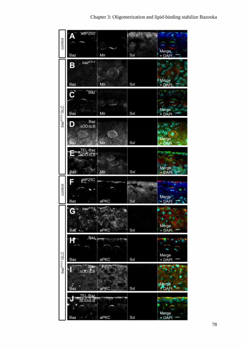
Chapter 3: Oligomerization and lipid-binding stabilize Bazooka
78
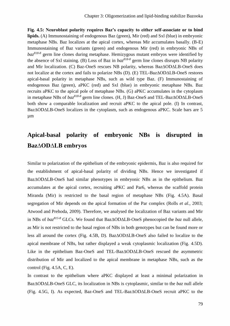
Chapter 3: Oligomerization and lipid-binding stabilize Bazooka
79
Fig. 4.5: Neuroblast polarity requires Baz’s capacity to either self-associate or to bind
lipids. (A) Immunostaining of endogenous Baz (green), Mir (red) and Sxl (blue) in embryonic
metaphase NBs. Baz localizes at the apical cortex, whereas Mir accumulates basally. (B-E)
Immunostaining of Baz variants (green) and endogenous Mir (red) in embryonic NBs of
baz818-8 germ line clones during metaphase. Hemizygous mutant embryos were identified by
the absence of Sxl staining. (B) Loss of Baz in baz818-8 germ line clones disrupts NB polarity
and Mir localization. (C) Baz-OneS rescues NB polarity, whereas Baz∆OD∆LB-OneS does
not localize at the cortex and fails to polarize NBs (D). (E) TEL-Baz∆OD∆LB-OneS restores
apical-basal polarity in metaphase NBs, such as wild type Baz. (F) Immunostaining of
endogenous Baz (green), aPKC (red) and Sxl (blue) in embryonic metaphase NBs. Baz
recruits aPKC to the apical pole of metaphase NBs. (G) aPKC accumulates in the cytoplasm
in metaphase NBs of baz818-8 germ line clones. (H, J) Baz-OneS and TEL-Baz∆OD∆LB-OneS
both show a comparable localization and recruit aPKC to the apical pole. (I) In contrast,
Baz∆OD∆LB-OneS localizes in the cytoplasm, such as endogenous aPKC. Scale bars are 5
µm
Apical-basal polarity of embryonic NBs is disrupted in
Baz∆OD∆LB embryos
Similar to polarization of the epithelium of the embryonic epidermis, Baz is also required for
the establishment of apical-basal polarity of dividing NBs. Hence we investigated if
Baz∆OD∆LB-OneS had similar phenotypes in embryonic NBs as in the epithelium. Baz
accumulates at the apical cortex, recruiting aPKC and Par6, whereas the scaffold protein
Miranda (Mir) is restricted to the basal region of metaphase NBs (Fig. 4.5A). Basal
segregation of Mir depends on the apical formation of the Par complex (Rolls et al., 2003;
Atwood and Prehoda, 2009). Therefore, we analysed the localization of Baz variants and Mir
in NBs of baz815-8 GLCs. We found that Baz∆OD∆LB-OneS phenocopied the baz null allele,
as Mir is not restricted to the basal region of NBs in both genotypes but can be found more or
less all around the cortex (Fig. 4.5B, D). Baz∆OD∆LB-OneS also failed to localize to the
apical membrane of NBs, but rather displayed a weak cytoplasmic localization (Fig. 4.5D).
Like in the epithelium Baz-OneS and TEL-Baz∆OD∆LB-OneS rescued the asymmetric
distribution of Mir and localized to the apical membrane in metaphase NBs, such as the
control (Fig. 4.5A, C, E).
In contrast to the epithelium where aPKC displayed at least a minimal polarization in
Baz∆OD∆LB-OneS GLC, its localization in NBs is cytoplasmic, similar to the baz null allele
(Fig. 4.5G, I). As expected, Baz-OneS and TEL-Baz∆OD∆LB-OneS recruit aPKC to the

Chapter 3: Oligomerization and lipid-binding stabilize Bazooka
80
apical cortex comparable to endogenous protein (Fig. 4.5F, H, J).
Thus, the functional redundancy of the OD and the LB motif are essential to polarize
embryonic NBs, since neither Mir nor aPKC exhibited a correct localization in Baz∆OD∆LB-
OneS GLC.
Baz∆OD∆LB is still recruited to the cortex by transmembrane
proteins
In order to elucidate the mechanism why Baz∆OD∆LB-OneS fails to accumulate at the apical
junctions and why it is non-functional, we tested whether the membrane recruitment by its
described interacting transmembrane proteins is disturbed. Therefore, we used Drosophila
S2R cells as a model system, because they do not exhibit a polarity or detectable amounts of
polarity proteins (Krahn et al., 2010b). In S2R cells, Baz-GFP clearly localizes to the cell
cortex (Fig. 4.6A), whereas Baz∆LB-GFP appears in cytoplasmic aggregates (Fig. 4.6B),
indicating that in S2R cells cortical localization of Baz depends exclusively on the binding to
phospholipids. Co-transfection of Baz∆LB-GFP with DE-Cad-RFP or Ed-RFP led to the
formation of cell-cell contacts of transfected cells. Both proteins recruited Baz∆LB-GFP to
the artificial cell-cell contacts (Fig. 4.6C-D). Additionally, the intracellular domain of the
atypical cadherin Starry night (Stan) fused to the extracellular domain of DE-Cad also
recruited Baz∆LB-GFP to ectopic cell-cell contacts (Fig. 4.6E). By contrast, the Stan isoform
Flamingo, which lacks the C-terminal PDZ binding motif did not recruit lipid binding
deficient Baz (data not shown). Expression of Baz∆OD∆LB-GFP alone or together with DE-
Cad, Ed or Stan showed that Baz∆OD∆LB-GFP was recruited to the cell-cell contacts, such
as its oligomerizing counterpart (Fig. 4.6F-I). Thus, Baz∆OD∆LB is still able to interact with
its reported recruiting transmembrane proteins. This is in line with the observation, that
deletion of the PDZ domains, which facilitate binding to Stan, Ed and Arm/DE-Cad, does not
disturb the localization of GFP-BazLB (Fig. 6J).
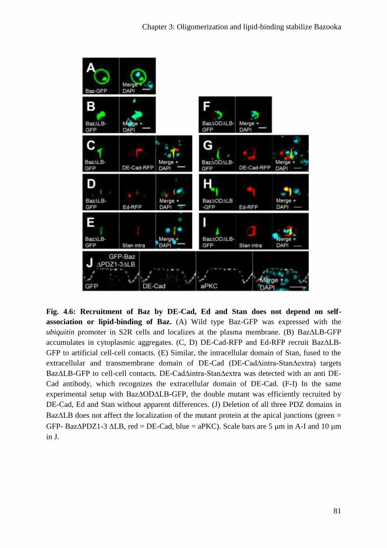
Chapter 3: Oligomerization and lipid-binding stabilize Bazooka
81
Fig. 4.6: Recruitment of Baz by DE-Cad, Ed and Stan does not depend on self-
association or lipid-binding of Baz. (A) Wild type Baz-GFP was expressed with the
ubiquitin promoter in S2R cells and localizes at the plasma membrane. (B) Baz∆LB-GFP
accumulates in cytoplasmic aggregates. (C, D) DE-Cad-RFP and Ed-RFP recruit Baz∆LB-
GFP to artificial cell-cell contacts. (E) Similar, the intracellular domain of Stan, fused to the
extracellular and transmembrane domain of DE-Cad (DE-Cad∆intra-Stan∆extra) targets
Baz∆LB-GFP to cell-cell contacts. DE-Cad∆intra-Stan∆extra was detected with an anti DE-
Cad antibody, which recognizes the extracellular domain of DE-Cad. (F-I) In the same
experimental setup with Baz∆OD∆LB-GFP, the double mutant was efficiently recruited by
DE-Cad, Ed and Stan without apparent differences. (J) Deletion of all three PDZ domains in
BazLB does not affect the localization of the mutant protein at the apical junctions (green =
GFP- BazPDZ1-3 LB, red = DE-Cad, blue = aPKC). Scale bars are 5 μm in A-I and 10 μm
in J.
Chapter 3: Oligomerization and lipid-binding stabilize Bazooka
82
Oligomerization protects Baz from degradation
Despite its capacity to interact with transmembrane proteins, Baz∆OD∆LB-OneS is non-
functional and does not localize to the apical junctions. Therefore, we tested, whether
simultaneous loss of oligomerization and lipid binding affects the protein stability, we blotted
baz815-8 GLC rescued with GFP-Baz and Baz-OneS variants to detect the exogenous Baz
protein. Indeed, we observed that loss of the dimerization domain caused a reduced amount of
Baz protein in the embryo, which is further enhanced upon the loss of the LB motif (Fig.
4.7A, C). In contrast, mutation of the LB motif alone did not affect the amount of protein
(Fig. 4.7C). Similar, the amount of Baz∆OD∆LB-OneS protein is strongly reduced in lysates
of GLC (Fig. 4.7A), whereas Baz-OneS and TEL-Baz∆OD∆LB-OneS were not affected by
enhanced protein degradation (Fig. 4.7A). The reduced amount of Baz∆OD∆LB-OneS was
not due to impaired gene expression, because all transgenes were expressed from the same
promoter and genomic location and exhibited comparable mRNA levels with no significant
differences (Fig. 4.7B).
Finally, we tested, whether the phenotypes observed in Baz∆OD∆LB rescued embryos are
only due to protein degradation of the mutant protein. Strikingly, overexpression of wild type
Baz-GFP, but not Baz∆OD∆LB-GFP rescued the embryonic lethality of baz815-8 (Fig. 4.3)
although both proteins are expressed at comparable levels (Suppl. Fig. 4.3A). Moreover,
Baz∆OD∆LB-GFP is still cytoplasmic, whereas its wild type counterpart localizes to the
apical junctions (Suppl. Fig. 4.3C in comparison to B). These data suggest, that Baz∆OD∆LB
fails to localize to the apical junctions, accumulates in the cytoplasm and is rapidly degraded.
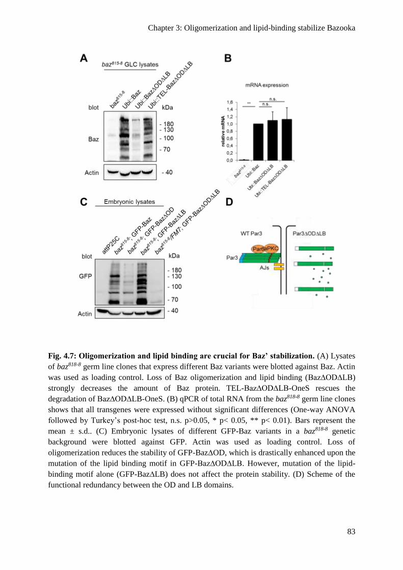
Chapter 3: Oligomerization and lipid-binding stabilize Bazooka
83
Fig. 4.7: Oligomerization and lipid binding are crucial for Baz’ stabilization. (A) Lysates
of baz818-8 germ line clones that express different Baz variants were blotted against Baz. Actin
was used as loading control. Loss of Baz oligomerization and lipid binding (Baz∆OD∆LB)
strongly decreases the amount of Baz protein. TEL-Baz∆OD∆LB-OneS rescues the
degradation of Baz∆OD∆LB-OneS. (B) qPCR of total RNA from the baz818-8 germ line clones
shows that all transgenes were expressed without significant differences (One-way ANOVA
followed by Turkey’s post-hoc test, n.s. p>0.05, * p< 0.05, ** p< 0.01). Bars represent the
mean ± s.d.. (C) Embryonic lysates of different GFP-Baz variants in a baz818-8 genetic
background were blotted against GFP. Actin was used as loading control. Loss of
oligomerization reduces the stability of GFP-Baz∆OD, which is drastically enhanced upon the
mutation of the lipid binding motif in GFP-Baz∆OD∆LB. However, mutation of the lipid-
binding motif alone (GFP-Baz∆LB) does not affect the protein stability. (D) Scheme of the
functional redundancy between the OD and LB domains.
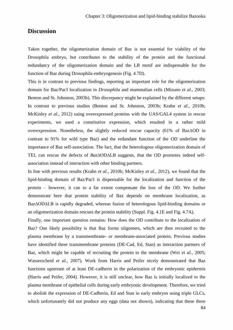
Chapter 3: Oligomerization and lipid-binding stabilize Bazooka
84
Discussion
Taken together, the oligomerization domain of Baz is not essential for viability of the
Drosophila embryo, but contributes to the stability of the protein and the functional
redundancy of the oligomerization domain and the LB motif are indispensable for the
function of Baz during Drosophila embryogenesis (Fig. 4.7D).
This is in contrast to previous findings, reporting an important role for the oligomerization
domain for Baz/Par3 localization in Drosophila and mammalian cells (Mizuno et al., 2003;
Benton and St. Johnston, 2003b). This discrepancy might be explained by the different setups:
In contrast to previous studies (Benton and St. Johnston, 2003b; Krahn et al., 2010b;
McKinley et al., 2012) using overexpressed proteins with the UAS/GAL4 system in rescue
experiments, we used a constitutive expression, which resulted in a rather mild
overexpression. Nonetheless, the slightly reduced rescue capacity (61% of BazOD in
contrast to 91% for wild type Baz) and the redundant function of the OD underline the
importance of Baz self-association. The fact, that the heterologous oligomerization domain of
TEL can rescue the defects of Baz∆OD∆LB suggests, that the OD promotes indeed self-
association instead of interaction with other binding partners.
In line with previous results (Krahn et al., 2010b; McKinley et al., 2012), we found that the
lipid-binding domain of Baz/Par3 is dispensable for the localization and function of the
protein – however, it can to a far extent compensate the loss of the OD. We further
demonstrate here that protein stability of Baz depends on membrane localization, as
Baz∆OD∆LB is rapidly degraded, whereas fusion of heterologous lipid-binding domains or
an oligomerization domain rescues the protein stability (Suppl. Fig. 4.1E and Fig. 4.7A).
Finally, one important question remains: How does the OD contribute to the localization of
Baz? One likely possibility is that Baz forms oligomers, which are then recruited to the
plasma membrane by a transmembrane- or membrane-associated protein. Previous studies
have identified three transmembrane proteins (DE-Cad, Ed, Stan) as interaction partners of
Baz, which might be capable of recruiting the protein to the membrane (Wei et al., 2005;
Wasserscheid et al., 2007). Work from Harris and Peifer nicely demonstrated that Baz
functions upstream of at least DE-cadherin in the polarization of the embryonic epidermis
(Harris and Peifer, 2004). However, it is still unclear, how Baz is initially localized to the
plasma membrane of epithelial cells during early embryonic development. Therefore, we tried
to abolish the expression of DE-Cadherin, Ed and Stan in early embryos using triple GLCs,
which unfortunately did not produce any eggs (data not shown), indicating that these three
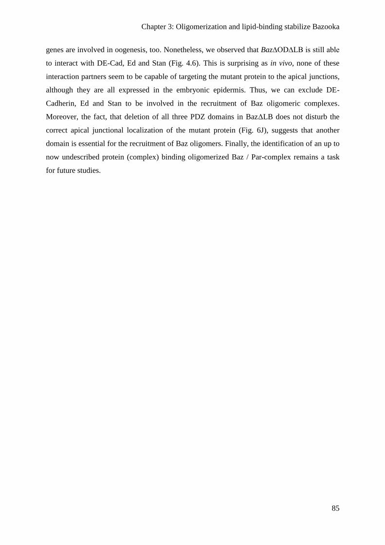
Chapter 3: Oligomerization and lipid-binding stabilize Bazooka
85
genes are involved in oogenesis, too. Nonetheless, we observed that Baz∆OD∆LB is still able
to interact with DE-Cad, Ed and Stan (Fig. 4.6). This is surprising as in vivo, none of these
interaction partners seem to be capable of targeting the mutant protein to the apical junctions,
although they are all expressed in the embryonic epidermis. Thus, we can exclude DE-
Cadherin, Ed and Stan to be involved in the recruitment of Baz oligomeric complexes.
Moreover, the fact, that deletion of all three PDZ domains in BazΔLB does not disturb the
correct apical junctional localization of the mutant protein (Fig. 6J), suggests that another
domain is essential for the recruitment of Baz oligomers. Finally, the identification of an up to
now undescribed protein (complex) binding oligomerized Baz / Par-complex remains a task
for future studies.
Chapter 3: Oligomerization and lipid-binding stabilize Bazooka
86
Supplemental figures
Supplemental Fig. 4.1: Localization of Baz variants in the embryonic epidermis. (A-D)
Immunostainings of different GFP-Baz variants in the embryonic epidermis. All transgenes
were expressed with the ubiquitin promotor in a wild type background. GFP (green), DE-Cad
(red) and Dlg (blue) were stained. (A) GFP-Baz1-968 localizes mainly at the apical junctions
with some baso-lateral mislocalization. (B) Mutation of the OD domain in this truncated
protein causes a cytoplasmic accumulation of GFP-Baz1-968∆OD. (C, D) Chimeric proteins
that carry the pleckstrin homology (PH) domains of either human PLCδ or Akt1 fused to the
C-terminus GFP-Baz∆OD1-1105 promote a cortical localization in the embryonic epidermis.
(E) Western blot of embryonic lysates of the Baz variants (A-D). The Baz variants were
detected with a GFP antibody and Actin was used as a loading control. Scale bars are 10 µm
Chapter 3: Oligomerization and lipid-binding stabilize Bazooka
87
Supplemental Fig. 4.2: Cuticle phenotypes of Baz variants. (A) Cuticle phenotypes were
classified into the four categories wt, shrunken with holes, holes and cuticle rests. (B) The
cuticle phenotypes of baz818-8 germ line clones that express different Baz variants with the
ubiquitin promoter were quantified (n=300 per genotype). Embryos that normally developed
and hatched are also included as “hatched”. baz818-8 germ line clones display holes or cuticle
rests. Embryos that express Baz-OneS hatch to a large extend (89,5%) or display either wt or
shrunken with hole phenotypes. No embryos hatch upon the expression of Baz∆OD∆LB-
OneS. Most embryos have either cuticle rests or holes phenotypes (58,1 and 16,3%,
respectively). Nevertheless, some embryos develop further and have a wt or shrunken with
holes phenotype (2,3 and 23,3%, respectively). The chimeric TEL-Baz∆OD∆LB-OneS
protein rescues the Baz∆OD∆LB phenotypes to a large extent as half of the embryos hatch
(50,2 %) or have either wt or shrunken with holes phenotypes (21,9 and 17,9%, respectively).
Scale bars are 200µm
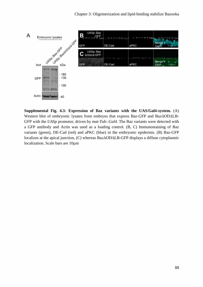
Chapter 3: Oligomerization and lipid-binding stabilize Bazooka
88
Supplemental Fig. 4.3: Expression of Baz variants with the UAS/Gal4-system. (A)
Western blot of embryonic lysates from embryos that express Baz-GFP and Baz∆OD∆LB-
GFP with the UASp promotor, driven by mat-Tub::Gal4. The Baz variants were detected with
a GFP antibody and Actin was used as a loading control. (B, C) Immunostaining of Baz
variants (green), DE-Cad (red) and aPKC (blue) in the embryonic epidermis. (B) Baz-GFP
localizes at the apical junction, (C) whereas Baz∆OD∆LB-GFP displays a diffuse cytoplasmic
localization. Scale bars are 10µm
Chapter 4: Structural analysis of the Par3 - Par6 interaction
89
Chapter 4: Structural analysis of the Par3 - Par6 interaction
Structural basis for the interaction between the cell polarity
proteins Par3 and Par6
Authors: Fabian A. Renschler, Susanne R. Bruekner, Paulin L. Salomon, Amrita Mukherjee,
Lars Kullmann, Mira C. Schütz-Stoffregen, Christine Henzler, Tony Pawson, Michael P.
Krahn and Silke Wiesner.
Personal contributions: Preparation of Drosophila embryos for immunofluorescence, S2R cell
culture experiments, data analysis and discussion of results
Polarity is a fundamental property of most cell types. The Par protein complex is a major
driving force in creating asymmetrically localized protein networks and consists of atypical
protein kinase C (aPKC), Par3, and Par6. Dysfunction of this complex causes developmental
abnormalities and diseases such as cancer. However, how Par3 interacts with Par6 has
remained enigmatic. Here, we identified a PDZ domain-binding motif (PBM) in Par6 that was
essential for Par3 interaction in vitro and Par3-mediated membrane localization of Par6 in
cultured cells. In fly embryos, we observed that the PBM functions in redundancy with the
PDZ domain to target Par6 to the cortex of epithelial cells. Our structural analyses by x-ray
crystallography and NMR spectroscopy showed that both the Par3 PDZ1 and PDZ3 domains
but not the PDZ2 engage in canonical PDZ:PBM interactions with the Par6 motif that we
identified. Par3 thus has the potential to recruit two Par6 proteins simultaneously. This may
facilitate the assembly of polarity protein networks through multivalent PDZ domain
interactions.

Chapter 4: Structural analysis of the Par3 - Par6 interaction
90
Introduction
Most cell types in multicellular organisms require an unequal distribution of proteins, lipids
and mRNA for their function (Nelson, 2003). This phenomenon is referred to as cell polarity.
During development, the differential segregation of cell fate determinants (anterior-posterior
polarity) forms the basis for asymmetric cell division and ultimately the generation of body
axes and cell diversity. In differentiated cells, partitioning of the cell membrane into
functionally discrete domains enables cells to execute their specialized functions within an
organism. Epithelia are a classic example of polarized cells and display an apico-basal
polarity. While the apical side faces the exterior or lumen, the basal domain is directed
towards the interior extracellular matrix and interstitial space in tissues and organs. The lateral
cell surface allows neighboring epithelial cells to adhere and to communicate through cell-cell
junctions. However, epithelial cells can also lose their polarity and acquire a migratory,
mesenchymal character. While this process is vital for embryonic development and tissue
repair, it is also adversely a key event in cancer metastasis. Elucidating the basic mechanisms
underlying cell polarity thus has broad implications for our understanding of developmental
and oncogenic processes.
Although polarized cells vary substantially in their morphology and function, polarity
depends in all cell types on the Par (partitioning-defective) protein complex that is conserved
across the animal phyla (Ohno, 2001). The Par complex constitutes the major molecular
machinery for creating the mutually exclusive signaling networks that cover large areas of the
cell cortex in all polarized cells. In epithelia, the Par complex is a key polarity regulator at the
apical surface and engages in intricate signaling networks with the Crumbs complex and cell
adhesion proteins such as E-cadherin and nectins (Shotgun (Shg) and Echinoid (Ed),
respectively, in Drosophila) to form and maintain cell junctions (Nelson, 2003; Tepass,
2012).
The Par complex comprises atypical protein kinase C (aPKC) and two scaffolding proteins,
Par3 (Bazooka (Baz) in Drosophila) and Par6 (Fig. 5.1A). While a single set of genes encodes
the Par complex in invertebrates, this gene family has expanded to two Par3 (Par3 and
Par3L), two aPKC (PKC/ι and PKCζ) and three Par6 (Par6α, Par6β and Par6γ) genes in
vertebrates (Noda et al., 2001; Gao et al., 2002; Suzuki et al., 2003). Par6 and aPKC hetero-
dimerize with high affinity via their N-terminal PB1 (Phox and Bem1) domains (Fig. 5.1A)
(Suzuki et al., 2001; Hirano et al., 2005). In addition, Par6 contains a Crib (Cdc42/Rac
interactive binding) motif directly preceding a PDZ protein-protein interaction domain. Par3

Chapter 4: Structural analysis of the Par3 - Par6 interaction
91
contains an N-terminal PB1-like homo-oligomerization domain (NTD) (Zhang et al., 2013a),
three central PDZ domains and a C-terminal region with an aPKC phosphorylation site
(Nagai-Tamai et al., 2002; Krahn et al., 2010b; Morais-de-Sá et al., 2010; Soriano et al.,
2016). Whether Par3 interacts directly with Par6 has remained controversial. The Par3 PDZ1
domain has been described to hetero-dimerize with the Par6 Crib-PDZ domain (Lin et al.,
2000; Joberty et al., 2000; Li et al., 2010b) (Fig. 5.1A). However, these reports disagree on
whether the Par6 Crib motif is essential for the Par3:Par6 interaction. Moreover, the in vivo
relevance of this interaction has been debated (Li et al., 2010b), and lastly the Par3:Par6
interaction has been reported to be indirect, requiring aPKC as a linker molecule (Suzuki et
al., 2001; Nagai-Tamai et al., 2002). Given these controversial observations, we sought to
investigate the mechanism of Par3:Par6 association on a structural and functional level.
PDZ domain interactions are a recurring theme in polarity and cell-cell adhesion protein
networks. PDZ domains fold into a six-stranded antiparallel -sheet capped by two -helices
(Morais Cabral et al., 1996) and predominantly bind with low affinity (typically in the M
range (Wiedemann et al., 2004; Stiffler et al., 2007)) to short (4-15 amino acids (aa)),
disordered sequences at the C-termini of target proteins, so-called PDZ-binding motifs
(PBMs). This interaction results in an augmentation of the PDZ -sheet by an antiparallel -
strand formed by the PBM. The four C-terminal residues constitute the specificity-
determining core PBM (Appleton et al., 2006) that can be categorized into three classes
depending on the aa at the -2 position relative to the C-terminus: X-[S/T]-X--COO- (class I),
X--X--COO- (class II) and X-[D/E]-X--COO- (class III), with X being any aa and being
a hydrophobic residue (Songyang et al., 1997). In addition, PDZ domains can recognize
internal peptide sequences (Penkert et al., 2004) or homo- or hetero-dimerize in some cases
(Lin et al., 2000; Joberty et al., 2000; Li et al., 2010b).
Here, we uncover the molecular basis of the Par3:Par6 interaction. We have identified a
previously unrecognized, highly conserved PBM in Par6 that is important for Par6
localization in vivo and essential for Par3:Par6 interaction in vitro. We find that both the Par3
PDZ1 and PDZ3 but not the PDZ2 domain can associate through canonical PDZ:PBM
interactions with the Par6 PBM and show that one Par3 protein has the potential to recruit two
Par6 molecules simultaneously. In vivo, we find that the Par6 PBM is critical for Par3-
mediated membrane recruitment of Par6 in cultured Drosophila Schneider cells (S2R) and
plays a role in Par6 localization to the cell cortex in fly embryonic epidermal cells. In sum,
our results provide important structural and functional details on the role of Par3 and Par6 in
organizing polarity complexes.
Chapter 4: Structural analysis of the Par3 - Par6 interaction
92
Methods
Reagents and Constructs
Par3 and Par6 constructs were cloned from C. elegans cDNA, S2R cell cDNA or a synthetic
gene fragment (LifeTechnologies) containing the Par3 PDZ domains (Drosophila
melanogaster), and pK-myc-Par3b (Hs. Par3) and pK-myc-Par6C (Hs. Par6) vectors
purchased from Addgene (plasmid #19388 and #15474). For NMR studies, gene fragments
amplified by PCR were cloned into the following vectors: pETZ2.1a (His6-Z domain-TEV)
for Ce. Par3 and Par6 constructs; pET-M30 (His6-GST-TEV) for Dm. Par3 PDZ constructs;
pET-M41 (His6-MBP-TEV) for the Dm. Par3 PDZ1:Par6 PBM fusion, Dm. Par6 PDZ and
Crib-PDZ and Hs. Par3 PDZ domains and the Hs. Par3:Par6 PBM fusion constructs;
pRTDuet-GB1 (His6-GB1-TEV) for all Par6 and Shg PBM peptides. For GST pull down
experiments, a gene fragment containing the three Dm. Par3 PDZ domains was cloned into a
pETM30-HA (His6-GST-TEV-HA) vector, while full-length and truncated Dm. Par6
constructs were cloned into a pET-M11-Sumo (His6-Sumo-TEV) vector.
For S2R cell transfection, Dm. Par3 and Dm. Par6 were cloned into pENTR vectors. Site-
directed mutagenesis was carried out with the Par6 pENTR vector as DNA template using the
following primers:
Par6PDZ: 5’-
GTGCCGGAAACGCATGGTGGAGGTGGAGGTCCGGCCAATCAGCGC-3’
Par6 PBM: 5’-ACGATAATGGCCAGCGATTAAATCGATGGAGTGCTGCATTTG-3’.
Par3 and wild-type and mutant Par6 variants were subsequently subcloned into pUGW and
pURW vectors of the Drosophila Gateway Collection (DGRC) (Sen et al., 2012),
respectively. For co-immunoprecipitation studies, the Myc-tag in the pK-myc-Par3b vector
was replaced by a Flag-tag using QuikChange mutagenesis. All deletions in the Hs. Par6
gene were generated by QuikChange mutagenesis using the pK-myc-Par6C vector as DNA
template. For a complete list of constructs, see Supplemental Table 5.3.
Expression and purification of recombinant proteins
All recombinant proteins were expressed in E. coli BL21-CodonPlus (DE3) RIL cells
(Stratagene) and purified by Ni-affinity and size-exclusion chromatography. To facilitate
peptide production, the Par6 and Shg PBMs were fused at their N-termini to the
immunoglobulin binding domain B1 of streptococcal protein G (GB1) domain followed by a
Chapter 4: Structural analysis of the Par3 - Par6 interaction
93
TEV protease cleavage site. For all NMR binding experiments, non-specific binding of GB1
alone was tested. For NMR studies, unlabeled His6-GB1-Par6 and His6-GB1-Shg peptides or
dmPar3 PDZ domains were expressed in LB medium, while 15N- or 13C,15N-labeled His6-
GB1-Shg, Par3 PDZ or Par6 (Crib-)PDZ domains were expressed in M9 minimal medium
with 15NH4Cl or 15NH4Cl and 13C-glucose as sole sources of nitrogen and carbon. All NMR
constructs were buffer exchanged into NMR buffer (20 mM Na phosphate pH 6.5 or 7.5, 150
mM NaCl, 1 mM DTT, and 0.03% NaN3) for triple resonance and CSP experiments.
Proteins for pull down experiments were expressed as His6-GST-HA-tagged Dm. Par3 and
His6-Sumo-tagged Dm. Par6 proteins in E. coli BL21-CodonPlus (DE3) RIL cells
(Stratagene) in LB medium and purified by Ni-affinity chromatography. Subsequently, buffer
was exchanged by dialysis to 50 mM Na phosphate pH 7.5, 150 mM NaCl, 1 mM DTT, 10 %
glycerol for Par6 proteins or to 20 mM Na phosphate pH 6.5, 150 mM NaCl, 1 mM DTT, and
10 % glycerol for Par3 proteins.
Pull down assays
Dm. Par3 proteins containing an N-terminal His6-GST-HA-tag were incubated with
glutathione beads (Macherey-Nagel) at 1 µM concentration together with 65 μM Dm. Par6
proteins that contained an N-terminal His6-Sumo-tag for 1 h at 4 °C in PD buffer (50 mM Na
phosphate pH 7.5, 150 mM NaCl, 10% glycerol, 2 mM DTT). The beads were washed four
times with PD buffer and specifically bound proteins eluted with PD buffer supplemented
with 25 mM reduced GSH. Eluted proteins were precipitated with 10% (w/v) TCA (Sigma)
on ice followed by centrifugation. Protein pellets were resuspended in SDS loading buffer,
resolved by SDS-PAGE and detected by Coomassie staining.
S2R cell culture
Immunostainings of transfected Schneider 2R cells was carried out as previously described
(Krahn et al., 2010b). In brief, S2R cells were transfected using FUGENE (Promega) with
Ubi::GFP-Par6 variants alone or together with Ubi::RFP-Baz. Cells were fixed with
paraformaldehyde and imaged with a Zeiss LSM 710 Meta confocal microscope. For RNAi-
mediated downregulation of aPKC was performed as described (Krahn et al., 2009). In brief,
a ~ 300bp dsRNA fragment was taken up by the cells and processed to several siRNAs. One
dsRNA thus results in several siRNAs and thereby reduces the probability of off-targets. The
efficiency of dsRNA-mediated gene knock down of aPKC was confirmed by a Western blot
of S2R cell lysates using aPKC (1:1000, Santa Cruz sc-216, 1:1000) and mouse actin (1:1000,
Chapter 4: Structural analysis of the Par3 - Par6 interaction
94
Santa Cruz sc-47778) antibodies. The following oligonucleotides were used to amplify a
fragment of aPKC mRNA for in vitro transcription into dsRNA:
aPKC-dsRNA-F: 5'-ACTTCGCGTTCTCCGC-3',
aPKC-dsRNA-R: 5'-TTGCTAGCTGGGTAAAATATTTTGA-3'.
HEK293T cell culture and co-immunoprecipitation
HEK293T cells were transfected with N-terminally Flag-tagged Hs. Par3 in a pKFlag and N-
terminally Myc-tagged Hs. Par6 in a pKMyc expression vector using Lipofectamin
(Invitrogen), grown in DMEM supplemented with 10% Fetal Bovine Serum (Invitrogen), 2
mM L-glutamine (Invitrogen), penicillin (Invitrogen) and streptomycin (Invitrogen) and
harvested three days after transfection. Cells were lysed in NET buffer (50 mM Tris-HCl pH
8.0, 150 mM NaCl, 0.1% (w/v) Triton X-100, 1 mM EDTA) supplemented with Proteinase
Inhibitor Cocktail (Roche). The supernatants were incubated with Flag M2 antibody (2
ug/mg cell lysate) and subsequently with GammaBind Plus Sepharose (GE Healthcare) for 1
h each at 4 °C. The beads were washed three times with NET buffer and once with Triton-free
NET buffer. Immunoprecipitated proteins were eluted with SDS loading buffer, separated by
SDS-PAGE and immunoblotted using mouse Flag M2 (1:1000) (Sigma) as primary and HRP-
coupled mouse IgG (1:10.000) as secondary antibody (ThermoScientific) or mouse Myc-HRP
(1:5000) (LifeTechnologies) antibody. Western blots were imaged by chemiluminescence.
Fly stocks and genetics
Fly stocks were cultured on standard cornmeal agar food and maintained at 25 °C. Transgenic
flies of Ubi::GFP-Par6 variants were established using the Phi-C31-Integrase system with
attP40 in a wild-type background (Groth et al., 2004). Fly embryo lysates were applied to
SDS-PAGE and immuno-blotted using rabbit GFP (1:500, sc8334, Santa Cruz) and mouse
actin (1:1000, Santa Cruz sc-47778) antibodies to ensure uniform expression of the Par6
mutants. Western blots were imaged by chemiluminescence. To evaluate the function of GFP-
Par6 and GFP-Par6 PDZ, PBM and PDZPBM mutants in a Par6-null background, the
transgenes were backcrossed into a homozygous par6226 mutant background (Petronczki and
Knoblich, 2001). However, only the wild-type and PBM transgenes can rescue the
embryonic lethality of par6226 mutant flies and be kept as stable stocks.
Immunohistochemistry
Drosophila stage 6-7 or stage 11 embryos were fixed in 4% formaldehyde, phosphate buffer

Chapter 4: Structural analysis of the Par3 - Par6 interaction
95
pH 7.4 as previously described (Sen et al., 2012). The primary antibodies used for indirect
immunofluorescence were as follows: mouse aPKC (PKC, 1:500, sc17781, Santa Cruz),
rabbit GFP (1:500, sc8334, Santa Cruz) and guinea pig Baz (1:500). Secondary antibodies
conjugated with Alexa 488, Alexa 568 and Alexa 647 (Life Technologies) were used at 1:400.
Images of the epidermis were taken from stage 11 embryos on a Zeiss LSM 710 Meta
confocal microscope or at stage 6-7 on a Leica SP8 confocal microscope. For correlation
analysis, the images displayed in Fig. 5.3 and Supplemental Fig. 5.6 were partitioned in three
regions covering almost the entire image. Pearson’s correlation coefficients were estimated
after automatic thresholding using the Costes’ approach as implemented in the JACoP v2.0
plugin of ImageJ (Fiji) (Costes et al., 2004; Bolte and Cordelières, 2006; Schindelin et al.,
2012).
NMR spectroscopy
CSP studies were performed with 75-100M samples of 15N-labeled protein in NMR buffer
(20 mM Na phosphate pH 6.5 (individual PDZ domains or Shg PBM) or 7.5, 150 mM NaCl,
1 mM DTT, and 0.03% NaN3) by recording 1H,15N-HSQC experiments on a 600 MHz Bruker
Avance-III spectrometer at 20 °C for the Dm. and Ce. Par3 and Par6 PDZ domains and at 30
°C for the human proteins. For CSP studies with the dmPar3 PDZ1-3 2-3loop module, we
recorded 1H,15N-TROSY experiments at 600 MHz and 30 °C in 20 mM Na phosphate pH 7.5,
150 mM NaCl and 1 mM DTT. Backbone resonance assignment for the Dm. Par3 PDZ
domains was performed at 20 °C and at 25 °C for the Hs. Par3 PDZ2 and PDZ3 domains by
recording 3D HNCACB, HNCOCACB, CCONH and HNH-NOESY spectra at 600 or 800
MHz. NMR data were processed using the nmrPipe/nmrDraw software suite (Delaglio et al.,
1995) and analyzed using XEasy (Bartels et al., 1995) and Sparky (Lee et al., 2015b). Figures
displaying NMR spectra were produced with nmrView (onemoonscientific.com). Average
CSPs used for binding site mapping were calculated in ppm as ((1H)2 + (0.25*15N)2)0.5,
where is the difference in chemical shift at a nine-fold stoichiometric excess of Par6 PBM
vs. the respective reference in the absence of ligand. For binding site mapping, a homology
model of the dmPar3 PDZ3 domain was generated using HHPred (Alva et al., 2016), while
PDB entries 2KOM and 2KOH were used for binding site mapping for the hsPar3 PDZ2
(Jensen et al., 2010) and PDZ3 (Tyler et al., 2010) domains, respectively. Of note, a highly
similar structure exists for the PDZ2 domain of rat Par3 (Wu et al., 2007).
Chapter 4: Structural analysis of the Par3 - Par6 interaction
96
Two-dimensional line shape fitting analysis
1H,15N-CSP studies for the Dm. Par3 PDZ domains were quantified using TITAN (Waudby et
al., 2016) according to instructions and online documentation (http://www.nmr-titan.com and
https://bitbucket.org/cwaudby/titan/wiki/Home). Spectra were acquired with 1024 and 128
points in the 1H and 15N dimensions, respectively, and processed with exponential window
functions with a line broadening of 4 Hz and 8 Hz. Spectra were zero-filled to 4096 and 1024
points in the 1H and 15N dimensions, respectively. In order to obtain comparable results for
different ligands, the same cross peaks were used for the analysis of each PDZ domain
(Supplemental Table 5.4). Errors were estimated with bootstrapping statistics on 100 replica.
Figures for line shape analyses were prepared with TITAN.
X-ray crystallography
The Dm. Par3 PDZ1:Par6 fusion construct was crystallized in 0.1 M BisTris pH 6.5, 2 M
(NH4)2SO4. Diffraction data were collected at 100 K using a wavelength of 1 Å and a
PILATUS 6M-F detector at the beamline PXII of the Swiss Light Source (PSI, Villigen,
Switzerland). Data were processed using XDS (Kabsch, 2010) and molecular replacement
was performed using Phaser (McCoy et al., 2007). The structure was finalized by iterative
manual modeling with Coot (Emsley et al., 2010) and refinement with Phenix (Adams et al.,
2010). Each asymmetric unit contains two protein chains with virtually identical
conformations (backbone r.m.s.d. of 0.031 Å for the PDZ1 domain and the core PBM). All
figures displaying protein structures were generated using PyMOL (http://www.pymol.org/).
Sequence alignment
Multiple sequence alignments were performed with MUSCLE (Edgar, 2004) and displayed
with ClustalX (Larkin et al., 2007). The position weighted matrix for the Par6 PBM was
created with WebLogo 3.4 (Crooks et al., 2004) using all Par6 PBMs displayed in
Supplemental Fig. 4.3. The overall height of the stack indicates the sequence conservation at
that position, while the height of a symbol within the stack indicates the relative frequency of
each aa at that position.
Chapter 4: Structural analysis of the Par3 - Par6 interaction
97
Results
Par6 contains a PBM that associates with the Par3 PDZ1 domain
In order to elucidate the molecular details of the Par3:Par6 interaction, we performed nuclear
magnetic resonance (NMR) binding studies. This method is a powerful tool to study
biomolecular interactions (Williamson, 2013; Wiesner and Sprangers, 2015), since the
measured chemical shifts of atomic nuclei, that is the peak positions in NMR spectra, are
highly sensitive to changes in the local chemical environment. In a standard experiment, an
unlabeled binding partner is titrated to an isotope-labeled, NMR-visible protein. This results
in concentration-dependent changes of the chemical shifts for the residues contacting the
ligand (chemical shift perturbation; CSP) that can be used to obtain dissociation constants
(Supplemental Fig. 5.1). Together with chemical shift assignments, this enables the mapping
of the ligand binding surface on a protein structure with close to atomic resolution.
Previous studies indicated a hetero-dimerization of the Par3 PDZ1 and the Par6 PDZ domains
(Lin et al., 2000; Joberty et al., 2000; Li et al., 2010b). To investigate the mechanism of
Par3:Par6 interaction, we first recorded 1H,15N-correlation NMR spectra of the 15N-labeled
PDZ1 domain of human Par3. Surprisingly, we observed that this PDZ domain was largely
unfolded in isolation (Supplemental Fig. 5.2A). The domain architectures of the Par complex
proteins are highly conserved in metazoans suggesting that Par3 and Par6 interact in a similar
manner (Ohno, 2001). We therefore investigated next whether the Drosophila melanogaster
(Dm.) or Caenorhabditis elegans (Ce.) Par3 PDZ1 domains interact with the respective Par6
PDZ or Crib-PDZ domains. Although both the Dm. and Ce. Par3 PDZ1 domains were folded
in isolation, we observed only very few changes in the NMR spectra of the 15N-labeled Par3
PDZ1 domains upon addition of the respective unlabeled Par6 PDZ (Supplemental Fig.
5.2B,C) or upon addition of the unlabeled Par3 PDZ1 domains to the respective 15N-labeled
Par6 Crib-PDZ domains (Supplemental Fig. 5.2D,E). Similarly, neither the Dm. Par3
(dmPar3) PDZ2 nor the PDZ3 domain induced substantial CSPs in the 15N-labeled Dm. Par6
(dmPar6) Crib-PDZ domain in our NMR experiments (Supplemental Fig. 5.2F,G). We thus
conclude that the Par3 PDZ domains do not associate directly with the Par6 (Crib-)PDZ
domain.
In search of an alternative binding mechanism we revisited the Par6 protein sequence and
identified a previously unrecognized class II (-X--COO-) PBM at its C-terminus (Fig.
5.1B). This motif is highly conserved in metazoans (Fig. 5.1B) with the notable exception of
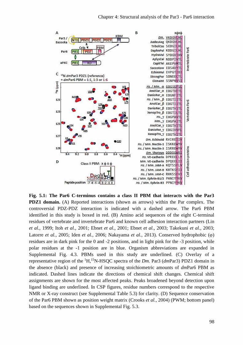
Chapter 4: Structural analysis of the Par3 - Par6 interaction
98
Fig. 5.1: The Par6 C-terminus contains a class II PBM that interacts with the Par3
PDZ1 domain. (A) Reported interactions (shown as arrows) within the Par complex. The
controversial PDZ-PDZ interaction is indicated with a dashed arrow. The Par6 PBM
identified in this study is boxed in red. (B) Amino acid sequences of the eight C-terminal
residues of vertebrate and invertebrate Par6 and known cell adhesion interaction partners (Lin
et al., 1999; Itoh et al., 2001; Ebnet et al., 2001; Ebnet et al., 2003; Takekuni et al., 2003;
Latorre et al., 2005; Iden et al., 2006; Nakayama et al., 2013). Conserved hydrophobic ()
residues are in dark pink for the 0 and -2 positions, and in light pink for the -3 position, while
polar residues at the -1 position are in blue. Organism abbreviations are expanded in
Supplemental Fig. 4.3. PBMs used in this study are underlined. (C) Overlay of a
representative region of the 1H,15N-HSQC spectra of the Dm. Par3 (dmPar3) PDZ1 domain in
the absence (black) and presence of increasing stoichiometric amounts of dmPar6 PBM as
indicated. Dashed lines indicate the directions of chemical shift changes. Chemical shift
assignments are shown for the most affected peaks. Peaks broadened beyond detection upon
ligand binding are underlined. In CSP figures, residue numbers correspond to the respective
NMR or X-ray construct (see Supplemental Table 5.3) for clarity. (D) Sequence conservation
of the Par6 PBM shown as position weight matrix (Crooks et al., 2004) (PWM; bottom panel)
based on the sequences shown in Supplemental Fig. 5.3.
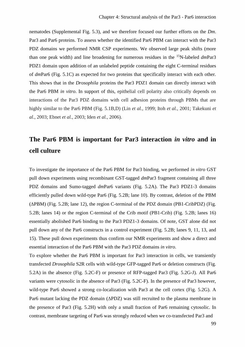
Chapter 4: Structural analysis of the Par3 - Par6 interaction
99
nematodes (Supplemental Fig. 5.3), and we therefore focused our further efforts on the Dm.
Par3 and Par6 proteins. To assess whether the identified Par6 PBM can interact with the Par3
PDZ domains we performed NMR CSP experiments. We observed large peak shifts (more
than one peak width) and line broadening for numerous residues in the 15N-labeled dmPar3
PDZ1 domain upon addition of an unlabeled peptide containing the eight C-terminal residues
of dmPar6 (Fig. 5.1C) as expected for two proteins that specifically interact with each other.
This shows that in the Drosophila proteins the Par3 PDZ1 domain can directly interact with
the Par6 PBM in vitro. In support of this, epithelial cell polarity also critically depends on
interactions of the Par3 PDZ domains with cell adhesion proteins through PBMs that are
highly similar to the Par6 PBM (Fig. 5.1B,D) (Lin et al., 1999; Itoh et al., 2001; Takekuni et
al., 2003; Ebnet et al., 2003; Iden et al., 2006).
The Par6 PBM is important for Par3 interaction in vitro and in
cell culture
To investigate the importance of the Par6 PBM for Par3 binding, we performed in vitro GST
pull down experiments using recombinant GST-tagged dmPar3 fragment containing all three
PDZ domains and Sumo-tagged dmPar6 variants (Fig. 5.2A). The Par3 PDZ1-3 domains
efficiently pulled down wild-type Par6 (Fig. 5.2B; lane 10). By contrast, deletion of the PBM
(PBM) (Fig. 5.2B; lane 12), the region C-terminal of the PDZ domain (PB1-CribPDZ) (Fig.
5.2B; lanes 14) or the region C-terminal of the Crib motif (PB1-Crib) (Fig. 5.2B; lanes 16)
essentially abolished Par6 binding to the Par3 PDZ1-3 domains. Of note, GST alone did not
pull down any of the Par6 constructs in a control experiment (Fig. 5.2B; lanes 9, 11, 13, and
15). These pull down experiments thus confirm our NMR experiments and show a direct and
essential interaction of the Par6 PBM with the Par3 PDZ domains in vitro.
To explore whether the Par6 PBM is important for Par3 interaction in cells, we transiently
transfected Drosophila S2R cells with wild-type GFP-tagged Par6 or deletion constructs (Fig.
5.2A) in the absence (Fig. 5.2C-F) or presence of RFP-tagged Par3 (Fig. 5.2G-J). All Par6
variants were cytosolic in the absence of Par3 (Fig. 5.2C-F). In the presence of Par3 however,
wild-type Par6 showed a strong co-localization with Par3 at the cell cortex (Fig. 5.2G). A
Par6 mutant lacking the PDZ domain (PDZ) was still recruited to the plasma membrane in
the presence of Par3 (Fig. 5.2H) with only a small fraction of Par6 remaining cytosolic. In
contrast, membrane targeting of Par6 was strongly reduced when we co-transfected Par3 and
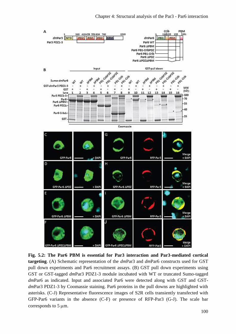
Chapter 4: Structural analysis of the Par3 - Par6 interaction
100
Fig. 5.2: The Par6 PBM is essential for Par3 interaction and Par3-mediated cortical
targeting. (A) Schematic representation of the dmPar3 and dmPar6 constructs used for GST
pull down experiments and Par6 recruitment assays. (B) GST pull down experiments using
GST or GST-tagged dmPar3 PDZ1-3 module incubated with WT or truncated Sumo-tagged
dmPar6 as indicated. Input and associated Par6 were detected along with GST and GST-
dmPar3 PDZ1-3 by Coomassie staining. Par6 proteins in the pull downs are highlighted with
asterisks. (C-J) Representative fluorescence images of S2R cells transiently transfected with
GFP-Par6 variants in the absence (C-F) or presence of RFP-Par3 (G-J). The scale bar
corresponds to 5m.
Chapter 4: Structural analysis of the Par3 - Par6 interaction
101
a Par6 mutant that lacked the PBM (PBM) (Fig. 5.2I) or a mutant lacking both the PDZ
domain and the PBM (PDZPBM) (Fig. 5.2J). To assess whether Par3-mediated membrane
targeting of Par6 may depend on endogenous aPKC, we performed Par6 recruitment assays in
S2R cells where we knocked down aPKC by RNAi (Supplemental Fig. 5.4A). We observed
that wild-type Par6 was still efficiently recruited to the cell cortex in the presence of Par3
(Supplemental Fig. 5.4B). In contrast, membrane targeting was compromised for all Par6
mutants (Supplemental Fig. 5.4C-E). aPKC knockdown thus only affects the localization of
the Par6 PDZ mutant (Supplemental Fig. 5.4D versus Fig. 5.2H), but not the PBM and
PDZPBM mutants. This suggests that the PDZ domain may have an additional function in
Par6 localization in the presence of aPKC. In line with our NMR binding studies and in vitro
assays, these in vivo results show that the PBM of Par6 plays an important role in the
interaction with Par3 and in Par3-mediated localization of Par6 to the cell cortex.
The PBM functions in redundancy with the PDZ domain in Par6
localization in vivo
To explore the in vivo relevance of our findings, we generated transgenic flies expressing
GFP-tagged Par6 under a constitutive promoter (Sen et al., 2012) and analyzed the embryos at
stage 11 (Fig. 5.3). As expected, we observed that wild-type GFP-Par6 accumulated
predominately at the cell-cell contacts of epidermal cells and co-localized with Par3 and
aPKC (Fig. 5.3A). Deletion of the Par6 PBM or the PDZ domain alone mildly but
consistently reduced cortical localization of Par6 (Fig. 5.3B,C). In contrast, deletion of both
the PDZ domain and the PBM caused an almost complete mislocalization of this Par6 mutant
to the cytosol (Fig. 5.3D). Of note, all GFP-Par6 variants were expressed similarly in fly
embryos (Supplemental Fig. 5.5A). Thus, both the PDZ domain and the PBM contribute to
correct Par6 localization in vivo with deletion of both domains resulting in almost complete
mislocalization of Par6. Yet, This may reflect an indirect association between Par3 and Par6
via aPKC as suggested previously (Suzuki et al., 2001; Nagai-Tamai et al., 2002) and / or
Par6 recruitment by other epithelial cell polarity regulators such as Crumbs or Stardust that
bind to the Par6 PDZ domain (Hurd et al., 2003b; Penkert et al., 2004; Lemmers et al., 2004;
Wang et al., 2004; Kempkens et al., 2006; Whitney et al., 2016).
Of these other polarity regulators Stardust is already expressed early on in fly embryos,
whereas Crumbs is only expressed at later stages (Krahn et al., 2010a; Sen et al., 2015). To

Chapter 4: Structural analysis of the Par3 - Par6 interaction
102
Fig. 5.3: Recruitment of Par6 to the cell cortex in fly epithelia depends on both the PBM
and the PDZ domain. (A-D) Localization of aPKC, Par3 and GFP-Par6 variants in the
epithelia of fly embryos (stage 11). The scale bar corresponds to 5m. Pearson’s correlation
coefficients (PCC) of Par6, aPKC, and Par3 fluorescence intensities were determined using
the Costes’ approach (Costes et al., 2004). The average PCCs estimated independently for
three different regions of each image are plotted along with their standard deviations (right
hand panels).
examine whether the observed localization of the Par6 variants may differ between
developmental stages, we analyzed Par6 localization in fly embryos during gastrulation (stage
6-7) and found that Par6 localization at stage 6-7 was highly similar to stage 11
(Supplemental Fig. 5.5B-E). Most notably, the PDZPBM mutant resulted in strong Par6
mislocalization both at stage 6-7 and stage 11. This demonstrates that in vivo Par6 recruitment
to the cell cortex critically depends on both the PDZ domain and the PBM and points to a
functional redundancy of these domains in Par6 localization in vivo.
In a par6-null background (par6226) (Petronczki and Knoblich, 2001), we found that wild-

Chapter 4: Structural analysis of the Par3 - Par6 interaction
103
type GFP-Par6 and GFP-Par6 PBM restored viability as well as aPKC and Par3 localization
(Supplemental Fig. 5.6), while the GFP-Par6 PDZ and GFP-Par6 PDZPBM could not
rescue the lethality of the par6-null allele. Similarly to our observations in the wild-type
background, lack of the PBM caused in stage 11 embryos a mild localization defect for Par6,
but not for Par3 and aPKC (Supplemental Fig. 5.6B). This confirms that the PBM contributes
to the correct localization of Par6 in vivo under physiological conditions and suggests that it
functions in redundancy with the PDZ domain to fully establish epithelial polarity.
Structural analysis of the Drosophila Par3 PDZ1:Par6 PBM
complex
Having established the importance of the Par6 PBM for Par3 interaction and Par3-mediated
localization in vitro and in vivo, we sought to gain structural insight into the Par3:Par6
complex. To this end, we crystallized a Dm. Par3:Par6 fusion construct comprising the Par3
PDZ1 domain and the C-terminal PBM octapeptide of Par6 (VKDGVLHL; core PBM is
underlined) and solved the X-ray structure of the PDZ1:PBM complex (Supplemental Table
5.1). Both the Par3 PDZ1 domain and the Par6 core PBM are well defined with the PDZ1
domain adopting the typical PDZ fold and the core PBM forming the canonical antiparallel -
strand with the PDZ1 2-strand (Fig. 5.4A, Supplemental Fig. 5.7A). The C-terminal
carboxylate group is involved in an extensive hydrogen bond network with the backbone of
the PDZ1 1-2 loop (Fig. 5.4A). The hydrophobic residues at the 0 and -2 positions of the
PBM are deeply buried in a hydrophobic pocket formed by the carboxylate binding loop
(Leu19), the 2 (Leu21, Ala23, Leu24, Pro25), 3 (Leu33) and 6 (Leu84, Val86) strands and the
2 helix (Val71, Leu75, Leu79) of the PDZ1 domain (Fig. 5.4A, Supplemental Fig. 5.7B).
Overall, our structure is highly similar to other PDZ:PBM complexes such as the INADL
PDZ3 in complex with a phage display-derived class II PBM peptide (Fig. 5.4B) (Ernst et al.,
2014) and Par3 PDZ3:PBM complexes (Supplemental Fig. 5.7C,D) (Feng et al., 2008; Tyler
et al., 2010). Outside the core PBM, we observed less well-defined electron density for the
Par6 peptide and three residues of the GS-linker (Supplemental Fig. 5.8A,B) indicating an
additional antiparallel -strand formed by the last aa of the GS-linker and the -7 and -6 PBM

Chapter 4: Structural analysis of the Par3 - Par6 interaction
104
Fig. 5.4: The Dm. Par3 PDZ1 and PDZ3 domains interact with Par6 through canonical
PDZ:PBM interactions. (A) Interaction network of the Par6 core PBM (dark pink) with the
Par3 PDZ1 domain (grey). Residues involved in hydrogen bonds (dashed lines) and side-
chain interactions are shown in stick representation with carbon, nitrogen and oxygen atoms
colored in grey (PDZ1) / dark-pink (PBM), blue and red, respectively. (B) The Par3 PDZ1 :
Par6 PBM complex is highly similar to the INADL PDZ3 in complex with a phage-derived
class II PBM peptide (PDB code: 4Q2N, (39)) shown in green (backbone r.m.s.d. = 1.64 Å).
(C) As Fig. 4.1C, but for the Par3 PDZ3 2-3loop domain. (D) Binding surface of the
dmPar6 PBM mapped onto a PDZ3 homology model and colored with a linear gradient from
white (CSP ≤ 0.05 ppm; cut-off) to red (CSP = 0.35 ppm). Unassigned residues are colored in
dark grey, while residues broadened beyond detection in the PDZ3 are in dark red. The Par6
core PBM is shown in yellow and was modeled by superposition of the dmPar3 PDZ1:PBM
and PDZ3 structures.
residues. However, the consistently high B-factors (Supplemental Table 5.1, Supplemental
Fig. 5.8C,D) strongly suggest that the residues outside the core PBM are not stably structured.
To validate key interactions of the Par6 PBM with the Par3 PDZ1 domain in our crystal
structure, we substituted each of the three C-terminal positions in the dmPar6 peptide with

Chapter 4: Structural analysis of the Par3 - Par6 interaction
105
Ala (Par6 L349A (L-2A), H350A (H-1A) and L351A (L0A)) and tested the Par3 binding
capabilities of the PBM mutants in NMR experiments (Supplemental Fig. 5.8E). Consistent
with our crystal structure, both the L-2A and the L0A mutations led to an almost complete loss
of Par6 binding, while the H-1A mutation lessened CSPs in the Par3 PDZ1 domain as
compared to the wild-type PBM (Supplemental Fig. 5.8E versus Fig. 5.1C). In sum, our
results demonstrate that Par6 associates with Par3 through a canonical PDZ:PBM interaction
that crucially depends on the -2 and 0 positions of the Par6 PBM.
The Par3 PDZ1 and PDZ3 domains both recognize the Par6 PBM
We noticed that the residues in the PDZ1 domain contacting the 0 and -2 PBM positions are
well-conserved in all three Par3 PDZ domains (Supplemental Fig. 5.9). We therefore also
tested the binding specificities of the Par3 PDZ2 and PDZ3 domains towards the Par6 PBM.
To this end, we performed NMR binding studies with the individual dmPar3 PDZ2 and PDZ3
domains and the dmPar6 PBM peptide. In contrast to the PDZ1 domain, we observed virtually
no CSPs for the Par3 PDZ2 domain upon addition of the Par6 PBM (Supplemental Fig.
5.10A). The Par3 PDZ2 domain thus possesses binding specificities that are distinct from the
Par3 PDZ1 domain.
The PDZ3 domain contains a long, disordered insertion in the 2-3 loop that is unique to
dmPar3 (Supplemental Fig. 5.9). Since this extension severely compromised spectral quality,
we used a PDZ3 domain with a truncated 2-3 loop (PDZ32-3loop) for our NMR studies
(Supplemental Fig. 5.9). As for the PDZ1 domain, addition of the Par6 PBM induced
numerous, large CSPs in the 15N-labeled PDZ32-3loop (Fig. 5.4C). This demonstrates that
the truncated PDZ3 domain is functional and interacts readily with the dmPar6 PBM. To map
the observed CSPs onto the surface of the PDZ3 domain, we obtained chemical shift
assignments for the PDZ3 domain and generated a homology model of the Par3 PDZ3
domain. As the PDZ1 domain, in this model the PDZ3 domain binds the Par6 PBM in a
canonical manner mainly involving the carboxylate binding loop, the 2 strand and the 2
helix (Fig. 5.4D).
To obtain quantitative insights into the Par3 PDZ interactions with the Par6 PBM, we
determined dissociation constants (Kd values) by NMR line shape fitting for the chemical
shift titrations of the Dm. Par3 PDZ1 and PDZ3 domains with the Par6 PBMs. Of note, since
the PDZ2 domain did exhibited no CSPs upon addition of the Par6 PBM, we did not fit these

Chapter 4: Structural analysis of the Par3 - Par6 interaction
106
data. We found that the Par6 PBM bound to the PDZ1 domain with a moderate affinity of 216
4 M and to the PDZ3 domain with a tighter affinity of 54 1 M for the PDZ3 domain
(Supplemental Table 5.2, Supplemental Fig. 5.11, 5.12). Next, we quantitatively addressed
the importance of the three C-terminal positions in the Par6 PBM for PDZ1 binding
(Supplemental Fig. 5.8E and 5.13). We found that mutation of the C-terminal position (L0A)
weakened the affinity of the PDZ1 domain for the Par6 PBM to 2.5 0.4 mM, for the -1
position (H-1A) to 964 60 M and for the -2 position (L-2A) to 4.0 1.1 mM, respectively
(Supplemental Table 5.2, Supplemental Fig. 5.13). This confirms that these mutations in the
core PBM indeed strongly compromise binding to the Par3 PDZ1 domain and that these
residues are thus important determinants for the interaction with the Par3 PDZ1 domain.
Overall, dissociation constants of up to a few hundreds of M have been observed for
physiologically relevant PDZ interactions (Stiffler et al., 2007). Consistent with our analyses
of cultured S2R cells (Fig. 5.2 and Supplemental Fig. 5.4) and fly embryos (Fig. 5.3 and
Supplemental Fig. 5.5-6), this shows that the interaction of the newly identified Par6 PBM
plays a role in Par3 PDZ binding in vitro and is physiologically relevant in vivo.
Par3 can interact with two Par6 proteins simultaneously in vitro
In polarized cells, Par complexes form micrometer-sized clusters that cover the apical plasma
membrane. Yet, how Par complexes assemble into these sizeable protein networks is largely
unknown. A prerequisite for forming higher order networks is that the proteins involved are
multivalent, that is they contain multiple independent binding sites, and engage in a multitude
of weak (M-affinity) interactions. To test whether the first and third PDZ domain of Par3
fulfill this requirement, we recorded 1H,15N-correlation spectra of the 15N-labeled Par3 PDZ1-
3 module that contains all three PDZ domains (PDZ1-3 2-3loop) and examined its Par6
binding capability. In this construct the 2-3 loop in the PDZ3 domain was removed to
improve spectral quality (see above). The NMR spectra of the Par3 PDZ1-3 module and the
individual PDZ domains superimpose well (Fig. 5.5A-C), and resonance assignments of the
individual domains could be transferred to the PDZ1-3 module. This demonstrates that the
individual PDZ domains in Par3 are structurally largely independent. Addition of unlabeled
Par6 PBM to the PDZ1-3 construct resulted in CSPs comparable to the isolated PDZ domains
(Fig. 5.5D-F versus 5.1C, 5.4C, Supplemental Fig. 5.10A). This shows that the Par3 PDZ
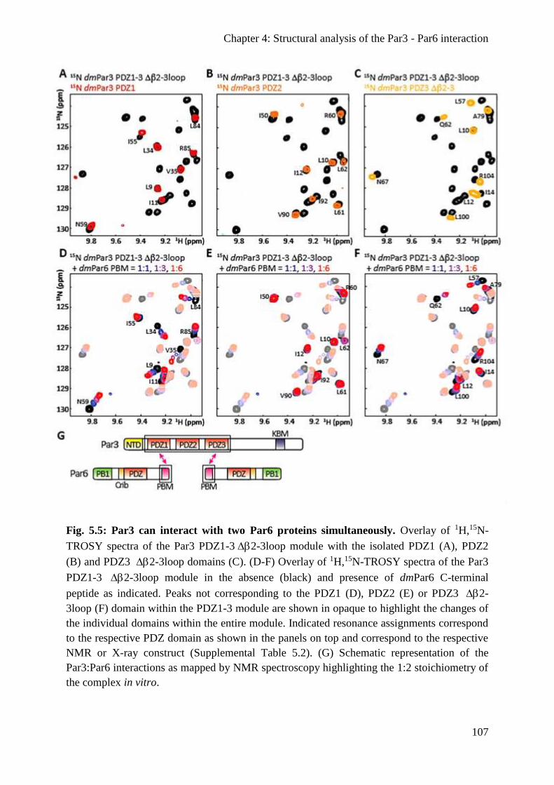
Chapter 4: Structural analysis of the Par3 - Par6 interaction
107
Fig. 5.5: Par3 can interact with two Par6 proteins simultaneously. Overlay of 1H,15N-
TROSY spectra of the Par3 PDZ1-32-3loop module with the isolated PDZ1 (A), PDZ2
(B) and PDZ3 2-3loop domains (C). (D-F) Overlay of 1H,15N-TROSY spectra of the Par3
PDZ1-3 2-3loop module in the absence (black) and presence of dmPar6 C-terminal
peptide as indicated. Peaks not corresponding to the PDZ1 (D), PDZ2 (E) or PDZ3 2-
3loop (F) domain within the PDZ1-3 module are shown in opaque to highlight the changes of
the individual domains within the entire module. Indicated resonance assignments correspond
to the respective PDZ domain as shown in the panels on top and correspond to the respective
NMR or X-ray construct (Supplemental Table 5.2). (G) Schematic representation of the
Par3:Par6 interactions as mapped by NMR spectroscopy highlighting the 1:2 stoichiometry of
the complex in vitro.

Chapter 4: Structural analysis of the Par3 - Par6 interaction
108
domains act as functionally independent entities within the PDZ1-3 module and that in vitro
one Par3 protein can simultaneously interact with two Par6 proteins through its PDZ1 and
PDZ3 domains (Fig. 5.5G). Par3 thus has the potential to engage in weak, multivalent
interactions with Par6 and may thereby promote the assembly of large-scale clusters of Par
complexes at the cell membrane in vivo.
The Par6 PBM can compete with the PBM of E-cadherin for Par3
binding
Cell adhesion proteins such as cadherins and nectins are well-known interaction partners of
Par3 and contain conserved class II PBMs that are highly similar to the Par6 PBM (Fig. 5.1B
and Supplemental Fig. 5.7C). In fact, the VE-cadherin PBM interacts with the PDZ3 domain
of murine Par3 with a Kd of ~ 6 M (Tyler et al., 2010) and thus ~ 9-fold (PDZ3) and ~ 36-
fold (PDZ1) tighter than its Dm. counterparts with the Par6 PBM (Supplemental Table 5.2).
To address whether the Par6 PBM could compete with such ligands for Par3 binding, we
performed NMR CSP studies with the 15N-labeled Dm. Par3 PDZ domains and added
increasing amounts of unlabeled Shg PBM to the individual domains. Similarly to the Par6
PBM, both the Par3 PDZ1 (Fig. 5.6A) and PDZ3 (Fig. 5.6B) domains show large CSPs for
numerous peaks in the 1H,15N-correlation spectra upon addition of the Shg PBM. In contrast,
the PDZ2 domain displayed only few changes (Supplemental Fig. 5.10B). Line shape fitting
analyses for the Shg PBM chemical shift titrations of all three Dm. Par3 PDZ domains yielded
Kd values of 128 4 M for the PDZ1, 954 45 M for the PDZ2 and 0.6 0.1 M for the
PDZ3 domain (Supplemental Table 5.2, Supplemental Fig. 5.11C,D, Supplemental Fig. 5.14
and Supplemental Fig. 5.12C,D). The Dm. Par3 PDZ domains thus bind tighter to the PBM of
Shg than to the Par6 PBM albeit only by a factor of ~ 2 for the PDZ1 domain.
To evaluate whether the PBM of Par6 and Shg compete for binding to the Par3 PDZ1 or
PDZ3 domains, we performed a set of NMR experiments on the 15N-labeled Shg PBM. First,
we recorded 1H,15N-correlation spectra of the peptide in the presence or absence of unlabeled
Dm. Par3 PDZ1 or PDZ3 domain. In both cases, this resulted in PDZ:Shg complex formation
as indicated by the observed CSPs (Fig. 5.6C,D). Subsequently, we added Par6 PBM to the
PDZ:Shg complex in a step-wise manner and observed for both the PDZ domains a release of

Chapter 4: Structural analysis of the Par3 - Par6 interaction
109
Fig. 5.6: The Par6 PBM can compete with Shg for Par3 binding. (A) Overlay of 1H,15N-
HSQC spectra of the Dm. Par3 PDZ1 domain in the absence and presence of the Shg PBM
peptide as indicated. (B) as (A), but for the Dm. Par3 PDZ32-3loop domain. (C) Overlay
of 1H,15N-HSQC spectra of the Shg PBM fused to GB1 in the absence (black) or presence of
dmPar3 PDZ1 domain (purple) and upon step-wise addition of the Par6 PBM (red and
orange). Arrows indicate the successive reversal of the chemical shifts from the Shg:PDZ
complex towards the unbound Shg PBM. (D) as (C), but for the PDZ3 PDZ32-3loop
domain.
the Shg peptide as indicated by chemical shift changes that reverted towards the unbound Shg
peptide (Fig. 5.6C,D). This demonstrates that the Par6 PBM can indeed compete with Shg for
Par3 PDZ binding. The relatively high stoichiometric amounts of Par6 PBM required to
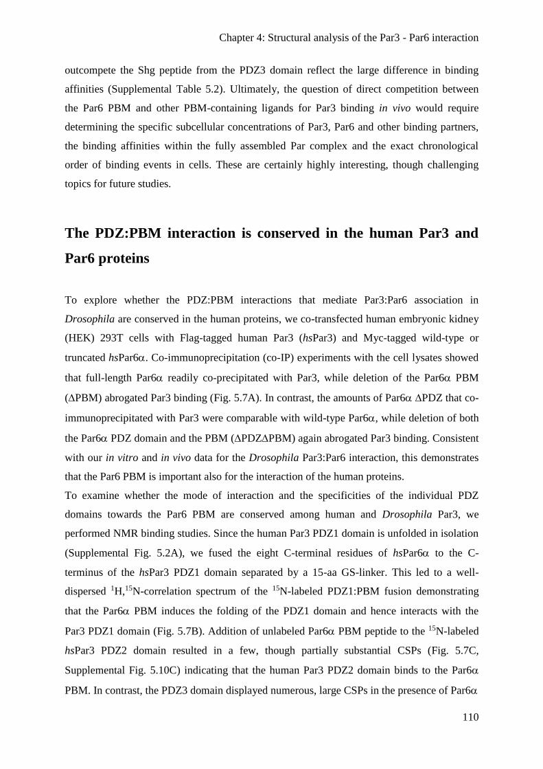
Chapter 4: Structural analysis of the Par3 - Par6 interaction
110
outcompete the Shg peptide from the PDZ3 domain reflect the large difference in binding
affinities (Supplemental Table 5.2). Ultimately, the question of direct competition between
the Par6 PBM and other PBM-containing ligands for Par3 binding in vivo would require
determining the specific subcellular concentrations of Par3, Par6 and other binding partners,
the binding affinities within the fully assembled Par complex and the exact chronological
order of binding events in cells. These are certainly highly interesting, though challenging
topics for future studies.
The PDZ:PBM interaction is conserved in the human Par3 and
Par6 proteins
To explore whether the PDZ:PBM interactions that mediate Par3:Par6 association in
Drosophila are conserved in the human proteins, we co-transfected human embryonic kidney
(HEK) 293T cells with Flag-tagged human Par3 (hsPar3) and Myc-tagged wild-type or
truncated hsPar6. Co-immunoprecipitation (co-IP) experiments with the cell lysates showed
that full-length Par6 readily co-precipitated with Par3, while deletion of the Par6 PBM
(PBM) abrogated Par3 binding (Fig. 5.7A). In contrast, the amounts of Par6 PDZ that co-
immunoprecipitated with Par3 were comparable with wild-type Par6, while deletion of both
the Par6 PDZ domain and the PBM (PDZPBM) again abrogated Par3 binding. Consistent
with our in vitro and in vivo data for the Drosophila Par3:Par6 interaction, this demonstrates
that the Par6 PBM is important also for the interaction of the human proteins.
To examine whether the mode of interaction and the specificities of the individual PDZ
domains towards the Par6 PBM are conserved among human and Drosophila Par3, we
performed NMR binding studies. Since the human Par3 PDZ1 domain is unfolded in isolation
(Supplemental Fig. 5.2A), we fused the eight C-terminal residues of hsPar6 to the C-
terminus of the hsPar3 PDZ1 domain separated by a 15-aa GS-linker. This led to a well-
dispersed 1H,15N-correlation spectrum of the 15N-labeled PDZ1:PBM fusion demonstrating
that the Par6 PBM induces the folding of the PDZ1 domain and hence interacts with the
Par3 PDZ1 domain (Fig. 5.7B). Addition of unlabeled Par6 PBM peptide to the 15N-labeled
hsPar3 PDZ2 domain resulted in a few, though partially substantial CSPs (Fig. 5.7C,
Supplemental Fig. 5.10C) indicating that the human Par3 PDZ2 domain binds to the Par6
PBM. In contrast, the PDZ3 domain displayed numerous, large CSPs in the presence of Par6
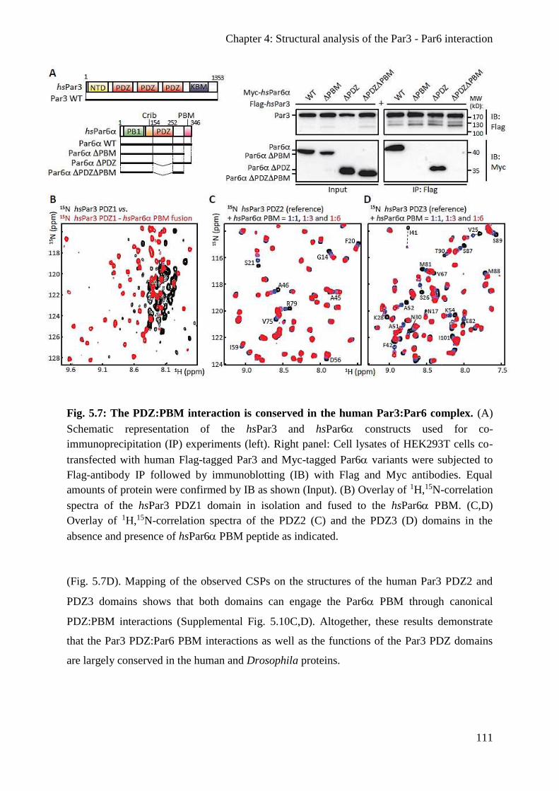
Chapter 4: Structural analysis of the Par3 - Par6 interaction
111
Fig. 5.7: The PDZ:PBM interaction is conserved in the human Par3:Par6 complex. (A)
Schematic representation of the hsPar3 and hsPar6 constructs used for co-
immunoprecipitation (IP) experiments (left). Right panel: Cell lysates of HEK293T cells co-
transfected with human Flag-tagged Par3 and Myc-tagged Par6 variants were subjected to
Flag-antibody IP followed by immunoblotting (IB) with Flag and Myc antibodies. Equal
amounts of protein were confirmed by IB as shown (Input). (B) Overlay of 1H,15N-correlation
spectra of the hsPar3 PDZ1 domain in isolation and fused to the hsPar6 PBM. (C,D)
Overlay of 1H,15N-correlation spectra of the PDZ2 (C) and the PDZ3 (D) domains in the
absence and presence of hsPar6 PBM peptide as indicated.
(Fig. 5.7D). Mapping of the observed CSPs on the structures of the human Par3 PDZ2 and
PDZ3 domains shows that both domains can engage the Par6 PBM through canonical
PDZ:PBM interactions (Supplemental Fig. 5.10C,D). Altogether, these results demonstrate
that the Par3 PDZ:Par6 PBM interactions as well as the functions of the Par3 PDZ domains
are largely conserved in the human and Drosophila proteins.
Chapter 4: Structural analysis of the Par3 - Par6 interaction
112
Discussion
In this study, we have identified a previously unrecognized PDZ-binding motif in Par6 that
mediates canonical PDZ:PBM interactions with the PDZ1 and PDZ3 domains of Par3. This
interaction mode is conserved amongst the human and Drosophila Par3 and Par6 proteins. We
demonstrate that the PBM, but not the PDZ domain, of Par6 is essential for interaction with
Par3 in vitro. Yet, the PBM seems to function in redundancy with the PDZ domain in Par6
localization in fly epithelia, as the individual deletions only mildly reduce cortical localization
in vivo, and deletion of both the PBM and the PDZ domain are required for almost complete
mislocalization of Par6.
Overall, we found no indication of a heterodimerization of the human, Dm. or Ce. Par3 PDZ
domains with the respective Par6 (Crib-)PDZ domain by NMR (Supplemental Fig. 5.2), in
GST-pull down (Fig. 5.2B) or co-IP experiments (Fig. 5.7A) nor in recruitment assays in S2R
cells (Fig. 5.2D). Our results thus provide important, novel insights into Par complex
assembly and contrast previous findings that Par3 and Par6 associate through PDZ:PDZ
interaction (Lin et al., 1999; Joberty et al., 2000; Li et al., 2010b). In support of our findings,
Par3 associates in epithelial cells with various cell adhesion proteins possessing class II PBMs
that are highly similar to the Par6 PBM (Fig. 5.1B) and we show here that the Dm. Par3 PDZ1
and PDZ3 domains also interact with the major cell adhesion protein E-cadherin (Shg) (Fig.
5.6A,B). These interactions have been shown to be crucial for the establishment and
maintenance of cell junctions and epithelial cell polarity (Lin et al., 1999; Itoh et al., 2001;
Takekuni et al., 2003; Ebnet et al., 2003; Iden et al., 2006; Tepass, 2012). Of note, as in the
case of Par6, these adhesion protein interactions are conserved among human and Drosophila
Par3. In sum, this supports the notion that class II PBMs constitute a general binding motif for
the Par3 PDZ1 and PDZ3 domains to recruit polarity and cell adhesion proteins for the
establishment and maintenance of cell polarity. Nematodes may constitute a notable
exception, as not only their Par6 but also their cadherin (HRM-1) proteins lack a detectable,
C-terminal PBM (Supplemental Fig. 5.3). Future studies will thus be required to elucidate the
exact mechanism of Par3 PDZ interactions and their role in cell polarity in nematodes.

Chapter 4: Structural analysis of the Par3 - Par6 interaction
113
The individual PDZ binding preferences allow for multivalent
Par3 interactions
Tandem arrangement of PDZ domains is a well-known mechanism for scaffolding proteins to
assemble different components of a signaling cascade through multivalent interactions
(Tsunoda et al., 1997). This is based on the fact that interaction domains embedded in
proteins can in general fold and function independently (Pawson and Nash, 2003). We find
that the isolated Par3 PDZ1 and PDZ3 domains can both independently interact with the Par6
PBM (Fig. 5.5D,F and 5.7B,D) and thus may function in redundancy. Indeed, these functional
redundancies allow each Par3 molecule to simultaneously recruit two Par6 proteins in vitro
(Fig. 5.5G). Together with Par6:aPKC hetero-dimerization and Par3 homo-oligomerization,
this provides a basis for the role of Par3 in enforcing the spatial segregation and the assembly
of large-scale, self-organizing Par complex networks at the cell cortex in vivo.
Concluding remarks
The mode of Par3:Par6 interaction has remained controversial. In a sense, this reflects the
challenges of delineating the specific functions of proteins and even more importantly their
individual domains and motifs in cell polarity. Functional coupling, redundant interactions,
differences in organism strains, cell types and protein constructs and finally the existence of
paralogs severely hamper in vivo analyses of polarity proteins (Nagai-Tamai et al., 2002;
Fievet et al., 2013). Moreover, the composition of polarity complexes is dynamic and may
depend on the cell type or developmental context (Henrique and Schweisguth, 2003). Lastly,
different populations of polarity proteins may even co-exist within a single cell (Goehring et
al., 2011). Altogether, this poses enormous obstacles for functional analyses as phenotypes
may be obscured in mutation studies. Detailed structural analyses are thus essential to
unambiguously determine the molecular basis of polarity complex formation. Moreover, the
fact that PBMs require a free C-terminus for their function posits that C-terminal tagging for
fluorescence microscopy or immunoblotting likely abrogates not only Par3:Par6 association,
but possibly also other, yet unidentified Par6 PBM interactions. Altogether, this may explain
some of the controversy about the Par3:Par6 interaction.
The fact that almost all polarity and cell adhesion proteins contain either at least one PDZ
domain or a PBM emphasizes the importance of PDZ:PBM interactions in the assembly of
Chapter 4: Structural analysis of the Par3 - Par6 interaction
114
cell polarity and adhesion signaling networks. Our work illustrates the difficulties in
predicting PDZ specificities, as the marked differences in Par6 and Shg recognition of the
Par3 PDZ2 (Fig. 5.5D-F, 5.6A,B and Supplemental Fig. 5.10B, Supplemental Table 5.2) were
unexpected on the sequence level (Supplemental Fig. 5.9). A detailed characterization of the
functional specificities and redundancies of PDZ domains, as presented here, is therefore
indispensable to decipher their contribution to polarity protein localization and function.
Ultimately, this will help to predict PDZ domain function and provide a better understanding
of the interaction networks underlying the establishment, maintenance and loss of cell polarity
and hence critical developmental and carcinogenic processes.

Chapter 4: Structural analysis of the Par3 - Par6 interaction
115
Supplemental figures
Supplemental Fig. 5.1: Chemical shift perturbation experiments. (A) Exemplary overlay
of 1H,15N-correlation spectra of a 15N-labeled protein in the absence and presence of an
unlabeled binding partner at varying stoichiometric ratios. A region of the spectrum
containing the NMR signals of two consecutive amino acids in the protein sequence is shown.
(B) Chemical structure of the protein backbone highlighting the 1H and 15N atoms (green
spheres) observed in the H,N-correlation spectra. The position of a peak in the spectrum
strongly depends on the local chemical environment of the respective amino acid (indicated
by light-blue spheres). Therefore, in contrast to an unfolded protein, peaks are distributed over
a wide range of 1H and 15N chemical shifts in a folded protein. (C) Ligand binding (shown in
orange) induces a change in the chemical environment, and hence in the chemical shifts, of
those amino acids that participate in ligand binding (aa #1), but not of those that are located
outside the binding pocket (aa #2).

Chapter 4: Structural analysis of the Par3 - Par6 interaction
116
Supplemental Fig. 5.2: The Par3 PDZ1 domain does not interact directly with the Par6
(Crib-)PDZ domain. (A) 1H,15N-correlation spectrum of the 15N-labeled Hs. Par3 PDZ1
domain showing that the domain is largely unstructured in isolation. The boxed region is
magnified in Fig. 4.7B. (B,C) Overlay of 1H,15N-correlation spectra of the D. melanogaster
(B) and C. elegans (C) Par3 PDZ1 domains in the absence and presence of a stoichiometric
excess of the respective Par6 PDZ domain as indicated. (D,E) As (B,C) but for the D.
melanogaster (D) and C. elegans (E) Par6 semiCRIB-PDZ constructs in the absence and
presence of a stoichiometric excess of the Par3 PDZ1 domains as indicated. (F,G) As (B,C)
but for the D. melanogaster Par6 Crib-PDZ construct in the absence and presence of a
stoichiometric excess of the Par3 PDZ2 (F) and PDZ3 (G) domains as indicated.

Chapter 4: Structural analysis of the Par3 - Par6 interaction
117
Supplemental Fig. 5.3: The Par6 C-terminus contains a conserved class II PDZ binding
motif. Sequences of the C-terminal 16 aa of various vertebrate (top), invertebrate (center) and
nematode (bottom) Par6 proteins. The class II PBM is highlighted with a red box and
conserved in all metazoans with the exception of nematodes. Conserved residues were color-
coded with ClustalX (Crooks et al., 2004).

Chapter 4: Structural analysis of the Par3 - Par6 interaction
118
Supplemental Fig. 5.4: Par6 localization in S2R cells upon aPKC knockdown. (A)
Western blot analysis of aPKC expression in S2R cell lysates in the absence or presence of
dsRNA for aPKC knockdown. (B-E) As Fig. 5.2G-J, but for S2R cells where aPKC is
knocked down by RNAi.
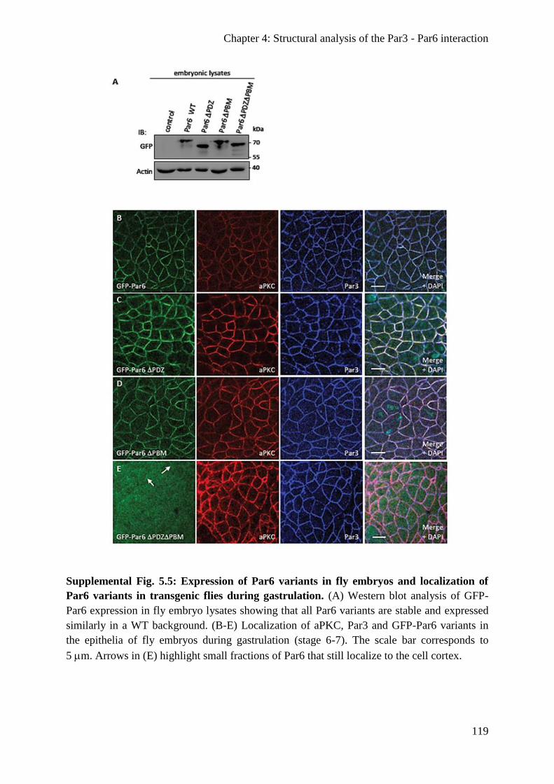
Chapter 4: Structural analysis of the Par3 - Par6 interaction
119
Supplemental Fig. 5.5: Expression of Par6 variants in fly embryos and localization of
Par6 variants in transgenic flies during gastrulation. (A) Western blot analysis of GFP-
Par6 expression in fly embryo lysates showing that all Par6 variants are stable and expressed
similarly in a WT background. (B-E) Localization of aPKC, Par3 and GFP-Par6 variants in
the epithelia of fly embryos during gastrulation (stage 6-7). The scale bar corresponds to
5m. Arrows in (E) highlight small fractions of Par6 that still localize to the cell cortex.
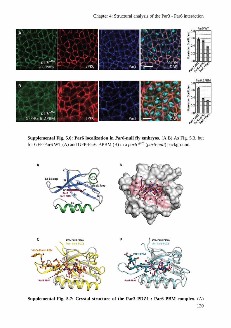
Chapter 4: Structural analysis of the Par3 - Par6 interaction
120
Supplemental Fig. 5.6: Par6 localization in Par6-null fly embryos. (A,B) As Fig. 5.3, but
for GFP-Par6 WT (A) and GFP-Par6 PBM (B) in a par6226 (par6-null) background.
Supplemental Fig. 5.7: Crystal structure of the Par3 PDZ1 : Par6 PBM complex. (A)
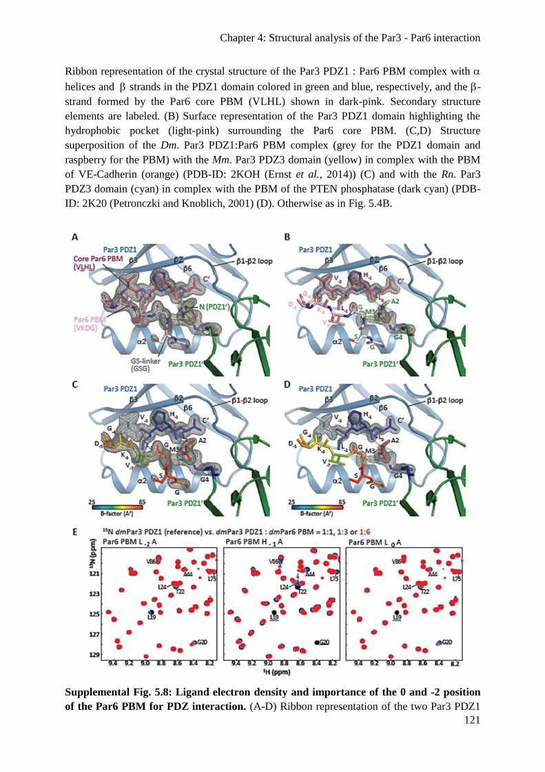
Chapter 4: Structural analysis of the Par3 - Par6 interaction
121
Ribbon representation of the crystal structure of the Par3 PDZ1 : Par6 PBM complex with
helices and strands in the PDZ1 domain colored in green and blue, respectively, and the -
strand formed by the Par6 core PBM (VLHL) shown in dark-pink. Secondary structure
elements are labeled. (B) Surface representation of the Par3 PDZ1 domain highlighting the
hydrophobic pocket (light-pink) surrounding the Par6 core PBM. (C,D) Structure
superposition of the Dm. Par3 PDZ1:Par6 PBM complex (grey for the PDZ1 domain and
raspberry for the PBM) with the Mm. Par3 PDZ3 domain (yellow) in complex with the PBM
of VE-Cadherin (orange) (PDB-ID: 2KOH (Ernst et al., 2014)) (C) and with the Rn. Par3
PDZ3 domain (cyan) in complex with the PBM of the PTEN phosphatase (dark cyan) (PDB-
ID: 2K20 (Petronczki and Knoblich, 2001) (D). Otherwise as in Fig. 5.4B.
Supplemental Fig. 5.8: Ligand electron density and importance of the 0 and -2 position
of the Par6 PBM for PDZ interaction. (A-D) Ribbon representation of the two Par3 PDZ1

Chapter 4: Structural analysis of the Par3 - Par6 interaction
122
domains in the asymmetric unit of the crystal (blue and green). The electron difference
density of the Par6 PBM, the three C-terminal residues of the GS-linker and the three N-
terminal residues of the neighboring PDZ1 domain is shown as 2FO-FC map contoured at a
level of 1.0 (A,C) and 2.0 (B,D). (C,D) As (A,B), but displaying the B-factor distribution of
the PBM highlighting the increased flexibility of the residues outside the core PBM. The
color scale for B-factors ranges from blue ( 25 Å2) to red ( 85 Å2). (E) NMR titration
experiments with 15N-labeled Par3 dmPar3 PDZ1 domain and Par6 core PBM mutants as
indicated. Otherwise as Fig. 5.1C.

Chapter 4: Structural analysis of the Par3 - Par6 interaction
123
Supplemental Fig. 5.9: Structure-based sequence alignment of Par3 PDZ domains.
Protein sequences of the individual Par3 PDZ domains were aligned with MUSCLE (Larkin
et al., 2007), manually edited where necessary to match known Par3 PDZ structures (PDB-

Chapter 4: Structural analysis of the Par3 - Par6 interaction
124
ID: 2KOM (Waudby et al., 2016) for PDZ2 and 2KOH (Ernst et al., 2014) for PDZ3) and
color-coded with ClustalX (Crooks et al., 2004). Secondary structure elements based on the
dmPar3 PDZ1 : dmPar6 PBM complex are shown with blue boxes and denoted on the top.
Residues within 5 Å proton-proton distance of the PBM are highlighted with circles and
dashed lines (red: -2 position; yellow: 0 position) for the Par3 PDZ1:Par6 and the highly
similar Par3 PDZ3:VE-cadherin complex (Ernst et al., 2014). This shows that all residues
contacting the specificity-determining 0 and -2 positions of the PBM are highly conserved
and thus do not allow a specificity prediction based on sequence alignments. The region
deleted in the dmPar3 PDZ3 2-3loop construct is indicated with a black box.
Supplemental Fig. 5.10: Interaction of the dmPar6 and Shg PBMs with the dmPar3
PDZ2 domain and Par6:Par3 interactions in the human proteins. (A,B) NMR titration of
the 15N-labeled dmPar3 PDZ2 domain with Par6 (A) or Shg (B) PBM as indicated. (C,D)
CSPs induced by the hsPar6 PBM mapped onto the structures of the human Par3 PDZ2
(PDB-ID: 2KOM (Waudby et al., 2016); left panel) and rat Par3 PDZ3 domain (PDB-ID:
2K20 (Petronczki and Knoblich, 2001); right panel) and colored with a linear gradient from
white (CSP ≤ 0.05 ppm) to green (C) or blue (D) (CSP = 0.35 ppm). Residues broadened
beyond detection in the PDZ3 are shown in dark-blue. Secondary structure elements are
labeled (for PDZ2 1: aa 5-12, 2: aa 20-24, 3: aa 35-40, 1: aa 45-49, 4: aa 56-61, 5: aa

Chapter 4: Structural analysis of the Par3 - Par6 interaction
125
64-65, 2: aa 71-80, 6: aa 86-93; for PDZ3 1: aa 6-14, 2: aa 25-31, 3: aa 38-46, 1: aa
51-54, 4: aa 62-67, 5: aa 70-71, 2: aa 77-89, 6: aa 97-105). Otherwise as Fig. 5.4D.
Supplemental Fig. 5.11: Line shape fitting analyses of the dmPar3 PDZ1 interactions
with the PBMs of dmPar6 or Shg. (A) 2D line shape fit of the Asn59 cross peak in the
individual 1H,15N-HSQC spectra of the dmPar3 PDZ1 domain in the absence or presence of
Par6 PBM. Observed data are presented as black lines, while fits are shown in magenta. Each
panel represents the fit of one titration point at the indicated stoichiometric ratio of
PDZ:PBM. (B,C) Representative region of the NMR titration of 15N-labeled dmPar3 PDZ1
with Par6 (B) or Shg (C). Contour plots of observed CSPs are shown in the left panel, while
the fit of the cross peaks is presented in the right panel with black lines indicating the course
of the fit CSPs between the reference point and the estimated saturation point. Titration steps
are color coded as indicated. The Asn59 cross peak shown in (A) and (D) is boxed. (D) as (A)
but for the Shg PBM.
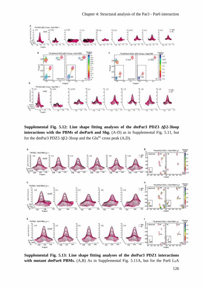
Chapter 4: Structural analysis of the Par3 - Par6 interaction
126
Supplemental Fig. 5.12: Line shape fitting analyses of the dmPar3 PDZ3 2-3loop
interactions with the PBMs of dmPar6 and Shg. (A-D) as in Supplemental Fig. 5.11, but
for the dmPar3 PDZ3 2-3loop and the Glu81 cross peak (A,D).
Supplemental Fig. 5.13: Line shape fitting analyses of the dmPar3 PDZ1 interactions
with mutant dmPar6 PBMs. (A,B) As in Supplemental Fig. 5.11A, but for the Par6 L0A

Chapter 4: Structural analysis of the Par3 - Par6 interaction
127
mutant. (C-F) as in (A,B), but for the Par6 H-1A (C,D) and L-2A (E,F) mutants.
Supplemental Fig. 5.14: Line shape fitting analyses of the dmPar3 PDZ2 interactions
with the Shg PBM. (A, B) as in Supplemental Fig. 5.11, but for the dmPar3 PDZ2 and the
Ile92 cross peak.
Supplemental Table 5.1. Statistics of X-ray data collection and model refinement for the
dmPar3 PDZ1:dmPar6 PBM complex1
Data collection
Resolution (Å) 40.96 – 1.50 (1.59 – 1.50) 2
Completeness (%) 99.8 (98.7)
No. of unique reflections 86,611 (13,880)
Redundancy 5.13 (4.88)
CC1/2* (%) 99.9 (70.2)
I / I 18.78 (2.12)
Wilson B-Factor (Å2) 31.13
FreeR (% of reflections) 5
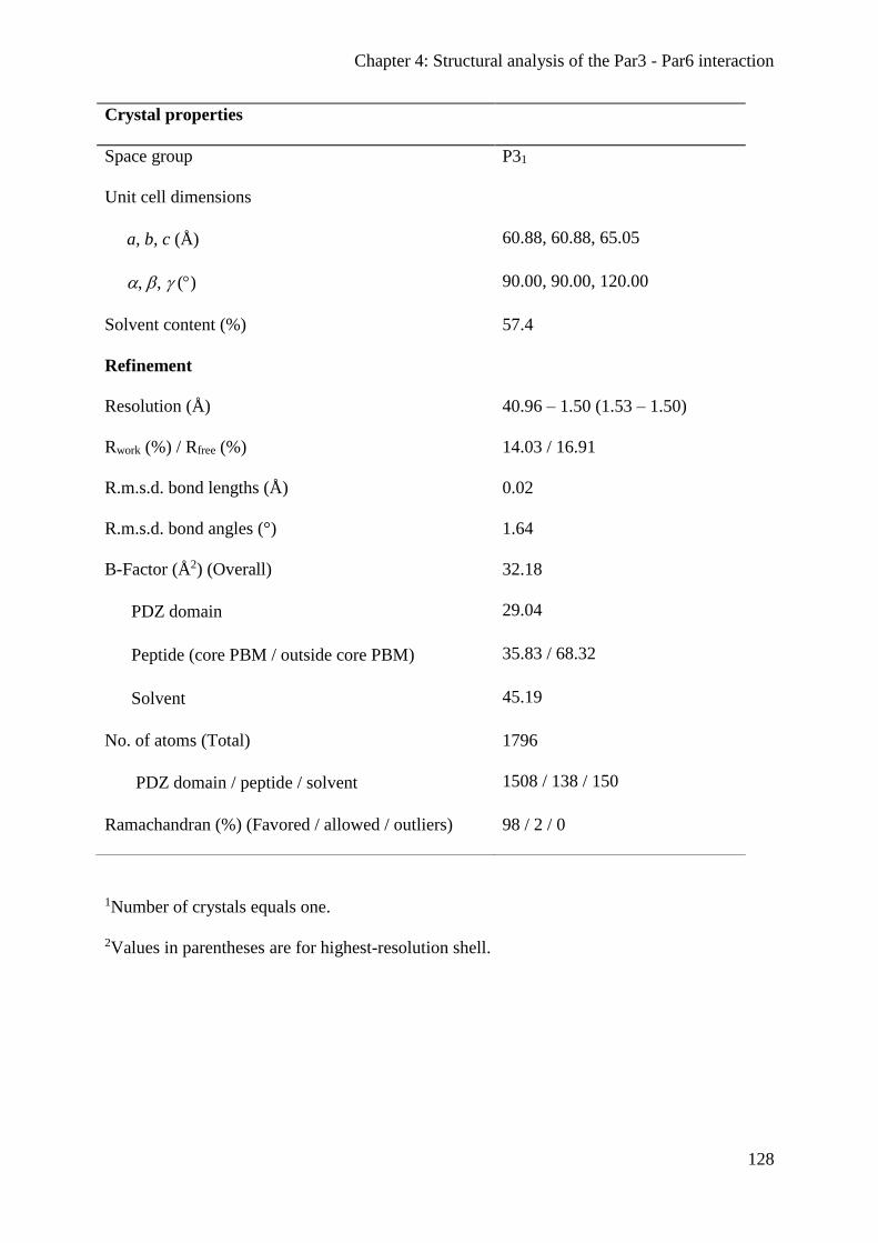
Chapter 4: Structural analysis of the Par3 - Par6 interaction
128
1Number of crystals equals one.
2Values in parentheses are for highest-resolution shell.
Crystal properties
Space group P31
Unit cell dimensions
a, b, c (Å) 60.88, 60.88, 65.05
, , () 90.00, 90.00, 120.00
Solvent content (%) 57.4
Refinement
Resolution (Å) 40.96 – 1.50 (1.53 – 1.50)
Rwork (%) / Rfree (%) 14.03 / 16.91
R.m.s.d. bond lengths (Å) 0.02
R.m.s.d. bond angles (°) 1.64
B-Factor (Å2) (Overall) 32.18
PDZ domain 29.04
Peptide (core PBM / outside core PBM) 35.83 / 68.32
Solvent 45.19
No. of atoms (Total) 1796
PDZ domain / peptide / solvent 1508 / 138 / 150
Ramachandran (%) (Favored / allowed / outliers) 98 / 2 / 0
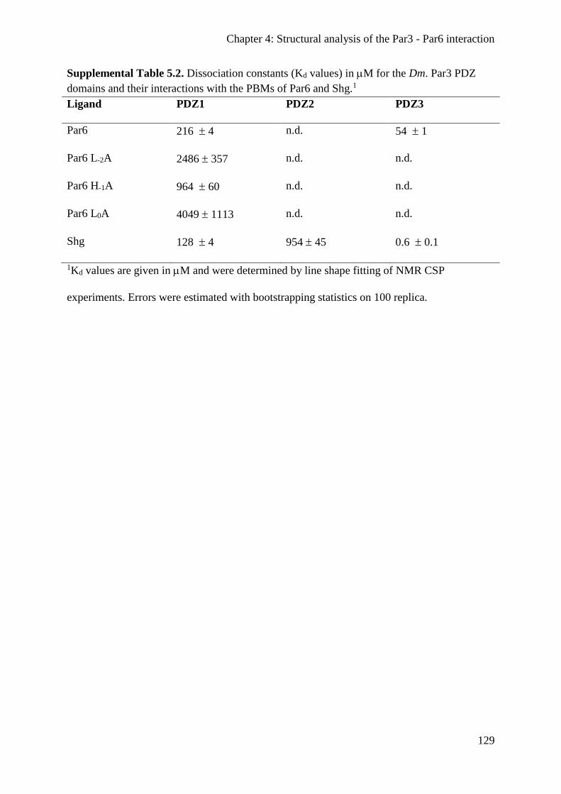
Chapter 4: Structural analysis of the Par3 - Par6 interaction
129
Supplemental Table 5.2. Dissociation constants (Kd values) in M for the Dm. Par3 PDZ
domains and their interactions with the PBMs of Par6 and Shg.1
Ligand PDZ1 PDZ2 PDZ3
Par6 216 4 n.d. 54 1
Par6 L-2A 2486 357 n.d. n.d.
Par6 H-1A 964 60 n.d. n.d.
Par6 L0A 4049 1113 n.d. n.d.
Shg 128 4 954 45 0.6 0.1
1Kd values are given in M and were determined by line shape fitting of NMR CSP
experiments. Errors were estimated with bootstrapping statistics on 100 replica.

Discussion
130
Discussion
Establishing a cellular polarity is a fundamental process and is a prerequisite for development
and the control of cellular processes. This study aimed to provide a better understanding of
the function and the regulation of key proteins that are essential for epithelial development
and the regulation of cellular homeostasis.
The scaffold protein Baz/Par3 and the serine/threonine kinase LKB1 cooperate to establish
the initial polarization of epithelial cells. In addition, LKB1 controls the metabolism and
proliferation of differentiated cells. Baz and LKB1 were the subjects of this study and further
characterized.
Within this study a so far uncharacterized lipid binding (LB) domain of LKB1 has been
identified. In vitro experiments managed to show that LKB1 prefers binding to PA, but also to
some extend to PtdIns(5)P, PtdIns(4,5)P2 and PtdIns(3,4,5)P3. Notably, binding to PA
activated LKB1, which was essential for Drosophila development, mouse axon formation and
to counteract mTOR activity in human cancer cell lines. Understanding the mechanism by
which potent tumour suppressors, such as LKB1 are activated is important in biomedical
science to develop and improve new strategies to treat human diseases. Here, using
Drosophila as a model organism an unknown mechanism to activate LKB1 has been
described which is also conserved in humans. Thus, although it can be challenging to study
human diseases like cancer in Drosophila the principle mechanisms of cell proliferation
control are conserved.
Moreover, it could have been shown that oligomerization and lipid binding of Baz function
redundantly to promote the apical localization of Baz in the embryonic epithelium and NBs.
Strikingly, neither oligomerization nor lipid binding were essential for viability, whereas loss
of both led to embryonic lethality. Moreover, a novel function of the oligomerization domain
has been identified, as an oligomerization-deficient protein is destabilized. Additionally, the
organization of the Par complex has been further analyzed. Although the general functions of
the Par complex (Baz/Par6/aPKC) in the establishment of a cell polarity became more
obvious in recent years, the formation of the complex itself remained controversial. However,
using a combined approach of structural biology and in vivo analysis a detailed study about
the Par3-Par6 interaction revealed novel insights in the complex assembly. A C-terminal PDZ
binding motif (PBM) of Par6 binds to the PDZ1 and PDZ3 domains of Par3, whereas the
previously reported PDZ-PDZ domain interaction of Par3 and Par6 was dispensable.
Considering the Par complex as a ternary complex is misleading, because of Par3’s capacity
Discussion
131
to bind two Par6 proteins simultaneously in vitro and form homo-oligomers. Thus, it is more
likely that the Par complex forms large clusters, which surround the apical domain. In
addition, a C-terminal PBM of DE-Cad is capable of binding the PDZ1 and PDZ3 domains of
Par3 independent of Armadillo. Therefore, an alternative mechanism to target Par3 to the
plasmamembrane has been described.
The membrane association of LKB1
This study identified a novel lipid binding (LB) domain of LKB1 and highlighted that the
membrane association of LKB1 provides an important cue to activate its catalytic activity.
Membrane-binding of LKB1 could be shown to be important for the development of
Drosophila and its function as a tumour suppressor in mammalian cells. LKB1 has previously
been reported to localize at the plasmamembrane of Drosophila and mammalian cells (Collins
et al., 2000; Sapkota et al., 2001; Martin and St Johnston, 2003; Sebbagh et al., 2009). It has
been assumed that this cortical localization is mediated by the addition of a farnesyl moiety,
which serves as a membrane anchor (Collins et al., 2000; Martin and St Johnston, 2003).
However, in vivo the farnesylation of LKB1 seems to be dispensable. Farnesylation-deficient
melanoma cells were still capable to suppress cell growth (Sapkota et al., 2001) and a
transgenic farnesylation-deficient knock-in mouse had only a decreased capacity to activate
AMPK, but had otherwise no obvious defects (Houde et al., 2014). Likewise, the
farnesylation-deficient flies in this study had a slightly decreased capacity to rescue an lkb1
null allele, but were viable. By contrast, loss of the identified LB domain strongly reduced
LKB1’s ability to rescue the mutant. Moreover, the double mutant (LKB1∆LB C564A)
completely failed to give rise to viable offspring. Thus, although the farnesylation of LKB1
seems to have an effect on its activity, the major mechanism that recruits LKB1 to the
plasmamembrane and fully activates it was its LB domain rather than farnesylation.
LKB1 preferentially bound to PA, but binding to other lipids (PtdIns(5)P, PtdIns(4,5)P2 and
PtdIns(3,4,5)P3) has also been observed. In response to PA binding the kinase activity of
LKB1 was strongly enhanced in vitro. PA is mainly produced by PLD and activates also other
kinases, such as mTOR (Fang et al., 2001). Since LKB1 and its major target to counteract cell
proliferation are activated by the same mechanism, a novel autoregulation of the activation
and repression of mTOR signaling has been identified. The association of elevated PLD2
expression and mTOR activity (as measured by the phosphorylation of S6K) with
Discussion
132
simultaneous loss of LKB1 in histological samples of human melanoma supports the
hypothesis of an auto-regulatory effect.
Binding of LKB1 to PtdIns(5)P was not further investigated in this study, because PtdIns(5)P
is a low-abundant phospholipid and enriched in endomembranes (Sarkes and Rameh, 2010;
Ramel et al., 2011), whereas this study focused on epithelial cells in which LKB1 localizes at
the plasmamembrane. However, with respect to data from non-epithelial cells (mouse
hepatocytes and embryonic fibroblasts) LKB1 localizes on late endosomes in a complex
containing AMPK, v-ATPase, Ragulator and AXIN, which is essential to activate LKB1
(Zhang et al., 2013b; Zhang et al., 2014). Although PtdIns(5)P is more prominent in the
membrane of the early endosome, it is also incorporated into the late endosome (Ramel et al.,
2011). The mechanism by which LKB1 is activated on late endosomes is not understood, yet.
Thus, PtdIns(5)P might contribute to the activation of LKB1 on endosomes. In a human
fibroblast cell line (IMR90) no cortical localization could be observed, which raises the
question if LKB1 simply localizes in the cytoplasm or in association with endosomes. By
using a fluorescent reporter that is fused to a PtdIns(5)P binding domain a possible
coincidence of the lipid and LKB1 on endosomes could be analyzed.
The elevated degradation of membrane-binding deficient LKB1 (LKB1∆LB C564A) in
Drosophila supports the idea of an endomembrane localization of LKB1. LKB1∆LB C564A
was absent from the plasmamembrane and accumulated in the cytoplasm, such as hLKB1 in a
fibroblast cell line (IMR90), but without the capacity to associate with lipids. In Drosophila,
LKB1∆LB C564A was in contrast to LKB1 affected by enhanced degradation. Therefore,
binding to either the plasmamembrane or to endomembranes might be a mechanism to protect
LKB1 from degradation. Accordingly, in flies that express membrane-binding deficient LKB1
fused to the PH domains of either PLCδ or Akt1 (to mediate membrane binding), the
transgenes were not affected by enhanced protein degradation. Nevertheless, a cell type
specific membrane association of LKB1 could be another explanation. Moreover, the C-
terminus of LKB1, which harbors the LB domain is affected by several posttranslational
modification, including phosphorylation and acetylation (Sapkota et al., 2001; Lan et al.,
2008). Strikingly, in non-polarized HEK293 cells, LKB1 has been reported to be acetylated
on lysine 416 (Lan et al., 2008), which is essential for binding lipids. This acetylation might
interfere with the membrane association of LKB1, but a role of this modification in vivo has
not been analyzed, yet.
Regarding the degradation of LKB1 two E3 ubiquitin ligases (CHIP and HERC2) have
already been described to promote the proteasomal degradation of LKB1 (Gaude et al., 2012;

Discussion
133
Bai et al., 2016). Given that a decreased amount of LKB1 is frequently found in various
tumours, identifying other E3 ubiquitin ligases that target LKB1 for degradation would help
to develop new strategies to treat cancer or to discover new biomarker. Some molecules that
inhibit specific E3 ubiquitin ligases are currently undergoing clinical trials (Bielskienė et al.,
2015).
The PA mediated activation of LKB1
Given that LKB1 was activated upon binding to PA it remains to be investigated how this
activation occurs on the molecular level. The structure of the LKB1/STRADα/Mo25 complex
has previously been resolved (Zeqiraj et al., 2009). Based on the reported crystal structure of
the complex, binding of STRADα to LKB1 causes an allosteric activation of LKB1, which is
stabilized by Mo25. However, in this study only truncated variants of STRADα (residues 59 –
431) and LKB1 (residues 43 – 347) have been analyzed (Zeqiraj et al., 2009). Therefore, a
putative role in the activation of LKB1 of the either the N- or C-terminus remains enigmatic.
Loss of the membrane binding capacity of LKB1 did not affect its association with STRADα
(Stlk in Drosophila), Mo25 and AMPK in the Drosophila embryo. Nevertheless, this complex
had a significantly decreased catalytic activity and failed to rescue an lkb1 null allele. The
chimeric proteins that were fused to the PH domains of either PLCδ or Akt1 were able to
restore the membrane localization of LKB1 and to rescue the null allele to a large extend. In
vitro, the addition of PA to the recombinant human LKB1/STRADα/Mo25 complex strongly
enhances its kinase activity, whereas adding either PtdIns(4,5)P2 or PtdIns(3,4,5)P3 led only
to a moderate activation. This indicates the specific association of LKB1 with PA is required
to fully activate its catalytic activity.
In order to better understand the PA-dependent activation of LKB1 at the molecular level the
structure of LKB1 in complex with liposomes containing PA could be determined by NMR.
The conformational change of LKB1 upon binding to PA should induce a chemical shift
perturbation (CSP), if the conformation of LKB1 changes upon binding PA. Therefore PA-
containing liposomes could be titrated to LKB1, which also allows determining the
dissociation constant (KD-value) that will provide information about the strength of the
interaction. Knowing the structure of the PA-activated LKB1 will help to gain insight to the
(mis)regulation of this important tumour suppressor. To this end, sequence optimized variants
of the human LKB1 and the membrane-binding deficient form have been generated, because

Discussion
134
of an inefficient translation of the human codon sequences in E. coli (data not shown).
Moreover, an attempt to gain insight into the PA-dependent regulation of LKB1 using atomic
force microscopy (AFM, in cooperation with AG Gießibl, University of Regensburg) has
been made. AFM bears the potential to provide structural information of molecules at atomic
resolution. Nevertheless, although we were able to visualize lipid head groups of a lipid
bilayer, which was adsorbed on mica, we could not acquire structural information about
membrane-bound recombinant LKB1 (data not shown).
The functional redundancy of the OD and LB motif
The N-terminal oligomerization domain (OD) of Baz/Par3 is highly conserved among animals
and has been described to be essential in Drosophila and mammalian cells (MDCK) to target
Baz/Par3 to the plasmamembrane (Benton and St. Johnston, 2003b; Mizuno et al., 2003). In
addition, loss of the OD results in a severely compromised capacity to rescue a baz null allele
(Benton and St. Johnston, 2003b). However, this study found that the OD is neither essential
to tether Baz to the plasmamembrane nor for viability of Drosophila embryos. Baz∆OD
localizes entirely at the apical junctions, overlapping with DE-Cad. Likewise, mutating the
lipid binding (LB) domain did not affect the localization or rescue ability as well, which is in
line with previous data (Krahn et al., 2010b). Surprisingly, loss of both domains
(Baz∆OD∆LB) completely abolished the cortical localization, ability to rescue a null allele
and protein stability of Baz. This indicates a functional redundancy between the OD and LB
motif, which cannot be compensated by any other domain (e.g. the PDZ domains) of Baz.
Given that a chimeric protein, which carries the oligomerization domain of the human TEL
protein efficiently restores the function of Baz∆OD∆LB, oligomerization per se rather than a
specific feature of the OD is enough for the function of Baz in the absence of the lipid binding
motif.
Although Baz has been reported to localize to the plasmamembrane by binding to Ed, Arm
and Stan via its PDZ domains (Wei et al., 2005; Wasserscheid et al., 2007), none of these
proteins seems to be capable to initially recruit Baz to the plasmamembrane. Strikingly, the
interaction of Baz∆OD∆LB with any of these proteins was impaired, as all of them managed
to recruit Baz∆OD∆LB to artificial cell-cell contacts in S2R cells. This suggests a role for
oligomerization and lipid binding as important initial cues to tether Baz to the
plasmamembrane. The observation that the simultaneous loss of the PDZ (interacts with Ed,

Discussion
135
Arm and Stan) domains and the LB motif did not affect the apical localization of Baz further
highlights the role of the OD and LB motif for the localization of Baz, rather than the PDZ
domains. Nevertheless, loss of all PDZ domains causes embryonic lethality (Krahn et al.,
2010b), whereas mutating either the OD or LB motif can be compensated and produces viable
offspring. The PDZ domains of Baz/Par3 have been reported to bind directly to phospholipids
in vitro (Wu et al., 2007; Yu and Harris, 2012), however in cell culture experiments the PDZ
domains were not able to mediate a cortical localization of Baz∆LB. This suggests, that the
association of the PDZ domains with phospholipids is either not relevant in vivo (at least not
in S2R cells) or is restricted to a certain function of Baz. Although the PDZ domains seem to
be dispensable for the initial recruitment of Baz to the plasmamembrane they become
indispensable during further development. The initial recruitment of Baz to the
plasmamembrane neither requires the OD nor the LB motif, but rather a specific function,
which is mediated in redundancy by them.
Oligomerization promotes the stability of Baz
In addition to the role of the OD in localizing Baz, this study described a novel function of the
OD to maintain the stability of Baz. Mutations that affect the critical residues, which are
required to mediate the self-association of Baz, markedly reduce the amount of Baz protein.
The enhanced degradation of oligomerization-deficient Baz was rescued by fusing the
heterologous OD of the human TEL protein to the Baz N-terminus. Therefore, although
oligomerization is not essential for viability it contributes to the stability of Baz. However, it
remains to be investigated by which mechanism the amount of Baz∆OD protein decreases.
Oligomerization-deficient Baz could either be prone to aberrant targeting of proteases or E3-
ubiquitin ligases that promote its proteasomal degradation. Baz has previously been reported
to have multiple truncated variants (based on observations from western blots), which arise
either from proteolytic cleavage or proteasomal degradation (Krahn et al., 2009). Thus, the
self-assembly of Baz might shield the protein from degradation.
In future studies it could become an objective to identify potential Baz proteases or E3-
ubiquitin ligases that degrade Baz. The immunoprecipitation of wildtype and oligomerization-
deficient Baz followed by mass spectrometry could be a promising approach to analyze
potential differences in the interactome of both proteins. Another approach for the analysis of
dimer-specific interaction partner would be bimolecular complementation affinity purification
Discussion
136
(BiCAP) (Croucher et al., 2016), which is also followed by mass spectrometry. The loss of
Baz’s oligomerization could favor the interaction with its degrading enzymes, which might
facilitate their identification.
Novel insights into the structural organization of the Par complex
This study provided a novel understanding of the molecular organization of the Par complex.
The current model of the Par complex supposes that Par6 binds to the first PDZ domain of
Baz/Par3 via its own PDZ domain, which might also require its CRIB motif (Lin et al., 2000;
Joberty et al., 2000). However, the data shown here reject this model. PDZ domains are
frequently bound by proteins that harbor a PDZ binding motif (PBM). Here, a so far
undescribed PBM at the C-terminus of Par6 has been identified. Loss of the Par6 PBM
abolishes the interaction with the PDZ domains of Baz in vitro and a recombinant Par6 CRIB-
PDZ protein fails to bind the Baz PDZ domains. In addition, Par6∆PDZ is efficiently
recruited to the cell cortex by Baz in S2R cells. Finally, the NMR spectra of neither Par6 PDZ
nor Par3 PDZ changed upon incubation with each other, which indicates that the two domains
do not interact with each other. Furthermore, in contrast to previous studies we found that
Par6 binds in addition to the first PDZ domain of Baz also to the third PDZ domains, which
enables Baz to bind two Par6 proteins simultaneously. The interaction between the Par6 PBM
and the Baz PDZ3 domain were even four times stronger than between Par6 PBM and Baz
PDZ1. Taken together, the interaction between Par6 and Baz does not depend on a PDZ-PDZ
interaction, but rather on the PBM of Par6. Nevertheless, in the epidermis of Drosophila
embryos the PDZ domain and the PBM of Par6 function redundantly to recruit Par6 to the
apical junctions. Moreover, embryos that express Par6∆PBM overcome the embryonic
lethality of a par6 null allele, whereas Par6∆PDZ expressing embryos die. The importance of
the Par6 PDZ domain for embryonic viability is most likely due to its function of binding to
the ECR1 domain of Sdt to join the Crb complex or to directly bind the PBM of Crb upon the
apical exclusion of Baz (Lemmers et al., 2004; Wang et al., 2004; Morais-de-Sá et al., 2010;
Whitney et al., 2016; Koch et al., 2016). Given that Baz also forms a complex with Sdt prior
to the expression of Crb (Krahn et al., 2010a), Par6∆PBM could associate with the Baz-Sdt
complex by binding Sdt during gastrulation and afterwards join to the Crb complex. Binding
either directly to Baz (via PBM) or indirectly in complex with Sdt (via PDZ domain) could
Discussion
137
explain the functional redundancy and would be in line with the cytoplasmic localization of
the double mutant (Par6∆PDZ∆PBM).
In addition to the novel PBM-PDZ interaction of Par6 and Baz, this study further showed a
direct interaction of DE-Cad and Baz for the first time. In Drosophila, the interaction of Baz
and DE-Cad has been described to be indirect and requires Arm (Wei et al., 2005). By
contrast, mammalian VE-Cad binds directly with its C-terminal PBM to the first and third
PDZ domains of Par3, which is essential for the polarization of endothelial cells (Iden et al.,
2006). Similar to the mammalian system, DE-Cad contains a class II PBM that mediates the
interaction with the first and third PDZ domain of Baz as well. Notably, the interaction of the
DE-Cad PBM with the third PDZ domain of Baz is more than 50 times stronger compared
towards the PBM of Par6. However, both PBM’s compete with each other for binding to the
third PDZ domain of Baz in vitro. The cellular context of this competition has not been
determined, yet, but the stronger affinity of the DE-Cad PBM towards Baz might contribute to
the observation of an apical exclusion of Baz (Nam and Choi, 2003; Vogelmann and Nelson,
2004; Harris and Peifer, 2005; Martin-Belmonte et al., 2007; Morais-de-Sá et al., 2010;
Doerflinger et al., 2010). To date two mechanisms have been described, which promote the
segregation of Baz from the SAR towards the AJ. First, the phosphorylation of Baz by aPKC
induces the dissociation of the Par complex and the translocation of Baz to the AJ (Morais-de-
Sá et al., 2010). Second, the phosphorylation of Arm by Pak4 is essential for the retention of
Baz at the AJ in Drosophila photoreceptor cells (Walther et al., 2016). The direct association
of DE-Cad with Baz might contribute to the accumulation of Baz at the AJ by sequestering it.
The role of Baz’s accumulation at the AJ in vivo still remains to be investigated, but it might
be necessary for Baz as an exocyst receptor to regulate the delivery of cell adhesion proteins
to the AJ.
References
138
References Adams, P. D., P. V. Afonine, G. Bunkóczi, V. B. Chen, I. W. Davis, N. Echols, J. J. Headd,
and L.-W. Hung, et al. 2010. PHENIX: A comprehensive Python-based system for
macromolecular structure solution. Acta Crystallogr D Biol Crystallogr 66(Pt 2):213–221.
Ahmed, S. M., and I. G. Macara. 2017. The Par3 polarity protein is an exocyst receptor
essential for mammary cell survival. Nat Commun 8:14867.
Alessi, D. R., K. Sakamoto, and J. R. Bayascas. 2006. LKB1-dependent signaling pathways.
Annu Rev Biochem 75:137–163.
Alexander, A., S.-L. Cai, J. Kim, A. Nanez, M. Sahin, K. H. MacLean, K. Inoki, and K.-L.
Guan, et al. 2010. ATM signals to TSC2 in the cytoplasm to regulate mTORC1 in
response to ROS. Proc Natl Acad Sci U S A 107(9):4153–4158.
Alva, V., S.-Z. Nam, J. Söding, and A. N. Lupas. 2016. The MPI bioinformatics Toolkit as an
integrative platform for advanced protein sequence and structure analysis. Nucleic Acids
Res 44(W1):W410-5.
Amin, N., A. Khan, D. St Johnston, I. Tomlinson, S. Martin, J. Brenman, and H. McNeill.
2009. LKB1 regulates polarity remodeling and adherens junction formation in the
Drosophila eye. Proc Natl Acad Sci U S A 106(22):8941–8946.
Anderson, J. M., and C. M. van Itallie. 2009. Physiology and function of the tight junction.
Cold Spring Harb Perspect Biol 1(2):a002584.
Appleton, B. A., Y. Zhang, P. Wu, J. P. Yin, W. Hunziker, N. J. Skelton, S. S. Sidhu, and C.
Wiesmann. 2006. Comparative structural analysis of the Erbin PDZ domain and the first
PDZ domain of ZO-1. Insights into determinants of PDZ domain specificity. J Biol Chem
281(31):22312–22320.
Arsham, A. M., J. J. Howell, and M. C. Simon. 2003. A novel hypoxia-inducible factor-
independent hypoxic response regulating mammalian target of rapamycin and its targets. J
Biol Chem 278(32):29655–29660.
Atwood, S. X., C. Chabu, R. R. Penkert, C. Q. Doe, and K. E. Prehoda. 2007. Cdc42 acts
downstream of Bazooka to regulate neuroblast polarity through Par-6 aPKC. J Cell Sci
120(Pt 18):3200–3206.
Atwood, S. X., and K. E. Prehoda. 2009. aPKC phosphorylates Miranda to polarize fate
determinants during neuroblast asymmetric cell division. Curr Biol 19(9):723–729.
Avtanski, D. B., A. Nagalingam, M. Y. Bonner, J. L. Arbiser, N. K. Saxena, and D. Sharma.
2015. Honokiol activates LKB1-miR-34a axis and antagonizes the oncogenic actions of
leptin in breast cancer. Oncotarget 6(30):29947–29962.
Baas, A. F., J. Boudeau, G. P. Sapkota, L. Smit, R. Medema, N. A. Morrice, D. R. Alessi, and
H. C. Clevers. 2003. Activation of the tumour suppressor kinase LKB1 by the STE20-like
pseudokinase STRAD. EMBO J 22(12):3062–3072.
Bai, B., A. W. C. Man, K. Yang, Y. Guo, C. Xu, H.-F. Tse, W. Han, and M. Bloksgaard, et al.
2016. Endothelial SIRT1 prevents adverse arterial remodeling by facilitating HERC2-
mediated degradation of acetylated LKB1. Oncotarget 7(26):39065–39081.
Bai, Y., T. Zhou, H. Fu, H. Sun, and B. Huang. 2012. 14-3-3 interacts with LKB1 via
recognizing phosphorylated threonine 336 residue and suppresses LKB1 kinase function.
FEBS Lett 586(8):1111–1119.
References
139
Barbier-Torres, L., T. C. Delgado, J. L. García-Rodríguez, I. Zubiete-Franco, D. Fernández-
Ramos, X. Buqué, A. Cano, and V. Gutiérrez-de Juan, et al. 2015. Stabilization of LKB1
and Akt by neddylation regulates energy metabolism in liver cancer. Oncotarget
6(4):2509–2523.
Bardeesy, N., M. Sinha, A. F. Hezel, S. Signoretti, N. A. Hathaway, N. E. Sharpless, M.
Loda, and D. R. Carrasco, et al. 2002. Loss of the Lkb1 tumour suppressor provokes
intestinal polyposis but resistance to transformation. Nature 419(6903):162–167.
Barnes, A. P., B. N. Lilley, Y. A. Pan, L. J. Plummer, A. W. Powell, A. N. Raines, J. R.
Sanes, and F. Polleux. 2007. LKB1 and SAD kinases define a pathway required for the
polarization of cortical neurons. Cell 129(3):549–563.
Bartels, C., T. H. Xia, M. Billeter, P. Güntert, and K. Wüthrich. 1995. The program XEASY
for computer-supported NMR spectral analysis of biological macromolecules. J Biomol
NMR 6(1):1–10.
Bartkova, J., J. Lukas, H. Müller, D. Lützhøft, M. Strauss, and J. Bartek. 1994. Cyclin D1
protein expression and function in human breast cancer. Int J Cancer 57(3):353–361.
Baumgartner, S., J. T. Littleton, K. Broadie, M. A. Bhat, R. Harbecke, J. A. Lengyel, R.
Chiquet-Ehrismann, and A. Prokop, et al. 1996. A Drosophila neurexin is required for
septate junction and blood-nerve barrier formation and function. Cell 87(6):1059–1068.
Benton, R., and D. St Johnston. 2003a. Drosophila PAR-1 and 14-3-3 inhibit Bazooka/PAR-3
to establish complementary cortical domains in polarized cells. Cell 115(6):691–704.
Benton, R., and D. St. Johnston. 2003b. A Conserved Oligomerization Domain in Drosophila
Bazooka/PAR-3 Is Important for Apical Localization and Epithelial Polarity. Current
Biology 13(15):1330–1334.
Betschinger, J., F. Eisenhaber, and J. A. Knoblich. 2005. Phosphorylation-induced
autoinhibition regulates the cytoskeletal protein Lethal (2) giant larvae. Curr Biol
15(3):276–282.
Betschinger, J., K. Mechtler, and J. A. Knoblich. 2003. The Par complex directs asymmetric
cell division by phosphorylating the cytoskeletal protein Lgl. Nature 422(6929):326–330.
Bielskienė, K., L. Bagdonienė, J. Mozūraitienė, B. Kazbarienė, and E. Janulionis. 2015. E3
ubiquitin ligases as drug targets and prognostic biomarkers in melanoma. Medicina
(Kaunas) 51(1):1–9.
Bilder, D., M. Li, and N. Perrimon. 2000. Cooperative regulation of cell polarity and growth
by Drosophila tumor suppressors. Science 289(5476):113–116.
Bilder, D., M. Schober, and N. Perrimon. 2003. Integrated activity of PDZ protein complexes
regulates epithelial polarity. Nat Cell Biol 5(1):53–58.
Bolte, S., and F. P. Cordelières. 2006. A guided tour into subcellular colocalization analysis in
light microscopy. J Microsc 224(Pt 3):213–232.
Bonaccorsi, S., V. Mottier, M. G. Giansanti, B. J. Bolkan, B. Williams, M. L. Goldberg, and
M. Gatti. 2007. The Drosophila Lkb1 kinase is required for spindle formation and
asymmetric neuroblast division. Development 134(11):2183–2193.
Boudeau, J., A. F. Baas, M. Deak, N. A. Morrice, A. Kieloch, M. Schutkowski, A. R.
Prescott, and H. C. Clevers, et al. 2003a. MO25alpha/beta interact with STRADalpha/beta
enhancing their ability to bind, activate and localize LKB1 in the cytoplasm. EMBO J
22(19):5102–5114.
References
140
Boudeau, J., M. Deak, M. A. Lawlor, N. A. Morrice, and D. R. Alessi. 2003b. Heat-shock
protein 90 and Cdc37 interact with LKB1 and regulate its stability. Biochem. J. 370(Pt
3):849–857.
Boudeau, J., G. Sapkota, and D. R. Alessi. 2003c. LKB1, a protein kinase regulating cell
proliferation and polarity. FEBS Lett 546(1):159–165.
Boudeau, J., G. Sapkota, and D. R. Alessi. 2003d. LKB1, a protein kinase regulating cell
proliferation and polarity. FEBS Lett 546(1):159–165.
Boudeau, J., J. W. Scott, N. Resta, M. Deak, A. Kieloch, D. Komander, D. G. Hardie, and A.
R. Prescott, et al. 2004. Analysis of the LKB1-STRAD-MO25 complex. J Cell Sci 117(Pt
26):6365–6375.
Bright, N. J., D. Carling, and C. Thornton. 2008. Investigating the regulation of brain-specific
kinases 1 and 2 by phosphorylation. J Biol Chem 283(22):14946–14954.
Bruntz, R. C., H. E. Taylor, C. W. Lindsley, and H. A. Brown. 2014. Phospholipase D2
mediates survival signaling through direct regulation of Akt in glioblastoma cells. J Biol
Chem 289(2):600–616.
Bultot, L., S. Horman, D. Neumann, M. P. Walsh, L. Hue, and M. H. Rider. 2009. Myosin
light chains are not a physiological substrate of AMPK in the control of cell structure
changes. FEBS Lett 583(1):25–28.
Calamaras, T. D., C. Lee, F. Lan, Y. Ido, D. A. Siwik, and W. S. Colucci. 2012. Post-
translational modification of serine/threonine kinase LKB1 via Adduction of the Reactive
Lipid Species 4-Hydroxy-trans-2-nonenal (HNE) at lysine residue 97 directly inhibits
kinase activity. J Biol Chem 287(50):42400–42406.
Calamaras, T. D., C. Lee, F. Lan, Y. Ido, D. A. Siwik, and W. S. Colucci. 2015. The lipid
peroxidation product 4-hydroxy-trans-2-nonenal causes protein synthesis in cardiac
myocytes via activated mTORC1-p70S6K-RPS6 signaling. Free Radic Biol Med 82:137–
146.
Calles, A., L. M. Sholl, S. J. Rodig, A. K. Pelton, J. L. Hornick, M. Butaney, C. Lydon, and S.
E. Dahlberg, et al. 2015. Immunohistochemical Loss of LKB1 Is a Biomarker for More
Aggressive Biology in KRAS-Mutant Lung Adenocarcinoma. Clin Cancer Res
21(12):2851–2860.
Carling, D., V. A. Zammit, and D. G. Hardie. 1987. A common bicyclic protein kinase
cascade inactivates the regulatory enzymes of fatty acid and cholesterol biosynthesis.
FEBS Lett 223(2):217–222.
Carrera, A. C. 2004. TOR signaling in mammals. J Cell Sci 117(Pt 20):4615–4616.
Carretero, J., P. P. Medina, R. Pio, L. M. Montuenga, and M. Sanchez-Cespedes. 2004. Novel
and natural knockout lung cancer cell lines for the LKB1/STK11 tumor suppressor gene.
Oncogene 23(22):4037–4040.
Casimiro, M. C., G. Di Sante, A. Di Rocco, E. Loro, C. Pupo, T. G. Pestell, S. Bisetto, and M.
A. Velasco-Velázquez, et al. 2017. Cyclin D1 Restrains Oncogene-Induced Autophagy by
Regulating the AMPK-LKB1 Signaling Axis. Cancer Res 77(13):3391–3405.
Chen, X., and I. G. Macara. 2005. Par-3 controls tight junction assembly through the Rac
exchange factor Tiam1. Nat Cell Biol 7(3):262–269.
Chou, T. B., and N. Perrimon. 1996. The autosomal FLP-DFS technique for generating
germline mosaics in Drosophila melanogaster. Genetics 144(4):1673–1679.

References
141
Cidlinsky, N., G. Dogliotti, T. Pukrop, R. Jung, F. Weber, and M. P. Krahn. 2016.
Inactivation of the LKB1-AMPK signaling pathway does not contribute to salivary gland
tumor development - a short report. Cell Oncol (Dordr) 39(4):389–396.
Collins, S. P., J. L. Reoma, D. M. Gamm, and M. D. Uhler. 2000. LKB1, a novel
serine/threonine protein kinase and potential tumour suppressor, is phosphorylated by
cAMP-dependent protein kinase (PKA) and prenylated in vivo. Biochem J 345 Pt 3:673–
680.
Corradetti, M. N., K. Inoki, N. Bardeesy, R. A. DePinho, and K.-L. Guan. 2004. Regulation
of the TSC pathway by LKB1: Evidence of a molecular link between tuberous sclerosis
complex and Peutz-Jeghers syndrome. Genes Dev 18(13):1533–1538.
Costes, S. V., D. Daelemans, E. H. Cho, Z. Dobbin, G. Pavlakis, and S. Lockett. 2004.
Automatic and quantitative measurement of protein-protein colocalization in live cells.
Biophys J 86(6):3993–4003.
Courchet, J., T. L. Lewis, S. Lee, V. Courchet, D.-Y. Liou, S. Aizawa, and F. Polleux. 2013.
Terminal axon branching is regulated by the LKB1-NUAK1 kinase pathway via
presynaptic mitochondrial capture. Cell 153(7):1510–1525.
Crooks, G. E., G. Hon, J.-M. Chandonia, and S. E. Brenner. 2004. WebLogo: A sequence
logo generator. Genome Res 14(6):1188–1190.
Croucher, D. R., M. Iconomou, J. F. Hastings, S. P. Kennedy, J. Z. R. Han, R. F. Shearer, J.
McKenna, and A. Wan, et al. 2016. Bimolecular complementation affinity purification
(BiCAP) reveals dimer-specific protein interactions for ERBB2 dimers. Sci Signal
9(436):ra69.
Dahmani, R., P.-A. Just, A. Delay, F. Canal, L. Finzi, C. Prip-Buus, M. Lambert, and P.
Sujobert, et al. 2015. A novel LKB1 isoform enhances AMPK metabolic activity and
displays oncogenic properties. Oncogene 34(18):2337–2346.
Damsky, W., G. Micevic, K. Meeth, V. Muthusamy, D. P. Curley, M. Santhanakrishnan, I.
Erdelyi, and J. T. Platt, et al. 2015. mTORC1 activation blocks BrafV600E-induced
growth arrest but is insufficient for melanoma formation. Cancer Cell 27(1):41–56.
Davies, H., G. R. Bignell, C. Cox, P. Stephens, S. Edkins, S. Clegg, J. Teague, and H.
Woffendin, et al. 2002. Mutations of the BRAF gene in human cancer. Nature
417(6892):949–954.
Delaglio, F., S. Grzesiek, G. W. Vuister, G. Zhu, J. Pfeifer, and A. Bax. 1995. NMRPipe: A
multidimensional spectral processing system based on UNIX pipes. J Biomol NMR
6(3):277–293.
Denison, F. C., N. J. Hiscock, D. Carling, and A. Woods. 2009. Characterization of an
alternative splice variant of LKB1. J Biol Chem 284(1):67–76.
Ding, L., G. Getz, D. A. Wheeler, E. R. Mardis, M. D. McLellan, K. Cibulskis, C. Sougnez,
and H. Greulich, et al. 2008. Somatic mutations affect key pathways in lung
adenocarcinoma. Nature 455(7216):1069–1075.
Doerflinger, H., N. Vogt, I. L. Torres, V. Mirouse, I. Koch, C. Nüsslein-Volhard, and D. St
Johnston. 2010. Bazooka is required for polarisation of the Drosophila anterior-posterior
axis. Development 137(10):1765–1773.

References
142
Dogliotti, G., L. Kullmann, P. Dhumale, C. Thiele, O. Panichkina, G. Mendl, R. Houben, and
S. Haferkamp, et al. 2017. Membrane-binding and activation of LKB1 by phosphatidic
acid is essential for development and tumour suppression. Nat Commun 8:15747.
Dolinsky, V. W., A. Y. M. Chan, I. Robillard Frayne, P. E. Light, C. Des Rosiers, and J. R. B.
Dyck. 2009. Resveratrol prevents the prohypertrophic effects of oxidative stress on LKB1.
Circulation 119(12):1643–1652.
Dorfman, J., and I. G. Macara. 2008. STRADalpha regulates LKB1 localization by blocking
access to importin-alpha, and by association with Crm1 and exportin-7. Molecular Biology
of the Cell 19(4):1614–1626.
Ebnet, K., M. Aurrand-Lions, A. Kuhn, F. Kiefer, S. Butz, K. Zander, M.-K. Meyer zu
Brickwedde, and A. Suzuki, et al. 2003. The junctional adhesion molecule (JAM) family
members JAM-2 and JAM-3 associate with the cell polarity protein PAR-3: A possible
role for JAMs in endothelial cell polarity. J Cell Sci 116(Pt 19):3879–3891.
Ebnet, K., A. Suzuki, Y. Horikoshi, T. Hirose, M. K. Meyer Zu Brickwedde, S. Ohno, and D.
Vestweber. 2001. The cell polarity protein ASIP/PAR-3 directly associates with junctional
adhesion molecule (JAM). EMBO J 20(14):3738–3748.
Edgar, R. C. 2004. MUSCLE: Multiple sequence alignment with high accuracy and high
throughput. Nucleic Acids Res 32(5):1792–1797.
Emsley, P., B. Lohkamp, W. G. Scott, and K. Cowtan. 2010. Features and development of
Coot. Acta Crystallogr D Biol Crystallogr 66(Pt 4):486–501.
Eneling, K., L. Brion, V. Pinto, M. J. Pinho, J. I. Sznajder, N. Mochizuki, K. Emoto, and P.
Soares-da-Silva, et al. 2012. Salt-inducible kinase 1 regulates E-cadherin expression and
intercellular junction stability. FASEB J 26(8):3230–3239.
Ernst, A., B. A. Appleton, Y. Ivarsson, Y. Zhang, D. Gfeller, C. Wiesmann, and S. S. Sidhu.
2014. A structural portrait of the PDZ domain family. J Mol Biol 426(21):3509–3519.
Esteve-Puig, R., F. Canals, N. Colomé, G. Merlino, and J. A. Recio. 2009. Uncoupling of the
LKB1-AMPKalpha energy sensor pathway by growth factors and oncogenic BRAF. PLoS
ONE 4(3):e4771.
Fang, Y., M. Vilella-Bach, R. Bachmann, A. Flanigan, and J. Chen. 2001. Phosphatidic acid-
mediated mitogenic activation of mTOR signaling. Science 294(5548):1942–1945.
Fehon, R. G., I. A. Dawson, and S. Artavanis-Tsakonas. 1994. A Drosophila homologue of
membrane-skeleton protein 4.1 is associated with septate junctions and is encoded by the
coracle gene. Development 120(3):545–557.
Feng, W., H. Wu, L.-N. Chan, and M. Zhang. 2007. The Par-3 NTD adopts a PB1-like
structure required for Par-3 oligomerization and membrane localization. EMBO J
26(11):2786–2796.
Feng, W., H. Wu, L.-N. Chan, and M. Zhang. 2008. Par-3-mediated junctional localization of
the lipid phosphatase PTEN is required for cell polarity establishment. J Biol Chem
283(34):23440–23449.
Ferlay, J., I. Soerjomataram, R. Dikshit, S. Eser, C. Mathers, M. Rebelo, D. M. Parkin, and D.
Forman, et al. 2015. Cancer incidence and mortality worldwide: Sources, methods and
major patterns in GLOBOCAN 2012. Int J Cancer 136(5):E359-86.

References
143
Fievet, B. T., J. Rodriguez, S. Naganathan, C. Lee, E. Zeiser, T. Ishidate, M. Shirayama, and
S. Grill, et al. 2013. Systematic genetic interaction screens uncover cell polarity regulators
and functional redundancy. Nat Cell Biol 15(1):103–112.
Fletcher, G. C., E. P. Lucas, R. Brain, A. Tournier, and B. J. Thompson. 2012. Positive
feedback and mutual antagonism combine to polarize Crumbs in the Drosophila follicle
cell epithelium. Curr Biol 22(12):1116–1122.
Fogarty, S., and D. G. Hardie. 2009. C-terminal phosphorylation of LKB1 is not required for
regulation of AMP-activated protein kinase, BRSK1, BRSK2, or cell cycle arrest. J Biol
Chem 284(1):77–84.
Foster, D. A., D. Salloum, D. Menon, and M. A. Frias. 2014. Phospholipase D and the
maintenance of phosphatidic acid levels for regulation of mammalian target of rapamycin
(mTOR). J Biol Chem 289(33):22583–22588.
Gan, B., J. Hu, S. Jiang, Y. Liu, E. Sahin, L. Zhuang, E. Fletcher-Sananikone, and S. Colla, et
al. 2010. Lkb1 regulates quiescence and metabolic homeostasis of haematopoietic stem
cells. Nature 468(7324):701–704.
Gao, L., I. G. Macara, and G. Joberty. 2002. Multiple splice variants of Par3 and of a novel
related gene, Par3L, produce proteins with different binding properties. Gene 294(1-
2):99–107.
Gaude, H., N. Aznar, A. Delay, A. Bres, K. Buchet-Poyau, C. Caillat, A. Vigouroux, and C.
Rogon, et al. 2012. Molecular chaperone complexes with antagonizing activities regulate
stability and activity of the tumor suppressor LKB1. Oncogene 31(12):1582–1591.
Geiss-Friedlander, R., and F. Melchior. 2007. Concepts in sumoylation: A decade on. Nat Rev
Mol Cell Biol 8(12):947–956.
Genova, J. L., and R. G. Fehon. 2003. Neuroglian, Gliotactin, and the Na+/K+ ATPase are
essential for septate junction function in Drosophila. J Cell Biol 161(5):979–989.
George, S. H. L., A. Milea, R. Sowamber, R. Chehade, A. Tone, and P. A. Shaw. 2016. Loss
of LKB1 and p53 synergizes to alter fallopian tube epithelial phenotype and high-grade
serous tumorigenesis. Oncogene 35(1):59–68.
Getsios, S., A. C. Huen, and K. J. Green. 2004. Working out the strength and flexibility of
desmosomes. Nat Rev Mol Cell Biol 5(4):271–281.
Ghosh, S., J. C. Strum, V. A. Sciorra, L. Daniel, and R. M. Bell. 1996. Raf-1 kinase possesses
distinct binding domains for phosphatidylserine and phosphatidic acid. Phosphatidic acid
regulates the translocation of Raf-1 in 12-O-tetradecanoylphorbol-13-acetate-stimulated
Madin-Darby canine kidney cells. J Biol Chem 271(14):8472–8480.
Gill, R. K., S.-H. Yang, D. Meerzaman, L. E. Mechanic, E. D. Bowman, H.-S. Jeon, S. Roy
Chowdhuri, and A. Shakoori, et al. 2011. Frequent homozygous deletion of the
LKB1/STK11 gene in non-small cell lung cancer. Oncogene 30(35):3784–3791.
Goehring, N. W., C. Hoege, S. W. Grill, and A. A. Hyman. 2011. PAR proteins diffuse freely
across the anterior-posterior boundary in polarized C. elegans embryos. J Cell Biol
193(3):583–594.
Goldstein, B., and I. G. Macara. 2007. The PAR proteins: Fundamental players in animal cell
polarization. Dev Cell 13(5):609–622.
Gomez-Cambronero, J. 2014. Phospholipase D in cell signaling: From a myriad of cell
functions to cancer growth and metastasis. J Biol Chem 289(33):22557–22566.

References
144
Goodwin, J. M., R. U. Svensson, H. J. Lou, M. M. Winslow, B. E. Turk, and R. J. Shaw.
2014. An AMPK-independent signaling pathway downstream of the LKB1 tumor
suppressor controls Snail1 and metastatic potential. Mol Cell 55(3):436–450.
Granot, Z., A. Swisa, J. Magenheim, M. Stolovich-Rain, W. Fujimoto, E. Manduchi, T. Miki,
and J. K. Lennerz, et al. 2009. LKB1 regulates pancreatic beta cell size, polarity, and
function. Cell Metab 10(4):296–308.
Graybill, C., B. Wee, S. X. Atwood, and K. E. Prehoda. 2012. Partitioning-defective protein 6
(Par-6) activates atypical protein kinase C (aPKC) by pseudosubstrate displacement. J
Biol Chem 287(25):21003–21011.
Groth, A. C., M. Fish, R. Nusse, and M. P. Calos. 2004. Construction of transgenic
Drosophila by using the site-specific integrase from phage phiC31. Genetics 166(4):1775–
1782.
Guldberg, P., P. thor Straten, V. Ahrenkiel, T. Seremet, A. F. Kirkin, and J. Zeuthen. 1999.
Somatic mutation of the Peutz-Jeghers syndrome gene, LKB1/STK11, in malignant
melanoma. Oncogene 18(9):1777–1780.
Gurumurthy, S., S. Z. Xie, B. Alagesan, J. Kim, R. Z. Yusuf, B. Saez, A. Tzatsos, and F.
Ozsolak, et al. 2010. The Lkb1 metabolic sensor maintains haematopoietic stem cell
survival. Nature 468(7324):659–663.
Guyer, R. A., and I. G. Macara. 2015. Loss of the polarity protein PAR3 activates STAT3
signaling via an atypical protein kinase C (aPKC)/NF-κB/interleukin-6 (IL-6) axis in
mouse mammary cells. J Biol Chem 290(13):8457–8468.
Gwinn, D. M., D. B. Shackelford, D. F. Egan, M. M. Mihaylova, A. Mery, D. S. Vasquez, B.
E. Turk, and R. J. Shaw. 2008. AMPK phosphorylation of raptor mediates a metabolic
checkpoint. Mol Cell 30(2):214–226.
Halbsgut, N., K. Linnemannstöns, L. I. Zimmermann, and A. Wodarz. 2011. Apical-basal
polarity in Drosophila neuroblasts is independent of vesicular trafficking. Molecular
Biology of the Cell 22(22):4373–4379.
Hara, K., Y. Maruki, X. Long, K.-i. Yoshino, N. Oshiro, S. Hidayat, C. Tokunaga, and J.
Avruch, et al. 2002. Raptor, a binding partner of target of rapamycin (TOR), mediates
TOR action. Cell 110(2):177–189.
Hardie, D. G., and D. R. Alessi. 2013. LKB1 and AMPK and the cancer-metabolism link - ten
years after. BMC Biol 11:36.
Harris, T. J. C. 2012. Adherens junction assembly and function in the Drosophila embryo. Int
Rev Cell Mol Biol 293:45–83.
Harris, T. J. C., and M. Peifer. 2004. Adherens junction-dependent and -independent steps in
the establishment of epithelial cell polarity in Drosophila. J Cell Biol 167(1):135–147.
Harris, T. J. C., and M. Peifer. 2005. The positioning and segregation of apical cues during
epithelial polarity establishment in Drosophila. J Cell Biol 170(5):813–823.
Hawley, S. A., J. Boudeau, J. L. Reid, K. J. Mustard, L. Udd, T. P. Mäkelä, D. R. Alessi, and
D. G. Hardie. 2003. Complexes between the LKB1 tumor suppressor, STRAD alpha/beta
and MO25 alpha/beta are upstream kinases in the AMP-activated protein kinase cascade. J
Biol 2(4):28.
Hawley, S. A., M. Davison, A. Woods, S. P. Davies, R. K. Beri, D. Carling, and D. G. Hardie.
1996. Characterization of the AMP-activated Protein Kinase Kinase from Rat Liver and
References
145
Identification of Threonine 172 as the Major Site at Which It Phosphorylates AMP-
activated Protein Kinase. J Biol Chem 271(44):27879–27887.
Hearle, N., V. Schumacher, F. H. Menko, S. Olschwang, L. A. Boardman, J. J. P. Gille, J. J.
Keller, and A. M. Westerman, et al. 2006. Frequency and spectrum of cancers in the
Peutz-Jeghers syndrome. Clin Cancer Res 12(10):3209–3215.
Hemminki, A., D. Markie, I. Tomlinson, E. Avizienyte, S. Roth, A. Loukola, G. Bignell, and
W. Warren, et al. 1998. A serine/threonine kinase gene defective in Peutz-Jeghers
syndrome. Nature 391(6663):184–187.
Henkels, K. M., G. P. Boivin, E. S. Dudley, S. J. Berberich, and J. Gomez-Cambronero. 2013.
Phospholipase D (PLD) drives cell invasion, tumor growth and metastasis in a human
breast cancer xenograph model. Oncogene 32(49):5551–5562.
Henkels, K. M., S. Short, H.-J. Peng, M. Di Fulvio, and J. Gomez-Cambronero. 2009. PLD2
has both enzymatic and cell proliferation-inducing capabilities, that are differentially
regulated by phosphorylation and dephosphorylation. Biochem Biophys Res Commun
389(2):224–228.
Henrique, D., and F. Schweisguth. 2003. Cell polarity: The ups and downs of the Par6/aPKC
complex. Curr Opin Genet Dev 13(4):341–350.
Hirano, Y., S. Yoshinaga, R. Takeya, N. N. Suzuki, M. Horiuchi, M. Kohjima, H. Sumimoto,
and F. Inagaki. 2005. Structure of a cell polarity regulator, a complex between atypical
PKC and Par6 PB1 domains. J Biol Chem 280(10):9653–9661.
Hirose, T., Y. Izumi, Y. Nagashima, Y. Tamai-Nagai, H. Kurihara, T. Sakai, Y. Suzuki, and
T. Yamanaka, et al. 2002. Involvement of ASIP/PAR-3 in the promotion of epithelial tight
junction formation. J Cell Sci 115(Pt 12):2485–2495.
Hirose, T., M. Karasawa, Y. Sugitani, M. Fujisawa, K. Akimoto, S. Ohno, and T. Noda. 2006.
PAR3 is essential for cyst-mediated epicardial development by establishing apical cortical
domains. Development 133(7):1389–1398.
Homem, C. C. F., and J. A. Knoblich. 2012. Drosophila neuroblasts: A model for stem cell
biology. Development 139(23):4297–4310.
Horikoshi, Y., S. Hamada, S. Ohno, and S. Suetsugu. 2011. Phosphoinositide binding by par-
3 involved in par-3 localization. Cell Struct Funct 36(1):97–102.
Horikoshi, Y., A. Suzuki, T. Yamanaka, K. Sasaki, K. Mizuno, H. Sawada, S. Yonemura, and
S. Ohno. 2009. Interaction between PAR-3 and the aPKC-PAR-6 complex is
indispensable for apical domain development of epithelial cells. J Cell Sci 122(Pt
10):1595–1606.
Hou, X., S. Xu, K. A. Maitland-Toolan, K. Sato, B. Jiang, Y. Ido, F. Lan, and K. Walsh, et al.
2008. SIRT1 regulates hepatocyte lipid metabolism through activating AMP-activated
protein kinase. J Biol Chem 283(29):20015–20026.
Houde, V. P., M. S. Ritorto, R. Gourlay, J. Varghese, P. Davies, N. Shpiro, K. Sakamoto, and
D. R. Alessi. 2014. Investigation of LKB1 Ser431 phosphorylation and Cys433
farnesylation using mouse knockin analysis reveals an unexpected role of prenylation in
regulating AMPK activity. Biochem J 458(1):41–56.
Huo, Y., and I. G. Macara. 2014. The Par3-like polarity protein Par3L is essential for
mammary stem cell maintenance. Nat Cell Biol 16(6):529–537.
References
146
Hurd, T. W., S. Fan, C. J. Liu, H. K. Kweon, K. Hakansson, and B. Margolis. 2003a.
Phosphorylation-dependent binding of 14-3-3 to the polarity protein Par3 regulates cell
polarity in mammalian epithelia. Current Biology 13(23):2082–2090.
Hurd, T. W., L. Gao, M. H. Roh, I. G. Macara, and B. Margolis. 2003b. Direct interaction of
two polarity complexes implicated in epithelial tight junction assembly. Nat Cell Biol
5(2):137–142.
Hurov, J. B., J. L. Watkins, and H. Piwnica-Worms. 2004. Atypical PKC phosphorylates
PAR-1 kinases to regulate localization and activity. Curr Biol 14(8):736–741.
Hutterer, A., J. Betschinger, M. Petronczki, and J. A. Knoblich. 2004. Sequential roles of
Cdc42, Par-6, aPKC, and Lgl in the establishment of epithelial polarity during Drosophila
embryogenesis. Dev Cell 6(6):845–854.
Iden, S., D. Rehder, B. August, A. Suzuki, K. Wolburg-Buchholz, H. Wolburg, S. Ohno, and
J. Behrens, et al. 2006. A distinct PAR complex associates physically with VE-cadherin in
vertebrate endothelial cells. EMBO Rep 7(12):1239–1246.
Inoki, K., Y. Li, T. Xu, and K.-L. Guan. 2003a. Rheb GTPase is a direct target of TSC2 GAP
activity and regulates mTOR signaling. Genes Dev 17(15):1829–1834.
Inoki, K., T. Zhu, and K.-L. Guan. 2003b. TSC2 mediates cellular energy response to control
cell growth and survival. Cell 115(5):577–590.
Insolera, R., S. Chen, and S.-H. Shi. 2011. Par proteins and neuronal polarity. Dev Neurobiol
71(6):483–494.
Itoh, M., H. Sasaki, M. Furuse, H. Ozaki, T. Kita, and S. Tsukita. 2001. Junctional adhesion
molecule (JAM) binds to PAR-3: A possible mechanism for the recruitment of PAR-3 to
tight junctions. J Cell Biol 154(3):491–497.
Izumi, Y., T. Hirose, Y. Tamai, S. Hirai, Y. Nagashima, T. Fujimoto, Y. Tabuse, and K. J.
Kemphues, et al. 1998. An atypical PKC directly associates and colocalizes at the
epithelial tight junction with ASIP, a mammalian homologue of Caenorhabditis elegans
polarity protein PAR-3. J Cell Biol 143(1):95–106.
Jaleel, M., A. McBride, J. M. Lizcano, M. Deak, R. Toth, N. A. Morrice, and D. R. Alessi.
2005. Identification of the sucrose non-fermenting related kinase SNRK, as a novel LKB1
substrate. FEBS Lett 579(6):1417–1423.
James, S. R., C. P. Downes, R. Gigg, S. J. Grove, A. B. Holmes, and D. R. Alessi. 1996.
Specific binding of the Akt-1 protein kinase to phosphatidylinositol 3,4,5-trisphosphate
without subsequent activation. Biochem J 315 (Pt 3):709–713.
Jansen, M., J. P. ten Klooster, G. J. Offerhaus, and H. Clevers. 2009. LKB1 and AMPK
family signaling: The intimate link between cell polarity and energy metabolism. Physiol
Rev 89(3):777–798.
Jensen, D. R., C. Woytovich, M. Li, P. Duvnjak, M. S. Cassidy, R. O. Frederick, L. F.
Bergeman, and F. C. Peterson, et al. 2010. Rapid, robotic, small-scale protein production
for NMR screening and structure determination. Protein Sci 19(3):570–578.
Jishage, K.-i., J.-i. Nezu, Y. Kawase, T. Iwata, M. Watanabe, A. Miyoshi, A. Ose, and K.
Habu, et al. 2002. Role of Lkb1, the causative gene of Peutz-Jegher's syndrome, in
embryogenesis and polyposis. Proc Natl Acad Sci U S A 99(13):8903–8908.
Joberty, G., C. Petersen, L. Gao, and I. G. Macara. 2000. The cell-polarity protein Par6 links
Par3 and atypical protein kinase C to Cdc42. Nat Cell Biol 2(8):531–539.
References
147
Jones, R. G., D. R. Plas, S. Kubek, M. Buzzai, J. Mu, Y. Xu, M. J. Birnbaum, and C. B.
Thompson. 2005. AMP-activated protein kinase induces a p53-dependent metabolic
checkpoint. Mol Cell 18(3):283–293.
Kabsch, W. 2010. XDS. Acta Crystallogr D Biol Crystallogr 66(Pt 2):125–132.
Karuman, P., O. Gozani, R. D. Odze, X. C. Zhou, H. Zhu, R. Shaw, T. P. Brien, and C. D.
Bozzuto, et al. 2001a. The Peutz-Jegher gene product LKB1 is a mediator of p53-
dependent cell death. Mol Cell 7(6):1307–1319.
Karuman, P., O. Gozani, R. D. Odze, X. C. Zhou, H. Zhu, R. Shaw, T. P. Brien, and C. D.
Bozzuto, et al. 2001b. The Peutz-Jegher Gene Product LKB1 Is a Mediator of p53-
Dependent Cell Death. Mol Cell 7(6):1307–1319.
Kemphues, K. J., J. R. Priess, D. G. Morton, and N. S. Cheng. 1988. Identification of genes
required for cytoplasmic localization in early C. elegans embryos. Cell 52(3):311–320.
Kempkens, O., E. Médina, G. Fernandez-Ballester, S. Ozüyaman, A. Le Bivic, L. Serrano,
and E. Knust. 2006. Computer modelling in combination with in vitro studies reveals
similar binding affinities of Drosophila Crumbs for the PDZ domains of Stardust and
DmPar-6. Eur J Cell Biol 85(8):753–767.
Kim, C. A., M. L. Phillips, W. Kim, M. Gingery, H. H. Tran, M. A. Robinson, S. Faham, and
J. U. Bowie. 2001. Polymerization of the SAM domain of TEL in leukemogenesis and
transcriptional repression. EMBO J 20(15):4173–4182.
Kim, D.-H., D. D. Sarbassov, S. M. Ali, J. E. King, R. R. Latek, H. Erdjument-Bromage, P.
Tempst, and D. M. Sabatini. 2002. mTOR interacts with raptor to form a nutrient-sensitive
complex that signals to the cell growth machinery. Cell 110(2):163–175.
Kimura, N., C. Tokunaga, S. Dalal, C. Richardson, K.-i. Yoshino, K. Hara, B. E. Kemp, and
L. A. Witters, et al. 2003. A possible linkage between AMP-activated protein kinase
(AMPK) and mammalian target of rapamycin (mTOR) signalling pathway. Genes Cells
8(1):65–79.
Klippel, A., W. M. Kavanaugh, D. Pot, and L. T. Williams. 1997. A specific product of
phosphatidylinositol 3-kinase directly activates the protein kinase Akt through its
pleckstrin homology domain. Mol Cell Biol 17(1):338–344.
Knoblich, J. A. 2008. Mechanisms of asymmetric stem cell division. Cell 132(4):583–597.
Koch, L., S. Feicht, R. Sun, A. Sen, and M. P. Krahn. 2016. Domain-specific functions of
Stardust in Drosophila embryonic development. R Soc Open Sci 3(11):160776.
Konen, J., S. Wilkinson, B. Lee, H. Fu, W. Zhou, Y. Jiang, and A. I. Marcus. 2016. LKB1
kinase-dependent and -independent defects disrupt polarity and adhesion signaling to
drive collagen remodeling during invasion. Molecular Biology of the Cell 27(7):1069–
1084.
Krahn, M. P., J. Bückers, L. Kastrup, and A. Wodarz. 2010a. Formation of a Bazooka-
Stardust complex is essential for plasma membrane polarity in epithelia. J Cell Biol
190(5):751–760.
Krahn, M. P., D. Egger-Adam, and A. Wodarz. 2009. PP2A antagonizes phosphorylation of
Bazooka by PAR-1 to control apical-basal polarity in dividing embryonic neuroblasts. Dev
Cell 16(6):901–908.
References
148
Krahn, M. P., D. R. Klopfenstein, N. Fischer, and A. Wodarz. 2010b. Membrane targeting of
Bazooka/PAR-3 is mediated by direct binding to phosphoinositide lipids. Curr Biol
20(7):636–642.
Krawchuk, D., S. Anani, N. Honma-Yamanaka, S. Polito, M. Shafik, and Y. Yamanaka.
2015. Loss of LKB1 leads to impaired epithelial integrity and cell extrusion in the early
mouse embryo. J Cell Sci 128(5):1011–1022.
Kuchinke, U., F. Grawe, and E. Knust. 1998a. Control of spindle orientation in Drosophila by
the Par-3-related PDZ-domain protein Bazooka. Current Biology 8(25):1357–1365.
Kuchinke, U., F. Grawe, and E. Knust. 1998b. Control of spindle orientation in Drosophila by
the Par-3-related PDZ-domain protein Bazooka. Current Biology 8(25):1357–1365.
Lai, D., Y. Chen, F. Wang, L. Jiang, and C. Wei. 2012. LKB1 controls the pluripotent state of
human embryonic stem cells. Cell Reprogram 14(2):164–170.
Lalli, G. 2009. RalA and the exocyst complex influence neuronal polarity through PAR-3 and
aPKC. J Cell Sci 122(Pt 10):1499–1506.
LaLonde, M. M., H. Janssens, E. Rosenbaum, S.-Y. Choi, J. P. Gergen, N. J. Colley, W. S.
Stark, and M. A. Frohman. 2005. Regulation of phototransduction responsiveness and
retinal degeneration by a phospholipase D-generated signaling lipid. J Cell Biol
169(3):471–479.
Lan, F., J. M. Cacicedo, N. Ruderman, and Y. Ido. 2008. SIRT1 modulation of the acetylation
status, cytosolic localization, and activity of LKB1. Possible role in AMP-activated
protein kinase activation. J Biol Chem 283(41):27628–27635.
Laprise, P., K. M. Lau, K. P. Harris, N. F. Silva-Gagliardi, S. M. Paul, S. Beronja, G. J.
Beitel, and C. J. McGlade, et al. 2009. Yurt, Coracle, Neurexin IV and the Na(+),K(+)-
ATPase form a novel group of epithelial polarity proteins. Nature 459(7250):1141–1145.
Larkin, M. A., G. Blackshields, N. P. Brown, R. Chenna, P. A. McGettigan, H. McWilliam, F.
Valentin, and I. M. Wallace, et al. 2007. Clustal W and Clustal X version 2.0.
Bioinformatics 23(21):2947–2948.
Latorre, I. J., M. H. Roh, K. K. Frese, R. S. Weiss, B. Margolis, and R. T. Javier. 2005. Viral
oncoprotein-induced mislocalization of select PDZ proteins disrupts tight junctions and
causes polarity defects in epithelial cells. J Cell Sci 118(Pt 18):4283–4293.
Lee, J. H., H. Koh, M. Kim, Y. Kim, S. Y. Lee, R. E. Karess, S.-H. Lee, and M. Shong, et al.
2007. Energy-dependent regulation of cell structure by AMP-activated protein kinase.
Nature 447(7147):1017–1020.
Lee, J. H., H. Koh, M. Kim, J. Park, S. Y. Lee, S. Lee, and J. Chung. 2006. JNK pathway
mediates apoptotic cell death induced by tumor suppressor LKB1 in Drosophila. Cell
Death Differ 13(7):1110–1122.
Lee, S.-W., C.-F. Li, G. Jin, Z. Cai, F. Han, C.-H. Chan, W.-L. Yang, and B.-K. Li, et al.
2015a. Skp2-dependent ubiquitination and activation of LKB1 is essential for cancer cell
survival under energy stress. Mol Cell 57(6):1022–1033.
Lee, W., M. Tonelli, and J. L. Markley. 2015b. NMRFAM-SPARKY: Enhanced software for
biomolecular NMR spectroscopy. Bioinformatics 31(8):1325–1327.
Lemmers, C., D. Michel, L. Lane-Guermonprez, M.-H. Delgrossi, E. Médina, J.-P. Arsanto,
and A. Le Bivic. 2004. CRB3 binds directly to Par6 and regulates the morphogenesis of
References
149
the tight junctions in mammalian epithelial cells. Molecular Biology of the Cell
15(3):1324–1333.
Li, B., H. Kim, M. Beers, and K. Kemphues. 2010a. Different domains of C. elegans PAR-3
are required at different times in development. Dev Biol 344(2):745–757.
Li, J., H. Kim, D. G. Aceto, J. Hung, S. Aono, and K. J. Kemphues. 2010b. Binding to PKC-
3, but not to PAR-3 or to a conventional PDZ domain ligand, is required for PAR-6
function in C. elegans. Dev Biol 340(1):88–98.
Li, T., D. Liu, X. Lei, and Q. Jiang. 2017. Par3L enhances colorectal cancer cell survival by
inhibiting Lkb1/AMPK signaling pathway. Biochem Biophys Res Commun 482(4):1037–
1041.
Limatola, C., D. Schaap, W. H. Moolenaar, and W. J. van Blitterswijk. 1994. Phosphatidic
acid activation of protein kinase C-zeta overexpressed in COS cells: Comparison with
other protein kinase C isotypes and other acidic lipids. Biochem J 304 (Pt 3):1001–1008.
Lin, D., A. S. Edwards, J. P. Fawcett, G. Mbamalu, J. D. Scott, and T. Pawson. 2000. A
mammalian PAR-3-PAR-6 complex implicated in Cdc42/Rac1 and aPKC signalling and
cell polarity. Nat Cell Biol 2(8):540–547.
Lin, D., G. D. Gish, Z. Songyang, and T. Pawson. 1999. The carboxyl terminus of B class
ephrins constitutes a PDZ domain binding motif. J Biol Chem 274(6):3726–3733.
Liu, L., F.-M. Siu, C.-M. Che, A. Xu, and Y. Wang. 2012a. Akt blocks the tumor suppressor
activity of LKB1 by promoting phosphorylation-dependent nuclear retention through 14-
3-3 proteins. Am J Transl Res 4(2):175–186.
Liu, W., K. B. Monahan, A. D. Pfefferle, T. Shimamura, J. Sorrentino, K. T. Chan, D. W.
Roadcap, and D. W. Ollila, et al. 2012b. LKB1/STK11 inactivation leads to expansion of
a prometastatic tumor subpopulation in melanoma. Cancer Cell 21(6):751–764.
Liu, Z., X. Dai, H. Zhu, M. Zhang, and M.-H. Zou. 2015. Lipopolysaccharides Promote S-
Nitrosylation and Proteasomal Degradation of Liver Kinase B1 (LKB1) in Macrophages
in Vivo. J Biol Chem 290(31):19011–19017.
Lizcano, J. M., O. Göransson, R. Toth, M. Deak, N. A. Morrice, J. Boudeau, S. A. Hawley,
and L. Udd, et al. 2004. LKB1 is a master kinase that activates 13 kinases of the AMPK
subfamily, including MARK/PAR-1. EMBO J 23(4):833–843.
MacMicking, J. D., C. Nathan, G. Hom, N. Chartrain, D. S. Fletcher, M. Trumbauer, K.
Stevens, and Q.-w. Xie, et al. 1995. Altered responses to bacterial infection and endotoxic
shock in mice lacking inducible nitric oxide synthase. Cell 81(4):641–650.
Mali, V. R., and S. S. Palaniyandi. 2014. Regulation and therapeutic strategies of 4-hydroxy-
2-nonenal metabolism in heart disease. Free Radic Res 48(3):251–263.
Martin, S. G., and D. St Johnston. 2003. A role for Drosophila LKB1 in anterior-posterior
axis formation and epithelial polarity. Nature 421(6921):379–384.
Martin-Belmonte, F., A. Gassama, A. Datta, W. Yu, U. Rescher, V. Gerke, and K. Mostov.
2007. PTEN-mediated apical segregation of phosphoinositides controls epithelial
morphogenesis through Cdc42. Cell 128(2):383–397.
Matsumoto, S., R. Iwakawa, K. Takahashi, T. Kohno, Y. Nakanishi, Y. Matsuno, K. Suzuki,
and M. Nakamoto, et al. 2007. Prevalence and specificity of LKB1 genetic alterations in
lung cancers. Oncogene 26(40):5911–5918.
References
150
McCaffrey, L. M., J. Montalbano, C. Mihai, and I. G. Macara. 2012. Loss of the Par3 polarity
protein promotes breast tumorigenesis and metastasis. Cancer Cell 22(5):601–614.
McCoy, A. J., R. W. Grosse-Kunstleve, P. D. Adams, M. D. Winn, L. C. Storoni, and R. J.
Read. 2007. Phaser crystallographic software. J Appl Crystallogr 40(Pt 4):658–674.
McKinley, R. F. A., C. G. Yu, and T. J. C. Harris. 2012. Assembly of Bazooka polarity
landmarks through a multifaceted membrane-association mechanism. J Cell Sci 125(Pt
5):1177–1190.
McTaggart, S. J. 2006. Isoprenylated proteins. Cell Mol Life Sci 63(3):255–267.
Mehenni, H., C. Gehrig, J. Nezu, A. Oku, M. Shimane, C. Rossier, N. Guex, and J. L. Blouin,
et al. 1998. Loss of LKB1 kinase activity in Peutz-Jeghers syndrome, and evidence for
allelic and locus heterogeneity. Am J Hum Genet 63(6):1641–1650.
Menon, S., and B. D. Manning. 2008. Common corruption of the mTOR signaling network in
human tumors. Oncogene 27 Suppl 2:S43-51.
Miyoshi, H., M. Nakau, T.-o. Ishikawa, M. F. Seldin, M. Oshima, and M. M. Taketo. 2002.
Gastrointestinal hamartomatous polyposis in Lkb1 heterozygous knockout mice. Cancer
Res 62(8):2261–2266.
Mizuno, K., A. Suzuki, T. Hirose, K. Kitamura, K. Kutsuzawa, M. Futaki, Y. Amano, and S.
Ohno. 2003. Self-association of PAR-3-mediated by the conserved N-terminal domain
contributes to the development of epithelial tight junctions. J Biol Chem 278(33):31240–
31250.
Mohseni, M., J. Sun, A. Lau, S. Curtis, J. Goldsmith, V. L. Fox, C. Wei, and M. Frazier, et al.
2014. A genetic screen identifies an LKB1-MARK signalling axis controlling the Hippo-
YAP pathway. Nat Cell Biol 16(1):108–117.
Morais Cabral, J. H., C. Petosa, M. J. Sutcliffe, S. Raza, O. Byron, F. Poy, S. M. Marfatia,
and A. H. Chishti, et al. 1996. Crystal structure of a PDZ domain. Nature 382(6592):649–
652.
Morais-de-Sá, E., V. Mirouse, and D. St Johnston. 2010. aPKC phosphorylation of Bazooka
defines the apical/lateral border in Drosophila epithelial cells. Cell 141(3):509–523.
Morton, J. P., N. B. Jamieson, S. A. Karim, D. Athineos, R. A. Ridgway, C. Nixon, C. J.
McKay, and R. Carter, et al. 2010. LKB1 haploinsufficiency cooperates with Kras to
promote pancreatic cancer through suppression of p21-dependent growth arrest.
Gastroenterology 139(2):586-97, 597.e1-6.
Mukhopadhyay, S., M. Saqcena, A. Chatterjee, A. Garcia, M. A. Frias, and D. A. Foster.
2015. Reciprocal regulation of AMP-activated protein kinase and phospholipase D. J Biol
Chem 290(11):6986–6993.
Nagai-Tamai, Y., K. Mizuno, T. Hirose, A. Suzuki, and S. Ohno. 2002. Regulated protein-
protein interaction between aPKC and PAR-3 plays an essential role in the polarization of
epithelial cells. Genes Cells 7(11):1161–1171.
Nagalingam, A., J. L. Arbiser, M. Y. Bonner, N. K. Saxena, and D. Sharma. 2012. Honokiol
activates AMP-activated protein kinase in breast cancer cells via an LKB1-dependent
pathway and inhibits breast carcinogenesis. Breast Cancer Res 14(1):R35.
Nakada, D., T. L. Saunders, and S. J. Morrison. 2010. Lkb1 regulates cell cycle and energy
metabolism in haematopoietic stem cells. Nature 468(7324):653–658.

References
151
Nakano, A., and S. Takashima. 2012. LKB1 and AMP-activated protein kinase: Regulators of
cell polarity. Genes Cells 17(9):737–747.
Nakayama, M., A. Nakayama, M. van Lessen, H. Yamamoto, S. Hoffmann, H. C. A. Drexler,
N. Itoh, and T. Hirose, et al. 2013. Spatial regulation of VEGF receptor endocytosis in
angiogenesis. Nat Cell Biol 15(3):249–260.
Nam, S.-C., and K.-W. Choi. 2003. Interaction of Par-6 and Crumbs complexes is essential
for photoreceptor morphogenesis in Drosophila. Development 130(18):4363–4372.
Nelson, W. J. 2003. Adaptation of core mechanisms to generate cell polarity. Nature
422(6933):766–774.
Nezu, J., A. Oku, and M. Shimane. 1999. Loss of cytoplasmic retention ability of mutant
LKB1 found in Peutz-Jeghers syndrome patients. Biochem Biophys Res Commun
261(3):750–755.
Noda, Y., M. Kohjima, T. Izaki, K. Ota, S. Yoshinaga, F. Inagaki, T. Ito, and H. Sumimoto.
2003. Molecular recognition in dimerization between PB1 domains. J Biol Chem
278(44):43516–43524.
Noda, Y., R. Takeya, S. Ohno, S. Naito, T. Ito, and H. Sumimoto. 2001. Human homologues
of the Caenorhabditis elegans cell polarity protein PAR6 as an adaptor that links the small
GTPases Rac and Cdc42 to atypical protein kinase C. Genes Cells 6(2):107–119.
Nony, P., H. Gaude, M. Rossel, L. Fournier, J.-P. Rouault, and M. Billaud. 2003. Stability of
the Peutz-Jeghers syndrome kinase LKB1 requires its binding to the molecular chaperones
Hsp90/Cdc37. Oncogene 22(57):9165–9175.
Ohno, S. 2001. Intercellular junctions and cellular polarity: The PAR-aPKC complex, a
conserved core cassette playing fundamental roles in cell polarity. Curr Opin Cell Biol
13(5):641–648.
Pawelczyk, T., and A. Matecki. 1999. Phospholipase C-delta3 binds with high specificity to
phosphatidylinositol 4,5-bisphosphate and phosphatidic acid in bilayer membranes. Eur J
Biochem 262(2):291–298.
Pawson, T., and P. Nash. 2003. Assembly of cell regulatory systems through protein
interaction domains. Science 300(5618):445–452.
Penkert, R. R., H. M. DiVittorio, and K. E. Prehoda. 2004. Internal recognition through PDZ
domain plasticity in the Par-6-Pals1 complex. Nat Struct Mol Biol 11(11):1122–1127.
Petronczki, M., and J. A. Knoblich. 2001. DmPAR-6 directs epithelial polarity and
asymmetric cell division of neuroblasts in Drosophila. Nat Cell Biol 3(1):43–49.
Pocha, S. M., and E. Knust. 2013. Complexities of Crumbs function and regulation in tissue
morphogenesis. Curr Biol 23(7):R289-93.
Price, N. L., A. P. Gomes, A. J. Y. Ling, F. V. Duarte, A. Martin-Montalvo, B. J. North, B.
Agarwal, and L. Ye, et al. 2012. SIRT1 is required for AMPK activation and the
beneficial effects of resveratrol on mitochondrial function. Cell Metab 15(5):675–690.
Raghu, P., M. Manifava, J. Coadwell, and N. T. Ktistakis. 2009. Emerging findings from
studies of phospholipase D in model organisms (and a short update on phosphatidic acid
effectors). Biochim Biophys Acta 1791(9):889–897.
Ramel, D., F. Lagarrigue, V. Pons, J. Mounier, S. Dupuis-Coronas, G. Chicanne, P. J.
Sansonetti, and F. Gaits-Iacovoni, et al. 2011. Shigella flexneri infection generates the
References
152
lipid PI5P to alter endocytosis and prevent termination of EGFR signaling. Sci Signal
4(191):ra61.
Ritho, J., S. T. Arold, and E. T. H. Yeh. 2015. A Critical SUMO1 Modification of LKB1
Regulates AMPK Activity during Energy Stress. Cell Rep 12(5):734–742.
Rizzo, M. A., K. Shome, C. Vasudevan, D. B. Stolz, T. C. Sung, M. A. Frohman, S. C.
Watkins, and G. Romero. 1999. Phospholipase D and its product, phosphatidic acid,
mediate agonist-dependent raf-1 translocation to the plasma membrane and the activation
of the mitogen-activated protein kinase pathway. J Biol Chem 274(2):1131–1139.
Rolls, M. M., R. Albertson, H.-P. Shih, C.-Y. Lee, and C. Q. Doe. 2003. Drosophila aPKC
regulates cell polarity and cell proliferation in neuroblasts and epithelia. J Cell Biol
163(5):1089–1098.
Rowan, A., V. Bataille, R. MacKie, E. Healy, D. Bicknell, W. Bodmer, and I. Tomlinson.
1999. Somatic mutations in the Peutz-Jeghers (LKB1/STKII) gene in sporadic malignant
melanomas. J Invest Dermatol 112(4):509–511.
Saito, M., M. Iwadate, M. Higashimoto, K. Ono, Y. Takebayashi, and S. Takenoshita. 2007.
Expression of phospholipase D2 in human colorectal carcinoma. Oncol Rep 18(5):1329–
1334.
Salinas-Saavedra, M., T. Q. Stephenson, C. W. Dunn, and M. Q. Martindale. 2015. Par
system components are asymmetrically localized in ectodermal epithelia, but not during
early development in the sea anemone Nematostella vectensis. Evodevo 6:20.
Sanchez-Cespedes, M., P. Parrella, M. Esteller, S. Nomoto, B. Trink, J. M. Engles, W. H.
Westra, and J. G. Herman, et al. 2002. Inactivation of LKB1/STK11 is a common event in
adenocarcinomas of the lung. Cancer Res 62(13):3659–3662.
Sapkota, G. P., J. Boudeau, M. Deak, A. Kieloch, N. Morrice, and D. R. Alessi. 2002a.
Identification and characterization of four novel phosphorylation sites (Ser31, Ser325,
Thr336 and Thr366) on LKB1/STK11, the protein kinase mutated in Peutz-Jeghers cancer
syndrome. Biochem. J. 362(Pt 2):481–490.
Sapkota, G. P., M. Deak, A. Kieloch, N. Morrice, A. A. Goodarzi, C. Smythe, Y. Shiloh, and
S. P. Lees-Miller, et al. 2002b. Ionizing radiation induces ataxia telangiectasia mutated
kinase (ATM)-mediated phosphorylation of LKB1/STK11 at Thr-366. Biochem. J. 368(Pt
2):507–516.
Sapkota, G. P., A. Kieloch, J. M. Lizcano, S. Lain, J. S. Arthur, M. R. Williams, N. Morrice,
and M. Deak, et al. 2001. Phosphorylation of the protein kinase mutated in Peutz-Jeghers
cancer syndrome, LKB1/STK11, at Ser431 by p90(RSK) and cAMP-dependent protein
kinase, but not its farnesylation at Cys(433), is essential for LKB1 to suppress cell vrowth.
J Biol Chem 276(22):19469–19482.
Sarkes, D., and L. E. Rameh. 2010. A novel HPLC-based approach makes possible the spatial
characterization of cellular PtdIns5P and other phosphoinositides. Biochem J 428(3):375–
384.
Sato, T., T. Hongu, M. Sakamoto, Y. Funakoshi, and Y. Kanaho. 2013. Molecular
mechanisms of N-formyl-methionyl-leucyl-phenylalanine-induced superoxide generation
and degranulation in mouse neutrophils: Phospholipase D is dispensable. Mol Cell Biol
33(1):136–145.

References
153
Schaur, R. 2003. Basic aspects of the biochemical reactivity of 4-hydroxynonenal. Molecular
Aspects of Medicine 24(4-5):149–159.
Schindelin, J., I. Arganda-Carreras, E. Frise, V. Kaynig, M. Longair, T. Pietzsch, S. Preibisch,
and C. Rueden, et al. 2012. Fiji: An open-source platform for biological-image analysis.
Nat Methods 9(7):676–682.
Schober, M., M. Schaefer, and J. A. Knoblich. 1999. Bazooka recruits Inscuteable to orient
asymmetric cell divisions in Drosophila neuroblasts. Nature 402(6761):548–551.
Schwamborn, J. C., and A. W. Püschel. 2004. The sequential activity of the GTPases Rap1B
and Cdc42 determines neuronal polarity. Nat Neurosci 7(9):923–929.
Scott, J. W., S. A. Hawley, K. A. Green, M. Anis, G. Stewart, G. A. Scullion, D. G. Norman,
and D. G. Hardie. 2004. CBS domains form energy-sensing modules whose binding of
adenosine ligands is disrupted by disease mutations. J. Clin. Invest. 113(2):274–284.
Sebbagh, M., M.-J. Santoni, B. Hall, J.-P. Borg, and M. A. Schwartz. 2009. Regulation of
LKB1/STRAD localization and function by E-cadherin. Curr Biol 19(1):37–42.
Sen, A., Z. Nagy-Zsvér-Vadas, and M. P. Krahn. 2012. Drosophila PATJ supports adherens
junction stability by modulating Myosin light chain activity. J Cell Biol 199(4):685–698.
Sen, A., R. Sun, and M. P. Krahn. 2015. Localization and Function of Pals1-associated Tight
Junction Protein in Drosophila Is Regulated by Two Distinct Apical Complexes. J Biol
Chem 290(21):13224–13233.
Sengupta, S., A. Nagalingam, N. Muniraj, M. Y. Bonner, P. Mistriotis, A. Afthinos, P.
Kuppusamy, and D. Lanoue, et al. 2017. Activation of tumor suppressor LKB1 by
honokiol abrogates cancer stem-like phenotype in breast cancer via inhibition of
oncogenic Stat3. Oncogene.
Seo, M. S., J. H. Kim, H. J. Kim, K. C. Chang, and S. W. Park. 2015. Honokiol activates the
LKB1-AMPK signaling pathway and attenuates the lipid accumulation in hepatocytes.
Toxicol Appl Pharmacol 284(2):113–124.
Shackelford, D. B., E. Abt, L. Gerken, D. S. Vasquez, A. Seki, M. Leblanc, L. Wei, and M. C.
Fishbein, et al. 2013. LKB1 inactivation dictates therapeutic response of non-small cell
lung cancer to the metabolism drug phenformin. Cancer Cell 23(2):143–158.
Shackelford, D. B., and R. J. Shaw. 2009. The LKB1-AMPK pathway: Metabolism and
growth control in tumour suppression. Nat Rev Cancer 9(8):563–575.
Shackelford, D. B., D. S. Vasquez, J. Corbeil, S. Wu, M. Leblanc, C.-L. Wu, D. R. Vera, and
R. J. Shaw. 2009. mTOR and HIF-1alpha-mediated tumor metabolism in an LKB1 mouse
model of Peutz-Jeghers syndrome. Proc Natl Acad Sci U S A 106(27):11137–11142.
Shahab, J., M. D. Tiwari, M. Honemann-Capito, M. P. Krahn, and A. Wodarz. 2015.
Bazooka/PAR3 is dispensable for polarity in Drosophila follicular epithelial cells. Biol
Open 4(4):528–541.
Shaw, R. J., N. Bardeesy, B. D. Manning, L. Lopez, M. Kosmatka, R. A. DePinho, and L. C.
Cantley. 2004a. The LKB1 tumor suppressor negatively regulates mTOR signaling.
Cancer Cell 6(1):91–99.
Shaw, R. J., M. Kosmatka, N. Bardeesy, R. L. Hurley, L. A. Witters, R. A. DePinho, and L.
C. Cantley. 2004b. The tumor suppressor LKB1 kinase directly activates AMP-activated
kinase and regulates apoptosis in response to energy stress. Proc Natl Acad Sci U S A
101(10):3329–3335.

References
154
Shaw, R. J., K. A. Lamia, D. Vasquez, S.-H. Koo, N. Bardeesy, R. A. DePinho, M.
Montminy, and L. C. Cantley. 2005. The kinase LKB1 mediates glucose homeostasis in
liver and therapeutic effects of metformin. Science 310(5754):1642–1646.
Shelly, M., L. Cancedda, S. Heilshorn, G. Sumbre, and M.-M. Poo. 2007. LKB1/STRAD
promotes axon initiation during neuronal polarization. Cell 129(3):565–577.
Shen, Y.-A. A., Y. Chen, D. Q. Dao, S. R. Mayoral, L. Wu, D. Meijer, E. M. Ullian, and J. R.
Chan, et al. 2014. Phosphorylation of LKB1/Par-4 establishes Schwann cell polarity to
initiate and control myelin extent. Nat Commun 5:4991.
Shen, Z., X.-F. Wen, F. Lan, Z.-Z. Shen, and Z.-M. Shao. 2002. The tumor suppressor gene
LKB1 is associated with prognosis in human breast carcinoma. Clin Cancer Res
8(7):2085–2090.
Shi, M., Y. Zheng, A. Garcia, L. Xu, and D. A. Foster. 2007. Phospholipase D provides a
survival signal in human cancer cells with activated H-Ras or K-Ras. Cancer Lett
258(2):268–275.
Shi, S.-H., L. Y. Jan, and Y.-N. Jan. 2003. Hippocampal neuronal polarity specified by
spatially localized mPar3/mPar6 and PI 3-kinase activity. Cell 112(1):63–75.
Smith, D. P., J. Spicer, A. Smith, S. Swift, and A. Ashworth. 1999a. The mouse Peutz-Jeghers
syndrome gene Lkb1 encodes a nuclear protein kinase. Hum Mol Genet 8(8):1479–1485.
Smith, D. P., J. Spicer, A. Smith, S. Swift, and A. Ashworth. 1999b. The Mouse Peutz-
Jeghers Syndrome Gene Lkbl Encodes a Nuclear Protein Kinase. Hum Mol Genet
8(8):1479–1485.
Song, P., Z. Xie, Y. Wu, J. Xu, Y. Dong, and M.-H. Zou. 2008. Protein kinase Czeta-
dependent LKB1 serine 428 phosphorylation increases LKB1 nucleus export and
apoptosis in endothelial cells. J Biol Chem 283(18):12446–12455.
Songyang, Z., A. S. Fanning, C. Fu, J. Xu, S. M. Marfatia, A. H. Chishti, A. Crompton, and
A. C. Chan, et al. 1997. Recognition of unique carboxyl-terminal motifs by distinct PDZ
domains. Science 275(5296):73–77.
Soriano, E. V., M. E. Ivanova, G. Fletcher, P. Riou, P. P. Knowles, K. Barnouin, A. Purkiss,
and B. Kostelecky, et al. 2016. aPKC Inhibition by Par3 CR3 Flanking Regions Controls
Substrate Access and Underpins Apical-Junctional Polarization. Dev Cell 38(4):384–398.
Sotillos, S., M. T. Díaz-Meco, E. Caminero, J. Moscat, and S. Campuzano. 2004. DaPKC-
dependent phosphorylation of Crumbs is required for epithelial cell polarity in Drosophila.
J Cell Biol 166(4):549–557.
Stace, C. L., and N. T. Ktistakis. 2006. Phosphatidic acid- and phosphatidylserine-binding
proteins. Biochim Biophys Acta 1761(8):913–926.
Stiffler, M. A., J. R. Chen, V. P. Grantcharova, Y. Lei, D. Fuchs, J. E. Allen, L. A.
Zaslavskaia, and G. MacBeath. 2007. PDZ domain binding selectivity is optimized across
the mouse proteome. Science 317(5836):364–369.
Suzuki, A., K. Akimoto, and S. Ohno. 2003. Protein kinase C lambda/iota (PKClambda/iota):
A PKC isotype essential for the development of multicellular organisms. J Biochem
133(1):9–16.
Suzuki, A., and S. Ohno. 2006. The PAR-aPKC system: Lessons in polarity. J Cell Sci 119(Pt
6):979–987.
References
155
Suzuki, A., T. Yamanaka, T. Hirose, N. Manabe, K. Mizuno, M. Shimizu, K. Akimoto, and
Y. Izumi, et al. 2001. Atypical protein kinase C is involved in the evolutionarily
conserved par protein complex and plays a critical role in establishing epithelia-specific
junctional structures. J Cell Biol 152(6):1183–1196.
Takekuni, K., W. Ikeda, T. Fujito, K. Morimoto, M. Takeuchi, M. Monden, and Y. Takai.
2003. Direct binding of cell polarity protein PAR-3 to cell-cell adhesion molecule nectin
at neuroepithelial cells of developing mouse. J Biol Chem 278(8):5497–5500.
Tanwar, P. S., G. Mohapatra, S. Chiang, D. A. Engler, L. Zhang, T. Kaneko-Tarui, Y.
Ohguchi, and M. J. Birrer, et al. 2014. Loss of LKB1 and PTEN tumor suppressor genes
in the ovarian surface epithelium induces papillary serous ovarian cancer. Carcinogenesis
35(3):546–553.
Tee, A. R., B. D. Manning, P. P. Roux, L. C. Cantley, and J. Blenis. 2003. Tuberous Sclerosis
Complex Gene Products, Tuberin and Hamartin, Control mTOR Signaling by Acting as a
GTPase-Activating Protein Complex toward Rheb. Current Biology 13(15):1259–1268.
Tepass, U. 2012. The apical polarity protein network in Drosophila epithelial cells:
Regulation of polarity, junctions, morphogenesis, cell growth, and survival. Annu Rev Cell
Dev Biol 28:655–685.
Tiainen, M., A. Ylikorkala, and T. P. Mäkelä. 1999. Growth suppression by Lkb1 is mediated
by a G(1) cell cycle arrest. Proc Natl Acad Sci U S A 96(16):9248–9251.
Tortelote, G. G., R. R. Reis, F. de Almeida Mendes, and J. G. Abreu. 2017. Complexity of the
Wnt/β‑catenin pathway: Searching for an activation model. Cell Signal.
Toschi, A., E. Lee, L. Xu, A. Garcia, N. Gadir, and D. A. Foster. 2009. Regulation of
mTORC1 and mTORC2 complex assembly by phosphatidic acid: Competition with
rapamycin. Mol Cell Biol 29(6):1411–1420.
Towler, M. C., S. Fogarty, S. A. Hawley, D. A. Pan, D. M. A. Martin, N. A. Morrice, A.
McCarthy, and M. N. Galardo, et al. 2008. A novel short splice variant of the tumour
suppressor LKB1 is required for spermiogenesis. Biochem J 416(1):1–14.
Trojan, J., A. Brieger, J. Raedle, M. Esteller, and S. Zeuzem. 2000. 5'-CpG island methylation
of the LKB1/STK11 promoter and allelic loss at chromosome 19p13.3 in sporadic
colorectal cancer. Gut 47(2):272–276.
Tsunoda, S., J. Sierralta, Y. Sun, R. Bodner, E. Suzuki, A. Becker, M. Socolich, and C. S.
Zuker. 1997. A multivalent PDZ-domain protein assembles signalling complexes in a G-
protein-coupled cascade. Nature 388(6639):243–249.
Tyler, R. C., F. C. Peterson, and B. F. Volkman. 2010. Distal interactions within the par3-VE-
cadherin complex. Biochemistry 49(5):951–957.
Vaahtomeri, K., and T. P. Mäkelä. 2011. Molecular mechanisms of tumor suppression by
LKB1. FEBS Lett 585(7):944–951.
van der Velden, Y. U., L. Wang, J. Zevenhoven, E. van Rooijen, M. van Lohuizen, R. H.
Giles, H. Clevers, and A.-P. G. Haramis. 2011. The serine-threonine kinase LKB1 is
essential for survival under energetic stress in zebrafish. Proc Natl Acad Sci U S A
108(11):4358–4363.
Várnai, P., and T. Balla. 1998. Visualization of phosphoinositides that bind pleckstrin
homology domains: Calcium- and agonist-induced dynamic changes and relationship to
myo-3Hinositol-labeled phosphoinositide pools. J Cell Biol 143(2):501–510.
References
156
Veleva-Rotse, B. O., J. L. Smart, A. F. Baas, B. Edmonds, Z.-m. Zhao, A. Brown, L. R. Klug,
and K. Hansen, et al. 2014. STRAD pseudokinases regulate axogenesis and LKB1
stability. Neural Dev 9:5.
Vogelmann, R., and W. J. Nelson. 2004. Fractionation of the Epithelial Apical Junctional
Complex: Reassessment of Protein Distributions in Different Substructures. Molecular
Biology of the Cell 16(2):701–716.
Walther, R. F., F. Nunes de Almeida, E. Vlassaks, J. J. Burden, and F. Pichaud. 2016. Pak4 Is
Required during Epithelial Polarity Remodeling through Regulating AJ Stability and
Bazooka Retention at the ZA. Cell Rep 15(1):45–53.
Wang, J.-W., Y. Imai, and B. Lu. 2007. Activation of PAR-1 kinase and stimulation of tau
phosphorylation by diverse signals require the tumor suppressor protein LKB1. J Neurosci
27(3):574–581.
Wang, Q., T. W. Hurd, and B. Margolis. 2004. Tight junction protein Par6 interacts with an
evolutionarily conserved region in the amino terminus of PALS1/stardust. J Biol Chem
279(29):30715–30721.
Wasserscheid, I., U. Thomas, and E. Knust. 2007. Isoform-specific interaction of
Flamingo/Starry Night with excess Bazooka affects planar cell polarity in the Drosophila
wing. Dev Dyn 236(4):1064–1071.
Watts, J. L., B. Etemad-Moghadam, S. Guo, L. Boyd, B. W. Draper, C. C. Mello, J. R. Priess,
and K. J. Kemphues. 1996. par-6, a gene involved in the establishment of asymmetry in
early C. elegans embryos, mediates the asymmetric localization of PAR-3. Development
122(10):3133–3140.
Watts, J. L., D. G. Morton, J. Bestman, and K. J. Kemphues. 2000. The C. elegans par-4 gene
encodes a putative serine-threonine kinase required for establishing embryonic
asymmetry. Development 127(7):1467–1475.
Waudby, C. A., A. Ramos, L. D. Cabrita, and J. Christodoulou. 2016. Two-Dimensional
NMR Lineshape Analysis. Sci Rep 6:24826.
Wei, S.-Y., L. M. Escudero, F. Yu, L.-H. Chang, L.-Y. Chen, Y.-H. Ho, C.-M. Lin, and C.-S.
Chou, et al. 2005. Echinoid is a component of adherens junctions that cooperates with DE-
Cadherin to mediate cell adhesion. Dev Cell 8(4):493–504.
Whitney, D. S., F. C. Peterson, A. W. Kittell, J. M. Egner, K. E. Prehoda, and B. F. Volkman.
2016. Binding of Crumbs to the Par-6 CRIB-PDZ Module Is Regulated by Cdc42.
Biochemistry 55(10):1455–1461.
Wiedemann, U., P. Boisguerin, R. Leben, D. Leitner, G. Krause, K. Moelling, R. Volkmer-
Engert, and H. Oschkinat. 2004. Quantification of PDZ domain specificity, prediction of
ligand affinity and rational design of super-binding peptides. J Mol Biol 343(3):703–718.
Wieschaus, E., and C. Nusslein-Volhard. 1986. Looking at embryos. In Roberts, D. B. (ed.):
“Drosophila: A Practical Approach.”. Oxford: IRL Press:199–227.
Wiesner, S., and R. Sprangers. 2015. Methyl groups as NMR probes for biomolecular
interactions. Curr Opin Struct Biol 35:60–67.
Wilkinson, S., Y. Hou, J. T. Zoine, J. Saltz, C. Zhang, Z. Chen, L. A. D. Cooper, and A. I.
Marcus. 2017. Coordinated cell motility is regulated by a combination of LKB1
farnesylation and kinase activity. Sci Rep 7:40929.
References
157
Williamson, M. P. 2013. Using chemical shift perturbation to characterise ligand binding.
Prog Nucl Magn Reson Spectrosc 73:1–16.
Winder, W. W., and D. G. Hardie. 1996. Inactivation of acetyl-CoA carboxylase and
activation of AMP-activated protein kinase in muscle during exercise. Am J Physiol 270(2
Pt 1):E299-304.
Wingo, S. N., T. D. Gallardo, E. A. Akbay, M.-C. Liang, C. M. Contreras, T. Boren, T.
Shimamura, and D. S. Miller, et al. 2009. Somatic LKB1 mutations promote cervical
cancer progression. PLoS ONE 4(4):e5137.
Wirtz-Peitz, F., T. Nishimura, and J. A. Knoblich. 2008. Linking cell cycle to asymmetric
division: Aurora-A phosphorylates the Par complex to regulate Numb localization. Cell
135(1):161–173.
Wodarz, A. 2002. Establishing cell polarity in development. Nat Cell Biol 4(2):E39-44.
Wodarz, A. 2005. Molecular control of cell polarity and asymmetric cell division in
Drosophila neuroblasts. Curr Opin Cell Biol 17(5):475–481.
Wodarz, A., A. Ramrath, A. Grimm, and E. Knust. 2000a. Drosophila atypical protein kinase
C associates with Bazooka and controls polarity of epithelia and neuroblasts. J Cell Biol
150(6):1361–1374.
Wodarz, A., A. Ramrath, A. Grimm, and E. Knust. 2000b. Drosophila Atypical Protein
Kinase C Associates with Bazooka and Controls Polarity of Epithelia and Neuroblasts. J
Cell Biol 150(6):1361–1374.
Wodarz, A., A. Ramrath, U. Kuchinke, and E. Knust. 1999. Bazooka provides an apical cue
for Inscuteable localization in Drosophila neuroblasts. Nature 402(6761):544–547.
Woods, A., S. R. Johnstone, K. Dickerson, F. C. Leiper, L. G. D. Fryer, D. Neumann, U.
Schlattner, and T. Wallimann, et al. 2003. LKB1 is the upstream kinase in the AMP-
activated protein kinase cascade. Current Biology 13(22):2004–2008.
Wu, H., W. Feng, J. Chen, L.-N. Chan, S. Huang, and M. Zhang. 2007. PDZ domains of Par-3
as potential phosphoinositide signaling integrators. Mol Cell 28(5):886–898.
Xie, Z., Y. Dong, R. Scholz, D. Neumann, and M.-H. Zou. 2008. Phosphorylation of LKB1 at
serine 428 by protein kinase C-zeta is required for metformin-enhanced activation of the
AMP-activated protein kinase in endothelial cells. Circulation 117(7):952–962.
Xie, Z., Y. Dong, J. Zhang, R. Scholz, D. Neumann, and M.-H. Zou. 2009. Identification of
the serine 307 of LKB1 as a novel phosphorylation site essential for its nucleocytoplasmic
transport and endothelial cell angiogenesis. Mol Cell Biol 29(13):3582–3596.
Xie, Z., Y. Dong, M. Zhang, M.-Z. Cui, R. A. Cohen, U. Riek, D. Neumann, and U.
Schlattner, et al. 2006. Activation of protein kinase C zeta by peroxynitrite regulates
LKB1-dependent AMP-activated protein kinase in cultured endothelial cells. J Biol Chem
281(10):6366–6375.
Yamanaka, T., Y. Horikoshi, N. Izumi, A. Suzuki, K. Mizuno, and S. Ohno. 2006. Lgl
mediates apical domain disassembly by suppressing the PAR-3-aPKC-PAR-6 complex to
orient apical membrane polarity. J Cell Sci 119(Pt 10):2107–2118.
Yamanaka, T., Y. Horikoshi, Y. Sugiyama, C. Ishiyama, A. Suzuki, T. Hirose, A. Iwamatsu,
and A. Shinohara, et al. 2003. Mammalian Lgl forms a protein complex with PAR-6 and
aPKC independently of PAR-3 to regulate epithelial cell polarity. Curr Biol 13(9):734–
743.

References
158
Yamanaka, T., Y. Horikoshi, A. Suzuki, Y. Sugiyama, K. Kitamura, R. Maniwa, Y. Nagai,
and A. Yamashita, et al. 2001. PAR-6 regulates aPKC activity in a novel way and
mediates cell-cell contact-induced formation of the epithelial junctional complex. Genes
Cells 6(8):721–731.
Yang, R., E. Kong, J. Jin, A. Hergovich, and A. W. Püschel. 2014. Rassf5 and Ndr kinases
regulate neuronal polarity through Par3 phosphorylation in a novel pathway. J Cell Sci
127(Pt 16):3463–3476.
Yang, W.-L., X. Zhang, and H.-K. Lin. 2010. Emerging role of Lys-63 ubiquitination in
protein kinase and phosphatase activation and cancer development. Oncogene
29(32):4493–4503.
Ylikorkala, A., E. Avizienyte, I. P. Tomlinson, M. Tiainen, S. Roth, A. Loukola, A.
Hemminki, and M. Johansson, et al. 1999. Mutations and impaired function of LKB1 in
familial and non-familial Peutz-Jeghers syndrome and a sporadic testicular cancer. Hum
Mol Genet 8(1):45–51.
Ylikorkala, A., D. J. Rossi, N. Korsisaari, K. Luukko, K. Alitalo, M. Henkemeyer, and T. P.
Mäkelä. 2001. Vascular abnormalities and deregulation of VEGF in Lkb1-deficient mice.
Science 293(5533):1323–1326.
Yoon, M.-S., C. L. Rosenberger, C. Wu, N. Truong, J. V. Sweedler, and J. Chen. 2015. Rapid
mitogenic regulation of the mTORC1 inhibitor, DEPTOR, by phosphatidic acid. Mol Cell
58(3):549–556.
Yu, C. G., and T. J. C. Harris. 2012. Interactions between the PDZ domains of Bazooka (Par-
3) and phosphatidic acid: In vitro characterization and role in epithelial development.
Molecular Biology of the Cell 23(18):3743–3753.
Zeng, P.-Y., and S. L. Berger. 2006. LKB1 is recruited to the p21/WAF1 promoter by p53 to
mediate transcriptional activation. Cancer Res 66(22):10701–10708.
Zeqiraj, E., B. M. Filippi, M. Deak, D. R. Alessi, and D. M. F. van Aalten. 2009. Structure of
the LKB1-STRAD-MO25 complex reveals an allosteric mechanism of kinase activation.
Science 326(5960):1707–1711.
Zhang, C.-S., S. A. Hawley, Y. Zong, M. Li, Z. Wang, A. Gray, T. Ma, and J. Cui, et al.
2017a. Fructose-1,6-bisphosphate and aldolase mediate glucose sensing by AMPK.
Nature 548(7665):112–116.
Zhang, C.-S., B. Jiang, M. Li, M. Zhu, Y. Peng, Y.-L. Zhang, Y.-Q. Wu, and T. Y. Li, et al.
2014. The lysosomal v-ATPase-Ragulator complex is a common activator for AMPK and
mTORC1, acting as a switch between catabolism and anabolism. Cell Metab 20(3):526–
540.
Zhang, W., X. Li, G. Song, and D. Luo. 2017b. Prognostic significance of LKB1 promoter
methylation in cutaneous malignant melanoma. Oncol Lett 14(2):2075–2080.
Zhang, Y., W. Wang, J. Chen, K. Zhang, F. Gao, B. Gao, S. Zhang, and M. Dong, et al.
2013a. Structural insights into the intrinsic self-assembly of Par-3 N-terminal domain.
Structure 21(6):997–1006.
Zhang, Y.-L., H. Guo, C.-S. Zhang, S.-Y. Lin, Z. Yin, Y. Peng, H. Luo, and Y. Shi, et al.
2013b. AMP as a low-energy charge signal autonomously initiates assembly of AXIN-
AMPK-LKB1 complex for AMPK activation. Cell Metab 18(4):546–555.
References
159
Zheng, B., J. H. Jeong, J. M. Asara, Y.-Y. Yuan, S. R. Granter, L. Chin, and L. C. Cantley.
2009. Oncogenic B-RAF negatively regulates the tumor suppressor LKB1 to promote
melanoma cell proliferation. Mol Cell 33(2):237–247.
Zheng, Y., V. Rodrik, A. Toschi, M. Shi, L. Hui, Y. Shen, and D. A. Foster. 2006.
Phospholipase D couples survival and migration signals in stress response of human
cancer cells. J Biol Chem 281(23):15862–15868.
Zhong, W., and W. Chia. 2008. Neurogenesis and asymmetric cell division. Curr Opin
Neurobiol 18(1):4–11.
Zhu, H., C. M. Moriasi, M. Zhang, Y. Zhao, and M.-H. Zou. 2013. Phosphorylation of serine
399 in LKB1 protein short form by protein kinase Cζ is required for its nucleocytoplasmic
transport and consequent AMP-activated protein kinase (AMPK) activation. J Biol Chem
288(23):16495–16505.
Zu, Y., L. Liu, M. Y. K. Lee, C. Xu, Y. Liang, R. Y. Man, P. M. Vanhoutte, and Y. Wang.
2010. SIRT1 promotes proliferation and prevents senescence through targeting LKB1 in
primary porcine aortic endothelial cells. Circ Res 106(8):1384–1393.