In Vivo Functional Specificity and Homeostasis of Drosophila 14-3-3 Proteins
15
Copyright Ó 2007 by the Genetics Society of America DOI: 10.1534/genetics.107.072280 In Vivo Functional Specificity and Homeostasis of Drosophila 14-3-3 Proteins Summer F. Acevedo, 1,2 K. Kirki Tsigkari, 1 Sofia Grammenoudi and Efthimios M. C. Skoulakis 3 Institute of Molecular Biology and Genetics, Biomedical Sciences Research Centre ‘‘Alexander Fleming,’’ 16672 Vari, Greece Manuscript received February 19, 2007 Accepted for publication July 5, 2007 ABSTRACT The functional specialization or redundancy of the ubiquitous 14-3-3 proteins constitutes a fundamental question in their biology and stems from their highly conserved structure and multiplicity of coexpressed isotypes. We address this question in vivo using mutations in the two Drosophila 14-3-3 genes, leonardo (14-3- 3z) and D14-3-3e. We demonstrate that D14-3-3e is essential for embryonic hatching. Nevertheless, D14-3-3e null homozygotes survive because they upregulate transcripts encoding the LEOII isoform at the time of hatching, compensating D14-3-3e loss. This novel homeostatic response explains the reported functional redundancy of the Drosophila 14-3-3 isotypes and survival of D14-3-3e mutants. The response appears unidirectional, as D14-3-3e elevation upon LEO loss was not observed and elevation of leo transcripts was stage and tissue specific. In contrast, LEO levels are not changed in the wing disks, resulting in the aberrant wing veins characterizing D14-3-3e mutants. Nevertheless, conditional overexpression of LEOI, but not of LEOII, in the wing disk can partially rescue the venation deficits. Thus, excess of a particular LEO isoform can functionally compensate for D14-3-3e loss in a cellular-context-specific manner. These results demon- strate functional differences both among Drosophila 14-3-3 proteins and between the two LEO isoforms in vivo, which likely underlie differential dimer affinities toward 14-3-3 targets. A fundamental issue concerning members of highly conserved protein families is the extent to which they are functionally redundant or exhibit specialized biological functions. The 14-3-3 proteins compose a highly conserved family of acidic molecules present in all eukaryotes (Aitken 1995; Wang and Shakes 1996; Rosenquist et al. 2000). 14-3-3’s share a common struc- ture composed of nine antiparallel a-helices forming a horseshoe shape with a negatively charged interior surface (Fu et al. 2000; Tzivion et al. 2001; Aitken et al. 2002; Bridges and Moorehead 2005; Van Heudsen 2005; Coblitz et al. 2006). Interactions among par- ticular amino acids in the first helix, with ones in helix 2 and helix 3 of another monomer, promote dimerization (Luo et al. 1995; Xiao et al. 1995; Fu et al. 2000; Van Heudsen 2005). Dimerization generates a tandem bind- ing surface, which can simultaneously bind to one or two sites on one target protein or to sites on two differ- ent client molecules. The dimers bind clients contain- ing phosphoserine- or phosphothreonine-containing motifs via highly conserved amino acids within the groove (Muslin et al. 1996; Yaffe and Elia 2001; Tzivion and Avruch 2002). 14-3-3 proteins can also bind targets with surfaces outside the conserved phosphopeptide- binding cleft (Benton et al. 2002; Wilker et al. 2005). 14-3-3 binding may allosterically stabilize conforma- tional changes, leading to activation or deactivation of the target or to interaction between two proteins (Yaffe 2002). Furthermore, 14-3-3 binding may mask or ex- pose interaction sites, often leading to changes in the subcellular localization of client proteins (Van Hemert et al. 2001; Aitken et al. 2002; Bridges and Moorehead 2005; Van Heudsen 2005). An extraordinary feature of this protein family is the high sequence conservation among isotypes, character- ized by long stretches of invariant amino acids (Wang and Shakes 1996; Gardino et al. 2006), suggesting functional redundancy. However, despite this extensive sequence identity, multiple 14-3-3 proteins exist in meta- zoans, indicating at least some functional specificity. Vertebrates contain seven distinct protein isotypes, b, e, z, g, h, u, and s (Aitken et al. 1995). In vertebrate brains where these proteins are highly abundant, there is some specificity in isotype distribution, but generally 14-3-3’s are expressed in complex overlapping patterns (Martin et al. 1994; Baxter et al. 2002). In addition, multiple heterodimers are possible in tissues that contain more than one isotype ( Jones et al. 1995). It is unclear whether the presence of multiple highly similar proteins with overlapping distribution reflects functional differences among them or represents a mechanism to ensure that ample functionally redundant 14-3-3’s are available to mediate the multiple essential cellular functions that require them (Van Heudsen 2005). Thus, the question of 14-3-3 functional specificity in vivo is fundamental in 1 These authors contributed equally to this work. 2 Present address: Oregon Health Sciences University, Behavioral Neuro- science L-470, 3181 SW Sam Jackson Park Rd., Portland, OR 97239. 3 Corresponding author: Institute of Molecular Biology and Genetics, Biomedical Sciences Research Centre ‘‘Alexander Fleming,’’ 34 Fleming Str., 16672 Vari, Greece. E-mail: skoulakis@fleming.gr Genetics 177: 239–253 (September 2007)
-
Upload
independent -
Category
Documents
-
view
3 -
download
0
Transcript of In Vivo Functional Specificity and Homeostasis of Drosophila 14-3-3 Proteins

Copyright � 2007 by the Genetics Society of AmericaDOI: 10.1534/genetics.107.072280
In Vivo Functional Specificity and Homeostasis of Drosophila 14-3-3 Proteins
Summer F. Acevedo,1,2 K. Kirki Tsigkari,1 Sofia Grammenoudi and Efthimios M. C. Skoulakis3
Institute of Molecular Biology and Genetics, Biomedical Sciences Research Centre ‘‘Alexander Fleming,’’ 16672 Vari, Greece
Manuscript received February 19, 2007Accepted for publication July 5, 2007
ABSTRACT
The functional specialization or redundancy of the ubiquitous 14-3-3 proteins constitutes a fundamentalquestion in their biology and stems from their highly conserved structure and multiplicity of coexpressedisotypes. We address this question in vivo using mutations in the two Drosophila 14-3-3 genes, leonardo (14-3-3z) and D14-3-3e. We demonstrate that D14-3-3e is essential for embryonic hatching. Nevertheless, D14-3-3e
null homozygotes survive because they upregulate transcripts encoding the LEOII isoform at the time ofhatching, compensating D14-3-3e loss. This novel homeostatic response explains the reported functionalredundancy of the Drosophila 14-3-3 isotypes and survival of D14-3-3e mutants. The response appearsunidirectional, as D14-3-3e elevation upon LEO loss was not observed and elevation of leo transcripts wasstage and tissue specific. In contrast, LEO levels are not changed in the wing disks, resulting in the aberrantwing veins characterizing D14-3-3e mutants. Nevertheless, conditional overexpression of LEOI, but not ofLEOII, in the wing disk can partially rescue the venation deficits. Thus, excess of a particular LEO isoformcan functionally compensate for D14-3-3e loss in a cellular-context-specific manner. These results demon-strate functional differences both among Drosophila 14-3-3 proteins and between the two LEO isoformsin vivo, which likely underlie differential dimer affinities toward 14-3-3 targets.
A fundamental issue concerning members of highlyconserved protein families is the extent to which
they are functionally redundant or exhibit specializedbiological functions. The 14-3-3 proteins compose ahighly conserved family of acidic molecules present inall eukaryotes (Aitken 1995; Wang and Shakes 1996;Rosenquist et al. 2000). 14-3-3’s share a common struc-ture composed of nine antiparallel a-helices forming ahorseshoe shape with a negatively charged interiorsurface (Fu et al. 2000; Tzivion et al. 2001; Aitken et al.2002; Bridges and Moorehead 2005; Van Heudsen
2005; Coblitz et al. 2006). Interactions among par-ticular amino acids in the first helix, with ones in helix 2and helix 3 of another monomer, promote dimerization(Luo et al. 1995; Xiao et al. 1995; Fu et al. 2000; Van
Heudsen 2005). Dimerization generates a tandem bind-ing surface, which can simultaneously bind to one ortwo sites on one target protein or to sites on two differ-ent client molecules. The dimers bind clients contain-ing phosphoserine- or phosphothreonine-containingmotifs via highly conserved amino acids within the groove(Muslin et al. 1996; Yaffe and Elia 2001; Tzivion andAvruch 2002). 14-3-3 proteins can also bind targetswith surfaces outside the conserved phosphopeptide-
binding cleft (Benton et al. 2002; Wilker et al. 2005).14-3-3 binding may allosterically stabilize conforma-tional changes, leading to activation or deactivation ofthe target or to interaction between two proteins (Yaffe
2002). Furthermore, 14-3-3 binding may mask or ex-pose interaction sites, often leading to changes in thesubcellular localization of client proteins (Van Hemert
et al. 2001; Aitken et al. 2002; Bridges and Moorehead
2005; Van Heudsen 2005).An extraordinary feature of this protein family is the
high sequence conservation among isotypes, character-ized by long stretches of invariant amino acids (Wang
and Shakes 1996; Gardino et al. 2006), suggestingfunctional redundancy. However, despite this extensivesequence identity, multiple 14-3-3 proteins exist in meta-zoans, indicating at least some functional specificity.Vertebrates contain seven distinct protein isotypes, b, e,z, g, h, u, and s (Aitken et al. 1995). In vertebrate brainswhere these proteins are highly abundant, there is somespecificity in isotype distribution, but generally 14-3-3’sare expressed in complex overlapping patterns (Martin
et al. 1994; Baxter et al. 2002). In addition, multipleheterodimers are possible in tissues that contain morethan one isotype ( Jones et al. 1995). It is unclear whetherthe presence of multiple highly similar proteins withoverlapping distribution reflects functional differencesamong them or represents a mechanism to ensure thatample functionally redundant 14-3-3’s are available tomediate the multiple essential cellular functions thatrequire them (Van Heudsen 2005). Thus, the questionof 14-3-3 functional specificity in vivo is fundamental in
1These authors contributed equally to this work.2Present address: Oregon Health Sciences University, Behavioral Neuro-
science L-470, 3181 SW Sam Jackson Park Rd., Portland, OR 97239.3Corresponding author: Institute of Molecular Biology and Genetics,
Biomedical Sciences Research Centre ‘‘Alexander Fleming,’’ 34 FlemingStr., 16672 Vari, Greece. E-mail: [email protected]
Genetics 177: 239–253 (September 2007)

understanding their biology. The highly overlappingisotype distribution in vertebrate models hinders system-atic investigation of this question.
To address the issue of functional specificity in vivo,we used Drosophila melanogaster, which offers a simple,but representative, genetically tractable metazoan sys-tem. It is simple because it contains only two 14-3-3 genes,an ortholog of the mammalian 14-3-3z (88% identity)leonardo and an ortholog of the e isotype, D14-3-3e
(Skoulakis and Davis 1998). It is representative becausethe two fly genes belong to the two different 14-3-3 con-servation groups (Wangand Shakes 1996; Skoulakis andDavis 1998). leonardo encodes two nearly identical proteinisoforms (LEO I and LEO II) via alternative splicing of theprimary transcript (Kockel et al. 1997; Philip et al. 2001),with modest tissue specificity (Philip et al. 2001). Incontrast, D14-3-3e encodes a single protein (Chang andRubin 1997), apparently present in all developmentalstages and tissues examined with only slight enrichment inthe adult brain (Tien et al. 1999; Philip et al. 2001).
Maternal LEO is required for normal chromosomeseparation during syncytial mitoses, whereas D14-3-3e
appears required to time them, suggesting distinct func-tions for the two 14-3-3’s in the single-celled syncytialembryo (Su et al. 2001). Maternal LEO is also essentialfor early Raf-dependent decisions that pattern the embryo(Li et al. 1997). Zygotic leo loss-of-function mutantsexhibit functional impairments of their embryonicand adult nervous system (Skoulakis and Davis 1996;Broadie et al. 1997; Philip et al. 2001). D14-3-3e func-tions in photoreceptor formation and appears involvedin development of the wing (Chang and Rubin 1997),but whether it is important for the function of thenervous system is unknown. LEO and D14-3-3e appearat least partially redundant for photoreceptor forma-tion (Karim et al. 1996; Chang and Rubin 1997). Fur-thermore, LEO and D14-3-3e have been reported tofunction redundantly in anterior–posterior axis forma-tion of the developing oocyte (Benton et al. 2002) andfollicle cell polarity (Benton and St Johnston 2003).
Nevertheless, three reasons motivated a systematicinvestigation of potential functional specificity of thetwo Drosophila 14-3-3 isotypes by searching for isotype-specific phenotypes. First, studies to date used a trans-poson allele of D14-3-3e ( j2B10), which may not be anull allele. In fact, although D14-3-3e has been reporteddispensable for viability (Chang and Rubin 1997), alethal deficiency uncovering this gene was used to showits involvement in Raf-mediated developmental pro-cesses in the embryo (Li et al. 2000). Second, leo muta-tions are homozygous lethal, suggesting that D14-3-3e
cannot functionally compensate for its loss, althoughLEO was suggested to at least partially compensate forthe lack of D14-3-3e in embryonic development (Chang
and Rubin 1997). Third, the dynamic expression pat-tern of 14-3-3’s during embryonic development andlarval and adult nervous systems (Skoulakis and Davis
1996; Tien et al. 1999; Philip et al. 2001) suggested in-volvement in additional processes other than photore-ceptor and oocyte development, which may specificallyrequire one but not the other. Our results demon-strate 14-3-3-isotype-specific functions and a tissue- andtemporal-specific transcriptional mechanism to com-pensate for loss of D14-3-3e and suggest dynamic tem-poral and spatial interactions of the two 14-3-3 isotypes.
MATERIALS AND METHODS
Drosophila culture and strains: Drosophila were cultured instandard wheat–flour–sugar food supplemented with soy flourand CaCl2 at 21�–23�, unless specified otherwise. The D14-3-3el(3)j2B10 mutant allele, which contains a P-transposon in intron1 of the gene, has been described previously (Chang andRubin 1997). Alleles D14-3-3eex5, D14-3-3eex4, and D14-3-3eex24
generated by mobilization of the transposon in D14-3-3el(3)j2B10
were a kind gift of Henry Chang and G. Rubin. The geneticbackground of these alleles was normalized using balancerchromosomes in a Cantonized w1118 background for D14-3-3eex5, D14-3-3eex4, and D14-3-3eex24. In contrast, free recombina-tion for six generations following the transposon-borne w1 asa selectable marker was allowed for D14-3-3el(3)j2B10. Allelismwas assessed by complementation tests of alleles normalizedover the balancer with D14-3-3el(3)j2B10 recovered after normal-ization. The lethal leo12X and leoP1188 alleles have been describedpreviously (Broadie et al. 1997; Philip et al. 2001) and werenormalized to the Cantonized w1118 genetic backround usingbalancer chromosomes.
Complementation tests for viability and wing cross-veindeficits were performed by crossing parents of the appropriategenotypes en masse and scoring the progeny of multiple suchcrosses per genotype. Viability was measured as the percentageof mutant homozygotes recovered from a cross of balancedparents, relative to the expected number if the homozygoteswere fully viable. The expected number of homozygotes, iffully viable, was estimated as one-third of the total progenyrecovered because homozygotes for the balancer chromo-somes die as embryos. To rescue lethality with heat-shock(HS)-inducible transgenes, crosses were performed and ani-mals were raised to adulthood in programmable cyclingincubators (Labline) as described (Philip et al. 2001) or atconstant 18� and 23�. Rescue for viability or cross-vein deficitswas calculated as the percentage of expected homozygousindividuals that increased upon transgene expression overthat obtained from the same strain in the absence of transgene½(% viable induced) � (% viable baseline)/(100 � % viablebaseline)�. Cross-vein deficit rescue was scored similarly. Eachcross was repeated minimally four independent times and thedata were pooled.
To determine the lethal phase of null homozygotes, em-bryos were collected from D14-3-3el(3)j2B10/TM3SerGFP andD14-3-3eex4/TM3SerGFP flies and manually separated into greenfluorescent protein (GFP) fluorescence negative (homozygousmutant) and GFP fluorescence positive. Homozygotes for thebalancers were avoided on the basis of their much moreintense fluorescence. After hatching, they were monitoredin separate food vials until emergence of adult flies at whichtime their genotype was verified again on the basis of adultvisible markers.
The hsleoI, hsleoII, and UAS-mycD14-3-3e transgenic strainshave been described before (Philip et al. 2001; Chen et al. 2003).To generate hsD14-3-3e, the entire D14-3-3e cDNA (Chang andRubin 1997) including the 39 untranslated region was placed
240 S. F. Acevedo et al.

into the P{CaSpeRHS} vector (Bourgouin et al. 1992) and mul-tiple transformant lines on different chromosomes, were ob-tained. Insertions on the third chromosome were selected andrecombined onto the D14-3-3el(3)j2B10- and D14-3-3eex4-bearingchromosomes with standard crosses. To generate UASleoI andUASleoII, the entire leo open reading frame was insertedin pUAST (Brand and Perrimon 1993) and multiple trans-formant lines were obtained. Again, insertions on the thirdchromosome were selected and recombined onto the D14-3-3el(3)j2B10- and D14-3-3eex4-bearing chromosomes.
Immunohistochemistry: Embryos were collected on applejuice plates, dechorionated, and fixed in 43.2 mm HEPES, 0.96mm MgSO4, 0.48 mm EGTA, pH 6.9, 1.6% formaldehyde in59% heptane, followed by rinses in methanol, 5% EGTA. Theembryos were rehydrated to BBT (140 mm NaCl, 2.7 mm KCl,4.3 mm Na2HPO4, 1.4 mm KH2PO4, pH 7.3, 0.1% Tween-20,1%, bovine serum albumin) and blocked for 1 hr in BBT-250(BBT, 250 mm NaCl), 10% normal goat serum. Incubationwith primary antibodies in 5% normal goat serum BBT-250 wasas follows: chicken anti-D14-3-3e, 1:3000; mAb-22c10, 1:2000½Developmental Hybridoma Studies Bank (DSHB), Universityof Iowa, Iowa City, IA�; mAb anti-FASIII, 1:10 (7G10-DSHB);mAb anti-NEUROTACTIN, 1:200 (BP106-DSHB); and rabbitpolyclonal anti-MEF2, 1:1000 (Nguyen and Xu 1998). Fluo-rescent (Molecular Probes, Eugene, OR) and HRP-conjugated( Jackson Immunochemicals) secondary antibodies were usedat 1:2000. Homozygous embryos were identified on the basis oftheir lack of signal against the balancer-chromosome-borneGFP. Embryos homozygous for the balancer were avoided onthe basis of their abnormal appearance. Anti-GFP antibodieswere a rabbit polyclonal, 1:40 (Santa Cruz), and a mAb 1:2000(Molecular Probes). Images were captured on a Zeiss Axiovert200 microscope.
Wing mounting: Wings were dissected in 95% ethanol andplaced in xylene for 10 min, washed twice with ethanol, andmounted in Canada balsam (C-1795, Sigma, St. Louis). Imageswere captured on a Zeiss Axiovert 35 microscope using a 320objective lens.
Western blot analysis: To obtain extracts from homozygousembryos, GFP fluorescence-negative embryos were handselected from eggs laid by D14-3-3el(3)j2B10/TM3SerGFP andD14-3-3el(3)j2B10/TM3SerGFP parents. Sibling GFP fluorescence-positive heterozygous embryos were selected as controls be-cause they fluoresced and appeared normal. Homozygotes forthe balancers were not used and were identified on the basisof their more intense fluorescence and abnormal appearancerelative to heterozygotes. The fidelity of the embryonic geno-type based on the above criteria was verified on similarlyselected embryos by immunohistochemistry. Single flies or anembryo equivalent to three fly heads per lane from control andmutant animals was homogenized in 10 ml of modified radio-immunoprecipitation assay buffer as previously described (Philip
et al. 2001). Blots were rabbit anti-LEO, 1:40,000; chicken anti-D14-3-3e, 1:5000; mAb antitubulin, 1:300 (E7-DSHB); mAbantisyntaxin, 1:500 (8C3-DSHB); and anti-cMyc, 1:200 (9E10-DSHB). Secondary antibodies were used 1:15,000 for anti-rabbitHRP, 1:5000 for anti-chicken HRP, and 1:5000 for anti-mouseHRP and the results were visualized with enhanced chemi-luminescence (Pierce, Rockford, IL). The results of at leastthree independent experiments utilizing different extractpreparations were quantified densitometrically and analyzedstatistically.
The chicken anti-D14-3-3e antibody was generated by im-munizing hens (Charles River Laboratories) with a his-tagged,bacterially expressed fragment of the D14-3-3e protein con-taining the amino-terminal 130 amino acids. IgY was purifiedfrom eggs using standard procedures (Charles River Labora-tories). Eggs from two different hens yielded antibodies with
nearly identical properties, but one of them was used through-out these experiments. The specificity of the anti-D14-3-3eantibodies was tested against recombinant D14-3-3e and D14-3-3z (LEO) (Skoulakis and Davis 1996) and fly lysates.
Reverse transcription–polymerase chain reaction analysisand quantitative PCR: Hand-selected embryos and larval wingdisk and brain samples were prepared and reverse transcription–polymerase chain reaction (RT–PCR) reactions with leoI, leo II,and D14-3-3e primers were performed as previously described(Philip et al. 2001). As an internal control, forward and reverseact5C primers were used to quantify the relative amount ofRNA in each sample. To identify hsleoI, the leoI forward primerwas used with SV40-specific reverse primer and, for hsleoII, theleoII forward primer was used with a hsp70-specific reverseprimer. For the quantitative RT–PCR experiments, newlyhatched larvae were hand selected on the basis of their lackof GFP fluorescence, and 1 mg of RNA (Philip et al. 2001) wassubjected to reverse transcription; the product was diluted1:100 and 4 ml were used per PCR reaction. Each reversetranscription was sampled four times per PCR run and fiveindependent experiments were performed. leoI, leoII, D14-3-3e, and act5C primers were used as described above. A cali-bration curve was constructed for each run and used to fit thevalues (Pfaffl 2001). Relative quantification was performedusing the MJ Opticon Monitor Analysis software (v3.1), with therelative quantification method DDCt (‘‘Guide to PerformingRelative Quantification of Gene Expression using Real-TimeQuantitative PCR,’’ Applied Biosystems, Foster City, CA).
Statistical analysis: Untransformed data from densitometricquantification of protein amounts and the results of cell-counting experiments and complementation tests were ana-lyzed using the JMP3.1 statistical software package (SASInstitute, Cary, NC). Following initial ANOVA, the data wereanalyzed by Student’s t-tests or planned comparisons to acontrol (Dunnett’s test) where appropriate.
RESULTS
Loss of D14-3-3e compromises viability: To unequiv-ocally determine whether D14-3-3e is required forviability, we sought to identify null alleles by character-izing derivatives of transposon mobilization from D14-3-3el(3)j2B10 (Chang and Rubin 1997). Southern analysis(not shown) demonstrated that D14-3-3eex4 harbors asmall deletion removing the first exon and part of thefirst intron of the gene. Allele D14-3-3eex24 results from alarge deletion (.10 kb) extending beyond the D14-3-3e
coding region and likely encompasses at least part of theCG7156 and CG18598 transcription units on either sideof the gene (Figure 1A). In contrast, excision of thetransposon in D14-3-3eex5 did not result in obvious DNArearrangements. Furthermore, genomic PCR and high-resolution acrylamide electrophoresis of the DNA flank-ing the transposon insertion from D14-3-3eex5 homozy-gotes did not indicate size differences from the w1118
control (not shown). These results, in addition to the fullviability of D14-3-3eex5 homozygotes (Table 1) and the lackof the visible phenotypes exhibited by D14-3-3el(3)j2B10,D14-3-3eex4 homozygotes, suggest that D14-3-3eex5 repre-sents a precise excision allele (Figure 1A). In accord withthese results, D14-3-3e protein was detected in D14-3-3eex5
homozygotes, but it was undetectable in D14-3-3el(3)j2B10,
14-3-3 Homeostasis in Drosophila 241

D14-3-3eex4 homozygotes and heteroallelics with D14-3-3eex24 (Figure 1B). Therefore, by molecular criteria, D14-3-3eex4 and D14-3-3eex24 represent null alleles. AlthoughD14-3-3el(3)j2B10 lacks detectable protein in these assays, weconsider it a strong hypomorph on the basis of thegenetic data below.
Although null, homozygotes for the D14-3-3el(3)j2B10
and D14-3-3eex4 alleles were recovered with lower fre-quency than expected if fully viable. This observationand the fact that the original D14-3-3el(3)j2B10 chromo-some was associated with a lethal mutation (Chang andRubin 1997) motivated us to perform complementationtests to determine whether D14-3-3e is dispensable forviability. To avoid complications, chromosomes bearingD14-3-3e mutations were introduced into our isogenizedw1118 background (see materials and methods). Evenin the normalized genetic background, a fraction of theexpected D14-3-3el(3)j2B10 and D14-3-3eex4 homozygotesand heteroallelics were recovered (Table 1). Thus, thereduced viability phenotype is fully recessive, mapsexclusively to mutations in the D14-3-3e gene, and doesnot appear to be modified by extragenic mutations.Although protein was not detectable in D14-3-3el(3)j2B10
adult homozygotes, the allele appears to be hypomor-phic because of the larger number of D14-3-3el(3)j2B10
homozygotes and D14-3-3el(3)j2B10/D14-3-3eex4 heteroal-lelics recovered compared to D14-3-3eex4 homozygotes.Because homozygotes were never recovered, the D14-3-3eex24 deficiency appears to disrupt neighboring gene(s),as suggested by the molecular data (Figure 1), and wasexcluded from further analyses.
The reduction in the number of D14-3-3eex4 and D14-3-3el(3)j2B10 homozygotes was fully rescued by induction ofhsD14-3-3e transgenes (Table 2). We used two indepen-dent transgenic lines, the high-expressing hsD14-3-3eH
and lower-expressing hsD14-3-3eL (supplemental Figure1 at http://www.genetics.org/supplemental/) with sim-ilar results. Lower, yet significant rescue, especially for
D14-3-3el(3)j2B10 homozygotes, was obtained when theanimals were raised at 23�, a consequence of high basaltransgene expression (supplemental Figure 1 at http://www.genetics.org/supplemental/). For transgene-carrying mutant animals raised at 18�, the number ofhomozygotes was similar to that obtained from mutantswithout the transgene. These results confirm that D14-3-3e loss results in significantly reduced viability. Given the‘‘leakiness’’ of the transgenes, to verify that it was indeedelevation of the transgenic protein that rescued thephenotype, we placed UAS-mycD14-3-3e transgenes intoD14-3-3eex4 and D14-3-3el(3)j2B10 mutant backgrounds.Ubiquitous expression of UAS-mycD14-3-3e transgeneswith the tubPGal4 driver fully rescued the lethality ofD14-3-3eex4 (Table 2) and D14-3-3el(3)j2B10 homozygotes(not shown).
These results indicate that loss of D14-3-3e results insignificantly reduced survival (58% of D14-3-3eex4
Figure 1.—D14-3-3e mutations and their ef-fects on protein accumulation. (A) The genomicregion and mutations of the D14-3-3e gene.Exons are represented by solid boxes and intronsand surrounding nontranscribed regions bylines. The P-element insertion in intron 1 is indi-cated by the arrow. The deleted DNA in D14-3-3eex4 and D14-3-3eex24 is indicated by the linesflanked by shaded boxes representing regionsof uncertainty at the ends of the deficiencies. Aperpendicular line indicates the precise excisionof the j2B10 transposon in the revertant alleleD14-3-3eex5. (B) Mutant homozygotes and hetero-allelic combinations yield adult animals lackingD14-3-3e protein demonstrated by semiquantita-tive Western blot analysis of whole-animal lysatesof the indicated genotypes. The neuronal proteinsyntaxin (SYX) was used to control for theamount loaded per lane. ex5 stands for D14-3-3eex5, j2B10 for D14-3-3el(3)j2B10, ex4 for D14-3-3eex4,and ex24 for D14-3-3eex24.
TABLE 1
Complementation for viability of D14-3-3e mutants
Genotype % viable n
D14-3-3eex5/D14-3-3eex5 100 550D14-3-3eex5/D14-3-3el(3)j2B10 100 413D14-3-3eex5/D14-3-3eex4 100 546D14-3-3eex5/D14-3-3eex24 100 660D14-3-3el(3)j2B10/D14-3-3el(3)j2B10 75 645D14-3-3el(3)j2B10/D14-3-3eex4 71 510D14-3-3el(3)j2B10/D14-3-3eex24 61 510D14-3-3eex4/D14-3-3eex4 42 495D14-3-3eex4/D14-3-3eex24 39 684D14-3-3eex24/D14-3-3eex24 0 650
D14-3-3eex4 and D14-3-3eex24 are novel mutant alleles of D14-3-3e. Viability was calculated as the fraction of adults of eachgenotype recovered from crosses of balanced individuals overthat expected if the mutant homozygotes or heteroallelicswere fully viable. n denotes the total number of flies scoredper cross.
242 S. F. Acevedo et al.

homozygotes die). Therefore, the protein is requiredfor complete viability in contrast to previous reportssuggesting that the gene is not essential (Chang andRubin 1997). In contrast, null alleles of the Drosophila14-3-3z gene leonardo are fully lethal when homozygous(Skoulakis and Davis 1996; Broadie et al. 1997).
Morphological characterization of D14-3-3e homozy-gous mutant embryos: We examined the fate of homo-zygous embryos to determine when D14-3-3e mutantsdie. They were identified because they lacked the fluo-rescence of the balancer-chromosome-borne GFP. Clearly,100% of null embryos that hatched successfully pro-ceeded to adulthood (Table 3), as their number ½72%for D14-3-3e(3)j2B10 and 40% for D14-3-3eex4� reflected thatof the adult homozygotes typically recovered. Therefore,D14-3-3e does not appear to be required for vital functionsin the larval and pupal stages, but it is critical at the time ofhatching. In agreement, embryos that failed to hatchremained alive for an additional 6–12 hr as indicated bytheir occasional peristaltic movements and, if manuallyremoved from the chorion, many survived to adulthood.
To determine whether null embryos failed to hatchbecause of developmental defects, we subjected them to
immunohistochemical analysis. Most D14-3-3e mutantembryos appeared smaller in size, but staining with anti-FASIII did not reveal gross morphological changes (Fig-ure 2, A–D). Because we hypothesized that the inability tohatch could be a reflection of neuro-developmental def-icits since both D14-3-3e and LEO are abundant in thistissue (Skoulakis and Davis 1996; Tien et al. 1999), we
TABLE 2
Transgenic rescue of D14-3-3e mutant lethality
Genotype Temperature % viable % rescue n
D14-3-3el(3)j2B10/D14-3-3el(3)j2B10 18� 75 — 645D14-3-3el(3)j2B10/D14-3-3el(3)j2B10 23� 77 8 526D14-3-3el(3)j2B10/D14-3-3el(3)j2B10 HS 78 12 522
D14-3-3el(3)j2B10/D14-3-3el(3)j2B10, hsD14-3-3e 18� 74 — 462D14-3-3el(3)j2B10/D14-3-3el(3)j2B10, hsD14-3-3e 23� 81 27a 487D14-3-3el(3)j2B10/D14-3-3el(3)j2B10, hsD14-3-3e HS 99 96.2a 508
D14-3-3eex4/D14-3-3eex4 18� 42 — 382D14-3-3eex4/D14-3-3eex4 23� 45 5 411D14-3-3eex4/D14-3-3eex4 HS 45 5 409
D14-3-3eex4/D14-3-3eex4, hsD14-3-3e 18� 40 — 495D14-3-3eex4/D14-3-3eex4, hsD14-3-3e 23� 44 7 488D14-3-3eex4/D14-3-3eex4, hsD14-3-3e HS 92 86.7a 488
D14-3-3eex4, tubPGal4/D14-3-3eex4 25� 39 — 245UASmycD14-3-3eL/1 ; D14-3-3eex4, tubPGal4/D14-3-3eex4 25� 87 78.6a 568UASmycD14-3-3eH/1 ; D14-3-3eex4/D14-3-3eex4 25� 42 5 268UASmycD14-3-3eH/1 ; D14-3-3eex4, tubPGal4/D14-3-3eex4 25� 100 100a 592
elavGal4/1; UASmyc D14-3-3eL/1 ; D14-3-3eex4/D14-3-3eex4 25� 97 95a 8121/1; UASmyc D14-3-3eH/1 ; D14-3-3eex4/D14-3-3eex4 25� 44 8.2 327elavGal4/1; UASmyc D14-3-3eH/1 ; D14-3-3eex4/D14-3-3eex4 25� 100 100a 825
Transgenic reversal of D14-3-3e homozygous mutant lethality. All transgenic strains and controls were grownunder the three regimes indicated under ‘‘Temperature’’—constant 18�, constant 23�, and HS conditions ofconstant 23� with three daily 30-min, 32� heat shocks. All Gal4 driven crosses were performed at constant25�. Viability was calculated as the fraction of adults of each genotype recovered from crosses of balanced in-dividuals over that expected if the mutant homozygotes were fully viable. ‘‘% rescue’’ was calculated as the per-centage increase in mutant homozygotes carrying transgenes over the ‘‘baseline’’ number (denoted by ‘‘—’’)obtained at 18� for experiments employing HS-inducible transgenes. n denotes the total number of flies scoredper cross. Significant rescue was not observed in the absence of the transgenes although a few more homozy-gotes were obtained under HS conditions.
a Significant rescue.
TABLE 3
Lethal phase of D14-3-3e null homozygotes
Genotype % hatching % eclosed n
D14-3-3el(3)j2B10/TM3SerGFP 100 100 228D14-3-3el(3)j2B10/D14-3-3el(3)j2B10 72 100 203D14-3-3eex4/TM3SerGFP 100 100 309D14-3-3eex4/D14-3-3eex4 40 100 216
D14-3-3e homozygous mutants die as embryos. The genotypesof the embryos were ascertained on the basis of the GFP fluores-cence as described in materials and methods and verifiedupon eclosure of adults. ‘‘% hatching’’ indicates the percentageof embryos yielding larvae, while ‘‘% eclosed’’ denotes the per-centage of hatched embryos that became adults. n denotes thetotal number of embryos assayed per genotype.
14-3-3 Homeostasis in Drosophila 243
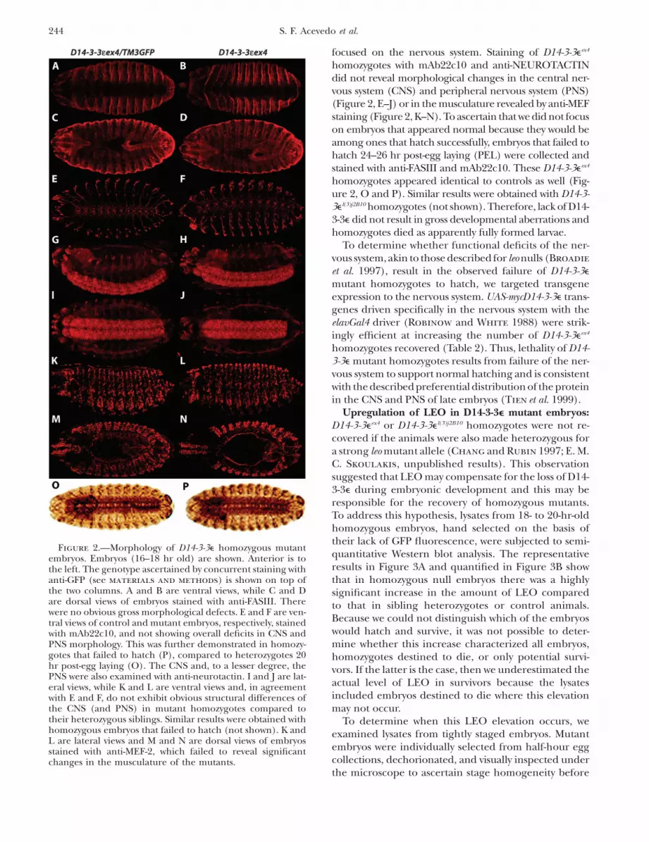
focused on the nervous system. Staining of D14-3-3eex4
homozygotes with mAb22c10 and anti-NEUROTACTINdid not reveal morphological changes in the central ner-vous system (CNS) and peripheral nervous system (PNS)(Figure 2, E–J) or in the musculature revealed by anti-MEFstaining (Figure 2, K–N). To ascertain that we did not focuson embryos that appeared normal because they would beamong ones that hatch successfully, embryos that failed tohatch 24–26 hr post-egg laying (PEL) were collected andstained with anti-FASIII and mAb22c10. These D14-3-3eex4
homozygotes appeared identical to controls as well (Fig-ure 2, O and P). Similar results were obtained with D14-3-3el(3)j2B10 homozygotes (not shown). Therefore, lack of D14-3-3e did not result in gross developmental aberrations andhomozygotes died as apparently fully formed larvae.
To determine whether functional deficits of the ner-vous system, akin to those described for leo nulls (Broadie
et al. 1997), result in the observed failure of D14-3-3e
mutant homozygotes to hatch, we targeted transgeneexpression to the nervous system. UAS-mycD14-3-3e trans-genes driven specifically in the nervous system with theelavGal4 driver (Robinow and White 1988) were strik-ingly efficient at increasing the number of D14-3-3eex4
homozygotes recovered (Table 2). Thus, lethality of D14-3-3e mutant homozygotes results from failure of the ner-vous system to support normal hatching and is consistentwith the described preferential distribution of the proteinin the CNS and PNS of late embryos (Tien et al. 1999).
Upregulation of LEO in D14-3-3e mutant embryos:D14-3-3eex4 or D14-3-3el(3)j2B10 homozygotes were not re-covered if the animals were also made heterozygous fora strong leo mutant allele (Chang and Rubin 1997; E. M.C. Skoulakis, unpublished results). This observationsuggested that LEO may compensate for the loss of D14-3-3e during embryonic development and this may beresponsible for the recovery of homozygous mutants.To address this hypothesis, lysates from 18- to 20-hr-oldhomozygous embryos, hand selected on the basis oftheir lack of GFP fluorescence, were subjected to semi-quantitative Western blot analysis. The representativeresults in Figure 3A and quantified in Figure 3B showthat in homozygous null embryos there was a highlysignificant increase in the amount of LEO comparedto that in sibling heterozygotes or control animals.Because we could not distinguish which of the embryoswould hatch and survive, it was not possible to deter-mine whether this increase characterized all embryos,homozygotes destined to die, or only potential survi-vors. If the latter is the case, then we underestimated theactual level of LEO in survivors because the lysatesincluded embryos destined to die where this elevationmay not occur.
To determine when this LEO elevation occurs, weexamined lysates from tightly staged embryos. Mutantembryos were individually selected from half-hour eggcollections, dechorionated, and visually inspected underthe microscope to ascertain stage homogeneity before
Figure 2.—Morphology of D14-3-3e homozygous mutantembryos. Embryos (16–18 hr old) are shown. Anterior is tothe left. The genotype ascertained by concurrent staining withanti-GFP (see materials and methods) is shown on top ofthe two columns. A and B are ventral views, while C and Dare dorsal views of embryos stained with anti-FASIII. Therewere no obvious gross morphological defects. E and F are ven-tral views of control and mutant embryos, respectively, stainedwith mAb22c10, and not showing overall deficits in CNS andPNS morphology. This was further demonstrated in homozy-gotes that failed to hatch (P), compared to heterozygotes 20hr post-egg laying (O). The CNS and, to a lesser degree, thePNS were also examined with anti-neurotactin. I and J are lat-eral views, while K and L are ventral views and, in agreementwith E and F, do not exhibit obvious structural differences ofthe CNS (and PNS) in mutant homozygotes compared totheir heterozygous siblings. Similar results were obtained withhomozygous embryos that failed to hatch (not shown). K andL are lateral views and M and N are dorsal views of embryosstained with anti-MEF-2, which failed to reveal significantchanges in the musculature of the mutants.
244 S. F. Acevedo et al.

preparation of lysates. We quantified LEO levels in laterstages of embryogenesis relative to maternally providedprotein (Li et al. 1997; Philip et al. 2001), which wefound relatively invariable over many different experi-ments (not shown). Thus, compared to 1- to 3-hr con-trol embryos, LEO levels were significantly higher in22-hr D14-3-3eex4 homozygotes and nearly doubledduring their 2-hr hatching delay (Figure 3, C and D).Similar results were obtained with D14-3-3el(3)j2B10 homo-zygotes. Therefore, elevation of LEO in D14-3-3e nullembryos occurs late in embryogenesis, particularly dur-ing the hatching delay exhibited by mutant homozy-
gotes. These results are consistent with two notions.First, elevation of LEO in D14-3-3e mutant homozygotesmay be the reason a fraction hatches and representsa compensatory mechanism for the loss of D14-3-3e.Alternatively, LEO elevation occurs in embryos unableto hatch and may represent a stress response that char-acterizes dying or dead embryos similar to the reportedpostmortem elevation of certain vertebrate 14-3-3 iso-types (Foundoulakis et al. 2001).
To investigate whether elevation of LEO was a con-sequence of increased transcription in D14-3-3e mu-tants, we estimated the relative levels of the two leotranscripts by quantitative PCR. LEOI and LEOII differby five amino acids encoded in the alternative, mutuallyexclusive exons 6 and 69 (Philip et al. 2001). We usednewly hatched homozygous mutant larvae for this quan-tification because, if detectable, it would indicate thatleo elevation occurs in animals that hatch. The 50%reduction in D14-3-3e transcripts in heterozygotes andthe lack of transcripts in homozygous mutants wereeasily detectable with our experimental conditions (Fig-ure 4A). In these animals, the level of leoI transcriptsremained unchanged, but we detected a significantincrease in the level of leoII mRNA in heterozygotes andD14-3-3eex4 homozygous mutant larvae. The leo genecontains two alternative 59 untranslated exons (exon 1and 19). Whereas leoI transcripts appear to always utilizeexon 19, leoII transcripts contain either the distal exon 19
Figure 3.—Elevation of LEO in D14-3-3e homozygous mu-tant embryos. (A) A representative blot of embryonic lysatesused in acquisition of the data on B. The genotypes of the em-bryos whose lysates were blotted are indicated on top of theblot: ex5/ex5 for D14-3-3eex5/D14-3-3eex5; j2B10/1 for D14-3-3el(3)j2B10/D14-3-3eex5; ex4/1 for D14-3-3eex4/D14-3-3eex5; j2B10/j2B10 for D14-3-3el(3)j2B10/D14-3-3el(3)j2B10; and ex4/ex4 for D14-3-3eex4/D14-3-3eex4. These abbreviations are used in A–D.b-Tub denotes b-tubulin, the protein used to normalize thelanes for the amount loaded. (B) The average ratios (6 stan-dard error of the mean or SEM) of the relative levels of LEO/b-Tub and D14-3-3e/b-Tub is shown from four individual blotssimilar to the one displayed in A. Ratios are shown relative tothose obtained from D14-3-3eex5 homozygotes, which werearbitrarily set to 1. The level of LEO accumulation was signif-icantly higher (**P , 0.001) in D14-3-3el(3)j2B10 and D14-3-3eex4
homozygotes compared to D14-3-3eex5 controls. (C) A repre-sentative blot of embryonic lysates of the indicated genotypesprepared at the particular times PEL. The latest collection wasat 24 hr for D14-3-3eex4 homozygotes because of their hatchingdelay, while the latest time point for control embryos was im-mediately before hatching at 22 hr. (D) The average ratio ofrelative levels of LEO/b-Tub 6 SEM for D14-3-3eex4 homozy-gotes compared to D14-3-3eex5 controls estimated from threeindependent blots similar to the one shown in C. There isa highly significant increase (**P , 0.001) in the amountof LEO in D14-3-3eex4 homozygotes during their 2-hr hatchingdelay. A smaller increase (*P , 0.05) in D14-3-3eex4 homozy-gotes was detected at 22-hr PEL in comparison to D14-3-3eex5
controls of the same age. Samples from D14-3-3eex4 homozy-gotes were not collected at 20-hr PEL and control samplescould not be collected at 24-hr PEL because the embryoshad hatched to larvae.
14-3-3 Homeostasis in Drosophila 245

or the proximal exon 1 (Kockel et al. 1997), suggestingdifferential use of promoters. We investigated whetherboth alternate exons are utilized in D14-3-3eex4 homo-zygotes to elevate leoII transcripts. However, the ratioof exon 1-containing transcripts in D14-3-3eex4 homozy-
gotes to those present in control animals remainedunchanged (1.015 6 0.037). In contrast, the number ofexon 19-containing transcripts in the mutants was sig-nificantly higher (Figure 4B). This suggests that eleva-tion of leoII transcripts in D14-3-3eex4 homozygotesinvolves increased utilization of the putative proximalpromoter and differential inclusion of exon 19 in thesetranscripts. These data strongly suggest that elevationof LEO protein levels are a consequence of upregula-tion of leoII transcripts. Since this is detected in mutantfirst instar larvae, the data suggest that elevation of leoIItranscripts allows hatching and survival in the fractionof D14-3-3e homozygotes where it occurs.
Conditional overexpression of leo transgenes rescueslethality of D14-3-3e null homozygotes: To rigorouslytest the hypothesis that elevation of LEOII is the reasonfor hatching and survival of mutant homozygotes, leoIand leoII transgenes were recombined onto the chro-mosomes bearing the D14-3-3eex4 and D14-3-3el(3)j2B10
mutations. If endogenous LEOII elevation suffices forsuccessful hatching of D14-3-3e homozygotes, then fur-ther increasing the level of this protein should increasethe number of homozygous mutant animals recovered.hsleoI and hsleoII transgenes exhibited leaky expressionat 25�, but expression was higher after three daily in-ductions (HS) and undetectable if flies were kept at18� (supplemental Figure 1 at http://www.genetics.org/supplemental/). Significantly, two independent inser-tions of a hsleoII trangene were able to conditionallyincrease (rescue) the number of D14-3-3eex4 and D14-3-3el(3)j2B10 homozygotes recovered (Table 4). In contrast,hsleoI transgenes rescued the phenotype only partially,despite the similarity in hsleoI and hsleoII transgene ex-pression (supplemental Figure 1).
Importantly, when driven by the neuronal-specificGal4 driver elav, UASleoII transgenes rescued fully andUASleoI partially, the lethality of D14-3-3eex4 homozy-gotes, consistent with the notion that they die becausetheir nervous system is unable to support hatching (Table4). This is not the result of differences in transgeneexpression levels, since independent leoI and leoII trans-genes inducible by different means yielded similar out-comes. These data suggest that LEOII and, to a lesserdegree, LEOI are functionally redundant with D14-3-3e
in the nervous system and confirm that LEOII elevationin a fraction of late D14-3-3e mutant embryos allows themto hatch. Furthermore, the data indicate that, despitethe minor, largely conservative differences (Philip et al.2001), the two LEO isoforms are not equivalent in com-pensating for the lack of D14-3-3e. Expression of leo trans-genes in the nervous system is not ectopic, as LEOaccumulates abundantly in this tissue (Skoulakis andDavis 1996; Broadie et al. 1997).
The level of D14-3-3e is not increased in homozy-gous leo null embryos: Is the level of one 14-3-3 iso-type always elevated when the other is reduced duringembryogenesis? Like null D14-3-3e embryos, homozygotes
Figure 4.—Elevation of leoII transcripts in D14-3-3eex4 mu-tant homozygotes. (A) The ratios of D14-3-3e/act5C, leoI/act5C, and leoII/act5C RT–PCR products in control animalswere arbitrarily set to 1 (open bars) and their relative levelsin D14-3-3eex4 heterozygotes (shaded bars) and homozygotes(solid bars) were determined. The mean 6 SEM of five inde-pendent experiments is shown. leoI levels relative to those ofact5C were not found significantly different in mutant heter-ozygotes and homozygotes. In contrast, the relative levels ofleoII mRNAs were significantly higher than controls in bothmutant heterozygotes (*P , 0.01) and homozygotes (**P ,0.001). (B) The ratios of leoII transcripts in D14-3-3eex4 homo-zygotes over those of control animals ½(ex4/ex4)/(ex5/ex5)� de-termined in experiments independent from those in A.Elevation of leoII in the mutants is detected because the ratiofor ‘‘total leoII’’ is .1. Using primers specific to exon 1 andexon 19, the levels of transcripts that include exon 19 in themutants were found significantly higher (*P , 0.01) thantranscripts that include exon 1. The mean 6 SEM of four in-dependent experiments is shown.
246 S. F. Acevedo et al.

for the strong hypomorphic transposon insertion alleleleoP1188 die as fully formed larvae, while the null leo12X
homozygotes exhibit deficits on their dorsal side in-cluding incomplete closure (Skoulakis and Davis 1996;Broadie et al. 1997). Semiquantitative Western blotanalysis indicated that the level of D14-3-3e remainedrelatively unchanged in homozygous leoP1188 and leo12X
mutant embryos in comparison to their heterozygoussiblings (Figure 5). LEO elevation was again readily detect-able in D14-3-3eex4 homozygotes. Therefore, we could notdetect reciprocal elevation of D14-3-3e upon loss of LEOin embryos.
The level of LEO is unchanged in D14-3-3e adultheads: Does LEO remain elevated in adult D14-3-3e
mutant homozygotes? Is D14-3-3e elevated in adult leo
mutant heterozygotes? To address these questions, wedetermined the relative levels of LEO in isolated headsof D14-3-3eex4 homozygotes and heterozygotes and ofD14-3-3e in leo mutant heterozygotes because bothproteins are enriched in adult brains (Skoulakis andDavis 1996; S. F. Acevedo, unpublished observations).Although the 50% reduction of D14-3-3e in D14-3-3eex4
heterozygotes and leo1188/1; D14-3-3eex4/1 animals wasreadily detectable, LEO levels in D14-3-3eex4 homozy-gotes were not significantly different from D14-3-3eex5
(Student’s t-tests, P ¼ 0.8678) or from w1118 controls(Figure 6). Similar results were obtained for D14-3-3el(3)j2B10 homozygotes (supplemental Figure 2 at http://www.genetics.org/supplemental/). Thus, LEO appearsto be elevated only in D14-3-3e mutant embryos around
TABLE 4
Rescue of D14-3-3e lethality with leo transgenes
Genotype Temperature % viable % rescue n
D14-3-3el(3)j2B10/D14-3-3el(3)j2B10 18� 75 — 645D14-3-3el(3)j2B10/D14-3-3el(3)j2B10 23� 77 8 526D14-3-3el(3)j2B10/D14-3-3el(3)j2B10 HS 78 12 522
D14-3-3el(3)j2B10/D14-3-3el(3)j2B10, hsleoIL 18� 76 — 462D14-3-3el(3)j2B10/D14-3-3el(3)j2B10, hsleoIL 23� 84 33 455D14-3-3el(3)j2B10/D14-3-3el(3)j2B10, hsleoIL HS 88 50 524
D14-3-3el(3)j2B10/D14-3-3el(3)j2B10, hsleoIIL 18� 78 — 442D14-3-3el(3)j2B10/D14-3-3el(3)j2B10, hsleoIIL 23� 95 77a 492D14-3-3el(3)j2B10/D14-3-3el(3)j2B10, hsleoIIL HS 100 100a 425
D14-3-3eex4/D14-3-3eex4 18� 42 — 382D14-3-3eex4/D14-3-3eex4 23� 45 5.5 411D14-3-3eex4/D14-3-3eex4 HS 45 5.5 409
D14-3-3eex4/D14-3-3eex4, hsleoIL 18� 49 — 481D14-3-3eex4/D14-3-3eex4, hsleoIL 23� 56 14 440D14-3-3eex4/D14-3-3eex4, hsleoIL HS 87 74.5a 493D14-3-3eex4/D14-3-3eex4, hsleoIH 18� 51 — 415D14-3-3eex4/D14-3-3eex4, hsleoIH 23� 60 18.5 433D14-3-3eex4/D14-3-3eex4, hsleoIH HS 85 69a 495
D14-3-3eex4/D14-3-3eex4, hsleoIIL 18� 49 — 496D14-3-3eex4/D14-3-3eex4, hsleoIIL 23� 69 39a 534D14-3-3eex4/D14-3-3eex4, hsleoIIL HS 100 100a 555D14-3-3eex4/D14-3-3eex4, hsleoIIH 18� 56 — 512D14-3-3eex4/D14-3-3eex4, hsleoIIH 23� 91 76.5a 534D14-3-3eex4/D14-3-3eex4, hsleoIIH HS 100 100a 485
D14-3-3eex4/D14-3-3eex4,UASleoI 25� 37 — 230elavGal4/1 ; D14-3-3eex4/D14-3-3eex4,UASleoI 25� 63 41a 331
D14-3-3eex4/D14-3-3eex4,UASleoII 25� 34 — 248elavGal4/1; D14-3-3eex4/D14-3-3eex4,UASleoII 25� 100 100a 380
leo transgenes rescue the lethality of D14-3-3e mutant homozygotes. The temperature conditions employedfor the crosses are shown under ‘‘Temperature.’’ Under HS conditions, flies were raised under constant 23�,except for three daily 30-min, 32� heat shocks. Viability was calculated as the fraction of adults of each genotyperecovered from crosses of balanced individuals over that expected if the mutant homozygotes were fully viable.‘‘% rescue’’ was calculated as the increase in mutant homozygotes carrying transgenes over the ‘‘baseline’’ num-ber (—). The superscripts H and L for leoI and leoII transgenes denote high- and low-expressing transgenes,respectively. n denotes the total number of flies scored per cross that yielded the mutant homozygotes carryingD14-3-3e or leo transgenes.
a Significant rescue.
14-3-3 Homeostasis in Drosophila 247

the time of hatching, and therefore it is unlikely that itfunctionally compensates for D14-3-3e loss in all tissues.Similarly, D14-3-3e is not elevated in the heads of leomutant heterozygotes.
Loss of D14-3-3e disrupts wing cross-vein formation:Adult D14-3-3eex4 and D14-3-3el(3)j2B10 homozygotes havesmaller wings than control flies. Cell counts along thelongitudinal veins of homozygous and heteroallelicadults indicated a 10% proportional reduction in lengthcompared to D14-3-3eex5 controls (not shown). This maybe a consequence of the overall body-size reduction alsoobserved in null homozygous and heteroallelic embryos(Figure 2), larvae, and adults. In addition, the majorityof adult D14-3-3eex4 and D14-3-3el(3)j2B10 homozygotesexhibited a conspicuous lack of the dorsal part of theposterior cross-vein and, with lesser penetrance, mal-formation of the anterior cross-vein (Figure 7, A.2–A.5).These defects map to D14-3-3e, as all mutant animalsexhibited the phenotype (Table 5A). Furthermore,posterior and anterior cross-vein deficits can be rescuedwith hsD14-3-3e transgenes (Figure 7, A.6 and A.7; Table5B), demonstrating that cross-vein formation indeedrequires D14-3-3e. We quantified deficits of the poste-rior cross vein because it exhibited greater penetrance
and therefore afforded more sensitivity to rescue experi-ments and is reported on the ‘‘% rescue’’ column inTable 5, B and C. Significant changes in the fraction ofwings that exhibited anterior cross-vein malformationsare denoted by footnote a in Table 5.
Overexpression of leoI partially rescues the wing-venation deficits of D14-3-3e mutants: Can LEO com-pensate for the D14-3-3e requirement in cross-veinformation as it did for hatching? leoI, but not leoII, isexpressed in wing disks (Figure 7B), whereas transcriptsfor both isoforms could be detected in brains from thesame w1118 larvae as reported previously (Philip et al.2001). To determine whether LEO compensates D14-3-3e loss in wing cross-vein formation, we examined thewings of D14-3-3eex4 homozygotes rescued from lethalityby hsleoI and hsleoII transgenes. Both hsleoI and hsleoIItransgenes were expressed in the wing disks after heat
Figure 6.—LEO is not significantly elevated in the heads ofadult D14-3-3e homozygous mutants, and the level of D14-3-3eis not changed in the heads of leo mutant heterozygotes. (A)The average ratios (6SEM) of LEO/SYX and D14-3-3e/SYX isshown from three individual Western blotting experiments,one of which is shown in B. Ratios are shown relative to thoseobtained from D14-3-3eex5/D14-3-3eex5 adults, which were arbi-trarily set to 1. Compared to the levels in D14-3-3eex5/D14-3-3eex5 controls, D14-3-3e was significantly reduced (*P ,0.01) in D14-3-3eex4/D14-3-3eex5 (ex4/1), leoP1188/1; D14-3-3eex4/1 (leoP1188/1; ex4/1) double heterozygotes and D14-3-3eex4/D14-3-3eex4 (ex4/ex4) homozygotes (**P , 0.001). Sim-ilarly, LEO was significantly (*P , 0.01) reduced in leoP1188/1and leoP1188/1; D14-3-3eex4/1 animals. However, LEO was notsignificantly elevated in the heads of D14-3-3eex4 homozygotesor D14-3-3e in the heads of leoP1188 heterozygotes. (B). A rep-resentative blot of head lysates from the indicated genotypesquantified in A. The neuronal protein SYNTAXIN (Syx) wasutilized to normalize the amount of each lysate loaded.
Figure 5.—D14-3-3e is not elevated in leo homozygous mu-tant embryos. The average ratios (6SEM) of LEO/b-TUB andD14-3-3e/b-TUB are shown from three individual experi-ments. Ratios are shown relative to those obtained fromD14-3-3eex5/D14-3-3eex5 embryos, which were arbitrarily set to1. The genotypes of the embryos whose lysates were blottedare ex5/ex5 for D14-3-3eex5/D14-3-3eex5; ex4/1 for D14-3-3eex4/D14-3-3eex5; ex4/ex4 for D14-3-3eex4/D14-3-3eex4, whereas fullgenotypes are shown for leo mutants. Compared to that inD14-3-3eex5 homozygotes, the level of LEO accumulation wassignificantly higher (**P , 0.001) in D14-3-3eex4 homozygotesand significantly reduced (**P , 0.001) in leo1188 homozy-gotes. The level of D14-3-3e was also significantly reducedin late D14-3-3eex4 homozygous embryos.
248 S. F. Acevedo et al.

shock (Figure 7C). However, although hsleoII is efficientat rescuing lethality, the wings of the same rescuedmutant homozygotes retained the posterior cross-veindeficit (Figure 7A.10) and often the anterior cross-veinremained malformed (Figure 7A.11). In contrast, a 25%reduction in posterior cross-vein deficits and a completerescue of anterior cross-vein malformation was observedin animals rescued from lethality with hsleoI transgenes(Table 5). Similar results were obtained with D14-3-3el(3)j2B10 homozygotes expressing hsleoI and hsleoII trans-genes (not shown). These data indicate that LEOI andD14-3-3e are partially redundant in processes requiredfor posterior cross-vein formation. Moreover, LEOII ismuch more inefficient in compensating D14-3-3e lossin anterior cross-vein formation. Therefore, the twoLEO isoforms again are not equivalent in their ability tosubstitute for the loss of D14-3-3e.
DISCUSSION
D14-3-3e is an essential gene: Our results utilizingnull alleles indicate that D14-3-3e is not dispensable forviability, but its loss is partially compensated by elevationof endogenous leo levels. Consequently, homozygotessurvive to adulthood, whose number is higher when thehypomorphic allele D14-3-3el(3)j2B10 is used. This is thelikely reason for the suggestion of previous reports thatthe gene is not essential (Chang and Rubin 1997; Benton
et al. 2002). This interaction is uncovered geneticallyby the inability to obtain D14-3-3eex4 and D14-3-3el(3)j2B10
homozygotes when one copy of leo is mutated (i.e.,leoP1188/1; D14-3-3el(3)j2B10/D14-3-3el(3)j2B10 animals). Em-bryos homozygous for mutant alleles do not exhibitobvious morphological defects (Figure 2) because ma-ternally provided D14-3-3e is likely sufficient to fulfill itsrequirement in syncytial cellular blastoderm and gas-trulating animals (Tien et al. 1999; Philip et al. 2001; Su
et al. 2001). D14-3-3e mutant homozygotes die ostensiblybecause lack of zygotic protein from the nervous systemrenders them unable to hatch. Similarly, LEO accumu-lates in embryonic motor neurons innervating the body-wall musculature and its loss in leo mutants is the likelyreason for their failure to hatch despite their apparentlynormal progression through development (Broadie
et al. 1997).14-3-3 homeostasis: Our results demonstrate that
LEOII overaccumulates in late D14-3-3e null embryosand that this elevation allows a fraction of them to hatchand survive. The conclusion is supported by the strikingincrease in the number of D14-3-3e mutant homozy-gotes that survive upon expression of leoII transgenes inthe nervous system (Table 4). Because endogenous LEOIIaccumulates preferentially in the CNS (Broadie et al.1997; Philip et al. 2001), our data suggest that its eleva-tion in this tissue leads to successful hatching and sur-vival of D14-3-3e mutant homozygotes. This ‘‘homeostatic’’response in D14-3-3e mutants is specific to late embryo-genesis after the maternally supplied D14-3-3e, whichperdures almost until stage 8 (S. F. Acevedo and K.Tsigkari, unpublished results), has decayed. There-fore, the response appears specific to a period when theoverall level of either 14-3-3’s or D14-3-3e, specifically, iscritically important for survival.
It appears that a mechanism sensing the absence ofD14-3-3e operates in embryos and responds by increas-ing the level of LEOII. Congruent with this, LEO ele-vation was not observed in embryos homozygous for thedominant-negative allele D14-3-3eE183K (Chang andRubin 1997), which compromises D14-3-3e functionally,but does not change its overall levels in the embryo(supplemental Table 1 at http://www.genetics.org/supplemental/). It is possible that this is the reasonthat D14-3-3eE183K homozygotes are never recovered. Fur-thermore, this response appears specific to the loss ofD14-3-3e, because levels of this protein remained
Figure 7.—Deficits in cross-vein formation of D14-3-3e mu-tants and transgenic rescue by D14-3-3e and leo transgenes. (A)Posterior cross veins are indicated by arrows, while anteriorcross veins are indicated by arrowheads in 1 and 6. Genotypesare 1, D14-3-3eex5/D14-3-3eex5; 2, D14-3-3el(3)j2B10/D14-3-3el(3)j2B10;3, D14-3-3eex4/D14-3-3eex4; 4, D14-3-3eex4/D14-3-3eex5; 5, D14-3-3el(3)j2B10/D14-3-3eex4 heteroallelic, exhibiting anterior cross-vein deficits also; 6, D14-3-3eex4/D14-3-3eex4, hsD14-3-3e raisedunder HS conditions; 7, D14-3-3el(3)j2B10/D14-3-3el(3)j2B10,hsD14-3-3e raised under HS conditions; 8, D14-3-3eex4/D14-3-3eex4, hsD14-3-3e raised at 18�; 9, D14-3-3eex4/D14-3-3eex4, hsleoIraised under HS conditions; 10, D14-3-3eex4/D14-3-3eex4, hsleoIIraised under HS conditions; 11, A D14-3-3eex4/D14-3-3eex4,hsleoII raised under HS conditions where both anterior andposterior cross veins remained defective. (B) Products of aRT–PCR experiment with RNA from larval wing disks andbrains with primers specific for leoI and leoII transcripts (bot-tom) and amplification of act5C transcripts (top amplicon) ascontrols for the quality of the transcription. leoII is not ex-pressed in larval wing disks. (C) Products of RT–PCR withtransgene-specific primers (Philip et al. 2001), indicating thatunder HS conditions both leoI and leoII transgenes are ex-pressed in dissected wing disks.
14-3-3 Homeostasis in Drosophila 249

normal in homozygous leo mutant embryos and adults(Figures 4 and 5), consistent with their strong lethalphenotype. Clearly, this sensing mechanism responds byincreased accumulation of leoII transcripts by prefer-ential utilization of one of two possible promotersand splicing of the primary transcript to include theleoII-specific exon 69 (Kockel et al. 1997; Philip et al.2001). How is lack of D14-3-3e sensed and how couldleoII transcription be increased? 14-3-3’s have beenreported to participate in nuclear/cytoplasmic traffick-ing of transcription factors (Brunet et al. 2002; Zhao
et al. 2004; Berdichevsky and Guarente 2006). There-fore, it is possible that loss of D14-3-3e enhancestranscription from the proximal promoter of the leogene by not mediating nuclear export of a factor thatbinds that site. Alternatively, D14-3-3e may be part of arepressing complex and, upon its loss, transcriptionfrom this site is enhanced. In contrast, excessive trans-genic elevation in the amount of D14-3-3e results inrecovery of few adults (,10% of expected) homozygousfor strong hypomorphic leo mutations (K. Tsigkari andE. M. C. Skoulakis, unpublished results). This suggests
that although D14-3-3e can at least partially compensatefor the loss of LEO in high concentrations, an endoge-nous molecular mechanism to elevate it in leo homozy-gotes does not appear to exist.
Although leoI transcripts accumulate in the wing disk,LEO does not appear to play a role in wing-vein forma-tion because animals that develop with as low as 10% ofnormal LEO do not exhibit wing aberrations (Philip
et al. 2001). Therefore, the venation deficits are a phe-notype specific to D14-3-3e mutant homozygotes. Inter-estingly, in congruence with the mechanism proposedabove, the leoI transcripts normally expressed in thattissue were not upregulated and leoII transcripts werenot ectopically transcribed in D14-3-3e mutant homozy-gote wing disks (S. F. Acevedo, unpublished observa-tions). This is because the proposed D14-3-3e-interactingfactor(s) required for exon 19-containing leoII transcrip-tion are likely absent from the wing disk where thesetranscripts do not normally accumulate. Exon 1-containingleoII transcripts do not appear to require such D14-3-3e-interacting factor(s), since these transcripts were notupregulated in embryos. Therefore, loss of D14-3-3e does
TABLE 5
Wing cross-vein deficits of D14-3-3e mutants and transgenic rescue
Genotype % anterior malformed % posterior malformed % rescue
A.D14-3-3eex5/D14-3-3eex5 0 0 —D14-3-3eex5/D14-3-3el(3)j2B10 0 0 —D14-3-3eex5/D14-3-3eex4 0 0 —D14-3-3el(3)j2B10/D14-3-3el(3)j2B10 25.0 75.0 —D14-3-3el(3)j2B10/D14-3-3eex4 26.7 82.4 —D14-3-3eex4/D14-3-3eex4 42.9 80.9 —
B.D14-3-3eex4/D14-3-3eex4, hsD14-3-3eL (18�) 40.6 83 —D14-3-3eex4/D14-3-3eex4, hsD14-3-3eL 9.3a 14a 82.3D14-3-3eex4/D14-3-3eex4, hsD14-3-3eH (18�) 41.8 79.5 —D14-3-3eex4/D14-3-3eex4, hsD14-3-3eH 0a 3.6a 95.6
C.D14-3-3eex4/D14-3-3eex4, hsleoIL (18�) 43.2 82.2 —D14-3-3eex4/D14-3-3eex4, hsleoIL 29a 72 11D14-3-3eex4/D14-3-3eex4, hsleoIH (18�) 41.3 80.9 —D14-3-3eex4/D14-3-3eex4, hsleoIH 0a 60 25.8D14-3-3eex4/D14-3-3eex4, hsleoIIL (18�) 43.4 82.6 —D14-3-3eex4/D14-3-3eex4, hsleoIIL 40 78.4 3.1D14-3-3eex4/D14-3-3eex4, hsleoIIH (18�) 41.5 81.8 —D14-3-3eex4/D14-3-3eex4, hsleoIIH 33 76.8 5.1
The wing cross-vein deficits are rescued by conditional hsD14-3-3e expression, but not by leo transgenes. (A)Percentage of D14-3-3e controls and mutant homozygotes that exhibited posterior and anterior cross-vein def-icits. Posterior and anterior cross-vein deficits were counted on the same wings and one wing was scored perindividual (n . 100). (B) The percentage of D14-3-3eex4/D14-3-3eex4, hsD14-3-3e exhibiting wing cross-vein def-icits raised under conditions of transgene silence (18�) or induction. The ‘‘% rescue’’ indicates the percentagedecrease in individuals with deficient wings and is shown only for posterior cross veins. (C). The percentage ofD14-3-3eex4/D14-3-3eex4 individuals expressing leoI and leoII transgenes exhibiting wing cross-vein deficits raisedunder conditions of transgene silence (18�) or induction. The ‘‘% rescue’’ indicates the percentage decrease inindividuals with deficient wings and is shown only for posterior cross veins.
a Large changes in the percentage of deficient wings (rescue).
250 S. F. Acevedo et al.

not alter the tissue specificity of leo transcriptionalregulation and specific isoform accumulation.
It is presently unclear whether this compensatory mech-anism is operant in other systems where mutant analysesof 14-3-3’s have been initiated. Interestingly, single nullsof either 14-3-3-encoding gene in Saccharomyces cerevisiaeare viable, while the double mutant is lethal (Roberts
et al. 1997) and similar results were obtained for the twoSchizosaccharomyces pombe genes (Ford et al. 1994). Theseobservations may reflect similar 14-3-3 ‘‘homeostatic’’mechanisms in these species. Directed reduction ofspecific 14-3-3 protein levels during Xenopus laevis de-velopment yielded gastrulation and patterning defectsfor all proteins tested except for 14-3-3z (Lau et al. 2006).Unlike Drosophila, Xenopus 14-3-3z may not be essen-tial for development, but it is also possible that loss ofthis isotype is specifically compensated for by elevationof the remaining 14-3-3’s. Such mechanisms, if extant inmammals, are likely to hinder genetic analysis of 14-3-3function, especially in the brain where all family mem-bers are expressed (Baxter et al. 2002). Interestingly,mice mutant for 14-3-3e exhibit severe brain abnormal-ities and die perinatally, yet a small fraction survive toadulthood appearing smaller, but otherwise normal(Toyo-oka et al. 2003), much like the Drosophila mutants.It is unknown whether 14-3-3z or other isotypes are ele-vated in these animals as predicted by our results.
Functional specificity and redundancy of 14-3-3’s:Functional specificity of 14-3-3 family members may bethe result of tissue or temporal-specific gene expressionand regulation or of isotype-specific ligand selectivity.Isotypes may have redundant functions within a cell ifthey are able to interact with the same targets. Eventhen, affinity differences toward common ligands pre-dicted by their amino-acid sequence and tertiary struc-ture (Gardino et al. 2006) may functionally differentiatecoexpressed 14-3-3’s.
Although both leoI and leoII transgenes rescued thelethality of D14-3-3e mutants, they clearly exhibited dif-ferent efficiency (Table 4). Rescue was invariably higherupon accumulation of LEOII either ubiquitously or spe-cifically in the nervous system. However, rescue requiredexcessive accumulation of LEOII to overcome loss ofD14-3-3e. In fact, the two- to threefold LEO elevationshown in Figure 3B could be as much as a 50–60% un-derestimate of the level of this protein in D14-3-3e
mutant embryos that hatch. Therefore, a large excess ofLEO appears to be necessary to functionally substituteD14-3-3e in the embryonic nervous system, which maybe attained only in a small number of mutant homo-zygotes. This probably reflects the affinity differencesthat LEO dimers exhibit toward client proteins normallybound either by D14-3-3e homodimers or by D14-3-3e/LEO heterodimers. If so, then even a small amount ofD14-3-3e would increase the number of mutant homo-zygotes obtained. In agreement with this, more homo-zygotes were recovered from the transposon allele D14-
3-3el(3)j2B10, which likely contains residual D14-3-3e (Table1). Differences in ligand binding between LEOI andLEOII are likely reflected in the large difference withwhich the two isoforms rescue the lethality of D14-3-3e
mutants. This is the first unequivocal demonstration offunctional differences between LEOI and LEOII. Thesedifferences must reside in the five unique amino acids ofhelix 6 that distinguish the two isoforms (Philip et al.2001). It is unknown whether LEOI, LEOII, or bothcontribute to the reported redundancy with D14-3-3e inphotoreceptor development and oocyte polarity(Chang and Rubin 1997; Benton et al. 2002; Benton
and St Johnston 2003).Interestingly, the functional redundancy of LEO iso-
forms with D14-3-3e is tissue specific. In contrast to theembryonic nervous system, enhanced accumulation ofLEOI, and not of LEOII, was able to compensate foranterior cross-vein deficits and partially for the posteriorcross vein (Table 5). Again, this suggests that D14-3-3e
ligands in the wing disk necessary for cross-vein forma-tion can be targeted by excess LEOI (and not LEOII).Hence, LEOII can be redundant with D14-3-3e specif-ically in the embryonic nervous system where it is pre-sumed to accumulate preferentially, while in the wingdisk LEOI, which is normally found in this tissue, is thepotential compensating isoform. Similarly, although thetwo S. cerevisiae 14-3-3 genes are functionally redundant forviability, only one, Bmh1p, is required for efficient forwardtransport to the endoplasmic reticulum (Michelsen et al.2006). Thus, redundancy of 14-3-3’s largely depends onthe specific function and interacting proteins that theyengage within a particular tissue or developmental context.
Collectively, our data show a tissue- and temporal-specific upregulation of leo transcription that can accountfor the apparent functional redundancy between LEOand D14-3-3e with respect to the lethality of D14-3-3e
mutants and possibly other processes requiring theseproteins. In addition, this analysis for the first timedemonstrates tissue and temporal functional differencesbetween the two LEO isoforms. Whether these functionaldifferences and the functional redundancy among theDrosophila 14-3-3’s will also occur in the adult nervoussystem where they are all most abundant is currentlyunknown. A previous study failed to uncover differencesbetween LEOI and LEOII with respect to learning andmemory (Philip et al. 2001). Nevertheless, our datastrongly support the notion that the existence of multiple14-3-3 isotypes in metazoans reflects a combination oftissue- and temporal-specific isotype expression, localiza-tion, and functional specialization. Importantly, this anal-ysis indicates that understanding the biological roles of14-3-3’s will require identification of proteins engaged byhomo- and heterodimers of particular composition in atissue- and temporal-specific manner.
The authors are indebted to H. Chang and G. Rubin for providingmutant stocks and plasmids, to C.-T. Chien for providing an anti-D14-3-3e antibody used in initial phases of this work, to Charity Moore for
14-3-3 Homeostasis in Drosophila 251

help with analysis of the wing phenotype, to Mary McCrady, CourtneySwayze, Alexandros Kanellopoulos, and Maria Anezaki for stockmaintenance and husbandry, and to M. Franco-Redrejo for invaluablehelp with quantitative RT–PCR. We thank Hanh Nguyen for anti-MEF2antibodies, the Developmental Hybridoma Studies Bank at the Univer-sity of Iowa (Iowa City, IA) for monoclonal antibodies, and Terry Orr-Weaver and Sumana Datta for valuable discussions and suggestions.This work was supported by a European Commission Marie Curiegrant (IRG-003570), by PENED (01ED207) from the Greek GeneralSecretariat for Research and Technology, and by the National ScienceFoundation (grant IBN-0080687).
LITERATURE CITED
Aitken, A., 1995 14–3-3 proteins on the MAP. Trends Biochem. Sci.20: 95–97.
Aitken, A., S. Howell, D. Jones, J. Madrazo and Y. Patel, 1995 14–3-3 a and d are the phosphorylated forms of Raf-activating 14–3-3 band z. J. Biol. Chem. 270: 5706–5709.
Aitken, A., H. Baxter, T. Dubios, S. Clokie, S. Mackie et al.,2002 14–3-3 proteins in cell regulation. Biochem. Soc. Trans.30: 351–360.
Baxter, H. C., W.-G. Liu, J. L. Forster, A. Aitken and J. R. Fraser,2002 Immunolocalisation of 14–3-3 isoforms in normal andscrapie-infected murine brain. Neuroscience 109: 5–14.
Benton, R., and D. St Johnston, 2003 Drosophila PAR-1 and 14–3-3 inhibit Bazooka/PAR-3 to establish complementary corticaldomains in polarized cells. Cell 115: 691–704.
Benton, R., I. M. Palacios and D. St. Johnston, 2002 Drosophila14–3-3/PAR-5 is an essential mediator of PAR1 function in axisformation. Dev. Cell 3: 659–671.
Berdichevsky, A., and L. Guarente, 2006 A stress response path-way involving sirtuins, forkheads and 14–3-3 proteins. Cell Cycle5: 2588–2591.
Bourgouin, C., S. E. Lundgren and J. B. Thomas, 1992 Apterous isa Drosophila LIM domain gene required for the development ofa subset of embryonic muscles. Neuron 9: 549–561.
Brand, A. H., and N. Perrimon, 1993 Targeted gene expression as ameans of altering cell fates and generating dominant pheno-types. Development 118: 401–415.
Bridges, D., and G. B. G. Moorehead, 2005 14–3-3 proteins: anumber of functions for a numbered protein. Sci. STKE 2005:re10.
Broadie, K., E. Rushton, E. M. C. Skoulakis and R. L. Davis,1997 Leonardo, a Drosophila 14–3-3 protein involved in learn-ing, regulates presynaptic function. Neuron 19: 391–402.
Brunet, A., F. Kanai, J. Stehn, J. Xu, D. Sarbassova et al.,2002 14–3-3 transits to the nucleus and participates in dynamicnucleocytoplasmic transport. J. Cell Biol. 156: 817–828.
Chang, H. C., and G. M. Rubin, 1997 14–3-3e positively regulatesRas mediated signaling in Drosophila. Genes Dev. 11: 1132–1139.
Chen, H.-K., P. Fernandez-Funez, S. F. Acevedo, Y. C. Lam, M. D.Kaytor et al., 2003 Interaction of akt-phosphorylated ataxin-1with 14–3-3 mediates neurodegeneration in spinocerebellar ataxiatype 1. Cell 113: 457–468.
Coblitz, B., M. Wu, S. Shikano and M. Li, 2006 C-terminal bind-ing: an expanded repertoire and function of 14–3-3 proteins.FEBS Lett. 580: 1531–1535.
Ford, J. C., F. al-Khodairy, E. Fotou, K. S. Sheldrick, D. J.Griffiths et al., 1994 14–3-3 protein homologs required forthe DNA damage checkpoint in fission yeast. Science 265: 533–535.
Foundoulakis, M., R. Hardmeier, H. Hoger and G. Lubec,2001 Postmortem changes in the level of brain proteins. Exp.Neurol. 167: 86–94.
Fu, H., R. R. Subramanian and S. C. Masters, 2000 14–3-3 proteins:structure, function and regulation. Annu. Rev. Pharmacol. Tox-icol. 40: 617–647.
Gardino, A. K., S. J. Smerdon and M. B. Yaffe, 2006 Structural de-terminants of 14–3-3 binding specificities and regulation of sub-cellular localization of 14–3-3-ligand complexes: a comparison of
the X-ray crystal structures of all human 14–3-3 isoforms. Semin.Cancer Biol. 16: 173–182.
Jones, D. H. A., H. Martin, J. Madrazo, K. A. Robinson, P. Neilsen
et al., 1995 Expression and structural analysis of 14–3-3 pro-teins. J. Mol. Biol. 245: 375–384.
Karim, F. D., H. C. Chang, M. Therrien, D. A. Wassarman, T.Laverty et al., 1996 A screen for genes that function down-stream of Ras1 during Drosophila eye development. Genetics143: 315–329.
Kockel, L., G. Vorbraggen, H. Jackle, M. Mlodzik and D.Bohnmann, 1997 Requirement for Drosophila 14–3-3 z in cellproliferation and Raf-dependent photoreceptor development.Genes Dev. 11: 1140–1147.
Lau, J. M., C. Wu and A. J. Muslin, 2006 Differential role of 14–3-3family members in Xenopus development. Dev. Dyn. 235:1761–1776.
Li, W., E. M. C. Skoulakis, R. L. Davis and N. Perrimon, 1997 TheDrosophila 14–3-3 protein Leonardo enhances Torso signalingthrough D-Raf in a Ras1-dependent manner. Development 124:4163–4171.
Li, W., E. Noll and N. Perrimon, 2000 Identification of autosomal re-gions involved in Drosophila Raf function. Genetics 156: 763–774.
Luo, Z., X. Zhang, U. Rapp and J. Avruch, 1995 Identification ofthe 14–3-3z domains important for self-association and Raf bind-ing. J. Biol. Chem. 270: 23681–23687.
Martin, H., J. Rostas, Y. Patel and A. Aitken, 1994 Subcellularlocalisation of 14–3-3 isoforms in rat brain using specific antibod-ies. J. Neurochem. 63: 2259–2265.
Michelsen, K., T. Mrowiec, K. E. Duderstadt, S. Frey, D. L. Minor
et al., 2006 A multimeric membrane protein reveals 14–3-3 iso-form specificity in forward transport in yeast. Traffic 7: 903–916.
Muslin, A. J., J. W. Tanner, P. M. Allen and A. S. Shaw,1996 Interaction of 14–3-3 with signaling proteins is mediatedby the recognition of phosphoserine. Cell 84: 889–897.
Nguyen, H. T., and X. Xu, 1998 Drosophila mef2 expression duringmesoderm development is controlled by a complex array of cis-acting regulatory modules. Dev. Biol. 204: 550–566.
Pfaffl, M. W., 2001 A new mathematical model for relative quanti-fication in real-time RT-PCR. Nucleic Acids Res. 29: 2002–2007.
Philip, N., S. Acevedo and E. M. C. Skoulakis, 2001 Conditionalrescue of olfactory learning and memory defects in mutants ofthe 14–3-3z gene leonardo. J. Neurosci. 21: 8417–8425.
Roberts, R. L., H. U. Mosch and G. R. Fink, 1997 14–3-3 proteinsare essential for RAS/MAPK cascade signaling during pseudohy-phal development in S. cerevisiae. Cell 89: 1055–1065.
Robinow, S., and K. White, 1988 The locus elav of Drosophila mela-nogaster is expressed in neurons at all developmental stages. Dev.Biol. 126: 294–303.
Rosenquist, M., P. Sehnke, R. J. Ferl, M. Sommarin and C. Larsson,2000 Evolution of the 14–3-3 protein family: Does the largenumber of isoforms in multicellular organisms reflect functionalspecificity? J. Mol. Evol. 51: 446–458.
Skoulakis, E. M. C., and R. L. Davis, 1996 Olfactory learningdeficits in mutants for leonardo, a Drosophila gene encodinga 14–3-3 protein. Neuron 17: 931–944.
Skoulakis, E. M. C., and R. L. Davis, 1998 14–3-3 proteins in neu-ronal development and function. Mol. Neurobiol. 16: 269–284.
Su, T. T., D. H. Parry, B. Donahoe, C.-T. Chien, P. H. O’Farrell
et al., 2001 Cell cycle roles for the two 14–3-3 proteins duringDrosophila development. J. Cell Sci. 114: 3445–3462.
Tien, A.-C., H.-Y. Hsei and C.-T. Chien, 1999 Dynamic expressionand cellular localization of the Drosophila 14–3-3e during embry-onic development. Mech. Dev. 81: 209–212.
Toyo-oka, K., A. Shionoya, M. J. Gambello, C. Cardoso, R.Leventer et al., 2003 14–3-3epsilon is important for neuronalmigration by binding to NUDEL: a molecular explanation forMiller-Dieker syndrome. Nat. Genet. 274: 285.
Tzivion, G., and J. Avruch, 2002 14–3-3 proteins: active cofactors incellular regulation by serine/threonine phosphorylation. J. Biol.Chem. 277: 3061–3064.
Tzivion, G., Y. H. Shen and J. Zuh, 2001 14–3-3 proteins: bringingnew definitions to scaffolding. Oncogene 20: 6331–6338.
van Hemert, M. J., H. Y. Steensma and G. P. H. van Heudsen,2001 14–3-3 proteins: key regulators of cell division, signallingand apoptosis. BioEssays 23: 936–946.
252 S. F. Acevedo et al.

van Heudsen, G. P. H., 2005 14–3-3 proteins: regulators of numer-ous eukaryotic proteins. IUBMB Life 57: 623–629.
Wang, W., and D. Shakes, 1996 Molecular evolution of the 14–3-3family. Mol. Evol. 43: 384–398.
Wilker, E. W., R. A. Grant, S. C. Artim and M. B. Yaffe, 2005 Astructural basis for 14–3-3sigma functional specificity. J. Biol.Chem. 280: 18891–18898.
Xiao, B., S. Smerdon, D. H. Jones, G. G. Dodson, Y. Soneji et al.,1995 Structure of a 14–3-3 protein and implications forcoordination of multiple signalling pathways. Nature 376: 188–191.
Yaffe, M. B., 2002 How do 14–3-3 proteins work? Gatekeeper phos-phorylation and the molecular anvil hypothesis. FEBS Lett. 513:53–57.
Yaffe, M. B., and A. E. H. Elia, 2001 Phosphoserine/threonine-binding domains. Curr. Opin. Cell Biol. 13: 131–138.
Zhao, X., L. Gan, H. Pan, D. Kan, M. Majeski et al., 2004 Multipleelements regulate nuclear/cytoplasmic shuttling of FOXO1:characterization of phosphorylation- and 14–3-3-dependentand -independent mechanisms. Biochem. J. 378: 839–849.
Communicating editor: W. M. Gelbart
14-3-3 Homeostasis in Drosophila 253