Iqg1p links spatial and secretion landmarks to polarity and cytokinesis
Upload
un-lincolnCategory
view
6download
0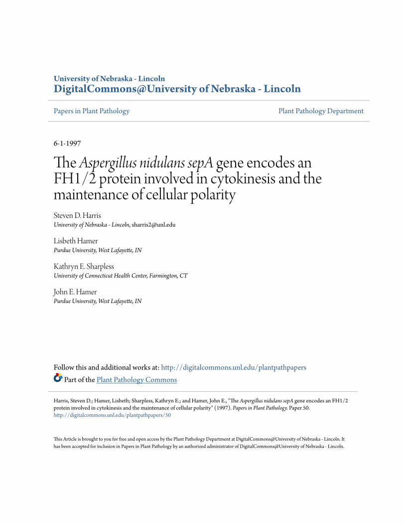
University of Nebraska - LincolnDigitalCommons@University of Nebraska - Lincoln
Papers in Plant Pathology Plant Pathology Department
6-1-1997
The Aspergillus nidulans sepA gene encodes anFH1/2 protein involved in cytokinesis and themaintenance of cellular polaritySteven D. HarrisUniversity of Nebraska - Lincoln, [email protected]
Lisbeth HamerPurdue University, West Lafayette, IN
Kathryn E. SharplessUniversity of Connecticut Health Center, Farmington, CT
John E. HamerPurdue University, West Lafayette, IN
Follow this and additional works at: http://digitalcommons.unl.edu/plantpathpapersPart of the Plant Pathology Commons
This Article is brought to you for free and open access by the Plant Pathology Department at DigitalCommons@University of Nebraska - Lincoln. Ithas been accepted for inclusion in Papers in Plant Pathology by an authorized administrator of DigitalCommons@University of Nebraska - Lincoln.
Harris, Steven D.; Hamer, Lisbeth; Sharpless, Kathryn E.; and Hamer, John E., "The Aspergillus nidulans sepA gene encodes an FH1/2protein involved in cytokinesis and the maintenance of cellular polarity" (1997). Papers in Plant Pathology. Paper 50.http://digitalcommons.unl.edu/plantpathpapers/50
The EMBO Journal Vol.16 No.12 pp.3474–3483, 1997
The Aspergillus nidulans sepA gene encodes anFH1/2 protein involved in cytokinesis and themaintenance of cellular polarity
types (Kishiet al., 1993; Mabuchiet al., 1993; LarochelleSteven D.Harris1, Lisbeth Hamer2,et al., 1996). This class of GTPases (includingrho,Kathryn E.Sharpless and John E.Hamer2
rac and cdc42) controls the re-organization of the actinDepartment of Microbiology, University of Connecticut Health Center, cytoskeleton required for formation of cellular extensionsFarmington, CT 06030-3205 and2Department of Biological Sciences, in fibroblasts (Chant and Stowers, 1995; Ridley, 1996).Purdue University, West Lafayette, IN 47907-1392, USA
Conceivably, activatedrho-related GTPases may trigger1Corresponding author recruitment of proteins required for nucleation and poly-
merization of actin filaments to the division site. ProteinsCytokinesis (septation) in the fungus Aspergillus such as the septins (Longtineet al., 1996) and the actin-nidulans occurs through the formation of a transient associated proteins anillin and radixin (Satoet al., 1991;actin ring at the incipient division site. Temperature- Field and Alberts, 1995) may then act in a downstreamsensitive mutations in thesepAgene prevent septation manner to control formation of the contractile ring.and cause defects in the maintenance of cellular polar- The actin cytoskeleton also plays a critical role inity, without affecting growth and nuclear division. The cytokinesis in fungal cells. In particular, the divisionsepA gene encodes a member of the growing family apparatus is thought to organize an actin ring at theof FH1/2 proteins, which appear to have roles in incipient division site (Raudaskoski, 1970; Hoch andmorphogenesis and cytokinesis in organisms such as Howard, 1980; Kilmartin and Adams, 1984; Marks andyeast andDrosophila. Results from temperature shift Hyams, 1985). Presumably, the actin ring directs vesicle-and immunofluorescence microscopy experiments mediated delivery of cell wall biosynthetic precursors tostrongly suggest thatsepAfunction requires a preceding this site. Ultimately, this leads to cell division via themitosis and thatsepAacts prior to actin ring formation. centripetal formation of a cross-wall, termed a septum.Deletion mutants ofsepAexhibit temperature-sensitive Genetic screens in both the fission yeastSchizosaccharo-growth and severe delays in septation at the permissive myces pombeand the budding yeastSaccharomyces cere-temperature, indicating that expression of another gene visiae have resulted in identification of a number ofmay compensate for the loss ofsepA. Conidiophores genes required for septum formation. In particular, sixformed by sepAmutants exhibit abnormal branching different genes have been shown to be required forof the stalk and vesicle. These results suggest that assembly of the actin ring inS.pombe(Changet al., 1996).sepA interacts with the actin cytoskeleton to promote Two of these genes,cdc3 and cdc8, encode the actin-formation of the actin ring during cytokinesis and that associated proteins profilin and tropomyosin respect-sepA is also required for maintenance of cellular
ively (Balasubramanianet al., 1992, 1994). A third gene,polarity during hyphal growth and asexual morpho-cdc4, may function in a calcium-dependent fashion togenesis.regulate actin ring formation (McCollumet al., 1995). InKeywords: Aspergillus/cellular polarity/cytokinesis/FH1/S.cerevisiae, the septins (encoded by theCDC3, CDC10,2 protein/septationCDC11 and CDC12 genes; reviewed by Longtineet al.,1996) may play a role in the re-organization of the actincytoskeleton which precedes septum formation (Adamsand Pringle, 1984). Despite the successful application of
Introduction genetic approaches to identify numerous gene productsrequired for septum formation in yeast cells, it is not yetCytokinesis requires assembly of the division apparatusclear how the actin ring is assembled.at a specified cortical site. In animal cells, a contractile
We have used a genetic approach to identify genering forms at the division site and then acts to cleave theproducts required for septum formation in the filamentouscell such that two daughter cells are produced (reviewedfungusAspergillus nidulans. Previous studies have demon-by Satterwhite and Pollard, 1992). Amongst the majorstrated that a transient actin ring forms at the incipientcomponents of the contractile ring are actin and myosin,division site in A.nidulans and that loss of functionalwhose interaction provides the mechanical forces neces-microfilaments prevents septum formation (Harriset al.,sary for contraction of the ring and cell cleavage (reviewed1994). Amongst a group of eight genes required for cellby Fishkind and Wang, 1995). Although signals emanatingdivision in A.nidulans, four (sepA, sepD, sepGandsepH)from the mitotic spindle are known to specify the locationappear to encode products which function late inof the division site (Rappaport, 1986), the mechanismsseptum formation (Harriset al., 1994). These genewhich lead to organization of the actin cytoskeleton at theproducts could be components of the division apparatusselected site remain poorly understood. Initial insightsor factors which regulate its assembly. Evidence supportinginto the nature of these mechanisms may be drawn fromthis notion comes from the observation that unlike thethe observation that small GTP binding proteins of the
rho class are required for cytokinesis in a number of cell ‘early acting’sep mutants (Harris and Hamer, 1995),
3474 © Oxford University Press
FH1/2 protein required for cytokinesis
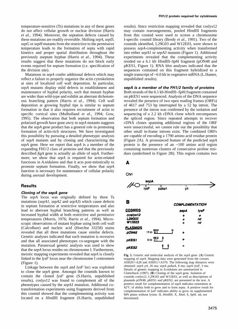
temperature-sensitive (Ts) mutations in any of these genes results). Since restriction mapping revealed that coslys12may contain rearrangements, pooledHindIII fragmentsdo not affect cellular growth or nuclear division (Harris
et al., 1994). Moreover, the septation defects caused by from this cosmid were used to screen a chromosomeI-specific cosmid library (Brodyet al., 1991). Two of thethese mutations are readily reversible. ShiftingsepA, sepD,
sepGor sepHmutants from the restrictive to the permissive cosmids identified, L29G03 and W12E03, were shown topossesssepA-complementing activity when transformedtemperature leads to the formation of septa with rapid
kinetics and proper spatial distribution throughout the into eithersepA1or sepA3mutants (Figure 1). Additionalexperiments revealed that the complementing activitypreviously aseptate hyphae (Harriset al., 1994). These
results suggest that these mutations do not block early resided on a 6.1 kbHindIII–SphI fragment (pON48 andpKES1, Figure 1). RNA blot analyses indicated that theevents required for septum formation (i.e. specification of
the division site). sequences contained on this fragment hybridized to asingle transcript of ~6.0 kb in vegetative mRNA (L.Hamer,Mutations insepAconfer additional defects which may
reflect a failure to properly organize the actin cytoskeleton unpublished results).at sites of localized cell wall deposition. In particular,sepAmutants display mild defects in establishment and sepA is a member of the FH1/2 family of proteins
Both strands of the 6.1 kbHindIII–SphI fragment containedmaintenance of hyphal polarity, such that mutant hyphaeare wider than wild-type and exhibit an aberrant dichotom- on pKES1 were sequenced. Analysis of the DNA sequence
revealed the presence of two open reading frames (ORFs)ous branching pattern (Harriset al., 1994). Cell walldeposition at growing hyphal tips is similar to septum of 4617 and 753 bp interrupted by a 52 bp intron. The
presence of the intron was confirmed by the isolation andformation in that it also requires recruitment of actin tospecific cortical sites (Mulhollandet al., 1994; Gow, sequencing of a 2.2 kb cDNA clone which encompasses
the spliced region. Since repeated attempts to recover1995). The observation that both septum formation andpolarized growth have gone awry insepAmutants suggests cDNA clones spanning additional regions of the ORF
were unsuccessful, we cannot rule out the possibility thatthat thesepAgene product has a general role in promotingformation of actin-rich structures. We have investigated other small in-frame introns exist. The combined ORFs
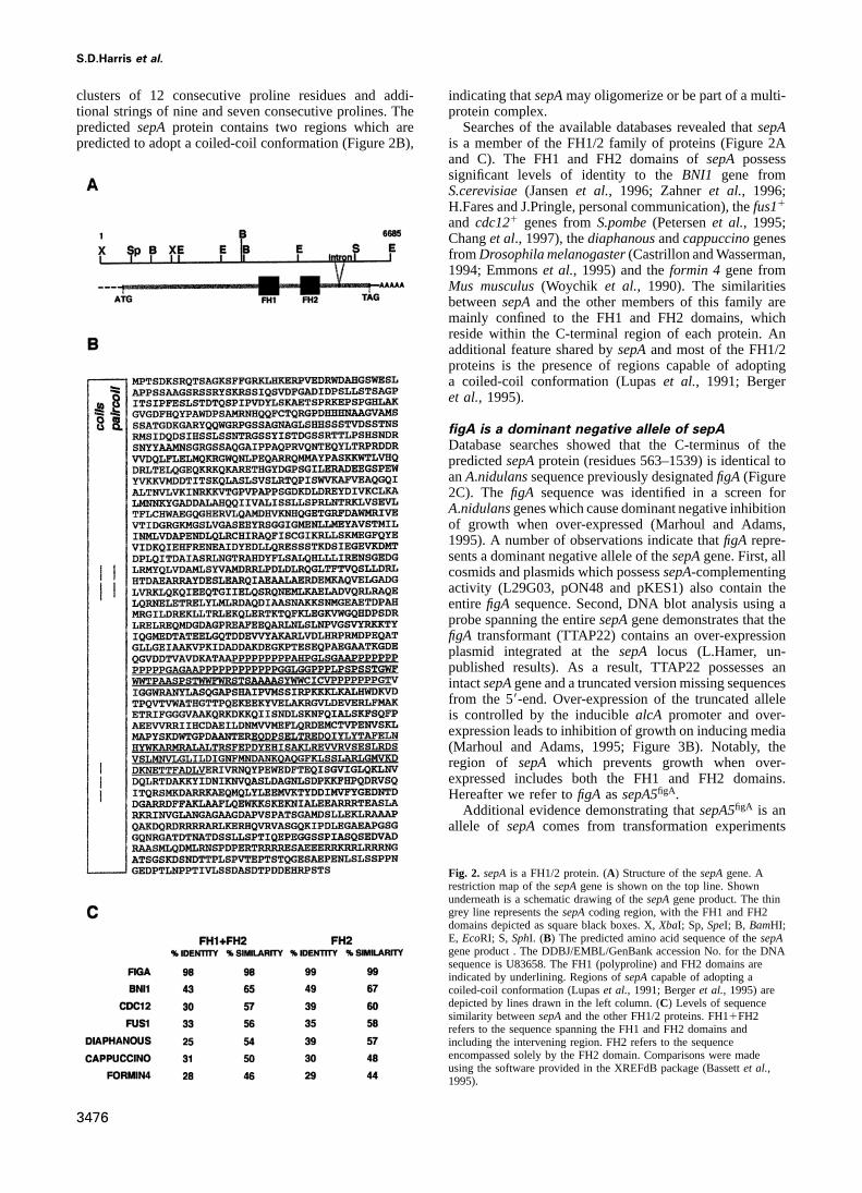
are capable of encoding a 1790 amino acid residue proteinthis possibility by pursuing a detailed phenotypic analysisof sepAmutants and by cloning and characterizing the (Figure 2A). A pronounced feature of the predictedsepA
protein is the presence of an ~100 amino acid regionsepAgene. Here we report thatsepAis a member of theexpanding FH1/2 class of proteins and that the previously containing numerous clusters of consecutive proline resi-
dues (underlined in Figure 2B). This region contains twodescribedfigA gene is actually an allele ofsepA. Further-more, we show thatsepA is required for actin-relatedfunctions inA.nidulansand that it acts post-mitotically topromote septum formation. Finally, we show thatsepAfunction is necessary for maintenance of cellular polarityduring asexual development.
Results
Cloning of the sepA geneThe sepA locus was originally defined by three Tsmutations (sepA1, sepA2andsepA3) which cause defectsin septum formation at restrictive temperatures and alsolead to aberrant hyphal branching patterns as well asincreased hyphal width at both restrictive and permissivetemperatures (Morris, 1976; Harriset al., 1994). Micro-scopic observations of mutant hyphae using both cell wall(Calcofluor) and nucleic acid (Hoechst 33258) stainsrevealed that all three mutations cause similar defects.Genetic analyses indicated that each mutation is recessiveand that all associated phenotypes co-segregate with themutation. Parasexual genetic analysis was used to showthat thesepAlocus resides on chromosome I. Furthermore,meiotic mapping experiments revealed thatsepAis closely Fig. 1. Genetic and molecular analysis of thesepAgene. (A) Genetic
mapping ofsepA. Mapping data were generated from the crosseslinked to thelysF locus near the chromosome I centromereASH203A28 and ASH513A376. The following map distances were(Figure 1).obtained:sepA–yA, 26 mu;sepA–pabaA, 8 mu;sepA–lysF, 3 mu.Linkage between thesepAand lysF loci was exploitedDetails of genetic mapping inA.nidulansare summarized in
to clone thesepAgene. Amongst the cosmids known to Clutterbuck (1987). (B) Cloning of thesepAgene. Isolation ofcontain the clonedlysF gene (S.Harris, unpublished cosmids coslys12, L29G03 and W12E03, as well as descriptions of
plasmids pON48, pKES1 and pKES2, are presented in the text. Aresults), coslys12 was found to complement all of thepositive result for complementation ofsepAindicates restoration atphenotypes caused by thesepA1mutation. Additional co-42°C of ability both to grow and to form septa. A positive result fortransformation experiments using fragments derived from complementation oflysF indicates ability to grow on supplemented
this cosmid showed that the complementing activity was MN plates without lysine. H,HindIII; X, XbaI; S, SphI. nd, notdetermined.located on aHindIII fragment (S.Harris, unpublished
3475
S.D.Harris et al.
clusters of 12 consecutive proline residues and addi- indicating thatsepAmay oligomerize or be part of a multi-protein complex.tional strings of nine and seven consecutive prolines. The
predictedsepA protein contains two regions which are Searches of the available databases revealed thatsepAis a member of the FH1/2 family of proteins (Figure 2Apredicted to adopt a coiled-coil conformation (Figure 2B),and C). The FH1 and FH2 domains ofsepA possesssignificant levels of identity to theBNI1 gene fromS.cerevisiae(Jansenet al., 1996; Zahneret al., 1996;H.Fares and J.Pringle, personal communication), thefus11
and cdc121 genes fromS.pombe(Petersenet al., 1995;Changet al., 1997), thediaphanousandcappuccinogenesfromDrosophila melanogaster(Castrillon and Wasserman,1994; Emmonset al., 1995) and theformin 4 gene fromMus musculus(Woychik et al., 1990). The similaritiesbetweensepAand the other members of this family aremainly confined to the FH1 and FH2 domains, whichreside within the C-terminal region of each protein. Anadditional feature shared bysepAand most of the FH1/2proteins is the presence of regions capable of adoptinga coiled-coil conformation (Lupaset al., 1991; Bergeret al., 1995).
figA is a dominant negative allele of sepADatabase searches showed that the C-terminus of thepredictedsepAprotein (residues 563–1539) is identical toanA.nidulanssequence previously designatedfigA (Figure2C). The figA sequence was identified in a screen forA.nidulansgenes which cause dominant negative inhibitionof growth when over-expressed (Marhoul and Adams,1995). A number of observations indicate thatfigA repre-sents a dominant negative allele of thesepAgene. First, allcosmids and plasmids which possesssepA-complementingactivity (L29G03, pON48 and pKES1) also contain theentirefigA sequence. Second, DNA blot analysis using aprobe spanning the entiresepAgene demonstrates that thefigA transformant (TTAP22) contains an over-expressionplasmid integrated at thesepA locus (L.Hamer, un-published results). As a result, TTAP22 possesses anintactsepAgene and a truncated version missing sequencesfrom the 59-end. Over-expression of the truncated alleleis controlled by the induciblealcA promoter and over-expression leads to inhibition of growth on inducing media(Marhoul and Adams, 1995; Figure 3B). Notably, theregion of sepA which prevents growth when over-expressed includes both the FH1 and FH2 domains.Hereafter we refer tofigA assepA5figA.
Additional evidence demonstrating thatsepA5figA is anallele of sepA comes from transformation experiments
Fig. 2. sepAis a FH1/2 protein. (A) Structure of thesepAgene. Arestriction map of thesepAgene is shown on the top line. Shownunderneath is a schematic drawing of thesepAgene product. The thingrey line represents thesepAcoding region, with the FH1 and FH2domains depicted as square black boxes. X,XbaI; Sp, SpeI; B, BamHI;E, EcoRI; S, SphI. (B) The predicted amino acid sequence of thesepAgene product . The DDBJ/EMBL/GenBank accession No. for the DNAsequence is U83658. The FH1 (polyproline) and FH2 domains areindicated by underlining. Regions ofsepAcapable of adopting acoiled-coil conformation (Lupaset al., 1991; Bergeret al., 1995) aredepicted by lines drawn in the left column. (C) Levels of sequencesimilarity betweensepAand the other FH1/2 proteins. FH11FH2refers to the sequence spanning the FH1 and FH2 domains andincluding the intervening region. FH2 refers to the sequenceencompassed solely by the FH2 domain. Comparisons were madeusing the software provided in the XREFdB package (Bassettet al.,1995).
3476
FH1/2 protein required for cytokinesis
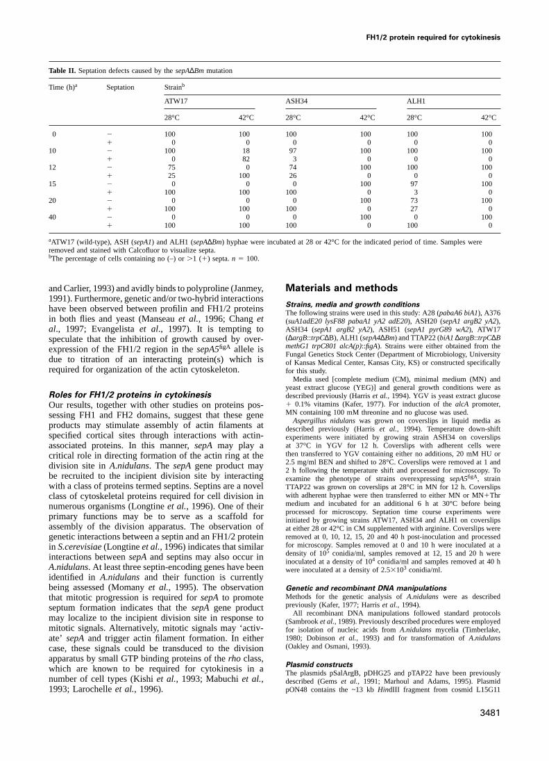
28°C, although they exhibited a highly restricted colonialphenotype (Figure 4C). This result indicates that thesepAgene is not essential for growth. However,sepA4∆Bmmutants failed to grow at 42°C (Figure 4C), conditionsunder which TssepAmutants form small, tightly restrictedcolonies. At 42°C,sepA4∆Bm spores germinate to formaberrantly branched hyphae devoid of septa. These resultsdemonstrate thatsepAfunction is essential for growth athigher temperature and also indicate that the Ts mutantsretain partial activity at 42°C.
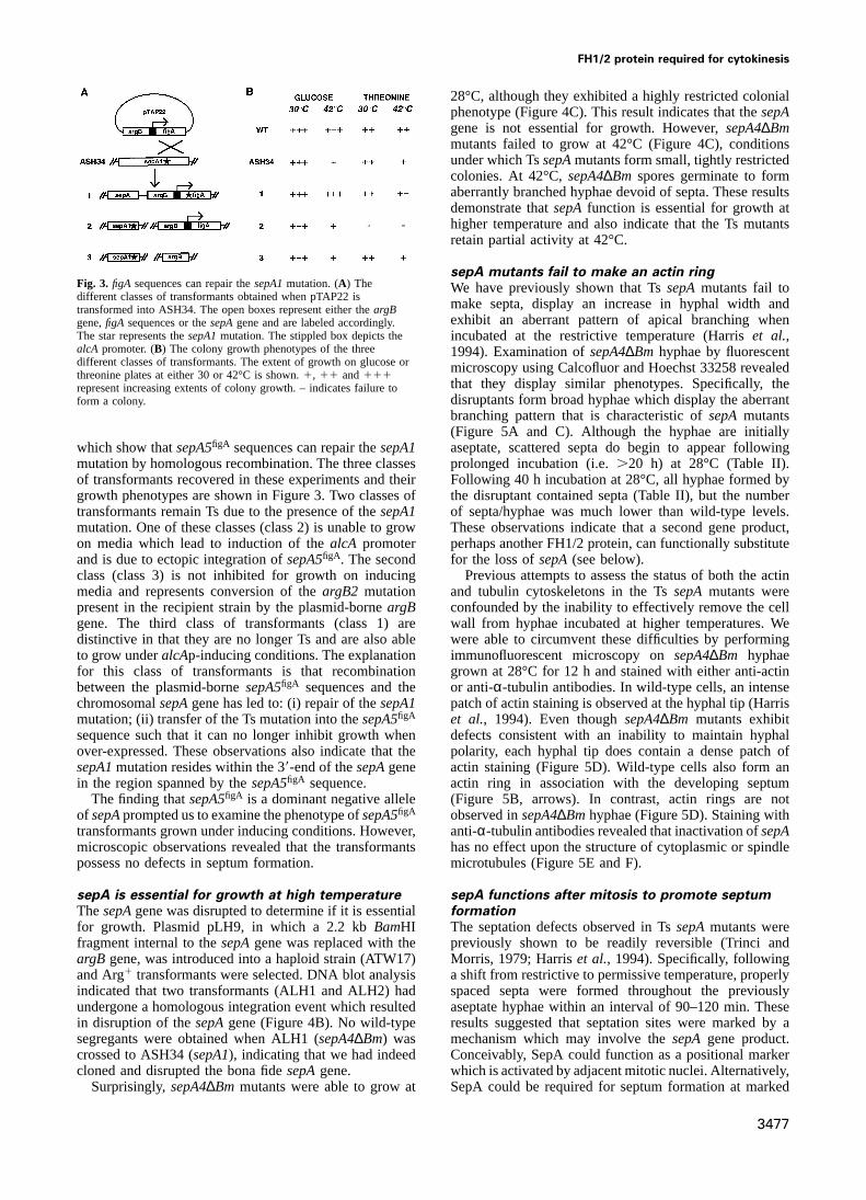
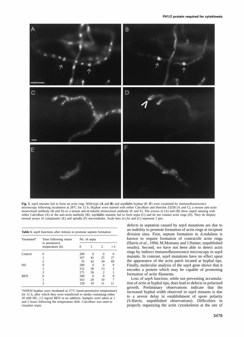
sepA mutants fail to make an actin ringFig. 3. figA sequences can repair thesepA1mutation. (A) The We have previously shown that TssepAmutants fail todifferent classes of transformants obtained when pTAP22 is make septa, display an increase in hyphal width andtransformed into ASH34. The open boxes represent either theargB
exhibit an aberrant pattern of apical branching whengene,figA sequences or thesepAgene and are labeled accordingly.The star represents thesepA1mutation. The stippled box depicts the incubated at the restrictive temperature (Harriset al.,alcA promoter. (B) The colony growth phenotypes of the three 1994). Examination ofsepA4∆Bm hyphae by fluorescentdifferent classes of transformants. The extent of growth on glucose or microscopy using Calcofluor and Hoechst 33258 revealedthreonine plates at either 30 or 42°C is shown.1, 11 and111
that they display similar phenotypes. Specifically, therepresent increasing extents of colony growth. – indicates failure todisruptants form broad hyphae which display the aberrantform a colony.branching pattern that is characteristic ofsepAmutants(Figure 5A and C). Although the hyphae are initiallyaseptate, scattered septa do begin to appear followingwhich show thatsepA5figA sequences can repair thesepA1
mutation by homologous recombination. The three classes prolonged incubation (i.e..20 h) at 28°C (Table II).Following 40 h incubation at 28°C, all hyphae formed byof transformants recovered in these experiments and their
growth phenotypes are shown in Figure 3. Two classes of the disruptant contained septa (Table II), but the numberof septa/hyphae was much lower than wild-type levels.transformants remain Ts due to the presence of thesepA1
mutation. One of these classes (class 2) is unable to grow These observations indicate that a second gene product,perhaps another FH1/2 protein, can functionally substituteon media which lead to induction of thealcA promoter
and is due to ectopic integration ofsepA5figA. The second for the loss ofsepA(see below).Previous attempts to assess the status of both the actinclass (class 3) is not inhibited for growth on inducing
media and represents conversion of theargB2 mutation and tubulin cytoskeletons in the TssepAmutants wereconfounded by the inability to effectively remove the cellpresent in the recipient strain by the plasmid-borneargB
gene. The third class of transformants (class 1) are wall from hyphae incubated at higher temperatures. Wewere able to circumvent these difficulties by performingdistinctive in that they are no longer Ts and are also able
to grow underalcAp-inducing conditions. The explanation immunofluorescent microscopy onsepA4∆Bm hyphaegrown at 28°C for 12 h and stained with either anti-actinfor this class of transformants is that recombination
between the plasmid-bornesepA5figA sequences and the or anti-α-tubulin antibodies. In wild-type cells, an intensepatch of actin staining is observed at the hyphal tip (HarrischromosomalsepAgene has led to: (i) repair of thesepA1
mutation; (ii) transfer of the Ts mutation into thesepA5figA et al., 1994). Even thoughsepA4∆Bm mutants exhibitdefects consistent with an inability to maintain hyphalsequence such that it can no longer inhibit growth when
over-expressed. These observations also indicate that the polarity, each hyphal tip does contain a dense patch ofactin staining (Figure 5D). Wild-type cells also form ansepA1mutation resides within the 39-end of thesepAgene
in the region spanned by thesepA5figA sequence. actin ring in association with the developing septum(Figure 5B, arrows). In contrast, actin rings are notThe finding thatsepA5figA is a dominant negative allele
of sepAprompted us to examine the phenotype ofsepA5figA observed insepA4∆Bmhyphae (Figure 5D). Staining withanti-α-tubulin antibodies revealed that inactivation ofsepAtransformants grown under inducing conditions. However,
microscopic observations revealed that the transformants has no effect upon the structure of cytoplasmic or spindlemicrotubules (Figure 5E and F).possess no defects in septum formation.
sepA is essential for growth at high temperature sepA functions after mitosis to promote septumformationThesepAgene was disrupted to determine if it is essential
for growth. Plasmid pLH9, in which a 2.2 kbBamHI The septation defects observed in TssepAmutants werepreviously shown to be readily reversible (Trinci andfragment internal to thesepAgene was replaced with the
argB gene, was introduced into a haploid strain (ATW17) Morris, 1979; Harriset al., 1994). Specifically, followinga shift from restrictive to permissive temperature, properlyand Arg1 transformants were selected. DNA blot analysis
indicated that two transformants (ALH1 and ALH2) had spaced septa were formed throughout the previouslyaseptate hyphae within an interval of 90–120 min. Theseundergone a homologous integration event which resulted
in disruption of thesepAgene (Figure 4B). No wild-type results suggested that septation sites were marked by amechanism which may involve thesepA gene product.segregants were obtained when ALH1 (sepA4∆Bm) was
crossed to ASH34 (sepA1), indicating that we had indeed Conceivably, SepA could function as a positional markerwhich is activated by adjacent mitotic nuclei. Alternatively,cloned and disrupted the bona fidesepAgene.
Surprisingly,sepA4∆Bm mutants were able to grow at SepA could be required for septum formation at marked
3477

S.D.Harris et al.
Fig. 4. sepAis essential for growth at high temperature. (A) A schematic diagram showing how thesepAdisruption was constructed. The structureof the disruption allele (sepA∆Bm) is shown. (B) Confirmation that thesepAgene was disrupted. A 1.8 kbBamHI fragment from pSalArgBcontainingargB (upper panel) and the 2.2 kbBamHI fragment from pLH14 containing part ofsepA(lower panel) were hybridized toBamHI-digested genomic DNA from the parent strain (ATW17) and two transformants. ATW17 should contain only the 2.2 kbBamHI fragment fromsepAand the disruptants should contain only the 1.8 kbBamHI fragment fromargB. (C) Growth of asepAdisruptant (ALH1) compared with othergenotypes [wild-type (ATW17) andsepA1(ASH34)]. Plates were incubated for 2 days at either 30 or 42°C. See text for details.
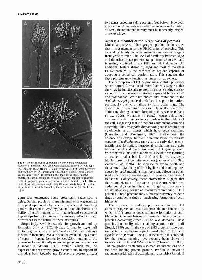
sites in a manner which is independent of mitosis. To mutants revealed that they are capable of undergoing celldivision and possess abundant septa (Figure 6; K.Sharplessdistinguish between these possibilities, we determined if
blocking progression through mitosis with either hydro- and S.Harris, unpublished results). This observation lendsadditional support to the notion that a second gene productxyurea (HU) or benomyl (BEN) could prevent formation
of septa when asepA1mutant is returned to the permissive may be able to functionally substitute forsepA duringconidiation. Since the initiation of conidiophore develop-temperature.
Growth of asepA1strain (ASH34) at semi-permissive ment is known to be suppressed when hyphae are grownin submerged culture (Timberlake, 1991), the delayedtemperature (37°C) for 12 h led to the formation of
aseptate hyphae (Table I). Within 2 h following a return appearance of septa insepA∆Bm cultures (Table II) indi-cates that expression of the other gene(s) cannot be subjectto the permissive temperature.70% of the hyphae had
formed at least one septum (Table I). However,,20% of to developmental control. However, the unfavorablegrowth conditions found in older cultures and associatedsepA1 hyphae could form septa when returned to the
permissive temperature in the presence of either HU or with initiation of conidiation may contribute to expressionof this gene(s). If a functional homolog ofsepA doesBEN (Table I). The residual septation observed in the
presence of these drugs is likely due to hyphae completing exist, it is not detectable in hybridization experimentsusing DNA probes derived from thesepAgene.mitosis at 28°C prior to imposition of the block. These
results show thatsepAfunction is tightly coupled to thecompletion of mitosis. Discussion
A characteristic feature of cell division is that it requiressepA is required for maintenance of cellularpolarity during conidiation dramatic re-organization of the actin cytoskeleton. In
fungal cells, septum formation proceeds via formation ofAlthough hyphae possessing a disruption of thesepAgenedisplay the same spectrum of morphogenetic defects (i.e. an actin ring at a precise cortical location with proper
timing relative to the cell cycle. Amongst the growing listfailure to form septa, aberrant branching, increased hyphalwidth) when incubated at 28°C as do TssepAmutants of proteins shown to be required for septum formation
are those whose known function is to control the dynamicsincubated at restrictive temperature, they are still able togrow and produce a conidiating colony. This observation of actin polymerization (Balasubramanianet al., 1992,
1994). We have found that theA.nidulans sepAgene,suggested that thesepAgene product is not required forconidiophore development. To test this idea further, we which is required for septum formation, encodes a protein
whose predicted properties suggest that it may interactexamined wild-type andsepA4∆Bmhyphae by DIC micro-scopy (Figure 6). Conidiophores produced bysepA4∆Bm with the actin cytoskeleton.mutants do not exhibit gross developmental defects andform abundant conidia. However, they do exhibit striking sepA is required for septum formation and
maintenance of hyphal polaritydefects in the maintenance of cellular polarity, leading toformation of branched stalks (Figure 6B) and split vesicles Temperature-sensitive mutations of thesepAgene cause
a failure to form septa, an increase in hyphal width and(Figure 6C, arrowhead). These observations show that thesepAgene product is required for maintenance of cellular aberrant branching from hyphal tips (Trinci and Morris,
1979; Harriset al., 1994). We have found that disruptionpolarity in additional A.nidulans cell types other thangrowing hyphae. of thesepAgene results in the same pleiotropic effects.
A number of observations support the notion that theExamination of conidiophores formed bysepA4∆Bm
3478
FH1/2 protein required for cytokinesis
Fig. 5. sepAmutants fail to form an actin ring. Wild-type (A andB) andsep4∆Bm hyphae (C–F) were examined by immunofluorescencemicroscopy following incubation at 28°C for 12 h. Hyphae were stained with either Calcofluor and Hoechst 33258 (A and C), a mouse anti-actinmonoclonal antibody (B and D) or a mouse anti-α-tubulin monoclonal antibody (E and F). The arrows in (A) and (B) show septal staining witheither Calcofluor (A) or the anti-actin antibody (B).sepA∆Bm mutants fail to form septa (C) and do not contain actin rings (D). They do displaynormal arrays of cytoplasmic (E) and spindle (F) microtubules. Scale bars in (A) and (C) represent 2µm.
defects in septation caused bysepAmutations are due toan inability to promote formation of actin rings at incipient
Table I. sepAfunctions after mitosis to promote septum formationdivision sites. First, septum formation inA.nidulans isknown to require formation of contractile actin ringsTreatmenta Time following return No. of septa
to permissive (Harriset al., 1994; M.Momany and J.Hamer, unpublishedtemperature (h) 0 1 2 .2 results). Second, we have not been able to detect actin
rings by indirect immunofluorescence microscopy insepAControl 0 200 0 0 0
mutants. In contrast,sepAmutations have no effect upon1 107 41 25 27the appearance of the actin patch located at hyphal tips.2 55 42 34 69
HU 0 200 0 0 0 Finally, molecular analysis of thesepAgene shows that it1 152 30 13 5 encodes a protein which may be capable of promoting2 171 26 2 1 formation of actin filaments.BEN 0 200 0 0 0
Loss ofsepAfunction, while not preventing accumula-1 163 20 10 7tion of actin at hyphal tips, does lead to defects in polarized2 159 19 11 11growth. Preliminary observations indicate that the
aASH34 hyphae were incubated at 37°C (semi-permissive temperature) increased hyphal width observed insepAmutants is duefor 12 h, after which they were transferred to media containing either to a severe delay in establishment of spore polarity20 mM HU, 2.5 mg/ml BEN or no addition. Samples were taken at 1
(S.Harris, unpublished observations). Difficulties inand 2 hours following the temperature shift. Calcofluor was used tovisualize septa. properly organizing the actin cytoskeleton at the site of
3479
S.D.Harris et al.
two genes encoding FH1/2 proteins (see below). However,since allsepAmutants are defective in septum formationat 42°C, the redundant activity must be inherently temper-ature sensitive.
sepA is a member of the FH1/2 class of proteinsMolecular analysis of thesepAgene product demonstratesthat it is a member of the FH1/2 class of proteins. Thisexpanding family includes members in species rangingfrom yeast to mice. The level of similarity betweensepAand the other FH1/2 proteins ranges from 28 to 65% andis mainly confined to the FH1 and FH2 domains. Anadditional feature shared bysepAand most of the otherFH1/2 proteins is the presence of regions capable ofadopting a coiled coil conformation. This suggests thatthese proteins may function as dimers or oligomers.
The participation of FH1/2 proteins in cellular processeswhich require formation of microfilaments suggests thatthey may be functionally related. The most striking conser-vation of function occurs betweensepAand bothcdc121
and diaphanous. We have shown that mutations in theA.nidulans sepAgene lead to defects in septum formation,presumably due to a failure to form actin rings. Thecdc121 gene is required for assembly of the contractileactin ring during septum formation inS.pombe(Changet al., 1996). Mutations incdc121 cause delocalizedclusters of actin patches to accumulate in the middle ofthe cell, suggesting that it functions early during actin ringassembly. TheDrosophila diaphanousgene is required forcytokinesis in all tissues which have been examined(Castrillon and Wasserman, 1994). Furthermore, theabsence of cleavage furrows in mutant larval neuroblastssuggests thatdiaphanousmay also act early during con-tractile ring formation. Functional similarities also existbetweensepA and theS.cerevisiae BNI1gene product.bni1mutants exhibit partial defects in cytokinesis (forminga broader mother–bud junction) and fail to display abipolar pattern of bud site selection (Jansenet al., 1996;
Fig. 6. The maintenance of cellular polarity during conidiation Zahneret al., 1996). The increase in hyphal width andrequires a functionalsepAgene. Conidiophores formed by wild-typethe aberrant branching of both hyphae and conidiophores(A) andsepA4∆Bm (B andC) cultures grown at 28°C were harvestedcaused bysepAmutations may represent defects in polar-and examined by DIC microscopy. Normally, a single conidiophore
vesicle (arrow in A) is formed at the apex of the stalk. InsepA ized growth which are analogous to those caused bybni1mutants the aerial conidiophore stalk frequently appears to generate mutations. Collectively, these observations suggest thatmultiple growing tips, resulting in formation of branched stalks (B) or
the re-organization of the actin cytoskeleton which pre-multiple vesicles upon a single stalk (C, arrowhead). Note the septumcedes cell division in animal and fungal cells occurs viaat the base of the stalk formed by thesepAmutant in (C). Scale bar,
5 µm. an evolutionarily conserved mechanism involving FH1/2proteins. These proteins may stimulate assembly of actinrings or contractile rings by nucleating formation of actingerm tube emergence could presumably cause such a
delay. Similar problems in maintaining actin organization filaments.The presence of multiple prolines within the FH1at hyphal tips could also lead to the aberrant branching
pattern observed insepAhyphae and conidiophores. The domain suggests at least two possible mechanisms bywhich FH1/2 proteins could stimulate formation of actinability of sepAmutants to form actin-based structures at
hyphal tips but not at septation sites may reflect intrinsic filaments. One mechanism is through interactions withproteins containing either SH3 or WW domains. Thesedifferences in the nature of these structures.
Surprisingly,sepA is essential for growth and colony proteins bind to ligands containing a proline-rich motif(Sudol, 1996) and, in the case of SH3 proteins, have beenformation only at 42°C. Hyphae formed bysepA null
mutants grow slowly at 28°C and exhibit severe delays implicated in mediating signal transduction to the actincytoskeleton (Pawson, 1995). Consistent with this possibil-in septum formation. We attribute the eventual appearance
of septa in hyphae formed bysepA∆Bm mutants to the ity, the mouse formins have recently been shown tointeract with SH3 and WW proteins (Chanet al., 1996).presence of a functionally redundant gene product (perhaps
a second A.nidulans FH1/2 protein) which may be The polyproline tracts may also mediate interactions withthe actin binding protein profilin. Profilin is known toexpressed under adverse growth conditions. In support of
this idea, bothS.pombeand Drosophila possess at least modulate the kinetics of actin filament assembly (Pantaloni
3480
FH1/2 protein required for cytokinesis
Table II. Septation defects caused by thesepA∆Bm mutation
Time (h)a Septation Strainb
ATW17 ASH34 ALH1
28°C 42°C 28°C 42°C 28°C 42°C
0 2 100 100 100 100 100 1001 0 0 0 0 0 0
10 2 100 18 97 100 100 1001 0 82 3 0 0 0
12 2 75 0 74 100 100 1001 25 100 26 0 0 0
15 2 0 0 0 100 97 1001 100 100 100 0 3 0
20 2 0 0 0 100 73 1001 100 100 100 0 27 0
40 2 0 0 0 100 0 1001 100 100 100 0 100 0
aATW17 (wild-type), ASH (sepA1) and ALH1 (sepA∆Bm) hyphae were incubated at 28 or 42°C for the indicated period of time. Samples wereremoved and stained with Calcofluor to visualize septa.bThe percentage of cells containing no (–) or.1 (1) septa.n 5 100.
and Carlier, 1993) and avidly binds to polyproline (Janmey, Materials and methods1991). Furthermore, genetic and/or two-hybrid interactions
Strains, media and growth conditionshave been observed between profilin and FH1/2 proteinsThe following strains were used in this study: A28 (pabaA6 biA1), A376in both flies and yeast (Manseauet al., 1996; Changet (suA1adE20 lysF88 pabaA1 yA2 adE20), ASH20 (sepA1 argB2 yA2),
ASH34 (sepA1 argB2 yA2), ASH51 (sepA1 pyrG89 wA2), ATW17al., 1997; Evangelistaet al., 1997). It is tempting to(∆argB::trpC∆B), ALH1 (sepA4∆Bm) and TTAP22 (biA1∆argB::trpC∆Bspeculate that the inhibition of growth caused by over-methG1 trpC801 alcA(p)::figA). Strains were either obtained from theexpression of the FH1/2 region in thesepA5figA allele isFungal Genetics Stock Center (Department of Microbiology, University
due to titration of an interacting protein(s) which is of Kansas Medical Center, Kansas City, KS) or constructed specificallyrequired for organization of the actin cytoskeleton. for this study.
Media used [complete medium (CM), minimal medium (MN) andyeast extract glucose (YEG)] and general growth conditions were as
Roles for FH1/2 proteins in cytokinesis described previously (Harriset al., 1994). YGV is yeast extract glucose1 0.1% vitamins (Kafer, 1977). For induction of thealcA promoter,Our results, together with other studies on proteins pos-MN containing 100 mM threonine and no glucose was used.sessing FH1 and FH2 domains, suggest that these gene
Aspergillus nidulanswas grown on coverslips in liquid media asproducts may stimulate assembly of actin filaments at described previously (Harriset al., 1994). Temperature down-shiftspecified cortical sites through interactions with actin- experiments were initiated by growing strain ASH34 on coverslips
at 37°C in YGV for 12 h. Coverslips with adherent cells wereassociated proteins. In this manner,sepA may play athen transferred to YGV containing either no additions, 20 mM HU orcritical role in directing formation of the actin ring at the2.5 mg/ml BEN and shifted to 28°C. Coverslips were removed at 1 anddivision site inA.nidulans. The sepAgene product may2 h following the temperature shift and processed for microscopy. To
be recruited to the incipient division site by interacting examine the phenotype of strains overexpressingsepA5figA, strainwith a class of proteins termed septins. Septins are a novelTTAP22 was grown on coverslips at 28°C in MN for 12 h. Coverslips
with adherent hyphae were then transferred to either MN or MN1Thrclass of cytoskeletal proteins required for cell division inmedium and incubated for an additional 6 h at 30°C before beingnumerous organisms (Longtineet al., 1996). One of theirprocessed for microscopy. Septation time course experiments wereprimary functions may be to serve as a scaffold for initiated by growing strains ATW17, ASH34 and ALH1 on coverslips
assembly of the division apparatus. The observation of at either 28 or 42°C in CM supplemented with arginine. Coverslips wereremoved at 0, 10, 12, 15, 20 and 40 h post-inoculation and processedgenetic interactions between a septin and an FH1/2 proteinfor microscopy. Samples removed at 0 and 10 h were inoculated at ain S.cerevisiae(Longtineet al., 1996) indicates that similardensity of 105 conidia/ml, samples removed at 12, 15 and 20 h wereinteractions betweensepAand septins may also occur ininoculated at a density of 104 conidia/ml and samples removed at 40 h
A.nidulans. At least three septin-encoding genes have been were inoculated at a density of 2.53103 conidia/ml.identified in A.nidulans and their function is currentlybeing assessed (Momanyet al., 1995). The observation Genetic and recombinant DNA manipulations
Methods for the genetic analysis ofA.nidulans were as describedthat mitotic progression is required forsepAto promotepreviously (Kafer, 1977; Harriset al., 1994).septum formation indicates that thesepA gene product
All recombinant DNA manipulations followed standard protocolsmay localize to the incipient division site in response to (Sambrooket al., 1989). Previously described procedures were employedmitotic signals. Alternatively, mitotic signals may ‘activ- for isolation of nucleic acids fromA.nidulans mycelia (Timberlake,
1980; Dobinsonet al., 1993) and for transformation ofA.nidulansate’ sepAand trigger actin filament formation. In either(Oakley and Osmani, 1993).case, these signals could be transduced to the division
apparatus by small GTP binding proteins of therho class,Plasmid constructswhich are known to be required for cytokinesis in a The plasmids pSalArgB, pDHG25 and pTAP22 have been previously
number of cell types (Kishiet al., 1993; Mabuchiet al., described (Gemset al., 1991; Marhoul and Adams, 1995). PlasmidpON48 contains the ~13 kbHindIII fragment from cosmid L15G111993; Larochelleet al., 1996).
3481
S.D.Harris et al.
(Brody et al., 1991) subcloned into pBS SK1 (J.Marhoul and T.Adams, cytokinesis in fission yeast, is a component of the cell division ringpersonal communication). Plasmid pKES1 contains the 6.1 kbHindIII– and interacts with profilin.J. Cell Biol., 137, 169–182.SphI fragment from pON48 subcloned into pUC119. Plasmid pKES2 Chant,J. and Stowers,L. (1995) GTPase cascades choreographing cellularcontains the partially digested 4.1 kbSphI fragment from pON48 behavior: movement, morphogenesis, and more.Cell, 81, 1–4.subcloned into pUC119. Plasmids for sequencing of the 59 sepAregion Clutterbuck,A.J. (1987)Aspergillus nidulans. In O’Brien,S.J. (ed.),were obtained by subcloning the following fragments from pON48 into Genetic Maps. Cold Spring Harbor Laboratory Press, Cold SpringpBK KS–: ~2.2 kb BamHI (pLH14); ~1.6 kbXbaI (pLH15); ~0.9 kb Harbor, NY, Vol. 4, pp. 325–335.XbaI–SpeI (pLH19); ~1.7 kb XbaI–BamHI (pLH20); ~1.0 kb EcoRI Dobinson,K.F., Harris,R. and Hamer,J.E. (1993) Grasshopper: a(pLH21). An ~2.0 kb cDNA fragment from aλgt10 library was retrotransposon ofMagnaporthe grisea. Mol. Plant Microbiol.cloned into pBS SK1 to form pLH22. pLH22 and pLH23 (containing Interact., 6, 114–126.an ~1.4 kbClaI fragment from pLH22) was used for sequencing of the Emmons,S., Phan,H., Calley,J., Chen,W., James,B. and Manseau,L.39 sepAregion. ThesepAdisruption plasmid pLH9 was constructed in (1995) cappuccino, a Drosophila maternal effect gene required forthree steps. First, the 6.1 kbHindIII–SphI fragment from pON48 was polarity of the egg and embryo, is related to the vertebratelimbsubcloned into pBR322 (pLH5). Second, pLH5 was digested withBamHI deformitylocus.Genes Dev., 9, 2482–2494.and re-ligated (pLH7). Third, the 1.8 kbBamHI fragment containing the Evangelista,M., Blundell,K., Longtine,M.S., Chow,C.J., Adames,N.,argB gene was ligated toBamHI-digested, dephosporylated pLH7. Pringle,J.R., Peter,M. and Boone,C. (1997) Bni1p, a yeast formin
linking Cdc42p and the actin cytoskeleton during polarizedSequencing and sequence analysis morphogenesis.Science, 276, 118–122.Sequencing of the plasmids pLH14, pLH15, pLH19, pLH20, pLH22 Field,C.M., and Alberts,B.M. (1995) Anillin, a contractile ring proteinand pLH23 was performed on a Pharmacia Alf sequencer using the T7, that cycles from the nucleus to the cell cortex.J. Cell Biol., 131,T3, Universal and Reverse primers. The sequence was analysed using 165–178.XREFdB database search tools (Bassettet al., 1995) and the Coils Fishkind,D.J. and Wang,Y. (1995) New horizons for cytokinesis.Curr.(Lupaset al., 1991) and Paircoil (Bergeret al., 1995) software. Opin. Cell Biol., 7, 23–31.
Gems,D., Johnstone,I.L. and Clutterbuck,A.J. (1991) An autonomouslyMicroscopy replicating plasmid transformsAspergillus nidulansat high frequency.Coverslips with adherent cells were processed for microscopy and stained Gene, 98, 61–67.with Hoechst 33258 and Calcofluor as described previously (Harris Gow,N.A.R. (1995) Tip growth and polarity. In Gow,N.A.R. andet al., 1994). Immunofluorescence microscopy for detection of actin and Gadd,G.M. (eds),The Growing Fungus. Chapman and Hall, London,tubulin cytoskeletons was performed using standard protocols (Oakley UK, pp. 277–299.and Osmani, 1993; Harriset al., 1994). Primary antibodies used were Harris,S.D. and Hamer,J.E. (1995)sepB: an Aspergillus nidulansgenethe mouse anti-actin N.350 monoclonal (Amersham) at 1:500 and the involved in chromosome segregation and the initiation of cytokinesis.mouse anti-α-tubulin DM1A monoclonal (Sigma Immunochemicals) at EMBO J., 14, 5244–5257.1:200. Secondary antibodies used were FITC-conjugated anti-mouseHarris,S.D., Morrell,J.L. and Hamer,J.E. (1994) Identification and(Sigma Immunochemical) at 1:200. characterization of Aspergillus nidulans mutants defective in
cytokinesis.Genetics, 136, 517–532.Hoch,H.C. and Howard,R.J. (1980) Ultrastructure of freeze-substitutedAcknowledgements
hyphae of the basidiomyceteLaetisaria arvalis. Protoplasma, 103,281–297.We thank John Marhoul and Tom Adams (Texas A&M University) for
Janmey,P.A. (1991) Polyproline affinity method for purification of plateletkindly sharing strains and plasmids. We also thank Johnny Fares, Johnprofilin and modification with pyrene-maleimide.Methods Enzymol.,Pringle and Fred Chang for the communication of unpublished results196, 92–99.and Susan Kaminskyj for assistance in the genetic mapping ofsepA.
Jansen,R.-P., Dowzer,C., Michaelis,C., Galova,M. and Nasmyth,K.This work was supported by grants from the American Cancer Society(1996) Mother cell-specific HO expression in budding yeast depends(IRG-152J) and the National Science Foundation (MCB 5-22270) toon the unconventional myosin Myo4p and other cytoplasmic proteins.S.D.H. and the National Institutes of Health (GM49867) to J.E.H.Cell, 84, 687–697.
Kafer,E. (1977) Meiotic and mitotic recombination inAspergillusReferences nidulansand its chromosomal aberrations.Adv. Genet., 19, 33–131.
Kilmartin,J.V. and Adams,A.E.M. (1984) Structural rearrangements ofAdams,A.E.M. and Pringle,J.R. (1984) Relationship of actin and tubulintubulin and actin during the cell cycle of the yeastSaccharomycesdistribution to bud growth in wild-type and morphogenetic mutantcerevisiae. J. Cell Biol., 98, 922–933.Saccharomyces cerevisiae. J. Cell Biol., 98, 934–945.
Kishi,K., Sasaki,T., Kuroda,S., Itoh,T. and Takai,Y. (1993) RegulationBalasubramanian,M.K., Helfman,D.M. and Hemmingsen,S.M. (1992) Aof cytoplasmic division ofXenopusembryo by rho p21 and itsnew tropomyosin essential for cytokinesis in the fission yeastS.inhibitory GDP/GTP exchange protein (rho GDI). J. Cell Biol., 120,pombe. Nature, 360, 84–87.1187–1195.Balasubramanian,M.K., Hirani,B.R., Burke,J.D. and Gould,K.L. (1994)
Larochelle,D.A., Vithalani,K.K. and De Lozanne,A. (1996) A novelThe Schizosaccharomyces pombe cdc31 gene encodes a profilinmember of therho family of small GTP-binding proteins is specificallyessential for cytokinesis.J. Cell. Biol., 125, 1289–1301.required for cytokinesis.J. Cell Biol., 133, 1321–1329.Bassett,D.E., Boguski,M., Spencer,F., Reeves,R., Goebl,M. and Hieter,P.
Longtine,M.S., DeMarini,D., Valencik,M., Al-Awar,O.S., Fares,H., De(1995) Comparative genomics, genome cross-referencing andVirglio,C. and Pringle,J.R. (1996) The septins: roles in cytokinesisXREFdb.Trends Genet., 11, 372–373.and other processes.Curr. Opin. Cell Biol., 8, 106–119.Berger,B., Wilson,D.B., Wolf,E., Tonchev,T., Milla,M. and Kim,P.S.
Lupas,A., van Dyke,M. and Stock,J. (1991) Predicting coiled coils from(1995) Predicting coiled coils by use of pairwise residue correlations.protein sequences.Science, 252, 1162–1164.Proc. Natl Acad. Sci. USA, 92, 8259–8263.
Mabuchi,I., Hamaguchi,Y., Fujimoto,H., Morii,N., Mishima,M. andBrody,H., Griffith,J., Cuticchia,A.J., Arnold,J. and Timberlake,W.E.Narumiya,S. (1993) Arho-like protein is involved in the organization(1991) Chromosome-specific recombinant DNA libraries from theof the contractile ring in dividing sand dollar eggs.Zygote, 1, 325–331.fungusAspergillus nidulans. Nucleic Acids Res., 19, 3109–3109.
Manseau,L., Calley,J. and Phan,H. (1996) Profilin is required for posteriorCastrillon,D.H. and Wasserman,S.A. (1994)diaphonousis required forpatterning of theDrosophilaoocyte.Development, 122, 21090–2116.cytokinesis in Drosophila and shares domains of similarity with
Marhoul,J.F. and Adams,T.H. (1995) Identification of developmentalthe the products of the limb deformity gene.Development, 120,regulatory genes inAspergillus nidulansby overexpression.Genetics,3367–3377.139, 537–547.Chan,D.C., Bedford,M.T. and Leder,P. (1996) Formin binding proteins
Marks,J. and Hyams,J.S. (1985) Localization of F-actin through the cellbear WWP/WW domains that bind proline-rich peptides anddivision cycle ofS. pombe. Eur. J. Cell Biol., 39, 27–32.functionally resemble SH3 domains.EMBO J., 15, 1045–1054.
McCollum,D., Balasubramanian,M.K., Pelcher,L.E., Hemmingsen,S.M.Chang,F., Woollard,A. and Nurse,P. (1996) Isolation and characterizationand Gould,K.L. (1995)Schizosaccharomyces pombe cdc41 geneof fission yeast mutants defective in the assembly and placement ofencodes a novel EF-hand protein essential for cytokinesis.J. Cellthe contractile actin ring.J. Cell Sci., 109, 131–142.
Chang,F., Drubin,D. and Nurse,P. (1997) cdc12p, a protein required for Biol., 130, 651–660.
3482
FH1/2 protein required for cytokinesis
Momany,M., Morrell,J.M., Harris,S.D. and Hamer,J.E. (1995) Septumformation inAspergillus nidulans. Can. J. Bot., 73, S369–S399.
Morris,N.R. (1976) Mitotic mutants ofAspergillus nidulans. Genet. Res.,26, 237–254.
Mulholland,J., Preuss,D., Moon,A., Wong,A., Drubin,D. and Botstein,D.(1994) Ultrastructure of the yeast actin cytoskeleton and its associationwith the plasma membrane.J. Cell Biol., 125, 381–391.
Oakley,B.R. and Osmani,S.A. (1993) Cell-cycle analysis using thefilamentous fungusAspergillus nidulans. In Fantes,P. and Brooks,R.(eds),The Cell Cycle: A Practical Approach. Oxford University Press,Oxford, UK, pp. 127–142.
Pantaloni,D. and Carlier,M.-F. (1993) How profilin promotes actinfilament assembly in the presence of thymosinβ4.Cell, 75, 1007–1014.
Pawson,T. (1995) Protein modules and signalling networks.Nature, 373,573–580.
Petersen,J., Weilguny,D., Egel,R. and Nielsen,O. (1995) Characterizationof fus1of Schizosaccharomyces pombe: a developmentally controlledfunction needed for conjugation.Mol. Cell. Biol., 15, 3697–3707.
Rappaport,R. (1986) Establishment of the mechanism of cytokinesis inanimal cells.Int. Rev. Cytol., 105, 245–281.
Raudaskoski,M. (1970) Occurrence of microtubules and microfilaments,and origin of septa in dikaryotic hyphae ofSchizophyllum commune.Protoplasma, 70, 415–422.
Ridley,A.J. (1996) Rho: theme and variations.Curr. Biol., 6, 1256–1264.Sambrook,J., Fritsch,E.F. and Maniatis,T. (1989)Molecular Cloning: A
Laboratory Manual, 2nd edn. Cold Spring Harbor Laboratory Press,Cold Spring Harbor, NY.
Sato,N., Yonemura,S., Obinata,T., Tsukita,S. and Tsukita,S. (1991)Radixin, a barbed end capping actin modulating protein is concentratedat the cleavage furrow during cytokinesis.J. Cell Biol., 113, 321–330.
Satterwhite,L.L. and Pollard,T.D. (1992) Cytokinesis.Curr. Opin. CellBiol., 4, 43–52.
Sudol,M. (1996) The WW module competes with the SH3 domain?Trends Biochem. Sci., 21, 161–163.
Timberlake,W.E. (1980) Developmental gene regulation inAspergillusnidulans. Dev. Biol., 78, 497–510.
Timberlake,W.E. (1991) Temporal and spatial controls ofAspergillusdevelopment.Curr. Opin. Genet. Dev., 1, 351–357.
Trinci,A.P.J. and Morris,N.R. (1979) Morphology and growth of atemperature-sensitive mutant ofAspergillus nidulanswhich formsaseptate mycelia at non-permissive temperatures.J. Gen. Microbiol.,114, 53–59.
Woychik,R.P., Maas,R.L., Zeller,R., Vogt,T.F. and Leder,P. (1990)‘Formins’: proteins deduced from the alternative transcripts of thelimb deformitygene.Nature, 346, 850–853.
Zahner,J.E., Harkins,H.A. and Pringle,J.R. (1996) Genetic analysis ofbipolar bud site selection in the yeastSaccharomyces cerevisiae. Mol.Cell. Biol., 16, 1857–1870.
Received on November 18, 1996; revised on February 17, 1997
3483
Copyright © 2022 FDOKUMEN