abaA Controls Phialide Differentiation in Aspergillus nidulans
Upload
independentCategory
view
2download
0
Plant-adapted green fluorescent protein is a versatilevital reporter for gene expression, protein localizationand mitosis in the filamentous fungus, Aspergillusnidulans
Jose Manuel Ferna ndez-A balos, 1 Helen Fox, 2 ChrisPitt, 2 Brian Wells 2 and John H. Doonan 2*1Departamento de Microbiologıa y Genetica, Universidadde Salamanca, Av. Campo Charro s/n. 37007Salamanca, Spain.2John Innes Centre, Colney Lane, Norwich NR4 7UH, UK.
Summary
Green fluorescent protein (GFP) is a useful reporter tofollow the in vivo behaviour of proteins, but the wild-type gfp gene does not function in many organisms,including many plants and filamentous fungi. Weshow that codon-modified forms of gfp , producedfor use in plants, function effectively in Aspergillusnidulans both as gene expression reporters and asvital reporters for protein location. To demonstratethe use of these modified gfp s as reporter genes wehave used fluorescence to follow ethanol-inducedGFP expression from the alcA promoter. Translationalfusions with the modified gfp were used to follow pro-tein location in living cells; plant ER-retention signalstargeted GFP to the endoplasmic reticulum, whereasfusion to the GAL4 DNA-binding domain targeted itto the nucleus. Nuclear-targeted GFP allowed real-timeobservation of nuclear movement and division. Thesemodified gfp genes should provide useful markersto follow gene expression, organelle behaviour andprotein trafficking in real time.
Introduction
The fungus Aspergillus nidulans is a useful model systemfor understanding the molecular basis of eukaryotic cellu-lar morphogenesis as well as for asking more specificquestions about fungal development. Genetic analysis ofcell division has identified several important regulators,motors and structural components required for nucleardivision (reviewed by Doonan, 1992). Genetic dissectionof nuclear movement has similarly identified a number of
novel genes and proven that cytoskeletal components,such as tubulin, are essential for correct nuclear position-ing (reviewed by Beckwith et al., 1995). Development inAspergillus involves the sequential production of a fewwell-defined cell types. An extensive collection of mutants,covering almost every aspect of the life of the fungus, hasallowed a detailed genetic dissection of morphogenesis(reviewed by Timberlake, 1990). In addition, Aspergillusnidulans is closely related to species of economic or medi-cal importance, making a good model for understandingthe molecular and cellular basis of growth in an importantgroup of organisms. A major limitation to exploiting thiswealth of biological materials is the absence of convenientmethods to monitor the behaviour of cellular componentsor to identify cell types within the living organism.
The green fluorescent protein (GFP), originally isolatedfrom the jellyfish Aequorea victoria, is an attractive markerto follow gene expression and protein location in livingcells (Chalfie et al., 1994). Other reporter genes havebeen developed and are widely used but most of these,for example glucuronidase (Jefferson et al., 1987) andluciferase (Gallie et al., 1989), require exogenous sub-strates and cell permeabilization. The GFP chromophoreis formed as a result of protein folding and fluorescencedoes not require any extrinsic factors except blue or UVlight. Thus, living cells can be observed with minimalperturbation and intercellular activities can be observeddirectly.
The native version of the gfp gene, however, is oftenpoorly expressed in heterologous systems and requiresmodification. For instance, some plant species recognizea splice site within the coding sequence and so producea partial gene product that does not fluoresce (Haseloffand Amos, 1995; Haseloff et al., 1997). GFP fluorescencein mammalian cells can be enhanced by culturing at lowtemperatures and expression can also be improved ifspecially adapted genes are used (Zolotukhin et al., 1996).The native gfp gene is poorly expressed in many fungiand Streptomycetes (Fernandez-Abalos, unpublished) butthe reason(s) for this is not clear. Suboptimal codon usagein the native gfp gene is a possible reason for poor expres-sion in fungi.
In this report, we show that several gfp genes with
Molecular Microbiology (1998) 27(1), 121–130
Q 1998 Blackwell Science Ltd
Received10 June, 1997; revised 29 September, 1997; accepted 2October, 1997. *For correspondence. E-mail [email protected];Tel. (01603) 452571; Fax (01603) 501771.
m
modified codon usage provide convenient reporters forgene expression in Aspergillus, whereas the native genedoes not. The underlying reason for this is a failure ofthe native gene to produce significant amounts of proteinin the fungus. The gfp genes that have been modified foruse in plants do produce significant amounts of protein.We have used these modified gfp genes to make trans-lational fusions that are targeted to either the endoplasmicreticulum (ER) or nucleus within living cells and have usedthe nuclear targeted GFP to follow the events of nucleardivision for the first time in a filamentous fungus. There-fore, when adapted to its host, this versatile reporter geneis an efficient and effective means of monitoring geneexpression and protein localization in filamentous fungi.
Results and discussion
gfp genes, optimized for expression in plants, function inAspergillus
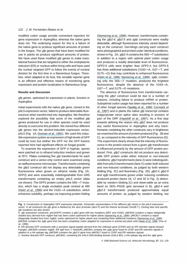
Initial experiments with the native gfp gene, cloned in thepAL5 expression vector, failed to produce detectable fluor-escence when transformed into Aspergillus. We thereforeexplored the possibility that some of the modified gfpgenes produced for use in other species could be useddirectly to overcome this problem. We cloned four differentgfp genes into the alcohol-inducible expression vector,pAL5 (Fig. 1A; Doonan et al., 1991). We used this induc-ible expression system as expression of high levels of GFPcould be toxic but neither the GFP nor the GFP fusionsreported here had significant effects on fungal growth.
To examine the expression of GFP in hyphae, sporeswere patched on to ethanol induction medium and grownat 258C. Plates containing five gfp transformants for eachconstruct and a vector-only control were examined usingan epifluorescence microscope. Transformants containingthe gfp2 construct did not display any detectable greenfluorescence when grown on ethanol media (Fig. 2A,GFP2) and were essentially indistinguishable from GR5transformants containing an empty pAL5 vector (datanot shown). The GFP2 protein contains the S65→T muta-tion, which has a single excitation peak centred at 490(Heim et al., 1995) and the V163→A substitution, whichenhances solubility, perhaps via improving protein folding
(Siemering et al., 1996). However, transformants contain-ing the gfp2-4, gfp2-5 and sgfp constructs were readilydetectable, although fluorescence intensity varied depend-ing on the construct. Germlings carrying each constructwere photographed and printed under identical conditions,shown in Fig. 2A. gfp2-4 contains the S65→T substitutionin addition to a region with optimal plant codon usageand produces a readily detectable level of fluorescence.GFP2-5 cells were brighter than GFP2-4, but GFP2-5has three additional substitutions (V163→A, I167→T, andS175→G) that may contribute to enhanced fluorescence(Heim et al., 1995; Siemering et al., 1996). sgfp, contain-ing only the S65→T mutation, produced the brightestfluorescence, despite the absence of the V163→A,I167→T, and S175→G mutations.
The absence of fluorescence from transformants car-rying the gfp2 construct could be due to a number ofreasons, including failure to produce mRNA or protein.Suboptimal codon usage has been reported for a numberof other fungal species (Spellig et al., 1996; Cormack etal., 1997) and in plants the native gfp transcript containsinappropriate intron splice sites resulting in excision ofpart of the ORF (Haseloff et al., 1997). As a first steptowards defining the reason why gfp2 does not produceany useful fluorescence in Aspergillus and why trans-fromants containing the other constructs vary in brightnesswe examined the amount of protein produced (Fig. 2B andC), as compared to the intensity of GFP fluorescence (D).This experiment clearly shows that the intensity of fluores-cence in the protein extract from a given gfp transfromantis influenced primarily by the amount of GFP protein pro-duced. First, gfp2 constructs did not produce any detect-able GFP protein under either induced or non-inducedconditions: gfp2 transformants (lane 2) were indistinguish-able from pAL5 transformants (lane V) under both inducedand non-induced conditions, as judged by both westernblotting (Fig. 2C) and fluorimetry (Fig. 2D). gfp2-4, gfp2-5and sgfp transfromants grown under inducing conditionsproduced protein (lanes 16, 17 and 32 in Fig. 2) detect-able by western blotting (C) and observable as an extraband on SDS–PAGE gels (arrowed in B). gfp2-4 andgfp2-5 transformants produced approximately equalamounts of protein, as judged by western blotting, but
Q 1998 Blackwell Science Ltd, Molecular Microbiology, 27, 121–130
Fig. 1. Construction of Aspergillus GFP expression plasmids. Schematic representation of the different gfp clones in the pAL5 expressionvector. In all constructs the gfp gene is flanked by the alcA promoter (alcA P) and the histone terminator (His2B T). Cloning sites and specificgfp mutations are also shown.A. gfp expression constructs. pAL5GFP2 contains a gfp2 gene with two mutations S65T and V163A. pMCB16 contains a region (diagonallyshaded box) derived from mgfp4 that has been codon optimized for higher plants (Siemering et al., 1996). pMCB17 contains a region(diagonally shaded box) from mgfp5, codon optimized for higher plants and containing three additional mutations (Siemering et al., 1996).pMCB32 contains the sgfp gene that has been completely codon adapted for expression in animal and plant cells (Haas et al., 1996, Chiu etal., 1996).B. ER-directed GFP constructs with a secretion signal peptide derived from the chitinase gene (ChSP) and an ER retention signal (stripedtriangle). pMCB40 contains mgfp5 -ER (gift from J. Haseloff); pMCB41 contains the sgfp gene fused to ChSP and ER retention signals inaddition to a HA epitope tag; pMCB45 contains the gfp2-5 gene from pMCB17 fused to ChSP and ER retention signals.C. Nuclear directed GFP construct, pMCB42, containing the GAL4 DNA-binding domain (GAL4 BD), a HA epitope tag and the sgfp gene.
122 J. M. Fernandez-Abalos et al.

Q 1998 Blackwell Science Ltd, Molecular Microbiology, 27, 121–130
GFP in Aspergillus 123

the brightness of gfp2-5 transformants was consistently1.5- to 2-fold greater than gfp2-4 transformants. The sub-stitutions, V163→A, I167→T, and S175→G, found inGFP2-5 therefore seem to have a significant enhancingeffect. In fluorimetry experiments, sgfp transformants
tended to produce higher intensities than gfp2-4 or gfp2-5.However, western blot experiments show that sgfp transfor-mants produce higher levels of protein, thereby contribut-ing to the increased fluorescent signal. These differencesare also reflected at the transcript level: the gfp 2 transcript
Q 1998 Blackwell Science Ltd, Molecular Microbiology, 27, 121–130
Fig. 2. Expression of GFP in Aspergillus.A. Confocal microscopy of GFP expression in Aspergillus hyphae. The GFP channel is shown on the left and the same field of view as seenby transmitted light is shown on the right. GFP2, pAL5GFP2; GFP2-4, pMCB16; GFP2-5, pMCB17; sGFP, pMCB32. Magnification ×1500B. SDS–PAGE gel (15%) of protein extracts from Aspergillus transformed with pAL5 vector (V); pAL5GFP2 (2); pMCB16 (16); pMCB17 (17)or pMCB32 (32) and grown under non-inducing or inducing conditions.C. western blot of SDS–PAGE gel with anti-GFP antibodies. The lanes are as described in B.D. Fluorimetry of GFP transformants in repressing and inducing media. Fluorescence units are arbitrary.E. Time course of GFP production after switching hyphae into ethanol media.
124 J. M. Fernandez-Abalos et al.

cannot be detected on Northern blots, whereas sgfp pro-duces stronger signals than gfp2-4 or gfp2-5. This suggeststhat the sgfp gene, having been modified throughout itsentire length, may produce a more stable transcript thanthe partially modified gfp2-4 and gfp2-5, which, in turn, aremore stable than the unmodified gfp gene. The sequencechanges made in the modified genes tend to increase theoverall GC content of the transcript and may lead to morestable transcripts. According to Haseloff et al. (1997) thechanges made in the gfp sequence affect the central regionof the gene (from NdeI to AccI sites, incorporated in ourgfp2-4 and gfp2-5 versions) and affect not only the siteof processing of the cryptic intron but also the usage ofmany codons in this region. The changes effectively pre-vent mis-splicing but no direct effect on translatability canbe drawn from their data. Interestingly, sgfp was designedwith modifications of the codon usage to adapt it to animalcells (Haas et al., 1996), but these modifications allowedimproved expression in plant cells and removed the crypticsplicing points (Chiu et al., 1996). We (J. M. Fernandez-Abalos, unpublished) have found that only the sgfp genefunctions in Streptomyces, a prokaryotic organism withhighly biased codon usage (Wright and Bibb, 1992). Takentogether, these observations suggest that the increase inGC content of the modified gfp genes increases the stabilityof the transcripts in these microorganisms.
To determine the extent to which gfp could be used as adynamic reporter of gene expression, we investigated thetime course of alcohol induction and repression of GFPexpression from the alcA promoter. Cells transformedwith pMCB32 (sgfp) were grown overnight (about 12 h)in shaken rich glucose medium (YG) at 378C and thenswitched to minimal ethanol medium at 308C. Proteinextracts were made from samples taken every 30 min forthe appearance of green fluorescence. No fluorescencewas detected until 1.5 h and thereafter accumulated overa period of several hours (Fig. 2E). When pMCB32 cellsgrown on ethanol were switched to minimal glucose med-ium, fluorescence (per mg of protein) was maintained at ahigh level for at least 4 h and then declined by about 40%every hour (data not shown), As the nuclear doubling timeis about 90–120 min under these conditions, this declinerepresents the combined effect of dilution by cell growthand protein degradation. Considering that the alcA promo-ter responds quickly to changes in carbon source (Patemanet al., 1983), this suggests that GFP protein is turned overrather slowly in Aspergillus.
Location of GFP within the hyphae
One of the major attractions of GFP is the ability to studydynamic events associated with protein targeting and itsregulation in vivo. The constructs pMCB16, 17 and 32were all designed to produce soluble GFP. As judged by
microscopic observation, soluble GFP was present through-out the cytoplasm and was able to enter the nucleus. Thenuclei often appeared brighter than surrounding cyto-plasm, suggesting that GFP accumulated within them. How-ever, GFP was excluded from vacuoles and mitochondria,which were present along the length of the hyphae. Immuno-gold staining of hyphae using antibodies against GFP con-firmed that GFP was absent from the mitochondria andvacuoles but abundant in the cytoplasm and nuclei (datanot shown).
Subcellular localization of GFP using targetingsequences
The ability of gfp transcriptional fusions to monitor proteintrafficking has been demonstrated in a number of systems(reviewed by Cubitt et al., 1995). To test if GFP could betargeted to a specific location, in this case the ER, wecloned mgfp5 (Siemering et al., 1996, J. Haseloff, per-sonal communication) containing ER retention signalsinto pAL5. Two other versions of GFP, GFP2-5 andsGFP, were also engineered to place the plant chitinaseexport signal at the N-terminus of GFP along with theER retention peptide signal on the C-terminus. These con-structs directed GFP to a tubular network within the cell.GFP2-5 produced the brightest fluorescence. A z-seriesof optical sections through hyphae (Fig. 3) shows GFP2-5directed to a branching tubular network containing variouslyshaped brighter nodes. This network extends throughoutthe cell to within 1–2 mm from the tip and surrounds thenucleus. Vacuoles, the cytoplasm and the nucleus all con-tained low levels of GFP. Equivalent constructs in plantshave been demonstrated to target to the ER (Boevink etal., 1996), illuminating a network similar to that which wedescribe here.
GFP can be used to follow nuclear division inAspergillus
GFP is widely used to follow organelle behaviour (forexample Rizzuto et al., 1995). To follow nuclear divisionwe used a GAL4 DNA-binding domain–GFP fusion, whichwe know locates to the nucleus of yeast cells (J. M. Fer-nandez-Abalos, unpublished). In Aspergillus the GAL4BD–sGFP fusion is targeted to the nucleoplasm and thespindle plaque, producing an image very similar to theDNA-specific dye DAPI. The nucleolus contains very lowlevels of GFP. A useful aspect of the GAL4 BD–sGFP pro-tein is its constant association with the nucleus throughoutthe cell cycle. This permits us to follow the dynamics ofnuclear division. Time-lapse photography (Fig. 4) wasused to follow a nucleus from the early stages of mitosis(panels 0 and 1,5), through mid-phase (panels 5 and 9),anaphase (panels 14.5 and 16.5) telophase (panels 18.5
Q 1998 Blackwell Science Ltd, Molecular Microbiology, 27, 121–130
GFP in Aspergillus 125

and 21) and decondensation as the daughter nuclei re-enterinterphase (panels 21, 24 and 29). Under these conditions(effective observation temperature, 198C), mitosis takesbetween 15 and 22 min.
By careful examination of optically sectioned nuclei inthe early stages of mitosis, we can discriminate up toseven discrete strands in individual nuclei. The GFPimage of early mitosis differs markedly form that observedusing DNA-specific dyes on fixed material in that thesestrands are not normally observed. However, as the laterstages of mitosis as observed by GAL4 BD–sGFP closelyresemble that of fixed material and the organism has eightgenetic linkage groups, we suggest that these strands mayrepresent individual chromosomes. During the early stagesof mitosis, the GFP-stained chromosomes appear to beattached at a single point, initially at their extremes butas mitosis proceeds the region of attachment becomesmore diffuse and tends to move towards the middle of thestrands. As the nuclei approach mid-phase, the chromo-somes condense further so that discrete strands areimpossible to resolve (panel 5). Anaphase A movementoccurs very quickly, taking less than 1 min to resolve thechromatids into two groups, about 1–1.5 mm apart. Afterthe initial separation, the daughter nuclei remain con-nected by a thin bridge of GFP-stained material and mayhold this configuration for several minutes. Checks onDNA segregation may occur during this apparent pauseas we have observed occasional bidirectional exchangesof material between the two chromatid masses: smallamounts of GFP-stained material move from one nucleusinto the other and then back (data not shown). When suchexchanges do occur, this phase of mitosis seems to beprolonged, taking up to 15 min in one extreme case (asagainst the norm of 3–4 min). These observations wouldbe compatible with the existence of a mid-anaphase check-point as observed in budding yeast (Yang et al., 1997)implicated as a final check on the correct segregation ofthe genetic material before the two daughters becomeseparate physical entities. After anaphase, the separationbetween daughter nuclei increases to a maximum of 3–5 mm over a period of about two minutes. As the nucleire-enter interphase (as judged by the reappearance of anucleolus) they may recoil together again slightly beforetaking up their final positions. Nuclear movement duringinterphase is relatively slow (compared with mitoticmovements). (See note added in proof.)
Conclusions
Unmodified gfp genes fail to produce useful green fluores-cence when expressed in Aspergillus. We have takenadvantage of the modified forms of gfp that have recentlybeen produced for use in plants to demonstrate their usefor following gene expression, protein localization and
Q 1998 Blackwell Science Ltd, Molecular Microbiology, 27, 121–130
Fig. 3. Plant ER-targeting and retention signals direct GFP to atubular network within the hyphae. A z-series of ER-retained GFPproduced by a transformant containing pMCB45 (ER-GFP2-5). Theimages are 3 mm apart. The final micrograph show a phase imageof the cell. Magnification ×3000.
126 J. M. Fernandez-Abalos et al.

nuclear behaviour in Aspergillus. Expression of GFP inArabidopsis is low because of the presence of a crypticintron, 84 bp long (Haseloff and Amos, 1995; Haseloff etal., 1997). Haseloff’s group modified gfp to specificallyprevent intron splicing by changing the codons in thisregion (modifications that are present in our gfp2-4 ver-sion) and introduced enhancing mutations (present in ourgfp2-5). Haas et al. (1996) have changed codon usagethroughout the entire gene producing the sgfp gene, whichshould also eliminate the splice recognition sites and,because of improved codon usage, may enhance proteinproduction (Chiu et al., 1996). Recently, the sgfp genehas been used as a vital marker in the phytopathogenic
fungus Ustilago maydis (Spellig et al., 1996) and Cor-mack et al. (1997) have shown that codon adaptation isalso necessary for expression of GFP in Candida. Here,we demonstrate that both the intron-modified gfp andthe completely synthetic sgfp genes are both effective inAspergillus. These data, combined with the observationthat the sgfp gene is the only one expressed in otherspecies with extreme codon bias such as Streptomyces(incidentally, the codon usage in sgfp closely approachesthe codon preference in Streptomyces, J. M. Fernandez-Abalos, unpublished), suggest that it may be necessaryto produce a specifically adapted gfp for each group oforganisms.
Q 1998 Blackwell Science Ltd, Molecular Microbiology, 27, 121–130
Fig. 4. A Yeast GAL4 DNA-binding domain–GFP fusion is targeted to the nucleus and provides the means to directly follow mitotic events inliving cells. A time lapse series of nuclear division as visualized by nuclear targeted GFP produced by a transformant containing pMCB42. Thetime in minutes is given in each frame. Magnification ×1500.
GFP in Aspergillus 127

Our results also indicate that subcellular localisation sig-nals from other species, such as the ER retention signalsfrom plants and the nuclear localization signals present inGAL4 DNA-binding domain from yeast, are functionallyconserved. Thus, GFP fusion proteins containing such sig-nals are effective for marking organelles and subcellularcompartments in filamentous fungi without major altera-tions and should be useful for following protein trafficking.However, we did not find that it was necessary to restrictGFP to the ER to obtain acceptable levels of fluorescenceas is the case in Arabidopsis (Haseloff et al., 1997). Finally,the GAL4 BD-GFP fusion marks nuclei and chromatin,allowing us to describe the dynamics of nuclear divisionin Aspergillus for the first time.
Experimental procedures
Construction of GFP expression plasmids
gfp expression constructs, summarized in Fig. 1, were pro-duced using standard protocols (Sambrook et al., 1986). Agfp variant with mutations S56→T and V163→A (calledgfp2 throughout this work) and cloned in pBluescript KS(þ)(plasmid pKSGFP2) was used as the starting point for mostof the constructions. The NdeI/Bst BI in gfp2 was replacedwith the corresponding NdeI/Bst BI fragment from mgfp4 ormgfp5 that carry modifications of the DNA sequence toadapt the codon usage in this region to that in higher plantsand to incorporate several point mutations (Siemering et al.,1996) giving gfp2-4 and gfp2-5 versions. All three variantswere inserted in the Aspergillus vector pAL5 (Doonan et al.,1991) as Asp718I/BamHI fragments to produce pAL5GFP2,pMCB16 and pMCB17 respectively (Fig. 1A).
In a different set of constructions we used a synthetic sgfpgene (sGFPS65T, Chiu et al., 1996, Haas et al., 1996, referredto as sgfp in this work) in which the whole sequence of the gfpgene has been changed to adapt its codon usage for mam-malian and plant cells and carrying mutation S65→T (Chiuet al., 1996; Haas et al., 1996,). As the starting point for ourconstructions we used plasmid pB1 (that contains sgfp beforethe ‘nos’ transcriptional terminator, J. Sheen personal com-munication). The ‘nos’ terminator was looped out by digestionwith Pst I and religation, giving plasmid pMCB30 that containsappropriate Asp718I and BamHI sites before and after sgfp,and that were used to transfer it to pAL5 to give plasmidpMCB32 (Fig. 1A).
Construction of GFP-translational fusions
For ER-targeted GFPs, we used mgfp5 (Siemering et al.,1996) that contains a plant chitinase signal peptide at theN-terminus and an ER retention signal (His–Asp–Glu–Leu)at the C-terminus, either directly or with replacement of theinternal region of the gene with appropriate fragments fromgfp2-5 or sgfp. All three versions were inserted in pAL5 at theAsp718I/SmaI sites, giving plasmids pMCB40 (with mgfp5),pMCB41 (with sgfp) and pMCB45 (with gfp2–5 ) (Fig. 1B).A translational fusion was made to target GFP to the nucleususing the GAL4 DNA-binding domain from budding yeast
(Fields and Song, 1989). The NcoI/BamHI fragment frompMCB30 that carries sgfp was inserted in NcoI/BamHI sitesof the yeast vector pAS2. This generates an in-frame fusionof sgfp with the DNA-binding domain of GAL4 (GAL4BD)and the HA tag present in the vector, and rendered plasmidpMCB31. Such a fusion is targeted to the nucleus in yeast(data not shown). Digestion of pMCB31 with HindIII (beforethe coding region of the GAL4BD) and Pst I (after sgfp) pro-vides a DNA fragment that was cloned in pBluescript SK(þ)to give plasmid pMCB38, when appropriate Asp718I andBamHI sites allowed for cloning of the fusion in pAL5. Theresulting plasmid, pMCB42, contains the fusion GAL4BD:HAtag:sGFP (Fig. 1C).
Culture conditions, strains and transformation
The Aspergillus strain GR5 (pyrG89, wA3), used for all mani-pulations, was grown on YG media (2% w/v yeast extract, 1%glucose, 1% agar) containing 10 mM uridine and uracil andtransformed as previously described (Osmani et al., 1988).Briefly, protoplasts were prepared from freshly germinatedconidiospores and transformed with plasmid DNA using PEG.Transformants were selected on YG media containing 0.6 MKCl and lacking uridine/uracil and allowed to sporulate. Con-idiospores were streaked to single colony three times onselective media to obtain pure transformants. Expression ofGFP was monitored microscopically by plating spores on mini-mal agar media containing 1% ethanol to induce gene expres-sion or on 100 mM glucose to repress expression (Waring etal., 1989); a coverslip was placed on top of the germlings andobservations made as described below.
To quantify the GFP production, spores (107 ml¹1) weregerminated in shaken liquid YG medium for 12 h, harvestedby filtration through Miracloth and washed with 200ml of sterilewater, resuspended in either induction (2% lactose/1% ethanol)or repression (3% glucose/1% sucrose) media and grown for4 h at 308C. Cells were harvested as before and disrupted inlysis buffer (100 mM Tris-HCl pH 7.5, 1 mM EDTA, 5 mMDTT, 1 mM freshly added PMSF, 5 mg ml¹1 aprotinin and5 mg ml¹1 pepstatin-A, Calera et al., 1997) in 2 ml tubes con-taining 0.5 g of 0.4–0.6 mm glass beads, using 3 × 20 s pulsesat 6.0 speed in a Hybaid Ribolyser. Lysates were cooled onice, spun at 14 000 r.p.m. for 5 min at room temperature(RT) and the cleared supernatant (representing total solubleproteins) was kept frozen at ¹208C. Protein concentrationwas determined using a modified Bradford assay (BioRad)and samples were prepared for SDS–PAGE by adding 90 mlof cleared lysate to 30 ml of preheated (958C) 4× sample buf-fer. A 10 mg sample of protein was loaded to each lane of a15% polyacrylamide gel. After separation, the gel was blottedto Immobilon P membrane (Millipore) and, after blocking in5% dried milk in Tris-buffered saline (TBS, 10 mM Tris-HCl,150 mM NaCl, pH 8), was probed with anti-GFP polyclonalantibodies (Clontech) at 1:20000 dilution in TBS containing1% dried milk. The blot was washed for 15 min each in (I)TBS þ 0.05% Trition X-100 (TBST); (ii) TBST þ 0.5 M NaCl;and (iii) TBST, before incubating with HRP-conjugated swineanti-rabbit IgG 1:10000 in TBS with 1% dried milk. After wash-ing as described above, the blot was developed using theSuperSignal ULTRA chemioluminscence detection system(Pierce) and recorded using Kodak X-Omat film. Bands were
Q 1998 Blackwell Science Ltd, Molecular Microbiology, 27, 121–130
128 J. M. Fernandez-Abalos et al.

scanned into Adobe Photoshop and the relative amounts ofGFP protein estimated using the same package.
Fluorimetry
Cell lysates obtained as described above were used for fluori-metry. Duplicate samples were measured in a Titertek Fluoro-skan II fluorimeter using a 480/538 nm excitation/emissionfilter block, with 100 mM Tris-HCl pH 7.5 buffer as a blank.Fluorescence was recorded as absolute fluorescence units,normalized against the protein concentration in the samplesand expressed as arbitrary fluorescence units per mg protein.Extracts from a transformant containing an empty pAL5expression plasmid were used to determine the backgroundfluorescence.
Light microscopy
Conidiospores from at least five independent transformantswere patched on ethanol induction media (Waring et al.,1989) and allowed to germinate overnight at 258C. To reducebackground the media did not contain riboflavin. Just beforeobservation a coverslip was placed on top of the germinatingspores and they were observed using Neofluar ×100 lens, NA1.3 on either a Zeiss epifluorescence microscope or on aBioRad MRC-600 confocal microscope using a 488 nm Kryp-ton laser and standard Zeiss FITC filter block. Images werecollected using NIH Image and transferred to Adobe Photo-shop version 4.0.
Note added in proof
While this work was under review the following paperdescribed the use of SGFP fusions with stuA to follow nuclearmigration in Aspergillus: Suelmann, R., Sievers, N., andFischer, R. (1997) Nuclear traffic in fungal hyphae: in vivostudy of nuclear migration and positioning in Aspergillusnidulans. Mol Microbiol 25: 757–769.
Acknowledgements
We are grateful to Jim Haseloff for modified mgfp4 and mgfp5clones and helpful advice, to Roger Tsein, Jen Sheen andTilman Spellig for the synthetic sgfp, to Fernando Leal foradvice on protein extracts and to Carol Wymer and PeterShaw for assistance with confocal microscopy and imageanalysis. J.M.F-A was supported by a short-term grant fromthe European Union PTP Programme and HF and CP by Stu-dentships from the BBSRC.
References
Beckwith, S.M., Roghi, C.H., and Morris, N.R. (1995) TheGenetics of nuclear migration in fungi. In Genetic Engin-eering. Setlow, J.K (ed.). New York: Plenum PublishingCorporation, pp. 165–180.
Boevink, P., Santa Cruz, S., Hawes, C., Harris, N., andOparka, K.J. (1996) Virus mediated delivery of the green
fluorescent protein to the endoplasmic reticulum of plantcells. Plant J 10: 935–941.
Calera, J.A., Ovejero, M.C., Lopez-Medrano, R., Segurado, M.,Puente, P., and Leal, F. (1997) Characterization of theAspergillus nidulans aspnd1 gene demonstrates that theASPND1 antigen, which it encodes, and several Aspergillusfumigatus immunodominant antigens belong to the samefamily. Infect Immun 65: 1335–1344.
Chalfie, M., Tu, Y., Euskirchen, G., Ward, W.W., and Prasher,D. (1994) Green Fluorescent Protein as a marker for geneexpression. Science 263: 802–805.
Chiu, W.-L., Niwa, Y., Zeng, W., Hirano, T., Kobayashi, H.,and Sheen, J. (1996) Engineered GFP as a vital reporterin plants. Current Biology 6: 325–330.
Cormack, B.P., Bertram, G., Egerton, M., Gow, N.A.R., Fal-kow, S., and Brown, A.J.P. (1997) Yeast-enhanced greenfluorescent protein (yEGFP): a reporter of gene expressionin Candida albicans. Microbiology 143: 303–311.
Cubitt, A.B., Heim, R., Adams, S.R., Boyd, A.E., Gross, L.A.,and Tsien, R.Y. (1995) Understanding, improving and usinggreen fluorescent proteins. Trends Bio Sci 20: 448–455.
Doonan, J.H. (1992) Cell growth and nuclear division in asimple multicellular organism, Aspergillus nidulans. J.CellSci 103: 599–611.
Doonan, J.H., MacKintosh, C., Cohen, P., Osmani, S.A., Lee,E., and Morris, N.R. (1991) A cDNA encoding rabbitmuscle phosphoprotein phosphatase complements theAspergillus cell cycle mutation, bimG11 J Biol Chem 266:18889–18894.
Fields, S., and Song, O. (1989) A novel genetic system todetect protein: protein interactions. Nature 340: 245–246.
Gallie, D.R., Lucas, W.J., and Walbot, V. (1989) VisualizingmRNA expression in plant protoplasts: factors influencingefficient mRNA uptake and translation. Plant Cell 1: 301–311.
Haas, J., Park, E.-C., and Seed, B. (1996) Codon usage limit-ation in the expression of HIV-1 envelope glycoprotein.Curr Biol 6: 315–324.
Haseloff, J., and Amos, B. (1995) GFP in plants. TrendsGenet 11: 328–329.
Haseloff, J., Siemering, K.R., Prasher, D., and Hodge, S.(1997) Removal of a cryptic intron and subcellular localis-ation of green fluorescent protein are required to marktransgenic Arabidopsis plants brightly. Proc Natl Acad SciUSA 94: 2122–2127.
Heim, R., Cubitt, A.B., and Tsien, R.Y. (1995) Improvedgreen fluorescence. Nature 373: 663–664.
Jefferson, R.A., Kavanagh, T.A., and Bevan, M.W. (1987)Assaying chimeric genes in plants: the GUS gene fusionsystem. Plant Mol Biol Rep. 5: 387–405.
Osmani, S., Engle, D., Doonan, J.H., and Morris, N.R. (1988)Spindle formation and chromatin condensation in cellsblocked at interphase by mutation of a negative cell cyclecontrol gene (bimE ) of Aspergillus nidulans. Cell 52: 241–251.
Pateman, J.A., Doy, C.H., Olsen, J.E., Norris, U., Creser, E.H.,and Hynes, M. (1983) Regulation of the alcohol dehydro-genase (ADH) and aldehyde dehydrogenase (AldDH) pro-moter in Aspergillus nidulans. Proc Royal Soc London B217:243–264.
Q 1998 Blackwell Science Ltd, Molecular Microbiology, 27, 121–130
GFP in Aspergillus 129
Rizzuto, R., Brini, M., Pizzo, P., Murgia, M., and Pozzan, T.(1995) Chimeric Green fluorescent protein as a tool forvisualizing subcellular organelles in living cells. Currentbiology 5: 635–642.
Sambrook, J., Fritsch, E.F., and Maniatis, T. (1986) Molecu-lar Cloning. A Laboratory Manual, 2nd edn. Cold SpringHarbor, New York: Cold Spring Harbor Laboratory Press.
Siemering, K.R., Golbick, R., Sever, R., and Haseloff, J.(1996) Mutations that suppress the thermosensitivity ofgreen fluorescent protein. Curr Biol 6: 1653–1663.
Spellig, T., Bottin, A., and Kahmann, R. (1996) Green fluo-rescent protein (GFP) as a new vital marker in thephytopathogenic fungus Ustilago maydis. Mol Gen Genet252: 503–509.
Timberlake, W.E. (1990) Molecular genetics of Aspergillusdevelopment. Ann Rev Genet 24: 5–36.
Waring, R.B., May, G.S., and Morris, N.R. (1989) Develop-ment of an inducible system in Aspergillus nidulans usingthe alcA and tubulin genes. Gene 79: 119–130.
Wright, J., and Bibb, M. (1992) Codon usage in the G þ C-rich Streptomyces genome. Gene 113: 55–65.
Yang, S.S., Yeh, E., Salmon, E.D., and Bloom., K (1997)Identification of a mid-anaphase checkpoint in buddingyeast. J Cell Biol 136: 345–354.
Zolotukhin, S., Potter, M., Hauswirth, W.W., Guy, J., andMuzyczka, N. (1996) A humanized green fluorescent pro-tein cDNA adapted for high-level expression in mammal-ian-cells. J Virol 70: 4646–4654.
Q 1998 Blackwell Science Ltd, Molecular Microbiology, 27, 121–130
130 J. M. Fernandez-Abalos et al.
Copyright © 2022 FDOKUMEN