Multifunctionality of plant ABC transporters - more than just detoxifiers

www.cell-research.com | Cell Research
Tingting Lin et al.857npg
ABC transporters, neural stem cells and neurogenesis – a different perspectiveTingting Lin1,*, Omedul Islam1,*, Klaus Heese1
1Department of Molecular and Cell Biology, School of Biological Sciences, Nanyang Technological University, 60 Nanyang Drive, Singapore 637551, Singapore
REVIEW
Cell Research (2006) 16: 857-871© 2006 IBCB, SIBS, CAS All rights reserved 1001-0602/06 $ 30.00 www.nature.com/cr
npg
Introduction
Taking advantage of the natural potential of self-renewal of the living body, “regenerative medicine” is sure going to play an essential role in providing an innovative way to treat many human disorders. Stem cells are the engines that drive the renewal of adult mammalian tissues. They divide continuously, throughout life, to produce new progeny cells that undergo a robust development program towards differentiation and maturation to replace old expired tissue cells. They are a unique population of cells capable of self-renewal and differentiation into different cell types. A major effort in regenerative medicine is the search for suitable
renewable sources of cells that can be used as means to treat human diseases. Stem cells, capable of eluding detec-tion by the host’s immune system and with the potential of expansion in culture, strike as a very promising source of cells for therapeutic applications [1].
Stem cells can be categorized as either embryonic stem (ES) or adult stem cells. Adult stem cells exist as undif-ferentiated cells interspersed among differentiated ones in a tissue or organ, and exhibit the intrinsic ability to self-renew and to potentially differentiate into the major specialized cell types of the tissues or organs. However, they cannot give rise to all cell types of an organism and can only differentiate into restricted lineages. Therefore, in contrast to totipotent ES cells, these tissue-specific stem cells are considered multipotent or pluripotent. A variety of stem cell populations have been discovered residing within adult tissues like the brain, bone marrow, blood, liver, pancreas and others [2, 3]. Of these, the hematopoi-etic stem cells (HSCs) from bone marrow can differentiate
*These two authors contributed equally to this workCorrespondence: Klaus HeeseTel: +65-6316-2848; Fax: +65-6791-3856; E-mail: [email protected]
Stem cells intrigue. They have the ability to divide exponentially, recreate the stem cell compartment, as well as cre-ate differentiated cells to generate tissues. Therefore, they should be natural candidates to provide a renewable source of cells for transplantation applied in regenerative medicine. Stem cells have the capacity to generate specific tissues or even whole organs like the blood, heart, or bones. A subgroup of stem cells, the neural stem cells (NSCs), is characterized as a self-renewing population that generates neurons and glia of the developing brain. They can be isolated, genetically manipulated and differentiated in vitro and reintroduced into a developing, adult or a pathologically altered central nervous system. NSCs have been considered for use in cell replacement therapies in various neurodegenerative diseases such as Parkinson’s disease and Alzheimer’s disease. Characterization of genes with tightly controlled expression patterns during differentiation represents an approach to understanding the regulation of stem cell commitment. The regulation of stem cell biology by the ATP-binding cassette (ABC) transporters has emerged as an important new field of investigation. As a major focus of stem cell research is in the manipulation of cells to enable differentiation into a targeted cell population; in this review, we discuss recent literatures on ABC transporters and stem cells, and propose an integrated view on the role of the ABC transporters, especially ABCA2, ABCA3, ABCB1 and ABCG2, in NSCs’ proliferation, differentiation and regulation, along with comparisons to that in hematopoietic and other stem cells.
Cell Research (2006) 16:857-871. doi:10.1038/sj.cr.7310107; published online 7 November 2006
Keywords: neural stem cells, ABC transporters, ABCB1, ABCG2, differentiation, stem cell marker
Cell Research | www.cell-research.com
ABC transporters and neurogenesis858npg
into all the different immune cells and have been routinely used to treat leukemia, lymphoma and immune deficien-cies [4, 5]. A certain class of HSCs from blood and bone marrow called the “side population” (SP) is described also as CD34-negative (-), c-Kit-positive (+) and Sca-1+ cells, according to their specific surface antigens [6, 7]. These bone marrow repopulating SP cells are probably among the best-characterized examples of pluripotent adult stem cells to date [8, 9].
We now know that different types of stem cells exist, but they are found in minute populations in the human body. Hence, the use of biologically unique stem cell markers is required to stringently identify and isolate these cells. The consensus working definition of stem cells is that they can both replenish their own population and differentiate to form committed daughter cells [10]. The candidate stem cell must also demonstrate the ability to proliferate, self-renew over an extended period of time (and not just once or a few times as observed with the progenitor cells), and generate a large pool of progeny that can differentiate into the primary cell types of the tissue from which it is obtained [11]. In addition to their ability to self-renew and differ-entiate, they are usually quiescent, dividing infrequently [12, 13]. To accurately define a cell as a stem cell, it is necessary not only to confirm the renewal capability of the founding cell or population of cells over an extended period of time but also to demonstrate the generation of progeny cells several orders of magnitude more numerous than the starting population.
The essential role of tissue stem cells is then to maintain and repair the tissue in which they are found. They also require the presence of a tightly regulated environmental niche comprising of other cell types, stroma and growth factors for survival [14, 15]. Due to the above-mentioned characteristics of stem cells, they are especially attractive candidates in neuroregenerative therapy applications. For instance, their self-renewing ability would be useful for the treatment of neurodegenerative diseases such as Parkinson’s disease (PD) and Alzheimer’s disease (AD). Since stem cells and their differentiation products are potentially of broad therapeutic uses, efforts that would enable the supply of a continual source of primitive stem cells in vitro will be vital in revolutionizing the treatment of many diseases [16-18].
The most remarkable changes in the brain that occur with aging may be the alterations in cognition and plasticity. Aging of the central nervous system (CNS) is associated with a progressive loss of neural functions, which is exac-erbated in neurodegenerative disorders such as AD [19-21]. Two major cell replacement strategies are considered for the therapy of neurodegenerative disorders such as AD, and involve transplantation of exogenous tissue/cells and
activation of proliferation of endogenous cells, respectively [22-25]. Transplanted tissue/cells are used to either directly replace the lost tissue or to deliver genetically engineered neural stem cells (NSCs) that can secrete factors which promote cell survival and proliferation. However, suc-cessful application of any cell replacement therapy likely will require the understanding of the complex relationships between NSCs and the more restricted neural and glial progenitor cells, as well as the underlying biology of their renewal and differentiation programs.
Neural stem cells
The anatomical location and lineage specificities of NSCs were only established when they were finally identi-fied in the subependymal region and in the hippocampal dentate gyrus (DG), where they divide to generate pro-genitors that migrate along the rostral migratory stream to differentiate in the olfactory bulb or to integrate into the surrounding hippocampal neural circuitry, respectively [26-28]. Similar to HSCs, these nestin+ NSCs may be defined operationally as cells that can continuously self-renew and have the potential to generate intermediate and mature cells of both glial and neural lineages [29]. Furthermore, NSCs have also been reported to differentiate into hematopoietic cells [30].
It has been found recently that both endogenous and transplanted NSCs seem to be attracted to various experi-mental brain lesions of disparate etiologies, such as tumors or areas of neurodegeneration [31]. For instance, NSCs have shown tropism toward gliomas and toward degenerating spinal cord motor neurons in a transgenic mouse model of amyotrophic lateral sclerosis [32-34]. Tumor-tropic NSCs have also been observed in peripheral malignancies apart from those primary brain malignancies. It is still challeng-ing to understand the fate of NSCs in brain lesions [35]. In certain pathologies, such as in stroke lesions, transplanted cells appear to form astrocytes and neurons [36]. Some-times, a glial fate, even if not ideal, may still be preferred over neural differentiation, as the latter might form abnor-mal and possibly damaging circuits. Reactive astrocytosis induced by inflammatory cytokines released by microglia in response to a pathological process is characterized by an increase in glial fibrillary acidic protein (GFAP), showing that GFAP is a marker in the differentiation of NSCs into astrocytes [37, 38].
Neurosphere cultures are regularly used as a source of primitive neural cells. Such cultures have been character-ized by Ruud Hulspas and Peter J Quesenberry as the means to purify neural progenitor cells, and several subpopulations of cells have been defined [39]. Neurospheres are useful to evaluate NSC multipotentiality (through the character-

www.cell-research.com | Cell Research
Tingting Lin et al.859npg
ization of cell phenotypes that arise from a differentiating sphere) and to analyze NSCs’ self-renewal capacity using clonal secondary neurosphere assays that assume isolated true stem cells can generate new spheres [40].
Neurospheres are floating structures that can be obtained by exposing dissociated embryonic or adult CNS tissue to growth factors [41-43]. These heterogeneous spheroid structures contain NSCs/progenitors and differentiated cells embedded in a complex extracellular matrix and organized three dimensionally (3D) with a core of dif-ferentiating GFAP+ and b-tubulin III+ cells surrounded by nestin+, epidermal growth factor receptor (EGFR)+ and b1-integrin+ undifferentiated cells [40]. Thus, neurospheres consist predominantly of committed progenitors mixed with differentiated astrocytes as well as neurons, with rela-tively few stem cells [44]. Histotypical 3D neurospheres in suspension culture are thus characterized as heterogeneous clusters containing unequal stem cell subtypes where nes-tin+ dividing cells surround a core of differentiated GFAP+ and b-tubulin III+ cells. Cell divisions can be found mainly at the edge of the neurosphere where NSC markers such as notch1 and nestin are also expressed [41]. This pattern suggests that NSCs may be a rare subgroup of nestin+ cells that simultaneously express a combination of markers, such as LeX/ssea1/b1-integrin/notch1 [40, 41, 45]. Furthermore, some adult astrocyte-like GFAP+ cells are actually NSCs, which are probably derived from an embryonic glial lineage [46]. Therefore, the 3D neurosphere organization could give some indication of whether a neurosphere is generated by a true stem cell or by a non-stem cell population when the neurospheres are characterized by staining with a stem cell marker like nestin or musashi-1 [47, 48]. The DNA-binding dye Hoechst-33342 has been used as a method to identify potential stem cells in a host of tissues, including the bone marrow, heart, lung, muscle, eye and pancreas [9]. The dual emission of the Hoechst dye generates a distinct SP from the whole population of cells. This unique seg-regation is conferred by the ATP-binding cassette (ABC) transporter proteins, such as ABCB1, which actively pump out the Hoechst dye. In fact, when the whole-cell population is treated with verapamil, an inhibitor of these transporters, the SP phenotype is lost [9]. Interestingly, it was observed that neurosphere cultures contain a relatively higher number of stem cells that stain weakly with Hoechst-33342 when cultured in presence of EGF and basic fibroblast growth fac-tor (bFGF); however, when cultured separately either with EGF or bFGF, they give rise to neurosphere populations with characteristics of freshly isolated embryonic mouse brain stem cells but with much fewer stem cells that stain weakly with Hoechst-33342 [41, 49].
Single-cell studies have provided new insights into NSC biology by demonstrating the role of symmetric and asym-
metric divisions in cell fate determination [40]. Asymmetric divisions are crucial to generate diversity, whereas symmet-ric divisions allow for maintenance of the stem cell pool or for the expansion of a progenitor population. The “feature” of NSCs may be a complex state that can be acquired or lost [50]. Both the survival capacity and proliferation potential of cells are fundamental for stem cell maintenance and re-newal, or alternatively may be essential for the generation of a cell population that can subsequently acquire stem cell properties. The environmental niche where the cells reside may play a fundamental instructive role in NSC develop-ment [51]. The term NSC may therefore need to include the capacity of a cell to survive and to depend on a niche to acquire or maintain stem cell status [52].
ABC transporters
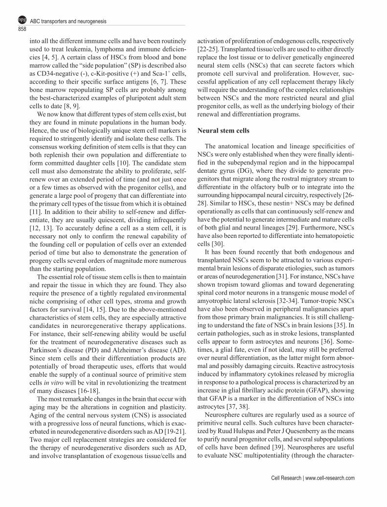
ABC transporters constitute one of the largest known superfamilies of proteins. These evolutionary highly con-served multispan transmembrane molecules use the energy of ATP hydrolysis to translocate a broad spectrum of mol-ecules across the cell membrane. To date, 48 members of the human family of ABC transporters have been identified which, based on their structural relatedness, are subdivided into seven families, designed ABC A-G. [53-58]. These ABC transporters participate in diverse cellular processes, including drug resistance and metabolism, transport of lipids and organic anions, and iron metabolism [54, 55]. The various transporters exhibit different catalytical proper-ties (for instance substrate specificity, mode of transport, transporter vs channel, protein-protein interactions), and additional substances that are transported by the ABC molecules include peptides, amino acids, carbohydrates, vitamins, glucoronide, glutathione conjugates and xeno-biotics [59-63]. In eukaryotes, the ABC transporters are located in the plasma membrane and the membranes of intracellular compartments such as the Golgi, endosomes and in the mitochondria [61]. When comparing various ABC transporter subfamilies – one major difference be-tween the transporters is the presence of the additional N-terminal extension in a transmembrane domain (called TMD0) in some members of the ABCC-subfamily [64]. In contrast, the order of the TMDs and nucleotide binding domains (NBDs) in the two-dimensional structure of most of the transporters is identical. Membrane-associated ABC transporters have been found in two forms, the full-length transporters that are characterized by two identical halves each containing the NBD and the half-transporters that function as homo- or heterodimers (Figure 1) [53-55,64]. As one of the best studied ABC transporters, the multidrug resistance (MDR1) P-glycoprotein (P-gp), also known as ABCB1, is a glycosylated membrane-associated enzyme
Cell Research | www.cell-research.com
ABC transporters and neurogenesis860npg
of the full-length form comprising 1 280 amino acids (with a molecular weight of 170 kDa), and is characterized by two identical halves, each with one NBD; it exports a wide range of diverse substrates [54, 55, 57]. Another member of the ABC transporter family that is intensively studied is ABCG2 (also known as MXR/BCRP1/ABCP), which is a half-transporter and requires dimerization for its func-tional activity [65, 66]. Unlike other ABC half-transporters,
which are localized to intracellular membranes at numer-ous compartments within the cell, ABCG2 is expressed exclusively in the plasma membrane [65, 66]. ABCG2 has been identified to confer resistance to anthracycline anticancer drugs, and is expressed in both malignant and normal tissues [67-70]. It is a membrane-associated 663 amino-acid transporter that is highly expressed in placenta and HSCs (SP/CD34) [71-73]. ABCG2 exhibits transporter
Figure 1 Structural features of ABC transporters. Most of the transporters are membrane-associated with six membrane-spanning regions, and are characterized by the presence of the ABC region. (A) ABCB1 typically is a full-length transporter of two identical halves as shown, with two NBDs that contain conserved sequences of the ABC. (B) ABCG2, on the other hand, is a half-transporter consisting of one NBD containing an ABC, followed by a six membrane-spanning domain.
Extrusion of xenobiotics
Cell membrane
Cytoplasm
NNBD
ATP ADP
NBD
ATPADP
Extrusion of cytotoxins
Cell membrane
Cytoplasm
NBD
ATP ADP
C
N
A
B
C

www.cell-research.com | Cell Research
Tingting Lin et al.861npg
activity for several exogenous substrates such as cytotoxic drugs, fluorescent dyes and endogenous substrates such as folic acid, sulfated conjugates of steroids, bile salts and porphyrins [74]. However, the precise physiological role for ABCG2 has not yet been clearly defined despite extensive research.
Several ABC transporters are also called multidrug resistance proteins (called MRPs or MDRs) because these membrane glycoproteins mediate the ATP-dependent ex-port of organic anions, including cytotoxic and antiviral drugs from cells [64, 59, 75, 76]. For instance, the human ABC transporter subfamily C (symbol ABCC) consists of 12 members, nine of which comprise a group of multi-drug resistance proteins (also known as MRP1-MRP9 or ABCC1-ABCC6, ABCC10-ABCC12) [64, 65, 76, 77]. In-sofar, MRP1-MRP6 are the best-characterized members of the ABCC subfamily. Conjugates of lipophilic compounds with glutathione, glucuronate or sulfate are the preferred physiological substrates of MRP1 and MRP3 [78-83]. MRP6 is described to transport glutathione conjugates as well as the endothelin receptor antagonist BQ123 [78-82]. Substrates for MRP4 and MRP5 include cyclic AMP, cyclic GMP and nucleotide analogues [78-82]. Reduced glutathione (GSH) has been recognized as a co-substrate for MRP1, for MRP2 and most recently for MRP4 [78-82]. In addition to being important export pumps for physiological substances, MRP subfamily members are involved in the active efflux of toxic organic anions [78-82]. MRP trans-porters have been shown to confer resistance to harmful drugs, possibly by the above-mentioned ATP-dependent export of such molecules [54-57].
ABCB1 is widely expressed in normal tissues with excretory functions such as the liver, kidney and intestine, and is also involved in barrier functions such as in the blood-brain barrier (BBB) and the blood-testis barrier [54, 57, 84, 85]. Apart from being induced by chemotherapeutic drugs, ABCB1 expression and upregulation of its activity have been observed in primary rat hepatocyte culture by EGF [86]. Also, ABCB1 expression actively protects cells against cell death and ABCG2 defends against natural heme metabolites [70, 87-91]. By transporting a variety of exog-enous and endogenous compounds out of cells, they are able to reduce the body load of potentially harmful substances. However, one side effect of this protective function is that they also eliminate various useful drugs from the body, causing drug resistance [55, 56, 92]. Interestingly, ABCB1 expression is also found to be induced in paradigms of tis-sue regeneration [92-100].
ABCB1 activation was studied in vivo mainly in the liver, and in vitro, in hepatocytes [86, 94-96]. In addition, transient ABCB1 upregulation in vivo, which is associated with the tissue stress response, has been described in neu-
rons and glial cells in the brain [97-99]; EGF was found to have a stimulatory effect on ABCB1 phosphorylation and multidrug resistance activity in a breast cancer cell line [100].
ABC transporters in the brain
In the brain, the proteins MRP1, MRP4 and MRP5 (ABCC family) were clearly localized, by confocal laser scanning microscopy, to the luminal side of brain capillary endothelial cells [101]. The MRP4 and MRP5 proteins were also detected in astrocytes of the subcortical white matter [101, 102]. Notably, MRP5 protein was present in pyramidal neurons [101]. Another study has revealed that MRP1 and MRP5 are more abundant in various brain cells than the other family members though MRP3 and MRP4 could also be detected in astrocytes [103]. MRP proteins may thus contribute to the resistance of the brain to several cytotoxic and antiviral drugs [104].
A recent real-time reverse transcription-polymerase chain reaction (RT-PCR) assay has been used to investi-gate the specific expression pattern of the ABC subfam-ily-A transporters in the brain and has shown that neurons express predominantly ABCA1 and ABCA3; astrocytes express ABCA1, ABCA2 and ABCA3; microglia express ABCA1 and oligodendrocytes express ABCA2 and ABCA3 [105]. With its expression in liver and brain, ABCB1 – the prototype of the B subclass of transporters – and ABCA1 regulate the high-density lipoprotein levels in the plasma and cholesterol contents of several cell types in these organs [106-114]. Most interestingly, the ABCB1 transporter also shows strong expression in neurons of the hippocampus formation, particularly in the granule cells of the DG [85]. Volk et al. [99] have demonstrated neuronal upregulation of ABCB1 expression in the CA3/CA4 region and hilus of the hippocampus formation 24 h after inducing a status epilepticus in rat brains. In general, however, ABCB1 is predominantly localized in the apical membrane of capil-lary endothelial cells which form the BBB, and in epithelial cells of the blood-cerebrospinal fluid barrier, while other cell types in the brain show little or no expression under normal conditions [84, 85, 97-99].
ABC transporters and diseases
In humans, genetic defects of ABC transporters have been implicated in several diseases involving transport deficiencies. Dysfunctions of these proteins have been caus-ally related to several pathological phenotypes, spanning from neurological or metabolic diseases to drug resistance [115-117]. For instance, the most common fatal autosomal recessive genetic disease affecting Caucasian populations,

Cell Research | www.cell-research.com
ABC transporters and neurogenesis862npg
cystic fibrosis, is caused by mutations in the ABCC7 trans-porter (or CFTR), which is a regulated ion channel [118]. Most of the naturally occurring mutations in the ABCC7 gene either induce an alteration in protein biosynthesis or lead to a defective channel function [60, 64, 119, 120]. Approximately 80% of all mutations found with the ABC transporters are located within the NBDs.
Recent findings have shown that ABCG2 is strongly ex-pressed on progenitor cells/reactive ductules in human liver, which might protect them from cytotoxic agents [121]. This could explain why in almost all conditions of liver damage and cell loss, progenitor cells/reactive ductules are able to withstand this damage and even expand and contribute to the repair process by differentiating into hepatocytes and/or cholangiocytes [121-125]. A rather basolateral hepatocytic expression in chronic biliary diseases may be an adaptive mechanism to pump bile constituents back into the sinu-soidal blood [120].
Since ABC transporters exert their functions as mem-brane associated multi-unit complexes by binding to other important functional proteins such as ion-channels [126, 127], this may explain the association of ABC transporter mutations with a wide spectrum of inherited diseases. Because ABC transporters likely exert their biological function in cooperation with possibly varying interacting partners, the observed range of clinical phenotypes associ-ated with mutations in one ABC molecule may also reflect genetic variations of proteins that specifically interact with ABC transporters. Moreover, in molecular terms, such functional pleiomorphism is related to the wide range of compounds that ABC family members can transport. Thus, the genotype/phenotype diversity in individuals with mu-tated ABC transporters may primarily reflect the functional complexity of these large multi-span polypeptides [60, 64, 119, 120].
ABC transporters and stem cells
It was first demonstrated by Chaudhary and Robinson that the ABCB1 transporter is highly expressed on CD34+ hematopoietic cells, suggesting that the efflux pump activity could be responsible for the low retention of the fluorescent dye rhodamine 123 in these primitive subsets of cells. Rh123 is another fluorescent dye whose efflux can be used to enrich for potential stem cells. In bone marrow, the percentage of cells in the Rh123-effluxing subset is similar to that in the Hoechst-dye-effluxing SP cells (i.e. about 10%). However, the former does not segregate into a population as distinct as the Hoechst-dye-effluxing SP cells and, thus, must be used in combination with other surrogate stem cell markers for stem cell isolation [9,128]. Later Goodell et al. [9] demonstrated that a highly enriched
stem cell fraction termed SP could be isolated following Hoechst-33342 staining. Recently, ABCA3, ABCB1 and ABCG2 were found in the primitive stem cells of differ-ent tissues [129-136]. In particular, ABCB1 is expressed in human CD34+ stem cells, which can be identified by their ability to transport fluorescent dyes like Rh123 and Hoechst-33342 [128, 129, 137-143]. Interestingly, the SP phenotype in rodent and human tissues often appears to be specifically determined by the expression of an ABC trans-porter: for example, both ABCB1 and ABCG2 transporters are highly expressed in the SP of stem cells from different tissues such as brain, bone marrow, pancreas, liver and oth-ers, all of which can be isolated based on the cells’ ability to promote the efflux of the Hoechst-33342 fluorescent dye [9, 129-136, 142-150]. Moreover, different research teams have demonstrated that ABCA3 and ABCG2 were expressed at higher levels in SP cells than in non-SP cells in human, rhesus monkey and mouse hematopoietic tis-sues; and microarray analysis indicated that several genes related to stem cells were substantially upregulated in the SP cells in comparison to non-SP cells [130, 151, 152]. This SP phenotype is present in several kinds of stem cells from different tissues, including the hematopoietic, meschenchymal, heart, liver, and pancreatic stem cells; it disappears with verapamil treatment thus indicating that the SP phenotype might result from the expression of ABC transporters in a primitive subset of stem cells in mammals [140, 142, 131, 145, 146, 148, 153, 154]. Since SP stem cells show high repopulating activity and ABCA3, ABCB1 and ABCG2 expression, it has been hypothesized that the special phenotype of SP cells might be controlled or regulated by the expression of ABC transporters [142, 144, 145, 149, 155, 156].
In the acute-myeloid-leukemia-derived AML-SP1 cell line, an increased expression of the ABC transporters MDR1, MRP, ABCG2 and ABCA3 was found in SP cells [142]. The detection of ABCA3 in leukemic progenitor cells merits further investigation with regard to the role in intracellular drug transport in AML blast cells. In vivo propagation of leukemias, such as AML-SP1, is a model system of maintaining the population heterogeneity of the AML disease, especially the unique characteristics of leukemic SP cells [142]. In another study, Chiba et al. [131] reported that the SP phenotype of stem-like cells isolated from hepatocellular carcinoma seemed not to be related to ABCG2 (Bcrp1), but rather to several other ABC transporters, notably ABCB1 (MDR1), ABCB2, ABCC7 and ABCA5, as they found a several fold upregulation of expression of these genes in “PLC/PRF/5”-SP cells as compared to non-SP cells.
A recent paper by Israeli et al. [93] summarizes the work on the ABCB1- and ABCG2-deficient mice, sug-
www.cell-research.com | Cell Research
Tingting Lin et al.863npg
gesting that ABCB1 and ABCG2 are expressed abundantly in stem cells and the extrusion of xenobiotics may not be their only function. While humans have only one ABCB1 gene (mdr1), mice have two, mdr1a (also called mdr3) and mdr1b (also called mdr1), together probably fulfilling the same function(s) as the single human mdr1 (ABCB1) [157-160]. To determine whether expression of ABC-like genes was needed for the SP phenotype, mice with targeted disruptions of the mdr1a/mdr1b genes were analyzed, and no difference in the number of SP cells relative to wild-type mice was observed, demonstrating that ABCB1 is not required for the SP phenotype. In the same study, it was found that cells transfected with a ABCG2 cDNA readily expelled Hoechst dye but not Rho123, and that efflux activ-ity was abolished by reserpine (an inhibitor of multidrug transporters [161]), and these findings are consistent with the Hoechst-low, Rho-bright phenotype of bone-marrow SP cells from ABCB1-negative mice [162, 163]. However, studies by Zhou et al. [147, 155, 164] have demonstrated that loss of ABCG2 gene expression leads to a significant reduction in the number of SP cells in the bone marrow and that ABCG2 expression is necessary for the SP phenotype in HSCs and they hypothesized that ABCG2 defines the SP stem cell phenotype and it provides protection from cyto-toxic substrates. Taken together, these results indicate a link between ABCG2 expression and the SP phenotype. Apart from that, the intensity of ABCG2 expression increased distally from the non-SP cells towards the SP cells, leading to the conclusion that the expression of ABCG2 transporter can directly confer the SP phenotype in transduced primary bone-marrow cells both in vitro and in vivo [162-165].
Finally, the same authors also found that the ABCG2 transporter was expressed in a highly regulated manner, with the highest expression in primitive cells and subse-quent downregulation following commitment to differen-tiation. Another research group could show that enforced expression of ABCG2 also inhibited hematopoietic devel-opment, and resulted in less progeny in the bone marrow and peripheral blood [149]. Therefore, it is suggested that ABCG2 expression may play a role in the self-renewal of the early stem cell by partially blocking differentiation. For instance, it is possible that the ABCG2 transporter is expel-ling a substrate capable of inducing differentiation, or that the ABCG2 transporter plays a role in mediating extracel-lular signals influencing stem cell interactions with the mi-croenvironment. In contrast, overexpression of ABCB1 in bone marrow cells led to proliferation of SP cells, resulting in prolonged survival in culture and enhanced repopulation after transplantation into mice [144]. Therefore ABCB1 expression may characterize proliferating stem cells, while ABCG2 expression may distinguish quiescent ones. Thus, in addition to the possible role of ABCB1 and ABCG2 as
a determinant of the SP phenotype, they could potentially serve as novel stem cell markers and may confer required functional properties to these cells, as suggested by the conserved SP phenotype in a wide variety of different types of stem cells. Further studies on mice deficient in or over-expressing both transporters will be required to un-derstand their precise roles in stem cells and to clarify the relationships between ABCG2/ABCB1 expression, the SP phenotype and expression of other markers such as CD34 and CD133 [140, 143, 148].
ABC transporters and NSCs
Hulspas and Quesenberry initially hypothesized that the SP phenotype in mouse NSCs was probably due to the expression of ABCB1 [39]. Recent results show that NSCs from mouse forebrain are contained in a population distinct from the SP [134]. Moreover, other research data show that the ABCA2 transporter is widely expressed in early neural progenitors developed in vitro from ES cells [166]. ABCA2 expression in the adult mouse and rat brains seems to be region-dependent because it is limited to the oligodendro-cytic lineage – unambiguously excluded from astrocytes – and to a subset of cortical GABAergic inter-neurons and pyramidal glutamatergic neurons where it could be local-ized to lysosomal-related organelles [166]. ABCA2 has also been suggested to be a marker of neural progenitors as it is expressed in the subventricular zone of the lateral ventricle and the DG of the hippocampal formation, sites of continual neurogenesis in the adult brain, and in nestin+ cells differentiated in vitro from ES cells [166].
It was only very recently that the distribution and func-tional properties of the transporters were studied in human neural stem/progenitor cells (hNSPCs). It was found that more than half of the hNSPCs within neurospheres ex-pressed nestin, an NSPC marker [167, 168]. Furthermore, all nestin+ cells simultaneously expressed ABCB1 [167, 168]. Moreover, when the hNSPCs were isolated by fluo-rescence activated cell sorting (FACS) using the ABCB1 antibody, there was an increase in nestin+ cells compared to cells separated by control IgG (Islam et al., unpublished observation). Taken together, these results suggest that this ABC transporter may contribute to neural stem/progenitor cell expansion in vitro.
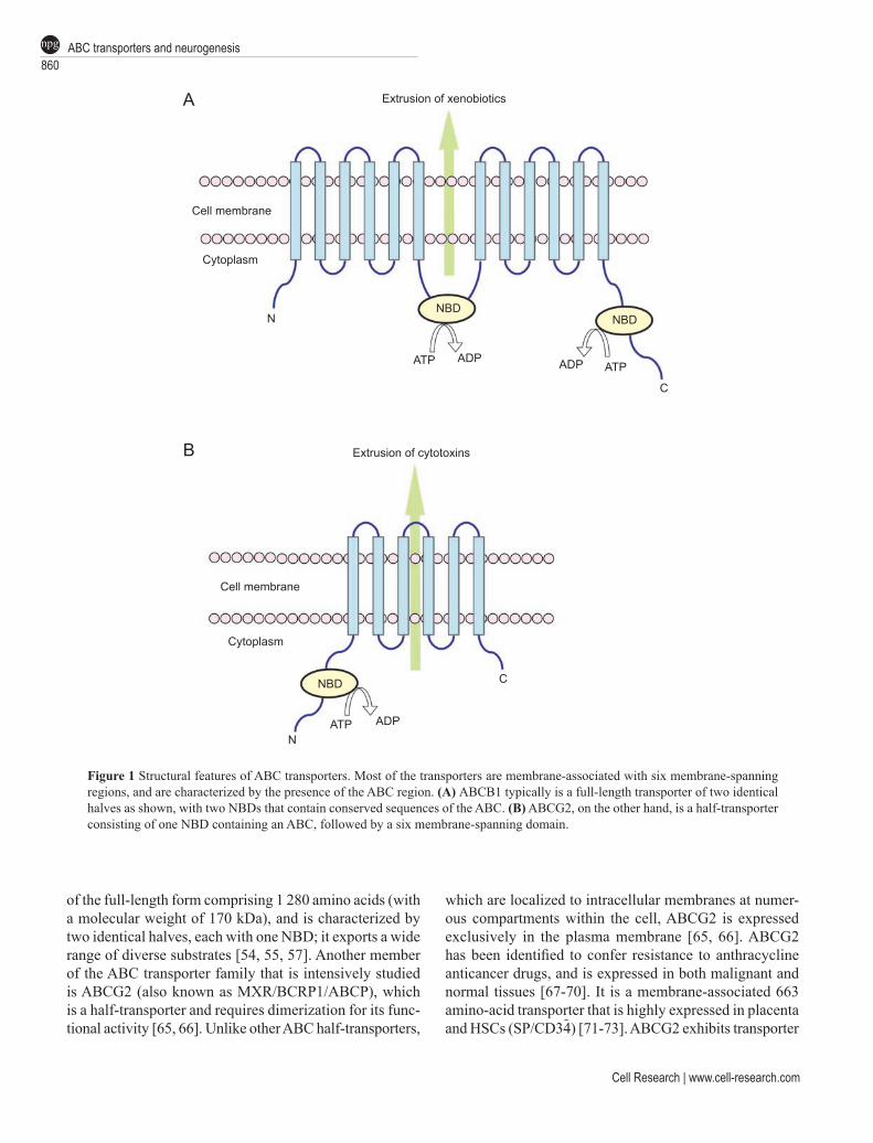
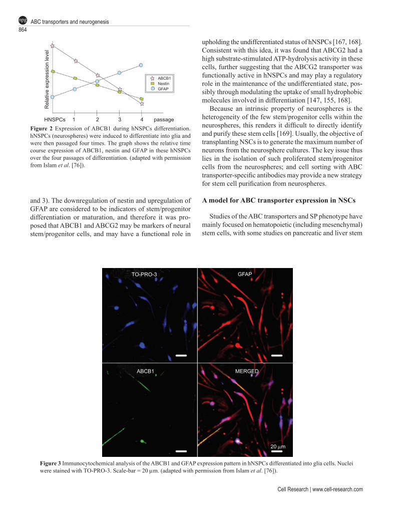
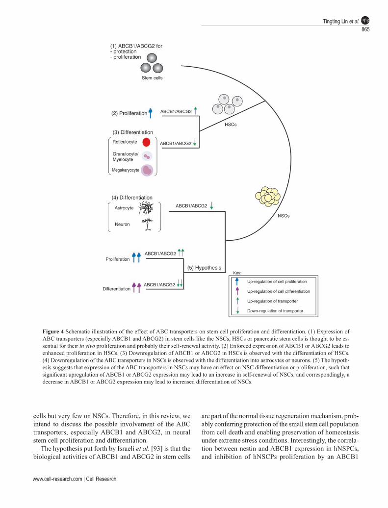
Further study revealed that cultured hNSPCs expressed functional ABCB1 as well as ABCG2 at the cell surface, and that their expression was downregulated during dif-ferentiation of hNSPCs, similar to the downregulation of ABCG2 in HSCs at the stage of lineage commitment [143, 167, 168]. It was observed that both ABC transporters were downregulated during hNSPC differentiation, together with nestin downregulation and GFAP upregulation (Figures 2
Cell Research | www.cell-research.com
ABC transporters and neurogenesis864npg
and 3). The downregulation of nestin and upregulation of GFAP are considered to be indicators of stem/progenitor differentiation or maturation, and therefore it was pro-posed that ABCB1 and ABCG2 may be markers of neural stem/progenitor cells, and may have a functional role in
upholding the undifferentiated status of hNSPCs [167, 168]. Consistent with this idea, it was found that ABCG2 had a high substrate-stimulated ATP-hydrolysis activity in these cells, further suggesting that the ABCG2 transporter was functionally active in hNSPCs and may play a regulatory role in the maintenance of the undifferentiated state, pos-sibly through modulating the uptake of small hydrophobic molecules involved in differentiation [147, 155, 168].
Because an intrinsic property of neurospheres is the heterogeneity of the few stem/progenitor cells within the neurospheres, this renders it difficult to directly identify and purify these stem cells [169]. Usually, the objective of transplanting NSCs is to generate the maximum number of neurons from the neurosphere cultures. The key issue thus lies in the isolation of such proliferated stem/progenitor cells from the neurospheres; and cell sorting with ABC transporter-specific antibodies may provide a new strategy for stem cell purification from neurospheres.
A model for ABC transporter expression in NSCs
Studies of the ABC transporters and SP phenotype have mainly focused on hematopoietic (including mesenchymal) stem cells, with some studies on pancreatic and liver stem
Figure 2 Expression of ABCB1 during hNSPCs differentiation. hNSPCs (neurospheres) were induced to differentiate into glia and were then passaged four times. The graph shows the relative time course expression of ABCB1, nestin and GFAP in these hNSPCs over the four passages of differentiation. (adapted with permission from Islam et al. [76]).
Figure 3 Immunocytochemical analysis of the ABCB1 and GFAP expression pattern in hNSPCs differentiated into glia cells. Nuclei were stained with TO-PRO-3. Scale-bar = 20 mm. (adapted with permission from Islam et al. [76]).
Rel
ativ
e ex
pres
sion
leve
l
HNSPCs 1 2 3 4 passage
ABCB1NestinGFAP
TO-PRO-3 GFAP
ABCB1 MERGED
20 mm
www.cell-research.com | Cell Research
Tingting Lin et al.865npg
cells but very few on NSCs. Therefore, in this review, we intend to discuss the possible involvement of the ABC transporters, especially ABCB1 and ABCG2, in neural stem cell proliferation and differentiation.
The hypothesis put forth by Israeli et al. [93] is that the biological activities of ABCB1 and ABCG2 in stem cells
are part of the normal tissue regeneration mechanism, prob-ably conferring protection of the small stem cell population from cell death and enabling preservation of homeostasis under extreme stress conditions. Interestingly, the correla-tion between nestin and ABCB1 expression in hNSPCs, and inhibition of hNSCPs proliferation by an ABCB1
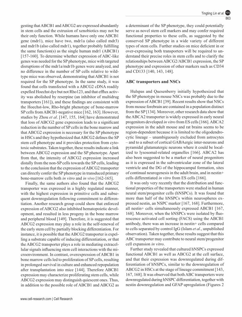
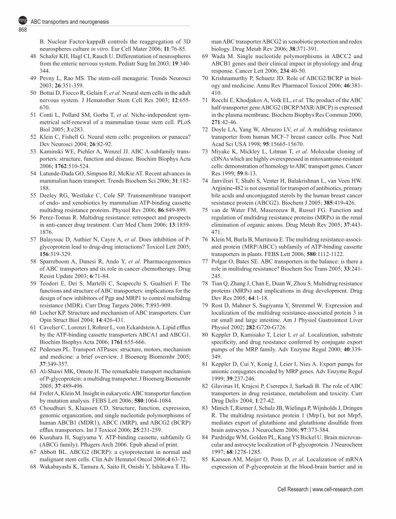
Figure 4 Schematic illustration of the effect of ABC transporters on stem cell proliferation and differentiation. (1) Expression of ABC transporters (especially ABCB1 and ABCG2) in stem cells like the NSCs, HSCs or pancreatic stem cells is thought to be es-sential for their in vivo proliferation and probably their self-renewal activity. (2) Enforced expression of ABCB1 or ABCG2 leads to enhanced proliferation in HSCs. (3) Downregulation of ABCB1 or ABCG2 in HSCs is observed with the differentiation of HSCs. (4) Downregulation of the ABC transporters in NSCs is observed with the differentiation into astrocytes or neurons. (5) The hypoth-esis suggests that expression of the ABC transporters in NSCs may have an effect on NSC differentiation or proliferation, such that significant upregulation of ABCB1 or ABCG2 expression may lead to an increase in self-renewal of NSCs, and correspondingly, a decrease in ABCB1 or ABCG2 expression may lead to increased differentiation of NSCs.

Cell Research | www.cell-research.com
ABC transporters and neurogenesis866npg
inhibitor (cyclosporine A) at a very low dose, suggested that the ABC transporter may contribute to neural stem/progenitor cell expansion [167, 168]. While ABCA2 shows higher expression in nestin+ mouse neural progenitors, at later developmental stages it undergoes a conspicuous downregulation, persisting only in limited subsets of dif-ferentiated neurons [166]. Based on these results and the observations that the ABC transporters are expressed at high levels in hNSPCs but are downregulated in differenti-ated hNSPCs (Figure 3), we hypothesize that these genes could potentially function as putative NSC markers in a similar way as p21CIP/WAF or musashi-1. For instance, while a high expression of ABCB1/ABCG2 reflects main-tenance of proliferating NSCs in an undifferentiated state, low expression characterizes progenitors differentiating into neurons and astrocytes, which by themselves do not (or only at a very low level) express ABCB1/ABCG2 at the end of differentiation (Figure 4) [167, 168]. Chiba et al. [131] suggested that ABCB1 rather than ABCG2 is involved in their “PLC/PRF/5” SP cell phenotype . The published studies on NSCs support the hypothesis that ABCB1 (MDR1) might be more important than ABCG2 in controlling the specific phenotype of NSCs [166-168], in contrast to the HSCs where ABCG2 is involved in the SP phenotype and considered as its molecular determinant. In short, ABC transporters have emerged as an important new field of investigation in the regulation of stem cell biology, and manipulation of this system may promote stem cell amplification via a common defence mechanism adopted by these cells through their high expression of ABC membrane transporters [170].
Clinical relevance and Conclusions
The clinical implications of NSCs are aplenty. While their exact function and distribution is currently being assessed, they represent an interesting cell population, which may be used to study factors important for the dif-ferentiation and characterization of neurons, astrocytes and oligodendrocytes. Recently, there have been reports of NSC transplantations attempting to achieve functional recovery from CNS damage [171-176], and recent evi-dence suggests that NSCs may be a suitable source for the treatment of neurological diseases [22-27, 177-179]. Due to their proliferative and differentiation capacity, NSCs will be important in fighting numerous brain disorders like AD, PD and Huntington’s disease, as well as spinal cord disorders. For instance, in the early stages of AD, a relatively discrete population of neurons is affected [19-21, 180], making it an ideal target for cell replacement therapy using endogenously activated NSCs. However, because curing AD requires the complete construction of the dam-
aged neural circuitry, it will be an especially challenging task to induce NSCs to differentiate into a certain cell-type, highlighting a crucial need for developing the ability to control and predict NSC differentiation. It has already been shown that human NSCs transplanted into aged rat brains can differentiate into neural cells and significantly improve the cognitive functions of these animals, further pointing to NSCs as a promising candidate for neuro-replacement therapies [16,17, 181]. If new bio-imaging technologies could allow an early diagnosis of the onset of AD when the loss of cholinergic neurons has just begun, it might be possible to restore the generation of cholinergic neurons through the transplantation of NSCs, which would provide a potential remedy for the treatment of this disease.
Based on the evidence presented here, we would like to propose that while the ABC transporters are useful as universal stem cell markers and are functionally expressed in hNSPCs, they may also play a crucial role in hNSPC self-renewal by preventing differentiation, and hence will probably be vital in future NSC-based therapy in treating neurodegenerative disorders like AD and PD. In addition, these NSC markers are localized at the cell surface and thus could become a very useful tool, as antibodies raised against these transporters could be utilized for isolating NSCs from neurospheres through techniques like flow cytometry. In this respect, the ABC transporter family members may have provided a model for studying the regulation of NSC self-renewal and differentiation. Many new questions remain regarding how these molecules might function and what evolutionarily conserved substrates may be modulated. More experiments involving the analysis of hNSPCs transfected with genes for expression of the different ABC transporters will definitely provide more conclusive evidence, and hopefully may also facilitate fu-ture breakthroughs in the treatment of neurodegeneration, a field that has perplexed researchers for centuries.
Acknowledgments
This work was supported by a grant (SBS/SUG/22/04) to KH from Nanyang Technological University. We are grateful to Ms S Ayyadhury for editorial assistance.
References
1 Priddle H, Jones DR, Burridge PW, Patient R. Hematopoiesis from human embryonic stem cells: overcoming the immune barrier in stem cell therapies. Stem Cells 2006; 24:815-824.
2 Taupin P. Neurogenesis in the adult central nervous system. C R Biol 2006; 329:465-475.
3 Erlandsson A, Morshead CM. Exploiting the properties of adult stem cells for the treatment of disease. Curr Opin Mol Ther 2006; 8:331-337.
www.cell-research.com | Cell Research
Tingting Lin et al.867npg
4 Chan RJ, Yoder MC. The multiple facets of hematopoietic stem cells. Curr Neurovasc Res 2004; 1:197-206.
5 Bonnet D. Normal and leukaemic stem cells. Br J Haematol 2005; 130:469-479.
6 Wu M, Wei YQ. Development of respiratory stem cells and progenitor cells. Stem Cells Dev 2004; 13:607-613.
7 Challen GA, Little MH. A side order of stem cells: the SP phe-notype. Stem Cells 2006; 24:3-12
8 Jackson KA, Majka SM, Wang H, et al. Regeneration of ischemic cardiac muscle and vascular endothelium by adult stem cells. J Clin Invest 2001; 107:1395-1402.
9 Goodell MA, Brose K, Paradis G, et al. Isolation and functional properties of murine hematopoietic stem cells that are replicating in vivo. J Exp Med 1996; 183:1797-1806.
10 Weissman IL. Stem cells: units of development, units of regen-eration, and units in evolution. Cell 2000; 100:157-168.
11 Shostak S. (Re)defining stem cells. Bioessays 2006; 28:301-308.
12 Venezia TA, Merchant AA, Ramos CA, et al. Molecular signa-tures of proliferation and quiescence in hematopoietic stem cells. PLoS Biol 2004; 2:e301.
13 Wilson A, Trumpp A. Bone-marrow haematopoietic-stem-cell niches. Nat Rev Immunol 2006; 6:93-106.
14 Rizo A, Vellenga E, de Haan G, Schuringa JJ. Signaling path-ways in self-renewing hematopoietic and leukemic stem cells: do all stem cells need a niche? Hum Mol Genet 2006; 15:R210-R219.
15 Scadden DT. The stem-cell niche as an entity of action. Nature 2006; 441:1075-1079.
16 Oliveira AA Jr., Hodges HM. Alzheimer’s disease and neural transplantation as prospective cell therapy. Curr Alzheimer Res 2005; 2:79-95.
17 Tanne JH. Activating stem cells may treat Alzheimer’s. BMJ 2005; 330:622.
18 Lindvall O, Kokaia Z. Stem cells for the treatment of neurological disorders. Nature 2006; 441:1094-1096.
19 Selkoe DJ, Schenk D. Alzheimer’s disease: molecular under-standing predicts amyloid-based therapeutics. Annu Rev Phar-macol Toxicol 2003; 43:545-584.
20 Swerdlow RH. Is aging part of Alzheimer’s disease, or is Alzheimer’s disease part of aging? Neurobiol Aging 2006. Epub ahead of print.
21 Harman D. Alzheimer’s disease pathogenesis: role of aging. Ann NY Acad Sci 2006; 1067:454-460.
22 Brazel CY, Rao MS. Aging and neuronal replacement. Ageing Res Rev 2004; 3:465-483.
23 Sailor KA, Ming GL, Song H. Neurogenesis as a potential thera-peutic strategy for neurodegenerative diseases. Expert Opin Biol Ther 2006; 6:879-890.
24 Ming GL, Song H. Adult neurogenesis in the mammalian central nervous system. Annu Rev Neurosci 2005; 28:223-250.
25 Okano H. Adult neural stem cells and central nervous system repair. Ernst Schering Res Found Workshop 2006; 60:215-228.
26 Tabar V, Panagiotakos G, Greenberg ED, et al. Migration and dif-ferentiation of neural precursors derived from human embryonic stem cells in the rat brain. Nat Biotechnol 2005; 23:601-606.
27 Jagasia R, Song H, Gage FH, Lie DC. New regulators in adult neurogenesis and their potential role for repair. Trends Mol Med 2006; 12:400-405.
28 Lennington JB, Yang Z, Conover JC. Neural stem cells and the regulation of adult neurogenesis. Reprod Biol Endocrinol 2003; 1:99.
29 Gage FH. Mammalian neural stem cells. Science 2000; 287:1433-1438.
30 Shih CC, Weng Y, Mamelak A, et al. Identification of a candidate human neurohematopoietic stem-cell population. Blood 2001; 98:2412-2422.
31 Muller FJ, Snyder EY, Loring JF. Gene therapy: can neural stem cells deliver? Nat Rev Neurosci 2006; 7:75-84.
32 Aboody KS, Brown A, Rainov NG, et al. Neural stem cells display extensive tropism for pathology in adult brain: evidence from intracranial gliomas. Proc Natl Acad Sci USA 2000; 97:12846-12851.
33 Chi L, Ke Y, Luo C, et al. Motor neuron degeneration promotes neural progenitor cell proliferation, migration, and neurogenesis in the spinal cords of amyotrophic lateral sclerosis mice. Stem Cells 2006; 24:34-43.
34 Glass R, Synowitz M, Kronenberg G, et al. Glioblastoma-induced attraction of endogenous neural precursor cells is associated with improved survival. J Neurosci 2005; 25:2637-2646.
35 Brown AB, Yang W, Schmidt NO, et al. Intravascular delivery of neural stem cell lines to target intracranial and extracranial tumors of neural and non-neural origin. Hum Gene Ther 2003; 14:1777-1785.
36 Kelly S, Bliss TM, Shah AK, et al. Transplanted human fetal neural stem cells survive, migrate, and differentiate in ischemic rat cerebral cortex. Proc Natl Acad Sci USA 2004; 101:11839-11844.
37 Fernaud-Espinosa I, Nieto-Sampedro M, Bovolenta P. Dif-ferential activation of microglia and astrocytes in aniso- and isomorphic gliotic tissue. Glia 1993; 8:277-291.
38 Wang K, Walz W. Unusual topographical pattern of proximal astrogliosis around a cortical devascularizing lesion. J Neurosci Res 2003; 73:497-506.
39 Hulspas R, Quesenberry PJ. Characterization of neurosphere cell phenotypes by flow cytometry. Cytometry 2000; 40:245-250.
40 Campos LS. Neurospheres: insights into neural stem cell biology. J Neurosci Res 2004; 78:761-769.
41 Reynolds BA, Rietze RL. Neural stem cells and neurospheres – re-evaluating the relationship. Nat Methods 2005; 2:333-336.
42 Engstrom CM, Demers D, Dooner M, et al. A method for clonal analysis of epidermal growth factor-responsive neural progeni-tors. J Neurosci Methods 2002; 117:111-121.
43 Kanemura Y, Mori H, Kobayashi S, et al. Evaluation of in vitro proliferative activity of human fetal neural stem/progenitor cells using indirect measurements of viable cells based on cellular metabolic activity. J Neurosci Res 2002; 69:869-879.
44 Garcion E, Halilagic A, Faissner A, et al. Generation of an environmental niche for neural stem cell development by the extracellular matrix molecule tenascin C. Development 2004; 131:3423-3432.
45 Nagato M, Heike T, Kato T, et al. Prospective characterization of neural stem cells by flow cytometry analysis using a combination of surface markers. J Neurosci Res 2005; 80:456-466.
46 Doetsch F. The glial identity of neural stem cells. Nat Neurosci 2003; 6:1127-1134.
47 Widera D, Mikenberg I, Kaus A, Kaltschmidt C, Kaltschmidt
Cell Research | www.cell-research.com
ABC transporters and neurogenesis868npg
B. Nuclear Factor-kappaB controls the reaggregation of 3D neurospheres culture in vitro. Eur Cell Mater 2006; 11:76-85.
48 Schafer KH, Hagl CI, Rauch U. Differentiation of neurospheres from the enteric nervous system. Pediatr Surg Int 2003; 19:340-344.
49 Pevny L, Rao MS. The stem-cell menagerie. Trends Neurosci 2003; 26:351-359.
50 Bottai D, Fiocco R, Gelain F, et al. Neural stem cells in the adult nervous system. J Hematother Stem Cell Res 2003; 12:655-670.
51 Conti L, Pollard SM, Gorba T, et al. Niche-independent sym-metrical self-renewal of a mammalian tissue stem cell. PLoS Biol 2005; 3:e283.
52 Klein C, Fishell G. Neural stem cells: progenitors or panacea? Dev Neurosci 2004; 26:82-92.
53 Kaminski WE, Piehler A, Wenzel JJ. ABC A-subfamily trans-porters: structure, function and disease. Biochim Biophys Acta 2006; 1762:510-524.
54 Latunde-Dada GO, Simpson RJ, McKie AT. Recent advances in mammalian haem transport. Trends Biochem Sci 2006; 31:182-188.
55 Deeley RG, Westlake C, Cole SP. Transmembrane transport of endo- and xenobiotics by mammalian ATP-binding cassette multidrug resistance proteins. Physiol Rev 2006; 86:849-899.
56 Perez-Tomas R. Multidrug resistance: retrospect and prospects in anti-cancer drug treatment. Curr Med Chem 2006; 13:1859-1876.
57 Balayssac D, Authier N, Cayre A, et al. Does inhibition of P-glycoprotein lead to drug-drug interactions? Toxicol Lett 2005; 156:319-329.
58 Sparreboom A, Danesi R, Ando Y, et al. Pharmacogenomics of ABC transporters and its role in cancer chemotherapy. Drug Resist Update 2003; 6:71-84.
59 Teodori E, Dei S, Martelli C, Scapecchi S, Gualtieri F. The functions and structure of ABC transporters: implications for the design of new inhibitors of Pgp and MRP1 to control multidrug resistance (MDR). Curr Drug Targets 2006; 7:893-909.
60 Locher KP. Structure and mechanism of ABC transporters. Curr Opin Struct Biol 2004; 14:426-431.
61 Cavelier C, Lorenzi I, Rohrer L, von Eckardstein A. Lipid efflux by the ATP-binding cassette transporters ABCA1 and ABCG1. Biochim Biophys Acta 2006; 1761:655-666.
62 Pedersen PL. Transport ATPases: structure, motors, mechanism and medicine: a brief overview. J Bioenerg Biomembr 2005; 37:349-357.
63 Al-Shawi MK, Omote H. The remarkable transport mechanism of P-glycoprotein: a multidrug transporter. J Bioenerg Biomembr 2005; 37:489-496.
64 Frelet A, Klein M. Insight in eukaryotic ABC transporter function by mutation analysis. FEBS Lett 2006; 580:1064-1084.
65 Choudhuri S, Klaassen CD. Structure, function, expression, genomic organization, and single nucleotide polymorphisms of human ABCB1 (MDR1), ABCC (MRP), and ABCG2 (BCRP) efflux transporters. Int J Toxicol 2006; 25:231-259.
66 Kusuhara H, Sugiyama Y. ATP-binding cassette, subfamily G (ABCG family). Pflugers Arch 2006. Epub ahead of print.
67 Abbott BL. ABCG2 (BCRP): a cytoprotectant in normal and malignant stem cells. Clin Adv Hematol Oncol 2006;4:63-72.
68 Wakabayashi K, Tamura A, Saito H, Onishi Y, Ishikawa T. Hu-
man ABC transporter ABCG2 in xenobiotic protection and redox biology. Drug Metab Rev 2006; 38:371-391.
69 Wada M. Single nucleotide polymorphisms in ABCC2 and ABCB1 genes and their clinical impact in physiology and drug response. Cancer Lett 2006; 234:40-50.
70 Krishnamurthy P, Schuetz JD. Role of ABCG2/BCRP in biol-ogy and medicine. Annu Rev Pharmacol Toxicol 2006; 46:381-410.
71 Rocchi E, Khodjakov A, Volk EL, et al. The product of the ABC half-transporter gene ABCG2 (BCRP/MXR/ABCP) is expressed in the plasma membrane. Biochem Biophys Res Commun 2000; 271:42-46.
72 Doyle LA, Yang W, Abruzzo LV, et al. A multidrug resistance transporter from human MCF-7 breast cancer cells. Proc Natl Acad Sci USA 1998; 95:15665-15670.
73 Miyake K, Mickley L, Litman T, et al. Molecular cloning of cDNAs which are highly overexpressed in mitoxantrone-resistant cells: demonstration of homology to ABC transport genes. Cancer Res 1999; 59:8-13.
74 Janvilisri T, Shahi S, Venter H, Balakrishnan L, van Veen HW. Arginine-482 is not essential for transport of antibiotics, primary bile acids and unconjugated sterols by the human breast cancer resistance protein (ABCG2). Biochem J 2005; 385:419-426.
75 van de Water FM, Masereeuw R, Russel FG. Function and regulation of multidrug resistance proteins (MRPs) in the renal elimination of organic anions. Drug Metab Rev 2005; 37:443-471.
76 Klein M, Burla B, Martinoia E. The multidrug resistance-associ-ated protein (MRP/ABCC) subfamily of ATP-binding cassette transporters in plants. FEBS Lett 2006; 580:1112-1122.
77 Polgar O, Bates SE. ABC transporters in the balance: is there a role in multidrug resistance? Biochem Soc Trans 2005; 33:241-245.
78 Tian Q, Zhang J, Chan E, Duan W, Zhou S. Multidrug resistance proteins (MRPs) and implications in drug development. Drug Dev Res 2005; 64:1-18.
79 Rost D, Mahner S, Sugiyama Y, Stremmel W. Expression and localization of the multidrug resistance-associated protein 3 in rat small and large intestine. Am J Physiol Gastrointest Liver Physiol 2002; 282:G720-G726.
80 Keppler D, Kamisako T, Leier I, et al. Localization, substrate specificity, and drug resistance conferred by conjugate export pumps of the MRP family. Adv Enzyme Regul 2000; 40:339-349.
81 Keppler D, Cui Y, Konig J, Leier I, Nies A. Export pumps for anionic conjugates encoded by MRP genes. Adv Enzyme Regul 1999; 39:237-246.
82 Glavinas H, Krajcsi P, Cserepes J, Sarkadi B. The role of ABC transporters in drug resistance, metabolism and toxicity. Curr Drug Deliv 2004; 1:27-42.
83 Minich T, Riemer J, Schulz JB, Wielinga P, Wijnholds J, Dringen R. The multidrug resistance protein 1 (Mrp1), but not Mrp5, mediates export of glutathione and glutathione disulfide from brain astrocytes. J Neurochem 2006; 97:373-384.
84 Pardridge WM, Golden PL, Kang YS Bickel U. Brain microvas-cular and astrocyte localization of P-glycoprotein. J Neurochem 1997; 68:1278-1285.
85 Karssen AM, Meijer O, Pons D, et al. Localization of mRNA expression of P-glycoprotein at the blood-brain barrier and in

www.cell-research.com | Cell Research
Tingting Lin et al.869npg
the hippocampus. Ann NY Acad Sci 2004; 1032:308-311.86 Hirsch-Ernst KI, Ziemann C, Schmitz-Salue C, Foth H, Kahl
GF. Modulation of P-glycoprotein and mdr1b mRNA expression by growth factors in primary rat hepatocyte culture. Biochem Biophys Res Commun 1995; 215:179-185.
87 Krishnamurthy P, Ross DD, Nakanishi T, et al. The stem cell marker Bcrp/ABCG2 enhances hypoxic cell survival through interactions with heme. J Biol Chem 2004; 279:24218-24225.
88 Krishnamurthy P, Schuetz JD. The ABC transporter Abcg2/Bcrp: role in hypoxia mediated survival. Biometals 2005; 18:349-358.
89 Mao Q, Unadkat JD. Role of the breast cancer resistance protein (ABCG2) in drug transport. AAPS J 2005; 7:E118-E133.
90 Sarkadi B, Ozvegy-Laczka C, Nemet K, Varadi A. ABCG2 – a transporter for all seasons. FEBS Lett 2004; 567:116-120.
91 Marchi N, Hallene KL, Kight KM, et al. Significance of MDR1 and multiple drug resistance in refractory human epileptic brain. BMC Med 2004; 2:37.
92 Liang XJ, Aszalos A. Multidrug transporters as drug targets. Curr Drug Targets 2006; 7:911-921.
93 Israeli D, Ziaei S, Gonin P, et al. A proposal for the physiological significance of mdr1 and Bcrp1/Abcg2 gene expression in normal tissue regeneration and after cancer therapy. J Theor Biol 2005; 232:41-45.
94 Nakatsukasa H, Silverman JA, Gant TW, et al. Expression of multidrug resistance genes in rat liver during regeneration and after carbon tetrachloride intoxication. Hepatology 1993; 18:1202-1207.
95 Teeter LD, Becker FF, Chisari FV, et al. Overexpression of the multidrug resistance gene mdr3 in spontaneous and chemically induced mouse hepatocellular carcinomas. Mol Cell Biol 1990; 10:5728-5735.
96 Hirsch-Ernst KI, Ziemann C, Foth H, et al. Induction of mdr1b mRNA and P-glycoprotein expression by tumor necrosis factor alpha in primary rat hepatocyte cultures. J Cell Physiol 1998; 176:506-515.
97 Lazarowski A, Ramos AJ, Garcia-Rivello H, et al. Neuronal and glial expression of the multidrug resistance gene product in an experimental epilepsy model. Cell Mol Neurobiol 2004; 24:77-85.
98 Ramos AJ, Lazarowski A, Villar MJ, et al. Transient expression of MDR-1/P-glycoprotein in a model of partial cortical devas-cularization. Cell Mol Neurobiol 2004; 24:101-107.
99 Volk HA, Burkhardt K, Potschka H, et al. Neuronal expression of the drug efflux transporter P-glycoprotein in the rat hippocampus after limbic seizures. Neuroscience 2004; 123:751-759.
100 Yang JM, Sullivan GF, Hait WN. Regulation of the function of P-glycoprotein by epidermal growth factor through phospholipase C. Biochem Pharmacol 1997; 53:1597-1604.
101 Nies AT, Jedlitschky G, Konig J, et al. Expression and immuno-localization of the multidrug resistance proteins, MRP1-MRP6 (ABCC1-ABCC6), in human brain. Neuroscience 2004; 129:349-360.
102 Hirrlinger J, Moeller H, Kirchhoff F, Dringen R. Expression of multidrug resistance proteins (Mrps) in astrocytes of the mouse brain: a single cell RT-PCR study. Neurochem Res 2005; 30:1237-1244.
103 Hirrlinger J, Konig J, Dringen R. Expression of mRNAs of multidrug resistance proteins (Mrps) in cultured rat astrocytes,
oligodendrocytes, microglial cells and neurones. J Neurochem 2002; 82:716-719.
104 Dallas S, Miller DS, Bendayan R. Multidrug resistance-associ-ated proteins: expression and function in the central nervous system. Pharmacol Rev 2006; 58:140-161.
105 Kim WS, Guillemin GJ, Glaros EN, Lim CK, Garner B. Quan-titation of ATP-binding cassette subfamily-A transporter gene expression in primary human brain cells. Neuroreport 2006; 17:891-896.
106 Hirsch-Reinshagen V, Zhou S, Burgess BL, et al. Deficiency of ABCA1 impairs apolipoprotein E metabolism in brain. J Biol Chem 2004; 279:41197-41207.
107 Brewer HB Jr, Remaley AT, Neufeld EB, Basso F, Joyce C. Regu-lation of plasma high-density lipoprotein levels by the ABCA1 transporter and the emerging role of high-density lipoprotein in the treatment of cardiovascular disease. Arterioscler Thromb Vasc Biol 2004; 24:1755-1760.
108 Le Goff W, Settle M, Greene DJ, Morton RE, Smith JD. Re-evaluation of the role of the multidrug-resistant P-glycoprotein in cellular cholesterol homeostasis. J Lipid Res 2006; 47:51-58.
109 Burns MP, Vardanian L, Pajoohesh-Ganji A, et al. The effects of ABCA1 on cholesterol efflux and Abeta levels in vitro and in vivo. J Neurochem 2006; 98:792-800.
110 Fukumoto H, Deng A, Irizarry MC, Fitzgerald ML, Rebeck GW. Induction of the cholesterol transporter ABCA1 in central ner-vous system cells by liver X receptor agonists increases secreted Abeta levels. J Biol Chem 2002; 277:48508-48513.
111 Wahrle SE, Jiang H, Parsadanian M, et al. Deletion of Abca1 increases Abeta deposition in the PDAPP transgenic mouse model of Alzheimer disease. J Biol Chem 2005; 280:43236-43242.
112 Koldamova R, Staufenbiel M, Lefterov I. Lack of ABCA1 con-siderably decreases brain ApoE level and increases amyloid de-position in APP23 mice. J Biol Chem 2005; 280:43224-43235.
113 Hirsch-Reinshagen V, Maia LF, Burgess BL, et al. The absence of ABCA1 decreases soluble ApoE levels but does not diminish amyloid deposition in two murine models of Alzheimer disease. J Biol Chem 2005; 280:43243-43256.
114 Karten B, Campenot RB, Vance DE, Vance JE. Expression of ABCG1, but not ABCA1, correlates with cholesterol release by cerebellar astroglia. J Biol Chem 2006; 281:4049-4057.
115 Leonard GD, Fojo T, Bates SE. The role of ABC transporters in clinical practice. Oncologist 2003; 8:411-424.
116 Stefkova J, Poledne R, Hubacek JA. ATP-binding cassette (ABC) transporters in human metabolism and diseases. Physiol Res 2004; 53:235-243.
117 Dean M, Allikmets R. Complete characterization of the human ABC gene family. J Bioenerg Biomembr 2001; 33:475-479.
118 Vergani P, Basso C, Mense M, Nairn AC, Gadsby DC. Control of the CFTR channel’s gates. Biochem Soc Trans 2005; 33:1003-1007.
119 McGinniss MJ, Chen C, Redman JB, et al. Extensive sequenc-ing of the CFTR gene: lessons learned from the first 157 patient samples. Hum Genet 2005; 118:331-338.
120 Vander Borght S, Libbrecht L, Katoonizadeh A, et al. Breast cancer resistance protein (BCRP/ABCG2) is expressed by pro-genitor cells/reactive ductules and hepatocytes and its expres-sion pattern is influenced by disease etiology and species type: possible functional consequences. J Histochem Cytochem 2006; 54:1051-1059.
Cell Research | www.cell-research.com
ABC transporters and neurogenesis870npg
121 Borst P, Elferink RO. Mammalian ABC transporters in health and disease. Annu Rev Biochem 2002; 71:537-592.
122 Ros JE, Roskams TA, Geuken M, et al. ATP binding cassette transporter gene expression in rat liver progenitor cells. Gut 2003; 52:1060-1067.
123 Ros JE, Libbrecht L, Geuken M, Jansen PL, Roskams TA. High expression of MDR1, MRP1, and MRP3 in the hepatic progenitor cell compartment and hepatocytes in severe human liver disease. J Pathol 2003; 200:553-560.
124 Libbrecht L, Desmet V, Roskams T. Preneoplastic lesions in human hepatocarcinogenesis. Liver Int 2005; 25:16-27.
125 Newsome PN, Hussain MA, Theise ND. Hepatic oval cells: helping redefine a paradigm in stem cell biology. Curr Top Dev Biol 2004; 61:1-28.
126 Inagaki N, Gonoi T, Clement JP IV, et al. Reconstitution of IKATP: an inward rectifier subunit plus the sulfonylurea receptor. Science 1995; 270:1166-1170.
127 Koriyama N, Kakei M, Nakazaki M, et al. PIP2 and ATP co-operatively prevent cytosolic Ca2+-induced modification of ATP-sensitive K+ channels in rat pancreatic beta-cells. Diabetes 2000; 49:1830-1839.
128 Chaudhary PM, Roninson IB. Expression and activity of P-glycoprotein, a multidrug efflux pump, in human hematopoietic stem cells. Cell 1991; 66:85-94.
129 Kim M, Turnquist H, Jackson J, et al. The multidrug resistance transporter ABCG2 (breast cancer resistance protein 1) effluxes Hoechst 33342 and is overexpressed in hematopoietic stem cells. Clin Cancer Res 2002; 8:22-28.
130 Hirschmann-Jax C, Foster AE, Wulf GG, et al. A distinct “side population” of cells with high drug efflux capacity in human tumor cells. Proc Natl Acad Sci USA 2004; 101:14228-14233.
131 Chiba T, Kita K, Zheng YW, et al. Side population purified from hepatocellular carcinoma cells harbors cancer stem cell-like properties. Hepatology 2006; 44:240-251.
132 Fuellen G, Spitzer M, Cullen P, Lorkowski S. Correspondence of function and phylogeny of ABC proteins based on an automated analysis of 20 model protein data sets. Proteins 2005; 61:888-899.
133 Lorkowski S, Cullen P. ABCG subfamily of human ATP-binding cassette proteins. Pure Appl Chem 2002; 74:2057-2081.
134 Mouthon MA, Fouchet P, Mathieu C, et al. Neural stem cells from mouse forebrain are contained in a population distinct from the ‘side population’. J Neurochem 2006. Epub ahead of print.
135 Haraguchi N, Utsunomiya T, Inoue H, et al. Characterization of a side population of cancer cells from human gastrointestinal system. Stem Cells 2006; 24:506-513.
136 Meissner K, Heydrich B, Jedlitschky G, et al. The ATP-binding cassette transporter ABCG2 (BCRP), a marker for side population stem cells, is expressed in human heart. J Histochem Cytochem 2006; 54:215-221.
137 McAlister I, Wolf NS, Pietrzyk ME, et al. Transplantation of hematopoietic stem cells obtained by a combined dye method fractionation of murine bone marrow. Blood 1990; 75:1240-1246.
138 Wolf NS, Kone A, Priestley GV, et al. In vivo and in vitro char-acterization of long-term repopulating primitive hematopoietic cells isolated by sequential Hoechst 33342-rhodamine 123 FACS selection. Exp Hematol 1993; 21:614-622.
139 Leemhuis T, Yoder MC, Grigsby S, et al. Isolation of primi-
tive human bone marrow hematopoietic progenitor cells us-ing Hoechst 33342 and Rhodamine 123. Exp Hematol 1996; 24:1215-1224.
140 Bunting KD. ABC transporters as phenotypic markers and func-tional regulators of stem cells. Stem Cells 2002; 20:11-20.
141 Matsuzaki Y, Kinjo K, Mulligan RC, Okano H. Unexpectedly efficient homing capacity of purified murine hematopoietic stem cells. Immunity 2004; 20:87-93.
142 Norwood K, Wang RY, Hirschmann-Jax C, et al. An in vivo propagated human acute myeloid leukemia expressing ABCA3. Leuk Res 2004; 28:295-299.
143 Scharenberg CW, Harkey MA, Torok-Storb B. The ABCG2 transporter is an efficient Hoechst 33342 efflux pump and is preferentially expressed by immature human hematopoietic progenitors. Blood 2002; 99:507-512.
144 Bunting KD, Zhou S, Lu T, et al. Enforced P-glycoprotein pump function in murine bone marrow cells results in expansion of side population stem cells in vitro and repopulating cells in vivo. Blood 2000; 96:902-909.
145 Lechner A, Leech CA, Abraham EJ, et al. Nestin-positive progenitor cells derived from adult human pancreatic islets of Langerhans contain side population (SP) cells defined by expres-sion of the ABCG2 (BCRP1) ATP-binding cassette transporter. Biochem Biophys Res Commun 2002; 293:670-674.
146 Kawanabe N, Murakami K, Takano-Yamamoto T. The presence of ABCG2-dependent side population cells in human periodontal ligaments. Biochem Biophys Res Commun 2006; 344:1278-1283.
147 Zhou S, Morris JJ, Barnes Y, et al. Bcrp1 gene expression is re-quired for normal numbers of side population stem cells in mice, and confers relative protection to mitoxantrone in hematopoietic cells in vivo. Proc Natl Acad Sci USA 2002; 99:12339-12344.
148 Naylor CS, Jaworska E, Branson K, Embleton MJ, Chopra R. Side population/ABCG2-positive cells represent a heterogeneous group of haemopoietic cells: implications for the use of adult stem cells in transplantation and plasticity protocols. Bone Marrow Transplant 2005; 35:353-360.
149 Ueda T, Brenner S, Malech HL, et al. Cloning and functional analysis of the rhesus macaque ABCG2 gene. Forced expression confers an SP phenotype among hematopoietic stem cell progeny in vivo. J Biol Chem 2005; 280:991-998.
150 Jonker JW, Freeman J, Bolscher E, et al. Contribution of the ABC transporters Bcrp1 and Mdr1a/1b to the side population phenotype in mammary gland and bone marrow of mice. Stem Cells 2005; 23:1059-1065.
151 Challen GA, Little MH. A side order of stem cells: the SP phe-notype. Stem Cells 2006; 24:3-12.
152 Scharenberg CW, Harkey MA, Torok-Storb B. The ABCG2 transporter is an efficient Hoechst 33342 efflux pump and is preferentially expressed by immature human hematopoietic progenitors. Blood 2002; 99:507-512.
153 Konya A, Andor A, Satorhelyi P, Nemeth K, Kurucz I. Inhibi-tion of the MDR1 transporter by new phenothiazine derivatives. Biochem Biophys Res Commun 2006; 346:45-50.
154 Ogihara T, Kamiya M, Ozawa M, et al. What kinds of substrates show P-glycoprotein-dependent intestinal absorption? Compari-son of verapamil with vinblastine. Drug Metab Pharmacokinet 2006; 21:238-244.
155 Zhou S, Schuetz JD, Bunting KD, et al. The ABC transporter
www.cell-research.com | Cell Research
Tingting Lin et al.871npg
Bcrp1/ABCG2 is expressed in a wide variety of stem cells and is a molecular determinant of the side-population phenotype. Nat Med 2001; 7:1028-1034.
156 Tadjali M, Zhou S, Rehg J, Sorrentino BP. Prospective isolation of murine hematopoietic stem cells by expression of an Abcg2/GFP allele. Stem Cells 2006; 24:1556-1563.
157 Chen CJ, Chin JE, Ueda K, et al. Internal duplication and homol-ogy with bacterial transport proteins in the mdr1 (P-glycoprotein) gene from multidrug-resistant human cells. Cell 1986; 47:381-389.
158 Gros P, Croop J, Housman D. Mammalian multidrug resistance gene: complete cDNA sequence indicates strong homology to bacterial transport proteins. Cell 1986; 47:371-380.
159 Hsu SI, Lothstein L, Horwitz SB. Differential overexpression of three mdr gene family members in multidrug-resistant J774.2 mouse cells. Evidence that distinct P-glycoprotein precursors are encoded by unique mdr genes. J Biol Chem 1989; 264:12053-12062.
160 Devault A, Gros P. Two members of the mouse mdr gene family confer multidrug resistance with overlapping but distinct drug specificities. Mol Cell Biol 1990; 10:1652-1663.
161 Wang EJ, Casciano CN, Clement RP, Johnson WW. Active transport of fluorescent P-glycoprotein substrates: evaluation as markers and interaction with inhibitors. Biochem Biophys Res Commun 2001; 289:580-585.
162 Schinkel AH, Smit JJ, van Tellingen O, et al. Disruption of the mouse mdr1a P-glycoprotein gene leads to a deficiency in the blood-brain barrier and to increased sensitivity to drugs. Cell 1994; 77:491-502.
163 Schinkel AH, Mayer U, Wagenaar E, et al. Normal viability and altered pharmacokinetics in mice lacking mdr1-type (drug-transporting) P-glycoproteins. Proc Natl Acad Sci USA 1997; 94:4028-4033.
164 Zhou S, Zong Y, Lu T, et al. Hematopoietic cells from mice that are deficient in both Bcrp1/Abcg2 and Mdr1a/1b develop nor-mally but are sensitized to mitoxantrone. Biotechniques 2003; 35:1248-1252.
165 Panwala CM, Jones JC, Viney JL. A novel model of inflammatory bowel disease: mice deficient for the multiple drug resistance gene, mdr1a, spontaneously develop colitis. J Immunol 1998; 161:5733-5744.
166 Broccardo C, Nieoullon V, Amin R, et al. ABCA2 is a marker of neural progenitors and neuronal subsets in the adult rodent brain. J Neurochem 2006; 97:345-355.
167 Islam MO, Kanemura Y, Tajria J, et al. Characterization of ABC transporter ABCB1 expressed in human neural stem/progenitor
cells. FEBS Lett 2005; 579:3473-3480.168 Islam MO, Kanemura Y, Tajria J, et al. Functional expression
of ABCG2 transporter in human neural stem/progenitor cells. Neurosci Res 2005; 52:75-82.
169 Suslov ON, Kukekov VG, Ignatova TN, et al. Neural stem cell heterogeneity demonstrated by molecular phenotyping of clonal neurospheres. Proc Natl Acad Sci USA 2002; 99:14506-14511.
170 Alison MR. Tissue-based stem cells: ABC transporter proteins take centre stage. J Pathol 2003; 21:547-550.
171 Roitberg BZ, Mangubat E, Chen EY, et al. Survival and early differentiation of human neural stem cells transplanted in a nonhuman primate model of stroke. J Neurosurg 2006; 105:96-102.
172 Cummings BJ, Uchida N, Tamaki SJ, Anderson AJ. Human neural stem cell differentiation following transplantation into spinal cord injured mice: association with recovery of locomotor function. Neurol Res 2006; 28:474-481.
173 Watson DJ, Walton RM, Magnitsky SG, Bulte JW, Poptani H, Wolfe JH. Structure-specific patterns of neural stem cell engraft-ment after transplantation in the adult mouse brain. Hum Gene Ther 2006; 17:693-704.
174 Park KI, Himes BT, Stieg PE, Tessler A, Fischer I, Snyder EY. Neural stem cells may be uniquely suited for combined gene therapy and cell replacement: Evidence from engraftment of Neurotrophin-3-expressing stem cells in hypoxic-ischemic brain injury. Exp Neurol 2006; 199:179-190.
175 Shindo T, Matsumoto Y, Wang Q, Kawai N, Tamiya T, Nagao S. Differences in the neuronal stem cells survival, neuronal differentiation and neurological improvement after transplanta-tion of neural stem cells between mild and severe experimental traumatic brain injury. J Med Invest 2006; 53:42-51.
176 Wang Q, Matsumoto Y, Shindo T, et al. Neural stem cells trans-plantation in cortex in a mouse model of Alzheimer’s disease. J Med Invest 2006; 53:61-69.
177 Sugaya K, Alvarez A, Marutle A, Kwak YD, Choumkina E. Stem cell strategies for Alzheimer’s disease therapy. Panminerva Med 2006; 48:87-96.
178 Garbossa D, Fontanella M, Fronda C, et al. New strategies for repairing the injured spinal cord: the role of stem cells. Neurol Res 2006; 28:500-504.
179 Martino G, Pluchino S. The therapeutic potential of neural stem cells. Nat Rev Neurosci 2006; 7:395-406.
180 Heese K, Akatsu H. Alzheimer’s disease – an interactive perspec-tive. Curr Alzheimer Res 2006; 3:109-121.
181 Sugaya K. Possible use of autologous stem cell therapies for Alzheimer’s disease. Curr Alzheimer Res 2005; 2:367-376.
Copyright © 2022 FDOKUMEN