High-affinity binding of seminal plasma PSP94 to human immunoglobulin is through the Fab domain

УДК 577.112: 543.544.17(045)
APPLICATION OF IMMUNOGLOBULIN-BINDING PROTEINS A, G,
L IN THE AFFINITY CHROMATOGRAPHY
О. V. SVYATENKO1, O. B. GORBATIUK2, О. А. VASYLCHENKO1
1National Aviation University, Komarova Avenue, 1,
Kyiv2State Institute of Genetic and Regenerative Medicine,
NAMS of
Ukraine, Chervonoarmiyska Str., 57/3, Kyiv
E-mail: [email protected]
Proteins A, G and L are native or recombinant
proteins of microbial origin that bind to mammalian
immunoglobulins. Preferably recombinant variants of
proteins A, G, L are nowadays used in biotechnology
for affinity sorbents production. In the work
comparative characteristics of proteins A, G, L,
affinity sorbents on the basis of them, advantages
and disadvantages of these proteins and their
application as ligands in the affinity chromatography
were conducted. Analysis of proteins A, G, L
properties was shown. Binding specificities and
affinities of these proteins differ between species1

and antibody subclass. Protein А has high affinity to
human IgG1, IgG2, IgG4, mouse IgG2a, IgG2b, IgG3,
goat and sheep IgG2, dog, cat, guinea pig, rabbit
IgG. Protein G binds strongly to human, mouse, cow,
goat, sheep and rabbit IgG. Protein L has ability of
strong binding to immunoglobulin kappa light chains
of human, mouse, rat and pig. Expediency of
application of affinity chromatography with usage of
sorbents on the basis of immobilized proteins A, G, L
are shown for isolation and purification of
antibodies different classes. Previously mentioned
method is used as an alternative to conventional
methods of protein purification, such as ion-
exchange, hydrophobic interactions, metal affinity
chromatography, ethanol precipitation due to
simplicity in usage, possibility of one-step
purification process, obtaining of proteins high
level purity, multiuse at maintenance of proper
storage and usage conditions. Affinity sorbents on
the basis of immobilized proteins A, G, L are used
not only for antibodies purification, but also for
extraction of different antibodies fractions from
blood serum.
2

Key words: affinity chromatography, Staphylococcus
protein А (SPA), peptostreptococcal protein L (PpL),
protein G, antibodies, immunoglobulins.
Introduction
Affinity chromatography is a type of adsorption
chromatography which is based on the exceptional
ability of biologically active substances to bind
specifically and reversibly to complementary
substances, which are called ligands. The complexes
of antibodies with antigens or haptens, enzymes with
their inhibitors, substrates, cofactors or effectors,
etc., сan be mentioned as examples. This highly
specific method has following advantages such as
rapid process of purification, application of
different eluting agents for obtaining highly
purified macromolecules fractions, sample
concentration [1]. As affinity chromatography rapidly
allows to obtain highly purified protein fractions,
so it is used for antibodies purification. Because
the purity level of antibodies has a great importance
for their application in the medical biotechnology
for diverse diseases therapy and fundamental
research. What’s more, affinity chromatography is
3

widely used for purified antibodies fractions
obtaining from cultural and ascitic liquids, blood
sera; and also for immunosorption of autoantibodies
and immune complexes from blood plasma of ill people
[2 – 3].
SPA is effective for application as ligand in the
affinity chromatography due to the fact that each of
its five domains can specifically interact with
constant domains of antibodies; and this provides
specific binding with IgG of different animal species
and human [4]. Proteins G and L are also successfully
used as ligands in the affinity chromatography
because protein G like protein A binds to the Fc
domain of the human IgG, protein L binds through
kappa light chain interactions without interfering
with an antibody's antigen-binding site [5,6].
In the review main advantages of proteins A, G, L
application as ligands in the affinity chromatography
are determined that are concerned with the
possibility of one-step high level purification of
antibodies different classes, or with different
antibody fractions from blood serum extraction.
Information, concerning affinity sorbents on the
basis of immobilized proteins A, G, L and their
application, is analyzed.
4

Properties of immunoglobulin-binding proteins
SPA structure and mechanism of interaction with
immunoglobulins
SPA is 42-kDa a cell wall-associated protein of
Staphylococcus aureus. SPA consists of five highly
homologous domains E, D, A, B, and C (Fig. 1), all
with IgG-binding activity, and region XM by means of
which protein A is anchored to the cell wall, a
signal sequence (S) processed during secretion of SPA
(Fig. 2) [1, 7].
Fig.1. Structure of domain В of SPА, homologous todomains А, С, D, E in ribbon presentation.
5

Fig. 2. The structure of SPA in schematic
presentation.
Each of the five domains in SPA is arranged in an
antiparallel three α-helical bundle of approximately
58 amino acids and the three dimensional structure is
stabilized through a hydrophobic core.
SPA possesses two distinct immunoglobulin-binding
activities: each domain can bind the constant region
Fc-region of IgG and variable fragment Fab-region
that is responsible for antigen recognition. 11 amino
acid residues of helix 1 and helix 2 of SPА interact
with constant region of IgG molecule. D and E domains
of SPA interact mainly with Fab domains of
immunoglobulin and have very low affinity to its Fc
domain, while A, B, C domains bind strongly to the
immunoglobulin Fc domain [8]. SPA interacts strongly
with human IgG1, IgG2 and IgG4, mouse IgG2a, IgG2b,
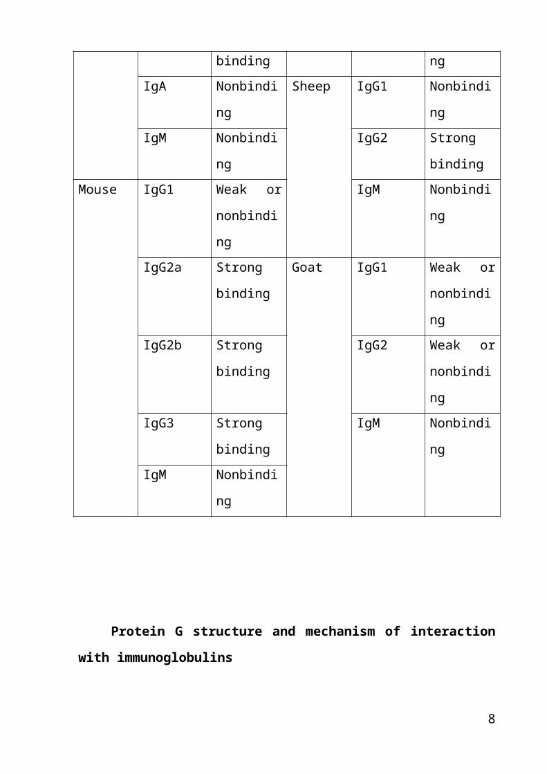
IgG3, rabbit IgG, sheep IgG2 (Table 1).
Binding site of immunoglobulins with SPA for most
IgG subclasses is localized on the region of heavy
chain which includes CH2 and CH3 domains [1]. This
property is widely used for antibodies purification.
SPA can be used for immunoglobulins fractionation
6
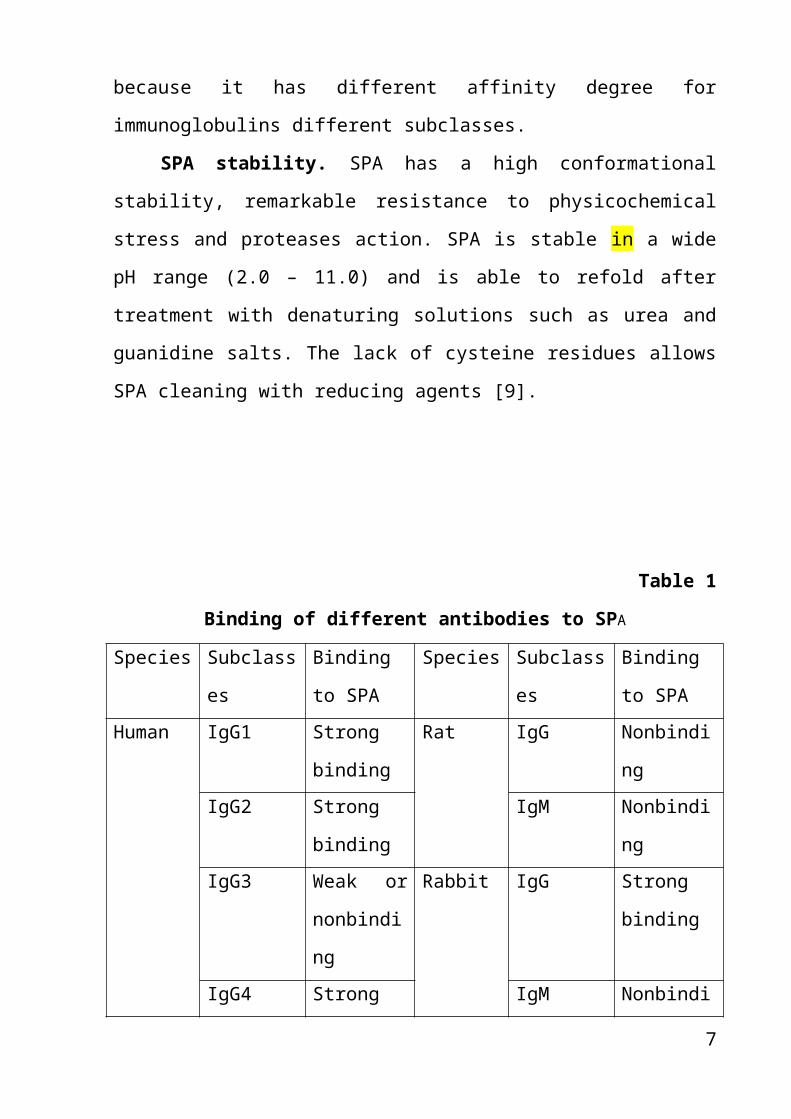
because it has different affinity degree for
immunoglobulins different subclasses.
SPA stability. SPA has a high conformational
stability, remarkable resistance to physicochemical
stress and proteases action. SPA is stable in a wide
pH range (2.0 – 11.0) and is able to refold after
treatment with denaturing solutions such as urea and
guanidine salts. The lack of cysteine residues allows
SPA cleaning with reducing agents [9].
Table 1
Binding of different antibodies to SPA
Species Subclass
es
Binding
to SPA
Species Subclass
es
Binding
to SPAHuman IgG1 Strong
binding
Rat IgG Nonbindi
ngIgG2 Strong
binding
IgM Nonbindi
ngIgG3 Weak or
nonbindi
ng
Rabbit IgG Strong
binding
IgG4 Strong IgM Nonbindi
7
binding ngIgA Nonbindi
ng
Sheep IgG1 Nonbindi
ngIgM Nonbindi
ng
IgG2 Strong
bindingMouse IgG1 Weak or
nonbindi
ng
IgM Nonbindi
ng
IgG2a Strong
binding
Goat IgG1 Weak or
nonbindi
ngIgG2b Strong
binding
IgG2 Weak or
nonbindi
ngIgG3 Strong
binding
IgM Nonbindi
ngIgM Nonbindi
ng
Protein G structure and mechanism of interaction
with immunoglobulins
8

Protein G with molecular weight 30 000 Daltons is
a cell-surface protein from Streptococcus: it contains in
its structure multiple copies of two different small
domains (COOH-terminal and NH2-terminal domains) which
can independently bind albumin and IgG. The COOH-
terminal domain is responsible for IgG binding,
whereas NH2-terminal domain of the protein binds human
serum albumin (HSA) [10, 11]. This property of
protein G is used for extraction of albumin form
blood serum. Protein G binds all the four subclasses
of human IgG [11].
Protein G binds to the Fc fragment of
immunoglobulins [12]. Protein G has three
immunoglobulin-binding domains (C1, C2 and C3), each
of 55 amino acid residues. It has been reported the
X-ray crystallographic structure of the C2 fragment
of protein G and the Fc domain of human IgG complex.
The binding site of protein G is located on the
interface between the CH2 and CH3 domains of the Fc
domain of IgG (Fig. 3) [5, 11].
Protein G binds stronger than protein A to
polyclonal IgGs from cow, horse, and sheep [5].
Protein G has a higher affinity for IgG than SPA,
binding constants of SPA and G are 8,02·103 and
3,29·104 respectively. Protein G binds with greater
9

capacity than SPA to several IgG subclasses such as
human IgG3, mouse IgG1 and rat IgG2a [6].
Consequently, protein G can be effective in that
cases during immunoglobulins purification when SPA
cannot be used. Both SPA and protein G ligands are
useful during antibodies purification and
fractionation by means of affinity chromatography,
but they are used for isolation of different classes
and subclasses of antibodies [12].
Fig. 3. X-ray crystallographic structure of the C2
fragment of Protein G (blue) interacting with the Fc
domain of human IgG (green).
PpL structure and mechanism of interaction with
immunoglobulins10

PpL is an immunoglobulin-binding protein that was
originally derived from the bacteria Peptostreptococcus
magnus, but now it is produced as recombinant
protein. The structure of PpL is comprised of two
antiparallel β-hairpins and one α-helix (Fig. 4). The
two hairpins have similar length [13].
Fig. 4. Three-dimensional structure of РрL, which
consists from four β-sheets I, II, III, IV and one α-
helix V [14].
The 62-residue immunoglobulin-binding domain of
PpL consists of central α-helix packed on four-
stranded β-sheet formed by N- and C-terminal β-
hairpins. The overall topology of the protein is
quite symmetric: the β-hairpins have similar lengths
[15].
11

PpL has the unique property to bind to kappa
light chain without interaction with an antibody's
antigen-binding site. This advantage gives PpL the
ability to bind a wider range of immunoglobulins
classes and subclasses such as human IgG2, IgG4,
human and mouse IgM, IgG1, IgG3, human IgA, human
IgD, mouse and rat IgG2a, IgG2b, rat IgG2c, IgG1 in
comparison with SPA and protein G. In addition, PpL
binds to single chain variable fragments (scFv)
without interfering to antigen binding site. PpL
binds kappa I, III and IV human light chains, but not
to kappa II in human and kappa I on mouse. PpL
recognizes 50 % of human and more than 75 % of murine
immunoglobulins [6, 16, 17]. PpL binds weakly to
rabbit immunoglobulins and does not bind to bovine,
goat or sheep immunoglobulins [18].
All these properties make PpL an excellent one in
application as ligand in the affinity chromatography,
giving possibility of recombinant ScFv molecules
purification, human and mouse IgM, IgG1, human IgD,
IgA, rat IgG1 isolation from blood serum compared
with protein G and SPA ligands. Therefore, in above
mentioned cases PpL is very beneficial and useful,
because proteins A and G cannot be applied.
12

Application of SPA in the affinity chromatography
There are many papers concerning IgG classes and
subclasses isolation using method of affinity
chromatography with application of affinity sorbent
with immobilized SPA on the Sepharose. In
biotechnology affinity chromatography with
application of sorbents on the basis of immobilized
SPA is one of the best technique for the purification
of monoclonal antibodies to homogeneity, due to its
simplicity and high degree of antibody specificity,
and also this method is applied for immunoadsorption
of antibodies from blood during treatment and therapy
of diabetes mellitus, rheumatoid arthritis and other
diseases.
SPA, immobilized on the Sepharose, provides
oriented immobilization of IgG (Fig.5).
Fig.5. Schematic drawing of immunoglobulin G,
immobilized orientedly by usage of SPA, covalently
13

attached to Sepharose. Covalent bonds are shown as
full lines.
Today high performance immunoaffinity
chromatography (HPIAC) is applied. SPA is used as a
coating to either solid or controlled pore glass
beads by cross linking with carbodiimide application.
This attachment helps to orient the antigen receptors
of the antibody toward the mobile phase of the
column. SPA binds to Fc domains of immunoglobulins by
means of hydrophobic interaction. [1].
Affinity sorbents on the basis of an immobilized
SPA. Affinity sorbents on the basis of the
immobilized SPA are available from several commercial
suppliers and vary with respect to the source of the
SPA (natural wild type or recombinant protein),
chemistry of immobilization, and bead characteristics
of a sorbent. The two leading manufacturers of
affinity sorbents on the basis of the immobilized SPA
are General Electric (GE) Healthcare and Millipore.
In affinity sorbent recombinant SPA is
immobilized on the Sepharose. SPA is coupled via the
C - terminal cysteine to the cyanogen bromide (CNBr)
- activated Sepharose matrix through a single
thioether linkage. Thioether coupling allows the
14

ligand to extend farther into the mobile phase space
than would be possible for a laterally immobilized
ligand, and this improves antibody binding [9]. In
addition effective coupling of SPA molecule to the
sorbent matrix provides absence of sorbent leaking,
obtaining of pure antibodies fractions.
By means of genetic engineering techniques B
domain of SPA was modified, and on its basis a new
ligand was created in the form of a tetramer of four
identical modified B domains. The absence of D and E
domains of SPA in the given ligand also helps to
eliminate variable region interactions, ligand
interacts only with antibodies Fc domains. As a
result antibodies binding heterogeneity is reduced,
i.e. ligand binds only antibodies with high affinity
to it. A newly developed sorbent, MabSelect SuRe,
withstands strong alkaline conditions allowing the
repeated use of 0.1 − 0.5 M NaOH for cleaning and
sanitization.
The sorbent with the immobilized SPA on porous
glass (ProSep A), on coated porous polystyrene
materials (POROS) are also produced commercially.
Process of affinity chromatography with
application of sorbents with the immobilized SPA
provides 5- to 10- fold increase of the product
15

concentration [9]. Antibodies purification with
application of affinity sorbents on the basis of the
immobilized SPA is effective due to its
physicochemical stability and low operating expenses
associated with resin cleaning and reuse.
Antibodies purification procedure
A typical chromatogram of antibodies purification
procedure by the way of usage of affinity
chromatography with application of the sorbent on the
basis of the immobilized SPA is shown in Fig. 5.
Loading/Binding. Most of the monoclonal antibodies
currently being used or investigated for therapeutic
applications are human molecules belonging to IgG
classes 1, 2, or 4, all of which bind strongly to
SPA. To the solution appointed for antibodies
dilution before applying to the chromatographic
column packed by the sorbent with immobilized SPA,
salt may be added for encouragement of monoclonal
antibody binding to the SPA, and ethylene diamine
tetraacetate to reduce proteolytic degradation, which
leads to ligand loss. Monoclonal antibodies
purification process runs several cycles on the above
mentioned chromatographic column to purify a single
batch. This reduces capital costs for such operation.
16

Washing procedure. For maximum removal of
nonspecifically bound material, pH of washing
solution must be low in order to untimely elution of
antibodies does not begin. In such way buffer
solutions with different combinations of salts and
detergents, salts and solvents, salts and polymers,
and high concentrations of Tris
(hydroxymethylaminomethane) buffer can be used.
Elution. Elution pH is typically set at the highest
possible value while maintaining high product yield.
Urea is an effective hydrogen donor/acceptor that can
outcompete hydrogen bonds, it could possibly be used
at low concentrations to facilitate protein elution
and keep product stability. Elution process on
chromatographic columns packed with the sorbent with
the immobilized SPA is conducted at low temperatures
in order to avoid proteins aggregation.
Regeneration. The ability of the sorbent with the
immobilized SPA to withstand a significant number of
its reuse cycles is an important factor for high
effective antibodies purification process for
therapeutic and laboratory application in
pharmaceutical biotechnology. Regeneration of the
sorbent with the immobilized SPA is typically carried
out with low NaOH concentrations (typically < 100 mM)
17

usage because native or recombinant SPA is stable in
slightly alkaline conditions [9].
Thus, antibodies purification process with
application of the sorbent on the basis of the
immobilized SPA provides ease and simplicity of
purification process development and performance,
high degree of protein purity because it includes
several cycles. Consequently, large purification
factor obtained from this process step helps to
simplify the entire downstream process of antibodies
purification. Such proteins purification process is
also economically beneficial because it doesn’t need
an equipment for antibodies additional purification
by other types of chromatography.
18

Fig. 5. A chromatogram of antibodies purification
procedure by the way of usage of affinity
chromatography with application of the sorbent on the
basis of the immobilized SPA.
Immunoadsorption with application of affinity sorbents with
immobilized SPA
It is known from literature sources that protein
A is used for autoimmune diseases, dilated
cardiomyopathy (DCM) treatment and therapy in the
clinical practice. For example, immunoadsorbtion with
application of columns with protein A sorbents. This
method of treatment enables to bind antibodies and
immune complexes and cause modification of immune
response. Immunoadsorbtion is also effective for
treatment of proteinuria with nephritic syndrome with
focal-segmental glomerulosclerosis, idiopatic
thrombocytopenic purpura, rheumatoid arthritis,
diabetes mellitus, and also used in kidney
transplantation. According to the clinical studies
during the treatment patients had decreased level of
immune complexes [19, 20, 21].
Diabetes mellitus is a metabolic disorder
resulted in severe systemic consequences: increased
levels of IgG, IgG3, diminishing of clinical and
19

humoral markers of heart failure, which can result in
significant cardiovascular morbidity and mortality.
Immunoadsorption (IA) was conducted applying columns
with a protein A, immobilized on the agarose,
(Immunosorba, Fresenius Medical Care, Bad Homburg,
Germany). Consequently, IgG level decreased from 10.7
g/L to 1.1 g/L (89.7 %) and IgG3 from 0.6 g/L to 0.2
g/L (66.7 %) [22].
Such dangerous autoimmune disease as systemic
lupus erythematosus (SLE) causes formation of
autoantibodies, for example, anti-double-stranded DNA
antibodies, and circulating immune complexes. One
randomized clinical trial in 20 SLE patients (two
groups with 10 patients in each) performed the
efficacy even of two different adsorption columns
(Immusorba and Ig-Therasorb). Both groups showed an
80% clinical response after 1 month [23].
DCM is a chronic myocardial disease which results
in progressive ventricular enlargement, myocardial
contractile dysfunction. DCM causes heart failure,
consequently, necessity of heart transplantation
arises. Immunoadsorption (IA) improved myocardial
inflammation in patients. Immunoglobulin extraction
was performed with IA column with protein A,
immobilized on agarose (Immunosorba, Fresenius
20

Medical Care, Bad Homburg, Germany). Consequently,
IgG decreased by 89.4% and IgG3 by 66.7% [24].
Therefore, IA with protein A is a very useful
tool for diseases therapy, which are concerned with
increased levels of antibodies in the blood stream.
IA applying protein A columns enables an efficient
isolation of antibodies and immune complexes from
patients blood, thus, a significant reduction of
antibodies levels, and, therefore, ameliorating in
patients cenesthesia, prolonged clinical benefits.
Protein G application in affinity chromatography
In the affinity chromatography recombinant form
of protein G, molecular weight 22 000 Da, is usually
used. In this variant of protein G albumin- and cell
surface binding sites have been eliminated in order
to reduce nonspecific binding during purification of
immunoglobulins. This advantage gives opportunity of
recombinant protein G application for albumin
separation from crude human immunoglobulin samples.
Immobilized protein G is most commonly used for
the purification of mammalian monoclonal and
polyclonal antibodies that do not bind strongly to
SPA. Optimal binding for most immunoglobulins to
21

protein G occurs at pH 5.0, although more neutral
Tris-HCl or phosphate buffers for binding (pH 7.5)
have been used in many studies.
Affinity sorbents with an immobilized protein G,
packed into chromatographic columns, are available
commercially today. The most popular manufacturers of
sorbents on the basis of the immobilized protein G
are Pierce and General Electric (GE) Healthcare.
Sorbents on the basis of the immobilized protein
G of Pierce manufacturing the recombinant form of
protein G immobilized to either 6 % cross-linked
beaded agarose or UltraLink Biosupport
chromatographic matrix are applied. Both types of
sorbents with the immobilized protein G are leach-
resistant and obtained matrix provides a minimal
nonspecific binding. Both affinity sorbents can be
regenerated and reused multiple times when stored
properly [16]. Such advantages of sorbents on the
basis of the immobilized protein G provide obtaining
pure immunoglobulin fractions without contaminant
proteins, time reduction and application of high
concentrations of such inexpensive reagent as NaOH
for the sorbent regeneration and cleaning.
GE Healthcare produces affinity sorbent on the
basis of the recombinant protein G. Protein G is
22

immobilized on the sepharose, activated by the
cyanogens bromide (CNBr). Such oriented
immobilization of protein G provides higher exposure
of mobile phase to this ligand, which gives
possibility of obtaining of high yield of separated
IgG (Table 2).
The sorbent on the basis of the immobilized
protein G from manufacturer GE Healthcare has such
advantages: maintaining of the IgG binding capacity
and recovery after storage in all commonly used
aqueous buffers and denaturants such as 6 M guanidine
hydrochloride and 8 M urea, and chaotropic salts such
as 3 M sodium isothiocyanate; binding of IgG over a
wide pH range. The affinity sorbent may be sanitized
by washing with 70 % ethanol.
Table 2
The dynamic IgG capacity of the sorbent on the basis
of the immobilized protein G on sepharose for variousspecies (evaluation performed at GE Healthcare)
Species Total IgG capacity(mg/ml)Human 17
Rat 7
Sheep 18
Rabbit 19
23

Goat 19
Guinea-pig 17
Cow 23
Mouse 6
Some of the most important application areas for
this affinity sorbent are the isolation and
purification or the removal of IgG from serum, the
purification of monoclonal antibodies. In cases when
IgG binds more strongly to the protein G than to the
SPA (or binds little or doesn’t bind to SPA), the
sorbent on the basis of the immobilized protein G
becomes a valuable tool to increase yield of
immunoglobulins.
Purification of IgG from serum can be carried out
effectively in a single step with the sorbent on the
basis of the immobilized protein G. IgG from human,
cow, horse, sheep, guinea-pig, dog, rabbit, mouse,
and rat can be successfully purified. Because of its
binding characteristics, the affinity sorbent on the
basis of the immobilized protein G from manufacturer
GE Healthcare is a valuable tool for the separation
of antibodies from ascite fluids, recombinant
antibodies. In fact, affinity chromatography with
application of this sorbent is suitable for
24

separations from cell culture fluid, where it is
necessary to purify rapidly large volumes [25].
In addition, there are also many studies in the
field of pharmaceutical biotechnology that describe
application of affinity chromatography using affinity
sorbents on the basis of the immobilized protein G
for antibodies purification.
The recombinant human anti-Rhesus D antibodies of
IgG1-subtype, produced by a Chinese Hamster Ovary
cell line, were purified using affinity sorbent on
the basis of the immobilized protein G. Yield of
antibodies was 57 % [26].
Recombinant human monoclonal IgG1 antibodies
produced by a transfected Chinese hamster ovary (CHO)
cell line were purified by application of affinity
sorbent “Poros G” on the basis of the immobilized
protein G using elution buffer of composition 0.1 M
acetic acid and 0.15 M sodium chloride at a pH of
2.5. Yield of antibodies was 4.64 mg/l [27, 28, 29,
30]. Monoclonal antibodies IgG1 to progesterone,
obtained from hybridoma cell line, were purified also
using the affinity sorbent on the basis of the
immobilized protein G [31].
Affinity chromatography with application of
sorbents on the basis of the immobilized protein G
25

for isolation of antibodies can be used in medical
biotechnology, e. g. for isolation of autoantibodies
to cytokines from human serum. Autoantibodies to
cytokines can contribute to disease predisposition or
pathogenesis. To describe the role of anti-cytokine
autoantibodies in disease pathogenesis, it is
important to quantitate accurately the levels of such
autoantibodies in different patient groups. This can
be made using enzyme-linked immunosorbent assay
(ELISA). Isolation of IgG autoantibodies to tumor
necrosis factor (TNF) from human serum was performed
by the method of affinity chromatography with
application of sorbent with the immobilized protein G
on the Sepharose (GE Healthcare). Antibodies were
eluted applying elution buffer (0.1 M Glycine–HCl, pH
2.5). Affinity chromatography with application of
sorbents on the basis of the immobilized protein G
enables to isolate of all four subclasses of
immunoglobulin G to TNF and to conduct further
analysis of their content applying ELISA [32 – 35].
Thus method of affinity chromatography with
immobilized protein G has great number of advantages
over other methods of proteins purification because
it enables to purify antibodies for different
purposes (e.g., in industrial biotechnology
26

purification of recombinant antibodies from cell
cultures, antibodies purification for application in
medical diagnostics of diseases), high level of
antibodies can be achieved without any particular
losses.
Recently, high-performance affinity particles
with immobilized genetically engineered fused protein
A/G are applied for antibody purification using
manual or robotic magnetic separators. Such
manufacturers as Thermo Scientific Pierce, BioVision
etc are produced such particles with immobilized
Protein A/G. Protein A/G is a recombinant fusion
protein which combines four Fc-binding domains from
SPA and two from protein G. This enables capture of
antibodies from a wider range of species and isotypes
than either protein alone. These magnetic beads with
the immobilized protein A/G has such advantages: low
non-specific binding, elimination of resin loss,
clean separate fractions of immunoglobulins
obtaining. They allow rapid obtaining of different
classes and subclasses antibody fractions with
respect to SPA, protein G affinities. Such method is
very useful during both fundamental research and
laboratory investigations in clinic [36].
27

PpL application in affinity chromatography and
immunoassays
PpL is very useful for purification of monoclonal
antibodies containing kappa light chains from culture
supernatant because it doesn’t bind bovine
immunoglobulins, which are present in the media serum
supplement. Also, in contrast to proteins A and G,
PpL is very effective at binding IgM. Although it
binds to the Fab portion of the immunoglobulin
monomer, PpL does not interfere with the antigen-
binding site of the antibody, in contrast to SPA,
which binds both Fab and Fc domains of immunoglobulin
during purification procedure. Therefore, PpL is used
in immunoprecipitation procedures and as a ligand in
the affinity chromatography [16].
In 2007 Roque et al. described the application of
an affinity sorbent on the basis of an immobilised
PpL of trademark ImmunoPure for purification of goat
serum and human serum. The percentages of human IgG
and Fab fragments obtained from this affinity sorbent
with the immobilized PpL were 75 % and 70 %,
respectively. 0.1M glycine-HCl buffer, pH 2.0, was
used [37].
28

PpL interacts mainly with region 1 (FR1) of kappa
light chain without interfering with the antigen-
binding site of the antibody. That’s why, PpL could
be used not only for the purification of human scFvs
but also as a secondary reagent to detect antigen–
scFv complexes in immunoassays. So this is one of the
advantages of PpL that marks it out proteins A and G
[38].
Thus in 2009 Muzard et al. reported a quick,
easy, and efficient method that involves substituting
a consensus sequence for the N-terminal sequence of
the antibody VL domain that does not react with PpL.
As this sequence mimics the antibody pattern
recognized by PpL, so such substitution allows the
detection and purification of murine label-free
scFvs, obtained from E. coli culture, and the
immunodetection of antigens using untagged antibody
fragments [39].
ScFv proteins were purified by loading
periplasmic preparations extracted from bacterial
culture onto an affinity sorbent with the immobilized
PpL on the agarose (volume of the chromatographic
column - 0.5 ml, Pierce Biotechnology, USA). During
the purification process scFv molecules were eluted
29

at pH 3.0. Preparations of purified scFvs were
homogeneous, no contaminants were detected.
PpL is a potentially valuable tool for the
purification, immobilization, and detection of
unlabeled scFvs even when they are associated with
their antigen [40]. The work of Muzard et al. shows
that introduction of DNA sequence of a PpL-binding
site to the ScFv by means genetic engineering methods
does not alter antibody expression as a functional
molecule in genetically modified bacteria of Escherichia
coli HB2151. The sequence grafted onto the scFvs is an
ideal affinity tag which allowed a single-step
purification of each antibody fragment [41].
One else example of PpL application for
antibodies purification is research of Cossins et.
al. The last obtained recombinant PpL, at high yield,
and this protein was used to produce the affinity
sorbent with the immobilized PpL on the sepharose.
PpL was shown to be over 95 % pure after sodium
dodecyl sulphate polyacrylamide gel electrophoresis
(SDS–PAGE) analysis. PpL was coupled to a solid
matrix at an efficiency of 94–98 %. Typical yields of
pure antibodies (>95 % purity) were in the range of
35–65 mg/L [42, 43].
30

There is also one example of PpL application in
the affinity chromatography but in fusion with
protein G, particularly their DNA sequences of single
light-chain binding domain of PpL and single Fc-
binding domain of protein G. This study shows that
despite a small size of the protein (molecular weight
is 16.5 kDa) each domain behaves independently from
each other with respect to the binding
characteristics. The PpL domain was able to bind two
equivalents of kappa light chains and the protein G
domain binds to Fc domain of IgG. LG protein was
expressed in E. coli cells with yield 75 mg/l.
In order to obtain an affinity sorbent with an
immobilized protein LG it was immobilized onto
thiopropyl-activated agarose through its C-terminal
Cys residue and packed in 1 ml chromatographic
column. This chromatographic column was used in
experiments for purification of human Fc fragments,
kappa chains, human Fc fragments combined with kappa
chains, yields were released approximately 94.1 % of
bound human Fc fragments, 87.8 % of bound kappa
chains and 96.4 % of combined bound human Fc + kappa
chains. This fact confirms that the immobilized
protein LG has the ability to bind both human Fc
fragments and kappa chains simultaneously so it can
31

be potential stable and multi-valent affinity ligand
[44 – 45]. Protein LG ligand allows purification and
isolation of immunoglobulins with high affinity to
both protein G and PpL from blood serum, ascite
fluids and obtaining of pure fractions of ScFv
molecules due to their binding to PpL by means of
kappa chains.
Due to its unique properties PpL has great
advantages as affinity ligand, because protein L
allows isolation of wide row of immunoglobulins,
including human and mouse IgM, IgG1, IgG3, human
IgG2, IgG4, IgA, IgD, rat IgG2c, IgG1, mouse and rat
IgG2a, IgG2b. What’s more, PpL doesn’t interact with
antigen-binding site of antibodies, this protein
interacts only with kappa light chains of
immunoglobulins. As proteins A and G have not high
affinity to human IgM, IgA, IgD, ScFv fragments,
don’t bind to mouse IgM, so PpL is successfully used
to isolate and purify them.
Conclusions
Nowadays in the affinity chromatography proteins
A, G, L are used as ligands and are applied for
32
purification and isolation for different classes of
immunoglobulins.
SPA binds strongly to human IgG1, mouse IgG3,
rabbit IgG, sheep IgG2. But both SPA and PpL interact
strongly with human IgG2 and IgG4, mouse IgG2a,
IgG2b. SPA is effective ligand for the affinity
chromatography due to its high conformational
stability, resistance to physicochemical stress and
proteases action, stability in the wide pH range (pH
2,0 – 11,0). In addition, affinity sorbents with the
immobilized SPA are applied in the procedure of
immunosorption of antibodies, immune complexes from
blood for therapy of autoimmune diseases, diabetes
mellitus etc.
Protein G is also successfully used as ligand in
the affinity chromatography due to presence in its
structure multiple copies of two different small
domains (COOH-terminal and NH2-terminal domains). It
can independently bind albumin and IgG,
immunoglobulins Fc fragments. Protein G binds
stronger than SPA to polyclonal IgGs from cow, horse,
and sheep; to human IgG3, mouse IgG1 and rat IgG2a.
It is active in wider pH range, in a wider variety of
buffers than SPA. So, in some cases protein G can be
used for antibody purification instead of SPA, for
33

albumin isolation, for division of complex antibody
mixtures on several fractions. Affinity sorbents with
immobilized protein G or PpL can be used for
purification of human IgG3, rat IgG2a, because the
last have high affinity to these ligands.
PpL binds to kappa light chains of antibodies,
that’s why it is effectively used as ligand for
affinity chromatography. In contrast to proteins A
and G, PpL has affinity to wider range of
immunoglobulin classes and subclasses (human and
mouse IgM, IgG1, IgG3, human IgG2, IgG4, IgA, IgD,
rat IgG2c, IgG1, mouse and rat IgG2a, IgG2b).
Affinity chromatography with PpL is used for
purification of ScFv fragments, comparing with SPA
and protein G ligands, which cannot bu used for this
purpose.
So by means of different affinity of protein A,
G, L to divergent classes and subclasses of
immunoglobulins they are used as ligands in the
affinity chromatography for diverse purification
purposes.
In the area of pharmaceutical biotechnology the
proteins A, G, L application is very useful and
beneficial, it is realized by means of
immunoglobulin-binding proteins properties improving,
34

genetic engineering fusion of DNA sequences encoded
PpL and protein G, SPA and protein G. Affinity
sorption on the basis of immobilized proteins A, G, L
can be used instead of conventional methods of
protein purification as ethanol precipitation and
different chromatography methods due to simplicity in
usage, opportunity of purification in a single step,
high level purity proteins receiving, multiuse at
upholding of proper disposal conditions.
References
1.Turkovi J. Bioaffinity chromatography. –
Netherlands: Elsevier Science Publishers, 1993. –
800 p.
2.Boi C., Dimartino S., Sarti G. C. Performance of a new
protein a affinity membrane for the primary
recovery of antibodies // Biotechnological
Programs. – 2008. – V. 24, № 3. – P. 640–647.
3.Hickstein H., Korten G., Bast R., Barz D., Templin R., Schneidewind
J. M., Kittner C., Nizze H., Schmidt R. Protein A
immunoadsorption (i. a.) in renal transplantation
patients with vascular rejection // Transfusion
Sciences. –– 1998. –19. – P. 53–57.
35
4.Graille M., Stura E. A., Corper A. L., Sutton B. J., Taussig M. J.,
Charbonnier J.-B., Silverman G. J. Crystal structure of a
Staphylococcus aureus protein. A domain complexed
with the Fab fragment of a human IgM antibody:
Structural basis for recognition of B-cell
receptors and superantigen activity // PNAS. –
2000. – P. 5399–5404.
5.Qian J., Khoury G. E., Issa H., Al-Qaoud K., Shihab P., Lowe C. R. A
synthetic Protein G adsorbent based on the multi-
component Ugi reaction for the purification of
mammalian immunoglobulins // Journal of
Chromatography B. – 2012. – V. 898. – P. 15 – 23.
6.Pierce Biotechnology. Instructions: Pierce™ Ig
Binding Proteins (Protein A, G, A/G and L). –
Rockford: Thermo Fisher Scientific Inc., 2013. –
6 p.
7. Pat. 6,548,639 B1 USА PCT/SE98/02036. IgG
binding protein from Staphylococcus and nucleotide
sequence encoding this protein / Frykberg L.;
Uppsala. – № 09/554,080 , decl. 12.05.00 ,
publ.15.04.03.
8.Nord K., Hober S., Linhult M. Protein A chromatography
for antibody purification // Journal of
Chromatography B. – 2007. – 848. – P. 40–47.
36

9. Gottschalk U. Process scale purification of
antibodies. – Hoboken: Wiley & Sons, 2009. – 430
p.
10. Sjobring U., Bjorck L., Kastern W. Streptococcal
Protein G Gene structure and protein binding
properties // The Journal of Biological
Chemistry. – 1991. – V. 366. - № 1. – P. 399 –
405.
11. Nitsche-Schmitz D. P., Johansson H. M., Sastalla I.,
Reissmann S., Frick I.-M., Chhatwal G. S. Group G
Streptococcal IgG Binding Molecules FOG and
Protein G Have Different Impacts on Opsonization
by C1q // The Journal of Biological Chemistry. –
2007. – V. 282. – № 24. – Р. 17530 – 17536.
12. Saha K., Bender F., Gizeli E. Comparative Study of IgG
Binding to Proteins G and A: Nonequilibrium
Kinetic and Binding Constant Determination with
the Acoustic Waveguide Device // Anal. Chem. –
2003. - № 75. – P. 835 – 842.
13. Karanicolas J., Brooks III C. L. The origins of
asymmetry in the folding transition states of
protein L and protein G // Protein Science. –
2002. – № 11. – P. 2351–2361.
14. Sadler D. P., Petrik E., Taniguchi Y., Pullen J. R., Kawakami M.,
Radford S. E., Brockwell D. J. Identification of a
37

Mechanical Rheostat in the Hydrophobic Core of
Protein L // J. Mol. Biol. – 2009. – № 393. – P.
237 – 248.
15. Kim D. E., Fisher C., Baker D. A Breakdown of Symmetry
in the Folding Transition State of Protein L //
J. Mol. Biol. – 2000. – № 298. – P. 971– 984.
16. Pierce Antibody purification [Electronic
resource]. – Access regime: www.piercenet.com.
17. Roquea C. A., Taipaa M. A., Lowe C. R. An artificial
protein L for the purification of immunoglobulins
and Fab fragments by affinity chromatography A //
Journal of Chromatography A. – 2005. – № 1064. –
P. 157–167.
18. Pierce Biotechnology. Instructions: Pierce®
Protein L Agarose. – Rockford: Thermo Fisher
Scientific Inc., 2008. – 4 p.
19. Snyder H. W., Cochran S. K., Balint J. P., Bertram J. H.,
Mittelman A., Guthrie T. H., Jones F. R. Experience With
Protein A-Immunoadsorption in Treatment-Resistant
Adult Immune Thrombocytopenic Purpura / H. W.
Snyder, S. K. Cochran, J. P. Balint, J. H.
Bertram, A. Mittelman, T. H. Guthrie, F. R. Jones
// Blood. – 1992. – 79, № 9. – p. 2237 – 2245.
20. McMillan R. Chronic idiopathic
thrombocytopenic purpura / R. McMillan // English
38
Journal of Medicine. – 1981. – № 304. – p. 1135 –
1147.
21. Clair St. W. E., Pisetsky D. S., Haynes B. F. Rheumatoid
Arthritis / St. W. E.Clair, D. S. Pisetsky, B. F.
Haynes. – [1st ed.] – Lippincott: Williams &
Wilkins, 2004. – 555 p.
22. Doesch A. O., Mueller S., Konstandin M., Celik S.,
Frankenstein L., Zugck C., Dengler T. J., Fleming T., Bierhaus A.,
Katus H. A. Effects of protein A immunoadsorption on
methylglyoxal levels in patients with chronic
dilated cardiomyopathy and diabetes mellitus //
Applied Cardiopulmonary Pathophysiology. - 2011
-. № 15. – Р. 3-13.
23. Schwenger V., Morath C. Immunoadsorption in
nephrology and kidney transplantation // Nephrol
Dial Transplant. – 2010. - № 25. – Р. 2407 –
2413.
24. Doesch A. O., Mueller S., Konstandin M., Celik S., Kristen A.,
Frankenstein L., Goeser S., Kaya Z., Zugck C., Dengler T. J., Katus H.
A. Effects of Protein A Immunoadsorption in
Patients with Chronic Dilated Cardiomyopathy //
Journal of Clinical Apheresis. - 2010. – №. 25. –
Р. 315 – 322.
39
25. Östervåla E. Protein G Sepharose 4 Fast Flow. –
Uppsala: GE Healthcare Bio-Sciences AB, 2007. – 6
p.
26. Schubert S., Freitag R. Comparison of ceramic
hydroxy- and fluoroapatite versus Protein A/G-
based resins in the isolation of a recombinant
human antibody from cell culture supernatant //
Journal of Chromatography A. – 2007. – № 1142. –
Р. 106 –113.
27. Gaza-Bulseco G., Hickman K., Sinicropi-Yao S., Hurkmans K.,
Chumsae C., Liu H. Effect of the conserved
oligosaccharides of recombinant monoclonal
antibodies on the separation by protein A and
protein G chromatography // Journal of
Chromatography A. – 2009. – № 1216. – P. 2382 –
2387.
28. Shields R.L., Lai J., Keck R., O’Connell L.Y., Hong K., Meng Y.G.,
Weikert S.H., Presta L.G. Lack of fucose on human IgG1
N-linked oligosaccharide improves binding to
human Fcgamma Rin and antibody dependent cellular
toxicity // J. Biol. Chem. – 2002. – № 277. – P.
26733 – 26740.
29. Davies J., Jiang L., Pan L.Z., LaBarre M. J., Anderson D., Reff
M. Expression of GnTIII in a recombinant anti-CD20
CHO production cell line: Expression of
40
antibodies with altered glycoforms leads to an
increase in ADCC through higher affinity for FC
gamma RIII // Biotechnol Bioeng. –2001. – № 74. –
P. 288 – 294.
30. Gaza-Bulseco G., Faldu S., Hurkmans K., Chumsae C., Liu H.
Effect of methionine oxidation of a recombinant
monoclonal antibody on the binding affinity to
protein A and protein G // Journal of
Chromatography B. – 2008. № 870. – P. 55 – 62.
31. Schenka J. A., Fettkec J., Lenza C., Albersd K., Mallwitzd F.,
Gajovic-Eichelmanne N., Ehrentreich-Furstere E., Kuschf E., Sellrie
F. Secretory leukocyte protease inhibitor (SLPI)
might contaminate murine monoclonal antibodies
after purification on protein G // Journal of
Biotechnology. – 2012. – № 158. – Р. 34 – 35.
32. Sennikov S.V., Golikova E.A., Kireev F.D., Lopatnikova J.A.
Purification of human immunoglobulin G
autoantibodies to tumor necrosis factor using
affinity chromatography and magnetic
separation // The Journal of Immunological
Methods. – 2013: [Electronic resource]. – Access
regime:
http://dx.doi.org/10.1016/j.jim.2013.01.012.
33. Patel S.Y., Ding L., Brown M. R., Lantz L., Gay T., Cohen S.,
Martyak L. A., Kubak B., Holland S. M. Anti-IFN-gamma
41

autoantibodies in disseminated nontuberculous
mycobacterial infections // The Journal of
Immunology. – 2005. – V. 175. – № 7. – P. 4769 –
4776.
34. Puel A., Picard C., Lorrot M., Pons C., Chrabieh M., Lorenzo L.,
Mamani-Matsuda M., Jouanguy E., Gendrel D., Casanova J. L.
Recurrent staphylococcal cellulitis and
subcutaneous abscesses in a child with
autoantibodies against IL-6 // The Journal of
Immunology. – 2008. – V. 180. – № 1. – P. 647 –
654.
35. Uchida K., Nakata K., Trapnell B. C., Terakawa T., Hamano
E., Mikami A., Matsushita I., Seymour J. F., Oh-Eda M., Ishige I.,
Eishi Y., Kitamura T., Yamada Y., Hanaoka K., Keicho N. High
affinity autoantibodies specifically eliminate
granulocyte–macrophage colony-stimulating factor
activity in the lungs of patients with idiopathic
pulmonary alveolar proteinosis // Blood. – 2004.
– V. 103. – № 3. – Р. 1089 – 1098.
36. Pierce Protein AG Magnetic Beads [Electronic
resource]. – Access regime:
http://www.piercenet.com/product/protein-ag-
magnetic-beads.
37. Roque A. C. A., Taipaa M. A., Lowe C. R. An artificial
protein L for the purification of immunoglobulins
42

and Fab fragments by affinity chromatograpy //
Journal of Chromatography A. – 2005. – № 1064. –
P. 157–167.
38. M. Graille, E. A. Stura, N. G. Housden, J. A. Beckingham, S. P.
Bottomley,D. Beale, M. J. Taussig, B. J. Sutton, M. G. Gore, J.-B.
Charbonnier Complex between Peptostreptococcus
magnus protein L and a human antibody reveals
structural convergence in the interaction modes
of Fab binding proteins // Structure. – 2001. –
V. 9. – P. 679–687.
39. Muzard J., Adi-Bessalem S., Juste M., Laraba-Djebari F.,
Aubrey N., Billiald P. Grafting of protein L-binding
activity onto recombinant antibody fragments //
Analytical Biochemistry. – 2009. – № 388. – P.
331–338.
40. Nilson B. H. K., Solomon A., Bjurck L., Еkerstrum B. Protein
L from Peptostreptococcus magnus binds to the K
light chain variable domain // The Journal of
Biological Chemistry. – 1992. – V. 267. – № 4. –
P. 2234-2239.
41. Housden N. G., Harrison S., Housden H. R., Thomas K.-A.,
Beckingham J. A., Roberts S. E., Bottomley S. P., Graille M., Stura E.,
Gore M. G. Observation and characterization of the
interaction between a single immunoglobulin
binding domain of protein L and two equivalents
43

of human k-light chains // Journal of Biological
Chemistry. – 2004. – № 279. – P. 9370 – 9378.
42. Cossins A. J., Harrison S., Popplewell A. G., Gore M. G.
Recombinant production of a VL single domain
antibody in Escherichia coli and analysis of its
interaction with peptostreptococcal protein L //
Protein expression and purification. – 2007. – №
51. – P. 253 – 259.
43. Murphy, J. P. Duggleby C. J., M. A. Atkinson, Trowern A. R.,
Atkinson T., Goward C. R. The functional unit of
Peptostreptococcal protein L // Molecular
Microbiology. – 1994. – V. 12. – № 6. – P. 911–
920.
44. Bottomley S. P., Beckingham J. A., Murphy J. P., Atkinson M.,
Sutton B. J., Gore M. G. Cloning, expression and
purification of PpL-1, a kappa-chain binding
protein, based upon protein L from
Peptostreptococcus magnus // Bioseparation. –
1995. – V. 5. – № 6. – P. 359 – 367.
45. Harrison S. L., Housden N. G., Bottomley S. P., Cossins A. J.,
Gore M. G. Generation and expression of a minimal
hybrid Ig-receptor formed between single domains
from proteins L and G // Protein Expression and
Purification. – 2008. – № 58. P. 12 – 22.
44

ВИКОРИСТАННЯ ІМУНОГЛОБУЛІН-ЗВ’ЯЗУВАЛЬНИХ
ПРОТЕЇНІВ А, G, L В АФІННІЙ ХРОМАТОГРАФІЇ
О. В. Святенко1, О. Б. Горбатюк2, О. А. Васильченко1 1Національний авіаційний університет, пр. Комарова, 1,
Київ
[email protected]Інститут генетичної та регенеративної медицини НАМН
України, вул. Червоноармійська, 57/3, Київ
Протеїни A, G та L – це нативні та рекомбінантні
протеїни бактеріального походження, що зв’язуються з
імуноглобулінами ссавців. У теперішній час в
біотехнології для виробництва афінних сорбентів
переважно використовуються рекомбінантні варіанти
протеїнів A, G та L. У роботі проведено порівняльну
характеристику протеїнів A, G, L, афінних сорбентів
на їхній основі, переваги та недоліки цих протеїнів
та їхнє використання як лігандів в афінній
хроматографії. Наведений аналіз властивостей
протеїнів A, G та L. Специфічність зв’язування та
афінність даних протеїнів розрізняються в залежності
від виду тварин та підкласу антитіл. Протеїн А має
високу афінність до IgG1, IgG2, IgG4 людини, IgG2a,
IgG2b, IgG3 миші, IgG2 кози та вівці, IgG собаки,
45

кота, кролика, морської свинки. Протеїн G міцно
зв’язується з IgG людини, миші, корови, кози, вівці
та кролика. Протеїн L має здатність до міцного
зв’язування з легкими каппа ланцюгами імуноглобулінів
людини, миші, щура та свині. Показана доцільність
застосування афінної хроматографії з використанням
сорбентів на основі іммобілізованих протеїнів A, G та
L для виділення та очищення антитіл різних класів.
Вищезазначений метод використовується як альтернатива
традиційним методам очищення протеїнів, таким як
іонообмінна, гідрофобних взаємодій, металo-афінна
хроматографія, осадження етанолом, завдяки простоті
використання, можливості одностадійного процесу
очищення, одержання протеїнів з високим рівнем
чистоти, багаторазового використання при дотриманні
правильних умов зберігання та експлуатації. Афінні
сорбенти на основі іммобілізованих протеїнів A, G та
L використовуються не лише для очищення антитіл, але
також для виділення різних фракцій антитіл з
сироватки крові.
Ключові слова: афінна хроматографія,
стафілококовий протеїн А (SPA), пептострептоковий
протеїн L (PpL), протеїн G, антитіла, імуноглобуліни.
46

ИСПОЛЬЗОВАНИЕ ИММУНОГЛОБУЛИН-СВЯЗЫВАЮЩИХ ПРОТЕИНОВ А,
G, L В АФФИННОЙ ХРОМАТОГРАФИИ
О. В. Святенко1, О. Б. Горбатюк2, О. А. Васильченко1 1Национальный авиационный университет, пр. Комарова,
1, Киев
[email protected]Институт генетической и регенеративной медицины НАН
Украины, ул. Красноармейская, 57/3, Киев
Протеины A, G и L – это нативные и рекомбинантные
протеины бактериального происхождения, которые
связываются с иммуноглобулинами млекопитающих. В
настоящее время в биотехнологии для производства
аффинных сорбентов преимущественно используются
рекомбинантные варианты протеинов A, G и L. В работе
проведена сравнительная характеристика протеинов A,
G, L , аффинных сорбентов на их основе, преимущества
и недостатки этих протеинов и их использование как
лигандов в аффинной хроматографии. Представлен анализ
свойств протеинов A, G и L. Специфичность связывания
и аффинность данных протеинов различаются в
зависимости от вида животного и подкласса антител.
Протеин А имеет высокую аффинность к IgG1, IgG2, IgG4
человека, IgG2a, IgG2b, IgG3 мыши, IgG2 козы и овцы,47

IgG собаки, кота, кролика, морской свинки. Протеин G
прочно связывается с IgG человека, мыши, коровы,
козы, овцы и кролика. Протеин L имеет способность к
прочному связыванию с легкими каппа цепями
иммуноглобулинов человека, мыши, крысы и свиньи.
Показана целесообразность использования аффинной
хроматографии с использованием сорбентов на основе
иммобилизованных протеинов А, G и L для выделения и
очищения антител разных классов. Вышеуказанный метод
используется как альтернатива традиционным методам
очищения протеинов, таким как ионообменная,
гидрофобных взаимодействий, металл-аффинная
хроматография, осаждение этанолом, благодаря простоте
использования, возможности одностадийного процесса
очищения, получения протеинов с высоким уровнем
чистоты, многоразового использования при соблюдении
правильных условий хранения и эксплуатации. Аффинные
сорбенты на основе иммобилизованных протеинов А, G и
L используются не только для очищения антител, но
также для выделения разных фракций антител из
сыворотки крови.
Ключевые слова: аффинная хроматография,
стафилококковый протеин А (SPA), пептострептококковый
48

протеин L (PpL), протеин G, антитела,
иммуноглобулины.
49
Copyright © 2022 FDOKUMEN