Purification of polyclonal antibodies from Cohn fraction II + III, skim milk, and whey by affinity...
11
Please cite this article in press as: Z. Liu, et al., J. Chromatogr. A (2012), http://dx.doi.org/10.1016/j.chroma.2012.09.026 ARTICLE IN PRESS G Model CHROMA-353629; No. of Pages 11 Journal of Chromatography A, xxx (2012) xxx–xxx Contents lists available at SciVerse ScienceDirect Journal of Chromatography A jou rn al h om epage: www.elsevier.com/locat e/chroma Purification of human immunoglobulins A, G and M from Cohn fraction II/III by small peptide affinity chromatography Zhuo Liu a , Patrick V. Gurgel a , Ruben G. Carbonell a,b,∗ a Department of Chemical and Biomolecular Engineering, North Carolina State University, Raleigh, NC 27695, USA b Golden LEAF Biomanufacturing Training and Education Center (BTEC), North Carolina State University, Raleigh, NC 27695, USA a r t i c l e i n f o Article history: Received 10 June 2012 Received in revised form 4 September 2012 Accepted 6 September 2012 Available online xxx Keywords: Hexamer peptide IVIG Cohn fraction Elution additive PEG Ammonium sulfate a b s t r a c t This work describes attempts to purify human IgG, IgA and IgM from Cohn fraction II/III using HWRGWV affinity peptide resin. The effects of peptide density and different elution additives on recovery of the three antibodies were investigated. At low peptide density, salting-in salts such as magnesium chloride and calcium chloride facilitated antibody elution. Ethylene glycol, urea and arginine also facilitated elu- tion because of their ability to decrease hydrophobic interactions, hydrogen bonding and electrostatic interactions. However, at high peptide density, no recovery improvements were observed because of increased non-specific hydrophobic interactions. The final elution conditions for each antibody were chosen based on the resulting yields and purities when a 10:2:1 mg/mL mixture of human IgG, IgA and IgM was used as starting material. Different pretreatment methods were employed in order to improve the purity of antibodies from Cohn fraction II/III. After pretreatment with caprylic acid precipitation or combination of caprylic acid and polyethylene glycol precipitation, purities over 95% and yields of about 60% were obtained for hIgG, which are comparable to current chromatographic purification methods involving two chromatography steps when hIgG is isolated from plasma fractions. A hIgA-enriched frac- tion with 42% hIgA and 56% hIgG, as well as a hIgM enriched fraction with 46% hIgM, 28% hIgA and 24% hIgG, were obtained as the by-products. © 2012 Elsevier B.V. All rights reserved. 1. Introduction Antibodies have been used as therapeutics in various forms for over a century since their first administration in the form of sera in the 1890s [1–5]. With more than 20 molecules in clinical use, the world’s therapeutic monoclonal antibodies market exceeded $40 billion in 2010 and is expected to reach $70 billion by 2015 [6,7]. Apart from the therapeutic monoclonal antibodies, the impor- tance of polyclonal antibodies obtained from human plasma has grown over the last 30 years. For more than 20 years, polyclonal antibodies derived from plasma have been used in the treatment of a wide range of primary and secondary immunodeficiencies. As one of the leading products of the human plasma fractionation industry, intravenous immunoglobulin (IVIg) is a sterile, highly purified immunoglobulin G (IgG) product manufactured from pooled human plasma [8]. Typically, IVIg contains more than 95% unmodified IgG and only trace amounts of immunoglobulin A (IgA) and immunoglobulin M (IgM) [9]. Intravenous immunoglobulin ∗ Corresponding author at: Department of Chemical and Biomolecular Engineer- ing, North Carolina State University, 911 Partners Way, Raleigh, NC 27695, USA. Tel.: +1 919 515 5118; fax: +1 919 515 3465. E-mail address: [email protected] (R.G. Carbonell). (IVIg) is not only standard therapy for most primary immunode- ficiencies (PID), but also is extensively used in the treatment of an increasing number of autoimmune and inflammatory diseases [10–13]. Immunoglobulin M enriched IVIg (IVIgM) and Immunoglobulin A and M enriched IVIg (IVIgAM) are powerful therapeutic agents, which can be used to prevent inflammation [14–16], combat bac- terial infections [17], in the treatment of severe sepsis and septic shock [18,19], to inhibit alloantigen-induced proliferation in mixed lymphocyte reaction in vitro [20] as well as reduce endotoxemia and its consequences in bone marrow transplant patients [21]. Traditional plasma fractionation is currently carried out using either the Cohn–Oncley [22,23] or the Kistler–Nitschmann [24] methods, which are processes that involve cold ethanol precip- itation with tightly controlled conditions including temperature, pH, ionic strength and ethanol concentration. Recently, chromato- graphic purification methods have been introduced which usually start with plasma or intermediates derived from the Cohn–Oncley or Kistler–Nitschmann process and involve at least two chromatog- raphy steps. For example, Gamunex ® (Talecris Biotherapeutics, RTP, NC, USA) uses Cohn–Oncley fraction II/III as starting material; after caprylate pretreatment to precipitate non-IgG proteins and to inactivate enveloped viruses, two successive anion-exchange columns are used to purify IgG and remove caprylate [25]. Both 0021-9673/$ – see front matter © 2012 Elsevier B.V. All rights reserved. http://dx.doi.org/10.1016/j.chroma.2012.09.026
Transcript of Purification of polyclonal antibodies from Cohn fraction II + III, skim milk, and whey by affinity...
G
C
Ps
Za
b
a
ARRAA
KHICEPA
1
oit$[tgaooippua
iT
0h
ARTICLE IN PRESS Model
HROMA-353629; No. of Pages 11
Journal of Chromatography A, xxx (2012) xxx– xxx
Contents lists available at SciVerse ScienceDirect
Journal of Chromatography A
jou rn al h om epage: www.elsev ier .com/ locat e/chroma
urification of human immunoglobulins A, G and M from Cohn fraction II/III bymall peptide affinity chromatography
huo Liua, Patrick V. Gurgela, Ruben G. Carbonell a,b,∗
Department of Chemical and Biomolecular Engineering, North Carolina State University, Raleigh, NC 27695, USAGolden LEAF Biomanufacturing Training and Education Center (BTEC), North Carolina State University, Raleigh, NC 27695, USA
r t i c l e i n f o
rticle history:eceived 10 June 2012eceived in revised form 4 September 2012ccepted 6 September 2012vailable online xxx
eywords:examer peptide
VIGohn fraction
a b s t r a c t
This work describes attempts to purify human IgG, IgA and IgM from Cohn fraction II/III using HWRGWVaffinity peptide resin. The effects of peptide density and different elution additives on recovery of thethree antibodies were investigated. At low peptide density, salting-in salts such as magnesium chlorideand calcium chloride facilitated antibody elution. Ethylene glycol, urea and arginine also facilitated elu-tion because of their ability to decrease hydrophobic interactions, hydrogen bonding and electrostaticinteractions. However, at high peptide density, no recovery improvements were observed because ofincreased non-specific hydrophobic interactions. The final elution conditions for each antibody werechosen based on the resulting yields and purities when a 10:2:1 mg/mL mixture of human IgG, IgA andIgM was used as starting material. Different pretreatment methods were employed in order to improve
lution additiveEGmmonium sulfate
the purity of antibodies from Cohn fraction II/III. After pretreatment with caprylic acid precipitation orcombination of caprylic acid and polyethylene glycol precipitation, purities over 95% and yields of about60% were obtained for hIgG, which are comparable to current chromatographic purification methodsinvolving two chromatography steps when hIgG is isolated from plasma fractions. A hIgA-enriched frac-tion with 42% hIgA and 56% hIgG, as well as a hIgM enriched fraction with 46% hIgM, 28% hIgA and 24%hIgG, were obtained as the by-products.
. Introduction
Antibodies have been used as therapeutics in various forms forver a century since their first administration in the form of seran the 1890s [1–5]. With more than 20 molecules in clinical use,he world’s therapeutic monoclonal antibodies market exceeded40 billion in 2010 and is expected to reach $70 billion by 20156,7]. Apart from the therapeutic monoclonal antibodies, the impor-ance of polyclonal antibodies obtained from human plasma hasrown over the last 30 years. For more than 20 years, polyclonalntibodies derived from plasma have been used in the treatmentf a wide range of primary and secondary immunodeficiencies. Asne of the leading products of the human plasma fractionationndustry, intravenous immunoglobulin (IVIg) is a sterile, highlyurified immunoglobulin G (IgG) product manufactured from
Please cite this article in press as: Z. Liu, et al., J. Chromatogr. A (2012), http
ooled human plasma [8]. Typically, IVIg contains more than 95%nmodified IgG and only trace amounts of immunoglobulin A (IgA)nd immunoglobulin M (IgM) [9]. Intravenous immunoglobulin
∗ Corresponding author at: Department of Chemical and Biomolecular Engineer-ng, North Carolina State University, 911 Partners Way, Raleigh, NC 27695, USA.el.: +1 919 515 5118; fax: +1 919 515 3465.
E-mail address: [email protected] (R.G. Carbonell).
021-9673/$ – see front matter © 2012 Elsevier B.V. All rights reserved.ttp://dx.doi.org/10.1016/j.chroma.2012.09.026
© 2012 Elsevier B.V. All rights reserved.
(IVIg) is not only standard therapy for most primary immunode-ficiencies (PID), but also is extensively used in the treatment ofan increasing number of autoimmune and inflammatory diseases[10–13].
Immunoglobulin M enriched IVIg (IVIgM) and ImmunoglobulinA and M enriched IVIg (IVIgAM) are powerful therapeutic agents,which can be used to prevent inflammation [14–16], combat bac-terial infections [17], in the treatment of severe sepsis and septicshock [18,19], to inhibit alloantigen-induced proliferation in mixedlymphocyte reaction in vitro [20] as well as reduce endotoxemiaand its consequences in bone marrow transplant patients [21].
Traditional plasma fractionation is currently carried out usingeither the Cohn–Oncley [22,23] or the Kistler–Nitschmann [24]methods, which are processes that involve cold ethanol precip-itation with tightly controlled conditions including temperature,pH, ionic strength and ethanol concentration. Recently, chromato-graphic purification methods have been introduced which usuallystart with plasma or intermediates derived from the Cohn–Oncleyor Kistler–Nitschmann process and involve at least two chromatog-raphy steps. For example, Gamunex® (Talecris Biotherapeutics,
://dx.doi.org/10.1016/j.chroma.2012.09.026
RTP, NC, USA) uses Cohn–Oncley fraction II/III as starting material;after caprylate pretreatment to precipitate non-IgG proteins andto inactivate enveloped viruses, two successive anion-exchangecolumns are used to purify IgG and remove caprylate [25]. Both
ING Model
C
2 togr.
BDiddcaact
obabrtsrafistrpitua
pHtrb
atedpifoepiuI
2
2
H(UHf7swrs
ARTICLEHROMA-353629; No. of Pages 11
Z. Liu et al. / J. Chroma
axter’s Gammagard SD® and IGIV 10% (Baxter International Inc,eerfield, IL, USA) are also manufactured in a similar manner,
nvolving two ion-exchange chromatography steps after solvent-etergent treatment of the ethanol fraction [26]. These processeso not use affinity chromatography due to limitations such as highost, low chemical and physical stability of ligands such as Protein And Protein G, leachability and toxicity-related issues. Other valu-ble immunoglobulins such as IgA and IgM are typically treated asontaminant proteins that need to be removed during the purifica-ion process.
Hexamer peptide ligand HWRGWV exhibits the ability to rec-gnize and bind hIgG through its Fc portion. This peptide haseen previously identified by our group through the screening of
synthetic solid phase combinatorial peptide library, and it haseen found that its selectivity to the Fc portion of IgG is compa-able to that of Protein A [27–29]. Previous studies showed thathe peptide ligand binds to hIgA and hIgM as well, having thetrongest binding affinity to hIgM, followed by hIgA and hIgG,espectively [30]. Ligand density had a marked effect on the puritiesnd recoveries obtained for the different proteins when being puri-ed from cMEM, with lower peptide densities (0.04 mequiv./g)howing lower recoveries and higher purities, while higher pep-ide densities (up to 0.55 mequiv./g) showing progressively higherecoveries, and progressively lower purities [30]. It was found that aH gradient elution was not able to separate the captured antibod-
es from the affinity column [30]. However, results also suggestedhat the three antibodies could be separated from a mixture bysing different elution solutions, due to their different bindingffinities to the peptide resin.
Experiments comparing the performance of the HWRGWVeptide resin and its terminal amine acetylated equivalent (Ac-WRGWV) showed that the terminal charge had an effect on
he capture and the pH-based elution of antibodies. However, theesults also suggest that the dominant electrostatic component ofinding was due to the histidine and arginine residues in the ligand.
In this work, the effects of different elution additives on the sep-ration of IgG, IgA and IgM were studied, in order to determinehe optimum conditions for maximizing the yields and purities forach of the proteins. Based on the results, the best elution con-itions and peptide density were chosen based on the yields andurities of the three antibodies from a starting solution consist-
ng of 10:2:1 mg/mL of IgG, IgA and IgM, respectively. Finally, Cohnraction II/III was used as the starting material for the purificationf human IgG, IgA and IgM using HWRGWV peptide resin. Differ-nt pretreatment methods were used in order to achieve enhancedurities and yields for the three antibodies. To our knowledge, this
s the first time in the literature that an affinity chromatography col-mn was employed to separate IgG, IgA and IgM from Cohn fraction
I/III in a single column.
. Experimental
.1. Materials
Peptides International (Louisville, KY, USA) synthesizedWRGWV directly on the resin Toyopearl AF-Amino-650M
particle size 65 �m) (Tosoh BioScience, Montgomeryville, PA,SA) using fluorenylmethyloxycarbonyl (Fmoc) chemistry. TheWRGWV resins were synthesized at different peptide densities
rom 0.04 to 0.55 mequiv./g. Phosphate-buffered saline (PBS) pH.4 (0.01 M phosphate, 0.138 M sodium chloride, and 2.7 mM potas-
Please cite this article in press as: Z. Liu, et al., J. Chromatogr. A (2012), http
ium chloride), urea, arginine and polyethylene glycol (MW4600)ere obtained from Sigma (St. Louis, MO, USA). Sodium chlo-
ide, sodium hydroxide, magnesium chloride, calcium chloride,odium acetate, glacial acetic acid, phosphoric acid, ammonium
PRESSA xxx (2012) xxx– xxx
sulfate, ammonium hydroxide, guanidine hydrochloride, Trishydrochloride, ethylene glycol and methanol were purchasedfrom Fisher Scientific (Pittsburgh, PA, USA). NuPAGE Novex 4–12%Bis–Tris gels, NuPAGE reducing agent, NuPAGE MOPS SDS runningbuffer, SeeBlue Plus2 pre-stained molecular weight marker andSimplyBlue SafeStain were all from Invitrogen (Carlsbad, CA, USA).Human IgG lyophilized power was purchased from Equitech-Bio(Kerrville, TX, USA). Human IgA and IgM were from Fitzgerald(Concord, MA, USA). Human IgG, IgA and IgM enzyme-linkedimmunosorbent assay (ELISA) kit were purchased from AlphaDiagnostic International (San Antonio, TX, USA). Microcon UltracelYM-3 centrifugal filters (regenerated cellulose, 3000 MWCO) werepurchased from Millipore (Billerica, MA, USA). 0.2 �m and 0.45 �mAcrodisc Syringe Filter were purchased from Pall Corporation (PortWashington, NY, USA). BCA (bicinchoninic acid) protein assay wasfrom Thermo Scientific (Rockford, IL USA). Microbore columns(30 mm × 2.1 mm) for all chromatographic experiments were fromAlltech (Deerfield, IL, USA).
2.2. Equipment
All chromatographic experiments were carried out on a Waters626LC system. Absorbance of protein samples was measuredat wavelength 280 nm using a built-in 2487 dual wave-lengthUV detector from Waters (Milford, MA, USA). ELISA wash stepwas performed on ELx50 Microplate Strip Washer from Bio-TekInstruments Inc. (Winooski, VT, USA), and ELISA protein concentra-tion was determined by �Quant Spectrophotometer from Bio-TekInstruments Inc. as well.
2.3. Chromatographic capture and recovery of human IgG, IgAand IgM
Peptide resins were dry packed in 30 mm × 2.1 mm I.D. micro-bore columns (0.1 mL) and swollen in 20% methanol at 0.05 mL/minfor 2 h followed by HPLC grade water and PBS washes. Beforesample loading, columns were equilibrated with pH 7.4 equilibra-tion buffer consisting of PBS + 1 M NaCl. Samples were manuallyinjected at room temperature using a 100 �L sample loop (Thom-son, Springfield, VA, USA). The samples were loaded at 0.05 mL/minfor 10 minutes, and then the flow rate was increased to 0.3 mL/minfor the remainder of the run. PBS + 1 M NaCl was also used as thebinding buffer, the column was washed sequentially with 3 mLbinding buffer, 6 mL elution buffer, and 6 mL 6 M guanidine-HClto regenerate the column, followed by PBS + 1 M NaCl for equilibra-tion. Acetate buffer (0.2 M) at pH 5.0 with different elution additivesincluding NaCl, MgCl2, CaCl2, ethylene glycol, urea and argininewere used as elution buffers. The recoveries of hIgG, hIgA, hIgMwere calculated based on the peak areas for elution and regener-ation assuming all injected proteins bound to the resin since onlysmall flow through peaks appeared for all injections.
2.4. Preliminary elution condition study for the isolation ofhuman IgG, IgA and IgM from a 10:2:1 mg/mL IgG, IgA, IgMmixture
Columns with peptide resin HWRGWV at the densities of0.11 mequiv./g and 0.23 mequiv./g were prepared and equilibratedby the same methods described in Section 2.3. A mixture of humanIgG, IgA and IgM in PBS at concentrations of 10, 2 and 1 mg/mLrespectively was manually injected at room temperature using a50 �L sample loop (Thomson, Springfield, VA, USA). The samples
://dx.doi.org/10.1016/j.chroma.2012.09.026
were loaded at 0.05 mL/min for 10 minutes, and then the flow ratewas increased to 0.3 mL/min for the rest of the run. PBS + 1 M NaClwas used as the binding buffer, the column was washed sequen-tially with 3 mL binding buffer, 6 mL hIgG elution buffer, 6 mL hIgA
ARTICLE IN PRESSG Model
CHROMA-353629; No. of Pages 11
Z. Liu et al. / J. Chromatogr. A xxx (2012) xxx– xxx 3
Table 1Elution conditions for the isolation of human IgG, IgA and IgM (10:2:1) from their mixture with HWRGWV at peptide density of 0.23 mequiv./g.
Run hIgG elution hIgA elution hIgM elution
10.2 M AB pH 5.0 0.2 M AB pH 5.0 + 0.2 M CaCl2
0.2 M AB pH 5.0 + 2 M MgCl2 (g*)2 0.2 M AB pH 5.0 + 1 M MgCl2
30.2 M AB pH 5.2 0.2 M AB pH 5.0 + 0.2 M CaCl2
0.2 M AB pH 5.0 + 0.5 M MgCl24 0.2 M AB pH 5.0 + 1 M MgCl25 0.2 M AB pH 5.0 + 2 M MgCl2
6
0.2 M AB pH 5.0
0.2 M AB pH 5.0 + 40% EG
0.2 M AB pH 5.0 + 2 M MgCl27 0.2 M AB pH 5.0 + 2 M urea8 0.2 M AB pH 5.0 + 0.5 M arginine9 0.2 M AB pH 5.0 + 1 M arginine
g*: gradient elution.
Table 2Elution conditions for the isolation of human IgG, IgA and IgM (10:2:1) from their mixture with HWRGWV at peptide density of 0.11 mequiv./g.
Run hIgG elution hIgA elution hIgM elution
10.2 M AB pH 5.0 0.2 M AB pH 5.0 + 0.2 M CaCl2
0.2 M AB pH 5.0 + 0.5 M MgCl22 0.2 M AB pH 5.0 + 1 M MgCl2
30.2 M AB pH 5.2 0.2 M AB pH 5.0 + 0.2 M CaCl2
0.2 M AB pH 5.0 + 1 M MgCl24 0.2 M AB pH 5.0 + 2 M MgCl25 0.2 M AB pH 3.0
60.2 M AB pH 5.0
0.2 M AB pH 5.0 + 40% EG 0.2 M AB pH 5.0 + 1 M MgCl27 0.2 M AB pH 5.0 + 2 M urea 0.2 M AB pH 5.0 + 2 M MgCl2
80.2 M AB pH 5.0 + 0.5 M arginine
0.2 M AB pH 5.0 + 1 M MgCl2
AB p
eHhatnA
2
2
cwtawf(r
22taotpshpwwma7
0.2 M AB pH 5.29
10 0.2 M
lution buffer, 6 mL hIgM elution buffer and 6 mL 6 M guanidine-Cl to regenerate the column. The selections of elution buffers foruman IgG, IgA and IgM are listed in Tables 1 and 2 for HWRGWVt 0.23 mequiv./g and 0.11 mequiv./g, respectively. All elution frac-ions were collected and the pH was immediately adjusted toeutral by adding an appropriate volume of 1 M Tris buffer pH 8.0.ll collected fractions were directly subjected to ELISA assays.
.5. Cohn fraction II/III sample preparation and pretreatments
.5.1. Cohn fraction II/III sample preparationFifty mg/mL Cohn fraction II/III containing 60–85% protein pur-
hased from Sigma (St. Louis, MO, USA) was dissolved in HPLC gradeater or PBS and stirred overnight at 4 ◦C. This original Cohn frac-
ion was named stock solution I. After centrifugation for 15 mint 6000 × g to remove any un-dissolved material, stock solution IIas obtained. The stock solution II was passed through a 0.45 �m
ollowed by 0.2 �m Acrodisc Syringe Filter from Pall CorporationPort Washington, NY, USA) to achieve stock solutions III and IV,espectively.
.5.2. Cohn fraction II/III pretreatments
.5.2.1. Caprylic acid and PEG pretreatment. The pH of stock solu-ion IV was adjusted to pH 5.0 at room temperature with 0.2 Mcetic acid. Caprylic acid was added to a concentration of 50 mMver a 5-min time-period. The suspension was mixed for 1 h andhe precipitate was removed by centrifugation at 14,000 × g. In theretreatment without polyethylene glycol (PEG) precipitation, theupernatant solution pH was raised to pH 7.0 with 0.2 M sodiumydroxide and filtered through a 0.2 �m syringe filter, while in theretreatment with PEG precipitation, the supernatant solution pHas adjusted to pH 5.5 with 0.2 M sodium hydroxide, PEG 4600
Please cite this article in press as: Z. Liu, et al., J. Chromatogr. A (2012), http
as added to reach a concentration of 3% (w/v). The solution wasixed for 1 h and the precipitate was removed by centrifugation
t 14,000 × g. Finally, the supernatant solution pH was raised to pH.0 with 0.2 M NaOH and filtered through a 0.2 �m syringe filter.
0.2 M AB pH 5.0 + 1 M arginine
H 5.0 + 1 M arginine 0.2 M AB pH 5.0 + 2 M MgCl2
2.5.2.2. PEG pretreatment. The stock solution IV pH was adjustedto 5.5 with 0.2 M acetic acid and PEG 4600 was added to achieveconcentration of 3% (w/v). The suspension was mixed for 1 h andthe precipitate was removed by centrifugation. After the pH wasraised to pH 7.0 with 0.2 M NaOH, the solution was filtered througha 0.2 �m syringe filter.
2.5.2.3. SAS pretreatment. A saturated ammonium sulfate (SAS)solution was prepared at room temperature. The pH was adjustedto pH 7.0 by addition of ammonium hydroxide, and the solution wasfiltered through 0.2 �m membrane to remove particulate material.Two milliliters of SAS was added over 5 min with gentle stirringto 4.8 mL stock solution IV. Stirring was continued at room tem-perature for 20 min, and the mixture was centrifuged for 10 minat 14,000 × g in 2 mL centrifugal tubes. The pellets were discarded,and 2.4 mL of SAS was added to the combined supernatant over aperiod of 5 min. After stirring for 30 min at room temperature, themixture was centrifuged for 15 min at 14,000 × g. The supernatantswere decanted as much solution as possible by inverting the tubesfor 5 min. Finally, 4.0 mL of HPLC grade water or PBS pH 7.4 wasadded to the combined pellets to dissolve all and the clear solutionwas filtered through 0.2 �m syringe filter.
2.6. Purification of human IgG, IgA and IgM from Cohn fractionII/III
A column with peptide resin HWRGWV at the density of0.11 mequiv./g was prepared and equilibrated by the same meth-ods described in Section 2.3. Cohn fraction II/III after preparationwith or without pretreatments was manually injected at roomtemperature using a 50 �L sample loop (Thomson, Springfield, VA,USA). The samples were loaded at 0.05 mL/min for 10 minutes, and
://dx.doi.org/10.1016/j.chroma.2012.09.026
then the flow rate was increased to 0.3 mL/min for the rest of therun. PBS + 1 M NaCl was used as the binding buffer, the columnwas washed sequentially with 3 mL binding buffer, 6 mL hIgG elu-tion buffer, 6 mL hIgA elution buffer, 6 mL hIgM elution buffer and
ARTICLE IN PRESSG Model
CHROMA-353629; No. of Pages 11
4 Z. Liu et al. / J. Chromatogr. A xxx (2012) xxx– xxx
F ( )a
6oplaw
2
aEnwt1tcpcrIpm(
3
3a
ahaemsr
fv
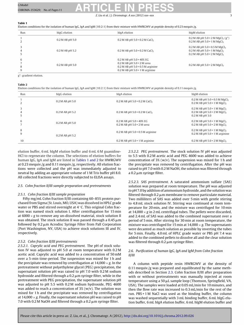
ig. 1. NaCl, MgCl2 and CaCl2 elution effects on human immunoglobulins A ( ), Gnd (B) 0.55 mequiv./g.
mL 6 M guanidine-HCl to regenerate the column. The selectionf elution buffers for human IgG, IgA and IgM was based on thereliminary elution condition study. All elution fractions were col-
ected and the pH was immediately adjusted to neutral by adding anppropriate volume of 1 M Tris buffer pH 8.0. All collected fractionsere directly subjected to ELISA assays.
.7. Sample analysis for yields and purities
The yields of purified human IgG, IgA and IgM were calculatedccording to the hIgG, hIgA, and hIgM concentration determined byLISA using human IgG, IgA and IgM ELISA kits all from Alpha Diag-ostic International. The flow through and hIgG elution fractionsere concentrated about 4 times and hIgA and IgM elution frac-
ions were concentrated about 20 times by centrifugation at 20 ◦C,4,000 × g for 10 and 75 min, respectively, using Microcon YM-3 fil-ers before the electrophoresis analysis. Concentrated fractions ofhromatography peaks were analyzed by sodium dodecyl sulfateolyacrylamide gel electrophoresis (SDS-PAGE) under reducingonditions using NuPAGE Novex 4–12% Bis–Tris gels with MOPSunning buffer on an X-cell SuperLockTM Mini-Cell system fromnvitrogen. The gels were stained by SimpleBlue SafeStain. Theurities of human IgG, IgA, and IgM elution peaks were deter-ined by densitometric measurement using software ImageJ 1.32j
National Institute of Health, Bethesda, MD, USA).
. Results and discussion
.1. Effects of elution additives on the recovery of human IgG, IgAnd IgM at different peptide densities
In affinity chromatography, the interactions of bound proteinnd the complementary ligand commonly involve electrostatic,ydrophobic, van der Waals and hydrogen bonding interactions,mong others. Therefore, the dissociation and subsequent recov-ry of bound protein are typically dependent on perturbing all or aajority of the physical interactions [31]. Different elution buffer
ystems have been employed in affinity chromatography for the
Please cite this article in press as: Z. Liu, et al., J. Chromatogr. A (2012), http
ecovery of antibodies as well as other proteins [32–35].In a previous study [30], we have shown that IgG can be eluted
rom the HWRGWV resin and its acetylated form at higher pHalues than IgA and IgM, and that ligand density has an effect on the
and M ( ) with peptide ligand HWRGWV at peptide densities (A) 0.04 mequiv./g
final purity and recovery values. We started the present work byinvestigating the effect of adding different elution additives to theelution buffer, using peptide resins at two different ligand densities.
3.1.1. Effects of electrolytesSalts, whether inorganic or organic, play an important role in
the majority of chromatography processes for protein purification.Salts can be used to facilitate binding and elution of proteins,or to suppress non-specific protein–protein or protein–surfaceinteractions [36]. There are two types of salt effects: nonspecificand specific effects. Nonspecific salt effects are simply due totheir ionic properties and are independent of the type of the salts.However, at higher salt concentrations, salts can exert specificeffects that depend on the type of salt being used. Such salt effectsare often described by the Hofmeister series and a mechanisticexplanation of action has been provided based on the surfacetension of salt solutions and the preferential interaction of saltswith the proteins [37].
In order to investigate the effect of salts on the dynamic elutionof hIgG, hIgA and hIgM, 0.2 M acetate buffer pH 5.0 was used aselution buffer, with the addition of different salts, including NaCl,MgCl2, and CaCl2 at concentrations ranging from 0 to 2.0 M. All saltswere used in their chloride ion form to eliminate the salt effect dueto anions since Cl− is a “neutral” anion in the Hofmeister series [38].
As shown in Fig. 1, the recoveries for all three antibodiesdecreased or remained low with increases in the NaCl concentra-tion. At low peptide density (0.04 mequiv./g), the weak salting-outsalt NaCl might have increased the specific binding affinity ofthe antibodies to the peptide ligand, resulting in lower recov-eries with increasing salt concentration. At high peptide density(0.55 mequiv./g), not only the elution peaks decreased, but also the6 M guanidine-HCl regeneration peaks became shorter and broader(data not shown). These effects might have resulted from eitherincreased specific interaction or increased non-specific hydropho-bic interactions at high peptide density [36]. The increased bindingaffinity with the addition of NaCl might explain the previouslyobserved increased yield and purity during hIgG isolation fromcMEM with the addition of NaCl in the loading buffer [28].
://dx.doi.org/10.1016/j.chroma.2012.09.026
With the addition of MgCl2 or CaCl2 (salting-in salts), no sig-nificant change in recovery was observed for hIgG at low peptidedensity (Fig. 1A), suggesting the possibility of counteracting effectsof salting in and increased specific binding affinity from the salts.
ARTICLE IN PRESSG Model
CHROMA-353629; No. of Pages 11
Z. Liu et al. / J. Chromatogr. A xxx (2012) xxx– xxx 5
F s A (
0
AwnaCettIcMsHv6(Mho
guH
3
tabpeh0it
(thadr
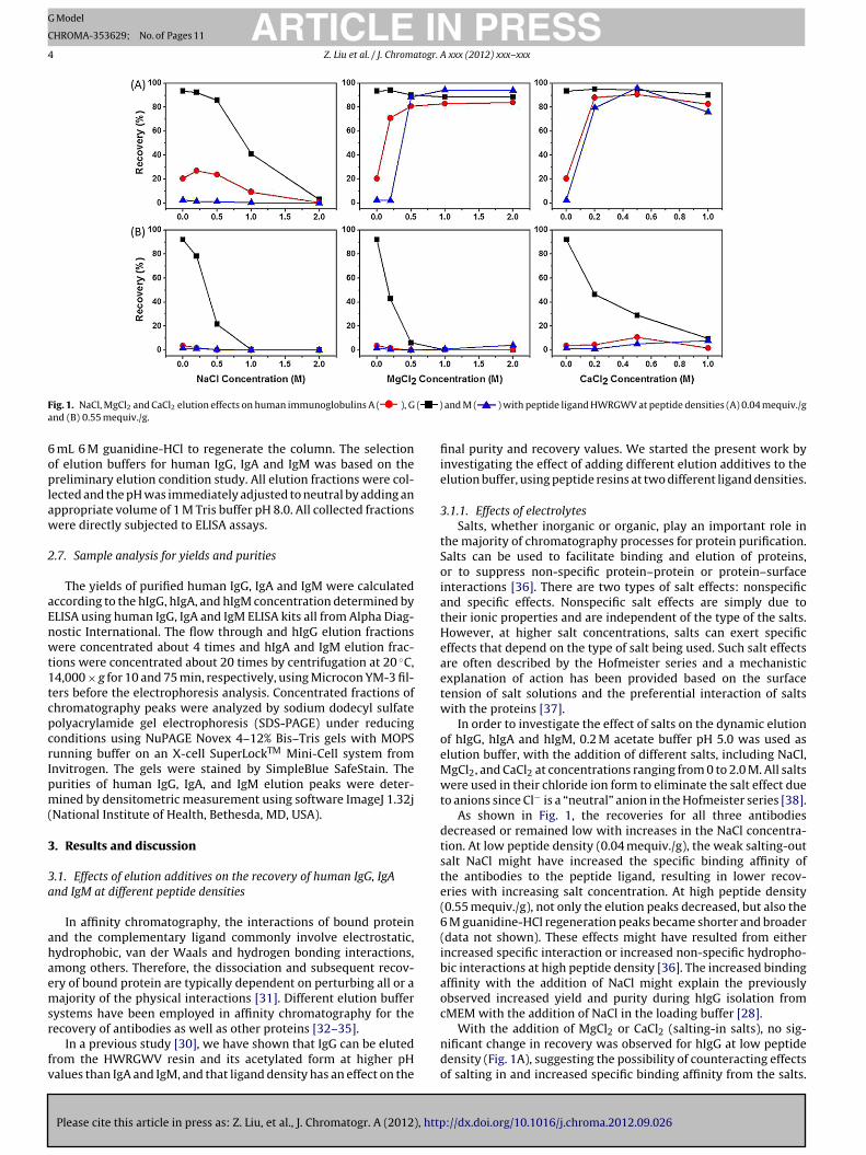
ig. 2. Ethylene glycol, urea and arginine elution effects on human immunoglobulin.04 mequiv./g and (B) 0.55 mequiv./g.
t high peptide density (Fig. 1B), the recovery of hIgG decreasedith increased salt concentration most likely because of domi-ant non-specific hydrophobic interactions. For hIgA and hIgMt low peptide density (Fig. 1A), the addition of both MgCl2 andaCl2 in the elution buffer seems to have facilitated the antibodylution resulting in better recoveries with increased salt concen-ration. This can be explained by the favorable interaction betweenhe divalent ions and antibodies at high salt concentration [37].n addition, the same level of recovery was achieved by a loweroncentration of CaCl2 compared to MgCl2 (0.2 M CaCl2 vs. 0.5 MgCl2) showing a stronger effect from CaCl2. This was expected
ince CaCl2 is a stronger salting-in salt from the Hofmeister series.owever, high concentrations of CaCl2 should be avoided to pre-ent protein aggregation since multiple peaks were observed in the
M guanidine-HCl regeneration step after elution with 1 M CaCl2data not shown). At high peptide density, the salting in effect of
gCl2 and CaCl2 could not overcome the increased non-specificydrophobic interactions. Therefore, no increase in recoveries wasbserved.
Overall, the results obtained from this set of experiments sug-est that salting-in electrolytes such as MgCl2 and CaCl2 could besed to elute antibodies from an affinity resin containing the ligandWRGWV at low ligand densities.
.1.2. Effects of non-electrolytesNon-electrolytes such as water-miscible organic solvents, dena-
uring agents and amino acids have been employed as solventdditives for protein purification by affinity chromatographyecause of their ability to suppress nonspecific protein–protein orrotein–surface interactions [31,37,39]. In order to investigate theffect of non-electrolyte additives on the dynamic elution of hIgG,IgA and hIgM, recovery of bound proteins was achieved by using.2 M acetate buffer pH 5.0 containing different elution additives
ncluding ethylene glycol, urea, and arginine at different concen-rations.
The effect of ethylene glycol at concentrations from 0 to 40%v/v) was examined. As shown in Fig. 2, at both peptide densi-ies, ethylene glycol had no effect on the recovery of hIgG and
Please cite this article in press as: Z. Liu, et al., J. Chromatogr. A (2012), http
IgM elution, with the recoveries for both proteins being virtu-lly constant in the ethylene glycol concentration range used. Theifference for the two proteins was that in the case of hIgG theecovery remained around 90–95%, while for hIgM the values for
), G ( ) and M ( ) with peptide ligand HWRGWV at peptide densities (A)
recovery were in the 0–5% range. For hIgA, the recovery increasedwith the increase in the ethylene glycol concentration at low pep-tide density (Fig. 2A, left panel), while they remained low at highpeptide density (Fig. 2B, left panel). Ethylene glycol has the abil-ity to suppress hydrophobic interactions and bind to the proteinsurface. Therefore, the elution mechanism of ethylene glycol issimilar to MgCl2 except that ethylene glycol enhances electro-static interactions by lowering the dielectric constant [37,40]. Asa result, the increased recovery at low peptide density might havebeen achieved by decreased hydrophobic interactions exceedingincreased electrostatic interactions. However, at high peptide den-sity, the decrease in hydrophobic interactions could not overcomethe increased non-specific interactions and electrostatic interac-tions leading to unimproved recovery.
Urea at concentrations ranging from 0 to 2.0 M was tested(Fig. 2, center panels), since higher urea concentration might affectthe protein structure and stability. Similar behavior was observedfor the effect of urea compared to ethylene glycol at both peptidedensities. Compared to ethylene glycol, urea exhibits affinityfor all side chains of amino acids instead of only non-polar sidechains, resulting in decreased hydrophobic interactions. At thesame time, urea can reduce polar interactions by forming morestable hydrogen bonds with peptide bonds than with water andslightly increasing the dielectric constant of water [35]. Therefore,at low peptide density, urea weakened the hydrophobic and polarinteractions to a sufficient extent that caused the elution of hIgA.However, at high peptide density, the dominant non-specifichydrophobic interactions could not be overcome by decreasedhydrophobic and polar interactions.
Arginine has been used for elution of antibodies from ProteinA affinity chromatography because of its ability to suppress aggre-gation and protein–protein or protein–surface interactions withoutdenaturing proteins [34,41,42]. The recovery of hIgG did not changewith the addition of arginine in the elution buffer at both peptidedensities (Fig. 2, right panels), remaining relatively high for all con-ditions. At low peptide density, the recovery of both hIgA and hIgMincreased to over 95% at an arginine concentration of 1.0 M. Never-theless, arginine seems to have had a stronger effect on hIgA elution
://dx.doi.org/10.1016/j.chroma.2012.09.026
because higher recovery was achieved with hIgA compared to hIgMat lower arginine concentration, which probably resulted from theweaker binding affinity of HWRGWV peptide ligand to hIgA. At highpeptide density, because of the dominant increased non-specific
ARTICLE IN PRESSG Model
CHROMA-353629; No. of Pages 11
6 Z. Liu et al. / J. Chromatogr. A xxx (2012) xxx– xxx
Table 3Yield and purity for isolated human IgG, IgA and IgM from their mixture with HWRGWV at peptide density of 0.23 mequiv./g.
Run hIgG hIgA hIgM
Yield Purity Yield Purity Yield Purity
195.3 ± 7.4 97.4 ± 1.8 16.9 ± 8.2 n/a
99.1 ± 7.7 99.22 54.9 ± 5.3 98.6
391.5 ± 5.6 98.3 ± 1.6 16.4 ± 3.2 n/a
3.1 ± 0.5 n/a4 38.9 ± 2.8 99.65 94.5 ± 3.9 92.4
6
95.4 ± 3.3 98.8 ± 0.9
8.0 ± 3.0 58.2 91.7 ± 14.4 49.07 2.3 ± 1.3
8 28.3 ± 10.0
9 75.8 ± 13.0
h1
biHp
3hm
ehsttpbl(bRs
TY
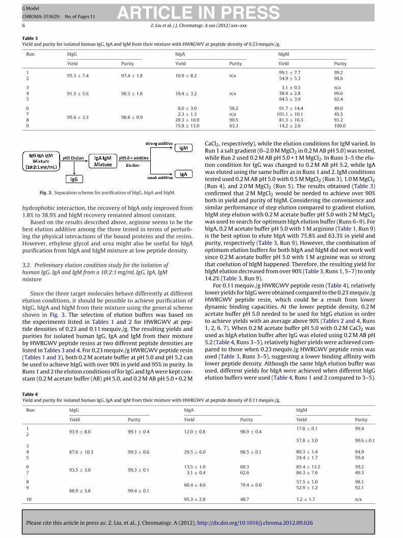
Fig. 3. Separation scheme for purification of hIgG, hIgA and hIgM.
ydrophobic interaction, the recovery of hIgA only improved from.8% to 38.9% and hIgM recovery remained almost constant.
Based on the results described above, arginine seems to be theest elution additive among the three tested in terms of perturb-
ng the physical interactions of the bound proteins and the resins.owever, ethylene glycol and urea might also be useful for hIgAurification from hIgA and hIgM mixture at low peptide density.
.2. Preliminary elution condition study for the isolation ofuman IgG, IgA and IgM from a 10:2:1 mg/mL IgG, IgA, IgMixture
Since the three target molecules behave differently at differentlution conditions, it should be possible to achieve purification ofIgG, hIgA and hIgM from their mixture using the general schemehown in Fig. 3. The selection of elution buffers was based onhe experiments listed in Tables 1 and 2 for HWRGWV at pep-ide densities of 0.23 and 0.11 mequiv./g. The resulting yields andurities for isolated human IgG, IgA and IgM from their mixturey HWRGWV peptide resins at two different peptide densities are
isted in Tables 3 and 4. For 0.23 mequiv./g HWRGWV peptide resin
Please cite this article in press as: Z. Liu, et al., J. Chromatogr. A (2012), http
Tables 1 and 3), both 0.2 M acetate buffer at pH 5.0 and pH 5.2 cane used to achieve hIgG with over 90% in yield and 95% in purity. Inuns 1 and 2 the elution conditions of for IgG and IgA were kept con-tant (0.2 M acetate buffer (AB) pH 5.0, and 0.2 M AB pH 5.0 + 0.2 M
able 4ield and purity for isolated human IgG, IgA and IgM from their mixture with HWRGWV
Run hIgG hIgA
Yield Purity Yield
193.9 ± 8.0 99.1 ± 0.4 12.0 ± 0.82
387.6 ± 10.3 99.3 ± 0.6 29.5 ± 6.04
5
693.5 ± 3.0 99.3 ± 0.1
13.5 ± 1.07 3.1 ± 0.4
8
88.9 ± 3.8 99.4 ± 0.160.4 ± 4.09
10 95.3 ± 2.8
n/a 101.1 ± 10.1 45.590.5 81.3 ± 10.3 91.263.3 14.2 ± 2.6 100.0
CaCl2, respectively), while the elution conditions for IgM varied. InRun 1 a salt gradient (0–2.0 M MgCl2 in 0.2 M AB pH 5.0) was tested,while Run 2 used 0.2 M AB pH 5.0 + 1 M MgCl2. In Runs 3–5 the elu-tion condition for IgG was changed to 0.2 M AB pH 5.2, while IgAwas eluted using the same buffer as in Runs 1 and 2. IgM conditionstested used 0.2 M AB pH 5.0 with 0.5 M MgCl2 (Run 3), 1.0 M MgCl2(Run 4), and 2.0 M MgCl2 (Run 5). The results obtained (Table 3)confirmed that 2 M MgCl2 would be needed to achieve over 90%both in yield and purity of hIgM. Considering the convenience andsimilar performance of step elution compared to gradient elution,hIgM step elution with 0.2 M acetate buffer pH 5.0 with 2 M MgCl2was used to search for optimum hIgA elution buffer (Runs 6–9). ForhIgA, 0.2 M acetate buffer pH 5.0 with 1 M arginine (Table 1, Run 9)is the best option to elute hIgA with 75.8% and 63.3% in yield andpurity, respectively (Table 3, Run 9). However, the combination ofoptimum elution buffers for both hIgA and hIgM did not work wellsince 0.2 M acetate buffer pH 5.0 with 1 M arginine was so strongthat coelution of hIgM happened. Therefore, the resulting yield forhIgM elution decreased from over 90% (Table 3, Runs 1, 5–7) to only14.2% (Table 3, Run 9).
For 0.11 mequiv./g HWRGWV peptide resin (Table 4), relativelylower yields for hIgG were obtained compared to the 0.23 mequiv./gHWRGWV peptide resin, which could be a result from lowerdynamic binding capacities. At the lower peptide density, 0.2 Macetate buffer pH 5.0 needed to be used for hIgG elution in orderto achieve yields with an average above 90% (Tables 2 and 4, Runs1, 2, 6, 7). When 0.2 M acetate buffer pH 5.0 with 0.2 M CaCl2 wasused as hIgA elution buffer after IgG was eluted using 0.2 M AB pH5.2 (Table 4, Runs 3–5), relatively higher yields were achieved com-pared to those when 0.23 mequiv./g HWRGWV peptide resin was
://dx.doi.org/10.1016/j.chroma.2012.09.026
used (Table 3, Runs 3–5), suggesting a lower binding affinity withlower peptide density. Although the same hIgA elution buffer wasused, different yields for hIgA were achieved when different hIgGelution buffers were used (Table 4, Runs 1 and 2 compared to 3–5).
at peptide density of 0.11 mequiv./g.
hIgM
Purity Yield Purity
98.9 ± 0.417.6 ± 0.1 99.4
57.8 ± 3.0 99.6 ± 0.1
98.5 ± 0.1 80.3 ± 1.4 94.924.4 ± 1.7 59.4
60.3 85.4 ± 13.2 59.2 62.6 86.3 ± 7.6 49.3
79.4 ± 0.657.5 ± 1.0 98.152.9 ± 1.2 92.1
48.7 1.2 ± 1.7 n/a

ING Model
C
togr.
Teu4om(
Hff0w2
3
prnub
mHgTr
Fad
ARTICLEHROMA-353629; No. of Pages 11
Z. Liu et al. / J. Chroma
hese results suggest that the yield of an eluted antibody is depend-nt not only on the elution buffer but also on the previous buffersed. The use of 0.2 M acetate buffer pH 5.0 with 2 M MgCl2 (Runs
and 7) is still the best option for hIgM elution. Without coelutionf hIgM, 0.2 M acetate buffer pH 5.0 with 0.5 M arginine is the opti-um selection for hIgA elution based on yield and purity results
Runs 8 and 9).Based on the overall results of the elution experiments,
WRGWV peptide resin at density of 0.11 mequiv./g was selectedor further studies on the purification of human IgG, IgA and IgMrom Cohn II/III. The elution buffers selected for each antibody were.2 M acetate buffer pH 5.0 for hIgG, 0.2 M acetate buffer pH 5.0ith 0.5 M arginine for hIgA and 0.2 M acetate buffer pH 5.0 with
M MgCl2 for hIgM.
.3. Cohn fraction II/III sample preparation and pretreatments
Since the original 50 mg/mL Cohn fraction II/III (60–85% purerotein) stock solution contained about 15–40% non-protein mate-ial, centrifugation and two filtration steps were used to removeon-dissolved contaminants. BCA protein assay and ELISA kits weresed to determine the total protein concentration and each anti-ody concentration after each step.
Different pretreatment methods were used to remove someajor contaminant proteins such as human serum albumin (HSA),
Please cite this article in press as: Z. Liu, et al., J. Chromatogr. A (2012), http
SA dimer, transferrin and �-1 antitrypsin before the chromato-raphic step in order to improve the purity of purified antibodies.he pretreatment methods used were modified based on previousesearch from literature [43,44].
ig. 4. Total protein and antibody concentrations in Cohn II/III dissolved in water (A)nd PBS (B) after each preparation or pretreatment step. The concentration standardeviations are shown by error bar.
PRESSA xxx (2012) xxx– xxx 7
The total protein as well as human IgG, IgA and IgM concentra-tion during the sample preparation and after each pretreatment areshown in Fig. 4. The volume factor for each pretreatment was takeninto account when the protein concentration was calculated. Thetotal protein concentration from Cohn fraction II/III after dissolv-ing in HPLC grade water and PBS were 33.2 and 34.9 mg/mL withslightly higher concentration achieved by PBS as the solvent. There-fore, higher concentrations were observed in PBS solvent than inHPLC grade water for human IgG, IgA and IgM after each prepa-ration and pretreatment step. After one centrifugation and twofiltration steps, yields of about 90% were obtained for total pro-teins, of which about 50% were antibodies, with IgG, IgA and IgMpresent at a ratio of about 10:2:1.
Caprylic acid can be used as free acid to inactivate of envelopedviruses and precipitate contaminating proteins [25]. PEG precipi-tation alone was able to remove contaminating proteins and thecombination of PEG precipitation with caprylic acid treatmentwas found not only for the removal of polymers but also for theremoval of non-enveloped viruses [44]. SAS precipitation has beenemployed to remove lipoproteins and fibrinogen without denatur-ation of IgM to achieve large scale recovery of IgM from humanplasma [43].
After the caprylic acid pretreatment, about 20 mg/mL of pro-teins were recovered, which was about 67% of the total proteins.The average recovery for hIgG in water and PBS was 86%, a valuethat was higher than the average recoveries of hIgA and hIgM of71% and 46%, respectively. As a result, the percentage of hIgG inthe solution increased from 40% to 50% or more after the pretreat-ment. Therefore, caprylic acid treatment seems to be a relativelygood pretreatment method to remove contaminating proteins forhIgG recovery. However, more than 50% of hIgM was lost duringthe precipitation which might be caused not only by the lower sol-ubility of hIgM (pI: 5.5–6.7) at the caprylic acid precipitation pH of5.0, but also by the large molecular structure as a pentamer withmolecular weight of 950 kDa.
With the combination of caprylic acid and PEG precipitation,the total protein concentration decreased to about 55% of thestock Cohn fraction II/III. The hIgG concentrations remained aboutthe same as the ones with only caprylic acid treatment, whichfurther increased the hIgG percentage to around 60% and con-firmed that the combination of caprylic acid and PEG precipitationworks well for removing contaminating proteins from Cohn frac-tion II/III for hIgG purification. However, only about 62% of hIgAand 30% of hIgM were recovered after caprylic acid and PEGprecipitations.
With PEG precipitation alone, for Cohn fraction II/III dissolvedin PBS, the recoveries for total protein as well as three antibod-ies were about the same at around 91%, except for hIgM with a72% recovery, further confirming that precipitation is not a goodmethod to recover large molecules such as hIgM. For Cohn frac-tion II/III dissolved in water, a 78% recovery was obtained fortotal protein compared to around 90% recoveries for both hIgGand hIgA, and 61% recovery for hIgM. Therefore, reasonably goodrecoveries were achieved for all three antibodies with poorercontaminant removal efficiency when using PEG pretreatmentalone.
SAS pretreatment, employed for large scale recovery of IgM fromhuman plasma [43], showed good recovery for hIgM, when com-pared to the other three pretreatment methods tested, with valuesof 77% and 62% achieved for Cohn fraction II/III dissolved in waterand PBS, respectively. The recoveries for total protein, hIgG andhIgA from Cohn fraction II/III dissolved in water and PBS are about
://dx.doi.org/10.1016/j.chroma.2012.09.026
the same with averages of 66%, 73% and 76%, respectively. There-fore, even with good efficiency for contaminants removal and highrecovery for hIgM, the hIgG recovery was low compared to otherthree pretreatment methods.
ARTICLE IN PRESSG Model
CHROMA-353629; No. of Pages 11
8 Z. Liu et al. / J. Chromatogr. A xxx (2012) xxx– xxx
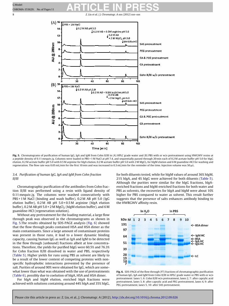
Fig. 5. Chromatograms of purification of human IgG, IgA and IgM from Cohn II/III in (A) HPLC grade water and (B) PBS with or w/o pretreatment using HWGWV resins ata 7.4, ane te bufr to 0.3
3I
t0Pebg
tFtmwcitf(bssw(
a
PBS as solvents, the recoveries for hIgA and hIgM were about 10%higher for PBS compared to water as solvent. This result furthersuggests that the presence of salts enhances antibody binding tothe HWRGWV affinity resin.
peptide density of 0.11 mequiv./g. Columns were loaded in PBS + 1 M NaCl at pH
lution, 0.2 M acetate buffer pH 5.0 with 0.5 M arginine for hIgA elution, 0.2 M acetaegeneration. The flow rate was 0.05 mL/min for the first 10 min and was increased
.4. Purification of human IgG, IgA and IgM from Cohn fractionI/III
Chromatographic purification of the antibodies from Cohn frac-ion II/III was performed using a resin with ligand density of.11 mequiv./g. The columns were washed consecutively withBS + 1 M NaCl (binding and wash buffer), 0.2 M AB pH 5.0 (IgGlution buffer), 0.2 M AB pH 5.0 + 0.5 M arginine (hIgA elutionuffer), 0.2 M AB pH 5.0 + 2 M MgCl2 (hIgM elution buffer), and 6 Muanidine-HCl (regeneration solution).
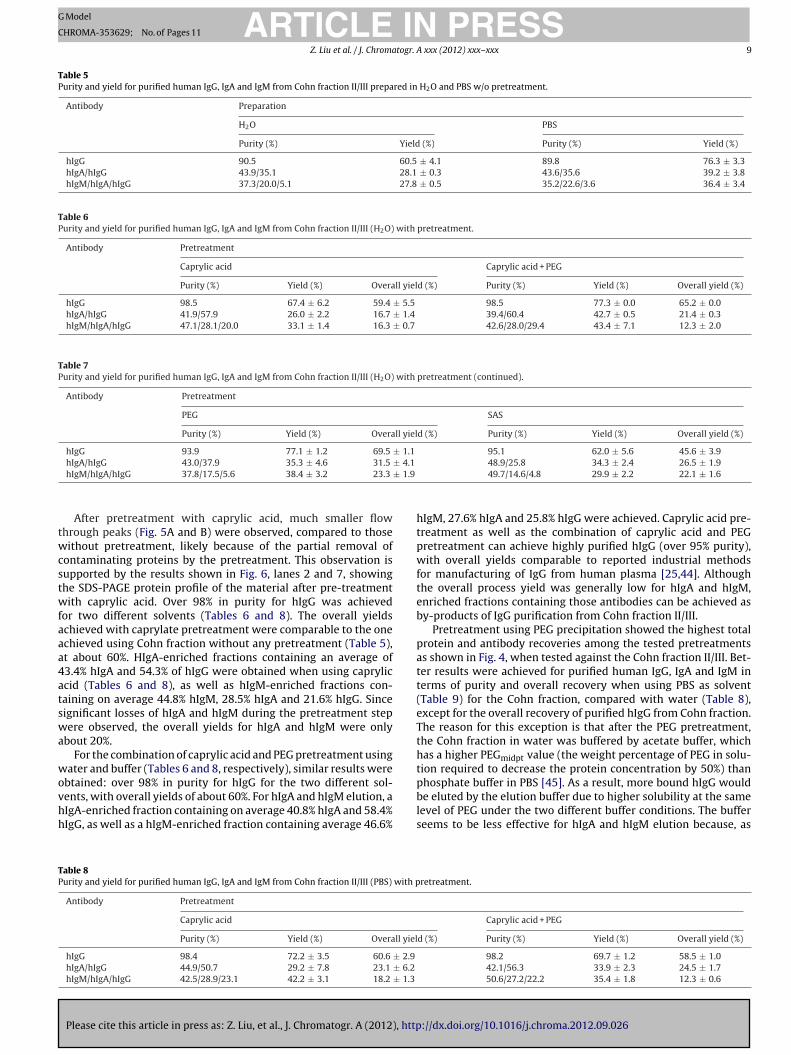
Without any pretreatment for the loading material, a large flowhrough peak was observed in the chromatograms as shown inig. 5.The results obtained by SDS-PAGE analysis (Fig. 6) showedhat the flow through peaks contained HSA and HSA dimer as the
ain contaminants. Since a large amount of contaminant proteinsas present in those runs, it lead to a lower dynamic binding
apacity, causing human IgG as well as IgA and IgM to be detectedn the flow through (unbound) fractions albeit at low concentra-ions. Therefore, the yields for purified hIgG were 60.5% and 76.3%or Cohn fraction II/III dissolved in water and PBS, respectivelyTable 5). Higher yields for runs using PBS as solvent are likely toe a result of the lower content of competing proteins with non-pecific hydrophobic interactions prevented by the presence ofalt. Purities of around 90% were obtained for IgG, which are some-
Please cite this article in press as: Z. Liu, et al., J. Chromatogr. A (2012), http
hat lower than what was obtained with the use of pretreatmentsTable 6), possibly due to coelution of hIgA, HSA and HSA dimer.
For hIgA and hIgM elution, enriched hIgA fractions werechieved with solutions containing around 44% hIgA and 35% hIgG,
d sequentially passed through 20 min each of 0.2 M acetate buffer pH 5.0 for hIgGfer pH 5.0 with 2 M MgCl2 for hIgM elution and 6 M guanidine-HCl for washing and
mL/min for the reminder of the time. Injection volume was 50 �L.
for both diluents tested, while for hIgM values of around 36% hIgM,21% hIgA, and 4% hIgG were achieved for both diluents (Table 5).Although the purities were similar for the hIgG fractions, hIgA-enriched fractions and hIgM-enriched fractions for both water and
://dx.doi.org/10.1016/j.chroma.2012.09.026
Fig. 6. SDS-PAGE of the flow through (FT) fractions of chromatographic purificationof human IgG, IgA and IgM from Cohn II/III in HPLC grade water or PBS with or w/opretreatment: lanes 1, 6: Cohn II/III w/o pretreatment, lanes 2, 7: after caprylic acidpretreatment, lanes 3, 8: after caprylic acid and PEG pretreatment, lanes 4, 9: afterPEG pretreatment, lanes 5, 10: after SAS pretreatment.
ARTICLE IN PRESSG Model
CHROMA-353629; No. of Pages 11
Z. Liu et al. / J. Chromatogr. A xxx (2012) xxx– xxx 9
Table 5Purity and yield for purified human IgG, IgA and IgM from Cohn fraction II/III prepared in H2O and PBS w/o pretreatment.
Antibody Preparation
H2O PBS
Purity (%) Yield (%) Purity (%) Yield (%)
hIgG 90.5 60.5 ± 4.1 89.8 76.3 ± 3.3hIgA/hIgG 43.9/35.1 28.1 ± 0.3 43.6/35.6 39.2 ± 3.8hIgM/hIgA/hIgG 37.3/20.0/5.1 27.8 ± 0.5 35.2/22.6/3.6 36.4 ± 3.4
Table 6Purity and yield for purified human IgG, IgA and IgM from Cohn fraction II/III (H2O) with pretreatment.
Antibody Pretreatment
Caprylic acid Caprylic acid + PEG
Purity (%) Yield (%) Overall yield (%) Purity (%) Yield (%) Overall yield (%)
hIgG 98.5 67.4 ± 6.2 59.4 ± 5.5 98.5 77.3 ± 0.0 65.2 ± 0.0hIgA/hIgG 41.9/57.9 26.0 ± 2.2 16.7 ± 1.4 39.4/60.4 42.7 ± 0.5 21.4 ± 0.3hIgM/hIgA/hIgG 47.1/28.1/20.0 33.1 ± 1.4 16.3 ± 0.7 42.6/28.0/29.4 43.4 ± 7.1 12.3 ± 2.0
Table 7Purity and yield for purified human IgG, IgA and IgM from Cohn fraction II/III (H2O) with pretreatment (continued).
Antibody Pretreatment
PEG SAS
Purity (%) Yield (%) Overall yield (%) Purity (%) Yield (%) Overall yield (%)
hIgG 93.9 77.1 ± 1.2 69.5 ± 1.1 95.1 62.0 ± 5.6 45.6 ± 3.9± 4.1
± 1.9
twcstwfaaa4atswa
wovhh
TP
hIgA/hIgG 43.0/37.9 35.3 ± 4.6 31.5
hIgM/hIgA/hIgG 37.8/17.5/5.6 38.4 ± 3.2 23.3
After pretreatment with caprylic acid, much smaller flowhrough peaks (Fig. 5A and B) were observed, compared to thoseithout pretreatment, likely because of the partial removal of
ontaminating proteins by the pretreatment. This observation isupported by the results shown in Fig. 6, lanes 2 and 7, showinghe SDS-PAGE protein profile of the material after pre-treatmentith caprylic acid. Over 98% in purity for hIgG was achieved
or two different solvents (Tables 6 and 8). The overall yieldschieved with caprylate pretreatment were comparable to the onechieved using Cohn fraction without any pretreatment (Table 5),t about 60%. HIgA-enriched fractions containing an average of3.4% hIgA and 54.3% of hIgG were obtained when using capryliccid (Tables 6 and 8), as well as hIgM-enriched fractions con-aining on average 44.8% hIgM, 28.5% hIgA and 21.6% hIgG. Sinceignificant losses of hIgA and hIgM during the pretreatment stepere observed, the overall yields for hIgA and hIgM were only
bout 20%.For the combination of caprylic acid and PEG pretreatment using
ater and buffer (Tables 6 and 8, respectively), similar results were
Please cite this article in press as: Z. Liu, et al., J. Chromatogr. A (2012), http
btained: over 98% in purity for hIgG for the two different sol-ents, with overall yields of about 60%. For hIgA and hIgM elution, aIgA-enriched fraction containing on average 40.8% hIgA and 58.4%IgG, as well as a hIgM-enriched fraction containing average 46.6%
able 8urity and yield for purified human IgG, IgA and IgM from Cohn fraction II/III (PBS) with p
Antibody Pretreatment
Caprylic acid
Purity (%) Yield (%) Overall yiel
hIgG 98.4 72.2 ± 3.5 60.6 ± 2.9
hIgA/hIgG 44.9/50.7 29.2 ± 7.8 23.1 ± 6.2
hIgM/hIgA/hIgG 42.5/28.9/23.1 42.2 ± 3.1 18.2 ± 1.3
48.9/25.8 34.3 ± 2.4 26.5 ± 1.949.7/14.6/4.8 29.9 ± 2.2 22.1 ± 1.6
hIgM, 27.6% hIgA and 25.8% hIgG were achieved. Caprylic acid pre-treatment as well as the combination of caprylic acid and PEGpretreatment can achieve highly purified hIgG (over 95% purity),with overall yields comparable to reported industrial methodsfor manufacturing of IgG from human plasma [25,44]. Althoughthe overall process yield was generally low for hIgA and hIgM,enriched fractions containing those antibodies can be achieved asby-products of IgG purification from Cohn fraction II/III.
Pretreatment using PEG precipitation showed the highest totalprotein and antibody recoveries among the tested pretreatmentsas shown in Fig. 4, when tested against the Cohn fraction II/III. Bet-ter results were achieved for purified human IgG, IgA and IgM interms of purity and overall recovery when using PBS as solvent(Table 9) for the Cohn fraction, compared with water (Table 8),except for the overall recovery of purified hIgG from Cohn fraction.The reason for this exception is that after the PEG pretreatment,the Cohn fraction in water was buffered by acetate buffer, whichhas a higher PEGmidpt value (the weight percentage of PEG in solu-tion required to decrease the protein concentration by 50%) than
://dx.doi.org/10.1016/j.chroma.2012.09.026
phosphate buffer in PBS [45]. As a result, more bound hIgG wouldbe eluted by the elution buffer due to higher solubility at the samelevel of PEG under the two different buffer conditions. The bufferseems to be less effective for hIgA and hIgM elution because, as
retreatment.
Caprylic acid + PEG
d (%) Purity (%) Yield (%) Overall yield (%)
98.2 69.7 ± 1.2 58.5 ± 1.042.1/56.3 33.9 ± 2.3 24.5 ± 1.750.6/27.2/22.2 35.4 ± 1.8 12.3 ± 0.6

ARTICLE IN PRESSG Model
CHROMA-353629; No. of Pages 11
10 Z. Liu et al. / J. Chromatogr. A xxx (2012) xxx– xxx
Table 9Purity and yield for purified human IgG, IgA and IgM from Cohn fraction II/III (PBS) with pretreatment (continued).
Antibody Pretreatment
PEG SAS
Purity (%) Yield (%) Overall yield (%) Purity (%) Yield (%) Overall yield (%)
hIgG 94.5 66.6 ± 1.8 60.7 ± 1.6 94.6 67.9 ± 3.7 48.6 ± 2.6 5.1
1.6
pdbcmcw
twhtnraf
4
olaeoaetaIpanirhsHn
bafIIpt
putwb9h
[[[
[
[
[
[
[
[[
[
[
[
[
[[
[
[[[
[[[
hIgA/hIgG 41.7/40.3 38.7 ± 5.6 35.2 ±hIgM/hIgA/hIgG 31.6/46.7/0 34.4 ± 1.8 31.3 ±
reviously mentioned, the yield of an eluted antibody seems to beependent not only on the elution buffer but also on the previousuffer used. The resulting purities and yields for hIgA and hIgM haveomparable values to those from Cohn fraction without pretreat-ent, with the PEG pretreatment achieving better results for IgG
ontent in the hIgA- and hIgM-enriched fractions (Tables 7 and 9)hen water was used as diluents.
Human IgG had the lowest observed recovery after the SAS pre-reatment step, resulting in the lowest overall yields with 45.6%ith water (Table 7) and 48.6% in PBS. Although the hIgA andIgM concentrations were relatively high after the pretreatment,he resulting overall yields (∼23.6%) for both hIgA and hIgM wereot very high. This might have been caused by ammonium sulfateesidue left in the Cohn fractions, suggesting that SAS would not be
good pretreatment method for hIgG, hIgA and hIgM purificationrom Cohn fraction II/III.
. Conclusions
This study has demonstrated the effects of elution additivesn the recovery of human IgG, IgA and IgM with small peptideigand HWRGWV. Unlike salting-out salts, salting-in salts MgCl2nd CaCl2 can be employed in the elution buffer to facilitate thelution of human IgA and IgM at low peptide density becausef favorable interactions between the divalent ions and boundntibodies. At high peptide density, none of these salts facilitatelution as a result of increased non-specific hydrophobic interac-ions. Among the three non-electrolyte elution additives examined,rginine showed the best results for improving recovery of humangA and IgM at low peptide density, resulting from its ability to sup-ress hydrophobic, H-bonding and electrostatic interactions. Still,t high peptide density, the recovery of human IgA and IgM didot improve or only improved to a small extent because of the
ncreased non-specific hydrophobic interactions. Based on theseesults, we can conclude that at low peptide density, the binding ofuman IgA and IgM with HWRGWV involves comparable electro-tatic and specific hydrophobic interactions together with possible-bonding. At high peptide density, the dominant binding force ison-specific hydrophobic interactions.
Based on the preliminary elution condition study, 0.2 M acetateuffer at pH 5.0 for hIgG, 0.2 M acetate buffer at pH 5.0 with 0.5 Mrginine for hIgA and 0.2 M acetate buffer at pH 5.0 with 2 M MgCl2or hIgM are the optimum elution buffers for purification of humangG, IgA and IgM from their mixture. The yield results for hIgA andgM also suggested that the yield of eluted antibody from HWRGWVeptide resin is affected not only by the elution buffer but by alsohe previous elution condition.
The applicability of small peptide ligand HWRGWV to achieveurification of human IgG, IgA and IgM from Cohn fraction II/IIIsing one column was demonstrated. Without pretreatment, bet-er results were achieved for Cohn fraction using PBS instead of
Please cite this article in press as: Z. Liu, et al., J. Chromatogr. A (2012), http
ater as solvent because of the decreased non-specific hydropho-ic interactions in the presence of salts. With the similar purities as0% for hIgG, 44% enriched hIgA with 35% hIgG and 36% enrichedIgM with 21% hIgA and 4% hIgM, higher yields (10% or more)
[
[
[
39.0/44.3 29.8 ± 4.5 22.2 ± 3.444.7/39.6/8.1 38.2 ± 1.8 23.7 ± 1.1
were obtained for all three purified antibodies from Cohn fractionin PBS. After the caprylic acid pretreatment or the combination ofcaprylic acid and PEG pretreatment, highly purified hIgG (over 95%purity) was achieved with about 60% in yield for both solvents,which is comparable to industrial chromatographic purification ofIgG isolated from plasma fractions. An hIgA-enriched fraction withaverage about 42% hIgA and 56% hIgG as well as an hIgM enrichedfraction with average about 46% hIgM, 28% hIgA and 24% hIgG wereobtained as the by-products of the purification of hIgG from Cohnfraction II/III. To our knowledge, it is the first time that an affin-ity chromatography column was used for the simultaneous captureand purification of human IgG, IgA and IgM from Cohn fraction II/III.
References
[1] R.A. Good, E. Lorenz, Cancer 68 (1991) 1415.[2] A. Casadevall, M.D. Scharff, Clin. Infect. Dis. 21 (1995) 150.[3] O.H. Brekke, I. Sandlie, Nat. Rev. Drug Discov. 2 (2003) 52.[4] A. Casadevall, E. Dadachova, L. Pirofski, Nat. Rev. Microbiol. 2 (2004) 695.[5] D. Schrama, R.A. Reisfeld, J.C. Becker, Nat. Rev. Drug Discov. 5 (2006) 147.[6] P. Chames, M. Van Regenmortel, E. Weiss, D. Baty, Br. J. Pharmacol. 157 (2009)
220.[7] J.H. Chon, G. Zarbis-Papastoitsis, New Biotechnol. 28 (2011) 458.[8] M. Radosevich, T. Burnouf, Vox Sang. 98 (2010) 12.[9] H.P. Hartung, J. Neurol. 255 (2008) 3.10] M.A. Keller, E.R. Stiehm, Clin. Microbiol. Rev. 13 (2000) 602.11] A. Rutter, T.A. Luger, J. Am. Acad. Dermatol. 44 (2001) 1010.12] H.P. Hartung, L. Mouthon, R. Ahmed, S. Jordan, K.B. Laupland, S. Jolles, Clin. Exp.
Immunol. 158 (2009) 23.13] S.V. Kaveri, M.S. Maddur, P. Hegde, S. Lacroix-Desmazes, J. Bayry, Clin. Exp.
Immunol. 164 (2011) 2.14] V. Hurez, M.D. Kazatchkine, T. Vassilev, S. Ramanathan, A. Pashov, B. Basuyaux,
Y. deKozak, B. Bellon, S.V. Kaveri, Blood 90 (1997) 4004.15] T. Vassilev, N. Mihaylova, E. Voynova, M. Nikolova, M. Kazatchkine, S. Kaveri,
Clin. Exp. Immunol. 145 (2006) 108.16] R. Rieben, A. Roos, Y. Muizert, C. Tinguely, A.F. Gerritsen, M.R. Daha, Blood 93
(1999) 942.17] R. Lissner, W.G. Struff, I.B. Autenrieth, B.G. Woodcock, H. Karch, Eur. J. Surg. 163
(1999) 17.18] A.R. Neilson, H. Burchardi, H. Schneider, J. Crit. Care 20 (2005) 239.19] M. Hentrich, K. Fehnle, H. Ostermann, J. Kienast, O. Cornely, C. Salat, R.
Ubelacker, D. Buchheidt, G. Behre, W. Hiddemann, X. Schiel, Crit. Care Med.34 (2006) 1319.
20] D. Nachbaur, M. Herold, A. Gachter, D. Niederwieser, Immunology 94 (1998)279.
21] S.K. Jackson, J. Parton, R.A. Barnes, C.H. Poynton, C. Fegan, Eur. J. Clin. Invest. 23(1993) 540.
22] E.J. Cohn, L.E. Strong, W.L. Hughes, D.J. Mulford, J.N. Ashworth, M. Melin, H.L.Taylor, J. Am. Chem. Soc. 68 (1946) 459.
23] J.L. Oncley, M. Melin, D.A. Richert, J.W. Cameron, P.M. Gross, J. Am. Chem. Soc.71 (1949) 541.
24] P. Kistler, H. Nitschmann, Vox Sang. 7 (1962) 414.25] W. Lebing, K.M. Remington, C. Schreiner, H.I. Paul, Vox Sang. 84 (2003)
193.26] W. Teschner, H.A. Butterweck, W. Auer, E.M. Muchitsch, A. Weber, S.L. Liu, P.S.
Wah, H.P. Schwarz, Vox Sang. 92 (2007) 42.27] H. Yang, P.V. Gurgel, R.G. Carbonell, J. Pept. Res. 66 (2005) 120.28] H.O. Yang, P.V. Gurgel, R.G. Carbonell, J. Chromatogr. A 1216 (2009) 910.29] H.O. Yang, P.V. Gurgel, D.K. Williams, B.G. Bobay, J. Cavanagh, D.C. Muddiman,
R.G. Carbonell, J. Mol. Recognit. 23 (2010) 271.30] Z. Liu, P.V. Gurgel, R.G. Carbonell, J. Chromatogr. A 1218 (2011) 8344.31] M.A. Firer, J. Biochem. Biophys. Methods 49 (2001) 433.32] V.C.W. Tsang, P. Wilkins, FASEB J. 5 (1991) A622.
://dx.doi.org/10.1016/j.chroma.2012.09.026
33] P.Y. Huang, G.A. Baumbach, C.A. Dadd, J.A. Buettner, B.L. Masecar, M. Hentsch,D.J. Hammond, R.G. Carbonell, Bioorg. Med. Chem. 4 (1996) 699.
34] T. Arakawa, J.S. Philo, K. Tsumoto, R. Yumioka, D. Ejima, Protein Expr. Purif. 36(2004) 244.
35] T. Arakawa, Y. Kita, H. Sato, D. Ejima, Protein Expr. Purif. 63 (2009) 158.
ING Model
C
togr.
[
[[[[[
[
ARTICLEHROMA-353629; No. of Pages 11
Z. Liu et al. / J. Chroma
36] K. Tsumoto, D. Ejima, A.M. Senczuk, Y. Kita, T. Arakawa, J. Pharm. Sci. 96 (2007)1677.
Please cite this article in press as: Z. Liu, et al., J. Chromatogr. A (2012), http
37] T. Arakawa, Y. Kita, D. Ejima, P. Gagnon, Protein Pept. Lett. 15 (2008) 544.38] Y.J. Zhang, P.S. Cremer, Curr. Opin. Chem. Biol. 10 (2006) 658.39] T. Arakawa, K. Tsumoto, Y. Kita, B. Chang, D. Ejima, Amino Acids 33 (2007) 587.40] N.E. Thompson, D.B. Aronson, R.R. Burgess, J. Biol. Chem. 265 (1990) 7069.41] D. Ejima, R. Yumioka, K. Tsumoto, T. Arakawa, Anal. Biochem. 345 (2005) 250.
[[
[
PRESSA xxx (2012) xxx– xxx 11
42] T. Arakawa, D. Ejima, K. Tsumoto, N. Obeyama, Y. Tanaka, Y. Kita, S.N. Timasheff,Biophys. Chem. 127 (2007) 1.
://dx.doi.org/10.1016/j.chroma.2012.09.026
43] A.H. Tatum, J. Immunol. Methods 158 (1993) 1.44] J. Parkkinen, A. Rahola, L. von Bonsdorff, H. Tolo, E. Torma, Vox Sang. 90 (2006)
97.45] T.J. Gibson, K. Mccarty, I.J. Mcfadyen, E. Cash, P. Dalmonte, K.D. Hinds, A.A.
Dinerman, J.C. Alvarez, D.B. Volkin, J. Pharm. Sci. 100 (2011) 1009.


![Polyclonal hematopoietic reconstitution in leukemia patients at remission after suppression of specific gene rearrangements [see comments]](https://static.fdokumen.com/doc/165x107/633576362532592417008ca6/polyclonal-hematopoietic-reconstitution-in-leukemia-patients-at-remission-after.jpg)