Skim and cream natural rubber particles: colloidal properties, coalescence and film formation
11
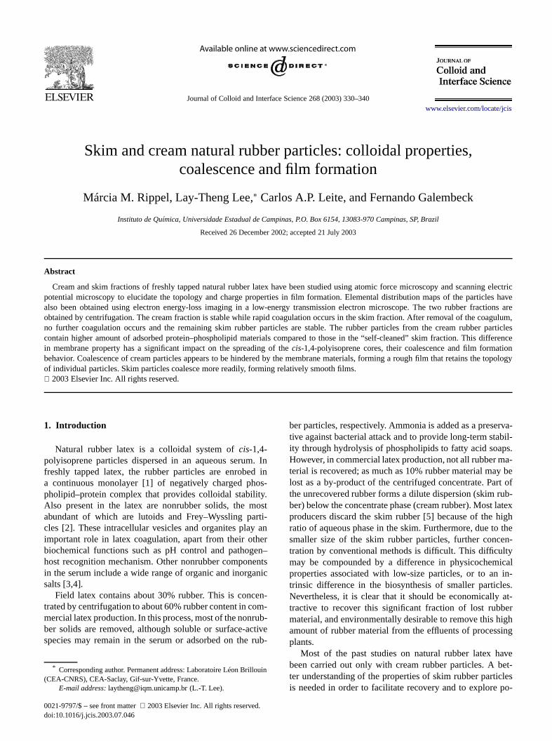
Journal of Colloid and Interface Science 268 (2003) 330–340 www.elsevier.com/locate/jcis Skim and cream natural rubber particles: colloidal properties, coalescence and film formation Márcia M. Rippel, Lay-Theng Lee, ∗ Carlos A.P. Leite, and Fernando Galembeck Instituto de Química, Universidade Estadual de Campinas, P.O. Box 6154, 13083-970 Campinas, SP, Brazil Received 26 December 2002; accepted 21 July 2003 Abstract Cream and skim fractions of freshly tapped natural rubber latex have been studied using atomic force microscopy and scanning electric potential microscopy to elucidate the topology and charge properties in film formation. Elemental distribution maps of the particles have also been obtained using electron energy-loss imaging in a low-energy transmission electron microscope. The two rubber fractions are obtained by centrifugation. The cream fraction is stable while rapid coagulation occurs in the skim fraction. After removal of the coagulum, no further coagulation occurs and the remaining skim rubber particles are stable. The rubber particles from the cream rubber particles contain higher amount of adsorbed protein–phospholipid materials compared to those in the “self-cleaned” skim fraction. This difference in membrane property has a significant impact on the spreading of the cis-1,4-polyisoprene cores, their coalescence and film formation behavior. Coalescence of cream particles appears to be hindered by the membrane materials, forming a rough film that retains the topology of individual particles. Skim particles coalesce more readily, forming relatively smooth films. 2003 Elsevier Inc. All rights reserved. 1. Introduction Natural rubber latex is a colloidal system of cis-1,4- polyisoprene particles dispersed in an aqueous serum. In freshly tapped latex, the rubber particles are enrobed in a continuous monolayer [1] of negatively charged phos- pholipid–protein complex that provides colloidal stability. Also present in the latex are nonrubber solids, the most abundant of which are lutoids and Frey–Wyssling parti- cles [2]. These intracellular vesicles and organites play an important role in latex coagulation, apart from their other biochemical functions such as pH control and pathogen– host recognition mechanism. Other nonrubber components in the serum include a wide range of organic and inorganic salts [3,4]. Field latex contains about 30% rubber. This is concen- trated by centrifugation to about 60% rubber content in com- mercial latex production. In this process, most of the nonrub- ber solids are removed, although soluble or surface-active species may remain in the serum or adsorbed on the rub- * Corresponding author. Permanent address: Laboratoire Léon Brillouin (CEA-CNRS), CEA-Saclay, Gif-sur-Yvette, France. E-mail address: [email protected] (L.-T. Lee). ber particles, respectively. Ammonia is added as a preserva- tive against bacterial attack and to provide long-term stabil- ity through hydrolysis of phospholipids to fatty acid soaps. However, in commercial latex production, not all rubber ma- terial is recovered; as much as 10% rubber material may be lost as a by-product of the centrifuged concentrate. Part of the unrecovered rubber forms a dilute dispersion (skim rub- ber) below the concentrate phase (cream rubber). Most latex producers discard the skim rubber [5] because of the high ratio of aqueous phase in the skim. Furthermore, due to the smaller size of the skim rubber particles, further concen- tration by conventional methods is difficult. This difficulty may be compounded by a difference in physicochemical properties associated with low-size particles, or to an in- trinsic difference in the biosynthesis of smaller particles. Nevertheless, it is clear that it should be economically at- tractive to recover this significant fraction of lost rubber material, and environmentally desirable to remove this high amount of rubber material from the effluents of processing plants. Most of the past studies on natural rubber latex have been carried out only with cream rubber particles. A bet- ter understanding of the properties of skim rubber particles is needed in order to facilitate recovery and to explore po- 0021-9797/$ – see front matter 2003 Elsevier Inc. All rights reserved. doi:10.1016/j.jcis.2003.07.046
Transcript of Skim and cream natural rubber particles: colloidal properties, coalescence and film formation
ng electricles havections areagulum,r particlesifferencetion
he topology
Journal of Colloid and Interface Science 268 (2003) 330–340www.elsevier.com/locate/jcis
Skim and cream natural rubber particles: colloidal properties,coalescence and film formation
Márcia M. Rippel, Lay-Theng Lee,∗ Carlos A.P. Leite, and Fernando Galembeck
Instituto de Química, Universidade Estadual de Campinas, P.O. Box 6154, 13083-970 Campinas, SP, Brazil
Received 26 December 2002; accepted 21 July 2003
Abstract
Cream and skim fractions of freshly tapped natural rubber latex have been studied using atomic force microscopy and scannipotential microscopy to elucidate the topology and charge properties in film formation. Elemental distribution maps of the particalso been obtained using electron energy-loss imaging in a low-energy transmission electron microscope. The two rubber fraobtained by centrifugation. The cream fraction is stable while rapid coagulation occurs in the skim fraction. After removal of the cono further coagulation occurs and the remaining skim rubber particles are stable. The rubber particles from the cream rubbecontain higher amount of adsorbed protein–phospholipid materials compared to those in the “self-cleaned” skim fraction. This din membrane property has a significant impact on the spreading of thecis-1,4-polyisoprene cores, their coalescence and film formabehavior. Coalescence of cream particles appears to be hindered by the membrane materials, forming a rough film that retains tof individual particles. Skim particles coalesce more readily, forming relatively smooth films. 2003 Elsevier Inc. All rights reserved.
. Ind inos-ty.ostrti-an
heren–ents
anic
en-om-rub-ctiverub
louin
rva-bil-ps.a-
y bert ofrub-latexhigh
theen-tyicalin-
es.at-erighing
aveet-
clespo-
1. Introduction
Natural rubber latex is a colloidal system ofcis-1,4-polyisoprene particles dispersed in an aqueous serumfreshly tapped latex, the rubber particles are enrobea continuous monolayer [1] of negatively charged phpholipid–protein complex that provides colloidal stabiliAlso present in the latex are nonrubber solids, the mabundant of which are lutoids and Frey–Wyssling pacles [2]. These intracellular vesicles and organites playimportant role in latex coagulation, apart from their otbiochemical functions such as pH control and pathoghost recognition mechanism. Other nonrubber componin the serum include a wide range of organic and inorgsalts [3,4].
Field latex contains about 30% rubber. This is conctrated by centrifugation to about 60% rubber content in cmercial latex production. In this process, most of the nonber solids are removed, although soluble or surface-aspecies may remain in the serum or adsorbed on the
* Corresponding author. Permanent address: Laboratoire Léon Bril(CEA-CNRS), CEA-Saclay, Gif-sur-Yvette, France.
E-mail address:[email protected] (L.-T. Lee).
0021-9797/$ – see front matter 2003 Elsevier Inc. All rights reserved.doi:10.1016/j.jcis.2003.07.046
-
ber particles, respectively. Ammonia is added as a presetive against bacterial attack and to provide long-term staity through hydrolysis of phospholipids to fatty acid soaHowever, in commercial latex production, not all rubber mterial is recovered; as much as 10% rubber material malost as a by-product of the centrifuged concentrate. Pathe unrecovered rubber forms a dilute dispersion (skimber) below the concentrate phase (cream rubber). Mostproducers discard the skim rubber [5] because of theratio of aqueous phase in the skim. Furthermore, due tosmaller size of the skim rubber particles, further conctration by conventional methods is difficult. This difficulmay be compounded by a difference in physicochemproperties associated with low-size particles, or to antrinsic difference in the biosynthesis of smaller particlNevertheless, it is clear that it should be economicallytractive to recover this significant fraction of lost rubbmaterial, and environmentally desirable to remove this hamount of rubber material from the effluents of processplants.
Most of the past studies on natural rubber latex hbeen carried out only with cream rubber particles. A bter understanding of the properties of skim rubber partiis needed in order to facilitate recovery and to explore
M.M. Rippel et al. / Journal of Colloid and Interface Science 268 (2003) 330–340 331
idalgyes.bbe
00oice.
g ate to
col-ree,ftered:tula
sy-ategh
inble
latex8 h,ingag-entol-texof
pro-
ac-
ith
es
tofaceacehatring
pHs co
bedsed by
lu-a
ta-insthe
ive
there-elf-re-
textextex
la-ec-n In-
dlolu-
e-
Md byriedres-
retheTheonto
ea-ith
iusan-plelder.re-to
nfor-
y. Inap-Hz
tential uses. In this paper, we present results on colloproperties of skim rubber particles, together with topoloand film-forming properties using microscopy techniquThese properties are compared with those of cream ruparticles.
2. Experimental
2.1. Sample preparation
Freshly tapped natural rubber latex of the RRIM 6clone of Hevea brasiliensiswas collected at the InstitutAgronômico de Campinas and stored immediately inThe latex was centrifuged within 2 h at 5◦C at 10 krpm for2 h. Since no ammonia was added to the latex, handlinlow temperature slows down degradative processes duattack by bacteria that are always present in normallylected latex. Bacteria can originate from the bark of the tfrom the sample container, or from the atmosphere. Acentrifugation, two fractions of the rubber were collectthe upper cream phase (paste) was collected with a spaand the lower skim phase (fluid) was collected with aringe. The cream rubber was redispersed in deionized wand filtered. The skim rubber was similarly filtered, althouredispersion was not necessary.
The skim rubber fraction was partially destabilized with24 h even when kept in the refrigerator. This is an inevitaprocess of spontaneous coagulation in freshly tappedwhere no ammonia is added as preservative. Within 4a large mass of irreversible rubbery coagulum, containabout 60% of the initial solid content, was formed. The coulum underwent syneresis, losing most of its liquid contto form a compact mass about a third of its original vume. This was followed by putrefaction: whereas fresh lasmells sweet, the coagulum had a foul odor, indicativebacterial and enzyme activity. Several factors have beenposed to explain this spontaneous destabilization [6]:
(i) development of acidic environment due to bacterialtivity;
(ii) interaction of adsorbed volatile fatty acid anions wcations;
(iii) aggregation by cationic protein and attack by enzymfrom the lutoid serum.
The isoelectric point of natural rubber latex is around 3.94.6, depending on the type of proteins found on the surof the latex particle [7]; therefore coagulation can take plwhen the pH falls below 5. However, it has been found tthe pH of the latex suspension does not fall below 6 duspontaneous coagulation; furthermore, maintaining theof the latex suspension at 7 does not prevent spontaneouagulation. The second possibility is interaction of adsorvolatile fatty acid anions with multivalent cations. Theanions (formate, acetate, and propionate) are produce
r
,
r
-
bacterial oxidation of carbohydrates in the serum. Third,toid serum can also release a host of inorganic cations (N+,K+, Ca2+, Mg2+) as well as cationic proteins that can desbilize the rubber latex particles [8]. The serum also contaenzymes(protease and phospholipase) that can attackprotein–phospholipid complex, which forms the protectenvelope of the rubber particle.
Interestingly, after the coagulum was removed fromskim fraction, no further coagulation occurred in themaining latex suspension. Thus, it appears that in this “scleaning” process, most of the destabilizing agents weremoved with the coagulum, leaving behind a very stable ladispersion. All the measurements of the skim rubber lain the present study were carried out on this remaining ladispersion.
2.2. Methods
Particle size and zeta potential of the natural rubbertex particles were determined by photon correlation sptroscopy and electrophoresis measurements (Brookhavestruments) in dispersions of∼0.05% by weight in deionizewater and in 10−3 mol l−1 KCl in the case of zeta potentiameasurement. The Brookhaven instrument uses the Smchowski approximationµe = εζ/η, whereµe is the elec-trophoretic mobility,ζ the zeta potential, andε and η thepermittivity and viscosity of the liquid, respectively. This rlation is a good approximation whenκa � 1 (κ is the doublelayer thickness anda is the particle radius). In our case,κa
values are between 12 and 30.FTIR spectroscopy was carried out using a BOME
(MB-Series) spectrometer. The samples were preparedeposition of latex dispersion onto a ZnSe support and dunder vacuum for 2 h. For each spectrum, 32 scans at aolution of 4 cm−1 were made.
2.2.1. Atomic force microscopy (AFM) and scanningelectric potential microscopy (SEPM)
Topographic information and electric mapping weacquired using AFM and SEPM, respectively, usingTopometrix Discoverer scanning probe microscope.samples were prepared by depositing latex dispersionsfreshly cleaved mica and dried at room temperature.
The noncontact mode was employed in the AFM msurement. The probe tip was made of silicon coated wplatinum and had a pyramidal form with a nominal radof 20 nm. The cantilever-tip system was oscillated mechically at the natural frequency of 40–70 kHz and tip-samdistance kept constant at 10 nm with a piezosample hoThe vertical movement of the piezosample holder (insponse to shift in oscillation frequency of the probe duetopographic change) was used to generate topographic imation (voltage supplied to it) at 300× 300 pixel resolution.
SEPM measurements were acquired simultaneousladdition to the above AFM noncontact mode, an AC wasplied to the conductive tip at a frequency that was 10 k
332 M.M. Rippel et al. / Journal of Colloid and Interface Science 268 (2003) 330–340
ngetherobeme-opstopothepli-tialThereg-ach
ereatedandrriedmi-ergyter-d atteallye elginndagesubele-al totheen-dingop
ness
then th
i-
-
usal toum.
les
ndtoruidsed-veith
theare
ea-es
po-clesarti-theam-
inglybed, de-the
incipaltural450,to
weenyl
eries
akspro-
twoiminse re-
orated1130the
H
less than the cantilever oscillation frequency. Any chain this frequency (owing to electrostatic interaction oftip and the sample) was canceled by a DC fed to the ptip. A four-quadrant photodetector was used to track thechanical oscillations of the probe tip. Two feedback loenabled independent and simultaneous analyses ofgraphic and electric information: the first loop to monitortip–sample distance, and the second, with a lock-in amfier, to monitor the AC frequency. Here, the electric potenimage was generated from the DC voltage fed to the tip.topography and electric potential signals were thereforeistered at different frequencies, independently and at epixel.
2.2.2. Energy-filtered analytical transmissionelectron microscopy (EFTEM)
The samples for transmission electron microscopy wprepared by depositing latex dispersions onto carbon-coparlodion films supported on 400-mesh copper gridsdried at room temperature. Elemental mapping was caout on a Carl Zeiss CEM 902 transmission electroncroscope (80 keV) equipped with a Castaing–Henry enfilter spectrometer. Characteristic energy losses from inaction of electrons with various elements were selecte303 eV for C, 410 eV for N, and 544 eV for O to creathe electron energy loss spectra (EELS). Only inelasticscattered monochromatic electrons were used to producemental maps generated by electron spectroscopy ima(ESI). A three-window method was used for backgrousubtraction: two preedge images and one postedge imAn extrapolated background image was calculated andtracted from the postedge image, giving the desiredmental map where the image brightness was proportionconcentration. However, it should be kept in mind thatproportionality coefficient between brightness and conctration varies from one elemental map to another, depenon the elemental cross section for inelastic scattering anderational parameters (filament current, aperture, brightadjustment).
3. Results
A first characterization of the two rubber fractions isparticle size and charge. The particles are separated icentrifugation process according to
v = (ρ − σ)ω2R
18ηd2.
v is the particle velocity,ρ andσ the densities of the partcle and fluid medium, respectively,d the particle size,η theviscosity of the fluid medium, andω2R the centrifugal acceleration, whereω is the angular speed andR the distancefrom the axis of the centrifuge. The particle velocity thvaries as the square of the particle size and is proportionthe difference in density between particle and fluid medi
-
-g
.-
-
e
Table 1Particle size and zeta potential values of cream and skim rubber partic
Fresh latex d (nm) ζ (mV)
Cream rubber 467 −70Skim rubber 297 −64
Accordingly, the particle size distribution will also depeon the condition and time of centrifugation. Another facwhich can affect the movement of the particle through a fl(at zero current) is the particle charge, which induces aimentation potential. More highly charged particles momore slowly through the liquid medium than those wlower charge, and in this case, cream less easily.
Under the centrifugation conditions employed here,average particle sizes of the cream and skim fractionsgiven in Table 1, together with zeta potential values msured in 10−3 mol l−1 KCl. As expected, the rubber particlin the cream are larger than those in the skim. The zetatential is negative for both cream and skim rubber partiand the closeness of their values indicates that the pcle surface potential does not play an important role increaming process. For these latex samples, although nomonia has been added, the surface charge is surprishigh. The negative charges arise principally from adsorsurface proteins whose isoelectric points are 3.9 to 4.6pending on the type of proteins found on the surface oflatex particle [7].
3.1. Infrared spectroscopy
The spectra for cream and skim latex are shownFigs. 1a and 1b, respectively. For both samples, the prinpeaks corresponding to isoprene functional groups of narubber are observed: 3033, 2961, 2927, 2855, 1664, 11375, and 837 cm−1. Several other peaks not associatedisoprene are also present: an intense broad band bet3200 and 3500 cm−1 indicating the presence of hydroxgroups, an amide (N–H) peak at 3280 cm−1 (more appar-ent in cream rubber), a carbonyl (C=O) peak at 1737 cm−1,a small amide (N–H) peak at 1548 cm−1 (both more ap-parent and more resolved in cream rubber), and a sof peaks between 1130 and 1010 cm−1 indicating oxy-genated compounds. The peak at around 1080 cm−1 is as-signed to the C–O group. In Table 2, the absorption peare assigned to two categories: polyisoprene units andteins/phospholipids.
The most significant difference in the spectra of therubber fractions is the much higher hydroxyl content in skrubber. These hydroxyl groups, from hydration of proteand phospholipids, are chemically bound and cannot bmoved even after drying under vacuum for two hoursmore. The second difference is the amount of oxygencompounds indicated by the absorption bands betweenand 1010 cm−1, which can be seen more clearly whenspectra are normalized to the 1375 cm−1 peak (shown in in-set). The 1375 cm−1 peak is assigned to the methyl C–
M.M. Rippel et al. / Journal of Colloid and Interface Science 268 (2003) 330–340 333
Fig. 1. FTIR spectra for cream (a) and skim (b) rubber particles. The insets show absorbance normalized to peak 1375 cm−1.
12].ption-
e
assIfstly
ts of
sid-sur-en-
amlopene
lus-i-
bending and is insensitive to environmental changes [In the normalized spectra, it can be seen that the absorbands between 1130 and 1010 cm−1 are higher in skim rubber: the peak intensity at 1080 cm−1 is 0.82, compared to0.69 for cream rubber. The 1548 cm−1 peak assigned to thamide group is also higher.
The above absorbance ratios are proportional to the mratios of individual constituents of the rubber material.the protein–phospholipid materials are concentrated moon the particle surface, the presence of higher amoun
these materials in skim rubber is not surprising conering the smaller size and therefore higher specificface area of skim particles. If the surface adsorption dsity (mol/cm2) of these materials were constant for creand skim particles, an assumption based on an enveof protein–phospholipid complex around the polyisoprehydrophobic core, the adsorption per unit mass (mo/g)would be inversely proportional to the particle size. ThAcream/Askim = dskim/dcream, whereA is the adsorption density in mol/cm2 andd the particle size. Taking the max
334 M.M. Rippel et al. / Journal of Colloid and Interface Science 268 (2003) 330–340
date
ids
imn ofer-ion,ameamzedingto
Theegglow
cor-ctedhesheirhela-
rage
mcu-
nmm-
en inor a
Table 2Peak assignments for the IR absorption band
Wavenumber (cm−1) Assignment Reference
Isoprene3033 =CH stretching [9–11]2962 C–H stretching of CH3 [9,12,13]2927 C–H stretching of CH2 [9,12,14]2855 C–H stretching of CH2 and CH3 [9,12,13]1664 C=C stretching [9,12,13]1450 C–H bending of CH2 [9,10,13–15]1375 C–H bending of CH3 [9,10,12–15]1127 C–H bending [11,13]837 C=CH wagging [9,15]
Protein/phospholipid3440 –OH stretching [13,16]3280 N–H stretching [13,16]1737 C=O stretching [14,17]1548 N–H bending [13]1080 C–O [12,15,17]1014 –O–O– [12–14]
mum peak around 1080 cm−1 to be indicative of oxygenatecompounds related to adsorbed protein–phospholipid mrial, we haveAcream/Askim ≈ 0.69/0.82= 0.84. This ratiois higher than the size ratiodskim/dcream= 297/467= 0.64,indicating that the surface density of protein–phospholipis in fact higher for cream rubber particles.
3.2. AFM results
3.2.1. Individual particlesParticle size Figures 2a and 2b show AFM images of skrubber particles deposited onto mica from a dispersioabout<0.02% by weight. The images are taken from diffent fields of the same sample. At this dilute concentratindividual particles can be seen. Several fields of the ssample, as well as different samples prepared from the sdispersion were imaged and the particle dimension analyThe particles are highly polydisperse with diameter rangfrom about 326 to 2850 nm, maximum height from 2.5178 nm, and particle volume from 0.0002 to 0.509 µm3 (de-duced from values of particle area and average height).rubber particles are therefore spread out with a friedshape. This fluid behavior is as expected owing to theTg of −73◦C for cis-1,4-polyisoprene [18].
From particle volume obtained in these images, theresponding diameter for a spherical particle, the expetopology in an aqueous dispersion, can be calculated. Tcalculated diameters range from 72 to 995 nm and tnormalized frequency distribution is shown in Fig. 2c. Thistogram shows a bimodal distribution: a primary popution occurring around 200 nm (median= 160 nm) and asecondary population around 600 nm. The number avemean diameter calculated from
d̄ =∑
nidi∑
ni
givesd̄ = 253 nm.
-
e.
e
(a)
(b)
(c)
Fig. 2. AFM images (20× 20 µm) of skim rubber particles deposited froa dispersion of<0.02% by weight (a and b). Histogram of diameters callated for corresponding spherical particles (c); step size= 30 nm.
The occurrence of a second population around 600is rather surprising. Under the centrifugation conditions eployed here, particles of this size range should have bethe cream phase. Density effect may play a role here: f
M.M. Rippel et al. / Journal of Colloid and Interface Science 268 (2003) 330–340 335
o
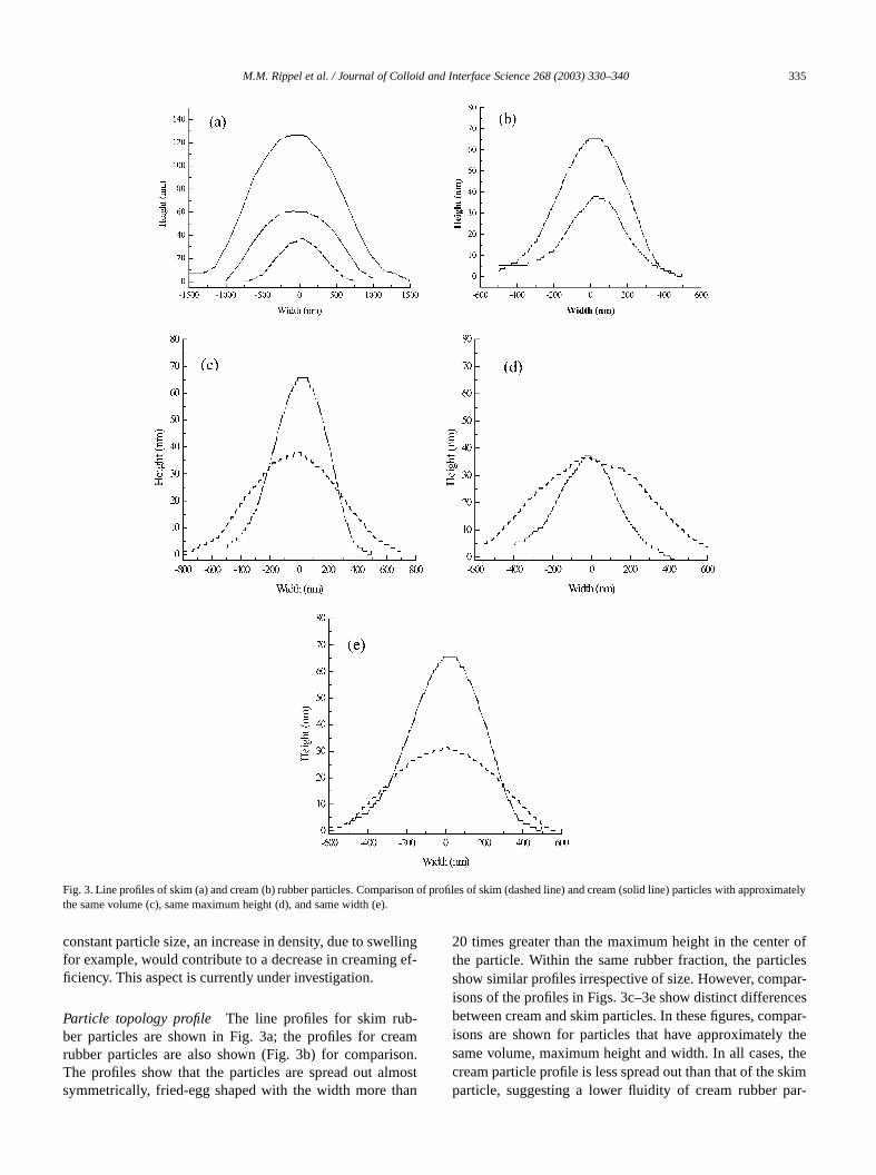
Fig. 3. Line profiles of skim (a) and cream (b) rubber particles. Comparison of profiles of skim (dashed line) and cream (solid line) particles with apprximatelythe same volume (c), same maximum height (d), and same width (e).llingef-
-amon.ost
an
r oflesar-cespar-thethekimar-
constant particle size, an increase in density, due to swefor example, would contribute to a decrease in creamingficiency. This aspect is currently under investigation.
Particle topology profile The line profiles for skim rubber particles are shown in Fig. 3a; the profiles for crerubber particles are also shown (Fig. 3b) for comparisThe profiles show that the particles are spread out almsymmetrically, fried-egg shaped with the width more th
20 times greater than the maximum height in the centethe particle. Within the same rubber fraction, the particshow similar profiles irrespective of size. However, compisons of the profiles in Figs. 3c–3e show distinct differenbetween cream and skim particles. In these figures, comisons are shown for particles that have approximatelysame volume, maximum height and width. In all cases,cream particle profile is less spread out than that of the sparticle, suggesting a lower fluidity of cream rubber p
336 M.M. Rippel et al. / Journal of Colloid and Interface Science 268 (2003) 330–340
e
le istheistri-ely.
dis-er-agesd tothe
gesmaltheslow.an
orese exion.d in
b) isco-thatnce
canhowEPM
ater
in7d)ughex-
rub-
andto aribu-gray, nofornt, acor-ringes acle.n ofthe
egg”ever,in-ofin-
te-
Fig. 4. AFM image (20× 20 µm) of fusion of skim rubber particles. Thparticles are deposited from a dispersion of∼0.02% by weight.
ticles. Note that the average size for the cream particlarger than that for the skim particle, but in order to makeabove comparisons, the low and high ends of the size dbutions are taken for cream and skim particles, respectiv
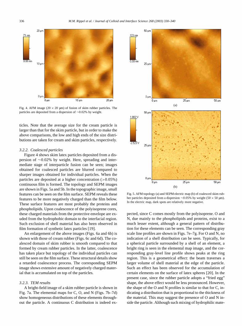
3.2.2. Coalesced particlesFigure 4 shows skim latex particles deposited from a
persion of∼0.02% by weight. Here, spreading and intmediate stage of interparticle fusion can be seen; imobtained for coalesced particles are blurred comparesharper images obtained for individual particles. Whenparticles are deposited at a higher concentration (>0.05%)continuous film is formed. The topology and SEPM imaare shown in Figs. 5a and 5b. In the topographic image, sfeatures can be seen on the film surface. SEPM revealsfeatures to be more negatively charged than the film beThese surface features are most probably the proteinsphospholipids. Upon coalescence of the polyisoprene cthese charged materials from the protective envelope aruded from the hydrophobic domain to the interfacial regSuch exclusion of shell material has also been observefilm formation of synthetic latex particles [19].
An enlargement of the above images (Figs. 6a and 6shown with those of cream rubber (Figs. 6c and 6d). Thealesced domain of skim rubber is smooth compared toformed by cream rubber particles. In the latter, coalescehas taken place but topology of the individual particlesstill be seen on the film surface. These structural details sa retarded coalescence process. The corresponding Simage shows extensive amount of negatively charged mial that is accumulated on top of the particles.
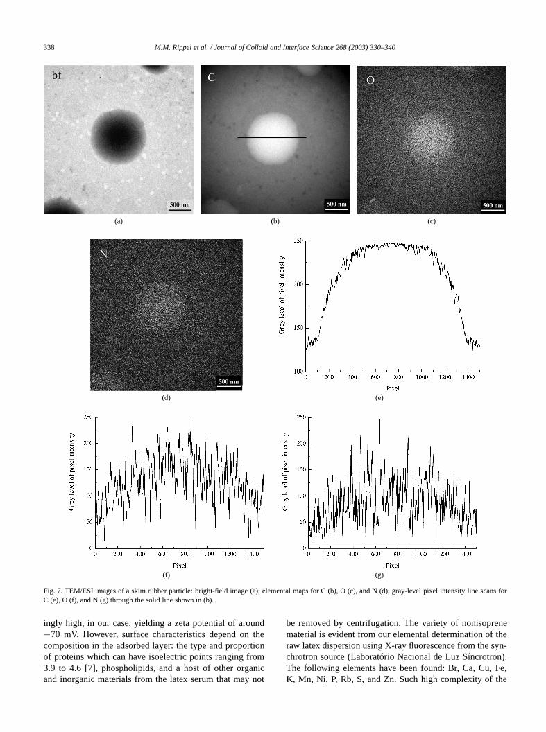
3.2.3. TEM resultsA bright-field image of a skim rubber particle is shown
Fig. 7a. The elemental maps for C, O, and N (Figs. 7b–show homogeneous distributions of these elements throout the particle. A continuous C distribution is indeed
le
d,-
-
-
(a)
(b)
Fig. 5. AFM topology (a) and SEPM electric map (b) of coalesced skimber particles deposited from a dispersion∼0.05% by weight (50× 50 µm).In the electric map, dark spots are relatively more negative.
pected, since C comes mostly from the polyisoprene. ON, due mainly to the phospholipids and proteins, existmuch lesser extent, although a general pattern of disttion for these elements can be seen. The correspondingscale line profiles are shown in Figs. 7e–7g. For O and Nindication of a shell distribution can be seen. Typically,a spherical particle surrounded by a shell of an elemebright ring is seen in the elemental map image, and theresponding gray-level line profile shows peaks at theregion. This is a geometrical effect: the beam traverslarger volume of shell material at the edge of the partiSuch an effect has been observed for the accumulatiocertain elements on the surface of latex spheres [20]. Inpresent case, since the rubber particle adopts a “friedshape, the above effect would be less pronounced. Howthe shape of the O and N profiles is similar to that for C,dicating a distribution that is proportional to the thicknessthe material. This may suggest the presence of O and Nside the particle. Although such mixing of hydrophilic ma

M.M. Rippel et al. / Journal of Colloid and Interface Science 268 (2003) 330–340 337
(a) (b)
(c) (d)
Fig. 6. Topology and electric maps for coalesced skim (a and b, respectively) and cream (c and d, respectively) rubber particles (10× 10 µm). Dark areas inthe electric maps are relatively more negative.
ousrti-obicto
ueno-
ingco-d 8d).
im-retetillionsid–berlinevely.o th
r inareultsn offull
kimns,the
la-ve-x.
ylespris-
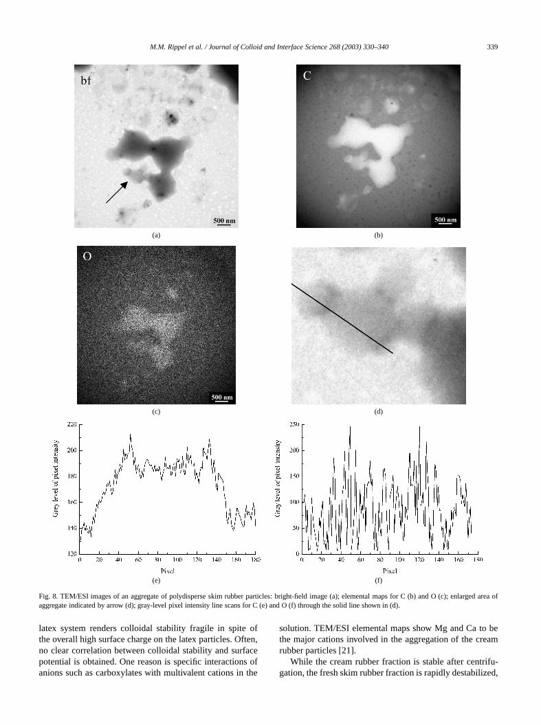
rial with the hydrophobic core is not expected in an aquedispersion, the situation may be different when the pacles are dried and subsequently exposed to a hydrophenvironment such as air. Here, diffusion of polyisoprenethe surface to reduce the interfacial tension, and conseqmixing of protein–phospholipid materials with the polyisprene may be possible.
Figure 8a shows the bright-field image of an interestaggregate of polydisperse particles in the initial stage ofalescence. The C and O maps are shown in Figs. 8b an(for this aggregate, the N content is too low to be imageAn interesting feature can be seen from the bright-fieldage: although the primary particles have lost their discfeature, their borders with neighboring particles can sbe distinguished. These borders show up as darker regindicative of denser materials. Indeed, the phospholipprotein complex that forms the membrane of the rubparticle is denser than the polyisoprene. The gray-levelscans for C and O are shown in Figs. 8e and 8f, respectiWeak peaks can be seen in the regions corresponding t
t
c
,
e
dark regions, showing these border materials to be richeC and O. This further confirms that the dense regionscomposed mainly of phospholipid materials. These resshow the initial stage of coalescence where interdiffusiopolyisoprene takes place across particle border withoutexpulsion of envelope material.
No further element, such as cations, is detected for srubber particle. In contrast, significant amounts of catiomainly calcium and magnesium, have been detected inaggregates of cream rubber particles [21].
4. Discussion
The hydrophobic polyisoprene core of natural rubbertex particle is surrounded by a protective hydrophilic enlope comprising principally protein–phospholipid compleUnder natural pH conditions (∼6), the protein is negativelcharged and imparts colloidal stability to the rubber particin an aqueous medium. The charge density can be sur
338 M.M. Rippel et al. / Journal of Colloid and Interface Science 268 (2003) 330–340
can
(a) (b) (c)
(d) (e)
(f) (g)
Fig. 7. TEM/ESI images of a skim rubber particle: bright-field image (a); elemental maps for C (b), O (c), and N (d); gray-level pixel intensity line ss forC (e), O (f), and N (g) through the solid line shown in (b).
ndthe
tionomnicnot
enetheyn-n).Fe,he
ingly high, in our case, yielding a zeta potential of arou−70 mV. However, surface characteristics depend oncomposition in the adsorbed layer: the type and proporof proteins which can have isoelectric points ranging fr3.9 to 4.6 [7], phospholipids, and a host of other orgaand inorganic materials from the latex serum that may
be removed by centrifugation. The variety of nonisoprmaterial is evident from our elemental determination ofraw latex dispersion using X-ray fluorescence from the schrotron source (Laboratório Nacional de Luz SíncrotroThe following elements have been found: Br, Ca, Cu,K, Mn, Ni, P, Rb, S, and Zn. Such high complexity of t
M.M. Rippel et al. / Journal of Colloid and Interface Science 268 (2003) 330–340 339
ed area o
(a) (b)
(c) (d)
(e) (f)
Fig. 8. TEM/ESI images of an aggregate of polydisperse skim rubber particles: bright-field image (a); elemental maps for C (b) and O (c); enlargfaggregate indicated by arrow (d); gray-level pixel intensity line scans for C (e) and O (f) through the solid line shown in (d).
often,aces ofthe
beam
ifu-ed,
latex system renders colloidal stability fragile in spitethe overall high surface charge on the latex particles. Ofno clear correlation between colloidal stability and surfpotential is obtained. One reason is specific interactionanions such as carboxylates with multivalent cations in
solution. TEM/ESI elemental maps show Mg and Ca tothe major cations involved in the aggregation of the crerubber particles [21].
While the cream rubber fraction is stable after centrgation, the fresh skim rubber fraction is rapidly destabiliz
340 M.M. Rippel et al. / Journal of Colloid and Interface Science 268 (2003) 330–340
l ofain-arebleb-
rent
ad-eamcles
raneconate-sity.canos-tanto-ctlyne,ition
[1]oveanenevior
teri-everford-aseerlesformingareed.
offilmatered
nse-rface
notfilmers.
gedhy-lm i
ilelms
althostto
re-ovesrod-imnds
x,texator,as)ly
ort ofibu-os
n
at-ork,
.I.of
an,
478.
lym.
stry,
vier,
886.87.
02)
forming an irreversible rubbery coagulum. After removathis coagulum, no further coagulation occurs in the reming dispersion. Thus, most of the destabilizing agentsremoved with the initial coagulum, leaving behind a stadispersion of skim particles. This “self-cleaned” skim ruber fraction has lower protein content and shows diffetopology and film properties compared to cream rubber.
The most novel and remarkable difference is their spreing behavior, as revealed by topography line profiles. Crparticles are found to spread less easily than skim partiIn view of the lowTg of cis-1,4-polyisoprene (−73◦C), thisspreading behavior may be due to an influence of membproperty. Our present results show that cream particlestain higher amount of adsorbed protein–phospholipid mrials and a correspondingly higher surface charge denThis in itself suggests a more rigid membrane, whichretard flow of core material. The type of proteins and phpholipids constituting the membrane is also an imporfactor in membrane rigidity. Different proteins form monlayers with different elastic properties that can be direrelated to colloidal stability [22]. Phosphatidylethanolamia phospholipid that is known to increase phase transtemperature is expected to decrease membrane fluidityOther adsorbed or surface-precipitated species not remby centrifugation or aggregation may also affect membrproperty. We point out that cross-linking in the polyisoprecore is also a possible explanation in the spreading behaThis aspect is currently under study in our laboratory.
In coalescence of the rubber particles, membrane maals are exuded to the surface of the coalesced film. Howfull expulsion of membrane material is not necessaryinterdiffusion of core materials to proceed. TEM/ESI stuies show cross border diffusion of polyisoprene in the cof skim particles. Films that are formed from skim rubbare relatively smooth and features from individual particare lost. The expelled charged materials segregate toislands on the polymer–air interface. Under similar drytime and conditions, films formed by cream particlesrough and the topology of individual particle is retainSignificant amount of charged materials remain on topthe particle. Here, full coalescence is not observed andformation appears to be hindered by the membrane mial. Such differences in topology and distribution of chargmaterial on the films are expected to bear important coquences on the film mechanical properties as well as suproperties.
5. Conclusions
Cream and skim fractions of natural rubber latex showonly differences in particle size but also in surface andforming properties. “Self-cleaned” skim rubber has lowprotein content, higher fluidity and forms smoother filmCream rubber particles contain higher amount of charmembrane material that appears to retard mobility of thedrophobic cores and their coalescence. The coalesced fi
.
-
.d
.
,
-
s
rough and retains the topology of individual particles whskim particles coalesce more readily and form smooth fiwhere features of individual particles are lost.
A first important consequence of our findings is a herelated issue. In natural rubber products, one of the mimportant concerns is its allergenic property attributedproteins in the rubber. The low-protein skim rubber thefore has potential uses in protective products such as gland masks, as well as other medical and infant related pucts. Second, the fluidity and interfacial morphology of skrubber can be exploited for potential uses in polymer bleand natural rubber based composite materials.
Acknowledgments
L.T. Lee thanks Fabio Z. Maia (Manager, AgrolateOlímpia) for helpful discussions on natural rubber laprocessing. We thank Paulo de S. Gonçalves (coordinPrograma Seringueira, Instituto Agronômico de Campinfor his kind cooperation and for furnishing us with freshtapped rubber latex. The authors acknowledge the suppCNPq, Fapesp, and Pronex/Finep/MCT. This is a contrtion from the Instituto do Milênio de Materiais Complex(PADCT).
References
[1] K. Cornish, D.F. Wood, J.J. Windle, Planta 210 (1999) 85.[2] M.R. Sethuraj, N.M. Mathew, Natural Rubber: Biology, Cultivatio
and Technology, Elsevier, Amsterdam, 1992.[3] K.F. Gazeley, A.D.T. Gorton, T.D. Pendle, in: E.D. Roberts (Ed.), N
ural Rubber Science and Technology, Oxford Univ. Press, New Y1988, Chap. 3.
[4] B.L. Archer, D. Barnard, E.G. Cockbain, P.B. Dickenson, AMcMullen, in: L. Bateman (Ed.), The Chemistry and PhysicsRubber-Like Substances, Maclaren, London, 1963, Chap. 3.
[5] F.Z. Maia, personal communication.[6] D.C. Blackley, Polymer Latices, Vol. 2, Types of Latices, Chapm
London, 1997.[7] W.W. Bowler, Ind. Eng. Chem. 45 (1953) 1790.[8] W.A. Southorn, E. Yip, J. Rubb. Res. Inst. Malaya 20 (1968) 201.[9] P. Phinyocheep, S. Duangthong, J. Appl. Polym. Sci. 78 (2000) 1
[10] Y.D. West, P.J. Hendra, A.M. Healy, Polymer 37 (1996) 4009.[11] S.W. Cornell, J.L. Koening, Macromolecules 2 (1969) 546.[12] H.M. Nor, J.R. Ebdon, Polymer 41 (2000) 2359.[13] F.J. Lu, S.L. Hsu, Rubber Chem. Technol. 60 (1987) 647.[14] R.A. Saunders, D.C. Smith, J. Appl. Phys. 20 (1949) 953.[15] J.W. Cook, S. Edge, D.E. Packham, A.S. Thompson, J. Appl. Po
Sci. 65 (1997) 1379.[16] C.H. Jones, Spectrochim. Acta Part A 47 (1991) 1313.[17] K. Isakson, Infrared Spectroscopy: Its Use in the Coating Indu
Federation of Societies for Paint Technology, Philadelphia, 1969.[18] J.A. Brydson, Rubbery Materials and Their Compounds, Else
London/New York, 1988.[19] A.J. Keslarek, C.A.R. Costa, F. Galembeck, Langmuir 17 (2001) 7[20] A.H. Cardoso, C.A.P. Leite, F. Galembeck, Langmuir 14 (1998) 31[21] M.M. Rippel, C.A.P. Leite, F. Galembeck, Anal. Chem. 74 (20
2541.[22] P.J. Wilde, Curr. Opin. Colloid Interface Sci. 5 (2000) 176.