Microstructure of acid–induced skim milk–locust bean gum–xanthan gels
14
* Corresponding author. E-mail address: christian.sanchez@ensaia.inpl-nancy.fr (C. Sanchez). International Dairy Journal 10 (2000) 199}212 Microstructure of acid-induced skim milk}locust bean gum}xanthan gels C. Sanchez*, R. Zuniga-Lopez, C. Schmitt, S. Despond, J. Hardy Laboratoire de Physico-Chimie et Ge & nie Alimentaires (LPGA), ENSAIA-INPL, 2 Avenue de la Fore L t de Haye BP 172, F-54505 Vandoeuvre Cedex, France Received 9 September 1999; accepted 4 April 2000 Abstract The microstructure of acid skim milk gels (14% w/w milk protein low heat powder) with or without addition of locust bean gum (LBG), xanthan gum (XG) and LBG/XG blends was determined by transmission electron microscopy (TEM), phase-contrast light microscopy (PCLM) and scanning electron microscopy (SEM). Three polysaccharide concentrations (0.001%, 0.02% and 0.1%, w/w) were used for binary mixtures. In the case of ternary mixtures, three LBG/XG weight ratios were used (4/16, 11/9 and 16/4) at 0.02% total polysaccharide concentration. Control acid skim milk gels were structured by a homogeneous network of casein particles (0.1}0.7 lm in diameter) and clusters immobilizing whey in small pores (1}5 lm in diameter). Filamentous structures and small aggregates were observed at the surface of casein particles. Low concentration of LBG or XG (0.001% w/w) did not a!ect markedly the microstructure of acid skim milk gels. Conversely, LBG or XG at 0.02 or 0.1% concentration and LBG/XG blends at the three ratios selected had a great in#uence on the gel microstructure. Although the size and surface structure of the casein particles were not modi"ed by the presence of polysaccharides, the primary casein network appeared very compact with a decrease of pore size and a large increase in the porosity of the network at the supramolecular level (sponge-like morphology). The e!ect is stronger for gels containing LBG and XG used at higher concentration and less apparent for gels containing LBG/XG blends. Skim milk/XG gels were highly organized into "brous structures whereas skim milk/LBG gels were more heterogeneous. These structures were discussed in the light of volume-exclusion e!ects (demixing) and speci"c interactions between casein micelles and polysaccharides. At the three weight ratios, skim milk/LBG/XG gels displayed both jagged `coral-likea, `veil-likea and "lamentous structures. These structures could originate from a secondary network constituted by the known LBG/XG synergistic interactions. ( 2000 Elsevier Science Ltd. All rights reserved. Keywords: Milk proteins; Polysaccharides; Phase separation; Interactions; Electron microscopy; TEM; SEM 1. Introduction Food polysaccharides such as, for instance, car- rageenans, pectin, starch and locust bean gum are added to yoghurt-type dairy desserts in order to provide good stability and desirable texture (Grindrod & Nickerson, 1968). Good examples are stirred yoghurts and non-fat or low-fat set-style yoghurts. The mechanical homogen- ization applied to obtain the former and the absence of fat or decrease in the fat content of the latter induce the formation of low consistency products and whey syner- esis. Depending on their functional properties, added food polysaccharides may gel or simply thicken the yoghurt, contributing to a better structuring, limited whey syneresis and smooth texture. About the information needed to better control the texture and stability of yoghurts containing polysacchar- ides, the knowledge of mixed gel structure is particularly important to understand the basis of their structural properties (Aguilera & Stanley, 1990). Pioneering trans- mission electron microscopy (TEM) and scanning elec- tron microscopy (SEM) studies performed in the 1970s and 1980s and latter have revealed that acid-induced skim milk gels are structured by a network of fused casein micelle clusters, immobilizing whey in pores of varying sizes (Kalab & Harwalkar, 1973, 1974; Kalab, Emmons & Sargant, 1975, 1976; Davies, Shankar, Brooker & Hobbs, 1978; Harwalkar & Kalab, 1980, 1981; Modler & Kalab, 1983; Kalab, Allan-Wojtas and Phipps-Todd, 1983; Tamime, Kalab, Allan-Wojtas & Davies, 1984; 0958-6946/00/$ - see front matter ( 2000 Elsevier Science Ltd. All rights reserved. PII: S 0 9 5 8 - 6 9 4 6 ( 0 0 ) 0 0 0 3 0 - 3
-
Upload
independent -
Category
Documents
-
view
1 -
download
0
Transcript of Microstructure of acid–induced skim milk–locust bean gum–xanthan gels

*Corresponding author.E-mail address: [email protected] (C. Sanchez).
International Dairy Journal 10 (2000) 199}212
Microstructure of acid-induced skim milk}locust beangum}xanthan gels
C. Sanchez*, R. Zuniga-Lopez, C. Schmitt, S. Despond, J. HardyLaboratoire de Physico-Chimie et Ge&nie Alimentaires (LPGA), ENSAIA-INPL, 2 Avenue de la ForeL t de Haye BP 172,
F-54505 Vandoeuvre Cedex, France
Received 9 September 1999; accepted 4 April 2000
Abstract
The microstructure of acid skim milk gels (14% w/w milk protein low heat powder) with or without addition of locust bean gum(LBG), xanthan gum (XG) and LBG/XG blends was determined by transmission electron microscopy (TEM), phase-contrast lightmicroscopy (PCLM) and scanning electron microscopy (SEM). Three polysaccharide concentrations (0.001%, 0.02% and 0.1%, w/w)were used for binary mixtures. In the case of ternary mixtures, three LBG/XG weight ratios were used (4/16, 11/9 and 16/4) at 0.02%total polysaccharide concentration. Control acid skim milk gels were structured by a homogeneous network of casein particles(0.1}0.7 lm in diameter) and clusters immobilizing whey in small pores (1}5 lm in diameter). Filamentous structures and smallaggregates were observed at the surface of casein particles. Low concentration of LBG or XG (0.001% w/w) did not a!ect markedlythe microstructure of acid skim milk gels. Conversely, LBG or XG at 0.02 or 0.1% concentration and LBG/XG blends at the threeratios selected had a great in#uence on the gel microstructure. Although the size and surface structure of the casein particles were notmodi"ed by the presence of polysaccharides, the primary casein network appeared very compact with a decrease of pore size anda large increase in the porosity of the network at the supramolecular level (sponge-like morphology). The e!ect is stronger for gelscontaining LBG and XG used at higher concentration and less apparent for gels containing LBG/XG blends. Skim milk/XG gels werehighly organized into "brous structures whereas skim milk/LBG gels were more heterogeneous. These structures were discussed in thelight of volume-exclusion e!ects (demixing) and speci"c interactions between casein micelles and polysaccharides. At the three weightratios, skim milk/LBG/XG gels displayed both jagged `coral-likea, `veil-likea and "lamentous structures. These structures couldoriginate from a secondary network constituted by the known LBG/XG synergistic interactions. ( 2000 Elsevier Science Ltd. Allrights reserved.
Keywords: Milk proteins; Polysaccharides; Phase separation; Interactions; Electron microscopy; TEM; SEM
1. Introduction
Food polysaccharides such as, for instance, car-rageenans, pectin, starch and locust bean gum are addedto yoghurt-type dairy desserts in order to provide goodstability and desirable texture (Grindrod & Nickerson,1968). Good examples are stirred yoghurts and non-fator low-fat set-style yoghurts. The mechanical homogen-ization applied to obtain the former and the absence offat or decrease in the fat content of the latter induce theformation of low consistency products and whey syner-esis. Depending on their functional properties, addedfood polysaccharides may gel or simply thicken the
yoghurt, contributing to a better structuring, limitedwhey syneresis and smooth texture.
About the information needed to better control thetexture and stability of yoghurts containing polysacchar-ides, the knowledge of mixed gel structure is particularlyimportant to understand the basis of their structuralproperties (Aguilera & Stanley, 1990). Pioneering trans-mission electron microscopy (TEM) and scanning elec-tron microscopy (SEM) studies performed in the 1970sand 1980s and latter have revealed that acid-inducedskim milk gels are structured by a network of fused caseinmicelle clusters, immobilizing whey in pores of varyingsizes (Kalab & Harwalkar, 1973, 1974; Kalab, Emmons& Sargant, 1975, 1976; Davies, Shankar, Brooker& Hobbs, 1978; Harwalkar & Kalab, 1980, 1981; Modler& Kalab, 1983; Kalab, Allan-Wojtas and Phipps-Todd,1983; Tamime, Kalab, Allan-Wojtas & Davies, 1984;
0958-6946/00/$ - see front matter ( 2000 Elsevier Science Ltd. All rights reserved.PII: S 0 9 5 8 - 6 9 4 6 ( 0 0 ) 0 0 0 3 0 - 3

Brooker & Wells, 1984; Heertje, Visser & Smits, 1985;Parnell-Clunies, Yakuda & Smith, 1987; Gastaldi,Lagaude & Tarodo De La Fuente, 1996; Tamime, Bar-rantes & Sword, 1996). This microstructure of acid milkgels is now widely recognized. Recent investigations us-ing the non-destructive confocal scanning laser micros-copy (CSLM) method have con"rmed the above-mentioned microstructure of acid-induced milk gels(Hassan, Franck, Farmer, Schmidt & Shalabi, 1995; Lu-cey, van Vliet, Grolle, Geurts & Walstra, 1997). Theorigin of such a microstructure lies obviously in themechanism of acid-induced aggregation/gelation of milkcasein micelles (Roefs, 1986; Banon & Hardy, 1991;Bremer, 1992; De Kruif, 1997; Lucey & Singh, 1998).Basically, casein micelles in milk are structurally modi-"ed by acidi"cation leading to a lowering of micelle stericstabilization. The destabilized casein micelles "rst formaggregates or clusters, mainly through hydrophobic in-teractions, that ultimately form the gel, i.e. a three-dimen-sional network exhibiting viscoelastic properties.
Surprisingly, very few studies have been published thatdeal with the microstructure of milk-polysaccharides acidgels. In fact, only milk-carrageenans and milk-starches gelshave received some attention. It has been shown by TEMand SEM that j-carrageenan (Kalab et al., 1975; Hood& Allen, 1977) or pregelatinized starch (Kalab et al., 1975)considerably changed the casein network. Clustering ofcasein particles and the increase of pore sizes were themajor characteristics of the studied gels. In a more proxi-mate study, set-style yoghurt containing various types ofstarch-based fat substitutes were observed by TEM andSEM (Tamime et al., 1996). As a general trend no majordi!erences could be observed in the casein network ofyoghurts. One exception was maltodextrin-based fat sub-stitutes that induced the formation of larger aggregates ofcasein particles after storage of the yoghurt. It appearedthat TEM and SEM methods are generally not suitable todetect the presence of polysaccharides in milk gels(Tamime et al., 1996), excepted by using freeze frac-ture/freeze etching techniques. The reason for this is thatpolysaccharides, especially starch, cannot be "xed by con-ventional "xatives such as glutaraldehyde. Also, criticalpoint drying cannot be applied to dry food samples con-taining starch (Kalab et al., 1983). However classical TEMand SEM are both e$cient microscopical methods todetect changes in casein networks as those induced by thepresence of polysaccharides.
In the present paper, we study the microstructure ofset-style yoghurts containing polysaccharides. A neutralpolysaccharide, the locust bean gum (LBG), and a poly-anionic polysaccharide, the xanthan gum (XG), wereselected. Polysaccharides were used alone or jointly.The interest in using both LBG and XG resides in thesynergistic gelation of the blend in a common solvent,as compared to the non-gelling behaviour of thepolysaccharides used alone (Cuvelier & Launay, 1986;
Goycoolea, Richardson, Gidley & Morris, 1995). Themicrostructure of mixed gels was determined by conven-tional TEM and SEM, as well as phase-contrast opticalmicroscopy. The objective was to detect the e!ects ofpolysaccharides on the casein network organization.Possible relationships between the obtained microstruc-ture of yoghurts, milk-proteins}polysaccharides interac-tions and aggregation/gelation of milk proteins are alsodiscussed in the following.
2. Materials and methods
2.1. Reconstituted skim milk
Reconstituted skim milk was prepared by dissolving 14or 28 g of low-heat skim milk powder (CH low heat,Ingredia, Arras, France) per 100 g of distilled water. Thepowder contained in w/w, 3.5% moisture, 34% protein,1% fat, 54% lactose and 75% minerals. The recon-stituted skim milk was stirred for 2 h at room temper-ature (+203C) and stored for about 12 h at 5$13C. Theskim milk containing 14% (w/w) of powder was used ascontrol milk and the skim milk containing 28% (w/w) ofpowder was used to prepare milk}polysaccharides mixeddispersions.
2.2. Milk/polysaccharides dispersions
Locust bean gum (LBG) and xanthan gum (XG) dis-persions were obtained at 80$13C by dispersing0.002% (w/w), 0.04% (w/w) and 0.2% (w/w) powder indistilled water. The LBG and XG powders (SBI, Boul-ogne, France) contained, respectively, in w/w, 10.1 and13.3% moisture, 66.7 and 78.4% polysaccharide, 8.6 and8.5% protein, 1.1 and 7.2% minerals. The dispersionswere stirred for 4 h at room temperature (+203C) andstored for 16}18 h at 5$13C. Distilled water was addedat the end of stirring in order to account for evaporation.Binary LBG/XG dispersions were prepared in the sameway. Three di!erent blends at 0.04% (w/w) total polysac-charide concentration were prepared containing (w/w)0.032% LBG/0.008% XG, 0.018% LBG/0.022% XGand 0.008% LBG/0.032% XG, respectively.
Mixed skim milk/LBG, skim milk/XG and skimmilk/LBG/XG dispersions were prepared by mixing at1 : 1 weight ratio reconstituted skim milk at 28% (w/w)powder and the di!erent polysaccharide stock disper-sions. About 0.025% NaN
3was added to the di!erent
blends, including the control skim milk at 14% (w/w)powder, in order to prevent bacterial growth. The blendswere stirred for 30 min at 85$13C and 900 r.p.m. ina Stephan UMC 5 electronic blender (Stephan, Marne laValleH e, France). Skim milk/LBG and skim milk/XG dis-persions contained 14% (w/w) milk powder and 0.001%,0.02% or 0.1% (w/w) polysaccharide. The three ternary
200 C. Sanchez et al. / International Dairy Journal 10 (2000) 199}212

skim milk/LBG/XG dispersions contained 14% (w/w)milk powder and 0.02% (w/w) polysaccharide (0.016%LBG/0.004% XG, 0.009% LBG/0.011% XG and0.004% LBG/0.016% XG).
2.3. Acid induced milk/polysaccharides gels
Control skim milk and skim milk/polysaccharides dis-persions heated at 853C for 30 min were cooled to 423Cin a temperature-controlled water bath. The dispersionswere acidi"ed at 423C for 24 h using 1.7% (w/w) glucono-d-lactone (GDL) until a pH of 4.60 ($0.02) was attained.The di!erent gels obtained were stored at 5$13C for24 h to obtain complete stabilization of the structure.Electron microscopy was performed on stabilized gels.Two experiments were carried out per dispersion.
2.4. Transmission electron microscopy (TEM ) and phasecontrast light microscopy (PCLM)
About "ve samples (+0.5}1.0 mm3) taken from 3 to4 cm under the surface of gels were "xed (24 h) in a 2.5%(w/w) glutaraldehyde solution bu!ered to pH 7.2 with0.1 M cacodylate-phosphate, then rinsed for 2}3 h withthe same bu!er. The "xed samples were post-"xed in 1%OsO
4bu!ered with 0.1 M cacodylate-phosphate for 2 h,
rinsed in bu!er, and dehydrated in series of ethanolsolutions (30, 50, 70, 80, 90 and 100% v/v ethanol). Thesamples were further dehydrated (3]20 min washes) inpropylene oxide, then mixed for 1 h each in solutionscontaining 2/1, 1/1 and 1/2 ratios of propylene ox-ide/EPON, and then mixed overnight in 100% EPONsolution (epoxy resin). EPON was polymerized at 563Cfor 48 h before sample sectioning. Semi-thin sections(+1.5 lm thick) were obtained using an ultramicrotome(Ultracut, Reichert Jung, Slough, Berks, England) andstained with razur-II (methylene blue based solution).Semi-thin sections were examined by phase contrastusing an Olympus BH-2 light microscope (OlympusOptical, Tokyo, Japan) at 250] magni"cation. Micro-graphs were obtained using a SC-35 Olympus camera(Olympus, Olympus Optical, Tokyo, Japan). Thin sec-tions (+70 nm) were stained for 30 min (373C in dark-ness conditions) with 3% uranyl acetate in double-distilled water, then stained for 30 min at 203C}223C inlead citrate solution. Thin sections were examined ina Phillips CM-120 electron microscope operating at 80kV (magni"cation: 3000], 10,000] and 22,000]).
2.5. Scanning electron microscopy (SEM)
About "ve samples (+0.5}1.0 mm3) taken from 3 to4 cm under the surface of gels were "xed (12 h) in a 2.5%glutaraldehyde solution bu!ered to pH 7.2 with 0.1 Mcacodylate-phosphate, then rinsed for 2}3 h with the samebu!er containing 7% saccharose. Fixed samples were
dehydrated in series of ethanol solutions (30, 50, 70, 80, 90and 100% v/v ethanol). Dehydrated samples were treatedfor 10 min in HMDS (hexamethyldisilazane) before airdrying. After drying, samples were gold-coated (+20 nmthick) by vacuum evaporation, and observed usinga S 240 Cambridge Stereoscan scanning electron micro-scope operating at 15}30 kV (70}10,000]magni"cation).Micrographs were taken on 35 mm Ilford FP4 120 "lms.
3. Results
3.1. Control acid induced skim milk gels
TEM micrographs at low magni"cation (3000]) re-vealed that control acid induced skim milk gels werestructured by a homogeneous network of small clustersand strands of casein particles (Fig. 1A). The size of caseinparticles and small clusters ranged from about 0.1 to1.0 lm in diameter. Control acid gels also displayedpores with apparent diameters in the range 1}5 lm. Thecasein particle chains and small clusters were moreclearly visible in Fig. 1B (10,000]magni"cation). Wealso observed in Fig. 1B "lamentous structures and smallaggregates located either at the surface of casein particlesor in the bulk. In fact, "lamentous structures or aggreg-ates were present in all casein particles, mostly protrud-ing from the particles surface (Fig. 1C). Such structuresprovided an e$cient connectivity * but not fusion *between casein particles. Further evidence of controlacid milk gels microstructure was obtained at low magni-"cation (250]) by phase-contrast light microscopy(PCLM).
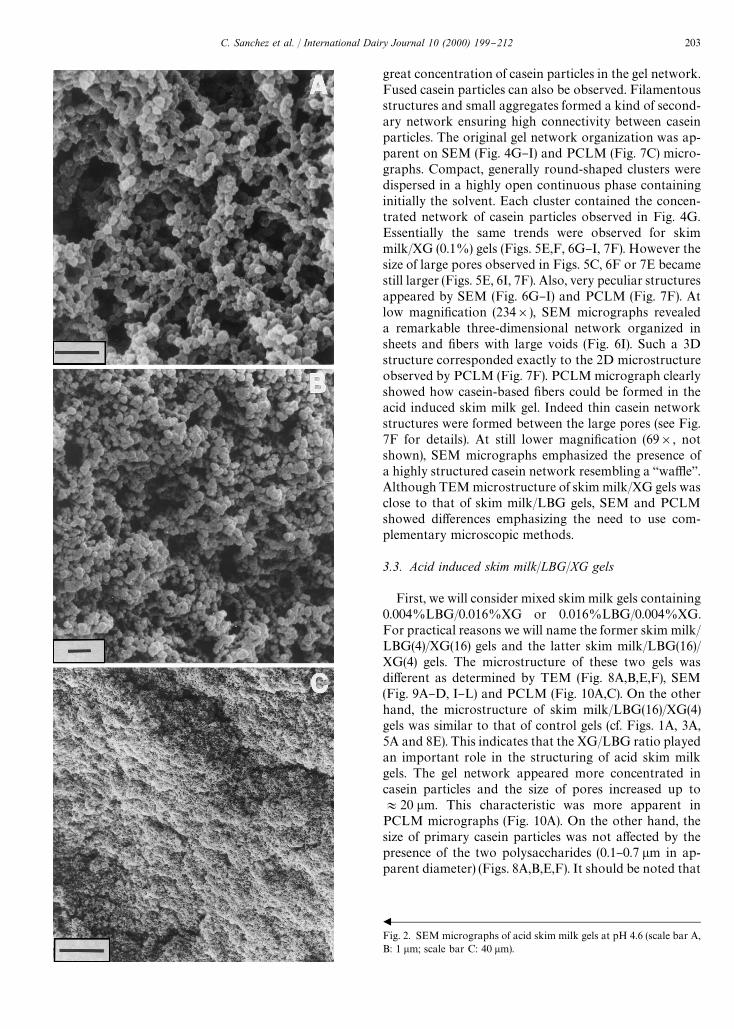
Despite some charging e!ects, SEM micrographs re-vealed the same features as TEM and PCLM, i.e. a ho-mogeneous highly branched porous network of caseinparticles grouped in chains and clusters (Fig. 2A}C). Thediameter of casein particles was in the range 0.1}0.5 lmand diameter of pores was approximately in the range1}3 lm (Fig. 2A,B). These values compared favorablywith the values estimated from TEM micrographs. Thesimultaneous presence of casein particle chains and iso-lated casein particles in the network examined by TEM,as compared to the absence of single casein particles inthe network examined by SEM, could be explained bythe angle of the section plane in the TEM method (Kalabet al., 1976).
3.2. Acid induced skim milk/LBG and skim milk/XG gels
No clear di!erence was observed by TEM between themicrostructure of control acid skim milk gels and that ofbinary gels containing 0.001% (w/w) LBG or XG (Figs.3A, B and 5A,B). The apparent diameter of casein par-ticles and pores did not change with the presence ofpolysaccharides. A detailed observation of the surface of
C. Sanchez et al. / International Dairy Journal 10 (2000) 199}212 201
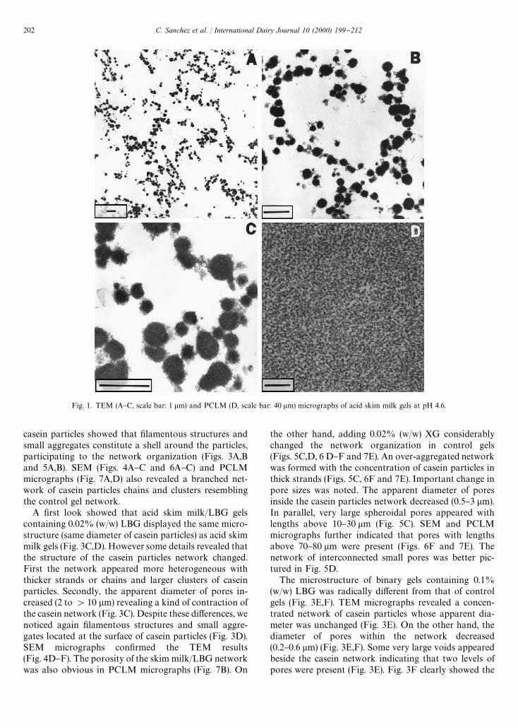
Fig. 1. TEM (A}C, scale bar: 1 lm) and PCLM (D, scale bar: 40 lm) micrographs of acid skim milk gels at pH 4.6.
casein particles showed that "lamentous structures andsmall aggregates constitute a shell around the particles,participating to the network organization (Figs. 3A,Band 5A,B). SEM (Figs. 4A}C and 6A}C) and PCLMmicrographs (Fig. 7A,D) also revealed a branched net-work of casein particles chains and clusters resemblingthe control gel network.
A "rst look showed that acid skim milk/LBG gelscontaining 0.02% (w/w) LBG displayed the same micro-structure (same diameter of casein particles) as acid skimmilk gels (Fig. 3C,D). However some details revealed thatthe structure of the casein particles network changed.First the network appeared more heterogeneous withthicker strands or chains and larger clusters of caseinparticles. Secondly, the apparent diameter of pores in-creased (2 to '10 lm) revealing a kind of contraction ofthe casein network (Fig. 3C). Despite these di!erences, wenoticed again "lamentous structures and small aggre-gates located at the surface of casein particles (Fig. 3D).SEM micrographs con"rmed the TEM results(Fig. 4D}F). The porosity of the skim milk/LBG networkwas also obvious in PCLM micrographs (Fig. 7B). On
the other hand, adding 0.02% (w/w) XG considerablychanged the network organization in control gels(Figs. 5C,D, 6 D}F and 7E). An over-aggregated networkwas formed with the concentration of casein particles inthick strands (Figs. 5C, 6F and 7E). Important change inpore sizes was noted. The apparent diameter of poresinside the casein particles network decreased (0.5}3 lm).In parallel, very large spheroidal pores appeared withlengths above 10}30 lm (Fig. 5C). SEM and PCLMmicrographs further indicated that pores with lengthsabove 70}80 lm were present (Figs. 6F and 7E). Thenetwork of interconnected small pores was better pic-tured in Fig. 5D.
The microstructure of binary gels containing 0.1%(w/w) LBG was radically di!erent from that of controlgels (Fig. 3E,F). TEM micrographs revealed a concen-trated network of casein particles whose apparent dia-meter was unchanged (Fig. 3E). On the other hand, thediameter of pores within the network decreased(0.2}0.6 lm) (Fig. 3E,F). Some very large voids appearedbeside the casein network indicating that two levels ofpores were present (Fig. 3E). Fig. 3F clearly showed the
202 C. Sanchez et al. / International Dairy Journal 10 (2000) 199}212
b
Fig. 2. SEM micrographs of acid skim milk gels at pH 4.6 (scale bar A,B: 1 lm; scale bar C: 40 lm).
great concentration of casein particles in the gel network.Fused casein particles can also be observed. Filamentousstructures and small aggregates formed a kind of second-ary network ensuring high connectivity between caseinparticles. The original gel network organization was ap-parent on SEM (Fig. 4G}I) and PCLM (Fig. 7C) micro-graphs. Compact, generally round-shaped clusters weredispersed in a highly open continuous phase containinginitially the solvent. Each cluster contained the concen-trated network of casein particles observed in Fig. 4G.Essentially the same trends were observed for skimmilk/XG (0.1%) gels (Figs. 5E,F, 6G}I, 7F). However thesize of large pores observed in Figs. 5C, 6F or 7E becamestill larger (Figs. 5E, 6I, 7F). Also, very peculiar structuresappeared by SEM (Fig. 6G}I) and PCLM (Fig. 7F). Atlow magni"cation (234]), SEM micrographs revealeda remarkable three-dimensional network organized insheets and "bers with large voids (Fig. 6I). Such a 3Dstructure corresponded exactly to the 2D microstructureobserved by PCLM (Fig. 7F). PCLM micrograph clearlyshowed how casein-based "bers could be formed in theacid induced skim milk gel. Indeed thin casein networkstructures were formed between the large pores (see Fig.7F for details). At still lower magni"cation (69], notshown), SEM micrographs emphasized the presence ofa highly structured casein network resembling a `wa%ea.Although TEM microstructure of skim milk/XG gels wasclose to that of skim milk/LBG gels, SEM and PCLMshowed di!erences emphasizing the need to use com-plementary microscopic methods.
3.3. Acid induced skim milk/LBG/XG gels
First, we will consider mixed skim milk gels containing0.004%LBG/0.016%XG or 0.016%LBG/0.004%XG.For practical reasons we will name the former skim milk/LBG(4)/XG(16) gels and the latter skim milk/LBG(16)/XG(4) gels. The microstructure of these two gels wasdi!erent as determined by TEM (Fig. 8A,B,E,F), SEM(Fig. 9A}D, I}L) and PCLM (Fig. 10A,C). On the otherhand, the microstructure of skim milk/LBG(16)/XG(4)gels was similar to that of control gels (cf. Figs. 1A, 3A,5A and 8E). This indicates that the XG/LBG ratio playedan important role in the structuring of acid skim milkgels. The gel network appeared more concentrated incasein particles and the size of pores increased up to+20 lm. This characteristic was more apparent inPCLM micrographs (Fig. 10A). On the other hand, thesize of primary casein particles was not a!ected by thepresence of the two polysaccharides (0.1}0.7 lm in ap-parent diameter) (Figs. 8A,B,E,F). It should be noted that
C. Sanchez et al. / International Dairy Journal 10 (2000) 199}212 203

Fig. 3. TEM micrographs of acid skim milk/LBG gels at pH 4.6 containing (w/w) 0.001% (A, B), 0.02% (C, D) and 0.1% (E, F) LBG. Scale bars fromleft to right: 1 lm.
neither skim milk/LBG(4)/XG(16) gels nor skimmilk/LBG(16)/XG(4) gels displayed similar microstruc-tures as binary gels containing about the same LBG orXG, concentration, i.e. 0.02% (w/w).
Acid skim milk/LBG(9)/XG(11) gels containing0.011% (w/w) XG and 0.009% (w/w) LBG were struc-tured by a homogeneous network of casein particleschains and clusters delimiting small pores (Figs. 8C,D,9E, 10B). The gel network was similar to that of controlgels and binary gels containing 0.001% (w/w) polysac-charides. Two general trends were repeated, i.e. the size ofprimary casein particles was not modi"ed by the pres-ence of XG or/and LBG, "lamentous structures andsmall aggregates were located at the surface of caseinparticles contributing to the gel network organization.
SEM micrographs showed original structures in ter-nary gels not observed by TEM or PCLM. Jagged`coral-likea appearance of part of the network was notedfor the three gels (Fig. 9D,H,L). This trend was seeminglymore pronounced for blends containing larger amount ofXG (Fig. 9D). Also, veil-like and "lamentous structureswere observed for all systems (Fig. 9B}L), especially forskim milk/XG(16)/LBG(4) and skim milk/XG(11)/LBG(9) gels, i.e. for gels containing more XG than LBG.
4. Discussion
4.1. Microstructure of control acid milk gels
TEM, SEM and PCLM micrographs showed thatcontrol acid skim milk gels were structured by a bran-ched network of casein particles forming chains andclusters, immobilizing the continuous phase into pores ofvarying (small) sizes. As indicated in the introduction,this acid induced microstructure has been demonstratedmany times in the literature both by TEM, SEM andCSLM. We clearly observed in TEM micrographs thepresence at the surface of casein particles of "lamentousstructures and small aggregates that apparently formeda shell around particles. These structures increase theconnectivity of casein particles and lower their ability tofuse or coalesce. Similar observations were already re-ported (Kalab et al., 1976; Davies et al., 1978; Harwalkar& Kalab, 1980; Modler & Kalab, 1983). It has beendemonstrated that heat treatment applied to milk beforeacidi"cation and gel formation, as in the present study(853C, 30 min), was the cause of the "lamentous struc-tures and small aggregates observed onto casein particles(Kalab et al., 1976). Heat treatment at temperature above
204 C. Sanchez et al. / International Dairy Journal 10 (2000) 199}212

Fig. 4. SEM micrographs of acid skim milk/LBG gels at pH 4.6 containing (w/w) 0.001% (A}C), 0.02% (D}F) and 0.1% (G}I) LBG. All scale barsrepresent 1 lm excepted micrographs C, F and I: 40 lm.
803C induces heat-denaturation of whey proteins * es-pecially b-lactoglobulin* that form a covalent complexwith the i-casein located at the surface of casein micelles(Davies et al., 1978). Such complexes are the basis of"lamentous structures and small aggregates. One impor-tant consequence of the presence of surface whey pro-tein}i-casein complexes is the possibility of bridgingother casein particles by interacting with other denaturedwhey proteins associated with micelles (Lucey & Singh,1998). This promotes the formation of a highly connect-
ive casein network as observed in our study and else-where (Kalab & Harwalkar, 1974; Modler & Kalab,1983; Lucey & Singh, 1998).
4.2. Microstructure of acid milk/polysaccharide gels
The addition of LBG and/or XG in skim milk inducesvarious e!ects depending on the polysaccharide concen-tration. In case of simultaneous addition of LBG andXG, the LBG/XG weight ratio plays an important role.
C. Sanchez et al. / International Dairy Journal 10 (2000) 199}212 205

Fig. 5. TEM micrographs of acid skim milk/XG gels at pH 4.6 containing (w/w) 0.001% (A, B), 0.02% (C, D) and 0.1% (E, F) XG. Scale bars from leftto right: 1 lm.
The in#uence of polysaccharides also depends on thelevel of microstructure under consideration. It was di!er-ent according to whether one considers the primarycasein particles building blocks or the association be-tween particles. The structure of casein particles, includ-ing the size, the shape and the existence of "lamentousstructures/small aggregates at their surface, was not af-fected by polysaccharides. However, LBG and XG con-siderably modi"ed the three-dimensional organization ofthe casein particles gel network.
The microstructure of acid milk gels containing0.001% (w/w) LBG or XG was close to that of controlgels (without added polysaccharides). The microstruc-tural similarities between skim milk and mixed gelsmeans either that LBG or XG have no e!ect on theformation of the casein network or that the used micro-scopical methods are not able to detect such e!ects. It ispossible that small microstructural di!erences betweengels could be occulted by the drastic preparation proced-ure applied to gel samples. On the other hand, the micro-structure of gels containing 0.02% (w/w), and especially0.1% (w/w), LBG and XG appears very di!erent fromthat of control gels. The most striking di!erence was theincrease in the density of casein particles in strands. As
a consequence, the size of pores increased up to tens ofmicrometers. In other words, there is a compaction of thecasein network and an increased porosity of gels at thesupramolecular level. This spongy characteristic appearsin gels containing 0.02% (w/w) polysaccharides, espe-cially with XG. It is greatly enhanced with both 0.1%(w/w) LBG or XG. In the case of skim milk/XG gels,SEM and PCLM micrographs showed highly structuredand open networks containing sheets of casein particlesnetwork. The structure of skim milk/LBG gels was alsoopen but the network was much less organised. Suchmicrostructures have never been reported previously foracid skim milk/polysaccharides gels. In fact, highly openaggregated structures have been described for heat-in-duced b-lactoglobulin gels containing 9.7% or 14.5%protein and 0.9% xanthan gum (Zasypkin, Dumay& Cheftel, 1996). Interestingly, the `sponge-likea struc-ture of skim milk/XG gels containing 0.02% (w/w) XG isvery close to that observed by photon microscopy inpressure-induced b-lactoglobulin gels (18.3% w/w pro-tein) (Zasypkin et al., 1996).
The explanation of skim milk gel microstructure in thepresence of LBG or XG lies obviously in the existenceof milk proteins/polysaccharides interactions. In the
206 C. Sanchez et al. / International Dairy Journal 10 (2000) 199}212

Fig. 6. SEM micrographs of acid skim milk/XG gels at pH 4.6 containing (w/w) 0.001% (A}C), 0.02% (D}F) and 0.1% (G}I) XG. Scale bars from leftto right represent 1 lm excepted micrographs C}I: 40 lm.
general case where repulsive interactions betweenproteins and polysaccharides predominate over attract-ive interactions, macromolecules in solution tend to stayaway from each other giving rise to a mutual exclusion ofaccessible volume for their centers of mass (Syrbe, Bauer& Klostermeyer, 1998). Practically, demixing occurs re-sulting in the preferential concentration of proteins inone phase and of polysaccharides in another phase. Thissegregative-type phase separation is usually named ther-modynamic incompatibility (Albertsson, 1971; Tol-
stoguzov, 1986). Thermodynamic incompatibility at pH'5.5 between milk proteins such as sodium caseinate orwhey proteins and polysaccharides such as xanthan orlocust bean gum is generally very pronounced (Grindrod& Nickerson, 1968; Antonov, Grinberg & Tolstoguzov,1977; Antonov, Grinberg, Zhuravskaya & Tolstoguzov,1982; Schmidt & Smith, 1992; Sanchez, Schmitt,Babak & Hardy, 1997). In the case of mixed systemscontaining colloidal casein micelles and non-adsorbingpolysaccharides, demixing is primarily caused by
C. Sanchez et al. / International Dairy Journal 10 (2000) 199}212 207

Fig. 7. PCLM micrographs of acid skim milk gels at pH 4.6 containing (w/w) 0.001% (A), 0.02% (B) and 0.1% (C) LBG or 0.001% (D), 0.02% (E) and0.1% (F) XG. Scale bars from left to right: 40 lm.
depletion #occulation rather than by thermodynamicincompatibility (Syrbe et al., 1998). This has been demon-strated for instance in skim milk-xanthan (Antonov,Sochinskii & Glotova, 1994), skim milk-carrageenan(Langendor!, Cuvelier, Launay & Parker, 1997), skimmilk-lactic bacteria exopolysaccharides (Tuinier & deKruif, 1998), skim milk-guar gum (Tuinier, ten Groten-huis & de Kruif, 2000) and micellar casein-locust beangum dispersions (Bourriot, Garnier & Doublier, 1999;Schorsch, Jones & Norton, 1999).
The elucidation of the phase separation mechanism isbeyond the scope of this paper. Here it su$ces to postu-late that binary dispersions at pH+6.5 containing0.02% (w/w) or 0.1% (w/w) polysaccharides display sep-arated phases, probably through #occulation depletion,one of them containing concentrated casein micelles andwhey proteins (Bourriot, 1999). During milk acidi"ca-tion, the balance between aggregation/gelation of con-centrated casein micelles and volume exclusion e!ectsresults in highly porous casein particles network as ob-served by TEM, SEM and PCLM. It is worth noting thatsuch sponge-like gel morphologies appear to be a univer-sal feature of phase separated asymmetric mixtures, com-posed of a network-forming component and a #uid
(Tanaka, 1996). One consequence of the important skimmilk/polysaccharides gels porosity was the apparentwhey o!. This in turn may also contribute to the caseinnetwork shrinkage.
The occurrence of two distinct microstructures forskim milk/LBG or skim milk/XG gels could be at-tributed to the di!erent structures, molecular weight andsolvent a$nity of the polysaccharides. These parametersare known to a!ect to a large extent protein}polysac-charide phase separation (Grinberg & Tolstoguzov,1997). In particular, LBG is a neutral polysaccharide insolution and XG is polyanionic. This could have impor-tant consequences in the structuring of skim milk gels asdemonstrated by small-angle neutron scattering on ther-mally induced BSA gels containing hydroxy ethyl cellu-lose (HEC, neutral polysaccharide) or carboxy methylcellulose (CMC, polyanionic polysaccharide) (Renard,BoueH & Lefebvre, 1998). An important "nding in thelatter study was that HEC a!ects BSA aggregation/gela-tion purely through volume exclusion e!ects, whilstCMC a!ects BSA aggregation/gelation through volumeexclusion e!ects and direct interaction with BSA,especially at the isoelectric point of protein. Assumingthat similar kinds of interactions occur in skim
208 C. Sanchez et al. / International Dairy Journal 10 (2000) 199}212
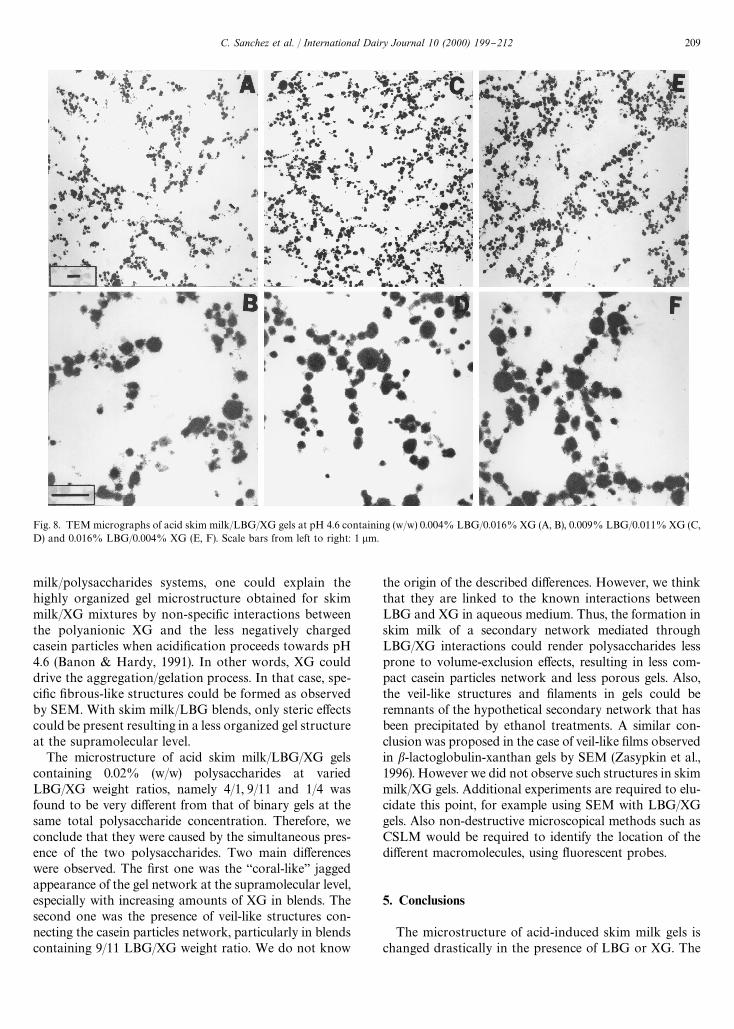
Fig. 8. TEM micrographs of acid skim milk/LBG/XG gels at pH 4.6 containing (w/w) 0.004% LBG/0.016% XG (A, B), 0.009% LBG/0.011% XG (C,D) and 0.016% LBG/0.004% XG (E, F). Scale bars from left to right: 1 lm.
milk/polysaccharides systems, one could explain thehighly organized gel microstructure obtained for skimmilk/XG mixtures by non-speci"c interactions betweenthe polyanionic XG and the less negatively chargedcasein particles when acidi"cation proceeds towards pH4.6 (Banon & Hardy, 1991). In other words, XG coulddrive the aggregation/gelation process. In that case, spe-ci"c "brous-like structures could be formed as observedby SEM. With skim milk/LBG blends, only steric e!ectscould be present resulting in a less organized gel structureat the supramolecular level.
The microstructure of acid skim milk/LBG/XG gelscontaining 0.02% (w/w) polysaccharides at variedLBG/XG weight ratios, namely 4/1, 9/11 and 1/4 wasfound to be very di!erent from that of binary gels at thesame total polysaccharide concentration. Therefore, weconclude that they were caused by the simultaneous pres-ence of the two polysaccharides. Two main di!erenceswere observed. The "rst one was the `coral-likea jaggedappearance of the gel network at the supramolecular level,especially with increasing amounts of XG in blends. Thesecond one was the presence of veil-like structures con-necting the casein particles network, particularly in blendscontaining 9/11 LBG/XG weight ratio. We do not know
the origin of the described di!erences. However, we thinkthat they are linked to the known interactions betweenLBG and XG in aqueous medium. Thus, the formation inskim milk of a secondary network mediated throughLBG/XG interactions could render polysaccharides lessprone to volume-exclusion e!ects, resulting in less com-pact casein particles network and less porous gels. Also,the veil-like structures and "laments in gels could beremnants of the hypothetical secondary network that hasbeen precipitated by ethanol treatments. A similar con-clusion was proposed in the case of veil-like "lms observedin b-lactoglobulin-xanthan gels by SEM (Zasypkin et al.,1996). However we did not observe such structures in skimmilk/XG gels. Additional experiments are required to elu-cidate this point, for example using SEM with LBG/XGgels. Also non-destructive microscopical methods such asCSLM would be required to identify the location of thedi!erent macromolecules, using #uorescent probes.
5. Conclusions
The microstructure of acid-induced skim milk gels ischanged drastically in the presence of LBG or XG. The
C. Sanchez et al. / International Dairy Journal 10 (2000) 199}212 209

210 C. Sanchez et al. / International Dairy Journal 10 (2000) 199}212

b
Fig. 9. SEM micrographs of acid skim milk/LBG/XG gels at pH 4.6 containing (w/w) 0.004% LBG/0.016% XG (A}D), 0.009% LBG/0.011% XG (E}H)and 0.016% LBG/0.004% XG (I}L). Scale bars from left to right (A}K) represent 1 lm excepted micrograph G (5 lm); scale bars (D, H, L): 40 lm.
Fig. 10. PCLM micrographs of acid skim milk gels at pH 4.6 contain-ing (w/w) 0.004% LBG/0.016% XG (A), 0.009% LBG/0.011% XG (B)and 0.016% LBG/0.004% XG (C). Scale bar: 40 lm.
origin of the structure changes can be traced primarily tothe existence of repulsive casein-polysaccharide interac-tions that promote demixing phenomena in initial disper-sions. The balance between demixing, aggregation/
gelation of caseins and possibly non-speci"c casein}poly-saccharide interactions ultimately determines the micro-structure of gels. The present study demonstrates thatvery original microstructures can be obtained dependingon the nature of the polysaccharide, its concentration,and the polysaccharide weight ratio in the case of ternarymixtures. This could open new opportunities in tailoringmilk protein-based gels.
Acknowledgements
The authors thank CONACYT (Mexico) and the`Ministere Franc7 ais des A!aires Etrangeresa (France) forthe Ph.D. grant of Ms R.Zuniga-Lopez.
References
Aguilera, J. -M., & Stanley, D. W. (1990). Microstructural principles offood processing and engineering. London and New York: ElsevierApplied Science.
Albertsson, P. -As . (1971). Partition of cell particles and macromolecules.New York: Wiley-Interscience.
Antonov, Yu. A., Grinberg, V. Ya., & Tolstoguzov, V. B. (1977). Phaseequilibria in water-protein-polysaccharide systems II. Water}casein}neutral polysaccharide systems. Colloid and Polymer Science,255, 937}947.
Antonov, Yu. A., Grinberg, V. Ya., Zhuravskaya, N. A., & Tolstoguzov,V. B. (1982). Concentration of the proteins of skimmed milk bymembraneless isobaric osmosis. Carbohydrate Polymers, 2, 81}90.
Antonov, Yu. A., Sochinskii, A. A., & Glotova, Yu. K. (1994). Thermo-dynamic compatibility of skim milk proteins with microbialpolysaccharides in aqueous media. Applied Biochemistry and Micro-biology, 30, 760}765.
Banon, S., & Hardy, J. (1991). Study of acid milk coagulation by anoptical method using light re#ection. Journal of Dairy Research, 58,75}84.
Bourriot, S., Garnier, C., & Doublier, J. -L. (1999). Phase separation,rheology and microstructure of micellar casein-guar gum mixtures.Food Hydrocolloids, 13, 43}49.
Bourriot, S. (1999). Comportement de phase, rhe&ologie et ultrastructuredes me& langes case& ines micellaires/polyosides. These de Doctorat,ENSIA, Massy, France.
Bremer, L.G.B. (1992). Fractal aggregation in relation to formation andproperties of particle gels. Ph.D. thesis, Wageningen AgriculturalUniversity, Wageningen, The Netherlands.
Brooker, B. E., & Wells, K. (1984). Preparation of dairy products forscanning electron microscopy: etching of epoxy resin-embeddedmaterial. Journal of Dairy Research, 51, 605}613.
Cuvelier, G., & Launay, B. (1986). Viscoelastic properties of xanthan-carob mixed gels. In G. O. Phillips, & P. A. Williams, Gums andStabilisers for the Food Industry, Vol. 3 (pp. 147}158). Oxford: IRLPress.
Davies, F. L., Shankar, P. A., Brooker, B. E., & Hobbs, D. G. (1978).A heat-induced change in the ultrastructure of milk and its e!ecton gel formation in yoghurt. Journal of Dairy Research,41, 131}135.
C. Sanchez et al. / International Dairy Journal 10 (2000) 199}212 211
De Kruif, C. G. (1997). Skim milk acidi"cation. Journal of Colloid andInterface Science, 185, 19}25.
Gastaldi, E., Lagaude, A., & Tarodo De La Fuente, B. (1996). Micellartransition state in casein between pH 5.5 and 5.0. Journal of FoodScience 61, 59-64, 68.
Goycoolea, F. M., Richardson, R. K., Gidley, M. J., & Morris, E. R.(1995). Stoichiometry and conformation of xanthan in synergisticgelation with locust bean gum or konjac glucomannan: Evidence forheterotypic binding. Macromolecules, 28, 8308}8320.
Grinberg, V. Ya., & Tolstoguzov, V. B. (1997). Thermodynamic incom-patibility of proteins and polysaccharides in solutions. Food Hydro-colloids, 11, 145}158.
Grindrod, J., & Nickerson, T. A. (1968). E!ect of various gums onskimmed milk and puri"ed milk proteins. Journal of Dairy Science,51, 834}841.
Harwalkar, V. R., & Kalab, M. (1980). Milk gel structure. XI. Electronmicroscopy of glucono-*-lactone-induced skim milk gels. Journal ofTexture Studies, 11, 35}49.
Harwalkar, V. R., & Kalab, M. (1981). E!ect of acidulants and temper-ature on microstructure, "rmness and susceptibility to syneresis ofskim milk gels. Scanning Electron Microscopy, III, 503}513.
Hassan, A. N., Franck, J. F., Farmer, M. A., Schmidt, K. A., & Shalabi,S. I. (1995). Formation of yogurt microstructure and three-dimen-sional visualization as determined by Confocal Scanning LaserMicroscopy. Journal of Dairy Science, 78, 2629}2636.
Heertje, I., Visser, J., & Smits, P. (1985). Structure formation in acidmilk gels. Food Microstructure, 4, 267}277.
Hood, L. F., & Allen, J. E. (1977). Ultrastructure of carrageenan-milksols and gels. Journal of Food Science, 42, 1062}1065.
Kalab, M., & Harwalkar, V. R. (1973). Milk gel structure. I. Applicationof Scanning Electron Microscopy to milk and other food gels.Journal of Dairy Science, 56, 835}842.
Kalab, M., & Harwalkar, V. R. (1974). Milk gel structure. II. Relationbetween "rmness and ultrastructure of heat-induced skim-milk gelscontaining 40}60% total solids. Journal of Dairy Research, 41,131}135.
Kalab, M., Emmons, D. B., & Sargant, A. G. (1975). Milk-gel structure.IV. Microstructure of yoghurts in relation to the presence of thick-ening agents. Journal of Dairy Research, 42, 453}458.
Kalab, M., Emmons, D. B., & Sargant, A. G. (1976). Milk-gel struc-ture.V. Microstructure of yoghurt as related to the heating of milk.Milchwissenschaft, 31, 402}408.
Kalab, M., Allan-Wojtas, P., & Phipps-Todd, B. (1983). Developmentof microstructure in set-style nonfat yoghurt * A review. FoodMicrostructure, 2, 51}66.
Langendor!, V., Cuvelier, G., Launay, B., & Parker, A. (1997). Gelationand #occulation of casein micelle/carrageenan mixtures. FoodHydrocolloids, 11, 35}40.
Lucey, J. A., van Vliet, T., Grolle, K., Geurts, T., & Walstra, P. (1997).Properties of acid casein gels made by acidi"cation with glucono-d-lactone. 2. Syneresis, permeability and microstructural properties.International Dairy Journal, 7, 389}397.
Lucey, J. A., & Singh, H. (1998). Formation and physical propertiesof acid milk gels: a review. Food Research International,30, 529}542.
Modler, H. W., & Kalab, M. (1983). Microstructure of yogurt stabilizedwith milk proteins. Journal of Dairy Science, 66, 430}437.
Parnell-Clunies, E., Yakuda, Y., & Smith, A. K. (1987). Microstructureof yogurt as a!ected by heat treatment of milk. Milchwissenschaft,42, 413}417.
Renard, D., BoueH , F., & Lefebvre, J. (1998). Solution and gelationproperties of protein-polysaccharide mixtures: Signature by small-angle neutron scattering and rheology. In G. O. Phillips, & P. A.Williams, Gums and Stabilisers for the Food Industry, Vol. 9 (pp.189}201). Oxford: IRL Press.
Roefs, S.P.F.M. (1986). Structure of acid casein gels. Ph.D. thesis,Wageningen Agricultural University, Wageningen, The Nether-lands.
Sanchez, C., Schmitt, C., Babak, V., & Hardy, J. (1997). Rheology ofwhey protein isolate-xanthan mixed solutions and gels. E!ect of pHand xanthan concentration. Nahrung, 41, 336}343.
Schmidt, K. A., & Smith, D. E. (1992). Milk reactivity of gum and milkprotein solutions. Journal of Dairy Science, 75, 3290}3295.
Schorsch, C., Jones, M. G., & Norton, I. T. (1999). Thermodynamicincompatibility and microstructure of milk protein/locust beangum/sucrose. Food Hydrocolloids, 13, 89}99.
Syrbe, A., Bauer, W. J., & Klostermeyer, H. (1998). Polymerscience concepts in dairy systems* an overview of milk protein andfood hydrocolloid interaction. International Dairy Journal, 8,179}193.
Tamime, A. Y., Kalab, M., Allan-Wojtas, P., & Davies, G. (1984).Microstructure of set-style yoghurt manufactured from cow's milkforti"ed by various methods. Food Microstructure, 3, 83}92.
Tamime, A. Y., Barrantes, E., & Sword, A. M. (1996). The e!ect of starchbased fat substitutes on the microstructure of set-style yogurt madefrom reconstituted skimmed milk powder. Journal of the Society ofDairy Technology, 49, 1}10.
Tanaka, H. (1996). Universality of viscoelastic phase separation indynamically asymmetric #uid mixtures. Physical Review Letters, 76,787}790.
Tolstoguzov, V. B. (1986). Functional properties of protein-polysac-charide mixtures. In J. R. Mitchell, & D. A. Ledward, Functionalproperties of macromolecules (pp. 385}415). London: Elsevier Ap-plied Science Publishers (Chapter 9).
Tuinier, R., & de Kruif, C. G. (1998). Depletion #occulation of caseinmicelles induced by exocellular polysaccharide of a lactic acidbacterium. In P. A. Williams, Gums and Stabilizers for the FoodIndustry, Vol. 9. Oxford: IRL Press.
Tuinier, R., ten Grotenhuis, E., & de Kruif, C. G. (2000). The e!ect ofdepolymerised guar gum on the stability of skim milk. Food Hydro-colloids, 14, 1}7.
Zasypkin, D. V., Dumay, E., & Cheftel, J. C. (1996). Pressure- andheat-induced gelation of mixed b-lactoglobulin/xanthan solutions.Food Hydrocolloids, 10, 203}211.
212 C. Sanchez et al. / International Dairy Journal 10 (2000) 199}212