Glycosaminoglycan modification of neuropilin-1 modulates VEGFR2 signaling
11
Glycosaminoglycan modification of neuropilin-1 modulates VEGFR2 signaling Yasunori Shintani 1,4 , Seiji Takashima 1,4, *, Yoshihiro Asano 1 , Hisakazu Kato 1 , Yulin Liao 1 , Satoru Yamazaki 2 , Osamu Tsukamoto 1 , Osamu Seguchi 1,2 , Hiroyuki Yamamoto 1,2 , Tomi Fukushima 2 , Kazuyuki Sugahara 3,5 , Masafumi Kitakaze 2, * and Masatsugu Hori 1 1 Department of Cardiovascular Medicine, Osaka University Graduate School of Medicine, Suita, Osaka, Japan, 2 Cardiovascular Division of Medicine, National Cardiovascular Center, Suita, Japan and 3 Department of Biochemistry, Kobe Pharmaceutical University, Higashinada-ku, Kobe, Japan Neuropilin-1 (NRP1) is a co-receptor for vascular endo- thelial growth factor (VEGF) that enhances the angiogenic signals cooperatively with VEGFR2. VEGF signaling is essential for physiological and pathological angiogenesis through its effects on vascular endothelial cells (ECs) and smooth muscle cells (SMCs), but the mechanisms coordi- nating this response are not well understood. Here we show that a substantial fraction of NRP1 is proteoglycan modified with either heparan sulfate or chondroitin sulfate on a single conserved Ser. The composition of the NRP1 glycosaminoglycan (GAG) chains differs between ECs and SMCs. Glycosylation increased VEGF binding in both cell types, but the differential GAG composition of NRP1 med- iates opposite responsiveness to VEGF in ECs and SMCs. Finally, NRP1 expression and its GAG modification post- transcriptionally regulate VEGFR2 protein expression. These findings indicate that GAG modification of NRP1 plays a critical role in modulating VEGF signaling, and may provide new insights into physiological and patholo- gical angiogenesis. The EMBO Journal (2006) 25, 3045–3055. doi:10.1038/ sj.emboj.7601188; Published online 8 June 2006 Subject Categories: signal transduction; proteins Keywords: glycosaminoglycan; neuropilin-1; VEGF; VEGFR2 Introduction Neuropilin-1 (NRP1) was originally discovered as a co-recep- tor for semaphorin-3A (Sema3A), an axon repellent factor (Kolodkin et al, 1997). However, NRP1 also acts as a co- receptor for vascular endothelial growth factor (VEGF), a molecule with no sequence or structural homology to Sema3A (Soker et al, 1998). VEGF (also referred as VEGF- A) is an essential factor promoting both embryonic angiogen- esis and postnatal neovascularization. Additionally, VEGF plays a significant role in causing pathological angiogenesis associated with tumor growth, age-related macular degenera- tion, diabetic retinopathy, and other conditions (Ferrara et al, 2003). Indeed, a blocking anti-VEGF antibody that disrupts VEGF signaling is a promising anticancer therapy currently in development (Hurwitz et al, 2004). VEGF has three receptors, VEGF receptor 1 and 2 (VEGFR1, VEGFR2), and neuropilin (Veikkola and Alitalo, 1999; Ferrara et al, 2003). VEGFR2 is the primary receptor mediating the angiogenic activity of VEGF (Shalaby et al, 1995; Ferrara et al, 2003), and NRP1 functions as a co- receptor to enhance VEGFR2 signaling (Soker et al, 1998). Indeed, genetic ablation of NRP1 leads to severely impaired vascular development (Kawasaki et al, 1999; Takashima et al, 2002; Gu et al, 2003), indicating that NRP1 is essential for VEGF-mediated angiogenesis. In addition to promoting angiogenesis, VEGF is now thought to be required for the maintenance and stabilization of mature blood vessels (Zachary, 2001; Saint-Geniez and D’Amore, 2004). Signaling through VEGFR2, VEGF induces not only endothelial cell (EC) proliferation but also cell survival (Gerber et al, 1998), and the loss of VEGF signals in the choroidal endothelium is one factor promoting age- related macular degeneration (Blaauwgeers et al, 1999). Smooth muscle cells (SMCs), another important component of the vessel wall, also express both NRP1 (Kitsukawa et al, 1995; Kawasaki et al, 1999) and VEGFR2 (Grosskreutz et al, 1999; Ishida et al, 2001). However, SMCs in mature vessels typically do not respond to VEGF signals except in certain conditions such as atherosclerosis (Carmeliet, 2003; Jain, 2003; Khurana et al, 2004). Therefore, we wished to identify the mechanism(s) responsible for different cellular responses to VEGF in ECs and SMCs. In this study, we demonstrate that a substantial fraction of NRP1 is proteoglycan modified with either heparan sulfate (HS) or chondroitin sulfate (CS) attached to a single con- served Ser. The type of NRP1 glycosaminoglycan (GAG) chain modification differs between ECs and SMCs. Finally, we show that the type of NRP1 GAG modification critically and differ- entially modulates VEGFR2 signals in SMCs and ECs. Results A substantial fraction of NRP1 is proteoglycan modified with HS or CS The differential responsiveness of ECs and SMCs to VEGF could be explained by a number of factors, and we initially investigated the ability of VEGF to bind to these cells. When human coronary artery smooth muscle cells (CASMCs) were Received: 13 February 2006; accepted: 16 May 2006; published online: 8 June 2006 *Corresponding authors. S Takashima, Department of Cardiovascular Medicine, Osaka University Graduate School of Medicine, 2-2 Yamadaoka, Suita, Osaka 565-0871, Japan. Tel.: þ 816 6879 3472; Fax: þ 816 6879 3473; E-mail: [email protected] or M Kitakaze, Cardiovascular Division of Medicine, National Cardiovascular Center, Suita, Japan. E-mail: [email protected] 4 These authors contributed equally to this work 5 Present address: Laboratory of Proteoglycan Signaling and Therapeutics, Graduate School of Life Science, Hokkaido University, Frontier Research Center for Post-genomic Science and Technology, Sapporo, Japan The EMBO Journal (2006) 25, 3045–3055 | & 2006 European Molecular Biology Organization | All Rights Reserved 0261-4189/06 www.embojournal.org & 2006 European Molecular Biology Organization The EMBO Journal VOL 25 | NO 13 | 2006 EMBO THE EMBO JOURNAL THE EMBO JOURNAL 3045
-
Upload
independent -
Category
Documents
-
view
0 -
download
0
Transcript of Glycosaminoglycan modification of neuropilin-1 modulates VEGFR2 signaling

Glycosaminoglycan modification of neuropilin-1modulates VEGFR2 signaling
Yasunori Shintani1,4, Seiji Takashima1,4,*,Yoshihiro Asano1, Hisakazu Kato1,Yulin Liao1, Satoru Yamazaki2, OsamuTsukamoto1, Osamu Seguchi1,2, HiroyukiYamamoto1,2, Tomi Fukushima2, KazuyukiSugahara3,5, Masafumi Kitakaze2,*and Masatsugu Hori1
1Department of Cardiovascular Medicine, Osaka University GraduateSchool of Medicine, Suita, Osaka, Japan, 2Cardiovascular Division ofMedicine, National Cardiovascular Center, Suita, Japan and3Department of Biochemistry, Kobe Pharmaceutical University,Higashinada-ku, Kobe, Japan
Neuropilin-1 (NRP1) is a co-receptor for vascular endo-
thelial growth factor (VEGF) that enhances the angiogenic
signals cooperatively with VEGFR2. VEGF signaling is
essential for physiological and pathological angiogenesis
through its effects on vascular endothelial cells (ECs) and
smooth muscle cells (SMCs), but the mechanisms coordi-
nating this response are not well understood. Here we
show that a substantial fraction of NRP1 is proteoglycan
modified with either heparan sulfate or chondroitin sulfate
on a single conserved Ser. The composition of the NRP1
glycosaminoglycan (GAG) chains differs between ECs and
SMCs. Glycosylation increased VEGF binding in both cell
types, but the differential GAG composition of NRP1 med-
iates opposite responsiveness to VEGF in ECs and SMCs.
Finally, NRP1 expression and its GAG modification post-
transcriptionally regulate VEGFR2 protein expression.
These findings indicate that GAG modification of NRP1
plays a critical role in modulating VEGF signaling, and
may provide new insights into physiological and patholo-
gical angiogenesis.
The EMBO Journal (2006) 25, 3045–3055. doi:10.1038/
sj.emboj.7601188; Published online 8 June 2006
Subject Categories: signal transduction; proteins
Keywords: glycosaminoglycan; neuropilin-1; VEGF; VEGFR2
Introduction
Neuropilin-1 (NRP1) was originally discovered as a co-recep-
tor for semaphorin-3A (Sema3A), an axon repellent factor
(Kolodkin et al, 1997). However, NRP1 also acts as a co-
receptor for vascular endothelial growth factor (VEGF),
a molecule with no sequence or structural homology to
Sema3A (Soker et al, 1998). VEGF (also referred as VEGF-
A) is an essential factor promoting both embryonic angiogen-
esis and postnatal neovascularization. Additionally, VEGF
plays a significant role in causing pathological angiogenesis
associated with tumor growth, age-related macular degenera-
tion, diabetic retinopathy, and other conditions (Ferrara et al,
2003). Indeed, a blocking anti-VEGF antibody that disrupts
VEGF signaling is a promising anticancer therapy currently
in development (Hurwitz et al, 2004).
VEGF has three receptors, VEGF receptor 1 and 2
(VEGFR1, VEGFR2), and neuropilin (Veikkola and Alitalo,
1999; Ferrara et al, 2003). VEGFR2 is the primary receptor
mediating the angiogenic activity of VEGF (Shalaby et al,
1995; Ferrara et al, 2003), and NRP1 functions as a co-
receptor to enhance VEGFR2 signaling (Soker et al, 1998).
Indeed, genetic ablation of NRP1 leads to severely impaired
vascular development (Kawasaki et al, 1999; Takashima et al,
2002; Gu et al, 2003), indicating that NRP1 is essential for
VEGF-mediated angiogenesis.
In addition to promoting angiogenesis, VEGF is now
thought to be required for the maintenance and stabilization
of mature blood vessels (Zachary, 2001; Saint-Geniez and
D’Amore, 2004). Signaling through VEGFR2, VEGF induces
not only endothelial cell (EC) proliferation but also cell
survival (Gerber et al, 1998), and the loss of VEGF signals
in the choroidal endothelium is one factor promoting age-
related macular degeneration (Blaauwgeers et al, 1999).
Smooth muscle cells (SMCs), another important component
of the vessel wall, also express both NRP1 (Kitsukawa et al,
1995; Kawasaki et al, 1999) and VEGFR2 (Grosskreutz et al,
1999; Ishida et al, 2001). However, SMCs in mature vessels
typically do not respond to VEGF signals except in certain
conditions such as atherosclerosis (Carmeliet, 2003; Jain,
2003; Khurana et al, 2004). Therefore, we wished to identify
the mechanism(s) responsible for different cellular responses
to VEGF in ECs and SMCs.
In this study, we demonstrate that a substantial fraction of
NRP1 is proteoglycan modified with either heparan sulfate
(HS) or chondroitin sulfate (CS) attached to a single con-
served Ser. The type of NRP1 glycosaminoglycan (GAG) chain
modification differs between ECs and SMCs. Finally, we show
that the type of NRP1 GAG modification critically and differ-
entially modulates VEGFR2 signals in SMCs and ECs.
Results
A substantial fraction of NRP1 is proteoglycan modified
with HS or CS
The differential responsiveness of ECs and SMCs to VEGF
could be explained by a number of factors, and we initially
investigated the ability of VEGF to bind to these cells. When
human coronary artery smooth muscle cells (CASMCs) wereReceived: 13 February 2006; accepted: 16 May 2006; publishedonline: 8 June 2006
*Corresponding authors. S Takashima, Department of CardiovascularMedicine, Osaka University Graduate School of Medicine, 2-2Yamadaoka, Suita, Osaka 565-0871, Japan. Tel.: þ 816 6879 3472;Fax: þ 816 6879 3473; E-mail: [email protected] orM Kitakaze, Cardiovascular Division of Medicine, NationalCardiovascular Center, Suita, Japan. E-mail: [email protected] authors contributed equally to this work5Present address: Laboratory of Proteoglycan Signaling andTherapeutics, Graduate School of Life Science, Hokkaido University,Frontier Research Center for Post-genomic Science and Technology,Sapporo, Japan
The EMBO Journal (2006) 25, 3045–3055 | & 2006 European Molecular Biology Organization | All Rights Reserved 0261-4189/06
www.embojournal.org
&2006 European Molecular Biology Organization The EMBO Journal VOL 25 | NO 13 | 2006
EMBO
THE
EMBOJOURNAL
THE
EMBOJOURNAL
3045
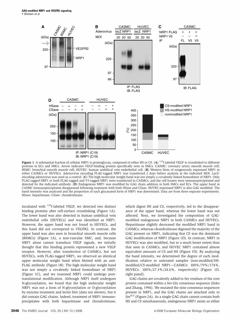
incubated with 125I-labeled VEGF, we detected two distinct
binding proteins after cell-surface crosslinking (Figure 1A).
The lower band was also detected in human umbilical vein
endothelial cells (HUVECs) and was identified as NRP1.
However, the upper band was not found in HUVECs, and
this band did not correspond to VEGFR2. In contrast, the
upper band was also seen in bronchial smooth muscle cells
(BSMCs) (Figure 1A), a non-vascular SMC, and, because
NRP1 alone cannot transduce VEGF signals, we initially
thought that this binding protein represented a new VEGF
receptor. However, after transfection of CASMCs, but not
HUVECs, with FLAG-tagged NRP1, we observed an identical
upper molecular weight band when blotted with an anti-
FLAG antibody (Figure 1B). The high molecular weight band
was not simply a covalently linked homodimer of NRP1
(Figure 1C), and we reasoned NRP1 could undergo post-
translational modification. Although NRP1 itself undergoes
N-glycosylation, we found that the high molecular weight
NRP1 was not a form of N-glycosylation or O-glycosylation
by enzyme treatment and lectin blot (data not shown), but it
did contain GAG chains. Indeed, treatment of NRP1 immuno-
precipitates with both heparitinase and chondroitinase,
which digest HS and CS, respectively, led to the disappear-
ance of the upper band, whereas the lower band was not
affected. Next, we investigated the composition of GAG-
modified endogenous NRP1 in both CASMCs and HUVECs.
Heparitinase slightly decreased the modified NRP1 band in
CASMCs, whereas chondroitinase digested the majority of the
GAG present on NRP1, indicating that CS was the dominant
GAG modification of NRP1 (Figure 1D). In contrast, NRP1 in
HUVECs was also modified, but to a much lesser extent than
that seen in CASMCs, and HUVEC NRP1 contained almost
equivalent amounts of CS and HS (Figure 1D). By analyzing
the band intensity, we determined the degree of each mod-
ification relative to untreated samples (non-modified/HS-
modified/CS-modified NRP1—CASMCs: 100%/79%/174%,
HUVECs: 100%/27.3%/24.6%, respectively) (Figure 1D,
right panel).
GAG chains are covalently added to Ser residues of the core
protein contained within a Ser-Gly consensus sequence (Esko
and Zhang, 1996). We mutated the nine consensus sequences
present in NRP1, and the GAG chains are attached only to
Ser612 (Figure 2A). As a single GAG chain cannot contain both
HS and CS simultaneously, endogenous NRP1 exists as either
BSM
C
A
CAS
MC
HU
VEC
(kDa)
NRP1
VEGFR2220
97
HSaseCSase
CASMC HUVECD
IP: NRP1 (C19)IB: NRP1 (C19)
250
150
100
(kDa)
CASMC HUVEC
50
100
150
200
250(%
)
0
400
350
300
nonmodified NRP1HS-modified NRP1CS-modified NRP1
IB: FLAG
C
220
97
CASMC
(kDa)
NRP1 FLAGNRP1 V5IP FL V5 V5
220
97
66
MOI
IP: FLAGIB: FLAG
B CASMC HUVEC
(kDa)
Adenovirus lacZ NRP1 lacZ NRP1
20 20 50 20 20 50
Figure 1 A substantial fraction of cellular NRP1 is proteoglycan, composed of either HS or CS. (A) 125I-labeled VEGF is crosslinked to differentproteins in ECs and SMCs. Arrow indicates VEGF-binding protein specifically seen in SMCs. CASMC: coronary artery smooth muscle cell;BSMC: bronchial smooth muscle cell; HUVEC: human umbilical vein endothelial cell. (B) Western blots of exogenously expressed NRP1 ineither CASMCs or HUVECs. Adenovirus encoding FLAG-tagged NRP1 was transfected 2 days before analysis at the indicated MOI. LacZ-encoding adenovirus was used as a control. (C) The high molecular weight band was not simply a covalently linked homodimer of NRP1. OnlyFLAG-tagged NRP1 or both FLAG-tagged and V5-tagged NRP1 were transfected in CASMCs, and the cell lysates were immunoprecipitated anddetected by the indicated antibody. (D) Endogenous NRP1 was modified by GAG chain addition in both SMCs and ECs. The upper band inCASMC immunoprecipitates disappeared following treatment with both HSase and CSase. HUVEC-expressed NRP1 is also GAG modified. Theband intensity was analyzed and the proportion of each glycanated form of NRP1 was determined. Data are from three separate experiments.HSase: heparitinase; CSase: chondroitinase.
GAG-modified NRP1 and VEGFR2 signalingY Shintani et al
The EMBO Journal VOL 25 | NO 13 | 2006 &2006 European Molecular Biology Organization3046

an HS proteoglycan or CS proteoglycan but not as a hybrid
proteoglycan. Moreover, as shown in Figure 1D, non-mod-
ified NRP1 (130 kDa, the core protein) was always detected
in both CASMCs and HUVECs. Ser612 is located in the bridge
region between the b1b2 and MAM domains of NRP1
(Figure 2D), and multiple sequence alignments suggested
that Ser612 was remarkably conserved among vertebrates
(Figure 2B), and the peptide sequence around Ser612 was
also well conserved, especially the acidic amino acids that are
important for HS attachment (Esko and Zhang, 1996). NRP2,
a mammalian homolog of NRP1, does not have this con-
served Ser residue, and adenovirus-mediated expression of
NRP2 in CASMCs demonstrated that NRP2 is not GAG
modified (Figure 2C).
GAG modification of NRP1 enhances VEGF binding
Addition of both chondroitinase and heparitinase to culture
medium completely digests all GAGs attached to other core
proteins on the cell surface, and this would dramatically
complicate the interpretation of any experiments using this
technique. Therefore, to further investigate the function of
the GAG of only NRP1, we used RNAi to knock down
endogenous NRP1 while expressing mutant NRP1. We de-
signed an siRNA, named N-G, targeting the GAG attachment
site of NRP1 (Figure 2D). We further generated two NRP1-
encoding adenovirus constructs: NRP1 S612A, in which
Ser612 was replaced by Ala612 and there was a three-base
mismatch with N-G siRNA; and NRP1 WT0, which contains
the glycan accepting residue but had a four-base mismatch
with N-G siRNA (Figure 2D). In cells transfected with N-G
siRNA, transfection of both adenovirus constructs led to the
expression of the appropriate NRP1 molecules. We confirmed
that addition of FLAG tag to NRP1 does not affect VEGF
binding (data not shown) and that the mutation itself (NRP1
S612A) did not change VEGF binding to the core protein of
NRP1 (Figure 2E).
We next examined the ability of VEGF to bind to experi-
mentally replaced NRP1 in both SMCs and ECs. Transfection
of both N-G siRNA and equal multiplicity of infection (MOI)
adenoviral constructs successfully replaced endogenous
NRP1 with either the GAG-acceptor (NRP1 WT0) or mutated
(NRP1 S612A) NRP1 (Figure 3A). Throughout these experi-
ments, MOIs were used to generate NRP1 WT0 or S612A
expression levels comparable to endogenous NRP1. To deter-
mine whether GAG modifications affect the ability of NRP1 to
bind VEGF, we measured the binding of 125I-labeled VEGF to
FLAG-tagged NRP1 in these cells. After incubation with 125I-
labeled VEGF, cell lysates were immunoprecipitated with an
Figure 2 (A) NRP1 is GAG modified on a single Ser612 residue. CASMCs were transfected with adenoviral vectors encoding WT or S612Amutant NRP1. NRP1 S612A is not GAG modified. (B) Multiple alignments of NRP1 from different species. Ser612 is highly conserved amongvertebrates. (C) NRP2, an NRP family member, is not GAG modified. (D) Design of siRNA and adenovirus constructs. Ser612 exists in the bridgeregion between the b1b2 and MAM domains. (E) Replacement of Ser612 by Ala612 of NRP1 did not change binding to VEGF. Cos7 cells weretransfected with either NRP1 WT0 or S612A expression vector and preincubated with heparitinase (1.5 mU/ml), heparinase (1.5 mU/ml), andchondroitinase (20 mU/ml) in the culture medium at 371C for 2 h to make NRP1 non-GAG form. After incubation with 125I-labeled VEGF for30 min at room temperature, cell lysates were immunoprecipitated by anti-NRP1 antibody, and the bound radioactivity was quantitated usinga gamma counter. Data are from three independent experiments. For panel E, error bars represent s.e.
GAG-modified NRP1 and VEGFR2 signalingY Shintani et al
&2006 European Molecular Biology Organization The EMBO Journal VOL 25 | NO 13 | 2006 3047

anti-FLAG antibody, and bound radioactivity was counted.
NRP1 WT0 bound VEGF with 3.87- and 2.27-fold higher than
NRP1 S612A in SMCs and ECs, respectively (Figure 3B). We
found that heparitinase and chondroitinase treatment with
these immunoprecipitates could not entirely eliminate the
enhancement of VEGF binding (Figure 3B), different from the
results of Figure 2E in which we showed that VEGF equally
binds NRP1 WT0 and S612A pretreated with heparitinase and
chondroitinase before the exposure to VEGF. These results
suggest that GAG modifications of NRP1 in SMCs and ECs
enhance VEGF binding mainly to NRP1 core protein and not
to only GAG chain of NRP1.
NRP1 GAG modifications lead to differential VEGF
responsiveness in SMCs and ECs
We next investigated whether GAG modifications of NRP1 in
both SMCs and ECs affected cellular responsiveness to VEGF.
VEGF increases the motility of vascular SMCs (Grosskreutz
et al, 1999; Ishida et al, 2001), and induces proliferation,
migration, and cell survival in ECs (Ferrara et al, 2003). These
actions are primarily mediated through the VEGFR2 signaling
pathway likely in conjunction with NRP1.
Notably, VEGF induced the migration of SMCs expressing
NRP1 S612A (non-modified) stronger than those expressing
NRP1 WT0 (GAG modified) (Figure 3C). In contrast, VEGF
increased the viability of ECs expressing NRP1 WT0 to a
greater extent than those expressing NRP1 S612A
(Figure 3D). The observed increased viability seen in ECs
expressing NRP1 WT0 in response to VEGF is consistent with
the increased VEGF binding shown in Figure 3B. However,
the decreased motility seen in SMCs expressing NRP1 WT0
was unexpected. To further explore this discrepancy, we
examined the influence of different GAG chains on the
expression of VEGR2 and the formation of the VEGF–
VEGFR2–NRP1 ternary complex, both important determi-
nants of VEGF signaling (Soker et al, 2002).
We first analyzed VEGFR2 protein expression in cells
expressing either NRP1 WT0 or S612A. In SMCs expressing
S612A mutant, VEGFR2 expression was two-fold higher than
in cells expressing NRP1 WT0 (Figure 4A (left) and B), but
Cel
l via
bilit
y 100
75
50
25
0
125
(%)
D
siRNA CTL N-G N-G
VEGF
adeno LacZCTL
EC cell survival
adeno NRP1 S612A
adeno NRP1 WT'
AsiRNA
adeno NRP1 S612A
N-G N-GN-G
adeno NRP1 WT'
adeno LacZ
CTL
SMC EC
IB: NRP1 (C19)
IB: tubulin
N-G N-GN-GCTL
C SMC migration
Adeno NRP1 S612AAdeno NRP1 WT'
Cel
ls m
igra
ted
per
HP
F
VEGF (ng/ml)
0 1 10 50
10%
FB
S
VEGF (ng/ml)
0 1 10 50
10%
FB
S
0
10
20
30
40
50
60
∗
∗
∗
∗
∗
∗
SMC ECB
1
2
3
4
5
Rel
ativ
e
I - V
EG
F b
indi
ng12
5
0siRNA
adeno NRP1 S612A
N-G N-GN-G
adeno NRP1 WT'
N-G N-GN-G
HSase/CSaseafter VEGF bind
N-G N-G
Figure 3 GAG modifications differentially affect NRP1 function in SMCs and ECs. (A) Experimental replacement of NRP1 in SMCs and ECs.After transfection with both N-G siRNA and adenoviral constructs, endogenous NRP1 was successfully replaced with either the glycanatedform (NRP1 WT0) or non-glycanated form (NRP1 S612A) of NRP1. Tubulin was used as a loading control. (B) Addition of GAG to NRP1enhances binding to VEGF in both types of cells. Two days after NRP1 replacement, cell lysates were immunoprecipitated with anti-FLAGantibody after incubation with 125I-labeled VEGF (25 ng/ml) for 40 min at room temperature, and bound radioactivity was quantitated usinga gamma counter. Heparitinase and chondroitinase treatment with these immunoprecipitates could not entirely eliminate the enhancementof VEGF binding. Data are from three independent experiments. (C) VEGF (50 ng/ml) induced greater cell migration in SMCs expressing non-modified NRP1 S612A than those expressing NRP1 WT0. Migrated cells were quantified by counting cells in three random high-power fields(HPF, � 200). Similar results were obtained from additional two independent experiments. (D) VEGF (50 ng/ml) increased cell viability in ECsexpressing NRP1 WT0 to a greater extent than in ECs expressing NRP1 S612A. Data are from three independent experiments. For panels B–D,error bars represent s.e. *Po0.05, versus adeno-NRP1 WT0 in panel B.
GAG-modified NRP1 and VEGFR2 signalingY Shintani et al
The EMBO Journal VOL 25 | NO 13 | 2006 &2006 European Molecular Biology Organization3048

expression of either NRP1 WT0 or S612A did not affect
VEGFR2 expression in ECs (Figure 4A (right) and B). The
increased VEGFR2 protein expression in SMCs was not
accompanied by changes in mRNA levels (Figure 4C), indi-
cating that post-transcriptional mechanisms regulate VEGFR2
expression.
Both the extent of GAG modification and the predominant
GAG chain added (i.e. HS or CS) differ between ECs and
SMCs. Therefore, we examined whether the type of GAG
modification affected ternary complex formation. We used
enzymatic digestions and 125I-labeled VEGF binding to assess
the contribution of CS- and HS-modified NRP1 to VEGF
binding, and found that all forms of NRP1 bound VEGF
equally well (Figure 4D). We next examined the ability of
GAG-modified NRP1 to associate with VEGFR2 in the pre-
sence of VEGF by co-immunoprecipitation. After pretreat-
ment with heparitinase and/or chondroitinase, V5-tagged
VEGFR2 was precipitated from SMC lysates in the presence
of VEGF. As shown in Figure 4E, CS-modified NRP1 mini-
mally associated with VEGFR2 compared to non-modified
or HS-modified NRP1.
Thus, in SMCs, GAG-modified NRP1 post-transcriptionally
downregulates VEGFR2 expression, and CS-modified NRP1
may act as a decoy receptor, rather than a co-receptor. It is
likely that a combination of these factors explains the differ-
ences in VEGF activity seen in SMCs expressing NRP1 WT0
or S612A (Figure 3C).
Based on the results of receptor complex formation in the
presence of VEGF in Figure 4E, we hypothesized that NRP1
might affect VEGFR2 internalization/degradation after ligand
binding, because degradation of the receptor tyrosine kinase
is an important regulator of signaling intensity (Duval et al,
2003; Rubin et al, 2005). Before exposure to VEGF, VEGFR2
expression was not different between ECs expressing NRP1
WT0 and S612A (Figure 4A). However, the rate of VEGFR2
degradation was decreased in NRP1 WT0 ECs compared to
NRP1 S612A ECs. Phosphorylated VEGFR2 was also much
higher in ECs expressing NRP1 WT0 than those expressing
NRP1 S612A at any time points after VEGF (Figure 4F). These
results suggested that the GAG modification of NRP1 en-
hances VEGF signaling in ECs by delaying the degradation
of VEGFR2 in the presence of VEGF, and not just by the
enhancement of VEGF binding.
NRP1 post-transcriptionally modulates VEGFR2
expression
NRP1 knockout mice exhibit severely impaired vascular
development and die around E13.5 (Kitsukawa et al, 1995;
Kawasaki et al, 1999). VEGF has several splicing isoforms (its
major forms in mice are VEGF120, 164, 188) and NRP1 does not
bind VEGF120. In contrast, VEGFR2 can bind all of VEGF
isoforms. Although NRP1 is a common receptor for both
VEGF and Sema3A, impaired VEGF signaling is responsible
for the observed vascular defects in these mice (Gu et al,
2003). However, the vascular defect in NRP1�/� mice is
more severe than that seen in VEGF120/120 mice, in which
only VEGF120 is expressed (Carmeliet et al, 1999; Stalmans
et al, 2003). Thus, NRP1 appears to play a more prominent
role in VEGF signaling than simply functioning as a co-
receptor for some VEGF isoforms. Based on the results that
CS-dominant GAG of NRP1 negatively affects VEGFR2 ex-
pression levels in SMCs, we hypothesized that NRP1 basically
stabilizes VEGFR2 leading to increased expression. Thus,
VEGFR2 expression should be lower in NRP1�/� mice,
leading to a more pronounced vascular phenotype.
To test this hypothesis in cells, we knocked down NRP1
in ECs using siRNA. Before the addition of VEGF, VEGFR2
expression was substantially decreased in cells transfected
with NRP1 siRNAs (Figure 5A). Two siRNAs targeting NRP1
were used to exclude the possibility of an off-target effect
of RNAi. VEGFR2 mRNA levels were unaffected by NRP1
knockdown, however (Figure 5B). Additionally, VEGFR1,
another VEGF receptor, was not affected by either NRP1
siRNA, suggesting that NRP1 specifically regulates VEGFR2
expression (Figure 5A). As VEGFR2 protein level was not
associated with transcription level, we conducted pulse–
chase experiments in HUVECs treated with NRP1 siRNA to
determine the rate of VEGFR2 degradation in the absence of
VEGF. Notably, we found that the rate of VEGFR2 degradation
was not changed by NRP1 knockdown (Figure 5C), which
was different from the results in the presence of VEGF
(Figure 4F).
We next examined whether the ability of NRP1 to promote
VEGFR2 expression was specific for ECs, and we generated
Flp293/VEGFR2 cells stably expressing VEGFR2. These cells
express much less NRP1 than either ECs or SMCs. When
these cells were transfected with NRP1, NRP1 was GAG
modified similar to ECs (Figure 5D), and VEGFR2 expression
was substantially upregulated (Figure 5D). VEGFR2 tran-
scription was not altered by NRP1 expression (data not
shown). Finally, when transfected cells were examined by
confocal microscopy, NRP1-expressing cells had substantially
higher cell-surface VEGFR2 levels compared to non-trans-
fected adjacent cells (Figure 5E).
Discussion
NRP1 GAG modifications differentially regulate VEGF
responsiveness in SMCs and ECs
In this study, we showed that a substantial fraction of NRP1
is proteoglycan modified with either HS or CS on a single
conserved Ser residue. Additionally, both the degree and
length of GAG modification and the predominant side chain
added differ between ECs and SMCs. In both ECs and SMCs,
GAG modifications enhanced VEGF binding, but GAG addi-
tion to NRP1 in ECs enhanced VEGF–VEGFR2 signaling.
In contrast, GAG-modified NRP1 negatively affected VEGF
activity in SMCs. Interestingly, in SMCs, GAG modification of
NRP1 post-transcriptionally downregulates VEGFR2 expres-
sion, and CS-modified NRP1, the major form of NRP1 in
SMCs (about 50% of total NRP1), may act as a decoy
receptor, rather than a co-receptor.
The mechanism by which the addition of GAGs to NRP1
enhanced VEGF signals in ECs remains unclear. We speculate
that the addition of HS chains to NRP1 promotes multimer-
ization. Exogenous heparin and HS bind NRP1 via its b1b2
domain and increase VEGF binding to NRP1 and VEGFR2
(Gitay-Goren et al, 1992; Mamluk et al, 2002). Additionally,
heparin can induce NRP1 multimerization in the presence or
absence of ligands (Fuh et al, 2000; Mamluk et al, 2002).
Binding between the NRP1 b1b2 domain and HS requires
only eight highly sulfated monosaccharide units (Mamluk
et al, 2002). Generally, in a single HS chain, sulfated sugar
residues occur in multiple clusters (containing 6–10 sugars)
GAG-modified NRP1 and VEGFR2 signalingY Shintani et al
&2006 European Molecular Biology Organization The EMBO Journal VOL 25 | NO 13 | 2006 3049

separated by regions of low sulfation (Gallagher, 2001). We
estimated the length of a single HS chain of NRP1 as about
50 kDa in ECs including at least 200 monosaccharide units,
and this modification is sufficient to bind multiple NRP1
molecules. Therefore, we speculate that a single NRP1 HS
chain could bind multiple NRP1 molecules and promote
NRP1 clustering. Such an NRP1 cluster could recruit sub-
stantial amounts of VEGFR2, and, in the presence of VEGF,
increase the binding frequency without affecting the dissocia-
tion constant. When the receptor complex with VEGF and
VEGFR2 is formed, VEGFR2 might stabilize and escape
internalization/degradation, and as a result it enhances
VEGF signal (as in Figure 4F). In contrast to HS, the role of
CS in VEGF signaling has not been well investigated. We
found that only chondroitin sulfate-E (CS-E, a subclass of CS
chains) enhances VEGF binding to NRP1 in ECs like heparin
N-G N-G N-G N-G
Rel
ativ
e V
EG
FR
2 pr
otei
n le
vel
1.0
2.0
0
0.5
1.5
siRNA
SMC ECB
NRP1 S612A
NRP1 WT '
1
0
Quantitative RT−PCRSMC
Rel
ativ
e tr
ansc
ript l
evel
NRP1VEGFR2
C
N-G N-GsiRNA
n.s.
NRP1 S612A
NRP1 WT '
IB: NRP1250
150
100
IB: VEGFR2
(kDa)
siRNA N-G N-GA SMC
IB: tubulin
EC
N-G N-G
NRP1 S612A
NRP1 WT '
EHSaseCSase
SMC NRP1 5% of input
VEGF
250
150
100
IP V5(VEGFR2)IB NRP1 (C19)
(kDa)
IB NRP1 (C19)
IB V5(VEGFR2)
1 2 3 4 5 6 7 8Lane
D
Cold VEGF
NRP1-GAG
NRP1-core
CS-modified NRP1 (HSase treated)
HS-modified NRP1 (CSase treated)
250 250100 10025 250 0
125SMC I-VEGF crosslinking
IP NRP1
1 2 3 4 5 6 7 8Lane
(ng/ml)
30
F
VEGF
PY20
VEGFR2
NRP1
Tubulin
HUVEC
(min)0 5 100 5 10 30
IP VEGFR2
NRP1 WT 'NG
(min)0 5 3010
806040200
100 (%)
VE
GF
R2
prot
ein
leve
l
NRP1 S612ANG
siRNA N-G N-G
NRP1 S612ANRP1 WT 'Adeno
*
*
*
GAG-modified NRP1 and VEGFR2 signalingY Shintani et al
The EMBO Journal VOL 25 | NO 13 | 2006 &2006 European Molecular Biology Organization3050

(Supplementary data A). However, CS-E is a very rare mod-
ification, and we found no CS-E on the GAG chains of NRP1
in both SMCs and ECs in our preliminary analysis (data not
shown). Furthermore, chondroitinase treatment with immuno-
precipitated NRP1 after VEGF did not change the VEGF
binding to NRP1 in both ECs and SMCs, whereas heparitinase
treatment decreased (Supplementary data B). These results
suggested that endogenous CS, which usually does not con-
tain CS-E, has no beneficial or unprofitable effect on VEGF
binding to NRP1 core and it is unlikely that endogenous CS
on the cell surface including CS chains of NRP1 could induce
NRP1 multimerization.
We have not ruled out the possibility that GAG modifica-
tion of NRP1 induces conformational changes in the binding
surface between VEGF and NRP1, or between VEGFR2 and
NRP1, and the addition of CS to NRP1 might hamper such
Figure 4 Different roles of the GAG of NRP1 on VEGFR2 in SMCs. (A) Experimental replacement with NRP1 S612A increased VEGFR2expression in SMCs, but replacement did not affect VEGFR2 expression in ECs. Two days after transfection with siRNA and adeno-NRP1, cellswere analyzed by Western blotting. Data are representative of at least three independent experiments. (B) Quantitative results of Western blot.(C) Experimental replacement with NRP1 S612A increased VEGFR2 protein expression without any transcriptional change in SMCs. Eachsample was analyzed in duplicate and the experiments were performed in triplicate for the full set of genes. (D) CS-modified NRP1 had thesame affinity for VEGF as HS-modified NRP1 and non-modified NRP1. Note that 125I-labeled VEGF bound CS-modified NRP1 with a similarratio before crosslink (upper band in lanes 4 and 8, CS-modified NRP1:HS-modified NRP1¼ 2:1). Increasing amounts of cold VEGF equallyinhibited 125I-labeled VEGF binding to all forms of NRP1. (E) Co-immunoprecipitation of NRP1 with VEGFR2. CS-modified NRP1 (left panel,about 250 kDa in lane 4) minimally associated with VEGFR2 compared to non-modified (130 kDa, in lanes 2, 4, 6, 8) and HS-modified NRP1(about 250 kDa in lane 6), although there was a two-fold excess of CS-modified NRP1 compared to HS-modified NRP1 at input (right panel).The membrane was stripped and re-probed with anti-V5 as a loading control. Data are representative of at least three independent experiments.(F) The rate of VEGFR2 degradation was decreased in NRP1 WT0 ECs compared to NRP1 S612A ECs. Phosphorylated VEGFR2 was also muchhigher in NRP1 WT0 ECs than in NRP1 S612A ECs at any time point after VEGF. Data are representative of at least three independentexperiments. For panels B, C, F, error bars represent s.e. *Po0.05, versus NG/ NRP1 S612A at the same period as in panel F. HSase:heparitinase; CSase: chondroitinase.
A EC
siRNA CTL N-1 N-G
VEGFR2
NRP1
Tubulin
VEGFR1 1
0
Quantitative RT−PCR
HUVEC
Rel
ativ
e tr
ansc
ript l
evel
VEGFR1NRP1
VEGFR2
B
0.320.1510.93
siRNA CTL N-1 N-G
0.960.8211.17
D
NRP1 WT
Mock
IP: VEGFR2IB: VEGFR2
IB: NRP1
IB: tubulin
3.621
Flp293/ VEGFR2
siRNA 0 30 60
CTL
N-G
C HUVEC
Labe
led
VE
GF
R2
0 30 60 90 (min)
CTLN-G80
60
40
20
0
100 (%)
VEGFR2 NRP1 DAPI Merge
NR
P1
WT
Moc
k
E
90 (min)
Figure 5 NRP1 post-transcriptionally regulates the expression of VEGFR2. (A) Both NRP1 siRNAs (N-G, N-1) decreased VEGFR2 expression.In contrast, VEGFR1 was not influenced by NRP1 knockdown. Tubulin was used as a loading control. Data are representative of at least threeindependent experiments. (B) Transcription levels of both VEGFR1 and VEGFR2 were not influenced by NRP1 knockdown. Each sample wasanalyzed in duplicate and experiments were performed in triplicate for the full set of genes. (C) Pulse–chase experiments in HUVECs. The rateof degradation of VEGFR2 was not changed by NRP1 knockdown. Data are from four independent experiments. (D) NRP1 significantlyupregulated VEGFR2 protein levels in Flp293/VEGFR2 cells. Transfected NRP1 in Flp293/VEGFR2 cells was GAG modified similar to ECs. Dataare representative of two independent experiments. (E) NRP1 regulates cell-surface VEGFR2 expression. Transient expression of FLAG-taggedNRP1 WT upregulated the cell membrane-associated VEGFR2 expression compared to adjacent non-transfected cells. Flp293/VEGFR2 cellswere transfected with either NRP1 WT or mock and stained without permeabilization using anti-VEGFR2 (green) and anti-FLAG-Cy3 (red).Blue: DAPI nuclear staining. For panels A and C, numeric represents the mean of band intensity of three experiments. For panel B, error barsrepresent s.e.
GAG-modified NRP1 and VEGFR2 signalingY Shintani et al
&2006 European Molecular Biology Organization The EMBO Journal VOL 25 | NO 13 | 2006 3051
allosteric effects in SMCs. Further structural studies examin-
ing the interaction of GAG-modified NRP1 with VEGFR2 will
clarify this issue.
NRP1 is also a receptor for Sema3A (Kolodkin et al, 1997).
It was recently found that GAG regulates the function of
Sema5A, a member of a different class of the semaphorin
family. Sema5A is able to exert both attractive and repulsive
effects, depending on association with HS and CS, respec-
tively (Kantor et al, 2004). This intriguing report raises the
possibility that GAG chains of NRP1 might also regulate
Sema3A binding and function. Further study is needed to
clarify the role of GAG modifications on NRP1/Sema3A
signaling. Furthermore, it was recently reported that FGF2
binds NRP1 (West et al, 2005). It might be interesting to
search the role of GAG modifications of NRP1 on FGF2
signaling.
What is the physiological relevance of GAG-mediated
differences in VEGF responsiveness in vascular cells?
The addition of GAG chains to NRP1 led to opposite effects
on VEGFR2 signaling in ECs and SMCs. SMCs migrate in
response to VEGF, and this migration is mediated mainly
through VEGFR2. However, cell density or passage number
affects SMC response to VEGF (Ishida et al, 2001). We
identified a specific lot of SMCs expressing comparable levels
of VEGFR2 as ECs. In these cells, the pattern of NRP1 GAG
modification was similar to that of ECs seen in this study
(Y Shintani and S Takashima, unpublished observations).
Additionally, GAG chain modifications changed with time in
culture or by incubating in ischemic conditions (preliminary
data). Thus, different ill-defined culture conditions can cause
changes in GAG addition to NRP1, and this can affect VEGF
signaling in both SMCs and ECs.
Based on the present results, it appears that differential
GAG chain addition to NRP1 can mediate opposite responses
to VEGF in ECs and SMCs in mature blood vessels. In
contrast, during active angiogenesis, SMCs need to migrate
and enclose ECs to form complete vessels. Under these
circumstances, both ECs and SMCs might express NRP1
with short GAG chains and respond similarly to VEGF for
efficient vessel formation. Indeed, VEGF has also been
recently implicated in the normal development of SMC-
surrounded coronary arteries and pericyte coverage in the
retinal vasculature (Benjamin et al, 1998; Carmeliet et al,
1999). Further investigation will be needed to clarify the
in vivo composition of GAG chains of NRP1.
NRP1 post-transcriptionally regulates the expression
of VEGFR2
We demonstrated that knockdown of NRP1 by RNAi post-
transcriptionally reduced VEGFR2 expression in ECs, suggest-
ing that NRP1 positively regulates the expression of VEGFR2.
In contrast, GAG addition to NRP1 in SMCs eliminated this
effect. As the transcription of VEGFR2 was not affected by
NRP1 expression or GAG modification, we conclude that
VEGFR2 expression is post-transcriptionally regulated by
NRP1.
Cell-surface proteins undergo a complex process including
co-translational folding, post-translational modifications, and
transport through various cellular compartments including
the ER and Golgi apparatus. Recent reports suggest that stable
cell-surface receptor expression in the absence of ligand
sometimes requires a specific adaptor protein or co-receptor
(McLatchie et al, 1998; Loconto et al, 2003; Saito et al, 2004).
Most identified receptors are complex proteins with seven
transmembrane domains, but a type I membrane receptor
such as VEGFR2 could also require such an adaptor or co-
receptor, for example, NRP1. NRP1 and VEGFR2 might inter-
act in the absence of VEGF and the addition of CS to NRP1
interferes with the trafficking and/or stability of VEGFR2.
Our findings suggested that NRP1 is a key modulator of
VEGF signaling. Further studies and technical advances are
needed to precisely characterize the importance of particular
GAG modifications on VEGF signaling in vitro and in vivo.
However, the role of NRP1 and its post-translational modifi-
cations may provide new insights into the growth of vascular
networks in physiological and pathological conditions.
Materials and methods
MaterialsWe utilized the following commercially available antibodies: anti-NRP1 antibody (C-19, Santa Cruz Inc.), anti-human VEGFR2 forWestern blot (A-3, Santa Cruz Inc.), for immunofluorescent study(ab9530, Abcam), for immunoprecipitation (C-1158, Santa CruzInc.), anti-alpha-tubulin (clone B-5-1-2, Sigma), anti-VEGFR1 (C-17,Santa Cruz Inc.), anti-FLAG M2 (Sigma), anti-V5 (Invitrogen),PY20-HRP-conjugated antibody (BD Biosciences). Heparitinase,heparinase, and chondroitinase were purchased from SeikagakuCorp.
Preparation, radioiodination of VEGF, and chemicalcrosslinkingRecombinant human VEGF165 was prepared in Hi5 cells using thebaculovirus system (Invitrogen) and purified with two-stepchromatography to over 95% purity as determined by silver stain.Na125I was purchased from Amersham Biosciences, and 125I-labeledVEGF was prepared using IODO-BEADS (Pierce). 125I-labeled VEGFcrosslinking study was performed as described previously (Sokeret al, 1998). Briefly, cells were grown to 90–95% confluence in a60 mm collagen 1-coated dish and labeled with 125I-labeled VEGF(25 ng/ml) using DSS (Pierce) according to the manufacturer’sinstructions. Cells were lysed with 1% Nonidet P-40 containingbuffer (1% Nonidet P-40, 0.15 M NaCl, 20 mM Tris pH 7.2,including protease inhibitor cocktail (Nacalai)) and then subjectedto SDS–PAGE using 5–10% gradient gel (Bio-Rad). Bound 125I-labeled VEGF was detected by autoradiography using the BASsystem (Fuji). For competitive binding analysis (Figure 4D), CASMCwas transfected with FLAG-tagged NRP1 at MOI 10 2 days before theexperiment. After crosslinking, cells lysates were immunoprecipi-tated with anti-FLAG M2 antibody, and immunoprecipitates werethen subjected to heparitinase (1.25 mU/ml) or chondroitinase(250 mU/ml) treatment. Cold VEGF was added 5 min before 125I-labeled VEGF for assessing binding affinity.
Expression vector and adenovirus constructsHuman NRP1 cDNA was obtained as described previously (Sokeret al, 1998). In this experiment, all construction was performedusing the Gateway system (Invitrogen) according to the manufac-turer’s instructions. With PCR primer designed to include or deletestop codon of NRP1, the amplified fragment was inserted intopENTR/D-TOPO (Invitrogen), named pENTR/NRP1 or pENTR/NRP1-cV5, respectively. To generate N-terminal FLAG-tagged NRP1(NRP1 FLAG), FLAG epitope (DYKDDDDK) was inserted just afterthe signal sequence of NRP1 (between Lys26 and Cys27) by PCR-based mutagenesis using pENTR/NRP1 as a template. Both NRP1WT0 and S612A were also generated by PCR-based mutagenesis(primer design is shown in Figure 2C). All the NRP1 constructswere recombined to mammalian expression vector, pEF-DEST51(Invitrogen) and pAd/CMV/V5-DEST (Invitrogen). Adenovirusconstructs were generated using ViraPower Adenoviral ExpressionSystem (Invitrogen) essentially as described by the manufacturer.Recombined vectors along with the supplied pAd/CMV/V5-DEST/lacZ were transfected into host HEK293A cells (Invitrogen). NRP2
GAG-modified NRP1 and VEGFR2 signalingY Shintani et al
The EMBO Journal VOL 25 | NO 13 | 2006 &2006 European Molecular Biology Organization3052

and VEGFR2 cDNAs were cloned into pENTR/D-TOPO from HUVECcDNA. Both adenoviruses and expression vectors of non-tagged andV5-tagged NRP2 and VEGFR2 were generated as those of NRP1.
Cell cultureHUVECs, human CASMCs, human BSMCs, and human aorticsmooth muscle cells (AoSMCs) were obtained from Clonetics. Theywere cultured in endothelial and smooth muscle cell medium(Clonetics) and used up to passage 5.
Treatment with heparitinase and chondroitinase, and analysisof the proportion of each glycanated NRP1Cells were lysed with lysis buffer (0.15 M NaCl, 1 mM EDTA, 20 mMTris pH 7.2, including protease inhibitor cocktail (Nacalai)).The aliquots of lysates were incubated with anti-NRP1 antibody(C-19), followed by addition of Protein G Sepharose (AmershamBioscience) for 1–2 h at 41C. Bound NRP1 was then subjected toheparitinase and/or chondroitinase treatment in enzyme-containedbuffer (1% Nonidet P-40, 0.15 M NaCl, 5�10�5 M Ca2þ , 20 mM TrispH 7.2, including heparitinase (1.25 mU/ml) and/or chondroitinase(25 mU/ml)) at 371C for 1 h. Enzyme-treated immunoprecipitateswere subjected to SDS–PAGE and Western blotting. By analyzingthe band intensity using ImageJ software (version 1.34s), theproportion of each glycanated form of NRP1 was determined.
RNAi and adenovirus transfection, experimental replacementof NRP1 with/without GAGHUVECs at 50–70% confluency were transfected with the indicatedsiRNA duplexes using Optifect (Invitrogen) according to themanufacturer’s instructions. AoSMCs (for Western blot, RT–PCR,VEGF binding and migration assay) or CASMCs (for 125I-labeledVEGF crosslinking and co-immunoprecipitation) at 80–90% con-fluency were transfected with Lipofectamine 2000 (Invitrogen).SiRNA was transfected at 50 nM in a 60 or 100 mm dish 4–6 h afterplating. After another 4 h, adeno- LacZ, NRP1 WT0, or S612A wasinfected at an MOI of 2 (for ECs) or 4–6 (for SMCs). NRP1 siRNAswere synthesized by Dharmacon Inc., and siRNAs sequences wereas follows: N-G: sense 50-cugccacaguggaacaggu-dTdT, N-1: sense50-gagagguccugaauguucc-dTdT. N-1 was the same sequence that hadbeen previously reported (Bachelder et al, 2003). SiRNA for non-silencing control used in this study was targeted for GL2: sense50-cguacgcggaauacuucga-dTdT (Elbashir et al, 2001). We chose thisoligonucleotide as control because VEGFR2 and NRP1 expressionlevel was not affected as compared with non-transfected cells,different from other several non-silencing control siRNAs that arecommercially available.
125I-labeled VEGF binding to NRP1Two days after transfection with siRNA (N-G) and adeno-NRP1 WT0
or S612A, both SMCs and ECs were incubated with 125I-labeledVEGF (25 ng/ml) for 40 min at room temperature. The cells werewashed twice with PBS and lysed with lysis buffer (0.15 M NaCl,1 mM EDTA, 20 mM Tris pH 7.2, including protease inhibitorcocktail (Nacalai)). Cell lysates were immunoprecipitated with anti-FLAG M2 agarose (Sigma) for 1 h at 41C. After washing with buffertwice, the immunoprecipitates were subjected to gamma counter(Beckman).
Cell migration assayEffects of VEGF on SMCs migration were studied using 24-wellTranswells microplate (Corning Inc.). Pore (8.0mm) polystyrenefilters were treated with 10mg/ml fibronectin (Sigma). Two daysafter the transfection with RNAi (N-G) and respective adenovirus(NRP1 WT0 or S612A), AoSMCs were trypsinized and then loadedinto the inner chamber at 1�104 cells/well. After incubation with orwithout VEGF (1, 10, 50 ng/ml) or 10% FBS at 371C for 6 h in a CO2
incubator, the upper side of the filters containing non-migrated cellswas wiped and rinsed. The filters were fixed and stained with Diff-Quiks. Migrating cells were quantified by counting cells in eachwell at three random high-power fields (� 200). All groups werestudied in triplicate.
Cell viabilityCell viability was assessed with a CellTiter 96 Aqueous OneSolution Cell Proliferation Assay System (Promega). HUVECs wereplated in a 96-well culture plate at a density of 5�104 cells/well in0.5% FBS in EBM2 medium (Clontech). Six hours after plating,
siRNA was transfected using Optifect at 100 nM; consequently,adenovirus addition was performed at MOI 2. Serum starvationwith or without VEGF (50 ng/ml) was performed 24 h aftertransfection and MTS reagent was added to each well 24 h later,and optical absorbance at 490 nm was measured with a microplatereader.
Quantitative RT–PCRTotal RNA was extracted using RNA-Bee-RNA Isolation Reagent(Tel-Test Inc.). Then, 1mg of total RNA was reverse-transcribedusing Omniscript RT (Qiagen) according to the manufacturer’sprotocol. Quantitative RT–PCR was performed with TaqMantechnology using the ABI Prism 7000 detection system (AppliedBiosystems) according to the manufacturer’s instructions. RT–PCRconditions were 2 min at 501C, 10 min at 951C, and 40 cycles of 15 sat 951C and 1 min at 601C. Data were normalized to 18S ribosome orGAPDH level. Each sample was analyzed in duplicate and theexperiments were replicated twice for the full set of genes. For 18Sribosome, GAPDH, VEGFR1, and VEGFR2, primers and probes wereobtained using TaqMan Assays-on-Demand gene expression pro-ducts (Applied Biosystems). For NRP1, primer sequences were asfollows: sense 50-CAAGGTGTTCATGAGGAAGTTCAA, antisense 50-CCGCAGCTCAGGTGTATCATAGT, probe FAM-50-TGACAGCAAACGCAAGGCGAAGTCTT-TAMRA.
Co-immunoprecipitation assayHEK293T cells in a 60 mm dish were transfected with 5 mg ofpEF-DEST51/VEGFR2 V5 using Lipofectamine 2000. Two days aftertransfection, we treated the cells with both 10 mU heparitinase and200 mU chondroitinase in serum-free medium to eliminate extra-cellular GAG for 2 h at 371C. Then, cells were lysed in lysis buffer(1% Nonidet P-40, 0.15 M NaCl, 20 mM Tris pH 7.2, includingprotease inhibitor cocktail (Nacalai)). We also prepared four sets ofendogenous CASMCs (100 mm plate, each), which were treatedwith heparitinase alone, chondroitinase alone, both heparitinaseand chondroitinase, or none in serum-free medium for 2 h at 371C.We then mixed with the aliquot of enzyme-treated VEGFR2 V5 celllysates and the lysate of each enzyme-treated CASMCs andincubated with anti-V5 agarose (Sigma) in the presence or absenceof VEGF (50 ng/ml) for 3 h at 41C. After extensive washing,immunoprecipitated samples were subjected to SDS–PAGE andWestern blotting.
Pulse–chase experimentsHUVECs were transfected with control or N-G siRNA. Two daysafter transfection, cells were labeled for 20 min at 371C with 20mCi[35S]methionine per milliliter in methionine-free Dulbecco’s mod-ified Eagle’s medium (DMEM; Invitrogen). The cells were thenwashed and chased in DMEM containing 10% FBS for the indicatedtime periods. At each time point of the chase, cell lysates wereimmunoprecipitated with anti-VEGFR2 antibody (C-1158) for 2 h at41C. The immunoprecipitates were subjected to SDS–PAGE using5% polyacrylamide gel. Labeled VEGFR2 was visualized byautoradiography and quantified using the BAS system (Fuji).
Generation of stable cell lineWe generated 293 cells which stably expressed VEGFR2 using theFlp-In system (Invitrogen) according to manufacturer’s instructions.After VEGFR2 cDNA in the pENTR/D-TOPO was recombined topEF5/FRT/V5-DEST (Invitrogen), we transfected this construct toFlp293 cells (Invitrogen) with Lipofectamine 2000 (Invitrogen) andestablished the 293/VEGFR2 cells by selection with 250mg/mlhygromycin (Invitrogen).
Immunofluorescent stainingFlp293/VEGFR2 cells were transfected with pDEST51-NRP1 FLAGusing Optifect (Invitrogen) on a poly-D-lysine-coated chamber-slide(Nunc). Two days later, cells were fixed with 2% paraformaldehydein PBS for 15 min at room temperature, washed twice in 0.1 Mglycine/PBS, and blocked with 10% FBS/PBS for 30 min at roomtemperature. The cells were probed with anti-VEGFR2 antibody(1:400 dilution in 10% FBS/PBS) for 1 h, then washed andincubated with AlexaFluor 488-conjugated goat antibody againstmouse IgG (1:1000 dilution in 10% FBS/PBS; Molecular Probes) for1 h. The cells were washed thoroughly and then probed with anti-FLAG M2 antibodies conjugated with Cy3 (1:1000 dilution in 10%FBS/PBS; Sigma) for 1 h. We mounted the preparations using
GAG-modified NRP1 and VEGFR2 signalingY Shintani et al
&2006 European Molecular Biology Organization The EMBO Journal VOL 25 | NO 13 | 2006 3053

PermaFluor Mountant Medium (Thermo) and took images withRadiance 2100 (Bio-Rad).
Data analysisStatistical significance was assessed with ANOVA using the Fisher’spost hoc test. A value of Po0.05 was considered to be statisticallysignificant.
Supplementary dataSupplementary data are available at The EMBO Journal Online.
Acknowledgements
We thank Drs M Takahashi, N Taniguchi, S Yamada, and H Kitagawafor thoughtful discussion, and A Ogal, Y Nagamachi, H Okuda, andM Nakamura for technical assistance. We thank Drs T Toyofuku andR Iwamoto for reading the manuscript. This study is supported byGrant-in-aid for Scientific Research (nos.16390225, 17390229) fromthe Ministry of Education, Science and Culture, Japan, a Grant fromJapan Cardiovascular Research Foundation, and the HumanFrontier Science Program.
References
Bachelder RE, Lipscomb EA, Lin X, Wendt MA, Chadborn NH,Eickholt BJ, Mercurio AM (2003) Competing autocrine pathwaysinvolving alternative neuropilin-1 ligands regulate chemotaxis ofcarcinoma cells. Cancer Res 63: 5230–5233
Benjamin LE, Hemo I, Keshet E (1998) A plasticity window forblood vessel remodelling is defined by pericyte coverage of thepreformed endothelial network and is regulated by PDGF-B andVEGF. Development 125: 1591–1598
Blaauwgeers HG, Holtkamp GM, Rutten H, Witmer AN, Koolwijk P,Partanen TA, Alitalo K, Kroon ME, Kijlstra A, van Hinsbergh VW,Schlingemann RO (1999) Polarized vascular endothelial growthfactor secretion by human retinal pigment epithelium and loca-lization of vascular endothelial growth factor receptors on theinner choriocapillaris. Evidence for a trophic paracrine relation.Am J Pathol 155: 421–428
Carmeliet P (2003) Angiogenesis in health and disease. Nat Med 9:653–660
Carmeliet P, Ng YS, Nuyens D, Theilmeier G, Brusselmans K,Cornelissen I, Ehler E, Kakkar VV, Stalmans I, Mattot V,Perriard JC, Dewerchin M, Flameng W, Nagy A, Lupu F, MoonsL, Collen D, D’Amore PA, Shima DT (1999) Impaired myocardialangiogenesis and ischemic cardiomyopathy in mice lacking thevascular endothelial growth factor isoforms VEGF164 andVEGF188. Nat Med 5: 495–502
Duval M, Bedard-Goulet S, Delisle C, Gratton JP (2003) Vascularendothelial growth factor-dependent down-regulation of Flk-1/KDR involves Cbl-mediated ubiquitination. Consequences onnitric oxide production from endothelial cells. J Biol Chem 278:20091–20097
Elbashir SM, Harborth J, Lendeckel W, Yalcin A, Weber K, Tuschl T(2001) Duplexes of 21-nucleotide RNAs mediate RNA interferencein cultured mammalian cells. Nature 411: 494–498
Esko JD, Zhang L (1996) Influence of core protein sequenceon glycosaminoglycan assembly. Curr Opin Struct Biol 6:663–670
Ferrara N, Gerber HP, LeCouter J (2003) The biology of VEGF and itsreceptors. Nat Med 9: 669–676
Fuh G, Garcia KC, de Vos AM (2000) The interaction of neuropilin-1with vascular endothelial growth factor and its receptor flt-1.J Biol Chem 275: 26690–26695
Gallagher JT (2001) Heparan sulfate: growth control with arestricted sequence menu. J Clin Invest 108: 357–361
Gerber HP, Dixit V, Ferrara N (1998) Vascular endothelial growthfactor induces expression of the antiapoptotic proteins Bcl-2 andA1 in vascular endothelial cells. J Biol Chem 273: 13313–13316
Gitay-Goren H, Soker S, Vlodavsky I, Neufeld G (1992) The bindingof vascular endothelial growth factor to its receptors is dependenton cell surface-associated heparin-like molecules. J Biol Chem267: 6093–6098
Grosskreutz CL, Anand-Apte B, Duplaa C, Quinn TP, Terman BI,Zetter B, D’Amore PA (1999) Vascular endothelial growth factor-induced migration of vascular smooth muscle cells in vitro.Microvasc Res 58: 128–136
Gu C, Rodriguez ER, Reimert DV, Shu T, Fritzsch B, Richards LJ,Kolodkin AL, Ginty DD (2003) Neuropilin-1 conveys semaphorinand VEGF signaling during neural and cardiovascular develop-ment. Dev Cell 5: 45–57
Hurwitz H, Fehrenbacher L, Novotny W, Cartwright T, HainsworthJ, Heim W, Berlin J, Baron A, Griffing S, Holmgren E, Ferrara N,Fyfe G, Rogers B, Ross R, Kabbinavar F (2004) Bevacizumab plusirinotecan, fluorouracil, and leucovorin for metastatic colorectalcancer. N Engl J Med 350: 2335–2342
Ishida A, Murray J, Saito Y, Kanthou C, Benzakour O, Shibuya M,Wijelath ES (2001) Expression of vascular endothelial growthfactor receptors in smooth muscle cells. J Cell Physiol 188:359–368
Jain RK (2003) Molecular regulation of vessel maturation. Nat Med9: 685–693
Kantor DB, Chivatakarn O, Peer KL, Oster SF, Inatani M, Hansen MJ,Flanagan JG, Yamaguchi Y, Sretavan DW, Giger RJ, Kolodkin AL(2004) Semaphorin 5A is a bifunctional axon guidance cueregulated by heparan and chondroitin sulfate proteoglycans.Neuron 44: 961–975
Kawasaki T, Kitsukawa T, Bekku Y, Matsuda Y, Sanbo M, Yagi T,Fujisawa H (1999) A requirement for neuropilin-1 in embryonicvessel formation. Development 126: 4895–4902
Khurana R, Zhuang Z, Bhardwaj S, Murakami M, De Muinck E, Yla-Herttuala S, Ferrara N, Martin JF, Zachary I, Simons M (2004)Angiogenesis-dependent and independent phases of intimalhyperplasia. Circulation 110: 2436–2443
Kitsukawa T, Shimono A, Kawakami A, Kondoh H, Fujisawa H(1995) Overexpression of a membrane protein, neuropilin, inchimeric mice causes anomalies in the cardiovascular system,nervous system and limbs. Development 121: 4309–4318
Kolodkin AL, Levengood DV, Rowe EG, Tai YT, Giger RJ, Ginty DD(1997) Neuropilin is a semaphorin III receptor. Cell 90:753–762
Loconto J, Papes F, Chang E, Stowers L, Jones EP, Takada T,Kumanovics A, Fischer Lindahl K, Dulac C (2003) Functionalexpression of murine V2R pheromone receptors involves selec-tive association with the M10 and M1 families of MHC class Ibmolecules. Cell 112: 607–618
Mamluk R, Gechtman Z, Kutcher ME, Gasiunas N, Gallagher J,Klagsbrun M (2002) Neuropilin-1 binds vascular endothelialgrowth factor 165, placenta growth factor-2, and heparin via itsb1b2 domain. J Biol Chem 277: 24818–24825
McLatchie LM, Fraser NJ, Main MJ, Wise A, Brown J, Thompson N,Solari R, Lee MG, Foord SM (1998) RAMPs regulate the transportand ligand specificity of the calcitonin-receptor-like receptor.Nature 393: 333–339
Rubin C, Gur G, Yarden Y (2005) Negative regulation of receptortyrosine kinases: unexpected links to c-Cbl and receptor ubiqui-tylation. Cell Res 15: 66–71
Saint-Geniez M, D’Amore PA (2004) Development and pathology ofthe hyaloid, choroidal and retinal vasculature. Int J Dev Biol 48:1045–1058
Saito H, Kubota M, Roberts RW, Chi Q, Matsunami H (2004) RTPfamily members induce functional expression of mammalianodorant receptors. Cell 119: 679–691
Shalaby F, Rossant J, Yamaguchi TP, Gertsenstein M, Wu XF,Breitman ML, Schuh AC (1995) Failure of blood-island formationand vasculogenesis in Flk-1-deficient mice. Nature 376: 62–66
Soker S, Miao HQ, Nomi M, Takashima S, Klagsbrun M (2002)VEGF165 mediates formation of complexes containing VEGFR-2and neuropilin-1 that enhance VEGF165-receptor binding. J CellBiochem 85: 357–368
Soker S, Takashima S, Miao HQ, Neufeld G, Klagsbrun M (1998)Neuropilin-1 is expressed by endothelial and tumor cells as anisoform-specific receptor for vascular endothelial growth factor.Cell 92: 735–745
Stalmans I, Lambrechts D, De Smet F, Jansen S, Wang J, Maity S,Kneer P, von der Ohe M, Swillen A, Maes C, Gewillig M, MolinDG, Hellings P, Boetel T, Haardt M, Compernolle V, Dewerchin M,Plaisance S, Vlietinck R, Emanuel B, Gittenberger-de Groot AC,
GAG-modified NRP1 and VEGFR2 signalingY Shintani et al
The EMBO Journal VOL 25 | NO 13 | 2006 &2006 European Molecular Biology Organization3054
Scambler P, Morrow B, Driscol DA, Moons L, Esguerra CV,Carmeliet G, Behn-Krappa A, Devriendt K, Collen D, ConwaySJ, Carmeliet P (2003) VEGF: a modifier of the del22q11(DiGeorge) syndrome? Nat Med 9: 173–182
Takashima S, Kitakaze M, Asakura M, Asanuma H, Sanada S,Tashiro F, Niwa H, Miyazaki Ji J, Hirota S, Kitamura Y,Kitsukawa T, Fujisawa H, Klagsbrun M, Hori M (2002)Targeting of both mouse neuropilin-1 and neuropilin-2 genesseverely impairs developmental yolk sac and embryonic angio-genesis. Proc Natl Acad Sci USA 99: 3657–3662
Veikkola T, Alitalo K (1999) VEGFs, receptors and angiogenesis.Semin Cancer Biol 9: 211–220
West DC, Rees CG, Duchesne L, Patey SJ, Terry CJ, Turnbull JE,Delehedde M, Heegaard CW, Allain F, Vanpouille C, Ron D, FernigDG (2005) Interactions of multiple heparin binding growth factorswith neuropilin-1 and potentiation of the activity of fibroblastgrowth factor-2. J Biol Chem 280: 13457–13464
Zachary I (2001) Signaling mechanisms mediating vascular protec-tive actions of vascular endothelial growth factor. Am J PhysiolCell Physiol 280: C1375–C1386
GAG-modified NRP1 and VEGFR2 signalingY Shintani et al
&2006 European Molecular Biology Organization The EMBO Journal VOL 25 | NO 13 | 2006 3055