Glycosaminoglycan-targeted fixation for improved bioprosthetic heart valve stabilization
8
Biomaterials 28 (2007) 496–503 Glycosaminoglycan-targeted fixation for improved bioprosthetic heart valve stabilization $ Jeremy J. Mercuri, Joshua J. Lovekamp, Dan T. Simionescu, Narendra R. Vyavahare Department of Bioengineering, Cardiovascular Implant Research Laboratory, Clemson University, 401 Rhodes Engineering Research Center, Clemson, SC 29634, USA Received 26 June 2006; accepted 5 September 2006 Available online 9 October 2006 Abstract Numerous crosslinking chemistries and methodologies have been investigated as alternative fixatives to glutaraldehyde (GLUT) for the stabilization of bioprosthetic heart valves (BHVs). Particular attention has been paid to valve leaflet collagen and elastin stability following fixation. However, the stability of glycosaminoglycans (GAGs), the primary component of the spongiosa layer of the BHV, has been largely overlooked despite recent evidence provided by our group illustrating their structural and functional importance. In the present study we investigate the ability of two different crosslinking chemistries: sodium metaperiodate (NaIO 4 ) followed by GLUT (PG) and 1-Ethyl-3-(3 dimethylaminopropyl) carbodiimide/N-hydroxysuccinimide (EDC/NHS) followed by GLUT (ENG) to stabilize GAGs within BHV leaflets and compare resulting leaflet characteristics with that of GLUT-treated tissue. Incubation of fixed leaflets in GAG- degrading enzymes illustrated in vitro resistance of GAGs towards degradation in PG and ENG treated tissue while GLUT fixation alone was not effective in preventing GAG loss from BHV leaflets. Following subdermal implantation, significant amounts of GAGs were retained in leaflets in the ENG group in comparison to GLUT-treated tissue, although GAG loss was evident in all groups. Utilizing GAG-targeted fixation did not alter calcification potential of the leaflets while collagen stability was maintained at levels similar to that observed in conventional GLUT-treated tissue. r 2006 Published by Elsevier Ltd. Keywords: Glutaraldehyde; Periodate; Carbodiimide; Hexosamine; Subdermal implant 1. Introduction In 2001, nearly 300,000 patients worldwide underwent valve-replacement surgery to manage valvular heart disease [1]. Dysfunctional valves are replaced by either mechanical valves fabricated from pyrolytic carbon or chemically crosslinked biological tissue such as porcine aortic valves or bovine pericardium often called bioprosthetic heart valves (BHVs). Glutaraldehyde (GLUT), an aliphatic dialdehyde, has been routinely used for fixation of BHVs. It has the ability to react with the free amine groups in tissue components, specifically collagen, to create tissue-stabilizing crosslinks. Although GLUT crosslinking provides tissue stability against biological breakdown, minimal immunogenicity, and sterility, it is generally recognized that this fixative contributes to loss of cell viability, leaflet calcification and structural dysfunction due to increased tissue stiffness. Approximately 20–30% of GLUT-fixed BHVs become dysfunctional within 10 years and more than 50% fail due to degeneration within 12 years post-operatively [2]. Glycosaminoglycans (GAGs), an integral component of native leaflets, are critical to valvular biomechanics. GAGs lack the amine functionality necessary for GLUT cross- linking to occur. Unlike collagen, these extracellular matrix components are not stabilized within the GLUT-fixed porcine valve leaflet. As a result GAGs are unremittingly lost from BHVs during in vitro fatigue experiments, storage, as well as when implanted in vivo [3–5]. Alternative fixation techniques using carbodiimides [1], epoxides [6], acyl azides [7], dye-mediated fixation [8], ultraviolet irradiation [9] and sodium periodate [10] have ARTICLE IN PRESS www.elsevier.com/locate/biomaterials 0142-9612/$ - see front matter r 2006 Published by Elsevier Ltd. doi:10.1016/j.biomaterials.2006.09.005 $ This work was supported by a grant from NIH (HL070969). Corresponding author. Tel.: +1 864 656 5558; fax: +1 864 656 4466. E-mail address: [email protected] (N.R. Vyavahare).
Transcript of Glycosaminoglycan-targeted fixation for improved bioprosthetic heart valve stabilization

ARTICLE IN PRESS
0142-9612/$ - se
doi:10.1016/j.bi
$This work�CorrespondE-mail addr
Biomaterials 28 (2007) 496–503
www.elsevier.com/locate/biomaterials
Glycosaminoglycan-targeted fixation for improved bioprostheticheart valve stabilization$
Jeremy J. Mercuri, Joshua J. Lovekamp, Dan T. Simionescu, Narendra R. Vyavahare�
Department of Bioengineering, Cardiovascular Implant Research Laboratory, Clemson University, 401 Rhodes Engineering Research Center,
Clemson, SC 29634, USA
Received 26 June 2006; accepted 5 September 2006
Available online 9 October 2006
Abstract
Numerous crosslinking chemistries and methodologies have been investigated as alternative fixatives to glutaraldehyde (GLUT) for
the stabilization of bioprosthetic heart valves (BHVs). Particular attention has been paid to valve leaflet collagen and elastin stability
following fixation. However, the stability of glycosaminoglycans (GAGs), the primary component of the spongiosa layer of the BHV, has
been largely overlooked despite recent evidence provided by our group illustrating their structural and functional importance. In the
present study we investigate the ability of two different crosslinking chemistries: sodium metaperiodate (NaIO4) followed by GLUT (PG)
and 1-Ethyl-3-(3 dimethylaminopropyl) carbodiimide/N-hydroxysuccinimide (EDC/NHS) followed by GLUT (ENG) to stabilize GAGs
within BHV leaflets and compare resulting leaflet characteristics with that of GLUT-treated tissue. Incubation of fixed leaflets in GAG-
degrading enzymes illustrated in vitro resistance of GAGs towards degradation in PG and ENG treated tissue while GLUT fixation
alone was not effective in preventing GAG loss from BHV leaflets. Following subdermal implantation, significant amounts of GAGs
were retained in leaflets in the ENG group in comparison to GLUT-treated tissue, although GAG loss was evident in all groups.
Utilizing GAG-targeted fixation did not alter calcification potential of the leaflets while collagen stability was maintained at levels similar
to that observed in conventional GLUT-treated tissue.
r 2006 Published by Elsevier Ltd.
Keywords: Glutaraldehyde; Periodate; Carbodiimide; Hexosamine; Subdermal implant
1. Introduction
In 2001, nearly 300,000 patients worldwide underwentvalve-replacement surgery to manage valvular heart disease[1]. Dysfunctional valves are replaced by either mechanicalvalves fabricated from pyrolytic carbon or chemicallycrosslinked biological tissue such as porcine aortic valvesor bovine pericardium often called bioprosthetic heartvalves (BHVs).
Glutaraldehyde (GLUT), an aliphatic dialdehyde, hasbeen routinely used for fixation of BHVs. It has the abilityto react with the free amine groups in tissue components,specifically collagen, to create tissue-stabilizing crosslinks.Although GLUT crosslinking provides tissue stability
e front matter r 2006 Published by Elsevier Ltd.
omaterials.2006.09.005
was supported by a grant from NIH (HL070969).
ing author. Tel.: +1864 656 5558; fax: +1 864 656 4466.
ess: [email protected] (N.R. Vyavahare).
against biological breakdown, minimal immunogenicity,and sterility, it is generally recognized that this fixativecontributes to loss of cell viability, leaflet calcification andstructural dysfunction due to increased tissue stiffness.Approximately 20–30% of GLUT-fixed BHVs becomedysfunctional within 10 years and more than 50% fail dueto degeneration within 12 years post-operatively [2].Glycosaminoglycans (GAGs), an integral component ofnative leaflets, are critical to valvular biomechanics. GAGslack the amine functionality necessary for GLUT cross-linking to occur. Unlike collagen, these extracellular matrixcomponents are not stabilized within the GLUT-fixedporcine valve leaflet. As a result GAGs are unremittinglylost from BHVs during in vitro fatigue experiments,storage, as well as when implanted in vivo [3–5].Alternative fixation techniques using carbodiimides [1],epoxides [6], acyl azides [7], dye-mediated fixation [8],ultraviolet irradiation [9] and sodium periodate [10] have

ARTICLE IN PRESSJ.J. Mercuri et al. / Biomaterials 28 (2007) 496–503 497
been used to over come the inadequacies associated withGLUT. Only periodate crosslinking has been investigatedspecifically for GAG-targeted fixation.
We hypothesize that loss of GAGs from valve leafletsmay contribute to the accelerated degeneration of GLUT-fixed BHVs. Moreover, fixation of these GAGs within thespongiosa layer of the valve leaflet may help to retainleaflet water content while improving valve biomechanicsand the long-term durability of the implant.
The primary aim of the present studies was to comparethe ability of sodium metaperiodate (NaIO4) and 1-ethyl-3-(3 dimethylaminopropyl) carbodiimide/N-hydroxysuccini-mide (EDC/NHS) to target GAG molecules and enhancetheir stabilization within the porcine aortic valve leafletboth in vitro and in vivo.
2. Materials and methods
GLUT (50% stock), hyaluronidase (from bovine testes, type IV-s,
3000–15,000U/mg), chondroitinase ABC (from Proteus Vulgaris, lyophi-
lized powder, 50–250U/mg), D(+)-glucosamine-HCL, collagenase type
VII from Clostridium histolyticum were all purchased from Sigma-Aldrich
Corporation (St. Louis, MO). P-dimethylaminobenzaldehyde was pur-
chased from EMD Chemicals, Inc. (Gibbstown, NJ). 1-ethyl-3-(3-
dimethylaminopropyl) carbodiimide (EDC) HCL and N-hydroxysuccini-
mide (NHS) and bicinchoninic assay (BCA) protein kits were purchased
from Pierce Biotech (Rockford, IL). Sodium periodate (meta) was
purchased from Fisher Scientific (Fairlawn, NJ).
2.1. Aortic valve leaflet collection and fixation
Porcine hearts were collected at the time of slaughter from a local
abattoir. Aortic roots were immediately dissected and the three leaflets
were separated by cutting between the leaflet commissures, leaving each
leaflet attached to its corresponding sinus and basal insertion. This
procedure was followed in order to minimize unnecessary GAG loss via
leaching through leaflet cut surfaces. Excised leaflet–sinus constructs were
rinsed, transported to the laboratory on ice in cold saline, and fixed within
3 h of harvest.
GLUT-fixed aortic valve leaflets were prepared by fixing leaflet–sinus
constructs using 0.6% GLUT in 50mM 4-(2-hydroxyethyl)-1-piperazi-
neethanesulfonic acid (HEPES) buffered saline solution at pH 7.4 at
ambient temperature. After 24 h, the solution was changed for an
identically buffered 0.2% GLUT solution in which the leaflets were
stored for a minimum of 6 days at ambient temperature.
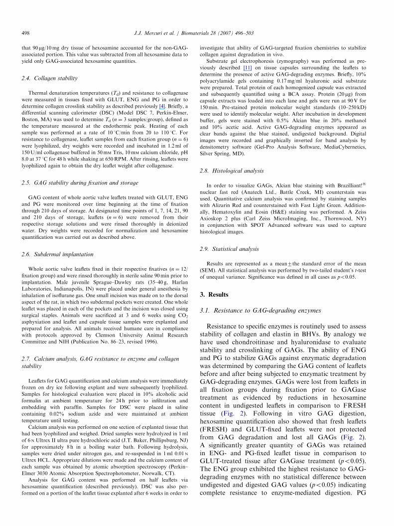
Fig. 1. Proposed reaction schematic for GAG-targeted fixation. Included is th
GAG (oval), (B) resulting reactive intermediate, (C) resulting crosslink with c
Carbodiimide fixation of fresh aortic valve leaflets was performed in a
30mM EDC/6mM NHS solution buffered with 50mM 4-morpholinoetha-
nesulfonic (MES) acid hydrate at a pH of 5.5 for 24 h at ambient
temperature. Following fixation leaflets were thoroughly rinsed in a 50mM
HEPES buffered (pH 7.4) saline solution and subsequently crosslinked
with 0.6% GLUT for 1 day followed by storage in 0.2% GLUT for 5 days
as described for the GLUT group. This group was designated as ENG.
Periodate fixation of leaflet tissue was performed using a 3.25mM
sodium periodate in a 100mM MES buffer at a pH of 5.0 at 4 1C for a
period of 2 h in the absence of light with gentle shaking. Unreacted
periodate was quenched in a 10% aqueous glycerol solution for 15min
with rinses in acidic water. The quenching step was followed by an acidic
GLUT fixation (0.6% GLUT in 100mM MES buffer solution at a pH of
5.0) for 22 h in order to further stabilize the leaflet extracellular matrix.
After acidic GLUT fixation, the standard GLUT procedure as described
above was used to fix and store the valve tissue. This group is designated
as PG. Schematics of ENG and PG crosslinking chemistries are shown in
Fig. 1.
2.2. Resistance to GAG degrading enzymes
Aortic valve leaflets were removed from their respective storage
conditions and dissected from the attached sinus. Leaflets were rinsed
thoroughly in 100mM ammonium acetate buffer (pH 7.4) and cut
symmetrically in half in the radial direction. Half leaflets were incubated
in 1.2ml of 5U/ml hyaluronidase and 0.1U/ml chondroitinase ABC
buffered in the aforementioned ammonium acetate buffer for 24 h at 37 1C
under vigorous shaking at 650 RPM. GAGs have been shown to be
completely removed from fresh leaflets under these conditions [4].
Undigested controls, consisted of corresponding half leaflets, were placed
in 1.2ml of the ammonium acetate buffer only. Following incubation in
enzyme, samples were rinsed thoroughly in three changes of 1.2ml of
distilled water while vigorously shaking for 5min each time. For studies
requiring whole leaflet samples, enzymatic GAG removal was performed
using 1.2ml buffered solutions of 10U/ml hyaluronidase and 0.2U/ml
chondroitinase ABC.
2.3. GAG quantification by hexosamine analysis
Total tissue hexosamines were quantified as previously published [4].
Briefly, lyophilized half leaflets (n ¼ 6 per group) were acid hydrolyzed,
reacted with a 3% acetylacetone in 1.25M sodium carbonate solution, 4ml
of absolute ethanol and 2ml of Ehrlich’s reagent (0.18M P-dimethylami-
nobenzaldehyde in 50% ethanol containing 3 NHCL) were subsequently
added. The samples were incubated for 45min in order to develop a color
product indicative of hexosamine quantities.
A baseline hexosamine value was determined from enzyme-treated
fresh leaflets. Total hexosamine content represents the sum of non-GAG
and GAG-associated hexosamines. From our experiments it was found
e structure of the repeating disaccharide unit: (A) fixative reaction site on
ollagen (CLGN).
ARTICLE IN PRESSJ.J. Mercuri et al. / Biomaterials 28 (2007) 496–503498
that 90 mg/10mg dry tissue of hexosamine accounted for the non-GAG-
associated portion. This value was subtracted from all hexosamine data to
yield only GAG-associated hexosamine quantities.
2.4. Collagen stability
Thermal denaturation temperatures (Td) and resistance to collagenase
were measured in tissues fixed with GLUT, ENG and PG in order to
determine collagen crosslink stability as described previously [4]. Briefly, a
differential scanning calorimeter (DSC) (Model DSC 7, Perkin-Elmer,
Boston, MA) was used to determine Td (n ¼ 3 samples/group), defined as
the temperature measured at the endothermic peak. Heating of each
sample was performed at a rate of 10 1C/min from 20 to 110 1C. For
resistance to collagenase, leaflet samples from each fixation group (n ¼ 6)
were lyophilized, dry weights were recorded and incubated in 1.2ml of
150U/ml collagenase buffered in 50mM Tris, 10mM calcium chloride, pH
8.0 at 37 1C for 48 h while shaking at 650RPM. After rinsing, leaflets were
lyophilized again to obtain the dry leaflet weight after collagenase.
2.5. GAG stability during fixation and storage
GAG content of whole aortic valve leaflets treated with GLUT, ENG
and PG were monitored over time beginning at the time of fixation
through 210 days of storage. At designated time points of 1, 7, 14, 21, 90
and 210 days of storage, leaflets (n ¼ 6) were removed from their
respective storage solutions and were rinsed thoroughly in deionized
water. Dry weights were recorded for normalization and hexosamine
quantification was carried out as described above.
2.6. Subdermal implantation
Whole aortic valve leaflets fixed in their respective fixatives (n ¼ 12/
fixation group) and were rinsed thoroughly in sterile saline 90min prior to
implantation. Male juvenile Sprague–Dawley rats (35–40 g, Harlan
Laboratories, Indianapolis, IN) were placed under general anesthesia by
inhalation of isoflurane gas. One small incision was made on to the dorsal
aspect of the rat, in which two subdermal pockets were created. One whole
leaflet was placed in each of the pockets and the incision was closed using
surgical staples. Animals were sacrificed at 3 and 6 weeks using CO2
asphyxiation and leaflet and capsule tissue samples were explanted and
prepared for analysis. All animals received humane care in compliance
with protocols approved by Clemson University Animal Research
Committee and NIH (Publication No. 86–23, revised 1996).
2.7. Calcium analysis, GAG resistance to enzyme and collagen
stability
Leaflets for GAG quantification and calcium analysis were immediately
frozen on dry ice following explant and were subsequently lyophilized.
Samples for histological evaluation were placed in 10% alcoholic acid
formalin at ambient temperature for 24 h prior to infiltration and
embedding with paraffin. Samples for DSC were placed in saline
containing 0.02% sodium azide and were maintained at ambient
temperature until testing.
Calcium analysis was performed on one section of explanted tissue that
had been lyophilized and weighed. Dried samples were hydrolyzed in 1ml
of 6 N Ultrex II ultra pure hydrochloric acid (J.T. Baker, Phillipsburg, NJ)
for approximately 8 h in a boiling water bath. Following hydrolysis,
samples were dried under nitrogen gas, and re-suspended in 1ml 0.01 N
Ultrex HCL. Appropriate dilutions were made and the calcium content of
each sample was obtained by atomic absorption spectroscopy (Perkin–
Elmer 3030 Atomic Absorption Spectrophotometer, Norwalk, CT).
Analysis for GAG content was performed on half leaflets via
hexosamine quantification (described previously). DSC was also per-
formed on a portion of the leaflet tissue explanted after 6 weeks in order to
investigate that ability of GAG-targeted fixation chemistries to stabilize
collagen against degradation in vivo.
Substrate gel electrophoresis (zymography) was performed as pre-
viously described [11] on tissue capsules surrounding the leaflets to
determine the presence of active GAG-degrading enzymes. Briefly, 10%
polyacrylamide gels containing 0.17mg/ml hyaluronic acid substrate
were prepared. Total protein of each homogenized capsule was extracted
and subsequently quantified using a BCA assay. Protein (20 mg) from
capsule extracts was loaded into each lane and gels were run at 90V for
150min. Pre-stained protein molecular weight standards (10–250 kD)
were used to identify molecular weight. After incubation in development
buffer, gels were stained with 0.5% Alcian blue in 20% methanol
and 10% acetic acid. Active GAG-degrading enzymes appeared as
clear bands against the blue stained, undigested background. Digital
images were recorded and graphically inverted for band analysis by
densitometry software (Gel-Pro Analysis Software, MediaCybernetics,
Silver Spring, MD).
2.8. Histological analysis
In order to visualize GAGs, Alcian blue staining with Brazilliant!s
nuclear fast red (Anatech Ltd., Battle Creek, MI) counterstain was
used. Quantitative calcium analysis was confirmed by staining samples
with Alizarin Red and counterstained with Fast Light Green. Addition-
ally, Hematoxylin and Eosin (H&E) staining was performed. A Zeiss
Axioskop 2 plus (Carl Zeiss MicroImaging, Inc., Thornwood, NY)
in conjunction with SPOT Advanced software was used to capture
histological images.
2.9. Statistical analysis
Results are represented as a mean7the standard error of the mean
(SEM). All statistical analysis was performed by two-tailed student’s t-test
of unequal variance. Significance was defined in all cases as po0.05.
3. Results
3.1. Resistance to GAG-degrading enzymes
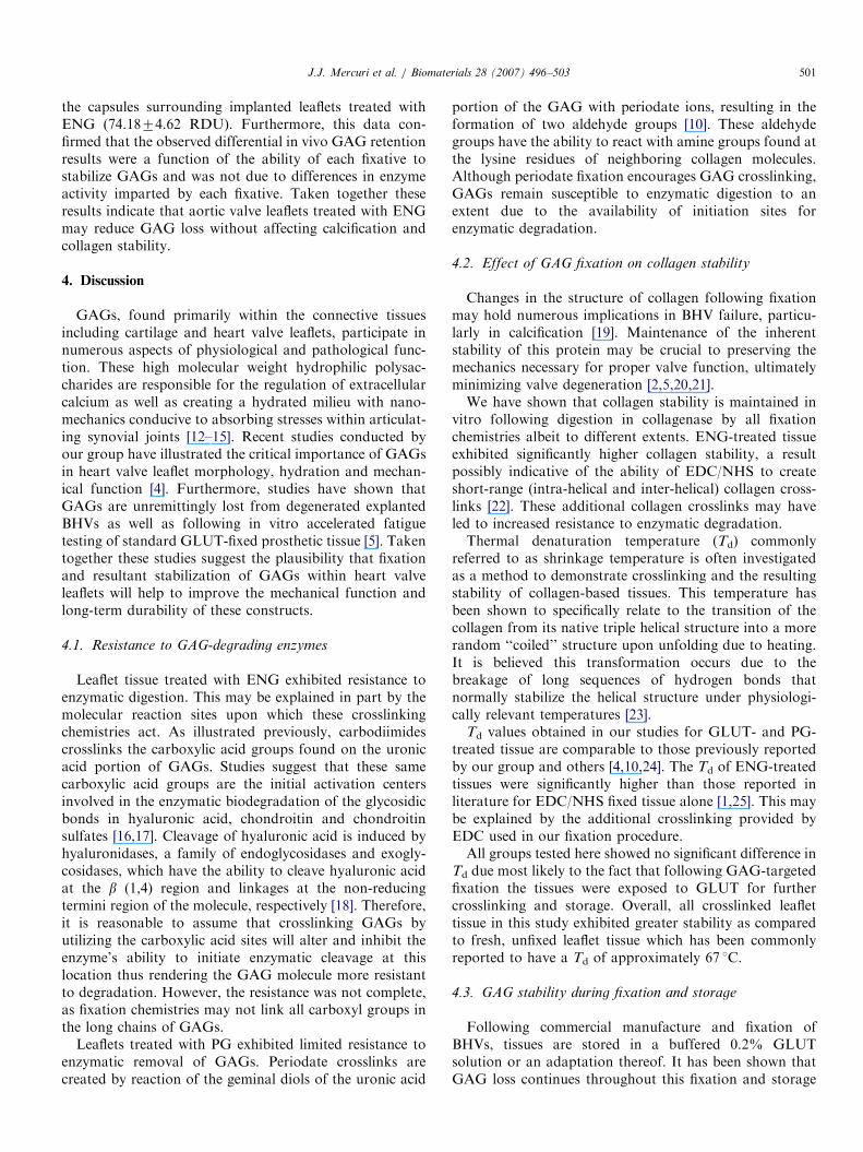
Resistance to specific enzymes is routinely used to assessstability of collagen and elastin in BHVs. By analogy wehave used chondroitinase and hyaluronidase to evaluatestability and crosslinking of GAGs. The ability of ENGand PG to stabilize GAGs against enzymatic degradationwas determined by comparing the GAG content of leafletsbefore and after being subjected to enzymatic treatment byGAG-degrading enzymes. GAGs were lost from leaflets inall fixation groups during fixation prior to GAGasetreatment as evidenced by reductions in hexosaminecontent in undigested leaflets in comparison to FRESHtissue (Fig. 2). Following in vitro GAG digestion,hexosamine quantification also showed that fresh leaflets(FRESH) and GLUT-fixed leaflets were not protectedfrom GAG degradation and lost all GAGs (Fig. 2).A significantly greater quantity of GAGs was retainedin ENG- and PG-fixed leaflet tissue in comparison toGLUT-treated tissue after GAGase treatment (po0.05).The ENG group exhibited the highest resistance to GAG-degrading enzymes with no statistical difference betweenundigested and digested GAG values (po0.05) indicatingcomplete resistance to enzyme-mediated digestion. PG

ARTICLE IN PRESSJ.J. Mercuri et al. / Biomaterials 28 (2007) 496–503 499
treatment, on the other hand, was only partially effective.These results suggest that GAG-targeted chemistries couldbe used to stabilize GAGs against enzyme-mediateddegradation.
3.2. Effect of GAG fixation on collagen stability
To confirm that GAG fixation chemistries do notdetrimentally alter collagen stability, we tested tissuethermal denaturation profiles and resistance to collagenase.Crosslinking of leaflets with all fixatives imparted signifi-cant collagen stability as shown by more than 95% massretention compared to FRESH tissue which retained lessthan 20% mass after collagenase treatment (Table 1). Incomparison to GLUT, both ENG and PG fixation groupsexhibited statistically greater collagen stability (po0.05)most likely due to additional crosslinks created by thesetreatments.
Thermal denaturation data indicated that GAG fixationchemistries were capable of crosslinking collagen compar-able to the GLUT control (Table 1). Overall, these resultssuggest that the GAG-targeted fixation chemistries testeddid not detrimentally alter leaflet collagen stability.
3.3. GAG stability during fixation and storage
To evaluate GAG stability, fixed aortic valve leafletswere kept in storage conditions (0.2% buffered GLUT at
0
50
100
150
FRESH GLUT ENG PG
ug
hex
osa
min
e/1
0mg
dry
leaf
let
tiss
ue
UndigestedDigested
*
*
Fig. 2. In vitro GAG content of fixed aortic valve leaflets before and
following exposure to GAG-degrading enzymes. * Indicates statistical
significance in comparison to GLUT-treated tissue (po0.05).
Table 1
Fixation %Mass retained (collagenase)a
Fresh 19.3472.86*
Glut 94.6170.60
ENG 96.5370.25*
PG 97.8770.37*
*Indicates a significant difference compared to GLUT within each respective+Indicates a significant difference when comparing within each fixation group
aIn vitro collagen stability of fresh and fixed aortic valve leaflets followingbAverage thermal denaturation temperatures (Td) of fixed leaflets (n ¼ 3/fix
ambient temperature). GAG quantities were determinedvia hexosamine quantification at different time points up to210 days.GAGs were lost from leaflet tissue during the fixation
period immediately following harvest. On average approxi-mately 20% of aortic valve leaflet GAGs were lost betweenfixation and the first 24 h of storage as denoted by theinitial steep sloped regions of the plots in Fig. 3. Over the210-day storage time course, GLUT-treated tissuelost an average of 28.91%74.13% of its original GAGcontent. No significant difference in GAG loss was seen tooccur at any time point between PG and GLUT-treatedtissue (p40.05) (Fig. 3). The GAG loss trend in ENG-treated tissue was shown to mimic that found in GLUT-treated tissue until 21 days of storage. At this timepoint ENG trend line began to deviate from GLUTshowing increased GAG retention in comparison toGLUT treated tissue, this difference became significant at210 days (p ¼ 0.0058). Histological staining for thepresence of GAGs with Alcian blue at 210 days confirmedquantitative results showing higher GAG staining in ENG-treated leaflets (data not shown). Together, this datasuggests that GAGs are stabilized following initial fixationand storage periods up to 1 week. Following 2 weeks ofstorage GAG loss from fixed tissue reaches an apparentsteady state and resident GAG levels are maintainedfor up to 210 days in storage for GLUT-, ENG- andPG-treated tissue.
Leaflet Td non-implantedb Leaflet Td 6-week explantb
— —
90.2370.05 89.9170.82
91.0270.48 91.0370.33
90.0370.33 84.9170.54+
column (po0.05).
between non-implanted and implanted leaflets (po0.05).
incubation in collagenase (n ¼ 6/fixation group).
ation group) obtained via differential scanning calorimetry.
0
50
100
150
0 20 40 60 80 100 120 140 160 180 200
Days
ug
hex
osa
min
e/1
0mg
dry
leaf
let
tiss
ue
GLUTENG
PG
*
Fig. 3. Hexosamine content representing GAG retention in fixed leaflets
at different time points over the 210-day storage period. * Indicates a
significant difference compared to GLUT (po0.05).

ARTICLE IN PRESSJ.J. Mercuri et al. / Biomaterials 28 (2007) 496–503500
3.4. In vivo resistance to calcification, GAG loss and
collagen stability
Whole aortic valve leaflets, treated with their respectivefixatives, were implanted subdermally into juvenile rats for3 and 6 weeks in order to determine the degree to whichGAG-targeted fixation chemistries alter the leaflet’s pro-pensity to calcify in vivo. Additionally, explanted tissuewas analyzed to determine GAG crosslink stability bymonitoring in vivo GAG retention.
GLUT-, ENG- and PG-fixed leaflets contained statisti-cally similar amounts of calcium (p40.05) following 3weeks of implant as shown in Fig. 4. At 6 weeks ofimplantation, leaflet calcification had increased for allgroups. Histological analysis using Alizarin Red stainingconfirmed quantitative results illustrating extensive calcifi-cation marked by strong red staining within leaflet tissuetreated with GLUT, ENG and PG (data not shown).
Following 3 weeks of implantation, all fixation groupslost significant amounts of GAGs in comparison totheir pre-implant GAG values (po0.05) (Fig. 5). GLUT-treated leaflets only retained 10.93%73.62% of theiroriginal GAG content. This percentage was not signifi-cantly different from PG-treated tissue, which exhibited
0
50
100
150
200
250
GLUT ENG PG
ug
cal
ciu
m/m
g d
ry le
afle
t ti
ssu
e
3 Weeks6 Weeks
Fig. 4. Calcification of leaflets following subdermal implantation. All
groups showed similar calcification levels.
0
50
100
GLUT ENG PG
ug
of
hex
osa
min
e/1
0mg
dry
leaf
let
tiss
ue
Pre-Implant3 Weeks6 Weeks
*
*
Fig. 5. Hexosamine quantification representing in vivo GAG retention
following subdermal implantation. * Indicates a significant difference
compared to GLUT at each respective time point (po0.05).
15.88%75.41% GAG retention (p ¼ 0.46). ENG stabili-zation conferred statistically greater GAG retention(45.69%712.02%) in vivo in comparison to GLUT-fixedtissue (p ¼ 0.033).After 6 weeks of implant, GAG loss continued in all
fixation groups. GLUT and PG-treated leaflets retained3.23%75.29% and 2.19%712.68% of their original GAGcontent and were not statistically different. The ENG-treated group retained 25.66%76.11% which was statis-tically greater in comparison to GLUT (po0.05). Histo-logical staining of GAGs confirmed quantitative results forGLUT-, ENG- and PG-treated leaflets (data not shown).DSC data obtained from leaflets following 6 weeks of
implant suggests that collagen stability is maintained in allfixation groups with the exception of PG treated tissue,which exhibited a significant decrease in the thermaldenaturation temperature of collagen (po0.003) (Table 1).Zymography was carried out to investigate the activity
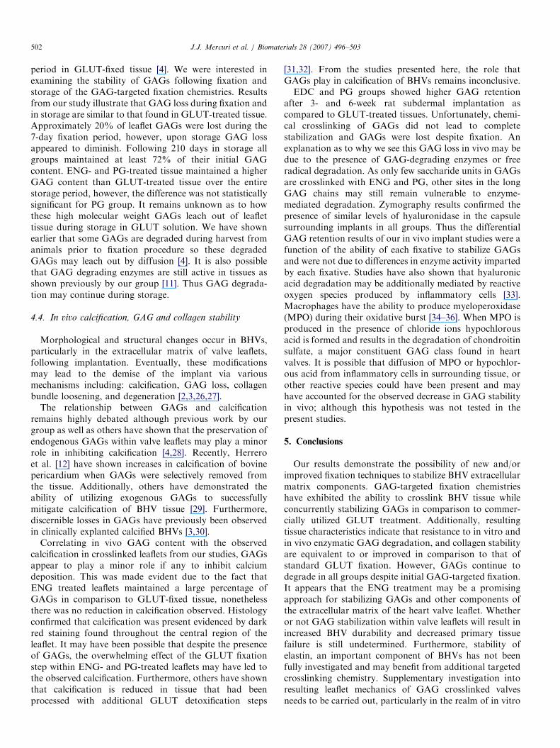
of GAG-degrading enzymes present in the capsulessurrounding fixed leaflets following 6 weeks of implant.Furthermore, we wanted to confirm that the fixativesthemselves did not diminish the enzyme activity, thusresulting in erroneous in vivo GAG retention data.Zymography and densitometry results suggest that GAG-degrading enzymes specifically hyaluronidase remain activeat 6 weeks of explant as evidenced by the light bandobserved at �55 kDa in each fixation group (Fig. 6).Moreover, densitometry results indicated that capsulessurrounding implanted leaflets treated with GLUT and PGon average contained statistically equivalent amounts ofenzyme as illustrated by similar relative densitometric units(RDU) (60.0073.77 and 47.9878.02 RDU, respectively).It was noticed, however, that significantly higher(p ¼ 0.039) amounts of active hyaluronidase was found in
Fig. 6. HA zymography results indicating the average relative amounts of
active GAG-degrading enzymes present in the capsules surrounding
implanted leaflets for each fixation group (RDU: relative densitomertic
unit). * Indicates a significant difference compared to GLUT (po0.05).
Inset: light bands at approximately 55 kDa indicating the characteristic
detection of GAG-degrading enzymes by hyaluronic acid zymography on
homogenized capsule extracts (protein molecular weight standards left
column).
ARTICLE IN PRESSJ.J. Mercuri et al. / Biomaterials 28 (2007) 496–503 501
the capsules surrounding implanted leaflets treated withENG (74.1874.62 RDU). Furthermore, this data con-firmed that the observed differential in vivo GAG retentionresults were a function of the ability of each fixative tostabilize GAGs and was not due to differences in enzymeactivity imparted by each fixative. Taken together theseresults indicate that aortic valve leaflets treated with ENGmay reduce GAG loss without affecting calcification andcollagen stability.
4. Discussion
GAGs, found primarily within the connective tissuesincluding cartilage and heart valve leaflets, participate innumerous aspects of physiological and pathological func-tion. These high molecular weight hydrophilic polysac-charides are responsible for the regulation of extracellularcalcium as well as creating a hydrated milieu with nano-mechanics conducive to absorbing stresses within articulat-ing synovial joints [12–15]. Recent studies conducted byour group have illustrated the critical importance of GAGsin heart valve leaflet morphology, hydration and mechan-ical function [4]. Furthermore, studies have shown thatGAGs are unremittingly lost from degenerated explantedBHVs as well as following in vitro accelerated fatiguetesting of standard GLUT-fixed prosthetic tissue [5]. Takentogether these studies suggest the plausibility that fixationand resultant stabilization of GAGs within heart valveleaflets will help to improve the mechanical function andlong-term durability of these constructs.
4.1. Resistance to GAG-degrading enzymes
Leaflet tissue treated with ENG exhibited resistance toenzymatic digestion. This may be explained in part by themolecular reaction sites upon which these crosslinkingchemistries act. As illustrated previously, carbodiimidescrosslinks the carboxylic acid groups found on the uronicacid portion of GAGs. Studies suggest that these samecarboxylic acid groups are the initial activation centersinvolved in the enzymatic biodegradation of the glycosidicbonds in hyaluronic acid, chondroitin and chondroitinsulfates [16,17]. Cleavage of hyaluronic acid is induced byhyaluronidases, a family of endoglycosidases and exogly-cosidases, which have the ability to cleave hyaluronic acidat the b (1,4) region and linkages at the non-reducingtermini region of the molecule, respectively [18]. Therefore,it is reasonable to assume that crosslinking GAGs byutilizing the carboxylic acid sites will alter and inhibit theenzyme’s ability to initiate enzymatic cleavage at thislocation thus rendering the GAG molecule more resistantto degradation. However, the resistance was not complete,as fixation chemistries may not link all carboxyl groups inthe long chains of GAGs.
Leaflets treated with PG exhibited limited resistance toenzymatic removal of GAGs. Periodate crosslinks arecreated by reaction of the geminal diols of the uronic acid
portion of the GAG with periodate ions, resulting in theformation of two aldehyde groups [10]. These aldehydegroups have the ability to react with amine groups found atthe lysine residues of neighboring collagen molecules.Although periodate fixation encourages GAG crosslinking,GAGs remain susceptible to enzymatic digestion to anextent due to the availability of initiation sites forenzymatic degradation.
4.2. Effect of GAG fixation on collagen stability
Changes in the structure of collagen following fixationmay hold numerous implications in BHV failure, particu-larly in calcification [19]. Maintenance of the inherentstability of this protein may be crucial to preserving themechanics necessary for proper valve function, ultimatelyminimizing valve degeneration [2,5,20,21].We have shown that collagen stability is maintained in
vitro following digestion in collagenase by all fixationchemistries albeit to different extents. ENG-treated tissueexhibited significantly higher collagen stability, a resultpossibly indicative of the ability of EDC/NHS to createshort-range (intra-helical and inter-helical) collagen cross-links [22]. These additional collagen crosslinks may haveled to increased resistance to enzymatic degradation.Thermal denaturation temperature (Td) commonly
referred to as shrinkage temperature is often investigatedas a method to demonstrate crosslinking and the resultingstability of collagen-based tissues. This temperature hasbeen shown to specifically relate to the transition of thecollagen from its native triple helical structure into a morerandom ‘‘coiled’’ structure upon unfolding due to heating.It is believed this transformation occurs due to thebreakage of long sequences of hydrogen bonds thatnormally stabilize the helical structure under physiologi-cally relevant temperatures [23].
Td values obtained in our studies for GLUT- and PG-treated tissue are comparable to those previously reportedby our group and others [4,10,24]. The Td of ENG-treatedtissues were significantly higher than those reported inliterature for EDC/NHS fixed tissue alone [1,25]. This maybe explained by the additional crosslinking provided byEDC used in our fixation procedure.All groups tested here showed no significant difference in
Td due most likely to the fact that following GAG-targetedfixation the tissues were exposed to GLUT for furthercrosslinking and storage. Overall, all crosslinked leaflettissue in this study exhibited greater stability as comparedto fresh, unfixed leaflet tissue which has been commonlyreported to have a Td of approximately 67 1C.
4.3. GAG stability during fixation and storage
Following commercial manufacture and fixation ofBHVs, tissues are stored in a buffered 0.2% GLUTsolution or an adaptation thereof. It has been shown thatGAG loss continues throughout this fixation and storage
ARTICLE IN PRESSJ.J. Mercuri et al. / Biomaterials 28 (2007) 496–503502
period in GLUT-fixed tissue [4]. We were interested inexamining the stability of GAGs following fixation andstorage of the GAG-targeted fixation chemistries. Resultsfrom our study illustrate that GAG loss during fixation andin storage are similar to that found in GLUT-treated tissue.Approximately 20% of leaflet GAGs were lost during the7-day fixation period, however, upon storage GAG lossappeared to diminish. Following 210 days in storage allgroups maintained at least 72% of their initial GAGcontent. ENG- and PG-treated tissue maintained a higherGAG content than GLUT-treated tissue over the entirestorage period, however, the difference was not statisticallysignificant for PG group. It remains unknown as to howthese high molecular weight GAGs leach out of leaflettissue during storage in GLUT solution. We have shownearlier that some GAGs are degraded during harvest fromanimals prior to fixation procedure so these degradedGAGs may leach out by diffusion [4]. It is also possiblethat GAG degrading enzymes are still active in tissues asshown previously by our group [11]. Thus GAG degrada-tion may continue during storage.
4.4. In vivo calcification, GAG and collagen stability
Morphological and structural changes occur in BHVs,particularly in the extracellular matrix of valve leaflets,following implantation. Eventually, these modificationsmay lead to the demise of the implant via variousmechanisms including: calcification, GAG loss, collagenbundle loosening, and degeneration [2,3,26,27].
The relationship between GAGs and calcificationremains highly debated although previous work by ourgroup as well as others have shown that the preservation ofendogenous GAGs within valve leaflets may play a minorrole in inhibiting calcification [4,28]. Recently, Herreroet al. [12] have shown increases in calcification of bovinepericardium when GAGs were selectively removed fromthe tissue. Additionally, others have demonstrated theability of utilizing exogenous GAGs to successfullymitigate calcification of BHV tissue [29]. Furthermore,discernible losses in GAGs have previously been observedin clinically explanted calcified BHVs [3,30].
Correlating in vivo GAG content with the observedcalcification in crosslinked leaflets from our studies, GAGsappear to play a minor role if any to inhibit calciumdeposition. This was made evident due to the fact thatENG treated leaflets maintained a large percentage ofGAGs in comparison to GLUT-fixed tissue, nonethelessthere was no reduction in calcification observed. Histologyconfirmed that calcification was present evidenced by darkred staining found throughout the central region of theleaflet. It may have been possible that despite the presenceof GAGs, the overwhelming effect of the GLUT fixationstep within ENG- and PG-treated leaflets may have led tothe observed calcification. Furthermore, others have shownthat calcification is reduced in tissue that had beenprocessed with additional GLUT detoxification steps
[31,32]. From the studies presented here, the role thatGAGs play in calcification of BHVs remains inconclusive.EDC and PG groups showed higher GAG retention
after 3- and 6-week rat subdermal implantation ascompared to GLUT-treated tissues. Unfortunately, chemi-cal crosslinking of GAGs did not lead to completestabilization and GAGs were lost despite fixation. Anexplanation as to why we see this GAG loss in vivo may bedue to the presence of GAG-degrading enzymes or freeradical degradation. As only few saccharide units in GAGsare crosslinked with ENG and PG, other sites in the longGAG chains may still remain vulnerable to enzyme-mediated degradation. Zymography results confirmed thepresence of similar levels of hyaluronidase in the capsulesurrounding implants in all groups. Thus the differentialGAG retention results of our in vivo implant studies were afunction of the ability of each fixative to stabilize GAGsand were not due to differences in enzyme activity impartedby each fixative. Studies have also shown that hyaluronicacid degradation may be additionally mediated by reactiveoxygen species produced by inflammatory cells [33].Macrophages have the ability to produce myeloperoxidase(MPO) during their oxidative burst [34–36]. When MPO isproduced in the presence of chloride ions hypochlorousacid is formed and results in the degradation of chondroitinsulfate, a major constituent GAG class found in heartvalves. It is possible that diffusion of MPO or hypochlor-ous acid from inflammatory cells in surrounding tissue, orother reactive species could have been present and mayhave accounted for the observed decrease in GAG stabilityin vivo; although this hypothesis was not tested in thepresent studies.
5. Conclusions
Our results demonstrate the possibility of new and/orimproved fixation techniques to stabilize BHV extracellularmatrix components. GAG-targeted fixation chemistrieshave exhibited the ability to crosslink BHV tissue whileconcurrently stabilizing GAGs in comparison to commer-cially utilized GLUT treatment. Additionally, resultingtissue characteristics indicate that resistance to in vitro andin vivo enzymatic GAG degradation, and collagen stabilityare equivalent to or improved in comparison to that ofstandard GLUT fixation. However, GAGs continue todegrade in all groups despite initial GAG-targeted fixation.It appears that the ENG treatment may be a promisingapproach for stabilizing GAGs and other components ofthe extracellular matrix of the heart valve leaflet. Whetheror not GAG stabilization within valve leaflets will result inincreased BHV durability and decreased primary tissuefailure is still undetermined. Furthermore, stability ofelastin, an important component of BHVs has not beenfully investigated and may benefit from additional targetedcrosslinking chemistry. Supplementary investigation intoresulting leaflet mechanics of GAG crosslinked valvesneeds to be carried out, particularly in the realm of in vitro

ARTICLE IN PRESSJ.J. Mercuri et al. / Biomaterials 28 (2007) 496–503 503
accelerated fatigue testing and in vivo trials in theorthotopic or heterotopic position before we begin toestablish if crosslinking of GAGs within BHV leaflets willhave a lasting effect on implant in-service life.
References
[1] Everaerts F, Torrianni M, van Luyn M, van Wachem P, Feijen J,
Hendriks M. Reduced calcification of bioprostheses, cross-linked via
an improved carbodiimide based method. Biomaterials 2004;25(24):
5523–30.
[2] Schoen FJ, Levy RJ. Founder’s Award, 25th annual meeting of the
Society for Biomaterials, perspectives. Providence, RI, April 28–May
2, 1999. Tissue heart valves: current challenges and future research
perspectives. J Biomed Mater Res 1999;47(4):439–65.
[3] Mako WJ. Loss of glycosaminoglycans (GAGs) from implanted
bioprosthetic heart valves. Circulation 1997(155):863.
[4] Lovekamp JJ, Simionescu DT, Mercuri JJ, Zubiate B, Sacks MS,
Vyavahare NR. Stability and function of glycosaminoglycans in
porcine bioprosthetic heart valves. Biomaterials 2006;27(8):1507–18.
[5] Vyavahare N, Ogle M, Schoen FJ, Zand R, Gloeckner DC, Sacks M,
et al. Mechanisms of bioprosthetic heart valve failure: fatigue causes
collagen denaturation and glycosaminoglycan loss. J Biomed Mater
Res 1999;46(1):44–50.
[6] van Wachem PB, Brouwer LA, Zeeman R, Dijkstra PJ, Feijen J,
Hendriks M, et al. Tissue reactions to epoxy-crosslinked porcine
heart valves post-treated with detergents or a dicarboxylic acid.
J Biomed Mater Res 2001;55(3):415–23.
[7] Petite H, Frei V, Huc A, Herbage D. Use of diphenylphosphorylazide
for cross-linking collagen-based biomaterials. J Biomed Mater Res
1994;28(2):159–65.
[8] Adams AK, Talman EA, Campbell L, McIlroy BK, Moore MA.
Crosslink formation in porcine valves stabilized by dye-mediated
photooxidation. J Biomed Mater Res 2001;57(4):582–7.
[9] Suh H, Hwang YS, Park JC, Cho BK. Calcification of leaflets from
porcine aortic valves crosslinked by ultraviolet irradiation. Artif
Organs 2000;24(7):555–63.
[10] Lovekamp J, Vyavahare N. Periodate-mediated glycosaminoglycan
stabilization in bioprosthetic heart valves. J Biomed Mater Res
2001;56(4):478–86.
[11] Simionescu DT, Lovekamp JJ, Vyavahare NR. Glycosaminoglycan-
degrading enzymes in porcine aortic heart valves: implications for
bioprosthetic heart valve degeneration. J Heart Valve Dis
2003;12(2):217–25.
[12] Jorge-Herrero E, Fernandez P, Gutierrez M, Castillo-Olivares JL.
Study of the calcification of bovine pericardium: analysis of the
implication of lipids and proteoglycans. Biomaterials 1991;12(7):
683–9.
[13] Wheeler AP, Sikes CS. Regulation of carbonate calcification by
organic matrix. Am Zool 1984;24:933–44.
[14] Claassen H, Cellarius C, Scholz-Ahrens KE, Schrezenmeir J, Gluer
CC, Schunke M, et al. Extracellular matrix changes in knee joint
cartilage following bone-active drug treatment. Cell Tissue Res
2006;00:1–11.
[15] Seog J, Dean D, Rolauffs B, Wu T, Genzer J, Plaas AH, et al.
Nanomechanics of opposing glycosaminoglycan macromolecules.
J Biomech 2005;38(9):1789–97.
[16] Menzel EJ, Farr C. Hyaluronidase and its substrate hyaluronan:
biochemistry, biological activities and therapeutic uses. Cancer Lett
1998;131(1):3–11.
[17] Zhong SP, Campoccia D, Doherty PJ, Williams RL, Benedetti L,
Williams DF. Biodegradation of hyaluronic acid derivatives by
hyaluronidase. Biomaterials 1994;15(5):359–65.
[18] Stair-Nawy S, Csoka AB, Stern R. Hyaluronidase expression in
human skin fibroblasts. Biochem Biophys Res Commun 1999;
266(1):268–73.
[19] Simionescu DT. Prevention of calcification in bioprosthetic heart
valves: challenges and perspectives. Expert Opin Biol Ther 2004;
4(12):1971–85.
[20] Sacks MS, Schoen FJ. Collagen fiber disruption occurs independent
of calcification in clinically explanted bioprosthetic heart valves.
J Biomed Mater Res 2002;62(3):359–71.
[21] Sauren AA, Kuijpers W, van Steenhoven AA, Veldpaus FE. Aortic
valve histology and its relation with mechanics-preliminary report.
J Biomech 1980;13(2):97–104.
[22] Sung HW, Chang WH, Ma CY, Lee MH. Crosslinking of biological
tissues using genipin and/or carbodiimide. J Biomed Mater Res A
2003;64(3):427–38.
[23] Wright NT, Humphrey JD. Denaturation of collagen via heating: an
irreversible rate process. Annu Rev Biomed Eng 2002;4:109–28.
[24] Connolly JM, Alferiev I, Clark-Gruel JN, Eidelman N, Sacks M,
Palmatory E, et al. Triglycidylamine crosslinking of porcine aortic
valve cusps or bovine pericardium results in improved biocompat-
ibility, biomechanics, and calcification resistance: chemical and
biological mechanisms. Am J Pathol 2005;166(1):1–13.
[25] Olde Damink LH, Dijkstra PJ, van Luyn MJ, van Wachem PB,
Nieuwenhuis P, Feijen J. In vitro degradation of dermal sheep
collagen cross-linked using a water-soluble carbodiimide. Biomater-
ials 1996;17(7):679–84.
[26] Valente M, Minarini M, Maizza AF, Bortolotti U, Thiene G. Heart
valve bioprosthesis durability: a challenge to the new generation of
porcine valves. Eur J Cardiothorac Surg 1992;6(Suppl 1):S82–90.
[27] Zilla P, Human P, Bezuidenhout D. Bioprosthetic heart valves: the
need for a quantum leap. Biotechnol Appl Biochem 2004;40(Part
1):57–66.
[28] Arenaz B, Maestro MM, Fernandez P, Turnay J, Olmo N, Senen J,
et al. Effects of periodate and chondroitin 4-sulfate on proteoglycan
stabilization of ostrich pericardium. Inhibition of calcification in
subcutaneous implants in rats. Biomaterials 2004;25(17):3359–68.
[29] Ohri R, Hahn SK, Hoffman AS, Stayton PS, Giachelli CM.
Hyaluronic acid grafting mitigates calcification of glutaraldehyde-
fixed bovine pericardium. J Biomed Mater Res A 2004;70(2):328–34.
[30] Grande-Allen KJ, Mako WJ, Calabro A, Shi Y, Ratliff NB, Vesely I.
Loss of chondroitin 6-sulfate and hyaluronan from failed porcine
bioprosthetic valves. J Biomed Mater Res A 2003;65(2):251–9.
[31] Trantina-Yates AE, Human P, Zilla P. Detoxification on top of
enhanced, diamine-extended glutaraldehyde fixation significantly
reduces bioprosthetic root calcification in the sheep model. J Heart
Valve Dis 2003;12(1):93–100 (discussion 100–1).
[32] Weissenstein C, Human P, Bezuidenhout D, Zilla P. Glutaraldehyde
detoxification in addition to enhanced amine cross-linking dramati-
cally reduces bioprosthetic tissue calcification in the rat model.
J Heart Valve Dis 2000;9(2):230–40.
[33] Schenck P, Schneider S, Miehlke R, Prehm P. Synthesis and
degradation of hyaluronate by synovia from patients with rheuma-
toid arthritis. J Rheumatol 1995;22(3):400–5.
[34] Moseley R, Waddington R, Evans P, Halliwell B, Embery G.
The chemical modification of glycosaminoglycan structure by
oxygen-derived species in vitro. Biochim Biophys Acta 1995;
1244(2–3):245–52.
[35] Moseley R, Waddington RJ, Embery G. Degradation of glycosami-
noglycans by reactive oxygen species derived from stimulated
polymorphonuclear leukocytes. Biochim Biophys Acta 1997;
1362(2–3):221–31.
[36] Rees MD, Hawkins CL, Davies MJ. Hypochlorite and superoxide
radicals can act synergistically to induce fragmentation of hyaluronan
and chondroitin sulphates. Biochem J 2004;381(Part 1):175–84.