Neuropilin-2 Mediates Axonal Fasciculation, Zonal Segregation, but Not Axonal Convergence, of...
16
Neuron, Vol. 33, 877–892, March 14, 2002, Copyright 2002 by Cell Press Neuropilin-2 Mediates Axonal Fasciculation, Zonal Segregation, but Not Axonal Convergence, of Primary Accessory Olfactory Neurons order neurons in the AOB (reviewed in Halpern, 1987; Dulac, 2000). Axons of chemosensory neurons in both olfactory sys- tems form specific and highly stereotyped projections to higher-order central nervous system (CNS) neurons. Jean-Franc ¸ ois Cloutier, 1,2,5 Roman J. Giger, 1,6,5 Georgy Koentges, 3,7 Catherine Dulac, 3 Alex L. Kolodkin, 1,4 and David D. Ginty 1,2,4 1 Department of Neuroscience 2 Howard Hughes Medical Institute Neurons distributed within four distinct zones of the The Johns Hopkins University School of Medicine main olfactory epithelium project their axons to four 725 North Wolfe Street specific zones in the MOB (Strotmann et al., 1992; Ress- Baltimore, Maryland 21205 ler et al., 1993; Vassar et al., 1993; Strotmann et al., 3 Department of Cellular and Molecular Biology 1994). Neurons of the main olfactory epithelium each Howard Hughes Medical Institute express one of approximately 1000 odorant receptors. Harvard University Axons of neurons expressing the same odorant receptor 16 Divinity Avenue converge on two bilaterally symmetric and topographi- Cambridge, Massachusetts 02138 cally fixed glomeruli of the approximately 1800 glomeruli in the MOB (Ressler et al., 1994; Vassar et al., 1994; Mombaerts et al., 1996). Zonal segregation is also ob- Summary served in the accessory olfactory system; cell bodies of chemosensory neurons found within the apical and The mechanisms that underlie axonal pathfinding of basal zones of the VNO extend axons that project to vomeronasal neurons from the vomeronasal organ either the anterior or posterior region of the ipsilateral (VNO) in the periphery to select glomeruli in the acces- AOB, respectively (Halpern et al., 1995). However, in sory olfactory bulb (AOB) are not well understood. contrast to the remarkable degree of axonal conver- Neuropilin-2, a receptor for secreted semaphorins, is gence into single glomeruli at fixed locations in the MOB, expressed in V1R- and V3R-expressing, but not V2R- axons of vomeronasal neurons that express the same expressing, postnatal vomeronasal neurons. Analysis pheromone receptor innervate multiple glomeruli clus- of the vomeronasal nerve in neuropilin-2 (npn-2) mu- tered within large, but topographically conserved, do- tant mice reveals pathfinding defects at multiple mains within the AOB (Belluscio et al., 1999; Rodriguez choice points. Vomeronasal sensory axons are se- et al., 1999). verely defasciculated and a subset innervates the main Vomeronasal neurons express putative pheromone olfactory bulb (MOB). While most axons of V1R- receptors (VRs), which are members of the large super- expressing neurons reach the AOB and converge into family of G protein-coupled seven-transmembrane distinct glomeruli in stereotypic locations, they are no spanning receptors (Dulac and Axel, 1995; Herrada and longer restricted to their normal anterior AOB target Dulac, 1997; Matsunami and Buck, 1997; Ryba and Tirin- zone. Thus, Npn-2 and candidate pheromone recep- delli, 1997; Pantages and Dulac, 2000). Neurons located tors play distinct and complementary roles in promot- in the apical (luminal) portion of the vomeronasal epithe- ing the wiring and patterning of sensory neurons in lium express either V1R or V3R receptors and, also, the the accessory olfactory system. G protein subunit G i2 . In contrast, sensory neurons located in the basal region of the vomeronasal epithe- lium express members of the V2R receptor family and Introduction the G protein subunit G o . V1R-expressing neurons project their axons via the vomeronasal nerve to glomer- The olfactory system of most terrestrial vertebrates is uli restricted to the anterior half of the AOB, whereas V2R comprised of two anatomically and functionally distinct neurons in the basal vomeronasal epithelium project to chemosensory systems that provide information about glomeruli restricted to the posterior half of the AOB. the chemical environment. Primary sensory neurons Relatively little is known about the molecular and cellular within the main olfactory epithelium located in the princi- mechanisms that govern the guidance of sensory neu- pal nasal cavity can relay odor information to second- ron axons to the AOB and the segregation of these order neurons in the MOB. In contrast, pheromone- axons into either the anterior or posterior zones of the responsive sensory neurons are found in the VNO, a AOB. bilateral sensory structure located at the base of the It is well established that growth cones of extending nasal septum, and relay their information to second- axons respond to a wide range of environmental cues that guide them to their targets. The semaphorins are a large family of glycoproteins that have been implicated 4 Correspondence: [email protected] (D.D.G.), [email protected] (A.L.K.) in the guidance of growing axons (Raper, 2000; Naka- 5 These authors contributed equally to this work. mura et al., 2000). The well-characterized class 3 se- 6 Present address: Center for Aging and Developmental Biology, creted semaphorins act as chemorepellents for specific, University of Rochester, 575 Elmwood Avenue, Rochester, New York yet partially overlapping populations of developing neu- 14642. rons. Neuropilin-1 (Npn-1) and neuropilin-2 (Npn-2) are 7 Present address: The Wolfson Institute for Biomedical Research, members of a small family of type-I transmembrane pro- University College London, Gower Street, London WC1E 6BT, United Kingdom. teins that are essential ligand-binding subunits of class
Transcript of Neuropilin-2 Mediates Axonal Fasciculation, Zonal Segregation, but Not Axonal Convergence, of...

Neuron, Vol. 33, 877–892, March 14, 2002, Copyright 2002 by Cell Press
Neuropilin-2 Mediates Axonal Fasciculation,Zonal Segregation, but Not Axonal Convergence,of Primary Accessory Olfactory Neurons
order neurons in the AOB (reviewed in Halpern, 1987;Dulac, 2000).
Axons of chemosensory neurons in both olfactory sys-tems form specific and highly stereotyped projectionsto higher-order central nervous system (CNS) neurons.
Jean-Francois Cloutier,1,2,5 Roman J. Giger,1,6,5
Georgy Koentges,3,7 Catherine Dulac,3
Alex L. Kolodkin,1,4 and David D. Ginty1,2,4
1Department of Neuroscience2 Howard Hughes Medical Institute
Neurons distributed within four distinct zones of theThe Johns Hopkins University School of Medicinemain olfactory epithelium project their axons to four725 North Wolfe Streetspecific zones in the MOB (Strotmann et al., 1992; Ress-Baltimore, Maryland 21205ler et al., 1993; Vassar et al., 1993; Strotmann et al.,3 Department of Cellular and Molecular Biology1994). Neurons of the main olfactory epithelium eachHoward Hughes Medical Instituteexpress one of approximately 1000 odorant receptors.Harvard UniversityAxons of neurons expressing the same odorant receptor16 Divinity Avenueconverge on two bilaterally symmetric and topographi-Cambridge, Massachusetts 02138cally fixed glomeruli of the approximately 1800 glomeruliin the MOB (Ressler et al., 1994; Vassar et al., 1994;Mombaerts et al., 1996). Zonal segregation is also ob-
Summary served in the accessory olfactory system; cell bodies ofchemosensory neurons found within the apical and
The mechanisms that underlie axonal pathfinding of basal zones of the VNO extend axons that project tovomeronasal neurons from the vomeronasal organ either the anterior or posterior region of the ipsilateral(VNO) in the periphery to select glomeruli in the acces- AOB, respectively (Halpern et al., 1995). However, insory olfactory bulb (AOB) are not well understood. contrast to the remarkable degree of axonal conver-Neuropilin-2, a receptor for secreted semaphorins, is gence into single glomeruli at fixed locations in the MOB,expressed in V1R- and V3R-expressing, but not V2R- axons of vomeronasal neurons that express the sameexpressing, postnatal vomeronasal neurons. Analysis pheromone receptor innervate multiple glomeruli clus-of the vomeronasal nerve in neuropilin-2 (npn-2) mu- tered within large, but topographically conserved, do-tant mice reveals pathfinding defects at multiple mains within the AOB (Belluscio et al., 1999; Rodriguezchoice points. Vomeronasal sensory axons are se- et al., 1999).verely defasciculated and a subset innervates the main Vomeronasal neurons express putative pheromoneolfactory bulb (MOB). While most axons of V1R- receptors (VRs), which are members of the large super-expressing neurons reach the AOB and converge into family of G protein-coupled seven-transmembranedistinct glomeruli in stereotypic locations, they are no spanning receptors (Dulac and Axel, 1995; Herrada andlonger restricted to their normal anterior AOB target Dulac, 1997; Matsunami and Buck, 1997; Ryba and Tirin-zone. Thus, Npn-2 and candidate pheromone recep- delli, 1997; Pantages and Dulac, 2000). Neurons locatedtors play distinct and complementary roles in promot- in the apical (luminal) portion of the vomeronasal epithe-ing the wiring and patterning of sensory neurons in lium express either V1R or V3R receptors and, also, thethe accessory olfactory system. G protein � subunit G�i2. In contrast, sensory neurons
located in the basal region of the vomeronasal epithe-lium express members of the V2R receptor family andIntroductionthe G protein � subunit G�o. V1R-expressing neuronsproject their axons via the vomeronasal nerve to glomer-
The olfactory system of most terrestrial vertebrates isuli restricted to the anterior half of the AOB, whereas V2R
comprised of two anatomically and functionally distinct neurons in the basal vomeronasal epithelium project tochemosensory systems that provide information about glomeruli restricted to the posterior half of the AOB.the chemical environment. Primary sensory neurons Relatively little is known about the molecular and cellularwithin the main olfactory epithelium located in the princi- mechanisms that govern the guidance of sensory neu-pal nasal cavity can relay odor information to second- ron axons to the AOB and the segregation of theseorder neurons in the MOB. In contrast, pheromone- axons into either the anterior or posterior zones of theresponsive sensory neurons are found in the VNO, a AOB.bilateral sensory structure located at the base of the It is well established that growth cones of extendingnasal septum, and relay their information to second- axons respond to a wide range of environmental cues
that guide them to their targets. The semaphorins area large family of glycoproteins that have been implicated4 Correspondence: [email protected] (D.D.G.), [email protected]
(A.L.K.) in the guidance of growing axons (Raper, 2000; Naka-5 These authors contributed equally to this work. mura et al., 2000). The well-characterized class 3 se-6 Present address: Center for Aging and Developmental Biology, creted semaphorins act as chemorepellents for specific,University of Rochester, 575 Elmwood Avenue, Rochester, New York
yet partially overlapping populations of developing neu-14642.rons. Neuropilin-1 (Npn-1) and neuropilin-2 (Npn-2) are7 Present address: The Wolfson Institute for Biomedical Research,members of a small family of type-I transmembrane pro-University College London, Gower Street, London WC1E 6BT,
United Kingdom. teins that are essential ligand-binding subunits of class

Neuron878
3 semaphorin holoreceptors (Chen et al., 1997; He and ous work showed that npn-2 is expressed in both theTessier-Lavigne, 1997; Kolodkin et al., 1997; Giger et VNO and AOB (Chen et al., 1997; Giger et al., 1998).al., 1998). Recent work shows that certain members of To further assess the potential involvement of secretedthe Plexin family of transmembrane proteins associate semaphorins and their receptors, the neuropilins, in thewith neuropilins and propagate intracellular signals development and guidance of VNO projections, we per-upon binding of secreted semaphorins to the receptor formed in situ hybridization experiments to determinecomplex (Takahashi et al., 1999; Tamagnone et al., 1999; the pattern of expression of neuropilins and secretedRohm et al., 2000; Takahashi and Strittmatter, 2001). semaphorins in the developing and adult accessory ol-
Npn-1 and Npn-2 bind secreted semaphorins with factory system.high affinity and have been shown to impart functional Vomeronasal neurons express putative pheromonespecificity toward different secreted semaphorins by receptors and either G�i2 or G�o subunits of hetero-discrete populations of neurons in vitro and in vivo. For trimeric G proteins. During late embryogenesis, segre-example, Npn-1 and Npn-2 are receptors for Sema3A gation of these two populations of neurons begins toand Sema3F, respectively, while Npn-1/Npn-2 hetero- take place within the VNO (Halpern et al., 1995; Dulacdimers have been implicated as receptors for Sema3C and Axel, 1995; Berghard and Buck, 1996); G�i2-express-(Chen et al., 1997; He and Tessier-Lavigne, 1997; Kolod- ing neurons are eventually restricted to the apical regionkin et al., 1997; Chen et al., 1998; Giger et al., 1998; of the VNO, while G�o-expressing neurons are found inTakahashi et al., 1998). In the developing nervous sys- the basal region (Figures 1B, 1C, 1E, and 1F). Whiletem, Npn-1 and Npn-2 are expressed in specific neu- npn-1 transcripts were not detected in the VNO (Kawa-ronal populations in complementary, yet partially over- kami et al., 1996; data not shown), npn-2 is stronglylapping patterns (Kawakami et al., 1996; Chen et al., expressed in developing neurons of the VNO as early as1997; He and Tessier-Lavigne, 1997; Kolodkin et al., embryonic day (E)13, and somewhat weaker expression1997; Giger et al., 1998). Likewise, the neuropilin ligands persists throughout adulthood (Chen et al., 1997; Gigershow distinct developmental distribution patterns. Mice et al., 1998; Figures 1A and 1D; data not shown). Npn-2 iswith targeted deletions in the npn-1 (Kitsukawa et al., at first expressed widely in the vomeronasal epithelium,1997) or npn-2 (Chen et al., 2000; Giger et al., 2000) loci and its expression then becomes gradually more re-display severe but distinct defects in nervous system stricted to the apical region of the VNO during late em-wiring and loss of responsiveness to Sema3A and bryogenesis and early postnatal development, ulti-Sema3F, directly demonstrating that Npn-1 and Npn-2 mately being confined to the apical portion of theare selective receptors for class 3 semaphorins required vomeronasal epithelium by postnatal day (P)15 (Figuresfor the development of overlapping but distinct sets of 1A and 1D). Immunohistochemical analysis using anti-central and peripheral nervous system projections. In bodies specific for Npn-2 revealed that Npn-2 is foundcontrast to npn-1 null mice, npn-2 null mice are viable on axons of the vomeronasal nerve, both adjacent tointo adulthood, allowing for analysis of Npn-2 function and along the nasal septum (Figures 1G and 1H). Fur-throughout neural development. thermore, analysis of the target area, the AOB, detected
Very little is known about the function of either Npn-2 Npn-2-expressing fibers exclusively in the anterior halfor its secreted semaphorin ligands during development of the AOB (Figures 7A and 7B; see below). Taken to-of the olfactory system. Analysis of neuropilin distribu- gether, these observations indicate that Npn-2 is a can-tion revealed that npn-2, but not npn-1, is strongly ex- didate for imparting guidance information to extendingpressed in a subset of vomeronasal sensory neurons axons of G�i2-expressing vomeronasal neurons.(Kawakami et al., 1996; Chen et al., 1997; Giger et al.,1998). Here, we report on the role of Npn-2 and class 3
Expression of Secreted Semaphorinssecreted semaphorins in the patterning of vomeronasalalong Vomeronasal Projections andsensory neuron projections to their targets in the AOB.in the Accessory Olfactory BulbOur results demonstrate that Npn-2 is required for axo-Before reaching the AOB, axons of vomeronasal neu-nal fasciculation, zonal segregation, but not conver-rons run along the medial surface of the MOB. In situgence of axons, of VNO neurons into AOB glomeruli.hybridization experiments revealed expression of se-The phenotypic defects in npn-2 mutant mice are increted semaphorins in regions near vomeronasal fibersstriking contrast to the wiring defects observed in VNOand in their target field in the AOB. In caudal sectionsneurons lacking individual V1R receptors, where theof the MOB at E16, sema3A and sema3B expressionvomeronasal nerve and zonal targeting of these neuronsis found throughout the MOB. sema3A appears to bein the AOB are unaffected, but axonal convergence isexpressed in a more ventral-lateral pattern in caudallost (Belluscio et al., 1999; Rodriguez et al., 1999). Thus,sections of the MOB at an earlier developmental timeNpn-2 and pheromone receptors function indepen-point (E13) (Schwarting et al., 2000). In contrast, sema3Cdently and play complementary roles during the estab-and sema3F transcripts are found at higher levels inlishment and patterning of primary accessory olfactorythe medial MOB, compared to the lateral MOB duringneuron projections.embryogenesis (Figures 2A–2D, Figure 3D). It is interest-ing to note that Npn-2 immunohistochemistry and AP-ResultsSema3F binding analyses indicate that Npn-2-express-ing primary olfactory neurons innervate the ventral-lateralnpn-2 Is Expressed in the Vomeronasal Organside in the caudal region of the MOB, complementaryThe molecular cues involved in guiding vomeronasal
projections to their target, the AOB, are unknown. Previ- to sema3F and sema3C expression (Figures 3A, 3B,

Neuropilin-2 Guides Accessory Olfactory Neurons879
Figure 1. Expression of Npn-2 in the Vomeronasal Organ (VNO)
(A–F) In situ hybridization of coronal sections of VNO at embryonic day (E)18 (A, B, and C) and postnatal day (P)15 (D, E, and F) with cRNAprobes specific for npn-2 (A and D), G�i2 (B and E), G�o (C and F). Expression of npn-2 is restricted to the apical portion of the VNO at P15(D), in contrast to E18 (A), when npn-2 expression is detected in both apical and basal regions. These apical VNO neurons project their axonsto the anterior region of the AOB.(G and H) Npn-2 is expressed on axons of the projecting vomeronasal nerve. Coronal sections of the nasal septum at E20 immunostainedwith anti-Npn-2. (G) shows a cross-section of the VNO, while (H) shows a section caudal to the one in (G) with the Npn-2-positive vomeronasalfibers projecting along the nasal septum. Arrowheads, vomeronasal nerve; S, septum; VNO, vomeronasal organ.Scale bar: 100 �m (A–C); 110 �m (D–H).
and 3D). In contrast, Npn-1-expressing main olfactory specificity of all probes used in our in situ hybridizations(data not shown).neurons project to the medial region of the caudal MOB
(Nagao et al., 2000; Schwarting et al., 2000) (Figure 3C). Interestingly, in contrast to the uniform patterns ofexpression of five class 3 semaphorins in the mitral cellThus, like VNO neurons, Npn-2-expressing primary ol-
factory neurons do not innervate the medial MOB, a layer of the AOB, Npn-2 protein is found at higher levelsin the anterior half of the mitral cell layer at E16, E18,structure that expresses relatively high levels of Npn-2
ligands. and P1(Figures 4D–4F). However, Npn-2 protein levelsin the AOB mitral cells are significantly decreased by P5Secreted semaphorins are also expressed in the tar-
get region of the vomeronasal nerve. In contrast to the and into adulthood (data not shown). In situ hybridizationexperiments reveal that npn-2 shows somewhat higherMOB, where expression of certain secreted sema-
phorins differs spatially, sema3A, sema3B, sema3C, expression in mitral cells of the anterior AOB at E16 butis expressed evenly throughout the AOB at P0 (data notsema3F (Figures 2E–2L), and sema3E (data not shown)
appear evenly expressed in the mitral cell layer of the shown).This late embryonic pattern of Npn-2 distributionis complementary to that observed for the cell adhesionAOB during embryonic and postnatal periods. sema3D
transcripts are undetectable in the AOB at these devel- molecule OCAM, which is restricted to the posterior halfof the AOB mitral cell layer at these same developmentalopmental stages (data not shown). Differential expres-
sion of this complete set of known class 3 secreted times (Figures 4A–4C; von Campenhausen et al., 1997).Thus, the expression patterns of certain class 3 sema-semaphorins in specific brain structures confirmed the

Neuron880
Figure 2. Expression of Secreted Semaphorins in the Main and Accessory Olfactory Bulbs
In situ hybridization of coronal (A–D) or parasagittal sections (E–L) of mouse E16 (A–H) or P5 olfactory bulbs (I–L) with cRNA probes specificfor sema3A (A, E, and I), sema3B (B, F, and J), sema3C (C, G, and K), and sema3F (D, H, and L). The secreted semaphorins sema3A, sema3B,sema3C, and sema3F are expressed in the mitral cells of the AOB (I and J). Arrowhead, AOB; S, septum. Arrows in (C) and (D) point to higherlevels of expression of sema3C (C) and sema3F (D) on the medial side of the olfactory bulb. The AOB is outlined by a dashed black line.Scale bar: 225 �m (A–D); 500 �m (E–H); 200 �m (I–L).
phorins and their receptor, Npn-2, are consistent with further evaluate the role played by Npn-2 in vomeronasalthese molecules playing a role in establishing the trajec- axon pathfinding, npn-2�/� mice expressing tau-lacZ intories and targeting of npn-2-expressing vomeronasal specific V1R-expressing populations of vomeronasalneurons. neurons were generated. The expression of tau-lacZ
in neurons expressing either VN2 or VN12 pheromonereceptors allowed us to visualize the projections of theseThe Vomeronasal Nerve Is Defasciculated andtwo populations of G�i2-expressing sensory neurons inVomeronasal Neurons Ectopically Innervatenpn-2�/� mice. Whole-mount X-Gal staining of 3-month-the Main Olfactory Bulb in npn-2�/� Miceold npn-2�/� mice illustrates VN12-expressing neuronsTo assess the involvement of Npn-2 in the developmentprojecting normally and in a tight fascicle as they growand guidance of vomeronasal neuron axons, we lookedalong the medial surface of the MOB toward the AOBat the integrity of VNO projections in mice harboring a(Figure 5C). In contrast, we found in npn-2�/� micenull mutation at the npn-2 locus (Giger et al., 2000). Inmarked defasciculation of these fibers at the level ofinitial experiments, coronal sections of the MOBs fromthe medial surface of the MOB (Figure 5D). A similar3-month-old wild-type and npn-2�/� mice were staineddefect was observed for VN2-expressing neurons (datawith Erythrina Cristagalli (EC) lectin to visualize all axonsnot shown). Much less, if any, defasciculation of tau-in the vomeronasal nerve (Tanaka et al., 1999). WhilelacZ-positive fibers was observed in the peripheral por-vomeronasal axons are bundled into two large, bilateraltion of the vomeronasal nerve in npn-2�/� mice. Four tonerves in wild-type and npn-2�/� mice (Figure 5A; datasix tightly fasciculated fiber bundles projected from thenot shown), many smaller bundles of fibers spanning aVNO along the nasal septum to the cribriform plate inlarge portion of the dorsal-ventral axis between theboth npn-2�/� and npn-2�/� mice; however, occasionallyMOBs were observed in npn-2�/� mice (Figure 5B). Thisin npn-2�/� mice, single VNO axons were observed adja-strongly suggests that Npn-2 function is necessary forcent to the main axon bundles (data not shown). Themaintaining the integrity of the vomeronasal nerve as itsevere defasciculated state of the central projection ofprojects along the medial MOB.the vomeronasal nerve does not appear to impede theGenetically modified mouse lines in which neuronsability of most vomeronasal axons to reach the AOBexpressing specific V1R receptors harbor the axonal(see Figures 7 and 8). However, a significant number ofmarker tau-�-galactosidase (tau-lacZ) have provided in-defasciculated VNO fibers were observed to terminatesight into the neuronal wiring of the accessory olfactory
system (Belluscio et al., 1999; Rodriguez et al., 1999). To in the MOB. These VNO axons project below the outer

Neuropilin-2 Guides Accessory Olfactory Neurons881
These errant vomeronasal fibers do not form new, dedi-cated glomeruli but, instead, appear to terminate withinMOB glomeruli (Figure 5F). These observations indicatethat Npn-2 is necessary for the segregation of pro-jections between the main and accessory olfactorysystems. The fasciculation of axons of vomeronasalneurons may prevent the ectopic innervation by vomero-nasal neurons of the MOB glomerular neuropil.
Vomeronasal Axons Are Responsive to a Npn-2Repellent Secreted by the Medial Surfaceof the Olfactory BulbThe distribution of secreted semaphorins in the mainand accessory olfactory systems, combined with vom-eronasal nerve defects observed in npn-2�/� mice, sug-gest that secreted semaphorins provide guidance infor-mation to VNO neurons. To determine whether secretedsemaphorins that are ligands for Npn-2 can act as repel-lents for vomeronasal neurons, vomeronasal epitheliafrom E14 mouse embryos were isolated and coculturedin close proximity to 293T cell aggregates transfectedwith control or AP-Sema3F expression vectors. Unfortu-nately, our attempts to grow vomeronasal epithelia iso-lated from older embryos (E16–E18) in which Npn-2 ex-pression may become restricted to G�i2-expressingneurons failed (data not shown). The growth of vomero-nasal fibers from either wild-type or npn-2�/� epitheliawas unaffected by control 293T cell aggregates (Figure6A; data not shown). In contrast, AP-Sema3F-express-ing aggregates strongly repelled murine vomeronasalaxons (Figure 6B). The repulsive effect of AP-Sema3Fis mediated by its receptor, Npn-2, since vomeronasalprojections from epithelia isolated from npn-2�/� em-bryos are completely unresponsive to AP-Sema3F (Fig-ure 6C). Intriguingly, all vomeronasal axons emanatingfrom these explants were repelled by AP-Sema3F invitro. Immunostaining of the explants with Npn-2 anti-bodies revealed that all projecting fibers express Npn-2(data not shown). Likewise, all fibers were found to ex-press the cell adhesion molecule OCAM (data notshown), which is normally restricted to G�i2-expressingneurons by late embryogenesis (Yoshihara et al., 1997).Figure 3. Axons of Primary Olfactory Neurons Expressing Npn-2While it is possible that Npn-2 is expressed by all VNOProject to the Lateral Side of the Olfactory Bulbsneurons at E14 in vivo, this pattern of expression could(A) Coronal section of a P1 brain incubated with AP-Sema3F. AP-also result from the in vitro growth conditions.Sema3F binding is detected on the ventral-lateral portion of the
olfactory bulbs (arrows) and on the projecting VN (arrowhead). Scale The defasciculated state of the vomeronasal nerve inbar: 320 �m. npn-2�/� mice suggests that a Npn-2 ligand expressed(B and C) Immunostaining of a coronal section of an E20 brain with on the medial surface of the MOB promotes fascicula-anti-Npn-2 (B) or anti-Npn1 (C). Npn-2 staining is restricted to the
tion of the VN and prevents ectopic innervation of theventral-lateral side of the olfactory bulb (arrows) and to the VNMOB. To test this possibility, vomeronasal epithelia wereprojecting to the AOB (arrowhead). Npn-1 is restricted to the dorsal-cocultured in close proximity to coronal slices of MOBs,medial side of the olfactory bulb (arrows on right olfactory bulb) and
absent from the VN (arrowhead in [C]). In (C), the number of medially and the growth of axons from vomeronasal neuronslocated glomeruli stained by anti-Npn-1 is lower in the left olfactory was monitored. Because of the relatively poor growthbulb as a result of a different plane of sectioning through the bulb. observed with murine vomeronasal neurons in vitro, weScale bar: 280 �m.
used rat vomeronasal epithelia, which grow much better(D) In situ hybridization of a coronal section from an E18 brain within our culture conditions. As observed with mouse epi-a cRNA probe for sema3F. sema3F expression is observed on thethelia, all fibers growing out of rat epithelia were foundmedial side of the olfactory bulbs. OB, olfactory bulb. Scale bar:
200 �m. to express Npn-2 and OCAM. We first tested the respon-siveness of rat vomeronasal neurons to Sema3F. Allexplants were scored according to the degree of ob-served repulsion, as shown schematically in Figure 6Dnerve layer and enter the glomerular layer, ectopically
innervating the medial and, occasionally, the ventral and (results are represented graphically in Figure 6H). Ro-bust growth of axons from vomeronasal neurons wasdorsal regions of the MOB (Figure 5E; data not shown).

Neuron882
Figure 4. Complementary Expression of Npn-2 and OCAM in Mitral/Tufted Cells of the Accessory Olfactory Bulb
(A–F) Immunostaining of sagittal sections of E16 (A and D), E18 (B and E), and P1 (C and F) brains with anti-OCAM (A–C) or anti-Npn-2 (D–F)(both red). All sections were incubated with FITC-conjugated Erythrina Cristagalli (EC) lectin (green) to visualize axons of vomeronasal neurons.Binding of the lectin to the glomerular layer of the AOB was detected in P1, but not in E16 or E18 brains. In the external plexiform layer,OCAM staining is restricted to the posterior half of the AOB, while Npn-2 staining is restricted to the anterior half. The glomerular layer of theAOB is outlined by a yellow dashed line, while the external plexiform layer containing mitral and tufted cells is outlined by a white dashedline. Ant, anterior; post, posterior. Scale bars: 80 �m (A, B, D, and E); 85 �m (C and F).
observed when E15 rat epithelia were cultured with 293T expression of sema3F in the medial region of the MOB,these in vitro repulsion experiments suggest thatcell aggregates transfected with a control vector (Fig-
ures 6E and 6H). As observed for murine vomeronasal Sema3F and its receptor Npn-2 promote fasciculationof the vomeronasal nerve to prevent ectopic innervationneurons, AP-Sema3F strongly repelled axons emanat-
ing from rat vomeronasal epithelia (Figures 6F and 6H). of the MOB by vomeronasal neurons and, thereby, con-tribute to the segregation of fiber of the main and acces-Similar to our observations in mouse explants, the repul-
sive effect of Sema3F appeared to be mediated through sory olfactory systems.Npn-2, since addition of function-blocking antibodiesraised against Npn-2 (Giger et al., 1998) to the growth Ectopic Innervation of the Posterior AOB
by G�i2-Expressing Vomeronasal Neuronsmedium attenuated this response (Figures 6G and 6H).As previously described, these antibodies do not com- in npn-2�/� Mice
The overlapping expression patterns of npn-2 and G�i2 inpletely block Sema3F signaling through Npn-2 (Giger etal., 1998). In accordance with the observation that Npn-1 the VNO suggested that npn-2-expressing VNO neurons
are targeted to the anterior half of the AOB. To identifyis not expressed in vomeronasal neurons, Sema3A andSema3C do not repel vomeronasal axons (data not the region of the AOB that is innervated by Npn-2-posi-
tive VNO neurons, two types of analyses were per-shown).We next asked whether a Npn-2 ligand expressed in formed. In the first series of experiments, immuno-
histochemical analysis using Npn-2 antibodies wasthe main olfactory bulb serves as a repellent for VNOaxons. For these experiments, rat vomeronasal epithelia performed on parasagittal sections of AOB from adult
mouse (Figure 7). In a second series of experiments,were cultured in proximity to either the medial or lateralsurface of coronal slices of main olfactory bulbs isolated whole-mount X-Gal staining of tau-lacZ-expressing
mice was used to assess the innervation pattern of thefrom early postnatal mice. While axons projecting fromepithelia grown on the lateral side of the main olfactory entire AOB (Figure 8).
In parasagittal sections of the AOB, Npn-2 proteinbulb were not obviously repelled (Figures 6I and 6L),axons projecting from the epithelia grown close to the was found exclusively in the anterior half of the AOB
glomerular layer, where G�i2-positive neurons normallymedial side were strongly repelled (Figures 6J and 6L).To test whether the medial bulb chemorepellent acts project (Figure 7A). Accordingly, AP-Sema3F binding
sites were also found in the anterior, but not the poste-on vomeronasal axons in a Npn-2-dependent fashion,Npn-2 function-blocking antibodies were added to the rior, AOB glomerular layer (Figure 7B). Additionally, in
situ hybridization analysis revealed that npn-2 is ex-growth medium of explants cultured next to the medialside of the olfactory bulb. The Npn-2 antibodies attenu- pressed throughout the mitral cell layer in adult AOB
(data not shown). This is in contrast to the graded ex-ated the repulsive effect of the medial region of the MOB(Figures 6K and 6L). Taken together with the robust pression of Npn-2 protein in E18 and P1 AOBs (Figure
Neuropilin-2 Guides Accessory Olfactory Neurons883
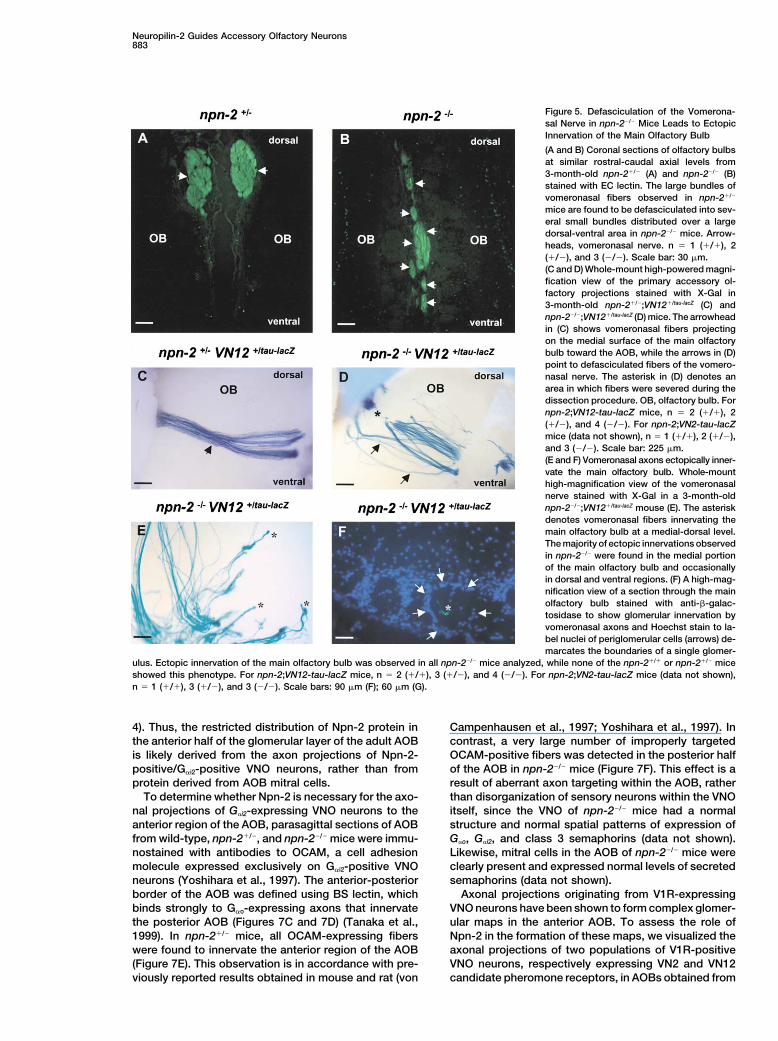
Figure 5. Defasciculation of the Vomerona-sal Nerve in npn-2�/� Mice Leads to EctopicInnervation of the Main Olfactory Bulb
(A and B) Coronal sections of olfactory bulbsat similar rostral-caudal axial levels from3-month-old npn-2�/� (A) and npn-2�/� (B)stained with EC lectin. The large bundles ofvomeronasal fibers observed in npn-2�/�
mice are found to be defasciculated into sev-eral small bundles distributed over a largedorsal-ventral area in npn-2�/� mice. Arrow-heads, vomeronasal nerve. n � 1 (�/�), 2(�/�), and 3 (�/�). Scale bar: 30 �m.(C and D) Whole-mount high-powered magni-fication view of the primary accessory ol-factory projections stained with X-Gal in3-month-old npn-2�/�;VN12�/tau-lacZ (C) andnpn-2�/�;VN12�/tau-lacZ (D) mice. The arrowheadin (C) shows vomeronasal fibers projectingon the medial surface of the main olfactorybulb toward the AOB, while the arrows in (D)point to defasciculated fibers of the vomero-nasal nerve. The asterisk in (D) denotes anarea in which fibers were severed during thedissection procedure. OB, olfactory bulb. Fornpn-2;VN12-tau-lacZ mice, n � 2 (�/�), 2(�/�), and 4 (�/�). For npn-2;VN2-tau-lacZmice (data not shown), n � 1 (�/�), 2 (�/�),and 3 (�/�). Scale bar: 225 �m.(E and F) Vomeronasal axons ectopically inner-vate the main olfactory bulb. Whole-mounthigh-magnification view of the vomeronasalnerve stained with X-Gal in a 3-month-oldnpn-2�/�;VN12�/tau-lacZ mouse (E). The asteriskdenotes vomeronasal fibers innervating themain olfactory bulb at a medial-dorsal level.The majority of ectopic innervations observedin npn-2�/� were found in the medial portionof the main olfactory bulb and occasionallyin dorsal and ventral regions. (F) A high-mag-nification view of a section through the mainolfactory bulb stained with anti-�-galac-tosidase to show glomerular innervation byvomeronasal axons and Hoechst stain to la-bel nuclei of periglomerular cells (arrows) de-marcates the boundaries of a single glomer-
ulus. Ectopic innervation of the main olfactory bulb was observed in all npn-2�/� mice analyzed, while none of the npn-2�/� or npn-2�/� miceshowed this phenotype. For npn-2;VN12-tau-lacZ mice, n � 2 (�/�), 3 (�/�), and 4 (�/�). For npn-2;VN2-tau-lacZ mice (data not shown),n � 1 (�/�), 3 (�/�), and 3 (�/�). Scale bars: 90 �m (F); 60 �m (G).
4). Thus, the restricted distribution of Npn-2 protein in Campenhausen et al., 1997; Yoshihara et al., 1997). Incontrast, a very large number of improperly targetedthe anterior half of the glomerular layer of the adult AOB
is likely derived from the axon projections of Npn-2- OCAM-positive fibers was detected in the posterior halfof the AOB in npn-2�/� mice (Figure 7F). This effect is apositive/G�i2-positive VNO neurons, rather than from
protein derived from AOB mitral cells. result of aberrant axon targeting within the AOB, ratherthan disorganization of sensory neurons within the VNOTo determine whether Npn-2 is necessary for the axo-
nal projections of G�i2-expressing VNO neurons to the itself, since the VNO of npn-2�/� mice had a normalstructure and normal spatial patterns of expression ofanterior region of the AOB, parasagittal sections of AOB
from wild-type, npn-2�/�, and npn-2�/� mice were immu- G�o, G�i2, and class 3 semaphorins (data not shown).Likewise, mitral cells in the AOB of npn-2�/� mice werenostained with antibodies to OCAM, a cell adhesion
molecule expressed exclusively on G�i2-positive VNO clearly present and expressed normal levels of secretedsemaphorins (data not shown).neurons (Yoshihara et al., 1997). The anterior-posterior
border of the AOB was defined using BS lectin, which Axonal projections originating from V1R-expressingVNO neurons have been shown to form complex glomer-binds strongly to G�o-expressing axons that innervate
the posterior AOB (Figures 7C and 7D) (Tanaka et al., ular maps in the anterior AOB. To assess the role ofNpn-2 in the formation of these maps, we visualized the1999). In npn-2�/� mice, all OCAM-expressing fibers
were found to innervate the anterior region of the AOB axonal projections of two populations of V1R-positiveVNO neurons, respectively expressing VN2 and VN12(Figure 7E). This observation is in accordance with pre-
viously reported results obtained in mouse and rat (von candidate pheromone receptors, in AOBs obtained from

Neuron884
Figure 6. Axons of Vomeronasal Neurons Are Repelled by Sema3F and Are Selectively Responsive to a Npn-2 Ligand Secreted by the MedialSide of the Olfactory Bulb In Vitro
(A–C) Vomeronasal epithelia from npn-2�/� (A and B) or npn-2�/� (C) E14 mouse embryos were cocultured with 293T cell aggregates transfectedwith either a control (A) or a AP-Sema3F expression vector (B and C). Axons growing out of vomeronasal epithelia from npn-2�/� mice (A) arerepelled by Sema3F, while axons from npn-2�/� mice (C) epithelia are unresponsive to Sema3F. n � 19 (�/�), 24 (�/�), and 3 (�/�). Scalebar: 80 �m.(D–H) Vomeronasal epithelia from E15 rat embryos were cultured with 293T cell aggregates transfected with a control (E) or AP-sema3Fexpression vector (F and G). Like murine vomeronasal axons, rat vomeronasal axons are repelled by Sema3F (F). Addition of anti-Npn-2 tothe growth medium partly blocked the Sema3F-induced repulsion (G). Arrowhead indicates nonrepelled fibers. Rat vomeronasal explantswere scored blind by assigning each explant a score of one to four, representing the degree of repulsion observed as presented in (D). Thepercentage of explants assigned to each score is represented graphically in (H). Comparisons between vector and AP-Sema3F, p � 0.0001;between AP-Sema3F and anti-Npn-2, p � 0.0001. Scale bar: 40 �m.(I–K) Vomeronasal epithelia from E15 rat embryos cultured in the proximity of the lateral (I) or medial (J and I) side of an olfactory bulb slicefrom P2 mice. Vomeronasal axons are repelled by factor(s) secreted by the medial side of the olfactory bulb (J), but are not repelled by thelateral side of the same bulb (I). Addition of anti-Npn-2 antibody to the growth medium partially inhibits the repulsion of vomeronasal axonsby the medial portion of the olfactory bulb (K). Arrowhead indicates nonrepelled fibers. The explants were scored blind by two observersusing criteria presented in (D). The percentage of explants assigned to each score by one of the observers is represented graphically in (L).Comparisons between responsiveness to the lateral and medial sides, p � 0.0001 for each observer; between medial and anti-Npn-2, p �
0.0001 for both observers. Scale bar: 40 �m.
npn-2�/�;VN2�/tau-lacZ or npn-2�/�;VN12�/tau-lacZ mice. 2 plays a major role in restricting the projection of G�i2-positive VNO neurons to the anterior AOB. Furthermore,Whole-mount X-Gal staining was performed on AOBs to
compare the VN2-and VN12 projection maps in npn-2�/� �-gal-positive fibers are seen converging into multipleglomeruli of the anterior AOB, suggesting that the ab-and npn-2�/� mice. Detailed analysis of the number, lo-
cation, and depth of blue glomeruli throughout the AOB sence of npn-2 expression does not affect glomerularconvergence of V1R-positive axons.allowed us to generate three-dimensional glomerular
maps. These analyses revealed that �-gal-positive fibers Taken together, these results indicate that npn-2 isnecessary for proper axonal targeting of G�i2-expressingappear almost evenly distributed between the anterior
and the posterior halves of the AOB in npn-2�/� (Figures neurons to the anterior region of the AOB. Are the axonsoriginating from the G�o population of VNO neurons simi-8B–8C� and 8E–8F�), whereas they are restricted to the
anterior AOB in npn-2�/� and wild-type animals (Figures larly affected in the npn-2 mutant? The accuracy of axo-nal projections from G�o-positive neurons to the poste-8A, 8A�, 8D, and 8D�). This result demonstrates that Npn-

Neuropilin-2 Guides Accessory Olfactory Neurons885
Figure 7. Loss of Zonal Targeting of Axons from G�i2-Expressing Vomeronasal Sensory Neurons within the Accessory Olfactory Bulb in npn-2�/�
Mice
(A and B) Npn-2-expressing vomeronasal axons of wild-type mice innervate the anterior accessory olfactory bulb. Parasagittal sections ofAOB from 2-month-old (A) or 2-week-old old npn-2�/� mice (B) stained with anti-Npn-2 (A) and AP-Sema3F (B). Scale bar: 120 �m.(C–H) Parasagittal sections of AOB from 3-month-old npn-2�/� (C and E), npn-2�/� (D and F), npn-2�/�;VN2�/tau-lacZ (G), and npn-2�/�;VN2�/tau-lacZ
(H) mice were stained with BS lectin (C and D), anti-OCAM (E and F), and anti-�-galactosidase (G and H). Projections of axons from G�i2-expressing neurons, detected using anti-OCAM (E and F) or anti-�-galactosidase (G and H) immunohistochemistry, are restricted to theanterior region of the AOB in npn-2�/� mice (E and I), while many of these fibers innervate the posterior AOB in npn-2�/� mice (F and H). Anasterisk indicates ectopic OCAM or �-galactosidase-positive fibers in the posterior half of the AOB from npn-2�/� mice. Sections (C) and (E)and (D) and (F) represent pairs of the same section doubly stained. For anti-OCAM staining, n � 2 (�/�), 2 (�/�), and 4 (�/�). For anti-�-galactosidase staining, n � 2 (�/�), 3 (�/�), and 5 (�/�).(I and J) Parasagittal sections of AOB from 3-month-old npn-2�/� (I) or npn-2�/� (J) mice were stained with an anti-G�o antibody. In contrastto axons of G�i2-expressing neurons, axons of G�o-expressing neurons are properly restricted to the posterior region of the AOB in npn-2�/�
mice. Arrowheads indicate the border between the anterior (ant) and posterior (post) regions of the AOB. n � 1 (�/�), 3 (�/�), and 4 (�/�).
rior AOB was assessed by anti-G�o immunostaining and plants of either anterior or posterior AOBs isolated fromearly postnatal rat brains and then processed for immu-BS lectin binding in npn-2�/� and npn-2�/� mice. Our
results show that G�o-expressing neurons are compara- nostaining with anti-tau-1. Rat vomeronasal epitheliawere used for these experiments because the poor out-bly restricted to the posterior half of the AOB in the
npn-2�/� and npn-2�/� (Figures 7I and 7J). This observa- growth of axons from mouse vomeronasal epithelia pre-tion is further supported by the similar binding we ob- cluded use of the mouse tissue for these experiments.serve of BS lectin to AOBs from npn-2�/� and npn- While a large number of vomeronasal axons grew ro-2�/� mice (data not shown). Grossly disrupted targeting, bustly into explants of anterior AOB (Figures 9A, 9B,therefore, appears restricted to G�i2-positive VNO neu- and 9Q), they grew away from explants of posterior AOBrons to which npn-2 expression is normally confined. (Figures 9E, 9F, and 9Q).
The axons observed projecting into the anterior AOBexplants must emanate from the VNO epithelial explantsVomeronasal Axons Are Responsive to a Npn-2for the following reasons. Fluorescent axons were de-Ligand Secreted by the Posterior Halftected in cocultures in which DiI crystals were placedof the Accessory Olfactory Bulbin the VNO explant (Figure 9D). In contrast, fluorescentThe ectopic innervation of the posterior half of the AOBaxons were never observed in cocultures in which DiIby axons of G�i2-positive VNO neurons observed incrystals were placed in the AOB explant (data notnpn-2�/� mice suggests that these fibers may normallyshown). Likewise, anti-tau staining of VNO/AOB cocul-be prevented from entering this region through Npn-2-tures at a time following axon outgrowth but prior todependent repulsion. To test this idea, we performedcontacting the AOB revealed robust projections fromcollagen gel coculture experiments in which E15 rat
vomeronasal epithelia were grown in proximity to ex- the VNO, but never from the AOB (Figure 9C). Taken

Neuron886
Figure 8. Spatial Distribution of Glomeruli Formed by VN2- and VN12-Expressing Vomeronasal Neurons in the AOB of npn-2�/� Mice
(A–F�) Whole-mount view of the AOB from 3-month-old npn-2�/�;VN2�/tau-lacZ (A and A�), npn-2�/�;VN2�/tau-lacZ (B–C�), npn-2�/�;VN12�/tau-lacZ (D andD�), and npn-2�/�;VN12�/tau-lacZ (E–F�). The spatial distribution and depth of glomeruli in the AOB glomerular layer is depicted using coloredspheres where red is for a superficial, green is for a medium deep, and blue is for a deep glomerulus (Belluscio et al., 1999). Prospective AOBantero-posterior boundary is indicated by a white line. In npn-2�/� mice, VN2- and VN12-expressing vomeronasal fibers are segregated tothe anterior half of the AOB and stereotypical glomerular mapping is observed (A, A�, D, and D�). In contrast, a large proportion of axons fromVN2- and VN12-expressing vomeronasal neurons are misrouted to the posterior half of the AOB ([B–C�] and [E–F�]). Insets are computergenerated maps of the pattern of posterior glomeruli expected if perfect mirror symmetry of anterior innervation is observed along the anterior-posterior boundary of the AOB (B�, C�, E�, and F�). A, anterior; P, posterior; L, lateral; M, medial. For npn-2;VN2-tau-LacZ mice, n � 1 (�/�),5 (�/�), and 5 (�/�). For npn-2;VN12-tau-LacZ mice, n � 2 (�/�), 5 (�/�), and 6 (�/�).

Neuropilin-2 Guides Accessory Olfactory Neurons887
Figure 9. Axons of Vomeronasal Neurons Are Responsive to a Npn-2 Ligand Secreted by the Posterior Half of the Accessory Olfactory Bulb
(A–L) Vomeronasal epithelia isolated from E15 rat embryos were cocultured with pieces of the anterior (A–D) or posterior region (E–J) of theAOB isolated from P1–P3 rat brains. Axons emanating from the vomeronasal epithelia are repelled by factor(s) secreted by the posterior AOBexplants (E, F, I, and J) but grow readily into anterior AOB explants (A and B). Axons emerging from the vomeronasal epithelium can bedetected prior to reaching the AOB by labeling with DiI (D) or anti-tau-1 (C). Addition of anti-Npn-2 IgG (G and H), but not preimmune IgG (Iand J), to the growth medium attenuates the repulsion of vomeronasal axons by the posterior AOB explants. Addition of anti-Npn-2 IgG didnot affect the outgrowth of axons from the vomeronasal epithelia when compared to preimmune IgG (K and L), nor did it promote outgrowthof fibers from the posterior AOB (O and P). The extent of growth of vomeronasal neurons into the AOB explants were scored blind by twoobservers by assigning each explant a score of one to four, representing the degree of ingrowth into the AOB explant as depicted in (S). Thepercentage of explants assigned to each score is represented graphically in (Q) for the comparison between anterior and posterior AOBexplants and in (R) for the comparison between preimmune and anti-Npn-2 treatments on repulsion by the posterior AOB. Differences betweenresponsiveness to the anterior and posterior AOB (p � 0.0001 for each of two observers) and between preimmune and anti-Npn-2 is p �
0.0001 for each of two observers.
together, these results suggest that the posterior half of projections from the posterior AOB (Figures 9K, 9L, and9P). In combination, these results indicate that the pos-the AOB secretes a factor(s) capable of repelling Npn-2-
positive vomeronasal neurons. terior AOB secretes a factor(s) that normally repels vom-eronasal neurons in a Npn-2-dependent manner.We next sought to determine whether the posterior
AOB repellent acts in a Npn-2-dependent fashion. Thus,in another series of experiments, we grew vomeronasal Discussionepithelial explants in proximity to posterior pieces of theAOB in medium containing either preimmune IgG or anti- Secreted semaphorins and their high-affinity receptors,
the neuropilins, have been implicated in the develop-Npn-2 (Figures 9G–9J and 9R). Addition of anti-Npn-2to the growth medium significantly attenuated the repul- ment of the nervous system through their chemorepul-
sive effect on a wide variety of neuronal projections.sive effect of the posterior AOB on vomeronasal neu-rons. Addition of anti-Npn-2 to the growth medium did Expression patterns of Npn-2 and its semaphorin li-
gands in the accessory olfactory system suggest thatnot alter the extent of outgrowth of axons from the vom-eronasal epithelia nor did it promote outgrowth of axonal Npn-2 plays a role in guiding and patterning axons of

Neuron888
vomeronasal neurons. Consistent with this model, we vate more dorsal or ventral regions of the MOB. Sema3Fexpressed in the medial MOB is likely to prevent ectopichave found that the secreted semaphorin Sema3F acts
in vitro as a potent vomeronasal fiber repellent in a innervation of the MOB by vomeronasal fibers projectingto the AOB. Thus, our results support a model in whichNpn-2-dependent manner. Importantly, in npn-2�/�
mice, the vomeronasal nerve is defasciculated and Npn-2 and its ligands are involved in maintaining theproper separation of the two olfactory systems.some vomeronasal neurons ectopically innervate the
MOB. Moreover, zonal segregation of axons from G�i2- Several studies have implicated the secreted sema-phorin Sema3A, a Npn-1 ligand, in the guidance of mainexpressing VNO neurons in the AOB is defective in npn-
2�/� mice. Finally, the posterior AOB secretes a factor olfactory neurons (Kobayashi et al., 1997; Pasterkampet al., 1999; Schwarting et al., 2000; Williams-Hogarththat repels axons of VNO neurons in a Npn-2-dependent
manner. Taken together, our results show that Npn-2 et al., 2000). Expression of a dominant-negative Npn-1molecule in olfactory neurons in chick leads to over-and its secreted semaphorin ligands play multiple roles
during the development of the accessory olfactory sys- shooting of main olfactory neurons through sema3A-expressing structures during the early establishment oftem, including regulating fasciculation of the vomerona-
sal nerve and zonal segregation of vomeronasal neu- these projections (Renzi et al., 2000). Moreover, micebearing a targeted deletion at the sema3A locus, thoughronal projections within the AOB.lethal at or near P1, show misrouting of Npn-1-positivefibers within the olfactory bulb in a pattern suggestingNpn-2 Is Required for Fasciculation of thethat Sema3A influences the establishment of these pro-Vomeronasal Nerve and for Segregationjections (Schwarting et al., 2000). Npn-1 and Npn-2 ap-of the Accessory Olfactory Systempear to be expressed in distinct populations of mainfrom the Main Olfactory Systemolfactory receptor neurons based on the segregation ofAnalysis of the vomeronasal nerve in npn-2�/� mice re-axons from Npn-1 and Npn-2-containing neurons in thevealed that Npn-2 is required to maintain axons of vom-MOB. Npn-2-expressing main olfactory neurons inner-eronasal neurons in a fasciculated state as they traversevate the lateral region of the MOB at caudal levels, whilethe medial surface of the MOB. The defasciculation ofNpn-1-expressing neurons more prominently innervatethe vomeronasal nerve observed in npn-2�/� mice mightthe medial region of the caudal MOB (Figure 3C;be due to the inability of Npn-2-deficient vomeronasalPasterkamp et al., 1998; Nagao et al., 2000; Schwartingneurons to respond to a Npn-2 ligand expressed in theet al., 2000). We have also noted ectopic projectionsmedial region of the MOB. Consistent with this idea, theof olfactory marker protein (OMP)-expressing fibers inmedial side of the MOB repels vomeronasal fibers in adeeper layers of the MOB beyond the glomerular layerNpn-2-dependent fashion in vitro. In contrast, the lateralin npn-2�/� mice (our unpublished data). It is thereforemain olfactory bulb does not repel vomeronasal neu-probable that expression of a Npn-2 ligand in the medialrons. The Npn-2 ligand Sema3F is an excellent candi-MOB serves not only to prevent ectopic innervation ofdate medial MOB repellent because it is more highlythe MOB by vomeronasal neurons, but also to preventexpressed medially than laterally in the MOB, and ininnervation by a subset of Npn-2-expressing main olfac-vitro Sema3F is a potent repellent for VNO neurons.tory neurons of the medial MOB. Combined with theExamination of the integrity of the vomeronasal nerveobservation that Sema3F can repel olfactory neurons inin mice harboring mutations in the genes encoding se-vitro (de Castro et al., 1999), it is likely that both Npn-1creted semaphorins will establish the identity of the li-and Npn-2 coordinately contribute to the proper wiringgand(s) responsible for promoting fasciculation of theof the main olfactory system.VN. How secreted semaphorins acting through Npn-2
promote fasciculation of the VN is still unclear. Onepossibility is that release of secreted semaphorins near Npn-2 Is Required for the Anterior-Posterior Zonal
Segregation, but Not for Axonal Convergencethe vomeronasal nerve forces VN bundling through sur-round repulsion. Alternatively, secreted semaphorins into Glomerular Domains
Remarkably, as many as one-half of VN2- and VN12-may regulate Npn-2-dependent adhesive properties ofvomeronasal neuron axons. expressing fibers were found to be misrouted to the
posterior region of the AOB in npn-2�/� mutant mice.The accessory and main olfactory systems are nor-mally segregated, despite the fact that axons of their Yet, despite this incorrect segregation, fibers that were
correctly targeted to the anterior AOB converge accu-respective primary sensory neurons are in close proxim-ity to one another as they extend through the cribriform rately into glomerular domains similar to those observed
in wild-type mice. This is in marked contrast to the ob-plate toward their central targets in the AOB and MOB.How are the projections of the primary sensory neurons servation that axons of vomeronasal neurons lacking
pheromone receptors segregate properly to the anteriorof the two olfactory systems kept separate? Our resultsindicate that Npn-2 is critical for preventing at least a half of the AOB but wander randomly in the anterior
glomerular layer and do not form glomeruli (Bellusciosubset of vomeronasal neuron axons from ectopicallyinnervating the MOB. In npn-2�/� mice, some vomerona- et al., 1999; Rodriguez et al., 1999). Therefore, Npn-2
appears essential for proper segregation of G�i2-positivesal fibers were found to ectopically innervate the MOB.These aberrant fibers often appear to terminate in glo- axons to the anterior AOB, but not for glomerular conver-
gence. Interestingly, V1R-expressing apical VNO neu-meruli, though further analysis is required to determinewhether active synapses are being formed. Most of rons mistargeted to the posterior AOB in npn-2�/� mu-
tants appear to contact posterior AOB glomeruli in athese errant fibers innervate the medial region of theMOB, although a minority of fibers were found to inner- nonrandom fashion, often exhibiting a striking pattern

Neuropilin-2 Guides Accessory Olfactory Neurons889
of innervation that resembles a mirror image of thatobserved in the anterior AOB (Figure 8). Further analyseswill address the suggestion from these data that a simi-lar, Npn-2-independent, glomerular map is encoded inboth zones of the AOB and whether or not V2R-express-ing basal VNO neurons utilize related guidance cues toestablish glomerular maps in the posterior AOB.
It is likely that other molecules provide guidance infor-mation within the AOB to direct axons to their appro-priate targets. A recent report suggests that ephrins andEph receptors contribute to guidance of vomeronasalprojections (Knoll et al., 2001). Ephrin A3 and A5 areexpressed in vomeronasal neurons, and EphB receptorsare expressed in the AOB during embryogenesis. Miceharboring a deletion at the ephrinA5 locus show someectopic innervation of the posterior AOB by some VN12-containing vomeronasal neurons. Thus, semaphorins
Figure 10. Npn-2 Is Required for Axonal Fasciculation and Tar-and ephrins may act in concert to restrict axon guidancegeting of Primary Accessory Olfactory Neuronsof V1R-expressing neurons within the AOB. The nearlyRepresentation of VNO projections in wild-type and npn-2�/� mice.even distribution of V1R fibers to the anterior and poste-G�i2-expressing (green) and G�o-expressing (orange) vomeronasalrior AOB in the npn-2�/� mutant strongly argues forneurons project to the anterior and posterior halves of the AOB,
Npn-2 playing a major role in this process. respectively. Axons of vomeronasal neurons projecting to the AOBThe exact mechanism by which Npn-2 mediates seg- grow along the medial surface of the olfactory bulb. Npn-2 is re-
regation of axons of V1R neurons to the anterior half of quired to maintain the nerve in a tightly fasciculated state. Expres-sion of a chemorepellent Npn-2 ligand by the medial surface of thethe AOB remains unknown. There are several possibili-olfactory bulb may maintain the fasciculation of the VN. In addition,ties that could explain the mistargeting of these neuronsNpn-2 dependent responsiveness of vomeronasal neurons to thisto the posterior AOB in npn-2�/� mice. First, improperligand prevents wandering of V1R-expressing neurons and ectopic
development or disorganization of the vomeronasal epi- innervation of the olfactory bulb by these neurons. By virtue of itsthelium may be indirectly responsible for this defect. expression pattern and in vitro repellent activity on vomeronasalThis seems unlikely, however, since in situ hybridization neurons, the secreted semaphorin Sema3F (red shading) is a good
candidate for regulating axonal fasciculation of the vomeronasalanalyses on the VNO of npn-2�/� mice showed that G�i2-nerve. Axons of vomeronasal neurons innervate the AOB in a segre-and G�o-positive neurons are properly restricted to thegated fashion. Npn-2 is essential for the segregation of G�i2-express-apical and basal zones of the VNO, respectively, anding neurons to the anterior half of the AOB. A large proportion of
that secreted semaphorins and the V1R tau-lacZ marker axons from V1R neurons are misrouted to the posterior half of thealso show identical distributions in the VNO in npn-2�/�
AOB in npn-2�/� mice. Nevertheless, fibers correctly targeted to theand npn-2�/� mice (data not shown). A second possibility anterior AOB converge accurately to form glomerular domains. In
contrast, V2R-expressing neurons are properly targeted to the pos-is that organization of fibers within the vomeronasalterior half of the AOB in npn-2�/� mice. Potential molecular mecha-nerve itself could be critical for the ultimate segregationnisms regulating the segregation of vomeronasal neurons in theof axons in the AOB. Defasciculation of the vomeronasalAOB are discussed in the text.
nerve could lead to loss of this intrinsic organizationthat, in turn, could result in improper entry of fibers intothe AOB and loss of zonal segregation of V1R axons. We in the AOB (Figure 10). V1R axons express Npn-2, theydo not favor this possibility either for several reasons. respond to the Npn-2 ligand Sema3F, and as many asImmunostaining of coronal sections through the vom- one-half of the V1R axons are mistargeted to the poste-eronasal nerve of wild-type mice using BS lectin, OCAM, rior half of the AOB in npn-2�/� animals. Importantly, inand G�o antibodies at caudal levels of the MOB showed vitro coculture experiments indicate that the posteriorthat G�i2- and G�o-positive neurons are not segregated AOB releases a chemorepellent ligand for Npn-2. Whatinto discrete bundles within the vomeronasal nerve (data is the posterior AOB repellant? Our analysis of secretednot shown). Also, many V1R-expressing axons enter the semaphorin expression has so far not revealed any clearAOB at the appropriate level, but they appear to make expression gradients in the mitral cell layer of the AOBincorrect steering decisions within the AOB neuropil (see that can obviously account for differential effects onFigure 8). Furthermore, it appears that the entire vomero- neurons expressing Npn-2. Nonetheless, the possibilitynasal nerve, including G�o-positive fibers, is defascicu- remains that a gradient of protein distribution for one orlated in npn-2�/� mice (Figure 5B). Despite this nerve several secreted semaphorins exists. The lack of gooddefect, G�o-positive fibers appear to innervate only the immunological reagents to detect these proteins makesposterior AOB in npn-2�/� mice; we did not observe it difficult to address this possibility. An alternate possi-loss of zonal segregation of these fibers. It is therefore bility is that cells of the posterior AOB express a Npn-2unlikely that defasciculation of sub-bundles within the ligand other than Sema3F that repels V1R-positivenerve is solely responsible for the incorrect segregation axons.of npn-2�/� V1R-containing axons in the AOB. While results of both in vivo and in vitro experiments
Our results support a model in which segregation of strongly support a model in which Npn-2 function isV1R-expressing axons to the anterior region of the AOB required within V1R-expressing axons, we cannot rule
out the possibility that Npn-2 on dendrites of mitral cellsis mediated by Npn-2 function on V1R-positive axons

Neuron890
times in PBS and then blocked for 1 hr at room temperature in TBSof the AOB also contributes to segregation of axonscontaining 0.24% gelatin and 0.4% Triton X-100. Sections werewithin the AOB. Indeed, Npn-2 is more highly expressedincubated overnight at 4C in blocking buffer containing purifiedin the mitral cell layer of the anterior AOB than posteriorNpn-2 IgG (2 mg/ml) diluted to 1:500. After several washes in PBS,
AOB during target innervation (Figure 4). Npn-2 on den- sections were incubated for 2 hr at room temperature in blockingdrites of anterior AOB mitral cells could facilitate in- solution containing either goat anti-rabbit Ig-HRP (1:1000; Amer-
sham) or alexa red conjugated goat anti-rabbit Ig (1:500, Moleculargrowth of axons of V1R neurons through sequestrationProbes).of Npn-2 ligands within the glomerular layer of the ante-
rior half of the AOB, which could, in turn, result in anX-Gal Staining and Glomerular Mappinganterior-posterior gradient of a repellent. Selective abla-Whole-mount X-Gal staining was performed as described (Bellusciotion of Npn-2 function in V1R-expressing neurons, aset al., 1999).
well as in target cells, will be needed to formally distin-guish between these and other possibilities. Finally, Collagen Gel Repulsion Assaysince axons of V2R-expressing neurons do not express VNOs were dissected from rat E15 and mouse E14 embryos andNpn-2, at least postnatally, and since they appear to incubated for 30 min on ice in a digestion solution prepared from
a 1:1 mix of 0.25% trypsin dissolved in Versene and 10 pancreatin.properly innervate the posterior region of the AOB inThe digestion was stopped by transferring the VNOs into L15 me-npn-2�/� mice, a future challenge will be to identify guid-dium containing 50% FBS and incubating for 1 hr on ice. Vomerona-ance cues and their receptors that control zonal segre-sal epithelia were seperated from the surrounding mesenchymal
gation of these projections. tissue and kept on ice in L15 medium containing 5% FBS. Vomero-Proper development of primary olfactory projections nasal epithelia were cultured on glass cover slips coated with poly-
in the main olfactory system relies on the expression of lysine (50 �g/ml) and laminin (10 �g/ml) for 3–4 days, in close proxim-ity to either 293T cell aggregates, mouse olfactory bulb slices, orodorant receptors in olfactory neurons and on their abil-pieces of rat AOB, as indicated in the figure legends. 293T cellity to respond to axonal guidance cues. It has now be-aggregates were prepared as previously described. 300–400 �mcome clear that, as observed in the main olfactory sys-olfactory bulb slices were prepared from P2 to P8 mouse brains
tem, establishment of connections in the primary using a vibratome. Slices of the rostral pole and most caudal partaccessory olfactory system is not only dependent on the of the bulb were discarded. Of the remaining slices, dorsal-ventralexpression of vomeronasal receptors in vomeronasal and medial-lateral orientation was determined based on morpholog-
ical criteria when the cocultures were assembled and embedded inneurons, but also on their ability to process guidancecollagen matrigel (5:1). For VNO-AOB coculture experiments, AOBsinformation provided by their surroundings. Our resultswere dissected from P1 to P3 rat brains and cut into three pieces.indicate that pheromone receptors and Npn-2 functionThe most anterior and posterior pieces were used in the experi-
independently and play complementary roles during ments, while pieces containing the anterior-posterior border werepatterning of primary accessory olfactory neuron projec- discarded. Cultures were allowed to grow for 2–4 days in Neurobasaltions. medium containing B27 and gentamycin before processing for im-
munostaining. The cultures were fixed in PFA (4%) for 1 hr, washedin PBS, and blocked in PHT (PBS containing horse serum [1%];Experimental ProceduresTriton X-100 [0.1%]). Primary antibodies were added to PHT con-taining Triton X-100 (0.3%) and incubated overnight at 4C. PrimaryIn Situ Hybridization and AP Fusion Protein Bindingantibodies used included anti-tau-1(1:1000; Chemicon), anti-OCAMNonradioactive, dioxygenin-labeled cRNA probes with either sense(1:1000), and anti-Npn-2 (1:500). The cultures were rinsed in PBSor antisense orientation were synthesized by in vitro transcription.and then incubated with secondary FITC- or Cy3-coupled secondaryProbes synthesized included rat sema3A (Giger et al., 1996), mouseantibody (1:1000) overnight at 4C. The explants were rinsed in PBSsema3B, mouse sema3C, rat sema3F (Giger et al., 2000), rat npn-2and mounted in fluoromount. For DiI labeling of explants, cultures(Giger et al., 1998), and rat G�i and G�o (Jones and Reed, 1987).were fixed overnight in PFA (4%). Crystals of DiI were introducedSections from mouse embryos at E16 and E18 or postnatal mice atinto the vomeronasal epithelia and the cultures were incubated atP5 and P15 were processed as previously described (Kolodkin et37C for 3 days to allow diffusion of the tracer. Cultures were thenal. 1997). AP fusion protein binding to tissue sections was performedrinsed in PBS and mounted in fluoromount. Confocal microscopyas described before (Giger et al., 1998).with z-stack analysis was performed to image the explants. Forquantification of axon repulsion, each explant was given a scoreImmunohistochemical Proceduresaccording to the degree of repulsion or ingrowth observed as repre-Adult (3- to 4-month-old) mice were anesthetized and perfusedsented in Figures 5D and 9A. This approach was used for scoringtranscardially with ice-cold PBS containing 4% paraformaldehyde.since the very small diameter of axons projecting from the vomero-Brains were dissected, postfixed for 3 hr in perfusion solution, andnasal epithelia prevented us from accurately tracing and counting/cryoprotected overnight in PBS containing 30% sucrose. Cryosec-measuring individual neurons. The explants were scored blind bytions (25 �m) were mounted on superfrost plus microscope slides,two independent observers and paired t test statistical analysis wasrinsed in TBS (50 mM Tris-HCl [pH 7.6] and 150 mM NaCl), andperformed.blocked for 2 hr in TNT (50 mM Tris-HCl [pH 7.6], 500 mM NaCl,
and 0.5% Triton X-100) containing 10% fetal bovine serum (FBS).Sections were incubated overnight with primary antibody at 4C in AcknowledgmentsTNT/2% FBS using the following dilutions: anti-OCAM (1:1000) (agift of Dr. Kensaku Mori), anti-�-galactosidase (1:500) (Sigma clone We thank Randy Reed, Gabrielle Ronnett, and Jeroen Pasterkamp
for comments on the manuscript and members of the Dulac, Ginty,Gal-13); anti-GTP Binding Protein Go� (1 �g/ml; Medical & BiologicalLaboratories Co.). After rinsing in TBS, primary antibody was detected and Kolodkin laboratories for helpful discussions. We are especially
grateful to Rebecca Kitko for technical assistance and Renate Hel-with the appropriate oregon green or Cy3-conjugated secondaryantibody (1:500; Molecular Probes) and either Fluorescein-Erythrina miss-Peralta for help with illustrations. We thank K. Mori for anti-
OCAM serum; J. Verhaagen, H. Betz, and S. Strittmatter for sema-Cristagalli lectin (1:500; Vector Laboratories) or Rhodamine-Griffonia(Bandeiraea) Simplicifolia Lectin I (1:500; Vector Laboratories) in phorin clones; and R. Reed for the G�i2 and G�o cDNA clones. Npn-2
mice are the property of Glaxo Smithkline Pharmaceuticals, andTNT containing 2% FBS. For anti-Npn-2 immunohistochemistry, E16to 3-month-old tissue was processed as described above. Prior to requests for these mice should be directed to Frank S. Walsh
([email protected]). This work was supported by the NIH/the incubation with anti-Npn-2, sections were pretreated overnightat 60C in 50% formamide, 2 SSC. Sections were rinsed several NIMH-R01MH59199 (D.D.G. and A.L.K.), the Howard Hughes Medical

Neuropilin-2 Guides Accessory Olfactory Neurons891
Institute (C.D. and D.D.G.), McKnight Endowment Fund for Neurosci- the EphA family in the topographic targeting of vomeronasal axons.Development 128, 895–906.ence (A.L.K.), NARSAD (R.J.G.), Human Frontier Science Program Or-
ganization, and Canadian Institutes of Health Research (J.-F.C.). Kobayashi, H., Koppel, A.M., Luo, Y., and Raper, J.A. (1997). A rolefor collapsin-1 in olfactory and cranial sensory axon guidance. J.
Received: July 18, 2001 Neurosci. 17, 8339–8352.Revised: January 11, 2002 Kolodkin, A.L., Levengood, D.V., Rowe, E.G., Tai, U.-T., Giger, R.J.,
and Ginty, D.D. (1997). Neuropilin is a semaphorin III receptor. CellReferences 90, 753–762.
Matsunami, H., and Buck, L.B. (1997). A multigene family encodingBelluscio, L., Koentges, G., Axel, R., and Dulac, C. (1999). A map of
a diverse array of putative pheromone receptors in mammals. Cellpheromone receptor activation in the mammalian brain. Cell 97,
90, 775–784.209–220.
Mombaerts, P., Wang, F., Dulac, C., Chao, S.K., Nemes, A., Mendel-Berghard, A., and Buck, L.B. (1996). Sensory transduction in vom-
sohn, M., Edmondson, J., and Axel, R. (1996). Visualizing an olfactoryeronasal neurons: evidence for G�o, G�i2, and adenylyl cyclase II as
sensory map. Cell 87, 675–686.major components of a pheromone signaling cascade. J. Neurosci.
Nagao, H., Yoshihiro, Y., Mitsui, S., Fujisawa, H., and Mori, K. (2000).16, 909–918.Two mirror image sensory maps with domain organizations in the
Chen, H., Chedotal, A., He, Z., Goodman, C.S., and Tessier-Lavigne,mouse main olfactory bulb. Neuroreport 11, 3023–3027.
M. (1997). Neuropilin-2, a novel member of the neuropilin family, isNakamura, F., Kalb, R.G., and Strittmatter, S.M. (2000). Moleculara high-affinity receptor for the semaphorins Sema E and Sema IV,basis of semaphorin-mediated axon guidance. J. Neurobiol. 44,but not Sema III. Neuron 19, 547–559.219–229.
Chen, H., He, Z., Bagri, A., and Tessier-Lavigne, M. (1998). Semapho-Pantages, E., and Dulac, C. (2000). A novel family of candidaterin-neuropilin interactions underlying sympathetic axon responsespheromone receptors in mammals. Neuron 28, 835–845.to class III semaphorins. Neuron 21, 1283–1290.Pasterkamp, R.J., De Winter, F., Holtmaat, A.J.G.D., and Verhaagen,Chen, H., Bagri, A., Zupicich, J.A., Zou, Y., Stoeckli, E., Pleasure,J. (1998). Evidence for a role of the chemorepellent semaphorin IIIS.J., Lowenstein, D.H., Skarnes, W.C., Chedotal, A., and Tessier-and its receptor neuropilin-1 in the regeneration of primary olfactoryLavigne, M. (2000). Neuropilin-2 regulates the development of selec-axons. J. Neurosci. 18, 9962–9976.tive cranial and sensory nerves and hippocampal mossy fiber pro-
jections. Neuron 25, 43–56. Pasterkamp, R.J., Ruitenberg, M.J., and Verhaagen, J. (1999). Sema-phorins and their receptors in olfactory axon guidance. Cell Mol.de Castro, F., Hu, L., Drabkin, H., Sotelo, C., and Chedotal, A. (1999).Biol. 45, 763–779.Chemoattraction and chemorepulsion of olfactory bulb axons by
different secreted semaphorins. J. Neurosci. 19, 4428–4436. Raper, J.A. (2000). Semaphorins and their receptors in vertebratesand invertebrates. Curr. Opin. Neurobiol. 10, 88–94.Dulac, C. (2000). Sensory coding of pheromone signals in mammals.
Curr. Opin. Neurobiol. 10, 511–518. Renzi, M.J., Wexler, T.L., and Raper, J.A. (2000). Olfactory sensoryaxons expressing a dominant-negative semaphorin receptor enterDulac, C., and Axel, R. (1995). A novel family of genes encodingthe CNS early and overshoot their target. Neuron 28, 437–447.putative pheromone receptors in mammals. Cell 83, 195–206.Ressler, K.J., Sullivan, S.L., and Buck, L.B. (1993). A zonal organiza-Giger, R.J., Wolfer, D.P., De Wit, G.M.J., and Verhaagen, J. (1996).tion of odorant receptor gene expression in the olfactory epithelium.Anatomy of rat semaphorin III/collapsin-1 mRNA expression andCell 73, 597–609.relationship to developing nerve tracts during neuroembryogenesis.
J. Comp. Neurol. 375, 378–392. Ressler, K.J., Sullivan, S.L., and Buck, L.B. (1994). Information cod-ing in the olfactory system: evidence for a stereotyped and highlyGiger, R.J., Urquhart, E.R., Gillespie, S.K.H., Levengood, D.V., Ginty,organized epitope map in the olfactory bulb. Cell 79, 1245–1255.D.D., and Kolodkin, A.L. (1998). Neuropilin-2 is a receptor for sema-
phorin IV: insight into the structural basis of receptor function and Rodriguez, I., Feinstein, P., and Mombaerts, P. (1999). Variable pat-terns of axonal projections of sensory neurons in the mouse vomero-specificity. Neuron 21, 1079–1092.nasal system. Cell 97, 199–208.Giger, R.J., Cloutier, J.F., Sahay, A., Prinjha, R.K., Levengood, D.V.,
Moore, S.E., Pickering, S., Simmons, D., Rastan, S., Walsh, F.S., et Rohm, B., Ottemeyer, A., Lohrum, M., and Puschel, A.W. (2000).Plexin/neuropilin complexes mediate repulsion by the axonal guid-al. (2000). Neuropilin-2 is required in vivo for selective axon guidance
responses to secreted semaphorins. Neuron 25, 29–41. ance signal semaphorin 3A. Mech. Dev. 93, 95–104.
Ryba, N.J., and Tirindelli, R. (1997). A new multigene family of puta-Halpern, M. (1987). The organization and function of the vomerona-sal system. Annu. Rev. Neurosci. 10, 325–362. tive pheromone receptors. Neuron 19, 371–379.
Schwarting, G.A., Kostek, C., Ahmad, N., Dibble, C., Pays, L., andHalpern, M., Shapiro, L.S., and Jia, C. (1995). Differential localizationof G proteins in the opossum vomeronasal system. Brain Res. 677, Puschel, A.W. (2000). Semaphorin 3A is required for guidance of
olfactory axons in mice. J. Neurosci. 20, 7691–7697.157–161.
He, S., and Tessier-Lavigne, M. (1997). Molecular basis of axonal Strotmann, J., Wanner, I., Krieger, J., Raming, K., and Breer, H.(1992). Expression of odorant receptors in spatially restricted sub-chemorepulsion: neuropilin is a semaphorin/collapsin receptor. Cell
90, 739–751. sets of chemosensory neurones. Neuroreport 3, 1053–1056.
Strotmann, J., Wanner, I., Helfrich, T., Beck, A., and Breer, H. (1994).Herrada, G., and Dulac, C. (1997). A novel family of putative phero-mone receptors in mammals with a topographically organized and Rostro-caudal patterning of receptor-expressing olfactory neurones
in the rat nasal cavity. Cell Tissue Res. 278, 11–20.sexually dimorphic distribution. Cell 90, 763–773.
Jones, D.T., and Reed, R.R. (1987). Molecular cloning of five GTP- Takahashi, T., and Strittmatter, S.M. (2001). PlexinA1 autoinhibitionby the plexin sema domain. Neuron 29, 429–439.binding protein cDNA species from rat olfactory neuroepithelium.
J. Biol. Chem. 262, 14241–14249. Takahashi, T., Nakamura, F., Jin, Z., Kalb, R., and Strittmatter, S.(1998). Semaphorins A and E act as antagonists of neuropilin-1 andKawakami, A., Kitsukawa, T., Takagi, S., and Fujisawa, H. (1996).
Developmentally regulated expression of a cell surface protein, neu- agonists of neuropilin-2 receptors. Nat. Neurosci. 1, 487–493.ropilin, in mouse nervous system. J. Neurobiol. 29, 1–17. Takahashi, T., Fournier, A., Nakamura, F., Wang, L.-H., Murakami,
Y., Kalb, R.G., Fujisawa, H., and Strittmatter, S.M. (1999). Plexin-Kitsukawa, T., Shimizu, M., Sanbo, M., Hirata, T., Taniguchi, M.,Bekku, Y., Yagi, T., and Fujisawa, H. (1997). Neuropilin-semaphorin neuropilin-1 complexes form functional semaphorin-3A receptors.
Cell 99, 59–69.III/D-mediated chemorepulsive signals play a crucial role in periph-eral nerve projection in mice. Neuron 19, 995–1005. Tamagnone, L., Artigiani, S., Chen, H., He, Z., Ming, G.-L., Song,
H.-J., Chedotal, A., Winberg, M.L., Goodman, C.S., Poo, M.-M., etKnoll, B., Zarbalis, K., Wurst, W., and Drescher, U. (2001). A role for

Neuron892
al. (1999). Plexins are a large family of receptors for transmembrane,secreted, and GPI-anchored semaphorins in vertebrates. Cell 99,71–80.
Tanaka, M., Treloar, H., Kalb, R.G., Greer, C.A., and Strittmatter,S.M. (1999). G(o) protein-dependent survival of primary accessoryolfactory neurons. Proc. Natl. Acad. Sci. USA 96, 14106–14111.
Vassar, R., Ngai, J., and Axel, R. (1993). Spatial segregation of odor-ant receptor expression in the mammalian olfactory epithelium. Cell74, 309–318.
Vassar, R., Chao, S.K., Sitcheran, R., Nunez, J.M., Vosshall, L.B.,and Axel, R. (1994). Topographic organization of sensory projectionsto the olfactory bulb. Cell 79, 981–991.
von Campenhausen, H., Yoshihara, Y., and Mori, K. (1997). OCAMreveals segregated mitral/tufted cell pathways in developing acces-sory olfactory bulb. Neuroreport 8, 2607–2612.
Williams-Hogarth, L.C., Puche, A.C., Torrey, C., Cai, X., Song, I.,Kolodkin, A.L., Shipley, M.T., and Ronnett, G.V. (2000). Expressionof semaphorins in developing and regenerating olfactory epithelium.J. Comp. Neurol. 423, 565–578.
Yoshihara, Y., Kawasaki, M., Tamada, A., Fujita, H., Hayashi, H.,Kagamiyama, H., and Mori, K. (1997). OCAM: a new member ofthe neural cell adhesion molecule family related to zone-to-zoneprojection of olfactory and vomeronasal axons. J. Neurosci. 17,5830–5842.