An expression signature of syndecan-1 (CD138), E-cadherin ...
12
Open Access Available online http://breast-cancer-research.com/content/9/1/R8 Page 1 of 12 (page number not for citation purposes) Vol 9 No 1 Research article An expression signature of syndecan-1 (CD138), E-cadherin and c-met is associated with factors of angiogenesis and lymphangiogenesis in ductal breast carcinoma in situ Martin Götte 1 , Christian Kersting 2 , Isabel Radke 1 , Ludwig Kiesel 1 and Pia Wülfing 1 1 Department of Obstetrics and Gynecology, Münster University Hospital, Domagkstrasse 11, Münster, D-48149, Germany 2 Department of Pathology, Münster University Hospital, Domagkstrasse, Münster, D-48149, Germany Corresponding author: Martin Götte, [email protected] Received: 28 Jul 2006 Revisions requested: 24 Aug 2006 Revisions received: 27 Nov 2006 Accepted: 23 Jan 2007 Published: 23 Jan 2007 Breast Cancer Research 2007, 9:R8 (doi:10.1186/bcr1641) This article is online at: http://breast-cancer-research.com/content/9/1/R8 © 2007 Götte et al.; licensee BioMed Central Ltd. This is an open access article distributed under the terms of the Creative Commons Attribution License (http://creativecommons.org/licenses/by/2.0 ), which permits unrestricted use, distribution, and reproduction in any medium, provided the original work is properly cited. Abstract Introduction Heparan sulphate proteoglycan syndecan-1 modulates cell proliferation, adhesion, migration and angiogenesis. It is a coreceptor for the hepatocyte growth factor receptor c-met, and its coexpression with E-cadherin is synchronously regulated during epithelial-mesenchymal transition. In breast cancer, changes in the expression of syndecan-1, E-cadherin and c-met correlate with poor prognosis. In this study we evaluated whether coexpression of these functionally linked prognostic markers constitutes an expression signature in ductal carcinoma in situ (DCIS) of the breast that may promote cell proliferation and (lymph)angiogenesis. Methods Expression of syndecan-1, E-cadherin and c-met was detected immunohistochemically using a tissue microarray in tumour specimens from 200 DCIS patients. Results were correlated with the expression patterns of angiogenic and lymphangiogenic markers. Coexpression of the three prognostic markers was evaluated in human breast cancer cells by confocal immunofluorescence microscopy and RT-PCR. Results Coexpression and membrane colocalization of the three markers was confirmed in MCF-7 cells. E-cadherin expression decreased, and c-met expression increased progressively in more aggressive cell lines. Tissue microarray analysis revealed strong positive staining of tumour cells for syndecan-1 in 72%, E-cadherin in 67.8% and c-met in 48.6% of DCIS. E-cadherin expression was significantly associated with c-met and syndecan-1. Expression of c-met and syndecan-1 was significantly more frequent in the subgroup of patients with pure DCIS than in those with DCIS and a coexisting invasive carcinoma. Levels of c-met and syndecan-1 expression were associated with HER2 expression. Expression of c-met significantly correlated with expression of endothelin A and B receptors, vascular endothelial growth factor (VEGF)-A and fibroblast growth factor receptor-1, whereas E-cadherin expression correlated significantly with endothelin A receptor, VEGF-A and VEGF-C staining. Conclusion Syndecan-1, E-cadherin and c-met constitute a marker signature associated with angiogenic and lymphangiogenic factors in DCIS. This coexpression may reflect a state of parallel activation of different signal transduction pathways, promoting tumour cell proliferation and angiogenesis. Our findings have implications for future therapeutic approaches in terms of a multiple target approach, which may be useful early in breast cancer progression. Introduction Syndecan-1/CD138 (Sdc1) is a cell surface heparan sulphate proteoglycan that is highly expressed by epithelial and plasma cells. Via its heparan sulphate chains, Sdc1 binds to a variety of growth and angiogenic factors and acts as a classical core- ceptor for growth factor receptors, thus promoting cell prolif- eration [1]. Moreover, Sdc1 interacts with ligands in the extracellular matrix and on cell surfaces, functioning as a cell bFGF = basic fibroblast growth factor; DCIS = ductal breast carcinoma in situ; E-cad = E-cadherin; ER = oestrogen receptor; ET = endothelin; ET A/ B R = endothelin A/B receptor; FGFR = fibroblast growth factor receptor; HER = human epidermal growth factor receptor; HGF = hepatocyte growth factor; PBS = phosphate-buffered saline; PR = progesterone receptor; RT-PCR = reverse transcription polymerase chain reaction; Sdc1 = syndecan- 1; TMA = tissue microarray; VEGF = vascular endothelial growth factor.
-
Upload
khangminh22 -
Category
Documents
-
view
0 -
download
0
Transcript of An expression signature of syndecan-1 (CD138), E-cadherin ...

Available online http://breast-cancer-research.com/content/9/1/R8
Open AccessVol 9 No 1Research articleAn expression signature of syndecan-1 (CD138), E-cadherin and c-met is associated with factors of angiogenesis and lymphangiogenesis in ductal breast carcinoma in situMartin Götte1, Christian Kersting2, Isabel Radke1, Ludwig Kiesel1 and Pia Wülfing1
1Department of Obstetrics and Gynecology, Münster University Hospital, Domagkstrasse 11, Münster, D-48149, Germany2Department of Pathology, Münster University Hospital, Domagkstrasse, Münster, D-48149, Germany
Corresponding author: Martin Götte, [email protected]
Received: 28 Jul 2006 Revisions requested: 24 Aug 2006 Revisions received: 27 Nov 2006 Accepted: 23 Jan 2007 Published: 23 Jan 2007
Breast Cancer Research 2007, 9:R8 (doi:10.1186/bcr1641)This article is online at: http://breast-cancer-research.com/content/9/1/R8© 2007 Götte et al.; licensee BioMed Central Ltd. This is an open access article distributed under the terms of the Creative Commons Attribution License (http://creativecommons.org/licenses/by/2.0), which permits unrestricted use, distribution, and reproduction in any medium, provided the original work is properly cited.
Abstract
Introduction Heparan sulphate proteoglycan syndecan-1modulates cell proliferation, adhesion, migration andangiogenesis. It is a coreceptor for the hepatocyte growth factorreceptor c-met, and its coexpression with E-cadherin issynchronously regulated during epithelial-mesenchymaltransition. In breast cancer, changes in the expression ofsyndecan-1, E-cadherin and c-met correlate with poorprognosis. In this study we evaluated whether coexpression ofthese functionally linked prognostic markers constitutes anexpression signature in ductal carcinoma in situ (DCIS) of thebreast that may promote cell proliferation and(lymph)angiogenesis.
Methods Expression of syndecan-1, E-cadherin and c-met wasdetected immunohistochemically using a tissue microarray intumour specimens from 200 DCIS patients. Results werecorrelated with the expression patterns of angiogenic andlymphangiogenic markers. Coexpression of the three prognosticmarkers was evaluated in human breast cancer cells by confocalimmunofluorescence microscopy and RT-PCR.
Results Coexpression and membrane colocalization of thethree markers was confirmed in MCF-7 cells. E-cadherinexpression decreased, and c-met expression increasedprogressively in more aggressive cell lines. Tissue microarray
analysis revealed strong positive staining of tumour cells forsyndecan-1 in 72%, E-cadherin in 67.8% and c-met in 48.6% ofDCIS. E-cadherin expression was significantly associated withc-met and syndecan-1. Expression of c-met and syndecan-1was significantly more frequent in the subgroup of patients withpure DCIS than in those with DCIS and a coexisting invasivecarcinoma. Levels of c-met and syndecan-1 expression wereassociated with HER2 expression. Expression of c-metsignificantly correlated with expression of endothelin A and Breceptors, vascular endothelial growth factor (VEGF)-A andfibroblast growth factor receptor-1, whereas E-cadherinexpression correlated significantly with endothelin A receptor,VEGF-A and VEGF-C staining.
Conclusion Syndecan-1, E-cadherin and c-met constitute amarker signature associated with angiogenic andlymphangiogenic factors in DCIS. This coexpression may reflecta state of parallel activation of different signal transductionpathways, promoting tumour cell proliferation and angiogenesis.Our findings have implications for future therapeuticapproaches in terms of a multiple target approach, which maybe useful early in breast cancer progression.
IntroductionSyndecan-1/CD138 (Sdc1) is a cell surface heparan sulphateproteoglycan that is highly expressed by epithelial and plasmacells. Via its heparan sulphate chains, Sdc1 binds to a variety
of growth and angiogenic factors and acts as a classical core-ceptor for growth factor receptors, thus promoting cell prolif-eration [1]. Moreover, Sdc1 interacts with ligands in theextracellular matrix and on cell surfaces, functioning as a cell
Page 1 of 12(page number not for citation purposes)
bFGF = basic fibroblast growth factor; DCIS = ductal breast carcinoma in situ; E-cad = E-cadherin; ER = oestrogen receptor; ET = endothelin; ETA/
BR = endothelin A/B receptor; FGFR = fibroblast growth factor receptor; HER = human epidermal growth factor receptor; HGF = hepatocyte growth factor; PBS = phosphate-buffered saline; PR = progesterone receptor; RT-PCR = reverse transcription polymerase chain reaction; Sdc1 = syndecan-1; TMA = tissue microarray; VEGF = vascular endothelial growth factor.

Breast Cancer Research Vol 9 No 1 Götte et al.
adhesion molecule [1]. We recently showed that Sdc1 is amodulator of proteolytic activities and chemokine functions invivo, which orchestrates leucocyte recruitment and tissueremodelling during inflammation and wound repair [2-4]. InSdc1-deficient and Sdc1-overexpressing mouse models,abnormal blood vessel formation is observed during woundrepair, confirming a role for Sdc1 as a regulator of angiogen-esis in vivo [2,4]. Because the biological functions of Sdc1potentially affect several steps in tumour progression, it is notsurprising that a prognostic value has been assigned tochanges in Sdc1 expression in several cancer types, includingcolorectal, gastric, pancreatic, prostate, lung, endometrial andovarian cancers, as well as squamous cell carcinoma of thehead and neck (for review, see Yip and coworkers [5]). Inbreast cancer, increased expression of Sdc1 correlates withan unfavourable prognosis [6-8] and poor response to chem-otherapy [9]. Of note, several proteins that are functionallylinked to Sdc1 by virtue of their biology are prognostic markerson their own (Table 1). In multiple myeloma, Sdc1 mediates lig-and binding and signalling through the hepatocyte growth fac-tor (HGF) receptor tyrosine kinase c-met, resulting inincreased cancer cell proliferation [10].
Similar mechanisms may be of relevance in breast cancer,because prognostic value has been established for c-metexpression in a number of clinical studies of breast cancerpatients (Table 1). Signal transduction mediated by c-metmodulates cell dissociation and motility, and protease overex-pression [11,12]. Moreover, ribozyme targeting of c-met in
mammary cancer cells reduced mammary cancer and tumour-associated angiogenesis in a xenograft model [12]. To mobi-lize its full transforming potential in breast cancer, c-metappears to depend on coactivating factors, such as overex-pression of additional proto-oncogenes (MYC, RON), or β4integrin activity [13-15]. Similarly, Sdc1 regulates αvβ3 integrinactivation and signalling in breast cancer cell lines [16,17].Sdc1-integrin complexes may thus synergistically contributeto tumour progression driven by c-met overexpression.
The calcium-dependent cell-cell adhesion molecule E-cad-herin (E-cad) is an established prognostic marker in breastcancer (Table 1). E-cad expression is irreversibly lost in inva-sive lobular breast cancer, and this feature has been used bypathologists to distinguish between ductal and lobular neopla-sia [18-20]. Like Sdc1, E-cad is mostly present in epithelialcells, and is required for maintaining the epitheloid phenotypeand inhibition of density-dependent cell growth [21]. Coordi-nated regulation of E-cad and Sdc1 expression is seen duringdevelopment [22] and in mammary tumour cells subjected toantisense RNA mediated downregulation of Sdc1 [23] or E-cad [24], respectively. Sdc1 and E-cad colocalize and coim-munoprecipitate with the transcriptional regulator β-catenin inepithelial cells, suggesting both a functional and physicalassociation [25]. Of note, expression of Sdc1 is required forproper development of a β-catenin responsive mammary epi-thelial progenitor cell population, which appears to be themechanistic principle underlying resistance of Sdc1-deficientmice to wnt-1 mediated breast cancer [5,26-28].
Table 1
Cancer-related functions and interrelation of Sdc1, c-met and E-cad
Marker Molecular characteristics Biological functions relevant to cancera
Clinical relevance to breast cancera
Relation to c-meta Relation to E-cada
Sdc1 Cell surface heparan sulphate proteoglycan
Cell and matrix receptor [1]Coreceptor for chemokines, angiogenic and growth factors, and modulator of proteolytic activity [2-4,16,57]
Positive correlation with poor prognosis and tumour angiogenesis [6-8,33]Predictive factor in neoadjuvant chemotherapy [9]
Sdc1 is c-met co-receptor in multiple myeloma [10]
Coordinated regulation and codistribution in mammary tumor cells and epithelial-mesenchymal transition [22-25]β-Catenin responsive progenitor cells depend on Sdc1 [27,28]
c-met Transmembrane tyrosine kinase receptor for hepatocyte growth factor
c-met pathway modulates cell dissociation and motility, protease overexpression and stimulates angiogenesis [11,12]
Expression associated with poor outcome in patients with (axillary) lymph node negative breast cancer [15,47,70]Negative prognostic factor in breast cancer [71,72]
Not applicable Correlation with abnormal β-catenin expression suggests downregulation of E-cad/β-catenin by c-met [73]
E-cad Epithelial calcium-dependent cell adhesion molecule
Ensures structural integrity, and contact inhibition of epithelia [21]Expression changes during epithelial-mesenchymal transition [40]Involved in β-catenin-mediated signalling [20]
Membranous staining is independent predictor for disease-free survival in lobular breast cancer [44]Loss of expression is associated with negative ER status, high histological grade, metastasis and poor prognosis in breast cancer [43,45,46,74,75]E-cad is used to distinguish ductal from lobular neopasia [18,19]
(See Sdc1) Not applicable
aSelected examples are given, along with references. E-cad, E-cadherin; ER, oestrogen receptor; Sdc, syndecan.
Page 2 of 12(page number not for citation purposes)

Available online http://breast-cancer-research.com/content/9/1/R8
In the present study we evaluated coexpression of the threefunctionally linked prognostic factors, namely Sdc1, E-cad andc-met, in ductal carcinoma in situ (DCIS) of the breast by sem-iquantitative immunohistochemistry of a tissue microarray con-taining tumour specimens from 200 patients. We recentlyshowed that several proangiogenic factors, including fibrob-last growth factor receptor (FGFR)-1, vascular endothelialgrowth factor (VEGF)-C, its receptor Flt-4, and the endothelinA receptor (ETAR), are highly expressed in DCIS, suggestingthat in situ carcinomas can induce angiogenesis and lym-phangiogenesis [29]. Although expression of endothelin andVEGF receptors was previously believed to be largelyrestricted to the vascular endothelium, these receptors havealso been detected in breast cancer cells, suggesting an auto-crine effect of their ligands on tumour cells [29-31]. BecauseSdc1 and c-met modulate angiogenesis in vivo[2,4,12,32,33], we determined the association of these mole-cules with markers of angiogenesis and lymphangiogenesis inDCIS. Moreover, we characterized the coexpression of Sdc1,E-cad and c-met in three differently aggressive human breastcancer cell lines by RT-PCR and by confocal immunofluores-cence microscopy. The aim of the study was to determinewhether coexpression of the functionally linked molecularmarkers Sdc1, E-cad and c-met may constitute a novel angio-genesis-associated molecular marker signature in DCIS.
Materials and methodsCell culture and confocal immunofluorescence microscopyThe human breast cancer cell lines MCF-7 and MDA-MB468were from our cell culture collection [34]. MCF-7 cells werecultured in RPMI-1640 medium (Gibco, Karlsruhe, Germany)supplemented with 10% foetal calf serum (PAA, Cölbe, Ger-many) and 2 mmol/l L-glutamine. MDA-MB231 (CLS CellLines Service, Eppelheim, Germany) and MDA-MB468 cellswere maintained in DMEM (Dulbecco's modified Eaglemedium) high-glucose medium (Invitrogen, Karlsruhe, Ger-many) supplemented with 1% L-glutamine, 1% penicillin-streptomycin and 10% foetal calf serum at 7.5% carbon diox-ide. For immunocytochemistry, cells were grown to 80% con-fluence in eight-well chamber slides (Corning Costar,Wiesbaden, Germany), fixed with ice-cold methanol, and sub-jected to immunostaining as follows. After a 30 min blockingstep with phosphate-buffered saline (PBS) containing 10%goat serum (DAKO, Glostrup Denmark), the slides werewashed with PBS, and the following primary antibodies wereused in an overnight incubation at 4°C at a dilution of 1:100 inPBS/1% bovine serum albumin: mouse anti-syndecan-1(clone BB4; Serotec, Duesseldorf, Germany), rabbit anti-c-met (Santa Cruz, Santa Cruz, CA, USA), and mouse anti-E-cadherin (BD Transduction Laboratories, San Diego, CA,USA). The slides were washed three times for 5 min with PBSand incubated with AlexaFluor 546-conjugated goat-anti-mouse IgG (Invitrogen) and AlexaFluor 488 conjugated don-key-anti-rabbit IgG (Invitrogen) secondary antibodies in PBS/
1% bovine serum albumin at a dilution of 1:600 at room tem-perature for 30 min. Samples that were incubated without aprimary antibody served as controls. Slides were washedthree times for 5 min with PBS and mounted using VectaSh-ield (Vector Labs, Burlingame, CA, USA). Laser scanningmicroscopy was performed using a Leica TCS SL confocalmicroscope (Leica, Bensheim, Germany).
RT-PCR analysisFor RT-PCR analysis, total cellular RNA was prepared fromMCF-7, MDA-MB468, and MDA-MB231 cells using the RNe-asy Mini kit (Qiagen, Hilden, Germany) in the presence ofRNAse-free DNAse. Total RNA 1 μg was reverse transcribedusing the Advantage RT-for-PCR Kit (BD Clontech, Heidel-berg, Germany) and amplified in a BioMetra PCR reactor(BioMetra, Göttingen, Germany). A DNA fragment corre-sponding to base pairs 1233–1303 of human Sdc-1 mRNA(accession # NM_001006946.1) was amplified using theprimers 5'-AGGACGAAGGCAGCTACTCCT-3' and 5'-TTT-GGTGGGCTTCTGGTAGG-3' and the following amplifica-tion cycle: 94°C for 4 min; 27 cycles of 94°C for 30 s, 60°Cfor 30 s and 72°C for 30 s; and 72°C for 5 min. The primers5'-TGGGTTATTCCTCCCATCAG-3' and 5'-TTTGTCAG-GGAGCTCAGGAT-3' were used to amplify a PCR fragmentcorresponding to base pairs 590–1066 of human E-cad(accession # NM_004360.2) and the following amplificationcycle: 94°C for 5 min; 25 cycles of 94°C for 60 s, 60°C for 60s, 72°C for 60 s; and 72°C for 10 min. Using the same ampli-fication protocol, the primers 5'-TTCGGGTTGTAGGAGTCT-TCT-3' and 5'-GGTTGCTGATTTTGGTCTTGC-3' were usedto amplify base pairs 3844–4105 of the human c-met mRNA(accession # NM_000245.2). The housekeeping gene β-actinwas used as an internal control and amplified using the prim-ers 5'-CAAAGACCTGTACGCCAACAC-3' and 5'-CAT-ACTCCTGCTTGCTGATCC-3', corresponding to base pairs943–1220 of human β-actin (accession # NM_001101) andthe following amplification cycle: 95°C for 3 min; 18 cycles of95°C for 1 min, 56°C for 1 min and 72°C for 1 min; and 72°Cfor 10 min.
PCR products were subjected to gel electrophoresis on 1.5%agarose gels. After ethidium bromide staining, DNA bandswere photographed under UV illumination using a BioDocAnalyze system (Biometra). Scanned bands were analyzedusing the Image J software (NIH, Bethesda, MA, USA) and sig-nal intensities were normalized for actin expression.
Patients and tumour samplesTo study expression and interrelation of c-met, Sdc1, and E-cad in preinvasive breast cancer, tissue samples from 200patients with DCIS were analyzed, with 96 patients exhibitinga pure DCIS and 104 patients showing association of theDCIS with an invasive breast carcinoma. The median age ofpatients was 59 years (range 18–94 years). Informed consentwas obtained from the patients, and the study was carried out
Page 3 of 12(page number not for citation purposes)

Breast Cancer Research Vol 9 No 1 Götte et al.
in compliance with the Helsinki Declaration. The criteria pro-posed by Holland and coworkers [35], which consider nucleargrading and architectural differentiation (cellular polarization),were applied to classify all cases, resulting in gradings of low(n = 54), intermediate (n = 49) and high (n = 94). Based onthese criteria, cases were finally classified as high-grade DCISor non-high-grade DCIS (low and intermediate grade). All tis-sue samples were routinely paraformaldehyde fixed, paraffinembedded and archived at the Gerhard-Domagk Institute ofPathology, Münster University Hospital, Münster, Germany.
To allow permit simultaneous immunohistochemical staining oftissue samples using identical staining conditions, tissuemicroarrays (TMAs) were constructed from donor tissueblocks as described previously [36]. Following evaluation ofhaematoxylin and eosin stained microscope slides, at leastfour morphologically representative regions of each donorblock were selected, and a TMA (2 cm × 3.5 cm) containingfour to eight core specimens for each DCIS patient wasassembled with a manual tissue arrayer (Beecher Instruments,Silver Springs, MD, USA) equipped with a 0.6 mm punch nee-dle. The presence of DCIS in the TMA samples was verified onhaematoxylin and eosin stained sections by a pathologist. Lob-ular carcinoma in situ (LCIS), defined by its distinctive histo-logical appearance (characterized by small cells of low nucleargrade that fill and expand lobules without penetration of thebasement membrane), was not included in the DCIS TMA. Incases of DCIS with a coexistent invasive carcinoma, onlyDCIS was included in the TMA sections because the invasivecomponent was not available for analysis.
Immunohistochemistry of TMAsConsecutive 3 μm sections were cut from the TMA blocks andplaced on poly-L-lysine coated coverslips. The dried cover-slips were subsequently de-parrafinized, rehydrated and sub-jected to antigen retrieval by autoclaving in citrate buffer (pH6.0; DAKO) or microwave treatment, as previously described[29]. After blocking of nonspecific binding, the sections wereincubated with the following primary antibodies (specific forhuman target proteins): mouse anti-syndecan-1 (clone BB4,Serotec; 1:100), rabbit anti-c-met (Santa Cruz; 1:100), mouseanti-E-cadherin (BD Transduction Laboratories; 1:100), rabbitanti-VEGF-A (Santa Cruz; 1:15), rabbit-anti-VEGF-C (SantaCruz; 1:25), rabbit anti-Flt-1 (Santa Cruz; 1:400), rabbit anti-KDR (Santa Cruz; 1:400), rabbit anti-Flt-4 (Santa Cruz;1:400), rabbit anti-bFGF (basic FGF; Santa Cruz; 1:2000),rabbit anti-FGFR1 (Santa Cruz; 1:400), mouse-anti-ET-1(endothelin-1; Affinity Bioreagents, Golden, CO, USA; 1:500),sheep anti-ETAR (ETAR; Alexis, Lausen, Switzerland; 1:800),sheep anti-ETBR (Alexis; 1:800). Oestrogen receptor (ER),progesterone receptor (PR) and human epidermal growth fac-tor receptor (HER)2 were detected using standard immuno-histochemical methods, as described previously [37]. Asnegative controls, the first antibody was either omitted or iso-type-specific control IgGs (monoclonal antibodies) or nonim-
mune rabbit IgG (polyclonal antibodies) diluted to the sameconcentration as the first antibody were used. Following wash-ing steps with PBS (three times for 5 min), the primary anti-bodies were detected using either the DAKO EnVision system(rabbit/mouse) with the Nova Red (Vector Labs) substrate(Sdc1, c-met and E-cad), APAAP (bFGF, Flt-1, KDR andFGFR1) or a labelled streptavidin-horseradish peroxidasemethod. For more details on retrieval techniques and incuba-tions, the reader is referred to the report by Wülfing and cow-orkers [29].
Slide scoring and statistical analysisSemiquantitative analysis of staining results of 902 individualtissue array cores was performed by two investigators in ablinded manner without knowledge of the clinical data for thecorresponding cases. Only core specimens that were con-firmed to contain DCIS were included in the analysis. Conse-quently, not all cases could be analyzed for each individualmarker. Sdc1, c-met and E-cad exhibited predominantly epi-thelial staining, which for the most part localized to the cellmembrane. Additional cytoplasmic staining was frequentlyobserved (for example, see Figure 3). Intensity of the immunos-taining was scored semiquantitatively on an arbitrary four-tiered scale (negative = 0, weak = 1+, moderate = 2+ andstrong = 3+). Grade 0 represented cases with no detectableimmunostaining of tumour cells, whereas cases graded as 1+exhibited weak staining of the majority of tumour cells. Tissuesections with a moderate or strong staining intensity werescored as 2+ or 3+, respectively. In the final score, sampleswith a moderate or strong immunostaining (score 2+ and 3+)were defined as 'positive', and samples with a weak or absentimmunostaining as 'negative'.
An analogous scoring system was used to evaluate FGFR1,ET-1, ETAR and ETBR (cytoplasmic immunostaining), VEGF-Cand Flt-4 (cytoplasmic and nuclear immunostaining), and KDRexpression (cytoplasmic with occasional membraneous stain-ing) [36]. Similarly, HER2 staining was scored on a scale rang-ing from 0 (absent) to 3+ (maximum cytomembranousstaining), with a score above 2+ considered HER2 positive.The cytoplasmic immunostaining intensity of Flt-1 was classi-fied on a three-tiered scale from 0 (negative) to 2+ (moderate),with cases scored as 1+ and 2+ considered Flt-1 'positive'.Expression of bFGF and VEGF-A was characterized as a neg-ative or positive reaction according to both the percentage ofstained tumour cells and the immunostaining intensity. Whenmore than 10% of the tumour cells exhibited moderate orstrong cytoplasmic immunoreaction, samples were defined as'positive' [31]. ER and PR status were defined 'positive' whenmore than 10% nuclei stained positively.
Staining results for c-met, Sdc1 and E-cad were correlatedwith each other and with the expression of angiogenic andlymphangiogenic markers, as well as with ER, PR and HER2status. In addition, expression of c-met, Sdc1 and E-cad was
Page 4 of 12(page number not for citation purposes)
Available online http://breast-cancer-research.com/content/9/1/R8
correlated with the type of DCIS (pure versus coexistent) andnuclear grading. Correlations were tested for statistical signif-icance by cross tables, applying Pearson's χ2 test and Fishers'exact test using SPSS version 10.0 for Windows (SPSS Inc.,Chicago, IL, USA). The Bonferroni adjustment was calculatedas αadj = 1 - (1 - α)1/n, with n = 17 tests [38].
ResultsColocalization and expression of Sdc1, E-cad and c-met in human breast cancer cell linesTo characterize the interrelationship between the molecularmarkers investigated in this study, we performed confocalimmunofluorescence microscopy colocalization experimentsof c-met with Sdc1 and E-cad in human breast cancer celllines. MCF-7, MDA-MB468 and MDA-MB231 cells, repre-senting breast cancer cell lines of increasing dedifferentiationand metastatic capacity [39], were stained with antibodiesspecific for c-met, Sdc1 and E-cad (Figure 1). Sdc1 immuno-reactivity was primarily located at the cell surface. In MDA-MB468 cells, a fuzzy membranous staining pattern of Sdc1was observed, possibly indicating alterations in cell-cell con-tacts. Similar to Sdc1, E-cad staining localized to the plasmamembrane. However, an inverse correlation of E-cad expres-sion with the degree of tumour cell de-differentiation wasobserved, because membraneous staining was almost absent
Figure 1
Immunolocalization of Sdc1, E-cad and c-met in human breast cancer cell linesImmunolocalization of Sdc1, E-cad and c-met in human breast cancer cell lines. The human breast cancer cell lines MCF-7, MDA-MB468 and MDA-MB231 were stained with antibodies specific for c-met (green flu-orescent secondary antibody) and Sdc1 or E-cad (red secondary anti-bodies), and observed by confocal immunofluorescence microscopy. Yellow staining denotes colocalization of c-met with Sdc1 or E-cad. E-cad, E-cadherin; Sdc, syndecan.
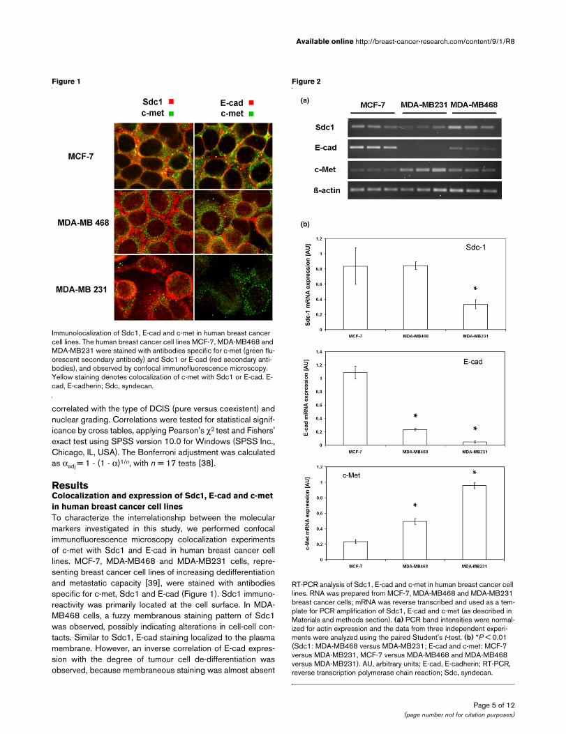
Figure 2
RT-PCR analysis of Sdc1, E-cad and c-met in human breast cancer cell linesRT-PCR analysis of Sdc1, E-cad and c-met in human breast cancer cell lines. RNA was prepared from MCF-7, MDA-MB468 and MDA-MB231 breast cancer cells; mRNA was reverse transcribed and used as a tem-plate for PCR amplification of Sdc1, E-cad and c-met (as described in Materials and methods section). (a) PCR band intensities were normal-ized for actin expression and the data from three independent experi-ments were analyzed using the paired Student's t-test. (b) *P < 0.01 (Sdc1: MDA-MB468 versus MDA-MB231; E-cad and c-met: MCF-7 versus MDA-MB231, MCF-7 versus MDA-MB468 and MDA-MB468 versus MDA-MB231). AU, arbitrary units; E-cad, E-cadherin; RT-PCR, reverse transcription polymerase chain reaction; Sdc, syndecan.
Page 5 of 12(page number not for citation purposes)

Breast Cancer Research Vol 9 No 1 Götte et al.
in MDA-MB231 cells. A punctate staining pattern was exhib-ited by c-met. In MCF-7 cells, c-met partially colocalized withSdc1 and E-cad. The degree of colocalization appeared to behigher in MCF-7 cells than in MDA-MB468 cells. In MDA-MB231 cells, colocalization with c-met could not be assessedbecause of the low level of E-cad expression.
We next compared expression of Sdc1, E-cad and c-met atthe mRNA level by semiquantitative RT-PCR (Figure 2). Similarlevels of Sdc1 expression were observed in MCF-7 and MDA-MB468 cells, but Sdc1 mRNA expression was significantlylower (P < 0.001) in MDA-MB231 cells than in MDA-MB468cells. With increasing de-differentiation and higher metastaticpotential (MCF-7 < MDA-MB468 < MDA-MB231), a signifi-cant decrease in E-cad mRNA expression was observed (Fig-ure 2b). Of note, expression levels of c-met exhibited an
inverse relation, with the highest expression in MDA-MB231cells (Figure 2).
Immunohistochemical staining of Sdc1, E-cad and c-met in DCISWe next employed TMA technology to determine the expres-sion of Sdc1, E-cad and c-met in 200 cases of DCIS of thebreast. Tumour cells exhibited variable expression of Sdc1,ranging from weak to strong. Staining was predominantlymembranous but often additionally cytoplasmic. Stromal cellsand myoepithelial cells showed no immunoreactivity (Figure3). For c-met there was weak to strong membrane bound andadditional cytoplasmic expression in stromal cells, and myoep-ithelial cells remained negative (Figure 3). Expression of E-cadherin was observed in almost all cases of DCIS, with themajority exhibiting strong immunoreactivity. Staining in tumourcells was membranous and cytoplasmic, whereas stroma andmyoepithelium remained negative (Figure 3).
Detailed staining results for all three markers in DCIS are listedin Table 2. Based on a four-tiered scale, moderate to strongpositive staining of tumour cells was observed for Sdc1 in 108out of 150 (72%), for c-met in 69 out of 142 (48.6%) and forE-cad in 101 out of 149 (67.8%) evaluable DCIS samples.We next investigated the interrelation of Sdc1, c-met and E-cad expression. Expression of E-cad was significantly corre-lated with expression of c-met (P = 0.004) and Sdc1 (P =0.007; Table 3).
Correlation of Sdc1, c-met and E-cad expression with histopathological characteristicsA significant correlation was observed between expression ofc-met and HER2 (P = 0.044), and there was a trend towardan association of HER2 with Sdc1 expression (P = 0.057;Table 3). No significant correlation of high-to-moderate c-met,Sdc-1 and E-cad expression with ER and PR status or high-grade DCIS [35] was observed (Table 3). Expression of c-met(P < 0.001) and Sdc1 (P = 0.037) was significantly more fre-quent in the subgroup of pure DCIS than in the subgroup ofDCIS with a coexistent invasive carcinoma (Table 4). Thesedifferences were not due to an unequal distribution of his-topathologic or clinical features, because analysis of the distri-bution of patients' age, ER, PR and HER2 status, and nucleargrading revealed no statistically significant differencesbetween three subgroups (data not shown).
Correlation of c-met, Sdc1 and E-cad expression with angiogenic and lymphangiogenic factorsExpression of four ligand-receptor pairs known to play a role inangiogenesis and lymphangiogenesis was analyzed to test fora potential association of c-met, Sdc1 and E-cad expressionwith such angiogenic and lymphangiogenic factors: VEGF-Aand its receptors Flt-1 and KDR; bFGF and its receptorFGFR1; the endothelin axis, comprised of ET-1 and its recep-
Figure 3
Representative immunohistochemical staining patterns for E-cad, c-met, and Sdc1 in DCISRepresentative immunohistochemical staining patterns for E-cad, c-met, and Sdc1 in DCIS. Examples for presence (positive) and absence (negative) of marker expression are shown. DCIS, ductal carcinoma in situ; E-cad, E-cadherin; Sdc, syndecan.
Table 2
Immunohistochemical analysis of Sdc1, c-met and E-cad expression in DCIS of the breast
Score c-met (n = 142) E-cad (n = 149) Sdc1 (n = 150)
0 10 (7) 11 (7.4) 4 (2.7)
1 63 (44.4) 37 (24.8) 38 (25.3)
2 51 (35.9) 89 (59.7) 103 (68.7)
3 18 (12.7) 12 (8.1) 5 (3.3)
Negative (score 0–1) 73(51.4) 48 (32.2) 42 (28)
Positive (score 2–3) 69 (48.6) 101 (67.8) 108 (72)
Values are expressed as number of evaluable tissue specimens (%). DCIS, ductal carcinoma in situ; E-cad, E-cadherin; Sdc, syndecan.
Page 6 of 12(page number not for citation purposes)

Available online http://breast-cancer-research.com/content/9/1/R8
tors ETAR and ETBR; and the lymphangiogenic factor VEGF-Cand its receptor Flt-4 (Table 5).
VEGF-A exhibited faint to moderate cytoplasmic staining oftumour cells. Positive staining was also observed in myoepi-thelial cells, whereas normal glandular epithelial cells as wellas stromal and connective tissue were mostly VEGF-A nega-tive. KDR staining presented as strong cytoplasmic staining.Normal glandular epithelial cells stained positive for KDR,whereas stroma and connective tissue exhibited no staining.Flt-1 immunoreactivity presented as a granular cytoplasmicstaining of tumour cells and of the peritumoural stroma. Normalglandular epithelial cells stained negative for Flt-1.
bFGF staining of tumour cells was predominantly nuclear andoccasionally cytoplasmic. Although the stroma was bFGF neg-ative, positive staining of normal glandular epithelial cells wasfrequently observed. Tumor cells exhibited strong cytoplasmicstaining for FGFR1, whereas normal glandular cells were mod-erately positive, and the majority of stromal cells were weaklypositive.
Expression of ET-1, ETAR and ETBR presented as positivecytoplasmic staining of tumour cells, and a faint additional stro-mal staining was observed in some ET-1 positive cases. Nor-mal glandular epithelial cells were mostly negative for markersof the endothelin axis.
VEGF-C staining presented as strong cytoplasmic staining oftumour cells, whereas normal breast epithelium was largelyVEGF-C negative, as was the stroma and connective tissue.Tumor cells stained strongly positive for Flt-4, and positivestaining was also frequently observed in normal glandular epi-thelium. In contrast, no Flt-4 staining was detected in thestroma and connective tissues.
A highly significant correlation of c-met expression with ETARand ETBR was found; 92.6% of c-met positive DCIS versus56.7% of c-met negative DCIS stained positive for ETAR (P <0.001), and 57.4% of c-met positive tumours versus 23.4% ofc-met negative tumours stained positive for ETBR (P < 0.001).Furthermore, c-met expression exhibited a significant positivecorrelation with expression of VEGF-A (P = 0.040) andFGFR1 (P = 0.028; Table 5).
Similar to c-met, E-cad expression was significantly correlatedwith ETAR expression (P < 0.001). In addition, we observed asignificant correlation of E-cad expression with positive stain-ing for VEGF-A (P = 0.005) and VEGF-C (P = 0.048).
Sdc1 exhibited a trend toward correlation with lymphang-iogenic factors (VEGF-C, P = 0.061; Flt-4, P = 0.071; Table5).
DiscussionIn this study we characterized coexpression of the functionallylinked prognostic markers Sdc1, E-cad and c-met in 200DCIS using TMA technology and in different human breastcancer cell lines. Our initial objective was to identify a group offunctionally interrelated markers that have prognostic signifi-cance in breast cancer. In this regard we observed significantcorrelations between expression levels for the markers c-met,Sdc1 and E-cad in DCIS. The markers c-met and Sdc1 weresignificantly more frequently expressed in pure DCIS than inDCIS with a coexistent invasive carcinoma. In the case ofSdc1, this finding may indicate downregulation during epithe-
Table 3
Interrelation of c-met, E-cad and Sdc1 expression in DCIS and correlation with histopathological characteristics
c-met E-cad Sdc1
Negative (n = 73)
Positive (n = 69)
P Negative (n = 48)
Positive (n = 101)
P Negative (n = 42)
Positive (n = 105)
P
c-met positive 0.0% 100.0% 30.8% 58.8% 0.004 38.2% 56.3% 0.071
E-cad positive 56.5% 80.6% 0.004 0.0% 100.0% 51.4% 76.0% 0.007
Sdc1 positive 66.7% 80.6% 0.071 58.5% 80.9% 0.007 0.0% 100.0%
High grade DCIS 47.2% 48.5% 0.877 47.9% 47.5% 0.960 37.5% 49.5% 0.193
ER positive 25.4% 31.3% 0.463 20.0% 31.5% 0.176 37.1% 26.0% 0.251
PR positive 33.9% 37.9% 0.643 37.8% 33.7% 0.655 40.0% 31.6% 0.368
ErbB2 positive 12.5% 27.3% 0.044 23.5% 23.9% 0.964 11.8% 28.0% 0.057
DCIS, ductal carcinoma in situ; E-cad, E-cadherin; ER, oestrogen receptor; PR, progesterone receptor; Sdc, syndecan.
Table 4
Expression of c-met, Sdc1 and E-cad in the subgroups of pure DCIS and DCIS with coexistent invasive carcinoma
Molecular marker Pure DCIS Coexistent DCIS P
Sdc1 59/74 (79.7%) 49/76 (64.5%) 0.037
c-met 47/72 (65.3%) 22/70 (31.4%) <0.001
E-cad 56/76 (73.7%) 45/73 (61.6%) 0.116
DCIS, ductal carcinoma in situ; E-cad, E-cadherin; Sdc, syndecan.
Page 7 of 12(page number not for citation purposes)

Breast Cancer Research Vol 9 No 1 Götte et al.
lial-mesenchymal transition [40,41], which has beendemonstrated in mammary epithelial cells in vitro [23,24] andduring development [22]. Regarding the association betweenthe expression pattern of Sdc1, E-cad and c-met with diseaseprogression, future studies could investigate the correlationwith expression for coexisting DCIS and invasive carcinoma.However, based on our cell culture data and on current knowl-edge on the prognostic value and biological function of themarkers (see Table 1), we speculate that a reduction in E-cadexpression and an upregulation of Sdc1 expression may occurin the invasive component of mixed lesions. In the case of c-met, our finding that c-met expression is highest in MDA-MB231 cells, and exhibits a positive correlation with other fea-tures of aggressiveness such as HER2 overexpression seem-ingly contradicts our observation that c-met expression ishigher in pure DCIS than in DCIS associated with invasivecancer. We recently observed significantly increased expres-sion of prognostically unfavourable proangiogenic factors inpure DCIS compared with coexistent DCIS [29]. This is con-sistent with findings reported by Teo and coworkers [42], whodescribed different vascular density and phenotypes in pureversus coexistent DCIS. Therefore, the preferential associa-tion of c-met expression with pure DCIS may be indicative ofthe increased angiogenic activity in pure DCIS [11,29].
The metastatic origin of the investigated human breast cancercell lines [39] has inherent limitations with respect to a directcomparison of marker expression in DCIS tissue. However, thepositive correlation of c-met, Sdc1 and E-cad in an early stageof tumour progression at the tissue level was largely supportedby our colocalization studies in these cell lines, in which partialcolocalization was observed between c-met and Sdc1 and E-cad, respectively, in the less aggressive MCF-7 and MDA-MB468 cell lines. In addition, Sdc1 and E-cad exhibited a sim-
ilar cell surface distribution in these cells. Moreover, progres-sive loss of E-cad expression and a progressive increase in c-met expression were observed with increasing de-differentia-tion and higher metastatic potential (MCF-7 < MDA-MB468 <MDA-MB231) of the breast cancer cell lines at the mRNAlevel.
These findings are similar to the expression patterns of E-cadand c-met observed in clinical studies. Reduced expression ofE-cad in breast carcinoma is associated with shortened dis-ease-free survival and high histological grade [43-46],whereas high expression of c-met is associated with moreaggressive disease and decreased disease-free survival innode-negative breast cancer [15,47] (see Table 1). In addi-tion, coordinated overexpression of c-met and the oncogeneRON is observed in MBA-MB231 but not in MCF-7 cells [15].Our observation of a synchronous downregulation of E-cadand Sdc1 mRNA in highly aggressive MDA-MB231 cells is inaccordance with previous in vitro experiments on mammaryepithelial cells [23,24]. Expression of Sdc1 can be regulatedat the post-transcriptional level [48,49], which may explain thestrong Sdc1 protein expression observed by confocal immun-ofluorescence microscopy in MDA-MB231 cells (Figure 1).The heterogeneity in Sdc1 and E-cad coexpression in breastcancer cell lines of different degrees of de-differentiation dem-onstrates that the concept of synchronous regulation of Sdc1and E-cad in epithelial cells [23,24] cannot be fully adapted tobreast cancer. Our data suggest that the degree of coexpres-sion is higher in breast cells that exhibit a more benign pheno-type, such as DCIS, whereas coexpression may be lost inmore advanced stages of tumour progression. These findingscould help to explain why no correlation was found between E-cad and Sdc1 expression in a patient collective containingductal invasive, lobular and tubular breast cancer tissues [8],
Table 5
Correlation of c-met, Sdc1 and E-cad expression with angiogenic and lymphangiogenic factors
c-met E-cad Sdc1
Negative (n = 73) Positive (n = 69) P Negative (n = 48) Positive (n = 101) P Negative (n = 42) Positive (n = 105) P
VEGF-A 36.2% 53.8% 0.040 26.2% 52.0% 0.005 55% 41.6% 0.149
Flt-1 18.6% 7.7% 0.063 14.3% 11.5% 0.642 12.8% 12.5% 0.959
KDR 48.4% 63.1% 0.094 47.4% 56.4% 0.347 45.7% 58.3% 0.198
VEGF-C 84.5% 94.1% 0.068 80.4% 91.8% 0.048 80.5% 91.5% 0.061
Flt-4 92.9% 98.6% 0.099 95.1% 97.0% 0.584 90.0% 97.2% 0.071
ET-1 47.7% 59.7% 0.176 52.4% 48.9% 0.709 47.4% 51.6% 0.661
ETAR 56.7% 92.6% <0.001 53.8% 86.9% <0.001 70.3% 79.0% 0.277
ETBR 23.4% 57.4% <0.001 30.8% 43.3% 0.177 31.6% 47.0% 0.102
BFGF 17.4% 11.9% 0.394 10.0% 13.4% 0.583 18.9% 11.4% 0.250
FGFR1 89.1% 98.4% 0.028 92.3% 95.9% 0.394 91.4% 96.0% 0.287
bFGF, basic fibroblast growth factor; E-cad, E-cadherin; ET, endothelin; ETA/BR, endothelin A/B receptor; FGFR, fibroblast growth factor receptor; Sdc, syndecan; VEGF, vascular endothelial growth factor.
Page 8 of 12(page number not for citation purposes)

Available online http://breast-cancer-research.com/content/9/1/R8
whereas a correlation was found in our more uniform DCIScollective.
We observed no significant association of c-met, E-cad andSdc-1 expression with hormone receptor status in DCIS. Inpatient collectives including more advanced stages of breastcancer progression, clinical and experimental data suggest apossible link between loss of E-cad expression and ER-nega-tive status [20,43,50]. Moreover, recent studies have linkedSdc1 expression to an aggressive, ER-negative breast carci-noma subtype [6-8]. In order to distinguish between low andhigh expression levels of possibly synchronously regulatedSdc1 and E-cad, both low expression and absence of stainingfor the markers investigated were scored as 'negative'.Although application of this scoring method was necessary todetect synchronous coregulation of marker expression, itmight have lead to failure to demonstrate significant associa-tions of E-cad expression with hormone receptor status andgrade of DCIS in the present study. Although it may occasion-ally be difficult to distinguish between DCIS and LCIS in caseswhere LCIS may involve extralobular ducts, we can largelyexclude the possibility that the analyzed DCIS cases wereerroneously scored as LCIS (see Materials and methods,above).
We found a significant correlation between HER2 and c-metexpression. This correlation may be of functional importance,because both molecules are tyrosine kinase receptors thatpromote cell proliferation [11,51]. Of note, Khoury and cow-orkers [51] recently demonstrated that HGF-mediated c-metsignalling in the presence of constitutively active HER2 leadsto a downregulation of E-cad and promotes epithelial-mesen-chymal transition in MDCK cells. Thus, signalling downstreamof the c-met receptor synergizes with HER2 to enhance amalignant phenotype. In a similar manner, additional receptorscould contribute to this synergism in signalling, and our findingof c-met correlating with FGFR1 expression does support thisview. Moreover, this hypothesis is supported by a study on theexpression of c-met and the tyrosine kinase receptor RON innode-negative breast cancer patients [15]. Ten-year disease-free survival was significantly decreased in patients withtumours expressing only one of the two markers, and it waseven worse in patients with RON-positive/c-met-positivetumours. Because Sdc1 is an established coreceptor for sev-eral receptor tyrosine kinases, including FGFR1 [1,3] and c-met [10], it could also contribute to synergistic signalling. Weobserved a trend toward a correlation of Sdc1 and HER2expression in DCIS (P = 0.057). This is in accordance with thestudy conducted by Barbareschi and coworkers [6], whichdemonstrated positive correlation of Sdc1 and HER2 in a col-lective of 254 invasive breast carcinomas. In accordance withits role as a signalling coreceptor, a recent study conducted in207 breast cancer patients demonstrated a significantassociation of Sdc1 overexpression with the Ki67 proliferationindex [8].
In DCIS, an increase in periductal blood vessels has been cor-related with recurrence of invasive disease [52],demonstrating the relevance of angiogenesis to tumour pro-gression. Expression of the angiogenic factor VEGF-A intumour cells from patients with DCIS was shown to correlatewith the degree of angiogenesis [53]. In the present study, wefound a significant correlation of both c-met and E-cad withVEGF-A expression in DCIS. In addition, c-met expressionexhibited a negative correlation with Flt-1 expression; Flt-1 is anegative regulator of VEGF availability, which is regarded asan antiangiogenic receptor. Our findings constitute the firstcharacterization of a proangiogenic role for c-met, Sdc1 andE-cad in DCIS, but they are in accordance with published workon the biological role of these molecules. Ribozyme targetingof c-met in mammary cancer cells reduced mammary cancerand tumour-associated angiogenesis in a xenograft model[12]. In addition, the HGF-antagonist NK4 inhibits c-met sig-nalling and angiogenesis in vitro [32] and reduces intratu-moural microvessel density in a hepatocellular carcinomaxenograft model [54]. Sdc1 acts as a coreceptor for severalangiogenic and growth factors, including multiple VEGF iso-forms and bFGF [1,3]. In vivo, we recently demonstratedincreased corneal angiogenesis in Sdc-1 deficient mice andthe formation of abnormally dilated blood vessels in skinwounds of Sdc-1 overexpressing mice, indicating a regulatoryrole for Sdc1 in angiogenesis [2,4]. Moreover, in a nudemouse xenograft model, coinjection of MDA-MB-231 breastcancer cells with Sdc-1 overexpressing and mock-transfectedmouse fibroblasts resulted in significantly elevated microves-sel density and a larger vessel area in tumours containingSdc1 overexpressing stroma cells [33]. A TMA analysis of 207human breast cancer samples by the same authors revealedthat stromal Sdc1 expression correlated with vessel densityand total vessel area, demonstrating a role for Sdc1 in breasttumour angiogenesis.
Although we observed a trend toward an association of Sdc1expression with markers of lymphangiogenesis, we could notdemonstrate a statistically significant correlation of Sdc1expression and proangiogenic markers in DCIS (Table 5). Thisfinding may suggest that the angiogenesis-associated func-tions of Sdc1 primarily contribute to more advanced stages oftumour progression. We have recently demonstrated thatVEGF-C and Flt-4, which are established mediators of lym-phangiogenesis [55,56], were expressed in the tumour cells ofa large proportion (88% to 95%) of DCIS specimens [29]. Inthe present study, we found a significant association of E-cadwith VEGF-C expression (P = 0.048). Althuogh very little isknown about the role of E-cad in lymphangiogenesis, by virtueof its biological role an involvement in this process could easilybe envisaged. Moreover, the correlations of VEGF-A/C with E-cad and of VEGF-A with c-met could be a sign of an ang-iogenic stimulation of the stroma by the tumour cells [57]. Fur-thermore, the preferential expression of Sdc1 and c-met inpure versus coexistent DCIS parallels the significantly more
Page 9 of 12(page number not for citation purposes)

Breast Cancer Research Vol 9 No 1 Götte et al.
common expression of proangiogenic factors in pure versuscoexistent DCIS [29].
ET-1 and its G-protein-coupled receptors ETAR and ETBR (theendothelin axis) are overexpressed in breast carcinomas, andinfluence angiogenesis, tumourigenesis and tumour progres-sion [34,36,58]. Moreover, we previously demonstrated over-expression of the ET axis in DCIS and observed an associationwith different angiogenic and lymphangiogenic factors [29].An important novel finding of the present study is the highlysignificant association of c-met and E-cad expression with ETreceptor expression. Similar to ETAR [34], c-met expression isupregulated by hypoxia [59], thus promoting invasive growthof tumour cells. In addition, the c-met ligand HGF has beenshown to inhibit ET-1 release in endothelial cells, indicating apossible link between c-met mediated signalling and the ETaxis [60]. However, the coexpression of c-met and the ETreceptors may simply indicate an upregulation of parallel sig-nalling pathways that utilize different upstream ligands andconverge intracellularly. Sdc1 and E-cad have been associ-ated with epithelial-mesenchymal transition [22,40], and it hasrecently been shown that ET-1 mediated signalling is requiredduring epithelial-mesenchymal transition in ovarian cancer pro-gression [61,62]. In ovarian cancer [61,62] and melanoma[63,64], activation of the ET axis results in downregulated E-cad expression. In DCIS, coexpression of ET receptors, E-cad,c-met and Sdc1 may constitute a specific expression signa-ture indicative of the transition of an early stage to later stagesof tumour progression.
One possible caveat associated with the present study is thelarge number of statistical tests that were performed (n = 17).Although the statistical analysis employed in this study permitseasier comparison and interpretation of P values within thecontext of the results from other studies, the reader must keepin mind that some significant associations may be falselypositive. Using a Bonferroni adjustment, the corrected signifi-cance level for the tests employed in this study would be P <0.003. However, although corrections for multiple compari-sons on the one hand reduce the chances of making a type Ierror, the chances of making a type II error are increased.Moreover, the relevance of the null hypothesis underlying Bon-ferroni adjustments has recently been questioned (see thereport by Perneger [38] for a discussion). Therefore, thereremains a lack of consensus within the scientific communityregarding whether corrections for multiple comparisons areapplicable.
ConclusionIn the present study we demonstrated coexpression of c-met,Sdc1 and E-cad in DCIS. Of note, expression of c-met, Sdc1and E-cad correlated with expression of angiogenic andlymphangiogenic factors, including endothelin receptorexpression. It is tempting to speculate that this expression sig-nature indicates a state of a parallel, synergistic activation of
multiple receptor pathways (receptor tyrosine kinases, G-pro-tein-coupled receptors and nuclear transcription factors),which vastly promote tumour cell proliferation and angiogen-esis in the tumour stroma. Since these activation pathways relyon different molecular mechanisms, therapeutic approachesmust take multiple targets into consideration. Inhibitors of tyro-sine kinases, of the endothelin axis, of heparan sulphate-mod-ulated signalling and of angiogenesis are currently beingevaluated in clinical trials or have already found their way intocancer therapies [5,65-69]. Data from the present study mayhelp in the development of more efficient therapeuticapproaches in breast cancer, addressing the need to targetsimultaneously multiple molecules that are relevant to this earlystep of breast cancer progression.
Competing interestsThe authors declare that they have no competing interests.
Authors' contributionsMG participated in the design and coordination of the study,participated in TMA slide analysis, performed the cell culturestudies and drafted the manuscript. CK performed construc-tion of the TMA and analysis of staining results. IR participatedin statistical analysis and helped to draft the manuscript. LKprovided general support, participated in study design andhelped to draft the manuscript. PW participated in the designand coordination of the study, performed the statistical analy-sis and drafted the manuscript.
AcknowledgementsThe authors should like to thank Vera Samoilova and Monika Offers for expert technical assistance, and PD Dr R-J Fischer (Institute for Medical Informatics and Biomathematics, University of Münster, Germany) for helpful comments on statistical analysis. This study was financially sup-ported by Münster University Hospital Grants 'Innovative Medizinische Forschung' IMF GÖ 1 2 04 15 (MG), IMF WÜ 1 2 03 32 (PW), IMF WÜ 1 1 05 27 (PW, IR) and Deutsche Forschungsgemeinschaft DFG GO 1392/1-1 (MG).
References1. Bernfield M, Götte M, Park PW, Reizes O, Fitzgerald ML, Lincecum
J, Zako M: Functions of cell surface heparan sulfateproteoglycans. Annu Rev Biochem 1999, 68:729-777.
2. Götte M, Joussen AM, Klein C, Andre P, Wagner DD, Hinkes MT,Kirchhof B, Adamis AP, Bernfield M: Role of syndecan-1 in leu-kocyte-endothelial interactions in the ocular vasculature.Invest Ophthalmol Vis Sci 2002, 43:1135-1141.
3. Götte M: Syndecans in inflammation. FASEB J 2003,17:575-591.
4. Elenius V, Götte M, Reizes O, Elenius K, Bernfield M: Inhibition bythe soluble syndecan-1 ectodomains delays wound repair inmice overexpressing syndecan-1. J Biol Chem 2004,279:41928-41935.
5. Yip GW, Smollich M, Götte M: Therapeutic value of gly-cosaminoglycans in cancer. Mol Cancer Ther 2006,5:2139-2148.
6. Barbareschi M, Maisonneuve P, Aldovini D, Cangi MG, PecciariniL, Angelo Mauri F, Veronese S, Caffo O, Lucenti A, Palma PD, etal.: High syndecan-1 expression in breast carcinoma is relatedto an aggressive phenotype and to poorer prognosis. Cancer2003, 98:474-483.
Page 10 of 12(page number not for citation purposes)

Available online http://breast-cancer-research.com/content/9/1/R8
7. Leivonen M, Lundin J, Nordling S, von Boguslawski K, Haglund C:Prognostic value of syndecan-1 expression in breast cancer.Oncology 2004, 67:11-18.
8. Baba F, Swartz K, van Buren R, Eickhoff J, Zhang Y, Wolberg W,Friedl A: Syndecan-1 and syndecan-4 are overexpressed in anestrogen receptor-negative, highly proliferative breast carci-noma subtype. Breast Cancer Res Treat 2006, 98:91-98.
9. Götte M, Kersting C, Ruggiero M, Tio J, Tulusan AH, Kiesel L, Wülf-ing P: Predictive value of syndecan-1 expression for theresponse to neoadjuvant chemotherapy of primary breastcancer. Anticancer Res 2006, 26:621-627.
10. Derksen PW, Keehnen RM, Evers LM, van Oers MH, SpaargarenM, Pals ST: Cell surface proteoglycan syndecan-1 mediateshepatocyte growth factor binding and promotes Met signalingin multiple myeloma. Blood 2002, 99:1405-1410.
11. Peruzzi B, Bottaro DP: Targeting the c-met signaling pathway incancer. Clin Cancer Res 2006, 12:3657-3660.
12. Jiang WG, Grimshaw D, Martin TA, Davies G, Parr C, Watkins G,Lane J, Abounader R, Laterra J, Mansel RE: Reduction of stromalfibroblast-induced mammary tumor growth, by retroviralribozyme transgenes to hepatocyte growth factor/scatter fac-tor and its receptor, c-MET. Clin Cancer Res 2003,9:4274-4281.
13. Bertotti A, Comoglio PM, Trusolino L: Beta4 integrin is a trans-forming molecule that unleashes Met tyrosine kinasetumorigenesis. Cancer Res 2005, 65:10674-10679.
14. Welm AL, Kim S, Welm BE, Bishop JM: MET and MYC cooperatein mammary tumorigenesis. Proc Natl Acad Sci USA 2005,102:4324-4329.
15. Lee WY, Chen HH, Chow NH, Su WC, Lin PW, Guo HR: Prog-nostic significance of co-expression of RON and MET recep-tors in node-negative breast cancer patients. Clin Cancer Res2005, 11:2222-2228.
16. Beauvais DM, Rapraeger AC: Syndecans in tumor cell adhesionand signaling. Reprod Biol Endocrinol 2004, 2:3.
17. Beauvais DM, Burbach BJ, Rapraeger AC: The syndecan-1 ecto-domain regulates alphavbeta3 integrin activity in human mam-mary carcinoma cells. J Cell Biol 2004, 167:171-181.
18. Lerwill MF: Current practical applications of diagnostic immu-nohistochemistry in breast pathology. Am J Surg Pathol 2004,28:1076-1091.
19. Acs G, Lawton TJ, Rebbeck TR, LiVolsi VA, Zhang PJ: Differentialexpression of E-cadherin in lobular and ductal neoplasms ofthe breast and its biologic and diagnostic implications. Am JClin Pathol 2001, 115:85-98.
20. Cowin P, Rowlands TM, Hatsell SJ: Cadherins and catenins inbreast cancer. Curr Opin Cell Biol 2005, 17:499-508.
21. Cavallaro U, Liebner S, Dejana E: Endothelial cadherins andtumor angiogenesis. Exp Cell Res 2006, 312:659-667.
22. Sun D, Mcalmon KR, Davies JA, Bernfield M, Hay ED: Simultane-ous loss of expression of syndecan-1 and E-cadherin in theembryonic palate during epithelial-mesenchymaltransformation. Int J Dev Biol 1998, 42:733-736.
23. Kato M, Saunders S, Nguyen H, Bernfield M: Loss of cell surfacesyndecan-1 causes epithelia to transform into anchorage-independent mesenchyme-like cells. Mol Biol Cell 1995,6:559-576.
24. Leppä S, Vleminckx K, Van Roy F, Jalkanen M: Syndecan-1expression in mammary epithelial tumor cells is E-cadherin-dependent. J Cell Sci 1996, 109:1393-1403.
25. Zimmermann P, Tomatis D, Rosas M, Grootjans J, Leenaerts I,Degeest G, Reekmans G, Coomans C, David G: Characterizationof syntenin, a syndecan-binding PDZ protein, as a componentof cell adhesion sites and microfilaments. Mol Biol Cell 2001,12:339-350.
26. Alexander CM, Reichsman F, Hinkes MT, Lincecum J, Becker KA,Cumberledge S, Bernfield M: Syndecan-1 is required for Wnt-1-induced mammary tumorigenesis in mice. Nat Genet 2000,25:329-332.
27. Liu BY, Kim YC, Leatherberry V, Cowin P, Alexander CM: Mam-mary gland development requires syndecan-1 to create abeta-catenin/TCF-responsive mammary epithelialsubpopulation. Oncogene 2003, 22:9243-9253.
28. Liu BY, McDermott SP, Khwaja SS, Alexander CM: The trans-forming activity of Wnt effectors correlates with their ability toinduce the accumulation of mammary progenitor cells. ProcNatl Acad Sci USA 2004, 101:4158-4163.
29. Wülfing P, Kersting C, Buerger H, Mattsson B, Mesters R, Gust-mann C, Hinrichs B, Tio J, Böcker W, Kiesel L: Expression pat-terns of angiogenic and lymphangiogenic factors in ductalbreast carcinoma in situ. Br J Cancer 2005, 92:1720-1728.
30. Wu Y, Hooper AT, Zhong Z, Witte L, Bohlen P, Rafii S, Hicklin DJ:The vascular endothelial growth factor receptor (VEGFR-1)supports growth and survival of human breast carcinoma. IntJ Cancer 2006, 119:1519-1529.
31. De Jong JS, van Diest PJ, van der Valk P, Baak JP: Expression ofgrowth factors, growth inhibiting factors, and their receptors ininvasive breast cancer. I: an inventory in search of autocrineand paracrine loops. J Pathol 1998, 184:44-52.
32. Jiang WG, Hiscox SE, Parr C, Martin TA, Matsumoto K, NakamuraT, Mansel RE: Antagonistic effect of NK4, a novel hepatocytegrowth factor variant, on in vitro angiogenesis of human vas-cular endothelial cells. Clin Cancer Res 1999, 5:3695-3703.
33. Maeda T, Desouky J, Friedl A: Syndecan-1 expression by stro-mal fibroblasts promotes breast carcinoma growth in vivo andstimulates tumor angiogenesis. Oncogene 2006,25:1408-1412.
34. Wülfing P, Götte M, Sonntag B, Kersting C, Schmidt H, WülfingC, Buerger H, Greb R, Böcker W, Kiesel L: Overexpression ofEndothelin-A-receptor in breast cancer: regulation by estradioland cobalt-chloride induced hypoxia. Int J Oncol 2005,26:951-960.
35. Holland R, Peterse JL, Millis RR, Eusebi V, Faverly D, van de VijverMJ, Zafrani B: Ductal carcinoma in situ: a proposal for a newclassification. Semin Diagn Pathol 1994, 11:167-180.
36. Wülfing P, Kersting C, Tio J, Fischer RJ, Wülfing C, Poremba C,Diallo R, Böcker W, Kiesel L: Endothelin-1-, endothelin-A-, andendothelin-B-receptor expression is correlated with vascularendothelial growth factor expression and angiogenesis inbreast cancer. Clin Cancer Res 2004, 10:2393-2400.
37. Wülfing P, Diallo R, Müller C, Wülfing C, Poremba C, Heinecke A,Rody A, Greb RR, Böcker W, Kiesel L: Analysis of cyclooxygen-ase-2 expression in human breast cancer: high throughput tis-sue microarray analysis. J Cancer Res Clin Oncol 2003,129:375-382.
38. Perneger TV: What's wrong with Bonferroni adjustments. BMJ1998, 316:1236-1238.
39. Lacroix M, Leclercq G: Relevance of breast cancer cell lines asmodels for breast tumours: an update. Breast Cancer ResTreat 2004, 83:249-289.
40. Lombaerts M, van Wezel T, Philippo K, Dierssen JW, ZimmermanRM, Oosting J, van Eijk R, Eilers PH, van de Water B, CornelisseCJ, et al.: E-cadherin transcriptional downregulation by pro-moter methylation but not mutation is related to epithelial-to-mesenchymal transition in breast cancer cell lines. Br JCancer 2006, 94:661-671.
41. Vos CB, Cleton-Jansen AM, Berx G, de Leeuw WJ, ter Haar NT,van Roy F, Cornelisse CJ, Peterse JL, van de Vijver MJ: E-cadherininactivation in lobular carcinoma in situ of the breast: an earlyevent in tumorigenesis. Br J Cancer 1997, 76:1131-1133.
42. Teo NB, Shoker BS, Jarvis C, Martin L, Sloane JP, Holcombe C:Vascular density and phenotype around ductal carcinoma insitu (DCIS) of the breast. Br J Cancer 2002, 86:905-911.
43. Siitonen SM, Kononen JT, Helin HJ, Rantala IS, Holli KA, Isola JJ:Reduced E-cadherin expression is associated with invasive-ness and unfavorable prognosis in breast cancer. Am J ClinPathol 1996, 105:394-402.
44. Rakha EA, Abd El Rehim D, Pinder SE, Lewis SA, Ellis IO: E-cad-herin expression in invasive non-lobular carcinoma of thebreast and its prognostic significance. Histopathology 2005,46:685-693.
45. Asgeirsson KS, Jonasson JG, Tryggvadottir L, Olafsdottir K, Sigur-geirsdottir JR, Ingvarsson S, Ogmundsdottir HM: Altered expres-sion of E-cadherin in breast cancer. patterns, mechanisms andclinical significance. Eur J Cancer 2000, 36:1098-1106.
46. Yoshida R, Kimura N, Harada Y, Ohuchi N: The loss of E-cad-herin, alpha- and beta-catenin expression is associated withmetastasis and poor prognosis in invasive breast cancer. Int JOncol 2001, 18:513-520.
47. Camp RL, Rimm EB, Rimm DL: Met expression is associatedwith poor outcome in patients with axillary lymph node nega-tive breast carcinoma. Cancer 1999, 86:2259-2265.
48. Hayashida K, Johnston DR, Goldberger O, Park PW: Syndecan-1expression in epithelial cells is induced by transforming
Page 11 of 12(page number not for citation purposes)
http://www.ncbi.nlm.nih.gov/entrez/query.fcgi?cmd=Retrieve&db=PubMed&dopt=Abstract&list_uids=9712528
http://www.ncbi.nlm.nih.gov/entrez/query.fcgi?cmd=Retrieve&db=PubMed&dopt=Abstract&list_uids=9712528
http://www.ncbi.nlm.nih.gov/entrez/query.fcgi?cmd=Retrieve&db=PubMed&dopt=Abstract&list_uids=9712528
http://www.ncbi.nlm.nih.gov/entrez/query.fcgi?cmd=Retrieve&db=PubMed&dopt=Abstract&list_uids=7545031
http://www.ncbi.nlm.nih.gov/entrez/query.fcgi?cmd=Retrieve&db=PubMed&dopt=Abstract&list_uids=7545031
http://www.ncbi.nlm.nih.gov/entrez/query.fcgi?cmd=Retrieve&db=PubMed&dopt=Abstract&list_uids=7545031
http://www.ncbi.nlm.nih.gov/entrez/query.fcgi?cmd=Retrieve&db=PubMed&dopt=Abstract&list_uids=8799827
http://www.ncbi.nlm.nih.gov/entrez/query.fcgi?cmd=Retrieve&db=PubMed&dopt=Abstract&list_uids=8799827
http://www.ncbi.nlm.nih.gov/entrez/query.fcgi?cmd=Retrieve&db=PubMed&dopt=Abstract&list_uids=8799827
http://www.ncbi.nlm.nih.gov/entrez/query.fcgi?cmd=Retrieve&db=PubMed&dopt=Abstract&list_uids=9582526
http://www.ncbi.nlm.nih.gov/entrez/query.fcgi?cmd=Retrieve&db=PubMed&dopt=Abstract&list_uids=9582526
http://www.ncbi.nlm.nih.gov/entrez/query.fcgi?cmd=Retrieve&db=PubMed&dopt=Abstract&list_uids=9582526
http://www.ncbi.nlm.nih.gov/entrez/query.fcgi?cmd=Retrieve&db=PubMed&dopt=Abstract&list_uids=7831528
http://www.ncbi.nlm.nih.gov/entrez/query.fcgi?cmd=Retrieve&db=PubMed&dopt=Abstract&list_uids=7831528
http://www.ncbi.nlm.nih.gov/entrez/query.fcgi?cmd=Retrieve&db=PubMed&dopt=Abstract&list_uids=9553006
http://www.ncbi.nlm.nih.gov/entrez/query.fcgi?cmd=Retrieve&db=PubMed&dopt=Abstract&list_uids=9365159
http://www.ncbi.nlm.nih.gov/entrez/query.fcgi?cmd=Retrieve&db=PubMed&dopt=Abstract&list_uids=9365159
http://www.ncbi.nlm.nih.gov/entrez/query.fcgi?cmd=Retrieve&db=PubMed&dopt=Abstract&list_uids=9365159
http://www.ncbi.nlm.nih.gov/entrez/query.fcgi?cmd=Retrieve&db=PubMed&dopt=Abstract&list_uids=8604681
http://www.ncbi.nlm.nih.gov/entrez/query.fcgi?cmd=Retrieve&db=PubMed&dopt=Abstract&list_uids=8604681

Breast Cancer Research Vol 9 No 1 Götte et al.
growth factor beta through a PKA-dependent pathway. J BiolChem 2006, 281:24365-24374.
49. Yeaman C, Rapraeger AC: Post-transcriptional regulation ofsyndecan-1 expression by cAMP in peritoneal macrophages. JCell Biol 1993, 122:941-950.
50. Parker C, Rampaul RS, Pinder SE, Bell JA, Wencyk PM, BlameyRW, Nicholson RI, Robertson JF: E-cadherin as a prognosticindicator in primary breast cancer. Br J Cancer 2001,85:1958-1963.
51. Khoury H, Naujokas MA, Zuo D, Sangwan V, Frigault MM, Petkie-wicz S, Dankort DL, Muller WJ, Park M: HGF converts ErbB2/Neu epithelial morphogenesis to cell invasion. Mol Biol Cell2005, 16:550-561.
52. Teo NB, Shoker BS, Jarvis C, Martin L, Sloane JP, Holcombe C:Angiogenesis and invasive recurrence in ductal carcinoma insitu of the breast. Eur J Cancer 2003, 39:38-44.
53. Guidi AJ, Schnitt SJ, Fischer L, Tognazzi K, Harris JR, Dvorak HF,Brown LF: Vascular permeability factor (vascular endothelialgrowth factor) expression and angiogenesis in patients withductal carcinoma in situ of the breast. Cancer 1997,80:1945-1953.
54. Heideman DA, Overmeer RM, van Beusechem VW, Lamers WH,Hakvoort TB, Snijders PJ, Craanen ME, Offerhaus GJ, Meijer CJ,Gerritsen WR: Inhibition of angiogenesis and HGF-cMET-elic-ited malignant processes in human hepatocellular carcinomacells using adenoviral vector-mediated NK4 gene therapy.Cancer Gene Ther 2005, 12:954-962.
55. Jeltsch M, Kaipainen A, Joukov V, Meng X, Lakso M, Rauvala H,Swartz M, Fukumura D, Jain RK, Alitalo K: Hyperplasia of lym-phatic vessels in VEGF-C transgenic mice. Science 1997,276:1423-1425.
56. Kinoshita J, Kitamura K, Kabashima A, Saeki H, Tanaka S, Sugi-machi K: Clinical significance of vascular endothelial growthfactor-C (VEGF-C) in breast cancer. Breast Cancer Res Treat2001, 66:159-164.
57. Jakobsson L, Kreuger J, Holmborn K, Lundin L, Eriksson I, KjellenL, Claesson-Welsh L: Heparan sulfate in trans potentiatesVEGFR-mediated angiogenesis. Dev Cell 2006, 10:625-634.
58. Wülfing P, Diallo R, Kersting C, Wülfing C, Poremba C, Greb RR,Böcker W, Kiesel L: Endothelin-1, endothelin-A- and Endothe-lin-B-receptor expression in preinvasive and invasive breastdisease. Oncol Rep 2004, 11:791-796.
59. Pennacchietti S, Michieli P, Galluzzo M, Mazzone M, Giordano S,Comoglio PM: Hypoxia promotes invasive growth by transcrip-tional activation of the met protooncogene. Cancer Cell 2003,3:347-361.
60. Haug C, Schmid-Kotsas A, Zorn U, Bachem MG, Schuett S,Gruenert A, Rozdzinski E: Hepatocyte growth factor is upregu-lated by low-density lipoproteins and inhibits endothelin-1release. Am J Physiol Heart Circ Physiol 2000,279:H2865-H2871.
61. Rosano L, Spinella F, Di Castro V, Nicotra MR, Dedhar S, de Her-reros AG, Natali PG, Bagnato A: Endothelin-1 promotes epithe-lial-to-mesenchymal transition in human ovarian cancer cells.Cancer Res 2005, 65:11649-11167.
62. Rosano L, Spinella F, Di Castro V, Decandia S, Nicotra MR, NataliPG, Bagnato A: Endothelin-1 is required during epithelial tomesenchymal transition in ovarian cancer progression. ExpBiol Med (Maywood) 2006, 231:1128-1131.
63. Bagnato A, Rosano L, Spinella F, Di Castro V, Tecce R, Natali PG:Endothelin B receptor blockade inhibits dynamics of cell inter-actions and communications in melanoma cell progression.Cancer Res 2004, 64:1436-1443.
64. Jamal S, Schneider RJ: UV-induction of keratinocyte endothe-lin-1 downregulates E-cadherin in melanocytes andmelanoma cells. J Clin Invest 2002, 110:443-452.
65. Bagnato A, Natali PG: Endothelin receptors as novel targets intumor therapy. J Transl Med 2004, 2:16.
66. Wakeling AE: Inhibitors of growth factor signalling. EndocrRelat Cancer 2005:S183-S187.
67. Ciardiello F, Troiani T, Caputo F, De Laurentiis M, Tortora G, Palm-ieri G, De Vita F, Diadema MR, Orditura M, Colantuoni G, et al.:Phase II study of gefitinib in combination with docetaxel asfirst-line therapy in metastatic breast cancer. Br J Cancer2006, 94:1604-1609.
68. Jimenez B, Volpert OV: Mechanistic insights on the inhibition oftumor angiogenesis. J Mol Med 2001, 78:663-672.
69. Folkman J: Angiogenesis. Annu Rev Med 2006, 57:1-18.70. Tolgay Ocal I, Dolled-Filhart M, D'Aquila TG, Camp RL, Rimm DL:
Tissue microarray-based studies of patients with lymph nodenegative breast carcinoma show that met expression is asso-ciated with worse outcome but is not correlated with epider-mal growth factor family receptors. Cancer 2003,97:1841-1848.
71. Lengyel E, Prechtel D, Resau JH, Gauger K, Welk A, Lindemann K,Salanti G, Richter T, Knudsen B, Vande Woude GF, et al.: C-metoverexpression in node-positive breast cancer identifiespatients with poor clinical outcome independent of Her2/neu.Int J Cancer 2005, 113:678-682.
72. Ghoussoub RA, Dillon DA, D'Aquila T, Rimm EB, Fearon ER, RimmDL: Expression of c-met is a strong independent prognosticfactor in breast carcinoma. Cancer 1998, 82:1513-1520.
73. Nakopoulou L, Gakiopoulou H, Keramopoulos A, Giannopoulou I,Athanassiadou P, Mavrommatis J, Davaris PS: c-met tyrosinekinase receptor expression is associated with abnormal beta-catenin expression and favourable prognostic factors in inva-sive breast carcinoma. Histopathology 2000, 36:313-325.
74. Heimann R, Lan F, McBride R, Hellman S: Separating favorablefrom unfavorable prognostic markers in breast cancer: therole of E-cadherin. Cancer Res 2000, 60:298-304.
75. Charpin C, Garcia S, Bonnier P, Martini F, Andrac L, Choux R,Lavaut MN, Allasia C: Reduced E-cadherin immunohistochem-ical expression in node-negative breast carcinomas correlateswith 10-year survival. Am J Clin Pathol 1998, 109:431-438.
Page 12 of 12(page number not for citation purposes)
http://www.ncbi.nlm.nih.gov/entrez/query.fcgi?cmd=Retrieve&db=PubMed&dopt=Abstract&list_uids=8394371
http://www.ncbi.nlm.nih.gov/entrez/query.fcgi?cmd=Retrieve&db=PubMed&dopt=Abstract&list_uids=8394371
http://www.ncbi.nlm.nih.gov/entrez/query.fcgi?cmd=Retrieve&db=PubMed&dopt=Abstract&list_uids=9366297
http://www.ncbi.nlm.nih.gov/entrez/query.fcgi?cmd=Retrieve&db=PubMed&dopt=Abstract&list_uids=9366297
http://www.ncbi.nlm.nih.gov/entrez/query.fcgi?cmd=Retrieve&db=PubMed&dopt=Abstract&list_uids=9366297
http://www.ncbi.nlm.nih.gov/entrez/query.fcgi?cmd=Retrieve&db=PubMed&dopt=Abstract&list_uids=9162011
http://www.ncbi.nlm.nih.gov/entrez/query.fcgi?cmd=Retrieve&db=PubMed&dopt=Abstract&list_uids=9162011
http://www.ncbi.nlm.nih.gov/entrez/query.fcgi?cmd=Retrieve&db=PubMed&dopt=Abstract&list_uids=9554529
http://www.ncbi.nlm.nih.gov/entrez/query.fcgi?cmd=Retrieve&db=PubMed&dopt=Abstract&list_uids=9554529
http://www.ncbi.nlm.nih.gov/entrez/query.fcgi?cmd=Retrieve&db=PubMed&dopt=Abstract&list_uids=9535397