Targeting Endothelial Progenitor Cells in Cancer as a Novel Biomarker and Anti-Angiogenic Therapy
Upload
independentCategory
view
0download
0
ORIGINAL ARTICLE 828J o u r n a l o fJ o u r n a l o f
CellularPhysiologyCellularPhysiology
Syndecan-4 Contributes toEndothelial TubulogenesisThrough Interactions With TwoMotifs Inside the Pro-AngiogenicN-Terminal Domain ofThrombospondin-1
SARA SANTANA NUNES,1 MARIANNA A. FERRARI DO OUTEIRO-BERNSTEIN,1LUIZ JULIANO,2 FRANCISCO VARDIERO,1 HELENA B. NADER,3 ANNE WOODS,4
CHANTAL LEGRAND,5,6AND VERONICA MORANDI1*
1Departamento de Biologia Celular e Genetica, Laboratorio de Biologia da Celula Endotelial e da Angiogenese (LabAngio),
Instituto de Biologia Roberto Alcantara Gomes, Universidade do Estado do Rio de Janeiro, UERJ, Rio de Janeiro, RJ, Brazil2Departamento de Biofısica Escola Paulista de Medicina, UNIFESP, Sao Paulo, SP, Brazil3Departamento de Bioquımica, Escola Paulista de Medicina, UNIFESP, Sao Paulo, SP, Brazil4Department of Cell Biology, University of Alabama at Birmingham, Birmingham, AL 35294-00065INSERM U553 (Institut National de la Sante et de la Recherche Medicale), Paris, France6Universite Denis Diderot, Paris, France
Thrombospondin-1 (TSP-1) is an extracellular matrix protein that modulates focal adhesion in mammalian cells and exhibits dual roles inangiogenesis. In a previous work, we showed that a recombinant 18 kDa protein encompassing the N-terminal residues 1-174 of humanTSP-1 (TSP18) induced tubulogenesis of human umbilical vein endothelial cells and protected them from apoptosis. Our results indicatedthat these effects were possibly mediated by syndecan-4 proteoglycan, since binding of TSP18 to endothelial extracts was inhibited by anti-syndecan-4 antibody. Syndecan-4 is a heparan-sulfate proteoglycan that regulates cell–matrix interactions and is the only member of itsfamily present in focal adhesions. In this report, we demonstrate that a monoclonal antibody against syndecan-4 blocks TSP18-inducedtubulogenesis. Furthermore, through 2D adhesion and 3D angiogenic assays, we demonstrate that two sequences, TSP Hep I and II, retainthe major pro-angiogenic activity of TSP18. These TSP-1 motifs also compete with the fibronectin Hep II domain for binding to syndecan-4on endothelial cell surface, indicating that they may exert their effects by interfering with the recognition of fibronectin by syndecan-4.Additionally, TSP18 and its derived peptides activate the PKC-dependent Akt-PKB signaling pathway. Blockage of PKC activationprevented HUVEC spreading when seeded on TSP18 fragment, and on TSP Hep I and TSP Hep II peptides, but not on gelatin-coatedsubstrates. Our results identify syndecan-4 as a novel receptor for the N-terminus of TSP-1 and suggest that TSP-1 N-terminalpro-angiogenic activity is linked to its capacity of interfering with syndecan-4 functions in the course of cell adhesion.
J. Cell. Physiol. 214: 828–837, 2008. � 2007 Wiley-Liss, Inc.
Contract grant sponsor: CNPq;Contract grant number: 550974/2002-4.Contract grant sponsor: FAPERJ;Contract grant numbers: E-26/170.227/2002, E-26/170.632/2003.Contract grant sponsor: PRONEX/FAPERJ 2004.Contract grant sponsor: CNPq/INSERM (France) CooperationAgreement.Contract grant sponsor: CAPES (Brazil)/COFECUB (France)Agreement;Contract grant number: 347/2001.
*Correspondence to: Veronica Morandi, Universidade do Estadodo Rio de Janeiro, Departamento de Biologia Celular e Genetica,Instituto de Biologia Roberto Alcantara Gomes, Rua Sao FranciscoXavier, 524 PHLC sala 205, Maracana, Rio de Janeiro, RJ, Brazil.E-mail: [email protected]
Received 11 October 2006; Accepted 7 August 2007
DOI: 10.1002/jcp.21281
Adult angiogenesis, the formation of new vessels either frompre-existing vascular structures or from the recruitment ofbone marrow-derived endothelial precursors (Carmeliet,2003), is highly dependent on the local levels of some majorangiogenic growth factors, such as VEGFs, FGFs, and theircellular receptors (Cross and Claesson-Welsh, 2001). In fact, ithas become clear in the last decade that cellular tubulogenesisresults from the interplay of both genetic andmicroenvironment determinants. In particular, it is nowrecognized that the transient composition of the extracellularmatrix (ECM) from a provisional environment (fibrin-rich) to anon-permissive state (basement membrane-rich) directs thedifferentiation of endothelial cells throughout vessel formation(Sanz and Alvarez-Vallina, 2003). This process also involvesproteases responsible for the generation of bioactive fragmentsand neo-epitopes from ECM macromolecules (Roy et al., 2006).
Thrombospondin-1 (TSP-1) is a multifunctional glycoproteinwhich displays both adhesive and anti-adhesive properties formammalian cells (Adams and Lawler, 2004). It was shown toexert a potent anti-angiogenic activity, both in vitro and in vivo,
� 2 0 0 7 W I L E Y - L I S S , I N C .
E N D O T H E L I A L S Y N D E C A N - 4 I N T E R A C T I O N W I T H T S P - 1 829
through its type I repeats and the pro-collagen domain (Tolsmaet al., 1993; Iruela-Arispe et al., 1999). However, under certainconditions, TSP-1 can also promote angiogenesis. We andothers have characterized pro-angiogenic effects, located at theN-terminal heparin-binding domain (HBD) of this glycoprotein(Chandrasekaran et al., 2000; Taraboletti et al., 2000; Ferrari doOuteiro-Bernstein et al., 2002). The HBD has been involved inthe disassembly of focal contacts and stimulation of endothelialcell migration (Murphy-Ullrich et al., 1993; Vogel et al., 1993).Importantly, a few recent clinical studies suggest that theanti-angiogenic capacity of TSP-1 was decreased in metastaticdeposits, as compared with less advanced primary tumors(Sutton et al., 2005) and that its expression by tumor cells wasassociated with the presence of venous invasion and with VEGFexpression (Poon et al., 2004). Moreover, it was shown thatsome human cancers can bypass the angioinhibitory effects ofTSP-1, by an intricate mechanism, which seems to involve thelocal balance of VEGF and TSP-1-activated transforming growthfactor-b (TGF-b) (Filleur et al., 2001; Fontana et al., 2005).These conflicting data raise questions about the clinicalsignificance of TSP-1 expression at sites of angiogenesis fordifferent tumor sets. Thus, clearly molecular mechanisms needto be further identified.
We have previously shown that a recombinant 18 kDa HBD(TSP18), derived from the structure of the endothelial TSP-1,exerts a pro-angiogenic activity on endothelial cells growing onfibrin matrices (Ferrari do Outeiro-Bernstein et al., 2002). Inaddition, we showed by ELISA that monoclonal antibodiesagainst the ectodomain of syndecan-4 prevented binding ofTSP18 to endothelial cell extracts up to 70% indicating that thepro-angiogenic effect of the TSP18 fragment was probably dueto the binding to syndecan-4.
The HS chains of syndecans, a proteoglycan family with fourmembers (syndecans 1-, 2-, 3-, and 4), allow interactions with alarge number of proteins, mainly heparin-binding growthfactors, such as FGFs, VEGFs, TGF-b, midkine, and pleiotrophin(Fears and Woods, 2006). ECM proteins carrying HBDs, such asfibronectin (FN) and laminin, are also recognized by the HSchains of syndecans (Woods et al., 2000; Suzuki et al., 2003). Inaddition to using HS/CS chains in the recognition ofextracellular ligands, syndecan-4 core protein can directlyengage in protein–protein interactions (McFall and Rapraeger,1997). These authors have demonstrated that a non-glycosylated recombinant fragment corresponding to theextracellular domain of syndecan-4, as well as a native shedsyndecan-4, can bind to the surface of endothelial cells andfibroblasts and also support cell adhesion when used as asubstrate. A recent study (Whiteford and Couchman, 2006)demonstrated that a conserved NXIP motif is required for celladhesion that seems to be dependent on b1 integrins. Amongthe four members of the syndecan family, syndecan-4 is the onlyone involved in the formation of FN-induced focal adhesions, incooperation with b1-integrin receptors (Woods andCouchman, 1994). Accordingly, the cytoplasmic V (variable)domain of the core protein of syndecan-4, but not of the othersyndecans, interacts with important focal contact components,such as protein-kinase Ca (PKCa), syndesmos, and a-actinin(Oh et al., 1997; Baciu et al., 2000; Greene et al., 2003),
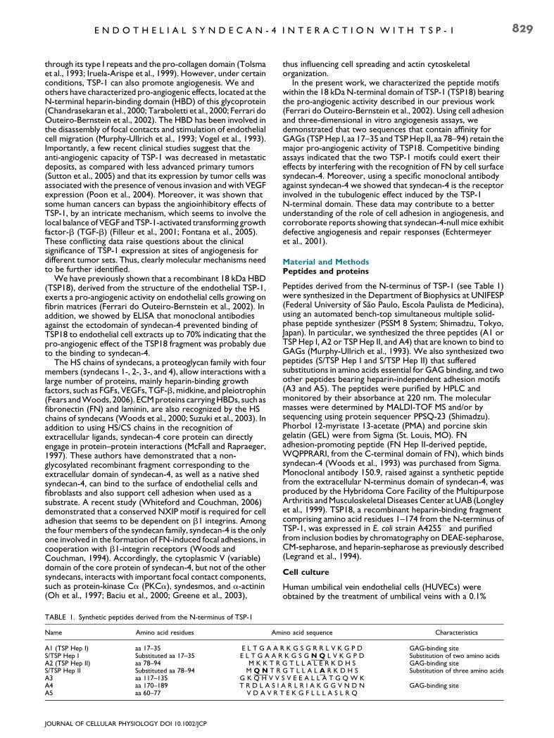
TABLE 1. Synthetic peptides derived from the N-terminus of TSP-1
Name Amino acid residues Am
A1 (TSP Hep I) aa 17–35 E L T G A AS/TSP Hep I Substituted aa 17–35 E L T G A A RA2 (TSP Hep II) aa 78–94 M K K T RS/TSP Hep II Substituted aa 78–94 M Q N T RA3 aa 117–135 G K Q H V VA4 aa 170–189 T R D L A S IA5 aa 60–77 V D A V R T
JOURNAL OF CELLULAR PHYSIOLOGY DOI 10.1002/JCP
thus influencing cell spreading and actin cytoskeletalorganization.
In the present work, we characterized the peptide motifswithin the 18 kDa N-terminal domain of TSP-1 (TSP18) bearingthe pro-angiogenic activity described in our previous work(Ferrari do Outeiro-Bernstein et al., 2002). Using cell adhesionand three-dimensional in vitro angiogenesis assays, wedemonstrated that two sequences that contain affinity forGAGs (TSP Hep I, aa 17–35 and TSP Hep II, aa 78–94) retain themajor pro-angiogenic activity of TSP18. Competitive bindingassays indicated that the two TSP-1 motifs could exert theireffects by interfering with the recognition of FN by cell surfacesyndecan-4. Moreover, using a specific monoclonal antibodyagainst syndecan-4 we showed that syndecan-4 is the receptorinvolved in the tubulogenic effect induced by the TSP-1N-terminal domain. These data may contribute to a betterunderstanding of the role of cell adhesion in angiogenesis, andcorroborate reports showing that syndecan-4-null mice exhibitdefective angiogenesis and repair responses (Echtermeyeret al., 2001).
Material and MethodsPeptides and proteins
Peptides derived from the N-terminus of TSP-1 (see Table 1)were synthesized in the Department of Biophysics at UNIFESP(Federal University of Sao Paulo, Escola Paulista de Medicina),using an automated bench-top simultaneous multiple solid-phase peptide synthesizer (PSSM 8 System; Shimadzu, Tokyo,Japan). In particular, we synthesized the three peptides (A1 orTSP Hep I, A2 or TSP Hep II, and A4) that are known to bind toGAGs (Murphy-Ullrich et al., 1993). We also synthesized twopeptides (S/TSP Hep I and S/TSP Hep II) that sufferedsubstitutions in amino acids essential for GAG binding, and twoother peptides bearing heparin-independent adhesion motifs(A3 and A5). The peptides were purified by HPLC andmonitored by their absorbance at 220 nm. The molecularmasses were determined by MALDI-TOF MS and/or bysequencing using protein sequencer PPSQ-23 (Shimadzu).Phorbol 12-myristate 13-acetate (PMA) and porcine skingelatin (GEL) were from Sigma (St. Louis, MO). FNadhesion-promoting peptide (FN Hep II-derived peptide,WQPPRARI, from the C-terminal domain of FN), which bindssyndecan-4 (Woods et al., 1993) was purchased from Sigma.Monoclonal antibody 150.9, raised against a synthetic peptidefrom the extracellular N-terminus domain of syndecan-4, wasproduced by the Hybridoma Core Facility of the MultipurposeArthritis and Musculoskeletal Diseases Center at UAB (Longleyet al., 1999). TSP18, a recombinant heparin-binding fragmentcomprising amino acid residues 1–174 from the N-terminus ofTSP-1, was expressed in E. coli strain A4255� and purifiedfrom inclusion bodies by chromatography on DEAE-sepharose,CM-sepharose, and heparin-sepharose as previously described(Legrand et al., 1994).
Cell culture
Human umbilical vein endothelial cells (HUVECs) wereobtained by the treatment of umbilical veins with a 0.1%
ino acid sequence Characteristics
R K G S G R R L V K G P D GAG-binding siteK G S G N Q L V K G P D Substitution of two amino acids
G T L L A L E R K D H S GAG-binding siteG T L L A L A R K D H S Substitution of three amino acidsS V E E A L L A T G Q W KA R L R I A K G G V N D N GAG-binding site
E K G F L L L A S L R Q
830 N U N E S E T A L .
collagenase IV solution (Sigma) as described by Jaffe et al., 1973.Except for fetal calf serum (FCS, Cultilab, Campinas, Brazil) andmedium 199/HEPES modification (M199, Sigma) all culturemedium supplies were from Invitrogen do Brasil (Sao Paulo, SP).Primary cells were seeded into 25 cm2 bottles (Corning,Cambridge, NY) coated with porcine skin gelatin (GEL), andgrown in M199 supplemented with 2 mM L-glutamine, 2.5mg/mlamphotericin B, 100 mg/ml penicillin, 100 mg/ml gentamycin,and 20% FCS. Cells were maintained at 378C in humidified5% CO2 atmosphere until they reached confluence. At thattime, cells were detached by brief treatment with a trypsin-EDTA solution, washed once by centrifugation, resuspended inM199 containing 0.1% bovine serum albumin (BSA) andimmediately used in the experiments described below.
Adhesion assays
Ninety-six-well microtiter plates were coated with FNadhesion-promoting peptide (FN Hep II peptide), TSP18, or thedifferent TSP-1 N-terminus-derived peptides (Table 1)(6 mg/cm2) in PBS, overnight at 48C, and saturated for 90 minwith M199 containing 0.1% BSA. Cells (1.5� 105 cells/cm2)were seeded onto the coated plates and allowed to adhere for1–2.5 h. Some experiments were carried out in the presence ofeither 10 mM cycloheximide or 100 nM cytochalasin E in orderto prevent the endogenous synthesis of proteins or cell uptakeof soluble peptides, respectively. To quantify cell adhesion,wells were rinsed thoroughly with M199 to removeunbound cells and the adherent cells were detected byincubating with M199 containing 1 mg/ml MTT(3-(4,5-dimethylthiazol-2-yl)-2,5-diphenyl tetrazoliumbromide) for 2 h at 378C. The solution was removed andreplaced by 200 ml of isopropyl alcohol and absorbance wasread at 595 nm. The cell number was calculated based on astandard curve made by seeding 3.5� 103 to 2� 105
HUVECs/cm2. In competition assays, cells were seeded in thepresence of 10, 50, or 100 mM of the different peptides derivedfrom the N-terminus of TSP-1 and quantification of adheredcells was made as described above.
In vitro fibrin gel assay
Fibrin gels used for three-dimensional cultures were formed byovernight polymerization of 3.0 mg/ml fibrinogen (Sigma)solutions with 1.0 IU/ml thrombin in PBS (pH 7.4) at 378C. Insome conditions, synthetic peptides derived from TSP-1N-terminus (Table 1) were included in the gels at 20 mg/ml.After equilibration in M199 supplemented with 2% FCS for 2 h,HUVECs (1.0� 105 cells/cm2) were seeded in M199 containing2% FCS and incubated at 378C in a humidified 5% atmosphere.Cultures were observed at different times, in order to followmodifications in endothelial cell tubulogenesis. In this model,cells initially migrate and organize themselves in the monolayer,resulting in the opening of cell-free areas within the first 24 h ofassay. After nearly 3 days, the portions of intact monolayersbecome scarse, and cells around the open areas align to formstructures that involve the participation of 5, 6, or more cellsshowing translucent cleft along their axis (lumen formation).The number of open areas generated by the reorganization ofcell monolayers (in the first 24 h of assay) or the number oftube-like structures containing at least six cellular bodies(for longer periods, up to 72 h of assay) was quantified, asdescribed previously (Ferrari do Outeiro-Bernstein et al.,2002). Conditions were run in duplicates and the experimentswere performed at least three times.
In vitro basement membrane gel assay
Basement membrane gels used for three-dimensional assayswere formed by the polymerization of Matrigel (11.46 mg/ml;Becton Dickinson Labware, Bedford, MA) for 30 min at 378C.
JOURNAL OF CELLULAR PHYSIOLOGY DOI 10.1002/JCP
Before polymerization, either 20 mg/ml TSP18 fragment or10 mg/ml of synthetic peptides from the N-terminus of TSP-1were included in the gels. Cells (1.0� 105 cells/cm2) wereseeded in M199 containing 2% FCS and incubated at 378C in ahumidified 5% CO2 atmosphere. Short-term cultures (6 h, at amaximum) were observed at different times in order to followmodifications in endothelial cell phenotype. Endothelial‘‘sprouts’’, defined as the extensions of cell membrane arisingfrom individual cell bodies, or from groups of cell bodies usuallyconnecting neighboring cells, were detectable after 2 h andwere quantified, by counting six high-power fields (100�magnification) in each condition, run in duplicate. Theexperiments were performed at least three times (n¼ 3).Controls using substituted peptides on GAG-bindingconsensus motifs were run in parallel.
PKC-inhibition adhesion assays
Adhesion assays were performed in 24-well plates coated withGEL, TSP18, and the peptides TSP Hep I and TSP Hep II, asdescribed above. HUVECs were seeded in the presence of10 nM Ro 318220 (bisindolylmaleimide-IX, BiomolInternational, Plymouth Meeting, PA), a selective inhibitor ofprotein kinase C (PKC). As control, cells were seeded in thepresence of vehicle (dimethyl sulfoxide, DMSO). Four differentfields per well were randomly selected and the degree ofspreading was analyzed. Each condition was done in triplicate.The experiment was repeated twice.
Detection of Akt/PKB activation
Six-well cell culture plates were coated with TSP18 or syntheticpeptides from the N-terminus of TSP-1 (10 mg/cm2) in PBS (pH7.4) overnight at 48C. The plates were blocked by incubationwith M199 supplemented with 1% BSA for 1 h at 378C. HUVECs(1.6� 105 cells/cm2) were seeded onto the coated plates inserum-free M199 containing 0.1% BSA. After 90 min at 378C in a5% CO2 atmosphere HUVEC monolayers were incubated ornot with another selective PKC inhibitor, 10 nMbisindolylmaleimide-I (BIM, Calbiochem, CA) or with Ro318220 for 30 min at 378C. Controls were performed using cellsuspensions incubated with or without PKC inhibitors and/orthe PKC activator PMA (30 nM, Calbiochem, CA) for 15 minafter BIM addition. Cells were lysed in 20 mM Tris-HCl, pH 7.4,150 mM NaCl, 1 mM EDTA, 1mM EGTA, 1% Triton X-100, 2.5sodium pyrophosphate, 1 mM b-glycerolphosphate, 1mMNa3VO4, 40 mM leupeptin, and 1 mM phenylmethylsulfonylfluoride. The protein content in the cell extracts wasdetermined by the method of Coomassie Plus (Invitrogen doBrasil). Akt was immunoprecipitated from cell lysates using apolyclonal anti-Akt polyclonal antibody (1:50, Cell SignalingTechnology, Danvers, MA) coupled to protein A/G-agarose(Santa Cruz Biotechnology, Santa Cruz, CA). Akt was analyzedby Western blotting. Equal amounts of proteins from eachsample were subjected to 10% SDS–PAGE and transferred toImmobilon PVDF membranes (Millipore, Sao Paulo, SP). Themembranes were incubated with anti-phospho Akt (Ser 473) oranti-Akt polyclonal antibody (Cell Signaling Technology). Thebands, detected by using the ECL-enhanced chemiluminescencekit from GE Healthcare Latin America (Sao Paulo, SP), werequantified by densitometry using Scion Image Software (ScionCo., Frederick, MD).
Statistical analysis
The results are presented as mean of three differentexperiments made in triplicate and analyzed by non-pairedStudent’s t-test using the Microsoft ExcelTM software statisticaltool or by ANOVA followed by Bonferroni’s t-test. Values ofP< 0.05 were considered statistically significant.

E N D O T H E L I A L S Y N D E C A N - 4 I N T E R A C T I O N W I T H T S P - 1 831
ResultsIdentification of the major motifs of TSP18exhibiting pro-angiogenic activity
The N-terminal domain of TSP-1 bears several cell-bindingdomains, which mediate the adhesion of different cell types, inboth GAG-dependent and independent manners, as describedby others (Murphy-Ullrich et al., 1993; Clezardin et al., 1997). Inorder to ascertain which regions inside TSP-1 N-terminaldomain are responsible for the adhesion of endothelial cells, weperformed adhesion assays using the different immobilizedsynthetic peptides described in Table 1. The panel shows fivepeptides (preliminarily named A1, A2, and A4, GAG-dependent; and A3 and A5, GAG-independent) previouslydescribed as adhesion motifs for endothelial or epithelial cells(Murphy-Ullrich et al., 1993; Clezardin et al., 1997). PeptidesA1, A2, A3, and A5 are fully contained in the primary sequenceof TSP18 (amino acids 1–174), whereas most of A4 amino acidsare not present in the primary structure of this fragment. Thepeptides were immobilized on 96-well microtiter plates andendothelial cells were allowed to adhere on protein supports
A
B
Fig. 1. A: Adhesion of HUVECs to different peptides derived fromtheN-terminus ofTSP-1. Cells (1.5T 105 cells/cm2)were seeded ontoplates coated with 6 mg/cm2 of different peptides (Table 1) derivedfrom the N-terminal domain of TSP-1 and blocked with 0.1% BSA.Experiments were carried out in the presence of 10 mMcycloheximide and, after 2.5 h, wells were rinsed and the attachedcells quantified as described in the section ‘‘Materials and Methods’’;(B) InhibitionofHUVECsadhesion toTSP18-coatedwells bydifferentsoluble peptides of TSP-1. HUVECs (1.5T 105 cells/cm2)were seededontoplates coatedwith 6mg/cm2ofTSP18 in thepresenceof 10, 50, or100 mM soluble TSP-1 peptides and 100 nM of cytochalasin E.Adhering cells were detected as above. The percentage of inhibitionof adhesion was calculated based on the number of cells adhered onwells coated with TSP18 with no soluble proteins added; P<0.02, ascompared to control.
JOURNAL OF CELLULAR PHYSIOLOGY DOI 10.1002/JCP
for 2.5 h, in the presence of cycloheximide at 10 mM to preventendothelial protein synthesis, thus ensuring that the adhesiondisplayed was entirely due to the protein used as support forcell adhesion. As shown in Figure 1A, although peptides A1, A2,and A4 promoted the highest adhesion among the fivesequences tested, all synthetic peptides were able to supportHUVEC attachment, as compared to control BSA.
Nonetheless, as the N-terminal region of TSP-1 is globular,some of these sequences could be cryptic and not available forcell attachment on the 18 kDa recombinant TSP18. Thus, wedesigned another set of experiments, where HUVECs wereseeded on plates coated with TSP18 in the presence of 10, 50,or 100 mM soluble synthetic peptides. The percentage ofinhibition of HUVEC adhesion to TSP18 was calculated bycomparing the adhesion obtained in the presence versusabsence of soluble peptides. Figure 1B shows that peptides A3,A4, and A5 did not significantly inhibit HUVEC adhesion toTSP18 at any of the concentrations used. On the other hand,peptide A2 significantly inhibited HUVEC adhesion to TSP18,starting at 10 mM with up to 65% inhibition at 100 mM. Solublepeptide A1 was also able to significantly inhibit HUVECadhesion to TSP18, but only at 100 mM concentration (40%inhibition). These data suggest that the sequences A2 and A1might be the major recognition motifs involved in the effectsdescribed for TSP18 fragment as an inducer of endothelialtubulogenesis.
To test this hypothesis, we performed experiments wheredifferent peptides from TSP-1 were included in fibrin gels priorto its polymerization (Ferrari do Outeiro-Bernstein et al., 2002)so as to look at their pro-angiogenic activity, as measured bytheir ability to induce tube-like structures in HUVECs. PeptideA3 completely failed to induce tube formation whereaspeptides A1 and A2 strongly induced angiogenic differentiation.Although to a lesser extent, peptides A4 and A5 also promotedsignificant formation of tubular structures (Fig. 2).
We have previously shown that the induction of tubeformation by TSP18 was disrupted by heparin (Ferrari doOuteiro-Bernstein et al., 2002). Since peptide A5 does not bearconsensus motifs for GAG binding and the highly positivelycharged residues of A4 peptide—[177] RLRIAKGGVNDN
Fig. 2. Effect of TSP-1 N-terminal peptides on HUVECdifferentiation in fibrin angiogenesis assay. Each of five peptides(A1 [TSP Hep I], A2 [TSP Hep II], A3, A4, and A5) derived fromthe N-terminal domain of TSP-1 was mixed to fibrinogen (3 mg/ml)polymerizing gels at the final concentration of 20 mg/ml. HUVECs(1.0T 105 cells/cm2) were seeded in M199 containing 2% FCS.Endothelial differentiation was determined at different days bycounting the differentiated tube or cord-like structures in sixhigh-power fields/well, as described in the section ‘‘Materials andMethods’’. Each condition was done in duplicate and the experimentwas repeated three times; P<0.05, as compared to control.
832 N U N E S E T A L .
[188], responsible for binding to heparin (Clezardin et al., 1997)are not contained in the primary structure of TSP18 fragment,we decided to focus our further experiments on the role of A1and A2 peptides. These peptides will be referred from now onas TSP Hep I and TSP Hep II, respectively, as previouslyproposed (Murphy-Ullrich et al., 1993).
The pro-angiogenic activity of TSP18 fragment isinhibited by an anti-syndecan-4 antibody
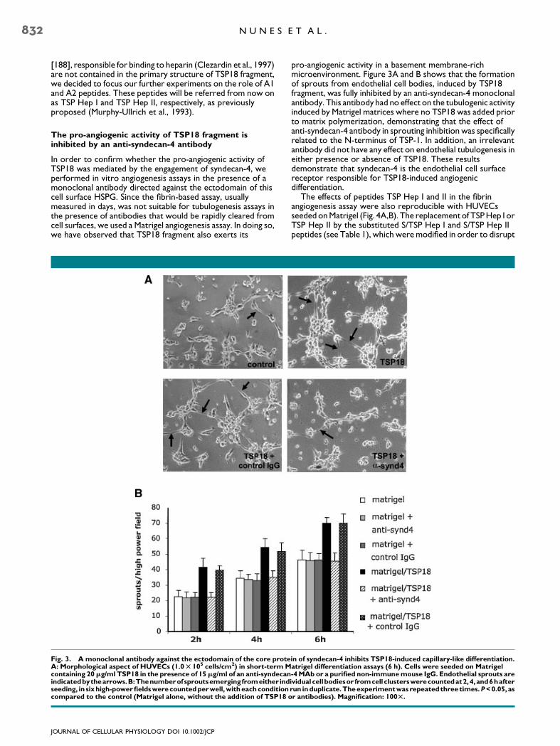
In order to confirm whether the pro-angiogenic activity ofTSP18 was mediated by the engagement of syndecan-4, weperformed in vitro angiogenesis assays in the presence of amonoclonal antibody directed against the ectodomain of thiscell surface HSPG. Since the fibrin-based assay, usuallymeasured in days, was not suitable for tubulogenesis assays inthe presence of antibodies that would be rapidly cleared fromcell surfaces, we used a Matrigel angiogenesis assay. In doing so,we have observed that TSP18 fragment also exerts its
Fig. 3. A monoclonal antibody against the ectodomain of the core proteA: Morphological aspect of HUVECs (1.0T 105 cells/cm2) in short-term Mcontaining 20mg/ml TSP18 in the presence of 15mg/ml of an anti-syndecanindicatedbythearrows.B:Thenumberofsproutsemerging fromeither indseeding, in sixhigh-powerfieldswerecountedperwell,witheachconditioncompared to the control (Matrigel alone, without the addition of TSP18 o
JOURNAL OF CELLULAR PHYSIOLOGY DOI 10.1002/JCP
pro-angiogenic activity in a basement membrane-richmicroenvironment. Figure 3A and B shows that the formationof sprouts from endothelial cell bodies, induced by TSP18fragment, was fully inhibited by an anti-syndecan-4 monoclonalantibody. This antibody had no effect on the tubulogenic activityinduced by Matrigel matrices where no TSP18 was added priorto matrix polymerization, demonstrating that the effect ofanti-syndecan-4 antibody in sprouting inhibition was specificallyrelated to the N-terminus of TSP-1. In addition, an irrelevantantibody did not have any effect on endothelial tubulogenesis ineither presence or absence of TSP18. These resultsdemonstrate that syndecan-4 is the endothelial cell surfacereceptor responsible for TSP18-induced angiogenicdifferentiation.
The effects of peptides TSP Hep I and II in the fibrinangiogenesis assay were also reproducible with HUVECsseeded on Matrigel (Fig. 4A,B). The replacement of TSP Hep I orTSP Hep II by the substituted S/TSP Hep I and S/TSP Hep IIpeptides (see Table 1), which were modified in order to disrupt
in of syndecan-4 inhibits TSP18-induced capillary-like differentiation.atrigel differentiation assays (6 h). Cells were seeded on Matrigel-4MAb or a purified non-immunemouse IgG. Endothelial sprouts areividualcellbodiesor fromcellclusterswerecountedat2,4,and6hafterrun induplicate.Theexperimentwasrepeatedthree times.P<0.05, asr antibodies). Magnification: 100T.
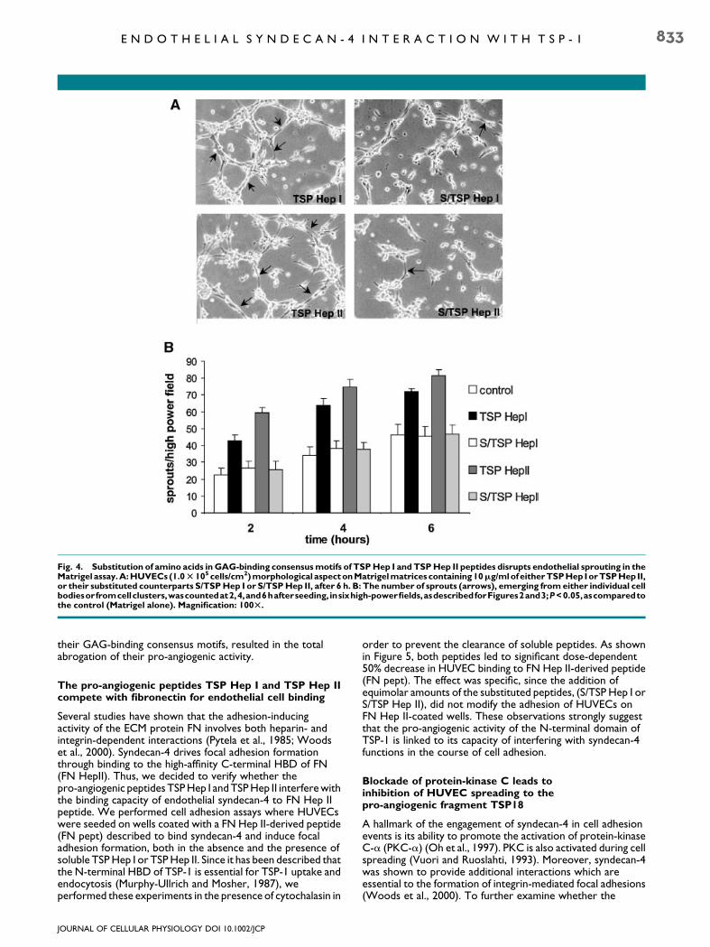
Fig. 4. Substitution of amino acids in GAG-binding consensusmotifs of TSPHep I and TSPHep II peptides disrupts endothelial sprouting in theMatrigel assay.A:HUVECs (1.0T 105cells/cm2)morphological aspectonMatrigelmatrices containing10mg/mlofeitherTSPHep IorTSPHepII,or their substituted counterparts S/TSPHep I or S/TSPHep II, after 6 h. B: The number of sprouts (arrows), emerging from either individual cellbodiesorfromcellclusters,wascountedat2,4,and6hafterseeding, insixhigh-powerfields,asdescribedforFigures2and3;P<0.05,ascomparedtothe control (Matrigel alone). Magnification: 100T.
E N D O T H E L I A L S Y N D E C A N - 4 I N T E R A C T I O N W I T H T S P - 1 833
their GAG-binding consensus motifs, resulted in the totalabrogation of their pro-angiogenic activity.
The pro-angiogenic peptides TSP Hep I and TSP Hep IIcompete with fibronectin for endothelial cell binding
Several studies have shown that the adhesion-inducingactivity of the ECM protein FN involves both heparin- andintegrin-dependent interactions (Pytela et al., 1985; Woodset al., 2000). Syndecan-4 drives focal adhesion formationthrough binding to the high-affinity C-terminal HBD of FN(FN HepII). Thus, we decided to verify whether thepro-angiogenic peptides TSP Hep I and TSP Hep II interfere withthe binding capacity of endothelial syndecan-4 to FN Hep IIpeptide. We performed cell adhesion assays where HUVECswere seeded on wells coated with a FN Hep II-derived peptide(FN pept) described to bind syndecan-4 and induce focaladhesion formation, both in the absence and the presence ofsoluble TSP Hep I or TSP Hep II. Since it has been described thatthe N-terminal HBD of TSP-1 is essential for TSP-1 uptake andendocytosis (Murphy-Ullrich and Mosher, 1987), weperformed these experiments in the presence of cytochalasin in
JOURNAL OF CELLULAR PHYSIOLOGY DOI 10.1002/JCP
order to prevent the clearance of soluble peptides. As shownin Figure 5, both peptides led to significant dose-dependent50% decrease in HUVEC binding to FN Hep II-derived peptide(FN pept). The effect was specific, since the addition ofequimolar amounts of the substituted peptides, (S/TSP Hep I orS/TSP Hep II), did not modify the adhesion of HUVECs onFN Hep II-coated wells. These observations strongly suggestthat the pro-angiogenic activity of the N-terminal domain ofTSP-1 is linked to its capacity of interfering with syndecan-4functions in the course of cell adhesion.
Blockade of protein-kinase C leads toinhibition of HUVEC spreading to thepro-angiogenic fragment TSP18
A hallmark of the engagement of syndecan-4 in cell adhesionevents is its ability to promote the activation of protein-kinaseC-a (PKC-a) (Oh et al., 1997). PKC is also activated during cellspreading (Vuori and Ruoslahti, 1993). Moreover, syndecan-4was shown to provide additional interactions which areessential to the formation of integrin-mediated focal adhesions(Woods et al., 2000). To further examine whether the
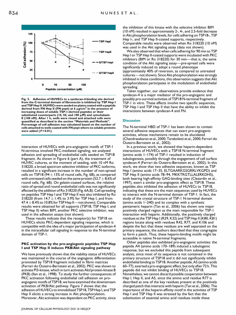
Fig. 5. Adhesion of HUVECs to a syndecan-4-binding site derivedfrom the C-terminal domain of fibronectin is inhibited by TSP Hep IandTSPHep II.HUVECswere seededonplates coatedwith a peptidederived from FN Hep II (FN pept) at 6 mg/cm2 in the presence ofincreasing doses of soluble TSP-1-derived peptides or theirsubstituted counterparts (10, 50, and 100 mM) and cytochalasinE (100 nM). After 1 h, wells were rinsed and attached cells werequantified as described in the section ‘‘Materials and Methods’’.Percentage of cell adhesion was calculated based on the number ofcells attached towells coatedwith FNpept where no soluble proteinswere added (P<0.01).
834 N U N E S E T A L .
interaction of HUVECs with pro-angiogenic motifs of TSP-1N-terminus involved PKC-mediated signaling, we analyzedadhesion and spreading of endothelial cells seeded on TSP18fragment. As shown in Figure 6 (part A), the treatment ofHUVEC cultures, at the moment of seeding, with 10 nM Ro318220, a broad spectrum selective inhibitor of PKC isoforms,resulted in a significant increase in the number of non-spreadcells on TSP18 (94� 13% of round cells, Fig. 6B), as comparedwith untreated cells seeded on the same protein (52.7� 8.6% ofround cells, Fig. 6B). On gelatin-coated surfaces, the relativeratio of spread and round endothelial cells was not significantlyaffected by the addition of Ro 318220 (Fig. 6A,B). Cell spreadingon peptides TSP Hep I and TSP Hep II was also inhibited by Ro318220 (from 14.7� 1.4% to 3.9% for TSP Hep I, and from47.4� 8.4% to 10.8% for TSP Hep II—not shown). Comparableresults were obtained for all supports (TSP18, TSP Hep I, andTSP Hep II) when BIM, another PKC selective inhibitor, wasused in the adhesion assays (not shown).
These results indicate that the receptor(s) for TSP18 onHUVECs elicits PKC-dependent signaling response, which iscompatible with the idea of a major participation of syndecan-4in the intracellular cell signaling in response to the N-terminaldomain of TSP-1.
PKC activation by the pro-angiogenic peptides TSP HepI and TSP Hep II induces PKB/Akt signaling pathway
We have previously shown that the viability status of HUVECswas maintained in the course of the angiogenic differentiationpromoted by TSP18 fragment included in fibrin matrices(Ferrari do Outeiro-Bernstein et al., 2002). PKC was shown toactivate PI3-kinase, which in turn activates Akt/protein-kinase B(PKB) (Ilan et al., 1998). To study the further consequences ofPKC activation following endothelial cell adhesion on pro-angiogenic motifs of TSP18, we have examined the downstreamactivation of PKB/Akt pathway. Figure 7 shows that theadhesion of HUVECs to immobilized TSP18, TSP Hep I, and TSPHep II elicits a strong increase in Akt phosphorylation.Moreover, Akt activation was dependent on PKC activity, since
JOURNAL OF CELLULAR PHYSIOLOGY DOI 10.1002/JCP
the inhibition of this kinase with the selective inhibitor BIM(10 nM) resulted in approximately 2-, 4-, and 2.5-fold decreasein Akt phosphorylation levels, for cells adhering on TSP18-, TSPHep I-, and TSP Hep II-coated supports, respectively.Comparable results were observed when Ro 318220 (10 nM)was used in the Akt signaling assay (data not shown).
We also observed that when cells adhering for 90 min to TSPHep I or TSP Hep II-coated supports were incubated with PKCinhibitors (BIM or Ro 318220) for 30 min—that is, the samecondition of the Akt signaling assay—pre-spread cells weresignificantly induced to adopt a round phenotype(approximately 40% of reversion, as compared to untreatedcultures—not shown). Since Akt phosphorylation was stronglyinhibited in these conditions, this observation suggests that Aktphosphorylation participates in the modulation of endothelialspreading.
Taken together, our observations provide evidence thatsyndecan-4 is a major mediator of the pro-angiogenic andpossibly pro-survival activities of TSP18 N-terminal fragment ofTSP-1 in vitro. These effects involve two specific sequences,TSP Hep I and TSP Hep II that have the ability to inhibit theinteractions between syndecan-4 and FN.
Discussion
The N-terminal HBD of TSP-1 has been shown to containseveral adhesive sequences that can exert pro-angiogenicactivities, whose mechanisms remain to be elucidated(Chandrasekaran et al., 2000; Taraboletti et al., 2000; Ferrari doOuteiro-Bernstein et al., 2002).
In a previous work, we showed that heparin-dependentinteractions of HUVECs with a TSP18 N-terminal fragment(amino acids 1–174) of TSP-1 resulted in induction oftubulogenesis, possibly through the engagement of cell surfacesyndecan-4 (Ferrari do Outeiro-Bernstein et al., 2002). In thiswork, we show that two adhesive sequences, known as TSPHep I (amino acids 17–35, ELTGAARKGSGRRLVKGPD) andTSP Hep II (amino acids 78–94, MKKTRGTLLALERKDHS),both bearing high-affinity GAG-binding sites (Clezardin et al.,1997), exert a similar pro-angiogenic effect as TSP18. Thesepeptides also inhibited the adhesion of HUVECs to TSP18,indicating that these are the main sequences used by HUVECsto interact with the N-terminal domain of TSP-1. In fact, thestudy of the crystal structure of TSP-1 N-terminal domain(amino acids 1–240) and its complex with a syntheticpentameric heparin (Tan et al., 2006) has shown one majorpositively charged region as the main responsible for TSP-1interaction with heparin. Additionally, the positively chargedresidues at the TSP Hep I (R29, K32) and TSP Hep II (K80, K81)regions locate along with residues R42, R77, and K106 anddespite the fact that these residues are well separated on theprimary sequence, the authors described that they congregateto form a patch. Thus, these heparin-binding motifs might beaccessible in native N-terminal fragments.
Other peptides also exhibited pro-angiogenic activities: thepeptide A4 (amino acids 170–189) induced a tubulogenicresponse, but we excluded this peptide from subsequentanalysis, since most of its sequence is not contained in theprimary structure of TSP18 and it did not significantly inhibitendothelial binding to TSP18. Another peptide, A5 (amino acids60–77), exerted a pro-angiogenic effect, but only after 72 h. Thispeptide did not inhibit binding of HUVECs to TSP18.Nonetheless, we cannot discard possible cooperation betweenHep I, Hep II, and A5, since the amino acid residue R77 isdescribed as one of the key residues present at the positivelycharged patch that interacts with heparin (Tan et al., 2006). Theimportance of the heparin-affinity motif in the activities of TSPHep I and TSP Hep II was stressed by the fact that thesubstitution of essential amino acid residues inside these
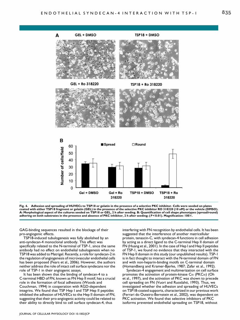
Fig. 6. Adhesion and spreading of HUVECs to TSP18 or gelatin in the presence of a selective PKC inhibitor. Cells were seeded on platescoated with either TSP18 fragment or gelatin (GEL) in the presence of the selective PKC inhibitor RO 318220 (10 nM) or the vehicle (DMSO).A: Morphological aspect of the cultures seeded on TSP18 or GEL, 2 h after seeding. B: Quantification of cell shape phenotypes (spread/round)adhering on both substrates in the presence and absence of PKC inhibitor, 2 h after seeding; (P<0.01). Magnification: 100T.
E N D O T H E L I A L S Y N D E C A N - 4 I N T E R A C T I O N W I T H T S P - 1 835
GAG-binding sequences resulted in the blockage of theirpro-angiogenic effects.
TSP18-induced tubulogenesis was fully abolished by ananti-syndecan-4 monoclonal antibody. This effect wasspecifically related to the N-terminal of TSP-1, since the sameantibody had no effect on endothelial tubulogenesis when noTSP18 was added to Matrigel. Recently, a role for syndecan-2 inthe regulation of angiogenesis of microvascular endothelial cellshas been proposed (Fears et al., 2006). However, the authorsneither address the role of intact cell surface syndecans nor therole of TSP-1 in their angiogenic assays.
It has been shown that the binding of syndecan-4 to aC-terminal HBD of FN, known as FN Hep II motif, has a crucialrole in the formation of focal adhesions (Woods andCouchman, 1994) in cooperation with RGD-dependentintegrins. We found that TSP Hep I and TSP Hep II peptidesinhibited the adhesion of HUVECs to the Hep II domain of FN,suggesting that their pro-angiogenic activity could be related totheir ability to directly bind to cell surface syndecan-4, thus
JOURNAL OF CELLULAR PHYSIOLOGY DOI 10.1002/JCP
interfering with FN recognition by endothelial cells. It has beensuggested that the interference of another matricellularprotein, tenascin-C, with syndecan-4 functions in cell adhesionby acting as a direct ligand to the C-terminal Hep II domain ofFN (Huang et al., 2001). In the case of Hep I and Hep II peptidesof TSP-1, we found no evidence that they interacted with theFN Hep II domain in this study (our unpublished results). TSP-1is in fact thought to interact with the N-terminal domain of FNand with non-heparin-binding motifs on C-terminal domain(Homandberg and Kramer-Bjerke, 1987; Zafar et al., 1992).
Syndecan-4 engagement and multimerization on cell surfacepromotes the activation of protein-kinase Ca (PKCa) (Ohet al., 1997), and the activation of PKC was shown to precedecell spreading on FN (Vuori and Ruoslahti, 1993). Thus, weinvestigated whether the adhesion and spreading of HUVECson TSP18-coated supports, characterized in our previous work(Ferrari do Outeiro-Bernstein et al., 2002), was dependent onPKC activation. We found that selective inhibitors of PKCisoforms prevented endothelial spreading on TSP18, without
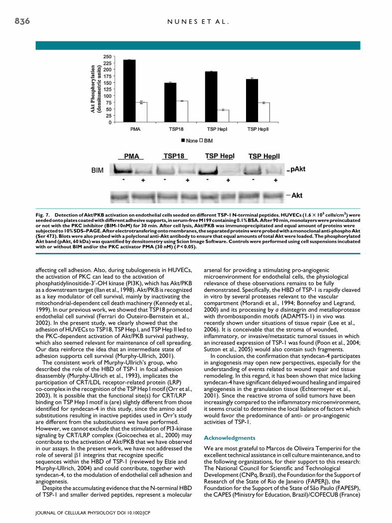
Fig. 7. Detection ofAkt/PKB activation on endothelial cells seeded ondifferent TSP-1N-terminal peptides. HUVECs (1.6T 105 cells/cm2)wereseededontoplatescoatedwithdifferentadhesivesupports, inserum-freeM199containing0.1%BSA.After90min,monolayerswerepreincubatedor not with the PKC inhibitor (BIM-10nM) for 30 min. After cell lysis, Akt/PKB was immunoprecipitated and equal amount of proteins weresubjectedto10%SDS–PAGE.Afterelectrotransferingontomembranes, theseparatedproteinswereprobedwithamonoclonalanti-phosphoAkt(Ser 473). Blotswere also probedwith a polyclonal anti-Akt antibody to ensure that equal amounts of total Aktwere loaded. The phosphorylatedAkt band (pAkt, 60 kDa)was quantified by densitometry using Scion Image Software. Controls were performed using cell suspensions incubatedwith or without BIM and/or the PKC activator PMA (30 nM) (P<0.05).
836 N U N E S E T A L .
affecting cell adhesion. Also, during tubulogenesis in HUVECs,the activation of PKC can lead to the activation ofphosphatidylinositide-30-OH kinase (PI3K), which has Akt/PKBas a downstream target (Ilan et al., 1998). Akt/PKB is recognizedas a key modulator of cell survival, mainly by inactivating themitochondrial-dependent cell death machinery (Kennedy et al.,1999). In our previous work, we showed that TSP18 promotedendothelial cell survival (Ferrari do Outeiro-Bernstein et al.,2002). In the present study, we clearly showed that theadhesion of HUVECs to TSP18, TSP Hep I, and TSP Hep II led tothe PKC-dependent activation of Akt/PKB survival pathway,which also seemed relevant for maintenance of cell spreading.Our data reinforce the idea that an intermediate state ofadhesion supports cell survival (Murphy-Ullrich, 2001).
The consistent work of Murphy-Ullrich’s group, whodescribed the role of the HBD of TSP-1 in focal adhesiondisassembly (Murphy-Ullrich et al., 1993), implicates theparticipation of CRT/LDL receptor-related protein (LRP)co-complex in the recognition of the TSP Hep I motif (Orr et al.,2003). It is possible that the functional site(s) for CRT/LRPbinding on TSP Hep I motif is (are) slightly different from thoseidentified for syndecan-4 in this study, since the amino acidsubstitutions resulting in inactive peptides used in Orr’s studyare different from the substitutions we have performed.However, we cannot exclude that the stimulation of PI3-kinasesignaling by CRT/LRP complex (Goicoechea et al., 2000) maycontribute to the activation of Akt/PKB that we have observedin our assays. In the present work, we have not addressed therole of several b1 integrins that recognize specificsequences within the HBD of TSP-1 (reviewed by Elzie andMurphy-Ullrich, 2004) and could contribute, together withsyndecan-4, to the modulation of endothelial cell adhesion andangiogenesis.
Despite the accumulating evidence that the N-terminal HBDof TSP-1 and smaller derived peptides, represent a molecular
JOURNAL OF CELLULAR PHYSIOLOGY DOI 10.1002/JCP
arsenal for providing a stimulating pro-angiogenicmicroenvironment for endothelial cells, the physiologicalrelevance of these observations remains to be fullydemonstrated. Specifically, the HBD of TSP-1 is rapidly cleavedin vitro by several proteases relevant to the vascularcompartment (Morandi et al., 1994; Bonnefoy and Legrand,2000) and its processing by a disintegrin and metalloproteasewith thrombosspondin motifs (ADAMTS-1) in vivo wasrecently shown under situations of tissue repair (Lee et al.,2006). It is conceivable that the stroma of wounded,inflammatory, or invasive/metastatic tumoral tissues in whichan increased expression of TSP-1 was found (Poon et al., 2004;Sutton et al., 2005) would also contain such fragments.
In conclusion, the confirmation that syndecan-4 participatesin angiogenesis may open new perspectives, especially for theunderstanding of events related to wound repair and tissueremodeling. In this regard, it has been shown that mice lackingsyndecan-4 have significant delayed wound healing and impairedangiogenesis in the granulation tissue (Echtermeyer et al.,2001). Since the reactive stroma of solid tumors have beenincreasingly compared to the inflammatory microenvironment,it seems crucial to determine the local balance of factors whichwould favor the predominance of anti- or pro-angiogenicactivities of TSP-1.
Acknowledgments
We are most grateful to Marcos de Oliveira Temperini for theexcellent technical assistance in cell culture maintenance, and tothe following organizations, for their support to this research:The National Council for Scientific and TechnologicalDevelopment (CNPq, Brazil), the Foundation for the Support ofResearch of the State of Rio de Janeiro (FAPERJ), theFoundation for the Support of the State of Sao Paulo (FAPESP),the CAPES (Ministry for Education, Brazil)/COFECUB (France)
E N D O T H E L I A L S Y N D E C A N - 4 I N T E R A C T I O N W I T H T S P - 1 837
Cooperation Agreement, and the PRONEX/FAPERJ Program(Brazilian Ministry for Science and Technology); and INSERM(Institut National de la Sante et de la Recherche Medicale,France).
Literature Cited
Adams JC, Lawler J. 2004. The thrombospondins. Int J Biochem Cell Biol 36:961–968.Baciu PC, Saoncella S, Lee SH, Denhez F, Leuthardt D, Goetinck PF. 2000. Syndesmos, a
protein that interacts with the cytoplasmic domain of syndecan-4, mediates cell spreadingand actin cytoskeletal organization. J Cell Sci 2:315–324.
Bonnefoy A, Legrand C. 2000. Proteolysis of subendothelial adhesive glycoproteins(fibronectin, thrombospondin, and von Willebrand factor) by plasmin, leukocyte cathepsinG, and elastase. Thromb Res 98:323–332.
Carmeliet P. 2003. Angiogenesis in health and disease. Nat Med 9:653–660.Chandrasekaran L, He CZ, Al-Barazi H, Krutzsch HC, Iruela-Arispe ML, Roberts DD. 2000.
Cell contact-dependent activation of integrin a3b1 integrin modulates endothelial cellresponses to thrombospondin-1. Mol Biol Cell 11:2885–2900.
Clezardin P, Lawler J, Amiral J, Quentin G, Delmas P. 1997. Identification of cell adhesive sitesin the N-terminal domain of thrombospondin-1. Biochem J 321:819–827.
Cross MJ, Claesson-Welsh L. 2001. FGF and VEGF function in angiogenesis: Signallingpathways, biological responses and therapeutic inhibition. Trends Pharmacol Sci 22:201–207.
Echtermeyer F, Streit M, Wilcox-Adelman S, Saoncella S, Denhez F, Detmar M, Goetinck PF.2001. Delayed wound repair and impaired angiogenesis in mice lacking syndecan-4. J ClinInvest 107:R9–R14.
Elzie CA, Murphy-Ullrich JE. 2004. The N-terminus of thrombospondin: The domain standsapart. Int J Biochem Cell Biol 36:1090–1101.
Fears CY, Woods A. 2006. The role of syndecans in disease and wound healing. Matrix Biol25:443–456.
Fears CY, Gladson CL, Woods A. 2006. Syndecan-2 is expressed in the microvasculature ofgliomas and regulates angiogenic processes in microvascular endothelial cells. J Biol Chem281:14533–14536.
Ferrari do Outeiro-Bernstein MA, Nunes SS, Andrade ACM, Alves TR, Legrand C, MorandiV. 2002. A recombinant NH2-terminal heparin binding domain of the adhesiveglycoprotein, thrombospondin-1, promotes endothelial tube formation and cell survival: Apossible role for syndecan-4 proteoglycan. Matrix Biol 21:311–324.
Filleur S, Volpert OV, Degeorges A, Voland C, Reiher F, Clezardin P, Bouck N, Cabon F. 2001.In vivo mechanisms by which tumors producing thrombospondin 1 bypass its inhibitoryeffects. Genes Dev 15:1373–1382.
Fontana A, Filleur S, Guglielmi J, Frappart L, Boissier S, Cabon F, Bruno-Bossio G, Clezardin P.2005. Human breast tumors override the antiangiogenic effect of stromalthrombospondin-1 in vivo. Int J Cancer 116:686–691.
Goicoechea S, Orr AW, Pallero MA, Eggleton P, Murphy-Ullrich JE. 2000. Thrombospondinmediates focal adhesion disassembly through interactions with cell surface calreticulin.J Biol Chem 275:36358–36368.
Greene DK, Tumova S, Couchman JR, Woods A. 2003. Syndecan-4 associates with alpha-actinin. J Biol Chem 278:7617–7623.
Homandberg GA, Kramer-Bjerke J. 1987. Thrombospondin binds to amino-terminalfragments of plasma fibronectin. Thromb Res 48:329–335.
Huang W, Chiquet-Ehrismann R, Moyano JV, Garcia-Pardo A, Orend G. 2001. Interference oftenascin-C with syndecan-4 binding to fibronectin blocks cell adhesion and stimulatestumor cell proliferation. Cancer Res 61:8586–8594.
Ilan N, Mahooti S, Madri JA. 1998. Distinct signal transduction pathways are utilized during thetube formation and survival phases of in vitro angiogenesis. J Cell Sci 111:3621–3631.
Iruela-Arispe ML, Lombardo M, Krutzsch HC, Lawler J, Roberts RR. 1999. Inhibition ofangiogenesis by thrombospondin-1 is mediated by 2 independent regions within the type 1repeats. Circulation 100:1423–1431.
Jaffe EA, Nachman RL, Becker CG, Minick CR. 1973. Culture of human endothelial cellsderived from umbilical cord veins. Identification by morphologic and immunologic criteria.J Clin Invest 52:2745–2756.
Kennedy SG, Kandel ES, Cross TK, Hay N. 1999. Akt/Protein kinase B inhibits cell death bypreventing the release of cytochrome c from mitochondria. Mol Cell Biol 19:5800–5810.
Lee NV, Sato M, Annis DS, Loo JA, Wu L, Mosher DF, Iruela-Arispe ML. 2006.ADAMTS1 mediates the release of antiangiogenic polypeptides from TSP1 and 2. EMBO J25:5270–5283.
JOURNAL OF CELLULAR PHYSIOLOGY DOI 10.1002/JCP
Legrand C, Morandi V, Mendelovitz S, Shaked H, Hartman JR, Panet A. 1994. Selectiveinhibition of platelet macroaggregate formation by a recombinant heparin-binding domainof human thrombospondin. Arterioscler Thromb 14:1784–1791.
Longley RL, Woods A, Fleetwood A, Cowling GJ, Gallagher JT, Couchman JR. 1999.Control of morphology, cytoskeleton and migration by syndecan-4. J Cell Sci 112:3421–3431.
McFall AJ, Rapraeger AC. 1997. Identification of an adhesion site within the syndecan-4extracellular protein domain. J Biol Chem 272:12901–12904.
Morandi V, Edelman L, Legrand YJ, Legrand C. 1994. Characterization of a novel monoclonalantibody (V58A4) raised against a recombinant NH2-terminal heparin-binding fragment ofhuman endothelial cell thrombospondin. FEBS Lett 346:156–160.
Murphy-Ullrich JE. 2001. The de-adhesive activity of matricellular proteins: Is intermediatecell adhesion an adaptive state? J Clin Invest 107:785–790.
Murphy-Ullrich JE, Mosher DF. 1987. Interactions of thrombospondin with endothelial cells:receptor-mediated binding and degradation. J Cell Biol 105:1603–1611.
Murphy-Ullrich JE, Gurusiddappa S, Frazier WA, Hook M. 1993. Heparin-binding peptidesfrom thrombospondins 1 and 2 contain focal adhesion-labilizing activity. J Biol Chem268:26784–26789.
Oh ES, Woods A, Couchman JR. 1997. Multimerization of the cytoplasmic domain ofsyndecan-4 is required for its ability to activate protein kinase C. J Biol Chem 272:11805–11811.
Orr AW, Pedraza CE, Pallero MA, Elzie CA, Goicoechea S, Strickland DK, Murphy-Ullrich JE.2003. Low density lipoprotein receptor-related protein is a calreticulin coreceptor thatsignals focal adhesion disassembly. J Cell Biol 161:1179–1189.
Poon RT, Chung KK, Cheung ST, Lau CP, Tong SW, Leung KL, Yu WC, Tuszynski G, Fan ST.2004. Clinical significance of thrombospondin expression in hepatocellular carcinoma. ClinCancer Res 10:4150–4157.
Pytela R, Pierschbacher MD, Ruoslahti E. 1985. Identification and isolation of a 140 kdcell surface glycoprotein with properties expected of a fibronectin receptor.Cell 40:191–198.
Roy R, Zhang B, Moses MA. 2006. Making the cut: Protease-mediated regulation ofangiogenesis. Exp Cell Res 312:608–622.
Sanz L, Alvarez-Vallina L. 2003. The extracellular matrix: A new turn-of-the-screw for anti-angiogenic strategies. Trends Mol Med 9:256–262.
Sutton CD, O’Byrne K, Goddard JC, Marshall L, Jones L, Garcea G, Dennison AR, Poston G,Lloyd DM, Berry DP. 2005. Expression of thrombospondin-1 in resected colorectal livermetastases predicts poor prognosis. Clin Cancer Res 11:6567–6573.
Suzuki N, Ichikawa N, Kasai S, Yamada M, Nishi N, Morioka H, Yamashita H, Kitagawa Y,Utani A, Hoffman MP, Nomizu M. 2003. Syndecan binding sites in the laminin alpha1 chain Gdomain. Biochemistry 42:12625–12633.
Tan K, Duquette M, Liu J, Zhang R, Joachimiak A, Wang J, Lawler J. 2006. The structures of thetrombospondin-1 N-treminal domain and its complex with a synthetic pentameric heparin.Structure 14:33–42.
Taraboletti G, Morbidelli L, Donnini S, Parenti A, Granger H, Giavazzi R, Ziche M. 2000. Theheparin binding 25 kDa fragment of thrombospondin-1 promotes angiogenesisand modulates gelatinase and TIMP-2 production in endothelial cells. FASEB J14:1674–1676.
Tolsma SS, Volpert OV, Good DJ, Frazier WA, Polverini PJ, Bouck N. 1993. Peptides derivedfrom two separate domains of the matrix protein thrombospondin-1 have antiangiogenicactivity. J Cell Biol 122:497–511.
Vogel T, Guo N, Krutzsch HC, Blake DA, Hartman J, Mendelovitz S, Panet A, Roberts DD.1993. Modulation of endothelial cell proliferation by recombinant heparin-binding domainand synthetic peptides from the type I repeats of thrombospondin. J Cell Biochem 53:74–84.
Vuori K, Ruoslahti E. 1993. Activation of protein kinase C precedes alpha 5 beta1 integrin-mediated cell spreading on fibronectin. J Biol Chem 268:21459–21462.
Whiteford JR, Couchman JR. 2006. A conserved NXIP motif id required for celladhesion properties of the syndecan-4 ectodomain. J Biol Chem 281:32156–32163.
Woods A, Couchman JR. 1994. Syndecan 4 heparan sulfate proteoglycan is aselectively enriched and widespread focal adhesion component. Mol BiolCell 5:183–192.
Woods A, McCarthy JB, Furcht LT, Couchman JR. 1993. A synthetic peptide from theCOOH-terminal heparin-binding domain of fibronectin promotes focal adhesionformation. Mol Biol Cell 4:605–613.
Woods A, Longley RL, Tumova S, Couchman JR. 2000. Syndecan-4 binding to the high affinityheparin-binding domain of fibronectin drives focal adhesion formation in fibroblasts. ArchBiochem Biophys 374:66–72.
Zafar RS, Zeng Z, Walz DA. 1992. Localization of two binding domains for thrombospondinwithin fibronectin. Arch Biochem Biophys 297:271–276.
Copyright © 2022 FDOKUMEN