Identification of bacteria in enrichment cultures of sulfate ...
11
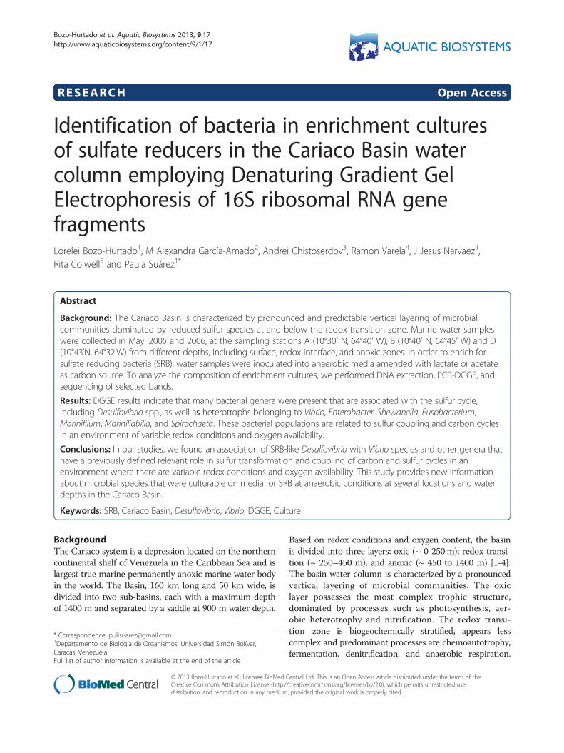
RESEARCH Open Access Identification of bacteria in enrichment cultures of sulfate reducers in the Cariaco Basin water column employing Denaturing Gradient Gel Electrophoresis of 16S ribosomal RNA gene fragments Lorelei Bozo-Hurtado 1 , M Alexandra García-Amado 2 , Andrei Chistoserdov 3 , Ramon Varela 4 , J Jesus Narvaez 4 , Rita Colwell 5 and Paula Suárez 1* Abstract Background: The Cariaco Basin is characterized by pronounced and predictable vertical layering of microbial communities dominated by reduced sulfur species at and below the redox transition zone. Marine water samples were collected in May, 2005 and 2006, at the sampling stations A (10°30′ N, 64°40′ W), B (10°40′ N, 64°45′ W) and D (10°43’N, 64°32’W) from different depths, including surface, redox interface, and anoxic zones. In order to enrich for sulfate reducing bacteria (SRB), water samples were inoculated into anaerobic media amended with lactate or acetate as carbon source. To analyze the composition of enrichment cultures, we performed DNA extraction, PCR-DGGE, and sequencing of selected bands. Results: DGGE results indicate that many bacterial genera were present that are associated with the sulfur cycle, including Desulfovibrio spp., as well as heterotrophs belonging to Vibrio, Enterobacter, Shewanella, Fusobacterium, Marinifilum, Mariniliabilia, and Spirochaeta. These bacterial populations are related to sulfur coupling and carbon cycles in an environment of variable redox conditions and oxygen availability. Conclusions: In our studies, we found an association of SRB-like Desulfovibrio with Vibrio species and other genera that have a previously defined relevant role in sulfur transformation and coupling of carbon and sulfur cycles in an environment where there are variable redox conditions and oxygen availability. This study provides new information about microbial species that were culturable on media for SRB at anaerobic conditions at several locations and water depths in the Cariaco Basin. Keywords: SRB, Cariaco Basin, Desulfovibrio, Vibrio, DGGE, Culture Background The Cariaco system is a depression located on the northern continental shelf of Venezuela in the Caribbean Sea and is largest true marine permanently anoxic marine water body in the world. The Basin, 160 km long and 50 km wide, is divided into two sub-basins, each with a maximum depth of 1400 m and separated by a saddle at 900 m water depth. Based on redox conditions and oxygen content, the basin is divided into three layers: oxic (~ 0-250 m); redox transi- tion (~ 250–450 m); and anoxic (~ 450 to 1400 m) [1-4]. The basin water column is characterized by a pronounced vertical layering of microbial communities. The oxic layer possesses the most complex trophic structure, dominated by processes such as photosynthesis, aer- obic heterotrophy and nitrification. The redox transi- tion zone is biogeochemically stratified, appears less complex and predominant processes are chemoautotrophy, fermentation, denitrification, and anaerobic respiration. * Correspondence: [email protected] 1 Departamento de Biología de Organismos, Universidad Simón Bolívar, Caracas, Venezuela Full list of author information is available at the end of the article AQUATIC BIOSYSTEMS © 2013 Bozo-Hurtado et al.; licensee BioMed Central Ltd. This is an Open Access article distributed under the terms of the Creative Commons Attribution License (http://creativecommons.org/licenses/by/2.0), which permits unrestricted use, distribution, and reproduction in any medium, provided the original work is properly cited. Bozo-Hurtado et al. Aquatic Biosystems 2013, 9:17 http://www.aquaticbiosystems.org/content/9/1/17
-
Upload
khangminh22 -
Category
Documents
-
view
2 -
download
0
Transcript of Identification of bacteria in enrichment cultures of sulfate ...
AQUATIC BIOSYSTEMSBozo-Hurtado et al. Aquatic Biosystems 2013, 9:17http://www.aquaticbiosystems.org/content/9/1/17
RESEARCH Open Access
Identification of bacteria in enrichment culturesof sulfate reducers in the Cariaco Basin watercolumn employing Denaturing Gradient GelElectrophoresis of 16S ribosomal RNA genefragmentsLorelei Bozo-Hurtado1, M Alexandra García-Amado2, Andrei Chistoserdov3, Ramon Varela4, J Jesus Narvaez4,Rita Colwell5 and Paula Suárez1*
Abstract
Background: The Cariaco Basin is characterized by pronounced and predictable vertical layering of microbialcommunities dominated by reduced sulfur species at and below the redox transition zone. Marine water sampleswere collected in May, 2005 and 2006, at the sampling stations A (10°30′ N, 64°40′ W), B (10°40′ N, 64°45′ W) and D(10°43’N, 64°32’W) from different depths, including surface, redox interface, and anoxic zones. In order to enrich forsulfate reducing bacteria (SRB), water samples were inoculated into anaerobic media amended with lactate or acetateas carbon source. To analyze the composition of enrichment cultures, we performed DNA extraction, PCR-DGGE, andsequencing of selected bands.
Results: DGGE results indicate that many bacterial genera were present that are associated with the sulfur cycle,including Desulfovibrio spp., as well as heterotrophs belonging to Vibrio, Enterobacter, Shewanella, Fusobacterium,Marinifilum, Mariniliabilia, and Spirochaeta. These bacterial populations are related to sulfur coupling and carbon cyclesin an environment of variable redox conditions and oxygen availability.
Conclusions: In our studies, we found an association of SRB-like Desulfovibrio with Vibrio species and other genera thathave a previously defined relevant role in sulfur transformation and coupling of carbon and sulfur cycles in anenvironment where there are variable redox conditions and oxygen availability. This study provides new informationabout microbial species that were culturable on media for SRB at anaerobic conditions at several locations and waterdepths in the Cariaco Basin.
Keywords: SRB, Cariaco Basin, Desulfovibrio, Vibrio, DGGE, Culture
BackgroundThe Cariaco system is a depression located on the northerncontinental shelf of Venezuela in the Caribbean Sea and islargest true marine permanently anoxic marine water bodyin the world. The Basin, 160 km long and 50 km wide, isdivided into two sub-basins, each with a maximum depthof 1400 m and separated by a saddle at 900 m water depth.
* Correspondence: [email protected] de Biología de Organismos, Universidad Simón Bolívar,Caracas, VenezuelaFull list of author information is available at the end of the article
© 2013 Bozo-Hurtado et al.; licensee BioMed CCreative Commons Attribution License (http:/distribution, and reproduction in any medium
Based on redox conditions and oxygen content, the basinis divided into three layers: oxic (~ 0-250m); redox transi-tion (~ 250–450 m); and anoxic (~ 450 to 1400 m) [1-4].The basin water column is characterized by a pronouncedvertical layering of microbial communities. The oxiclayer possesses the most complex trophic structure,dominated by processes such as photosynthesis, aer-obic heterotrophy and nitrification. The redox transi-tion zone is biogeochemically stratified, appears lesscomplex and predominant processes are chemoautotrophy,fermentation, denitrification, and anaerobic respiration.
entral Ltd. This is an Open Access article distributed under the terms of the/creativecommons.org/licenses/by/2.0), which permits unrestricted use,, provided the original work is properly cited.
Bozo-Hurtado et al. Aquatic Biosystems 2013, 9:17 Page 2 of 11http://www.aquaticbiosystems.org/content/9/1/17
The anoxic zone presumably has the simplest trophicstructure that appears to be supported by fermentation,sulfate reduction, methanogenesis and anaerobic methaneoxidation [3]. Many studies have been conducted in theCariaco Basin to understand how microorganisms are dis-tributed in the stratified water column environment andhow they influence geochemical processes [1,3,5]. Interest-ingly, high levels of sulfide present in the Cariaco Basinhave been attributed to biological sulfate reduction [6].This is the first attempt to identify bacteria related withsulfate reduction using enrichments cultures for theCariaco Basin.Several studies have been published describing the mi-
crobial community associated with the Cariaco Basin sulfurcycle. Tuttle and Jannasch (1973) isolated several sulfideand thiosulfate-oxidizing bacteria and Morris et al. (1985)isolated Alteromonas sp. from thiosulfate-containing en-richment cultures [7,8]. With the development of molecu-lar biology, culture-independent methods have been usedto detect SRB populations in the Cariaco Basin [1,5,9]. To
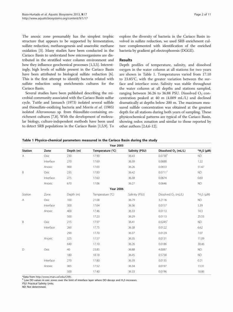
Table 1 Physico-chemical parameters measured in the Cariaco
Year 2
Station Zone Depth (m) Temperature (°C)
A Oxic 230 17.90
Interface 270 17.69
Anoxic 900 17.03
B Oxic 235 17.83
Interface 275 17.63
Anoxic 670 17.06
Year 2
Station Zone Depth (m) Temperature (°C)
A Oxic 100 21.08
Interface 300 17.64
Anoxic 400 17.46
500 17.23
B Oxic 215 17.97
Interface 260 17.75
290 17.70
Anoxic 325 17.57
640 17.10
D Oxic 40 23.85
180 18.18
Interface 270 17.80
Anoxic 365 17.52
500 17.40
*Data from http://www.imars.usf.edu/CAR/.† Low DO values in oxic zones over the limit of interface layer where DO decays anPSU: Practical Salinity Units.ND: Not determined.
explore the diversity of bacteria in the Cariaco Basin in-volved in sulfate reduction, we used SRB enrichment cul-ture complemented with identification of the enrichedbacteria by gradient gel electrophoresis (DGGE).
ResultsDepth profiles of temperature, salinity, and dissolvedoxygen in the water column at all stations for two yearsare shown in Table 1. Temperatures varied from 17.10to 23.85°C, with the greater variation between the sur-face and interface zone. Salinity was stable throughoutthe water column at all depths and stations sampled,ranging between 36.26 to 36.88 PSU. Dissolved O2 con-centration peaked at 40 m (4.009 mL/L) and declineddramatically at depths below 200 m. The maximum mea-sured sulfide concentration was obtained at the greatestdepth for all stations during both years of sampling. Thesephysicochemical patterns are typical of the Cariaco Basin,showing redox zonation and similar to those reported byother authors [2,4,6-12].
Basin during the study
005
Salinity (PSU) Dissolved O2 (mL/L) *H2S (μM)
36.43 0.0738† ND
36.39 0.0688 1.22
36.26 0.0653 51.47
36.42 0.0711† ND
36.38 0.0674 0.69
36.27 0.0646 ND
006
Salinity (PSU) Dissolved O2 (mL/L) *H2S (μM)
36.79 3.2116 ND
36.36 0.0157 5.39
36.33 0.0113 14.3
36.29 0.0113 25.55
36.41 0.0245† ND
36.38 0.0122 6.62
36.37 0.0129 7.07
36.35 0.0131 11.09
36.26 0.0186 38.46
36.88 4.0087 ND
36.45 0.5738 ND
36.39 0.0135 0.21
36.34 0.0197 13.31
36.33 0.0196 18.86
d H2S increases.
Bozo-Hurtado et al. Aquatic Biosystems 2013, 9:17 Page 3 of 11http://www.aquaticbiosystems.org/content/9/1/17
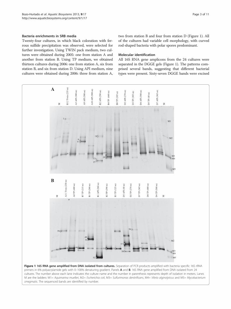
Bacteria enrichments in SRB mediaTwenty-four cultures, in which black coloration with fer-rous sulfide precipitation was observed, were selected forfurther investigation. Using TWIN pack medium, two cul-tures were obtained during 2005: one from station A andanother from station B. Using TP medium, we obtainedthirteen cultures during 2006: one from station A, six fromstation B, and six from station D. Using API medium, ninecultures were obtained during 2006: three from station A,
1
6
89
11
13
15
17
19
A9
API
(50
0 m
)
B2
AP
I (2
90 m
)
B3.
2 T
win
(235
m)
M A3
API
(30
0 m
)
A10
API
(90
0 m
)
B4
TP
(64
0 m
)A
39
4041 46
4951
52
53 55
A3.
1 T
win
(230
m)
M A8
TP
(400
m)
B7
TP
(215
m)
B1
TP
(260
m)
B1
AP
I (2
60 m
)
B2
TP
(290
m)
B3
TP
(325
m)
B
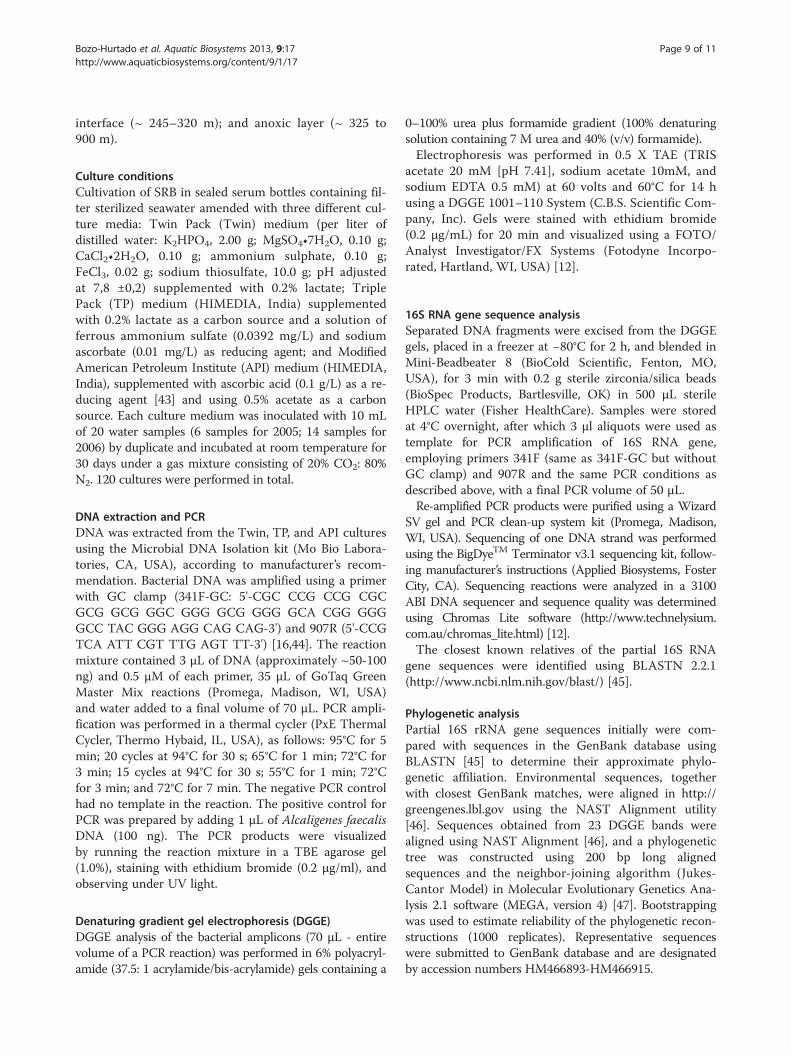
Figure 1 16S RNA gene amplified from DNA isolated from cultures. Sprimers in 6% polyacrylamide gels with 0–100% denaturing gradient. Panecultures. The number above each lane indicates the culture name and theM are the ladders: M1= Aquimarina muelleri, M2= Escherichia coli, M3= Sulfusmegmatis. The sequenced bands are identified by number.
two from station B and four from station D (Figure 1). Allof the cultures had variable cell morphology, with curvedrod-shaped bacteria with polar spores predominant.
Molecular identificationAll 16S RNA gene amplicons from the 24 cultures wereseparated in the DGGE gels (Figure 1). The patterns com-prised several bands, suggesting that different bacterialtypes were present. Sixty-seven DGGE bands were excised
M1
M2
M4
M5
M3
D2
API
(27
0 m
)
D5
TP
(40
m)
B8
TP
(245
m)
D3
TP
(305
m)
D4
API
(50
0 m
)
D7
API
(36
5 m
)
M
M2
M4
57
58
63
59
67
M3
M5
D8
TP
(400
m)
D5
API
(40
m)
D2
TP
(270
m)
D7
TP
(365
m)
D4
TP
(500
m)
M
eparation of PCR products amplified with bacteria specific 16S rRNAls A and B. 16S RNA gene amplified from DNA isolated from 24number in parenthesis represents depth of isolation in meters. Lanesrimonas denitrificans, M4= Vibrio alginolyticus and M5= Mycobacterium

Bozo-Hurtado et al. Aquatic Biosystems 2013, 9:17 Page 4 of 11http://www.aquaticbiosystems.org/content/9/1/17
and sequenced, but only 23 quality sequences weregenerated. These sequences were compared with existingsequences in the NCBI public database using the BLASTalgorithm. Results of the BLAST analyses are summarizedin Table 2. A prevalent band in the DGGE gels was ob-served for all samples, except D3 TP (305 m). Bands show-ing similar mobility gave highly similar or identicalsequences with the molecular marker M4 (Vibrioalginolyticus) (Figure 1). Several of these bands (6, 40, 41,46, 53, 55 and 63) were sequenced and shared 99-100%similarity with Vibrio species, forming a linkage with V.campbelli, V. harveyi, and V. alginolyticus. Band 63was distant from the main Vibrio species cluster,appearing to be more closely related to Vibrio fortis(98%) (Figure 2). Additionally, two bands (17, 19)showed 96% similarity with the Vibrio cluster, forming
Table 2 Bacteria detected in the present study
Station Zone Depth Culture/ Sample Band closest
A Oxic 230 m A3.1 TWIN 39 Uncultu
Uncultu
40 Vibrio p
Interface 300 m A3 API 6 Vibrio s
Anoxic 400 m A8 TP 41 Vibrio p
500 m A9 API 8 Uncultu
Fusoba
9 Uncultu
Fusoba
B Oxic 215 m B7 TP 46 Vibrio s
235 m B3 2 TWIN 1 Uncultu
Uncultu
Interface 260 m B1 TP 49 Uncultu
Fusoba
51 Uncultu
Fusoba
B1 API 52 Enterob
290 m B2 API 11 Uncultu
Marinif
13 Desulfo
15 Desulfo
B2 TP 53 Vibrio h
Anoxic 325 m B3 TP 55 Vibrio p
640 m B4 TP 17 Vibrio s
19 Vibrio s
D Oxic 40 m D5 API 57 Spiroch
58 Uncultu
Interface 270 m D2 TP 59 Marinila
Anoxic 365 m D7 TP 63 Vibrio s
500 m D4 TP 67 Shewan
a clade with V. shilonii and V. aestuarianus (Table 2and Figure 2). These results indicate that Vibrio spe-cies are common in our enrichments from CariacoBasin water column.Three bands showing similar migration patterns had
high sequence similarity (96–99%) with the 16S rRNA ofbacteria belonging to the Desulfovibrio genus (Figure 3).Bands 13 and 15 clustered with a Desulfovibrio sp. isolatedfrom a shallow submarine hydrothermal system in a trop-ical environment [13]. Band 58 had 99% similarity with anuncultured Desulfovibrio from sediment samples of asaline meromictic Lake in Japan [14] and clustered withD. zosterae at ≥ 96% similarity (Table 2 and Figure 3). Thefour other bands in the TP cultures from several depths(215–400 m), with similar patterns as Desulfovibrio, wereobserved (Figure 1) but not successfully sequenced. Over
Blast relative (GenBank accession number) % identity
red bacterium clone 1NT1c10_D09 (GQ413739) 99
red Bacteroidetes bacterium clone PG-16-1-2 (EU626578) 93
arahaemolyticus (EU155540) 99
p. (EU854873) 99
arahaemolyticus (GU064371) 99
red bacterium clone 1NT1c10_A05 (GQ413699). 99
cterium perfoetens (M58684) 95
red bacterium clone 1NT1c10_A05 (GQ413699) 99
cterium perfoetens (M58684) 97
p. (EU854855) 99
red bacterium clone SGUS1101 (FJ202956) 99
red Clostridia bacterium clone 4DP1-A6 (EU780347) 99
red bacterium clone 1NT1c10_A05 (GQ413699) 99
cterium perfoetens (M58684) 96
red bacterium clone 1NT1c10_A05 (GQ413699) 99
cterium perfoetens (M58684) 96
acter cloacae ATCC13047-T (AJ251469) 100
red bacterium clone RefT1c10 (GQ413678) 99
ilum fragile (FJ394546) 96
vibrio sp. An30N (AB301719) 99
vibrio sp. An30N (AB301719) 96
arveyi (HM008704) 99
arahaemolyticus (FJ547093) 100
p. (GU223598) 99
p. (EF587982) 98
aeta sp. Antartic (M87055) 95
red bacterium clone L-D-2 (AB154510) Desulfovibrio sp. 98
bilia salmonicolor strain AQBPPR1 (GU198996) 95
p (FJ952814) 99
ella sp (GQ203107) 98
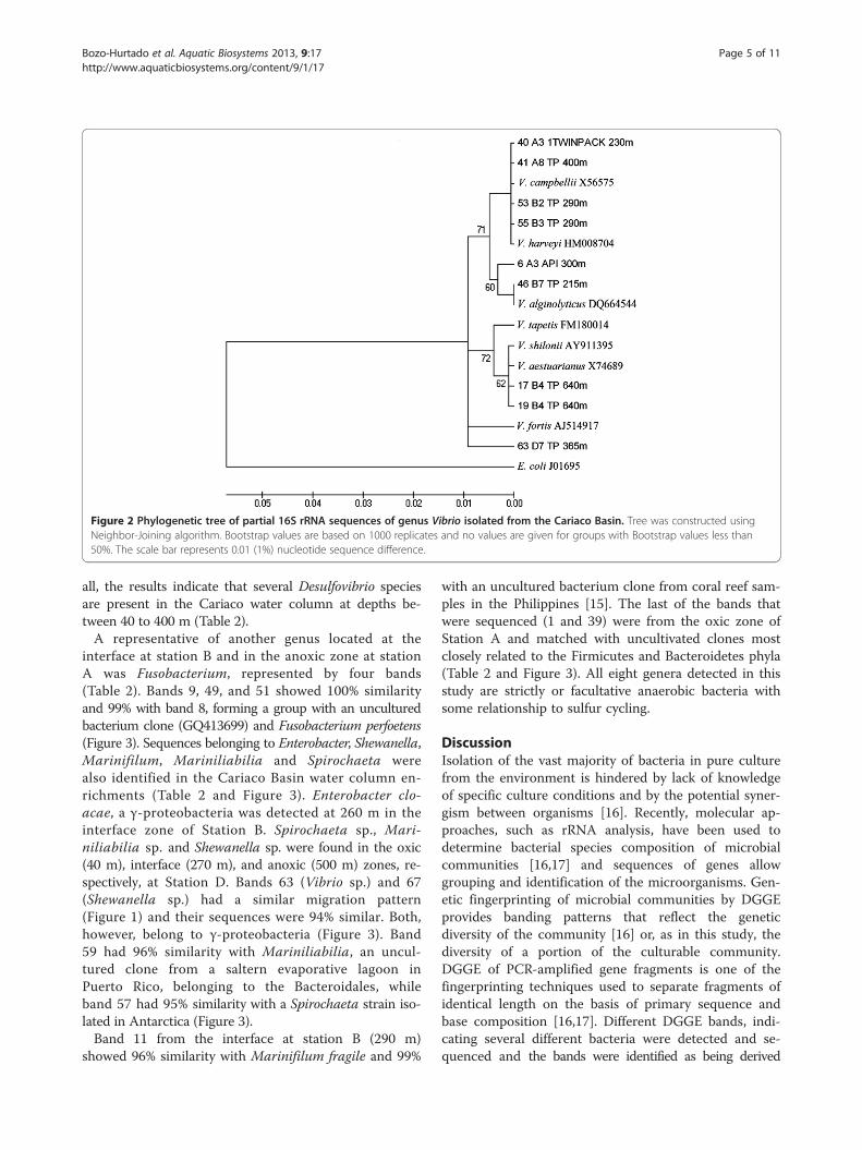
Figure 2 Phylogenetic tree of partial 16S rRNA sequences of genus Vibrio isolated from the Cariaco Basin. Tree was constructed usingNeighbor-Joining algorithm. Bootstrap values are based on 1000 replicates and no values are given for groups with Bootstrap values less than50%. The scale bar represents 0.01 (1%) nucleotide sequence difference.
Bozo-Hurtado et al. Aquatic Biosystems 2013, 9:17 Page 5 of 11http://www.aquaticbiosystems.org/content/9/1/17
all, the results indicate that several Desulfovibrio speciesare present in the Cariaco water column at depths be-tween 40 to 400 m (Table 2).A representative of another genus located at the
interface at station B and in the anoxic zone at stationA was Fusobacterium, represented by four bands(Table 2). Bands 9, 49, and 51 showed 100% similarityand 99% with band 8, forming a group with an unculturedbacterium clone (GQ413699) and Fusobacterium perfoetens(Figure 3). Sequences belonging to Enterobacter, Shewanella,Marinifilum, Mariniliabilia and Spirochaeta werealso identified in the Cariaco Basin water column en-richments (Table 2 and Figure 3). Enterobacter clo-acae, a γ-proteobacteria was detected at 260 m in theinterface zone of Station B. Spirochaeta sp., Mari-niliabilia sp. and Shewanella sp. were found in the oxic(40 m), interface (270 m), and anoxic (500 m) zones, re-spectively, at Station D. Bands 63 (Vibrio sp.) and 67(Shewanella sp.) had a similar migration pattern(Figure 1) and their sequences were 94% similar. Both,however, belong to γ-proteobacteria (Figure 3). Band59 had 96% similarity with Mariniliabilia, an uncul-tured clone from a saltern evaporative lagoon inPuerto Rico, belonging to the Bacteroidales, whileband 57 had 95% similarity with a Spirochaeta strain iso-lated in Antarctica (Figure 3).Band 11 from the interface at station B (290 m)
showed 96% similarity with Marinifilum fragile and 99%
with an uncultured bacterium clone from coral reef sam-ples in the Philippines [15]. The last of the bands thatwere sequenced (1 and 39) were from the oxic zone ofStation A and matched with uncultivated clones mostclosely related to the Firmicutes and Bacteroidetes phyla(Table 2 and Figure 3). All eight genera detected in thisstudy are strictly or facultative anaerobic bacteria withsome relationship to sulfur cycling.
DiscussionIsolation of the vast majority of bacteria in pure culturefrom the environment is hindered by lack of knowledgeof specific culture conditions and by the potential syner-gism between organisms [16]. Recently, molecular ap-proaches, such as rRNA analysis, have been used todetermine bacterial species composition of microbialcommunities [16,17] and sequences of genes allowgrouping and identification of the microorganisms. Gen-etic fingerprinting of microbial communities by DGGEprovides banding patterns that reflect the geneticdiversity of the community [16] or, as in this study, thediversity of a portion of the culturable community.DGGE of PCR-amplified gene fragments is one of thefingerprinting techniques used to separate fragments ofidentical length on the basis of primary sequence andbase composition [16,17]. Different DGGE bands, indi-cating several different bacteria were detected and se-quenced and the bands were identified as being derived
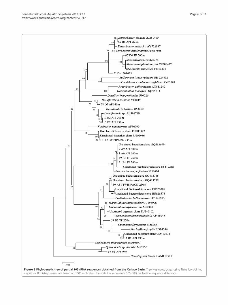
Figure 3 Phylogenetic tree of partial 16S rRNA sequences obtained from the Cariaco Basin. Tree was constructed using Neighbor-Joiningalgorithm. Bootstrap values are based on 1000 replicates. The scale bar represents 0.05 (5%) nucleotide sequence difference.
Bozo-Hurtado et al. Aquatic Biosystems 2013, 9:17 Page 6 of 11http://www.aquaticbiosystems.org/content/9/1/17

Bozo-Hurtado et al. Aquatic Biosystems 2013, 9:17 Page 7 of 11http://www.aquaticbiosystems.org/content/9/1/17
from the genera Vibrio, Desulfovibrio, Enterobacter,Shewanella, Fusobacterium, Marinifilum, Mariniliabilia andSpirochaeta. Although 200bp sequence length can be con-sidered too short for a phylogenetic analyses we foundedthat ours sequences correspond to genera, groups andclasses like Vibrio sp., CFB group, gamma and deltaproteobacteria that has been reported in the water columnof the Cariaco basin previously [1,5,9,10,12].The genus Vibrio encompasses a diverse group of het-
erotrophic marine bacteria and is widespread in theaquatic environment, occupying a variety of ecologicalniches. There are indications that vibrios play a role innutrient cycling by taking up dissolved organic matter[18]. Vibrio-affiliated sequences were detected by DGGEgel analysis in the sample from both years (Figure 1) and16S rRNA sequence similarity with of V. campbellii,V. harveyi, and V. alginolyticus (Figure 2). The Vibriocore cluster (including Vibrio harveyi, V. alginolyticusand V. campbellii) is often difficult to resolve solelyby 16S rRNA gene heterogeneity, since species withinthe V. harveyi clade has a very high degree of bothgenetic and phenotypic similarity. These species havemore than 99% sequence identity in the 16S rRNAgene [19,20].Vibrio harveyi is found in a free-living state in aquatic
environments and as part of the normal microbiota of mar-ine animals. However, many variants of V. harveyi havebeen recognized as significant pathogens of aquaculturedmarine fish [21,22], crustaceans [23], lobsters [24], andcorals [25]. Moreover, since 1993, V. harveyi has been re-covered from diseased fish and penaeids in Venezuelan wa-ters close to the Cariaco Basin (Paria Peninsula, SucreState) [21].Three bands 17, 19 and 63 had low 16S rRNA similarity
(around 96-98%) with known vibrio species (Figure 2).Bands 17 and 19 were 96% similar to Vibrio shilonii. Thisspecies has been associated with healthy or necrotic coralsin the Caribbean and Pacific reefs [18,26-30]. V. shiloniihas recently reported in Cariaco Basin waters in the oxiclayer using specific primers for Vibrio species in Station A[12] while we founded at 640 m depth (anoxic zone) in Sta-tion B. Band 63 is more closely related to V. fortis (98%).This species was detected directly in water samples be-tween 200 m and 1300 m of the Station A in a previous re-port [12] were becomes a prominent Vibrio sp. in theredoxcline and anoxic zone. Furthermore, Raina et al.(2009) found Vibrio and Shewanella species to be able todegrade the sulfur compounds, e.g. DMSP, DMS andacrylic acid, associated with coral reef tissue and in the sur-rounding water, suggesting a role for these genera in thebiogeochemical cycling of sulfur [31].Expected was the finding of Desulfovibrio in the SRB
culture (Figure 3). During the last two decades, an increas-ing number of novel sulfate-reducing bacteria have been
isolated from a wide variety of environments, wherestrains of the genus Desulfovibrio are commonly found[32,33]. In this study, we detected Desulfovibrio species inthe water column at 40 to 400 m depth, between the oxicand strictly anaerobic zones. Hastings and Emerson(1988) reported sulfate reduction in the presence ofoxygen in and above the chemocline of the Cariacobasin and recent reports, using molecular techniques,showed sulfate reducing δ-proteobacteria cells weremainly associated with the oxic-anoxic interface zoneand in the water column up to the aerobic zone (30 m)[1,5,9]. The Desulfovibrio phylotypes detected in thisstudy were most similar to the uncultivated environmentalclones of sulfate-reducing δ-proteobacteria and thosemainly from tropical marine environments (Table 2).Other bacteria identified among our cultures were
Enterobacter, Shewanella, Fusobacterium, Mariniliabiliaand Spirochaeta (Table 2 and Figure 3). Spirochaeta genuswas report in sediments from Guaymas Basin [34].Enterobacter sp., Fusobacterium perfoetens and Spirochaetasp. are active in marine biocorrosion, formation of biofilmson carbon steel surfaces, and corrosion of oil field pipelines[35-37]. Our study showed four bands that were mostsimilar to an uncultured bacterium clone related toFusobacterium that had been isolated from coral reef sam-ples [15]. Shewanella sp. has been associated with Vibriosp., in the corrosion of carbon steel in saline media. Thesefacultatively anaerobic bacteria can consume residual oxy-gen and thereby provide ecological niches for growth ofSRB. Depending on environmental conditions, Shewanellasp. can produce hydrogen sulfate from elemental sulfur, re-duce ferric iron and use cathodic hydrogen, competingwith SRB for H2 as an energy source [38].The genus Marinilabilia was created to include
the marine, facultative anaerobic Cytophaga species,Cytophaga salmonicolor and Cytophaga agarovorans [39].Taxonomic investigations have shown an overlap betweenthe genera Cytophaga and Flavobacterium and thesegroups were then called the Cytophaga-Flavobacteriumcomplex. Molecular investigations revealed an unexpectedrelationship between the Cytophaga-Flavobacteriumgroup and the genus Bacteroides (CFB group) [39,40]. TheCFB group had previously been reported to occurthroughout the entire water column in the Cariaco Basin[1,9], in sediments from Guaymas Basin [34] and in anoxiccultures of rice paddy soil [41]. Our study showed thatMarinilabilia salmonicolor, Marinifilum fragile, and anuncultured Bacteroidetes marine species of the CFB groupwere present, along with SRB, near the redox interface.The lactate-sulfate media can enrich for SRB using lac-
tate as an electron donor for the reduction of sulfate.However, other anaerobic or facultative microbes not re-ducing sulfate may also be found. Here we analyzed en-richments which showed the presence of a black FeS
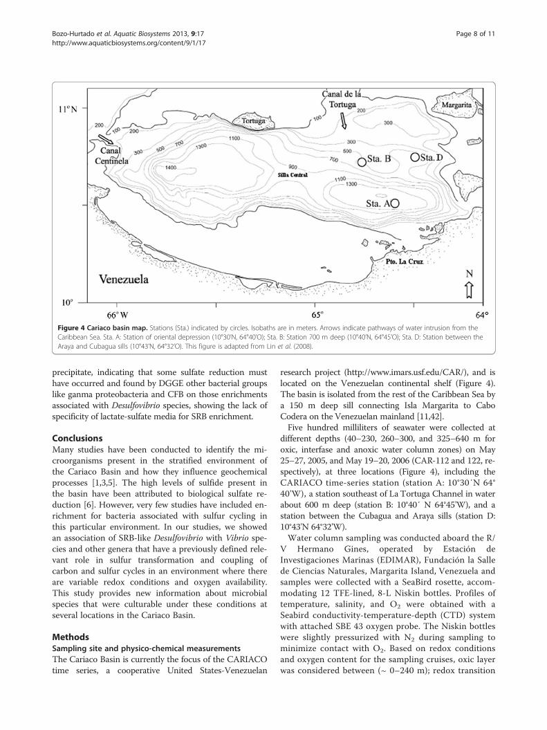
Figure 4 Cariaco basin map. Stations (Sta.) indicated by circles. Isobaths are in meters. Arrows indicate pathways of water intrusion from theCaribbean Sea. Sta. A: Station of oriental depression (10°30’N, 64°40’O); Sta. B: Station 700 m deep (10°40’N, 64°45’O); Sta. D: Station between theAraya and Cubagua sills (10°43’N, 64°32’O). This figure is adapted from Lin et al. (2008).
Bozo-Hurtado et al. Aquatic Biosystems 2013, 9:17 Page 8 of 11http://www.aquaticbiosystems.org/content/9/1/17
precipitate, indicating that some sulfate reduction musthave occurred and found by DGGE other bacterial groupslike ganma proteobacteria and CFB on those enrichmentsassociated with Desulfovibrio species, showing the lack ofspecificity of lactate-sulfate media for SRB enrichment.
ConclusionsMany studies have been conducted to identify the mi-croorganisms present in the stratified environment ofthe Cariaco Basin and how they influence geochemicalprocesses [1,3,5]. The high levels of sulfide present inthe basin have been attributed to biological sulfate re-duction [6]. However, very few studies have included en-richment for bacteria associated with sulfur cycling inthis particular environment. In our studies, we showedan association of SRB-like Desulfovibrio with Vibrio spe-cies and other genera that have a previously defined rele-vant role in sulfur transformation and coupling ofcarbon and sulfur cycles in an environment where thereare variable redox conditions and oxygen availability.This study provides new information about microbialspecies that were culturable under these conditions atseveral locations in the Cariaco Basin.
MethodsSampling site and physico-chemical measurementsThe Cariaco Basin is currently the focus of the CARIACOtime series, a cooperative United States-Venezuelan
research project (http://www.imars.usf.edu/CAR/), and islocated on the Venezuelan continental shelf (Figure 4).The basin is isolated from the rest of the Caribbean Sea bya 150 m deep sill connecting Isla Margarita to CaboCodera on the Venezuelan mainland [11,42].Five hundred milliliters of seawater were collected at
different depths (40–230, 260–300, and 325–640 m foroxic, interfase and anoxic water column zones) on May25–27, 2005, and May 19–20, 2006 (CAR-112 and 122, re-spectively), at three locations (Figure 4), including theCARIACO time-series station (station A: 10°30′N 64°40'W), a station southeast of La Tortuga Channel in waterabout 600 m deep (station B: 10°40′ N 64°45'W), and astation between the Cubagua and Araya sills (station D:10°43'N 64°32'W).Water column sampling was conducted aboard the R/
V Hermano Gines, operated by Estación deInvestigaciones Marinas (EDIMAR), Fundación la Sallede Ciencias Naturales, Margarita Island, Venezuela andsamples were collected with a SeaBird rosette, accom-modating 12 TFE-lined, 8-L Niskin bottles. Profiles oftemperature, salinity, and O2 were obtained with aSeabird conductivity-temperature-depth (CTD) systemwith attached SBE 43 oxygen probe. The Niskin bottleswere slightly pressurized with N2 during sampling tominimize contact with O2. Based on redox conditionsand oxygen content for the sampling cruises, oxic layerwas considered between (~ 0–240 m); redox transition
Bozo-Hurtado et al. Aquatic Biosystems 2013, 9:17 Page 9 of 11http://www.aquaticbiosystems.org/content/9/1/17
interface (~ 245–320 m); and anoxic layer (~ 325 to900 m).
Culture conditionsCultivation of SRB in sealed serum bottles containing fil-ter sterilized seawater amended with three different cul-ture media: Twin Pack (Twin) medium (per liter ofdistilled water: K2HPO4, 2.00 g; MgSO4•7H2O, 0.10 g;CaCl2•2H2O, 0.10 g; ammonium sulphate, 0.10 g;FeCl3, 0.02 g; sodium thiosulfate, 10.0 g; pH adjustedat 7,8 ±0,2) supplemented with 0.2% lactate; TriplePack (TP) medium (HIMEDIA, India) supplementedwith 0.2% lactate as a carbon source and a solution offerrous ammonium sulfate (0.0392 mg/L) and sodiumascorbate (0.01 mg/L) as reducing agent; and ModifiedAmerican Petroleum Institute (API) medium (HIMEDIA,India), supplemented with ascorbic acid (0.1 g/L) as a re-ducing agent [43] and using 0.5% acetate as a carbonsource. Each culture medium was inoculated with 10 mLof 20 water samples (6 samples for 2005; 14 samples for2006) by duplicate and incubated at room temperature for30 days under a gas mixture consisting of 20% CO2: 80%N2. 120 cultures were performed in total.
DNA extraction and PCRDNA was extracted from the Twin, TP, and API culturesusing the Microbial DNA Isolation kit (Mo Bio Labora-tories, CA, USA), according to manufacturer’s recom-mendation. Bacterial DNA was amplified using a primerwith GC clamp (341F-GC: 5'-CGC CCG CCG CGCGCG GCG GGC GGG GCG GGG GCA CGG GGGGCC TAC GGG AGG CAG CAG-3') and 907R (5'-CCGTCA ATT CGT TTG AGT TT-3') [16,44]. The reactionmixture contained 3 μL of DNA (approximately ~50-100ng) and 0.5 μM of each primer, 35 μL of GoTaq GreenMaster Mix reactions (Promega, Madison, WI, USA)and water added to a final volume of 70 μL. PCR ampli-fication was performed in a thermal cycler (PxE ThermalCycler, Thermo Hybaid, IL, USA), as follows: 95°C for 5min; 20 cycles at 94°C for 30 s; 65°C for 1 min; 72°C for3 min; 15 cycles at 94°C for 30 s; 55°C for 1 min; 72°Cfor 3 min; and 72°C for 7 min. The negative PCR controlhad no template in the reaction. The positive control forPCR was prepared by adding 1 μL of Alcaligenes faecalisDNA (100 ng). The PCR products were visualizedby running the reaction mixture in a TBE agarose gel(1.0%), staining with ethidium bromide (0.2 μg/ml), andobserving under UV light.
Denaturing gradient gel electrophoresis (DGGE)DGGE analysis of the bacterial amplicons (70 μL - entirevolume of a PCR reaction) was performed in 6% polyacryl-amide (37.5: 1 acrylamide/bis-acrylamide) gels containing a
0–100% urea plus formamide gradient (100% denaturingsolution containing 7 M urea and 40% (v/v) formamide).Electrophoresis was performed in 0.5 X TAE (TRIS
acetate 20 mM [pH 7.41], sodium acetate 10mM, andsodium EDTA 0.5 mM) at 60 volts and 60°C for 14 husing a DGGE 1001–110 System (C.B.S. Scientific Com-pany, Inc). Gels were stained with ethidium bromide(0.2 μg/mL) for 20 min and visualized using a FOTO/Analyst Investigator/FX Systems (Fotodyne Incorpo-rated, Hartland, WI, USA) [12].
16S RNA gene sequence analysisSeparated DNA fragments were excised from the DGGEgels, placed in a freezer at −80°C for 2 h, and blended inMini-Beadbeater 8 (BioCold Scientific, Fenton, MO,USA), for 3 min with 0.2 g sterile zirconia/silica beads(BioSpec Products, Bartlesville, OK) in 500 μL sterileHPLC water (Fisher HealthCare). Samples were storedat 4°C overnight, after which 3 μl aliquots were used astemplate for PCR amplification of 16S RNA gene,employing primers 341F (same as 341F-GC but withoutGC clamp) and 907R and the same PCR conditions asdescribed above, with a final PCR volume of 50 μL.Re-amplified PCR products were purified using a Wizard
SV gel and PCR clean-up system kit (Promega, Madison,WI, USA). Sequencing of one DNA strand was performedusing the BigDyeTM Terminator v3.1 sequencing kit, follow-ing manufacturer’s instructions (Applied Biosystems, FosterCity, CA). Sequencing reactions were analyzed in a 3100ABI DNA sequencer and sequence quality was determinedusing Chromas Lite software (http://www.technelysium.com.au/chromas_lite.html) [12].The closest known relatives of the partial 16S RNA
gene sequences were identified using BLASTN 2.2.1(http://www.ncbi.nlm.nih.gov/blast/) [45].
Phylogenetic analysisPartial 16S rRNA gene sequences initially were com-pared with sequences in the GenBank database usingBLASTN [45] to determine their approximate phylo-genetic affiliation. Environmental sequences, togetherwith closest GenBank matches, were aligned in http://greengenes.lbl.gov using the NAST Alignment utility[46]. Sequences obtained from 23 DGGE bands werealigned using NAST Alignment [46], and a phylogenetictree was constructed using 200 bp long alignedsequences and the neighbor-joining algorithm (Jukes-Cantor Model) in Molecular Evolutionary Genetics Ana-lysis 2.1 software (MEGA, version 4) [47]. Bootstrappingwas used to estimate reliability of the phylogenetic recon-structions (1000 replicates). Representative sequenceswere submitted to GenBank database and are designatedby accession numbers HM466893-HM466915.

Bozo-Hurtado et al. Aquatic Biosystems 2013, 9:17 Page 10 of 11http://www.aquaticbiosystems.org/content/9/1/17
Competing interestsThe authors declare that they have no competing interests.
Authors’ contributionsLBH participated in the design of the study, carried out the samplingcollection and molecular genetic studies, participated in the phylogeneticanalyses and the sequence alignment, and co-wrote the manuscript. MAGparticipated in the phylogenetic analyses of the samples and the sequencealignment, and co-wrote the manuscript. AC participated in the design ofthe study and coordination of the molecular genetic studies. RV and JJNcompiled the physic-chemical data related to the Cariaco Basin and B/OHermano Gines logistic and sampling collection. RC drafts the manuscriptand improves the discussion of the results. PS participated in the design ofthe study, carried out the sampling collection and the phylogenetic analyses,and co-wrote the manuscript. All authors read and approved the finalmanuscript.
AcknowledgmentsThe authors would like to give special thanks to the Captain and crew of theHermano Gines, and Yrene Astor and Javier Camparo at the Fundación LaSalle in Isla Margarita for their help and logistical support. The authorsgratefully acknowledge Maria Jose Rodriguez for her assistance with theDGGE. Special thanks to V. Edgcomb, M. I. Scranton and G. Taylor forassistance in sampling collection. This work was supported by a grant fromthe National Science Foundation, NSF Grant microbial observatory MCB-0348045 to A.C and the Decanato de Investigación y Desarrollo of theUniversidad Simón Bolívar to P.S. The hydrographic and other oceanographicobservations at the CARIACO Ocean Time Series were supported by NSFGrant OCE-0326268 to Frank Muller-Karger and National Oceanic andAtmospheric Agency (NOAA) Grant No. S0660009 and National Institute ofHealth Grant No. 2RO1A1039129-11A2-NIH provided support for Rita R.Colwell.
Author details1Departamento de Biología de Organismos, Universidad Simón Bolívar,Caracas, Venezuela. 2Centro de Biofísica y Bioquímica, Instituto Venezolanode Investigaciones Científicas, Caracas, Venezuela. 3Department of Biology,University of Louisiana at Lafayette, Lafayette, Louisiana, USA. 4EDIMAR,Fundación La Salle, Margarita, Venezuela. 5University of Maryland, CollegePark, Maryland, USA.
Received: 30 May 2012 Accepted: 24 August 2013Published: 28 August 2013
References1. Madrid VM, Taylor GT, Scranton MI, Chistoserdov AY: Phylogenetic diversity
of bacterial and archaeal communities in the anoxic zone of the CariacoBasin. Appl Environ Microbiol 2001, 67:1663–1674.
2. Astor YM, Müller-Karger F, Scranton MI: Seasonal and interannual variationin the hydrography of the Cariaco Basin: implications for basinventilation. Cont Shelf Res 2003, 23:125–144.
3. Taylor GT, Hein C, Iabichella M: Temporal variations in viral distributions inthe anoxic Cariaco Basin. Aquatic Microbiol Ecol 2003, 30:103–116.
4. Tedesco T, Thunell R, Astor Y, Karger FM: The oxygen isotope compositionof planktonic foraminifera from the Cariaco Basin, Venezuela: seasonaland interannual variations. Mar Micropaleontol 2007, 62:180–193.
5. Lin X, Scranton MI, Chistoserdov AY, Varela R, Taylor G: Spatiotemporaldynamics of bacterial populations in the anoxic Cariaco Basin.Limnol Oceanogr 2008, 53:37–51.
6. Hasting D, Emerson S: Sulfate reduction in the presence of low oxygenlevels in the water column of the Cariaco Trench. Limnol Oceanogr 1988,33:391–396.
7. Tuttle JH, Jannasch HW: Sulfide and thiosulfate-oxidizing bacteria inanoxic marine basins. Mar Biol 1973, 20:64–70.
8. Morris I, Glover HE, Kaplan WA, Kelly DP, Weightman AL: Microbial activityin the Cariaco Trench. Microbios 1985, 42:133–144.
9. Lin X, Wakeham SG, Putman IF, Astor YM, Scranton MI, Chistoserdov AY,Taylor GT: Comparison of vertical distributions of prokaryotic assemblagesin the anoxic Cariaco Basin and Black Sea by use of fluorescence in situhybridization. Appl Environ Microbiol 2006, 72:2679–2690.
10. Lin X, Scranton MI, Varela R, Chistoserdov AY, Taylor GT: Compositionalresponses of bacterial communities to redox gradients and grazing inthe anoxic Cariaco Basin. Aquat Microb Ecol 2007, 47:57–72.
11. Müller-Karger F, Varela R, Thunell R, Scranton MI, Bohrer R, Taylor G, CapeloJ, Astor Y, Tappa E, Ho T-Y, Walsh JJ: Annual cycle of primary productionin the Cariaco Basin: response to upwelling and implications for verticalexport. J Geophys Res 2001, 106:4527–4542.
12. Garcia-Amado MA, Bozo-Hurtado L, Astor Y, Suarez P, Chistoserdov A:Denaturing gradient gel electrophoresis analyses of the verticaldistribution and diversity of Vibrio spp. populations in the Cariaco Basin.FEMS Microbiol Ecol 2011, 77:347–356.
13. Hirayama H, Sunamura M, Takai K, Nunoura T, Noguchi T, Oida H, FurushimaY, Yamamoto H, Oomori T, Horikoshi K: Culture-dependent and -independent characterization of microbial communities associated witha shallow submarine hydrothermal system occurring within a coral reefoff Taketomi Island, Japan. Appl Environ Microbiol 2007, 73:7642–7656.
14. Koizumi Y, Kojima H, Fukui M: Potential sulfur metabolisms and associatedbacteria within anoxic surface sediment from saline meromictic LakeKaiike (Japan). FEMS Microbiol Ecol 2005, 52:297–305.
15. Garren M, Raymundo L, Guest J, Harvell CD, Azam F: Resilience of coral-associated bacterial communities exposed to fish farm effluent.PLoS ONE 2009, 4:1–9.
16. Schäfer H, Muyzer G: Denaturing gradient gel electrophoresis in marinemicrobial ecology. In Marine Microbiology, Methods in Microbiology, Volume30. Edited by Paul JH. London: Academic Press; 2001:425–468.
17. Teske A, Sigalevich P, Cohen Y, Muyzer G: Molecular identification ofbacteria from a coculture by denaturing gradient gel electrophoresis of16S ribosomal DNA fragments as a tool for isolation in pure cultures.Appl Environ Microbiol 1996, 62:4210–4215.
18. Thompson FL, Iida T, Swings J: Biodiversity of vibrios. Microbiol Mol Biol Rev2004, 68:403–431.
19. Gomez-Gil B, Thompson FL, Thompson CC, Swings J: Vibrio rotiferianus sp.nov., isolated from cultures of the rotifer Brachionus plicatilis. Int J SystEvol Microbiol 2003, 53:239–243.
20. Sawabe T, Kita-Tsukamoto K, Thompson FL: Inferring the evolutionaryhistory of vibrios by means of multilocus sequence analysis. J Bacteriol2007, 189:7932–7936.
21. Alvarez JD, Austin B, Alvarez AM, Reyes H: Vibrio harveyi: a pathogen ofpenaeid shrimps and fish in Venezuela. J Fish Dis 1998, 21:313–316.
22. Pujalte MJ, Sitja-Bobadilla A, Maclan MC, Belloch C, Alvarez-Pellitero P, Perez-Sanchez J, Uruburu F, Garay E: Virulence and molecular typing of Vibrioharveyi strains isolated from cultured dentex, gilthead sea bream andEuropean sea bass. Syst Appl Microbiol 2003, 26:284–292.
23. Lavilla-Pitogo CR, Leaño EM, Paner MG: Mortalities of pond-cultured juvenileshrimp, Penaeus monodon, associated with dominance of luminescentvibrios in the rearing environment. Aquaculture 1998, 164:337–349.
24. Bourne DG, Høj L, Webster NS, Swan J, Hall MR: Biofilm developmentwithin a larval rearing tank of the tropical rock lobster, Panulirus ornatus.Aquaculture 2006, 260:27–38.
25. Sutherland KP, Porter JW, Torres C: Disease and immunity in Caribbean andIndo-Pacific zooxanthellate corals. Mar Ecol Prog Ser 2004, 266:273–302.
26. Banin E, Israely T, Kushmaro A, Loya Y, Orr E, Rosenberg E: Penetration ofthe coral-bleaching bacterium Vibrio shiloi into Oculina patagonica.Appl Environ Microbiol 2000, 66:3031–3036.
27. Kushmaro A, Banin E, Loya Y, Stackebrandt E, Rosenberg E: Vibrio shiloi sp.nov., the causative agent of bleaching of the coral Oculina patagonica.Int J Syst Evol Microbiol 2001, 51:1383–1388.
28. Thompson FL, Hoste B, Thompson CC, Huys G, Swings J: The coralbleaching vibrio shiloi kushmaro et al. 2001 Is a later synonym ofvibrio mediterranei pujalte and garay 1986. Syst Appl Microbiol 2001,24:516–519.
29. Chimetto LA, Brocchi M, Gondo M, Thompson CC, Gomez-Gil B, ThompsonFL: Genomic diversity of vibrios associated with the Brazilian coralMussismilia hispida and its sympatric zoanthids (Palythoa caribaeorum,Palythoa variabilis and Zoanthus solanderi). J Appl Microbiol 2009,106:1818–1826.
30. Teplitski M, Ritchie K: How feasible is the biological control of coraldiseases? Trends Ecol Evol 2009, 24:378–385.
31. Raina JB, Tapiolas D, Willis BL, Bourne DG: Coral-associated bacteria andtheir role in the biogeochemical cycling of sulfur. Appl Environ Microbiol2009, 75:3492–3501.
Bozo-Hurtado et al. Aquatic Biosystems 2013, 9:17 Page 11 of 11http://www.aquaticbiosystems.org/content/9/1/17
32. Teske A, Wawer C, Muyzer G, Ramsing NB: Distribution of sulfate-reducingbacteria in a stratified fjord (Mariager Fjord, Denmark) as evaluated bymost-probable-number counts and denaturing gradient gelelectrophoresis of PCR-amplified ribosomal DNA fragments. Appl EnvironMicrobiol 1996, 62:1405–1415.
33. Basso O, Caumette P, Magot M: Desulfovibrio putealis sp. nov., a novelsulfate-reducing bacterium isolated from a deep subsurface aquifer.Int J Syst Evol Microbiol 2005, 55:101–104.
34. Dhillon A, Teske A, Dillon J, Stahl DA, Sogin ML: Molecular characterizationof sulfate-reducing bacteria in the Guaymas Basin. Appl Environ Microbiol2003, 69:2765–2772.
35. Neria-González I, Wang ET, Ramírez F, Romero JM, Hernández-Rodríguez C:Characterization of bacterial community associated to biofilms ofcorroded oil pipelines from the southeast of Mexico. Anaerobe 2006,12:122–133.
36. Bermont-Bouis D, Janvier M, Grimont PA, Dupont I, Vallaeys T: Both sulfate-reducing bacteria and Enterobacteriaceae take part in marinebiocorrosion of carbon steel. J Appl Microbiol 2007, 102:161–168.
37. Agrawal A, Vanbroekhoven K, Lal B: Diversity of culturable sulfidogenicbacteria in two oil–water separation tanks in the north-eastern oil fieldsof India. Anaerobe 2010, 16:12–18.
38. Herrera LK, Videla HA: Role of iron-reducing bacteria in corrosion andprotection of carbon steel. Int Biodeterioration & Biodegradation 2009,63:891–895.
39. Nakagawa Y, Yamasato K: Emendation of the genus Cytophaga andtransfer of Cytophaga agarovorans and Cytophaga salmonicolor toMarinilabilia gen. nov.: phylogenetic analysis of the Flavobacterium-Cytophaga complex. Int J Syst Bacteriol 1996, 46:599–603.
40. Suzuki M, Nakagawa Y, Harayama S, Yamamoto S: Phylogenetic analysis ofgenus Marinilabilia and related bacteria based on the amino acidsequences of GyrB and emended description of Marinilabiliasalmonicolor with Marinilabilia agarovorans as its subjective synonym.Int J Syst Bacteriol 1999, 49:1551–1557.
41. Hengstmann U, Chin K-J, Janssen PH, Liesack W: Comparative phylogeneticassignment of environmental sequences of genes encoding 16S rRNAand numerically abundant culturable bacteria from an anoxic rice paddysoil. Appl Environ Microbiol 1999, 65:5050–5058.
42. Müller-Karger F, Varela R, Thunell R, Scranton MI, Bohrer R, Taylor G, Capelo J,Astor Y, Tappa E, Ho T-Y, Iabichella M, Walsh JJ, Diaz JR: Sediment recordlinked to surface processes in the Cariaco Basin. EOS. AGU TransactionsAmerican Geophysical Union 2000, 81:529–535.
43. HiMedia Laboratories: The HiMedia Manual for Microbiology LaboratoryPractice. India; 1998.
44. Muyzer G, De Waal E, Uitterlinden A: Profiling of complex microbialpopulations by denaturing gradient gel electrophoresis analysis ofpolymerase chain reaction-amplified genes coding for 16S rRNA.Appl Environ Microbiol 1993, 59:695–700.
45. Altschul SF, Madden TL, Schäffer AA, Zhang J, Zhang Z, Miller W, Lipman DJ:Gapped BLAST and PSI-BLAST: a new generation of protein databasesearch programs. Nucleic Acids Res 1997, 25:3389–3402.
46. DeSantis TZ, Hugenholtz P, Keller K, Brodie EL, Larsen N, Piceno YM, PhanR, Andersen GL: NAST: a multiple sequence alignment server forcomparative analysis of 16S rRNA genes. Nucleic Acids Res 2006,34:W394–W399.
47. Tamura K, Dudley J, Nei M, Kumar S: MEGA4: molecular evolutionarygenetics analysis (MEGA) software version 4.0. Mol Biol Evol 2007,24:1596–1599.
doi:10.1186/2046-9063-9-17Cite this article as: Bozo-Hurtado et al.: Identification of bacteria inenrichment cultures of sulfate reducers in the Cariaco Basin watercolumn employing Denaturing Gradient Gel Electrophoresis of 16Sribosomal RNA gene fragments. Aquatic Biosystems 2013 9:17.
Submit your next manuscript to BioMed Centraland take full advantage of:
• Convenient online submission
• Thorough peer review
• No space constraints or color figure charges
• Immediate publication on acceptance
• Inclusion in PubMed, CAS, Scopus and Google Scholar
• Research which is freely available for redistribution
Submit your manuscript at www.biomedcentral.com/submit


![[Composite Cultures] - CORE](https://static.fdokumen.com/doc/165x107/6325e67de491bcb36c0a86c0/composite-cultures-core.jpg)