An Unusual Syncytia-Inducing Human Immunodeficiency Virus Type 1 Primary Isolate from the Central...
10
Journal of NeuroVirology, 9: 432–441, 2003 c 2003 Taylor & Francis Inc. ISSN: 1355-0284 print / 1538-2443 online DOI: 10.1080/13550280390218706 An unusual syncytia-inducing human immunodeficiency virus type 1 primary isolate from the central nervous system that is restricted to CXCR4, replicates efficiently in macrophages, and induces neuronal apoptosis Yanjie Yi, 1 Wei Chen, 2 Ian Frank, 1 Joann Cutilli, 1 Anjali Singh, 1 Linda Starr-Spires, 1 Jerrold Sulcove, 2 Dennis L Kolson, 2 and Ronald G Collman 1 1 Departments of Medicine and 2 Neurology, University of Pennsylvania School of Medicine, Philadelphia, Pennsylvania, USA Macrophage/microglia cells are the principal targets for human immunodefi- ciency virus type 1 (HIV-1) in the central nervous system (CNS). Prototype HIV- 1 isolates from the CNS are macrophage (M)-tropic, non–syncytia-inducing (NSI), and use CCR5 for entry (R5 strains), but whether syncytia-inducing (SI) CXCR4-using X4 strains might play a role in macrophage/microglia infection and neuronal injury is unknown. To explore the range of features among HIV-1 primary isolates from the CNS, the authors analyzed an HIV-1 strain (TYBE) from cerebrospinal fluid of an individual with acquired immunodeficiency syndrome (AIDS) that was unusual because it was SI. Like other CNS iso- lates, HIV-1/TYBE replicated to high level in primary human macrophages, but, in contrast to CNS prototypes, TYBE used CXCR4 exclusively to infect macrophages. A functional TYBE env clone confirmed the X4 phenotype and displayed a highly charged V3 sequence typical of X4 strains. Supernatant from TYBE-infected primary human macrophages induced apoptosis of neu- rons. Thus, TYBE represents a novel type of CNS-derived HIV-1 isolate that is CXCR4-restricted yet replicates efficiently in macrophages and induce neu- ronal injury. These results demonstrate that HIV-1 variants in the CNS may possess a broader range of biological characteristics than generally appreci- ated, raise the possibility that X4 strains may participate in AIDS neuropatho- genesis, and provide a prototype clade B HIV-1 strain that replicates efficiently in primary macrophages through the exclusive use of CXCR4 as a coreceptor. Journal of NeuroVirology (2003) 9, 432–441. Keywords: chemokine receptor; envelope; neuroAIDS; tropism Address correspondence to Ronald G Collman, 522 Johnson Pavilion, 36th and Hamilton Walk, Philadelphia, PA 19104-6061, USA. E-mail: [email protected] The authors thank L Wojick for expert technical assistance, J Strizki for AMD3100, and blood donors who generously provided cells for this research. This work was supported by NIH grants to R.G.C. and D.L.K. Received 1 July 2002; revised 9 October 2002; accepted 2 December 2002. Introduction Human immunodeficiency virus (HIV) encephalopa- thy (HIVE) is directly linked to virus replica- tion within the central nervous system (CNS). Macrophage/microglia cells are the principal targets for productive infection in the CNS, and infection of these cells plays a critical, albeit still incompletely understood, role in the development of HIVE. The
-
Upload
independent -
Category
Documents
-
view
1 -
download
0
Transcript of An Unusual Syncytia-Inducing Human Immunodeficiency Virus Type 1 Primary Isolate from the Central...

Journal of NeuroVirology, 9: 432–441, 2003c© 2003 Taylor & Francis Inc.ISSN: 1355-0284 print / 1538-2443 onlineDOI: 10.1080/13550280390218706
An unusual syncytia-inducing humanimmunodeficiency virus type 1 primary isolatefrom the central nervous system that is restrictedto CXCR4, replicates efficiently in macrophages,and induces neuronal apoptosis
Yanjie Yi,1 Wei Chen,2 Ian Frank,1 Joann Cutilli,1 Anjali Singh,1 Linda Starr-Spires,1 Jerrold Sulcove,2
Dennis L Kolson,2 and Ronald G Collman1
1Departments of Medicine and 2Neurology, University of Pennsylvania School of Medicine,Philadelphia, Pennsylvania, USA
Macrophage/microglia cells are the principal targets for human immunodefi-ciency virus type 1 (HIV-1) in the central nervous system (CNS). Prototype HIV-1 isolates from the CNS are macrophage (M)-tropic, non–syncytia-inducing(NSI), and use CCR5 for entry (R5 strains), but whether syncytia-inducing (SI)CXCR4-using X4 strains might play a role in macrophage/microglia infectionand neuronal injury is unknown. To explore the range of features among HIV-1primary isolates from the CNS, the authors analyzed an HIV-1 strain (TYBE)from cerebrospinal fluid of an individual with acquired immunodeficiencysyndrome (AIDS) that was unusual because it was SI. Like other CNS iso-lates, HIV-1/TYBE replicated to high level in primary human macrophages,but, in contrast to CNS prototypes, TYBE used CXCR4 exclusively to infectmacrophages. A functional TYBE env clone confirmed the X4 phenotype anddisplayed a highly charged V3 sequence typical of X4 strains. Supernatantfrom TYBE-infected primary human macrophages induced apoptosis of neu-rons. Thus, TYBE represents a novel type of CNS-derived HIV-1 isolate thatis CXCR4-restricted yet replicates efficiently in macrophages and induce neu-ronal injury. These results demonstrate that HIV-1 variants in the CNS maypossess a broader range of biological characteristics than generally appreci-ated, raise the possibility that X4 strains may participate in AIDS neuropatho-genesis, and provide a prototype clade B HIV-1 strain that replicates efficientlyin primary macrophages through the exclusive use of CXCR4 as a coreceptor.Journal of NeuroVirology (2003) 9, 432–441.
Keywords: chemokine receptor; envelope; neuroAIDS; tropism
Address correspondence to Ronald G Collman, 522 JohnsonPavilion, 36th and Hamilton Walk, Philadelphia, PA 19104-6061,USA. E-mail: [email protected]
The authors thank L Wojick for expert technical assistance,J Strizki for AMD3100, and blood donors who generously providedcells for this research. This work was supported by NIH grants toR.G.C. and D.L.K.
Received 1 July 2002; revised 9 October 2002; accepted 2December 2002.
Introduction
Human immunodeficiency virus (HIV) encephalopa-thy (HIVE) is directly linked to virus replica-tion within the central nervous system (CNS).Macrophage/microglia cells are the principal targetsfor productive infection in the CNS, and infection ofthese cells plays a critical, albeit still incompletelyunderstood, role in the development of HIVE. The
Tropism and neuronal injury by an X4 CNS HIV-1 isolateY Yi et al 433
mechanisms by which macrophage/microglia infec-tion lead to neurological dysfunction and damage arebelieved to involve viral gene products, particularlythe envelope glycoprotein, as well as cellular solu-ble factors that are induced by viral infection (re-viewed in Kaul et al, 2001). HIV-1 isolates are gener-ally grouped into macrophage (M)-tropic, T-cell line(T)-tropic, and dual-tropic variants. M-tropic isolatesreplicate in primary macrophages (and microglia)and lymphocytes but not cell lines, are non–syncytia-inducing (NSI) in vitro, and use the chemokine re-ceptor CCR5 as a coreceptor for entry (R5 strains).T-tropic isolates replicate in lymphocytes and CD4-positive cell lines but not primary macrophages, aresyncytia-inducing (SI) in vitro, and use the corecep-tor CXCR4 (X4 strains). Dual-tropic strains replicatein all three target cell types, form syncytia in vitro,and use both CCR5 and CXCR4 (R5X4 strains). Proto-type HIV-1 isolates derived from the CNS all displaythe M-tropic, CCR5-dependent features of R5 vari-ants, which is consistent with macrophages and mi-croglia as the principal CNS viral reservoir (Gartneret al, 1986; Cheng-Mayer et al, 1989). As a result, thistype of variant is generally considered to be exclu-sively responsible for infection in the CNS.
Recently, however, several observations haveraised the question of whether CXCR4-using isolatesmay play a role in CNS infection in acquired immun-odeficiency syndrome (AIDS). We and others recentlyshowed that primary human macrophages expressCXCR4 as well as CCR5, and that certain primaryHIV-1 isolates are able to infect macrophages throughCXCR4, even though prototype laboratory-adaptedSI X4 strains cannot (Yi et al, 1998, 1999; Simmonset al, 1998; Verani et al, 1998). Microglia also expressCXCR4 (Albright et al, 1999; Lavi et al, 1997). Thisindicates the possibility that variants with the po-tential to infect the principal CNS reservoir might bebroader than generally appreciated. In vivo, CXCR4 iswidely expressed within the CNS, both in conjunc-tion with CD4 on macrophages and microglia, andin the absence of CD4 on neurons and other cells(Lavi et al, 1997; Westmoreland et al, 1998; Vallatet al, 1998). Importantly, HIV-induced neuronal in-jury can result from interaction between the viral en-velope glycoprotein gp120 and CXCR4 on neurons orother target cells (Hesselgesser et al, 1998; Bezzi et al,2001; Catani et al, 2000). Furthermore, in mixed braincultures in vitro, CXCR4-using isolates may possessparticular neurotoxic potential (Ohagen et al, 1999;Zheng et al, 1999a, 1999b). However, all of these ob-servations and potential mechanisms are irrelevantif isolates that interact with CXCR4 are not foundwithin the CNS in infected individuals.
Because of this, we sought to reexamine the is-sue of viral phenotype and coreceptor choice amongisolates from the CNS, within the context of cur-rent understanding of macrophage entry pathways.To this end, we undertook careful analysis of an HIV-1strain (TYBE) derived from cerebrospinal fluid (CSF)
of an individual with AIDS (Yi et al, 1999) that dif-fered from prototype CNS isolates in that it repli-cated in cell lines and was SI in culture. Strain TYBEreplicated efficiently in primary macrophages, andmacrophage infection resulted in the production ofneurotoxic secretory products. However, TYBE uti-lized CXCR4 exclusively as a coreceptor for infection.Thus, HIV-1 variants in the CNS may have a broaderrange of biological and genetic features than gener-ally recognized. These results add credibility to thepossibility that X4 strains may participate in infec-tion and disease pathogenesis related to the CNS.
Results
Characterization of an SI primary HIV-1 isolatefrom the CNSWe previously generated a panel of primary isolatesfrom the CSF of infected patients with neurologi-cal symptoms undergoing lumbar puncture for diag-nostic purposes. Of eight primary isolates from CSFthat were studied, one obtained from a man withneurological symptoms and designated HIV-1/TYBE(Yi et al, 1999) showed an unexpected phenotype inpreliminary analysis. In contrast to prototype CNSstrains, TYBE was SI in culture. To ensure that wewere dealing with a single viral species rather thana mixture of variants, the TYBE primary isolate wassubjected to biological cloning by limiting dilution onprimary human peripheral blood mononuclear cells(PBMCs). Five biological clones of TYBE were ana-lyzed and all five exhibited the same SI phenotype(data not shown), so one clone was selected at ran-dom for further study.
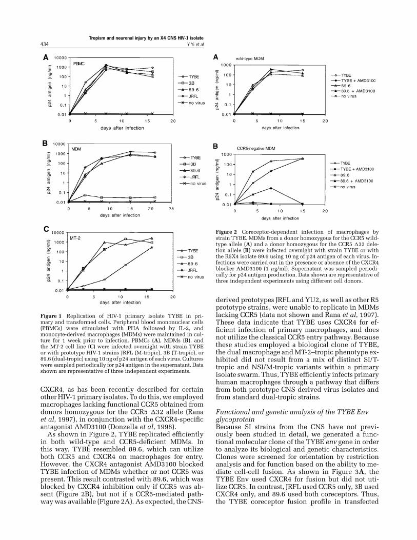
We tested the ability of the TYBE isolate to replicatein primary macrophages, primary lymphocytes, andthe MT-2 cell line. In parallel, we utilized the proto-type CNS-derived R5 M-tropic isolate JRFL, the X4T-tropic strain 3B, and the R5X4 dual-tropic isolate89.6. As shown in Figure 1, HIV-1/TYBE replicated inmonocyte-derived macrophages (MDMs), MT-2 cells,and PBMCs. This pattern was similar to that seenwith the dual-tropic prototype 89.6, which uses bothCCR5 and CXCR4 for entry. However, TYBE differedmarkedly from the CNS prototype JRFL, which repli-cated in MDMs and PBMCs but not in MT-2 cells, asexpected given the fact that JRFL is restricted to CCR5use. TYBE also differed from the CXCR4-restrictedprototype 3B, which replicates in PBMCs and celllines but is unable to utilize CXCR4 on macrophages.Thus, TYBE represents an unusual type of CNS-derived HIV-1 variant.
CNS-derived syncytia-inducing strain TYBE usesCXCR4 for infection of macrophagesBecause of the unexpected cell-line tropism andSI phenotype of strain TYBE, we tested whetherit utilized the traditional macrophage entry core-ceptor CCR5, or whether it could use macrophage
Tropism and neuronal injury by an X4 CNS HIV-1 isolate434 Y Yi et al
Figure 1 Replication of HIV-1 primary isolate TYBE in pri-mary and transformed cells. Peripheral blood mononuclear cells(PBMCs) were stimulated with PHA followed by IL-2, andmonocyte-derived macrophages (MDMs) were maintained in cul-ture for 1 week prior to infection. PBMCs (A), MDMs (B), andthe MT-2 cell line (C) were infected overnight with strain TYBEor with prototype HIV-1 strains JRFL (M-tropic), 3B (T-tropic), or89.6 (dual-tropic) using 10 ng of p24 antigen of each virus. Cultureswere sampled periodically for p24 antigen in the supernatant. Datashown are representative of three independent experiments.
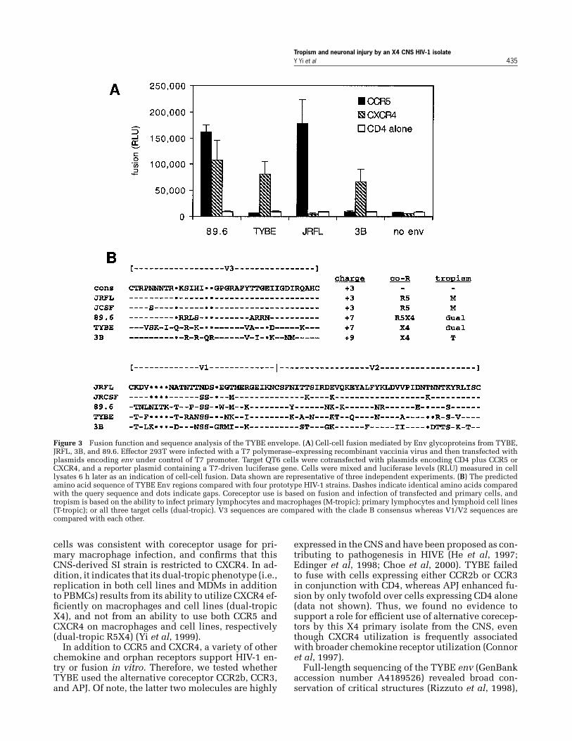
CXCR4, as has been recently described for certainother HIV-1 primary isolates. To do this, we employedmacrophages lacking functional CCR5 obtained fromdonors homozygous for the CCR5 �32 allele (Ranaet al, 1997), in conjunction with the CXCR4-specificantagonist AMD3100 (Donzella et al, 1998).
As shown in Figure 2, TYBE replicated efficientlyin both wild-type and CCR5-deficient MDMs. Inthis way, TYBE resembled 89.6, which can utilizeboth CCR5 and CXCR4 on macrophages for entry.However, the CXCR4 antagonist AMD3100 blockedTYBE infection of MDMs whether or not CCR5 waspresent. This result contrasted with 89.6, which wasblocked by CXCR4 inhibition only if CCR5 was ab-sent (Figure 2B), but not if a CCR5-mediated path-way was available (Figure 2A). As expected, the CNS-
Figure 2 Coreceptor-dependent infection of macrophages bystrain TYBE. MDMs from a donor homozygous for the CCR5 wild-type allele (A) and a donor homozygous for the CCR5 �32 dele-tion allele (B) were infected overnight with strain TYBE or withthe R5X4 isolate 89.6 using 10 ng of p24 antigen of each virus. In-fections were carried out in the presence or absence of the CXCR4blocker AMD3100 (1 μg/ml). Supernatant was sampled periodi-cally for p24 antigen production. Data shown are representative ofthree independent experiments using different cell donors.
derived prototypes JRFL and YU2, as well as other R5prototype strains, were unable to replicate in MDMslacking CCR5 (data not shown and Rana et al, 1997).These data indicate that TYBE uses CXCR4 for ef-ficient infection of primary macrophages, and doesnot utilize the classical CCR5 entry pathway. Becausethese studies employed a biological clone of TYBE,the dual macrophage and MT-2–tropic phenotype ex-hibited did not result from a mix of distinct SI/T-tropic and NSI/M-tropic variants within a primaryisolate swarm. Thus, TYBE efficiently infects primaryhuman macrophages through a pathway that differsfrom both prototype CNS-derived virus isolates andfrom standard dual-tropic strains.
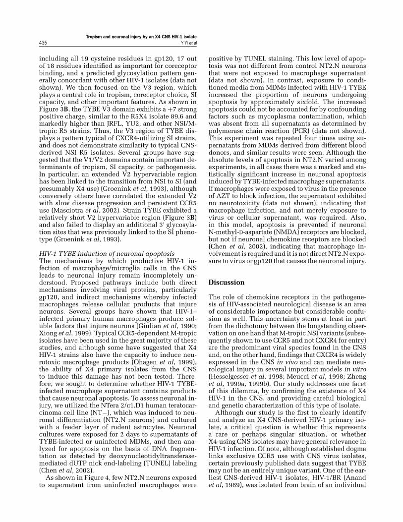
Functional and genetic analysis of the TYBE EnvglycoproteinBecause SI strains from the CNS have not previ-ously been studied in detail, we generated a func-tional molecular clone of the TYBE env gene in orderto analyze its biological and genetic characteristics.Clones were screened for orientation by restrictionanalysis and for function based on the ability to me-diate cell-cell fusion. As shown in Figure 3A, theTYBE Env used CXCR4 for fusion but did not uti-lize CCR5. In contrast, JRFL used CCR5 only, 3B usedCXCR4 only, and 89.6 used both coreceptors. Thus,the TYBE coreceptor fusion profile in transfected
Tropism and neuronal injury by an X4 CNS HIV-1 isolateY Yi et al 435
Figure 3 Fusion function and sequence analysis of the TYBE envelope. (A) Cell-cell fusion mediated by Env glycoproteins from TYBE,JRFL, 3B, and 89.6. Effector 293T were infected with a T7 polymerase–expressing recombinant vaccinia virus and then transfected withplasmids encoding env under control of T7 promoter. Target QT6 cells were cotransfected with plasmids encoding CD4 plus CCR5 orCXCR4, and a reporter plasmid containing a T7-driven luciferase gene. Cells were mixed and luciferase levels (RLU) measured in celllysates 6 h later as an indication of cell-cell fusion. Data shown are representative of three independent experiments. (B) The predictedamino acid sequence of TYBE Env regions compared with four prototype HIV-1 strains. Dashes indicate identical amino acids comparedwith the query sequence and dots indicate gaps. Coreceptor use is based on fusion and infection of transfected and primary cells, andtropism is based on the ability to infect primary lymphocytes and macrophages (M-tropic); primary lymphocytes and lymphoid cell lines(T-tropic); or all three target cells (dual-tropic). V3 sequences are compared with the clade B consensus whereas V1/V2 sequences arecompared with each other.
cells was consistent with coreceptor usage for pri-mary macrophage infection, and confirms that thisCNS-derived SI strain is restricted to CXCR4. In ad-dition, it indicates that its dual-tropic phenotype (i.e.,replication in both cell lines and MDMs in additionto PBMCs) results from its ability to utilize CXCR4 ef-ficiently on macrophages and cell lines (dual-tropicX4), and not from an ability to use both CCR5 andCXCR4 on macrophages and cell lines, respectively(dual-tropic R5X4) (Yi et al, 1999).
In addition to CCR5 and CXCR4, a variety of otherchemokine and orphan receptors support HIV-1 en-try or fusion in vitro. Therefore, we tested whetherTYBE used the alternative coreceptor CCR2b, CCR3,and APJ. Of note, the latter two molecules are highly
expressed in the CNS and have been proposed as con-tributing to pathogenesis in HIVE (He et al, 1997;Edinger et al, 1998; Choe et al, 2000). TYBE failedto fuse with cells expressing either CCR2b or CCR3in conjunction with CD4, whereas APJ enhanced fu-sion by only twofold over cells expressing CD4 alone(data not shown). Thus, we found no evidence tosupport a role for efficient use of alternative corecep-tors by this X4 primary isolate from the CNS, eventhough CXCR4 utilization is frequently associatedwith broader chemokine receptor utilization (Connoret al, 1997).
Full-length sequencing of the TYBE env (GenBankaccession number A4189526) revealed broad con-servation of critical structures (Rizzuto et al, 1998),
Tropism and neuronal injury by an X4 CNS HIV-1 isolate436 Y Yi et al
including all 19 cysteine residues in gp120, 17 outof 18 residues identified as important for coreceptorbinding, and a predicted glycosylation pattern gen-erally concordant with other HIV-1 isolates (data notshown). We then focused on the V3 region, whichplays a central role in tropism, coreceptor choice, SIcapacity, and other important features. As shown inFigure 3B, the TYBE V3 domain exhibits a +7 strongpositive charge, similar to the R5X4 isolate 89.6 andmarkedly higher than JRFL, YU2, and other NSI/M-tropic R5 strains. Thus, the V3 region of TYBE dis-plays a pattern typical of CXCR4-utilizing SI strains,and does not demonstrate similarity to typical CNS-derived NSI R5 isolates. Several groups have sug-gested that the V1/V2 domains contain important de-terminants of tropism, SI capacity, or pathogenesis.In particular, an extended V2 hypervariable regionhas been linked to the transition from NSI to SI (andpresumably X4 use) (Groenink et al, 1993), althoughconversely others have correlated the extended V2with slow disease progression and persistent CCR5use (Masciotra et al, 2002). Strain TYBE exhibited arelatively short V2 hypervariable region (Figure 3B)and also failed to display an additional 3′ glycosyla-tion sites that was previously linked to the SI pheno-type (Groenink et al, 1993).
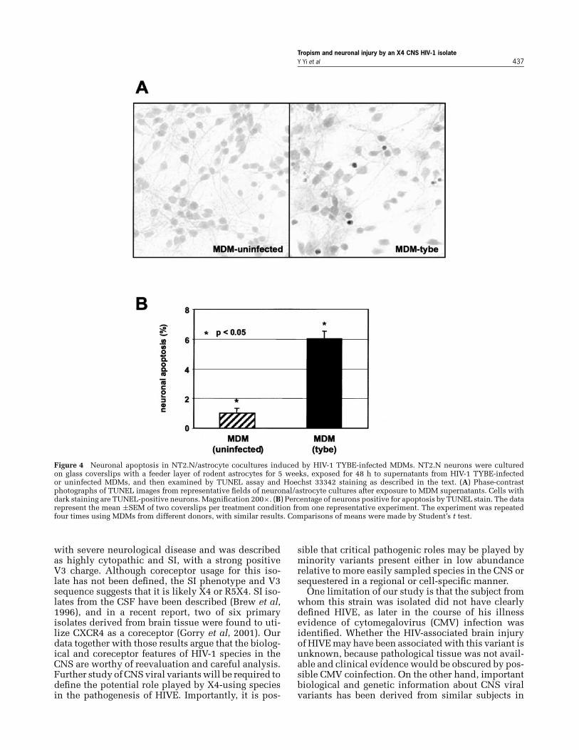
HIV-1 TYBE induction of neuronal apoptosisThe mechanisms by which productive HIV-1 in-fection of macrophage/microglia cells in the CNSleads to neuronal injury remain incompletely un-derstood. Proposed pathways include both directmechanisms involving viral proteins, particularlygp120, and indirect mechanisms whereby infectedmacrophages release cellular products that injureneurons. Several groups have shown that HIV-1–infected primary human macrophages produce sol-uble factors that injure neurons (Giulian et al, 1990;Xiong et al, 1999). Typical CCR5-dependent M-tropicisolates have been used in the great majority of thesestudies, and although some have suggested that X4HIV-1 strains also have the capacity to induce neu-rotoxic macrophage products (Ohagen et al, 1999),the ability of X4 primary isolates from the CNSto induce this damage has not been tested. There-fore, we sought to determine whether HIV-1 TYBE-infected macrophage supernatant contains productsthat cause neuronal apoptosis. To assess neuronal in-jury, we utilized the NTera 2/c1.D1 human teratocar-cinoma cell line (NT−), which was induced to neu-ronal differentiation (NT2.N neurons) and culturedwith a feeder layer of rodent astrocytes. Neuronalcultures were exposed for 2 days to supernatants ofTYBE-infected or uninfected MDMs, and then ana-lyzed for apoptosis on the basis of DNA fragmen-tation as detected by deoxynucleotidyltransferase-mediated dUTP nick end-labeling (TUNEL) labeling(Chen et al, 2002).
As shown in Figure 4, few NT2.N neurons exposedto supernatant from uninfected macrophages were
positive by TUNEL staining. This low level of apop-tosis was not different from control NT2.N neuronsthat were not exposed to macrophage supernatant(data not shown). In contrast, exposure to condi-tioned media from MDMs infected with HIV-1 TYBEincreased the proportion of neurons undergoingapoptosis by approximately sixfold. The increasedapoptosis could not be accounted for by confoundingfactors such as mycoplasma contamination, whichwas absent from all supernatants as determined bypolymerase chain reaction (PCR) (data not shown).This experiment was repeated four times using su-pernatants from MDMs derived from different blooddonors, and similar results were seen. Although theabsolute levels of apoptosis in NT2.N varied amongexperiments, in all cases there was a marked and sta-tistically significant increase in neuronal apoptosisinduced by TYBE-infected macrophage supernatants.If macrophages were exposed to virus in the presenceof AZT to block infection, the supernatant exhibitedno neurotoxicity (data not shown), indicating thatmacrophage infection, and not merely exposure tovirus or cellular supernatant, was required. Also,in this model, apoptosis is prevented if neuronalN-methyl-D-aspartate (NMDA) receptors are blocked,but not if neuronal chemokine receptors are blocked(Chen et al, 2002), indicating that macrophage in-volvement is required and it is not direct NT2.N expo-sure to virus or gp120 that causes the neuronal injury.
Discussion
The role of chemokine receptors in the pathogene-sis of HIV-associated neurological disease is an areaof considerable importance but considerable confu-sion as well. This uncertainty stems at least in partfrom the dichotomy between the longstanding obser-vation on one hand that M-tropic NSI variants (subse-quently shown to use CCR5 and not CXCR4 for entry)are the predominant viral species found in the CNSand, on the other hand, findings that CXCR4 is widelyexpressed in the CNS in vivo and can mediate neu-rological injury in several important models in vitro(Hesselgesser et al, 1998; Meucci et al, 1998; Zhenget al, 1999a, 1999b). Our study addresses one facetof this dilemma, by confirming the existence of X4HIV-1 in the CNS, and providing careful biologicaland genetic characterization of this type of isolate.
Although our study is the first to clearly identifyand analyze an X4 CNS-derived HIV-1 primary iso-late, a critical question is whether this representsa rare or perhaps singular situation, or whetherX4-using CNS isolates may have general relevance inHIV-1 infection. Of note, although established dogmalinks exclusive CCR5 use with CNS virus isolates,certain previously published data suggest that TYBEmay not be an entirely unique variant. One of the ear-liest CNS-derived HIV-1 isolates, HIV-1/BR (Anandet al, 1989), was isolated from brain of an individual
Tropism and neuronal injury by an X4 CNS HIV-1 isolateY Yi et al 437
Figure 4 Neuronal apoptosis in NT2.N/astrocyte cocultures induced by HIV-1 TYBE-infected MDMs. NT2.N neurons were culturedon glass coverslips with a feeder layer of rodent astrocytes for 5 weeks, exposed for 48 h to supernatants from HIV-1 TYBE-infectedor uninfected MDMs, and then examined by TUNEL assay and Hoechst 33342 staining as described in the text. (A) Phase-contrastphotographs of TUNEL images from representative fields of neuronal/astrocyte cultures after exposure to MDM supernatants. Cells withdark staining are TUNEL-positive neurons. Magnification 200×. (B) Percentage of neurons positive for apoptosis by TUNEL stain. The datarepresent the mean ±SEM of two coverslips per treatment condition from one representative experiment. The experiment was repeatedfour times using MDMs from different donors, with similar results. Comparisons of means were made by Student’s t test.
with severe neurological disease and was describedas highly cytopathic and SI, with a strong positiveV3 charge. Although coreceptor usage for this iso-late has not been defined, the SI phenotype and V3sequence suggests that it is likely X4 or R5X4. SI iso-lates from the CSF have been described (Brew et al,1996), and in a recent report, two of six primaryisolates derived from brain tissue were found to uti-lize CXCR4 as a coreceptor (Gorry et al, 2001). Ourdata together with those results argue that the biolog-ical and coreceptor features of HIV-1 species in theCNS are worthy of reevaluation and careful analysis.Further study of CNS viral variants will be required todefine the potential role played by X4-using speciesin the pathogenesis of HIVE. Importantly, it is pos-
sible that critical pathogenic roles may be played byminority variants present either in low abundancerelative to more easily sampled species in the CNS orsequestered in a regional or cell-specific manner.
One limitation of our study is that the subject fromwhom this strain was isolated did not have clearlydefined HIVE, as later in the course of his illnessevidence of cytomegalovirus (CMV) infection wasidentified. Whether the HIV-associated brain injuryof HIVE may have been associated with this variant isunknown, because pathological tissue was not avail-able and clinical evidence would be obscured by pos-sible CMV coinfection. On the other hand, importantbiological and genetic information about CNS viralvariants has been derived from similar subjects in
Tropism and neuronal injury by an X4 CNS HIV-1 isolate438 Y Yi et al
the past, including the HIV-1 strains JRFL and JRCSF,which also were derived from an individual withCMV encephalitis (Koyanagi et al, 1987; Pang et al,1991). Thus, although strain TYBE may serve as aprototype of a CNS-derived CXCR4-restricted, highlyM-tropic variant, a clear-cut link to in vivo pathogen-esis remains to be fully defined. Another limitationof our study is that this isolate was obtained from theCSF rather than primary brain tissue. Although theorigin of virus in the CSF remains uncertain, someauthors have suggested that early in disease it arisesfrom the extraneural sources, whereas later in dis-ease it originates from intraparenchymal sources inthe CNS compartment (Haas et al, 2000; Ellis et al,2000). The patient from whom TYBE was isolatedhad advanced AIDS, but the unavailability of primarybrain tissue precludes direct comparison of this iso-late with variants that may be present within the brainitself.
Neurological complications of AIDS occur mostfrequently in the late stages of infection. It has gen-erally been assumed that the higher prevalence withadvanced disease reflects either the development ofprogressive immune depletion, or a time-dependentaccumulation of intra-CNS infection. However, late-stage disease is also the time when CXCR4-using vari-ants tend to emerge in the systemic compartment.Our results, together with those of others describingCXCR4-linked in vitro mechanisms of neuronal in-jury (Hesselgesser et al, 1998; Ohagen et al, 1999;Zheng et al, 1999a, 1999b), raise the question ofwhether CXCR4 use per se may have a contribu-tory role in the late-stage prevalence of neurologi-cal complications. It would be useful to determinewhether the emergence of X4 variants (in extraneu-ral sites) is associated with a greater propensity todevelop neurological disease when controlled forother parameters of immune deficiency or viral load.Of note, our study cannot answer the question ofwhether selective utilization of CXCR4 versus CCR5for macrophage infection affects the level of neu-rotoxicity induced, because most strains we havetested, including R5 and R5X4 variants, induced neu-ronal apoptosis, and TYBE was within the generalrange seen among all isolates (data not shown).
In conclusion, we present the first well-characterized CXCR4-restricted SI HIV-1 primaryisolate from the CNS. Our results demonstrate thatsuch a phenotype is compatible with CNS infectionand/or invasion, and suggest that the ability to repli-cate efficiently in macrophages is a necessary featurefor CNS infection regardless of coreceptor usage.Combined with CXCR4-mediated mechanisms ofHIV-1 neuronal injury identified in vitro, our dataraise the possibility that X4 viruses may contributeto neurological disease in AIDS. In addition, thisstrain may serve as a useful prototype among clade Bstrains for a primary isolate with a CXCR4-restrictedhighly M-tropic phenotype.
Materials and methods
Virus isolationCSF was obtained by lumbar puncture, clarified bycentrifugation, and virus was isolated from cell-freefluid by coculture with PBMCs using standard meth-ods. Coculture supernatant was tested periodicallyfor viral p24 antigen by enzyme-linked immunosor-bent assay (ELISA) (Coulter, Hialeah, FL), and when apositive value was detected, the virus was amplifiedin PBMCs, clarified by centrifugation, quantified byp24 antigen content, and stored at −80◦C. Biologicalcloning was carried out by limiting dilution on pri-mary human PBMCs. Of note, in a previous publica-tion (Yi et al, 1999), the TYBE primary isolate swarmwas incorrectly described as derived from the CSFcell pellet rather from cell-free CSF.
Cells and infectionsBlood donors were screened for the CCR5 �32 dele-tion allele by PCR as described (Rana et al, 1997)and only donors homozygous for the wild-type allelewere used, unless otherwise specified. PBMCs wereisolated from heparinized blood by Ficol-Hypaqueseparation. Monocytes were purified from PBMCs bya stringent two-step selective adherence procedure asdescribed (Collman et al, 1989), plated at 2× 10⁵ cellsper well in 48-well plates and cultured for 1 weekto allow differentiation into MDMs prior to infec-tion. PBMCs were depleted of monocytes and stimu-lated with phytohemagglutinin (PHA) for 3 days priorto infection, and then maintained with interleukin(IL)-2. MDMs, PBMCs, and MT2 cells were infectedusing 10 ng of p24 antigen of each virus, washed ex-tensively, and supernatant was sampled periodicallyfor p24 antigen release. To define the role of CCR5in infection, we used macrophages from donors ho-mozygous for the defective CCR5 �32 allele, and totest the role of CXCR4, we used the specific antago-nist AMD3100 (Donzella et al, 1998).
Envelope cloningPBMCs were infected with HIV-1 TYBE and lysed3 days later (100 mM KCl, 20 mM Tris pH 8.0, 0.1%NP-40, 0.5 mg/ml proteinase K) to obtain total cellu-lar DNA. DNA-containing lysate from 2× 10⁵ cellswas subject to PCR amplification using env amplifi-cation primers as described previously (Singh et al,1999) and rTth polymerase (Perkin-Elmer, FosterCity, CA) for enhanced fidelity. Amplification usedan initial incubation at 94◦C for 4 min, followed by35 cycles of 94◦C for 1 min, 54◦C for 5 min, and 72◦Cfor 7 min, and a final extension at 72◦C for 10 min.PCR products were then cloned into the vector PCR-blunt (Invitrogen, Carlsbad, CA), which contains theT7 promoter upstream of the cloning site. Cloneswere screened for orientation by restriction analysisand for function based on the ability to mediate

Tropism and neuronal injury by an X4 CNS HIV-1 isolateY Yi et al 439
cell-cell fusion. Fusion was tested using a standardrecombinant vaccinia virus–based cell-cell fusionassay in which Env is expressed under control ofthe T7 promoter, and each coreceptor is expressedin conjunction with CD4 in target QT6 quail cells(Singh et al, 1999).
Neuronal apoptosis assayNeuronal injury was examined using the NTera2/c1.D1 human teratocarcinoma cell line (NT−),which was induced to neuronal differentiation(NT2.N neurons) as previously described (Pleasureet al, 1992). Mature NT2.N neurons were culturedon glass coverslips at a density of 2× 10⁵ cells/cm2
with a feeder layer of rodent astrocytes. The astrocytefeeder layer (5× 10⁴ cells/cm2) was prepared fromrat embryos as previously described (Llanes et al,1995) in accordance to standard protocols, compli-ant with National Institutes of Health (NIH) guide-lines and approved by the University of Pennsyl-vania Institutional Review Board. Neuronal cultureswere maintained for 5 to 7 weeks to ensure full ex-pression of NMDA and non-NMDA type glutamate
References
Albright AV, Shieh JTC, Itoh T, Lee B, Pleasure D, O’ConnorMJ, Doms RW, Gonzalez-Scarano F (1999). Microgliaexpress CCR5, CXCR4, and CCR3, but of these, CCR5is the principal coreceptor for human immunodefi-ciency virus type 1 dementia isolates. J Virol 73: 205–213.
Anand R, Thayer R, Srinivasan A, Nayyar S, Gardner M,Luciw P, Dandekar S (1989). Biological and molecu-lar characterization of human immunodeficiency virus(HIV-1BR) from the brain of a patient with progressivedementia. Virology 168: 79–89.
Bezzi P, Domercq M, Brambilla L, Galli R, Schols D, DeClercq E, Vescovi A, Bagetta G, Kollias G, Meldolesi J,Volterra A (2001). CXCR4-activated astrocyte glutamaterelease via TNFalpha: amplification by microglia trig-gers neurotoxicity. Nat Neurosci 4: 702–710.
Brew BJ, Evans L, Byrne C, Pemberton L, Hurren L (1996).The relationship between AIDS dementia complex andthe presence of macrophage tropic and non-syncytiuminducing isolates of human immunodeficiency virustype 1 in the cerebrospinal fluid. J NeuroVirol 2: 152–157.
Catani MV, Corasaniti MT, Navarra M, Nistico G,Finazzi-Agro A, Melino G (2000). gp120 induces celldeath in human neuroblastoma cells through the CXCR4and CCR5 chemokine receptors. J Neurochem 74: 2373–2379.
Chen W, Sulcove J, Frank I, Jaffer S, Ozdener H,Kolson DL (2002). Development of a human neuronalcell model for human immunodeficiency virus (HIV)-infected macrophage-induced neurotoxicity: apoptosisinduced by HIV type 1 primary isolates and evidencefor involvement of the Bcl-2/Bcl-xL-sensitive intrinsicapoptosis pathway. J Virol 76: 9407–9419.Cheng-Mayer C, Weiss C, Seto D, Levy JA (1989). Isolatesof human immunodeficiency virus type 1 from the brain
receptors (Younkin et al, 1993) and then exposed tomacrophage supernatants at a final dilution of 1:4(125 μl of supernatant in 500 μl total volume perwell). Forty-eight hours later, the cultures were fixedin 3.7% formaldehyde and subjected to terminalTUNEL with a TdT-FragEL DNA fragmentation de-tection kit (Oncogene Research Products, Cambridge,MA) according to the manufacturer’s protocol, usinga diaminobenzidine (DAB) substrate. Coverslips werecounterstained with Hoechst 33342 (Calbiochem,San Diego, CA) to assess nuclear morphology. Fivefields surrounding the center point of the coverslipwere selected at random and digitally photographedunder brightfield microscopy on an Olympus IX70inverted scope with a Hamamatsu Color-Chilled3 CCD camera (Hamamatsu Phototonics K.K. Japan).TUNEL-positive and TUNEL-negative neurons werecounted by blind examiner utilizing Scion Im-age software (version 1.62c; Scion, Frederick, MD).A minimum of 1000 neurons were scored oneach coverslip and two to three independentlytreated coverslips were scored for each treatmentcondition.
may constitute a special group of the AIDS virus. ProcNatl Acad Sci U S A 86: 8575–8579.
Choe W, Albright A, Sulcove J, Jaffer S, HesselgesserJ, Lavi E, Crino P, Kolson DL (2000). Functional ex-pression of the seven-transmembrane HIV-1 co-receptorAPJ in neural cells. J NeuroVirol 6(Suppl 1): S61–S69.
Collman R, Hassan NF, Walker R, Godfrey B, CutilliJ, Hastings JC, Friedman H, Douglas SD, NathansonN (1989). Infection of monocyte-derived macrophageswith human immunodeficiency virus type 1 (HIV-1).Monocyte-tropic and lymphocyte-tropic strains of HIV-1 show distinctive patterns of replication in a panel ofcell types. J Exp Med 170: 1149–1163.
Connor RI, Sheridan KE, Ceradini D, Choe S, Landau NR(1997). Change in coreceptor use correlates with diseaseprogression in HIV-1-infected individuals. J Exp Med185: 621–628.
Donzella GA, Schols D, Lin SW, Este JA, Nagashima KA,Maddon PJ, Allaway GP, Sakmar TP, Henson G, DeClercq E, Moore JP (1998). AMD3100, a small moleculeinhibitor of HIV-1 entry via the CXCR4 co-receptor. NatMed 4: 72–77.
Edinger AL, Hoffman TL, Sharron M, Lee B, Yi Y, ChoeW, Kolson DL, Mitrovic B, Zhou Y, Faulds D, CollmanRG, Hesselgesser J, Horuk R, Doms RW (1998). An or-phan seven-transmembrane domain receptor expressedwidely in the brain functions as a coreceptor for humanimmunodeficiency virus type 1 and simian immunode-ficiency virus. J Virol 72: 7934–7940.
Ellis RJ, Gamst AC, Capparelli E, Spector SA, Hsia K,Wolfson T, Abramson I, Grant I, McCutchan JA (2000).Cerebrospinal fluid HIV RNA originates from both localCNS and systemic sources. Neurology 54: 927–936.
Gartner S, Markovits P, Markovitz DM, Betts RF, PopovicM (1986). Virus isolation from and identification of
Tropism and neuronal injury by an X4 CNS HIV-1 isolate440 Y Yi et al
HTLV-III/LAV-producing cells in brain tissue from a pa-tient with AIDS. JAMA 256: 2365–2371.
Giulian D, Vaca K, Noonan CA (1990). Secretion of neuro-toxins by mononuclear phagocytes infected with HIV-1.Science 250: 1593–1596.
Gorry PR, Bristol G, Zack JA, Ritola K, Swanstrom R, BirchCJ, Bell JE, Bannert N, Crawford K, Wang H, Schols D, DeClercq E, Kunstman K, Wolinsky SM, Gabuzda D (2001).Macrophage tropism of human immunodeficiency virustype 1 isolates from brain and lymphoid tissues predictsneurotropism independent of coreceptor specificity. JVirol 75: 10073–10089.
Groenink M, Fouchier RAM, Broersen S, Baker CH, KootM, van’t Wout AB, Huisman HG, Miedema F, TersmetteM, Schuitemaker H (1993). Relation of phenotype evo-lution of HIV-1 to envelope V2 configuration. Science260: 1513–1516.
Haas DW, Clough LA, Johnson BW, Harris VL, Spearman P,Wilkinson GR, Fletcher CV, Fiscus S, Raffanti S, DonlonR, McKinsey J, Nicotera J, Schmidt D, Shoup RE, KatesRE, Lloyd RM Jr, Larder B (2000). Evidence of a sourceof HIV type 1 within the central nervous system by ul-traintensive sampling of cerebrospinal fluid and plasma.AIDS Res Hum Retroviruses 16: 1491–1502.
He JL, Chen YZ, Farzan M, Choe HY, Ohagen A, Gartner S,Busciglio J, Yang XY, Hofmann W, Newman W, MackayCR, Sodroski J, Gabuzda D (1997). CCR3 and CCR5 areco-receptors for HIV-1 infection of microglia. Nature385: 645–649.
Hesselgesser J, Taub D, Baskar P, Greenberg M, Hoxie J,Kolson DL, Horuk R (1998). Neuronal apoptosis inducedby HIV-1 gp120 and the chemokine SDF-1α is mediatedby the chemokine receptor CXCR4. Curr Biol 8: 595–598.
Kaul M, Garden GA, Lipton SA (2001). Pathways to neu-ronal injury and apoptosis in HIV-associated dementia.Nature 410: 988–994.
Koyanagi Y, Miles S, Mitsuyasu RT, Merrill JE, Vinters HV,Chen ISY (1987). Dual infection of the central nervoussystem by AIDS viruses with distinct cellular tropisms.Science 236: 819–822.
Lavi E, Strizki JM, Ulrich AM, Zhang W, Fu L, WangQ, O’Connor M, Hoxie JA, Gonzalez-Scarano F (1997).CXCR-4 (Fusin), a co-receptor for the type 1 human im-munodeficiency virus (HIV-1), is expressed in the hu-man brain in a variety of cell types, including microgliaand neurons. Am J Pathol 151: 1035–1042.
Llanes C, Collman RG, Hrin R, Kolson DL (1995). Acetyl-cholinesterase expression in NTera 2 human neuronalcells: a model for developmental expression in the ner-vous system. J Neuroscience Res 42: 791–802.
Masciotra S, Owen SM, Rudolph D, Yang C, Wang B,Saksena N, Spira T, Dhawan S, Lal RB (2002). Temporalrelationship between V1V2 variation, macrophage repli-cation, and coreceptor adaptation during HIV-1 diseaseprogression. AIDS 16: 1887–1898.
Meucci O, Fatatis A, Simen AA, Bushell TJ, Gray PW, MillerRJ (1998). Chemokines regulate hippocampal neuronalsignaling and gp120 neurotoxicity. Proc Natl Acad SciU S A 95: 14500–14505.
Ohagen A, Ghosh S, He J, Huang K, Chen Y, Yuan M,Osathanondh R, Gartner S, Shi B, Shaw G, Gabuzda D(1999). Apoptosis induced by infection of primary braincultures with diverse human immunodeficiency virustype 1 isolates: evidence for a role of the envelope. JVirol 73: 897–906.
Pang S, Vinters HV, Akashi T, O’Brien WA, Chen ISY (1991).HIV-1 env sequence variation in brain tissue of patientswith AIDS-related neurologic disease. J Acquir ImmuneDefic Syndr 4: 1082–1092.
Pleasure SJ, Page C, Lee VM-Y (1992). Pure, postmitotic,polarized human neurons derived from NTera 2 cellsprovide a system for expressing exogenous proteins interminally differentiated neurons. J Neurosci 12: 1802–1815.
Rana S, Besson G, Cook DG, Rucker J, Smyth RJ, Yi Y, TurnerJD, Guo H-H, Du J-G, Peiper SC, Lavi E, Samson M,Libert F, Liesnard C, Vassart G, Doms RW, ParmentierM, Collman RG (1997). Role of CCR5 in infection of pri-mary macrophages and lymphocytes by M-tropic strainsof HIV: Resistance to patient-derived and prototype iso-lates resulting from the �ccr5 mutation. J Virol 71: 3219–3227.
Rizzuto CD, Wyatt R, Hernandez-Ramos N, Sun Y, KwongPD, Hendrickson WA, Sodroski J (1998). A con-served HIV gp120 glycoprotein structure involved inchemokine receptor binding. Science 280: 1949–1953.
Simmons G, Reeves JD, McKnight A, Dejucq N, Hibbitts S,Power CA, Aarons E, Schols D, De Clercq E, ProudfootAE, Clapham PR (1998). CXCR4 as a functional corecep-tor for human immunodeficiency virus type 1 infectionof primary macrophages. J Virol 72: 8453–8457.
Singh A, Besson G, Mobasher A, Collman RG (1999). Pat-terns of chemokine receptor fusion cofactor utilizationby human immunodeficiency virus type 1 variants fromthe lungs and blood. J Virol 73: 6680–6690.
Vallat AV, de Girolami U, He JL, Mhashilkar A, Marasco W,Shi B, Gray F, Bell J, Keohane C, Smith TW, GabuzdaD (1998). Localization of HIV-1 co-receptors CCR5 andCXCR4 in the brain of children with AIDS. Am J Pathol152: 167–178.
Verani A, Pesenti E, Polo S, Tresoldi E, Scarlatti G, LussoP, Siccardi AG, Vercelli D (1998). CXCR4 is a func-tional coreceptor for infection of human macrophagesby CXCR4-dependent primary HIV-1 isolates. J Immunol161: 2084–2088.
Westmoreland SV, Rottman JB, Williams KC, Lackner AA,Sasseville VG (1998). Chemokine receptor expression onresident and inflammatory cells in the brain of macaqueswith simian immunodeficiency virus encephalitis. AmJ Pathol 152: 659–665.
Xiong HG, Zeng YC, Zheng JL, Thylin M, GendelmanHE (1999). Soluble HIV-1 infected macrophage secre-tory products mediate blockade of long-term potentia-tion: a mechanism for cognitive dysfunction in HIV-1-associated dementia. J NeuroVirol 5: 519–528.
Yi Y, Isaacs SN, Williams DA, Frank I, Schols D, DeClercq E, Kolson DL, Collman RG (1999). Role ofCXCR4 in cell-cell fusion and infection of monocyte-derived macrophages by primary human immunodefi-ciency virus type 1 (HIV-1) strains: two distinct mecha-nisms of HIV-1 dual tropism. J Virol 73: 7117–7125.
Yi Y, Rana S, Turner JD, Gaddis N, Collman RG (1998).CXCR-4 is expressed by primary macrophages and sup-ports CCR5-independent infection by dual-tropic butnot T-tropic isolates of human immunodeficiency virustype 1. J Virol 72: 772–779.
Younkin DP, Tang C-M, Hardy M, Reddy UR, Shi Q-Y,Pleasure SJ, Lee VM-Y, Pleasure D (1993). Inducible ex-pression of neuronal glutamate receptor channels in theNT2 human cell line. Neurobiology 90: 2174–2178.

Tropism and neuronal injury by an X4 CNS HIV-1 isolateY Yi et al 441
Zheng J, Ghorpade A, Niemann D, Cotter RL, Thylin MR,Epstein L, Swartz JM, Shepard RB, Liu X, Nukuna A,Gendelman HE (1999a). Lymphotropic virions affectchemokine receptor-mediated neural signaling andapoptosis: implications for human immunodeficiencyvirus type 1-associated dementia. J Virol 73: 8256–8267.
Zheng J, Thylin MR, Ghorpade A, Xiong H, PersidskyY, Cotter R, Niemann D, Che M, Zeng YC, GelbardHA, Shepard RB, Swartz JM, Gendelman HE (1999b).Intracellular CXCR4 signaling, neuronal apoptosisand neuropathogenic mechanisms of HIV-1-associateddementia. J Neuroimmunol 98: 185–200.