Attenuation of Obliterative Bronchiolitis by a CXCR4 Antagonist in the Murine Heterotopic Tracheal...
14
Attenuation of Obliterative Bronchiolitis by a CXCR4 antagonist in the murine heterotopic tracheal transplant model Jianguo Xu 1,2 , Edilson Torres 1,2 , Ana Mora 1,2,3 , Hyunsuk Shim 4 , Allan Ramirez 1,3 , David Neujahr 1,3 , Kenneth L. Brigham 1,2,3 , and Mauricio Rojas 1,2,3,* 1 Division of Pulmonary, Allergy and Critical Care Medicine, Emory University School of Medicine. Atlanta, GA 30322 2 Center for Translational Research in the Lung, Emory University School of Medicine. Atlanta, GA 30322 3 McKelvey Center for Lung Transplantation, Department of Medicine, Emory University School of Medicine. Atlanta, GA 30322 4 Department of Hematology/Oncology, Winship Cancer Institute, Emory University School of Medicine. Atlanta, GA 30322 Abstract Long-term success in lung transplantation is limited by obliterative bronchiolits (OB), while the mechanism for this disease is not well-understood. Chemokine SDF-1 and its receptor CXCR4 have been reported to be involved in several fibrogenic processes by recruiting inflammatory and fibroblast progenitor cells into injured tissues. We hypothesized that SDF-1/CXCR4 axis also plays a role in the pathogenesis of OB. Using the mouse heterotopic allogeneic airway transplant model, we transplanted mouse tracheas from Balb/C donors into C57BL/6 recipients. At day 10 after transplant, we found high expression of SDF-1 in cells in sub-epithelial layers of the allograft. Approximately 26% of cells infiltrating the allograft were CD45 + CXCR4 + as was determined by flow cytometry analysis. Treatment of the recipients with a CXCR4 antagonist, TN 14003, decreased cell infiltration into the grafts at day 10 post implantation. At day 42 a significant reduction of the luminal occlusion was found in the TN14003 treated animals compared to controls (57.40% versus 98.21%, p<0.01). To demonstrate the relevance of SDF-1/CXCR4 axis in OB, sections of lung tissue obtained from lung transplanted patients with OB, were examined for SDF-1 and CXCR4 expression. We found higher number of CXCR4 and SDF-1 positive cells in samples from patients with OB compared with normal lungs. These findings provide new insights into the mechanisms of lung chronic rejection and may lead to new intervention tools for the treatment of OB. Introduction Obliterative bronchiolitis (OB) is the major long-term complication affecting lung transplant recipients. OB is characterized by a decrease in the mid-portion of the expiratory flow curve, progressive dyspnea accompanied by a non-productive cough, and a clear chest radiograph. Pathologically, there is chronic inflammation involving the bronchial epithelium, leading to *Corresponding author: Mauricio Rojas, Division of Pulmonary, Allergy and Critical Care Medicine, Center for Translational Research of the Lung, Emory University School of Medicine. Atlanta, GA 30322 Tel: (404)712-2169; FAX (404)712-2974; Email: [email protected]. Publisher's Disclaimer: This is a PDF file of an unedited manuscript that has been accepted for publication. As a service to our customers we are providing this early version of the manuscript. The manuscript will undergo copyediting, typesetting, and review of the resulting proof before it is published in its final citable form. Please note that during the production process errors may be discovered which could affect the content, and all legal disclaimers that apply to the journal pertain. NIH Public Access Author Manuscript J Heart Lung Transplant. Author manuscript; available in PMC 2009 December 1. Published in final edited form as: J Heart Lung Transplant. 2008 December ; 27(12): 1302–1310. doi:10.1016/j.healun.2008.08.010. NIH-PA Author Manuscript NIH-PA Author Manuscript NIH-PA Author Manuscript
-
Upload
independent -
Category
Documents
-
view
1 -
download
0
Transcript of Attenuation of Obliterative Bronchiolitis by a CXCR4 Antagonist in the Murine Heterotopic Tracheal...

Attenuation of Obliterative Bronchiolitis by a CXCR4 antagonist inthe murine heterotopic tracheal transplant model
Jianguo Xu1,2, Edilson Torres1,2, Ana Mora1,2,3, Hyunsuk Shim4, Allan Ramirez1,3, DavidNeujahr1,3, Kenneth L. Brigham1,2,3, and Mauricio Rojas1,2,3,*
1 Division of Pulmonary, Allergy and Critical Care Medicine, Emory University School of Medicine. Atlanta,GA 30322
2 Center for Translational Research in the Lung, Emory University School of Medicine. Atlanta, GA 30322
3 McKelvey Center for Lung Transplantation, Department of Medicine, Emory University School of Medicine.Atlanta, GA 30322
4 Department of Hematology/Oncology, Winship Cancer Institute, Emory University School of Medicine.Atlanta, GA 30322
AbstractLong-term success in lung transplantation is limited by obliterative bronchiolits (OB), while themechanism for this disease is not well-understood. Chemokine SDF-1 and its receptor CXCR4 havebeen reported to be involved in several fibrogenic processes by recruiting inflammatory and fibroblastprogenitor cells into injured tissues. We hypothesized that SDF-1/CXCR4 axis also plays a role inthe pathogenesis of OB. Using the mouse heterotopic allogeneic airway transplant model, wetransplanted mouse tracheas from Balb/C donors into C57BL/6 recipients. At day 10 after transplant,we found high expression of SDF-1 in cells in sub-epithelial layers of the allograft. Approximately26% of cells infiltrating the allograft were CD45+CXCR4+ as was determined by flow cytometryanalysis. Treatment of the recipients with a CXCR4 antagonist, TN 14003, decreased cell infiltrationinto the grafts at day 10 post implantation. At day 42 a significant reduction of the luminal occlusionwas found in the TN14003 treated animals compared to controls (57.40% versus 98.21%, p<0.01).To demonstrate the relevance of SDF-1/CXCR4 axis in OB, sections of lung tissue obtained fromlung transplanted patients with OB, were examined for SDF-1 and CXCR4 expression. We foundhigher number of CXCR4 and SDF-1 positive cells in samples from patients with OB compared withnormal lungs. These findings provide new insights into the mechanisms of lung chronic rejectionand may lead to new intervention tools for the treatment of OB.
IntroductionObliterative bronchiolitis (OB) is the major long-term complication affecting lung transplantrecipients. OB is characterized by a decrease in the mid-portion of the expiratory flow curve,progressive dyspnea accompanied by a non-productive cough, and a clear chest radiograph.Pathologically, there is chronic inflammation involving the bronchial epithelium, leading to
*Corresponding author: Mauricio Rojas, Division of Pulmonary, Allergy and Critical Care Medicine, Center for Translational Researchof the Lung, Emory University School of Medicine. Atlanta, GA 30322 Tel: (404)712-2169; FAX (404)712-2974; Email:[email protected]'s Disclaimer: This is a PDF file of an unedited manuscript that has been accepted for publication. As a service to our customerswe are providing this early version of the manuscript. The manuscript will undergo copyediting, typesetting, and review of the resultingproof before it is published in its final citable form. Please note that during the production process errors may be discovered which couldaffect the content, and all legal disclaimers that apply to the journal pertain.
NIH Public AccessAuthor ManuscriptJ Heart Lung Transplant. Author manuscript; available in PMC 2009 December 1.
Published in final edited form as:J Heart Lung Transplant. 2008 December ; 27(12): 1302–1310. doi:10.1016/j.healun.2008.08.010.
NIH
-PA Author Manuscript
NIH
-PA Author Manuscript
NIH
-PA Author Manuscript

gradual obliteration of small airways by inflammatory infiltrates, proliferating fibroblasts,collagen and matrix deposition(1). Commonly, OB is steadily progressive and fatal. Althoughtreatment usually consists of intensifying immunosuppressive therapy, there is no therapy forOB (2).
The chemokine, stromal cell-derived factor-1 (SDF-1, also called CXCL-12) is achemoattractant for a broad range of cell types (3–5) and is involved in the mobilization ofcells from BM to peripheral blood and thence to injured tissues. Studies from others and ourlab have shown the role of SDF-1/CXCR4 axis in lung injury and fibrosis induced by bleomycin(6,7).
SDF-1/CXCR4 axis might participate in the development of inflammation. AMD3100, a potentCXCR4 antagonist, attenuates allergic lung inflammation and airway hyperreactivity as wellas autoimmune joint inflammation in IFN-γ receptor-deficient mice(8,9). Furthermore, severalreports support a role for this pathway in the tissue migration of leukocytes, includingneutrophils, in animal models of acute lung injury induced by LPS (10).
Based upon the importance of SDF-1/CXCR4 axis in many cellular injury processes, wehypothesize that SDF-1/CXCR4 axis takes part in OB in recruiting inflammatory cells and thatCXCR4 inhibition would ameliorate OB in mouse trachea allografts.
Materials and MethodsTrachea grafting
Animals were transplanted as previously described (11,12) Briefly, Balb/c donor mice (female)were euthanized and the trachea was resected and immediately placed in ice-cold PBS withpenicillin G sodium (100 U/ml), streptomycin sulfate (100 μg/ml) (Life Technologies). Balb/c and C57BL/6 recipient mice were anesthetized with ketamine/xylazine (100 and 2 mg/kgi.p.; Phoenix Pharmaceuticals, St. Joseph, MO), and a 0.5-cm horizontal incision was made,and subcutaneous pockets were formed by blunt dissection. Two trachea grafts were placedheterotopically into the pockets, and the wound was closed with suture. No immunosuppressiveagents were given to any graft recipient.
Experimental groupsMice were divided into three groups: isograft, allograft, and allograft plus CXCR4 antagonistTN14003. There were 18 recipient mice and 36 transplanted tracheas in each group. Fourteenrecipient mice were sacrificed on day 10 and the rest on day 42. At least 4 trachea sampleswere examined in each study setting. TN14003 was administered to mice dailyintraperitoneally at a dose of 160ng/g initiated one day before the transplantation procedure.
Flow cytometryTwo or three tracheal grafts were pooled for each flow cytometry determination. The trachealtransplants were diced with a clean blade and then incubated in collagenase A (Roche) andDNase I (Sigma-Aldrich) at 37°C for 60 minutes to create a single cell suspension. Thesuspension was filtered through a 70 μM cell strainer (BD Bioscience, Mountain View, CA).Trachea cells were harvested by centrifugation for 5 minutes at 500g. The following conjugatedantibodies were used for the staining: PE anti-mouse CD45, streptavidin-PerCP-Cy5.5 plusbiotinylated anti-mouse CXCR4 (BD-Pharmingen, Mountain View, CA). A mouse IgG wasused as isotype control. At least 50,000 events were collected using the FACSCalibur flowcytometer (BD Biosciences, Mountain View, CA) and analyzed using FlowJo (Tree Star, SanCarlos, CA) software.
Xu et al. Page 2
J Heart Lung Transplant. Author manuscript; available in PMC 2009 December 1.
NIH
-PA Author Manuscript
NIH
-PA Author Manuscript
NIH
-PA Author Manuscript

Western blotTransplanted tracheas were homogenized in protein extract solution containing 0.1% TritonX-100, 100 mM NaCl, 10 mM Hepes, pH 7.9, 1 mM EDTA, and 0.5 mM PMSF (Sigma-Aldrich) on ice, and centrifuged at 13,000 g for 10 min at 4°C. The protein concentrations inthe lysates were determined using the Bradford method (Bio-Rad Laboratories). Samples (20μg protein per lane) were run on 4–20% SDS-PAGE gels (Invitrogen, Carlsbad, CA) and thentransferred to nitrocellulose membranes. Blots were then incubated with a goat anti-mouseCXCR4 antibody (Abcam, Cambridge, MA) or a mouse anti-β-actin antibody (Sigma, St.Louis, MO). And recognized by horseradish peroxidase-conjugated anti-goat antibody(Amersham Biosciences, Piscataway, NJ). Finally, blots were visualized viachemiluminescence using the SuperSignal West Pico kit (Pierce, Rockford, IL). Expression ofeach band was normalized to its corresponding β-actin band.
Computerized morphometryComputerized morphometry was determined as described by Farivar et al.(13) Images of H&E-stained tracheal sections were taken with a high-resolution digital camera attached to amicroscope. Images analyzed using NIH Image software. The percentage of luminalobstruction was derived by outlining the inner surface of the cartilage. A line was drawn byconnecting the 2 ends of the tracheal cartilage. The cursor was used to trace the inner surfaceof the actual residual lumen. The cross-sectional area within the actual residual lumen was thensubtracted from the entire area contained within the cartilage. The percentage airwayobstruction was then calculated using the following formula: (area within cartilage-area withinresidual lumen)/area within carriage × 100%.
CXCR4 antagonistThe CXCR4 antagonist TN14003 (14) was synthesized by the Microchemical Core Facility atEmory University. TN14003 was designed based on a specific CXCR4 inhibitor T140, a 14-residue peptide. TN14003 was generated by amidating the COOH-terminal of T140 and bysubstituting basic residues with non-basic polar amino acids to reduce the total-positive chargesof the molecule.(15) TN14003 is less cytotoxic and more stable in serum compared with T140.The concentrations of T140 and TN14003 required for 50% protection of HIV-inducedcytopathogenicity in MT-4 cells (EC50) are 3.3 and 0.6 nM, respectively. The concentrationsof T140 and TN14003 that induce a 50% reduction of the viability of MT-4 cells [50% cytotoxicconcentration (CC50)] are 59 and 410 μM, respectively. These results reflect the improvedtherapeutic index for TN14003 over T140 (SITN14003 = 680,000; SIT140 = 17,879; selectiveindex (SI) = CC50/EC50).
Histology, immunohistochemistry and immunofluorescenceFive grafts from both isograft and allograft groups were used for immunohistochemistry andimmunofluorescence analysis. To determine the SDF-1 and CXCR4 positive cells in the grafts,frozen sections were stained with a rabbit anti-SDF-1 antibody (e-Bioscience, San Diego, CA)and a rabbit anti-CXCR4 antibody (Abcam, Cambridge, MA). Slides were then treated with aFITC or rhodamine-conjugated donkey anti-rabbit antibody. Nuclei were detected by DAPIstaining. For experiments to determine obstruction, sections were stained with H&E for routinehistologic examination and Masson’s trichrome staining to delineate collagen. To determinethe CD4 and CD8 infiltration, tracheas were stained with an anti-CD4 (Santa CruzBiotechnology, Santa Cruz, CA) and anti-CD8 (eBioscience) antibodies. DAB was used as thechromagen (Vector Laboratories, Burlingame, CA). In human tissue, immunohistochemistrywas performed with an antibody for human SDF-1 and an antibody for human CXCR4 (R &D Systems, Minneapolis, MN). DAB was used as the chromogen.
Xu et al. Page 3
J Heart Lung Transplant. Author manuscript; available in PMC 2009 December 1.
NIH
-PA Author Manuscript
NIH
-PA Author Manuscript
NIH
-PA Author Manuscript

Patient populationWith Institutional Review Board (IRB) approval (Emory University), four pairs of archivedlung samples with the diagnosis of OB and normal control were enrolled into the study.
Statistical MethodsFor comparisons between groups, paired or unpaired t-test and one-way analysis of variance(ANOVA) tests were used (p values <0.05 were considered significant). We used GraphPadPrism and GraphPad InStat to calculate the statistics.
ResultsSDF-1 and CXCR4 expression in a mouse OB model
To determine whether SDF-1 expressing cells were increased in the transplants, a mouseheterotopic allogeneic airway transplant model was employed in the study. Donor trachealsegments from BALB/c (allografts) and C57BL/6 (isografts) were heterotopically transplantedinto C57BL/6 recipients. Sections from isografts and allografts harvested 10 days aftertransplant were stained with antibodies specific for SDF-1 and CXCR4. As illustrated in thephotomicrographs in figure 1, staining for SDF-1 positive cells in allografts was dramaticallyincreased as compared with isografts. The SDF-1 positive cells were located at subepitheliumlayer. Subepithelial inflammation and injury is a characteristic of OB.(16) These resultsindicate that SDF-1 was produced at the site of injury and inflammation. On the other hand,CXCR4 positive cells filled the thickened trachea wall at day 10 after transplant as shown inimmunofluorescence staining (Figure 1B). Increased levels of CXCR4 were also revealed byWestern blot analysis using cell lysates from transplants (Figure 1C). These results indicatethat SDF-1/CXCR4 may be involved in the pathogenesis of OB.
A CXCR4 antagonist attenuates the occlusion of lumen in allograftsTo determine the underling mechanism of OB, we then examined whether loss of epitheliumand lumen occlusion would be blocked by a CXCR4 antagonist TN14003, which has beenshown to alleviate lung fibrosis induced by bleomycin in a mouse model.(6) Mice were dividedinto three groups, isograft, allograft and allograft+TN14003 which was given dailyintraperitoneally at the dose of 160ng/g. When allografts were examined on day 10 post-transplant, epithelium was lost in allograft (Figure 2A). However, epiethelium was still intactin both isograft and TN14003 treatment group. When allografts were analyzed at 42 days aftertransplantation, the obliterative process was already fully developed (Figure 2A). Luminalobstruction was 98.21% in the transplanted tracheas. Transplanted tracheas from mice treatedwith TN14003 were found to exhibit only 57.40% occlusion. This reduction in luminalobstruction was statistically significant at p < 0.01 (Figure 2B).
TN14003 reduces CD45+CXCR4+ cell infiltrationWe next examined whether there was an alteration in total leukocyte infiltration into tracheatransplants and, if so, which subsets were affected. Using flow cytometry analysis, we noticedthat there was a significant increase in the accumulation of CXCR4+CD45+ in the allograftsday 10 post-transplant. Furthermore, the accumulation was blocked by the CXCR4 antagonistTN14003 (Figure 3A). To determine the identity of the accumulated inflammatory cells,immunohistochemistry was used to detect CD4 T cells, CD8 T cells, and neutrophils in thetransplants. The results revealed that the majority of the cells were CD4+ and CD8+ T cells(Figure 3B) and very few neutrophils. Furthermore, infiltration of CD4+ and CD8+ T cells wereboth reduced with TN14003 treatment.
Xu et al. Page 4
J Heart Lung Transplant. Author manuscript; available in PMC 2009 December 1.
NIH
-PA Author Manuscript
NIH
-PA Author Manuscript
NIH
-PA Author Manuscript

Effect of TN14003 on cytokine levelsIncreased expression of TGF- β1 has been shown as key factors in OB (17–19). We determinedthe TGF-β1 expression in trachea transplants by immunohistochemistry (Figure 4A). At day10 after transplantion, TGF-β1 staining was quite extensive in the allograft section as comparedwith that of isografts. In comparion, sections from allografts treated with TN14003 showedonly scattered positivity. We also determined cytokine levels in the serum of recipient mice atday 10 after transplant using Luminex. G-CSF, IL-2 and IL-1α levels were significantlyincreased in allografts and the increase was blocked by TN14003 (Figure 4B). These resultsrevealed that G-CSF, IL-2, IL-1α and TGF-β1 levels were increased in allografts and reducedby treatment with CXCR4 antagonist.
SDF-1 and CXCR4 are increased in lungs of humans with OBThe above data implicate the SDF-1/CXCR4 axis in the pathogenesis of trachea transplants inmice, but whether similar pathogenetic processes are operative in OB in humans remains to beshown. We analyzed tissue samples of normal human lungs and samples of human lungs frompatients with the clinical picture of OB. Figure 5 shows photomicrographs of sections fromnormal and OB lungs stained with antibodies specific for either SDF-1 or CXCR4. SDF-1 andCXCR4 positive cells were clearly identifiable in lungs from OB patients, but very few wereobserved in normal lungs. In both cases, positively stained cells were more frequent in the OBlungs. These results are in concordance to the data from the rodent OB model and suggest thatrecruitment of CXCR4+ cells by SDF-1, to the lungs is important in the pathogenesis of OB.
DiscussionIn this study, we sought to investigate the mechanism by which cells are recruited to thetransplants in a mouse heterotopic airway transplant model and to determine the consequencesof inhibiting the recruitment. Based on studies in the literature, we hypothesized that the SDF-1/CXCR4 axis is a primary system for inflammatory cell recruitment to the allografts andtherefore could be important in the fibrotic response.
SDF-1 is produced by stromal cells. Chemokine receptor CXCR4 expression has been foundin leukocytes, including peripheral blood lymphocytes, neutrophils and monocytes.(20)CXCR4 is by far the most widely expressed of the functional chemokine receptor innonhematopoietic cells (21). High transcript levels were demonstrable in several tissues,including heart, brain, liver and colon (22). At basal state, tissue fibroblasts and leukocytescontribute to the production of SDF1 and CXCR4 respectively (23).
To demonstrate the role of SDF-1 in the pathogenesis of OB, we used the HTT model describedby Hertz et al (11) in which a transplantation of a trachea from a genetically discordant donormouse into a subcutaneous pocket in the back of a recipient mouse resulted in rejection of thetracheal airway similar to those seen in human OB. Airway rejection in the HTT model, ispreceded by substantial periairway/subepithelial mononuclear cell infiltration similar tolymphocytic bronchiolitis that peaks between Days 10 and 14, followed by lumen obliterationcomparable to human OB by Days 21 to 28. In contrast to the rejection pathology observed intracheal airway allografts, genetically identical airway isografts have normal-appearingtracheal histology at Day 28, with evidence of neovascularization (1). Despite a high level ofreproducibility in the HTT model, and the similarities to human OB, there are limitations tothe model. First, the tracheal allograft is not a functional airway. A second limitation to theHTT model, and commonly cited is that the transplanted airway is not primarily vascularized,different to the human lung transplant. Although limitations in the murine HTT experimentalmodel certainly exist, similarities to human OB pathology make it very useful to study immunemechanisms of airway rejection(1).
Xu et al. Page 5
J Heart Lung Transplant. Author manuscript; available in PMC 2009 December 1.
NIH
-PA Author Manuscript
NIH
-PA Author Manuscript
NIH
-PA Author Manuscript

Our results showed that SDF-1 producing cells were increased and located at subepitheliallayer in allografts, which is a characteristic site for inflammation and injury in this model.CXCR4 expressing cells and levels in the allografts were significantly increased as determinedby Western blot. Flow cytometry analysis revealed that there was a significant increase inCD45+CXCR4+ cells in allografts. A big proportion of the accumulated cells were CD4 andCD8 T cells as determined by immunohistochemical analysis. A CXCR4 antagonist, TN14003,blocked CD4 and CD8 cell accumulation and alleviated epithelial loss and lumen occlusion.In lungs from patients with OB, we found increased positive staining cells for SDF-1 andCXCR4 compared to normal human lungs. Taken together these data are consistent with thehypothesis that SDF-1/CXCR4 axis played a role in the pathogenesis of OB. OB may involvecontinued expression of SDF-1 in the bronchiole and consequent continued recruitment ofinflammatory cells.
SDF-1 is chemotactic for human lymphoid, myeloid and CD34 positive cells. Lymphopoiesisand myelopoiesis are markedly reduced in CXCR4 and SDF-1 deficient mice.(24) A varietyof stem cells express CXCR4,(5,25,26) including hematopoietic stem cells(27), as well asprogenitor cells committed to neural,(24) myocardial,(28) and endothelial(29,30)differentiation pathways. Inflammatory cells, such as lymphocytes, eosinophils, andneutrophils, also express CXCR4 which contributes to the chemotaxis, activation, andhomeostasis of these cells.(31–33) SDF-1/CXCR4-induced chemotaxis of T (34). SDF-1/CXCR4 interactions play a central role in the CD4+ T cell accumulation in rheumatoid arthritissynovium.(35) Rheumatoid fibroblast-like synoviocytes overexpress SDF-1, which supportsdistinct patterns and rates of CD4+ and CD8+ T cell migration within synovial tissue.(36)
Studies from our lab demonstrated that TN14003 reduces lung injury and fibrosis induced bybleomycin.(6) Watanabe et al. showed AMD3100, a CXCR4 antagonist in clinical trial, havethe similar effect. The CXCR4 antagonist TN14003 we used is a peptide with specificity forthe receptor that, like other similar agents, has anti-HIV-1 activity(15,37,38) and inhibitsmetastases of breast cancer in animal models.(14,39,40) Treatment of mice with either theCXCR4 antagonist that we used in the present study or with a neutralizing antibody to SDF-1alleviates OB, although the fibrosis was not completely prevented in either case. It is possibleCXCR4 independent pathways may also be involved in the process.
CD4+ and CD8+ T cells are important for the immunological rejection of most tissue allografts.CD4+ T cell ‘helper’ activity promotes the differentiation and continued Ag responsiveness ofgraft-reactive precursor cytolytic CD8+ T cells, as a consequence of both direct cell-to-cellcontact as well as cooperation through the activation of APCs (41). APCs serves as a platformfor CD8+ T cells to benefit from IL-2 produced by neighboring CD4 T cells (42). CD4 T cellscan also act primarily on dendritic cells (DCs) to increase their ability to stimulate CD8 T cells(40).
The studies we report here implicate the SDF-1/CXCR4 axis in the pathogenesis of OB inmice. The fact that the lungs of patients with OB contain increased numbers of cells expressingSDF-1 and CXCR4 is consistent with the idea that chronic active injury in the lungs isaccompanied by continued expression of SDF-1 and continued recruitment of CXCR4+lymphocytes that lead to the rejection.
Nonstandard abbreviations used(OB)
obliterative bronchiolitis
(IPF) idiopathic pulmonary fibrosis
Xu et al. Page 6
J Heart Lung Transplant. Author manuscript; available in PMC 2009 December 1.
NIH
-PA Author Manuscript
NIH
-PA Author Manuscript
NIH
-PA Author Manuscript

(BM) bone marrow
(PBS) phosphate buffered saline
(SDF-1) stromal cell-derived factor-1
(FACS) fluorescence-activated cell sorting
(APC) antigen-presenting cell
References1. McDyer JF. Human and murine obliterative bronchiolitis in transplant. Proceedings of the American
Thoracic Society 2007;4:37–43. [PubMed: 17202290]2. Nicod LP. Mechanisms of airway obliteration after lung transplantation. Proceedings of the American
Thoracic Society 2006;3:444–9. [PubMed: 16799090]3. Ceradini DJ, Kulkarni AR, Callaghan MJ, et al. Progenitor cell trafficking is regulated by hypoxic
gradients through HIF-1 induction of SDF-1. Nat Med 2004;10:858–64. [PubMed: 15235597]4. Heissig B, Hattori K, Dias S, et al. Recruitment of stem and progenitor cells from the bone marrow
niche requires MMP-9 mediated release of kit-ligand. Cell 2002;109:625–37. [PubMed: 12062105]5. Petit I, Goichberg P, Spiegel A, et al. Atypical PKC-zeta regulates SDF-1-mediated migration and
development of human CD34+ progenitor cells. J Clin Invest 2005;115:168–76. [PubMed: 15630457]6. Xu J, Mora A, Shim H, Stecenko A, Brigham KL, Rojas M. Role of the SDF-1/CXCR4 Axis in the
Pathogenesis of Lung Injury and Fibrosis. Am J Respir Cell Mol Biol 2007;37:291–9. [PubMed:17463394]
7. Phillips RJ, Burdick MD, Hong K, et al. Circulating fibrocytes traffic to the lungs in response toCXCL12 and mediate fibrosis. J Clin Invest 2004;114:438–46. [PubMed: 15286810]
8. Lukacs NW, Berlin A, Schols D, Skerlj RT, Bridger GJ. AMD3100, a CxCR4 antagonist, attenuatesallergic lung inflammation and airway hyperreactivity. The American journal of pathology2002;160:1353–60. [PubMed: 11943720]
9. Matthys P, Hatse S, Vermeire K, et al. AMD3100, a potent and specific antagonist of the stromal cell-derived factor-1 chemokine receptor CXCR4, inhibits autoimmune joint inflammation in IFN-gammareceptor-deficient mice. J Immunol 2001;167:4686–92. [PubMed: 11591799]
10. Petty JM, Sueblinvong V, Lenox CC, et al. Pulmonary stromal-derived factor-1 expression and effecton neutrophil recruitment during acute lung injury. J Immunol 2007;178:8148–57. [PubMed:17548653]
11. Hertz MI, Jessurun J, King MB, Savik SK, Murray JJ. Reproduction of the obliterative bronchiolitislesion after heterotopic transplantation of mouse airways. The American journal of pathology1993;142:1945–51. [PubMed: 8506960]
12. Kelly KE, Hertz MI, Mueller DL. T-cell and major histocompatibility complex requirements forobliterative airway disease in heterotopically transplanted murine tracheas. Transplantation1998;66:764–71. [PubMed: 9771840]
13. Farivar AS, Woolley SM, Naidu BV, et al. Poly (ADP) ribose synthetase inhibition reducesobliterative airway disease in rat tracheal allografts. J Heart Lung Transplant 2004;23:993–1002.[PubMed: 15312830]
14. Liang Z, Wu T, Lou H, et al. Inhibition of breast cancer metastasis by selective synthetic polypeptideagainst CXCR4. Cancer Res 2004;64:4302–8. [PubMed: 15205345]
15. Tamamura H, Fujisawa M, Hiramatsu K, et al. Identification of a CXCR4 antagonist, a T140 analog,as an anti-rheumatoid arthritis agent. FEBS Lett 2004;569:99–104. [PubMed: 15225616]
Xu et al. Page 7
J Heart Lung Transplant. Author manuscript; available in PMC 2009 December 1.
NIH
-PA Author Manuscript
NIH
-PA Author Manuscript
NIH
-PA Author Manuscript

16. Markopoulo KD, Cool CD, Elliot TL, et al. Obliterative bronchiolitis: varying presentations andclinicopathological correlation. Eur Respir J 2002;19:20–30. [PubMed: 11843321]
17. Sime PJ, Xing Z, Graham FL, Csaky KG, Gauldie J. Adenovector-mediated gene transfer of activetransforming growth factor-beta1 induces prolonged severe fibrosis in rat lung. J Clin Invest1997;100:768–76. [PubMed: 9259574]
18. Ramirez AM, Takagawa S, Sekosan M, Jaffe HA, Varga J, Roman J. Smad3 deficiency amelioratesexperimental obliterative bronchiolitis in a heterotopic tracheal transplantation model. The Americanjournal of pathology 2004;165:1223–32. [PubMed: 15466388]
19. Zhou H, Latham CW, Zander DS, Margolin SB, Visner GA. Pirfenidone inhibits obliterative airwaydisease in mouse tracheal allografts. J Heart Lung Transplant 2005;24:1577–85. [PubMed:16210133]
20. Loetscher M, Geiser T, O’Reilly T, Zwahlen R, Baggiolini M, Moser B. Cloning of a human seven-transmembrane domain receptor, LESTR, that is highly expressed in leukocytes. The Journal ofbiological chemistry 1994;269:232–7. [PubMed: 8276799]
21. Baggiolini M, Dewald B, Moser B. Human chemokines: an update. Annual review of immunology1997;15:675–705.
22. Federsppiel B, Melhado IG, Duncan AM, et al. Molecular cloning of the cDNA and chromosomallocalization of the gene for a putative seven-transmembrane segment (7-TMS) receptor isolated fromhuman spleen. Genomics 1993;16:707–12. [PubMed: 8325644]
23. Eitner F, Cui Y, Hudkins KL, Alpers CE. Chemokine receptor (CXCR4) mRNA-expressingleukocytes are increased in human renal allograft rejection. Transplantation 1998;66:1551–7.[PubMed: 9869099]
24. Zou YR, Kottmann AH, Kuroda M, Taniuchi I, Littman DR. Function of the chemokine receptorCXCR4 in haematopoiesis and in cerebellar development. Nature 1998;393:595–9. [PubMed:9634238]
25. Rosu-Myles M, Gallacher L, Murdoch B, et al. The human hematopoietic stem cell compartment isheterogeneous for CXCR4 expression. Proc Natl Acad Sci U S A 2000;97:14626–31. [PubMed:11121064]
26. Lapidot T, Kollet O. The essential roles of the chemokine SDF-1 and its receptor CXCR4 in humanstem cell homing and repopulation of transplanted immune-deficient NOD/SCID and NOD/SCID/B2m(null) mice. Leukemia 2002;16:1992–2003. [PubMed: 12357350]
27. Aiuti A, Webb IJ, Bleul C, Springer T, Gutierrez-Ramos JC. The chemokine SDF-1 is achemoattractant for human CD34+ hematopoietic progenitor cells and provides a new mechanismto explain the mobilization of CD34+ progenitors to peripheral blood. J Exp Med 1997;185:111–20.[PubMed: 8996247]
28. Damas JK, Eiken HG, Oie E, et al. Myocardial expression of CC- and CXC-chemokines and theirreceptors in human end-stage heart failure. Cardiovasc Res 2000;47:778–87. [PubMed: 10974226]
29. Dar A, Goichberg P, Shinder V, et al. Chemokine receptor CXCR4-dependent internalization andresecretion of functional chemokine SDF-1 by bone marrow endothelial and stromal cells. NatImmunol 2005;6:1038–46. [PubMed: 16170318]
30. Yamaguchi J, Kusano KF, Masuo O, et al. Stromal cell-derived factor-1 effects on ex vivo expandedendothelial progenitor cell recruitment for ischemic neovascularization. Circulation 2003;107:1322–8. [PubMed: 12628955]
31. Nagase H, Miyamasu M, Yamaguchi M, et al. Expression of CXCR4 in eosinophils: functionalanalyses and cytokine-mediated regulation. J Immunol 2000;164:5935–43. [PubMed: 10820276]
32. Bleul CC, Fuhlbrigge RC, Casasnovas JM, Aiuti A, Springer TA. A highly efficacious lymphocytechemoattractant, stromal cell-derived factor 1 (SDF-1). J Exp Med 1996;184:1101–9. [PubMed:9064327]
33. Yousefi S, Cooper PR, Potter SL, Mueck B, Jarai G. Cloning and expression analysis of a novel G-protein-coupled receptor selectively expressed on granulocytes. J Leukoc Biol 2001;69:1045–52.[PubMed: 11404393]
34. Okabe S, Tauchi T, Nakajima A, et al. Depsipeptide (FK228) preferentially induces apoptosis inBCR/ABL-expressing cell lines and cells from patients with chronic myelogenous leukemia in blastcrisis. Stem Cells Dev 2007;16:503–14. [PubMed: 17610380]
Xu et al. Page 8
J Heart Lung Transplant. Author manuscript; available in PMC 2009 December 1.
NIH
-PA Author Manuscript
NIH
-PA Author Manuscript
NIH
-PA Author Manuscript
35. Nanki T, Hayashida K, El-Gabalawy HS, et al. Stromal cell-derived factor-1-CXC chemokinereceptor 4 interactions play a central role in CD4+ T cell accumulation in rheumatoid arthritissynovium. J Immunol 2000;165:6590–8. [PubMed: 11086103]
36. Bradfield PF, Amft N, Vernon-Wilson E, et al. Rheumatoid fibroblast-like synoviocytes overexpressthe chemokine stromal cell-derived factor 1 (CXCL12), which supports distinct patterns and rates ofCD4+ and CD8+ T cell migration within synovial tissue. Arthritis Rheum 2003;48:2472–82.[PubMed: 13130466]
37. Murakami T, Nakajima T, Koyanagi Y, et al. A small molecule CXCR4 inhibitor that blocks T cellline-tropic HIV-1 infection. J Exp Med 1997;186:1389–93. [PubMed: 9334379]
38. Schols D, Struyf S, Van Damme J, Este JA, Henson G, De Clercq E. Inhibition of T-tropic HIV strainsby selective antagonization of the chemokine receptor CXCR4. J Exp Med 1997;186:1383–8.[PubMed: 9334378]
39. Kucia M, Reca R, Miekus K, et al. Trafficking of normal stem cells and metastasis of cancer stemcells involve similar mechanisms: pivotal role of the SDF-1-CXCR4 axis. Stem Cells 2005;23:879–94. [PubMed: 15888687]
40. Smith CM, Wilson NS, Waithman J, et al. Cognate CD4(+) T cell licensing of dendritic cells in CD8(+) T cell immunity. Nat Immunol 2004;5:1143–8. [PubMed: 15475958]
41. Richards DM, Zhang N, Dalheimer SL, Mueller DL. Allopeptide-specific CD4(+) T cells facilitatethe differentiation of directly alloreactive graft-infiltrating CD8(+) T Cells. Am J Transplant2007;7:2269–78. [PubMed: 17845562]
42. Cassell D, Forman J. Linked recognition of helper and cytotoxic antigenic determinants for thegeneration of cytotoxic T lymphocytes. Ann N Y Acad Sci 1988;532:51–60. [PubMed: 2460011]
Xu et al. Page 9
J Heart Lung Transplant. Author manuscript; available in PMC 2009 December 1.
NIH
-PA Author Manuscript
NIH
-PA Author Manuscript
NIH
-PA Author Manuscript

Figure 1.SDF-1/CXCR4 expression at day 10 post-transplant. Mice were sacrificed day 10 post-transplant. (A) Frozen trachea sections from isograft and allograft groups were stained with arabbit anti-SDF-1 antibody (red fluorescence). Photographs were taken using a fluorescencemicroscope at 20x magnification. (B) Frozen sections from isografts and allografts were stainedwith a rabbit anti-CXCR4 antibody. (C) CXCR4 protein expression as shown in western blot.
Xu et al. Page 10
J Heart Lung Transplant. Author manuscript; available in PMC 2009 December 1.
NIH
-PA Author Manuscript
NIH
-PA Author Manuscript
NIH
-PA Author Manuscript

Figure 2.CXCR4 antagonist reduces epithelial loss and trachea occlusion. C57BL/6 mice weretransplanted heterotopically with C57BL/6 (isograft) or BALB/c (allograft) trachea. Inallograft group, half of the mice were injected daily with 160ng/g of CXCR4 antagonistTN14003, a 14 amino acid peptide with loop structure. Tracheal grafts were harvested on days10 and 42 post-transplant, stained with H&E (figure 2A, 2B) and percentage of tracheaocclusion determined on day 42 (figure 2C). Values represent mean ± standard error. n=4, *,p<0.05.
Xu et al. Page 11
J Heart Lung Transplant. Author manuscript; available in PMC 2009 December 1.
NIH
-PA Author Manuscript
NIH
-PA Author Manuscript
NIH
-PA Author Manuscript

Figure 3. TN14003 reduces CD45+CXCR4+ cell infiltration(A) Trachea cells from day 10 post-transplant in isograft (Iso), allograft (Allo) and allograftplus TN14003 groups were labeled with anti-mouse CXCR4 and CD45 and subjected to FACSanalysis. (B) Paraffin sections from Isografts, allografts and allografts plus TN14003 groupswere stained with an anti-CD4 or an anti-CD8 antibody. Cells with dark brown stainingrepresent positive cells for CD4 or CD8.
Xu et al. Page 12
J Heart Lung Transplant. Author manuscript; available in PMC 2009 December 1.
NIH
-PA Author Manuscript
NIH
-PA Author Manuscript
NIH
-PA Author Manuscript

Figure 4. Effect of TN14003 on cytokine levels(A) TGF-β1 expression in trachea 10 days post-transplant was determined byimmunohistochemistry. (B) Cytokine levels in the serum of recipient mice at day 10 post-transplant were determined using Luminex. Values represent mean ± standard error. *, **,p<0.05, n=4.
Xu et al. Page 13
J Heart Lung Transplant. Author manuscript; available in PMC 2009 December 1.
NIH
-PA Author Manuscript
NIH
-PA Author Manuscript
NIH
-PA Author Manuscript
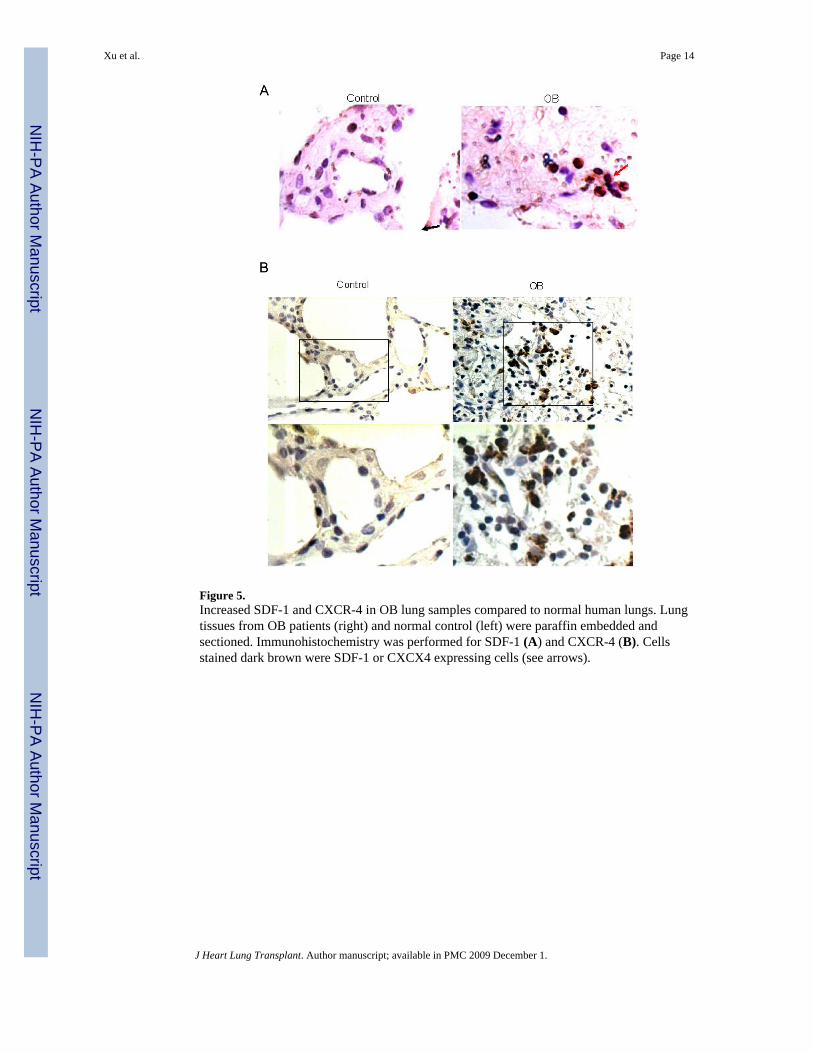
Figure 5.Increased SDF-1 and CXCR-4 in OB lung samples compared to normal human lungs. Lungtissues from OB patients (right) and normal control (left) were paraffin embedded andsectioned. Immunohistochemistry was performed for SDF-1 (A) and CXCR-4 (B). Cellsstained dark brown were SDF-1 or CXCX4 expressing cells (see arrows).
Xu et al. Page 14
J Heart Lung Transplant. Author manuscript; available in PMC 2009 December 1.
NIH
-PA Author Manuscript
NIH
-PA Author Manuscript
NIH
-PA Author Manuscript