Babesia lengau associated with cerebral and haemolytic babesiosis in two domestic cats
Upload
mzumbeuniversityCategory
view
1download
0
1
DOI:10.2298/ABS140410009K
DETERMINATION OF THE PRESENCE OF BABESIA DNA IN BLOOD
SAMPLES OF CATTLE, CAMEL AND SHEEP IN IRAN BY PCR
F. Khamesipour1,2 *
, A. Doosti2, A. Koohi
3, M. Chehelgerdi
2, A. Mokhtari-Farsani
1,2,
and A.A. Chengula4
1 Young Researchers and Elite Club, Shahrekord Branch, Islamic Azad University,
Shahrekord, Iran
2 Biotechnology Research Center, Shahrekord Branch, Islamic Azad University,
Shahrekord, Iran
3 Faculty of Veterinary Medicine, Shahrekord Branch, Islamic Azad University,
Shahrekord, Iran
4 Department of Veterinary Microbiology and Parasitology, Faculty of Veterinary
Medicine, Sokoine University of Agriculture, Morogoro, Tanzania
Corresponding author: [email protected]
Abstract - Babesia species are protozoan parasites that parasitize the erythrocytes of domestic
animals and humans, causing anemia in the host affected. These parasites cause a zoonotic
2
disease known as babesiosis. Polymerase chain reaction (PCR) has proven to be very
sensitive for detecting Babesia in blood samples of affected animals, particular in ruminants.
The purpose of the current study was to determine the presence of Babesia DNA in the blood
samples obtained from cattle, camel and sheep in Iran. In addition, the study aimed at
establishing a rapid, reliable, specific and sensitive molecular tool, the PCR, for the detection of
Babesia DNA in ruminants and dromedaries. Blood samples were collected from 372 ruminants
and dromedaries (155 cattle, 95 sheep and 122 camel) kept at the Livestock Experimental
Station. The animals came from randomly selected herds located in the important livestock-
production regions of Iran of Isfahan and Chaharmahal va Bakhtiary during December 2012 to
March 2013. PCR was used to detect Babesia DNA in the blood samples whereby an amplified
band size of 428 bp was considered positive for Babesia spp. The results indicated that 7.10%
(n= 155), 6.56% (n= 122) and 0.00% (n= 95) of the blood samples from cattle, camel and sheep
were positive for Babesia DNA, respectively.The findings from this study revealed that there
were Babesia DNA in blood taken from cattle and camel. To our knowledge, this is the first
report to show the presence of Babesia DNA in blood samples of Iranian ruminants and
dromedaries in Chaharmahal Va Bakhtiari and Isfahan provinces by PCR method. Though,
diagnosis of low-level infections by the parasite is important for the epidemiological studies.
Our findings support the power of PCR test for Babesia DNA detection in blood samples and
could be easily used for routine diagnosis.
Key words: Babesia spp.; cattle; sheep; camel; blood; PCR
Received April 10, 2014; Accepted October 13, 2014

3
INTRODUCTION
Tick infestation and the resulting transmission of serious pathogens in ruminants is one
of the most important problems of the livestock industry in developing countries (Ghirbi
et al., 2008; Adham et al., 2009; Aktas et al., 2012). Concerning camel disease, camels
were formerly considered resistant to most of the diseases commonly affecting
livestock, but as more research was conducted, camels were found to be susceptible to a
large number of pathogenic agents (Rahimi et al., 2012; Khamesipour et al., 2014a;
Khamesipour et al., 2014b; Khamesipour et al., 2014c).
In 1957, the first demonstrated case of human babesiosis in the world was
reported in a Yugoslavian farmer (Skrabalo and Deanovi, 1957). Babesia species are
tick-borne hemoprotozoan parasites that parasitize erythrocytes leading to anemia in the
host. Numerous different species exist by varying host specificity and are found all over
Asia, the Middle East, Europe, Africa and North America (Calder et al., 1996; Aktas et
al., 2007; Altay et al., 2008; Martins et al., 2008; Heidarpour Bami et al., 2009; Razmi
et al., 2013). Infection occurs in domestic animals, including cattle, horse, sheep, goats,
pigs and dogs (Fahrimal et al., 1992; Bock et al., 2004).
Bovine babesiosis is a major tick-borne disease of cattle caused by protozoan
parasites (Babesia spp.). Of the 1.2 x 10⁹ cattle in the world, over 500 million of these
cattle are possibly at risk of having bovine babesiosis. Out of at least six Babesia
species that have an important effect on livestock health and productivity, two species,
Babesia bovis (B. bovis) and B. bigemina have the highest impact. In cattle, B. bigemina
can cause massive destruction of the red blood cells leading to severe anaemia and

4
hemoglobinuria. This end result in red urine (due to haemoglobin in urine) and the
disease can kill cattle inside a week (Uilenberg, 2006).
B.bovis is more dangerous than B. bigemina because it is less sensitive to some
babesiacidal compounds, making it problematic to cure the infected animals. Animals
that survive a Babesia infection generally become carriers of the parasite and serve as
reservoirs for transmission (Chaudhry et al., 2010).
Ovine babesiosis is an important disease in livestock with high mortality and
morbidity, resulting in high economic losses globally. It is one of the most important
sheep disease in the Mediterranean and other regions where the vector tick,
Rhipicephalus bursa, is present (Aktas et al., 2007; Altay et al., 2008). Sheep that
recover from babesiosis become asymptomatic carriers (Aktas et al., 2005).
Camels were infected with Babesia caballi for the first record in Sudan (Abd-
Elmaleck et al., 2014), So, that the infection of Camelus dromedaries by Babesia sp. is
the first record in Egypt. Babesia caballi is a hemoparasitic protozoan of the Phylum
Apicomplexa that is transmitted naturally in New World by Anocentor nitens ticks
(Abd-Elmaleck et al., 2014).
Carrier hosts infected with Babesia are difficult to detect due to the low numbers
of parasites that occur in peripheral blood. However, the diagnosis of low-level
infections by the parasite is important for control and epidemiological studies (Fahrimal
et al., 1992).
Indirect Fluorescent Antibody Test (IFAT) is the most widely used test for the
detection of antibodies to Babesia spp. However, serological cross reactions make
species diagnosis difficult (OIE, 2005). The diagnosis of ruminant piroplasmosis is
generally based upon the microscopic examination of Giemsa stained blood smears and
5
via clinical signs in acute cases. Afterward acute infections, healthier animals frequently
sustain sub clinical infections, which are microscopically not detectable (Perez-Llaneza
et al., 2010; Schneider et al., 2011). They can be measured as a source of infection for
the latent vector that make natural transmission of the disease. Serological techniques
are frequently employed in determining subclinical infections despite lacking the
sensitivity and specificity for detecting carrier state, especially when establishing the
infection status (Durrani et al., 2006; Iseki et al., 2010; Terkawi et al., 2011).
PCR has proven to be very sensitive particular in detecting B. bovis and B.
bigemina in carrier cattle (Salem et al., 1992; Calder et al., 1996). Consequently, DNA
amplification techniques, which are more specific and sensitive than other conventional
techniques, may facilitate and be used as a forceful tool for the diagnosis of babesiosis
(Schnittger et al., 1990; Nagore et al., 2003; Aktas et al., 2005; Aktas et al., 2007;
Martins et al., 2008; Bhoora et al., 2009; Iqbal et al., 2011).
The purpose of the current study was to determine the presence of Babesia DNA
in the blood samples of cattle, camel and sheep in Iran and to establish a reliable,
specific and sensitive molecular tool (PCR), for the detection of Babesia DNA for rapid,
accurate and easy diagnosis of babesiosis. To overcome the economic losses early and
proper diagnosis of babesiosis is important in carrier cattle which could be achieved
only through highly sensitive techniques like PCR. The present project was aimed at
standardizing the molecular diagnostic PCR technique for the early and accurate
diagnosis of babesiosis in ruminants in livestock production regions of Iran including
Isfahan and Chaharmahal Va Bakhtiary.
MATERIALS AND METHODS
6
Sampling and DNA isolation
Blood samples were collected from 372 ruminants and dromedaries kept at
Livestock Experimental Station (155 cattle, 95 sheep and 122 camel) in Iran. Animals
were randomly selected from herds located in the important livestock production
regions of Iran (Isfahan and Chaharmahal Va Bakhtiary) during December to March
2012-2013. Blood was collected from the jugular vein of the animals and immediately
preserved in 10 ml by adding 0.5M EDTA. Samples were sent to the Biotechnology
Research Center of Islamic Azad University of Shahrekord Branch in cool box with ice
packs and was stored -20°C for further use. DNA from the samples was isolated in the
Biotechnology Research Center of Islamic Azad University of Shahrekord Branch.
Genomic DNA was extracted from specimens using DNA extraction kit (Cinnagen,
Tehran, Iran) according to the manufacturer’s protocol. The concentration of DNA was
measured at 260 nm optical density according to the method described by Sambrook &
Russell (Sambrook and Russell 2001). The extracted DNA of blood sample was kept
frozen at -20°C until its use.
PCR amplification
Polymerase chain reaction was performed on the genomic DNA of Babesia spp.
to amplify a 428 base pair (bp) small 18S rRNA gene fragment using the following set
of primers: Bab-sp-F: 5’- GTTTCTGCCCCATCAGCTTGAC-3’ and Bab-sp-R: 5’
CAAGACAAAAGTCTGCTTGAAAC -3’ (Hilpertshauser et al 2006). The procedure
for amplification of the DNA was done as reported previously (Hilpertshauser et al
2006). The amplification of Babesia spp. DNA was done using thermocycler
(Eppendorf, Hamburg, Germany). PCR reaction was performed as follows (45 cycles):
7
denaturation at 94 °C for 30 s, annealing at 61 °C for 30 s, extension at 72 °C for 45 s
and then final incubation at 72 °C for 10 min. The amplicons were stained with
ethidium bromide and electrophoresd in 1.5% agarose gel at 80V for 30 min. PCR
products were visualized and photographed using UVIdoc gel documentation systems
(Uvitec, UK). The PCR products were compared against a 100 bp DNA marker
(Fermentas, Germany).
Statistical analysis
Analysis of data was performed by the SPSS version 17.0 computer software
(SPSS, Chicago, IL).
RESULTS
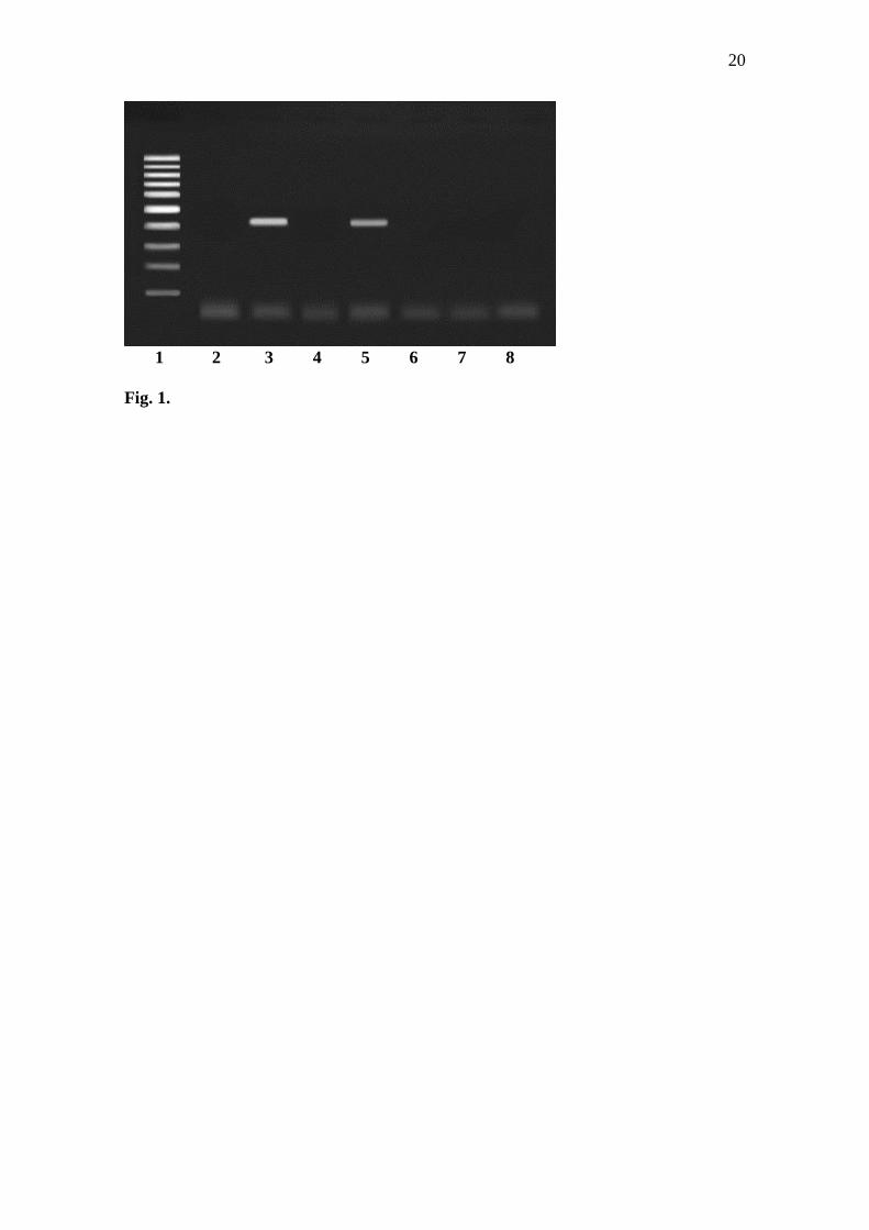
In this study, PCR was used to detect the presence of Babesia DNA in blood samples of
372 ruminants and dromedaries (155 cattle, 95 sheep and 122 camels) in livestock
production regions of Iran. The tested sample was considered positive if a 428-bp small
18S rRNA gene fragment of Babesia spp. was amplified during PCR (Fig. 1). The
findings from this study were clustered based on the location where animals sampled
were obtained and on the sex of the animals sampled. The Babesia DNA was were
present in cattle and camel but not in sheep (Table 1) in both provinces of Iran.
DISCUSSION
8
Babesia is one of the most important blood parasites and tick-borne zoonoses affecting
cattle and sheep and in its acute forms, it causes lower production performance of the
affected animals (Talkhan et al., 2010; Ziapour et al., 2011).
Babesia bigemina and B. bovis are recognized to be pathogenic in cattle
according to previous studies (Uilenberg, 2006). In 1968, B. divergens and B. microti
were recognized as the cause of human babesiosis and small mammalian hosts in
Europe and US, respectively (Senanayake et al., 2012). Ixodes spp. and Rhipicephalus
spp. have been implicated in the transmission of human and bovine Babesia spp.,
respectively (Uilenberg, 2006). It is probable that 1.2 billion cattle are exposed to
babesiosis in many countries of the world including Asia, Australia, Africa, South and
Central America and the United States (Terkawi et al., 2011).
Few papers have reported the Babesia sp. in Camelus dromedarius (Abd-
Elmaleck, 2014). Abd-Elmaleck et al. (2014), reported out of ninety eight of camels
(Camelus dormadarius) examined, only forty eight (48.9 %) were found to be infected
with blood protozoan parasites (Trypanosoma evansi, Theileria sp. and Babesia sp.).
The higher incidence of infection were found in males (36.7%) whereas, (12.24%) in
females. Also, forty six from ninety eight examined of Camelus dromedarius (46.9 %)
were found infected with Babesia sp. (Abd-Elmaleck, 2014), which showed the higher
than prevalence in this study.
It is imperative to develop sensitive tools for the effective diagnosis of
babesiosis and drugs for its treatment in order to reduce the economic losses incurred as
result of the disease. A number of conventional and modern techniques are used for the
detection of Babesia spp. in host animals. The most commonly used is the microscopic

9
examination of blood smears stained by Giemsa, which is typically adequate for
detection of acute infections. Due to its low sensitivity, this technique cannot be used
for the detection of carrier animals due to low parasitemia. Serological techniques are
not specific for any Babesia spp. due to cross-reactivity and therefore, cannot be relied
on (D’Oliveira et al., 1997). In addition, false positive and negative results are often
observed in serological techniques. A problem discussed in protozoan infections is the
characterization and determination of transmitter agent. Because many analyses were
previously performed using salivary gland smears, for example methyl green-pyronin
staining or Feulgen staining techniques, in some cases the transfer vector remains
unidentified (Guglielmone et al., 1997). The staining of tick salivary glands can confirm
the Babesia spp. infection of ticks, but the main problems with this technique are its
long duration, low sensitivity, and the difficulty of differentiating the species involved
(Oliviera-Sequeira et al., 2005). The use of molecular methods (such as PCR) for the
detection and identification of different microorganisms has gained popularity among
scientists in recent years. This is because molecular methods are more specific and
sensitive than other traditional diagnostic techniques (Sparagano, 1999; Almeria et al.,
2001; Altay et al., 2008). In recent times, DNA amplification techniques have been
developed and used for the detection of Babesia spp. (Schnittger et al., 1990).
Information on the prevalence of tick-borne pathogens in potential vector ticks of the
area is essential for the identification of tick-borne diseases. Altay et al. (2008) found
that R. bursa was the main vector for cattle Babesia spp. in eastern Turkey (where it is
contiguous with the present surveyed areas). Some previous studies carried out in the
Mediterranean region stated that B. bigemina and B. bovis are transmitted via R. bursa
10
(Bouattour and Darghouth, 1996; Ravindran et al., 2006; Altay et al., 2007; Ghirbi et
al., 2010).
In the current study, 372 blood samples of cattle, camel and sheep were tested
for the presence of Babesia DNA using PCR method. The results indicated that 7.10%
(n= 155), 6.56% (n= 122) and 0.00% (n= 95) of the blood samples from cattle, camel
and sheep, respectively, were positive for Babesia spp.
A molecular study of Theileria and Babesia in cattle from Isfahan province,
Central Iran, using blood samples collected from March to July 2009 indicated 23.9% of
the samples were positive for Theileria spp. and none of them was positive for Babesia
spp. (Noaman, 2013). These findings are contrary to the present study. The current
study indicates that Babesia spp. were present in the study area. Although Babesia spp.
in sheep (B. ovis and B. motasi) have been reported in previous studies in Iran (Shayan
et al., 2008; Ziapour et al., 2011; Motavalli et al., 2013), none of the samples from
sheep in our study was positive for Babesia spp.. In small ruminants, diseases caused by
protozoans (Theileria and Babesia) have been reported to cause high economic losses
worldwide (Shayan et al., 2008).
Chaudhry et al. (2010), reported an overall prevalence of 29% for Babesia spp.
using PCR, whereby 11% were positive for B. bovis and 18% for B. bigemina. Calder et
al. (1996), reported the average sensitivities of three PCR-based tests for B. bovis to
range from 58 to 70% for a single determination, while that of Complement Fixation
test was 6%. In general, babesiosis has the highest distribution and leads to the highest
mortality in domestic animals in many areas of Iran (Ziapour et al., 2011).
The results of this study showed that the samples of cattle and camel served as
reservoirs of babesiosis in Iran (With regards to camels this is speculation and this does

11
not mean that camel is a host for Babesia). Consequently, it could be stated that the
animal reservoirs increase the risk of the potential spread of disease to other animals and
especially humans, and this deserves special attention. The study has indicated that the
use of PCR in the surveillance of babesiosis will enable the detection of asymptomatic
carrier animals that could not be detected using conventional methods.
REFERENCES
Abd-Elmaleck, B.S., Abed, G.H. and Mandourt, A.M. (2014). Some Protozoan Parasites
Infecting Blood of Camels (Camelus dromedarius) at Assiut Locality, Upper
Egypt. J. Bacteriol. Parasitol. 5: 184. doi: 10.4172/2155-9597.1000184.
Adham, F.K., Abd-el-Samie E.M., Gabre, R.M. and H. El-Hussein (2009). Detection of
tick blood parasites in Egypt using PCR assay I-Babesia bovis and Babesia
bigemina. Parasitol. Res. 105: 721-730.
Aktas, M., Altay, K. and N. Dumanli (2005). Development of a polymerase chain
reaction method for diagnosis of Babesia ovis infection in sheep and goats. Vet.
Parasitol. 133: 277-281.
Aktas, M., Altay, K. and N. Dumanli (2007). Determination of prevalence and risk
factors for infection with Babesia ovis in small ruminants from Turkey by
polymerase chain reaction. Parasitol. Res. 100(4): 797-802.
Aktas, M., Altay, K., Ozubek, S. and N. Dumanli (2012). A survey of ixodid ticks
feeding on cattle and prevalence of tick-borne pathogens in the Black Sea region
of Turkey. Vet. Parasitol. 187: (3-4), 567-571.
12
Almeria, J., Castella, D., Ferrer, A., Ortuño, A., Estrada-Peña, J.F. and G. Gutiérrez
(2001). Bovine piroplasms in Minorca (Balearic Islands, Spain): a comparison
of PCR-based and light microscopy detection. Vet. Parasitol. 99: 249-259.
Altay, K., Aktas, M. and N. Dumanli (2008). Detection of Babesia ovis by PCR in
Rhipicephalus bursa collected from naturally infested sheep and goats. Res. Vet.
Sci. 85 (1): 116-119.
Altay, K., Dumanli, N., Holman, P.J. and M. Aktas (2007). Molecular identification,
genetic diversity and distribution of Theileria and Babesia spp infecting small
ruminants. Veterinary Parasitology 147: 121-127.
Altay, K., Fatih Aydin, M., Dumanli, N., M. Aktas (2008). Molecular detection of
Theileria and Babesia infection in cattle. Vet. Parasitol. 158: 295-301.
Bhoora, R., Franssen, L., Oosthuizen, M.C., Guthrie, A.J., Zweygarth, E., Penzhorn,
B.L., Jongejan, F. and N.E. Collins (2009). Sequence heterogeneity in the 18S
rRNA gene within Theileria equi and Babesia caballi from horses in South
Africa. Vet. Parasitol. 159: 112-120.
Bock, R., Jackson, L., de Vos, A. and W. Jorgensen (2004). Babesiosis of cattle.
Parasitology 129: 247-269.
Bouattour, A. and M.A. Darghouth (1996). First report of Babesia divergens in Tunisia.
Vet. Parasitol. 63: 161-165.
Calder, J.A.M., Reddy, G.R., Chieves, L., Courtney, C.H., Littell, R., Livnegood, J.R.,
Norval, R.A.I., Smith, C. and J.B. Dame (1996). Monitoring Babesia bovis
infections in cattle by using PCR-based tests. J. Clin. Microbiol. 34: 2748-2755.
13
Chaudhry, Z.I., Suleman, M., Younus, M. and A. Aslim (2010). Molecular detection of
Babesia bigemina and Babesia bovis in crossbred carrier cattle through PCR.
Pak. J. Zool. 42(2): 201-204.
D’Oliveira, C., Van der Wide, M., Jacquiet, P. and F. Jongejan (1997). Detection of
Theileria annulata by the PCR in ticks (Acari: Ixodidae) collected from cattle in
Mauritania. Exp. Appl. Acarol. 21: 279-291.
Durrani, A.Z., Kamal, N. and M.S. Khan (2006). Incidence of theileriosis and estimation
of packed cell volume, total erythrocyte count and hemoglobin in buffaloes. J.
Animal Plant Sci. 16: 85-88.
Fahrimal, Y., Goff, W.L. and D.P. Jasmer (1992). Detection of Babesia bovis carrier
cattle by using polymerase chain reaction amplification of parasite DNA. J.
Clin. Microbiol. 30: 1374-1379.
Ghirbi, Y.M., Hurtado, A., Brandika, J., Khlif, K., Ketata, Z. and A. Bouattour (2008). A
molecular survey of Theileria and Babesia parasites in cattle, with a note on the
distribution of ticks in Tunisia. Parasitol. Res. 103: 435-442.
Ghirbi, M., Hurtado, A. and A. Bouattour (2010). Theileria and Babesia parasites in
ticks in Tunisia. J. Emergency Med. 57: 49-51.
Guglielmone, A.A., Gaido, A.B., Aguirre, D.A. and M.M. Cafrune (1997). Some
quantitative aspects of natural babesial infection in the haemolymph of
Boophilus microplus engorged female ticks. Parasite 4: 337-341.
Heidarpour Bami, M., Haddadzadeh, H.R., Kazemi, B., Khazraiinia, P., Bandehpour,
M. and M. Aktas (2009). Molecular identification of ovine Theileria species by a
new PCR–RFLP method. Vet. Parasitol. 161(3–4): 171-177.
14
Hilpertshauser, H., Deplazes, P., Schnyder, M., Gern, L. and A. Mathis (2006). Babesia
spp. identified by PCR in ticks collected from domestic and wild ruminants in
southern Switzerland. Appl. Environ. Microbiol. 72: 6503-6507.
Iqbal, F., Fatima, M., Shahnawaz, S., Naeem, M., Shaikh, R.S., Shaikh, A.S., Aktas, M.
and M. Ali (2011). A study on the determination of risk factors associated with
babesiosis and prevalence of Babesia sp., by PCR amplification, in small
ruminants from southern Punjab (Pakistan). Parasite 8 (3): 229-234.
Iseki, H., Zhou, Z., Kim, C., Inpankaew, T., Sununta, C., Yokoyama, N., Xuan, X.,
Jittapalapong, S. and I. Igarashi (2010). Seroprevalence of Babesia infections of
dairy cows in northern Thailand. Vet. Parasitol. 170: 193-196.
Khamesipour, F., Doosti, A., Iranpour Mobarakeh, H. and Komba E.V.G (2014a).
Detection of Toxoplasma gondii in cattle, camels and sheep in Isfahan and
Chaharmahal va Bakhtiary provinces, Iran. Jundishapur. J. Microbiol. 7(6):
e17460 , DOI: 10.5812/jjm.17460.
Khamesipour, F., Doosti, A. and Mazrouei Sebdani, M (2014b). Survey for the Presence
of Mycobacterium avium subsp. paratuberculosis in the Bull Frozen Semen
Samples and Blood Samples of Cattle, Sheep and Camel by Nested-PCR.
Kafkas. Univ. Vet. Fak. Derg. 20 (5): 681-686, DOI: 10.9775/kvfd.2014.10837.
Khamesipour, F., Rahimi, E., Shakerian, A., Doosti, A. and Momtaz, H (2014c).
Molecular study of the prevalence of Brucella abortus and Brucella melitensis in
the blood and lymph nodes samples of slaughtered camels by Polymerase Chain
Reaction (PCR) in Iran. Acta. Vet. Beograd. 64 (2): 245–256, DOI:
10.2478/acve-2014-0023.
15
Martins, T.M., Pedro, O.C., Caldeira, R.A., Do Rosário, V.E., Neves, L. and A.
Domingos (2008). Detection of bovine babesiosis in Mozambique by a novel
seminested hot-start PCR method. Vet. Parasitol. 153: 225-230.
Motavalli, H.S., Fakhar, M., Sharif, M., As, P., Sharbatkhori, M., Tavakoli, R. and G. Sh
(2013). Research in molecular medicine molecular identification of ovine
Babesia spp. in north of Iran. Res. Molec. Med. 1: 36-40.
Nagore, D., García-sanmartín, J., García-pérez, A.I., Juste, R.A., A. Hurtado (2004).
Identification, genetic diversity and prevalence of Theileria and Babesia species
in sheep population from northern Spain. Int. J. Parasitol. 34: 1059-1067.
Noaman, V. (2013). A molecular study on Theileria and Babesia in cattle from Isfahan
province, Central Iran. 37:208–210.
OIE. (2005). Manual of standard tests and vaccines, Office International des
Epizooites, World Organization for Animal Health, Chap. 2.3.8. www.oie.int.
Oliviera-Sequeira, T.C.G., Oliviera, M.C.S., Aruaujo, J.P. and A.F.T. Amarante (2005).
PCR-based detection of Babesia bovis and Babesia bigemina in their natural
host Boophilus microplus and cattle. Int. J. Parasitol. 35: 105-111.
Perez-Llaneza, A., Caballero, M., Baravalle, F., Mesplet, M., Mosqueda, J., Suarez,
C.E., Echaide, I., Katzer, F., Pacheco, G.M., Florin-Christensen, M. and L.
Schnittger (2010). Distinguishing Babesia bovis geographic isolates. Vet.
Parasitol. 167: 196-204.
Ravindran, R., Rao, J.R. and A.K. Mishra (2006). Detection of Babesia bigemina DNA
in ticks by DNA hybridization using a nonradioactive probe generated by
arbitrary PCR. Vet. Parasitol. 141: 181-185.
16
Rahimi, E., Khamesipour, F., Yazdi, F. and Momtaz, H (2012). Isolation and
Characterization of Enterohaemorragic Escherichia coli O157:H7 and EHEC
O157:NM from Raw Bovine, Camel, Water Buffalo, Caprine and Ovine Milk in
Iran. Kafkas. Univ. Vet. Fak. Derg. 18 (4): 559-564.
Razmi, G., Pourhosseini, M., Yaghfouri, S., Rashidi, A. and M. Seidabadi (2013).
Molecular detection of Theileria spp. and Babesia spp. in sheep and ixodid ticks
from the northeast of Iran. J. Parasitol. 99 (1): 77-81.
Sambrook, J. and D. Russell (2001). Molecular Cloning: a Laboratory Manual, 3rd edn.
Cold Spring Harbor, NY: Cold Spring Harbor Laboratory.
Salem, G.H., Liu, X., Johnsrude, J.D., Dame, J.B. and G.R. Roman (1999).
Development and evaluation of an extra chromosomal DNA-based PCR test for
diagnosing bovine babesiosis. Molec. Cell. Probes 2: 107-113.
Senanayake, S.N., Paparini, A., Latimer, M. andriolo, K., Dasilva, A.J., Wilson, H.,
Xayavong, M.V., Collignon, P.J., Jeans, P. and P.J. Irwin (2012). First report of
human babesiosis in Australia. Med. J. Australia 196 (5): 350-352.
Schneider, D.A., Yan, I.H., Bastos, R.G., Johnson, W.C., Gavin, P.R., Allen, A.J.,
Barrington, G.M., Herrmann-Hoesing, L.M., Knowles, D.P. and W.L. Goff
(2011). Dynamics of bovine spleen cell populations during the acute response to
Babesia bovis infection: an immunohistological study. Parasite Immunol. 33:
34-44.
Schnittger, L., Yin, H., Qi, B., Gubbels, M.J., Beyer, D., Niemann, S., Jongejan, F.,
Ahmed, J.S. and F.R. Ahall (1990). Theileria annulata: control measures,
diagnosis and the potential use of subunit vaccines. Vet. Res. 56: 24-32.

17
Shayan, P., Hooshmand, E. and S. Nabian (2008). Biometrical and genetical
characterization of large Babesia ovis in Iran. 217–221.
Skrabalo, Z. and Z. Deanovi (1957). Piroplasmosis in man: report on a case. Doc. Med.
Georgr. Trop. 9: 6-11.
Sparagano, O. (1999). Molecular diagnosis of Theileria and Babesia species. Vet.
Parasitol. 13: 83-92.
Talkhan, O.F.A., Radwan, M.E.I. and M.A. Ali (2010). Cattle babesiosis and associated
biochemical alteration in Kalubyia Governorate. Nat. Sci. 12: 24-27.
Terkawi, M.A., Huyen, N.X., Shinuo, C., Inpankaew, T., Maklon, K., Aboulaila, M.,
Ueno, A., Goo, Y.K., Yokoyama, N., Jittapalapong, S., Xuan, X. and I. Igarashi
(2011). Molecular and serological prevalence of Babesia bovis and Babesia
bigemina in water buffaloes in the northeast region of Thailand. Vet. Parasitol.
178: 201-207.
Uilenberg, G. (2006). Babesia − A historical overview. Vet. Parasitol. 138, 3-10.
Ziapour, S.P., Esfandiari, B. and M.R. Youssefi, (2011). Study of the prevalence of
babesiosis in domesticated animals with suspected signs in Mazandaran
province, north of Iran, during 2008. J. An. Vet. Adv. 10: 712-714.
18
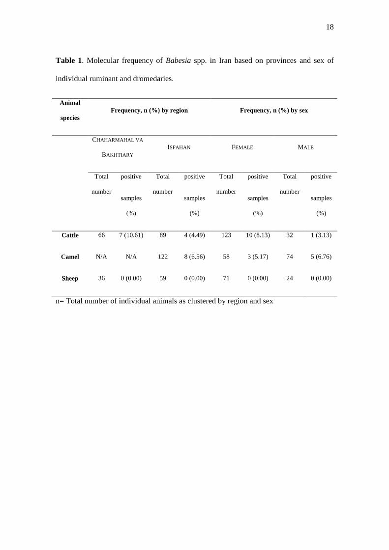
Table 1. Molecular frequency of Babesia spp. in Iran based on provinces and sex of
individual ruminant and dromedaries.
Animal
species
Frequency, n (%) by region Frequency, n (%) by sex
CHAHARMAHAL VA
BAKHTIARY
ISFAHAN FEMALE MALE
Total
number
positive
samples
(%)
Total
number
positive
samples
(%)
Total
number
positive
samples
(%)
Total
number
positive
samples
(%)
Cattle 66 7 (10.61) 89 4 (4.49) 123 10 (8.13) 32 1 (3.13)
Camel N/A N/A 122 8 (6.56) 58 3 (5.17) 74 5 (6.76)
Sheep 36 0 (0.00) 59 0 (0.00) 71 0 (0.00) 24 0 (0.00)
n= Total number of individual animals as clustered by region and sex
19
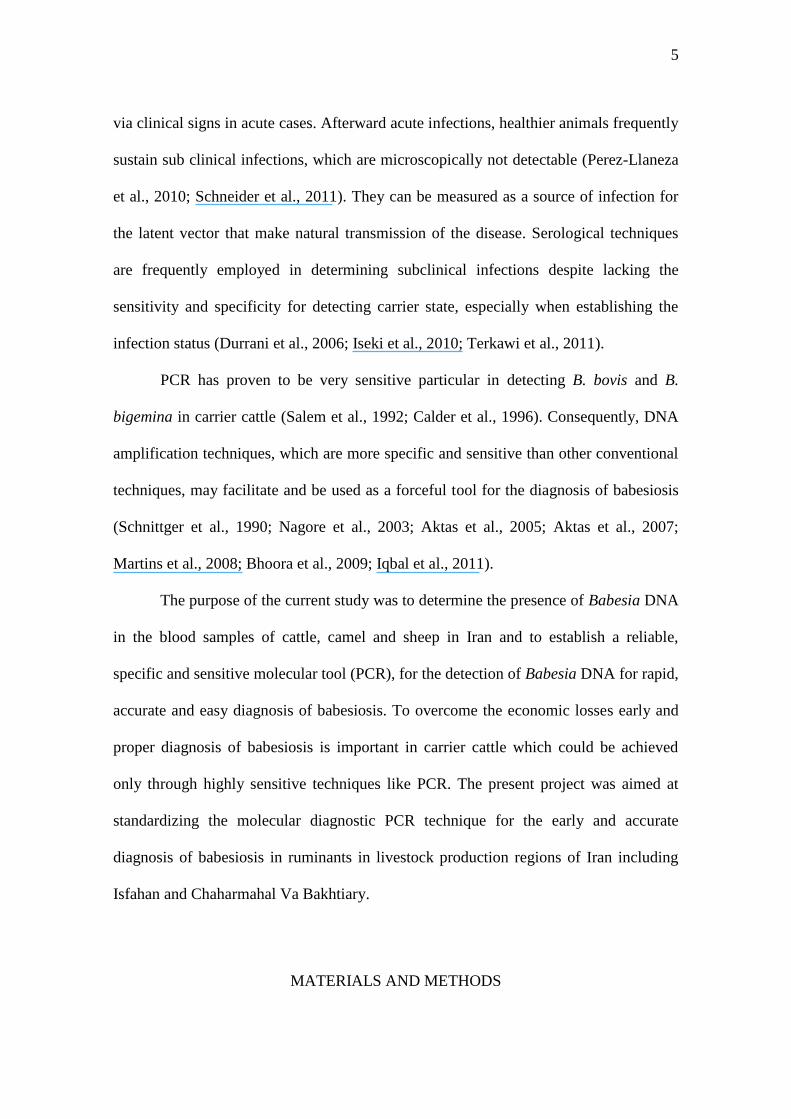
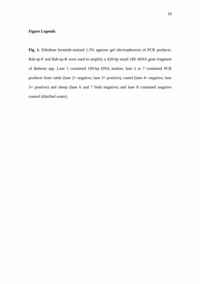
Figure Legends
Fig. 1. Ethidium bromide-stained 1.5% agarose gel electrophoresis of PCR products.
Bab-sp-F and Bab-sp-R were used to amplify a 428-bp small 18S rRNA gene fragment
of Babesia spp. Lane 1 contained 100-bp DNA marker, lane 2 to 7 contained PCR
products from cattle (lane 2= negative; lane 3= positive), camel (lane 4= negative; lane
5= positive) and sheep (lane 6 and 7 both negative) and lane 8 contained negative
control (distilled water).
20
1 2 3 4 5 6 7 8
Fig. 1.
Copyright © 2022 FDOKUMEN