a sheep model - SciELO
9
r e v b r a s o r t o p . 2 0 1 7; 5 2(1) :52–60 SOCIEDADE BRASILEIRA DE ORTOPEDIA E TRAUMATOLOGIA www.rbo.org.br Original article Biomechanical evaluation of the long head of the biceps brachii tendon fixed by three techniques: a sheep model Carlos Henrique Ramos ∗ , Júlio Cezar Uili Coelho Universidade Federal do Paraná, Curitiba, PR, Brazil a r t i c l e i n f o Article history: Received 22 February 2016 Accepted 18 March 2016 Available online 30 December 2016 Keywords: Biceps braquii Humerus Biomechanic Tendons a b s t r a c t Objective: To evaluate the biomechanical properties of the fixation of the long head of the biceps brachii into the humeral bone with suture anchors, interference screw, and soft tissue suture, comparing strength, highest traction load, and types of fixation failure. Methods: Thirty fresh-frozen sheep shoulders were used, separated into three groups of ten for each technique. After fixation, the tendons were subjected to longitudinal continuous loading, obtaining load-to-failure (N) and displacement (mm). Results: The mean load-to-failure for suture anchors was 95 ± 35.3 N, 152.7 ± 52.7 N for interference screw, and 104.7 ± 23.54 N for soft tissue technique. There was a statistically sig- nificant difference (p < 0.05), with interference screw demonstrating higher load-to-failure than suture anchor fixation (p = 0.00307) and soft tissue (p = 0.00473). The strength of inter- ference screw was also superior when compared with the other two methods (p = 0.0000127 and p = 0.00000295, respectively). There were no differences between suture anchors and soft tissue technique regarding load-to-failure (p = 0.9420) and strength (p = 0.141). Conclusion: Tenodesis of the long head of the biceps brachii with interference screw was stronger than the suture anchors and soft tissue techniques. The other two techniques did not differ between themselves. © 2016 Sociedade Brasileira de Ortopedia e Traumatologia. Published by Elsevier Editora Ltda. This is an open access article under the CC BY-NC-ND license (http:// creativecommons.org/licenses/by-nc-nd/4.0/). Avaliac ¸ão biomecânica da fixac ¸ão do tendão da cabec ¸a longa do bíceps braquial por três técnicas: modelo em ovinos Palavras-chave: Bíceps braquial Úmero r e s u m o Objetivo: Avaliar biomecanicamente a fixac ¸ão da cabec ¸a longa do bíceps braquial no úmero com âncoras ósseas, parafuso de interferência e sutura em partes moles e comparar resistên- cia, forc ¸a máxima de trac ¸ão e tipos de falha na fixac ¸ ão. Study conducted at the Centro Universitário (UniBrasil), at the Pontifícia Universidade Católica do Paraná, and at the Universidade Federal do Paraná, Curitiba, PR, Brazil. ∗ Corresponding author. E-mail: [email protected] (C.H. Ramos). http://dx.doi.org/10.1016/j.rboe.2016.12.008 2255-4971/© 2016 Sociedade Brasileira de Ortopedia e Traumatologia. Published by Elsevier Editora Ltda. This is an open access article under the CC BY-NC-ND license (http://creativecommons.org/licenses/by-nc-nd/4.0/).
-
Upload
khangminh22 -
Category
Documents
-
view
1 -
download
0
Transcript of a sheep model - SciELO
r e v b r a s o r t o p . 2 0 1 7;5 2(1):52–60
SOCIEDADE BRASILEIRA DEORTOPEDIA E TRAUMATOLOGIA
www.rbo.org .br
Original article
Biomechanical evaluation of the long head of thebiceps brachii tendon fixed by three techniques: asheep model�
Carlos Henrique Ramos ∗, Júlio Cezar Uili Coelho
Universidade Federal do Paraná, Curitiba, PR, Brazil
a r t i c l e i n f o
Article history:
Received 22 February 2016
Accepted 18 March 2016
Available online 30 December 2016
Keywords:
Biceps braquii
Humerus
Biomechanic
Tendons
a b s t r a c t
Objective: To evaluate the biomechanical properties of the fixation of the long head of the
biceps brachii into the humeral bone with suture anchors, interference screw, and soft tissue
suture, comparing strength, highest traction load, and types of fixation failure.
Methods: Thirty fresh-frozen sheep shoulders were used, separated into three groups of ten
for each technique. After fixation, the tendons were subjected to longitudinal continuous
loading, obtaining load-to-failure (N) and displacement (mm).
Results: The mean load-to-failure for suture anchors was 95 ± 35.3 N, 152.7 ± 52.7 N for
interference screw, and 104.7 ± 23.54 N for soft tissue technique. There was a statistically sig-
nificant difference (p < 0.05), with interference screw demonstrating higher load-to-failure
than suture anchor fixation (p = 0.00307) and soft tissue (p = 0.00473). The strength of inter-
ference screw was also superior when compared with the other two methods (p = 0.0000127
and p = 0.00000295, respectively). There were no differences between suture anchors and
soft tissue technique regarding load-to-failure (p = 0.9420) and strength (p = 0.141).
Conclusion: Tenodesis of the long head of the biceps brachii with interference screw was
stronger than the suture anchors and soft tissue techniques. The other two techniques did
not differ between themselves.© 2016 Sociedade Brasileira de Ortopedia e Traumatologia. Published by Elsevier Editora
Ltda. This is an open access article under the CC BY-NC-ND license (http://
creativecommons.org/licenses/by-nc-nd/4.0/).
Avaliacão biomecânica da fixacão do tendão da cabeca longa do bícepsbraquial por três técnicas: modelo em ovinos
r e s u m o
Palavras-chave:
Bíceps braquial
Úmero
Objetivo: Avaliar biomecanicamente a fixacão da cabeca longa do bíceps braquial no úmero
com âncoras ósseas, parafuso de interferência e sutura em partes moles e comparar resistên-
cia, forca máxima de tracão e tipos de falha na fixacão.
� Study conducted at the Centro Universitário (UniBrasil), at the Pontifícia Universidade Católica do Paraná, and at the UniversidadeFederal do Paraná, Curitiba, PR, Brazil.
∗ Corresponding author.E-mail: [email protected] (C.H. Ramos).
http://dx.doi.org/10.1016/j.rboe.2016.12.0082255-4971/© 2016 Sociedade Brasileira de Ortopedia e Traumatologia. Published by Elsevier Editora Ltda. This is an open access articleunder the CC BY-NC-ND license (http://creativecommons.org/licenses/by-nc-nd/4.0/).
r e v b r a s o r t o p . 2 0 1 7;5 2(1):52–60 53
Biomecânica
Tendões
Métodos: Foram usados 30 ombros de ovinos frescos, divididos em três grupos de dez para
cada técnica. Após fixacão, os tendões foram submetidos a tracão longitudinal contínua até
falha do sistema e obtiveram-se forca máxima de tracão (N) e deslocamento (mm).
Resultados: A forca máxima de tracão foi em média 95 ± 35,3 N para âncoras ósseas,
152,7 ± 52,7 N para parafuso de interferência e 104,7 ± 23,54 N para partes moles. Houve
diferenca estatisticamente significativa (p < 0,05): o parafuso de interferência demonstrou
forca máxima de tracão superior às fixacões com âncoras ósseas (p = 0,00307) e partes moles
(p = 0,00473). A resistência com parafuso de interferência também foi superior à dos out-
ros dois métodos (p = 0,0000127 e p = 0,0000029,5 respectivamente). Âncoras ósseas e partes
moles não apresentaram diferencas, tanto para forca máxima de tracão (p = 0,9420) quanto
para resistência (p = 0,141).
Conclusão: A tenodese da cabeca longa do bíceps braquial com parafuso de interferência
demonstra maior resistência quando comparada com as técnicas com âncoras ósseas e
partes moles. As duas últimas técnicas não diferem.
© 2016 Sociedade Brasileira de Ortopedia e Traumatologia. Publicado por Elsevier
Editora Ltda. Este e um artigo Open Access sob uma licenca CC BY-NC-ND (http://
creativecommons.org/licenses/by-nc-nd/4.0/).
I
Dfvaudti(horpmpsstccfaEtrviumuTach
Tenodesis with interference screw
ntroduction
isorders of the long head of biceps brachii tendon (LHBBT) arerequent causes of shoulder pain. Treatment should be conser-ative (analgesics, anti-inflammatories, and physiotherapy,mong others); however, when conservative treatment isnsuccessful, surgery is indicated. The recommended proce-ure is tenotomy of the long head of the biceps (sectioninghe tendon at the level of its insertion in the glenoid cav-ty) with or without tenodesis of the long head of the bicepsfixating the biceps tendon into the bicipital groove of theumerus). Tenodesis has been suggested as advantageousver isolated tenotomy, as it maintains the length/tensionatio and the flexion and supination strength of the elbow,reventing atrophy, pain at the site, and cosmetic defor-ity. Recent advances have allowed tenodesis to be preferably
erformed arthroscopically, which, despite promoting resultsimilar to open surgery, offers advantages such as smallerurgical wound, lower post-operative pain, preservation ofhe deltoid muscle, and earlier return to activities, espe-ially when associated with simultaneous repair of the rotatoruff.1–3 Among the arthroscopic fixation methods, the mostrequently used are bone anchors, interference screw (IS),nd soft tissue suture without the need for implants.1,2,4–8
arly postoperative mobilization of the upper limb is impor-ant for recovery, but may endanger tenodesis with possibleelease of the tendon. To avoid this issue, the system that pro-ides the highest resistance should be used. Another aspects the increased cost of the procedure when implants aresed; soft tissue technique is cheaper. Identifying whichethod is more resistant would justify the use or non-
se of implants, defining the most cost-effective technique.here is no consensus in the literature regarding which fix-
9–24
tion method is more resistant. This study aimed toompare three techniques for fixation of the LHBBT in theumerus (bone anchors, IS, and soft tissue suture) regardingresistance of the fixation, load-to-failure (LTF), and systemfailure.
Materials and methods
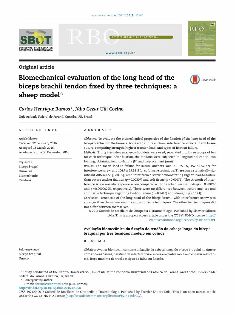
After approval by the Research Ethics Committee of the Hospi-tal do Trabalhador of Universidade Federal do Paraná, 30 freshshoulder specimens from skeletally immature Texel sheep,aged between six and 12 months, were acquired from a spe-cialized company. The specimens were frozen immediatelyafter slaughter and were kept at −20 ◦C until 24 h before prepa-ration. Samples were then thawed at room temperature toundergo tenodesis. Samples were prepared with dissection ofthe humeral bone; only the biceps and the anterior portionof the rotator cuff inserted into the greater tuberosity of thehumerus were preserved. The proximal biceps tendon wassectioned at the glenoid labrum in the upper portion of theglenoid cavity (scapular bone), maintaining its distal insertioninto the cubitus bone (Fig. 1). The specimens were dividedinto three groups of ten, according to the type of tenodesis;ten pieces were thawed at a time, with an interval of 15 daysbetween each test.
Tenodesis with bone anchors
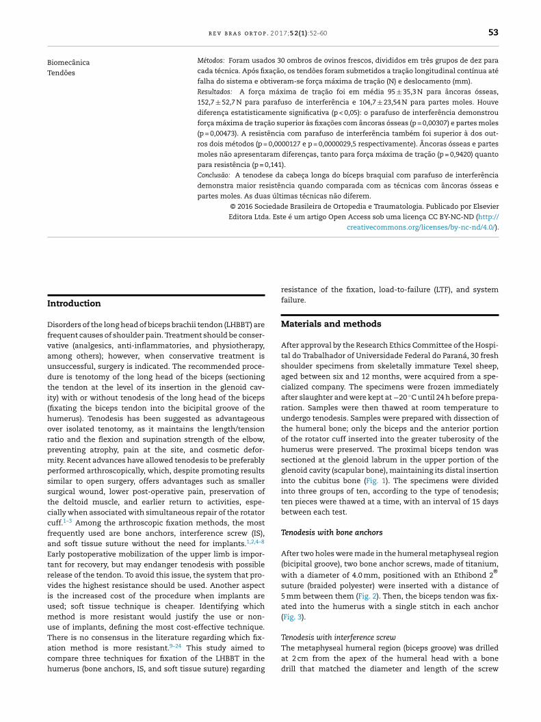
After two holes were made in the humeral metaphyseal region(bicipital groove), two bone anchor screws, made of titanium,
with a diameter of 4.0 mm, positioned with an Ethibond 2®
suture (braided polyester) were inserted with a distance of5 mm between them (Fig. 2). Then, the biceps tendon was fix-ated into the humerus with a single stitch in each anchor(Fig. 3).
The metaphyseal humeral region (biceps groove) was drilledat 2 cm from the apex of the humeral head with a bonedrill that matched the diameter and length of the screw
54 r e v b r a s o r t o p . 2 0 1 7;5 2(1):52–60
Cb
Bc
H
Bg
H
baRc
Fig. 1 – Photograph of the parts obtained by dissection; the distal insertion of the biceps in the cubitus bone (a) and theone
rotator cuff to the humerus (b) were maintained, Cb, cubital b(7 × 20 mm). The free end of the tendon was repaired using
continuous suture with Ethibond 2®
and inserted into thehumeral orifice for fixation with IS, parallel to the tendon fibers(Figs. 4 and 5). During screw insertion, the tendon was kept intraction against the opposite cortex through prior transfixa-tion of the repair wire with a fenestrated steel guidewire tokeep it within the bone hole.
Biceps tenodesis in soft tissueThe bicipital tendon suture was made on the remaining por-tion of the rotator cuff that was maintained in the greatertuberosity of the humerus, with three simple stitches usingEthibond 2
®sutures (Fig. 6).
Measurements were made with a Vonder®
precision metal-lic caliper (150 mm-6′′). After surgical preparation, material
was sent to the Biomechanics Laboratory of the UniversidadeTecnológica Federal do Paraná (UTFPR) for testing.For axial traction, the universal MTS 810 (100 KN) hydraulicequipment for tensile tests was used, with a second actuator
Fig. 2 – Photographs showing the humeral preparation with bongroove (b) and type of anchor used (c).
; Bc, biceps; Bg, bicipital groove; H, humerus; Rc, rotator cuff.
(MTS 242.02) adapted with a load cell (model 661.19F-02, MTSSystems Corporation, with a capacity of 10KN, and applied ata speed of 5 mm/min.
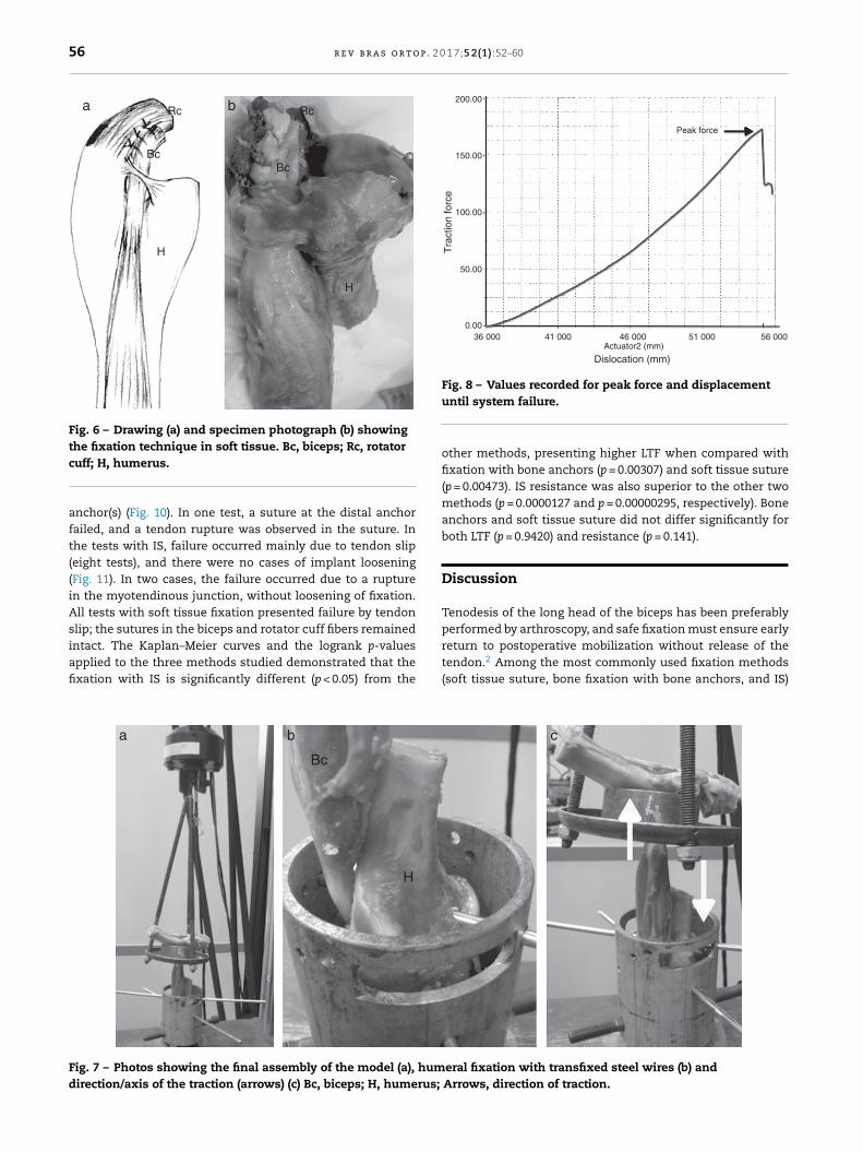
Models were fitted so that the traction to be exerted in thelongitudinal direction in relation to the axes of the tendonand humerus would simulate the normal direction of contrac-tion by the biceps. Iron devices were developed and placed onthe tensile equipment to fixate the ends of the specimen. Atthe top, the ulnar bone was supported on this device and thetendon was passed through the center hole. For lower stabi-lization, a cylindrical adapter was fixed on the traction table.This piece had interior lateral perforations for humeral sta-bilization, with two strong steel wires of a diameter (5 mm),cross-transfixated in the metaphyseal region. The distancebetween the extremities varied according to the length of themuscle/tendon pair. For a better fit, the length of the humerus
was cut at the middle third during assembly (Fig. 7). With thehelp of MTS-Test Star II software, 790.90, TestWorks – 1994,graphics indicating the LTF (N) were made. LTF was definede perforations (a), insertion of the anchors in the bicipital

r e v b r a s o r t o p . 2 0 1 7;5 2(1):52–60 55
Bg
HBc
Ba
Bg
ba
HBc
Fig. 3 – Drawing (a) and specimen photograph (b) showingthe fixation technique with bone anchors. Ba, boneanchors; Bc, biceps; Bg, bicipital groove; H, humerus.
aoawisfc
ncieca
BgBg
H
H
Bc
BcBc
Is Is
Is
ba
Fig. 5 – Drawing (a) and specimen photograph (b) showingthe fixation technique with interference screw. Bc, biceps;
(57–212 N) for IS; 104.7 ± 23.54 N (75.9–145 N) for soft tissues. Inthe technique with bone anchors, failure occurred mainly at
F(
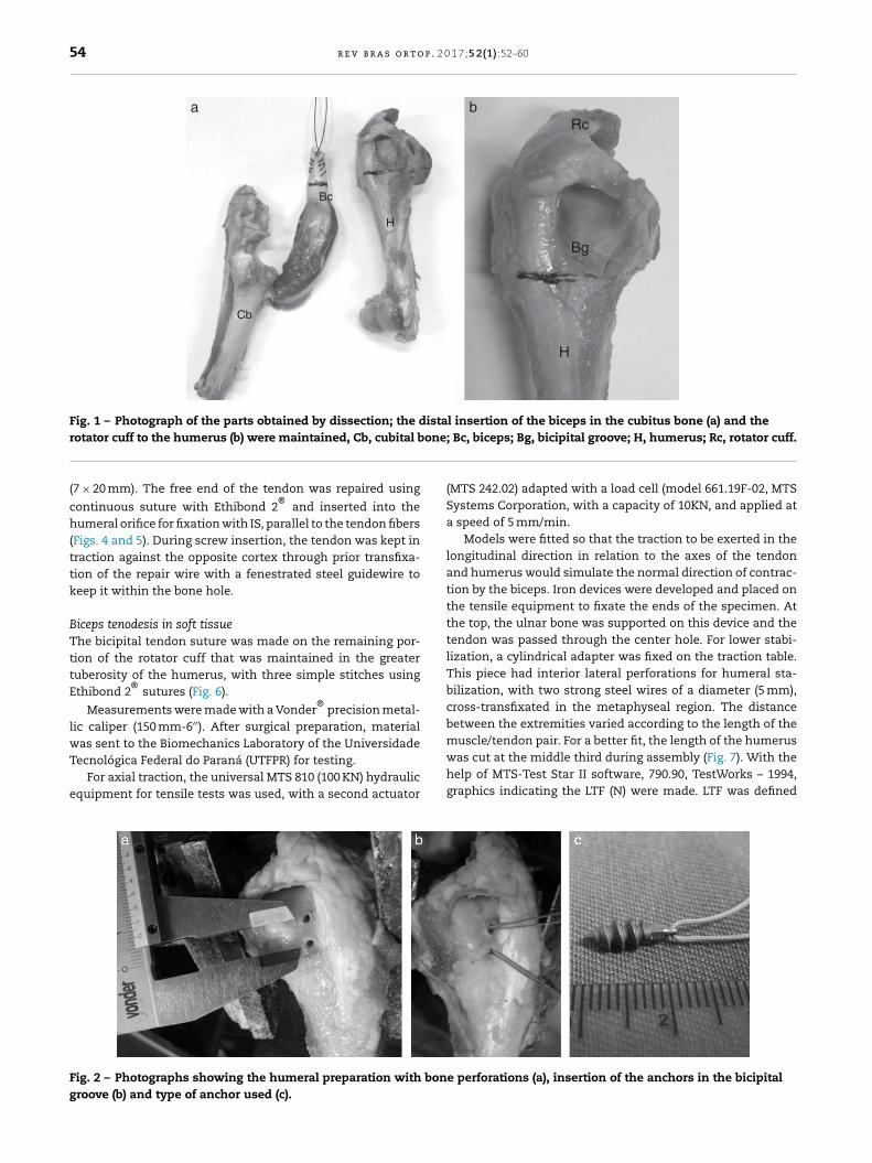
s the force required to break the fixation (system failure),btained with peak of the curve, represented by the verticalxis of the graph. The system was considered to have failedhen it lost tensile strength, whether by tendon slip or loosen-
ng or rupture of the fixation, even in cases without completeeparation of the tendon-bone system. The dislocation untilailure was represented by the horizontal axis. Resistance wasalculated by dividing the LTF by the dislocation (Fig. 8).
The results were statistically analyzed with Kaplan–Meieron-parametric estimation of the survival function. The 95%onfidence intervals for the probability of the system not fail-ng until a certain force (LTF and resistance) were compared forvery method. For quantitative evidence, the logrank signifi-
ance test was used and the p-value was calculated (significantt p < 0.05).H
Bc
ba
ig. 4 – Photographs showing the fixation technique with interfeb) and type of screw (c) Bc, biceps; H, humerus; Arrow, humeral
Bg, bicipital groove; Is, interference screw; H, humerus.
The tests were done with Microsoft®
Excel XP and Origin6.1 Pro
®programs. For the statistical analysis and adjustment
of the reliability/survival models, the statistical software R,which is open source, was used.
Results
The LTF and resistance results for the three methods, pre-sented as maximums, minimums, and means, are describedin Tables 1 and 2. The central data values, dispersion, andpossible outliers are shown in box-plot graphs for the samevariables (Fig. 9). The mean LTF for the three methods was:95 ± 35.3 N (range 50–156 N) for bone anchors; 52.7 ± 152.7 N
the junction of the suture in the tendon (nine tests), with fibertear in the longitudinal direction without release of the bone
c
rence screw: humeral perforation (arrow) (a), tendon repairdrilling.
56 r e v b r a s o r t o p . 2 0 1 7;5 2(1):52–60
Rc Rc
BcBc
H
H
a b
Fig. 6 – Drawing (a) and specimen photograph (b) showingthe fixation technique in soft tissue. Bc, biceps; Rc, rotator
200.00
150.00
100.00
50.00
0.0036 000 41 000 46 000
Peak force
Actuator2 (mm)
Dislocation (mm)
Tra
ctio
n fo
rce
51 000 56 000
Fig. 8 – Values recorded for peak force and displacement
cuff; H, humerus.
anchor(s) (Fig. 10). In one test, a suture at the distal anchorfailed, and a tendon rupture was observed in the suture. Inthe tests with IS, failure occurred mainly due to tendon slip(eight tests), and there were no cases of implant loosening(Fig. 11). In two cases, the failure occurred due to a rupturein the myotendinous junction, without loosening of fixation.All tests with soft tissue fixation presented failure by tendon
slip; the sutures in the biceps and rotator cuff fibers remainedintact. The Kaplan–Meier curves and the logrank p-valuesapplied to the three methods studied demonstrated that thefixation with IS is significantly different (p < 0.05) from theBc
H
a b
Fig. 7 – Photos showing the final assembly of the model (a), humdirection/axis of the traction (arrows) (c) Bc, biceps; H, humerus;
until system failure.
other methods, presenting higher LTF when compared withfixation with bone anchors (p = 0.00307) and soft tissue suture(p = 0.00473). IS resistance was also superior to the other twomethods (p = 0.0000127 and p = 0.00000295, respectively). Boneanchors and soft tissue suture did not differ significantly forboth LTF (p = 0.9420) and resistance (p = 0.141).
Discussion
Tenodesis of the long head of the biceps has been preferably
performed by arthroscopy, and safe fixation must ensure earlyreturn to postoperative mobilization without release of thetendon.2 Among the most commonly used fixation methods(soft tissue suture, bone fixation with bone anchors, and IS)c
eral fixation with transfixed steel wires (b) andArrows, direction of traction.

r e v b r a s o r t o p . 2 0 1 7;5 2(1):52–60 57
4
Bone anchors Interference screws
Res
ista
nce
(N /
mm
)
Soft tissue
6
8
10
12
14
Fig. 9 – Central data values, dispersion, and possibleoutliers of the long head of the biceps strength aftertenodesis by bone anchors, interference screw, and softtissue fixation without implant (n = 30).
Bc
H
Fig. 10 – Specimen photograph showing suture slip in thetendon due to tear in the test with bone anchors (arrow).Bc, biceps; H, humerus; Arrow, tendon tear.
Table 1 – Load-to-failure of the tendon of the long headof the biceps after tenodesis with bone anchors,interference screw, and soft parts suture withoutimplant (n = 30).
Boneanchors
N
ISN
Soft tissueN
Mean 95 152.7 104.7Maximum 15 212 145Minimum 50 57 75.9SD 35.3 52.7 23.5
SD, standard deviation; n, number of tests; N, Newton; IS, interfer-ence screw.
Bc
H
Fig. 11 – Specimen photograph showing the slip of thetendon fixated with interference screw (arrow). Bc, biceps;H, humerus; Arrow, tendon slip.
Table 2 – Resistance of the tendon of the long head of thebiceps after tenodesis with bone anchors, interferencescrew, and soft parts suture without implant (n = 30).
Boneanchors
N/mm
ISN/mm
Soft tissueN/mm
Mean 4.7 9.9 4.1Maximum 7.7 13.9 5.6Minimum 2.8 6.4 3.2SD 1.5 2.3 0.6
SD, standard deviation; n, number of tests; N, Newton/mm; IS, inter-ference screw.
there is no consensus on which offers greater resistance. Moststudies have compared the resistance between bone anchorsand IS; there is less information regarding soft tissue suture.No studies that compared the three types were retrieved inthe literature. Nevertheless, according to the literature, themean LTF for bone anchors is 188 N, ranging from 68.5 N to310 N. For IS, the mean is 241 N (159–480 N) and for soft tis-sue fixation, the mean is 179 N (142–216 N).9–24 In the presentstudy, these values were 95 N, 152.7 N, and 104.7 N, respec-tively; when compared separately with the literature, thesevalues were lower for the three methods. This difference canbe attributed to the different methodologies adopted, or to fac-tors such as types of specimens (human cadavers, sheep, pigs),bone density, nature of the implants (metal, bioabsorbable),different types of sutures and bone anchors, frequency in thedisplacement of traction throughout the tendon, or surgicaltechnique. For bone anchors and soft tissue sutures, simple
sutures were made. U-type or loop sutures can modify theresistance to the test.22,25 Alterations can also occur with useof one or two anchors, as two-implant models are generally
p . 2 0
fixation with bone anchors and/or soft tissue fixation, failure
58 r e v b r a s o r t o
more resistant.14 Anchors prepared with double wire havealso showed greater resistance.21 Another factor is the differ-ence observed between cyclical or continuous tensile tests.The present study followed a similar model to that describedby Bradbury et al.,18 who tested biceps resistance with con-tinuous traction and justified that fixation failures wouldoccur postoperatively in a single, sudden flexion or supinationmovement of the forearm, unlike the plastic deformation sim-ulated by cyclic testing. Nonetheless, those authors suggestedthat cyclic tests should be used for comparing techniques, dueto the possibility of variation. This can be considered one ofthe limitations of the present study. Although some authorsincluded other techniques in their studies, some comparisonsbetween IS and bone anchors were retrieved in the litera-ture. Most showed a statistically significant difference. Thissuggests that fixation with IS provides greater resistance com-pared with bone anchors.10,12,15,16,22 Other studies observedno statistical difference between these techniques, despitethe fact that the LTF was always higher for fixation withIS.11,14,19–21,23 Regarding soft tissue fixation, two biomechani-cal studies that compared bone anchors with IS were retrievedin the literature, and no statistical difference in resistancewas observed in both studies. Nevertheless, in the secondstudy, the soft tissue suture was made in the tendon of thepectoralis major muscle with more than two stitches, whichmay explain the higher LTF when compared with IS.17,24 Thepresent study demonstrated that IS fixation is more resistantand presents higher LTF than the other two methods. Themain mechanism of failure in the IS method in the presentstudy was tendon slip without screw loosening, observed in80% of the tests. In only two situations was the system resis-tance higher, with failure at the myotendinous junction. Thissituation is similar to the literature, which indicates that fail-ures in the implant-bone system are rare.26 In the methodswith bone anchors and soft tissue, failure occurred mainly inthe tendon-suture junction, without wire rupture or implantrelease. Failures occurred in the longitudinal direction of thetendon fibers and of the tensile forces, which produced tear.Lopez-Vidriero et al.17 observed the same effect and concludedthat the quality of the tendon is important for these two typesof fixation. Another possible failure is due to a wire ruptureat the junction with the anchor; other authors indicated thatthese represent the majority of cases. In these, failure mayoccur due to lower resistance and quality of the suture or dueto the quality of the anchor, as irregularities in the wire pas-sage hole would cause greater friction and would make thewire fragile.10,12,27,28 For practical comparisons regarding thesafety of tenodesis, the observations by Jazrawi et al. should beadded29: those authors defined 52 N as the mean force exertedon the biceps tendon to keep the arm in flexion without resis-tance, and 110 N as the force to sustain 1 kg. When performingtenotomy without fixation, the possibility of retraction of thetendon is high, as demonstrated by Wolf et al. after mean trac-tion of only 110 N caused migration.13 Even with the articularportion of the thickened tendon (the common degenerativeprocess), failure occurs on average after traction of 33 N.30
Attempts to increase resistance with preservation of the supe-rior portion of the labrum at the biceps during tenotomy werenot shown to be safe, with medium strength of only 73 N.18
1 7;5 2(1):52–60
Therefore, tenodesis should be performed when the objec-tive is to prevent retraction of the tendon. With this data,even with the lower resistance values observed in the presentstudy when compared with the literature, the authors sug-gest that all three techniques would be safe for early activepostoperative mobilization of the upper limb, provided thatthis mobilization is made no weight or resistance. Never-theless, significant fluctuations were observed; in one studywith bone anchors, fixation failed at 50 N traction, and inone study with IS, failed at 57 N. Fixation with IS, despitedemonstrating greater mean resistance, presented a higherstandard deviation with greater variation of high and lowvalues. Perhaps this technique is more susceptible to errorsunder greater influence of factors such as variations in theratio of screw diameter/tendon and/or bone hole, as well asbone and/or tendon quality. Other factors to be considered arevariations that can occur in surgical procedures, including sur-gical technique and surgeon’s experience. This could justifythe oscillations; however, in the present study’s methodol-ogy, these variables were carefully kept equal. Brand et al.31
demonstrated that bone density may interfere with the resis-tance of IS fixation. This factor should be taken into if thefixation is made at the supra or sub-pectoral level, especiallyif performed in patients with osteoporosis. Moreover, vari-able diameters of the tendon, of the screw, and of the bonedrilling, as well as the nature of the implant (metallic orbioabsorbable), do not appear to interfere with the ultimateresistance.32,33 The techniques with bone anchors and softtissues, albeit offering less resistance, were more constant.Other studies are needed to explain this finding. Some consid-erations can be made about the use of the three techniques:in elderly patients or patients with osteoporosis, fixation ofLHBBT in the soft tissues may be more resistant than boneanchors and IS, which depend on good bone quality. Simi-larly, soft tissue suture should perhaps be avoided in situationsin which tendons are affected by degeneration, thus present-ing poor quality. Young patients with good bone quality andhigher functional demands would be favorable candidatesfor the use of implants. This profile includes patients whoneed greater security in the fixation, cases in which IS ispreferred to bone anchors. If the cost factor is relevant, softtissue fixation could be justified if tendon quality is favor-able.
Conclusion
The LHBBT fixation method with IS is more resistant thanfixation with bone anchors and soft tissue technique. Nostatistically significant difference in resistance was observedbetween the latter two methods. The fixation method with ISpresents a significantly higher LTF when compared with boneanchors and soft tissues methods. No statistically significantdifference in LTF was observed between the latter two meth-ods. Main failure mechanism of IS fixation is tendon slip. In
occurs predominantly due to tendon rupture. No failure dueto implant loosening was observed in the IS and bone anchormethods.
. 2 0 1
C
T
A
TgBRpF
r
1
1
1
1
1
1
1
1
1
1
2
2
2
2
2
2
2
2
2
2
3
r e v b r a s o r t o p
onflicts of interest
he authors declare no conflicts of interest.
cknowledgements
o Professor Mauro Albano and Edmar Stieven Filho for theuidance provided during this study. To Professor Dr. Pauloorges and to the mechanical engineering undergraduateoberto Luis de Assumpcão for technical support in the testserformed in the laboratory of the Universidade Tecnológicaederal do Paraná.
e f e r e n c e s
1. Checchia SL, Doneux PS, Miyazaki AN, Silva LA, Fregoneze M,Ossada A, et al. Biceps tenodesis associated with arthroscopicrepair of rotator cuff tears. J Shoulder Elbow Surg.2005;14(2):138–44.
2. Checchia SL, Santos PD, Miyazaki AN, Fregoneze M, Silva LA,Leite FSF, et al. Avaliacão dos resultados da tenodeseartroscópica do bíceps, utilizando-se parafuso deinterferência bioabsorvível. Rev Bras Ortop. 2007;42(8):237–43.
3. Gurnani N, van Deurzen DF, Janmaat VT, van den BekeromMP. Tenotomy or tenodesis for pathology of the long head ofthe biceps brachii: a systematic review and meta-analysis.Knee Surg Sports Traumatol Arthrosc. 2016;24(12):3765–71.
4. Boileau P, Krishnan SG, Coste JS, Walch G. Arthroscopic bicepstenodesis: a new technique using bioabsorbable interferencescrew fixation. Arthroscopy. 2002;18(9):1002–12.
5. Gartsman GM, Hammerman SM. Arthroscopic bicepstenodesis: operative technique. Arthroscopy. 2000;16(5):550–2.
6. Sekiya JK, Elkousy HA, Rodosky MW. Arthroscopic bicepstenodesis using the percutaneous intra-articular transtendontechnique. Arthroscopy. 2003;19(10):1137–41.
7. Gumina S, Carbone S, Perugia D, Perugia L, Postacchini F.Rupture of the long head biceps tendon treated withtenodesis to the coracoid process. Results at more than 30years. Int Orthop. 2011;35(5):713–6.
8. Lo IK, Burkhart SS. Arthroscopic biceps tenodesis using abioabsorbable interference screw. Arthroscopy.2004;20(1):85–95.
9. Jayamoorthy T, Field JR, Costi JJ, Martin DK, Stanley RM, HearnTC. Biceps tenodesis: a biomechanical study of fixationmethods. J Shoulder Elbow Surg. 2004;13(2):160–4.
0. Ozalay M, Akpinar S, Karaeminogullari O, Balcik C, Tasci A,Tandogan RN, et al. Mechanical strength of four differentbiceps tenodesis techniques. Arthroscopy. 2005;21(8):992–8.
1. Mazzocca AD, Bicos J, Santangelo S, Romeo AA, Arciero RA.The biomechanical evaluation of four fixation techniques forproximal biceps tenodesis. Arthroscopy. 2005;21(11):1296–306.
2. Richards DP, Burkhart SS. A biomechanical analysis of twobiceps tenodesis fixation techniques. Arthroscopy.2005;21(7):861–6.
3. Wolf RS, Zheng N, Weichel D. Long head biceps tenotomyversus tenodesis: a cadaveric biomechanical analysis.Arthroscopy. 2005;21(2):182–5.
4. Kilicoglu O, Koyuncu O, Demirhan M, Esenyel CZ, Atalar AC,
Ozsoy S, et al. Time-dependent changes in failure loads of 3biceps tenodesis techniques: in vivo study in a sheep model.Am J Sports Med. 2005;33(10):1536–44.3
7;5 2(1):52–60 59
5. Kusma M, Dienst M, Eckert J, Steimer O, Kohn D. Tenodesis ofthe long head of biceps brachii: cyclic testing of five methodsof fixation in a porcine model. J Shoulder Elbow Surg.2008;17(6):967–73.
6. Golish SR, Caldwell PE 3rd, Miller MD, Singanamala N,Ranawat AS, Treme G, et al. Interference screw versus sutureanchor fixation for subpectoral tenodesis of the proximalbiceps tendon: a cadaveric study. Arthroscopy.2008;24(10):1103–8.
7. Lopez-Vidriero E, Costic RS, Fu FH, Rodosky MW.Biomechanical evaluation of 2 arthroscopic biceps tenodeses:double-anchor versus percutaneous intra-articulartranstendon (PITT) techniques. Am J Sports Med.2010;38(1):146–52.
8. Bradbury T, Dunn WR, Kuhn JE. Preventing the Popeyedeformity after release of the long head of the biceps tendon:an alternative technique and biomechanical evaluation.Arthroscopy. 2008;24(10):1099–102.
9. Patzer T, Santo G, Olender GD, Wellmann M, Hurschler C,Schofer MD. Suprapectoral or subpectoral position for bicepstenodesis: biomechanical comparison of four differenttechniques in both positions. J Shoulder Elbow Surg.2012;21(1):116–25.
0. Tashjian RZ, Henninger HB. Biomechanical evaluation ofsubpectoral biceps tenodesis: dual suture anchor versusinterference screw fixation. J Shoulder Elbow Surg.2013;22(10):1408–12.
1. Baleani M, Francesconi D, Zani L, Giannini S, Snyder SJ.Suprapectoral biceps a biomechanical comparison of a newsoft anchor tenodesis technique versus interference screwbiceps tendon fixation. Clin Biomech (Bristol, Avon).2015;30(2):188–94.
2. Patzer T, Rundic JM, Bobrowitsch E, Olender GD, Hurschler C,Schofer MD. Biomechanical comparison of arthroscopicallyperformable techniques for suprapectoral biceps tenodesis.Arthroscopy. 2011;27(8):1036–47.
3. Papp DF, Skelley NW, Sutter EG, Ji JH, Wierks CH, Belkoff SM,et al. Biomechanical evaluation of open suture anchorfixation versus interference screw for biceps tenodesis.Orthopedics. 2011;34(7):e275–8.
4. Ahmed M, Young BT, Bledsoe G, Cutuk A, Kaar SG.Biomechanical comparison of long head of biceps tenodesiswith interference screw and biceps sling soft tissuetechniques. Arthroscopy. 2013;29(7):1157–63.
5. Lafosse L, Van Raebroeckx A, Brzoska R. A new technique toimprove tissue grip: the lasso-loop stitch. Arthroscopy.2006;22(11):1246.e1–3.
6. Koch BS, Burks RT. Failure of biceps tenodesis withinterference screw fixation. Arthroscopy. 2012;28(5):735–40.
7. Meyer DC, Nyffeler RW, Fucentese SF, Gerber C. Failure ofsuture material at suture anchor eyelets. Arthroscopy.2002;18(9):1013–9.
8. Deakin M, Stubbs D, Bruce W, Goldberg J, Gillies RM, WalshWR. Suture strength and angle of load application in a sutureanchor eyelet. Arthroscopy. 2005;21(12):1447–51.
9. Jazrawi LM, Rokito AS, Birdzell MG, Zuckerman JD.Biomechanics of the elbow. In: Nordin M, Frankel VH, editors.Basic biomechanics of the musculoskeletal system.Philadelphia: Lippincott Williams & Wilkins; 2001.p. 340–57.
0. Ahmad CS, DiSipio C, Lester J, Gardner TR, Levine WN,Bigliani LU. Factors affecting dropped biceps deformity aftertenotomy of the long head of the biceps tendon. Arthroscopy.2007;23(5):537–41.
1. Brand JC Jr, Pienkowski D, Steenlage E, Hamilton D, JohnsonDL, Caborn DN. Interference screw fixation strength of aquadrupled hamstring tendon graft is directly related to bone

p . 2 0
3
3DL. Quadrupled semitendinosus-gracilis autograft fixation in
60 r e v b r a s o r t o
mineral density and insertion torque. Am J Sports Med.2000;28(5):705–10.
2. Slabaugh MA, Frank RM, Van Thiel GS, Bell RM, Wang VM,Trenhaile S, et al. Biceps tenodesis with interference screwfixation: a biomechanical comparison of screw length anddiameter. Arthroscopy. 2011;27(2):161–6.
1 7;5 2(1):52–60
3. Caborn DN, Coen M, Neef R, Hamilton D, Nyland J, Johnson
the femoral tunnel: a comparison between a metal and abioabsorbable interference screw. Arthroscopy.1998;14(3):241–5.