Review - SciELO
40
Review J. Braz. Chem. Soc., Vol. 27, No. 3, 435-474, 2016. Printed in Brazil - ©2016 Sociedade Brasileira de Química 0103 - 5053 $6.00+0.00 http://dx.doi.org/10.5935/0103-5053.20150332 *e-mail: [email protected], [email protected] Antinociceptive Effect of Essential Oils and Their Constituents: an Update Review Eder J. Lenardão,* ,a Lucielli Savegnago,* ,b Raquel G. Jacob, a Francine N. Victoria a and Débora M. Martinez a a Laboratório de Síntese Orgânica Limpa, LASOL, CCQFA and b Grupo de Pesquisa em Neurobiotecnologia, GPN, CDTec, Universidade Federal de Pelotas, UFPel, CP 354, 96020‑250 Pelotas‑RS, Brazil Plants and essential oils (EOs) have been used for centuries in folk medicine to treat diverse disorders, including analgesic to pain relief. In this context, the antinociceptive activity of EOs has been attracted attention since the management of pain continues being a major challenge for medicine. This review provides an overview of published reports on the antinociceptive activity of EOs and their constituents from 2000 until the first half of 2015. In this review are compiled the data on the antinociceptive activity of 63 EOs and 26 of their constituents with a discussion about the nociception model used to access the analgesic effect. These data were also analyzed in relation to ethnopharmacological and toxicological data available in the literature. As can be seen by the analysis of more than 300 articles, EOs and their constituents show antinociceptive effects in different models and their action mechanism is quite variable. Although there are a few essential oils or their isolated constituents on the phytopharmaceuticals market, this review intends to put in evidence the often-underexploited vast source of natural compounds with therapeutic potential in pain relief. Keywords: essential oil, antinociceptive, analgesic, natural compound, pain 1. Introduction Over the past few decades, strategies of drug discovery have been generally focused on an approach based on single targets along with the rapid growth in genetics and molecular biology. Medicinal plants have been used in developing countries as alternative treatments to health problems. 1 Many plant extracts and essential oils (EOs) isolated from plants have shown in vitro and in vivo biological activities, which have inspired intense research on their use in traditional medicine. 2 Natural products are fundamental to pain treatment. They yield new analgesics and play an important role in the study of pain mechanisms. 3 Historically, the majority of new drugs has been directly produced from natural products (secondary metabolites) or from semi-synthetic compounds. 4,5 In recent years, an increasing number of studies have demonstrated that natural products from folk remedies have contributed significantly to the discovery of modern drugs worldwide. 6-8 A successful example of a drug obtained from natural product is morphine, an opioid drug extracted from the plant Papaver somniferum. 9 In an excellent survey, 210 medicinal plants, involving 79 families, were summarized about their analgesic effects by Almeida et al. 6 More recently, Yunes et al. 8 revised the development of analgesic drugs from glycosides, alkaloids, flavonoids and terpenes. Plant EOs are complex mixtures of volatile compounds, which are isolated by physical methods (pressing and distillation) from a whole plant or plant parts (leaf, bark, fruits, flowers, etc.). The major components of the EOs are derived from only three biosynthetic pathways: (i) the methylerythritol phosphate (mep) pathway, which leads to mono- and diterpenes; (ii) the mevalonate pathway, leading to sesquiterpenes; and (iii) the shikimate pathway to phenylpropenes. 10 The identity and the relative quantity of these volatile substances in the EO is quite variable and they have various ecological roles in the plant, including the attraction of pollinating insects, as internal messengers and as a protective substance against herbivores. 11 The pharmacological effects of EOs and their major constituents have been focused by many research groups in the last years. 12-20 There are in the literature several excelent reviews dedicated to the pharmacological activities of these
-
Upload
khangminh22 -
Category
Documents
-
view
4 -
download
0
Transcript of Review - SciELO
ReviewJ. Braz. Chem. Soc., Vol. 27, No. 3, 435-474, 2016.
Printed in Brazil - ©2016 Sociedade Brasileira de Química0103 - 5053 $6.00+0.00
http://dx.doi.org/10.5935/0103-5053.20150332
*e-mail: [email protected], [email protected]
Antinociceptive Effect of Essential Oils and Their Constituents: an Update Review
Eder J. Lenardão,*,a Lucielli Savegnago,*,b Raquel G. Jacob,a Francine N. Victoriaa and Débora M. Martineza
aLaboratório de Síntese Orgânica Limpa, LASOL, CCQFA and bGrupo de Pesquisa em Neurobiotecnologia, GPN, CDTec, Universidade Federal de Pelotas, UFPel, CP 354,
96020‑250 Pelotas‑RS, Brazil
Plants and essential oils (EOs) have been used for centuries in folk medicine to treat diverse disorders, including analgesic to pain relief. In this context, the antinociceptive activity of EOs has been attracted attention since the management of pain continues being a major challenge for medicine. This review provides an overview of published reports on the antinociceptive activity of EOs and their constituents from 2000 until the first half of 2015. In this review are compiled the data on the antinociceptive activity of 63 EOs and 26 of their constituents with a discussion about the nociception model used to access the analgesic effect. These data were also analyzed in relation to ethnopharmacological and toxicological data available in the literature. As can be seen by the analysis of more than 300 articles, EOs and their constituents show antinociceptive effects in different models and their action mechanism is quite variable. Although there are a few essential oils or their isolated constituents on the phytopharmaceuticals market, this review intends to put in evidence the often-underexploited vast source of natural compounds with therapeutic potential in pain relief.
Keywords: essential oil, antinociceptive, analgesic, natural compound, pain
1. Introduction
Over the past few decades, strategies of drug discovery have been generally focused on an approach based on single targets along with the rapid growth in genetics and molecular biology. Medicinal plants have been used in developing countries as alternative treatments to health problems.1 Many plant extracts and essential oils (EOs) isolated from plants have shown in vitro and in vivo biological activities, which have inspired intense research on their use in traditional medicine.2
Natural products are fundamental to pain treatment. They yield new analgesics and play an important role in the study of pain mechanisms.3 Historically, the majority of new drugs has been directly produced from natural products (secondary metabolites) or from semi-synthetic compounds.4,5 In recent years, an increasing number of studies have demonstrated that natural products from folk remedies have contributed significantly to the discovery of modern drugs worldwide.6-8 A successful example of a drug obtained from natural product is morphine, an opioid
drug extracted from the plant Papaver somniferum.9 In an excellent survey, 210 medicinal plants, involving 79 families, were summarized about their analgesic effects by Almeida et al.6 More recently, Yunes et al.8 revised the development of analgesic drugs from glycosides, alkaloids, flavonoids and terpenes.
Plant EOs are complex mixtures of volatile compounds, which are isolated by physical methods (pressing and distillation) from a whole plant or plant parts (leaf, bark, fruits, flowers, etc.). The major components of the EOs are derived from only three biosynthetic pathways: (i) the methylerythritol phosphate (mep) pathway, which leads to mono- and diterpenes; (ii) the mevalonate pathway, leading to sesquiterpenes; and (iii) the shikimate pathway to phenylpropenes.10 The identity and the relative quantity of these volatile substances in the EO is quite variable and they have various ecological roles in the plant, including the attraction of pollinating insects, as internal messengers and as a protective substance against herbivores.11
The pharmacological effects of EOs and their major constituents have been focused by many research groups in the last years.12-20 There are in the literature several excelent reviews dedicated to the pharmacological activities of these

Antinociceptive Effect of Essential Oils and Their Constituents: an Update Review J. Braz. Chem. Soc.436
natural occurring compounds, including antinociceptive,16,17 antioxidant,18 anti-cancer19 and anti-inflamatory activities.20
Owing to the new attraction for natural products like EOs and their major constituents, despite their wide use as analgesic in folk medicine, it is important to develop a better understanding of their antinociceptive action for new applications in human health. In Brazil, an example of successful application is the EO obtained from Cordia verbenacea (Boraginaceae), an anti-inflammatory medicine for topical use (Acheflan®).21 A very interesting review covering patents involving the use of terpenes and EOs like pain relievers was recently published by Guimarães et al.22 In that comprehensive review, 17 patents were critically analyzed and it was showed that, despite the large and intensive research in academy on natural products-based drug-discovery, plant EOs and their constituents potential as analgesic drugs remains underexplored.
The high volatility, low stability, along with the hydrophobicity of most EOs and their major consituents are among the reasons of the discrepancy between the high number of articles on their in vivo and in vitro activities and the restrict number of commercial products using these natural compounds.22 This picture started to change in the last years, since new approaches have been developed aiming to increase the therapeutic properties of EOs and their components, notedly the use of drug-delivery structures, like β-cyclodextrins (β-CD).23-25 It was observed that the inclusion of EOs with β-CD protects the EO against oxidation, heat and light degradation and reduces losses due evaporation and moisture.23 In this line, it is worth noting the work from Quintans and co-workers25 which obtained excellent outcomes using β-CD/EO complex in studies on the antinociceptive activity of several EOs and also some of their constituents. These studies opened a new perspective on the use of EOs and other volatile compounds as antinociceptive agents, improving the stability and on water solubility, reducing the volatility and faciliting the handle of the EO and individual components.24,25
Based on these considerations, the goal of this paper is to provide an overview of the published data on the antinociceptive activity of EOs and their constituents compiling the published data from 2000 to the first half of 2015. We searched Scopus, Medline, DOAJ, Web of Science and SciFinder and the search terms were relevant to the review subject being limited to the use of EOs and/or their isolated constituents, mainly terpenoids, in pain relief.
Studies involving the use of aqueous or ethanolic extracts, as well as semi-synthetic compounds will not be discussed in this review. Similarly, those papers describing preliminary screening, using non-specific tests, such as acetic acid-induced writhings alone, with no additional
evidences on the antinociceptive effect of the EO or their constituents, were not included.
The first part of this review is dedicated to the description of the concepts involved in the nociception, followed by the major clinical treatments used to treat pain and the main tests used to evaluate the antinociceptive activity; after that, the antinociceptive effect of EOs and their constituents is presented, in order to make the reading more understanding through the text.
2. Nociception
Nociceptive pain comprises the processes of transduction, conduction, transmission and perception. The nociceptive signaling in physiological pain is initiated by activation of the specialized pain receptors (nociceptors), which are polymodal sensory fibers of the primary sensory neurons located in trigeminal and dorsal root ganglia with unmyelinated (C-fiber) or thinly myelinated (Aδ-fiber) axons. The conversion of a noxious thermal, mechanical, or chemical stimulus into electrical activity in the peripheral terminals of nociceptor sensory fibers consists in the transduction phase. This process is mediated by specific receptor ion channels, expressed only by nociceptors.26
The conduction phase consists in the passage of action potentials from the peripheral terminal along axons to the central terminal of nociceptors in the central nervous system, along unmyelinated (C-fibers), slow conducting and more rapidly conducting primary sensory ones. This sensory inflow then activates secondary sensory neurons in the dorsal horn of the spinal cord through synaptic transfer, in the transmission phase, which project to the cortex via a relay in the thalamus. Therefore, the transmission is the synaptic transfer and modulation of input from one neuron to another.26,27
The dorsal horn of the spinal cord is the site where the primary afferent fibers synapse with second-order neurons. It is also, where complex interactions occur between excitatory and inhibitory interneuron and where descending inhibitory tracts from higher centers exert their effect.3 Large fiber inputs from other sensory modalities and descending pathways can modulate the activity in the dorsal horn (Figure 1).28
Figure 1. The pain pathway.

Lenardão et al. 437Vol. 27, No. 3, 2016
The multiplicity of events that occur during pain transmission in both, the peripheral and central nervous systems are arising from the direct or indirect action of chemical mediators. These include arachidonic acid metabolites (prostaglandins and leukotrienes), peptides (kinins, tachykinins, calcitonin gene related peptide, galanin, cholecystokinin, vasoactive intestinal peptide), serotonin, acetylcholine, cytokines, nerve growth factor, glutamate, nitric oxide, adenosine triphosphate (ATP), adenosine diphosphate (ADP), adenosine and protons, among others. These mediators can be produced or released following tissue injury or by exogenous irritants (formalin, acetic acid, capsaicin, etc.).29-31
These mediators can act via a multiplicity of receptors that are widely distributed through central and peripheral nerves, many of which are coupled to heterotrimeric G-proteins and associated with the formation of multiple second messengers, such as protein kinases A, C and G, cAMP, cGMP and mobilization of intracellular calcium. Other neurotransmitters, such as excitatory amino acids and acetylcholine (acting at the nicotinic receptor), directly activate ion channels, and in turn control the membrane ion permeability.32,33 Several factors, including physical damage to tissues, exposure to some inflammatory mediators, such as prostaglandin E2, bradykinin, substance P, histamine, adenosine and serotonin are known to cause sensitization of nerve ending nociceptors to mechanical and thermal stimuli (Figure 2).29-31
3. Clinical Treatments
The drugs most often used to treat pain and inflammation are the non-steroidal anti-inflammatory (NSAIDs) and opioids, despite their well-known adverse effects.34,35
Opioid analgesics are used for the treatment of moderate to severe acute pain and currently are the most effective and frequently used drugs in patients with refractory malignant and non-malignant pain. Opioids include the broad category of compounds that are agonists of opioid receptors and that elicit actions typical of morphine.36
The term “opiate” typically includes the opioid alkaloids derived from the opium poppy seeds and includes the opium resin, codeine and morphine. The term opioid includes natural and semi-synthetic opiates (such as hydrocodone and oxycodone) and synthetic opioids (such as fentanyl and methadone).36 Opioids can alter the central pain-related systems, resulting in opioid tolerance (a decrease in the analgesic effect of opioids), dependence (a behavioral state requiring continued use of opioids to avoid a series of aversive withdrawal syndromes) and withdrawal syndrome, which are the most predominant behavioral consequence of long-term usage of opioids; other effects are pruritus, nausea, slowing of gastrointestinal (GI) function, urinary retention and sexual dysfunction.37,38 Therefore, repeated use of opiate analgesic drugs like morphine for the relief of chronic pain may result in the development of opiate tolerance and dependence. A consequence of these effects is a narrowing of the drug’s therapeutic index, increase of side effects39 and a significant hampering of the effective treatment of chronic pain with opioid analgesics.40,41
The term NSAIDs (non-steroidal anti-inflammatory drugs) is used to refer to both, non-selective (nsNSAIDs) and cyclo-oxygenase COX-2 selective inhibitors (COXIBs). NSAIDs have a spectrum of analgesic, anti-inflammatory and antipyretic effects and are effective analgesics in a variety of acute pain states.42 NSAIDs are a disparate group of weakly acidic, highly protein-bounding compounds having the common pharmacological property of inhibiting
Figure 2. Events that occur during pain transmission in both the peripheral and central nervous systems.

Antinociceptive Effect of Essential Oils and Their Constituents: an Update Review J. Braz. Chem. Soc.438
prostaglandin biosynthesis. It was demonstrated that the acid moiety of these molecules can be extensively accumulated in inflamed tissues, where the NSAIDs exert their effects.43,44
Despite the diverse chemical structures of NSAIDs, the analgesic effect of these drugs is mainly due to their common property of inhibiting cyclo-oxygenases (COX) involved in the formation of prostaglandins, which are formed by the conversion of arachidonic acid.45 There are two isoforms of COX: COX-1 (expressed in most tissues) and COX-2 (expressed in kidney, central nervous system, and cardiovascular system-endothelium and induced in response to inflammatory stimuli), both are formed from arachidonic acid (Figure 3). The two COX isoforms share 60% homology in their amino acids sequence and are both integral membrane homodimer proteins of the endoplasmic reticulum and nucleus, with roughly comparable kinetics. However, they differ in their regulatory mechanisms, cell localization, and function. A third isoform (COX-3 or COX-1b) was first described in canine as a splice variant of COX-1 gene, but its physiological role at this point remains unknown.46
COX-1 isoform is expressed in most tissues, producing prostaglandins that play an important protective role in the gut by stimulating the synthesis and secretion of mucus and bicarbonate, increasing mucosal blood flow and promoting epithelial proliferation. When NSAIDs inhibit this enzyme, they create a gastric environment that is more susceptible to topical attack by endogenous and exogenous factors.47
The main problem in using NSAIDs are the adverse effects, especially gastrointestinal morbidities, including complications in both, upper and lower gastrointestinal tracts, prothrombotic effects and peptic ulceration.47
COX-2 was first identified as a key element of the acute inflammatory response because its expression is rapidly induced by various inflammogens.48 COXIBs selectively inhibit the inducible cyclo-oxygenase enzyme COX-2 and they offer the potential for effective analgesia with fewer
side effects than NSAIDs. Available COXIBs include celecoxib, etoricoxib and parecoxib.49
Paracetamol (acetaminophen) is used in clinical practice and is an effective analgesic and antipyretic. Because paracetamol has fewer side effects than NSAIDs, it can be used in substitution to them in some treatments.49 One of the mechanisms of action of paracetamol appears to be linked to the serotoninergic system and it is possible that other drugs with serotoninergic effects could affect pain relief. Botting and Ayoub50 demonstrated that analgesia and hypothermia due to paracetamol are mediated by inhibition of COX 3 in the central nervous system and lowering in PGE2 levels.
Some of the agents recently used for pain relief (gabapentin, pregabalin, lamotrigine, topiramate, tramadol and venlafaxine) are believed to inhibit central sensitization by blocking the activity of glutamate, excitatory neuropeptides, and presynaptic calcium channels, while enhancing inhibitory pathways mediated by serotonin, norepinephrine, and γ-aminobutyric acid (GABA).49
The serotoninergic system has gained much attention as a therapeutic target for treating migraine pain and it is implicated in other pain conditions.51 Therapeutics targeting 5-HT receptors and 5-HT re-uptake are being examined in clinical trials for their ability to treat the pain associated with migraine51 and fibromyalgia.52 To date, the triptan class of medicines used to treat migraine and cluster headache, including the 5-HT1B/1D agonist sumatriptan, are the only 5-HT selective therapeutics that treat pain successfully in the clinic. Other centrally-acting drugs, such as venlafaxine and duloxetine, which act as dual norepinephrine and 5-HT reuptake inhibitors, have shown some efficacy in the treatment of various pain symptoms, including fibromyalgia.53
The adverse effects of NSAIDs and opioids drugs have inspired the search for safer and more effective anti-inflammatory and analgesic drugs. The current trend of research is the investigation of medicines of plant origin as a source of new chemical substances with potential therapeutic effects, because of their availability and accessibility with minimal side effects.54,55
Natural substances obtained from plants have played an extremely important role in the development of analgesic drugs and in the understanding of the complex mechanisms involved in pain transmission and pain relief.55 The first commercial pure natural product introduced for therapeutic use was morphine, marketed by Merck in 1826 and the first semi-synthetic pure drug Aspirin®, based on salicin, a glycoside obtained from the bark of Salix alba, was introduced by Bayer in 1899.4 This led to the isolation of early drugs such as cocaine, codeine, digitoxin, quinine and pilocarpine, and several other recent plant derived
Figure 3. Cyclo-oxygenases as response to inflammatory stimuli. Where: NSAIDs: non-steroidal anti-inflammatory drugs; COX: cyclo-oxygenases; COXIBs: cyclo-oxygenase-2 inhibitors.
Lenardão et al. 439Vol. 27, No. 3, 2016
compounds, which have undergone development and have been commercialized as drugs, which include Paclitaxel from Taxus brevifolia, for the treatment of lung, ovarian and breast cancers.56
4. Experimental Models to Evaluate the Antinociceptive Effect in Natural Products Research
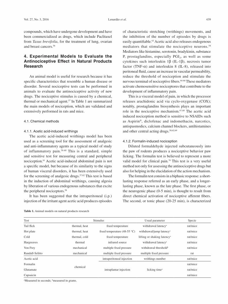
An animal model is useful for research because it has specific characteristics that resemble a human disease or disorder. Several nociceptive tests can be performed in animals to evaluate the antinociceptive activity of new drugs. The nociceptive stimulus is caused by a chemical, thermal or mechanical agent.57 In Table 1 are summarized the main models of nociception, which are validated and extensively performed in rats and mice.
4.1. Chemical methods
4.1.1. Acetic acid-induced writhingsThe acetic acid-induced writhings model has been
used as a screening tool for the assessment of analgesic and anti-inflammatory agents as a typical model of study of inflammatory pain.58-60 This is a standard, simple and sensitive test for measuring central and peripheral nociception.61 Acetic acid-induced abdominal pain is not a specific model, but because of its similarity to the signs of human visceral disorders, it has been extensively used for the screening of analgesic drugs.57,62 This test is based in the induction of abdominal writhings, causing algesia by liberation of various endogenous substances that excite the peripheral nociceptors.58
It has been suggested that the intraperitoneal (i.p.) injection of the irritant agent acetic acid produces episodes
of characteristic stretching (writhings) movements, and the inhibition of the number of episodes by drugs is easily quantifiable.63 Acetic acid also releases endogenous mediators that stimulate the nociceptive neurons.58 Mediators like histamine, serotonin, bradykinin, substance P, prostaglandins, especially PGI2, as well as some cytokines such interleukin 1β (IL-1β), necrosis tumor factor (TNF-α) and interleukin 8 (IL-8), released into peritoneal fluid, cause an increase in vascular permeability, reduce the threshold of nociception and stimulate the nervous terminal of nociceptive fibers.64-66 These mediators activate chemosensitive nociceptores that contribute to the development of inflammatory pain.
This is a visceral model of pain, in which the processor releases arachidonic acid via cyclo-oxygenase (COX); notably, prostaglandins biosynthesis plays an important role in the nociceptive mechanism.67,68 The acetic acid induced nociception method is sensitive to NSAIDs such as Aspirin®, diclofenac and indomethacin, narcotics, antispasmodics, calcium channel blockers, antihistamines and other central acting drugs.58,62,69
4.1.2. Formalin-induced nociceptionDiluted formaldehyde injected subcutaneously into
the paw of rodents produces a nociceptive behavior paw licking. The formalin test is believed to represent a more valid model for clinical pain.70 This test is a very useful method not only for assessing the antinociceptive drugs but also for helping in the elucidation of the action mechanism.
The formalin test consists in a biphasic response: a short-lasting response referred as an early phase, and a longer-lasting phase, known as the late phase. The first phase, or the neurogenic phase (0-5 min), is thought to result from direct chemical activation of nociceptive afferent fibers. The second, or tonic phase (20-25 min), is characterized
Table 1. Animal models on natural products research
Test Stimulus Usual parameter Specie
Tail flick thermal, heat fixed temperature withdrawal latencya rat/mice
Hot plate thermal, heat fixed temperature (48-55 °C) withdrawal/jump latencya rat/mice
Cold thermal, cold fixed temperature lifting or shaking latencya rat/mice
Hargreaves thermal infrared source withdrawal latencya rat/mice
Von Frey mechanical multiple fixed pressure withdrawal thresholdb rat/mice
Randall-Selitto mechanical multiple fixed pressure multiple fixed pressure rat
Acetic acid
chemical
intraperitoneal injection writhings number rat/mice
Formalin
intraplantar injection licking timea
rat/mice
Glutamate rat/mice
Capsaicin rat/mice
aMeasured in seconds; bmeasured in grams.

Antinociceptive Effect of Essential Oils and Their Constituents: an Update Review J. Braz. Chem. Soc.440
by an inflammatory process triggered by a combination of stimuli, including inflammation of the peripheral tissues and mechanisms of central sensitization.70-72
The biphasic component of formalin-induced nociception reflects different underlying mechanisms: the first phase reflects centrally mediated pain with release of substance P.57,73 The second one depends of a combination of ongoing inputs from nociceptive afferents, due to the release of excitatory amino acids, PGE2, nitric oxide (NO), tachykinin, kinins, among other peptides and, at least in part, of central sensitization.74,75
Formalin activates the primary afferent sensory neurons through a specific and direct action on the transient receptor potential cation channel, member A1 (TRPA1), which is highly expressed by a subset of the C-fiber nociceptors.76 It is generally agreed that N-methyl-D-aspartate (NMDA) receptors contribute to the persistent chemical stimulus during the late phase of central sensitization of dorsal horn neurons.77
It has been shown that drugs that act mainly centrally, such as opioids and narcotics, inhibit both phases of formalin-induced pain, while drugs as Aspirin®, hydrocortisone and dexamethasone, which are primarily peripherally acting, only inhibit the late phase.78,79 Exclusive inhibition of the formalin test’s second phase is a typical characteristic of cyclo-oxygenases inhibitors.80
4.1.3. Capsaicin-induced nociceptionCapsaicin (8-methyl-N-vanillyl-6-noneamide), the
pungent active ingredient of hot chili peppers, produces painful sensations upon cutaneous application by activating transient receptor potential vanilloid receptor-1 (TRPV-1), located on peripheral terminals of nociceptors.81 The intradermic injection of capsaicin is regarded as a potentially predictive model of neuropathic pain in humans, because of its qualitative, mechanistic, and pharmacological similarity to neuropathic pain states.82-84
The capsaicin test is widely used as a model of pain in mice,85 rats86 and humans.87 The subcutaneous (s.c.) injection of capsaicin into the hind paw of mice produces a short-lasting paw-licking/biting response.88 The acute nociceptive response (flinching, licking and biting of the hind paw) occurs immediately following an intraplantar capsaicin injection and persists for about 5 min. The activation of primary afferents nociceptors by capsaicin causes the release of nociceptive transmitters, substance P and glutamate from the dorsal spinal cord in vivo and in vitro.89,90
It is believed that capsaicin activates a non-selective ionotropic channel in the C-fiber of nociceptive afferents through VRTP1 receptors.91-93 Furthermore, some studies attribute to capsaicin the release of neuropeptides,
excitatory amino acids such as glutamate and aspartate, nitric oxide and pro-inflammatory mediators in the periphery and the transmission of nociceptive information to the spinal cord.94,95
4.1.4. Glutamate-induced nociceptionGlutamate is the major excitatory amino acid
neurotransmitter present in the central nervous system, where it participates in a great diversity of biological functions, such as learning, memory, neurodegenerative diseases and neuronal death.96
The intraplantar injection of glutamate into the mouse hind paw produces nociceptive-like behaviors of rapid onset and short duration (about 15 min).85 Accumulating evidence now suggests that there is an excess of excitatory amino acids, mainly glutamate, following injury at the spinal cord or following certain inflammatory process, suggesting that excitatory amino acids might play a relevant role in sensory transmission.97-99
The nociceptive response induced by glutamate is primarily mediated by the release of neuropeptides from sensory fibers, namely neurokinins and kinins.96 In addition, glutamate is found in sensory C-fibers where it is believed to play a role in the transmission of nociceptive mechanisms at the spinal cord.100
The nociceptive response caused by glutamate involves peripheral, spinal and supraspinal sites of action. It has been reported that the glutamate injection stimulates marked nociceptive reactions, that are mediated by neuropeptides (like SP) liberated from sensory fibers. Besides, the activation of glutamate receptors like α-amino-3-hydroxy-5-methyl-4-isoxazolepropionate (AMPA), kainate and NMDA receptors, play an important role modulating this nociceptive response96,101,102 by stimulating the production of a sort of intracellular second messengers, including NO.103
There is large evidence that substances that are capable of blocking ionotropic and metabotropic glutamate receptors exhibit pronounced antinociceptive and analgesic effects in several mammalian species, including humans.104,105
4.2. Thermal methods
4.2.1. Tail flickThe tail flick is one of the oldest nociceptive tests.
The measured parameter is the latency, in seconds, for tail flick reflex following tail exposure to a heat stimulus. The stimulus may be applied by dipping the tail tip into a bath at a controlled temperature (55 oC) or by an infrared heat beam. The apparatus allows an automated detection of the tail flick and measuring of its latency. The tail flick is a spinal reflex, but it is subject to supraspinal influences that

Lenardão et al. 441Vol. 27, No. 3, 2016
can affect the reflex.106 This test is highly sensitive to opiate drugs57 and, according to Grumbach,107 the effectiveness of analgesic agents in this pain model is highly correlated with relief of human pain.
4.2.2. Hot plate The hot plate is a classic test in the field. The test
consists in placing a rodent on an enclosed hot plate and measuring the latency to lick a hind paw or jump out of the enclosure.108 The temperature is often set at 52 or 55 °C and the set up allows observing baseline latencies between 5 and 10 s for paw licking. The temperature is 10-15 °C higher than the response threshold of heat nociceptors, which reflects the time required for skin temperature to increase until detection of the nociceptive stimulus, and the delay to provoke the withdrawal response.109
The advantages of the hot plate test are that it is objective, quantifiable, can be administered repeatedly without causing inflammation, and assesses supraspinally-organized responses to a noxious stimulus. Although, good correspondence between drugs that produce antinociception on this test and drugs used clinically to treat pain were observed.110
Despite they are suitable for measuring the effects of opioid analgesics, tests based on the use of thermal stimuli, such as the hot plate and tail flick, are not sensitive to the analgesic effects of nonsteroidal anti-inflammatory agents.57,110
4.3. Mechanical methods
4.3.1. Von FreySince von Frey invented his “hairs” in the 1890s,
they have been used countless times in sensory testing and there is a vast literature in that subject.111 The von
Frey is a mechanical test that involves the application of a calibrated and graduated force to a sensory field by using filaments (the hairs); the assessed force is that at the point when the von Frey hair bends. Von Frey hairs is a subjective test, once the patient under evaluation needs to report the sensation. In animal experiments, the end point is usually a more objective parameter such as the discharge of a neuron.112
4.3.2. Randall SelittoIn this mechanical test, the effectiveness of
antinociceptive drugs is accessed by observing the response thresholds of the animal to a gradually increasing mechanical pressure on an inflamed paw.113 This method is somewhat better suited for the detection of such thresholds than the von Frey filaments, because it avoids the manual application of the force and, thus, provides a better consistency of stimuli.57
5. Antinociceptive Effect of Essential Oils
Plants are used worldwide for the treatment of diseases and novel drugs continue to be developed from plants. The number of species of plants used in traditional medicine overcomes 20,000, making them a potential source for prospecting new drugs.114
In this context, EOs have been received much attention since they present many biological activities, like antioxidant, antibacterial, antidepressant and antinociceptive ones.12-20,115 The EOs and the respective nociception model used to determine their antinociceptive activity that are discussed in this work are summarized on Table 2. The EOs are arranged according to the botanical family of the plant they were obtained and in an alphabetical order (Table 2, column 1).
Table 2. Models used to evaluate the antinociceptive effect of EOs
Plant essential oils Performed test Route of administration Tested dose / (mg kg-1) Reference
Anacardiaceae
Schinus terebinthifolius RaddiSNIa animals
mechanical sensitivity cold hyperalgesia
oral 10-100 116
Annonaceae
Duguetia lanceolata A. St.-Hil.acetic acid
formalin test (phases I and II)intraperitoneal 1-200 117,118
Xylopia laevigata (Mart.) R. E. Fr.acetic acid
formalin test (phases I and II)intraperitoneal 12.5-50 119
Apiaceae
Bunium persicum (Boiss.) B. Fedtsch.
acetic acid formalin test (phases I and II)
oral 100-400b 120
acetic acid opioidergic and
histamine H1 and H2 receptorsoral 100-400b 121

Antinociceptive Effect of Essential Oils and Their Constituents: an Update Review J. Braz. Chem. Soc.442
Plant essential oils Performed test Route of administration Tested dose / (mg kg-1) Reference
Carum copticum (L.) Benth. & Hook. f. ex C. B. Clarke
formalin test (phases I and II) intraperitoneal 20 122
Cuminum cyminum L.formalin test (phases I and II)
tail flickintraperitoneal 0.0125-2c 123
Distichoselinum tenuifolium (Lag.) F. García Mart. & Silvestre
acetic acid hot plate
formalin test (phases I and II)oral 25-75 124
Heracleum persicum Desf. ex Fisch., C. A. Mey. & Avé-Lall.
acetic acid formalin test (phase II)
oral 50-100 125
Pimpinella anisum L.tail flick
formalin test (phases I and II)intraperitoneal 125-250 126
Asteraceae
Achillea aleppica DC. 4-benzoquinona intraperitonal 200 127
Ageratum fastigiatum (Gardner) R. M. King & H. Rob.
acetic acid hot plate
formalin test (phases I and II)intraperitonal 100-200 128
Artemisia absinthium L.acetic acid
formalin test hot plate
intraperitonal 2-8 129
Artemisia dracunculus L.acetic acid
formalin test hot plate
intraperitonal 10-300 130
Vanillosmopsis arborea (Gardner) Baker
mustard model visceral nociception intraperitonal 5-50 131
acetic acid formalin test (phase I and II)
intraperitonal 5-50 131,132
Burseraceae
Protium heptaphyllum (Aubl.) Marchand
formalin test (phases I and II) hot plate capsaicin tail flick
oral 50-100 133
Cyperaceae
Cyperus esculentus L. formalin test (phases I and II) oral 250-500 134
Cyperus rotundus L. formalin test (phases I and II) oral 250-500 134
Remirea maritima Aubl.acetic acid
formalin test (phases I and II)oral 50-200 135
Euphorbiaceae
Croton adamantinus Müll. Arg.acetic acid
formalin testoral 10-100 136
Croton cordiifolius Baill.
acetic acid formalin test (phases I and II)
glutamate capsaicin
evaluation of opioid involvement
intraperitoneal 50-100 137
Croton nepetaefolius Baill.
acetic acid hot plate
formalin test (phases I and II) capsaicin
oral 30-300 138
Croton sonderianus Müll. Arg.
acetic acid capsaicin
formalin test (phases I and II) opioid system
potassium channels
oral 50-200 69
Lamiaceae
Hyptis pectinata (L.) Poit.acetic acid hot plate
opioid mechanismintraperitoneal 10-100 139,140
Lavandula angustifolia Mill.acetic acid
formalin test (phase I and II)oral 50 and 200 141
Table 2. Models used to evaluate the antinociceptive effect of EOs (cont.)

Lenardão et al. 443Vol. 27, No. 3, 2016
Plant essential oils Performed test Route of administration Tested dose / (mg kg-1) Reference
Lavandula hybrida Balb. ex Ging.
acetic acid hot plate
opioid mechanism cholinergic system
oral 100 142
Melissa officinalis L. formalin test (phases I and II) oral 0.01, 0.02 and 0.04 143
Mentha × villosa Huds.acetic acid
formalin test (phase II)oral 100-200 144
Nepeta cataria L.tail immersion
acetic acidintraperitoneal 0.0005 and 0.001c 145
Nepeta crispa Willd.tail flick
formalin test (phases I and II)intraperitoneal 30-200 146
Nepeta pogonosperma Jamzad & Assadi
tail flick formalin test (phases I and II)
intraperitoneal 50-200 147
acetic acid hot plate
opioid system formalin test (phase I and II)
subcutaneous 50-200 148
Ocimum basilicum L.formalin test (phase I and II)
capsaicin glutamate
intraperitoneal 50-200 149
Ocimum gratissimum L.
acetic acid formalin test
oral 30-300 150
hot plate formalin test (phase I and II)
oral 10-40 151
Ocimum micranthum Willd.
acetic acid formalin test (phase I and II)
oral 15-100 152
acetic acid formalin test (phase II)
via NOoral 1-10 153
PFIR (pain-induced functional impairment model)
opioid and serotoninergic systemsoral 50-200 154
Rosmarinus officinalis L.acetic acid hot plate
oral 70-500 155
acetic acid formalin test (phases I and II)
hot plateoral 10-300 156
Satureja hortensis L.formalin test (phases I and II)
acetic acid opioid and adenosinergic system
oral 50-200 157
acetic acid formalin test (phase II)
intraperitoneal 100-400b 158
Teucrium polium L. acetic acid intraperitoneal 9.37-150 159
tail flick intraperitoneal 100-200 160
Vitex agnus‑castus L.tail immersion test
formalin test (phases I and II) acetic acid
subcutaneous 50-62.5 161
Vitex negundo L.acetic acid hot plate
oral 150-250 162
Zataria multiflora Boiss.acetic acid
formalin test (phase I and II)intraperitoneal 0.3c 163
Myrtaceae
Eucalyptus camaldulensis Dehnh.hot plate tail flick
opioid systemintraperitoneal 0.3 164
Eucalyptus citriodora Hook.acetic acid hot plate
intraperitoneal 50 165
Eucalyptus globulus Labill.acetic acid hot plate
intraperitoneal 0.1-100 165
Table 2. Models used to evaluate the antinociceptive effect of EOs (cont.)

Antinociceptive Effect of Essential Oils and Their Constituents: an Update Review J. Braz. Chem. Soc.444
Plant essential oils Performed test Route of administration Tested dose / (mg kg-1) Reference
Eucalyptus tereticornis Sm.acetic acid hot plate
intraperitoneal 0.1-100 165
Eugenia candolleana DC.acetic acid
formalin test (phase II)intraperitoneal 25, 50 and 100 166
Eugenia uniflora L.acetic acid hot plate
oral 100 and 200 21
Myrcia ovata Cambess.acetic acid
formalin test (phase II) tail flick
oral 200-300 167
Ugni myricoides (Kunth) O. Bergcarragenan-induced mechanical hypernociception
complete Freundʼs adjuvant (CFA)oral 5-50 168
Piperaceae
Peperomia serpens (Sw.) Loud.acetic acid hot plate
formalin test (phases I and II)oral 62.5-500 169
Piper aleyreanum C. DC.formalin test
opioid mechanism studiesoral 10-100 170
Poaceae
Chrysopogon zizanioides (L.) Roberty
acetic acid formalin test (phase II)
hot plateintraperitoneal 50-100 171
Cymbopogon citratus (DC.) Stapf
hot plate acetic acid
formalin test (phases I and II) opioid system
oral or intraperitoneal 5-100 172
Cymbopogon nardus (L.) Rendlehot plate
acetic acid tail flick
oral 0.5-4.0 173
Cymbopogon winterianus Jowitthot plate
acetic acid formalin test (phases I and II)
oral 50-200 174
Ranunculaceae
Nigella sativa L.
hot plate tail-pinch acetic acid
formalin test (phase I) indirect activation of the supraspinal μ1- and
k-opioid receptor
oral 50-400 175
Rutaceae
Choisya ternata Kunth
acetic acid hot plate
oral 3-30 176
formalin test (phases I and II) oral 3-30 177
Citrus bergamia Risso & Poit.capsaicin
opioid systemintraplantar 10-20 178
Citrus limon (L.) Osbeckacetic acid
opioid systemoral 50-150 179
Verbenaceae
Lippia gracilis Schauer
acetic acid oral 50-200 180
acetic acid hot plate
formalin test (phases I and II) via NO and cholinergic and opioid systems
oral 10-300 181
Lippia grata Schauerformalin test (phases I and II)
glutamate capsaicin
oral 6-24 182
Lippia sidoides Cham.acetic acid hot plate
subcutaneous 100-400 183
Table 2. Models used to evaluate the antinociceptive effect of EOs (cont.)

Lenardão et al. 445Vol. 27, No. 3, 2016
Plant essential oils Performed test Route of administration Tested dose / (mg kg-1) Reference
Zingiberaceae
Alpinia zerumbet (Pers.) B. L. Burtt & R. M. Sm.
acetic acid hot plate
formalin test opioid system
oral 100-300 184
Zingiber zerumbet (L.) Roscoe ex Sm.
acetic acid capsaicin glutamate
PMA (intraperitoneal) activation of L-arginine/NO, cGMP, protein kinase C, ATP-sensitive K+ channel pathway
intraperitoneal and oral 50-300 185,186
acetic acid formalin test (phase I and II)
hot plate opioid system
intraperitoneal 30-300 187,188
aSpared nerve injury (SNI); bunit: μL kg-1; cunit: mL kg-1.
Table 2. Models used to evaluate the antinociceptive effect of EOs (cont.)
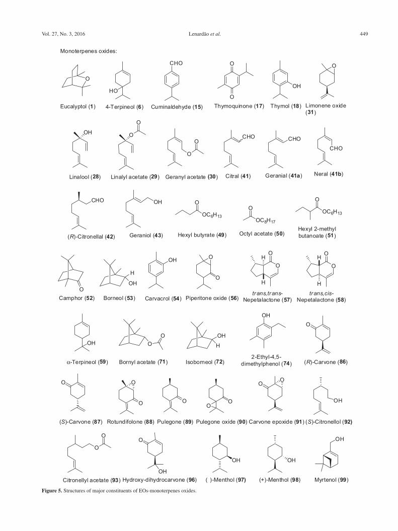
The Table 2 presents also the administration route and doses used in the articles discussed in this review. The structures of the major constituents of the EOs discussed in this section are presented on Figures 4-7. The compounds are arranged as monoterpenoids (Figure 4), monoterpenoids oxides (Figure 5), sesquiterpenoids (Figure 6) and sesquiterpenoids oxides (Figure 7).
EOs of Achillea species, knowed as yarrows (Compositae family), have been the subject of several investigations; these species are used in Turkish folk medicine. In 2006, Isçan et al.127 studied the antinociceptive effect of Achillea aleppica DC. subsp. aleppica and A. schischkinii Sosn. aerial parts EOs in the p-benzoquinone-induced abdominal constriction test. The main component of both EOs was eucalyptol 1 (32.5 and 26.1%, respectively) and the EO of A. aleppica subsp. aleppica was found to contain also 6.6% of bisabolol 2 and its derivatives. The authors observed that the EO of A. aleppica significantly reduced the writhes induced by p-benzoquinone. An acute toxicity assay of the A. aleppica EO was realized; the animals were observed during 48 h and according to the authors, any apparent acute toxicity was observed.
Ageratum fastigiatum (Gardner) R. M. King & H. Rob is a plant well distributed in Minas Gerais State, Southestern Brazil and is popularly called “matapasto”.189,190 The EO of A. fastigiatum is constituted mainly by diterpenes, triterpenes191,192 and, together with A. conyzoides, this plant is indicated in folk medicine as anti-inflammatory, analgesic and antimicrobial.193,194 Del-Vechio-Vieira et al.128 determined the chemical composition and performed a study about analgesic effects of the EO of A. fastigiatum. The major constituents of the EO are germacrene D 3, α-humulene 4 and β-cedrene 5. The EO inhibited the acetic acid-induced writhing and the formalin first phase and second phase.128
Alpinia species includes important medicinal plants that are widely distributed in tropical and sub-tropical regions and are cultivated for medicinal purposes.195 Alpinia zerumbet (Pers) B. L. Burtt & R. M. Sm. (Zingiberaceae) is an aromatic plant that is widely distributed in tropical and sub-tropical regions. This plant is popularly known as “colônia” in the Northeastern of Brazil, and is used in folk medicine in the treatment of intestinal disorders and hypertension.196 The leaves EO is rich in 4-terpineol 6 (28.1%), eucalyptol 1 (15.0%) and γ-terpinene 7 (13.7%) and it was effective in the acetic acid induced writhing test and in the hot-plate test, increasing the latency time. Besides, it was verified that the EO reduced paw licking time in both phases of the formalin test and the mechanism of action probably involves the participation of opiate receptors.184 The authors did not report any data about the toxic effect of the EO. However, the genotoxicity of A. zerumbet EO was recently studied on peripheral blood leukocytes in vitro and in vivo using the alkaline single-cell gel electrophoresis test (comet assay). In the in vitro tests, increasing concentrations (50-500 μg mL-1) of EO and methylmethanesulfonate (0.4 μmol L-1) as the positive control were used. According to the results, at the higher concentration (500 μg mL-1) A. zerumbet EO caused a significant increase in the cell DNA damage index in the in vitro assay.197
Hadi et al.129 studied the chemical composition and the analgesic effect of Artemisia absinthium (Asteraceae family) leaves EO. The main constituents found in the EO were nerolidol 8 (49.91%), santolina triene 9 (15.58%), α-pinene 10 (6.99%) and trans-β-farnesene 11 (4.95%). In this study, authors used male albino mice and the acetic acid-induced writhing test, formalin and hot plate assays. The results demonstrated that the administration of EO caused an inhibition of writhings in the acetic acid

Antinociceptive Effect of Essential Oils and Their Constituents: an Update Review J. Braz. Chem. Soc.446
assay comparable with the reference drug (Aspirin®). The EO presented effect in the late phase of formalin test with inhibition of 91%, results similar to observed using morphine (positive control, 5 mg kg-1). In the hot plate test, the EO increased the reaction time of mice after 30 min of treatment. One interesting point of the article are the data about the acute toxicity; according to the authors, this EO is safe at the effective doses, since the test of acute toxicity indicated that the EO is toxic only at higher doses.129
Artemisia dracunculus L. (Asteraceae family), popularly known as “tarragon”, is a plant used in folk medicine for the treatment of pain and gastrointestinal disturbances.198 The major components of the essential oil are 3,7-dimethyl-1,3,7-octatriene 12 (38.4%), α-pinene 10 (37.0%), estragole 13 (8.6%) and limonene 14 (6.3%).199 The antinociceptive effect of leaves EO of “tarragon” was assessed in the formalin, acetic acid and hot plate tests.130 According to the authors, “tarragon” EO demonstrated peripheral (acetic acid and formalin) and central (hot plate) antinociceptive effects. Authors also studied the involvement of the opioid system in the nociceptive response of tarragon EO, but according to them, these receptors are not involved. The acute toxicity of tarragon EO was evaluated and the LD50 was found to be 1250 mg kg-1.
Bunium persicum (Boiss) B. Fedtsh or Carum persicum Boiss. is a grassy plant of Apiaceae family with the common name of “wild caraway”, which grows in warm climate areas of Middle East and Central Asia.200 In Iran, the fruits or the aerial parts of the plant have been used traditionally as anticonvulsant, antihelmintic, anti-asthma, digestant, antiflatulent, diuretic and analgesic.201,202 In 2011, Hajhashemi et al.120 described the antinociceptive and anti-inflamatory activities of B. persicum fruits EO using the acetic acid and formalin tests to evaluate the analgesic effect. By the gas chromatography mass spectrometry (GC-MS) analysis, the authors identified 10 compounds, with γ-terpinene 7 (46.1%), cuminaldehyde 15 (23.9%) and p-cymene 16 (15.9%) being the main components of the EO. The EO significantly (p < 0.01) reduced the acetic acid-induced writhings and the pain response of both early and late phases of the formalin test. The authors declared that the analgesic effect might be due, at least in part, to the presence of γ-terpinene 7 and p-cymene 16.120
More recently, Zendehdel et al.121 described a study on the mechanism of the antinociceptive action of B. persicum seeds EO in the acetic acid-induced nociception model. The authors observed that the EO inhibited the writhing in mice in a dose dependent manner and this effect was attenuated by a pre-treatment with naloxone, chloropheniramine and cimetidine. These results suggest that B. persicum EO-induced analgesia could be mediated via opioidergic
and histamine H1 and H2 receptors and once again, the antinociceptive activity was attributed to the presence of p-cymene 16, γ-terpinene 7 and terpenoid oxides in the EO.121
Hejazian122 explored the antinociceptive activity of Carum copticum Benth. fruits EO. C. copticum is a plant of Apiaceae family and the aqueous extracts from its seeds are used in household remedies and also as a spice in food in India.202 The main constituents of the C. copticum seeds EO are p-cymene 16 (37.3%) and thymoquinone 17 (13.7%).122 The author used the formalin test to access the antinociceptive activity of the EO. He observed that the EO had no effect in the phase I and a significant effect in the phase II of the test, which was the same as 1 mg kg-1 of morphine sulphate. The presence of thymol 18 in the EO is, according the author, the possible responsible by the antinociceptive activity. Opioid receptors are not involved in the mechanism of antinociception, once naloxon, an opioid antagonist, could not reverse the analgesic effect observed in the formalin test.122
Radulovic et al.176 investigate the composition, antinociceptive and anti-inflammatory effects of the Choisya ternata leaves EO and three of their components, methyl, propyl and isopropyl N-methylanthranilate 19.
C. ternata, popularly known as “Mexican orange”, has highly fragrant flowers and is a popular horticultural shrub. The infusion of leaves of C. ternata is used in Mexico as an antispasmodic and possess “simulative properties”.203 The major components of the leaves EO are sabinene 20 (ca. 30%), 4-terpineol 6 (10%), myrcene 21 (7.8-8.3%), β-phellandrene 22 (5.4-6.6%) and γ-terpinene 7 (4.2-4.7%). Isopropyl N-methylanthranilate 19 and the methyl and propyl esters analogs were synthesized and evaluated, with the C. ternata EO, for their antinociceptive effects. The authors observed that the compound 19 and the EO produced dose-related and significant antinociception in chemical (acetic acid-induced visceral pain) and thermal (hot-plate test) models of nociception in mice.176 The C. ternata EO was also evaluated in the two phases of the formalin test.177 According to the authors, these results indicate that EO could be acting through inhibition of the formation and/or liberation of the mediators in the paw tissue or by direct blockage of the receptors.176 Among the individual compounds, isopropyl N-methylanthranilate was the more active, while methyl N-methylanthranilate presented the lower activity; however, it was still better than acetylsalicylic acid (200 mg kg-1) in the acetic acid-induced test and comparable to morphine (5 mg kg-1) in the hot plate test.176
Chrysopogon zizanioides L. Roberty (Poaceae family), popularly known as “vetiver” and “grama-das-índias”, is

Lenardão et al. 447Vol. 27, No. 3, 2016
used in the folk medicine of Brazil as analgesic and sedative. Lima et al.171 studied the chemical composition and the antinociceptive properties of the EO of roots of C. zizanoides at doses of 50 and 100 mg kg-1. The major compounds found in the EO were khusimol 23 (19.6%), E-isovalencenol 24 (13.2%), α-vetivone 25 (5.2%), vetiselinenol 26 (5.1%) and α-cadinol 27 (5.0%). The EO presented effect in the acetic acid-induced writhing test similarly to the positive control, morphine (3 mg kg-1), but contrasting to morphine, the opioid antagonist naloxone did not reverse the effect of the EO. In the formalin test, the EO was effective in reducing the licking response only in the second phase (inflammatory response) and in the hot plate it did not present any effect, indicating that a peripheral antinociceptive effect is involved. No data about the toxicological profile of C. zizanoides EO was found on literature.
Citrus bergamia or Citrus aurantium ssp. bergamia (Rutaceae family), popularly known as “bergamot”, is one of the most common and familiar plants worldwide. In 2011, Sakurada et al.178 studied the antinociceptive action of the intraplantar administration of fruits of “bergamot” EO, linalool 28 and linalyl acetate 29, which are the main components of the EO in the capsaicin model and they observed that the three tested substances reduced significantly the nociceptive response. In addition, the authors showed that the antinociceptive response of the “bergamot” EO and linalool 28 is mediated by the modulation of peripheral opioids receptors.
Citrus limon L. Osbeck (Rutaceae family) is a plant from the north and northeast of Brazil, known by the popular name of “limoeiro”.204 Infusions prepared with the aerial parts (leaves) of C. limon are used in folk medicine for the treatment of obesity, diabetes, blood lipid lowering, cardiovascular diseases and brain disorders.205,206 The antinociceptive effect of the EO of aerial parts of C. limon was evaluated by the acetic acid-induced writhings, formalin and hot plate assays and the results indicate that the EO has peripheral and central antinociceptive effects.179
The main components of the leaves EO are limonene 14 (52.8%), geranyl acetate 30 (9.9%) and limonene oxide 31 (7.1%). The EO reduced writhings and lickings nociceptive responses in the acetic acid and first and second phases of formalin tests. The EO increased the latency time in the hot plate test. Furthermore, the pre-treatment with naloxone caused an antagonistic effect on antinociceptive effect of EO of C. limon; thus, opioid receptors are involved.146 The authors did not report any data about toxicological studies of C. limon EO. Toxicological informations of plants are important because many natural products exert significant redox activities, which are related to their therapeutic properties, even a possible toxic effect.63
Ximenes et al.136 studied the chemical compositon and the antinociceptive activity of the EO of Croton adamantinus Müll. Arg. (Euphorbiaceae family). This plant is popularly known as “carrasco” and has been used in the semi-arid region of Northeast Brazil to treat inflammation, skin and gastric disorders.138,207 According to the authors,136 the main components of “carrasco” leaves EO are methyleugenol 32 (14.8%) and eucalyptol 1 (13.7%). It was observed a mild antinociceptive effect in the early phase of formalin test and an activity higher than morphine (positive control) at the second phase. In the assay of abdominal contortions induced by acetic acid, the EO was more effective than indomethacin in decreasing the number of abdominal contortions. The authors also reported the results on the lethal dose of EO; according to them, a limit test of 1000 mg kg-1 was performed to estimate the toxicity of the EO and did not result in any death or changes in the gross necropsy. This is a complementary and important result to the antinociceptive data, once it demonstrated that the effective doses of C. adamanthinus did not present acute toxic potential.136
The shrub Croton cordiifolius Baill., Euphorbiaceae family, known as “quebra-faca” is one of the about 350 species of the Croton genus that are found in Brazil.208 The plant is popularly used in the northeast of Brazil to treat medical conditions, such as general inflammation, pain, and gastrointestinal disturbances.209 In 2015, Nogueira et al.137 described the chemical composition and the antinociceptive activity of C. cordiifolius EO in mice. Eucalyptol 1 (25.09%) and α-phellandrene 33 (15.43%) are the major constituents, according to the GC-MS analysis. The antinociceptive activity was evaluated using the acetic acid, formalin, capsaicin and glutamate tests. The authors observed that the C. cordiifolius EO reduced the number of writhing responses induced by acetic acid and decreased the licking times in both phases of the formalin test. The EO was also effective in the glutamate test and no effect was observed in the capsaicin test. According to the authors, the antinociceptive effect of C. cordiifolius EO could involve the inhibition of the glutamatergic system, once naloxone, an opioid antagonist, did not affect its antinociceptive effect in the writhing test.137
Croton nepetaefolius Baill. (Euphorbiaceae family), popularly called “marmeleiro vermelho”, is an aromatic plant native of the Northeast of Brazil, where it is extensively used in folk medicine as a sedative, orexigen and antispasmodic agent.210 The leaves EO was effective in acetic acid-induced writhing test; in the hot plate test, the EO significantly increased the latency.138 Also, in the formalin test, EO reduced paw licking in both phases with the mechanisms remaining to be

Antinociceptive Effect of Essential Oils and Their Constituents: an Update Review J. Braz. Chem. Soc.448
elucidated. The main constituents found in this EO were eucalyptol 1 (31.5%), (E)-caryophyllene 34 (17.2%) and methyleugenol 32 (10.3%). According to Fontenelle et al.211 the intra-peritoneal administration of different doses of C. nepetifolius EO induced no remarkable alterations in the behavior pattern of mice, such as: trembles, convulsions, dyspnea and ataxia. After the intraperitoneal administration, the calculated LD50 was 163.8 mg kg-1.
The species Croton sonderianus Müll. Arg. (Euphorbiaceae) is a widespread shrub largely grown in northeastern parts of Brazil, popularly known as “marmeleiro preto”. This plant is used as fire wood due to the high content of essential oil that may vary from 0.5 to 1.5%. Leaves and barks are used as an infusion or simply chewed as a folk medicine for the treatment of gastrointestinal disturbances, rheumatism and headache.212 The EO of C. sonderianus leaves is rich in monoterpenes and sesquiterpenes, such as cis-calamenene 35 (10.9%), bicyclogermacrene 36 (10.2%), guaiazulene 37 (8.3%), spathulenol 38 (7.2%), (E)-caryophyllene 34 (6.9%), β-phellandrene 22 (6.2%), α-guaiene 39 (6.6%), eucalyptol 1 (4.2%) and others. When the EO was given orally, it produced significant inhibitions on chemical nociception induced by acetic acid, formalin and capsaicin injections in mice. The antinociception probably involves glibenclamide ATP-sensitive K+ channels, according to Santos et al.69 Doses employed in this study were
considered non-toxic once the EO at doses up to 3.0 g kg-1 did not cause any behavioral impairment or overt toxicity in mice (unpublished observations).
The fruits of Cuminum cyminum Linn., a wild grassy plant of Umbelliferae family, is used in Iranian folk medicine to treat diarrhea, toothache and epilepsy.213 The major constituents of C. cyminum EO are γ-terpinene 7 (29.1%), p-cymene 16 (25.2%), β-pinene 40 (19.9%) and cuminaldehyde 15 (18.7%).214 Sayyah et al.123 studied the effect of the C. cyminum EO in two models of nociception, formalin and tail flick tests. The pretreatment with the EO significantly reduced the formalin-induced nociception for 1 h; the effect being more pronounced in the late phase, while no effect was observed on tail flick response. The LD50 for the C. cyminum EO used in the study was determined as 0.59 (0.52-0.68) mL kg-1.123
Cymbopogon citratus (DC.) Stapf (Poaceae) is an herb known worldwide as lemongrass and the EO of its leaves is source of citral 41 (ranging from 47-86% in weight). Viana et al.172 described the antinociceptive activity of leaves EO of C. citratus, West Indian type, which increased the reaction time in the hot plate test. At lower doses the EO inhibited the abdominal contraction. On the other hand, in the formalin test, the administration via i.p. was more effective on the inhibition of the licking time at the second phase than the oral administration at the same doses. Viana’s work did not present data about the toxicological profile of
Santolina triene (9) α-Pinene (10) 3,7-Dimethyl-1,3,7-octatriene (12) (R)-Limonene (14)
p-Cymene (16) Sabinene (20) Myrcene (21) β-Phellandrene (22)
α-Phellandrene (33) β-Pinene (40)
γ-Terpinene (7)
Monoterpenes:
Camphene (55) α-Terpinene (65) (Z)-Salvene (76)
Figure 4. Structures of major constituents of EOs-monoterpenes.
Lenardão et al. 449Vol. 27, No. 3, 2016
O
Eucalyptol (1)
HO
4-Terpineol (6) Cuminaldehyde (15)
CHO
O
O
Thymoquinone (17) Thymol (18)
OH
OH
Linalool (28)
O
Linalyl acetate (29)
O
Geranyl acetate (30)
O
O
Limonene oxide(31)
O
Monoterpenes oxides:
CHO
Citral (41)
CHO
Geranial (41a)
CHO
Neral (41b)
CHO
(R)-Citronellal (42) Geraniol (43)
OH O
OC6H13
Hexyl butyrate (49)
O
OC8H17
Octyl acetate (50)
O
OC6H13
Hexyl 2-methylbutanoate (51)
OCamphor (52) Borneol (53)
OH
Carvacrol (54) Piperitone oxide (56)
O
O
OH
H
O
trans,trans-Nepetalactone (57)
OH
H
O
trans,cis-Nepetalactone (58)
OH
α-Terpineol (59) Bornyl acetate (71) Isoborneol (72)
OH
2-Ethyl-4,5-dimethylphenol (74)
O
(R)-Carvone (86)
O
(S)-Carvone (87) Rotundifolone (88)
O
O
Pulegone (89)
O
Pulegone oxide (90)
OO
O
Carvone epoxide (91)
O
(S)-Citronellol (92)
OH
Citronellyl acetate (93)
O
OO
Hydroxy-dihydrocarvone (96)OH
OH
(� )-Menthol (97)
OH
(+)-Menthol (98)
OH
Myrtenol (99)
H
OH
OO OH
H
Figure 5. Structures of major constituents of EOs-monoterpenes oxides.

Antinociceptive Effect of Essential Oils and Their Constituents: an Update Review J. Braz. Chem. Soc.450
lemongrass EO; however, according to Fandohan et al.215 it did not show any acute (1 day) and sub-acute (14 days) toxicity at doses of 5-1500 mg kg-1 body weight, but at higher doses, 2000 and 3000 mg kg-1, abnormalities were observed. The reported LD50 was > 3500 mg kg-1 body weight.
Cymbopogon nardus (L.) Rendle, is a widespread plant used in culinary, perfumery and in popular medicine in the treatment of rheumatism, fever, menstrual and digestive problems.216 The C. nardus EO is commercially used as a mosquito repellent, and several pharmacological properties have been attributed to the EO and its major constituent, (R)-citronellal 37, including antifungal,217 antibacterial218 and anti-cancer activities.219 Abena et al.173 described in 2007 a comparative study between the chemical composition and the antinociceptive activity of C. nardus EOs of plants cultived in Congo and Benin. The major constituents in both EOs are citronellal 42 (37.5 and 41.3%) and geraniol 43 (29.4 and 23.4%) respectively, among other more than 20 identified compounds. The three antinociceptive tests used (acetic acid,
hot plate and tail flick) show that the two EOs are actives. The effect in the acetic acid-induced test was similar for both EOs. However, the EO from Benin was more effective in the hot plate test, while the Congolese EO was more active in the tail flick model.173
Cymbopogon winterianus Jowitt is an aromatic grass cultivated in India and Brazil that is traditionally used as an insect repellent.220 The main components of C. winterianus leaf EO are geraniol 43 (36%) and citronellal 42 (42.7%) and besides a repellent, it has antimycotic and acaricidal activities.221 The infusion of the leaf and unguent have been used in northeastern Brazil for the treatment of pain and anxiety.174 Leite et al.174 studied the antinociceptive activity of C. winterianus leaf EO in the acetic acid-induced writhing, formalin (phases I and II) and in the hot plate models. The authors observed that the EO reduced the number of writhings in the acetic acid and paw licking times in the first (0-5 min) and second (15-30 min) phases of the formalin tests, respectively. No effect was observed, however, in the hot-plate test at all the tested doses.174
Germacrene D (3) α-Humulene (4) β-Cedrene (5) β-Farnesene (11)
HH
(E)-Caryophyllene (34) cis-Calamene (35) Bicyclogermacrene (36) Guaiazulene (37)
α-Guaiene (39)
Sesquiterpenes:
β-Elemene (44)
H
β-Selinene (46) (E)-α-bis-Abolene (60)
Rotundene (70) (α)-Funebrene (77) γ-Muurolene (81) δ-Cadinene (82)
Figure 6. Structures of major constituents of EOs-sesquiterpenes.

Lenardão et al. 451Vol. 27, No. 3, 2016
Cyperus esculentus L. and C. rotundus L., are sedges of the family of Cyperaceae, which grow naturally in tropical, subtropical and temperate region and are widely distributed in the Mediterranean area. C. esculentus and C. rotundus are used for the treatment of spasms stomach
disorder and as an anti-inflammatory in traditional medicine of India, China and Japan.222 Biradar et al.134 evaluated the antinociceptive effect of C. esculentus and C. rotundus EOs in the formalin test and verified that they are equaly active in both phases; however, a slightly superior effect was
Figure 7. Structures of major constituents of EOs-sesquiterpenes oxides and other compounds.
OH
Nerolidol (8)
H
Khusimol (23)
HO
HO
(E )-Isovalencenol (24) α-Vetivona (25)
O
Vetiselinenol (26)
OH
HO
α-Cadinol (27)
HO
H
H
Spathulenol (38)
Sesquiterpenes oxides:
HH
O
Caryophyllene oxide (45)
O
H
Atractylone (47)
O
H
Furanoeudesmene (48)
HOLedol (63)
O
O
O
OH
Remirol (66)
OO
OHO
Isoevodionol (68) Cyperotundone (69)
O
O
OH
HO
OH OApigenin (75)
HOGlobulol (78) Humulene epoxide (79)
O
α-Copaene (83)
O
Zerumbone (84)
O O
O
O
O
Evodione (94)
OH
Farnesol (95)
Other compounds:O
O
NH
Isopropyl N-methylanthranilate (19)
O
OH
Phenylacetic acid (73)
NO2
1-Nitro-2-phenylethane (NPE)(85)
O
Estragole (13)
O
O
(E )-Methylcinnamate (61)
OO
(Z)-Methylcinnamate(62)
trans-Anethole(64)
O
O
O
Methyleugenol (32)
O
O
Elemicin (80)
O
H
H
H
H

Antinociceptive Effect of Essential Oils and Their Constituents: an Update Review J. Braz. Chem. Soc.452
observed in the second, inflammatory phase of the formalin test. Triterpenoids, flavonoids, proteins and saponins were described as the major active constituents; however, there was no information about the main volatile compounds of the EOs. A study of the acute toxicity was performed using albine rats and no mortality was observed at the dose of 5000 mg kg-1 after 24 h.134
Distichoselinum tenuifolium (Lag.) F. García Mart. & Silvestre is a plant widely used in traditional medicine in Portugal for the treatment of contact dermatitis and skin infections.223 Goés et al.124 studied the chemical composition and the antinociceptive effect of D. tenuifolium ripe umbels EO in rats using the acetic acid, hot plate and formalin nociception tests. The authors found myrcene 21 (85.0%) as the major constituent of the EO. The treatment with D. tenuifolium EO decreased the writhing induced by acetic acid. The EO administration reduced the licking time at both first and second phases of the formalin test and it was more effective then indomethacin, used as the control.124
Duguetia lanceolata A. St.-Hil (Annonaceae) popularly known as “pindaíba”, “beribá” or “pinhão”, is a perennial species distributed in several regions of Brazil.224 In folk medicine, this plant has been used as an anti-inflammatory, cicatrizing and antimicrobial agent.224 The EO of barks of D. lanceolata is rich in β-elemene 44, caryophyllene oxide 45 and β-selinene 46 and it has shown antinociceptive effect in rat and the mechanism probably involves central and peripheral actions. Sousa et al.117 described a significant reducing in the number of writhing and the lick of the paw (in the first and second phases). Recent studies of the same group118 demonstrated the toxic effect of D. lanceolata EO in Artemia salina Leach (Brine Shrimp Lethality Bioassay), presenting a lethal concentration (LC50) of 49.0 μg mL-1.
Silva et al.165 studied the antinociceptive and anti-inflamatory effects of the EO of three different Eucalyptus species: E. citriodora Hook, E. globulus and E. tereticornis. Eucalyptus are traditionally used as analgesic, anti-inflammatory and antipyretic remedies for the symptoms of respiratory infections, such as cold, flu, and sinus congestion.225 Besides, the Eucalyptus EOs are also widely used in cosmetics, food and pharmaceutical industries.226 E. citriodora EO has citronellal 42 as the major component (up to 60%), whereas E. globulus and E. tereticornis EOs contain 60-90% of the monoterpenoid eucalyptol 1. The authors observed that the EOs decreased the number of acetic acid-induced writhes in mice (43-73%) compared to the animals that received vehicle only. The effect was dose-dependent for the E. tereticornis EO only and the E. citriodora EO was the most effective at the higher dose. Similarly, all the EOs were effective in the hot plate test, significantly extending the reaction time to after 30 min of
treatment (i.p.), as compared to the corresponding control groups.165
Eugenia candolleana DC. (Myrtaceae) is commonly known as “murta”, a rare Eugenia from the Northwestern Brazilian rainforests, bearing a small, dark-purple ripening fruit with a mildly sweet and firm pulp.166 The infusion of the fresh leaves has been used in folk medicine for the treatment of pain and fever. The leaves EO of E. candolleana reduced the number of writhes significantly in a writhing test as well as the number of paw licks during the second phase of formalin test after i.p. injection. No information about the chemical composition of the E. candolleana used in the study was found, except that monoterpenoids and sesquiterpenoids are predominant. According the authors, the antinociceptive activity of the EO probably is mediated via a peripheral pathway.166
Eugenia uniflora L. (Myrtaceae) is known as “Brazilian cherry tree” (or “pitangueira”). Their leaves are used in infusions or decoctions in popular medicine to treat inflammations, against rheumatic pains and fever, as hypoglycemiant, diuretic and to avoid stomach problems.227 The antinocicpetive effect of the leaves EO and their isolated terpenoids (a mixture of atractylone 47 and 3-furanoeudesmene 48 in a 2:1 ratio) were evaluated by Amorim et al.21 The EO and their main constituents given orally, 1 h before the noxious stimulus in mice, significantly inhibited the acetic acid-induced abdominal constrictions and increased the latency time in the hot plate test. Victoria et al.228 studied the acute toxicity of E. uniflora EO in mice and data demonstrated that the LD50 is higher than 200 mg kg-1, since at this concentration any signal of toxicity was observed.
Fruits of Heracleum persicum Desf. (Apiaceae) are widely used as spices and the young stems are also used for making pickles. In Iranian folk medicine, fruits of H. persicum are used as a carminative and pain killer herbal drug.229 The main constituents of H. persicum fruits EO are hexyl butyrate 49 (56.5%), octyl acetate 50 (16.5%) and hexyl 2-methylbutanoate 51 (5.2%) and it was evaluated for their antinociceptive action by the acetic acid-induced writhings and formalin pain models.125 The authors observed that the oral administration of the EO reduced the number of writhings induced by acetic acid while by intra-peritoneal injection the EO was not effective. The EO also did not reduce the licking response induced by the intraplantar injection of formalin in any of used concentrations. Manzoomi et al.230 studied the possible toxic effect of H. persicum EO and found a LC50 value of 337.58 μL L-1.
The gender Hyptis (Lamiaceae family) consists of approximately 400 species distributed from the South
Lenardão et al. 453Vol. 27, No. 3, 2016
of the United States to Argentina231 and exhibits a major morphological diversity in the Brazilian Cerrado.232 Hyptis pectinata L. Poit is an aromatic shrub largely grown in the northeastern parts of Brazil and its leaves EO is rich in (E)-caryophyllene 34 (40.9%) and caryophyllene oxide 45 (38.0%). Arrigoni-Blank et al.139 studied the chemical composition and antinociceptive activity of leaves EO of six genotypes of H. pectinata. The authors observed that all genotypes had variation in their chemical composition and all of them presented antinociceptive effect in two models using mice (hot plate and acetic acid-induced writhing). According to the authors, the antinociceptive action involves the participation of opioid receptors. In other study, Raymundo et al.140 demonstrated that the antinociceptive effects of the H. pectinata are mediated by opioid and cholinergic receptors in mice. The EO also increased baseline measurements and the area under the curve in measurements on the hot plate model and was effective on second phase of formalin test. The acute toxicity of this EO was studied in male and female mice, by the oral administration of a single dose of 500 mg kg-1 of H. pectinata EO and, according to the presented data, any signal of toxicity was observed after 14 days.140
The antinociceptive effect of the EO of leaves of Laurus nobilis L. (Lauraceae), an evergreen and widely distributed plant in the Mediterranean area and Europe, was evaluated.233 Folk remedies in different countries use this plant to treat numerous diseases. In Iranian traditional medicine, the leaves have been used topically for relieving rheumatic pains.234 The main components of leaf EO of L. nobilis are eucalyptol 1 (44.1%), eugenol 32 (15.16%) and sabinene 20 (6.2%).235 The pre-treatment of mice with the EO induced an increase in the tail flick latency and significantly reduced the nociception in the second phase of formalin test. According to the authors, the EO up to a dose of 0.3 mg kg-1 presented no lethality. However, above this dose some deaths were observed.233
Lavandula angustifolia Mill. (Lamiaceae), commonly known in Iran as “Ostokhoddous”, is a widely distributed aromatic herb.236 This plant is well known among people as a powerful aromatic and medicinal herb and it is used in traditional and folk medicines of different parts of world for the treatment of several gastrointestinal, nervous and rheumatic disorders.237,238 Mice pre-treated with leaves EO of L. angustifolia by oral route presented reduced writhes. The same doses were effective in the first phase of formalin test and all of them were effective in the second phase of the test. The EO of L. angustifolia is rich in eucalyptol 1, camphor 52 and borneol 53.239 Evandri et al.141 studied the possible mutagenic effect of L. angustifolia leaves EO in the bacterial mutagenicity test (main test) using the plate
incorporation method. The results demonstrated that EO did not increase the number of revertants in two Salmonella strains (S. typhimurium strain TA98 and S. typhimurium strain TA100) and in the E. coli WP2 uvrA strain.141 Studies about the mutagenic effects of EOs are very important considering the increasing use and their frequent presence in products for personal care and medicines.
EO of Lavandula hybrida E. Rev. ex Briq. leaves had linalool 28 and linalyl acetate 29 as major constituents and, when orally administered or inhaled for 60 min, it reduced the acetic acid-writhing response; in the hot plate test, the involvement of opioid as well as cholinergic pathways was identified.142 On the other hand, no analgesic results were obtained from inhalation of linalool and linalyl acetate in the hot plate test. Only linalool (by oral route) was effective in the acetic acid test. The authors also studied the acute gastrointestinal ulcerogenicity of an oral treatment (100 mg kg-1) with EO and the results demonstrated that any damage on gastric mucosa was observed.142
Lippia gracilis Schauer (Verbenaceae) is known in Brazil as “alecrim-da-chapada” and its EO is highlighted because it presents high contents of monoterpenes, with carvacrol 54 (up 50%), p-cymene 16 (ca. 11%) and γ-terpinene 7 (8%) being the main components.240 Several communities in Northeastern Brazil use L. gracilis to treat cough, bronchitis, nasal congestion, and headache.241 The EO is known to possess antimicrobial activity and is used externally to treat cutaneous diseases, burns, wounds and ulcers.242 Mendes et al.180 analyzed the antinociceptive effect of the EO of L. gracilis leaves. The antinociceptive effect was evaluated by the acetic acid-induced writhing test, and the L. gracilis EO was effective at the tested doses. In addition, Guilhon et al.181 studied the antinociceptive action as well as the mechanisms involved in this effect. The authors used chemical and thermal methods to determine the antinociceptive effect, like acetic-acid, formalin and hot plate tests. Increasing doses of EO decreased significantly the writhing numbers in the acetic acid assay. The peripheral antinociceptive effect was confirmed by the formalin test, with the EO presenting effect only in the second phase (10-100 mg kg-1), demonstrating the possible anti-inflammatory effect of the EO. The central effect was measured in the hot plate assay and the L. gracilis EO increased significantly the latency in mice at the same doses. Additionally, the mechanism of antinociceptive effect was assessed by the involvement of opioid, cholinergic and nitric oxide (NO) systems, all related to the central antinociceptive effect (hot plate assay), and cholinergic and nitric oxide (NO) systems, which are related with peripheral antinociception (acetic acid and formalin).181 The acute toxicity of EO was studied

Antinociceptive Effect of Essential Oils and Their Constituents: an Update Review J. Braz. Chem. Soc.454
by the authors using different oral doses (2, 3 and 4 g kg-1) in a group of male and female mice and no signs of intoxication, including convulsion, death, or gastric ulcer, were observed even after 5 days of a single dose. Lethality was not observed at the highest dose of EO, indicating that it was nearly nontoxic in mice up to this dose and that it was not possible to determine the LD50.
Another species of the genus Lippia, L. grata Schauer is an endemic bush of northeastern Brazil. The leaf EO of L. grata presented antispasmodic activitiy, which was attributed to the presence of carvacrol 54 and thymol 18.243 In 2014, Siqueira-Lima et al.182 described the antinociceptive effect of β-CD complex of L. grata leaf EO in orofacial nociception in mice. It was observed that oral treatment with β-CD/EO reduced the nociception in both phases of the formalin test and also protected against nociception induced by capsaicin and glutamate. In contrast to previously reported, camphene 55 (11.3%), camphor 52 (27.2%), (E)-caryophyllene 34 (11.6%), bicyclogermacrene 36 (9.4%) and borneol 53 (6.0%) were the main components found in the EO used in this study. The authors observed a high stability of the β-CD/EO complex (up to 200 oC) and the antinociceptive activity was attributed to the terpenoids camphor, borneol and β-caryophyllene, which contributed to activation of the motor cortex, NRP and PAG (cerebral areas involved in pain modulation).182
Marçal et al.183 examinated the chemical composition and the antinociceptive effect of the Lippia sidoides Cham. (Verbenaceae) EO on mice. L. sidoides is an aromatic shrub, used in Brazilian folk medicine to treat inflammation, pain and bacterial infections.244 The major constituents found in the EO were p-cymene 16 (26.8%), thymol 18 (21.9%) and myrcene 21 (12.8%) and a dose-dependent antinociceptive effect was observed in the acetic acid-induced writhing test. The L. sidoides EO increased the latency time in the hot plate test, but no effect was observed in the presence of naloxone (3 mg kg-1, i.p.), indicating that the opioidergic system is involved in the antinociceptive effect.183
Melissa officinalis is a widespread plant used in perfumes, cosmetics, tea and food products and possess sedative, spasmolytic and antibacterial properties.245,246 The major components of the M. officinalis leaf EO are citronellal 42 (39%) and citral 41 (33%).143 In a very interesting work, Hasanein and Riahi143 studied the antinociceptive effect of M. officinalis EO in an experimental model of diabetic hyperalgesia. The authors used healthy (control) and diabetic rats in the formalin test, and they observed that the hyperalgesia was completely reversed after a cronical oral treatment of the diabetic rats with M. officinalis EO. On the other hand, the EO caused less intensive nociceptive effect in the control rats during
both phases of the formalin test. These findings indicate for a promise treatment with M. officinalis EO for painful diabetic neuropathy.143
Many Mentha species are used worldwide as choleretic, spasmolytic and analgesic agents.247 In the Northeast of Brazil, Mentha × villosa Huds. (Lamiaceae), an aromatic herb, is widely used in folk medicine as a stomachic and anxiolytic agent.196 The chemical composition analysis of the EO of M. × villosa revealed the piperitone oxide 56 as being the major constituent. Sousa et al.144 studied the antinociceptive effect of the leaves EO of M. × villosa and of piperitone oxide and they observed that both reduced the writhing response on the acetic acid test and in the second phase of formalin induced pain. Besides, other important finding is that the opioid system is not involved in the antinociceptive effect of the EO and of piperitone oxide. In the thermal models of induce nociception, tail immersion test and hot plate, the EO and pure compound 56 were not significantly effective. At similar doses, 80 and 100 mg kg-1, the leaves EO of M. × villosa had no toxic effects when administered to rats for 30 days.144
Myrcia ovata Cambess. is popularly known as “laranjinha do mato” and their leaves are frequently used as an infusion in folk medicine to treat gastric diseases and diarrhea.248 Dos Santos et al.167 determined the chemical compostion and the antinociceptive activity of the M. ovata EO in the acetic-acid induced writhing, formalin (phases I and II) and tail flick tests. The GC-MS analysis revealed the monoterpene geranial 41a (52.6%) and its isomer neral 41b (37.14%), i.e., citral 41, as the major components of the EO. The authors observed that the M. ovata EO orally administered inhibited writhing induced by acetic acid by 29 and 51%, respectively. The EO was effective in both phases of the formalin test, with a more pronounced activity in the second one. At doses of 200 and 300 mg kg-1 the inhibition was similar to that of morphine at 5.01 mg kg-1. A maximal effect on the tail flick test of 34 and 91% was observed at doses of 200 and 300 mg kg-1, respectively. The antinociceptive effect was reduced by pretreatment with naloxone, suggesting that the antinociceptive activity might involve the opioid system. The EO did not induce motor impairment and no toxic effect was observed after oral administration (LD50 > 3000 mg kg-1).167
Nepeta L. (Lamiaceae family) is a genus formed by perennial or annual herbs distributed in Europe, Asia, North Africa and in Near East and has around 300 species, 75 of which are found in Iran.249 Antispasmodic, diuretic, expectorant, antiseptic and antiasthmatic are among the activities assigned to Nepeta species. N. cataria is one of the representative of this genus that has been used for a long time in teas, dyes or infusions and in the North

Lenardão et al. 455Vol. 27, No. 3, 2016
American folk medicine.250 Ricci et al.145 determined the chemical profile and explored the potential of the N. cataria L. var. citriodora (Becker) Balb. (Lamiaceae) EO as an antinociceptive agent in the acetic acid-induced nociception and in the tail immersion tests in mice. The major constituents of the N. cataria EO are trans,trans-nepetalactone 57 (50.4%) and trans,cis-nepetalactone 58 (21.7%). The EO at the dose of 0.0005 mL kg-1 (i.p.) reduced significantly the mice writhing responses in the acetic acid-induced model and was slightly less active than morphine (20 mg kg-1) in the tail immersion test. The authors observed that the EO effects started before than the morphine ones, i.e., 15 min after the treatment, and remained up to 45 min after its administration.145
Nepeta crispa Willd. (Lamiaceae family) is an aromatic and medicinal plant endemic to Iran and its aerial parts are used in the Iranian folk medicine as a sedative, carminative and as a restorative tonic for nervous and respiratory disorders.251 The major components of the N. crispa leaves and flowers are eucalyptol 1 (71.0%), β-pinene 40 (5.0%) and α-terpineol 59 (4.1%).252 Ali et al.146 evaluated the effect of N. crispa EO in the tail flick and formalin nociception models. In the first phase of formalin test, it was observed reduction of jerking counts, licking and flexing duration in all doses, whereas in the second phase the EO was effective only at doses of 100 and 200 mg kg-1.146
One of the more recently identified species of Nepeta genus in Iran was N. pogonosperma Jamzad et Assadi, reported in 1984.249 Ali et al.147 studied the chemical composition of the N. pogonosperma EO and evaluated its antinociceptive effect (i.p.) in the tail flick and in the formalin tests. Similarly to other Nepeta plants, the N. pogonosperma EO is mainly constituted by eucalyptol 1 (31.2%) and trans,cis-nepetalactone 58 (14.5%), followed by α-terpineol 59 (5.4%) and (E)-α-bisabolene 60 (5.4%), among other terpenoids in minor amounts.147 Authors observed that the EO presents antinociception at doses of 100 and 200 mg kg-1 in the tail flick test and in both phases of the formalin test at all doses.
Nigella sativa L. (Ranunculaceae) seeds have been used for thousands of years as a spice and food preservative, as well as a protective and curative remedy for numerous disorders. Abdel-Fattah et al.175 studied the antinociceptive effect of the EO of seeds of N. sativa and its major component, thymoquinone 17. Authors used four experimental models of induced pain and observed that the oral administration of N. sativa EO significantly increased the latency of nociceptive responses in the hot-plate and tail-pinch tests. The writhing behavior caused by i.p. injection of 0.6% acetic acid was attenuated in a dose-dependent manner. N. sativa EO also significantly
suppressed the nociceptive response in the early phase, but did not reduce the response in the late phase of the formalin test. Regarding the antinociceptive effect of thymoquinone, the systemic administration by different routes: p.o. (5 and 10 mg kg-1), i.p. (4 and 6 mg kg-1) and intracerebroventricular, i.c.v., (2 and 4 μg mL-1) dose-dependently attenuated the nociceptive response in the early and late phases of the formalin test. The s.c. and i.c.v. injection of naloxone significantly antagonized the effect of systemic thymoquinone (i.p.) on the early phase response. In contrast, s.c. and i.c.v. naloxone failed in reversing the thymoquinone-induced antinociception in the late phase. Studies demonstrated that the oral administration of the seed EO at doses up to 10 mL kg-1 in rats and mice did not cause any mortality or overt toxicity during the observation period of 48 h.253 This was recently confirmed when it was demonstrated that oral administration of the EO of N. sativa at a dose of 10 mL kg-1, for up to 12 weeks did not cause any mortality or significant alterations of the key hepatic enzymes in rats.254 However, acute administration of high doses (2 g kg-1 or more) caused hypoactivity and difficulty in respiration.255
Ocimum (Lamiaceae) is a genus that comprises more than 150 species, which are distributed from tropical to subtropical regions.256 Ocimum plants are rich in essential oils which contain mainly myrcene 21, eugenol 32, and (E)-caryophyllene 34.257 These substances have a proven influence on the central nervous system. Several species of Ocimum have been reported with regard to their antinociceptive properties, such as O. micranthum Willd, O. basilicum L., O. L. and O. sanctum L.
O. basilicum L., popularly known as “basil”, is an aromatic herb used in Brazil to treat illnesses such as respiratory and rheumatic problems, vomiting, and pain.258
The leaves EO of O. basilicum reduced significantly the number of writhings on the acetic acid assay and the number of lickings of both phases of the formalin assay.148 Besides, it increased the time for mice response to the thermal stimulus at 50 mg kg-1 in different times of exposure. Moreover, this study revealed the modulation of opioid mechanism on the antinociceptive response of this EO on the hot plate test. The authors also demonstrated the lack of toxicity or mortality at doses of 250 mg kg-1. The lethal dose for 50% of animals (LD50) was of 532 mg kg-1, which could be associated to the high content of linalool in the EO.148
In other study, Venâncio et al.149 also analyzed the antinociceptive response of leaves EO of O. basilicum on the acetic acid, formalin, capsaicin, glutamate tests and in the orofacial pain. In the EO used by the authors, linalool 28 was the major component (76%), followed by geraniol 43 (11%). The results indicated an action of the EO on phases I

Antinociceptive Effect of Essential Oils and Their Constituents: an Update Review J. Braz. Chem. Soc.456
and II of the formalin assay and in the glutamate and capsaicin tests. In the same work, the authors compared the activity of the EO with that of pure (−)-linalool and very close results were obtained in both cases.149
Another species of Ocimum plant with antinociceptive effect is O. gratissimum L., known as “alfavaca” and widely used in the culinary as a condiment. In 2003, Rabelo et al.150 analyzed the chemical profile and the antinociceptive action of the EO of the leaves of O. gratissimum on the acetic acid-induced writhings and in the formalin assays. The major constituents found in the EO were eugenol 32 (52.1%), eucalyptol 1 (29.2%) and β-selinene 46 (5.6%). The authors observed that the EO produced a dose-dependent inhibition of acetic acid-induced writhing, comparable to indomethacin (an analgesic drug) and of the second phase of formalin-induced pain. A significant reduction on the first phase of formalin test was observed only at doses of 300 mg kg-1.150 The acute toxicity of O. gratissimum was studied in three month-old Wistar albino rats at doses of 5-3500 mg kg-1, during 14 days. According to the results, rats which received 5-500 mg kg-1 of O. gratissimum EO also showed normal general behavior on day 1. At 1000 mg kg-1, animals became torpid just after gavage, but this lasted no more than 30 min. In contrast, rats that received 1500 mg kg-1 stayed torpid all the day 1. Those that received doses above 2000 mg kg-1 died all less than 24 h after the EO administration. The LD50 in rats was 1750 mg kg-1.
More recently, a study performed using O. gratissimum EO and two of its main constituents, myrcene 21 and eugenol 32 (40 mg kg-1), revealed peripheral and central antinociceptive effects at the formalin (two phases) and hot plate tests, respectively.151 To extend the studies, the authors analyzed the participation of the opioid system in the antinociceptive effect of the EO (20-40 mg kg-1) and the isolated constituents in the hot plate test. Results showed that the pre-treatment of mice with naloxone, a non-selective opioid antagonist, have blocked the antinociceptive effect of EO, myrcene and eugenol, suggesting that modulation of opioid receptors is involved in the antinociceptive action. The authors also studied the acute toxicity of the EO in mice and observed few signs of toxicity, such as ptosis and reductions in defecation and ambulation, particularly at the highest dose (300 mg kg-1).151
O. micranthum Willd is an herb native of the lowlands of Central and South America and West Indian. It is used locally to flavour beverages and soups, and for the treatment of fever, stomach disturbances and dysentery. A decoction of the plant is also used for nervous disorders, earache, colic and convulsion in children as well as for painful menstruation.259 The antinociceptive effect of the
leaves EO of O. micranthum Willd was evaluated by the acetic acid and formalin tests.152 The authors also analyzed the influence of the opioids and nitric oxide systems on the antinociceptive response. Results demonstrated that the EO of leaves of O. micranthum Willd has activity in the acetic acid-induced writhing involving the opioid mechanism and in both phases of formalin test with participation of the opioid and nitric oxide systems. In a more recent work, Pinho et al.153 studied the antinociceptive effect of O. micranthum leaves EO, (E)-caryophyllene 34, (E)-methyl cinnamate 61 and (Z)-methyl cinnamate 62 (the major constituents of the EO) by the acetic acid, formalin and hot plate tests. The EO was effective on reducing the nociceptive response in the acetic acid and in the second phase of the formalin assay, indicating a peripheral action. (Z)‑Methyl cinnamate 62 reduced the licking response on the first and second phases of the formalin assay, while the (E)-isomer 61 was effective only in reducing the nociceptive response in the second phase, while (E)-caryophyllene 34 did not present effect in the three tests used in this study.228
Peperomia serpens (Sw.) Loud. (Piperaceae) is largely used in popular medicine to treat inflammation and pain. P. serpens is an epiphyte herbaceous liana, with petiolate, cordate and succulent leaves, known as “carrapatinho” or “carapitinha”; it grows wild on different host trees in the Amazon rainforest. The decoction of its leaves is recommended due to their anti-inflammatory and analgesic properties, particularly against flu, asthma, cough, earache and irritation provoked by ant bites.260 Pinheiro et al.169 evaluated the chemical composition and the antinociceptive effect of the EO of the whole plant. (E)-Nerolidol 8 (38.0%), ledol 63 (27.1%) and α-humulene 4 (11.5%) were found in major amounts in the EO that was effective in reducing significantly writhings induced by acetic acid and in the two phases of formalin test (188.8 mg kg-1). However, the EO did not increase the response latency of noxious behavior, presenting peripheral instead of central effects. Furthermore, it was observed that the antinociceptive effect of P. serpens EO did not involve the modulation of opioid receptors. Studies on the acute toxicity demonstrated that neither mortality nor sign of toxicity were detected during the behavioral observations, indicating no toxicity of this EO.169
Pimpinella anisum L., knowed as anise, is a member of Apiaceae family, which is widespread in tropical regions of the world. Aniseed, its extracts and EO are used as a food spice in many countries and in traditional medicine as a carminative, diuretic, expectorant, antiseptic and antispasmodic.261 Jamshidzadeh et al.126 evaluated the chemical profile of P. anisum seeds EO and its antinociceptive effect in the tail flick and formalin tests. The authors identified 15 compounds in the EO and trans-

Lenardão et al. 457Vol. 27, No. 3, 2016
anethole 64 (87.6%) was the major constituent and the responsible for most of its properties. The EO enhanced the tail flick reaction time in rats compared to the control group (DMSO) in 30, 60, 120 min after the injection. When 500 mg kg-1 of EO was administered, the antinociceptive effect was superior to that of paracetamol (100 mg kg-1) at times of 60 and 120 min. The P. anisum EO was effective in both early and late phases of the formalin test at doses of 250 and 500 mg kg-1. The effect was comparable to that of paracetamol at the dose of 500 mg kg-1 in phase I and at doses of 250 and 500 mg kg-1 in the phase II.126
The genus Piper is one of the five members of the Piperaceae family and it is present in almost all regions of Latin America. A large number of species of Piper are known by the pharmacological and insecticidal properties of their EOs.262 This plant has been used as immunomodulator, analgesic and antidepressant in folk medicine. Lima et al.170 studied the chemical profile and the antinociceptive effect of the aerial parts EO of Piper aleyreanum C. DC., popularly known in Brazil as “pimenta longa” and “pimenta de cobra”. The main constituents of the P. aleyreanum EO described in the Lima’s study were caryophyllene oxide 45 (11.5%), β-pinene 40 (9.0%), spathulenol 38 (6.7%), camphene 55 (5.2%) and β-elemene 44 (4.7%). The EO presented antinociceptive effect in both phases of the formalin model. However, this EO presents a more pronounced effect in the second phase of this pain model, evidencing the anti-inflammatory potential of this plant. The antinociceptive mechanism was also studied and it was observed that the opioid system is not involved in the effect of the EO of P. aleyreanum. The possible toxic effects of P. aleyreanum EO has not been studied yet; this lack of information can compromise results of antinociceptive effect, since pharmacological and toxicological data need to be complementary.170
Rao et al.133 studied the chemical profile and the antinociceptive effect of the Protium heptaphyllum resin EO. The gum and oleoresins of P. heptaphyllum (Burseraceae family) are used in folk medicine as anti-inflamatory, analgesic, expectorant and wound-healing.263 The major components of the P. heptaphyllum EO are monoterpenes eucalyptol 1 (58.7%), α-terpinene 65 (13.7%) and α-phellandrene 33 (10.4%). The antinociceptive effects of the EO were evaluated using four different models in mice: formalin-induced nociception, capsaicin-induced paw licking, tail flick and hot plate tests. The P. heptaphyllum EO suppressed only the second phase response of formalin test, which was resistant to naloxone. In the capsaicin test, the EO produced antinociception at both tested doses, as evidenced by suppression of the hind-paw licking response. In the tail flick test, the EO significantly prolonged the
response latency, whereas no effect was observed in the hot plate test at the used doses.133 According to the authors, these findings suggest that the P. heptaphyllum EO is an orally effective antinociceptive agent with peripheral and spinal levels of action.
Remirea maritima is a tropical species of the Cyperaceae family, known in some regions of Brazil as “capim-da-praia”, which is used in the folk medicine to treat diarrhea, kidney disease, high fever, pain and inflammations.264 Rabelo et al.135 described recently the chemical profile and a series of biological properties of R. maritima roots and rhizome EO, including the activity in the acetic acid and formalin nociceptive tests. The main components of the EO are remirol 66 (43.2%), cyperene 67 (13.8%), isoevodionol 68 (5.8%), cyperotundone 69 (5.7%), caryophyllene oxide 45 (4.9%) and rotundene 70 (4.6%). The EO significantly inhibits the acetic acid-induced writhings and the two phases of formalin-induced nociception in mice.135
Martínez et al.154 studied the antinociceptive properties of the EO from aerial parts of Rosmarinus officinalis L. (Lamiaceae), commonly known as “alecrim”. This plant is used in aromatherapy, a form of alternative and complementary medicine that uses EOs and has been commonly employed in folk medicine since ancient times to minimize painful conditions in humans.265 The authors observed that the EO showed a dose-dependent antinociceptive effect, manifested by a significant reduction in the dysfunction in the pain-induced functional impairment model in rat (PIFIR model), mainly at high doses. This model assess the antinociceptive effect of both analgesic and non-steroidal antiinflammatory drugs. The rats receive a unilateral intra-articular knee injection of a uric acid suspension in mineral oil to produce acute inflammation, pain, and functional motor impairment. The antinociceptive activity is assessed by measuring the capacity to walk with the injured extremity. The procedure determines both the potencies of analgesic drugs and the time course of the effect.266 Analysis by GC-MS indicated the presence of α-pinene 10 (14.1%), camphene 55 (11.5%), β-pinene 40 (12.0%), myrcene 21 (3.3%), α-phellandrene 33 (7.9%), camphor 52 (8.7%), eucalyptol 1 (8.6%), bornyl acetate 71 (6.49%), borneol 53 (4.85%) and isoborneol 72 (3.5%) in the EO.265 The antinociceptive effects of R. officinalis EO were evaluated in the presence of 0.12 mg kg-1
WAY100635, s.c. (an antagonist of 5-HT(1A) receptors) or 1 mg kg-1 naloxone, i.p. (an antagonist of endogenous opioid receptors), demonstrating in both cases inhibition of the antinociceptive response. This study suggests the involvement, at least in part, of the serotoninergic system via 5-HT(1A) receptors and endogenous opioids in the antinociceptive effect of R. officinalis EO in the PIFIR model.

Antinociceptive Effect of Essential Oils and Their Constituents: an Update Review J. Braz. Chem. Soc.458
The antinociceptive activity and the chemical compostition of R. officinalis EO were assessed by Takaki et al.155 using the acetic acid and hot plate tests.249 The major compounds found in the EO were myrcene 21 (24.6%), eucalyptol 1 (19.8%), phenylacetic acid 73 (10.3%) and 2-ethyl-4,5-dimethylphenol 74 (6.5%). EO decreased the number of acetic acid-induced writhes in mice but it did not present antinociceptive effect in the hot plate test with no extention of the latency time compared with the control animals.155 A similar lack of effect in the hot plate test was observed by de Faria et al.,156 who studied the antinociceptive and anti-inflammatory activities of R. officinalis EO. In this study, the authors examined also the effect of the EO in the acetic acid- and formalin-induced nociceptive models and they observed a dose-dependent effect of the EO in the writhing test, with an ED50 of 260 mg kg-1. The oral treatment with EO inhibited the phase I (40%) and the phase II (48%) in the formalin assay. The EO did not show toxicity in the LD50 assay at a dose of 2000 mg kg-1.156 The possible mutagenic effect of R. officinalis in Salmonella typhimurium TA908 strain was investigated by Zegura et al.267 and no mutagenicity was observed.
Satureja hortensis L. (Lamiaceae) is one of the most important of twelve Iranian Satureja species cultivated in several areas of Iran. Whole dried herb has been widely used in food as a flavor component and in folk and traditional medicine as a carminative and diuretic. The antinociceptive effects of the EO of aerial parts157 and seeds158 of S. hortensis have been reported by Hajhashemi et al.158 The EO of the aerial parts (leaves and flowers) are mainly constituted by carvacrol 54, apigenin 75 and apigenin derivatives, while γ-terpinene 7 (50.5%) and thymol 18 (32.7%) were the two main constituents of the seed EO.158 The EO of the aerial parts presented antinociceptive action in the acetic acid-induced writhings and in both phases of the formalin test, but failed in the tail flick test.157 The seeds EO presented antinociceptive effects in the acetic acid test, while in the formalin-induced pain test it inhibits only the second phase (the inflammatory pain).158 Both articles did not report on the possible toxic effects of the S. hortensis EO, an important data in the study of a new phytomedicine.
Schinus terebinthifolius fruits, bark and leaf have been used in the popular medicine as anti-inflamatory, wound healing, analgesic among others medicinal uses.268-271 The main components of the S. terebinthifolius fruits EO are α-pinene 10, sabinene 20, (Z)-salvene 76, β-pinene 40, α-funebrene 77, (R)-(+)-limonene 14, myrcene 21 and α-phellandrene 33.116 Piccinelli et al.116 described the antihyperalgesic effect of the S. terebinthifolius fruits EO and two of its main constituents, (R)-(+)-limonene 14 and α-phellandrene 33, in two models of spared nerve
injury (SNI) neuropathic pain in rats. It was observed that the daily oral administration of the EO and the isolated compounds (R)-(+)-limonene and α-phellandrene for up to 15 days, respectively, inhibited the SNI-induced mechanical hyperalgesia. The S. terebinthifolius EO reduced in 100% the SNI-induced increasing in sensitivity to a mechanical stimulus after 10-15 days of treatment, while using (R)-(+)-limonene and α-phellandrene the inhibition was of 63 and 55%, respectively.116
The genus Teucrium (Lamiaceae family) is represented for more than 340 species.272 Teucrium polium L. is a wild-growing flowering plant, which is found abundantly in Southwestern Asia, Europe and North Africa. It has been used as a medicinal herb in folk medicine for over 2000 years as diuretic, diaphoretic, tonic, antipyretic and antispasmodic.272 Abdollahi et al.159 reported their observations on the antinociceptive effect of the T. polium aerial parts EO and observed a decreasing in the number of writhings on acetic acid test. The chemical profile and the antinociceptive activity of T. polium EO were evaluated by Skouti et al.160 in 2012. The main components of the aerial parts EO are α-pinene 10 (27.5%) and β-pinene 40 (12.4%). The EO was effective in the tail flick test, increasing the latency time in a dose dependent manner. The authors observed that the EO caused hepatotoxicity after treatment for 21 days.160
Quintão et al.168 analyzed the chemical composition of the EO from the leaves of Ugni myricoides (Kunth) O. Berg (Myrtaceae), which contains six major constituents: α-pinene 10 (52.1%), eucalyptol 1 (11.9%), α-humulene 4 (4.6%), caryophyllene oxide 45 + globulol 78 (4.5%), humulene epoxide 79 (4.2%) and (E)-caryophyllene 34 (2.9%). In Costa Rica, this shrub is popularly known as “arrayán” and “mirto”.273 The antinociceptive effects of U. myricoides EO were compared with those of indomethacin (5 or 10 mg kg-1, p.o.), a drug used clinically to treat inflammation and it significantly prevents mechanical hypernociception induced by carrageenan or complete Freund’s adjuvant (CFA) in mice. Repeated treatment with U. myricoides EO, α-pinene, or gabapentin also abolished the mechanical sensitization induced by CFA, or following the partial ligation of the sciatic nerve (PLSN). The authors advocated that the relevant effects of U. myricoides EO are due to the presence of α-pinene.168
Leite et al.131 analyzed the antinociceptive effect of Vanillosmopsis arborea (Gardner) Baker (Compositae family) EO in a visceral model of pain. Visceral pain is the most common form of pain for which the patients often look for medical care and in this study, authors used the formalin, capsaicin and mustard oil as visceral pain inducers. The V. arborea leaves EO contains up to 50 and

Lenardão et al. 459Vol. 27, No. 3, 2016
90% of α-(−)-bisabolol 2, which is the responsible for most of its biological activities.274 The authors observed that the nociceptive response was significantly inhibited by the EO in the three tested models. Authors also observed that the serotoninergic, nitrergic, ATP-sensitive K+ channels and TRPV1 receptors are not involved in the antinociceptive effect of the EO in the mustard oil test.131
More recently, Santos et al.132 peformed a study on the chemical profile and antinociceptive activity of the V. arborea barks EO. Similarly to the leaves, the barks EO is predominantly α-bisabolol 2 (70%). α-Cadinol 27 (8.4%) and elemicin 80 (6.21%) were also indentified among the major compounds of the EO. The V. arborea EO inhibited the acetic acid-induced abdominal constrictions and was effective in both phases of the formalin test, predominantly in the second phase.
Khalilzadeh et al.161 described the chemical profile of the Vitex agnus‑castus leaves EO and evaluated its antinociceptive effect in rats. Vitex agnus‑castus (Lamiaceae family) is a shrub known as monk pepper or chaste tree and is widely distributed in the Middle East and Mediterranean region.275 In the folk medicine, V. agnus‑castus is used as an anti-inflamatory, analgesic and in the treatment of menstrual problems and sexual dysfunction.276 The authors identified 22 compounds in the EO among which α-pinene 10 (14.8%), limonene 14 (10.3%), caryophyllene 34 (6.9%), β-farnesene 11 (5.9%) and sabinene 20 (5.3%) were in higher amounts.275 The authors used three different nociceptive models (acetic acid-induced writhing, formalin and tail immersion tests) to evaluate possible peripheral and central nociceptive effects of the V. agnus‑castus EO. It was observed that the EO reduced the number of abdominal writhes in comparison with the control group (piroxicam, 5 mg kg-1) in the acetic acid-induced test. The V. agnus‑castus EO (s.c.) induced antinociceptive effect compared to the control group in both first and second phases of the formalin test. In the tail immersion test, the EO was effective on increasing the latency at various time points post-treatment. The pretreatment with naloxone (a non-selective opioid receptors antagonist) or atropine (1 mg kg-1) prevented the nociceptive effect of the EO on both phases of the formalin test as well as tail immersion test. These findings suggest that the activation of endogenous opioidergic system and acetylcholine muscarinic receptors are involved in the antinociception induced by the V. agnus‑castus EO. Moreover, the acute toxicity was evaluated and any mortality was observed even at a high dose (5 g kg-1, p.o.) of the EO.161
Vitex negundo Linn plant is another Vitex with a vast use in popular medicine, including as an analgesic, anti-inflammatory, bronchial relaxant, anti-allergic, anti-arthritic
among other applications.277 Khokra et al.162 evaluated the antinociceptive effects of V. negundo leaves, flowers, dry ripe and green fruit EOs in mice using the acetic acid-induced writhing and the hot plate tests. The authors observed that all the EOs inhibit the acetic acid-induced writhing response, with the leaves EO presenting the maximum inhibition (60%) while other oils resulted in 50-55% inhibition. Similarly, all the EOs were effective in the hot plate test, with the flower’s EO being moderately active.162 The major components of the EO were not determined in the present study.
Leaves and flowers of Xylopia laevigata are used in Northeast Brazil to treat painful disorders, heart disease and inflammatory conditions. Recently, it was described the anticancer activity of X. laevigata EO.278 Queiroz et al.119 determined the chemical composition of the X. laevigata leaf EO and investigated its antinociceptive and anti-inflammatory activities. The main components of the EO are γ-muurolene 81 (17.8%), δ-cadinene 82 (12.2%), bicyclogermacrene 36 (7.8%), and α-copaene 83 (7.2%). It was observed that the X. laevigata EO was effective in inhibiting the acetic acid-induced writhings and in the two phases of the formalin-induced nociception in mice.
Previous reports indicate the popular use of Zataria multiflora Boiss. (Lamiaceae) as an effective remedy for treating pain and gastrointestinal disorders.150 Jaffary et al.163 demonstrated the antinociceptive effect of aerial parts of Z. multiflora EO in the acetic acid-induced writhings and formalin test in rats. There are many reports about the antibacterial activity of Z. multiflora EO;279-281 however, any information about the toxicological potential in eukaryotic cells was reported.
Zingiberaceae species are among the most prolific plants in the tropical rainforests. In folk medicine, the decoction of Zingiber zerumbet (L.) Roscoe ex Sm. rhizomes is normally drunk to treat indigestion, stomachache, fever and worm infestation.187 The main compounds found in the Z. zerumbet rhizomes EO were zerumbone 84 (69.9%) and α-humulene 4 (12.9%).188 The antinociceptive effect of the Z. zerumbet rhizome EO has been studied and it was effective in the acetic acid induced writhings, in both phases of the formalin test and in the hot plate.186 Moreover, there is involvement of the opioid system in the peripheral (formalin) and central (hot plate) antinociception. In another work, the same authors extended the studies on the antinociceptive effect of the EO using different tests like capsaicin, glutamate and phorbol 12-myristate 13-acetate (PMA)-induced nociception.186 The involvement of L-arginine/NO, cyclic guanosine monophosphate (cGMP) and ATP-sensitive K+ channel pathway in the acetic acid-induced writhings was also evaluated. Results demonstrated

Antinociceptive Effect of Essential Oils and Their Constituents: an Update Review J. Braz. Chem. Soc.460
that the EO reduced the time spent licking on the capsaicin, glutamate and PMA tests. Another important additional finding of this study was the demonstration of the possible involvement of the glutamatergic system, TRPV1 receptor and L-arginine, nitric oxide, cGMP, PKC/ATP-sensitive K+ channel pathways in the EO-induced antinociception in mice. The acute toxicity of Z. zerumbet EO was evaluated by the administration of EO i.p. at the doses of 300, 1000 and 5000 mg kg-1 and no mortality was observed even at the highest dose during the observation period.186 These results indicate that it might have a reasonable safety margin concerning acute toxicity.
6. Antinociceptive Effect of Compounds Isolated from EOs
As mentioned before, in the Section 1 of this review, EOs are very complex natural mixtures of volatile compounds, which can contain a several tens or even hundreds of components at quite different concentrations.10,11,282 They are characterized by two or three major components at fairly high concentrations (20-70%) compared to others
compounds, wich are present in trace amounts, but it is not a general rule. The aromatic properties of some EOs, however, are determined by few compounds, even if present in low amount.10
Regarding the biological activity of EOs, it is common to speculate when their biological effects are the result of a synergism of all molecules or reflect only the activity of their main constituents.115 Generally, the major components are found to reflect quite well the biophysical and biological features of the EOs from which they were isolated,283 the amplitude of these effects being just dependent on their concentration when they were tested alone or included in the EO. Thus, synergistic functions of the various molecules presented in an EO, in comparison to the action of one or two of its main components, could be questionable.115
We will discuss in this section studies on the antinociceptive effect of EO constituents, isolated or used as a mixture. The chemical structures of these compounds are presented in Figures 4-7 and the antinociceptive models used to assess their activity is presented in the Table 3 following the same order they appears in this section (column 1).
Table 3. Compilation of antinociceptive assays using EOs major constituents
Essential oil constituent Performed test Route of administration Tested dose / (mg kg-1) Reference
Eucalyptol 1hot plate tail flick
opioid systemoral 100-400 284
hot plate tail flick
intraperitoneal 0.3 164
3-Furanoeudesmene 48acetic acid hot plate
oral 100-200 21
1-Nitro-2-phenylethane 85acetic acid
formalin test (phase II) opioid system
intraperitoneal 15-50 285
Atractylone 47acetic acid hot plate
oral 100-200 21
(E)-Caryophyllene 34
hot plate formalin
opioid system neuropathic pain
mechanical hypernocicception thermal hypernociception
opioid and endocannabinoid systems
oral 1.5-10 286
(−)-Carvone 86 acetic acid intraperitoneal 250 287
acetic acid formalin test (phases I and II)
opioid systemintraperitoneal 250 288
acetic acid intraperitoneal 250 289
(+)-Carvone 87 acetic acid intraperitoneal 250 289
Carvacrol 54acetic acid
formalin test (phases I and II) hot plate
25-100 63
Citral 41 formalin test (phase II) oral 100-1000 290

Lenardão et al. 461Vol. 27, No. 3, 2016
Essential oil constituent Performed test Route of administration Tested dose / (mg kg-1) Reference
Citronellal 42
orofacial nociception: formalin test (phases and II)
capsaicin glutamate
intraperitoneal 50-200 291
acetic acid formalin test (phases I and II)
hot plateintraperitoneal 50-200 292
Citronellol 92acetic acid
formalin test (phases I and II) hot plate
intraperitoneal 25-100 293
formalin test (phases I and II) capsaicin glutamate
intraperitoneal 25-100 294
Citronellyl acetate 93
acetic acid formalin test (phases I and II)
capsaicin glutamate
oral 100-200 295
Evodione 94acetic acid tail flick
oral 50-100 296
Carvone epoxide 91 acetic acid intraperitoneal 250 289
acetic acid formalin hot plate
intraperitoneal 100-300 297
Farnesol 95acetic acid formalin
intraperitoneal 50-200 298
Hydroxydihydrocarvone 96
acetic acid tail immersion test
hot plate formalin phases I and II
opioid system
intraperitoneal 25-400 299,300
(+)-Limonene 14 SNIa animals 10 116
acetic acid formalin test (phase II)
no opioid system25 and 50 301
Limonene oxide 31 acetic acid intraperitoneal 250 289
glutamate mechanisms operated by ionotropic
glutamate receptors
intraperitoneal intrathecal
oral
10-200
0.1-3b 5-100
302
Linalool 28neuropathic pain induced by spinal
nerve ligationsubcutaneous 50-150 303
acetic acid hot plate
opioid and cholinergic systemssubcutaneous 25-100 304
acetic acid formalin test (phases I and II)
cholinergic systemsubcutaneous 25-100 304
hyperalgesia induced by carrageenan, L-glutamate and prostaglandin E2
oral 50-150 305
hot plate subcutaneous 25-75 306
glutamate subcutaneous 25-75 307
(−)-Menthol 97 (+)-Menthol 98
hot plate acetic acid
opioid system
oral or intracerebroventricular
3 and 10 10c
308
Isopropyl N-methylanthranilate 19
acetic acid hot plate
oral 0.3 and 3 176
Methyleugenol 32formalin test (phase II)
NMDAd receptorsoral 3 and 10 309
Table 3. Compilation of antinociceptive assays using EOs major constituents (cont.)

Antinociceptive Effect of Essential Oils and Their Constituents: an Update Review J. Braz. Chem. Soc.462
Eucalyptol (1,8-cineole) 1 is a terpene oxide, major constituent of the EOs of most Eucalyptus species, which is present also in many other EOs. This compound shows antinociceptive effect by inhibiting the acetic acid-induced increase in peritoneal capillary permeability activity in mice.284 Eucalyptol significantly inhibited the paw licking response in both phases of formalin test, with a significant inhibition only at the second phase. This effect was not reversed by pretreatment with naloxone suggesting the involvement of a non-opioid mechanism.285 According to the authors, the oral administration of eucalyptol up to 4 g kg-1 in mice did not induce mortality.
Aniba canellila (Kunth) Mez (Lauraceae), an aromatic plant from the Amazon region known as “casca-preciosa” is a medicinal plant used in the Amazon folk therapeutic as antispasmodic, antidiarreic, carminative, tonic agent and a stimulant of the digestive and central nervous systems.312 The main constituent of its EO is 1-nitro-2-phenylethane (NPE, 85) in concentrations of 39-95% m/m. This compound showed dose-dependent antinociceptive activity by i.p. administration in the acetic acid-induced writhing test. Additionally, NPE presented a significant antinociceptive effect in reducing the licking time in the second phase (inflammatory) of the formalin test. Besides, the authors observed that opioid receptors are involved in the antinociceptive action.286 The acute toxicity of NPE was previously studied by the same research group, using male Swiss mice that receive oral doses of 500-1000 mg kg-1 and the LD50 was 712 ± 176.39 mg kg-1.
Carvacrol 54 is the predominant monoterpenic phenol in many EOs of the Lamiaceae family, including the genus
Origanum, Satureja, Thymbra, Thymus and Corydothymus species. Carvacrol 54 presents antinociceptive activity by reducing the number of writhings in the acetic acid test and reducing the time spent licking the paw in the formalin first and second phases. In the glutamate and capsaicin tests, carvacrol was effective at doses slightly lowers. Moreover, it increased the latency on the hot plate test.63 Authors did not report any data about toxicological effects of carvacrol; however, Monzote et al.287 investigated the toxic effects of carvacrol on mammalian mitochondria, and data demonstrated that it inhibits the mitochondrial electron-transferring complex I in uncoupled and coupled respiration.
(R)-(−)-Carvone (p-mentha-6,8-dien-2-one, 86) is a monoterpene that is found as the main active component of Mentha plant species like Mentha spicata L. (Lamiaceae). This monoterpene produced slightly higher antinociceptive effect in inhibition of the writhing response than its enantiomer (S)-(+)-carvone 87 when administered 30 min before the acetic acid injection (0.8%, 0.1 mL 10 g-1, i.p.).313 This difference in effects indicates an influence of the chirality of these enantiomers on the pharmacological activity. According to Gonçalves et al.288 (−)-carvone 86 presented antinociceptive effect by writhing reduction in the first and second phases of the formalin test, suggesting that this activity could be connected with central or peripheral mechanisms. Besides, the opioid system does not participate of the mechanism in the modulation of pain promoted by (−)-carvone. Jenner et al.314 reported the acute toxicity of carvone in rats at doses of 1260-2130 mg kg-1
and the LD50 was of 1640 mg kg-1.
Essential oil constituent Performed test Route of administration Tested dose / (mg kg-1) Reference
Myrtenol 99
acetic acid formalin hot plate glutamate capsaicin
intraperitoneal 75 310
ʼ-Phellandrene 33 SNIa animals oral 10 116
ʼ-Pinene 10carrageenan (mechanic
hipernociception) CFAe (EOf and ʼ-pinene)
oral 5-25 273
ʼ-Pinene 40hot plate tail flick
opioid systemintraperitoneal 0.3 164
(+)-Pulegone 89 acetic acid intraperitoneal 250 289
formalin test (phases I and II) hot plate
intraperitoneal 250 311
Pulegone oxide 90 acetic acid intraperitoneal 250 289
Rotundifolone 86 acetic acid intraperitoneal 250 289
Thymoquinone 17 formalin test (phase I and II) intracerebroventricular 1-4g 175aSpared nerve injury (SNI); bunit: μg site-1; cunit: μg mL-1; dNMDA: N-methyl-D-aspartate; eCFA: complete Freund’s adjuvant; fEO: essential oil; gunit: μg mouse-1.
Table 3. Compilation of antinociceptive assays using EOs major constituents (cont.)

Lenardão et al. 463Vol. 27, No. 3, 2016
The antinociceptive activity of both enantiomers of carvone (86 and 87) was evaluated by de Sousa et al.289 during their studies on the determination of a relationship between the chemical structure of rotundifolone 88 and its antinociceptive activity. Rotundifolone is present in many Mentha species EO, such as M. × villosa, which has cardiovascular relaxant of intestinal smooth muscle and antinociceptive properties.315 In this interesting study, the authors compared the antinociceptive effect in the acetic acid-induced test of (−) 86 and (+)-carvone 87, rotundifolone 88, limonene oxide 31, (+)-pulegone 89, pulegone oxide 90 and carvone epoxide 91. The authors observed that all six analogs were more effective than rotundifolone in reducing the number of writhings, with (−) and (+)-carvon being the more active ones. The structure-activity analysis showed that the antinociceptive activity is influenced by the presence of the epoxide group as well as by the position of the functional group on the six-membered ring.289
Citral 41, a mixture of cis- and trans-3,7-dimethyl-2,6-octadienal 41a and 41b, is a monoterpene that occurs naturally in herbs, plants, and citrus fruits, including C. citratus and C. nardus L. Rendle. When rats were orally treated it was observed antinociceptive effect during the second phase of the formalin test.290 In the same study, the authors investigated the antinociceptive interaction of naproxen (anti-inflammatory drug that is used for treating painful conditions) and citral after systemic administration. Dieter et al.316 evaluated the toxicity of citral in male and female F344/N rats and B6C3F1 mice. According to the authors, no animal died during a treatment of 14 days.
Citronellal 42 is a monoterpenic aldehyde typically isolated as a non-racemic mixture of their R and S enantiomers from the EO of Eucalyptus citriodora Hook (Myrtaceae), Cymbopogon nardus L. Rendle (Poaceae), C. citratus (DC) Stapf. (Poaceae), C. winterianus Jowitt (Poaceae) and Java citronella.317 The antinociceptive effect of citronellal has shown to involve the modulation of neuropathic and inflammatory pain in tests of orofacial pain induced by formalin, capsaicin, and glutamate in mice.291
Citronellal reduced the number of writhes in the acetic-acid induced test in a dose-dependent manner.292 In the hot plate test, citronellal increased the latency time and this effect was blocked by naloxone. These findings indicates that citronellal has a central analgesic effect. The authors did not report data about the toxicological role of citronellal. Data of our research group, however, demonstrated that (R)-citronellal 42 did not present any signal of toxicity in 72 hours at doses of 200 and 400 mg kg-1 (p.o.) (data not published).
Citronellol 92 is a monoterpenic alcohol naturally occurring as pure enantiomers R and S in EOs of various
aromatic plant species, such as C. citratus (DC) Stapf. (Poaceae).317 In mice, when evaluated in the acetic acid-induced abdominal writhing test, (S)-citronellol 92 reduced the amount of writhes and inhibited both phases of formalin-induced licking.293 (S)-Citronellol caused a significant increase in the latency response on the hot-plate test according to the authors.293 The same group studied the effect of (S)-citronellol in orofacial nociceptive models induced by formalin, capsaicin and glutamate.294 (S)-Citronellol was effective at all the doses in the tested models and they observed that the opioid receptors are involved. The authors attributed the antinociceptive action of compound 92 to the inhibition of peripheral mediators and to the activation of CNS regions involved in pain.
Citronellyl acetate (3,7-dimethyl-6-octen-1-yl acetate, 93) is a monoterpene which is present mainly in Eucalyptus citriodora as a secondary metabolite. Besides being frequently used in perfumery industries, citronellyl acetate has a list of interesting biological activities, including antihepatoma, fungicidal, larvicidal, bactericidal and repelling/insecticidal activities. Rios et al.295 evaluated the effect of cytronellyl acetate in three different models of induced nociceptivity: acetic acid, formalin and glutamate. It was observed that citronellyl acetate decreases the number of writhings (ED50 = 74.4 mg kg-1) for up to 4 h. Two doses of 93 promoted antinociception in both early and later phases of the formalin test and in the glutamate and capsaicin ones. To verify the possible mechanism involved in the antinociception effect of citronellyl acetate, the authors performed additional tests, such as menthol, acidified saline, cynnamaldehyde, PMA and 8-Br-cAMP. They demonstrated that the K+
ATP channel is involved in the antinociceptive mechanism of citronellyl acetate that could modulate TRPV1, K+
ATP, and ASIC, as well as glutamate receptors.295
The antinociceptive activity of evodione 94, isolated from leaves EO of Melicope lunu‑ankenda (Gaertn.) T. G. Hartley (Rutaceae), was demonstrated by Johnson et al.296 The authors observed activity in models of acetic acid-induced writhing and tail immersion assay. Evodione was effective when administrated 30 min before the chemical stimulus.
The antinociceptive effect of carvone epoxide 91 in mice was demonstred by da Rocha et al.297 Compound 91 is a monoterpene found in the EO of Carum carvi,318 Kaempferia galangal319 and other plants.317 In the acetic acid-induced writhings, carvone epoxide presented effect similar to those using morphine (used as a positive control). Compound 91 reduced pain in mice in the first and second phases of the formalin test. In the hot plate test, carvone epoxide altered the latency of response to thermal
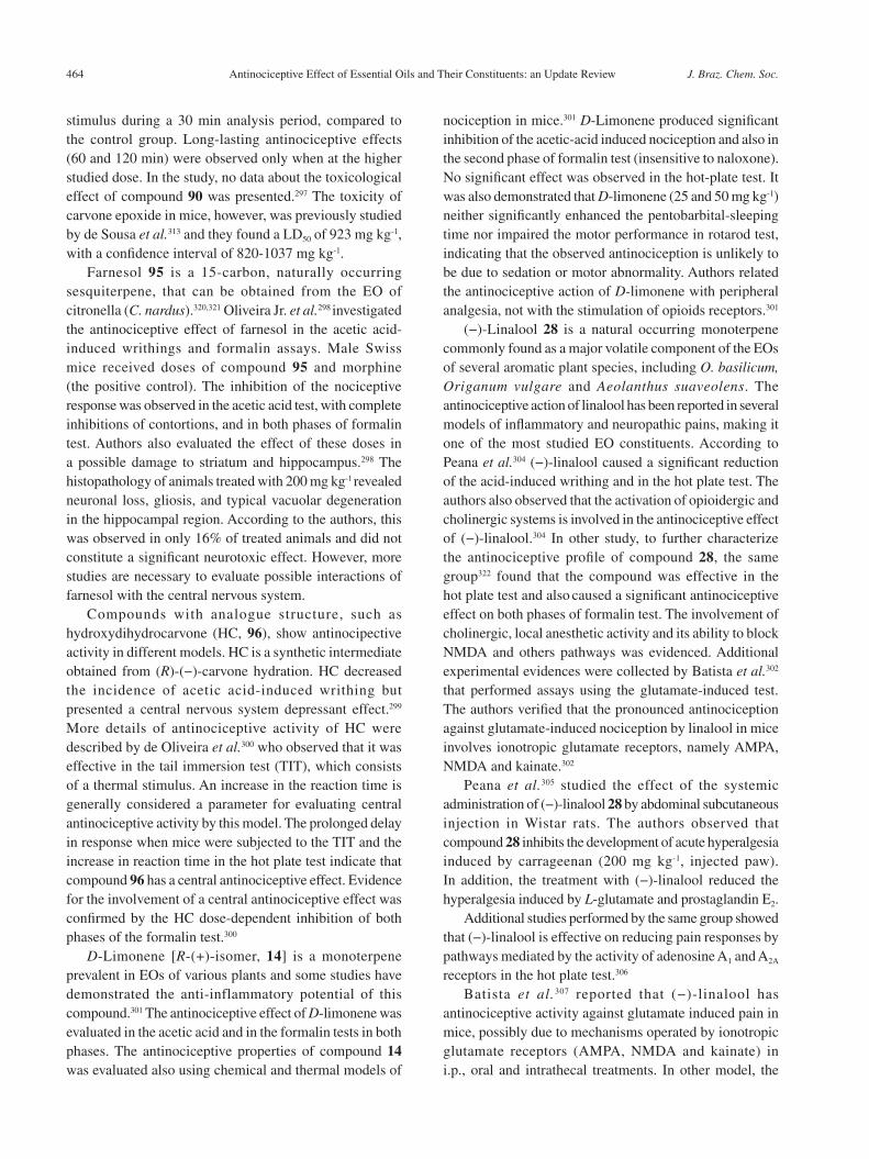
Antinociceptive Effect of Essential Oils and Their Constituents: an Update Review J. Braz. Chem. Soc.464
stimulus during a 30 min analysis period, compared to the control group. Long-lasting antinociceptive effects (60 and 120 min) were observed only when at the higher studied dose. In the study, no data about the toxicological effect of compound 90 was presented.297 The toxicity of carvone epoxide in mice, however, was previously studied by de Sousa et al.313 and they found a LD50 of 923 mg kg-1, with a confidence interval of 820-1037 mg kg-1.
Farnesol 95 is a 15-carbon, naturally occurring sesquiterpene, that can be obtained from the EO of citronella (C. nardus).320,321 Oliveira Jr. et al.298 investigated the antinociceptive effect of farnesol in the acetic acid-induced writhings and formalin assays. Male Swiss mice received doses of compound 95 and morphine (the positive control). The inhibition of the nociceptive response was observed in the acetic acid test, with complete inhibitions of contortions, and in both phases of formalin test. Authors also evaluated the effect of these doses in a possible damage to striatum and hippocampus.298 The histopathology of animals treated with 200 mg kg-1 revealed neuronal loss, gliosis, and typical vacuolar degeneration in the hippocampal region. According to the authors, this was observed in only 16% of treated animals and did not constitute a significant neurotoxic effect. However, more studies are necessary to evaluate possible interactions of farnesol with the central nervous system.
Compounds with analogue structure, such as hydroxydihydrocarvone (HC, 96), show antinocipective activity in different models. HC is a synthetic intermediate obtained from (R)-(−)-carvone hydration. HC decreased the incidence of acetic acid-induced writhing but presented a central nervous system depressant effect.299 More details of antinociceptive activity of HC were described by de Oliveira et al.300 who observed that it was effective in the tail immersion test (TIT), which consists of a thermal stimulus. An increase in the reaction time is generally considered a parameter for evaluating central antinociceptive activity by this model. The prolonged delay in response when mice were subjected to the TIT and the increase in reaction time in the hot plate test indicate that compound 96 has a central antinociceptive effect. Evidence for the involvement of a central antinociceptive effect was confirmed by the HC dose-dependent inhibition of both phases of the formalin test.300
D-Limonene [R-(+)-isomer, 14] is a monoterpene prevalent in EOs of various plants and some studies have demonstrated the anti-inflammatory potential of this compound.301 The antinociceptive effect of D-limonene was evaluated in the acetic acid and in the formalin tests in both phases. The antinociceptive properties of compound 14 was evaluated also using chemical and thermal models of
nociception in mice.301 D-Limonene produced significant inhibition of the acetic-acid induced nociception and also in the second phase of formalin test (insensitive to naloxone). No significant effect was observed in the hot-plate test. It was also demonstrated that D-limonene (25 and 50 mg kg-1) neither significantly enhanced the pentobarbital-sleeping time nor impaired the motor performance in rotarod test, indicating that the observed antinociception is unlikely to be due to sedation or motor abnormality. Authors related the antinociceptive action of D‑limonene with peripheral analgesia, not with the stimulation of opioids receptors.301
(−)-Linalool 28 is a natural occurring monoterpene commonly found as a major volatile component of the EOs of several aromatic plant species, including O. basilicum, Origanum vulgare and Aeolanthus suaveolens. The antinociceptive action of linalool has been reported in several models of inflammatory and neuropathic pains, making it one of the most studied EO constituents. According to Peana et al.304 (−)-linalool caused a significant reduction of the acid-induced writhing and in the hot plate test. The authors also observed that the activation of opioidergic and cholinergic systems is involved in the antinociceptive effect of (−)-linalool.304 In other study, to further characterize the antinociceptive profile of compound 28, the same group322 found that the compound was effective in the hot plate test and also caused a significant antinociceptive effect on both phases of formalin test. The involvement of cholinergic, local anesthetic activity and its ability to block NMDA and others pathways was evidenced. Additional experimental evidences were collected by Batista et al.302 that performed assays using the glutamate-induced test. The authors verified that the pronounced antinociception against glutamate-induced nociception by linalool in mice involves ionotropic glutamate receptors, namely AMPA, NMDA and kainate.302
Peana et al.305 studied the effect of the systemic administration of (−)-linalool 28 by abdominal subcutaneous injection in Wistar rats. The authors observed that compound 28 inhibits the development of acute hyperalgesia induced by carrageenan (200 mg kg-1, injected paw). In addition, the treatment with (−)-linalool reduced the hyperalgesia induced by L-glutamate and prostaglandin E2.
Additional studies performed by the same group showed that (−)-linalool is effective on reducing pain responses by pathways mediated by the activity of adenosine A1 and A2A receptors in the hot plate test.306
Batista et al.307 reported that (−)-linalool has antinociceptive activity against glutamate induced pain in mice, possibly due to mechanisms operated by ionotropic glutamate receptors (AMPA, NMDA and kainate) in i.p., oral and intrathecal treatments. In other model, the

Lenardão et al. 465Vol. 27, No. 3, 2016
mechanical allodynia, developed and maintained over time following spinal nerve ligation, the number of neuropathic animals was reduced when linalool was administered for seven consecutive days.303
Batista et al.307 showed that (−)-linalool displays significant antinociceptive effects in models of chronic pain in mice, specifically in reduction of mechanical hypersensitivity induced by the neuropathic pain (PSNL) and in the mechanical and cold hypersensitivity caused by a chronic inflammatory model (CFA). Authors administered linalool chronically twice a day for 10 days. The ability to inhibit nociception by proinflammatory cytokines and to modulate the NMDA glutamatergic receptor may be responsible for the activity of linalool in these models.
Menthol (97 and 98) is a natural compound present in several EOs from different species of plants, such as Mentha piperita and M. arvensis and is largely used in the pharmaceutical, cosmetics and food chemistry industries. In 2002, Galeotti et al.308 studied the antinociceptive effect of the two optical isomers of menthol: (−)-menthol 97 and (+)-menthol 98. The results demonstrated peripheral and central effects in mice in the assays of acetic acid and hot plate for the (−)-menthol, while its enantiomer (+)-menthol did not present any effect. Moreover, the study revealed that the antinociceptive effect of (−)-menthol involves the modulation of opioid system.
Methyleugenol (1-allyl-3,4-dimethoxybenzene, 32) is an alkenylbenzene compound found in several EOs (basil, clove, lemongrass and others). According data of Yano et al.309 methyleugenol significantly inhibited the duration of the second phase of formalin test and also inhibited pain-related behaviors induced by intrathecal injection of N-methyl-D-aspartic acid (NMDA). The antinociceptive mechanism can be due to the inhibition of NMDA receptor-mediated hyperalgesia via GABAA receptors.
Silva et al.310 evaluated the antinociceptive effect of myrtenol 99, a monoterpenic alcohol found in EOs of some aromatic plants, such as Tanacetum vulgare323 and Aralia cachemirica.324 The antinociceptive effect was investigated in male Swiss mice using acetic acid-induced writhings, hot plate, formalin, glutamate and capsaicin tests. In this study, morphine (5 mg kg-1, i.p.) and dizocilpine (MK-801, 0.03 mg kg-1, i.p.) were used as the positive controls. Myrtenol reduced 69.6% of writhing responses in the acetic acid assay, 97.6% of the licking time in the second phase of formalin test, 44.9% of licking time in the glutamate test and inhibited 36.9% of the response in capsaicin assay.310 According to these results, myrtenol presented a potential as an antinociceptive drug; however, this work did not report data about its toxicological role. The LD50 of myrtenol was
determined by Bhatia et al.325 and values of 2.45 g kg-1 (for males) and 0.63 g kg-1 (for females) were found.
The effect of (E)‑caryophyllene 34 on acute and chronic pains was studied by Paula-Freire et al.286 (E)-Caryophyllene is a bicyclical sesquiterpene and is one of the major active principles from Cannabis sativa,326 Ocimum gratissimum327 and Cordia verbenaceae.328 Compound 34 was administered in male C57BL/6J mice. Morphine (5 mg kg-1, i.p.) was used as positive control for the acute assays and pregabalin (20 mg kg-1) for the chronic ones. According the authors, (E)-caryophyllene showed marked oral antinociceptive properties in both acute and chronic models of pain. The study of the mechanism of action revealed the involvement of opioid and endocannabinoid pathways. This work did not show any data about the toxicological profile of (E)‑caryophyllene.
β-Pinene 40 and eucalyptol 1 are the major components of the EO of Eucalyptus camaldulensis Dehnh. The antinociceptive action of compounds 40 and 1 was evaluated using the tail flick and hot plate tests.164 Both compounds presented antinociceptive activity in the tail flick and hot plate tests. Moreover, the antinociceptive effect of eucalyptol was almost equivalent to that of morphine and the effect of β-pinene was reversed by the pre-administration of naloxone, indicating the possible participation of opioid receptors on the antinociceptive response.
The major component of the EO from plants of the Labiatae family is pulegone 89, a monoterpene with two enantiomeric forms. (R)-(+)-Pulegone is the major constituent (73.4 %) of Mentha pulegium flowers EO.329 The antinociceptive properties of (R)-(+)-pulegone 89 was studied by de Sousa et al.311 using the formalin and the hot plate tests. It was observed that (R)-(+)-pulegone inhibits in a dose-dependent manner both phases of the formalin test, similarly to morphine and the effect was not blocked by naloxone. A positive effect of (R)-(+)-pulegone was also observed in the hot plate test, with an increase in the reaction time latency.329
7. Conclusions
This review attempts to shed light on the therapeutic potential of EOs and some of their isolated volatile constituents in the prevention or therapy of pain. As can be seen by the analysis of more than 300 articles, EOs and their constituents show antinociceptive effects in different models and their action mechanism is quite variable. There are, however, a limited number of commercially available analgesic drugs based on or made from EOs and terpenoids. The new techniques to improve the effectiveness of terpenoids as a therapetutic agent, such as the complexation

Antinociceptive Effect of Essential Oils and Their Constituents: an Update Review J. Braz. Chem. Soc.466
with β-CD, which allows the controlled liberation of the active molecule, can contribute to increase the number of natural compounds-based drugs in the market. The chemical modification of EOs and their constituents, aiming to improve the water-solubility or to reduce their volatility are also alternatives to be considered. The authors hope that this review attracts the attention of researchers who are seeking for new, natural occurring drugs to consider EOs and their constituents in a near future for additional clinical evaluations and possible applications.
Acknowledgements
The authors are grateful to CAPES, CNPq and FAPERGS for financial support.
Eder J. Lenardão was born in 1968 in Sabáudia‑PR, Brazil. He received his BSc from State University of Londrina and MSc degree from Federal University of Santa Maria‑RS under the guidance of Prof Claudio C. Silveira. In 1997, he earned a PhD degree in organic
chemistry at University of São Paulo, under the guidance of Prof Miguel J. Dabdoub and in 2003, he worked with Prof Antonio L. Braga at UFSM as a postdoctoral fellow. His research interest lies in the area of organic and green chemistry.
Lucielli Savegnago is graduated in Pharmacy‑Clinical Analysis from the Federal University of Santa Maria (2001), Master’s in Biochemistry by the Federal University of Santa Maria (2004) and PhD in Biochemistry from Federal University of Santa Maria
(2007). Her current research interests center around the study of organoselenium compounds with antioxidant, antinociceptive and antidepressant‑like activities.
Raquel G. Jacob was born in 1964 in Rio de Janeiro‑RJ, Brazil. She received her BSc (1989) and MSc degrees (1994) from Federal University of Rio de Janeiro (Brazil) under the guidance of Prof Claudio C. Neto. In 1998, she earned a PhD
degree in organic chemistry at University of São Paulo, under the guidance of Prof Miguel J. Dabdoub and in 2005 she worked with Prof Antonio L. Braga at UFSM
as a postdoctoral fellow. Her research interest lies in the area of structural modifications in essential oils and their constituents by using clean methods.
Francine N. Victoria was born in Pelotas, Brazil. She received her MSc dregree (2010) and her PhD degree in Food Science and Technology (2013), working under the guidance of Prof Eder João Lenardão, at the Federal University of Pelotas (UFPel).
Currently, she is Professor at the Center of Chemical, Pharmaceutical and Food Sciences at UFPel. Her scientific interests include biological activity of natural compounds, like essential oils and fruits extracts, with emphasis in their effect on the oxidative stress.
Débora M. Martinez was born in Cascavel, Brazil. She received her MSc dregree from the Federal University of Pelotas‑RS in 2011 and her PhD degree in Food Science and Technology (2014) working under the direction of Prof Eder João Lenardão.
She is currently postdoctoral fellow with Prof Rafael Radi at the Center for Free Radical and Biomedical Research (CEINBIO), Uruguay, with a CNPq Postdoctoral Fellowship. Her current research interests center on the study of natural antioxidants analogs against nitroxidative stress conditions in model systems.
References
1. Calixto, J. B.; Campos, M. M.; Santos, A. R. S.; Botanical
Analgesic & Antiinflamatory Drug, Ethnopharmacology,
Vol. II ‑ Encyclopedia of Life Support Systems (EOLSS); EOLSS
Publishers: Oxford, 2007.
2. Martínez, M. J.; Betancourt, J.; Alonso-González, N.; Jauregui,
A.; J. Ethnopharmacol. 1996, 52, 171.
3. Calixto, J. B.; Beirith, A.; Ferreira, J.; Santos, A. R. S.; Cechinel
Filho, V.; Yunes, R. A.; Phytother. Res. 2000, 14, 401.
4. Butler, D.; Nature 2008, 453, 840.
5. Li, J. W. H.; Vederas, J. C.; Science 2009, 325, 161.
6. Almeida, R. N.; Navarro, D. S.; Barbosa-Filho, J. M.;
Phytomedicine 2001, 8, 310.
7. Silva, M. A. G.; Aquino, N. M. R.; Moura, B. A.; de Sousa, H.
L.; Lavor, E. P. H.; Vasconcelos, P. F.; Macedo, D. S.; de Sousa,
D. P.; Vasconcelos, S. M. M.; de Sousa, F. C. F.; Fitoterapia
2009, 80, 506.
8. Yunes, R. A.; Cechinel Filho, V.; Ferreira, J.; Calixto, J. B.;
Stud. Nat. Prod. Chem. 2005, 30, 191.

Lenardão et al. 467Vol. 27, No. 3, 2016
9. Farmilo, C. G.; Rhodes, H. L. J.; Hart, H. R. L.; Taylor, H.;
Bull. Narc. 1953, 5, 26.
10. Franz, C.; Novak, J. In Handbook of Essential Oils: Science,
Technology, and Applications; Baser, K. H. C.; Buchbauer, G.,
eds.; CRC Press: New York, 2010.
11. Harrewijn, P.; van Oosten, A. M.; Piron, P. G. M.; Natural
Terpenoids as Messengers: a Multidisciplinary Study of Their
Production, Biologica1 Functions, and Practical Applications;
Kluwer Academic Publishers: Dordrecht, 2000.
12. Dobetsberger, C.; Buchbauer, D.; Flavour Frag. J. 2011, 26,
300.
13. Adorjan, B.; Buchbauer, G.; Flavour Frag. J. 2010, 25, 407.
14. Navarra, M.; Mannucci, C.; Delbò, M.; Calapai, G.; Front.
Pharmacol. 2015, 6, 36.
15. Woronuk, G.; Demissie, Z.; Rheault, M.; Mahmoud, S.; Planta
Medica 2011, 77, 7.
16. de Sousa, D. P.; Molecules 2011, 16, 2233.
17. Guimarães, A. G.; Quintans, J. S.; Quintans Jr., L. J.; Phytother.
Res. 2013, 27, 1.
18. Gonzalez-Burgos, E.; Gomez-Serranillos, M. P.; Curr. Med.
Chem. 2012, 19, 5319.
19. Huang, M.; Lu, J. J.; Huang, M. Q.; Bao, J. L.; Chen, X. P.;
Wang, Y. T.; Expert Opin. Invest. Drugs 2012, 21, 1801.
20. Sá, R. C. S.; Andrade, L. N.; Oliveira R. R. B.; de Sousa, D. P.;
Molecules 2014, 19, 1459.
21. Amorim, A. C. L.; Lima, C. K. F.; Hovell, A. M. C.; Miranda,
A. L. P.; Rezende, C. M.; Phytomedicine 2009, 16, 923.
22. Guimarães, A. G.; Serafini, M. R.; Quintans Jr., L. J.; Expert
Opin. Ther. Pat. 2014, 24, 243.
23. Marreto, R. N.; Almeida, E. E. C. V.; Alves, P. B.; Niculau, E.
S.; Nunes, R. S.; Matos, C. R. S.; Araújo, A. A. S.; Thermochim.
Acta 2008, 475, 53.
24. Pinho, E.; Grootveld, M.; Soares, G.; Henriques, M.; Carbohydr.
Polym. 2014, 101, 121.
25. Brito, R. G.; Araújo, A. A.; Quintans, J. S.; Sluka, K. A.;
Quintans Jr., L. J.; Expert Opin. Drug Deliv. 2015, 12, 1677.
26. Clifford, J.; Woolf, M. D.; Ann. Intern. Med. 2004, 140, 441.
27. Burian, M.; Geisslinger, G.; Pharmacol. Ther. 2005, 107, 139.
28. Gottschalk, A.; Smith, D. S.; Am. Fam. Physician 2001, 6,
1979.
29. Besson, J. M.; Lancet 1999, 353, 1610.
30. Collis, M. G.; Hourani, S. M. O.; Trends Pharmacol. Sci. 1993,
14, 360.
31. Millan, M. J.; Prog. Neurobiol. 1999, 57, 1.
32. Hille, B.; Ion Channels of Excitable Membranes, 3rd ed.; Sinauer
Associates: Sunderland, 2001.
33. Wood, J. N.; Docherty, R.; Annu. Rev. Physiol. 1997, 59, 457.
34. Pang, M.; Kim, Y.; Jung, K. W.; Cho, S.; Lee, D. H.; Drug
Discovery Today 2012, 17, 425.
35. Steinmeyer, J.; Arthritis Res. 2000, 2, 379.
36. Warner, E. A.; Am. J. Med. 2012, 12, 1155.
37. Haghparast, A.; Khani, A.; Lashgari, R.; Fallahian, S.; Drug
Alcohol Depen. 2008, 93, 185.
38. Pourmotabbed, A.; Rostamian, B.; Manouchehri, G.; Pirzadeh-
Jahromi, G.; Sahraei, H.; Ghoshooni, H.; Zardooz, H.;
Kamalnegad, M.; J. Ethnopharmacol. 2004, 95, 431.
39. Pasternak, G. W.; J. Clin. Invest. 2007, 117, 3185.
40. Bie, B.; Pan, Z. Z.; Mol. Pain 2005, 1, 1.
41. Nestler, E. J.; Trends Pharmacol. Sci. 2004, 25, 210.
42. Furst, D. E.; Paulus, H. E. In Arthritis and Allied Conditions;
McCarty, D. J.; Koopman, W. J., eds.; Lea & Febiger:
Philadelphia, 1993.
43. Kirtikara, K.; Swangkul, S.; Ballou, L. R.; Inflamm. Res. 2001,
50, 327.
44. Smith, W. L.; Meade, E. A.; de Witt, D. L.; Ann. N. Y. Acad.
Sci. 1994, 744, 50.
45. Garavito, R. M.; de Witt, D. L.; Biochim. Biophys. Acta 1999,
1441, 278.
46. Aïd, S.; Bosetti, F.; Biochimie 2011, 93, 46.
47. Scheiman, J. M.; Gastroenterol. Clin. North Am. 1996, 25,
279.
48. O’Banion, M. K.; Crit. Rev. Neurobiol. 1999, 13, 45.
49. Macintyre, P. E.; Scott, D. A.; Schug, S. A.; Visser, E. J.; Walker,
S. M.; APM:SE Working Group of the Australian and New
Zealand College of Anaesthetists and Faculty of Pain Medicine,
Acute Pain Management: Scientific Evidence, 3rd ed.; ANZCA
& FPM: Melbourne, 2010.
50. Botting, R.; Ayoub, S. S.; Prostaglandins, Leukotrienes Essent.
Fatty Acids 2005, 72, 85.
51. Chen, L. C.; Ashcroft, D. M.; Headache 2008, 48, 236.
52. Hauser, W.; Bernardy, K.; Uceyler, N.; Sommer, C.; JAMA, J.
Am. Med. Assoc. 2009, 301, 198.
53. Andrews, N.; O’Neill, M. F.; Curr. Opin. Pharmacol. 2011, 11,
39.
54. Calixto, J. B.; Scheidt, C.; Otuki, M.; Santos, A. R. S.; Expert
Opin. Emerging Drugs 2001, 6, 261.
55. Calixto, J. B.; Otuki, M. F.; Santos, A. R. S.; Planta Med. 2003,
69, 973.
56. Veeresham, C.; J. Adv. Pharm. Technol. Res. 2012, 3, 200.
57. Le Bars, D.; Gozariu, M.; Cadden, S. W.; Pharmacol. Rev. 2001,
53, 597.
58. Collier, H. O.; Dinneen, L. C.; Johnson, C. A.; Schneider, C.;
Br. J. Pharmacol. Chemother. 1968, 32, 295.
59. Vinegar, R.; Truax, J. F.; Selph, J. L.; Johnston, P. R. In
Handbook of Experimental Pharmacology, Vol. 50/II; Vane, J.
R.; Ferreira, S. H., eds.; Springer-Verlag: Berlin, 1979.
60. Koster, R.; Anderson, M.; de Beer, E. J.; Fed. Proc. 1959, 18,
412.
61. Fukawa, K.; Kawano, O.; Hibi, M.; Misaki, M.; Ohba, S.;
Hatanaka, Y.; J. Pharmacol. Methods 1980, 4, 251.
62. Vogel, H. G.; Vogel, W. H.; Drug Discovery and Evaluation;
Springer: Berlin, 1997.

Antinociceptive Effect of Essential Oils and Their Constituents: an Update Review J. Braz. Chem. Soc.468
63. Guimarães, A. G.; Oliveira, G. F.; Melo, M. S.; Cavalcanti, S.
C. H.; Antoniolli, A. R.; Bonjardim, L. R.; Silva, F. A.; Santos,
J. P. A.; Rocha, R. F.; Moreira, J. C. F.; Araújo, A. A. S.; Gelain,
D. P.; Quintans Jr., L. J.; Basic Clin. Pharmacol. Toxicol. 2010,
107, 949.
64. Deraedt, R.; Jougney, S.; Delevalcee, F.; Falhout, M.; Eur. J.
Pharmacol. 1980, 61, 17.
65. Ikeda, Y.; Ueno, A.; Naraba, H.; Oh-Ishi, S.; Life Sci. 2001, 69,
2911.
66. Martínez, V.; Thakur, S.; Mogil, J. S.; Taché, Y.; Mayer, E. A.;
Pain 1999, 81, 163.
67. Duarte, I. D.; Nakamura, M.; Ferreira, S. H.; Braz. J. Med. Biol.
Res. 1988, 21, 341.
68. Franzotti, E. M.; Santos, C. V. F.; Rodrigues, H. M. S. L.;
Mourão, R. H. V.; Andrade, M. R.; Antoniolli, A. R.; J.
Ethnopharmacol. 2000, 72, 273.
69. Santos, F. A.; Jeferson, F. A.; Santos, C. C.; Silva, E. R.; Rao,
V. S. N.; Life Sci. 2005, 77, 2953.
70. Hunskaar, S.; Hole, K.; Pain 1987, 30, 103.
71. Coderre, T. J.; Melzack, R.; J. Neurosci. 1992, 12, 3665.
72. Abbadie, C.; Taylor, B. K.; Peterson, M. A.; Basbaum, A. I.;
Pain 1997, 69, 101.
73. Raboisson, P.; Dallel, R.; Neurosci. Biobehav. Rev. 2004, 28,
219.
74. Capuano, A.; de Corato, A.; Treglia, M.; Tringali, G.; Russo,
C. D.; Navarra, P.; Eur. J. Pharmacol. 2009, 605, 57.
75. Okuse, K.; Int. J. Biochem. Cell Biol. 2007, 39, 490.
76. McNamara, C. R.; Mandel-Brehm, J.; Bautista, D. M.; Siemens,
J.; Deranian, K. L.; Zhao, M.; Hayward, N. J.; Chong, J. A.;
Julius, D.; Moran, M. M.; Fanger, C. M.; Proc. Natl. Acad. Sci.
U.S.A. 2007, 104, 13525.
77. Hunter, J. C.; Singh, L.; Neurosci. Lett. 1994, 174, 217.
78. Chen, Y. F.; Tsai, H. Y.; Wu, T. S.; Planta Med. 1995, 61, 2.
79. Elisabetsky, E.; Amador, T. A.; Albuquerque, R. R.; Nunes, D.
S.; Carvalho, A. C. T.; J. Ethnopharmacol. 1995, 48, 77.
80. Rosland, J. H.; Tjolsen, A.; Maehle, B.; Hole, D. K.; Pain 1990,
42, 235.
81. Caterina, M. J.; Leffler, A.; Malmberg, A. B.; Martin, W. J.;
Trafton, J.; Petersen-Zeitz, K. R.; Koltzenburg, M.; Basbaum,
A. I.; Julius, D.; Science 2000, 288, 306.
82. Gottrup, H.; Juhl, G.; Kristensen, A. D.; Lai, R.; Chizh, B. A.;
Brown, J.; Bach, F. W.; Jensen, T. S.; Anesthesiology 2004, 101,
1400.
83. Torebjork, H. E.; Lundberg, L. E.; la Motte, R. H.; J. Physiol.
1992, 448, 765.
84. Woolf, C. J.; Pain 2011, 152, S2.
85. Sakurada, T.; Katsumata, K.; Tan-No, K.; Sakurada, S.; Kisara,
K.; Neuropharmacology 1992, 31, 1279.
86. Pelissier, T.; Pajot, J.; Dallel, R.; Pain 2002, 96, 81.
87. Hughes, A.; Macleod, A.; Growcott, J.; Thomas, I.; Pain 2002,
99, 323.
88. Sakurada, T.; Yogo, H.; Katsumata, K.; Tan-No, K.; Sakurada,
S.; Kisara, K.; Brain Res. 1994, 646, 319.
89. Gamse, R.; Molnar, A.; Lembeck, F.; Life Sci. 1979, 25, 629.
90. Sorkin, L. S.; McAdoo, D. J.; Brain Res. 1993, 607, 89.
91. Calixto, J. B.; Kassuya, C. A.; Ferreira, A. E.; Pharmacol. Ther.
2005, 106, 179.
92. di Marzo, V.; Blumberg, P. M.; Szallasi, A.; Curr. Opin.
Neurobiol. 2002, 12, 372.
93. Szallasi, A.; Cortright, D. N.; Blum, C. A.; Eid, S. R.; Nat. Rev.
Drug Discovery 2007, 6, 357.
94. Sakurada, T.; Matsumura, T.; Moriyama, T.; Sakurada, C.;
Ueno, S.; Sakurada, S.; Pharmacol. Biochem. Behav. 2003, 75,
115.
95. Santos, A. R. S.; Calixto, J. B.; Neuropeptides 1997, 31,
381.
96. Beirith, A.; Santos, A. R. S.; Calixto, J. B.; Brain Res. 2002,
924, 219.
97. Jackson, D. L.; Graff, C. B.; Richardson, J. D.; Hargreaves, K.
M.; Eur. J. Pharmacol. 1995, 284, 321.
98. Zhou, S.; Bonasera, L.; Carlton, S. M.; NeuroReport 1996, 7,
895.
99. McNearney, T.; Speegle, D.; Lawand, N. B.; Lisse, J.; Westlund,
K. N.; J. Rheumatol. 2000, 27, 739.
100. Hudspith, M. J.; Br. J. Anaesth. 1997, 78, 731.
101. Neugebauer, V.; Expert Rev. Neurother. 2001, 1, 207.
102. Neugebauer, V.; Trends Neurosci. 2001, 24, 550.
103. Carlton, S. M.; Zhou, S.; Coggeshall, R. E.; Brain Res. 1998,
790, 160.
104. Chizh, B. A.; Amino Acids 2002, 23, 169.
105. Lufty, K.; Cai, S. X.; Woodward, R. M.; Weber, E.; Pain 1997,
70, 31.
106. Yaksh, T. L.; Rudy, T. A.; Pain 1978, 4, 299.
107. Grumbach, L. In The Prediction of Analgesic Activity in Man
by Animal Testing; Knighton, R. S.; Dumke, P. R., eds.; Pain
Little Brown and Co.: Boston, 1966.
108. Bannon, A. W. In Current Protocols in Pharmacology; John
Wiley & Sons: New Jersey, 2001, ch. 5.7.1-11.
109. Yeomans, D. C.; Proudfit, H. K.; Pain 1996, 68, 141.
110. Taber, R. I. In Advances in Biochemical Psychopharmacology;
Braude, M. C.; Harris, L. S.; May, E. L.; Smith, J. P.; Villareal,
J. E., eds.; Raven Press: New York, 1974.
111. Pearce, J. M. S.; J. Neurol., Neurosurg. Psychiatry 2006, 77,
1317.
112. Lambert, G. A.; Mallos, G.; Zagami, A. S.; J. Neurosci. Methods
2009, 177, 420.
113. Randall, L. O.; Selitto, J. J.; Arch. Int. Pharmacodyn. Ther.
1957, 111, 409.
114. Vaidya, A. B.; Antarkar, V. D. S.; J. Assoc. Physicians India
1994, 42, 221.
115. Bakkali, F.; Averbeck, S.; Averbeck, D.; Idaomar, M.; Food
Chem. Toxicol. 2008, 46, 446.
Lenardão et al. 469Vol. 27, No. 3, 2016
116. Piccinelli, A. C.; Santos, J. A.; Konkiewitz, E. C.; Oesterreich,
S. A.; Formagio, A. S. N.; Croda, J.; Ziff, E. B.; Kassuya, C.
A. L.; Nutr. Neurosci. 2015, 18, 217.
117. Sousa, O. V.; Soares Jr., D. T.; Del-Vechio, G.; Mattosinhos, R.
G.; Gattass, C. R.; Kaplan, M. A. C.; Rev. Bras. Farmacogn.
2004, 14, 11.
118. Sousa, O. V.; Del-Vechio-Vieira, G.; Alves, M. S.; Araújo, A. A.
L.; Pinto, M. A. O.; Amaral, M. P. H.; Rodarte, M. P.; Kaplan,
M. A. C.; Molecules 2012, 17, 11056.
119. Queiroz, J. C. C.; Antoniolli, A. R.; Quintans Jr., L. J.; Brito, R.
G.; Barreto, R. S. S.; Costa, E. V.; da Silva, T. B.; Prata, A. P.
N.; de Lucca Jr., W.; Almeida, J. R. G. S.; Lima, J. T.; Quintans,
J. S. S.; Sci. World J. 2014, 2014, 816450.
120. Hajhashemi, V.; Sajjadi, S. E.; Zomorodkia, M.; Pharm. Biol.
2011, 49, 146.
121. Zendehdel, M.; Torabi, Z.; Hassanpour, S.; Vet. Med.‑Czech.
2015, 60, 63.
122. Hejazian, S. H.; World J. Med. Sci. 2006, 2, 95.
123. Sayyah, M.; Peirovi, A.; Kamalinejad, M.; Iran. Biomed. J.
2002, 6, 141.
124. Goés, L. D. M.; Carvalho, H. O.; Duarte, J. L.; Salgueiro, L. R.;
Cavaleiro, C.; Perazzo, F. F.; Carvalho, J. C. T.; Afr. J. Pharm.
Pharmacol. 2015, 9, 460.
125. Hajhashemi, V.; Sajjadi, S. E.; Heshmati, M.; J. Ethnopharmacol.
2009, 124, 475.
126. Jamshidzadeh, A.; Hamedi, A.; Altalqi, A.; Najibi, A.; Int. J.
Pharm. Res. Scholars 2014, 3, 228.
127. Isçan, G.; Kirimer, N.; Kürkçüoglu, M.; Arabaci, T.; Küpeli,
E.; Baser, K. H.; J. Agric. Food Chem. 2006, 54, 170.
128. Del-Vechio-Vieira, G.; de Sousa, O. V.; Miranda, M. A.; Senna-
Valle, L.; Kaplan, M. A. C.; Braz. Arch. Biol. Technol. 2009,
52, 1115.
129. Hadi, A.; Hossein, N.; Shirin, P.; Najmeh, N.; Abolfazl, M.; Int.
J. Pharm. Sci. Rev. Res. 2014, 24, 237.
130. Maham, M.; Moslemzadeh, H.; Jalilzadeh-Amin, G.; Pharm.
Biol. 2014, 52, 208.
131. Leite, G. O.; Leite, L. H. I.; Sampaio, R. S.; Araruna, M. K. A.;
Rodrigues, F. F. G.; Menezes, I. R. A.; Costa, J. G. M.; Campos,
A. R.; Biomed. Prev. Nutr. 2011, 1, 216.
132. Santos, N. A.; Viana, G. B.; Cunha, W.; Campos, A. R.; da
Costa, J. M.; Int. J. Green Pharm. 2015, 9, 138.
133. Rao, V. S.; Maia, J. L.; Oliveira, F. A.; Lemos, T. L. G.;
Chaves, M. H.; Santos, F. A.; Nat. Prod. Commun. 2007, 2,
1199.
134. Biradar, S.; Kangralkar, V. A.; Mandavkar, Y.; Thakur, M.;
Chougule, N.; Int. J. Pharm. Pharm. Sci. 2010, 2, 112.
135. Rabelo, A. S.; Serafini, M. R.; Rabelo, T. K.; de Melo, M. G.;
Prado, D. S.; Gelain, D. P.; Moreira, J. C.; Bezerra, M. S.;
da Silva, T. B.; Costa, E. V.; Nogueira, P. C. L.; Moraes, V.
R. S.; Prata, A. P. N.; Quintans Jr., L. J.; Araújo, A. A.; BMC
Complementary Altern. Med. 2014, 14, 514.
136. Ximenes, R. M.; Nogueira, L. M.; Cassundé, N. M. R.; Jorge,
R. J. B.; Santos, S. M.; Magalhães, L. P. M.; Silva, M. R.; Viana,
G. S. B.; Araújo, R. M.; de Sena, K. X. F. R.; Albuquerque, J.
F. C.; Martins, R. D.; J. Nat. Med. 2013, 67, 758.
137. Nogueira, L. M.; da Silva, M. R.; dos Santos, S. M.; de
Albuquerque, J. F. C.; Ferraz, I. C.; de Albuquerque, T. T.; Mota,
C. R. F. C.; Araújo, R. M.; Viana, G. S. B.; Martins, R. D.; Havt,
A.; Ximenes, R. M.; J. Evidence‑Based Complementary Altern.
Med. 2015, article ID 620865.
138. Abdon, A. P. V.; Leal-Cardoso, J. H.; Coelho-de-Souza, A. N.;
Morais, S. M.; Santos, C. F.; Braz. J. Med. Biol. Res. 2002, 35,
1215.
139. Arrigoni-Blank, M. F.; Antoniolli, A. R.; Caetano, L. C.;
Campos, D. A.; Blank, A. F.; Alves, P. B.; Phytomedicine 2008,
15, 334.
140. Raymundo, L. J. R. P.; Guilhon, C. C.; Alviano, D. S.; Matheus,
M. E.; Antoniolli, A. R.; Cavalcanti, S. C. H.; Alves, P. B.;
Alviano, C. S.; Fernandes, P. D.; J. Ethnopharmacol. 2011,
134, 725.
141. Evandri, M. G.; Battinelli, L.; Daniele, C.; Mastrangelo, S.;
Bolle, P.; Mazzanti, G.; Food Chem. Toxicol. 2005, 43,
1381.
142. Barocelli, E.; Calcina, F.; Chiavarini, M.; Impicciatore, M.;
Bruni, R.; Biachi, A.; Ballabeni, V.; Life Sci. 2004, 76,
213.
143. Hasanein, P.; Riahi, H.; Med. Princ. Pract. 2015, 24, 47.
144. Sousa, P. J. C.; Linard, C. G. B. M.; Batista, D. A.; Oliveira, A.
C.; Coelho-de-Souza, A. N.; Leal-Cardoso, J. H.; Braz. J. Med.
Biol. Res. 2009, 42, 655.
145. Ricci, E. L.; Toyama, D. O.; Lago, J. H. G.; Romoff, P.; Kirsten,
T. B.; Reis-Silva, T. M.; Bernardi, M. M.; J. Health Sci. Inst.
2010, 28, 289.
146. Ali, T.; Javan, M.; Sonboli, A.; Semnanian, S.; Nat. Prod. Res.
2012, 26, 1529.
147. Ali, T.; Javan, M.; Sonboli, A.; Semnanian, S.; Daru, J. Pharm.
Sci. 2012, 20, 1.
148. Araújo, B. S.; Venâncio, A. M.; Onofre, A. S. C.; Lira, A. F.;
Alves, P. B.; Blank, A. F.; Antoniolli, A. R.; Marchioro, M.;
Estevam, C. S.; Planta Med. 2011, 77, 825.
149. Venâncio, A. M.; Marchioro, M.; Estavam, C. S.; Melo, M. S.;
Santana, M. T.; Onofre, A. S. C.; Guimarães, A. G.; Oliveira,
M. G. B.; Alves, P. B.; Pimentel, H. C.; Quintans Jr., L. J.; Braz.
J. Pharmacog. 2011, 21, 1043.
150. Rabelo, M.; Souza, E. P.; Soares, P. M. G.; Miranda, A. V.;
Matos, F. J. A.; Criddle, D. N.; Braz. J. Med. Biol. Res. 2003,
36, 521.
151. Paula-Freire, L. I.; Andersen, M. L.; Molska, A. G. R.; Köhn,
D. O.; Carlini, E. L. A.; Phytother. Res. 2013, 27, 1220.
152. Lino, C. S.; Gomes, P. B.; Lucetti, D. L.; Diógenes, J. P. L.;
Sousa, F. C. F.; Silva, M. G. V.; Viana, G. S. V.; Phytother. Res.
2005, 19, 708.
Antinociceptive Effect of Essential Oils and Their Constituents: an Update Review J. Braz. Chem. Soc.470
153. Pinho, J. P. M.; Silva, A. S. B.; Pinheiro, B. G.; Sombra, I.;
Bayma, J. C.; Lahlou, S.; Sousa, J. C.; Magalhães, P. J. C.;
Planta Med. 2012, 78, 681.
154. Martínez, A. L.; González-Trujano, M. E.; Pellicer, F.; López-
Muñoz, F. J.; Navarrete, A.; Planta Med. 2009, 75, 508.
155. Takaki, I.; Bersani-Amado, L. E.; Vendruscolo, A.; Sartoretto,
S. M.; Diniz, S. P.; Bersani-Amado, C. A.; Cuman, R. K. N.; J.
Med. Food 2008, 11, 741.
156. de Faria, L. R. D.; Lima, C. S.; Perazzo, F. F.; Carvalho, J. C.
T.; Int. J. Pharm. Sci. Rev. Res. 2011, 7, 1.
157. Hajhashemi, V.; Ghannadi, A.; Pezeshkian, S. K.; J.
Ethnopharmacol. 2002, 82, 83.
158. Hajhashemi, V.; Zolfaghari, B.; Yousefi, A.; Med. Princ. Pract.
2012, 21, 178.
159. Abdollahi, M.; Karimpour, H.; Monsef-Esfehani, H. R.;
Pharmacol. Res. 2003, 48, 31.
160. Skouti, E.; Kattah, A.; Alachkar, A.; Hedda, J. B.; Vincieri, F.;
Int. J. Pharm. Pharm. Sci. 2012, 4, 193.
161. Khalilzadeh, E.; Saiah, G. F.; Hasannejad, H.; Ghaderi,
A.; Ghaderi, S.; Hamidian, G.; Mahmoudi, R.; Eshgi, D.;
Zangisheh, M.; Avicenna J. Phytomed. 2015, 5, 218.
162. Khokra, S. L.; Jain, S.; Kaushik, P.; Kaushik, D.; J. Biol. Act.
Prod. Nat. 2012, 2, 239.
163. Jaffary, F.; Ghannadi, A.; Siahpoush, A.; Fitoterapia 2004, 75,
217.
164. Liapi, C.; Anifandis, G.; Chinou, I.; Kourounakis, A. P.;
Theodosopoulos, S.; Galanopoulou, P.; Planta Med. 2007, 73,
1247.
165. Silva, J.; Abebe, W.; Sousa, S. M.; Duarte, V. G.; Machado, M.
I. L.; Matos, F. J. A.; J. Ethnopharmacol. 2003, 89, 277.
166. Guimarães, A. G.; Melo, M. S.; Bonfim, R. R.; Passos, L. O.;
Machado, S. M. F.; Ribeiro, A. S.; Sobral, M.; Thomazzi, S.
M.; Quintans Jr., L. J.; Rev. Bras. Farmacogn. 2009, 19, 883.
167. dos Santos, G. C. M.; Gomes, G. A.; Gonçalves, G. M.; de
Sousa, L. M.; Santiago, G. M. P.; de Carvalho, M. G.; Marinho,
B. G.; Planta Med. 2014, 80, 1588.
168. Quintão, N. L.; da Silva, G. F.; Antonialli, C. S.; Rocha, L. W.;
Cechinel Filho, V.; Cicció, J. F.; Planta Med. 2010, 76, 1411.
169. Pinheiro, B. G.; Silva, A. S. B.; Souza, G. E. P.; Figueiredo,
J. G.; Cunha, F. Q.; Lahlou, S.; Silva, J. K. R.; Maia, J. G. S.;
Sousa, P. J. C.; J. Ethnopharmacol. 2011, 138, 479.
170. Lima, D. K.; Ballico, L. J.; Lapa, F. R.; Gonçalves, H. P.; de
Souza, L. M.; Iacomini, M.; Werner, M. F.; Baggio, C. H.;
Pereira, I. T.; da Silva, L. M.; Facundo, V. A.; Santos, A. R.; J.
Ethnopharmacol. 2012, 142, 274.
171. Lima, G. M.; Melo, M. S.; Serafini, M. R.; Cavalcanti, S. C.
H.; Gelain, D. P.; Santos, J. P. A.; Blank, A. F.; Alves, P. B.;
Oliveira Neta, P. M.; Lima, J. T.; Rocha, R. F.; Moreira, J. C.
F.; Araújo, A. A. S.; Braz. J. Pharmacog. 2012, 22, 443.
172. Viana, G. S. B.; Vale, T. G.; Pinho, R. S. N.; Matos, F. J. A.; J.
Ethnopharmacol. 2000, 70, 323.
173. Abena, A. A.; Gbenou, J. D.; Yayi, E.; Moudachiroub, M.;
Ongokac, R. P.; Ouamba, J. M.; Silou, T.; Afr. J. Tradit.,
Complementary Altern. Med. 2007, 4, 267.
174. Leite, B. L. S.; Bonfim, R. R.; Antoniolli, A. R.; Thomazzi, S.
M.; Araújo, A. A. S.; Blank, A. F.; Estevam, C. S.; Cambui, E.
V. F.; Bonjardim, L. R.; Albuquerque Jr., L. R. C.; Quintans Jr.,
L. J.; Pharm. Biol. 2010, 48, 1164.
175. Abdel-Fattah, A. M.; Matsumoto, K.; Watanabe, H.; Eur. J.
Pharmacol. 2000, 400, 89.
176. Radulovic, N. S.; Miltojevic, A. B.; McDermott, M.; Waldren,
S.; Parnell, A.; Pinheiro, M. M. G.; Fernandes, P. D.; Menezes,
F. S.; J. Ethnopharmacol. 2011, 135, 610.
177. Pinheiro, M. M. G.; Miltojevic, A. B.; Radulovic, N. S.; Abdul-
Wahab, I. R.; Boylan, F.; Fernandes, P. D.; PLoS One 2015, 10,
e0121063.
178. Sakurada, S.; Sakurada, T.; Mizoguchi, H.; Kuwahta, H.;
Katsuyama, S.; Komatsu, T.; Morrone, L. A.; Corasaniti, M.
T.; Bagetta, G.; Pharmacol. Biochem. Behav. 2011, 97, 436.
179. Campêlo, L. M. L.; Almeida, A. A. C.; Freitas, R. L. M.;
Cerqueira, G. S.; Sousa, G. F.; Saldanha, G. B.; Feitosa, C. M.;
Freitas, R. M.; J. Biomed. Biotechnol. 2011, article ID 678673.
180. Mendes, S. S.; Bomfim, R. R.; Jesus, H. C. T.; Alves, P. B.;
Blank, A. F.; Estevam, C. S.; Antoniolli, A. R.; Thomazzi, S.
M.; J. Ethnopharmacol. 2010, 129, 391.
181. Guilhon, C. C.; Raymundo, L. J. R. P.; Alviano, D. S.; Blank,
A. F.; Arrigoni-Blank, M. F.; Matheus, M. E.; Cavalcanti, S. C.
H.; Alviano, C. S.; Fernandes, P. D.; J. Ethnopharmacol. 2011,
135, 406.
182. Siqueira-Lima, P. S.; Araújo, A. A. S.; Lucchese, A. M.;
Quintans, J. S. S.; Menezes, P. P.; Alves, P. B.; de Lucca Jr., W.;
Santos, M. R. V.; Bonjardim, L. R.; Quintans Jr., L. J.; Basic
Clin. Pharmacol. Toxicol. 2014, 114, 188.
183. Marçal, R. M.; Ptak, D. M.; Krempser, R. R.; Krempser, M. R.;
Cardoso, G. C.; Santos, R. B.; Blank, A. F.; Alves, P. B.; Planta
Med. 2006, 72, 291.
184. Pinho, F. V. S. A.; Coelho-de-Souza, A. N.; Morais, S. M.;
Santos, C. F.; Leal-Cardoso, J. H.; Phytomedicine 2005, 12, 482.
185. Sulaiman, M. R.; Mohamad, T. A. S. T.; Mossadeq, W. S. M.;
Moin, S.; Yusof, M.; Mokhtar, A. F.; Zakaria, Z. A.; Israf, D.
A.; Lajis, N.; Planta Med. 2010, 76, 107.
186. Khalid, M. H.; Akhtar, M. N.; Mohamad, A. S.; Perimal,
E. K.; Akira, A.; Israf, D. A.; Lajis, N. Sulaiman, M. R.; J.
Ethnopharmacol. 2011, 137, 345.
187. Tang, W.; Eisenbrand, G.; Chinese Drugs of Plant Origin.
Chemistry, Pharmacology and Use in Traditional and Modern
Medicine; Springer-Verlag: Kaiserlautern, 1992.
188. Vendruscolo, A.; Takaki, I.; Bersani-Amado, L. E.; Dantas, J. A.;
Bersani-Amado, C. A.; Cuman, R. K. N.; Indian J. Pharmacol.
2006, 38, 58.
189. Almeida, A. M.; Prado, P. I.; Lewinsohn, T. M.; Plant Ecol.
2004, 174, 163.
Lenardão et al. 471Vol. 27, No. 3, 2016
190. Guimarães, A. J. M.; Araújo, G. M.; Corrêa, G. F.; Acta Bot.
Bras. 2002, 16, 317.
191. Bohlmann, F.; Ahmed, M.; King, R. M.; Robinson, H.;
Phytochemistry 1981, 20, 1434.
192. Bohlmann, F.; Ludwig, G.-W.; Jakupovic, J.; King, R. M.;
Robinson, H. A.; Phytochemistry 1983, 22, 983.
193. Lorenzi, H.; Matos, F. J. A.; Plantas Medicinais no Brasil;
Instituto Plantarum: Nova Odessa, 2002.
194. Carvalho, J. C. T.; Fitoterápicos Antiinflamatórios: Aspectos
Químicos, Farmacológicos e Aplicações Terapêuticas; Tecmedd
Editora: São Paulo, 2004.
195. Arambewela, L. S. R.; Arawwawala, L. D. A. M.; Ratnasooriya,
W. D.; J. Ethnopharmacol. 2004, 95, 311.
196. Leal-Cardoso, J. H.; Fonteles, M. C.; An. Acad. Bras. Ciênc.
1999, 71, 207.
197. Cavalcanti, B. C.; Ferreira, J. R.; Cabral, I. O.; Magalhães, H.
I.; de Oliveira, C. C.; Rodrigues, F. A.; Rocha, D. D.; Barros,
F. W.; da Silva, C. R.; Júnior, H. V.; Canuto, K. M.; Silveira, E.
R.; Pessoa, C.; Moraes, M. O.; Food Chem. Toxicol. 2012, 50,
4051.
198. Balza, F.; Towers, G. H. N.; Phytochemistry 1984, 23, 2333.
199. Zhang, Y.; Zhang, J.; Yao, J.; Yang, Y. L.; Wang, L.; Dong, L.
N.; China J. Chin. Mater. Med. 2005, 30, 594.
200. Sofi, P. A.; Zeerak, N. A.; Singh, P.; Turk. J. Biol. 2009, 33,
249.
201. Salehi, P.; Mohammadi, F.; Asghari, B.; Chem. Nat. Compd.
2008, 44, 111.
202. Devasankaraiah, G.; Hanin, I.; Haranath, P. S. R. K.;
Ramanamurthy, P. S. V.; Br. J. Pharmacog. 1974, 52, 613.
203. Creche, J.; Tremouillaux-Guiller, J.; Groeger, D.; Chenieux, J.
C.; Rideau, M. In Biotechnology in Agriculture and Forestry 24
(Medicinal and Aromatic Plants V); Bajaj, Y. P. S., ed.; Springer:
Berlin, 1993.
204. Benavente-García, O.; Castillo, J.; Marin, F. R.; Ortuno, A.; del
Río, J. A.; J. Agric. Food Chem. 1997, 45, 4505.
205. Miyake, Y.; Yamamoto, K.; Morimitsu, Y.; Osawa, T.; J. Agric.
Food Chem. 1997, 45, 4619.
206. Monforte, M.; Trovato, A.; Kirjarainen, S. A.; Forestieri, M.;
Galati, E. M.; lo Curto, R. B.; Farmaco 1995, 9, 595.
207. Salantino, A.; Salantino, M. L. F.; Negri, G.; J. Braz. Chem.
Soc. 2007, 18, 11.
208. Berry, P. E.; Hipp, A. L.; Wurdack, K. J.; van Ee, B.; Riina, R.;
Am. J. Bot. 2005, 92, 1520.
209. Cartaxo, S. L.; Souza, M. M. A.; de Albuquerque, U. P.; J.
Ethnopharmacol. 2010, 131, 326.
210. Craveiro, A. A.; Fernandes, A. G.; Andrade, C. H. S.; Matos,
F. J. A.; Alencar, J. W.; Ciênc. Cult. 1977, 29, 445.
211. Fontenelle, R. O. S.; Moraisa, S. M.; Brito, E. H. S.; Brilhante,
R. S. N.; Cordeiro, R. A.; Nascimento, N. R. F.; Kerntopf, M.
R.; Sidrimc, J. J. C.; Rocha, M. F. G.; J. Appl. Microbiol. 2008,
104, 1383.
212. Matos, F. J. A.; Plantas da Medicina Popular do Nordeste:
Propriedades Atribuídas e Confirmadas; EUFC: Fortaleza,
1999.
213. Saini, N.; Singh, G. K.; Nagori, B. P.; Int. J. Res. Ayurveda
Pharm. 2014, 5, 74.
214. Borges, P.; Pino, J.; Nahrung 1993, 37, 123.
215. Fandohan, P.; Gnonlonfin, B.; Laleye, A.; Gbenou, J. D.;
Darboux, R.; Moudachirou, M.; Food Chem. Toxicol. 2008,
46, 2493.
216. Barceloux, D. G.; Citronella Oil [Cymbopogon nardus (L.)
Rendle], in Medical Toxicology of Natural Substances: Foods,
Fungi, Medicinal Herbs, Plants, and Venomous Animals; John
Wiley & Sons: Hoboken, 2008.
217. Nakahara, K.; Alzoreky, N. S.; Yoshihashi, T.; Nguyen, H. T.
T.; Trakoontivakorn, G.; JARQ 2003, 37, 249.
218. Victoria, F. N.; Radatz, C. S.; Sachini, M.; Jacob, R. G.; Alves,
D.; Savegnago, L.; Perin, G.; Motta, A. S.; da Silva, W. P.;
Lenardão, E. J.; Food Control 2012, 23, 95.
219. Stone, S. C.; Vasconcellos, F. A.; Lenardão, E. J.; do Amaral,
R. C.; Jacob, R. G.; Leite, F. P. L.; Int. J. Appl. Res. Nat. Prod.
2013, 6, 11.
220. Tawatsin, A.; Wratten, S. D.; Scott, R. R.; Thavara, U.;
Techadamrongsin, Y.; J. Vector Ecol. 2001, 26, 76.
221. Cassel, E.; Vargas, R. M. F.; J. Mex. Chem. Soc. 2006, 50,
126.
222. Kilani, S.; Ben Ammar, R.; Bouhlel, I.; Abdelwahed, A.; Hayder,
N.; Mahmoud, A.; Ghedira, K.; Chekir-Ghedira, L.; Environ.
Toxicol. Pharmacol. 2005, 20, 478.
223. Morillas-Sánchez, L.; Fernández-López, C.; Blancoana 2006,
22, 65.
224. Leboeuf, M.; Cave, A.; Bhaumik, P. K.; Mukherjee, B.;
Mukherjee, R.; Phytochemistry 1982, 21, 2783.
225. Parker, J. N.; Parker, P. M.; Eucalyptus: A Medical
Dictionary, Bibliography, and Annotated Research Guide to
Internet References; ICON Health Publications: San Diego,
2004.
226. Coppen, J. J. W.; Eucalyptus the Genus Eucalyptus; Taylor &
Francis: London, 2002.
227. Auricchio, M. T.; Bacchi, E. M.; Rev. Inst. Adolfo Lutz 2003,
62, 55.
228. Victoria, F. N.; Lenardão, E. J.; Savegnago, L.; Perin, G.;
Guimarães, R. J.; Alves, D.; da Silva, W. P.; da Motta, A. S.;
Nascente, P. S.; Food Chem. Toxicol. 2012, 50, 2668.
229. Asgarpanah, J.; Mehrabani, G. D.; Ahmadi, M.; Ranjbar, R.;
Ardebily, M. S.; J. Med. Plants Res. 2012, 6, 1813.
230. Manzoomi, N.; Ganbalani, G. N.; Dastjerdi, H. R.; Fathi, S. A.
A.; Mun. Ent. Zool. 2010, 5, 118.
231. Bordignon, S. A. L.; Napaea 1992, 8, 1.
232. Fiaschi, P.; Pirani, J. R.; Rev. Bras. Bot. 2008, 31, 633.
233. Sayyah, M.; Saroukhani, G.; Peirovi, A.; Kamalinejad, M.;
Phytother. Res. 2003, 17, 733.

Antinociceptive Effect of Essential Oils and Their Constituents: an Update Review J. Braz. Chem. Soc.472
234. Zargari, A.; Medicinal Plants, Vol. 4; Tehran University
Publications: Tehran, 1990.
235. Riaz, M.; Ashraf, C. M.; Chaudhary, F. M.; Pak. J. Sci. Ind. Res.
1989, 32, 33.
236. Omidbaigi, R.; Production and Processing of Medicinal Plants,
Vol. 3; Astane Ghods Publications: Mashhad, 2000.
237. Duke, J. A.; CRC Handbook of Medicinal Herbs; CRC Press:
Boca Raton, 1989.
238. Leung, A. Y.; Foster, S.; Encyclopedia of Common Natural
Ingredients Used in Food, Drugs and Cosmetics; Wiley: New
York, 1996.
239. Hajhashemi, V.; Ghannadi, A.; Sharif, B.; J. Ethnopharmacol.
2003, 89, 67.
240. Neto, R. M.; Matos, F. J. A.; Andrade, V. S.; de Melo, M. C.
N.; Carvalho, C. B. M.; Guimarães, S. B.; Pessoa, O. D. L.;
Silva, S. L.; Silva, S. F. R.; Vasconcelos, P. R. L.; Rev. Bras.
Farmacogn. 2010, 20, 261.
241. Albuquerque, F. S.; Peso-Aguiar, M. C.; Assunção-Albuquerque,
M. J.; Braz. J. Biol. 2008, 68, 837.
242. Pascual, M. E.; Slowing, K.; Carretero, E.; Sánchez-Mata, D.;
Villar, A.; J. Ethnopharmacol. 2001, 76, 201.
243. Viana, G. S. B.; Matos, F. F.; Araújo, W. L.; Matos, F. J. A.;
Craveiro, A. A.; Q. J. Crude Drug Res. 1981, 19, 1.
244. Aguiar, M. L. B. A.; Matos, F. J. A.; Moura, V. R. A.; Ciênc.
Cult. 1984, 36, S547.
245. Canadanovic-Brunet, J.; Cetkovic, G.; Djilas, S.; Tumbas,
V.; Bogdanovic, G.; Mandic, A.; Markov, S.; Cvetkovic, D.;
Canadanovic, V.; J. Med. Food 2008, 11, 133.
246. Moradkhani, H.; Sargsyan, E.; Bibak, H.; Naseri, B.; Sadat-
Hosseini, M.; Fayazi-Barjin, A.; Meftahizade, H.; J. Med. Plants
Res. 2010, 4, 2753.
247. McKay, D. L.; Blumberg, J. B.; Phytother. Res. 2006, 20,
619.
248. Andrade, G. S.; Guimarães, A. G.; Santana, M. T.; Siqueira, R.
S.; Passos, L. O.; Machado, S. M. F.; Ribeiro, A. S.; Sobral, M.;
Almeida, J. R. G. S.; Quintans Jr., L. J.; Rev. Bras. Farmacogn.
2012, 21, 181.
249. Jamzad, Z.; Ingrouille, M.; Simmonds, M.; Taxon. 2003, 52,
92.
250. Lynn, C.; Smitherman, M. D.; James, J.; Mathur, A.; Pediatrics
2005, 115, 297.
251. Sonboli, A.; Salehi, P.; Yousefzadi, M.; Z. Naturforsch. 2004,
59c, 653.
252. Mojaba, F.; Nickavara, B.; Tehrani, H. H.; Iran. J. Pharm. Sci.
2009, 5, 43.
253. Khanna, T.; Zaidi, F. A.; Dandiya, P. C.; Fitoterapia 1993, 5,
407.
254. Zaoui, A.; Cherrah, Y.; Mahassine, N.; Alaoui, K.; Amarouch,
H.; Hassar, M.; Phytomedicine 2002, 9, 69.
255. Badary, O. A.; Al-Shabanah, O. A.; Nagi, M. N.; Al-Bekairi,
A. M.; Almazar, M. M. A.; Drug Dev. Res. 1998, 44, 56.
256. Telci, I.; Bayram, E.; Yilmaz, G.; Avci, B.; Biochem. Syst. Ecol.
2006, 34, 489.
257. Trevisan, M. T. S.; Silva, M. G. V.; Pfundstein, B.; Spiegelhalder,
B.; Owen, R. W.; J. Agric. Food Chem. 2006, 54, 4378.
258. Boskabady, M. H.; Kiani, S.; Haghiri, B.; Daru, J. Pharm. Sci.
2005, 13, 28.
259. Braga, R.; Plantas do Nordeste, Especialmente do Ceará,
Coleção Mossoroense, XLIII, 3ª ed.; Editora Universidade
Federal do Rio Grande do Norte: Mossoró, 1992.
260. Grenand, P.; Moretti, C.; Jacquemin, H.; Farmacopées
Traditionnelles en Guyane; Orstom Edition: Paris, 1987.
261. Gülçın, I.; Oktay, M.; Kireçci, E.; Küfrevioglu, O. I.; Food
Chem. 2003, 83, 371.
262. Amaral, F. M. M.; Ribeiro, M. N. S.; Barbosa-Filho, J. M.;
Reis, A. S.; Nascimento, F. R. F.; Macedo, R. O.; Rev. Bras.
Farmacog. 2006, 16, 696.
263. Siani, A. C.; Ramos, M. F. S.; de Lima, M. O.; Santos, R.;
Ferreira, F. E.; Soares, E. C.; Susunaga, G. S.; Guimarães, A. C.;
Zoghbi, M. G. B.; Henriques, M. G. M. O.; J. Ethnopharmacol.
1999, 66, 57.
264. Allan, R. D.; Correl, R. L.; Wells, R. J.; Tetrahedron Lett. 1969,
10, 4669.
265. Kim, M. J.; Nam, E. S.; Paik, S. I.; J. Korean Acad. Nurs. 2005,
35, 186.
266. López-Muñoz, F. J.; Salazar, L. A.; Castañeda-Hernández, G.;
Villarreal, J. E.; Drug Dev. Res. 1993, 28, 169.
267. Zegura, B.; Dobnik, B.; Niderl, M. N.; Filipi, M.; Environ.
Toxicol. Pharmacol. 2011, 32, 296.
268. dos Santos, O. J.; Barros-Filho, A. K.; Malafaia, O.; Ribas-Filho,
J. M.; Santos, R. H.; Santos, R. A.; ABCD: Arq. Bras. Cir. Dig.
2012, 25, 140.
269. Coutinho, I. H. I. L. S.; Torres, O. J. M.; Matias, J. E. F.; Coelho,
J. C. U.; Stahlke Jr., H. J.; Agulham, M. A.; Bachle, E.; Camargo,
P. A. M.; Pimentel, S. K.; de Freitas, A. C. T.; Acta Cir. Bras.
2006, 21, 49.
270. Cavalher-Machado, S. C.; Rosas, E. C.; Brito, F. A.; Heringe,
A. P.; de Oliveira, R. R.; Kaplan, M. A.; Figueiredo, M. R.;
Henriques, M. G. M. O.; Int. Immunopharmacol. 2008, 8, 1552.
271. Formagio, A. S. N.; Iriguchi, E. K. K.; Roveda, L. M.; Vieira, M.
C.; Cardoso, C. A. L.; Zarate, N. A. H.; Tabaldi, L. A.; Kassuya,
C. A. L.; Lat. Am. J. Pharm. 2011, 30, 1555.
272. Galati, E. M.; Mondello, M. R.; D’Aquino, A.; Miceli, N.;
Sanogo, R.; Tzakou, O.; Monforte, M. T.; J. Ethnopharmacol.
2000, 72, 337.
273. Rosário, A. S.; Secco, R. S.; da Silva, J. B. F.; Acta Amaz. 2004,
34, 139.
274. Kamatou, G. P. P.; Viljoen, A. M.; J. Am. Oil Chem. Soc. 2010,
87, 1.
275. Stojkovic, D.; Sokovic, M.; Glamoclija, J.; Dzamic, A.; Ciric,
A.; Ristic, M.; Grubisic, D.; Food Chem. 2011, 128, 1017.
276. Nasri, S.; Ebrahimi, S.; J. Babol Univ. Med. Sci. 2006, 7, 49.
Lenardão et al. 473Vol. 27, No. 3, 2016
277. Ravishankar, B.; Bhaskaran, R.; Sasikala, C. K.; Bull. Med.
Ethano. Biol. Res. 1985, 6, 72.
278. Quintans, J. S. S.; Soares, B. M.; Ferraz, R. P. C.; Oliveira, A.
C. A.; da Silva, T. B.; Menezes, L. R. A.; Sampaio, M. F. C.;
Prata, A. P. N.; Moraes, M. O.; Pessoa, C.; Antoniolli, A. R.;
Costa, E. V.; Bezerra, D. P.; Planta Med. 2013, 79, 123.
279. Basti, A. A.; Misaghi, A.; Khaschabi, B.; LWT ‑ Food Sci.
Technol. 2007, 40, 973.
280. Misaghi, A.; Basti, A. A.; Food Control 2007, 18, 1043.
281. Moosavy, M.; Basti, A. A.; Misaghia, A.; Salehi, T. Z.;
Abbasifar, R.; Mousavid, H. A. E.; Alipoura, M.; Razavie, N.
E.; Gandomia, H.; Nooria, N.; Food Res. Int. 2008, 41, 1050.
282. Li, Y.; Fabiano-Tixier, A. S.; Chema, F.; Essential Oils as
Reagents in Green Chemistry; Springer: London, 2014.
283. Ipek, E.; Zeytinoglu, H.; Okay, S.; Tuylu, B. A.; Kurkcuoglu,
M.; Baser, K. H. C.; Food Chem. 2005, 93, 551.
284. Santos, F. A.; Rao, V. S.; Phytother. Res. 2000, 14, 240.
285. de Lima, A. B.; Santana, M. B.; Cardoso, A. S.; da Silva,
J. K. R.; Maia, J. G. S.; Sousa, P. J. C. C.; Phytomedicine 2009,
16, 555.
286. Paula-Freire, L. I. G.; Andersen, M. L.; Gama, V. S.; Molska,
G. R.; Carlini, E. L. A.; Phytomedicine 2014, 21, 356.
287. Monzote, L.; Stamberg, W.; Staniek, K.; Gille, L.; Toxicol. Appl.
Pharmacol. 2009, 240, 337.
288. Gonçalves, J. C.; Oliveira, F. S.; Benedito, R. B.; de Sousa,
D. P.; de Almeida, R. N.; de Araújo, D. A.; Biol. Pharm. Bull.
2008, 31, 1017.
289. de Sousa, D. P.; Júnior, E. V. M.; Oliveira, F. S.; de Almeida, R.
N.; Nunes, X. P.; Barbosa-Filho, J. M.; Z. Naturforsch. 2007,
62c, 39.
290. Ortiz, M. I.; Montiel, M. L. R.; García, M. P. G.; Monter, H.
A. P.; Hernandez, G. C.; Cortés, R. C.; Arch. Pharm. Sci. Res.
2010, 33, 1691.
291. Quintans Jr., L. J.; Melo, M. S.; de Sousa, D. P.; Araújo, A. A.
S.; Onofre, A. C. S.; Gelain, D. P.; Gonçalves, J. C. R.; Araújo,
D. A. M.; Almeida, J. R. G. S.; Bonjardim, L. R.; J. Orofac.
Pain 2010, 24, 305.
292. Melo, M. S.; Sena, L. C. S.; Barreto, F. J. N.; Bonjardim, L.
R.; Almeida, J. R. G. S.; Lima, J. T.; de Sousa, D. P.; Quintans
Jr., L. J.; Pharm. Biol. 2010, 48, 411.
293. Brito, R. G.; Guimarães, A. G.; Quintans, J. S. S.; Santos, M.
R. V.; de Sousa, D. P.; Passos Jr., D. B.; Lucca Jr., W.; Brito,
F. A.; Barreto, E. O.; Oliveira, A. P.; Quintans Jr., L. J.; J. Nat.
Med. 2012, 66, 637.
294. Brito, R. G.; Santos, P. L.; Prado, D. S.; Santana, M. T.; Araújo,
A. A. S.; Bonjardim, L. R.; Santos, M. R. V.; de Lucca Jr., W.;
Oliveira, A. P.; Quintans Jr., L. J.; Basic Clin. Pharmacol.
Toxicol. 2013, 112, 215.
295. Rios, E. R. V.; Rocha, N. F. M.; Carvalho, A. M. R.; Vasconcelos,
L. F.; Dias, M. L.; de Sousa, D. P.; de Sousa, F. C. F.; Fonteles,
M. M. F.; Chem.‑Biol. Interact. 2013, 203, 573.
296. Johnson, A.; Kumar, R. A.; Rasheed, S. A.; Chandrika,
S.; Chandrasekhar, A.; Baby, S.; Subramoniam, A.; J.
Ethnopharmacol. 2010, 130, 267.
297. da Rocha, M. L.; Oliveira, L. E. G.; Santos, C. C. M. P.; Sousa,
D. P.; Almeida, R. P.; Araújo, D. A. M.; J. Nat. Med. 2013, 67,
743.
298. Oliveira Jr., W. M.; Benedito, R. B.; Pereira, W. B.; Torres, P.
A.; Ramos, C. A. F.; Costa, J. P.; Tomé, A. R.; de Sousa, D. P.;
Freitas, R. M.; Diniz, M. F. F. M.; Almeida, R. N.; Fundam.
Clin. Pharmacol. 2013, 27, 419.
299. de Sousa, D. P.; Oliveira, F. S.; Almeida, R. N.; Biol. Pharm.
Bull. 2006, 29, 811.
300. de Oliveira, F. S.; de Sousa, D. P.; de Almeida, R. N.; Biol.
Pharm. Bull. 2008, 31, 588.
301. Amaral, J. F.; Silva, M. I.; Neto, M. R.; Neto, P. F.; Moura, B. A.;
de Melo, C. T.; de Araújo, F. L.; de Sousa, D. P.; de Vasconcelos,
P. F.; de Vasconcelos, S. M.; de Sousa, F. C.; Biol. Pharm. Bull.
2007, 30, 1217.
302. Batista, P. A.; Werner, M. F. P.; Oliveira, E. C.; Burgos, L.;
Pereira, P.; Brum, L. F. S.; dos Santos, A. R. S.; Neurosci. Lett.
2008, 440, 299.
303. Berliocchi, L.; Russo, R.; Levato, A.; Fratto, V.; Bagetta, G.;
Sakurada, S.; Sakurada, T.; Mercuri, N. B.; Corasaniti, M. T.;
Int. Rev. Neurobiol. 2009, 85, 221.
304. Peana, A. T.; D’Aquila, P. S.; Chessa, M. L.; Moretti, M. D. L.;
Serra, G.; Pippia, P.; Eur. J. Pharmacol. 2003, 460, 37.
305. Peana, A. T.; Montis, M. G.; Sechi, S.; Sircana, G.; D’Aquila,
P. S.; Pippia, P.; Eur. J. Pharmacol. 2004, 497, 279.
306. Peana, A. T.; Rubattu, P.; Piga, G. G.; Fumagalli, S.; Boatto,
G.; Pippia, P.; Montis, M. G.; Life Sci. 2006, 78, 2471.
307. Batista, P. A.; Werner, M. F. P.; Oliveira, E. C.; Burgos, L.;
Pareira, P.; Brum, L. F. S.; Story, G. M.; Santos, A. R. S.; J.
Pain 2010, 11, 1222.
308. Galeotti, N.; Mannelli, L. C.; Mazzanti, G.; Bartolini, A.;
Ghelardini, C.; Neurosci. Lett. 2002, 322, 145.
309. Yano, S.; Suzuki, Y.; Yukurihara, M.; Kase, Y.; Takeda, S.;
Watanabe, S.; Aburada, M.; Miyamoto, K.; Eur. J. Pharmacol.
2006, 553, 99.
310. Silva, R. O.; Salvadori, M. S.; Sousa, F. B. M.; Santos, M. S.;
Carvalho, N. S.; Sousa, D. P.; Gomes, P. S.; Oliveira, F. A.;
Barbosa, A. L. R.; Freitas, R. M.; de Almeida, R. N.; Medeiros,
J. R.; Flavour Fragrance J. 2014, 29, 184.
311. de Sousa, D. P.; Nóbrega, F. F. F.; de Lima, M. R. V.; de Almeida,
R. N.; Z. Naturforsch. 2011, 66c, 353.
312. Estrella, E.; Plantas Medicinales Amazonicas: Realidad y
Perspectivas; GEF/PNUD: Lima, 1995.
313. de Sousa, D. P.; Nóbrega, F. F. F.; Claudino, F. S.; de Almeida,
R. N.; Leite, J. R.; Mattei, R.; Braz. J. Pharmacog. 2007, 17,
170.
314. Jenner, P. M.; Hagan, E. C.; Taylor, J. M.; Cook, E. L.; Fitzhugh,
O. G.; Food Cosmet. Toxicol. 1964, 2, 327.

Antinociceptive Effect of Essential Oils and Their Constituents: an Update Review J. Braz. Chem. Soc.474
315. Raya, M. D. P.; Utrilla, M. P.; Navarro, M. C.; Jimenez, J.;
Phytother. Res. 1990, 4, 232.
316. Dieter, M. P.; Goehl, T. J.; Jameson, C. W.; Elwell, M. R.;
Hildebrandt, P. K.; Yuan, J. H.; Food Chem. Toxicol. 1993, 31,
463.
317. Pybus, D.; Sell, C.; The Chemistry of Fragrances; The Royal
Society of Chemistry: Cambridge, 1999.
318. Iacobellis, N. S.; lo Cantore, P.; Capasso, F.; Senatore, F.; J.
Agric. Food Chem. 2005, 53, 57.
319. Jirovetz, L.; Buchbauer, G.; Shafi, P. M.; Abraham, G. T.; Acta
Pharm. Turc. 2001, 43, 107.
320. Khan, R.; Sultana, S.; Chem.‑Biol. Interact. 2011, 192, 193.
321. Zhao, J.; Nan, P.; Zhong, Y.; Z. Naturforsch. 2004, 59c, 153.
322. Peana, A. T.; de Montis, M. G.; Nieddu, E.; Spano, M. T.;
D’Aquila, P. S.; Pippia, P.; Eur. J. Pharmacol. 2004, 485, 165.
323. Verma, R. S.; Padali, R. C.; Yadav, A.; Chauhan, A.; Rec. Nat.
Prod. 2010, 4, 163.
324. Bell, S. G.; Chen, X.; Sowden, R. J.; Xu, F.; Williams, J. N.;
Wong, L.; Rao, Z.; J. Am. Chem. Soc. 2003, 125, 705.
325. Bhatia, S. P.; McGinty, D.; Letizia, C. S.; Api, A. M.; Food
Chem. Toxicol. 2008, 46, S237.
326. Malingré, T.; Hendriks, H.; Batterman, S.; Bos, R.; Visser, J.;
Planta Med. 1975, 28, 56.
327. Silva, M. G. V.; Craveiro, A. A.; Matos, F. J. A.; Machado, M.
I. L.; Alencar, J. W.; Fitoterapia 1999, 70, 32.
328. Carvalho Jr., P. M.; Rodrigues, R. F. O.; Sawaya, A. C. H. F.;
Marques, M. O. M.; Shimizu, M. T.; J. Ethnopharmacol. 2004,
95, 297.
329. Lorenzo, D.; Paz, D.; Dellacassa, E.; Davies, P.; Vila, R.;
Cañigueral, S.; Braz. Arch. Biol. Technol. 2002, 45, 519.
Submitted: June 30, 2015
Published online: November 25, 2015
FAPERGS/CAPES has sponsored the publication of this article.