Characterization of proteinases from the midgut of Rhipicephalus (Boophilus) microplus involved in...
15
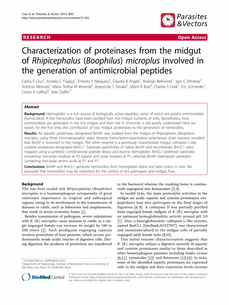
RESEARCH Open Access Characterization of proteinases from the midgut of Rhipicephalus (Boophilus) microplus involved in the generation of antimicrobial peptides Carlos E Cruz 1 , Andréa C Fogaça 1 , Ernesto S Nakayasu 2 , Cláudia B Angeli 1 , Rodrigo Belmonte 1 , Igor C Almeida 2 , Antônio Miranda 3 , Maria Terêsa M Miranda 4 , Aparecida S Tanaka 5 , Glória R Braz 6 , Charles S Craik 7 , Eric Schneider 7 , Conor R Caffrey 8 , Sirlei Daffre 1* Abstract Background: Hemoglobin is a rich source of biologically active peptides, some of which are potent antimicrobials (hemocidins). A few hemocidins have been purified from the midgut contents of ticks. Nonetheless, how antimicrobials are generated in the tick midgut and their role in immunity is still poorly understood. Here we report, for the first time, the contribution of two midgut proteinases to the generation of hemocidins. Results: An aspartic proteinase, designated BmAP, was isolated from the midgut of Rhipicephalus (Boophilus) microplus using three chromatographic steps. Reverse transcription-quantitative polymerase chain reaction revealed that BmAP is restricted to the midgut. The other enzyme is a previously characterized midgut cathepsin L-like cysteine proteinase designated BmCL1. Substrate specificities of native BmAP and recombinant BmCL1 were mapped using a synthetic combinatorial peptide library and bovine hemoglobin. BmCL1 preferred substrates containing non-polar residues at P2 subsite and polar residues at P1, whereas BmAP hydrolysed substrates containing non-polar amino acids at P1 and P1’. Conclusions: BmAP and BmCL1 generate hemocidins from hemoglobin alpha and beta chains in vitro. We postulate that hemocidins may be important for the control of tick pathogens and midgut flora. Background The one-host ixodid tick Rhipicephalus ( Boophilus ) microplus is a haematophagous ectoparasite of great veterinary importance in tropical and subtropical regions, owing to its involvement in the transmission of diseases to cattle, such as babesiosis and anaplasmosis, that result in severe economic losses [1]. Besides transmission of pathogens, severe infestations with R.(B.) microplus cause anaemia in cattle, as a sin- gle engorged female can increase its weight by 100 to 200 times [2]. Such prodigious engorging capacity involves proteolysis of host proteins, which occurs pre- dominantly inside acidic vesicles of digestive cells. Dur- ing digestion the products of proteolysis are transferred to the haemocel whereas the resulting heme is continu- ously segregated into hemosomes [3-5]. In ixodid ticks, the main proteolytic activities in the midgut are acidic aspartic and cysteine proteinases; exo- peptidases may also participate in the final stages of digestion [6-9]. A cathepsin D was partially purified from engorged female midguts of R.(B.) microplus with an optimum hemoglobinolytic activity around pH 3.0 [7]. Also, a hemoglobinolytic cathepsin L-like enzyme, named BmCL1 [GenBank:AF227957], was characterized and immunolocalized to the midgut cells of partially engorged adult female ticks [8,10]. This initial enzyme characterization suggests that R.(B.) microplus utilizes a digestive network of aspartic and cysteine proteinases similar to those described in other hematophagous parasites including Ixodes ricinus [6,11], nematodes [12] and flatworms [13,14]. In ticks, some of the identified aspartic proteinases are expressed only in the midgut and their expression levels increase * Correspondence: [email protected] 1 Department of Parasitology, Institute of Biomedical Sciences, University of São Paulo, São Paulo, SP 05508-900, Brazil Cruz et al. Parasites & Vectors 2010, 3:63 http://www.parasitesandvectors.com/content/3/1/63 © 2010 Cruz et al; licensee BioMed Central Ltd. This is an Open Access article distributed under the terms of the Creative Commons Attribution License (http://creativecommons.org/licenses/by/2.0), which permits unrestricted use, distribution, and reproduction in any medium, provided the original work is properly cited.
Transcript of Characterization of proteinases from the midgut of Rhipicephalus (Boophilus) microplus involved in...
RESEARCH Open Access
Characterization of proteinases from the midgutof Rhipicephalus (Boophilus) microplus involved inthe generation of antimicrobial peptidesCarlos E Cruz1, Andréa C Fogaça1, Ernesto S Nakayasu2, Cláudia B Angeli1, Rodrigo Belmonte1, Igor C Almeida2,Antônio Miranda3, Maria Terêsa M Miranda4, Aparecida S Tanaka5, Glória R Braz6, Charles S Craik7, Eric Schneider7,Conor R Caffrey8, Sirlei Daffre1*
Abstract
Background: Hemoglobin is a rich source of biologically active peptides, some of which are potent antimicrobials(hemocidins). A few hemocidins have been purified from the midgut contents of ticks. Nonetheless, howantimicrobials are generated in the tick midgut and their role in immunity is still poorly understood. Here wereport, for the first time, the contribution of two midgut proteinases to the generation of hemocidins.
Results: An aspartic proteinase, designated BmAP, was isolated from the midgut of Rhipicephalus (Boophilus)microplus using three chromatographic steps. Reverse transcription-quantitative polymerase chain reaction revealedthat BmAP is restricted to the midgut. The other enzyme is a previously characterized midgut cathepsin L-likecysteine proteinase designated BmCL1. Substrate specificities of native BmAP and recombinant BmCL1 weremapped using a synthetic combinatorial peptide library and bovine hemoglobin. BmCL1 preferred substratescontaining non-polar residues at P2 subsite and polar residues at P1, whereas BmAP hydrolysed substratescontaining non-polar amino acids at P1 and P1’.
Conclusions: BmAP and BmCL1 generate hemocidins from hemoglobin alpha and beta chains in vitro. Wepostulate that hemocidins may be important for the control of tick pathogens and midgut flora.
BackgroundThe one-host ixodid tick Rhipicephalus (Boophilus)microplus is a haematophagous ectoparasite of greatveterinary importance in tropical and subtropicalregions, owing to its involvement in the transmission ofdiseases to cattle, such as babesiosis and anaplasmosis,that result in severe economic losses [1].Besides transmission of pathogens, severe infestations
with R. (B.) microplus cause anaemia in cattle, as a sin-gle engorged female can increase its weight by 100 to200 times [2]. Such prodigious engorging capacityinvolves proteolysis of host proteins, which occurs pre-dominantly inside acidic vesicles of digestive cells. Dur-ing digestion the products of proteolysis are transferred
to the haemocel whereas the resulting heme is continu-ously segregated into hemosomes [3-5].In ixodid ticks, the main proteolytic activities in the
midgut are acidic aspartic and cysteine proteinases; exo-peptidases may also participate in the final stages ofdigestion [6-9]. A cathepsin D was partially purifiedfrom engorged female midguts of R. (B.) microplus withan optimum hemoglobinolytic activity around pH 3.0[7]. Also, a hemoglobinolytic cathepsin L-like enzyme,named BmCL1 [GenBank:AF227957], was characterizedand immunolocalized to the midgut cells of partiallyengorged adult female ticks [8,10].This initial enzyme characterization suggests that
R. (B.) microplus utilizes a digestive network of asparticand cysteine proteinases similar to those described inother hematophagous parasites including Ixodes ricinus[6,11], nematodes [12] and flatworms [13,14]. In ticks,some of the identified aspartic proteinases are expressedonly in the midgut and their expression levels increase
* Correspondence: [email protected] of Parasitology, Institute of Biomedical Sciences, University ofSão Paulo, São Paulo, SP 05508-900, Brazil
Cruz et al. Parasites & Vectors 2010, 3:63http://www.parasitesandvectors.com/content/3/1/63
© 2010 Cruz et al; licensee BioMed Central Ltd. This is an Open Access article distributed under the terms of the Creative CommonsAttribution License (http://creativecommons.org/licenses/by/2.0), which permits unrestricted use, distribution, and reproduction inany medium, provided the original work is properly cited.

during blood feeding [11]. In R. (B.) microplus, the con-tribution of other cysteine-proteinases involved in vitel-lin degradation has also been addressed [16,17].Digestion of host blood proteins, particularly hemoglo-
bin, is essential not only to supply nutrients for moltingand vitellogenesis, but also to generate hemocidins thatmay act as a defense mechanism against microorganisms[18-20]. The first isolated hemocidin came from themidgut contents of R. (B.) microplus, and was namedHb 33-61. Its synthetic analogue was active againstGram-positive bacteria and fungi in micromolar concen-trations [18].The structure of the synthetic amidated peptide Hb
33-61a [21] as well as its truncated analogues [22] waselucidated by H1-NMR in micelles of SDS. This datasuggested that the amino-terminal region of Hb 33-61aserves as an anchor to stabilize the peptide into the SDSmembrane, whereas a C-terminal alpha helical portionis responsible for membrane permeabilization. Interest-ingly, several other hemocidins generated through pro-teolytic digestion in vitro contain a high a-helicalcontent [23-25].Here we report the characterization of two proteinases
from R. (B.) microplus midgut involved in hemoglobino-lysis and in the production of hemocidins. To ourknowledge, this is the first report describing proteinasesfrom a living organism that are responsible for the pro-duction of a hemocidin in vivo. The present report high-lights the potential role of hemocidins in the control ofpathogens acquired by ticks as well as the midgut flora.
ResultsProteinase activity in R. (B.) microplus midguthomogenate and digestive cell lysateTo determine which proteinase classes from the midguthomogenate and digestive cell lysate are responsible forthe cleavage of hemoglobin to generate the hemocidinHb 33-61, we used the fluorescent substrates SF 29-35(Abz-LERMFLSQ-EDDnp) and SF 57-67 (Abz-GHGAK-VAAALTQQ-EDDnp) containing the amino acidsequences 29-35 and 57-67 of the a-chain of bovinehemoglobin, and flank the N- and C-terminus of Hb 33-61, respectively, in combination with specific inhibitors.High fluorescence was detected in midgut homogenate
(Table 1A) and digestive cells (Table 1B) at pH 4.5 usingSF 29-35, and this activity was inhibited only by pepsta-tin. At pH 7.0, only a trace of enzymatic activity wasdetected. Thus, we conclude that midgut-associatedacidic aspartic proteinases are involved in the cleavageof SF 29-35.Conversely, using SF 57-67, a low fluorescence was
detected at pH 4.5 and 7.0. However, this activity wassignificantly inhibited by E-64 at pH 4.5 and no signifi-cant enzyme inhibition was observed in the presence of
pepstatin at either pH value (Table 1, panels A and B).Therefore, we conclude that midgut acidic cysteine-pro-teinases are involved in the cleavage of SF 57-67 andthat their specific activity was 40 times lower than thatof aspartic proteinases in both midgut homogenate anddigestive cells.
Purification, kinetic analyses and cleavage specificity ofBmAPBmAP was purified to apparent homogeneity in threechromatographic steps. Midgut homogenate supernatantwas initially resolved using a Mono Q column. Theeluted fractions were screened with SF 29-35 and a sin-gle activity peak, eluted at 80 mM NaCl, was detectedand inhibited by pepstatin (Figure 1A). This active frac-tion was collected and further resolved by hydrophobicinteraction chromatography. A single activity peak waseluted at 1.2 M (NH4)2SO4 (Figure 1B). As a last purifi-cation step, active fractions were pooled and loadedonto a pepstatin affinity column and a single active frac-tion was obtained by a stepwise elution with 0.1 M Tris-HCl, 1 M NaCl, pH 8.6 (Figure 1C). Enzyme recoveriesafter each purification step were 67%, 30% and 21%,respectively. The purified enzyme was designated BmAP.SDS-PAGE analysis of BmAP showed a protein band
at approximately 42 kDa (Figure 1D). The protein bandwas excised, reduced with DTT, alkylated with iodoace-tamide, and in-gel digested with trypsin. The resultingtryptic peptides were analysed by LC-MS/MS and thecollected MS/MS spectra searched against the non-redundant NCBI and R. (B.) microplus BmiGI databases,resulting in the identification of the peptidesVVFDTGSSNLWVPSSK, FDGILGLGYPR and YYTIFDR.Together, these three peptides unambiguously matchedto a single aspartic proteinase sequence from the BmiGIdatabase (contig #3, BmiGI number TC 18173) [Gen-Bank:FJ655904].After purification of BmAP, some of its kinetic prop-
erties were determined. The enzyme has an optimumactivity at pH 4.5 (Figure 2A) and thermally inactivateswith a half-life of 8 min at 68°C, according to apparentfirst-order kinetics, suggesting the presence of only oneisoform in the sample (Figure 2B).Analysis of the cleavage specificity of BmAP using SF
29-35 resulted in the detection of singly charged ions atm/z 702.3, corresponding to the fragment FLSQ-EDDnp(theoretical m/z of 702.4 Da), demonstrating the clea-vage of SF 29-35 at Met32/Phe33 (Figure 2C). Addition-ally, an ion at m/z 927.7 was detected, corresponding tothe fragment Abz-LERMFL (theoretical m/z of 927.5Da), and consequently to the cleavage at Leu34/Ser35(Figure 2D).Chromatographic fractions eluted from the Mono Q
column were also assayed with SF57-67. Two active
Cruz et al. Parasites & Vectors 2010, 3:63http://www.parasitesandvectors.com/content/3/1/63
Page 2 of 15

fractions, eluted at 0.16 and 0.22 M NaCl, were inhib-ited only by E-64, and these fractions were active againstZ-Phe-Arg-MCA but not against Z-Arg-Arg-MCA,which suggests that they contain cathepsin L-like protei-nases (data not shown).
cDNA sequencing of BmAPUsing specific oligonucleotides designed from the identi-fied contig #3, a full-length cDNA was obtained. Itsnucleotide sequence encodes a protein containing 392amino-acid residues, with a predicted molecular mass of42.2 kDa (Figure 3). Using Signal P, the most likely sig-nal peptide cleavage site was identified between residues20 (Ala) and 21 (Leu) of the preprotein, resulting in a372 amino-acid mature polypeptide with a molecularmass of 40.2 kDa and a theoretical isoelectric pointof 8.2.The primary structure of the deduced amino acid
sequence shows the presence of two catalytic triads atAsp89-Thr90-Gly91 and Asp276-Thr277-Gly278 (Figure 3).A SwissModel protein structure homology modellingindicates the presence of three internal disulfide bridgesat Cys102-109, Cys267-271 and Cys310-347. Amino acidsequence alignment using Clustal × showed an identityof 82% and 52% with cathepsins D from Haemaphysalislongicornis [GenBank:AB218595] and Ixodes ricinus[GenBank:EF428204], respectively, and 44% and 30%
identity with the R. (B.) microplus aspartic proteinasesTHAP [GenBank:AF286865] and BYC [GenBank:AY966003], respectively (Figure 4).
Determination of expression levels of BmAP and BmCL1Quantitative RT-PCR profiling of mRNA levels in themidgut, salivary glands and ovaries of fully engorgedfemale ticks revealed that BmAP is only expressed inthe midgut, showing a ΔCt value of 6.99 ± 0.68. Like-wise, BmCL1 is only expressed in the midgut of adultfemales, with a ΔCt value of 1.91 ± 0.25.
Expression of BmCL1 and determination of its substratespecificityThe recombinant cathepsin-L proteinase BmCL1 [Gen-Bank:AF227957] was expressed in Pichia pastoris as anapparently mature, fully active enzyme with a molecularmass of approximately 30 kDa, as determined by SDS-PAGE (Figure 5A). In a thermal inactivation assay, therecombinant enzyme showed a linear decrease in activ-ity, with a half-life of 3.7 min, confirming the presenceof only one isoform purified from Pichia culture med-ium (Figure 5B).LC/MS analysis of the products derived from the
hydrolysis of SF 57-67 by BmCL1 detected singlycharged ions at m/z 839.5, corresponding to the mass ofthe peptide AALTQQ-EDDnp (theoretical m/z of 839.5)
Table 1 Specific enzyme activities in the tick midgut
A
SF 29-35
pH 4.5 pH 7.0
Enzyme activity + pepstatin + E-64 Enzyme activity + pepstatin + E-64
5,471 ± 238 320 ± 6.3a 5,295 ± 173 81 ± 25 65 ± 9.4 62 ± 7.4
SF 57-67
pH 4.5 pH 7.0
Enzyme activity + pepstatin + E-64 Enzyme activity + pepstatin + E-64
141 ± 18 139 ± 17 88 ± 14a 171 ± 21 184 ± 10 168 ± 20
B
SF 29-35
pH 4.5 pH 7.0
Enzyme activity + pepstatin + E-64 Enzyme activity + pepstatin + E-64
6,072 ± 797 329 ± 27a 6,115 ± 206 146 ± 59 154 ± 21 109 ± 29
SF 57-67
pH 4.5 pH 7.0
Enzyme activity + pepstatin + E-64 Enzyme activity + pepstatin + E-64
138 ± 11 108 ± 6.6 33 ± 12a 319 ± 33 320 ± 20 320 ± 26
Enzyme activities (RFU × 103 × min-1 × mg prot-1) were determined in midgut homogenates (A) and digest cell lysate (B) in the presence of pepstatin and E-64at pH 4.5 and 7.0 using SF 29-35 and SF 57-67. Data represent mean ± SEM from three samples. a: p < 0.05.
Cruz et al. Parasites & Vectors 2010, 3:63http://www.parasitesandvectors.com/content/3/1/63
Page 3 of 15

and at m/z 483.2, corresponding to the peptide QQ-EDDnp (theoretical m/z of 483.3 Da), demonstratingcleavage of this substrate at Ala63/Ala64 and Thr67/Gln68, respectively (Figure 5C). The fragment AALT,formed by the simultaneous cleavage of SF 57-67 atAla63/Ala64 and Thr67/Gln68, was also detected at m/z374.5 (theoretical m/z of 375.4 Da) (Figure 5C). Interest-ingly, LC/MS analysis after hydrolysis of SF 57-67 by thecathepsin L-like proteinases partially resolved by anion-exchange chromatography or midgut homogenate at pH4.5 resulted in the detection of the same ions at m/z483.2 and 839.5 (data not shown), which correspond tothe cleavage sites determined for BmCL1.To fully understand the substrate specificity of recom-
binant BmCL1, assays were performed with a positionalscanning synthetic combinatorial library (PS-SCL). Clea-vage specificity profiling at pH 4.5 using the PS-SCLrevealed that BmCL1 preferentially cleaves substratescontaining polar side chains at position P1 (Figure 6,panel A). As expected for papain-like cysteine protei-nases [26], Arg was preferred at P1, but other residuescould also be accommodated at this position, includingGln, Lys and Met. At P2, BmCL1 preferred aliphaticresidues (mainly Val and Leu) and aromatic residues(mainly Phe and Tyr) (Figure 6, panel A). Moreover, theenzyme preferred polar residues at P3 and P4 (Figure 6,panel A). Similar specificities at P1-P4 were observed atpH 5.5, with slightly higher hydrolysis rates beingdetected with Leu over Phe at P2 (Figure 6, panel B). AtpH 5.5, BmCL1 could also hydrolyse peptides containingThr and Glu at P1 (Figure 6, panel B). This preferencefor Val/Leu at P2 supports the observed cleavage of SF57-67 at Ala63/Ala64 and Thr67/Gln68.
Generation of hemoglobin-derived peptides by BmApand BmCL1Figure 7 summarizes the cleavage specificities of nativeBmAP, recombinant BmCL1 and both enzymes againsteither alpha or beta subunit of bovine hemoglobinin vitro. Mass spectrometric analysis of the peptidesgenerated by BmAP identified 20 peptide sequences,with molecular masses ranging from 1085 Da to 3257Da; sixteen were derived from the a subunit and fourfrom the b subunit of bovine hemoglobin (Figure 7,panel A). BmAP preferentially hydrolysed hemoglobin atsites containing non-polar amino acids at P1 (81% ofthe cleavage sites), of which the most prevalent residueswere Leu and Phe (62%) and predominantly at sites con-taining hydrophobic residues at P1’ (65%). Cleavage siteswere found in alpha helical and random coil regions ofhemoglobin and resulted in the generation of severalpeptides previously shown to be antimicrobial [25], suchas a1-29, a1-32, a84-98 and b1-13 (Figure 7, panel A) aswell as potential antimicrobials, such as a33-43 and
Figure 1 Purification of aspartic proteinases from tick midguthomogenates. Purification was accomplished in threechromatographic steps: anion exchange chromatography (A),hydrophobic interaction chromatography (B) and pepstatin affinitychromatography (C) in FPLC system. Enzyme activity was assayedwith SF 29-35 (-o-). Absorbance was monitored at 280 nm (- -). NaClgradients (—) were developed as described in Methods. (D) SDS-PAGE of active fraction eluted from the pepstatin affinitychromatography (see also Additional File 1).
Cruz et al. Parasites & Vectors 2010, 3:63http://www.parasitesandvectors.com/content/3/1/63
Page 4 of 15

a129-141, due to their sequence similarity with the anti-microbials a34-46 and a133-141, respectively [25].Mass spectrometric analysis of the peptides generated
by in vitro hemoglobinolysis using recombinant BmCL1identified 30 peptide sequences (Figure 7, panel B). Con-sidering all the cleavage sites identified, 63% of themwere composed of polar residues at P1. In contrast, 81%of them had apolar residues at P2, with a preference forLeu and Val. No apparent substrate preference wasfound at the P1’ or P2’ subsites. Cleavage sites werefound in alpha helical and random coil regions of hemo-globin and resulted in the generation of several peptides,two of which were previously shown to be antimicrobial:a133-141 [25] and b127-145 (Daffre, unpublished data)(Figure 7, panel B). In addition, peptides a4-25, a130-141, a131-141, b135-145 and b138-145 are potential
antimicrobials, considering their sequence similaritywith known antimicrobials [25] (Figure 7, panel B). Like-wise, hemoglobinolysis with both proteinases allowed forthe identification of 30 peptides (Figure 7, panel C), twoof which were previously reported as human hemocidins(a1-25 and a1-33) [27] and 10 of them as potential anti-microbials [25,27] (Figure 7, panel C).Although the hemocidin Hb 33-61 previously isolated
from R. (B.) microplus midgut [18] was not generated byin vitro hemoglobinolysis, cleavage at Met32/Phe33 wasshown to occur through the activity of BmAP, asobserved by the formation of a1-32 and a33-43 (Figure7, panel A). As expected, cleavage at Lys61/Val62 togenerate Hb 33-61 was not observed in hemoglobin,considering the specificity of BmCL1 (Figure 6, panels Aand B). However, the peptides a48-63 and a50-63,
Figure 2 Biochemical characterization of BmAP. (A) Effect of pH on BmAP activity. Buffer used: 100 mM citrate-phosphate pH 2.5-7.5. (B)Thermal inactivation of BmAP at 68°C at pH 4.5. (C) and (D) Determination of the cleavage sites of SF 29-35 by BmAP using LC/MS.
Cruz et al. Parasites & Vectors 2010, 3:63http://www.parasitesandvectors.com/content/3/1/63
Page 5 of 15

produced by the activity of BmCL1, and a40-63, pro-duced by the activity of both enzymes, were identified inthe hydrolysate (Figure 7, panels B and C), demonstrat-ing cleavages related with Hb 33-61.Considering the structure/activity studies of Hb 33-61
and its truncated analogues Hb 33-52, Hb 40-61 and Hb
48-61 [21,22], we decided to compare the anticandidalactivity of the peptide containing the additional residuesVal62 and Ala63 (Hb 40-63) with these peptides pre-viously characterized. To accomplish that, Hb40-63 wassynthesized by the solid-phase methodology, purified byRP-HPLC and characterized by LC-MS. Its MIC against
Figure 3 Nucleotide and deduced amino acid sequence of R. (B.) microplus aspartic proteinase cDNA. The predicted 20-aa signal peptideis underlined. The conserved residues involved in catalysis are in rectangles. The regions identified by LC-MS/MS are shaded. The cysteineresidues involved in dissulfide bond formation are in black. The GenBank accession number for this sequence is FJ655904.
Cruz et al. Parasites & Vectors 2010, 3:63http://www.parasitesandvectors.com/content/3/1/63
Page 6 of 15

Candida albicans was 3.12-6.25 μM, which is 4-fold moreactive than Hb 33-61 and its analogue Hb 40-61 [22].
DiscussionHydrolysis of the Abz-containing fluorogenic substratesin the presence of midgut homogenate and digestive celllysate showed that aspartic proteinases are involved inthe cleavage of SF 29-35, whereas cysteine proteinaseshydrolyse SF 57-67. Considering that specific activitiesin midgut homogenate were very similar to the activitiesin digestive cells (Table 1), hemoglobin digestion seemsto occur intracellularly, which is consistent with pre-vious data [5].In a previous work, a R. (B.) microplus midgut aspartic
proteinase was partially resolved by ion exchange chro-matography [7]. Here we describe the purification, clon-ing and cleavage specificity of an aspartic proteinase andconfirmed its identity as a cathepsin D. By using threechromatographic steps and performing a thermal inacti-vation assay, we found only one aspartic proteinase iso-form in the tick midgut. Analysis by RT-qPCR showedthat BmAP is only expressed in the midgut, but at pre-sent we are uncertain whether its expression increasesduring blood feeding, as observed for other tick asparticproteinases [28].
A few aspartic proteinases have been characterizedfrom the eggs of R. (B.) microplus. Tick heme-bindingaspartic proteinase (THAP) is a cathepsin-D like protei-nase involved in vitellin digestion, and contains con-served cysteine residues and two typical active sites [29].Another tick aspartic proteinase named BYC (Boophilusyolk cathepsin), in contrast, lacks the second Asp cataly-tic residue. Like BmAP, BYC prefers substrates contain-ing hydrophobic residues at the P1 and P1’ subsites,which is in accordance with the specificity of mostaspartic proteinases reported, including those fromhumans, the schistosome bloodfluke and ixodid ticks[6,12,30-32]. Importantly, we show that BmAP isresponsible for the generation of antimicrobial peptides,such as a1-32 (Figure 7, panel A), which was previouslyreported as being active against a broad range of micro-organisms [25].The second proteinase under investigation, BmCL1,
was previously shown to be immunolocalized in midgutcells of partially engorged adult females, corroboratingits involvement in hemoglobinolysis [8]. It was also veri-fied that BmCL1 is only expressed in the midgut [8],and this data was here confirmed by RT-qPCR. In othertick species, cysteine proteinases were also reported tocontribute to hemoglobinolysis at acidic pH, and to be
Figure 4 Amino acid sequence alignment of aspartic proteinases. BmAP, Rhipicephalus (B.) microplus aspartic proteinase [GenBank:FJ655904];HlAP, Haemaphysalis longicornis aspartic proteinase [GenBank:BAE53722]; IrAP, Ixodes ricinus aspartic proteinase [GenBank:EF428204]; THAP, tickheme-binding aspartic proteinase [GenBank:AF286865]; BYC, Boophilus Yolk pro-Cathepsin [GenBank:AY966003]. The catalytic residues areindicated with asterisks.
Cruz et al. Parasites & Vectors 2010, 3:63http://www.parasitesandvectors.com/content/3/1/63
Page 7 of 15

upregulated by blood feeding [6,15]. Interestingly, alongipain has been characterized in Haemaphysalis long-icornis which may function in the regulation of theirvectorial capacity for Babesia parasites [33].Most variations in papain-like proteases occur at resi-
dues 67 and 205 at the S2 subsite of the active site, andthese residues may reflect differences in substrate speci-ficity [34]. Like human cathepsin L, BmCL1 possesses aLeu in position 67, but unlike the human orthologue, itpossesses a Gln in residue 205 instead of Ala [10],which may explain the lower specificity for bulky aro-matic residues compared with other cathepsin L protei-nases at pH 5.5 [26]. However, cathepsin L proteinasespossessing other residues at position 205, such as Valand Ser, have also been reported [11,35]. Thus, addi-tional studies will be essential to elucidate the characterof the S2 pocket of BmCL1.
BmCL1 was successfully expressed in its active form,which allowed for the characterization of its substratespecificity. PS-SCL mapping revealed that BmCL1 pre-fers substrates containing polar residues at P1, as pre-viously reported for other cysteine proteinases fromtrematode flukes and mammals [26,34,36]. BmCL1showed a broad specificity at positions P3 and P4, whichis in accordance with the specificities of other cathepsinsL using similar libraries [26,34]. For P2, PS-SCL map-ping demonstrated BmCL1’s preference for substratescontaining nonpolar residues (mainly Val, Leu, Phe andTyr), similarly to what was reported for human cathe-psin L but, unlike cathepsin K, BmCL1 does not accom-modate Pro at P2 and, unlike cathepsin B, does notaccept Arg at P2 [26,37,38].Also, PS-SCL data suggest that BmCL1 can process
hemoglobin at multiple sites. In fact, Val, Phe and Leu
Figure 5 Biochemical properties of recombinant BmCL1 expressed in Pichia pastoris. (A) SDS-PAGE of BmCL1 purified on ion exchangechromatography, as described in Methods. (B) Thermal inactivation at 68°C at pH 4.5. (C) Determination of the cleavage sites of SF 57-67 byBmCL1 using LC/MS.
Cruz et al. Parasites & Vectors 2010, 3:63http://www.parasitesandvectors.com/content/3/1/63
Page 8 of 15

were the most prevalent residues at P2 using both PS-SCL and hemoglobin. With the PS-SCL, BmCL1 pre-ferred Val over Phe over Leu at pH 4.5, whereas inhemoglobin it preferred Leu over Val over Phe at thesame pH. Likewise, a preference for Leu/Val wasobserved for Plasmodium falcipain-2 and falcipain-3using a similar P1-P4 library [39].The PS-SCL specificity also showed that BmCL1 does
not hydrolyse substrates containing Ala at P2, whichcorresponds with the proteinase not hydrolyzing alphaglobin at Lys61/Val62 to generate the C-terminus of Hb33-61 (which has Ala at P2). Moreover, when SF 57-67was employed as substrate, cleavage at Ala63/Ala64(with Val at P2) was detected (Figure 5C), and the samecleavage pattern occurred when using midgut extract orthe cysteine proteinases partially purified from the mid-gut (data not shown). Thus we hypothesize that cysteineproteinases are indirectly involved in the generation ofHb 33-61, and midgut exopeptidases may furtherdegrade peptide fragments having Ala 63 at the C-termi-nus providing Hb 33-61 in vivo.Hydrolysis of hemoglobin by either or both BmAP and
BmCL1 resulted in the generation of several peptides(summarized in Figure 7), some of which have primarystructures similar to known antimicrobials, suggestingan antimicrobial activity. Interestingly, hydrolysis ofhemoglobin by BmAP and BmCL1 resulted in the gen-eration of hemocidin Hb 40-63. The synthetic form ofHb 40-63 presented a MIC of 3.12-6.25 μM against
Candida albicans, which is four times more potent thanHb 33-61 and the truncated analogue Hb 40-61, sug-gesting that the presence of Val62 and Ala63 at the car-boxy-terminus induces a more favorable peptideinteraction with the fungal plasma membrane. Also, thepeptide Hb 34-63 identified in this hydrolysate (Figure7, panel C), very likely possesses antifungal activity, con-sidering it contains the same structural elements of Hb33-61 [21].Similarly to the proteinase classes identified in the
midgut of R. (B.) microplus, a network of aspartic andcysteine proteinases has been described in the midgut ofIxodes ricinus, and this network is induced upon bloodfeeding and degrades hemoglobin at acidid conditions[6]. In that work, a peptidase-specific cleavage map wasobtained using hemoglobin as substrate, demonstratingthat aspartic proteinases, supported by cathepsin L andlegumain, are responsible for the primary cleavage ofhemoglobin, whereas cathepsins B, L and C are involvedin the generation of smaller peptides [6]. Interestingly,Ixodes aspartic proteinases showed preference for hydro-phobic residues at the P1 and P1’ subsites (Leu and Pheat P1), whereas cysteine proteinases preferred polar resi-dues at P1 [6], similarly to the specificity observed forBmAP and BmCL1.Endogenously generated hemocidins may help control
host-acquired pathogens as well as tick midgut flora. Inagreement with this hypothesis, our group has identifiedthe hemocidin Hb 98-114, which was confirmed to be
Figure 6 Subsite specificity of recombinant BmCL1 using a tetrapeptide library. A P1-P4 positional scanning synthetic combinatorial library(PS-SCL) was used to determine specificities at pH 4.5 (A) and pH 5.5 (B). Activities are represented as the percentage of the maximum activityfor each subsite position. Data represent means ± SEM of three runs.
Cruz et al. Parasites & Vectors 2010, 3:63http://www.parasitesandvectors.com/content/3/1/63
Page 9 of 15

endogenously generated in the midgut, and its primarysequence corroborates the specificity data presentedherein (Daffre, unpublished data). The characterizationof this biologically active peptide is currently underwayand its activity is being tested against natural pathogensof this tick species.
ConclusionDuring blood feeding, ticks may acquire several patho-gens from their host and become efficient vectors of anumber of disease-causing organisms, such as Ana-plasma marginale [40,41]. Considering that the midgutis the primary interface of the tick-pathogen interaction,this organ needs to have an efficient innate defensemechanism in order to control pathogens as well asmidgut flora. The immune responses of midgut epithe-lial cells to parasite invasion are well-known in otherhematophagous vectors, e.g. mosquitoes [42], but littleinformation is available for ticks [43,44]. A major
component of immunity in the tick midgut may includeantimicrobial fragments generated by endogenousproteolytic activity [18-20].We conclude that the enzyme network present in the
tick midgut involves aspartic and cysteine proteinases,as shown in other parasites [6,11,14]. Additionally, basedon the complementarity of specificity of BmAP andBmCL1 at P1, these proteinases may act in a coordinatefashion to digest hemoglobin and generate hemocidinsin the tick midgut.
MethodsAnimalsA Rhipicephalus (Boophilus) microplus tick colony(Porto Alegre strain, Babesia spp.-free) was reared oncalves maintained at the Center of Biotechnology, Fed-eral University of Rio Grande do Sul, Porto Alegre, RS,Brazil. Host-detached fully engorged females were col-lected and maintained at 28°C and 80% relative humidity
Figure 7 Peptides generated by acid hemoglobinolysis. Peptides were identified by LC-MS/MS after hemoglobinolysis with native BmAP (A),BmCL1 (B) or both enzymes (C). Digestion was performed in 100 mM citrate-phosphate buffer pH 4.5 at 37°C for up to 4 h, as described inMethods.
Cruz et al. Parasites & Vectors 2010, 3:63http://www.parasitesandvectors.com/content/3/1/63
Page 10 of 15
in a BOD incubator (Fanem). Tick rearing followedinstitutional guidelines and was approved by the EthicsCommittee of the Federal University of Rio Grandedo Sul.
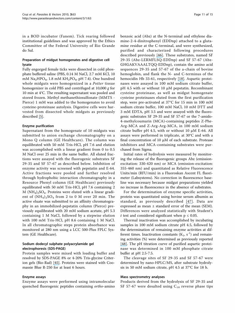
Preparation of midgut homogenates and digestive celllysateFully engorged female ticks were dissected in cold phos-phate buffered saline (PBS, 0.14 M NaCl, 2.7 mM KCl, 10mM Na2HPO4, 1.8 mM KH2PO4, pH 7.4). One hundredwhole midguts were homogenized in a Potter tissuehomogenizer in cold PBS and centrifuged at 10,000 g for10 min at 4°C. The resulting supernatant was pooled andstored frozen. Methyl methanethiosulfonate (MMTS -Pierce) 1 mM was added to the homogenates to avoidcysteine-proteinase autolysis. Digestive cells were har-vested from dissected whole midguts as previouslydescribed [5].
Enzyme purificationSupernatant from the homogenate of 10 midguts wassubmitted to anion exchange chromatography on aMono Q column (GE Healthcare). The column wasequilibrated with 50 mM Tris-HCl, pH 7.6 and elutionwas accomplished with a linear gradient from 0 to 0.5M NaCl over 25 min in the same buffer. All eluted frac-tions were assayed with the fluorogenic substrates SF29-35 and SF 57-67 as described below. Inhibition ofenzyme activity was screened with pepstatin and E-64.Active fractions were pooled and further resolvedthrough hydrophobic interaction chromatography in aResource Phenyl column (GE Healthcare) previouslyequilibrated with 50 mM Tris-HCl, pH 7.6 containing 2M (NH4)2SO4. Proteins were eluted with a linear gradi-ent of (NH4)2SO4 from 2 to 0 M over 20 min. Theactive eluate was submitted to an affinity chromatogra-phy in an immobilized-pepstatin column (Pierce) pre-viously equilibrated with 20 mM sodium acetate, pH 5.3containing 1 M NaCl, followed by a stepwise elutionwith 100 mM Tris-HCl, pH 8.6 containing 1 M NaCl.In all chromatographic steps protein absorbance wasmonitored at 280 nm using a LCC 500-Plus FPLC Sys-tem (GE Healthcare).
Sodium dodecyl sulphate polyacrylamide gelelectrophoresis (SDS-PAGE)Protein samples were mixed with loading buffer andresolved by SDS-PAGE 8% or 4-20% Tris-glycine Criter-ion gels (Bio-Rad) [45]. Proteins were stained with Coo-massie Blue R-250 for at least 6 hours.
Enzyme assaysEnzyme assays were performed using intramolecularquenched fluorogenic peptides containing ortho-amino
benzoic acid (Abz) at the N-terminal and ethylene dia-mine-2-4-dinitrophenyl (EDDnp) attached to a gluta-mine residue at the C-terminal, and were synthesized,purified and characterized following proceduresdescribed previously [46]. These substrates, named SF29-35 (Abz-LERMFLSQ-EDDnp) and SF 57-67 (Abz-GHGAKVAAALTQQ-EDDnp), contain the amino acidsequences 29-35 and 57-67 of the a-chain of bovinehemoglobin, and flank the N- and C-terminus of thehemocidin Hb 33-61, respectively [18]. Aspartic protei-nases were assayed in 100 mM sodium citrate buffer,pH 4.5 with or without 10 μM pepstatin. Recombinantcysteine proteinase, as well as midgut homogenatecysteine proteinases eluted from the first purificationstep, were pre-activated at 37°C for 15 min in 100 mMsodium citrate buffer, 100 mM NaCl, 10 mM DTT and3 mM EDTA, pH 3.5 and were assayed with the fluoro-genic substrates SF 29-35 and SF 57-67 or the 7-amido-4-methylcoumarin (MCA)-containing peptides Z-Phe-Arg-MCA and Z-Arg-Arg-MCA, in 100 mM sodiumcitrate buffer pH 4.5, with or without 10 μM E-64. Allassays were performed in triplicate, at 30°C and with afinal concentration of 10 μM of each substrate. Proteaseinhibitors and MCA-containing substrates were pur-chased from Sigma.Initial rates of hydrolysis were measured by monitor-
ing the release of the fluorogenic groups Abz (emission-excitation: 330-420 nm) or MCA (emission-excitation:355-460 nm) and quantitated as Relative FluorescenceUnits/min (RFU/min) in a Fluoroskan Ascent FL fluori-meter (Labsystems). No correction in fluorescence base-line was necessary because midgut preparations yieldedno increase in fluorescence in the absence of substrates.For the determination of enzyme specific activities,
protein was quantitated using bovine serum albumin asstandard, as previously described [47]. Data areexpressed as mean ± standard error of the mean (SEM).Differences were analyzed statistically with Student’st test and considered significant when p ≤ 0.05.Thermal inactivation was accomplished by incubating
samples in 100 mM sodium citrate pH 4.5, followed bythe determination of remaining enzyme activities at dif-ferent times. Inactivation constants (K1, s
-1) and remain-ing activities (%) were determined as previously reported[48]. The pH titration curve of purified aspartic protei-nase was determined in 100 mM phosphate citratebuffer at pH 2.5-7.5.The cleavage sites of SF 29-35 and SF 57-67 were
determined by nano-HPLC/MS, after substrate hydroly-sis in 50 mM sodium citrate, pH 4.5 at 37°C for 18 h.
Mass spectrometry analysesProducts derived from the hydrolysis of SF 29-35 andSF 57-67 were desalted using C18 reverse phase tips
Cruz et al. Parasites & Vectors 2010, 3:63http://www.parasitesandvectors.com/content/3/1/63
Page 11 of 15

(ZipTip, Millipore) and loaded onto a fused silica capil-lary column (0.1 × 150 mm, Polymicro) packed withVydac C18 (10-15 μm, 300 Å) beads and coupled to anano-HPLC system (Ultimate model, Dionex). Peptideswere eluted with a linear gradient from 5% to 56%acetonitrile in 0.2% formic acid over 60 min anddirectly analysed in a LCQ™ Duo mass spectrometer(Thermo Scientific), according to a previously reportedprocedure [49].For sequencing of proteins separated by SDS-PAGE,
samples were ‘in-gel’ digested according to a publishedprocedure [50]. Tryptic fragments were desalted in Zip-Tip and analysed by nano-HPLC-MS/MS through a120-min gradient from 5% to 56% acetonitrile in 0.2%formic acid. MS/MS analyses were performed with Bio-works Browser version 3.3 (Thermo Scientific). Peptideswere identified with Sequest® algorithm and validated byconsidering a ΔCn ≥ 0.05 and an XCorr ≥ 1.5, 2.0 and2.5 for singly-, doubly- and triply-charged peptides,respectively. MS/MS spectra were correlated with aNCBI non-redundant database and with a Rhipicephalus(B.) microplus normalized cDNA library (BmiGI data-base - http://www.tigr.org) [51].Peptides derived from in vitro hemoglobinolysis were
desalted in Zip-Tip and sequenced by nano-HPLC-MS/MS using an LCQ™ Duo mass spectrometer. Peptideswere analysed against a bovine protein database usingSequest and validated by considering a ΔCn ≥ 0.085 andan XCorr ≥ 1.8, 2.5 and 3.5 for singly-, doubly- and tri-ply-charged peptides, respectively.
Molecular cloning and quantification of transcript levelsof midgut proteinasesFor RNA extraction, midguts, ovaries and salivary glandswere transferred to a sterile tube containing Trizol (Invi-trogen) following manufacturer’s instructions. Onemicrogram of total RNA was used as template forreverse transcription (RT) using the SMART RACEcDNA Amplification Kit (Clontech). A cDNA encodingan aspartic proteinase was amplified from midgutcDNA using the specific oligonucleotides 5′-CTCGA-GATGGAAGCAAGGCTGTCGAC-3′ (sense) and 5′-GGATTCGCACAGTCTCGCTAGGAATTC-3′ (anti-sense). These oligonucleotides were designed based onthe cDNA sequence of an aspartic proteinase obtainedfrom the BmiGI database. Briefly, 26 cDNA sequencescoding for aspartic proteinases were assembled into fivecontigs and one singleton using the sequence assemblyprogram cap3 with the default parameters provided bythe server [52]. Contig #3 (BmiGI number TC 18173),which is comprised of 8 EST sequences, was used forprimer design.PCR reactions were performed with Taq DNA poly-
merase (Invitrogen) using 35 cycles of 1 min at 94°C,
1 min at 53°C and 1 min at 72°C. The unique ampliconobtained was cloned into pGEM-T Easy Vector System I(Promega) and sequenced using the Big Dye TerminatorCycle Sequencing kit (Applied Biosystems) in an ABIPrism 310 Automated Sequencer (Applied Biosytems).The nucleotide sequence was subjected to similaritysearches in a NCBI non-redundant database usingBLAST and multiple alignments were done using Clus-tal × 2.0 [53]. Secondary structure was predicted usingSwiss-Model Workspace [54]. Signal peptide cleavagewas identified with SignalP 3.0 [55]. The nucleotidesequence corresponding to the full-length open readingframe of this proteinase is deposited in GenBank underaccession number FJ655904.Aspartic proteinase and cysteine proteinase mRNA
levels were determined by RT-qPCR in salivary glands,ovaries and midguts of ticks. Fourty nanograms ofcDNA were subjected to RT-qPCR using the followingoligonucleotides: i. aspartic proteinase, sense (5′-ATAT-CACCCTTGGCACACC-3′) and antisense (5′-GAGATTCGCTATGGTAGCGG-3′); ii. cysteine protei-nase, sense (5′-CTGGAGGGACAGCATTTTCT-3′) andantisense (5′-ATGGTTGTGAAGGTGGTCTC-3′); andiii. 40 S ribosomal protein S3A (gi156026300), sense (5′-GGACGACCGATGGCTACCT-3′) and antisense (5′-TGAGTTGATTGGCGCACTTCT-3′) Amplificationswere performed using Script One-Step RT-PCR kit withSYBR Green (Bio-Rad), in iQ5 Thermal Cycler equip-ment (Bio-Rad). Amplification levels were normalizedagainst the 40 S ribosomal protein S3A using the ΔCt
method [56]. Experiments were done in triplicate anddata are represented as mean ± SEM.
Recombinant expression of BmCL1The DNA fragment encoding the gene of the cathepsinL BmCL1 [GenBank:AF227957] was amplified by PCR,using as template the pMAL-p expression vector con-taining the BmCL1 sequence [10]. The BmCL1 DNAwas cloned into pPIC9 vector, and the protein wasexpressed in the methylotrophic yeast Pichia pastorisand purified as previously reported by others [57].
Determination of P1-P4 specificity using a syntheticcombinatorial librarySubstrate specificities were determined using a completediverse positional scanning synthetic combinatoriallibrary (PS-SCL). Each library contained 160,000 differ-ent tetrapeptide sequences conjugated with the 7-amino-4-carbamoylmethylcoumarin (ACC) fluorophore.Synthesis of this library has been described previously[26]. One-microliter aliquots from each of the 20 subli-braries of the P1-, P2-, P3- and P4- libraries were addedto the wells of a 96-well Microfluor-1 U-bottom plate(Dynex Technologies). Assays were carried out in
Cruz et al. Parasites & Vectors 2010, 3:63http://www.parasitesandvectors.com/content/3/1/63
Page 12 of 15

100 mM phosphate citrate, 10 mM DTT, 1 mM EDTAand 1% DMSO, at pH 4.5 and pH 5.5. Initial rates ofhydrolysis were continuously monitored by the releaseof ACC using a SpectraMax Gemini fluorescence spec-trometer (Molecular Devices) with excitation and emis-sion at 380 and 460 nm, respectively, and cutoff at 435nm. Cysteine proteinases were pre-activated and assayedas described above.
In vitro hemoglobinolysis and cleavage site identificationBovine hemoglobin (Sigma) was incubated with asparticproteinase (BmAP) and/or cysteine proteinase (BmCL1)in 100 mM phosphate citrate buffer, pH 4.5 at 37°C forup to 4 h. Aliquots of the hydrolysate were collected at15 min (cysteine proteinase digestion) and at 120 min(aspartic proteinase digestion) and peptides weresequenced by LC-MS/MS as described above.
Peptide synthesisThe peptide Hb 40-63 was synthesized, purified andcharacterized as previously described [22].
Anticandidal assayAnticandidal activity was monitored against Candidaalbicans (strain MDM8) using a liquid growth inhibitionassay as described previously [22]. The MIC value wasrecorded as the range between the highest concentrationof the peptide at which yeast growth was observed andthe lowest concentration that caused 100% inhibition ofyeast growth. The peptide concentrations tested were inthe range of 0.78-100 μM.
Additional material
Additional file 1: MS/MS spectra of the aspartic proteinase aminoacid sequence. LC-MS/MS data were searched against a non-redundantNCBI database, and the amino acid sequences YYTIFDR (A),VVFDTGSSNLWVPSSK (B) and FDGILGLGYPR (C) were identified bySequest and validated using the parameters described in Methods.Peptide probabilities, DCn values and Xcorr values are given for eachpeptide spectra.
AbbreviationsABZ: ortho-amino benzoic acid; DTT: dithiothreitol; E-64: trans-epoxysuccinyl-L-leucylamido (4-guanidino) butane; EDTA: ethylenediamine tetra-acid acid;MCA: 7-amido-4-methylcoumarin; MIC: minimum inhibitory concentration;MS/MS: tandem mass spectrometry; NMR: nuclear magnetic resonance; PCR:polymerase chain reaction; PS-SCL: positional scanning syntheticcombinatorial library; SDS: sodium dodecyl sulphate; RT-qPCR: real-timepolymerase chain reaction;
AcknowledgementsThe BmCL1 plasmid construction was kindly provided by Dr. Itabajara V. daSilva Junior from Federal University of Rio Grande do Sul, Porto Alegre, RS,Brazil. The authors are grateful to M.S. Elaine Nogueira for her involvementin the synthesis and biological assays of Hb 40-63 and the Biomolecule CoreAnalysis at the Border Biomedical Research Center/Biology/UTEP (NIH grant
# 5G12RR008124-16A1), for the access to the LC-MS instruments. This projectwas supported by NIH R01 CA128765 and P01 AI35707 (CSC and ES) andgrants from FAPESP and CNPq (SD and CEC), including part support withthe Sandler Foundation for a research visit by CEC to the Sandler Center forDrug Discovery.
Author details1Department of Parasitology, Institute of Biomedical Sciences, University ofSão Paulo, São Paulo, SP 05508-900, Brazil. 2The Border Biomedical ResearchCenter, Department of Biological Sciences, UTEP, El Paso, TX 79968, USA.3Department of Biophysics, Federal University of São Paulo, São Paulo, SP04044-020, Brazil. 4Department of Biochemistry, Institute of Chemistry, USP,São Paulo, SP 05508-900, Brazil. 5Department of Biochemistry, FederalUniversity of São Paulo, São Paulo, SP 04044-020, Brazil. 6Department ofBiochemistry, Institute of Chemistry, Federal University of Rio de Janeiro, Riode Janeiro, RJ 21941-909, Brazil. 7Department of Pharmaceutical Chemistry,University of Califormia San Francisco, San Francisco, CA 94158, USA.8Sandler Center for Drug Discovery, University of California San Francisco,San Francisco, CA 94158, USA.
Authors’ contributionsCEC and SD designed the study and prepared the manuscript. CECperformed most of the experimental work. ACF helped perform the qPCRexperiments. CBA, ICA and ESN helped perform the MS/MS experiments andanalyse sequencing data. AM synthesized and purified the Abz-containingfluorogenic substrates. MTMM synthesized and purified Hb 40-63. ASTexpressed BmCL1 in Pichia pastoris. GRB carried out all the sequenceannotation and contig assembly. CSC, ES and CRC helped design andperform the PS-SCL experiments. RB helped analyze MS/MS data. All authorsread and approved the final manuscript.
Competing interestsThe authors declare that they have no competing interests.
Received: 26 March 2010 Accepted: 27 July 2010Published: 27 July 2010
References1. Jonsson NN, Bock RE, Jorgensen WK: Productivity and health effects of
anaplasmosis and babesiosis on Bos indicus cattle and their crosses, andthe effects of differing intensity of tick control in Australia. Vet Parasitol2008, 155:1-9.
2. Jonsson NN: The productivity effects of cattle tick (Boophilus microplus)infestation on cattle, with particular reference to Bos indicus cattle andtheir crosses. Vet Parasitol 2006, 137:1-10.
3. Gough JM, Kemp DH: Acid phosphatase in midgut digestive cells inpartially fed females of the cattle tick Boophilus microplus. J Parasitol1995, 81:341-9.
4. Lara FA, Lins U, Paiva-Silva G, Almeida IC, Braga CM, Miguens FC,Oliveira PL, Dansa-Petretski M: A new intracellular pathway of haemdetoxification in the midgut of the cattle tick Boophilus microplus:aggregation inside a specialized organelle, the hemosome. J Exp Biol2003, 206:1707-15.
5. Lara FA, Lins U, Bechara GH, Oliveira PL: Tracing heme in a living cell:hemoglobin degradation and heme traffic in digest cells of the cattletick Boophilus microplus. J Exp Biol 2005, 208:3093-101.
6. Horn M, Nussbaumerova M, Sanda M, Kovarova Z, Srba J, Franta Z, Sojka D,Bogyo M, Caffrey CR, Kopacek P, Mares M: Hemoglobin digestion inblood-feeding ticks: mapping a multipeptidase pathway by functionalproteomics. Chem Biol 2009, 16:1053-63.
7. Mendiola J, Alonso M, Marquetti MC, Finlay C: Boophilus microplus:Multiple proteolytic activities in the midgut. Exp Parasitol 1996, 82:27-33.
8. Renard G, Lara FA, de Cardoso FC, Miguens FC, Dansa-Petretski M,Termignoni C, Masuda A: Expression and immunolocalization of aBoophilus microplus cathepsin L-like enzyme. Insect Mol Biol 2002,11:325-28.
9. Motobu M, Tsuji N, Miyoshi T, Huang X, Islam MK, Alim MA, Fujisaki K:Molecular characterization of a blood-induced serine carboxypeptidasefrom the ixodid tick Haemaphysalis longicornis. FEBS J 2007, 274:3299-312.
10. Renard G, Garcia JF, Cardoso FC, Richter MF, Sakanari JA, Ozaki LS,Termignoni C, Masuda A: Cloning and functional expression of a
Cruz et al. Parasites & Vectors 2010, 3:63http://www.parasitesandvectors.com/content/3/1/63
Page 13 of 15

Boophilus microplus cathepsin L-like enzyme. Insect Biochem Molec 2000,30:1017-26.
11. Sojka D, Franta Z, Horn M, Hajdusek O, Caffrey CR, Mares M, Kopacek P:Profiling of proteolytic enzymes in the gut of the tick Ixodes ricinusreveals an evolutionarily conserved network of aspartic and cysteinepeptidases. Parasit Vectors 2008, 1:7.
12. Williamson AL, Lecchi P, Turk BE, Choe Y, Hotez PJ, McKerrow JH,Cantley LC, Sajid M, Craik CS, Loukas A: A multi-enzyme cascade ofhemoglobin proteolysis in the intestine of blood-feeding hookworms. JBiol Chem 2004, 279:35950-7.
13. Caffrey CR, McKerrow JH, Salter JP, Sajid M: Blood ‘n’ guts: an update onschistosome digestive peptidases. Trends Parasitol 2004, 20:241-48.
14. Delcroix M, Sajid M, Caffrey CR, Lim KC, Dvorak J, Hsieh I, Bahgat M,Dissous C, McKerrow JH: A multienzyme network functions in intestinalprotein digestion by a platyhelminth parasite. J Biol Chem 2006,281:39316-29.
15. Yamaji K, Tsuji N, Miyoshi T, Islam MK, Hatta T, Alim MA, Takenaka A,Fujisaki K: Hemoglobinase activity of a cysteine protease from the ixodidtick Haemaphysalis longicornis. Parasitol Int 2009, 58:232-7.
16. Estrela A, Seixas A, Termignoni C: A cysteine endopeptidase from tick(Rhipicephalus (Boophilus) microplus) larvae with vitellin digestionactivity. Comp Biochem Physiol B Biochem Mol Biol 2007, 148:410-6.
17. Seixas A, Dos Santos PC, Velloso FF, Vaz ID, Masuda A, Horn F,Termignoni C: A Boophilus microplus vitellin-degrading cysteineendopeptidase. Parasitology 2003, 126:155-63.
18. Fogaca AC, da Silva PI Jr, Miranda MTM, Bianchi AG, Miranda A, Ribolla PE,Daffre S: Antimicrobial activity of a bovine hemoglobin fragment in thetick Boophilus microplus. J Biol Chem 1999, 274:25330-4.
19. Nakajima Y, Ogihara K, Taylor D, Yamakawa M: Antibacterial hemoglobinfragments from the midgut of the soft tick, Ornithodoros moubata(Acari: Argasidae). J Med Entomol 2003, 40:78-81.
20. Sonenshine DE, Hynes WL, Ceraul SM, Mitchell R, Benzine T: Host bloodproteins and peptides in the midgut of the tick Dermacentor variabiliscontribute to bacterial control. Exp Appl Acarol 2005, 36:207-23.
21. Sforca ML, Machado A, Figueredo RC, Oyama S Jr, Silva FD, Miranda A,Daffre S, Miranda MTM, Spisni A, Pertinhez TA: The micelle-boundstructure of an antimicrobial peptide derived from the alpha-chain ofbovine hemoglobin isolated from the tick Boophilus microplus.Biochemistry 2005, 44:6440-51.
22. Machado A, Sforca ML, Miranda A, Daffre S, Pertinhez TA, Spisni A,Miranda MTM: Truncation of amidated fragment 33-61 of bovine alpha-hemoglobin: effects on the structure and anticandidal activity.Biopolymers 2007, 88:413-26.
23. Parish CA, Jiang H, Tokiwa Y, Berova N, Nakanishi K, McCabe D,Zuckerman W, Xia MM, Gabay JE: Broad-spectrum antimicrobial activity ofhemoglobin. Bioorg Med Chem 2001, 9:377-82.
24. Froidevaux R, Krier F, Nedjar-Arroume N, Vercaigne-Marko D, Kosciarz E,Ruckebusch C, Dhulster P, Guillochon D: Antibacterial activity of a pepsin-derived bovine hemoglobin fragment. Febs Letters 2001, 491:159-63.
25. Nedjar-Arroume N, Dubois-Delval V, Adje EY, Traisnel J, Krier F, Mary P,Kouach M, Briand G, Guillochon D: Bovine hemoglobin: an attractivesource of antibacterial peptides. Peptides 2008, 29:969-77.
26. Choe Y, Leonetti F, Greenbaum DC, Lecaille F, Bogyo M, Bromme D,Ellman JA, Craik CS: Substrate profiling of cysteine proteases using acombinatorial peptide library identifies functionally unique specificities. JBiol Chem 2006, 281:12824-32.
27. Mak P, Wojcik K, Wicherek L, Suder P, Dubin A: Antibacterial hemoglobinpeptides in human menstrual blood. Peptides 2004, 25:1839-47.
28. Boldbaatar D, Sikalizyo Sikasunge C, Battsetseg B, Xuan X, Fujisaki K:Molecular cloning and functional characterization of an asparticprotease from the hard tick Haemaphysalis longicornis. Insect BiochemMolec 2006, 36:25-36.
29. Sorgine MHF, Logullo C, Zingali RB, Paiva-Silva GO, Juliano L, Oliveira PL: Aheme-binding aspartic proteinase from the eggs of the hard tickBoophilus microplus. J Biol Chem 2000, 275:28659-65.
30. Brindley PJ, Kalinna BH, Wong JYM, Bogitsh BJ, King LT, Smyth DJ, Verity CK,Abbenante G, Brinkworth RI, Fairlie DP, Smythe ML, Milburn PJ, Bielefeldt-Ohmann H, Zheng Y, McManus DP: Proteolysis of human hemoglobin byschistosome cathepsin D. Mol Biochem Parasitol 2001, 112:103-12.
31. Pimenta DC, Oliveira A, Juliano MA, Juliano L: Substrate specificity ofhuman cathepsin D using internally quenched fluorescent peptides
derived from reactive site loop of kallistatin. Bba-Protein Struct M 2001,1544:113-22.
32. Nascimento-Silva MC, Leal AT, Daffre S, Juliano L, da Silva Vaz I Jr, Paiva-Silva Gde O, Oliveira PL, Sorgine MH: BYC, an atypical asparticendopeptidase from Rhipicephalus (Boophilus) microplus eggs. CompBiochem Physiol B Biochem Mol Biol 2008, 149:599-607.
33. Tsuji N, Miyoshi T, Battsetseg B, Matsuo T, Xuan X, Fujisaki K: A cysteineprotease is critical for Babesia spp. transmission in Haemaphysalis ticks.PLoS Pathog 2008, 4:e1000062.
34. Stack CM, Caffrey CR, Donnelly SM, Seshaadri A, Lowther J, Tort JF,Collins PR, Robinson MW, Xu W, McKerrow JH, Craik CS, Geiger SR,Marion R, Brinen LS, Dalton JP: Structural and functional relationships inthe virulence-associated cathepsin L proteases of the parasitic liverfluke, Fasciola hepatica. J Biol Chem 2008, 283:9896-908.
35. Cristofoletti PT, Ribeiro AF, Terra WR: The cathepsin L-like proteinasesfrom the midgut of Tenebrio molitor larvae: sequence, properties,immunocytochemical localization and function. Insect Biochem Molec2005, 35:883-901.
36. Sajid M, McKerrow JH, Hansell E, Mathieu MA, Lucas KD, Hsieh I,Greenbaum D, Bogyo M, Salter JP, Lim KC, Franklin C, Kim JH, Caffrey CR:Functional expression and characterization of Schistosoma mansonicathepsin B and its trans-activation by an endogenous asparaginylendopeptidase. Mol Biochem Parasitol 2003, 131:65-75.
37. Lecaille F, Choe Y, Brandt W, Li Z, Craik CS, Bromme D: Selective inhibitionof the collagenolytic activity of human cathepsin K by altering its S2subsite specificity. Biochemistry 2002, 41:8447-54.
38. Lecaille F, Bromme D, Lalmanach G: Biochemical properties andregulation of cathepsin K activity. Biochimie 2008, 90:208-26.
39. Subramanian S, Hardt M, Choe Y, Niles RK, Johansen EB, Legac J, Gut J,Kerr ID, Craik CS, Rosenthal PJ: Hemoglobin cleavage site-specificity of thePlasmodium falciparum cysteine proteases falcipain-2 and falcipain-3.PLoS One 2009, 4:e5156.
40. Aguirre DH, Gaido AB, Vinabal AE, De Echaide ST, Guglielmone AA:Transmission of Anaplasma marginale with adult Boophilus microplusticks fed as nymphs on calves with different levels of rickettsaemia.Parasite 1994, 1:405-7.
41. Sonenshine DE: Biology of Ticks. New York/Oxford Oxford University Press1991.
42. Barillas-Mury C, Kumar S: Plasmodium-mosquito interactions: a tale ofdangerous liaisons. Cell Microbiol 2005, 7:1539-45.
43. Sonenshine DE, Hynes WL: Molecular characterization and related aspectsof the innate immune response in ticks. Front Biosci 2008, 13:7046-63.
44. Taylor D: Innate immunity in ticks: a review. J Acarol Soc Jpn 2006,15:109-27.
45. Laemmli UK: Cleavage of structural proteins during the assembly of thehead of bacteriophage T4. Nature 1970, 227:680-5.
46. Kiyota S, Franzoni L, Nicastro G, Benedetti A, Oyama S Jr, Viviani W,Gambarini AG, Spisni A, Miranda MT: Introduction of a chemical constraintin a short peptide derived from human acidic fibroblast growth factorelicits mitogenic structural determinants. J Med Chem 2003, 46:2325-33.
47. Bradford MM: A rapid and sensitive method for the quantitation ofmicrogram quantities of protein utilizing the principle of protein-dyebinding. Anal Biochem 1976, 72:248-54.
48. Segel IH: Enzyme kinetics: behavior and analysis of rapid equilibriumand steady-state enzyme systems. New York Wiley-Interscience 1975.
49. Silva PI Jr, Daffre S, Bulet P: Isolation and characterization of gomesin, an18-residue cysteine-rich defense peptide from the spider Acanthoscurriagomesiana hemocytes with sequence similarities to horseshoe crabantimicrobial peptides of the tachyplesin family. J Biol Chem 2000,275:33464-70.
50. Shevchenko A, Wilm M, Vorm O, Mann M: Mass spectrometric sequencingof proteins silver-stained polyacrylamide gels. Anal Chem 1996, 68:850-8.
51. Guerrero FD, Miller RJ, Rousseau ME, Sunkara S, Quackenbush J, Lee Y,Nene V: BmiGI: A database of cDNAs expressed in Boophilus microplus,the tropical/southern cattle tick. Insect Biochem Molec 2005, 35:585-95.
52. Huang X, Madan A: CAP3: A DNA sequence assembly program. GenomeRes 1999, 9:868-77.
53. Larkin MA, Blackshields G, Brown NP, Chenna R, McGettigan PA,McWilliam H, Valentin F, Wallace IM, Wilm A, Lopez R, Thompson JD,Gibson TJ, Higgins DG: Clustal W and Clustal X version 2.0. Bioinformatics2007, 23:2947-8.
Cruz et al. Parasites & Vectors 2010, 3:63http://www.parasitesandvectors.com/content/3/1/63
Page 14 of 15
54. Arnold K, Bordoli L, Kopp J, Schwede T: The SWISS-MODEL workspace: aweb-based environment for protein structure homology modelling.Bioinformatics 2006, 22:195-201.
55. Nielsen H, Engelbrecht J, Brunak S, von Heijne G: Identification ofprokaryotic and eukaryotic signal peptides and prediction of theircleavage sites. Protein Eng 1997, 10:1-6.
56. Livak KJ, Schmittgen TD: Analysis of relative gene expression data usingReal Time quantitative PCR and the 2(-Delta Delta C(T)) Method. Methods2001, 25:402-8.
57. Sasaki SD, Tanaka AS: rBmTI-6, a Kunitz-BPTI domain protease inhibitorfrom the tick Boophilus microplus, its cloning, expression andbiochemical characterization. Vet Parasitol 2008, 155:133-41.
doi:10.1186/1756-3305-3-63Cite this article as: Cruz et al.: Characterization of proteinases from themidgut of Rhipicephalus (Boophilus) microplus involved in thegeneration of antimicrobial peptides. Parasites & Vectors 2010 3:63.
Submit your next manuscript to BioMed Centraland take full advantage of:
• Convenient online submission
• Thorough peer review
• No space constraints or color figure charges
• Immediate publication on acceptance
• Inclusion in PubMed, CAS, Scopus and Google Scholar
• Research which is freely available for redistribution
Submit your manuscript at www.biomedcentral.com/submit
Cruz et al. Parasites & Vectors 2010, 3:63http://www.parasitesandvectors.com/content/3/1/63
Page 15 of 15