Aspartic protease inhibitors containing tertiary alcohol transition-state mimics
29
Invited review Aspartic protease inhibitors containing tertiary alcohol transition-state mimics Hitesh V. Motwani, Maria De Rosa, Luke R. Odell, Anders Hallberg, Mats Larhed * Department of Medicinal Chemistry, Organic Pharmaceutical Chemistry, BMC, Uppsala University, P.O. Box 574, SE-75123 Uppsala, Sweden article info Article history: Received 9 September 2014 Received in revised form 12 November 2014 Accepted 19 November 2014 Available online 20 November 2014 Keywords: Protease inhibitors Tertiary alcohol Transition-state mimic HIV Malaria Alzheimer abstract Aspartic proteases (APs) are a class of enzymes engaged in the proteolytic digestion of peptide substrates. APs play important roles in physiological and infectious pathways, making them plausible drug targets. For instance in the treatment of HIV infections, access to an efficient combination of protease and reverse transcriptase inhibitors have changed a terminal illness to a chronic but manageable disease. However, the benefits have been limited due to the emergence of drug resistant viral strains, poor pharmacokinetic properties of peptidomimetic inhibitors and adverse effects associated with the treatment. In the 1980s, D. Rich and co-workers proposed a novel strategy for the development of AP inhibitors by replacing the secondary hydroxyl group with a tertiary alcohol as part of the transition state (TS) mimicking moiety. This strategy has been extensively explored over the last decade with a common belief that masking of the polar group, e.g. by intramolecular hydrogen bonding, has the potential to enhance transcellular transport. This is the first review presenting the advances of AP inhibitors comprising a tertiary hydroxyl group. The inhibitors have been classified into different tert-hydroxy TS mimics and their design stra- tegies, synthesis, biological activities, structureeactivity-relationships and X-ray structures are discussed. © 2014 Elsevier Masson SAS. All rights reserved. 1. Introduction 1.1. Aspartic proteases: mechanism of action, relevance in the field of drug discovery and development of transition state mimicking inhibitors Proteases are a family of enzymes that catalyze the hydrolysis of peptide bonds in proteins and polypeptides, a reaction that is vital to both physiological and pathological processes. The human genome encodes over 500 proteases (MEROPS database), making this group one of the largest enzyme families. Proteases are commonly classified into six classes: serine, threonine, cysteine, glutamic, metallo and aspartic proteases (APs) [1e3]. APs are the smallest class in the human genome with only 15 members, but they have long been a rich source of potential drug molecules in the field of drug discovery [4]. The APs have been classified into two clans: clan AA and clan AD, based on their tertiary structures. Clan AA consists of two families: family A1, which contains the classical aspartic proteases (renin, pepsin A, pepsin C, cathepsin D, cathepsin E, BACE-1, BACE-2 and napsin A) and family A2, which contains proteases such as HIV-1 protease that can be integrated into the human genome by retroviruses. Clan AD comprises of presenilins and signal peptide peptidase, which are the intra-membrane cleaving proteases [1e3]. APs use an aspartic acid (Asp) dyad to hydrolyze peptide bonds. Most APs bind to 6e10 amino acid residues in the substrate, which can then be used to design substrate-based inhibitors. APs also have one or more flaps in their structure that close down to cover the substrate/inhibitor, resulting in further interactions within the complex. For example, in the case of the dimeric AP HIV-1 protease, it is thought that a water molecule in the active site is activated by a deprotonated aspartic acid (Asp25 or Asp125), which facilitates a nucleophilic addition to the amide carbonyl carbon in the substrate to form a tetrahedral intermediate compound [the transition state (TS) [5]; Fig. 1]. The other aspartic acid can donate a proton to the Abbreviations: AIDS, acquired immune deficiency syndrome; AD, Alzheimer's disease; Ab, amyloid beta; AP, aspartic protease; Asp, aspartic acid; Cl int , intrinsic clearance; CC 50 , 50% of the cytotoxic concentration; EC 50 , half the maximum effective concentration; HIV, human immunodeficiency virus; IC 50 , concentration required to inhibit enzyme activity by 50%; K i , inhibition constant; Me 3 AHPPA, 4- amino-3-hydroxy-3-methyl-5-phenylpentanoic acid; Me 3 Sta, 4-amino-3-hydroxy- 3,6-dimethylheptanoic acid; P app , permeability; PDB, protein data bank; PI, protease inhibitor; SAR, structureeactivity-relationship; TBS, tert-butyldimethylsilyl; TS, transition state; WHO, World Health Organization. * Corresponding author. E-mail address: [email protected] (M. Larhed). Contents lists available at ScienceDirect European Journal of Medicinal Chemistry journal homepage: http://www.elsevier.com/locate/ejmech http://dx.doi.org/10.1016/j.ejmech.2014.11.036 0223-5234/© 2014 Elsevier Masson SAS. All rights reserved. European Journal of Medicinal Chemistry 90 (2015) 462e490
-
Upload
independent -
Category
Documents
-
view
0 -
download
0
Transcript of Aspartic protease inhibitors containing tertiary alcohol transition-state mimics

lable at ScienceDirect
European Journal of Medicinal Chemistry 90 (2015) 462e490
Contents lists avai
European Journal of Medicinal Chemistry
journal homepage: http: / /www.elsevier .com/locate/ejmech
Invited review
Aspartic protease inhibitors containing tertiary alcoholtransition-state mimics
Hitesh V. Motwani, Maria De Rosa, Luke R. Odell, Anders Hallberg, Mats Larhed*
Department of Medicinal Chemistry, Organic Pharmaceutical Chemistry, BMC, Uppsala University, P.O. Box 574, SE-751 23 Uppsala, Sweden
a r t i c l e i n f o
Article history:Received 9 September 2014Received in revised form12 November 2014Accepted 19 November 2014Available online 20 November 2014
Keywords:Protease inhibitorsTertiary alcoholTransition-state mimicHIVMalariaAlzheimer
Abbreviations: AIDS, acquired immune deficiencydisease; Ab, amyloid beta; AP, aspartic protease; Aspclearance; CC50, 50% of the cytotoxic concentratioeffective concentration; HIV, human immunodeficienrequired to inhibit enzyme activity by 50%; Ki, inhibiamino-3-hydroxy-3-methyl-5-phenylpentanoic acid;3,6-dimethylheptanoic acid; Papp, permeability; PDB, pinhibitor; SAR, structureeactivity-relationship; TBStransition state; WHO, World Health Organization.* Corresponding author.
E-mail address: [email protected] (M. La
http://dx.doi.org/10.1016/j.ejmech.2014.11.0360223-5234/© 2014 Elsevier Masson SAS. All rights re
a b s t r a c t
Aspartic proteases (APs) are a class of enzymes engaged in the proteolytic digestion of peptide substrates.APs play important roles in physiological and infectious pathways, making them plausible drug targets.For instance in the treatment of HIV infections, access to an efficient combination of protease and reversetranscriptase inhibitors have changed a terminal illness to a chronic but manageable disease. However,the benefits have been limited due to the emergence of drug resistant viral strains, poor pharmacokineticproperties of peptidomimetic inhibitors and adverse effects associated with the treatment. In the 1980s,D. Rich and co-workers proposed a novel strategy for the development of AP inhibitors by replacing thesecondary hydroxyl group with a tertiary alcohol as part of the transition state (TS) mimicking moiety.This strategy has been extensively explored over the last decade with a common belief that masking ofthe polar group, e.g. by intramolecular hydrogen bonding, has the potential to enhance transcellulartransport. This is the first review presenting the advances of AP inhibitors comprising a tertiary hydroxylgroup. The inhibitors have been classified into different tert-hydroxy TS mimics and their design stra-tegies, synthesis, biological activities, structureeactivity-relationships and X-ray structures are discussed.
© 2014 Elsevier Masson SAS. All rights reserved.
1. Introduction
1.1. Aspartic proteases: mechanism of action, relevance in the fieldof drug discovery and development of transition state mimickinginhibitors
Proteases are a family of enzymes that catalyze the hydrolysis ofpeptide bonds in proteins and polypeptides, a reaction that is vitalto both physiological and pathological processes. The humangenome encodes over 500 proteases (MEROPS database), makingthis group one of the largest enzyme families. Proteases arecommonly classified into six classes: serine, threonine, cysteine,
syndrome; AD, Alzheimer's, aspartic acid; Clint, intrinsicn; EC50, half the maximumcy virus; IC50, concentrationtion constant; Me3AHPPA, 4-Me3Sta, 4-amino-3-hydroxy-rotein data bank; PI, protease, tert-butyldimethylsilyl; TS,
rhed).
served.
glutamic, metallo and aspartic proteases (APs) [1e3]. APs are thesmallest class in the human genome with only 15 members, butthey have long been a rich source of potential drugmolecules in thefield of drug discovery [4]. The APs have been classified into twoclans: clan AA and clan AD, based on their tertiary structures. ClanAA consists of two families: family A1, which contains the classicalaspartic proteases (renin, pepsin A, pepsin C, cathepsin D, cathepsinE, BACE-1, BACE-2 and napsin A) and family A2, which containsproteases such as HIV-1 protease that can be integrated into thehuman genome by retroviruses. Clan AD comprises of presenilinsand signal peptide peptidase, which are the intra-membranecleaving proteases [1e3].
APs use an aspartic acid (Asp) dyad to hydrolyze peptide bonds.Most APs bind to 6e10 amino acid residues in the substrate, whichcan then be used to design substrate-based inhibitors. APs also haveone or more flaps in their structure that close down to cover thesubstrate/inhibitor, resulting in further interactions within thecomplex. For example, in the case of the dimeric AP HIV-1 protease,it is thought that awater molecule in the active site is activated by adeprotonated aspartic acid (Asp25 or Asp125), which facilitates anucleophilic addition to the amide carbonyl carbon in the substrateto form a tetrahedral intermediate compound [the transition state(TS) [5]; Fig. 1]. The other aspartic acid can donate a proton to the

Fig. 1. The mechanism of peptide bond cleavage by an AP, as exemplified by HIV-1 protease. Since HIV-1 protease is a dimeric C-2 symmetric AP, the active aspartic acids aredenoted Asp25 and Asp125.
Fig. 2. The nomenclature commonly used to describe amino acid moieties and thecorresponding enzyme pockets of APs. The scissile bond is denoted by a dashed line.
H.V. Motwani et al. / European Journal of Medicinal Chemistry 90 (2015) 462e490 463
nitrogen in the amide bond and the TS thus separates into twoproducts: an acid fragment (the N-terminal product) and an aminefragment (the C-terminal product) [6,7]. A detailed description ofthe proteases molecular structures is beyond the scope of this re-view and have been reviewed elsewhere [2,3,8].
Following standard nomenclature, the amino acids on the C-terminal side of the scissile bond are denoted P10-P20-P30…-Pn0,while those on the N-terminal side are denoted P1-P2-P3…-Pn. Thecorresponding pockets in the enzyme are denoted S10-S20-S30…-Sn0
and S1-S2-S3…-Sn on the C-terminal and N-terminal sides of thescissile bond, respectively (Fig. 2) [9]. The crystal structures of mostof the A1 human APs have been solved [10e15]; some of them arepresented in later sections of this review as X-ray crystallographydepictions of enzymeeinhibitor complexes.
There are a number of factors that should be considered whendeveloping protease inhibitors (PIs) as oral therapeutic drugs. Theideal PI should be very potent and highly selective for the specificprotease, with appropriate pharmacokinetic and pharmacody-namic characteristics. Because peptides are usually associated withlow bioavailability and short half-lives, the candidates should beminimally peptidic in nature. Further, to be potential pharmaceu-tical compounds, they should have low toxicity, a high therapeuticindex, high membrane permeation characteristics, good oralbioavailability and a clearance rate that will allow administration ofonly one or two doses a day. Strategies for discovering PIs includenatural product screening, mimicking the natural peptide substrateand replacing the scissile amide bond with a non-cleavable isostereand computer-assisted substrate-based design using informationon the structure of the substrate or inhibitoreenzyme complexobtained by nuclear magnetic resonance (NMR) spectroscopy and/or X-ray crystallography.
The investigations into the use of APs as drug targets werepioneered with the development of renin and HIV-1 PIs [3,16,17].Substrate-based inhibitors, which mimicked the TS in the peptidecleavage process but contained a non-cleavable isostere in place ofthe scissile amide bond, were developed in these early strategies[3,16]. Pepstatin A (Iva-Val-Val-Sta-Ala-Sta), first isolated by Ume-zawa et al. [18], is a naturally occurring inhibitor of APs that hasbeen used as a model compound in this respect. Pepstatin A con-tains the amino acid statine [(3S,4S)-4-amino-3-hydroxy-6-methylheptanoic acid; Sta] as a putative TS mimic and is activeagainst most APs with inhibition constant (Ki) values in the range of0.1e1 nM. Synthetic manipulations of the pepstatin structure haveled to the discovery of novel, potent renin and other AP inhibitors.
The AP renin plays an important physiological role in theregulation of blood pressure. It controls the first (rate-limiting)enzymatic step in the renin-angiotensin system (RAS) by catalyzingthe cleavage of the Leu10eVal11 peptide bond in angiotensinogen,to release the decapeptide angiotensin I. Renin is an essential andspecific enzyme and angiotensinogen is its only known physio-logical substrate [19,20]. Despite the fact that the first renin
inhibitor, Aliskiren, was only approved in 2007 [21], the results ofresearch efforts in this area over the past 30 years have proveninvaluable when targeting other APs such as HIV-1 protease.Although no tertiary alcohol renin inhibitors have been reported todate, we decided to briefly include them in this review because thefirst APs to be targeted for drug development, pioneered by DanielRich in the early 1980s, were the renin inhibitors [22,23]. Fig. 3shows examples of various potent secondary alcohol-based renininhibitors. The concept of replacing the scissile peptide bond bynon-cleavable isosteres in the TS intermediate was introduced bySzelke et al. [24] as a result of the early research into renin in-hibitors. This strategy has since become so successful that it hasbecome the primary method of designing AP inhibitors [3,25,26].
Most of the AP inhibitors available on the market today(excluding tipranavir) possess peptidomimetic characteristics witha non-hydrolyzable bond replacing the scissile bond. The mostcommonly used TS mimic, as seen in the HIV-1 PIs, is thehydroxyethylenemoiety [17]. However, many other TSmimics havebeen successfully tested as AP inhibitors and these are exemplifiedin Fig. 4. These sec-hydroxy TS mimics have been reviewed exten-sively in the literature, for example by Leung et al. [25] and Cooper[26].
Our group has been engaged in the development of novel HIV-1PIs since the mid 1990s [27e46]. Inspired by the work of Lam et al.[47], we initially targeted symmetric and asymmetric cyclic HIV-1PIs [48e51]. Following the approval of linear HIV-1 PIs by theFDA in 1995, the focus moved to linear inhibitors. Structural frag-ments that seemed potentially interesting were obtained frommolecules such as indinavir, launched in 1996 and atazanavir,launched in 2003 (Fig. 5). Interestingly, atazanavir was the first PI tobe recommended for once-daily administration, thus reducing thetablet burden for the patient. The prediction of binding affinities/energies and selectivity obtained from computational moleculardynamics simulations [52,53] and free-energy perturbation simu-lations [54], most of which were conducted by Johan Åqvist's teamin the early 2000s, was an important contribution to the develop-ment of AP inhibitors.

Fig. 3. Renin inhibitors containing sec-hydroxy TS mimics. Tetrapeptide TS mimetic inhibitors CGPO38560A [concentration required to inhibit enzyme activity by 50% (IC50) 1 nM],remikiren (IC50 0.7 nM) and zankiren (IC50 1.1 nM); the non-peptidomimetic inhibitor aliskiren (IC50 0.6 nM); and the 3,4,5 substituted piperidine Ro 66-1132 (IC50 0.07 nM).Remikiren and zankiren contain dihydroxy isosteres.
Fig. 4. Examples of sec-hydroxy (and ketone-containing) TS mimics.
H.V. Motwani et al. / European Journal of Medicinal Chemistry 90 (2015) 462e490464
1.2. Tert-hydroxy transition-state mimics: overview andclassification
In order to act as potent antivirals, AP inhibitors must be able topass across the lipid bilayer of epithelial cell membranes. Althoughlipophilicity is often used as a molecular descriptor to predict thepassive diffusion of molecules through membranes, this descriptordoes not correlate well with the membrane permeation propertiesof small peptides across Caco-2 cell monolayers. An alternativeproperty, which does correlate with membrane permeation char-acteristics, is the hydrogen bonding potential [55,56]. The des-olvation energy needed for a compound to enter into the lipophilicmembrane phase from the aqueous phase is decreased in com-pounds with intramolecular hydrogen bonds because of thereduced hydrogen bonding potential. This correlation between the
masking of polar groups and enhanced membrane permeation hasbeen described in several reports [57e60]. Most of the early ther-apeutically useful AP inhibitors contain a polar secondary alcohol inthe analog unit mimicking the TS. Thus, enhanced membranepermeation is obtained by masking the crucial hydroxyl group inthese compounds via internal hydrogen bonding in the appropriatechemical environment.
In general, a TS mimic containing a tert-alcohol can be createdby relocating the “central” secondary hydroxyl group in the ana-logs, e.g. hydroxyl ethylene, statine and hydroxyethylamine (Fig. 4).The reason for this relocation is to mask the hydroxy unit and thiscan be obtained by, for example, increasing internal hydrogenbonding, which will subsequently enhance transcellular transport,a step towards achieving improved oral bioavailability. Theenhancement of membrane permeation by masking polar groups

Fig. 5. FDA-approved HIV-1 PIs containing sec-hydroxy TS mimics.
H.V. Motwani et al. / European Journal of Medicinal Chemistry 90 (2015) 462e490 465
has been effectively used in various scenarios. It was through thecombination of this idea with preliminary modeling and severalyears' experience with sec-OH-containing inhibitors that our focuswas shifted to developing HIV-1 PIs with the novel tertiary-hydroxyethyl hydrazide as a TS mimic (refer Section 3 for moredetails). The first HIV-1 PI scaffold with a tertiary alcohol TS mimicwas presented from our group by Ekegren et al. in 2005; thiscompound had a one-carbon spacer between the tertiary alcoholand the hydrazide group [38].
Fig. 6 provides a classification of AP inhibitors containing atertiary alcohol TS mimic, found in the literature. In the following
sections we provide a detailed view of these AP inhibitors, based onchemistry, biological evaluation and X-ray crystallography. Becausethere are many of such inhibitors in the literature, we focus mainlyon those with a Ki value <10 nM. While many tertiary alcohol in-hibitors with a Ki value >10 nM are presented in the literature, onlya few of these are included in this review, when considerednecessary for structureeactivity-relationship (SAR) comparisons.
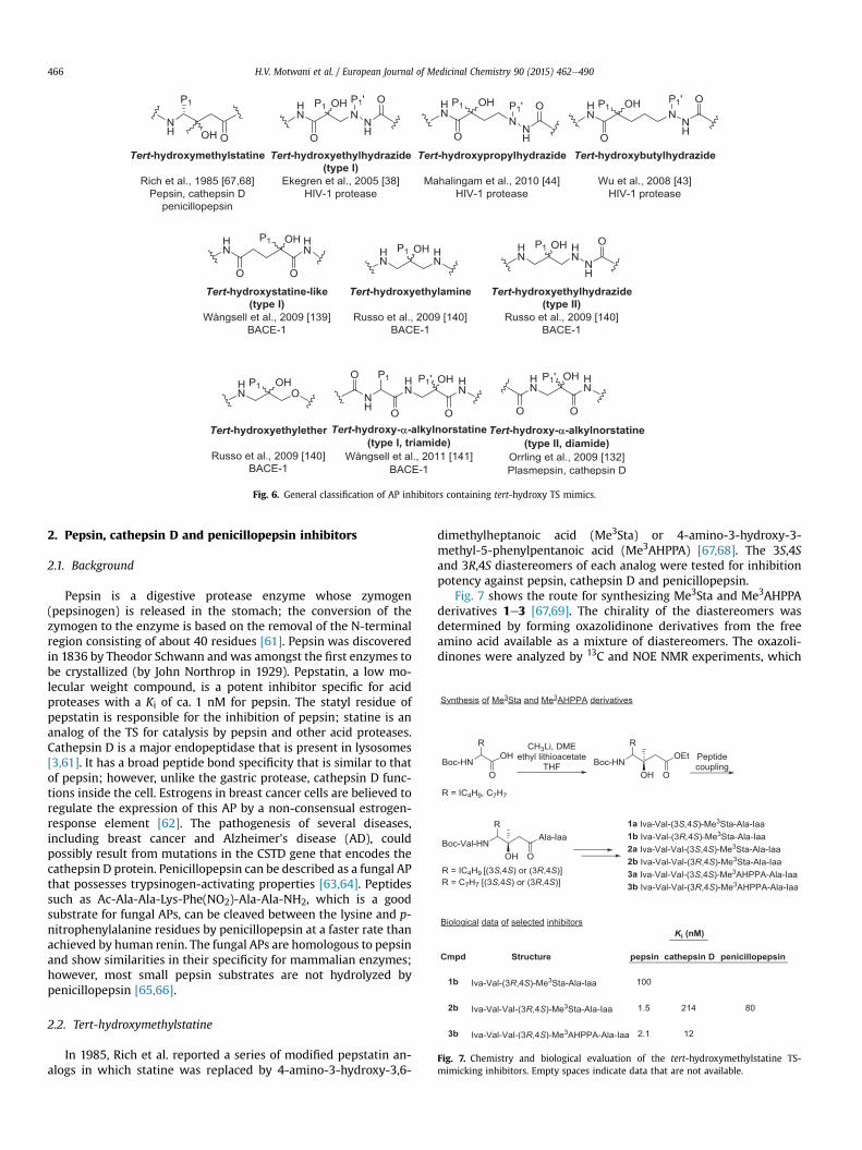
Fig. 6. General classification of AP inhibitors containing tert-hydroxy TS mimics.
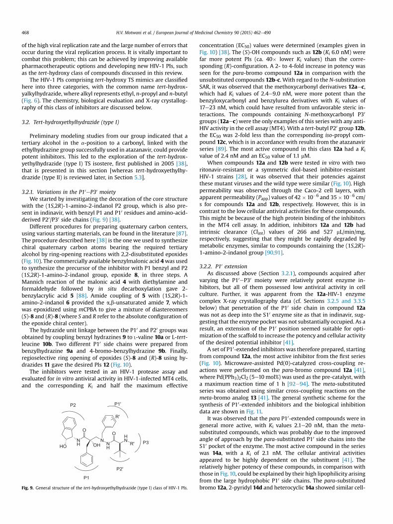
Fig. 7. Chemistry and biological evaluation of the tert-hydroxymethylstatine TS-mimicking inhibitors. Empty spaces indicate data that are not available.
H.V. Motwani et al. / European Journal of Medicinal Chemistry 90 (2015) 462e490466
2. Pepsin, cathepsin D and penicillopepsin inhibitors
2.1. Background
Pepsin is a digestive protease enzyme whose zymogen(pepsinogen) is released in the stomach; the conversion of thezymogen to the enzyme is based on the removal of the N-terminalregion consisting of about 40 residues [61]. Pepsin was discoveredin 1836 by Theodor Schwann and was amongst the first enzymes tobe crystallized (by John Northrop in 1929). Pepstatin, a low mo-lecular weight compound, is a potent inhibitor specific for acidproteases with a Ki of ca. 1 nM for pepsin. The statyl residue ofpepstatin is responsible for the inhibition of pepsin; statine is ananalog of the TS for catalysis by pepsin and other acid proteases.Cathepsin D is a major endopeptidase that is present in lysosomes[3,61]. It has a broad peptide bond specificity that is similar to thatof pepsin; however, unlike the gastric protease, cathepsin D func-tions inside the cell. Estrogens in breast cancer cells are believed toregulate the expression of this AP by a non-consensual estrogen-response element [62]. The pathogenesis of several diseases,including breast cancer and Alzheimer's disease (AD), couldpossibly result from mutations in the CSTD gene that encodes thecathepsin D protein. Penicillopepsin can be described as a fungal APthat possesses trypsinogen-activating properties [63,64]. Peptidessuch as Ac-Ala-Ala-Lys-Phe(NO2)-Ala-Ala-NH2, which is a goodsubstrate for fungal APs, can be cleaved between the lysine and p-nitrophenylalanine residues by penicillopepsin at a faster rate thanachieved by human renin. The fungal APs are homologous to pepsinand show similarities in their specificity for mammalian enzymes;however, most small pepsin substrates are not hydrolyzed bypenicillopepsin [65,66].
2.2. Tert-hydroxymethylstatine
In 1985, Rich et al. reported a series of modified pepstatin an-alogs in which statine was replaced by 4-amino-3-hydroxy-3,6-
dimethylheptanoic acid (Me3Sta) or 4-amino-3-hydroxy-3-methyl-5-phenylpentanoic acid (Me3AHPPA) [67,68]. The 3S,4Sand 3R,4S diastereomers of each analog were tested for inhibitionpotency against pepsin, cathepsin D and penicillopepsin.
Fig. 7 shows the route for synthesizing Me3Sta and Me3AHPPAderivatives 1e3 [67,69]. The chirality of the diastereomers wasdetermined by forming oxazolidinone derivatives from the freeamino acid available as a mixture of diastereomers. The oxazoli-dinones were analyzed by 13C and NOE NMR experiments, which

Active site
Asp25 Asp125
Flaps
cavity
Fig. 8. The secondary structure of HIV-1 protease shown as a ribbon cartoon with theflaps closed on top and the active site as the central cavity (PDB code: 2xye).
H.V. Motwani et al. / European Journal of Medicinal Chemistry 90 (2015) 462e490 467
showed that the main product for both the Me3Sta and Me3AHPPAanalogswas the 3S,4S-diastereomer. The diastereomeric amino acidmixture was bound to L-Ala-Iaa and the subsequent diastereomericdipeptides were separated using column chromatography andwerethen used to form the desired final inhibitors (Fig. 7) [67,69]. Fig. 7also shows the Ki values for tert-hydroxy compounds 1b, 2b and 3b.One peptide (3R,4S) from each diastereomeric pair was 2e3 ordersof magnitude more potent as an inhibitor of the AP (whetherpepsin, penicillopepsin or cathepsin) than the other (3S,4S).Increasing the length of the peptide chain generally resulted inmore potent inhibitors and the difference between the Ki values forthe respective diastereomers also increased. This is exemplified bycomparing 1bwith 2b; when the peptide chain was lengthened byone residue, the inhibition constant for pepsin was decreased by afactor of nearly 100.
The inhibitors were more potent against pepsin than againstcathepsin D. However, when the iso-butyl side chain in Sta wasreplaced by a benzyl group to form AHPPA analogs, the inhibitionactivity against cathepsin D was increased in contrast to thatagainst pepsin. Inhibitor 3b, a Me3AHPPA derivative, was ca. 20times more active than the corresponding Me3Sta derivative 2b.
In summary, it was observed that in all AP inhibition tests the3R,4S diastereomers of both the Me3Sta and Me3AHPPA inhibitorswere more potent than the corresponding 3S,4S diastereomers. TheKi values of the best inhibitors were in the range 1.5e12 nM.Interestingly, these results were in contrast to those obtained forthe sec-OH-based inhibitors containing statine or AHPPA de-rivatives, in which the 3S,4S diastereomer was the more potentinhibitor for each of the diastereomeric pairs of derivatives. Thestatine- and AHPPA-based inhibitors were only ca. 10-fold morepotent than the corresponding Me3Sta and Me3AHPPA inhibitors,which as expected were 100- to 1000-fold more potent than in-hibitors without the C-3 OH group. Rich et al. also found thatconformational changes in porcine pepsin were induced by(3R,4S)eMe3Sta derivatives that were comparable to changesinduced by pepstatin binding. These changes were not induced bythe corresponding 3S,4S analog [68].
3. HIV-1 protease inhibitors
3.1. Background
The nucleotide sequence of the HIV genome, published in 1985by Wain-Hobson et al. [70], revealed the coding sequences of 15viral proteins, including the AP HIV-1 protease. HIV-1 proteaseconsists of a two-fold symmetric dimer that possesses two identicalsubunits, each containing 99 amino acids. The HIV protease in-cludes a conserved catalytic site, comprising the catalytic triadAsp25, Thr26 and Gly27 in each subunit, which is accountable forits activity [71,72]. The conformation of the dimers is changed, withloss of symmetry, by binding to natural asymmetric substrates or aPI. The relevant cleavage sites are located by the protease more byrecognition of peptide shape than by the amino acid sequence. Aprecise order for the cleavages, with cleavage rates specific for eachsite, is generated by small variations amongst the different cleavageloci. As the active form of the HIV-1 protease is a dimer, it has beensuggested that the activity of the protease is influenced by theconcentrations of its subunits [71,72]. The degradation of viralproteins prior to the budding of new viral particles is thus pre-vented. As soon as the role of the protease was elucidated in theviral replication cycle, it was identified as a possible drug target.Gag and GagePol polyproteins are processed by HIV-1 protease toform active proteins and enzymes together with the structuralcapsid proteins (MA, CA, NC) and the enzymes (PR, RT, IN), to makenine cleavage sites in total [73]. The general catalytic mechanism
for AP, as described in section 1.1 (Fig. 1), is believed to be followedfor the peptide bond cleavage process by HIV-1 proteases.
X-ray analysis of HIV-1 protease co-crystallized with substrate-based inhibitors was first carried out in 1989 [71,74e76]. Twostructural flaps, one from each monomer, cover the active site(Fig. 8). These flaps are flexible and can be in an open or closedposition. The entry of substrates into the enzyme is easier whenthey are in the open position, while the processing of substrates isenhanced when they are in the closed position. The active site ofthe protease is bound competitively by the PI, and Ile50 and Ile150(from monomers one and two, respectively) can incorporate astructural water molecule when the flaps are in the closed position.
Over the past 20e30 years, many structural and mechanisticinsights have been gained by studying the HIV protease and thisresulted in the establishment of several inhibitory principles. HIV-1PIs were the first of the AP inhibitors to reach the market and theycontinue to serve as prototype inhibitors for the whole AP enzymefamily. Nevertheless, nearly three decades after the discovery ofAIDS, the pandemic of this viral disease is still on the rise; world-wide at the end of 2012 ca. 35 million people were living with thedisease and 1.6 million people had died of AIDS-related illnesses inthe same year [World Health Organization (WHO), Global HealthObservatory]. Currently, there are 11 FDA-approved HIV PIs; sa-quinavir is now marketed as saquinavir mesylate and amprenavirhas been replaced by its prodrug fosamprenavir (Fig. 5). Introduc-tion of the HIV-1 PIs in the 1990s [77e80] had a large impact onHIV-infected patients, as seen by the substantial increase in sur-vival rates and improved quality of life. However, the clinical ben-efits of this class of antiviral agents are restricted by a number offactors. Firstly, many of the marketed HIV-1 PIs, especially in thefirst generation, had poor pharmacokinetic properties, resulting inpoor aqueous solubility, low metabolic stability, high proteinbinding and poor membrane permeation. This limitation meantthat high doses were required to keep the viral plasma levels downand this subsequently resulted in poor patient compliance becauseof the severe side effects from the high doses. Some of the sideeffects caused by the PIs were gastrointestinal complicationsincluding nausea, vomiting and diarrhea, and metabolic disorderssuch as hyperlipidemia, lipodystrophy and insulin resistance[81,82]. However, many of the pharmacokinetic problems havebeen addressed by new contributions to the available arsenal ofHIV-1 PIs, for instance the development of non-peptidomimeticinhibitors as possible therapeutic option [83], and the addition oflow-dose ritonavir as a pharmacokinetic booster [84]. Anotherserious threat to efficient HIV treatment is the development ofresistance to the marketed HIV-1 PIs [85,86]. This is a consequence
H.V. Motwani et al. / European Journal of Medicinal Chemistry 90 (2015) 462e490468
of the high viral replication rate and the large number of errors thatoccur during the viral replication process. It is vitally important tocombat this problem; this can be achieved by improving availablepharmacotherapeutic options and developing new HIV-1 PIs, suchas the tert-hydroxy class of compounds discussed in this review.
The HIV-1 PIs comprising tert-hydroxy TS mimics are classifiedhere into three categories, with the common name tert-hydrox-yalkylhydrazide, where alkyl represents ethyl, n-propyl and n-butyl(Fig. 6). The chemistry, biological evaluation and X-ray crystallog-raphy of this class of inhibitors are discussed below.
3.2. Tert-hydroxyethylhydrazide (type I)
Preliminary modeling studies from our group indicated that atertiary alcohol in the a-position to a carbonyl, linked with theethylhydrazine group successfully used in atazanavir, could providepotent inhibitors. This led to the exploration of the tert-hydrox-yethylhydrazide (type I) TS isostere, first published in 2005 [38],that is presented in this section [whereas tert-hydroxyethylhy-drazide (type II) is reviewed later, in Section 5.3].
3.2.1. Variations in the P10eP30 moietyWe started by investigating the decoration of the core structure
with the (1S,2R)-1-amino-2-indanol P2 group, which is also pre-sent in indinavir, with benzyl P1 and P10 residues and amino-acid-derived P20/P30 side chains (Fig. 9) [38].
Different procedures for preparing quaternary carbon centers,using various starting materials, can be found in the literature [87].The procedure described here [38] is the one we used to synthesizechiral quaternary carbon atoms bearing the required tertiaryalcohol by ring-opening reactions with 2,2-disubstituted epoxides(Fig. 10). The commercially available benzylmalonic acid 4was usedto synthesize the precursor of the inhibitor with P1 benzyl and P2(1S,2R)-1-amino-2-indanol group, epoxide 8, in three steps. AMannich reaction of the malonic acid 4 with diethylamine andformaldehyde followed by in situ decarboxylation gave 2-benzylacrylic acid 5 [88]. Amide coupling of 5 with (1S,2R)-1-amino-2-indanol 6 provided the a,b-unsaturated amide 7, whichwas epoxidized using mCPBA to give a mixture of diastereomers(S)-8 and (R)-8 (where S and R refer to the absolute configuration ofthe epoxide chiral center).
The hydrazide unit linkage between the P10 and P20 groups wasobtained by coupling benzyl hydrazines 9 to L-valine 10a or L-tert-leucine 10b. Two different P10 side chains were prepared frombenzylhydrazine 9a and 4-bromo-benzylhydrazine 9b. Finally,regioselective ring opening of epoxides (S)-8 and (R)-8 using hy-drazides 11 gave the desired PIs 12 (Fig. 10).
The inhibitors were tested in an HIV-1 protease assay andevaluated for in vitro antiviral activity in HIV-1-infected MT4 cells,and the corresponding Ki and half the maximum effective
Fig. 9. General structure of the tert-hydroxyethylhydrazide (type I) class of HIV-1 PIs.
concentration (EC50) values were determined (examples given inFig. 10) [38]. The (S)-OH compounds such as 12b (Ki 6.0 nM) werefar more potent PIs (ca. 40� lower Ki values) than the corre-sponding (R)-configuration. A 2- to 4-fold increase in potency wasseen for the para-bromo compound 12a in comparison with theunsubstituted compounds 12b-c. With regard to the N-substitutionSAR, it was observed that the methoxycarbonyl derivatives 12aec,which had Ki values of 2.4e9.0 nM, were more potent than thebenzyloxycarbonyl and benzylurea derivatives with Ki values of17e23 nM, which could have resulted from unfavorable steric in-teractions. The compounds containing N-methoxycarbonyl P30
groups (12aec) were the only examples of this series with any anti-HIV activity in the cell assay (MT4). With a tert-butyl P20 group 12b,the EC50 was 2-fold less than the corresponding iso-propyl com-pound 12c, which is in accordance with results from the atazanavirseries [89]. The most active compound in this class 12a had a Kivalue of 2.4 nM and an EC50 value of 1.1 mM.
When compounds 12a and 12b were tested in vitro with tworitonavir-resistant or a symmetric diol-based inhibitor-resistantHIV-1 strains [28], it was observed that their potencies againstthese mutant viruses and the wild type were similar (Fig. 10). Highpermeability was observed through the Caco-2 cell layers, withapparent permeability (Papp) values of 42� 10�6 and 35�10�6 cm/s for compounds 12a and 12b, respectively. However, this is incontrast to the low cellular antiviral activities for these compounds.This might be because of the high protein binding of the inhibitorsin the MT4 cell assay. In addition, inhibitors 12a and 12b hadintrinsic clearance (Clint) values of 266 and 527 mL/min/mg,respectively, suggesting that they might be rapidly degraded bymetabolic enzymes, similar to compounds containing the (1S,2R)-1-amino-2-indanol group [90,91].
3.2.2. P10 extensionAs discussed above (Section 3.2.1), compounds acquired after
varying the P10eP30 moiety were relatively potent enzyme in-hibitors, but all of them possessed low antiviral activity in cellculture. Further, it was apparent from the 12a-HIV-1 enzymecomplex X-ray crystallography data (cf. Sections 3.2.5 and 3.3.5below) that penetration of the P10 side chain in compound 12awas not as deep into the S10 enzyme site as that in indinavir, sug-gesting that the enzyme pocket was not substantially occupied. As aresult, an extension of the P10 position seemed suitable for opti-mization of the scaffold to increase the potency and cellular activityof the desired potential inhibitor [41].
A set of P10-extended inhibitors was therefore prepared, startingfrom compound 12a, the most active inhibitor from the first series(Fig. 10). Microwave-assisted Pd(0)-catalyzed cross-coupling re-actions were performed on the para-bromo compound 12a [41],where Pd(PPh3)2Cl2 (5e10 mol%) was used as the pre-catalyst, witha maximum reaction time of 1 h [92e94]. The meta-substitutedseries was obtained using similar cross-coupling reactions on themeta-bromo analog 13 [41]. The general synthetic scheme for thesynthesis of P10-extended inhibitors and the biological inhibitiondata are shown in Fig. 11.
It was observed that the para P10-extended compounds were ingeneral more active, with Ki values 2.1e20 nM, than the meta-substituted compounds, which was probably due to the improvedangle of approach by the para-substituted P10 side chains into theS10 pocket of the enzyme. The most active compound in the serieswas 14a, with a Ki of 2.1 nM. The cellular antiviral activitiesappeared to be highly dependent on the substituent [41]. Therelatively higher potency of these compounds, in comparison withthose in Fig.10, could be explained by their high lipophilicity arisingfrom the large hydrophobic P10 side chains. The para-substitutedbromo 12a, 2-pyridyl 14d and heterocyclic 14a showed similar cell-

Fig. 10. Synthesis and biological evaluation of the tert-hydroxyethylhydrazide (type I) TS-mimicking HIV-1 PIs with variations in the P10eP30 moiety.
H.V. Motwani et al. / European Journal of Medicinal Chemistry 90 (2015) 462e490 469
based antiviral potency (EC50 0.90e1.1 mM), whereas 3-pyridyl 14cwas more potent (EC50 0.18 mM). Membrane permeation and livermicrosome stability studies (Caco-2 assay) were conducted on the3-pyridyl-extended compound 14c and its meta-analog 15a [41].The Papp values were 33 � 10�6 and 11 � 10�6 cm/s, respectively,suggesting excellent permeation for the para compound 14c incontrast to the meta compound [41]. The intrinsic clearance forboth inhibitors was lower than that for the compounds in Fig. 10(Clint 154 and 190 mL/min/mg, respectively).
3.2.3. Variations in the P2 moietyAlthough highly potent inhibitors were obtained from the P10
extended side chains (see above), the inhibitors were prone to rapidmetabolismwhen evaluated with liver microsomes, as indicated bytheir intrinsic clearance values [41]. This may have been due tobenzylic oxidation of the P2 group (1S,2R)-1-amino-2-indanol
[90,91], which led us to investigate variations in the P2 group[40]. Motivation for this study also came from considering the FDA-approved inhibitors saquinavir, nelfinavir and amprenavir, whichhave cyclic P2 substituents, whereas L-valine and L-tert-leucine arepresent as P2/P3 and P20/P30 side chains in the atazanavir analogseries. A set of compounds with varying P2 groups was thus pre-pared by coupling (S)-16with various amides and opening the ringwith a para-bromobenzylhydrazide 11, as shown in Fig. 12 [40].
Compounds 17a and 17b, with amino-acid-derived iso-propyl P2groups, showed high potency (Ki 5.1 and 7.4 nM, respectively) andthey also had anti-HIV activity in the cell-based assay, with EC50values down to 3.1 mM. However, relatively low potency wasobserved for compounds with cyclic P2 groups (Ki ca. 100 nM) [40].Compounds 17a-b with non-prime-side amino acid residues couldinteract with both S2 and S3 enzyme sub-sites, which is unfavor-able for the cyclic P2 groups. These two features, suitable size of the

Fig. 11. Chemistry and biological evaluation of the tert-hydroxyethylhydrazide (type I) TS-mimicking HIV-1 PIs with P10 extensions.
H.V. Motwani et al. / European Journal of Medicinal Chemistry 90 (2015) 462e490470
iso-propyl group for the S2 pocket and appropriate S3 interactions,could be the reason for the potency of compounds 17a and 17b.
3.2.4. Central amide functionalityIn an attempt to reduce the backbone for this class of TS mimics,
we tested compounds 18a-b, which had a central amide groupinstead of the hydrazide function [44]. Both compounds werebiologically inactive, with Ki values >5 mM and EC50 values >10 mM;hence, these compounds were not studied further (Fig. 13).
3.2.5. X-ray crystallography of the tert-hydroxyethylhydrazide(type I) TS-mimicking inhibitors
When compound 12a (Fig. 10) was co-crystallized with HIV-1protease, a 3D structure (PDB code 2cej, Fig. 14) [41] was ob-tained from the X-ray diffraction data [38,41]. The (S)-configurationat the quaternary carbon atom in the TS mimic was confirmed fromthe X-ray crystallography structure. It was observed that the ter-tiary hydroxyl groupwas hydrogen bound to one of the catalyticallyactive aspartic acid residues (Asp125) residing in the enzyme'sactive site (Fig. 14), and hydrazide a- and b-nitrogens and Gly27were involved in hydrogen bonding. Arg108 dictated the posi-tioning of the P10 substituent, where the electronegative Br groupwas closely packed against the positively charged Arg side chain.
3.3. Tert-hydroxybutylhydrazide
As reviewed above, in Section 3.2, the first HIV-1 PI scaffold thatused a tertiary alcohol as a TS mimic contained a one-carbon spacerbetween the tertiary alcohol and the hydrazide group. In order toimprove the binding properties of the inhibitor to the catalyticallyactive site of the aspartic acid, we extended the length of the in-hibitor backbone to three carbon atoms, with the first study pub-lished in 2008 [43]. The SAR investigation, including organicsyntheses, biological evaluations and X-ray crystallography studieswith tert-hydroxybutylhydrazide TS mimics (three-carbon spacer),is presented in this section.
3.3.1. Variations in the P10 moietyA library of synthesized compounds with different aromatic P10
substituents [43], prepared using palladium-catalyzed, microwave-assisted coupling reactions and the biological activities of these PIs,in terms of the inhibitory potency of the protease and the antiviralactivity in the cell-based assay, are described here.
The non-prime side with the three-carbon spacer was synthe-sized according to Fig. 15 [43], starting from the commerciallyavailable (S)-2-hydroxy-3-phenylpropionic acid 19. First, 2,2-dimethoxypropane 20 was used to protect the alcohol and acidfunctionalities in 19 and the obtained intermediate 21 was reactedwith methyl acrylate 22. Intramolecular lactone formation with amethyl ester from deprotection of the dioxolane 23, followed by

Fig. 12. Chemistry and biological evaluation of the tert-hydroxyethylhydrazide (type I) TS-mimicking HIV-1 PIs with variations in the P2 moiety.
Fig. 13. HIV-1 PIs 18a-b.
H.V. Motwani et al. / European Journal of Medicinal Chemistry 90 (2015) 462e490 471
peptide coupling with (1S,2R)-1-amino-2-indanol 6, resulted in thecyclic compound 24 as a mixture of diastereomers. Intermediate(R)-24with the preferred stereochemistry at the quaternary carbonwas protected by tert-butyldimethylsilyl (TBS)
trifluoromethanesulfonate and separated by column chromatog-raphy. Subsequently, reduction of the lactone and a protection-deprotection sequence led to the alcohol (R)-25. Initially, L-tert-leucine was selected as the P20 group and methyl carbamate as theP30 group for the prime side of the inhibitors, based on early studieson inhibitors in cell-based assays [38,41,89]. A series of hydrazideswas synthesized to investigate the effects of various alkyl-, aryl-and biaryl components in P10. b-Nitrogen-alkylated compounds 28that contained P10 precursor groups, with variations in the size,polarity and hydrogen-bonding potential, were obtained viareductive amination of primary hydrazide 27 with various alde-hydes 26 (Fig. 15) [43]. The desired inhibitors 29 were synthesizedby Dess-Martin periodinane oxidation of (R)-25, followed byreductive amination of the corresponding aldehyde and hydrazides28 [95].
Biological inhibitory data for selected compounds from thisseries with variations at the P10 site are shown in Fig. 15. All of thecompounds with a R-configuration at the tertiary alcohol and free

Fig. 14. X-ray crystallography structure of the 12a-HIV-1 protease complex; hydrogenbonds are highlighted as green dotted lines. Reproduced from Ref. [41] with permis-sion from the American Chemical Society. (For interpretation of the references to colorin this figure legend, the reader is referred to the web version of this article.)
Fig. 15. Chemistry and biological evaluation of the tert-hydroxybutylhy
H.V. Motwani et al. / European Journal of Medicinal Chemistry 90 (2015) 462e490472
hydroxyl groups, except one with a tert-butyl-substituted thiazolegroup at P10, exhibited relatively high potency against the HIV-1protease, with Ki values of 2.3e11 nM [43]. It is worth notinghere that the R stereochemistry of the tert-hydroxybutylhydrazidecorresponds to the S stereochemistry of the tertiary alcohol for thetert-hydroxyethylhydrazide-based inhibitors. Poor inhibitory po-tencies were observed for compounds with small P10 groups, e.g.halogens or a cyano group in the 4-position of the P10 benzyl group(29f-g, EC50 1.2 and 0.85 mM, respectively) and also for compoundswith large and/or very lipophilic P10 groups. Intermediate potency(EC50 0.47e0.56 mM)was observed for 4-substituted inhibitors withmedium-sized groups such as 2-pyridyl (29d), as well as for thebicycle 29b. Compounds 29a, 29c, 29e and 29h were the mostpotent in the series (EC50 0.17e0.22 mM); these contained relativelyhydrophilic aromatic groups with heteroatoms in the second P10
aryl group. A compound containing 2-chloro-5-methoxy thiazole atthe P10 position, which was inspired by a GlaxoSmithKline mole-cule structure [96], did not improve the cell activity (EC50 0.60 mM).Surprisingly, in comparison to the 2-pyridyl inhibitor (29d), PIs
drazide TS-mimicking HIV-1 PIs with variations in the P10 moiety.
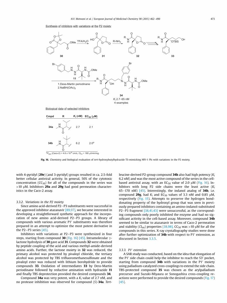
Fig. 16. Chemistry and biological evaluation of tert-hydroxybutylhydrazide TS-mimicking HIV-1 PIs with variations in the P2 moiety.
H.V. Motwani et al. / European Journal of Medicinal Chemistry 90 (2015) 462e490 473
with 4-pyridyl (29e) (and 3-pyridyl) groups resulted in ca. 2.5-foldbetter cellular antiviral activity. In general, 50% of the cytotoxicconcentration (CC50) for all of the compounds in the series was>10 mM. Inhibitors 29a and 29g had good permeation character-istics in the Caco-2 assay.
3.3.2. Variations in the P2 moietySince amino acid-derived P2eP3 substituents were successful in
the approved inhibitor atazanavir [89,97], we became interested indeveloping a straightforward synthetic approach for the incorpo-ration of new amino acid-derived P2eP3 groups. A library ofcompounds with various aromatic P10 substituents was thereforeprepared in an attempt to optimize the most potent derivative inthe P2eP3 series [45].
Inhibitors with variations at P2eP3 were synthesized in foursteps, starting from compound 30 (Fig. 16) [45]. Intramolecular g-lactone hydrolysis of 30 gave acid 31. Compounds 32were obtainedby peptide coupling of the acid and various methyl-amide-derivedamino acids. Further, the lactone moiety in 32 was reduced, theprimary alcohol was protected by pivaloyl chloride, the tertiaryalcohol was protected by TBS trifluoromethanesulfonate and thepivaloyl ester was reduced with lithium borohydride to providecompounds 33. Oxidation of intermediates 33 by Dess-Martinperiodinane followed by reductive amination with hydrazide 11and finally TBS deprotection provided the desired compounds 34.
Compound 34a was very potent, with a Ki value of 2.7 nM, andno protease inhibition was observed for compound (S)-34a. Tert-
leucine-derived P2-group compound 34b also had high potency (Ki6.2 nM) andwas the most active compound of the series in the cell-based antiviral assay, with an EC50 value of 2.0 mM (Fig. 16). In-hibitors with long P2 side chains were the least active (Ki65e170 nM) [45]. Interestingly, the indanol analog of 34b, i.e.compound 29g, had Ki and EC50 values of 3.3 nM and 0.85 mM,respectively (Fig. 15). Attempts to preserve the hydrogen bond-donating property of the hydroxyl group that was seen in previ-ously prepared inhibitors containing an amino-indanol-substitutedP2eP3 fragment [38,41,43] were unsuccessful, as the correspond-ing compounds only poorly inhibited the enzyme and had no sig-nificant activity in the cell-based assay. Moreover, compound 34bseemed to be similar to atazanavir in terms of Caco-2 permeationand stability (Clint) properties [38,98]. CC50 was >10 mM for all thecompounds in this series. X-ray crystallography studies were doneafter further optimization of 34b with respect to P10 extension, asdiscussed in Section 3.3.5.
3.3.3. P10 extensionA SAR study was conducted, based on the idea that elongation of
the P10 side chain could help the inhibitor to reach the S30 pocket,starting from compound 34b with variations in the P10 moietyusing palladium-catalyzed cross-couplings to extend the side chain.TBS-protected compound 35 was chosen as the arylpalladiumprecursor and Suzuki-Miyaura or Sonogashira cross-coupling re-actions were performed to provide the desired compounds (Fig. 17)[45].

Fig. 17. Chemistry and biological evaluation of tert-hydroxybutylhydrazide TS-mimicking HIV-1 PIs with P10 extensions.
H.V. Motwani et al. / European Journal of Medicinal Chemistry 90 (2015) 462e490474
Unexpectedly, no noteworthy considerable enhancement of theantiviral activity was observed for the compounds with elongatedP10 side chains in comparison to compound 34b, as shown in Fig.17.The highest potency in the series was obtained by compound 36a,with a Ki value of 3.1 nM and an EC50 value of 1.0 mM. The Ki valuesfor compounds 36a, 36c and 36d were in the range of 3.1e3.6 nM,whereas further extensionwas tolerated but not helpful, as seen forcompound 36b (Ki 3.4 nM). The potency of the 3-pyridyl 36a and 3-ethynyl pyridyl 36b P10-elongated inhibitors was similar to that ofthe corresponding 2-pyridyl and 4-pyridyl compounds. Further, P10
elongation, as seen in compound 36e, increased Caco-2 cellpermeation and decreased the metabolic stability (cf. Clint),whereas placing pyridyl at the P10 position (36a and 36c) decreasedthe permeation but increased the metabolic stability. The cyto-toxicity was >10 mM for all the compounds in the series.
3.3.4. Variations in P1eP3 macrocyclizationEncouraged by the often positive pharmacokinetic and phar-
macodynamic outcomes associated with various macrocyclizationsof linear bioactive compounds [99], we decided to apply thisstrategy to prepare a new series [51] of HIV-1 PIs related to ata-zanavir but containing a tertiary alcohol as part of the TS-mimicking core structure (Fig. 18) [41,100]. The presence of pep-tide bonds, high flexibility and exposure of polar functionalities inlinear molecules can result in metabolic instability and poormembrane permeation, andmacrocyclization of the linear peptidescould help to reduce the peptidic characteristics associated withincreased structural rigidity [101]. Cyclizations also provide betterresistance to proteolytic digestion by facilitating internal hydrogen
bonding, thus helping to improve bioavailability and enabling thecompound to reach its intracellular targets [99].
Although the macrocyclization strategy has been previouslyinvestigated for the design of HIV-1 PIs [102,103], no examples ofmacrocyclic tertiary alcohols had been reported at this stage. In ourfirst attempt, we prepared a library of P1eP3macrocycles including14- and 15-membered rings with bromine at the para position onthe P10 side [51]. The synthetic route is depicted in Fig. 18.Commercially available p-bromobenzaldehyde 37 was condensedwith N-acetylglycine and the resulting oxazolone 38 was reducedby treatment with hydrochloric acid/amalgamated zinc. The ob-tained diol 39was protected using 2,2-dimethoxypropane followedby a lithium diisopropylamide-mediated conjugate addition withmethyl acrylate providing the aryl bromide 40, which was treatedwith trifluoroacetic acid resulting in the carboxylic acid. Couplingwith amines furnished intermediates 41. The lactone moiety ofcompounds (R)-41 and (S)-41 was reduced using lithium borohy-dride, followed by a couple of protection/deprotection steps on theprimary and tertiary alcohols to give the precursor 42 for thesubstitution of the bromide on the P1 side. A vinyl group wasintroduced via a microwave-assisted Suzuki cross-coupling reac-tion [42,94,104] with the aim of preparing 14-membered macro-cycles. The allyl group was introduced by a microwave-promotedStille coupling [105], to obtain 15-membered macrocycles.
Oxidation of intermediate alcohol 42 by Dess-Martin period-inane followed by reductive amination with hydrazide 11 and finalTBS deprotection provided the desired linear compounds 44, whichwere used as precursors for the macrocyclization via microwave-

Fig. 18. Chemistry and biological evaluation of the tert-hydroxybutylhydrazide TS-mimiciking HIV-1 PIs with macrocyclization of P1eP3.
H.V. Motwani et al. / European Journal of Medicinal Chemistry 90 (2015) 462e490 475
induced ring-closing metathesis using the second generationHoveyda-Grubbs catalyst [106].
The HIV-1 protease inhibition and antiviral activity of bothlinear (R)-44 and (S)-44 and macrocyclic (R)-45 compounds wereevaluated; the most significant results are shown in Fig. 18. Alllinear compounds with an R configuration of the tertiary alcoholhad strong inhibition profiles (10 nM � Ki � 37 nM) whereascompounds with an S configurationwere inactive, with Ki values upto 100 times higher (720 nM � Ki � 1000 nM). These results wereexpected from our previous studies [40,43]. The macrocycles fur-nished 2- to 3-fold better Ki values than the linear compounds aswell as improved EC50 values (down to 0.37 mM). It is worthnoticing that the linear PIs permeated well (Papp > 40 � 10�6 cm/s)in the Caco-2 assay and macrocycles (R)-45b and (R)-45c had low
EC50 values (0.37e1.0 mM) with satisfactory permeation(Papp ¼ 9e10 � 10�6 cm/s).
Although the best macrocycles in this study had lower EC50values than the corresponding linear compounds, the potencyenhancement was only modest. Therefore, we decided to optimizethe P10 position by introducing (hetero)aromatic moieties [107].The P10 substituents were introduced via Suzuki-Miyaura cross-coupling using their respective boronic acids, Herrmann's palla-dacycle (as a palladium precatalyst) and the pre-ligand tri(tert-butyl)phosphonium tetrafluoroborate [108e110], or palladium(II)acetate as catalyst and triphenylphosphine as ligand (Fig. 19, a)[111]. However, these protocols were both unsuccessful when thecross-coupling reaction was performed with 2-(pyridyl/thiazolyl)boronic acids, possibly due to rapid protodeboronation and/or

Fig. 19. Chemistry and biological evaluation of the P10-functionalized tert-hydroxybutylhydrazide TS-mimicking HIV-1 PIs with macrocyclization of P1eP3.
H.V. Motwani et al. / European Journal of Medicinal Chemistry 90 (2015) 462e490476

Fig. 20. X-ray crystallography structures of compounds 29g and 12a co-crystallized with HIV-1 protease, showing the active site of the protease. Reproduced from Ref. [43] withpermission from the American Chemical Society.
H.V. Motwani et al. / European Journal of Medicinal Chemistry 90 (2015) 462e490 477
polymerization [112e114]. Thus, a different approach, using pre-synthesized diaryl aldehydes, was chosen to introduce the 2-pyridyl and 2-thiazolyl moieties into the P10 position (Fig. 19, b)[107]. Deprotection of the TBS-protected tertiary hydroxyl group,using tetrabutylammonium fluoride at room temperature, fur-nished the desired linear inhibitors (R)-46 with high yields.
Macrocyclization of linear inhibitors (R)-46 was thereafter per-formed via a microwave-induced ring-closing metathesis reaction(Fig. 19, c) [115,116]. Fourteen-membered macrocycles were ob-tained as, in some cases, the reactions gave a mixture of isomersbecause of double-bond migration and ring-contraction reactions[117e119].
In almost all cases, the P1eP3 macrocyclization approach fur-nished improved inhibitors 47 in terms of inhibition potency andantiviral activity (Fig.19), with Ki values in the range of 2.2e31.5 nMand EC50 values in the range of 0.13e3.05 mM. The macrocycle witha phenyl moiety at the P10 position was the only exception and thiswas the least potent inhibitor in this series (Ki 120 nM and EC507.35 mM) [107]. As expected from our previous study [51], the 14-membered macrocycles were, in all cases, less potent than thecorresponding 15-membered cyclic inhibitors. Double-bondmigration from the 2,3 to the 1,2 position of the 15-memberedmacrocycles did not strongly affect the inhibition profile, as theinhibitors had similar Ki values. Macrocyclic molecules containing2-thiazolyl or 2-pyridyl groups were highly potent; 47b was thebest inhibitor (Ki 2.2 nM) and also had very good antiretroviralactivity (EC50 0.2 mM).
3.3.5. X-ray crystallography of the tert-hydroxybutylhydrazide TS-mimicking inhibitors
X-ray crystallography results showing the arrangement of thebenzyl bromide compound 29g (PDB ID 2uxz) (Fig. 15) and com-pound 12a (one-carbon spacer, Fig. 10, PDB-ID 2bqv) and therelevant hydrogen bonds to the enzyme amino acid residues fromthe active site of the PI are shown in Fig. 20 [43]. Compound 29g
had 54 contact points with the protein and compound 12a had 48contact points. There were five direct hydrogen bonds betweencompound 29g and the protein, and three bonds via water mole-cules. The tertiary alcohol of 29g could form a hydrogen bond toonly one of the aspartic residues in the active site, possibly becauseof their extended central carbon skeleton; in comparison, 12a,could form hydrogen bonds to both of the aspartic residues(although the bond to Asp125 was weak, with a binding distance of3.3 Å). Compound 29g had a relatively strong interaction withAsp25 (2.9 Å) and the para-bromo atom on the P10 benzyl groupwas directed towards the solvent. The bromine atom in inhibitor12a had four close-packed contact points with the lining residues.
X-ray crystallography data were obtained for nine (four linearand five macrocycle) P10-functionalized inhibitor/HIV-1 proteasecomplexes [107]. A detailed comparison of linear and macrocyclicscaffolds was carried out and the global effect of the macro-cyclization approach on the binding profiles of the inhibitors wasanalyzed (Fig. 21). The linear and macrocyclic scaffolds bound in asimilar fashion to the enzyme with only minor differences. Themain effect of the macrocyclization seems to be that the introducedconstraints made the macrocycles slightly more compact than thecorresponding linear analogs, which translates to a better fitting ofthe macrocycles. Interestingly, this improved accommodation ofthe macrocyclic structures at the non-prime site of the enzymeaffected the hydrogen-bonding distances between the tertiary hy-droxyl group in the PIs and the aspartic residues Asp25/Asp125 andGly27 in the active site of the enzyme.
The adjustment of the macrocyclic scaffold in the P1eP3 regionwas also reflected in the rotation of the iso-propyl group at the P2pocket. For the P1eP3 macrocyclic compounds, the iso-propylgroup appeared to be forced to point away from the nitrogen as theiso-propyl group was closer to the backbone of the protein. Thegroups in the P1 and P3 positions of the linear compounds did notseem to interact closely with the protein. In contrast, the two allyl

Fig. 21. Comparison of the non-cyclic (R)-46d (PDB ID 4cpr, turquoise) and the corresponding macrocycle (R)-47d (PDB code: 4cpx, green) complexes. The macrocyclic compoundmimicked the binding of the linear PI very well, differing significantly only at the point of macrocyclization at the S3 site and at the hetero-aromatic substituent at the S10 site, thelatter having partial rotational freedom. Reproduced from Ref. [107] with permission from the American Chemical Society. (For interpretation of the references to color in this figurelegend, the reader is referred to the web version of this article.)
H.V. Motwani et al. / European Journal of Medicinal Chemistry 90 (2015) 462e490478
groups in both the P1 and P3 positions seemed to be more flexibleand were directed away from the protein surface [107].
3.4. Tert-hydroxypropylhydrazide
As reviewed in Sections 3.2 and 3.3, the HIV-1 PIs with one orthree carbon spacers were potent inhibitors, with Ki values down to2.1 nM and good Caco-2 membrane permeation. However, thelinear compounds containing these TS mimics had only moderatecellular antiviral activities, with the best EC50 value of 170 nM(compounds 29e and 29h, Fig. 15) and poor stability in liver mi-crosomes. It therefore seemed logical to prepare new moleculeswith a TS mimic containing a two-carbon spacer between thequaternary carbon and the hydrazide b-nitrogen. This was mainlybecause X-ray crystallography data from the previous two
categories of inhibitors (one and two carbon spacers, Sections 3.2and 3.3), in combination with modeling studies, indicated that atwo-carbon distance would bring the OH group nearer to Asp25 atthe active site, which might increase the potency as a result of thestronger interaction.
3.4.1. Variations in the P10 moietyThe first aim of our SAR study of the tert-hydroxypropylhy-
drazide class of inhibitors was to optimize the nature of the groupat the P10 site in an attempt to improve the cell-based antiviralactivity.
Commercially available g-butyrolactone 48 was used to syn-thesize bromides (S)-49 and (R)-49 (Fig. 22), which were separatedby column chromatography and used as aryl precursors for cross-coupling reactions to obtain a series of para P10-extended

Fig. 22. Chemistry and biological evaluation of the tert-hydroxypropylhydrazide TS-mimicking HIV-1 PIs with variations in the P10 moiety.
H.V. Motwani et al. / European Journal of Medicinal Chemistry 90 (2015) 462e490 479
inhibitors 50, having retained the S or R configuration at the qua-ternary carbon. Pyridine compounds 50b and 50cwere synthesizedby Stille couplings [109], using the corresponding tributyltin re-agents [44].
The antiviral activities of the S compounds 50a, 50b and 50c aresummarized in Fig. 22 [44]. All of the compounds in the series hadhigh potencies, with Ki values in the range of 1.2e12 nM.When (R)-50awas tested for antiviral activity it was seen to be inactive, with aKi value > 5 mM. P10 substitution of the S isomer distinctly influ-enced the cell activity and high antiviral activity (EC50 3�13 nM)
was observed for all the tested 4-aryl-elongated compounds,except for the N-(4-phenyl)acetamide (compound 16 in an earlierpaper [44]). Cytotoxicity (CC50 < 10 mM) was exhibited by thebromo- ((S)-50a) and 3-pyridyl- ((S)-50c) substituted compounds(CC50 7.9 and 4.9 mM, respectively). A stability test using a livermicrosome homogenate was performed on the 2-pyridyl derivative(S)-50b (the best compound in this series, according to the Ki/EC50/CC50 profile), its parent compound (S)-50a and the related inhibitor(S)-50c. Compound (S)-50b, with the atazanavir prime side, wasrelatively more resistant to degradation by metabolic enzymes

Fig. 23. Chemistry and biological evaluation of the tert-hydroxypropylhydrazide TS-mimicking HIV-1 PIs with variations in the P2 moiety.
Table 1Antiviral activity against selected HIV-1 PI-resistant isolates and inhibition data.
Entry Mutations in protease EC50 (mM)
(S)-50a (S)-50b (S)-50c
1 Wild-type (wt) 0.040 0.007 0.0092 G48V, L90M 0.008 0.008 0.0103 A71V, I84V L90M 0.044 0.007 0.0194 V32I, M46I A71V, V82A 0.070 0.006 0.0145 V32I, M46I V82A 0.074 0.024 0.0276 M46I, V82F, I84V 0.24 0.13 0.60
Ki (nM)7a L63P, V82T, I84V 30 16 16
a Atazanavir Ki ¼ 6.5 nM.
H.V. Motwani et al. / European Journal of Medicinal Chemistry 90 (2015) 462e490480
(Clint 20 mL/min/mg), while compounds (S)-50a and (S)-50cdegraded rapidly (Clint > 300 mL/min/mg). However, excellent Caco-2 membrane permeation was observed for compounds (S)-50a and(S)-50c, with Papp values of 42 � 10�6 cm/s and 26 � 10�6 cm/s,respectively; compound (S)-50b had a slower penetration with aPapp of 3.5 � 10�6 cm/s.
Encouraged by the enzyme inhibition data, antiviral activity andcell toxicity properties, compounds (S)-50a, (S)-50b and (S)-50cwere subsequently tested against selected PI-resistant isolates ofHIV-1 (Table 1) [44]. Incubation of the cell-free virus withincreasing concentrations of inhibitors saquinavir (entries 2 and 3in Table 1) and ritonavir (entries 5 and 6) induced clinically relevantmutations in the HIV-1 protease genome to acquire the desiredresistant isolates. In comparison to the wild-type enzyme, the threeS inhibitors had equal or greater potency against the viral isolatewith G48V and L90M mutations in the protease, which generallyoccur in saquinavir-treated patients [120]. Inhibitors (S)-50a, (S)-50b and (S)-50c were highly potent against one of the isolatescontaining V82A and M46I mutations and (S)-50b was equipotentagainst this isolate and the wild-type isolate (entry 4). The
mutations V82A and M46I, located at the protease S10 site and inthe flap region, respectively, are known to cause resistance againstseveral of the FDA-approved HIV-1 PIs [85,86]. The four universalprotease-associated mutations (UPAMs) occur at amino acids 33,82, 84 and 90; these are usually observed in patients with disease

Fig. 24. X-ray crystallography structures of 14d, 50b and 29g co-crystallized with HIV-1 protease. Reproduced from Ref. [44] with permission from the American Chemical Society.
H.V. Motwani et al. / European Journal of Medicinal Chemistry 90 (2015) 462e490 481
resistant to HIV therapy [120]. The three PIs were relatively potentin the presence of one (entries 2, 4 and 5) or two (entry 3) UPAMs;however, there was a loss of potency in the presence of the muta-tion involving residues 82 and 84 (entry 6).
3.4.2. Variations in the P2 moietyThe indanolamine moiety in some of the synthesized inhibitors
was metabolically unstable because of the propensity of the indanto undergo 30-hydroxylation [90,91]. Inhibitors with variations inthe P2 moiety were therefore synthesized and investigated (com-pounds 52e54, Fig. 23) [44].
Compounds 52e54 were prepared via a coupling reaction fromthe starting materials 51 and the hydrazine 11. Compound (S)-52was then used as a precursor for the Suzuki and Stille cross cou-plings to obtain the desired compounds 55aec (Fig. 23) [44].
Compounds with P2 variations (52e55, with an S configurationat the tertiary alcohol) were highly potent against the protease. Kivalues ranged from 1.0 nM (compound 55b) to 3.2 nM (compound54) and the compounds had good antiviral activity (EC5037e170 nM, Fig. 23) [44]. However, none of these compounds(52e55) showed any improvement in their Ki/EC50/CC50 profilesover compound (S)-50b (Fig. 22). Hence, this series of compoundswas not evaluated further, in terms of either biological activity or X-ray crystallography.
3.4.3. X-ray crystallography of the tert-hydroxypropylhydrazide TS-mimicking inhibitors
X-ray crystallography structures of compounds 50b (two-carbonspacer, Fig. 22, PDB code 2wkz),14d (one-carbon spacer, Fig. 11, PDBcode 2cem) and 29g (Fig. 15, three-carbon spacer, PDB code 2uxz)
Fig. 25. Lactam-based HIV-1 PIs 56a and 56b.
are depicted in Fig. 24 [44]. In comparison to the 14d- and 29g-enzyme complexes, compound 50b was rotated 180� at the activesite. Compounds 50b and 14d had a shorter binding distance (2.7 Å)from the tertiary alcohol to Asp 25/125 than compound 29g (2.9 Åto Asp25). Compound 50b formed one hydrogen bond from theindanolamine hydroxy group to the backbone NH group of Asp29(3.0 Å) and another hydrogen bond to a water molecule (3.1 and2.8 Å), whereas there were two hydrogen bonds from the inda-nolamine hydroxyl group to Gly127 and Asp129 in compounds 14dand 29g. The tertiary alcohol in 50b was hydrogen bonded to thebackbone carbonyl of Gly27 (2.6 Å), but this was more difficult in14d as the hydroxyl group was too far away (3.3 Å). There wereseven and five hydrogen bonds to the protein and via water mol-ecules, respectively, for 50b, six and five for 14d, and five and threefor 29g. The arrangement of the hydrogen bonds between theconserved water molecule and the non-prime-side amide carbonyl,the hydrazide carbonyl, and the enzyme backbone NH groups ofIle50 and Ile150 was similar for compounds 50b, 14d and 29g.
3.5. Current status and prospects
We have reviewed a relatively new generation of HIV-1 PIscontaining tertiary-alcohol-based TS mimics. The inhibitors havebeen classified according to the number of carbons used to elongatethe core structure: one (tert-hydroxyethylhydrazide), two (tert-hydroxypropylhydrazide) or three (tert-hydroxybutylhydrazide).The PIs in the two-carbon spacer series weremore potent inhibitorsof HIV-1 protease; the best compound, 50b, which had a pyridine inthe P10 terminal substituent, had a Ki value of 1.7 nM. However,inhibitor 50b was slightly less potent than atazanavir (Ki 0.5 nM)and indinavir (Ki 0.3 nM) (Fig. 5). The higher inhibition potency of
Fig. 26. Norstatine-based plasmepsin inhibitor KNI-10006.

Fig. 27. Chemistry and biological evaluation of the tert-hydroxy-a-alkylnorstatine TS-mimicking plasmepsin inhibitors.
H.V. Motwani et al. / European Journal of Medicinal Chemistry 90 (2015) 462e490482
the two-carbon spacer series was also supported by the X-raycrystallography results; Fig. 24 shows inhibitor 50b co-crystallizedwith HIV-1 protease. The tertiary alcohol had hydrogen bonds notonly to Asp125, but also to the Gly27 carbonyl in the proteinbackbone. The P1 substituent of 50b also fit the S1 and S3 pockets ofthe enzyme better, as seen from the improved interaction withVal182 and Arg108.
The EC50 values were in general more than 20-fold better for theinhibitors with two-carbon spacers than for those with one- orthree-carbon spacers, probably due to improved masking by thetertiary alcohol of the former. For example, with pyridine as the P10
substituent, the EC50 value for inhibitor 50b (Fig. 22) was 7 nM,while the values for the one- and three-carbon spacer inhibitorswere 180 nM (compound 14c, Fig 11) and 170 nM (29e, Fig. 15),respectively. Notably, as shown by the antiviral cell-based assay,inhibitor 50b had better antiviral activity than indinavir (EC5050 nM) and similar activity to atazanavir (EC50 8 nM), lopinavir(EC50 10 nM) and darunavir (EC50 4 nM). A significant improvementin the EC50 values was obtained by the macrocyclization approachat the P1eP3 site; the EC50 value for the three-carbon-tetheredcompound 47b (Fig. 19) was 0.2 mM and the Ki value was 2.2 nM,whereas the corresponding values for the non-cyclized inhibitor46b were EC50 2.7 mM and Ki 8.0 nM.
Inhibitor 50b had a low Clint (20 mL/min/mg) in the livermicrosome assay, which is considered to be promising. It has beenpreviously established that HIV-1 PIs are rapidly metabolized/degraded by cytochrome P450 enzymes, which means that theyhave low oral bioavailability [121]. Nevertheless, the knownmetabolic problems associated with the indanolamine moietyrequired further investigation. The inhibitors obtained when theindanolamine was replaced by N-alkylated amino acid-basedmoieties were less potent. Further, the enzyme assay results withHIV-1 protease-resistant viral isolates were favorable for inhibitor50b, except when there were mutations at residues 82 and 84(Table 1), probably because of the loss of hydrophobic interactions,as seen from the co-crystallized X-ray structures (Fig. 24).
Based on this evidence, it can be concluded that PI 50b is ahighly promising lead structure in the field of development of newHIV-1 PIs. It was considered worthwhile establishing stronger
symmetric hydrogen bonds in the new inhibitors, to both the cat-alytic residues of the protease, Asp 25 and Asp 125. With this inmind, we have recently developed a strategy for relocating thehydroxyl group one position away from the backbone, thus furtherstrengthening the central TS mimic. In the first series of examples,we prepared inhibitors with a b-hydroxy g-lactam group contain-ing a sec-hydroxy and a two- or three-carbon spacer [46]. Func-tionalization of the two most potent inhibitors (3R,4S)-56a and(3R,4R)-56b (Fig. 25) by heteroaromatic moieties in the p-benzylP10 position provided compounds with Ki values down to 0.7 nMand EC50 values down to 40 nM. However, this strategy will not befurther discussed in this review as it does not involve a tertiaryalcohol as the TS mimic. Nonetheless, it is felt that combining ter-tiary alcohol TS-mimicking structures with stereopure lactam-based moieties that also provide rigidity to the backbone of theinhibitor could be a potential path to follow. An alternative strategyfor improving the pharmacokinetic profile and potency of the in-hibitors could be macrocyclization at the P1eP3 site of the two-carbon spacer series, in particular for compound 50b.
4. Plasmepsin inhibitors
4.1. Background
Malaria, one of the earliest diseases known to man, is endemicin more than 100 countries. Despite considerable scientific ad-vances and the development of modern drugs, more than threebillion people are still living under the threat of malarial infection.The WHO estimated that ca. 200 million clinical cases occurredglobally in 2012, resulting in nearly half a million deaths [122].Malaria in humans is caused by four species of protozoal parasitesof the Plasmodium genus, Plasmodium falciparum (Pf), Plasmodiummalariae (Pm), Plasmodium ovale (Po) and Plasmodium vivax (Pv); ofthese, Pf is responsible for most malarial fatalities. Plasmodiumparasites are spread by female mosquitoes of the genus Anopheles.Parasitic strains resistant to the currently available antimalarialdrugs are evolving at an alarming rate, indicating an acute need fornew therapeutic agents [122,123].

H.V. Motwani et al. / European Journal of Medicinal Chemistry 90 (2015) 462e490 483
Sequencing of the Pf genome revealed a number of promisingnew drug targets for treating malaria [124e126]. For example, theenzymes used by the parasites to degrade hemoglobin into smallerpeptides and amino acids have been investigated as potentialantimalarial targets. Four of these, the APs plasmepsin I, II and IVand the histo-aspartic protease HAP, are known to be involved inhemoglobin degradation in the parasite digestive vacuole (DV)[127e129]. Orthologs of only the Pf plasmepsin IV (PfPM4) havebeen identified in the other three human malarial parasites, Pv, Pmand Po [130]. Thus, it is likely that an inhibitor of PfPM4 wouldreduce parasitic growth, regardless of the infecting species.
4.2. Tert-hydroxy-a-alkylnorstatine
In 2003, Nezami et al. reported a norstatine-based compound(KNI-10006, Fig. 26) that could inhibit all four plasmepsins in theDV of Pf at nanomolecular concentration ranges [131]. The IC50values for this PI against PfPM2 and PfPM4 were 39 nM and 15 nM,respectively, and the Ki value for PfPM2 was ca. 1 nM. However,
Fig. 28. Computer-generated PfPM4 enzymeeinhibitor binding modes, (R)-58 in or-ange and (S)-58 in purple. Hydrogen bonds are indicated by yellow dashed lines [132].(For interpretation of the references to color in this figure legend, the reader is referredto the web version of this article.)
KNI-10006 was also active against human cathepsin D, with a Kivalue of 2 nM and the compound had a low capacity for decreasingparasitic growth in Pf-infected erythrocyte cultures, with an IC50 of6.8 mM, probably because of low cell or vacuole membranepermeation.
We used the tertiary alcohol TS-mimicking strategy to overcomethese short-comings, and prepared tert-OH-a-alkylnorstatine-typeinhibitors by varying the P10 pseudoresidue and changing the ste-reochemistry and the size of the P1 a-substituent [132]. The in-termediate N-unsubstituted a-benzyl and a-phenylnorstatineswere obtained by opening the rings of the respective diaster-eomerically pure epoxides 57 using aqueous ammonia. Subsequentcondensation with the P2 group, 2-(2,6-dimethylphenoxy)aceticacid, gave the final stereopure inhibitors 58 (Fig. 27); each of thefour diastereomers was isolated by high-performance liquid chro-matography (HPLC) [132].
Fig. 27 shows the Ki values of tertiary alcohol a-phenylnorstatinePIs (S)-58 and (R)-58, which were obtained from the epoxides (S)-57 and (R)-57, respectively. These Ki values were used to evaluatethe efficacy and selectivity for inhibiting the paralogous enzymesPfPM2 and PfPM4 and the orthologs of the latter, PmPM4, PoPM4and PvPM4. When compared to KNI-10006, (S)-58 and (R)-58weremore than 4 orders of magnitude less potent, suggesting that thesteric congestion obtained by altering the P1 side chain in thenorstatine scaffold from the b-to the a-carbon was not suitable forthe enzymeeinhibitor interaction. In general, the a-benzylnor-statines were less active than the a-phenylnorstatines. Compound(S)-58 was the most potent inhibitor of PoPM4, with a Ki value of0.11 mM and also had the best antiviral activity in the whole seriesfor all the tested enzymes. The preferred P10 motif for all a-phe-nylnorstatines was L-Dmt, which was also incorporated into (S)-58.With regard to PvPM4, all inhibitors were active, with Ki values of0.12e2.2 mM.
4.3. Computational evaluation of the tert-hydroxy-a-alkylnorstatine TS-mimicking inhibitors
Fig. 28 shows the structures of the PfPM4 enzymeeinhibitorcomplexes, for inhibitors (R)-58 and (S)-58, as average coordinatesof the molecular dynamics trajectories sampled for binding freeenergy calculations [132]. In inhibitor (R)-58, which has the sameabsolute configuration at P10 as KNI-10006, the binding pose wasconserved. The a-hydroxy TS-mimicking group interacted withAsp34, whereas the adjacent P1 carbonyl group pointed in theopposite direction, interacting with Thr217 in the S1 subsite. In (S)-58, the carbonyl group at P1 pointed towards the tip of the flap andthe carbonyl in P10 pointed towards the inner part of the enzyme,interacting with Thr217.
4.4. Prospects
Even though there are only small differences in the active sitesof the four PM4 enzymes, studies on the tert-hydroxy-a-alkylnor-statine inhibitors showed certain variations in the respective in-hibition activity dependable on the enzyme orthologue. The designand computational information of the tertiary alcohol TSmimicking inhibitors reported could provide assistance forimproved design of new and more active PM4 inhibitors.
5. BACE-1 inhibitors
5.1. Background
AD is a common form of dementia among the elderly; world-wide ca. 35 million people currently live with the condition [133].

Fig. 29. Chemistry and biological evaluation of the tert-hydroxystatin-like TS-mimicking BACE-1 inhibitors.
H.V. Motwani et al. / European Journal of Medicinal Chemistry 90 (2015) 462e490484
The diagnostic signs of AD include accumulation and extracellularaggregation of insoluble amyloid beta (Ab) deposits (plaques) andintracellular neurofibrillary tangles in the brain. BACE-1 (b-secre-tase-1 or memapsin-2) processes the amyloid precursor proteinduring Ab production [134]. Reduction of Ab formation has longbeen associated with the human AP BACE-1 and the evaluation ofthis human enzyme as a therapeutic target has generated a lot ofinterest [135,136]. A study in BACE-1 knockout mice showed thatAb was not produced, suggesting that AD can be influenced by theinhibition of BACE-1 [137,138].
Our earlier work on HIV-I PIs (discussed in Section 3) showedthat inhibitors containing a shielded tertiary hydroxyl group as theTS mimic improved Caco-2 cell membrane permeation whileretaining good viral inhibition. And so forth we became interestedin developing BACE-1 inhibitors that contained a tertiary hydroxylgroup as the central TS isostere. Below, we describe the variousclasses of BACE-1 inhibitors in the literature that have tertiary hy-droxyl TS mimics.
5.2. Tert-hydroxystatin-like inhibitors
In 2009, our laboratory reported a series of tert-hydroxystatin-like BACE-1 inhibitors that had a benzyl or phenylethylene group inthe P1 position [139]. Two tertiary statine-like moieties (denotedType I and Type II, Fig. 29) were obtained; these were used with asubstituted isophthalamide containing an inverted amide bond andvariations at the P20-P30 position, with the aim of binding to the S20-S30 BACE-1 enzyme subsites.
Fig. 29 shows the synthesis of this class of inhibitors [139]. 2-Hydroxy-3-phenyl-propionic acid 59a (Type I) and 2-hydroxy-4-phenyl-butyric acid 59b (Type II) were treated stepwise; viz. pro-tection, methyl acrylate alkylation of ketals and then intermolec-ular lactone formation. Compounds 60a-b were used as thecommon intermediates for Types I and II inhibitors in this class.Coupling the acid part with the selected amines, followed by ringopening, rendered Type I inhibitors 61. To obtain Type II inhibitors

Fig. 30. Chemistry and biological evaluation of the tert-hydroxyethyl-based TS-mimicking BACE-1 inhibitors.
H.V. Motwani et al. / European Journal of Medicinal Chemistry 90 (2015) 462e490 485
62, a series of reactions involving TBS protection, peptide couplingand deprotection was carried out.
The PIs synthesized were tested against BACE-1 and the IC50
values were determined (Fig. 29) [139]. It soon became evident thatthe Type I tertiary TS mimics with modifications to a tertiary hy-droxyl group on the same carbon as the presumed P1 substituentwere not appropriate for the BACE-1 protease active site. This led usto the generic tert-hydroxystatin-like Type II inhibitors, with thecentral building block containing two available protected carbox-ylic acid functionalities that could be used orthogonally. This classof inhibitors had improved BACE-1 inhibition activity [139]. Theactivity after elongation at the P1-position to two carbons was ca.10-fold better than for one carbon [139]. The best compounds inthis series were 62b and 62c (IC50 0.25 and 0.23 mM, respectively).As the 62c and 62d epimers had similar potencies, it was assumedthat the absolute stereochemistry at the quaternary center was lesssignificant in the tertiary alcohol-shielded BACE-1 inhibitors. Whenthe most potent inhibitors from the series were screened for inhi-bition of human cathepsin D, they were selective for BACE-1 (CatDKi > 5000 nM). Caco-2 analysis for compounds 62b and 62c showedrelatively poor membrane permeation (Papp < 1 � 10�6 and1.2 � 10�6 cm/s, respectively), suggesting that this class of in-hibitors requires further optimization to obtain acceptable phar-macokinetic properties.
5.3. Tert-hydroxyethylamine, tert-hydroxyethyl hydrazide (type II)and tert-hydroxyethylether
While we were exploring the potential of BACE-1 inhibitorscontaining tertiary alcohol TS mimics, we reported three more thatclosely resembled each other: tert-hydroxyethylamine, tert-
hydroxyethyl hydrazide (type II) and tert-hydroxyethylether [140].This part of the work was inspired by previously prepared BACE-1inhibitors containing sec-hydroxyethylamine isostere derivatives[137]. Inhibitors with the general structure of 65 containing tertiaryalcohol TS mimics and prime side X-R chains were obtained byamide coupling of the non-prime building block 63 (5-substitutedisophthalamic acid) with aminoalcohols 64 with different func-tional X-R groups (Fig. 30) [140]. However, significant BACE-1 in-hibition was not obtained (IC50 > 5.4 mM) for these compounds,even when various P10-P20 groups were tested. This led us to syn-thesize and test compounds 66a and 66b with an inverted amideon the non-prime side. Compound 66b, in which a para-methoxygroup was incorporated as the P10 substituent, was the most potentBACE-1 inhibitor in this class (Fig. 30).
5.4. Tert-hydroxy-a-alkylnorstatine (type I, triamide)
In the process of expanding the tertiary alcohol-containingBACE-1 inhibitors, we investigated the a-alkylnorstatine (type I,triamide) TS mimic [141], and subsequently synthesized about 20new inhibitors containing a-phenylnorstatine, a-benzylnorstatine,iso-serine and b-alanine moieties. The synthesis of these inhibitorsbased on fluorenylmethyloxycarbonyl (Fmoc) solid-phase chemis-try, and the biological evaluation and X-ray crystallography of themost active compounds co-crystallized with BACE-1 protease aredescribed below.
Fig. 31 shows the final steps in the synthesis of the inhibitorscontaining an a-phenylnorstatine (n ¼ 1) or a-benzylnorstatine(n ¼ 2) TS mimic. Epoxides 67 were attached to a resin (2-chlorotrityl chloride). Ammonia was used to open the epoxides67, which were diversified with various Fmoc-protected amino

Fig. 31. Chemistry and biological evaluation of the tert-hydroxy-a-alkylnorstatine TS-mimicking BACE-1 inhibitors.
H.V. Motwani et al. / European Journal of Medicinal Chemistry 90 (2015) 462e490486
acids using solid-phase chemistry. Next, tetrazole 68 was attachedand the final compounds 69 were cleaved from the resin, followedby HPLC purification. The modification from the phenylnorstatine(sec-alcohol, 69a) TS mimic to the a-phenylnorstatine (tert-alcohol)-containing core structure resulted in decreased inhibitionpotency, as seen with compounds 69a (IC50 8.2 nM) and 69c(epimeric 1:1 mixture, IC50 0.43 mM). Increasing the tether by onecarbon (a-benzylnorstatine) gave a slightly more active inhibitor(69b). The respective diastereomers (R)-69b and (S)-69b had IC50values of 0.19 mM and 1.4 mM, respectively.
Two more series of BACE inhibitors, one containing sec-hydroxyTS mimics and one without the tert-alcohol and the aromatictether, were synthesized to investigate the usefulness of the phenyland benzyl groups; however, the PIs were comparatively lesspotent than the tertiary alcohol-based inhibitors. Further, startingfrom Fmoc-protected iso-serine and leucine, two more compoundswith reduced C-terminals were synthesized; however, these werealso less potent, with IC50 values of 4e6 mM.
5.5. X-ray crystallography
Fig. 32 shows the X-ray crystallography structure (PDB code:3KYR) of inhibitor (R)-69b co-crystallized with BACE-1 [141]. Thehydrogen bond to Asp228 is from the N-terminal amine and not thetert-hydroxy. The tetrazole carbonyl (Fig. 32 a) of the inhibitor isclose to Arg235 and there is a hydrogen bond between one of thenitrogens in the tetrazole and Tyr198. Further, there is an interac-tion between the N-terminal carbonyl in the inhibitor and the flapresidue Thr72, while the Val-NH (Fig. 32 b) coordinates with Gly34and the Val-carbonyl coordinates with Thr198. There is hydrogenbonding between Leu-NH (Fig. 32 c) and Pro70 (flap), and the Leu-carbonyl is in close proximity to Thr198. There are polar in-teractions between the C-terminal carboxylic functionality (Fig. 32e) and Thr68 and Lys75, and hydrogen bonding between thecarbonyl next to the tert-alcohol and Arg128. The inhibitor (R)-69bis locked in its arrangement by several internal interactions. Thephenylethyl side chain (Fig. 32 d) of the a-benzylnorstatine isosteremay not be essential for protein interaction as it is solvent-exposedand does not interact with the protein.

Fig. 32. X-ray crystallography structure of inhibitor (R)-69b (green) co-crystallizedwith BACE-1 [141]. (For interpretation of the references to color in this figurelegend, the reader is referred to the web version of this article.)
H.V. Motwani et al. / European Journal of Medicinal Chemistry 90 (2015) 462e490 487
5.6. Prospects
The BACE-1 inhibitors comprising a tertiary alcohol TS mimicthat were synthesized and evaluated had moderate to low inhibi-tion activity. The most potent PI synthesized was compound (R)-69b having an IC50 value of 0.19 mM. A novel binding mode for thetert-alcohol containing a-alkylnorstatine class of compounds wassuggested by X-ray crystallography, where the N-terminal amineacted as the TS-isostere and not the tert-hydroxy group. Thesefindings might by important for future drug discovery towards newBACE-1 inhibitors.
6. Conclusions and future directions
APs are validated drug targets. Further, the small size of thehuman AP family becomes an advantage when designing PIs thatwill target non-human APs from pathogenic viruses (such as HIV-1protease) and parasites (such as the plasmepsins) because therewill be less problems with selectivity. Since the 1980s when thefirst tert-hydroxy containing AP inhibitor was described, enormousprogress has been achieved with a vast number of tertiary alcoholcontaining AP inhibitors reported. AP substrates have been suc-cessfully converted to linear and cyclic peptidomimetic inhibitorscomprising a central tertiary hydroxyl group in the core structure.In this review, we structurally classified the tert-hydroxy TS mimicsfor APs inhibitors into ten different types. X-ray crystallography,computational structure-based design and synthesis of potent PIswith high selectivity, improved bioavailability and unique
resistance profile are summarized. Over the last decade, our grouphas provided a contribution in the development of HIV-1 PIscomprising tert-hydroxy TS analogs. Moreover, a significant efforthas been directed towards extending the tert-hydroxy-based drugdiscovery strategy across the AP family. Although advancementshave been made with regard to structural information and inhibi-tory activities of APs, there is still scope for improvement, partic-ularly with metabolic stability, membrane permeability and in vivotoxicity.
Potent PIs against pepsin and HIV-1 protease have been devel-oped with the tert-hydroxy substrate-based analogs mimicking thetetrahedral intermediate observed in the peptide cleavage mech-anism. Inhibitor 50b, a tert-hydroxypropylhydrazide TS mimic,with Ki 1.7 nM and EC50 7 nM against HIV-1 protease is proposed asone of the promising leads for improving the pharmaceuticalcharacteristics. A macrocyclization strategy, as exemplified bycompound 47b, is highlighted for further optimization to improveits pharmacokinetic profile.
In the future, additional studies will be necessary on the tert-hydroxy based strategy to advance the inhibitors into clinical trials.This review adds insights into the structural requirement forimprovement of both enzymatic and cellular potency and cellpermeability of AP inhibitors to be useful as therapeutic agents. Webelieve that with the advancement in studies of some of the TSmimics discussed in this review, novel marketed tertiary alcoholcontaining protease inhibitor(s) will not be too long away fromnow, thus providing alternative options for the treatment of variousdiseases.
Notes
The authors declare no competing financial interest.
Acknowledgment
We thank Uppsala University, Medivir AB and the SwedishPharmaceutical Society for awarding Elisabeth och Alfred Ahlqvistsstiftelse to HVM.
References
[1] P.B. Szecsi, The aspartic proteases, Scand. J. Clin. Lab. Investig. 52 (1992)5e22.
[2] D.R. Davies, The structure and function of the aspartic proteinases, Annu.Rev. Biophys. Bio. 19 (1990) 189e215.
[3] J. Eder, U. Hommel, F. Cumin, B. Martoglio, B. Gerhartz, Aspartic proteases indrug discovery, Curr. Pharm. Des. 13 (2007) 271e285.
[4] D.H. Rich, F.G. Salituro, M.W. Holladay, Conformationally directed drugdesign, in: J.A. Vida, M. Gordon (Eds.), ACS Symposium Series, vol. 251, 1984,pp. 213e237.
[5] V.L. Schramm, Transition states, analogues, and drug development, ACSChem. Biol. 8 (2013) 71e81.
[6] D.B. Northrop, Follow the protons: a low-barrier hydrogen bond unifies themechanisms of the aspartic proteases, Acc. Chem. Res. 34 (2001) 790e797.
[7] B. Veerapandian, J.B. Cooper, A. Sali, T.L. Blundell, R.L. Rosati, B.W. Dominy,D.B. Damon, D.J. Hoover, Direct observation by X-ray-analysis of the tetra-hedral intermediate of aspartic proteinases, Pro. Sci. 1 (1992) 322e328.
[8] B.M. Dunn, Structure and mechanism of the pepsin-like family of asparticpeptidases, Chem. Rev. 102 (2002) 4431e4458.
[9] I. Schechter, A. Berger, On the size of the active site in proteases. I. Papain,Biochem. Biophys. Res. Commun. 27 (1967) 157e162.
[10] J. Rahuel, J.P. Priestle, M.G. Grutter, The crystal-structures of recombinantglycosylated human renin alone and in complex with a transition-stateanalog inhibitor, J. Struct. Biol. 107 (1991) 227e236.
[11] M. Fujinaga, M.M. Chernaia, N.I. Tarasova, S.C. Mosimann, M.N.G. James,Crystal-structure of human pepsin and its complex with pepstatin, Prot. Sci.4 (1995) 960e972.
[12] P. Metcalf, M. Fusek, 2 crystal-structures for cathepsin-D - the lysosomaltargeting signal and active-site, EMBO J. 12 (1993) 1293e1302.
[13] N. Ostermann, B. Gerhartz, S. Worpenberg, J. Trappe, J. Eder, Crystal structureof an activation intermediate of cathepsin E, J. Mol. Biol. 342 (2004)889e899.

H.V. Motwani et al. / European Journal of Medicinal Chemistry 90 (2015) 462e490488
[14] H. Shimizu, A. Tosaki, K. Kaneko, T. Hisano, T. Sakurai, N. Nukina, Crystalstructure of an active form of BACE1, an enzyme responsible for amyloid betaprotein production, Mol. Cell. Biol. 28 (2008) 3663e3671.
[15] V. Schauer-Vukasinovic, D. Bur, E. Kitas, D. Schlatter, G. Rosse, H.W. Lahm,T. Giller, Purification and characterization of active recombinant humannapsin A, Eur. J. Biochem. 267 (2000) 2573e2580.
[16] C. Dash, A. Kulkarni, B. Dunn, M. Rao, Aspartic peptidase inhibitors: impli-cations in drug development, Crit. Rev. Biochem. Mol. Biol. 38 (2003)89e119.
[17] A. Brik, C.H. Wong, HIV-1 protease: mechanism and drug discovery, Org.Biomol. Chem. 1 (2003) 5e14.
[18] H. Umezawa, T. Aoyagi, H. Morishim, M. Matsuzak, M. Hamada, T. Takeuchi,Pepstatin, a new pepsin inhibitor produced by actinomycetes, J. Antibiot. 23(1970) 259e262.
[19] I.A. Reid, B.J. Morris, W.F. Ganong, The renin-angiotensin system, Annu. Rev.Physiol. 40 (1978) 377e410.
[20] J.M. Wood, J.L. Stanton, K.G. Hofbauer, J. Enzyme Inhib. 1 (1987) 169e185.[21] H.M. Siragy, Renin inhibition: a newmodality for hypertension management,
Curr. Hypertens. Rep. 9 (2007) 291e294.[22] D.H. Rich, E.T.O. Sun, E. Ulm, Synthesis of analogs of the carboxyl protease
inhibitor pepstatin e effect of structure on inhibition of pepsin and renin,J. Med. Chem. 23 (1980) 27e33.
[23] J. Boger, N.S. Lohr, E.H. Ulm, M. Poe, E.H. Blaine, G.M. Fanelli, T.Y. Lin,L.S. Payne, T.W. Schorn, B.I. Lamont, T.C. Vassil, I.I. Stabilito, D.F. Veber,D.H. Rich, A.S. Bopari, Novel renin inhibitors containing the amino acidstatine, Nature 303 (1983) 81e84.
[24] M. Szelke, D.M. Jones, B. Atrash, A. Hallett, B. Leckie, Peptides: structure andfunction, in: F.J. Hruby, D.H. Rich (Eds.), Proceedings of the Eighth AmericanPeptide Symposium, 1983, pp. 579e582.
[25] D. Leung, G. Abbenante, D.P. Fairlie, Protease inhibitors: current status andfuture prospects, J. Med. Chem. 43 (2000) 305e341.
[26] J.B. Cooper, Aspartic proteinases in disease: a structural perspective, Curr.Drug Targets 3 (2002) 155e173.
[27] G. Zuccarello, A. Bouzide, I. Kvarnstrom, G. Niklasson, S.C.T. Svensson,M. Brisander, H. Danielsson, U. Nillroth, A. Karlen, A. Hallberg, B. Classon,B. Samuelsson, HIV-1 protease inhibitors based on acyclic carbohydrates,J. Org. Chem. 63 (1998) 4898e4906.
[28] M. Alterman, M. Bj€orsne, A. Muehlman, B. Classon, I. Kvarnstr€om,H. Danielson, P.O. Markgren, U. Nillroth, T. Unge, A. Hallberg, B. Samuelsson,Design and synthesis of new potent C-2-symmetric HIV-1 protease in-hibitors. Use of L-mannaric acid as a peptidomimetic scaffold, J. Med. Chem.41 (1998) 3782e3792.
[29] M. Alterman, H.O. Andersson, N. Garg, G. Ahlsen, S. Lovgren, B. Classon,U.H. Danielson, I. Kvarnstrom, L. Vrang, T. Unge, B. Samuelsson, A. Hallberg,Design and fast synthesis of C-terminal duplicated potent C2-symmetric P1/P10-modified HIV-1 protease inhibitors, J. Med. Chem. 42 (1999) 3835e3844.
[30] J. Hult�en, H.O. Andersson, W. Schaal, H.U. Danielson, B. Classon,I. Kvarnstrom, A. Karlen, T. Unge, B. Samuelsson, A. Hallberg, Inhibitors of theC-2-symmetric HIV-1 protease: nonsymmetric binding of a symmetric cyclicsulfamide with ketoxime groups in the P2/P2 ' side chains, J. Med. Chem. 42(1999) 4054e4061.
[31] J. Wachtmeister, A. Muhlman, B. Classon, I. Kvarnstrom, A. Hallberg,B. Samuelsson, Impact of the central hydroxyl groups on the activity ofsymmetrical HIV-1 protease inhibitors derived from L-mannaric acid, Tet-rahedron 56 (2000) 3219e3225.
[32] K. Oscarsson, B. Classon, I. Kvarnstrom, A. Hallberg, B. Samuelsson, Solidphase assisted synthesis of HIV-1 protease inhibitors. Expedient entry tounsymmetrical substitution of a C-2 symmetric template, Can. J. Chem. 78(2000) 829e837.
[33] M. Alterman, Design and Synthesis of HIV-1 Protease Inhibitors, UppsalaUniversity, 2001, pp. 1e70.
[34] D. Pyring, L. Lindberg, A. Rosenquist, G. Zuccarello, I. Kvarnstrom, H. Zhang,L. Vrang, T. Unge, B. Classon, A. Hallberg, B. Samuelsson, Design and synthesisof potent C-2-symmetric diol-based HIV-1 protease inhibitors: effects offluoro substitution, J. Med. Chem. 44 (2001) 3083e3091.
[35] A. Muhlman, B. Classon, A. Hallberg, B. Samuelsson, Synthesis of potent C-2-symmetric, diol-based HIV-1 protease inhibitors. Investigation of thioalkyland thioaryl P1/P10 substituents, J. Med. Chem. 44 (2001) 3402e3406.
[36] K. Oscarsson, M. Lahmann, J. Lindberg, J. Kangasmetsa, T. Unge, S. Oscarson,A. Hallberg, B. Samuelsson, Design and synthesis of HIV-1 protease in-hibitors. Novel tetrahydrofuran P2/P20-groups interacting with Asp29/30 ofthe HIV-1 protease. Determination of binding from X-ray crystal structure ofinhibitor protease complex, Bioorg. Med. Chem. 11 (2003) 1107e1115.
[37] H.O. Andersson, K. Fridborg, S. L€owgren, M. Alterman, A. Muhlman,M. Bj€orsne, N. Garg, I. Kvarnstr€om, W. Schaal, B. Classon, A. Karlen,U.H. Danielsson, G. Ahlsen, U. Nillroth, L. Vrang, B. Oberg, B. Samuelsson,A. Hallberg, T. Unge, Optimization of P1-P3 groups in symmetric andasymmetric HIV-1 protease inhibitors, Eur. J. Biochem. 270 (2003)1746e1758.
[38] J.K. Ekegren, T. Unge, M.Z. Safa, H. Wallberg, B. Samuelsson, A. Hallberg,A new class of HIV-1 protease inhibitors containing a tertiary alcohol in thetransition-state mimicking scaffold, J. Med. Chem. 48 (2005) 8098e8102.
[39] J. Wannberg, N.F.K. Kaiser, L. Vrang, B. Samuelsson, M. Larhed, A. Hallberg,High-speed synthesis of potent C2-symmetric HIV-1 protease inhibitors byin-situ aminocarbonylations, J. Comb. Chem. 7 (2005) 611e617.
[40] J.K. Ekegren, J. Gising, H. Wallberg, M. Larhed, B. Samuelsson, A. Hallberg,Variations of the P2 group in HIV-1 protease inhibitors containing a tertiaryalcohol in the transition-state mimicking scaffold, Org. Biomol. Chem. 4(2006) 3040e3043.
[41] J.K. Ekegren, N. Ginman, A. Johansson, H. Wallberg, M. Larhed, B. Samuelsson,T. Unge, A. Hallberg, Microwave-accelerated synthesis of P10-extended HIV-1protease inhibitors encompassing a tertiary alcohol in the transition-statemimicking scaffold, J. Med. Chem. 49 (2006) 1828e1832.
[42] J. Wannberg, Y.A. Sabnis, L. Vrang, B. Samuelsson, A. Karlen, A. Hallberg,M. Larhed, A new structural theme in C2-symmetric HIV-1 protease in-hibitors: ortho-substituted P1/P10 side chains, Bioorg. Med. Chem. 14 (2006)5303e5315.
[43] X.Y. Wu, P. Ohrngren, J.K. Ekegren, J. Unge, T. Unge, H. Wallberg,B. Samuelsson, A. Hallberg, M. Larhed, Two-carbon-elongated HIV-1 proteaseinhibitors with a tertiary-alcohol-containing transition-state mimic, J. Med.Chem. 51 (2008) 1053e1057.
[44] A.K. Mahalingam, L. Axelsson, J.K. Ekegren, J. Wannberg, J. Kihlstrom, T. Unge,H. Wallberg, B. Samuelsson, M. Larhed, A. Hallberg, HIV-1 protease inhibitorswith a transition-state mimic comprising a tertiary alcohol: improved anti-viral activity in cells, J. Med. Chem. 53 (2010) 607e615.
[45] P. Ohrngren, X.Y. Wu, M. Persson, J.K. Ekegren, H. Wallberg, L. Vrang,A. Rosenquist, B. Samuelsson, T. Unge, M. Larhed, HIV-1 protease inhibitorswith a tertiary alcohol containing transition-state mimic and various P2 andP10 substituents, Med. Chem. Comm. 2 (2011) 701e709.
[46] X. Wu, P. Ohrngren, A. Joshi, A. Trejos, M. Persson, R.K. Arvela, H. Wallberg,L. Vrang, A. Rosenquist, B. Samuelsson, J. Unge, M. Larhed, Synthesis, X-rayanalysis, and biological evaluation of a new class of stereopure lactam-basedHIV-1 protease inhibitors, J. Med. Chem. 55 (2012) 2724e2736.
[47] P.Y.S. Lam, P.K. Jadhav, C.J. Eyermann, C.N. Hodge, Y. Ru, L.T. Bacheler,O.M.J. Meek, M. Rayner, Rational design of potent, bioavailable, nonpeptidecyclic ureas as HIV protease inhibitors, Science 263 (1994) 380e384.
[48] J. Hult�en, N.M. Bonham, U. Nillroth, T. Hansson, G. Zuccarello, A. Bouzide,J. Aqvist, B. Classon, U.H. Danielson, A. Karlen, I. Kvarnstrom, B. Samuelsson,A. Hallberg, Cyclic HIV-1 protease inhibitors derived from mannitol: syn-thesis, inhibitory potencies, and computational predictions of binding af-finities, J. Med. Chem. 40 (1997) 885e897.
[49] A. Ax, A.A. Joshi, K.M. Orrling, L. Vrang, B. Samuelsson, A. Hallberg, A. Karlen,M. Larhed, Synthesis of a small library of non-symmetric cyclic sulfamideHIV-1 protease inhibitors, Tetrahedron 66 (2010) 4049e4056.
[50] A. Ax, W. Schaal, L. Vrang, B. Samuelsson, A. Hallberg, A. Karlen, Cyclic sul-famide HIV-1 protease inhibitors, with sidechains spanning from P2/P20 toP1/P10 , Bioorg. Med. Chem. 13 (2005) 755e764.
[51] A. Joshi, J.B. Veron, J. Unge, A. Rosenquist, H. Wallberg, B. Samuelsson,A. Hallberg, M. Larhed, Design and synthesis of P1-P3 macrocyclic tertiary-alcohol-comprising HIV-1 protease inhibitors, J. Med. Chem. 56 (2013)8999e9007.
[52] K. Ersmark, I. Feierberg, S. Bjelic, J. Hult�en, B. Samuelsson, J. Aqvist,A. Hallberg, Synthesis, biological evaluation, and molecular docking studiesof novel 1,3,4-oxadiazole derivatives possessing benzotriazole moiety as FAKinhibitors with anticancer activity, Bioorg. Med. Chem. 11 (2003)3723e3729.
[53] J. Marelius, M. Graffner-Nordberg, T. Hansson, A. Hallberg, J. Aqvist,Computation of affinity and selectivity: binding of 2,4-diaminopteridine and2,4-diaminoquinazoline inhibitors to dihydrofolate reductases, J. Comput.Aided Mol. Des. 12 (1998) 119e131.
[54] M. Graffner-Nordberg, J. Marelius, S. Ohlsson, A. Persson, G. Swedberg,P. Andersson, S.E. Andersson, J. Aqvist, A. Hallberg, Computational pre-dictions of binding affinities to dihydrofolate reductase: synthesis and bio-logical evaluation of methotrexate analogues, J. Med. Chem. 43 (2000)3852e3861.
[55] P. Artursson, K. Palm, K. Luthman, Caco-2 monolayers in experimental andtheoretical predictions of drug transport, Adv. Drug Deliv. Rev. 46 (2001)27e43.
[56] R.A. Conradi, A.R. Hilgers, N.F. Ho, P.S. Burton, The influence of peptidestructure on transport across Caco-2 cells. II. Peptide bond modificationwhich results in improved permeability, Pharm. Res. 9 (1992) 435e439.
[57] D.C. Kim, P.S. Burton, R.T. Borchardt, A correlation between the permeabilitycharacteristics of a series of peptides using an in vitro cell culture model(Caco-2) and those using an in situ perfused rat ileum model of the intestinalmucosa, Pharm. Res. 10 (1993) 1710e1714.
[58] M.A. Roseman, Hydrophobicity of the peptide C¼O/H-N hydrogen-bondedgroup, J. Mol. Biol. 201 (1988) 621e623.
[59] R.A. Conradi, A.R. Hilgers, P.S. Burton, J.B. Hester, Epithelial cell permeabilityof a series of peptidic HIV protease inhibitors: aminoterminal substituenteffects, J. Drug Target 2 (1994) 167e171.
[60] V.A. Ashwood, M.J. Field, D.C. Horwell, C. Julien-Larose, R.A. Lewthwaite,S. McCleary, M.C. Pritchard, J. Raphy, L. Singh, Utilization of an intramolecularhydrogen bond to increase the CNS penetration of an NK(1) receptorantagonist, J. Med. Chem. 44 (2001) 2276e2285.
[61] J. Tang, R.N.S. Wong, Evolution in the structure and function of asparticproteases, J. Cell. Biochem. 33 (1987) 53e63.
[62] V. Cavailles, P. Augereau, H. Rochefort, Cathepsin D gene is controlled by amixed promoter, and estrogens stimulate only TATA-dependent transcrip-tion in breast cancer cells, Proc. Natl. Acad. Sci. U. S. A. 90 (1993) 203e207.

H.V. Motwani et al. / European Journal of Medicinal Chemistry 90 (2015) 462e490 489
[63] T. Hofmann, R.S. Hodges, A new chromophoric substrate for penicillopepsinand other fungal aspartic proteinases, Biochem. J. 203 (1982) 603e610.
[64] F.G. Salituro, N. Agarwal, T. Hofmann, D.H. Rich, Inhibition of aspartic pro-teinases by peptides containing lysine and ornithine side-chain analogues ofstatine, J. Med. Chem. 30 (1987) 286e295.
[65] G. Mains, M. Takahash, J. Sodek, T. Hofmann, The specificity of pen-icillopepsin, Can. J. Biochem. 49 (1971) 1134e1149.
[66] M.N.G. James, A. Sielecki, F. Salituro, D.H. Rich, T. Hofmann, Conformationalflexibility in the active sites of aspartyl proteinases revealed by a pepstatinfragment binding to penicillopepsin, Proc. Natl. Acad. Sci. U. S. A. 79 (1982)6137e6141.
[67] D.H. Rich, Pepstatin-derived inhibitors of aspartic proteinases. A close look atan apparent transition-state analogue inhibitor, J. Med. Chem. 28 (1985)263e273.
[68] D.H. Rich, M.S. Bernatowicz, N.S. Agarwal, M. Kawai, F.G. Salituro,P.G. Schmidt, Inhibition of aspartic proteases by pepstatin and 3-methylstatine derivatives of pepstatin. Evidence for collected-substrateenzyme inhibition, Biochemistry 24 (1985) 3165e3173.
[69] M. Kawai, A.S. Boparai, M.S. Bernatowicz, D.H. Rich, Synthesis of novel 3-methylstatine analogs. Assignment of absolute configuration, J. Org. Chem.48 (1983) 1876e1879.
[70] S. Wain-Hobson, P. Sonigo, O. Danos, S. Cole, M. Alizon, Nucleotide sequenceof the AIDS virus, LAV, Cell 40 (1985) 9e17.
[71] M.A. Navia, P.M. Fitzgerald, B.M. McKeever, C.T. Leu, J.C. Heimbach,W.K. Herber, I.S. Sigal, P.L. Darke, J.P. Springer, Three-dimensional structureof aspartyl protease from human immunodeficiency virus HIV-1, Nature 337(1989) 615e620.
[72] A. Wlodawer, J.W. Erickson, Structure-based inhibitors of HIV-1 protease,Annu. Rev. Biochem. 62 (1993) 543e585.
[73] M. Prabu-Jeyabalan, E. Nalivaika, C.A. Schiffer, Substrate shape determinesspecificity of recognition for HIV-1 protease: analysis of crystal structures ofsix substrate complexes, Structure 10 (2002) 369e381.
[74] M. Miller, J. Schneider, B.K. Sathyanarayana, M.V. Toth, G.R. Marshall,L. Clawson, L. Selk, S.B.H. Kent, A. Wlodawer, Structure of complex of syn-thetic HIV-1 protease with a substrate-based inhibitor at 2.3 Å resolution,Science 246 (1989) 1149e1152.
[75] A. Wlodawer, M. Miller, M. Jaskolski, B.K. Sathyanarayana, E. Baldwin,I.T. Weber, L.M. Selk, L. Clawson, J. Schneider, S.B.H. Kent, Conserved foldingin retroviral proteases: crystal structure of a synthetic HIV-1 protease, Sci-ence 245 (1989) 616e621.
[76] R. Lapatto, T. Blundell, A. Hemmings, J. Overington, A. Wilderspin, S. Wood,J.R. Merson, P.J. Whittle, D.E. Danley, K.F. Geoghegan, X-ray analysis of HIV-1proteinase at 2.7 Å resolution confirms structural homology among retro-viral enzymes, Nature 342 (1989) 299e302.
[77] N.A. Roberts, J.A. Martin, D. Kinchington, A.V. Broadhurst, J.C. Craig,I.B. Duncan, S.A. Galpin, B.K. Handa, J. Kay, A. Krohn, R.W. Lambert,J.H. Merrett, J.S. Mills, K.E.B. Parkes, S. Redshaw, A.J. Ritchie, D.L. Taylor,G.J. Thomas, P.J. Machin, Rational design of peptide-based HIV proteinaseinhibitors, Science 248 (1990) 358e361.
[78] D.J. Kempf, K.C. Marsh, J.F. Denissen, E. Mcdonald, S. Vasavanonda,C.A. Flentge, B.E. Green, L. Fino, C.H. Park, X.P. Kong, N.E. Wideburg,A. Saldivar, L. Ruiz, W.M. Kati, H.L. Sham, T. Robins, K.D. Stewart, A. Hsu,J.J. Plattner, J.M. Leonard, D.W. Norbeck, ABT-538 is a potent inhibitor ofhuman immunodeficiency virus protease and has high oral bioavailability inhumans, Proc. Natl. Acad. Sci. U. S. A. 92 (1995) 2484e2488.
[79] B.D. Dorsey, R.B. Levin, S.L. McDaniel, J.P. Vacca, J.P. Guare, P.L. Darke,J.A. Zugay, E.A. Emini, W.A. Schleif, J.C. Quintero, J.H. Lin, I.W. Chen,M.K. Holloway, P.M.D. Fitzgerald, M.G. Axel, D. Ostovic, P.S. Anderson,J.R. Huff, L-735,524: the design of a potent and orally bioavailable HIV pro-tease inhibitor, J. Med. Chem. 37 (1994) 3443e3451.
[80] A.K. Patick, H. Mo, M. Markowitz, K. Appelt, B. Wu, L. Musick, V. Kalish,S. Kaldor, S. Reich, D. Ho, S. Webber, Antiviral and resistance studies ofAG1343, an orally bioavailable inhibitor of human immunodeficiency virusprotease, Antimicrob. Agents Chemother. 40 (1996) 1575.
[81] A. Mastrolorenzo, S. Rusconi, A. Scozzafava, G. Barbaro, C.T. Supuran, In-hibitors of HIV-1 protease: current state of the art 10 years after theirintroduction. From antiretroviral drugs to antifungal, antibacterial andantitumor agents based on aspartic protease inhibitors, Curr. Med. Chem. 14(2007) 2734e2748.
[82] T. Hawkins, Understanding and managing the adverse effects of antiretro-viral therapy, Antivir. Res. 85 (2010) 201e209.
[83] X.L.Z. Pang, G. Zhai, Advances in non-peptidomimetic HIV protease in-hibitors, Curr. Med. Chem. 21 (2014) 1997e2011.
[84] G.N. Kumar, A.D. Rodrigues, A.M. Buko, J.F. Denissen, Cytochrome P450-mediated metabolism of the HIV-1 protease inhibitor ritonavir (ABT-538)in human liver microsomes, J. Pharmacol. Exp. Ther. 277 (1996) 423e431.
[85] F. Clavel, A.J. Hance, HIV drug resistance, N. Engl. J. Med. 350 (2004)1023e1035.
[86] L. Men�endez-Arias, Targeting HIV: antiretroviral therapy and developmentof drug resistance, Trends Pharmacol. Sci. 23 (2002) 381e388.
[87] E.J. Corey, A. Guzman-Perez, The catalytic enantioselective construction ofmolecules with quaternary carbon stereocenters, Angew. Chem. Int. Ed. 37(1998) 388e401.
[88] X. Liu, E. Hu, X. Tian, A. Mazur, F.H. Ebetino, Enantioselective synthesis ofphosphinyl peptidomimetics via an asymmetric Michael reaction of
phosphinic acids with acrylate derivatives, J. Organomet. Chem. 646 (2002)212e222.
[89] G. Bold, A. F€assler, H.G. Capraro, R. Cozens, T. Klimkait, J. Lazdins, J. Mestan,B. Poncioni, J. R€osel, D. Stover, M. Tintelnot-Blomley, F. Acemoglu, W. Beck,E. Boss, M. Eschbach, T. Hurlimann, E. Masso, S. Roussel, K. Ucci-Stoll,D. Wyss, M. Lang, New aza-dipeptide analogues as potent and orallyabsorbed HIV-1 protease inhibitors: candidates for clinical development,J. Med. Chem. 41 (1998) 3387e3401.
[90] J.H. Lin, Role of pharmacokinetics in the discovery and development of in-dinavir, Adv. Drug Deliv. Rev. 39 (1999) 33e49.
[91] S.K. Balani, B.H. Arison, L. Mathai, L.R. Kauffman, R.R. Miller, R.A. Stearns,I.W. Chen, J.H. Lin, Metabolites of L-735,524, a potent HIV-1 protease in-hibitor, in human urine, Drug Metab. Dispos. 23 (1995) 266e270.
[92] M. Larhed, C. Moberg, A. Hallberg, Microwave-accelerated homogeneouscatalysis in organic chemistry, Acc. Chem. Res. 35 (2002) 717e727.
[93] M. Larhed, J. Wannberg, A. Hallberg, Controlled microwave heating as anenabling technology: expedient synthesis of protease inhibitors in perspec-tive, QSAR Comb. Sci. 26 (2007) 51e68.
[94] J. Gising, L.R. Odell, M. Larhed, Microwave-assisted synthesis of small mol-ecules targeting the infectious diseases tuberculosis, HIV/AIDS, malaria andhepatitis C, Org. Biomol. Chem. 10 (2012) 2713e2729.
[95] M. Larhed, G. Lindeberg, A. Hallberg, Rapid microwave-assisted Suzukicoupling on solid-phase, Tetrahedron Lett. 37 (1996) 8219e8222.
[96] J.F. Miller, C.W. Andrews, M. Brieger, E.S. Furfine, M.R. Hale, M.H. Hanlon,R.J. Hazen, I. Kaldor, E.W. McLean, D. Reynolds, D.M. Sammond,A. Spaltenstein, R. Tung, E.M. Turner, X.R. Xu, R.G. Sherrill, Ultra-potent P1modified arylsulfonamide HIV protease inhibitors: the discovery of GW0385,Bioorg. Med. Chem. Lett. 16 (2006) 1788e1794.
[97] A. F€assler, G. Bold, H.G. Capraro, R. Cozens, J. Mestan, B. Poncioni, J. R€osel,M. Tintelnot-Blomley, M. Lang, Aza-peptide analogues as potent humanimmunodeficiency virus type-1 protease inhibitors with oral bioavailability,J. Med. Chem. 39 (1996) 3203e3216.
[98] M.F. Wempe, P.L. Anderson, Atazanavir metabolism according to CYP3A5status: an in vitro-in vivo assessment, Drug Metab. Dispos. 39 (2011)522e527.
[99] E. Marsault, M.L. Peterson, Macrocycles are great cycles: applications, op-portunities, and challenges of synthetic macrocycles in drug discovery,J. Med. Chem. 54 (2011) 1961e2004.
[100] R. Colonno, R. Rose, C. McLaren, A. Thiry, N. Parkin, J. Friborg, Identification ofI50L as the signature atazanavir (ATV)-resistance mutation in treatment-naive HIV-1-infected patients receiving ATV-containing regimens, J. Infect.Dis. 189 (2004) 1802e1810.
[101] T. Rezai, B. Yu, G.L. Millhauser, M.P. Jacobson, R.S. Lokey, Testing theconformational hypothesis of passive membrane permeability using syn-thetic cyclic peptide diastereomers, J. Am. Chem. Soc. 128 (2006)2510e2511.
[102] B.L. Podlogar, R.A. Farr, D. Friedrich, C. Tarnus, E.W. Huber, R.J. Cregge,D. Schirlin, Design, synthesis, and conformational-analysis of a novelmacrocyclic HIV-protease inhibitor, J. Med. Chem. 37 (1994) 3684e3692.
[103] Y. Tojo, Y. Koh, M. Amano, M. Aoki, D. Das, S. Kulkarni, D.D. Anderson,A.K. Ghosh, H. Mitsuya, Novel protease inhibitors (PIs) containing macro-cyclic components and 3(R),3a(S),6a(R)-bis-tetrahydrofuranylurethane thatare potent against multi-PI-resistant HIV-1 variants in vitro, Antimicrob.Agents Chemother. 54 (2010) 3460e3470.
[104] D. N€oteberg, W. Schaal, E. Hamelink, L. Vrang, M. Larhed, High-speed opti-mization of inhibitors of the malarial proteases plasmepsin I and II, J. Comb.Chem. 5 (2003) 456e464.
[105] M. Larhed, M. Hoshino, S. Hadida, D.P. Curran, A. Hallberg, Rapid fluorousstille coupling reactions conducted under microwave irradiation, J. Org.Chem. 62 (1997) 5583e5587.
[106] Y. Coquerel, J. Rodriguez, Microwave-assisted olefin metathesis, Eur. J. Org.Chem. (2008) 1125e1132.
[107] M. De Rosa, J. Unge, H.V. Motwani, Å. Rosenquist, L. Vrang, H. Wallberg,M. Larhed, Macrocyclic transition-state mimicking HIV-1 protease inhibitorsencompassing a tertiary alcohol, J. Med. Chem. 57 (2014) 6444e6457.
[108] E.I. Negishi, Fundamental Properties of Palladium and Patterns of the Re-actions of Palladium and Complexes, in: Handbook of OrganopalladiumChemistry for Organic Synthesis, vol. 1, 2002, pp. 17e35.
[109] M. Larhed, A. Hallberg, Microwave-promoted palladium-catalyzed couplingreactions, J. Org. Chem. 61 (1996) 9582e9584.
[110] C.O. Kappe, D. Dallinger, The impact of microwave synthesis on drug dis-covery, Nat. Rev. Drug Discov. 5 (2006) 51e63.
[111] C.S. Callam, T.L. Lowary, Suzuki cross-coupling reactions: synthesis of un-symmetrical biaryls in the organic laboratory, J. Chem. Educ. 78 (2001)947e948.
[112] N. Miyaura, A. Suzuki, Palladium-catalyzed cross-coupling reactions oforganoboron compounds, Chem. Rev. 95 (1995) 2457e2483.
[113] K. Billingsley, S.L. Buchwald, Highly efficient monophosphine-based catalystfor the palladium-catalyzed SuzukieMiyaura reaction of heteroaryl halidesand heteroaryl boronic acids and esters, J. Am. Chem. Soc. 129 (2007)3358e3366.
[114] E. Tyrrell, P. Brookes, The synthesis and applications of heterocyclic boronicacids, Synthesis-Stuttgart (2003) 469e483.
H.V. Motwani et al. / European Journal of Medicinal Chemistry 90 (2015) 462e490490
[115] H. Clavier, C.A. Urbina-Blanco, S.P. Nolan, Indenylidene ruthenium complexbearing a sterically demanding NHC ligand: an efficient catalyst for olefinmetathesis at room temperature, Organometallics 28 (2009) 2848e2854.
[116] D. Rix, F. Caijo, I. Laurent, F. Boeda, H. Clavier, S.P. Nolan, M. Mauduit,Aminocarbonyl group containing Hoveyda-Grubbs-type complexes: syn-thesis and activity in olefin metathesis transformations, J. Org. Chem. 73(2008) 4225e4228.
[117] F.C. Courchay, J.C. Sworen, I. Ghiviriga, K.A. Abboud, K.B. Wagener, Under-standing structural isomerization during ruthenium-catalyzed olefinmetathesis: a deuterium labeling study, Organometallics 25 (2006)6074e6086.
[118] B. Schmidt, An olefin metathesis/double bond isomerization sequencecatalyzed by an in situ generated ruthenium hydride species, Eur. J. Org.Chem. (2003) 816e819.
[119] A. Furstner, O.R. Thiel, L. Ackermann, H.J. Schanz, S.P. Nolan, Rutheniumcarbene complexes with N,N'-bis(mesityl)imidazol-2-ylidene ligands: RCMcatalysts of extended scope, J. Org. Chem. 65 (2000) 2204e2207.
[120] C. de Mendoza, V. Soriano, Resistance to HIV protease inhibitors: mecha-nisms and clinical consequences, Curr. Drug Metab. 5 (2004) 321e328.
[121] C. Armbruster, HIV-1 infection: recent developments in treatment and cur-rent management strategies, Anti-Infect. Agents Med. Chem. (Formerly Curr.Med. Chem. Anti-Infect. Agents) 7 (2008) 201e214.
[122] World Malaria Report, World Health Organisation, Geneva, 2013, pp. 1e255.[123] M.S. Alilio, I.C. Bygbjerg, J.G. Breman, Are multilateral malaria research and
control programs the most successful? Lessons from the past 100 years inAfrica, Am. J. Trop. Med. Hyg. 71 (2004) 268e278.
[124] A. Cohen, A. Dumetre, N. Azas, A decade of Plasmodium falciparum metabolicpathways of therapeutic interest to develop new selective antimalarialdrugs, Mini-Rev. Med. Chem. 13 (2013) 1340e1347.
[125] D.E. Neafsey, Nat. Genet. 45 (2013) 589e590.[126] K.G. Le Roch, D.W.D. Chung, N. Ponts, Genomics and integrated systems
biology in Plasmodium falciparum: a path to malaria control and eradication,Parasite Immunol. 34 (2012) 50e60.
[127] J.B. Dame, G.R. Reddy, C.A. Yowell, B.M. Dunn, J. Kay, C. Berry, Sequence,expression and modeled structure of an aspartic proteinase from the humanmalaria parasite Plasmodium falciparum, Mol. Biochem. Parasitol. 64 (1994)177e190.
[128] L. Tyas, I. Gluzman, R.P. Moon, K. Rupp, J. Westling, R.G. Ridley, J. Kay,D.E. Goldberg, C. Berry, Naturally-occurring and recombinant forms of theaspartic proteinases plasmepsins I and II from the human malaria parasitePlasmodium falciparum, FEBS Lett. 454 (1999) 210e214.
[129] R. Banerjee, J. Liu, W. Beatty, L. Pelosof, M. Klemba, D.E. Goldberg, Fourplasmepsins are active in the Plasmodium falciparum food vacuole, includinga protease with an active-site histidine, Proc. Natl. Acad. Sci. U. S. A. 99(2002) 990e995.
[130] J.B. Dame, C.A. Yowell, L. Omara-Opyene, J.M. Carlton, R.A. Cooper, T. Li,Plasmepsin 4, the food vacuole aspartic proteinase found in all Plasmodiumspp. infecting man, Mol. Biochem. Parasitol. 130 (2003) 1e12.
[131] A. Nezami, T. Kimura, K. Hidaka, A. Kiso, J. Liu, Y. Kiso, D.E. Goldberg, E. Freire,High-affinity inhibition of a family of Plasmodium falciparum proteases by adesigned adaptive inhibitor, Biochemistry 42 (2003) 8459e8464.
[132] K.M. Orrling, M.R. Marzahn, H. Gutierrez-de-Teran, J. Aqvist, B.M. Dunn,M. Larhed, a-Substituted norstatines as the transition-state mimic in in-hibitors of multiple digestive vacuole malaria aspartic proteases, Bioorg.Med. Chem. 17 (2009) 5933e5949.
[133] World Alzheimer Report, Journey of Caring: an Analysis of Long-term Carefor Dementia, Alzheimer's Disease International, 2013, pp. 1e88.
[134] A.K. Ghosh, S. Gemma, J. Tang, b-Secretase as a therapeutic target for Alz-heimer's disease, Neurotherapeutics 5 (2008) 399e408.
[135] S.L. Cole, R. Vassar, BACE1 structure and function in health and Alzheimer'sdisease, Curr. Alzheimer Res. 5 (2008) 100e120.
[136] S.L. Cole, R. Vassar, The role of amyloid precursor protein processing byBACE1, the beta-secretase, in Alzheimer disease pathophysiology, J. Biol.Chem. 283 (2008) 29621e29625.
[137] R. Silvestri, Boom in the development of non-peptidic beta-secretase(BACE1) inhibitors for the treatment of Alzheimer's disease, Med. Res. Rev.29 (2009) 295e338.
[138] S.S. Sisodia, P.H. St George-Hyslop, g-Secretase, notch, Ab and Alzheimer'sdisease: where do the presenilins fit in? Nat. Rev. Neurosci. 3 (2002)281e290.
[139] F. Wangsell, F. Russo, J. Savmarker, A. Rosenquist, B. Samuelsson, M. Larhed,Design and synthesis of BACE-1 inhibitors utilizing a tertiary hydroxyl motifas the transition state mimic, Bioorg. Med. Chem. Lett. 19 (2009) 4711e4714.
[140] F. Russo, F. Wangsell, J. Savmarker, M. Jacobsson, M. Larhed, Synthesis andevaluation of a new class of tertiary alcohol based BACE-1 inhibitors, Tet-rahedron 65 (2009) 10047e10059.
[141] F. Wangsell, P. Nordeman, J. Savmarker, R. Emanuelsson, K. Jansson,J. Lindberg, A. Rosenquist, B. Samuelsson, M. Larhed, Investigation of a-phenylnorstatine and a-benzylnorstatine as transition state isostere motifsin the search for new BACE-1 inhibitors, Bioorg. Med. Chem. 19 (2011)145e155.
Hitesh V. Motwani received his Bachelor in Pharmaceutical Sciences (2003) fromMumbai University, India and M.Sc. in Drug Chemistry (2005) from Newcastle Uni-versity, UK. A Ph.D. in Environmental Chemistry (2011) was obtained from StockholmUniversity, Sweden, under the supervision of Professor Margareta T€ornqvist. Subse-quently, two years of post-doctoral study in Medicinal Chemistry was done in Profes-sor Mats Larhed's group at Uppsala University, Sweden. Hitesh is currently Researcherat the Dept. of Materials and Environmental Chemistry, Stockholm University. Hisresearch interest spans the disciplines of Organic- and Bioorganic Chemistry, MedicinalChemistry and Chemical Toxicology. At present, the research is focused to investigatethe interactions of electrophilically reactive compounds with biomolecules, e.g. DNAand hemoglobin, to understand the associated toxicological behavior.
Maria De Rosa received her Laurea in Chemistry and Pharmaceutical Technologies, atSapienza University of Rome, Italy (2007). Her Ph.D. was obtained in 2012 from Uni-versity of Siena, Italy, under the supervision of Professor Federico Corelli. Post-doctoralstudy in Medicinal Chemistry was carried out (2012e2013) in the group of ProfessorMats Larhed, Uppsala University, Sweden. At present, Maria is Researcher at theDepartment of Cell and Molecular Biology, Uppsala University, Sweden. Her researchinterest spans the disciplines of Organic and Medicinal Chemistry focusing on designand synthesis of new anti-tuberculosis entities.
Luke R. Odell was born in Tamworth, Australia in 1981. He graduated with an HonoursBSc. in Forensic Science from the University of Newcastle, Australia in 2002. Hecompleted his PhD studies at the same university under the guidance of ProfessorAdam McCluskey in 2006 working on the synthesis of enzyme inhibitors. In 2006, hetook up a postdoctoral position with Professor Mats Larhed at Uppsala University. Since2011, he has been an Associate Professor at Uppsala University and research interestsinclude microwave-assisted chemistry, heterocyclic chemistry, PET chemistry and me-dicinal chemistry.
Anders Hallberg received his PhD from Lund University in 1980. After a postdoctoralperiod at the University of Arizona he joined Astra in Lund where he was laterappointed Director of the Department of a Medicinal Chemistry. In 1990, AndersHallberg was appointed to the Chair in Medicinal Chemistry at Uppsala University. Hewas elected President of Uppsala University in 2006 and held that position to 2012. Hisresearch interests include peptidomimetics, protease inhibitors and the developmentof fast, robust and selective organic reactions.
Mats Larhed received his PhD in 1997 and became a full professor in 2007. He holdsthe position as Chair of the Division of Organic Pharmaceutical Chemistry and as Headof Preclinical PET at Uppsala University. Dr Larheds main research focus has been to-wards the development of fast and selective synthetic methods for use in preparativemedicinal chemistry. His work in microwave-assisted metal-catalysis covers differenttypes of palladium-catalyzed coupling reactions, gas-free carbonylations and thedevelopment of non-resonant continuous flow methodologies. During the last tenyears he has been increasingly engaged in the development of enzyme inhibitors forpotential treatment of HIV, Malaria, Alzheimers disease and tuberculosis.