Synergy of Nitric Oxide and 1-Methylcyclopropene Treatment ...
Upload
independentCategory
view
1download
0
To cite this version:
Xavier D’Anglemont De Tassigny, Celine Campagne, Sophie Steculorum, Vincent Prevot.Estradiol induces physical association of neuronal nitric oxide synthase with NMDA receptorand promotes nitric oxide formation via estrogen receptor activation in primary neuronal cul-tures.. Journal of Neurochemistry, Wiley-Blackwell, 2009, 109 (1), pp.214-24. <10.1111/j.1471-4159.2009.05949.x>. <inserm-00358721>
HAL Id: inserm-00358721
http://www.hal.inserm.fr/inserm-00358721
Submitted on 17 Aug 2009
HAL is a multi-disciplinary open accessarchive for the deposit and dissemination of sci-entific research documents, whether they are pub-lished or not. The documents may come fromteaching and research institutions in France orabroad, or from public or private research centers.
L’archive ouverte pluridisciplinaire HAL, estdestinee au depot et a la diffusion de documentsscientifiques de niveau recherche, publies ou non,emanant des etablissements d’enseignement et derecherche francais ou etrangers, des laboratoirespublics ou prives.

This is an Accepted Article that has been peer-reviewed and approved for publication in the Journal
of Neurochemistry, but has yet to undergo copy-editing and proof correction. Please cite this article
as an “Accepted Article”; doi: 10.1111/j.1471-4159.2009.05949.x
Manuscript received 09/01/2009; revised 21/01/2009; accepted 22/01/2009
Estradiol induces physical association of neuronal nitric oxide synthase
with N-methyl-D-aspartic acid receptor and promotes nitric oxide
formation via estrogen receptor activation in primary neuronal cultures
Xavier d’Anglemont de Tassigny 1,2*§, Céline Campagne 1,2*, Sophie Steculorum 1,2 and
Vincent Prevot 1,2
1 Inserm, Jean-Pierre Aubert Research Centre, Unit 837, Development and plasticity of the postnatal brain, place de Verdun, 59045 Lille cedex, France
2 Université Lille 2, Faculté de Médecine, Institut de médecine prédictive et recherche thérapeutique, place de Verdun, 59046 Lille cedex, France
* XdAdT and CC contributed equally to this study § Present address: Reproductive Physiology Group, Department of Physiology, Development
and Neuroscience, University of Cambridge, Cambridge, CB2 3EG, UK. Corresponding author: Vincent Prevot, Ph.D., Inserm U837, Bâtiment Biserte,
Place de Verdun, 59045 Lille Cedex, France Tel : +33 320-62-20-64 Fax : +33 320-53-85-62 E-mail : [email protected]
Acknowledgments This research was supported by the Institut National de la Santé et de la Recherche Médicale (Inserm, France) grant U837, the Fondation pour le Recherche Médicale (Equipe FRM), l’Agence Nationale de la Recherche (ANR), the Université Lille 2, the imaging Core of IFR114. XdAdT and CC were Ph.D. students supported by a fellowship from Inserm and the Région Nord Pas de Calais. We thank Dr. PC Emson (Medical Research Council, Laboratory for Molecular Research, Cambridge, UK) for his generous supply of antibodies against nNOS. We thank Dr. Amanda Sferruzzi-Perri for comments on the manuscript.

2
Abstract
Estrogens and nitric oxide (NO) exert wide-ranging effects on brain function. Recent
evidence suggested that one important mechanism for the regulation of NO production may
reside in the differential coupling of the calcium-activated neuronal NO synthase (nNOS) to
glutamate N-Methyl-D-Aspartate (NMDA) receptor channels harboring NR2B subunits by
the scaffolding protein postsynaptic density-95 (PSD95), and that estrogens promote the
formation of this ternary complex. Here, we demonstrate that 30-min estradiol-treatment
triggers the production of NO by physically and functionally coupling NMDA receptors to
nNOS in primary neurons of the rat preoptic region in vitro. The ability of estradiol to
activate neuronal NO signaling in preoptic neurons and to promote changes in protein-protein
interactions is blocked by ICI 182,780, an estrogen receptor antagonist. In addition, blockade
of NMDA receptor NR2B subunit activity with ifenprodil or disruption of PSD95 synthesis
in preoptic neurons by treatment with an antisense oligodeoxynucleotide inhibited the
estradiol-promoted stimulation of NO release in cultured preoptic neurons. Thus, estrogen
receptor-mediated stimulation of the nNOS/PSD95/NMDA receptor complex assembly is
likely to be a critical component of the signaling process by which estradiol facilitates
coupling of glutamatergic fluxes for NO production in neurons.
Keywords: nNOS, NMDA receptor, estradiol, estrogen receptor, PSD-95, hypothalamus
Running title: Estrogen receptor-mediated nNOS/NMDA-R coupling
3
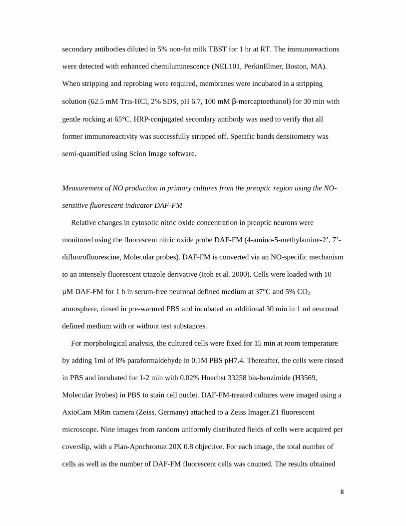
Introduction
Estradiol is the most biologically prevalent and active compound of a class of steroids
called estrogens, and it exerts potent and wide-ranging effects on the brain (Maggi et al.
2004). Besides their well-known effects on the hypothalamic-pituitary-gonadal axis,
estrogens have multiple and complex influences on brain structure and physiology (Maggi et
al. 2004; Woolley 2007). Natural fluctuations of estrogen levels across the estrous cycle have
been shown to cause cyclic changes in dendritic spine density and synaptogenesis in the rat
hippocampus (Woolley and McEwen 1992). Likewise, estradiol treatment in ovariectomized
female rats controls dynamic changes in spine density in the hippocampus (Gould et al. 1990)
and hypothalamus (Calizo and Flanagan-Cato 2000). In addition to these structural effects on
neuronal connectivity, recent studies showed estrogens to play a distinct role in controlling
nitric oxide (NO) production (Lamar et al. 1999; d'Anglemont de Tassigny et al. 2007).
NO is a major messenger molecule involved in various cellular functions in the brain such
as apoptosis, differentiation, development, synaptic plasticity and neurosecretion (Prast and
Philippu 2001; Boehning and Snyder 2003; McCann et al. 2003). Neuronal NO synthase
(nNOS) is the predominant source of NO in neurons (Boehning and Snyder 2003). Neuronal
NOS is primarily activated by its interaction with the Ca2+-calmodulin complex when
intracellular Ca2+ increases (Bredt and Snyder 1990; Abu-Soud et al. 1994). Although there
are several distinct calcium pools at the synapse, only Ca2+ influx through the N-Methyl-D-
Aspartate (NMDA) glutamate receptor efficiently activates nNOS (Kiedrowski et al. 1992) as
calmodulin is physically associated to the NMDA receptor (Hisatsune et al. 1997). A physical
approximation of nNOS to the NMDA receptor determines this specificity and is only
permitted via postsynaptic density protein PSD95 (Christopherson et al. 1999), which acts as
an adaptor protein via PDZ (PSD-95/Discs-large/Zona occludens) domain interaction
(Christopherson et al. 1999; Sattler et al. 1999; Aarts et al. 2002). Therefore, NO production

4
highly depends on i) NMDA receptor activation (Bredt and Snyder 1989; Garthwaite and
Boulton 1995), and ii) nNOS subcellular localization within the neuron.
Interestingly, we have recently reported that estrogens promote cyclic fluctuations in the
association between nNOS and the NMDA receptor subunit 2B (NR2B) in the preoptic
region of the hypothalamus in female rats during the ovarian cycle (d'Anglemont de Tassigny
et al. 2007). Thus, one mechanism involved in the regulation by estradiol of nNOS activity in
neurons may reside in its increased coupling to PSD-95/NR2B complex to facilitate the
access to the Ca2+-calmodulin located just underneath the NMDA receptor. However, the
molecular mechanisms underlying these changes remain elusive, and in particular, whether
classical estrogen receptors activation is required to promote nNOS/NMDA receptor complex
formation and thereby coupling glutamatergic fluxes with NO production is unknown.
Using neurons-containing primary cultures from the preoptic region of neonate rats, we
report here a role for estrogen receptors in regulating protein-protein interactions, the
induction of nNOS/NMDA receptor complex formation and its translation into NO release in
response to the gonadal steroid estradiol. This effect on NO production is dependent on the
activation of NR2B NMDA receptor subunits by glutamate and on PSD-95 expression.
Materials and Methods
Primary neuron-containing cultures from the preoptic region
Primary cultures were prepared from newborn (P0) Sprague-Dawley rats (Janvier Saint-
Berthevin, France). Both male and female pups were used for experiments. Even though,
circulating testosterone at birth is already higher in males as compared to females (Amateau
and McCarthy 2004), hormone exposure is too short to mediate masculinization of the P0
male brain and to promote irreversible changes in hormone-sensitive neurons (Finn et al.
1996). After decapitation and removal of the brain, the meninges and optic chiasm were

5
discarded and the preoptic region was isolated under a binocular magnifying glass with
Wecker’s scissors (Moria, France). The external limits for this dissection (adapted to P0 brain
size from adult brain) are: lateral, the external border of the Medial Preoptic Area (MPO);
dorsal, the internal border of anterior commissures (aco); and antero-posterior limits are
+0.95 to -0.51 mm from bregma, according to the Swanson Atlas (Swanson 1996). Preoptic
region explants were placed in ice-cold Dulbecco’s Modified Eagle Medium (DMEM,
Invitrogen). Each explant was cut into 5-6 smaller pieces and incubated for 1 hr at 37°C and a
5% CO2 atmosphere in DMEM containing papain (33 U/ml, 3126, Worthington-Cooper,
Lakewood, NJ), deoxyribonuclease I (DNase I, 125 U/ml, D4527, Sigma) and L-cysteine
(2.5mM, Sigma) for papain activation. Papain-incubated tissues were washed twice in
DMEM with the trypsin inhibitor ovomucoid (1.54 mg/ml, 109878, Roche Diagnostics),
DNase I (125 U/ml, Sigma) and bovine serum albumin (0.62 mg/ml, A7906, Sigma) to end
the enzymatic reaction. The fragments were crushed through a 20 µm nylon mesh (Sefar
America Inc., Kansas City, MO) and the dissociated cells were centrifuged at 90xg for 10
min and resuspended in serum-free neuronal defined medium consisting of Neurobasal-A
medium without phenol red (12340-015, Invitrogen) with 2% (v/v) B-27 supplement (17504-
044, Invitrogen), 1% (v/v) GlutaMAX (35050-038, Invitrogen) and 2% (v/v) antibiotics
(penicillin/streptomycin, 10378-016, Invitrogen). Cells were counted with a hemacytometer
(Thoma Cell, Marienfield, Germany). For immunoprecipitation and immunoblotting
experiments, 2x10⁶ cells were plated in 10 cm-diameter poly-L-lysine (MW > 300,000,
P5899, Sigma)-coated dishes (Falcon). Primary cell cultures used for fluorescent experiments
were plated onto 12mm-diameter poly-L-lysine-coated coverslips in 24-well-plates (Falcon)
with a density of 4x10⁵ cells per well. Primary cultures were maintained in an incubator at
37°C and 5% CO2 for 9 days in vitro (DIV; time of plating, DIV0), their medium was
changed after 2 days of culture and subsequently 3 times a week. 17-β-estradiol was obtained

6
from Sigma (E-8875). ICI 182,780 (ICI, 1047) and ifenprodil (0545) were supplied by Tocris
(Ellisville, MO).
Immunohistofluorescence
Coverslips with adhered cells were immersed in 4% paraformaldehyde and 5% sucrose in
0.1M PBS pH7.4, warmed to 37°C for 10min, washed three times in PBS, and permeabilized
with 50% ethanol for 60min at 4°C. The cells were then washed three times in PBS and
incubated in 5% bovine albumin (A7906, Sigma) in 0.3% Triton X-100 PBS (PBST) for
60min at RT with agitation. After blocking, both sheep polyclonal anti-nNOS (1:3000; Dr.
PC Emson (Medical Research Council, Laboratory for Molecular Research, Cambridge, UK)
(Herbison et al. 1996) and rabbit polyclonal anti-NR2B (1:500; 71-8600, Zymed
Laboratories, San Francisco, CA) (Christopherson et al. 1999) were applied in 5% bovine
albumin PBST, and the cultures were incubated overnight at 4°C. Coverslips were
extensively washed in PBST and incubated in PBST containing secondary anti-sheep Alexa
Fluor 568 conjugate (1:500) and biotinylated anti-rabbit (1:500) for 60min at room
temperature with agitation. After three washes, cultures were incubated with a streptavidin
Alexa Fluor 488 conjugate (1:500) in PBST for 60min at RT. After washing, coverslips were
mounted on slides in Permafluor medium (434990, Immunon, Pittsburgh, PA). Images were
acquired with a TCS SP confocal system (Leica, Nussloch, Germany).
Protein extraction and coimmunoprecipitation
After treatment, cells were briefly washed with ice-cold PBS and snap frozen on dry ice.
They were lysed in 500µl freshly prepared lysis buffer (pH 7.4, 25mM Tris, 50mM β-
glycerophosphate, 1.5mM EGTA, 0.5mM EDTA, 1mM sodium pyrophosphate, 1mM sodium
orthovanadate, 10µg/ml leupeptin and pepstatin, 10µg/ml aprotinin, 100µg/ml PMSF, and

7
1% Triton X-100). Protein concentrations of cell lysate were determined using the Bradford
method (BioRad, Hercules, CA). Equal amounts of protein (500 µg) in a total volume of
750µl lysis buffer were incubated with gentle rocking at 4°C overnight with 1 µg of rabbit
polyclonal IgG anti-nNOS (sc-8309; Santa Cruz, CA) or 2 µg of mouse monoclonal anti-
PSD95 (MA1-045 and MA1-046; Affinity BioReagents, Golden,CO) (Kornau et al. 1995).
Thereafter, 60 µl of protein A-sepharose beads in lysis buffer (1:1 blend) was added to each
sample and incubated for an additional 3h with gentle rocking at 4°C. The sepharose beads
were pelleted by brief centrifugation, the supernatant was collected and 375µl of 3X sample
buffer (187mM Tris-Base, 9% SDS, 15% glycerol, 15% β-mercaptoethanol and bromophenol
blue, pH 6.8 in 1X final) was added to it for analysis of non-immunoprecipitated proteins.
The beads were washed three times with ice-cold lysis buffer and boiled for 5 min in 50µl of
2X sample buffer. Beads alone incubated with proteins extracts were used as negative
controls. When necessary, the samples were stored at -80°C until use.
Western blotting analysis
Samples were reboiled for 5 min after thawing and electrophoresed for 75min at 150 V in
7% Tris-acetate, or for 50 min at 200 V in 4-12% MES precast SDS-polyacrylamide gels
according to the protocol supplied with the NuPAGE system (Invitrogen, Carlsbald, CA).
After size-fractionation, the proteins were transferred onto polyvinylidene difluoride (PVDF)
0.2 µm pore-size membranes (LC2002, Invitrogen) in the blot module of the NuPAGE sytem
(Invitrogen) for 75 min at room temperature (RT). Blots were blocked for 1 hr in TBS with
0.05% Tween 20 (TBST) and 5% non-fat milk at RT reacted overnight at 4°C with primary
antibody against nNOS (1:500; sc-8309), PSD95 (1:500; MA1-046), NR2B (1:500; 71-8600
Zymed Laboratories, San Francisco, CA) or NR2A (1:500; AB1555P Chemicon), and
washed four times with TBST before being exposed to horseradish peroxidase-conjugated
8
secondary antibodies diluted in 5% non-fat milk TBST for 1 hr at RT. The immunoreactions
were detected with enhanced chemiluminescence (NEL101, PerkinElmer, Boston, MA).
When stripping and reprobing were required, membranes were incubated in a stripping
solution (62.5 mM Tris-HCl, 2% SDS, pH 6.7, 100 mM β-mercaptoethanol) for 30 min with
gentle rocking at 65°C. HRP-conjugated secondary antibody was used to verify that all
former immunoreactivity was successfully stripped off. Specific bands densitometry was
semi-quantified using Scion Image software.
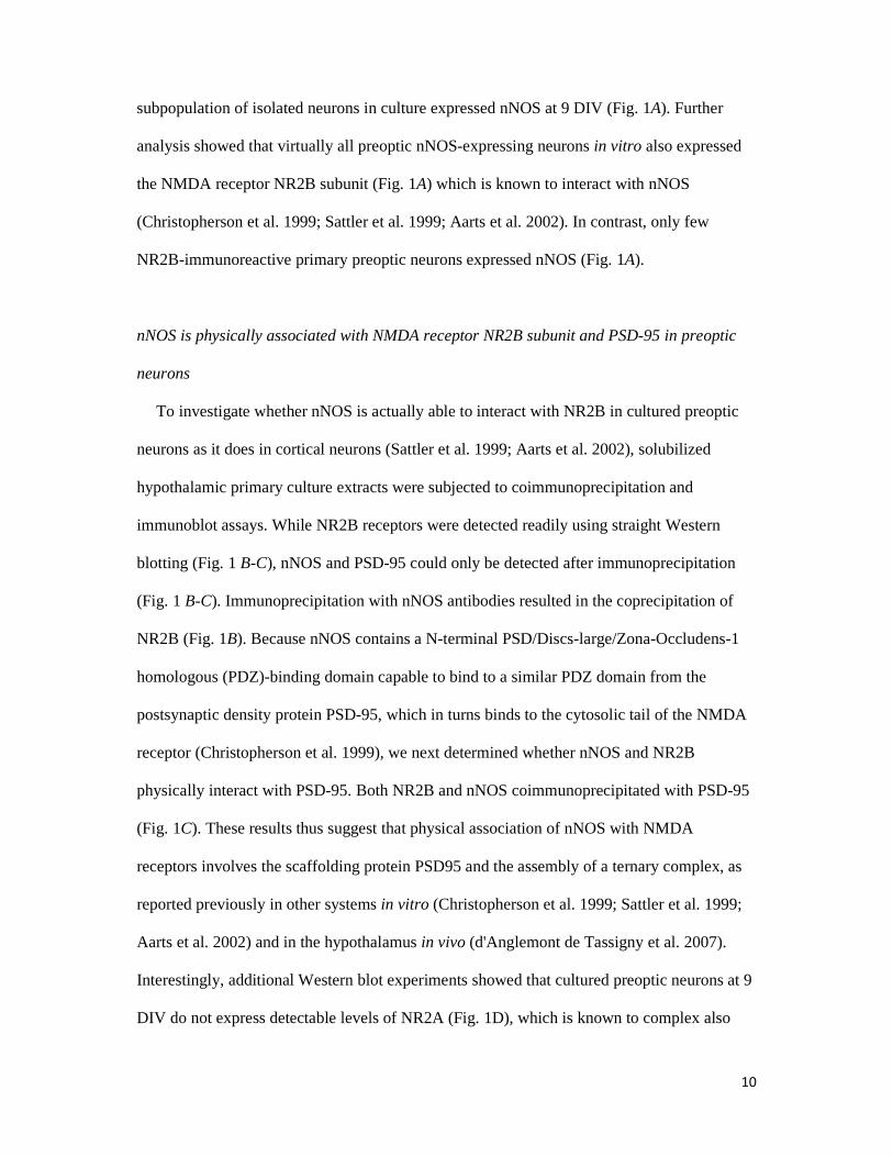
Measurement of NO production in primary cultures from the preoptic region using the NO-
sensitive fluorescent indicator DAF-FM
Relative changes in cytosolic nitric oxide concentration in preoptic neurons were
monitored using the fluorescent nitric oxide probe DAF-FM (4-amino-5-methylamine-2’, 7’-
difluorofluorescine, Molecular probes). DAF-FM is converted via an NO-specific mechanism
to an intensely fluorescent triazole derivative (Itoh et al. 2000). Cells were loaded with 10
µM DAF-FM for 1 h in serum-free neuronal defined medium at 37°C and 5% CO2
atmosphere, rinsed in pre-warmed PBS and incubated an additional 30 min in 1 ml neuronal
defined medium with or without test substances.
For morphological analysis, the cultured cells were fixed for 15 min at room temperature
by adding 1ml of 8% paraformaldehyde in 0.1M PBS pH7.4. Thereafter, the cells were rinsed
in PBS and incubated for 1-2 min with 0.02% Hoechst 33258 bis-benzimide (H3569,
Molecular Probes) in PBS to stain cell nuclei. DAF-FM-treated cultures were imaged using a
AxioCam MRm camera (Zeiss, Germany) attached to a Zeiss Imager.Z1 fluorescent
microscope. Nine images from random uniformly distributed fields of cells were acquired per
coverslip, with a Plan-Apochromat 20X 0.8 objective. For each image, the total number of
cells as well as the number of DAF-FM fluorescent cells was counted. The results obtained

9
from the analysis of the nine fields per coverslip were added, and the percentage of cells
bearing DAF-FM fluorescence calculated. For each experimental condition, at least six
coverslips were analyzed, and the results were averaged.
Primary cell culture treatment with antisense oligodeoxynucleotides (ODNs)
Fifteen-nucleotide oligomer phosphorothioated antisense ODNs (5’-
GAATGGGTCACCTCC-3’) corresponding to nucleotides 435-449 of rat PSD-95/SAP90
(Gen-Bank accession number M96853) were synthesised by MWG Biotech (Roissy, France).
This ODN sequence was previously shown to specifically inhibit PSD-95 expression in
neurons of the rat preoptic region in vitro (d'Anglemont de Tassigny et al. 2007). ODNs (5
µM) were added to the culture medium during feeding at 2, 4, 6 and 8 DIV. Cultures were
used for DAF-FM experiments at 9 DIV.
Statistics
The differences between several groups were analysed by one-way ANOVA followed by
the Student-Newman-Keuls multiple comparison test for unequal replication. Before
statistical analysis, percentages were subjected to arc-sine transformation to convert them
from binomial to normal distribution. The level of significance was set at p < 0.05.
Results
NMDA receptor NR2B subunit is expressed by NO-producing neurons of the preoptic region
in vitro
To determine whether hypothalamic preoptic neurons in vitro express nNOS, we subjected
primary cultures prepared from newborn rat preoptic regions to immunocytochemical
procedures. Immunohistofluorescence–confocal microscopy studies showed that a
10
subpopulation of isolated neurons in culture expressed nNOS at 9 DIV (Fig. 1A). Further
analysis showed that virtually all preoptic nNOS-expressing neurons in vitro also expressed
the NMDA receptor NR2B subunit (Fig. 1A) which is known to interact with nNOS
(Christopherson et al. 1999; Sattler et al. 1999; Aarts et al. 2002). In contrast, only few
NR2B-immunoreactive primary preoptic neurons expressed nNOS (Fig. 1A).
nNOS is physically associated with NMDA receptor NR2B subunit and PSD-95 in preoptic
neurons
To investigate whether nNOS is actually able to interact with NR2B in cultured preoptic
neurons as it does in cortical neurons (Sattler et al. 1999; Aarts et al. 2002), solubilized
hypothalamic primary culture extracts were subjected to coimmunoprecipitation and
immunoblot assays. While NR2B receptors were detected readily using straight Western
blotting (Fig. 1 B-C), nNOS and PSD-95 could only be detected after immunoprecipitation
(Fig. 1 B-C). Immunoprecipitation with nNOS antibodies resulted in the coprecipitation of
NR2B (Fig. 1B). Because nNOS contains a N-terminal PSD/Discs-large/Zona-Occludens-1
homologous (PDZ)-binding domain capable to bind to a similar PDZ domain from the
postsynaptic density protein PSD-95, which in turns binds to the cytosolic tail of the NMDA
receptor (Christopherson et al. 1999), we next determined whether nNOS and NR2B
physically interact with PSD-95. Both NR2B and nNOS coimmunoprecipitated with PSD-95
(Fig. 1C). These results thus suggest that physical association of nNOS with NMDA
receptors involves the scaffolding protein PSD95 and the assembly of a ternary complex, as
reported previously in other systems in vitro (Christopherson et al. 1999; Sattler et al. 1999;
Aarts et al. 2002) and in the hypothalamus in vivo (d'Anglemont de Tassigny et al. 2007).
Interestingly, additional Western blot experiments showed that cultured preoptic neurons at 9
DIV do not express detectable levels of NR2A (Fig. 1D), which is known to complex also

11
PSD95 (Sans et al. 2000; Liu et al. 2004; Cui et al. 2007). Altogether these results suggest
that association of nNOS with NMDA receptors is likely to involve the specific participation
of NR2B in primary cultures of neurons from the preoptic region.
Estradiol promotes nNOS/NR2B complex formation in preoptic neurons
To explore the possible role of estrogens in triggering the physical linkage of nNOS to
NR2B in hypothalamic neurons, additional experiments were performed on cultured preoptic
neurons. Treatment of hypothalamic cultures with physiological levels of estradiol (1 nM) for
30 min or 48h and immunoprecipitation of the protein extracts with rabbit polyclonal
antibodies to nNOS, followed by Western blot analysis to NR2B, showed an increased
physical approximation of nNOS and NR2B after 30 min estradiol-treatment when compared
to control cultures (Fig. 2; n = 4 independent experiments, one Way ANOVA, p = 0.025
control vs. 30-min estradiol-treatment). A similar trend was monitored after 48-h estradiol-
treatment but did not reach statistical significance (Fig. 2; n = 4 independent experiments,
one Way ANOVA, p = 0.057 control vs. 48-h estradiol-treatment).
Estradiol promotes neuronal NO formation via estrogen receptor activation
We next investigated whether estradiol-mediated increase in physical approximation of
nNOS with NMDA receptor NR2B subunit is correlated with changes in NO formation in
cultured preoptic neurons. To provide microscopic visualization of NOS catalytic activity, we
used the NO-sensitive fluorescent indicator DAF-FM (Kojima et al. 1999; Itoh et al. 2000;
Canabal et al. 2007). Application of 1 nM estradiol for 30 min induced a two-fold increase in
the relative number of cells exhibiting DAF-FM fluorescence (Fig. 3A-B). Estradiol-induced
increase in NO production was completely blocked by the treatment of neuronal cultures with
an estrogen receptor antagonist, ICI 182,780 (1 µM), for 30 min before exposing them to
12
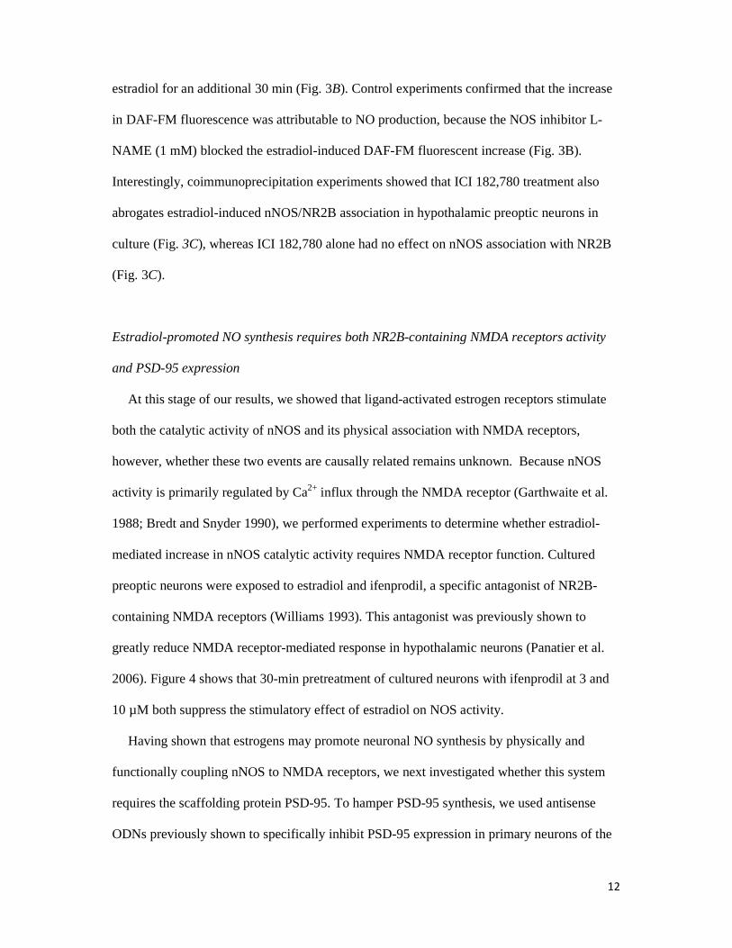
estradiol for an additional 30 min (Fig. 3B). Control experiments confirmed that the increase
in DAF-FM fluorescence was attributable to NO production, because the NOS inhibitor L-
NAME (1 mM) blocked the estradiol-induced DAF-FM fluorescent increase (Fig. 3B).
Interestingly, coimmunoprecipitation experiments showed that ICI 182,780 treatment also
abrogates estradiol-induced nNOS/NR2B association in hypothalamic preoptic neurons in
culture (Fig. 3C), whereas ICI 182,780 alone had no effect on nNOS association with NR2B
(Fig. 3C).
Estradiol-promoted NO synthesis requires both NR2B-containing NMDA receptors activity
and PSD-95 expression
At this stage of our results, we showed that ligand-activated estrogen receptors stimulate
both the catalytic activity of nNOS and its physical association with NMDA receptors,
however, whether these two events are causally related remains unknown. Because nNOS
activity is primarily regulated by Ca2+ influx through the NMDA receptor (Garthwaite et al.
1988; Bredt and Snyder 1990), we performed experiments to determine whether estradiol-
mediated increase in nNOS catalytic activity requires NMDA receptor function. Cultured
preoptic neurons were exposed to estradiol and ifenprodil, a specific antagonist of NR2B-
containing NMDA receptors (Williams 1993). This antagonist was previously shown to
greatly reduce NMDA receptor-mediated response in hypothalamic neurons (Panatier et al.
2006). Figure 4 shows that 30-min pretreatment of cultured neurons with ifenprodil at 3 and
10 µM both suppress the stimulatory effect of estradiol on NOS activity.
Having shown that estrogens may promote neuronal NO synthesis by physically and
functionally coupling nNOS to NMDA receptors, we next investigated whether this system
requires the scaffolding protein PSD-95. To hamper PSD-95 synthesis, we used antisense
ODNs previously shown to specifically inhibit PSD-95 expression in primary neurons of the

13
rat preoptic region in culture conditions identical to those used in the present study
(d'Anglemont de Tassigny et al. 2007). Application of the ODN in the culture media (5 µM
for 7 d) significantly impaired the capability of estradiol to increase nNOS catalytic activity
in vitro (Figure 4). Altogether, these results suggest that coupling of nNOS to NMDA
receptors via PSD-95 through estrogen receptors-dependent mechanisms is likely to be an
integral component of the process by which nNOS is activated by estradiol in neurons of the
hypothalamic preoptic region in vitro.
Discussion
The present results indicate that ligand-mediated stimulation of estrogen receptors in
hypothalamic preoptic region neurons leads to activation of NO signaling in nNOS-
expressing cells. The cellular underpinnings of this activation include physical approximation
of nNOS with NMDA receptor NR2B subunit and activation of nNOS by a mechanism that
appears to require the activity of NMDA receptors, which constitutes the main stimulatory
calcium influx pathway for this enzyme (Garthwaite et al. 1988; Bredt and Snyder 1990). Our
findings identify a distinct role for estrogen receptors in the modulation of inter-neuronal
communication processes involving glutamatergic NMDA receptor activation of NO
signaling. This pathway may be used by endogenous estrogens to alternate coupling and
uncoupling of glutamatergic fluxes for NO production during the ovarian-cycle in the female
rat, thereby regulating NOergic neurotransmission and properties of synaptic transmission
(Savchenko et al. 1997; Wang et al. 2005).
It was recently shown that high endogenous estrogen levels in female rats coincide with
increase in both the physical association of nNOS to NR2B and the magnitude of NO release
in the preoptic region (d'Anglemont de Tassigny et al. 2007). By now showing that the
estradiol-induced NO release is dependent of the NR2B-containing NMDA receptors activity
within neurons, our current results expand our previous in vivo observations and suggest that

14
the two events are causally linked (Figure 5). In addition to demonstrating the interdependent
involvement of nNOS/NMDA receptor coupling in estradiol-induced activation of neuronal
NO signaling by glutamate, our NOS catalytic activity assays and coimmunoprecipitation
analyses showed that classical estrogen receptor signaling may directly contribute to this
process. The estrogen receptor antagonist ICI 182,780 that competitively inhibits binding of
estradiol to the estrogen receptor and impairs receptor dimerisation (Osborne et al. 2004)
indeed prevented both estradiol-stimulated NR2B/nNOS complex formation and NO
production in cultured preoptic neurons (Figure 5).
It was demonstrated previously that physical coupling of nNOS to NMDA receptors
involves the scaffolding protein PSD-95 and the assembly of the ternary complex
(Christopherson et al. 1999) that efficiently couples Ca2+ influx, via NMDA receptors, to NO
synthesis and activity (Sattler et al. 1999; Aarts et al. 2002; Ishii et al. 2006). Since
impairment of PSD-95 synthesis via treatment with an antisense ODN in the culture media
blunts the estradiol-promoted activation of NOS activity without affecting basal NOS
activity, our data point to PSD-95 as a key component of the signaling pathway by which
estradiol mediates its stimulatory effects on NO production in preoptic neurons. Furthermore,
we demonstrate that blocking the NR2B-containing NMDA receptors with ifenprodil also
prevents the estradiol-induced NO production. This suggests that the estradiol - NMDA
receptor- NO pathway is essential in our in vitro system. The NMDA receptor subunit 2A
(NR2A) that is expressed in the preoptic region (Gore et al. 2000) has also been shown to
interact with PSD95 (Sans et al. 2000; Liu et al. 2004; Cui et al. 2007). Intriguingly, our data
show that while NR2A is expressed in the hypothalamus of adult rats, this NMDA receptor
subunit is not expressed at detectable levels in primary cultures of preoptic neurons obtained
from newborns. Differences in neuronal maturation could explain this apparent discrepancy,
as shown previously for other neuronal populations both in vitro (Brewer et al. 2007) and in

15
vivo (Liu et al. 2004). These results together with the blockade of estrogen-promoted NO
release by micromolar concentrations of ifenprodil strongly suggest that NR2B-containing
NMDA channels are involved in the coupling of glumatergic fluxes for NO production in
preoptic neurons.
We know very little about the mechanism used by estrogen receptors to promote the
recruitment of nNOS to the NMDA receptor. A non-genomic action of estradiol could be
involved in this mechanism, as changes in nNOS/PSD-95/NR2B are observable within 30
minutes. The question of whether the non-genomic effects of estradiol are mediated by
membrane-bound estrogen receptors may be of low relevance since the plasma membrane is
not a barrier for estradiol to enter into cells (Warner and Gustafsson 2006). However, the
nature and location of the receptor might have a profound effect on its ability to induce the
nNOS/PSD-95/NR2B ternary complex formation. The use of ICI 182,780 prevents the
estradiol-induced nNOS/NR2B association and subsequent NO release. In addition to bind to
classical estrogen receptors, ICI 182,780 was shown to act on the recently discovered
transmembrane estrogen receptor GPR30 (Prossnitz et al. 2008). While ICI 182,780 blocks
the classical ERα and ERβ, it activates GPR30 (Filardo et al. 2000; Filardo et al. 2002;
Thomas et al. 2005). Our results thus almost certainly discard the participation of GPR30
membrane estrogen receptor. There is some evidence that estradiol acts in neurons to
stimulate rapid PSD-95 new protein synthesis by promoting protein translation of dendrite-
localized mRNA transcripts via the Akt/protein kinase B pathway (Akama and McEwen
2003). Estradiol also may mediate coalescence of cytoskeleton-tethered nNOS (Haraguchi et
al. 2000) to PSD-95 through spine formation (Amateau and McCarthy 2002; Abraham et al.
2004), which would require remodeling of the actin cytoskeleton (Hering and Sheng 2001).
16
Nonetheless, additional studies are required to determine whether such phenomena occur
within the hypothalamus to modulate the nNOS subcellular localization.
A key physiological output for the estradiol effect on nNOS/NMDA receptor association
and NO production in neurons of the preoptic region is the onset of the preovulatory surge.
The central control of reproduction operates through the timely activation of gonadotropin-
releasing hormone (GnRH) neurons, the final pathway for neural control of ovulation (Ojeda
et al. 2002; Herbison and Neill 2006). GnRH neurons’ activity is finely tuned by several
factors in response to sex steroids feedback. In females, a prolonged high concentration of
estrogens induce the preovulatory GnRH/LH surge by modulating neuronal pathways
upstream to GnRH neurons that are located within the preoptic region of the hypothalamus in
rodents (Herbison and Neill 2006). Estradiol interacts with glutamate-containing pathways of
the preoptic region to mediate their stimulatory effect on GnRH secretion (Brann and Mahesh
1991; Jarry et al. 1992). These effects involve activation of NMDA receptors (Ojeda et al.
1990; Brann and Mahesh 1991) and require the production of NO (Bonavera et al. 1993;
Brenman et al. 1996; d'Anglemont de Tassigny et al. 2007). Previous studies performed in
rodents have described the relationship existing between GnRH neurons and NO (Bhat et al.
1995; Herbison et al. 1996; Clasadonte et al. 2008) and have demonstrated that endogenous
NO production was capable of influencing GnRH neuronal activity in the preoptic region
(Clasadonte et al. 2008). Moreover, most nNOS neurons of the preoptic region express both
estrogen receptor α (Scordalakes et al. 2002; Sato et al. 2005) and NR2B-containing NMDA
receptors (d'Anglemont de Tassigny et al. 2007). It thus becomes increasingly clear that these
neurons may be critical for the estrogen positive feedback to GnRH neurons.
In conclusion, we demonstrate in vitro that estradiol stimulates neuronal NO production by
inducing physical association of nNOS to NMDA receptors in an estrogen receptor-
dependent manner. Our results together with previous studies identify a distinct role for
17
estrogens and estrogen receptors in modulating the coupling of glutamatergic fluxes for NO
production in neurons of the CNS.
References
Aarts M., Liu Y., Liu L., Besshoh S., Arundine M., Gurd J. W., Wang Y. T., Salter M. W. and
Tymianski M. (2002) Treatment of ischemic brain damage by perturbing NMDA receptor- PSD-95 protein interactions. Science 298, 846-850.
Abraham I. M., Todman M. G., Korach K. S. and Herbison A. E. (2004) Critical in vivo roles for classical estrogen receptors in rapid estrogen actions on intracellular signaling in mouse brain. Endocrinology 145, 3055-3061.
Abu-Soud H. M., Yoho L. L. and Stuehr D. J. (1994) Calmodulin controls neuronal nitric-oxide synthase by a dual mechanism. Activation of intra- and interdomain electron transfer. J Biol Chem 269, 32047-32050.
Akama K. T. and McEwen B. S. (2003) Estrogen stimulates postsynaptic density-95 rapid protein synthesis via the Akt/protein kinase B pathway. J.Neurosci. 23, 2333-2339.
Amateau S. K. and McCarthy M. M. (2002) A novel mechanism of dendritic spine plasticity involving estradiol induction of prostaglandin-E2. J.Neurosci. 22, 8586-8596.
Amateau S. K. and McCarthy M. M. (2004) Induction of PGE2 by estradiol mediates developmental masculinization of sex behavior. Nat Neurosci 7, 643-650.
Bhat G. K., Mahesh V. B., Lamar C. A., Ping L., Aguan K. and Brann D. W. (1995) Histochemical localization of nitric oxide neurons in the hypothalamus: association with gonadotropin-releasing hormone neurons and co-localization with N-methyl-D-aspartate receptors. Neuroendocrinology 62, 187-197.
Boehning D. and Snyder S. H. (2003) Novel neural modulators. Annu.Rev.Neurosci. 26, 105-131. Bonavera J. J., Sahu A., Kalra P. S. and Kalra S. P. (1993) Evidence that nitric oxide may mediate the
ovarian steroid-induced luteinizing hormone surge: involvement of excitatory amino acids. Endocrinology 133, 2481-2487.
Brann D. W. and Mahesh V. B. (1991) Endogenous excitatory amino acid involvement in the preovulatory and steroid-induced surge of gonadotropins in the female rat. Endocrinology 128, 1541-1547.
Bredt D. S. and Snyder S. H. (1989) Nitric oxide mediates glutamate-linked enhancement of cGMP levels in the cerebellum. Proc.Natl.Acad.Sci.U.S.A 86, 9030-9033.
Bredt D. S. and Snyder S. H. (1990) Isolation of nitric oxide synthetase, a calmodulin-requiring enzyme. Proc.Natl.Acad.Sci.U.S.A 87, 682-685.
Brenman J. E., Chao D. S., Gee S. H., McGee A. W., Craven S. E., Santillano D. R., Wu Z., Huang F., Xia H., Peters M. F., Froehner S. C. and Bredt D. S. (1996) Interaction of nitric oxide synthase with the postsynaptic density protein PSD-95 and alpha1-syntrophin mediated by PDZ domains. Cell 84, 757-767.
Brewer L. D., Thibault O., Staton J., Thibault V., Rogers J. T., Garcia-Ramos G., Kraner S., Landfield P. W. and Porter N. M. (2007) Increased vulnerability of hippocampal neurons with age in culture: temporal association with increases in NMDA receptor current, NR2A subunit expression and recruitment of L-type calcium channels. Brain Res 1151, 20-31.
Calizo L. H. and Flanagan-Cato L. M. (2000) Estrogen selectively regulates spine density within the dendritic arbor of rat ventromedial hypothalamic neurons. J.Neurosci. 20, 1589-1596.
Canabal D. D., Song Z., Potian J. G., Beuve A., McArdle J. J. and Routh V. H. (2007) Glucose, insulin, and leptin signaling pathways modulate nitric oxide synthesis in glucose-inhibited neurons in the ventromedial hypothalamus. Am.J.Physiol Regul.Integr.Comp Physiol 292, R1418-R1428.

18
Christopherson K. S., Hillier B. J., Lim W. A. and Bredt D. S. (1999) PSD-95 assembles a ternary complex with the N-methyl-D-aspartic acid receptor and a bivalent neuronal NO synthase PDZ domain. J.Biol.Chem. 274, 27467-27473.
Clasadonte J., Poulain P., Beauvillain J. C. and Prevot V. (2008) Activation of neuronal nitric oxide release inhibits spontaneous firing in adult gonadotropin-releasing hormone neurons: a possible local synchronizing signal. Endocrinology 149, 587-596.
Cui H., Hayashi A., Sun H. S., Belmares M. P., Cobey C., Phan T., Schweizer J., Salter M. W., Wang Y. T., Tasker R. A., Garman D., Rabinowitz J., Lu P. S. and Tymianski M. (2007) PDZ protein interactions underlying NMDA receptor-mediated excitotoxicity and neuroprotection by PSD-95 inhibitors. J Neurosci 27, 9901-9915.
d'Anglemont de Tassigny X., Campagne C., Dehouck B., Leroy D., Holstein G. R., Beauvillain J. C., Buee-Scherrer V. and Prevot V. (2007) Coupling of neuronal nitric oxide synthase to NMDA receptors via postsynaptic density-95 depends on estrogen and contributes to the central control of adult female reproduction. J.Neurosci. 27, 6103-6114.
Filardo E. J., Quinn J. A., Bland K. I. and Frackelton A. R., Jr. (2000) Estrogen-induced activation of Erk-1 and Erk-2 requires the G protein-coupled receptor homolog, GPR30, and occurs via trans-activation of the epidermal growth factor receptor through release of HB-EGF. Mol Endocrinol 14, 1649-1660.
Filardo E. J., Quinn J. A., Frackelton A. R., Jr. and Bland K. I. (2002) Estrogen action via the G protein-coupled receptor, GPR30: stimulation of adenylyl cyclase and cAMP-mediated attenuation of the epidermal growth factor receptor-to-MAPK signaling axis. Mol Endocrinol 16, 70-84.
Finn P. D., McFall T. B., Clifton D. K. and Steiner R. A. (1996) Sexual differentiation of galanin gene expression in gonadotropin-releasing hormone neurons. Endocrinology 137, 4767-4772.
Garthwaite J. and Boulton C. L. (1995) Nitric oxide signaling in the central nervous system. Annu.Rev.Physiol 57, 683-706.
Garthwaite J., Charles S. L. and Chess-Williams R. (1988) Endothelium-derived relaxing factor release on activation of NMDA receptors suggests role as intercellular messenger in the brain. Nature 336, 385-388.
Gore A. C., Yeung G., Morrison J. H. and Oung T. (2000) Neuroendocrine aging in the female rat: the changing relationship of hypothalamic gonadotropin-releasing hormone neurons and N-methyl-D-aspartate receptors. Endocrinology 141, 4757-4767.
Gould E., Woolley C. S., Frankfurt M. and McEwen B. S. (1990) Gonadal steroids regulate dendritic spine density in hippocampal pyramidal cells in adulthood. J.Neurosci. 10, 1286-1291.
Haraguchi K., Satoh K., Yanai H., Hamada F., Kawabuchi M. and Akiyama T. (2000) The hDLG-associated protein DAP interacts with dynein light chain and neuronal nitric oxide synthase. Genes Cells 5, 905-911.
Herbison A. E. and Neill J. D. (2006) Physiology of the Gonadotropin-Releasing Hormone Neuronal Network, in Knobil and Neill's Physiology of Reproduction, Vol. Third Edition, pp 1415-1482. Elsevier.
Herbison A. E., Simonian S. X., Norris P. J. and Emson P. C. (1996) Relationship of neuronal nitric oxide synthase immunoreactivity to GnRH neurons in the ovariectomized and intact female rat. J.Neuroendocrinol. 8, 73-82.
Hering H. and Sheng M. (2001) Dendritic spines: structure, dynamics and regulation. Nat.Rev.Neurosci. 2, 880-888.
Hisatsune C., Umemori H., Inoue T., Michikawa T., Kohda K., Mikoshiba K. and Yamamoto T. (1997) Phosphorylation-dependent regulation of N-methyl-D-aspartate receptors by calmodulin. J Biol Chem 272, 20805-20810.
Hollmann M. and Heinemann S. (1994) Cloned glutamate receptors. Annu.Rev.Neurosci. 17, 31-108. Ishii H., Shibuya K., Ohta Y., Mukai H., Uchino S., Takata N., Rose J. A. and Kawato S. (2006)
Enhancement of nitric oxide production by association of nitric oxide synthase with N-methyl-D-aspartate receptors via postsynaptic density 95 in genetically engineered Chinese hamster ovary cells: real-time fluorescence imaging using nitric oxide sensitive dye. J.Neurochem. 96, 1531-1539.
Itoh Y., Ma F. H., Hoshi H., Oka M., Noda K., Ukai Y., Kojima H., Nagano T. and Toda N. (2000) Determination and bioimaging method for nitric oxide in biological specimens by diaminofluorescein fluorometry. Anal.Biochem. 287, 203-209.

19
Jarry H., Hirsch B., Leonhardt S. and Wuttke W. (1992) Amino acid neurotransmitter release in the preoptic area of rats during the positive feedback actions of estradiol on LH release. Neuroendocrinology 56, 133-140.
Kiedrowski L., Costa E. and Wroblewski J. T. (1992) Glutamate receptor agonists stimulate nitric oxide synthase in primary cultures of cerebellar granule cells. J.Neurochem. 58, 335-341.
Kojima H., Urano Y., Kikuchi K., Higuchi T., Hirata Y. and Nagano T. (1999) Fluorescent Indicators for Imaging Nitric Oxide Production. Angew.Chem.Int.Ed Engl. 38, 3209-3212.
Kornau H. C., Schenker L. T., Kennedy M. B. and Seeburg P. H. (1995) Domain interaction between NMDA receptor subunits and the postsynaptic density protein PSD-95. Science 269, 1737-1740.
Lamar C. A., Bhat G. K., Mahesh V. B. and Brann D. W. (1999) Evidence that neuronal nitric oxide synthase but not heme oxygenase increases in the hypothalamus on proestrus afternoon. Neuroendocrinology 70, 360-367.
Liu X. B., Murray K. D. and Jones E. G. (2004) Switching of NMDA receptor 2A and 2B subunits at thalamic and cortical synapses during early postnatal development. J Neurosci 24, 8885-8895.
Maggi A., Ciana P., Belcredito S. and Vegeto E. (2004) Estrogens in the nervous system: mechanisms and nonreproductive functions. Annu.Rev.Physiol 66, 291-313.
McCann S. M., Haens G., Mastronardi C., Walczewska A., Karanth S., Rettori V. and Yu W. H. (2003) The role of nitric oxide (NO) in control of LHRH release that mediates gonadotropin release and sexual behavior. Curr.Pharm.Des 9, 381-390.
Ojeda S. R., Urbanski H. F., Costa M. E., Hill D. F. and Moholt-Siebert M. (1990) Involvement of transforming growth factor alpha in the release of luteinizing hormone-releasing hormone from the developing female hypothalamus. Proc.Natl.Acad.Sci.U.S.A 87, 9698-9702.
Ojeda S. R., Terasawa E., Pfaff D., Arnold.A, Etgen A., Fahrbach S., Moss R. and Rubin R. (2002) Neuroendocrine regulation of puberty, pp 589-659. Elsevier, New York.
Osborne C. K., Wakeling A. and Nicholson R. I. (2004) Fulvestrant: an oestrogen receptor antagonist with a novel mechanism of action. Br.J.Cancer 90 Suppl 1, S2-S6.
Panatier A., Theodosis D. T., Mothet J. P., Touquet B., Pollegioni L., Poulain D. A. and Oliet S. H. (2006) Glia-derived D-serine controls NMDA receptor activity and synaptic memory. Cell 125, 775-784.
Prast H. and Philippu A. (2001) Nitric oxide as modulator of neuronal function. Prog.Neurobiol. 64, 51-68.
Prossnitz E. R., Oprea T. I., Sklar L. A. and Arterburn J. B. (2008) The ins and outs of GPR30: a transmembrane estrogen receptor. J Steroid Biochem Mol Biol 109, 350-353.
Sans N., Petralia R. S., Wang Y. X., Blahos J., 2nd, Hell J. W. and Wenthold R. J. (2000) A developmental change in NMDA receptor-associated proteins at hippocampal synapses. J Neurosci 20, 1260-1271.
Sato S., Braham C. S., Putnam S. K. and Hull E. M. (2005) Neuronal nitric oxide synthase and gonadal steroid interaction in the MPOA of male rats: co-localization and testosterone-induced restoration of copulation and nNOS-immunoreactivity. Brain Res. 1043, 205-213.
Sattler R., Xiong Z., Lu W. Y., Hafner M., MacDonald J. F. and Tymianski M. (1999) Specific coupling of NMDA receptor activation to nitric oxide neurotoxicity by PSD-95 protein. Science 284, 1845-1848.
Savchenko A., Barnes S. and Kramer R. H. (1997) Cyclic-nucleotide-gated channels mediate synaptic feedback by nitric oxide. Nature 390, 694-698.
Scordalakes E. M., Shetty S. J. and Rissman E. F. (2002) Roles of estrogen receptor alpha and androgen receptor in the regulation of neuronal nitric oxide synthase. J.Comp Neurol. 453, 336-344.
Swanson L. W. (1996) Structure of the rat brain. Elsevier Science Publishers, Amsterdam. Thomas P., Pang Y., Filardo E. J. and Dong J. (2005) Identity of an estrogen membrane receptor
coupled to a G protein in human breast cancer cells. Endocrinology 146, 624-632. Wang H. G., Lu F. M., Jin I., Udo H., Kandel E. R., de Vente J., Walter U., Lohmann S. M., Hawkins
R. D. and Antonova I. (2005) Presynaptic and postsynaptic roles of NO, cGK, and RhoA in long-lasting potentiation and aggregation of synaptic proteins. Neuron 45, 389-403.
Warner M. and Gustafsson J. A. (2006) Nongenomic effects of estrogen: why all the uncertainty? Steroids 71, 91-95.

20
Williams K. (1993) Ifenprodil discriminates subtypes of the N-methyl-D-aspartate receptor: selectivity and mechanisms at recombinant heteromeric receptors. Mol.Pharmacol. 44, 851-859.
Woolley C. S. (2007) Acute effects of estrogen on neuronal physiology. Annu.Rev.Pharmacol.Toxicol. 47, 657-680.
Woolley C. S. and McEwen B. S. (1992) Estradiol mediates fluctuation in hippocampal synapse density during the estrous cycle in the adult rat. J.Neurosci. 12, 2549-2554.
21
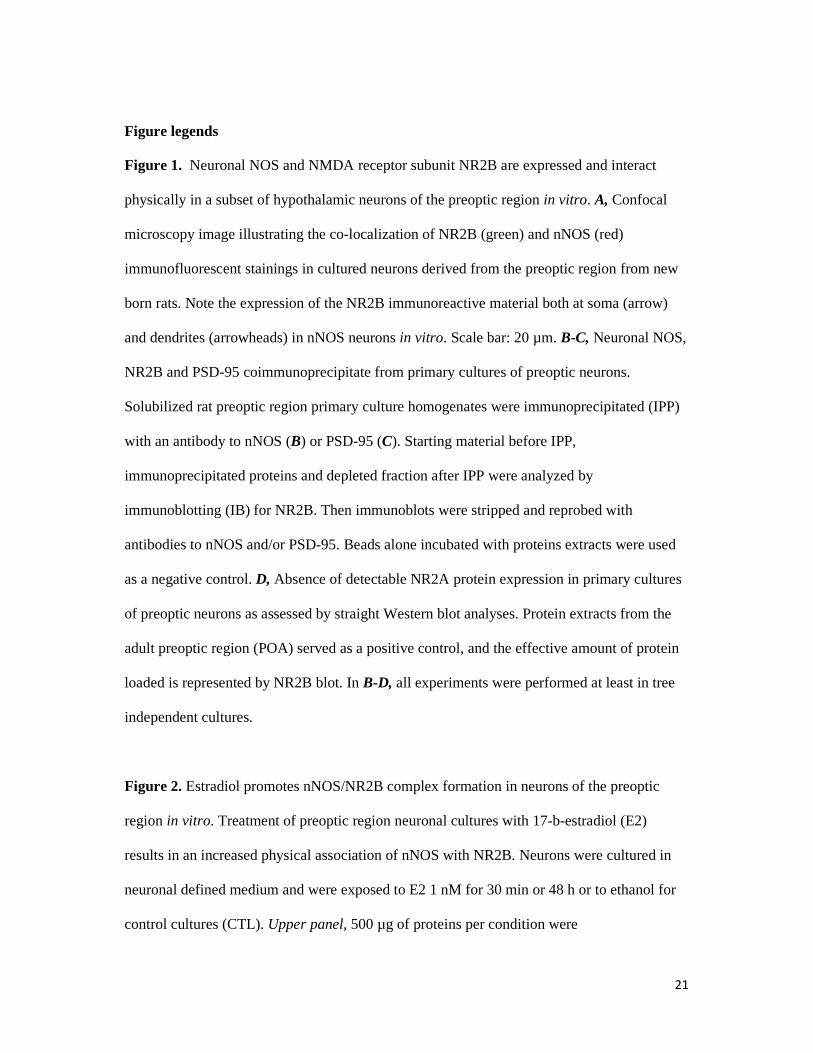
Figure legends Figure 1. Neuronal NOS and NMDA receptor subunit NR2B are expressed and interact
physically in a subset of hypothalamic neurons of the preoptic region in vitro. A, Confocal
microscopy image illustrating the co-localization of NR2B (green) and nNOS (red)
immunofluorescent stainings in cultured neurons derived from the preoptic region from new
born rats. Note the expression of the NR2B immunoreactive material both at soma (arrow)
and dendrites (arrowheads) in nNOS neurons in vitro. Scale bar: 20 µm. B-C, Neuronal NOS,
NR2B and PSD-95 coimmunoprecipitate from primary cultures of preoptic neurons.
Solubilized rat preoptic region primary culture homogenates were immunoprecipitated (IPP)
with an antibody to nNOS (B) or PSD-95 (C). Starting material before IPP,
immunoprecipitated proteins and depleted fraction after IPP were analyzed by
immunoblotting (IB) for NR2B. Then immunoblots were stripped and reprobed with
antibodies to nNOS and/or PSD-95. Beads alone incubated with proteins extracts were used
as a negative control. D, Absence of detectable NR2A protein expression in primary cultures
of preoptic neurons as assessed by straight Western blot analyses. Protein extracts from the
adult preoptic region (POA) served as a positive control, and the effective amount of protein
loaded is represented by NR2B blot. In B-D, all experiments were performed at least in tree
independent cultures.
Figure 2. Estradiol promotes nNOS/NR2B complex formation in neurons of the preoptic
region in vitro. Treatment of preoptic region neuronal cultures with 17-b-estradiol (E2)
results in an increased physical association of nNOS with NR2B. Neurons were cultured in
neuronal defined medium and were exposed to E2 1 nM for 30 min or 48 h or to ethanol for
control cultures (CTL). Upper panel, 500 µg of proteins per condition were
22
immunoprecipitated (IPP) with a specific nNOS antibody, electrophoresed and immunobloted
(IB) for NR2B. Then the immunoblot was stripped and reprobed with antibodies to nNOS to
ensure that equal amounts of nNOS protein had been immunoprecipitated and loaded on the
gel. Lower panel, bar graph showing the quantitative analysis of the differential
NR2B/nNOS association among E2 treatment (n = 4 independent experiments; * p = 0.025
vs CTL). Error bars indicate SEM. Statistical differences were established using a one way
ANOVA by the Student-Newman-Keuls multiple comparison test.
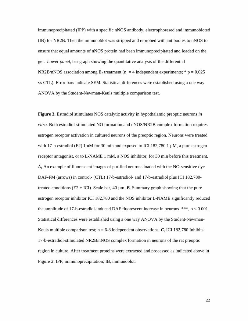
Figure 3. Estradiol stimulates NOS catalytic activity in hypothalamic preoptic neurons in
vitro. Both estradiol-stimulated NO formation and nNOS/NR2B complex formation requires
estrogen receptor activation in cultured neurons of the preoptic region. Neurons were treated
with 17-b-estradiol (E2) 1 nM for 30 min and exposed to ICI 182,780 1 µM, a pure estrogen
receptor antagonist, or to L-NAME 1 mM, a NOS inhibitor, for 30 min before this treatment.
A, An example of fluorescent images of purified neurons loaded with the NO-sensitive dye
DAF-FM (arrows) in control- (CTL) 17-b-estradiol- and 17-b-estradiol plus ICI 182,780-
treated conditions (E2 + ICI). Scale bar, 40 µm. B, Summary graph showing that the pure
estrogen receptor inhibitor ICI 182,780 and the NOS inhibitor L-NAME significantly reduced
the amplitude of 17-b-estradiol-induced DAF fluorescent increase in neurons. ***, p < 0.001.
Statistical differences were established using a one way ANOVA by the Student-Newman-
Keuls multiple comparison test; n = 6-8 independent observations. C, ICI 182,780 Inhibits
17-b-estradiol-stimulated NR2B/nNOS complex formation in neurons of the rat preoptic
region in culture. After treatment proteins were extracted and processed as indicated above in
Figure 2. IPP, immunoprecipitation; IB, immunoblot.

23
Figure 4. Estradiol requires both NR2B-containing NMDA receptors activity and PSD-95
expression to mediate its stimulatory effect on NO production in hypothalamic preoptic
neurons in vitro. Neurons were treated with 17-b-estradiol (E2) 1 nM for 30 min and exposed
to ifenprodil (3 and 10 µM), a NR2B antagonist, or PSD-95 antisense ODNs 5 µM for 30 min
or 7 d, respectively, before this treatment. Statistical differences were established using a one
way ANOVA by the Student-Newman-Keuls multiple comparison test. **, p < 0.01, n = 4-6
independent observations.
Figure 5. Schematic representation of the possible estradiol-mediated changes in protein-
protein interactions involved in the control of nNOS activity in neurons of the preoptic
region. Neuronal NOS activity is primarily regulated by increases in the local intracellular
[Ca2+], which activates nNOS through calmoduling (CaM) binding (Bredt and Snyder 1990).
Importantly, Ca2+ influx through the NMDA receptor (NMDA-R) but not other Ca2+ influx
pathways efficiently promotes NO synthesis (Garthwaite et al. 1988; Bredt and Snyder 1990)
One estrogen receptor (ER)-dependent mechanism used for the regulation of nNOS activity
may reside in the alternative coupling and uncoupling of the enzyme to NR2B-containing
NMDA receptors channels by the scaffolding protein PSD-95, in the presence or the absence
of estrogens, respectively. PSD-95 acts as an adaptor protein, thereby physically and
functionally coupling NMDA receptors to nNOS and thus enables to couple glutamatergic
fluxes with NO production. NO is formed enzymatically from L-arginine (L-Arg) in
equimolar amounts with L-citrulline (L-Cit) by nNOS. Functional glutamate NMDA
receptors are composed of NR1 and NR2 subunits (Hollmann and Heinemann 1994). The
estrogen receptor antagonist ICI 182,780 competitively inhibits binding of estradiol to the
estrogen receptor and impairs receptor dimerisation (Osborne et al. 2004); Antisense
oligodeoxynucleotides to PSD-95 (AS PSD-95) selectively decreases PSD-95 expression in

24
primary cultures of preoptic neurons (d'Anglemont de Tassigny et al. 2007); infenprodil is a
specific antagonist of NR2B-containing NMDA receptors (Williams 1993).

25

26

27

28

29
Copyright © 2022 FDOKUMEN