Comment on “The Molecular Evolutionary Patterns of the Insulin/FOXO Signaling Pathway&rdquo
Upload
khangminh22Category
view
1download
0
Enlighten – Research publications by members of the University of Glasgow http://eprints.gla.ac.uk
Kanao, T., Sawada, T., Davies, S., Ichinose, H., Hasegawa, K., Takahashi, R., Hattori, N., and Imai, Y. (2012) The nitric oxide-cyclic GMP pathway regulates FoxO and alters dopaminergic neuron survival in Drosophila. PLoS ONE, 7 (2). e30958. ISSN 1932-6203 http://eprints.gla.ac.uk/62015/ Deposited on: 23 August 2012

The Nitric Oxide-Cyclic GMP Pathway Regulates FoxOand Alters Dopaminergic Neuron Survival in DrosophilaTomoko Kanao1, Tomoyo Sawada4,5, Shireen-Anne Davies6, Hiroshi Ichinose7, Kazuko Hasegawa8,
Ryosuke Takahashi4,5, Nobutaka Hattori2,5, Yuzuru Imai3*
1 Research Institute for Diseases of Old Age, Juntendo University Graduate School of Medicine, Tokyo, Japan, 2 Department of Neurology, Juntendo University Graduate
School of Medicine, Tokyo, Japan, 3 Department of Neuroscience for Neurodegenerative Disorders, Juntendo University Graduate School of Medicine, Tokyo, Japan,
4 Department of Neurology, Kyoto University Graduate School of Medicine, Kyoto, Japan, 5 CREST (Core Research for Evolutionary Science and Technology), JST, Saitama,
Japan, 6 Institute of Molecular, Cell and Systems Biology, College of Medical, Veterinary and Life Sciences, University of Glasgow, Glasgow, Scotland, United Kingdom,
7 Department of Life Science, Graduate School of Bioscience and Biotechnology, Tokyo Institute of Technology, Yokohama, Japan, 8 Department of Neurology, National
Hospital Organization, Sagamihara National Hospital, Sagamihara, Japan
Abstract
Activation of the forkhead box transcription factor FoxO is suggested to be involved in dopaminergic (DA)neurodegeneration in a Drosophila model of Parkinson’s disease (PD), in which a PD gene product LRRK2 activates FoxOthrough phosphorylation. In the current study that combines Drosophila genetics and biochemical analysis, we show thatcyclic guanosine monophosphate (cGMP)-dependent kinase II (cGKII) also phosphorylates FoxO at the same residue asLRRK2, and Drosophila orthologues of cGKII and LRRK2, DG2/For and dLRRK, respectively, enhance the neurotoxic activity ofFoxO in an additive manner. Biochemical assays using mammalian cGKII and FoxO1 reveal that cGKII enhances thetranscriptional activity of FoxO1 through phosphorylation of the FoxO1 S319 site in the same manner as LRRK2. ADrosophila FoxO mutant resistant to phosphorylation by DG2 and dLRRK (dFoxO S259A corresponding to human FoxO1S319A) suppressed the neurotoxicity and improved motor dysfunction caused by co-expression of FoxO and DG2. Nitricoxide synthase (NOS) and soluble guanylyl cyclase (sGC) also increased FoxO’s activity, whereas the administration of a NOSinhibitor L-NAME suppressed the loss of DA neurons in aged flies co-expressing FoxO and DG2. These results stronglysuggest that the NO-FoxO axis contributes to DA neurodegeneration in LRRK2-linked PD.
Citation: Kanao T, Sawada T, Davies S-A, Ichinose H, Hasegawa K, et al. (2012) The Nitric Oxide-Cyclic GMP Pathway Regulates FoxO and Alters DopaminergicNeuron Survival in Drosophila. PLoS ONE 7(2): e30958. doi:10.1371/journal.pone.0030958
Editor: Philipp J. Kahle, Hertie Institute for Clinical Brain Research and German Center for Neurodegenerative Diseases, Germany
Received September 18, 2011; Accepted December 28, 2011; Published February 29, 2012
Copyright: � 2012 Kanao et al. This is an open-access article distributed under the terms of the Creative Commons Attribution License, which permitsunrestricted use, distribution, and reproduction in any medium, provided the original author and source are credited.
Funding: This study was supported by funding from the Inamori Foundation, the Uehara Memorial Foundation, Dainippon Sumitomo Pharma, and the Programfor Young Researchers from Special Coordination Funds for Promoting Science and Technology commissioned by MEXT in Japan. The funders had no role instudy design, data collection and analysis, decision to publish, or preparation of the manuscript.
Competing Interests: The authors have read the journal’s policy and have the following conflicts: this work was partly supported by Dainippon SumitomoPharma. This does not alter the authors’ adherence to all the PLoS ONE policies on sharing data and materials.
* E-mail: [email protected]
Introduction
PD, one of the most common movement disorders, is
characterized by age-dependent impairments of several nervous
systems including the midbrain DA system. The degeneration of
DA neurons in the substantia nigra produces the prominent motor
symptoms of PD. Postmortem inspections and studies with
neurotoxin-based PD models suggest a multifactorial etiology
involving inflammation, mitochondrial dysfunction, iron accumu-
lation and oxidative stress. NO, a free gaseous signaling molecule,
has also been implicated in PD [1,2]. The signaling function of
NO is dependent on the dynamic regulation of its synthase, NOS.
There are three types of NOS, neuronal NOS (nNOS), endothelial
NOS (eNOS) and inducible NOS (iNOS), in humans whereas the
Drosophila genome has only a single orthologue, dNOS. High levels
of nNOS and iNOS have been reported in the substantia nigra of
PD patients [3,4] and animal models of PD [5,6]. Overproduction
of NO is suggested to cause DNA damage, protein modifications
and cell toxicity mainly mediated by the reactive species
peroxynitrite, which may be generated with dopamine metabolism
in DA neurons. In the etiology of PD, overproduction of NO could
be caused either by upregulation of iNOS in activated glia cells
[3,5] or by an increase in intracellular calcium, for example, after
glutamate excitotoxicity [7].
The discovery of genes linked to rare familial forms of PD has
provided vital clues to understanding the cellular and molecular
pathogenesis of the disease. Missense mutations in the Leucine-rich
repeat kinase 2 (LRRK2)/Dardarin gene cause autosomal dominant
late onset familial PD as well as sporadic PD [8,9,10]. The clinical
symptoms and pathology caused by LRRK2 mutations closely
resemble those of the sporadic form of PD, suggesting that the
LRRK2 pathogenic pathway may underlie the general PD
etiology. The LRRK2 gene encodes a large protein with multiple
domains including a GTPase domain and a kinase domain [8,9].
Several amino acid substitutions are identified as pathogenic
mutations linked to PD [11]. Mutations in the kinase domain of
human LRRK2 such as G2019S and I2020T have been reported
to produce enhanced kinase activity in vitro, suggesting that gain-of-
function mutations of LRRK2 cause neurodegeneration
[12,13,14]. However, how these mutations present in the LRRK2
gene lead to the progressive loss of DA neurons and other
associated pathologies is still unknown.
PLoS ONE | www.plosone.org 1 February 2012 | Volume 7 | Issue 2 | e30958

Because various key signaling pathways are conserved between
humans and Drosophila, genetic and functional studies using
Drosophila models for familial PD have revealed crucial signal
transductions that affect the pathogenesis of PD [15]. We have
previously reported that a Drosophila LRRK2 orthologue, dLRRK
phosphorylates Drosophila FoxO (dFoxO) at Ser259, which
stimulates the expression of a pro-apoptotic dFoxO target, hid,
and leads to neurodegeneration in Drosophila [16]. The event was
further enhanced by transgenic expression of pathogenic dLRRK
proteins such as dLRRK I1915T (corresponding to I2020T in
humans). However, a kinase-dead form of dLRRK (dLRRK 3KD)
did not completely suppress a synergic effect caused by the co-
expression of dFoxO with dLRRK, suggesting that some other
factor(s) modulates this pathway. Here, we report that cGKII also
phosphorylates FoxO and activates FoxO-transcriptional activity
in the same manner as LRRK2/dLRRK by using biochemical
studies of mammalian cGKII and FoxO1. Moreover, by using
Drosophila models, our data suggest that NO signaling and its
downstream effector cGKII/DG2 contribute to DA neurodegen-
eration.
Results
cGK genetically interacts with FoxO and activates FoxOactivity
We previously reported a genetic interaction between FoxO and
LRRK2/dLRRK in Drosophila [16]. To identify components of the
LRRK2-FoxO signaling pathway, we screened for modifiers (Fig. 1
and Fig. S1A). Kinases reported to affect the activity of FoxO were
expressed with dFoxO in the Drosophila eye. As reported,
transgenic expression of AKT suppressed FoxO-mediated devel-
opmental defects in the eye. The expression of MST/Hippo
resulted in extensive degeneration, which did not appear to be
dependent on FoxO (Fig. 1). Expression of one of the Drosophila
cGMP-dependent kinases (cGKs), DG2, leads to strong optic
degeneration in conjunction with dFoxO (Fig. 1 and Fig. S2A),
while the other kinases had little effect on the developmental
defects caused by FoxO (Fig. 1). Removal of one copy of the dg2
gene improved the defects, suggesting that endogenous DG2
activity contribute to the dFoxO-mediated neurodegeneration
(Fig. 2H compared with B).
Next we examined whether DG2 is an upstream kinase of
dLRRK, or whether DG2 acts independently of dLRRK by
means of a combination of genetic interaction tests, reporter assays
for FoxO and in vitro kinase assay. Co-expression of dLRRK
harboring a PD-related mutant I1915T together with DG2
dramatically enhanced the toxicity of dFoxO (Fig. 2D compared
with C). However, expression of dLRRK 3KD or removal of the
dLRRK gene did not suppress the eye phenotype caused by
dFoxO-DG2 at all (Fig. 2E and J compared with C). Co-
expression of DG2 and dLRRK I1915T produced a normal eye,
suggesting that the phenotype is dependent on the level of dFoxO
protein (Fig. 2I compared with D).
Co-expression of dFoxO with DG2, but not GFP or DG1, in
Drosophila eyes caused appearance of a slower migrated dFoxO
protein in western blot analysis (Fig. 3A), which indicates
phosphorylation of dFoxO [16]. Consistent with the result,
knockdown of DG2 decreased a phosphorylated form of
endogenous dFoxO in Drosophila brain tissue (Fig. 3B). In Drosophila
S2 cells, transient expression of DG2 together with 8-bromogua-
nosine-39, 59-cyclic monophosphate (8-Br-cGMP), a membrane
permeable analogue for cGMP, also stimulates phosphorylation of
endogenous dFoxO (Fig. 3C, lane 3).
Two groups of cGKs, the soluble type I (cGKI a and b) and the
membrane-bound type II (cGKII), have been reported in
vertebrates. In Drosophila, there are two genes encoding cGK,
namely dg1 and dg2 [17]. As reported [18], the gene products DG1
and DG2 are located in the cytoplasm and at the cytoplasmic
membrane, respectively (Fig. 3E and F). Interestingly, expression
of DG1 had little effect on the degeneration of the eye mediated by
dFoxO, suggesting that DG1 and DG2 have different roles in vivo
(Fig. S1B, S2B and S2C). Although predictions of amino acid
sequence indicate that DG2 is more similar as a cGKI a/bhomologue [19], their subcellular distribution suggests that DG2 is
functionally more similar to cGKII (Fig. 3G–J) [18,20,21].
Consistent with the idea, transgenic expression of human cGKII
exacerbated eye degeneration by dFoxO (Fig. 2G compared with
B) whereas expression of cGKII alone did not affect the eye
development (Fig. 2F). Interestingly, cGKII appeared to recruit
FoxO1 to the cytoplasmic membrane of human cultured cells
(Fig. 3K–M) while there was no evidence that cGKI associates
with cGKII in vivo (Fig. S3). In addition, we observed that cGKII is
Figure 1. Screening of kinases that affect the eye phenotypes caused by dFoxO. Drosophila orthologues of reported FoxO kinases wereexpressed with (upper row) or without (lower) dFoxO in Drosophila eyes using the GMR-GAL4 driver. GFP served as a control. The Drosophila DG2 ispresumably functionally equivalent to the vertebrate cGKII. Reported phosphorylation sites and newly identified sites that are phosphorylated bycGKII (S152–155 and S319) in human FoxO1 are indicated by black and red lines, respectively. FHD, forkhead domain; NLS, nuclear localization signal;NES, nuclear export signal; TA, transactivation domain. Overexpressing lines used for crosses are: UAS-GFP (GFP), UAS-AKT1 (AKT), UAS-DG2 (DG2),UAS-hippo (MST), UAS-CDK1-Myc (CDK1), UAS-CDK2-Myc (CDK2), UAS-dLRRK (dLRRK), CkIaEP1555 (CK1a), mnbEY14320 (DYRK1), UAS-bsk (JNK), UAS-dIKKß(IKKb).doi:10.1371/journal.pone.0030958.g001
Nitric Oxide Signaling Modulates FoxO in Neurons
PLoS ONE | www.plosone.org 2 February 2012 | Volume 7 | Issue 2 | e30958

Figure 2. DG2 as well as dLRRK additively enhances FoxO-mediated developmental defects in the Drosophila eye. (A–J) SEM images ofthe eye of flies expressing the indicated genes. The genotypes are: GMR-Gal4 (A), GMR-Gal4/UAS-EGFP (B), UAS-DG2; GMR-Gal4, UAS-dFoxO (C), UAS-DG2;GMR-Gal4, UAS-dFoxO; UAS-dLRRK I1915T (D), UAS-DG2; GMR-Gal4, UAS-dFoxO; UAS-dLRRK 3KD (E), GMR-Gal4; UAS-cGKII (F), GMR-Gal4, UAS-dFoxO; UAS-cGKII (G), GMR-Gal4, UAS-dFoxO/DG2k04703 (H), UAS-DG2; GMR-Gal4; UAS-dLRRK I1915T (I), UAS-DG2; GMR-Gal4, UAS-dFoxO; e03680/e03680 (J).doi:10.1371/journal.pone.0030958.g002
Figure 3. DG2 modulates FoxO in vivo. (A) dFoxO and the indicated transgenes were expressed in the Drosophila eyes using the GMR-GAL4driver. Extracts from brain tissues were subjected to western blot analysis. dFoxO-P; a phosphorylated form of dFoxO. (B) DG2 RNAi or GFP RNAiconstructs were expressed in the Drosophila brain using the elav-GAL4 driver. Western blot analysis for endogenous dFoxO was carried out as in (A).(C) Drosophila S2 cells were transfected with or without C-terminally Myc-tagged DG2 (DG2-Myc). Thirty-six hrs post transfection, cells were treatedwith or without 10 mM 8-Br-cGMP for 30 min. Cell lysate were then subjected to western blot analysis. (D) Human 293T cells were transfected with orwithout cGKII-Myc, and were treated with 8-Br-cGMP as in (C). Phosphorylation of the S319 site in endogenous FoxO1 was detected with phospho-specific antibody. (E, F) S2 cells expressing DG2-Myc (E) or DG1-Myc (F) were visualized with anti-Myc antibody (green), by counterstaining with DAPI(blue color). (G–J) HeLa cells expressing AKT-PH-GFP (green) along with cGKII-Myc (G, H) or cGKI-Myc (I, J) were visualized with anti-Myc antibody(red), by counterstaining with DAPI (blue color). AKT-PH-GFP was used for a marker protein of the plasma membrane [62]. (K–M) Flp-In T-REx-293cells harboring EGFP-FoxO1 gene were transiently transfected with cGKII-Myc, and EGFP-FoxO1 was induced with doxycycline. Enlarged views of theplasma membrane regions in cGKII-positive (Box1) and negative (Box2) cells are also shown in (M). Accumulation of FoxO1 along with cGKII in theplasma membrane is indicated by arrowheads. Scale bars = 5 mm for (E, F), 25 mm for (G–J) and 10 mm for (K–M).doi:10.1371/journal.pone.0030958.g003
Nitric Oxide Signaling Modulates FoxO in Neurons
PLoS ONE | www.plosone.org 3 February 2012 | Volume 7 | Issue 2 | e30958

abundantly expressed in DA neurons in the substantia nigra of
mice (Fig. S4). We then focused on mammalian cGKII as a cGK
that might be associated with the pathology of PD. Reporter assays
for FoxO transcriptional activity revealed that cGKII stimulated
FoxO activity in cultured mammalian cells and that co-expression
of hLRRK2 with cGKII caused a 3-fold increase in FoxO activity
(Fig. 4A). A kinase-dead form of hLRRK2 (hLRRK2 3KD) did
not suppress the activation of FoxO by cGKII to the control level.
Similarly, a kinase-dead form of cGKII (cGKII KD) failed to
suppress FoxO’s activation by LRRK2 (Fig. 4B). The results of the
genetic interaction tests and the reporter assays suggested that
cGKII and LRRK2 have additive effects on the regulation of
FoxO activity.
cGK directly phosphorylates FoxO in vitroPreviously, we have demonstrated that LRRK2 phosphory-
lates, and enhances the neurotoxic activity of, FoxO. Using in
vitro kinase assays, we tested whether cGKII stimulates the kinase
activity of LRRK2 through phosphorylation, or whether cGKII
directly activates FoxO as shown in a study on LRRK2 [16]. We
transfected HEK293 cells with a FLAG-tagged cGKII or FLAG-
cGKII KD plasmid and affinity-purified these proteins using
anti-FLAG columns (Fig. 5B). We observed that cGKII WT but
not KD specifically phosphorylated GST-FoxO1 in the presence
of cGMP (Fig. 5C), and that cGKII targeted at least two sites of
FoxO1, which were in FoxO-N and FoxO-C (Fig. 5A and D). A
previous report has shown that cGKIa phosphorylates the
human FoxO1 forkhead domain mainly at S152–155 and S184,
by which the DNA-binding activity of FoxO1 is abolished [22].
We found that cGKII also phosphorylates FoxO1 at S152–155
and that these residues are major sites of phosphorylation in
FoxO-N (Fig. S5A and B). However, the replacement of serine
with alanine at S152–155 had little effect on the FoxO-
transcriptional stimulation by cGKII and the binding to 14-3-
3e protein, which regulates the cytosolic localization of FoxO, in
this context (Fig. S5C and D). Next, we determined phosphor-
ylation sites in FoxO-C. Experiments with several truncated
FoxO1 mutants narrowed down the phosphorylation sites in
FoxO-C and identified S319 as a major phospho-residue
targeted by cGKII (Fig. 5E and F). We also confirmed that
overexpression of cGKII in the presence of 8-Br-cGMP
stimulates the phosphorylation of the FoxO1 S319 site in human
cultured cells (Fig. 3D, lane 2). Although cGKIa also
phosphorylated GST-tagged full-length FoxO1 in vitro, the
S319 site did not appear to be a major phosphorylation site
(Fig. S6). The S319 site was also targeted by LRRK2 as shown
previously (Fig. 5F) and co-incubation of cGKII and LRRK2
enhanced phosphorylation of the FoxO-C fragment in in vitro
kinase assays (lane 5 compared with lane 1 in Fig. 5G). In
contrast to the phosphorylation of FoxO at S152–155, the
replacement of serine with alanine at S319 suppressed FoxO-
transcriptional activity and abolished cGKII-mediated stimula-
tion of FoxO, suggesting that phosphorylation at S319 has a
major effect on the activity mediated by cGKII as well as
LRRK2 (Fig. 4C) [16].
Figure 4. cGKII stimulates FoxO-transcriptional activity. (A, B) cGKII and LRRK2 additively stimulate FoxO-transcriptional activity. FoxO-transcriptional activity was measured in extracts prepared from 293T cells transfected with the indicated plasmids and a plasmid for FoxO1, a FoxOreporter plasmid containing Firefly luciferase, and a plasmid for Renilla luciferase to monitor the transfection efficiency. The relative FoxO-transcriptional activity (Firefly luciferase activity) normalized to Renilla luciferase activity is presented. Data are presented as the mean 6 SE for threeindependent experiments. b-galactosidase (Mock) served as a transfection control. (C) Introduction of the S319A (SA) mutation in FoxO1 reducedFoxO activity. Data are presented as the mean 6 SE for three independent experiments. *, p,0.05; **, p,0.01. Co-transfection of kinase-dead formsof cGKII and LRRK2 also sitmulated FoxO (#, p,0.05 vs. Control in B).doi:10.1371/journal.pone.0030958.g004
Nitric Oxide Signaling Modulates FoxO in Neurons
PLoS ONE | www.plosone.org 4 February 2012 | Volume 7 | Issue 2 | e30958

Figure 5. cGKII phosphorylates FoxO1 in vitro. (A) The recombinant FoxO1 proteins used as substrates. GST, GST-tag. Numbers indicatecorresponding amino acid residues of FoxO1. (B) FLAG-tagged cGKII and FLAG-cGKII KD were immunoprecipitated from FLAG-tagged cGKII or FLAG-cGKII KD-transfected 293T cells as kinase sources. Western blotting confirmed that the amounts of the two proteins obtained were similar. (C, D) Invitro kinase assays of cGKII using recombinant GST-FoxO1 as a substrate. In the presence of cGMP, cGKII WT but not cGKII KD phosphorylated GST-FoxO, GST-FoxO-N, and GST-FoxO-C. Autoradiography (P32) and Coomassie brilliant blue (CBB) staining of the gels are shown. Note cGKII proteins by
Nitric Oxide Signaling Modulates FoxO in Neurons
PLoS ONE | www.plosone.org 5 February 2012 | Volume 7 | Issue 2 | e30958

cGK phosphorylates LRRK2, but does not affect thekinase activity of LRRK2 in vitro
To examine the possibility that cGKII activates the kinase
activity of LRRK2, or that LRRK2 activated cGKII, we further
performed in vitro kinase assays using 4E-BP1 and FoxO-N as
substrates (Fig. 5H). As reported [14], LRRK2 specifically
phosphorylated 4E-BP1, which is not dependent on cGMP, while
cGKII failed to do so (lanes 4 and 5 compared with lane 2 in
Fig. 5H). cGKII and cGKII KD had little effect on the kinase
activity of LRRK2 toward 4E-BP1 (lanes 6 and 7 vs. lanes 4 and 5
in Fig. 5H). cGKII but not cGKII KD or LRRK2 effectively
phosphorylated FoxO-N (lane 9 compared with lanes 10 and 11 in
Fig. 5H). Again LRRK2 had little effect on the kinase activity of
cGKII toward FoxO-N (lane 12 compared with lanes 9 and 13 in
Fig. 5H). However, cGKII also appeared to phosphorylate
LRRK2 without modifying the kinase activity of LRRK2 (lane 6
vs. lanes 5, and lane 12 vs. lane 11 in Fig. 5H and Fig. S7). The in
vitro observation that cGKII and LRRK2 act independently was
consistent with the results of the genetic test (Fig. 2) and the
reporter assay (Fig. 4).
Phosphorylation of FoxO by DG2 as well as dLRRK causesDA neurodegeneration
We next examined the pathological consequence of the
phosphorylation of FoxO by DG2 and dLRRK in Drosophila.
Ubiquitous or pan-neuronal expression of DG2 or dFoxO using
GAL4 drivers for constitutive expression caused death. We then
employed the mifepristone-inducible GAL4 system (GeneSwitch-
GAL4) that drives the tissue-specific expression of upstream
activating sequence (UAS)-constructs in post-mitotic cells. Pan-
neuronal co-expression of dFoxO with DG2, but not the
expression of either dFoxO or DG2 alone, caused significant
neuronal loss in the PPM1/2 cluster Tyrosine hydroxylase (TH)-
positive neurons of the adult brain (Fig. 6A). Expression of
dLRRK I1915T exacerbated the neurotoxicity mediated by
dFoxO and DG2 co-expression (Fig. 6A). In this context, the
introduction of the S259A mutation, which corresponds to S319A
in human FoxO1, attenuated the toxic interaction of dFoxO with
DG2 (Fig. 6B). Consistent with the viability of TH-positive
neurons, the motor activity of the flies expressing dFoxO and DG2
was impaired (Fig. 6C). Co-expression of dLRRK I1915T further
worsened the motor dysfunction (Fig. 6C). Treatment with 1 mM
L-3,4-dihydroxyphenylalanine (L-DOPA) significantly improved
the locomotor activity of dFoxO and DG2-coexpressing flies
(Fig. 6D), suggesting that the reduction in motor activity reflects
DA degeneration. The expression of only DG2 mildly affected
lifespan (Fig. 6E), whereas the co-expression of DG2 and dFoxO
significantly shortened lifespan (Fig. 6E). However, the dFoxO
S259A mutation failed to suppress the decrease in lifespan caused
by the co-expression of dFoxO and DG2, suggesting that the toxic
interaction of DG2 with dFoxO that affects lifespan is produced by
a different mechanism rather than phosphorylation at S259 by
DG2 (Fig. 6F). We then examined whether endogenous dFoxO
contributes to DG2-mediated toxicity in Drosophila (Fig. 7A and B).
Pan-neuronal expression of DG2 alone by the GeneSwitch-GAL4
driver caused mild motor defect (Fig. 7A). Removal of one copy of
functional FoxO allele had little effect on the motor function
(Fig. 7A) and lifespan (Fig. 7B) whereas it partly suppressed DG2-
mediated motor dysfunction (Fig. 7A) and reduction in lifespan
(Fig. 7B). These results suggested that endogenous dFoxO is also
involved in neurodegeneration by DG2.
NO signal leads to DA neurodegeneration through DG2-FoxO
The activation of cGK requires cGMP. cGMP is generated by
the NO-mediated activation of sGCs as well as ligands-mediated
activation of receptor GCs [23,24,25]. However, as NO generated
by NOS has been implicated in PD, the role of NOS-sGC was
investigated via functional assays in Drosophila. We tested whether
the Drosophila NO signal components dNOS and sGC are indeed
involved in FoxO and DG2-mediated DA neurodegeneration in
Drosophila (Fig. 8). Genetic interaction tests showed that co-
expression of dNOS enhances the FoxO-mediated degeneration in
the eye (Fig. 8B). In contrast, knockdown of sGC a or b subunits
partially improved the phenotype of dFoxO expression (Fig. 8C
and D). Moreover, knockdown of sGCa or removal of one copy of
the DG2 genes improved the eye degeneration caused by co-
expression of dFoxO with dNOS (Fig. 8E and F compared with B).
In the context of pan-neuronal expression of FoxO and DG2 in
Drosophila, treatment with a NOS inhibitor, Nv-Nitro-L-Arginine-
Methyl-Ester (L-NAME), but not the inactive D-enantiomer D-
NAME, significantly suppressed loss of the PPM1/2 and PPL1
cluster DA neurons (Fig. 9A–E). In this setting, L-NAME
treatment specifically reduced phosphorylation of dFoxO
(Fig. 9F). The endogenous function of dNOS-DG2 signaling in
DA neurodegeneration was estimated by survival assays of DG2 or
dNOS mutant flies administrated with a PD-related toxin,
paraquat, where both mutant lines showed significant resistances
to paraquat (Fig. 9G). These results suggested that DG2/cGKII
activated by NO signal could affect the survival of DA neurons
through FoxO.
Discussion
We have previously demonstrated that dLRRK/LRRK2
phosphorylates and stimulates FoxO, which confers neurotoxic
activity to FoxO, activating the expression of pro-apoptotic
proteins such as Bim/Hid [16]. Searching for LRRK2-FoxO
signaling components, we found that Drosophila cGK DG2 also
exacerbates FoxO-mediated neurotoxicity. The current study
suggests that cGKII/DG2 activates FoxO similar to, but
independently of, LRRK2. However, in spite of the similar
activation mechanism, the genetic results suggested that the Hid-
DIAP-Dronc pathway is not a major cause of the optic
degeneration by DG2-FoxO (Fig. S8A–D). Supporting this result,
a quantitative RT-PCR analysis showed that DG2 or DG2/
dFoxO does not effectively stimulate FoxO-mediated transactiva-
tion of hid as well as 4E-BP (Fig. S8E and F). We attempted to
determine downstream effector(s) of DG2-dFoxO using a
combination of microarrays, real-time PCR and Drosophila genetic
screening, but could not identify any candidate genes, suggesting
CBB staining were difficult to detect in spite of the presence of autophosphorylation signals of cGKII (cGKII in P32) (E) cGKII and LRRK2 phosphorylatedthe P3 but not P4 or P5 protein. Autophosphorylation signals of cGKII and LRRK2 are also shown (cGKII and LRRK2 in P32). The mockimmunoprecipitate (Mock) served as a control. (F) In vitro kinase assay using P3 and a series of P3 mutants where the candidate phosphorylationresidues are replaced with alanine (refer to [16] for information on the mutated residues). The phosphorylation by cGKII or LRRK2 was decreased inthe P3 S319A mutant. (G) Co-incubation of cGKII and LRRK2 enhanced GST-FoxO-C phosphorylation. (H) cGKII failed to stimulate LRRK2 kinaseactivity and LRRK2 failed to stimulate cGKII kinase activity. His-tagged 4E-BP1 and GST-FoxO-N served as LRRK2-specific and cGKII-specific substrates,respectively.doi:10.1371/journal.pone.0030958.g005
Nitric Oxide Signaling Modulates FoxO in Neurons
PLoS ONE | www.plosone.org 6 February 2012 | Volume 7 | Issue 2 | e30958

Figure 6. Neuronal activation of dFoxO by DG2 affects the maintenance of DA neurons in Drosophila. (A) The number of protocerebralposterior medial (PPM) 1/2 clusters of Tyrosine hydroxylase (TH)-positive DA neurons in 24-day-old adult flies. Neuron-specific expression of dFoxO,dLRRK I1915T and/or DG2 was induced following the administration of the activator RU486 (25 mg/mL) in the elav-GeneSwitch-GAL4 (elav-GS)crosses. elav-GS/+ served as a control. Data are presented as the mean 6 SE for three repeated experiments (*, P,0.05; **, p,0.01). (B) Co-expressionof the dFoxO S259A (SA) mutant with DG2 suppressed the loss of PPM 1/2 TH-positive neurons. Flies were treated as in (A). (C) Adult aged fliesexpressing dFoxO and DG2 under the control of elav-GS showed motor defects, while the expression of dFoxO alone had little effect. The valuesrepresent means 6 SE for 20 trials in six independent experiments (*, p,0.05; ***, p,0.001). (D) Treatment with 1 mM L-DOPA in phosphate-bufferedsaline (PBS), but not with PBS alone, for 4 days rescued the loss of climbing ability in dFoxO and DG2-expressing flies. dFoxO served as a control. Thevalues represent means 6 SE for 20 trials in six independent experiments (**, p,0.01). (E) Flies from each genotype were subjected to survival assays
Nitric Oxide Signaling Modulates FoxO in Neurons
PLoS ONE | www.plosone.org 7 February 2012 | Volume 7 | Issue 2 | e30958

that DG2 has more complex functions in gene regulation. For
example, DG2 might modulate another transcription regulator
through phosphorylation along with dFoxO.
Activation of the NOS-sGC pathway leads to increased cGMP
levels [26], which in turn has physiological consequences by
regulating cGMP effector proteins such as cGMP-regulated ion
channels, cGMP-regulated phosphodiesterases, and cGKs [25,27].
It is widely appreciated that cGKs have a variety of roles in tissues,
and in the central nervous system. For instance, cGKs regulate
neurotransmitter release/uptake and receptor trafficking, neuronal
differentiation and axon guidance, synaptic plasticity and memory
through the phosphorylation of substrates [27,28,29]. There are
two cGK isoforms, cGKI a/b and cGKII, in vertebrates. While
cGKI a/b is cytosolic and mainly found in the cerebellum,
cerebral cortex, hippocampus, hypothalamus, and olfactory bulb
of the brain, cGKII is located in the cellular membranes and
widely distributed in the brain [30,31,32]. Here, we demonstrated
that cGKII is abundantly expressed in DA neurons in the
substantia nigra of the murine midbrain, suggesting that cGKII
has a pathogenic role similar to DG2.
What signal mediates stimulation of cGMP synthesis and
subsequent cGKII activation in PD remains unclear. The
activation of microglia is believed to be one of the pathological
processes [33,34], which might begin with the release of
aggregated proteins such as oligomeric a-synuclein from neurons
into the extracellular space [35]. Inflammation will be amplified by
microglial activation and the release of proinflammatory cytokines
and inducible NOS [5]. Similarly, dNOS, the only NOS
orthologue in Drosophila, is involved in an immune response [36].
Thus, inducible NOS responding early to inflammation could be a
trigger of the cGKII-FoxO-mediated neurotoxic pathway in
humans. In this context, pathogenic LRRK2 with increased
kinase activity might potentiate the above pathogenic mechanism.
We found that cGKII physically interacts with LRRK2 (Fig. S9),
and that they are co-localized at the endosomes (Fig. S10)
although our current study suggests LRRK2 and cGKII act
independently in the context of FoxO activation. However, we
observed that co-expression of cGKII KD and LRRK2 3KD
partially stimulates FoxO (Fig. 4B). These kinases have been
reported to form a dimmer when activated [29,37,38]. Thus
overexpression of kinase-dead forms of cGKII and LRRK2 may
accidentally recruit and activate the endogenous kinases in 293T
cells although we could not detect the endogenous expression of
cGKII in this cell line.
The involvement of NO signaling in PD has been suggested by
the findings of higher levels of nNOS and iNOS in the
nigrostriatal region and basal ganglia in post mortem PD brains
[3,4]. The emerging evidence for pathogenic roles of microglia
and astrocytes in PD now supports the idea that glia-induced
inflammation and NO production promote the disease’s devel-
opment. However, most studies with post mortem samples or PD
models showed only that NO could be a generator of oxidative
stress since NO is a free radical involved in a wide range of
physiologic events [39]. A very recent study on rodent models of
PD have shown that specific inhibition of sGC-cGMP signaling
improves basal ganglia dysfunction and motor symptoms,
suggesting that NO signaling could act specifically on PD etiology
[40]. Our study here provides the possibility that NO signaling
downstream to cGK along with FoxO has a pathogenic role in
PD.
The relationship between the NO signal and FoxO has been
pointed out in a report on a tail suspension-induced model of
muscle atrophy, where nNOS-NO is suggested to induce muscle
atrophy by upregulating the muscle-specific E3 ubiquitin ligases
MuRF-1 and atrogin-1/MAFbx through FoxO activation. Since,
the AKT signal is not involved in this mechanism, the molecular
mechanism by which FoxO is regulated by nNOS-NO remains
unknown [41]. Considering our finding regarding neurodegen-
eration, cGK may regulate FoxO as a mediator of the NO signal
in the atrophic muscles as well. Studies have shown that cGK
Figure 7. Reduction of endogenous dFoxO activity suppresses DG2-mediated toxicity. (A) The climbing activity was measured as inFigure 6C. The values represent means 6 SE for 20 trials in three independent experiments (*, p,0.05). (B) Survival assays of female adults (n = 105–106) were performed as in Figure 6E. DG2 vs. DG2, dFoxO (+/2), p,0.01; DG2, dFoxO (+/2) vs. Control, p,0.01. The genotypes are: elav-GS/+(Control), UAS-DG2; elav-GS (DG2), elav-GS/dFoxO21 (dFoxO (+/2)), UAS-DG2; elav-GS/dFoxO21 (DG2, dFoxO (+/2)).doi:10.1371/journal.pone.0030958.g007
at 29uC. elav-GS/+ served as a control. Female adults (n = 119–121) were fed yeast paste containing 25 mg/mL_ RU486. Expression of DG2 shortenedlifespan compared with the control (DG2 vs. Control, p,0.01; dFoxO, DG2 vs. Control, p,0.0001). (F) Flies from each genotype (n = 119–122) weresubjected to survival assays as in (E). Pan-neuronal expression of dFoxO SA alone had no significant effect on lifespan when compared with that ofdFoxO. Co-expression of dFoxO SA with DG2 failed to attenuate the effect of dFoxO-DG2 combination on lifespan (dFoxO SA, DG2 vs. dFoxO, DG2;p = 0.485).doi:10.1371/journal.pone.0030958.g006
Nitric Oxide Signaling Modulates FoxO in Neurons
PLoS ONE | www.plosone.org 8 February 2012 | Volume 7 | Issue 2 | e30958
indirectly activates FoxO4 through activation of the JNK
pathway [42,43], which provides anti-tumor effects in colon
cancer cells. Although the proposed sites of phosphorylation by
JNK do not appear to be conserved in dFoxO, there is
substantial evidence that JNK-FoxO regulates different cellular
processes including anti-aging and cell death in Drosophila
[44,45,46]. Thus, DG2 could also stimulate the JNK pathway
in conjunction with FoxO, widely affecting a variety of cellular
mechanisms. This idea could explain why the FoxO SA mutant
failed to suppress the DG2-mediated decrease in lifespan of
Drosophila (Fig. 6E and F).
Although more studies are needed in mammalian systems, our
finding of a novel link between the NO signal and FoxO in
neurodegeneration suggests that appropriate pharmacological
control of the NO pathway would prevent or diminish
pathological problems in PD.
Materials and Methods
Drosophila geneticsThe Drosophila cultures and crosses were performed on standard fly
food containing yeast, cornmeal and molasses, and flies were raised
at 25uC unless otherwise stated. General fly stocks and GAL4 lines
were obtained from the Bloomington Drosophila stock center. These
flies have been described previously: UAS-dFoxO [47], UAS-dFoxO
S259A [16], UAS-DG1 [18], UAS-DG2 [18], UAS-dNOS [48], UAS-
hLRRK2 WT [49], UAS-hLRRK2 I2020T [49], UAS-dLRRK WT [14],
UAS-dLRRK I1915T [14], UAS-dLRRK 3KD [14], e03680 (dLRRK
null) [14], elav-GeneSwitch [50], UAS–hipo/MST [51], UAS-dIKKß
[52], UAS-CKIa RNAi [53], dFoxO21 [54], dNOSD15 [55], UAS-AKT1
(Bloomington stock #8191), UAS-CDK1-Myc (#6642), UAS-CDK2-
Myc (#6634), UAS-bsk/JNK (#6407), mnbEY14320/DYRK1EY14320
(#21430), CkIaEP1555 (#17009, [56]), DG2k04703 (#10382), UAS-
sGCa99BRNAi (#28748), UAS-sGCb100BRNAi (#28786), hid1 (#631),
DIAP1 (#618), and UAS-Dronc RNAi (NIG-fly 8091R-2 III). UAS-
human cGKII was generated in the Davies lab.
AntibodiesThe anti-a-Tubulin (DM1A), anti-b-Tubulin (Tub2.1) and anti-
FLAG (M2) antibodies were purchased from Sigma-Aldrich. The
anti-FoxO1 (#9454) antibody was obtained from Cell Signaling
Technology. The anti-Myc (4A6), anti-Actin (MAB1501) and anti-
phospho-FoxO1 (Ser319, 51136-1) antibodies were purchased
from Millipore, Chemicon and Signalway, respectively. The rabbit
anti-Drosophila TH and anti-dFoxO polyclonal antibody has been
described previously [16,57]. Anti-cGKII [30] and anti-cGKIa[31] were kindly provided by Drs. M. Hoffmeister and P.
Weinmeister, respectively. The rabbit anti-hLRRK2 polyclonal
antibodies were raised against GST-hLRRK2 (823–1004 aa) and
(1868–2138 aa) produced in E. coli BL21(DE3)pLysS (Novagen).
PlasmidscDNA for human cGKIa and rat cGKII, kindly provided by
Drs. S. Lohmann and A. Smolenski, was subcloned into pcDNA3-
Myc or pcDNA3-FLAG. A plasmid for EGFP-FoxO1 was a kind
gift from Dr. T. Unterman. A plasmid for AKT-PH-GFP was
from Addgene. Plasmids for FLAG-hLRRK2 and FLAG-dLRRK
[14], mouse FoxO1, and human 4E-BP1 and the luciferase
reporter plasmid for FoxO (TK.IRS3) have been reported
elsewhere [58]. The plasmid for DG2 was also reported previously
[18]. Mutations were introduced using the QuikChange II XL
Site-directed mutagenesis kit (Stratagene). Although we used
mouse FoxO1 cDNA as a mammalian FoxO gene, the numbering
is based on the human sequence to avoid confusion. Thus,
Ser149–152, Ser181 and Ser316 in mouse FoxO1 correspond to
Ser152–155, Ser184 and Ser319 in human FoxO1, respectively.
The kinase-dead form of rat cGKII (cGKII KD) was generated by
replacing Asp549 with alanine, which corresponds to bovine
cGKIa D501A mutation described in [59].
In vitro phosphorylation assayFLAG-cGKII, FLAG-hLRRK2, and mock fractions immuno-
purified from transfected and mock-transfected 293T cells were
used as kinase sources. The same batches of kinase fractions were
used throughout the experiments, and their quality and quantity
was confirmed by western blot as shown in Fig. 5B and S6. Five
micrograms of GST-FoxO1, mutant forms of GST-FoxO1 and
His-4E-BP1 were incubated with the kinase sources in a kinase
reaction buffer containing 20 mM HEPES (pH7.4), 15 mM
MgCl2, 5 mM EGTA, 0.1% Triton X-100, 0.5 mM DTT,
1 mM b-glycerolphosphate, and 2.5 mCi [c-32P]-ATP in the
presence or absence of 30 mM cGMP for 30 min at 30uC. The
reaction mixture was then suspended in SDS sample buffer and
subjected to SDS-PAGE and autoradiography.
Cell culture, immunopurification and western blottingTransfection of human embryonic kidney 293T and Drosophila
Schneider 2 (S2) cells, immunopurification from the transfected
cell or mouse brain lysate, and western blotting were performed as
described previously [16,60,61]. Flp-In T-REx-293 cell line
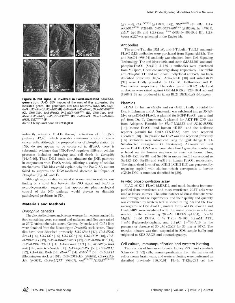
Figure 8. NO signal is involved in FoxO-mediated neurode-generation. (A–D) SEM images of the eyes of flies expressing theindicated genes. The genotypes are: GMR-Gal4/UAS-dNOS (A), GMR-Gal4, UAS-dFoxO/UAS-dNOS (B), GMR-Gal4, UAS-dFoxO; UAS-sGCa99BRNAi
(C), GMR-Gal4, UAS-dFoxO; UAS-sGCb100BRNAi (D), GMR-Gal4, UAS-dFoxO/UAS-dNOS; UAS-sGCa99BRNAi (E), GMR-Gal4, UAS-dFoxO/UAS-dNOS, DG2k04703 (F).doi:10.1371/journal.pone.0030958.g008
Nitric Oxide Signaling Modulates FoxO in Neurons
PLoS ONE | www.plosone.org 9 February 2012 | Volume 7 | Issue 2 | e30958

harboring doxycycline-inducible EGFP-FoxO1 gene was generat-
ed according to the manufacturer’s instructions (Invitrogen).
Scanning Electron Microscopy (SEM)Adult flies were processed as described previously [14]. SEM
images were obtained at The Biomedical Research Core of
Tohoku University Graduate School of Medicine.
Lifespan and survival assaysTwenty female adult flies per vial were maintained at 29uC,
transferred to fresh fly food vials containing 250 ml of yeast paste
and 25 mg/ml of RU486, and scored for survival every 4 days. To
control for isogeny, all fly lines were backcrossed to the w2 wild-
type background for six generations or were generated on the w2
background, and thus have matched genetic backgrounds.
Survival assays of flies treated with 2 mM paraquat were
performed as described previously [14].
Climbing assayThe climbing assay was performed as described previously [14].
Briefly, twenty flies were placed in a plastic vial (18.6 cm in
height63.5 cm2 in area) and gently tapped to bring them down to
the bottom of the vial. Flies were given 18 s to climb and the
number of flies more than 6 cm from the bottom was counted.
Twenty trials were performed for the same set of flies. Flies at 20
days of age were left untreated or treated with 1 mM L-DOPA for
4 days, then subjected to climbing assays.
Whole-mount immunostainingTotal number of TH-positive neurons were calculated following
whole-mount immunostaining of brain samples as described
previously [57]. All immunohistochemical analyses were per-
formed using a Carl Zeiss laser scanning microscope system.
Statistical analysisThe one-way repeated measures ANOVA was used to
determine significant differences between multiple groups unless
otherwise indicated. If a significant result was achieved (p,0.05),
the means of the control and the specific test group were analyzed
using the Tukey-Kramer test. For lifespan assays, a Kaplan-Meier
analysis with the log-rank test was performed.
Supporting Information
Figure S1 Evaluation of mnb and dg1 expression inmnbEY14320 and UAS-DG1 fly lines in the presence of theGAL4 driver. Total RNA was extracted from the Da-Gal4
crosses. The mnb, an orthologue of mammalian DYRK1, dg1 and
rp49 transcript levels were measured by real-time PCR. mnb (A) or
dg1 (B) transcript levels normalized to those of rp49 are presented.
(TIF)
Figure 9. Inhibition of NO signal improves DG2-dFoxO-mediated DA neurodegeneration. (A) Newly eclosed normal w-flies (Control) or transgenics harboring elav-GS.UAS-dFoxO/UAS-DG2(n = 22 in each) were fed a yeast paste containing 50 mg/mL_ RU486with or without 10 mM L-NAME or D-NAME every 4 days at 29uC. Thegraph presents the number of PPM 1/2 and PPL1 clusters of TH-positiveneurons in 20-day-old adult flies. PPM1 and PPM2 clusters werecounted together. Data are presented as the mean 6 SE for threeexperiments (*, p,0.05 vs. Control). PPL, the protocerebral posteriorlateral. Non; RU486 only. (B) A representative image of TH-positive
PPL1, PPM1, PPM2 (upper circle) and PPM3 (lower circle) neurons of awild-type w- adult fly. Arrowheads indicate a pair of PPM1 neurons.Bar = 50 mm. (C–E) Representative images of TH-positive neuronstreated as in A. (F) Brain tissues of dFoxO transgenic flies treated withL-NAME or D-NAME were subjected to western blot analysis with anti-dFoxO. dFoxO SA mutant was also included as a non-phosphorylatedcontrol. Transgenes were expressed by the elav-GS driver. (G) Reductionof dNOS and DG2 activities confers stress resistance against 2 mMparaquat treatment. dNOS (2/2) vs. Control, p,0.0001; DG2 (+/2) vs.Control, p,0.01. The genotypes are: w- (Control), dNOSD15/dNOSD15
(dNOS (2/2)), DG2k04703/+ (DG2 (+/2)).doi:10.1371/journal.pone.0030958.g009
Nitric Oxide Signaling Modulates FoxO in Neurons
PLoS ONE | www.plosone.org 10 February 2012 | Volume 7 | Issue 2 | e30958
Figure S2 DG1 does not exacerbate dFoxO-mediatedeye degeneration. Transgenic expression of DG2 alone did not
produce eye degeneration, and DG1 had little effect on the eye
phenotype caused by expression of dFoxO (when compared to
Figure 2B). (C) The numbers of ommatidia per fly eye (from 5 flies)
were quantified. *, p,0.05; N.S., non-significant. The genotypes
are: UAS-DG2; GMR-Gal4 (A), GMR-Gal4, UAS-dFoxO; UAS-DG1
(B), GMR-Gal4/UAS-EGFP (EGFP), GMR-Gal4, UAS-dFoxO/UAS-
EGFP (dFoxO, EGFP), GMR-Gal4, UAS-dFoxO; UAS-DG1 (dFoxO,
DG1) (C).
(TIF)
Figure S3 cGKII does not form a stable complex withcGKIa or FoxO1. Lysate from 293T cells transfected with
cGKII-FLAG together with or without FoxO1-Myc or cGKIa-
Myc was immunoprecipitated with anti-FLAG antibody (FLAG-
IP). Immunoprecipitates and total soluble lysates (Lysate) were
analyzed by western blotting.
(TIF)
Figure S4 cGKII is expressed in DA neurons of themurine midbrain. Immunolocalization of cGKIa (green in A,C), cGKII (green in B, D) and TH (red) in coronal sections of the
substantia nigra (A, B) and striatum (C, D) of the brain. Yellow in
B indicates the expression of cGKII in TH-positive neuronal
processes (arrow heads) as well as cell bodies (arrows). The right
columns of each panel show high-magnification images of the
boxes in the left columns. Scale bars = 20 mm.
(TIF)
Figure S5 Mutations of cGKII phosphorylation siteslocalized in FoxO1-N do not affect the FoxO-transcrip-tional activity. (A) Reported phosphorylation sites in FoxO1 by
cGKI are depicted [22]. Phospho-resistant mutants, where the
indicated Ser or Thr residues are replaced with alanine, are also
shown. (B) The phospho-signal by cGKII was decreased in GST-
FoxO-4M compared with GST-FoxO-N WT (lane 3 vs. lane 2),
but was no longer decreased in GST-FoxO-5M (data not shown),
suggesting that S184 is not a major phosphorylation site by cGKII.
(C) The FoxO1 4M mutation had little effect on FoxO-
transcriptional activity stimulated by cGKII and/or LRRK2. (D)
Effects of the 4M mutation on physical interaction between FoxO1
and 14-3-3e were estimated in 293T cells. FoxO1-Myc-6x His was
pulled down with Ni-NTA beads from the lysate of cells expressing
the indicated transgenes.
(TIF)
Figure S6 The Ser319 site of FoxO1 is not a major targetof cGKI in vitro. In vitro kinase assay was performed as in Fig. 5.
P3 SA; a P3 mutant in which the Ser319 residue is replaced with
alanine. Autophosphorylation signals of cGKII and cGKI are also
shown in the upper panel.
(TIF)
Figure S7 cGKII phosphorylates LRRK2. (A) cGKII WT
but not cGKII KD phosphorylates LRRK2 3KD (lane 9) as well
as LRRK2 WT (lane 6) in in vitro kinase assay. In vitro kinase assay
was performed as in Fig. 5. (B) Western blot analysis with anti-
FLAG indicates similar amounts of FLAG-LRRK2 WT and
FLAG-LRRK2 3KD were used in the kinase assay.
(TIF)
Figure S8 Hid is not a major gene responsible for FoxO-DG2-mediated optic degeneration. Introduction of loss-of-
function alleles of a pro-apoptotic gene hid (B) or anti-apoptotic
DIAP (C), or knockdown of Dronc, a caspase downstream of Hid
(D), had little effects on the eye phenotype by co-expression of
dFoxO and DG2 (A). The genotypes are: UAS-DG2; GMR-Gal4,
UAS-dFoxO (A), UAS-DG2; GMR-Gal4, UAS-dFoxO; hid1 (B), UAS-
DG2; GMR-Gal4, UAS-dFoxO; DIAP1 (C), UAS-DG2; GMR-Gal4,
UAS-dFoxO; UAS-DroncRNAi (D). (E) Real-time RT-PCR analysis
for hid and 4E-BP was performed using total RNA from S2 cells
expressing the indicated gene combinations. Values are presented
as the mean 6 SE for three repeated experiments. *, p,0.05 vs.
Control.
(TIF)
Figure S9 cGKII is associated with LRRK2. (A) Lysate
from 293T cells transfected with FLAG-tagged LRRK2 with or
without Myc-cGKII was immunoprecipitated with anti-FLAG
antibody (FLAG-IP). Immunoprecipitates and total soluble lysates
(lysate) were analyzed by western blotting. (B) The diagram
represents LRRK2 and the mutants used to determine the cGKII-
binding domain. Numbers in parentheses indicate corresponding
amino acid residues of LRRK2. LRR, leucine-rich repeat; ROC,
Ras in complex proteins; COR, C-terminal of Roc; Kinase,
protein kinase domain; WD40, WD40 domain. (C) Immunopre-
cipitation-western blot analysis as in (A) revealed cGKII to be
associated with LRRK2-C. (D) cGKII associates strongly with
LRRK2-C3, and weakly with LRRK2–C1 and –C2. (E) Endog-
enous interaction of cGKII but not cGKIa with LRRK2 in brain
tissue. Mouse brain tissues were lysed as described [60], then the
supernatant fractions were immunoprecipitated (IP) with anti-
cGKII or anti-cGKIa antibodies. The co-precipitated LRRK2
was detected by western blotting using anti-LRRK2 antibody.
293T lysate expressing FLAG-LRRK2 or FLAG-cGKII served as
a positive control.
(TIF)
Figure S10 cGKII is co-localized with LRRK2 at theendosomes. (A) Immunolocalization of cGKII and LRRK2 in
293T cells expressing FLAG-LRRK2 and Myc-cGKII. cGKII
and LRRK2 were visualized with anti-Myc (green) or anti-
LRRK2 antibody (red). LRRK2 is localized at the Rab-positive
endosomes (data not shown). cGKII is localized at the cytoplasmic
membrane and partly in the cytoplasmic compartments. cGKII
and LRRK2 were co-localized at the Rab-positive endosomes
(yellow). Scale bar = 10 mm. (B–E) Immunolocalization of cGKII
(red) in 293T cells expressing Myc-cGKII and EGFP-tagged Rabs
(green). Cytosolic cGKII is located mainly at Rab4- and Rab5-
positive endosomes, and partially at Rab7- or Rab11-positive
endosomes.
(TIF)
Acknowledgments
We thank A. Yasui, S. Nakajima, S. Kanno and M. Kaji for excellent
technical support and equipment, and T. Furuyama, T. Unterman, G.
Halder, K.V. Anderson, J. Jiang, T. Osterwalder S. Lohmann, A.
Smolenski, M. Hoffmeister, F. Hofmann, P. Weinmeister, PH. O’Farrell
and M. Fukuda for the generous supply of materials.
Author Contributions
Conceived and designed the experiments: TK YI. Performed the
experiments: TK TS YI. Analyzed the data: TK YI. Contributed
reagents/materials/analysis tools: TS SD HI KH RT NH. Wrote the
paper: YI.
Nitric Oxide Signaling Modulates FoxO in Neurons
PLoS ONE | www.plosone.org 11 February 2012 | Volume 7 | Issue 2 | e30958
References
1. Steinert JR, Chernova T, Forsythe ID (2010) Nitric oxide signaling in brain
function, dysfunction, and dementia. Neuroscientist 16: 435–452.
2. West AR, Tseng KY (2011) Nitric Oxide-Soluble Guanylyl Cyclase-Cyclic GMPSignaling in the Striatum: New Targets for the Treatment of Parkinson’s
Disease? Front Syst Neurosci 5: 55.
3. Hunot S, Boissiere F, Faucheux B, Brugg B, Mouatt-Prigent A, et al. (1996)
Nitric oxide synthase and neuronal vulnerability in Parkinson’s disease.
Neuroscience 72: 355–363.
4. Eve DJ, Nisbet AP, Kingsbury AE, Hewson EL, Daniel SE, et al. (1998) Basal
ganglia neuronal nitric oxide synthase mRNA expression in Parkinson’s disease.Brain Res Mol Brain Res 63: 62–71.
5. Liberatore GT, Jackson-Lewis V, Vukosavic S, Mandir AS, Vila M, et al. (1999)
Inducible nitric oxide synthase stimulates dopaminergic neurodegeneration in
the MPTP model of Parkinson disease. Nat Med 5: 1403–1409.
6. Muramatsu Y, Kurosaki R, Watanabe H, Michimata M, Matsubara M, et al.
(2003) Cerebral alterations in a MPTP-mouse model of Parkinson’s disease–animmunocytochemical study. J Neural Transm 110: 1129–1144.
7. Dreyer J, Schleicher M, Tappe A, Schilling K, Kuner T, et al. (2004) Nitricoxide synthase (NOS)-interacting protein interacts with neuronal NOS and
regulates its distribution and activity. J Neurosci 24: 10454–10465.
8. Paisan-Ruiz C, Jain S, Evans EW, Gilks WP, Simon J, et al. (2004) Cloning of
the gene containing mutations that cause PARK8-linked Parkinson’s disease.Neuron 44: 595–600.
9. Zimprich A, Biskup S, Leitner P, Lichtner P, Farrer M, et al. (2004) Mutations inLRRK2 cause autosomal-dominant parkinsonism with pleomorphic pathology.
Neuron 44: 601–607.
10. Healy DG, Falchi M, O’Sullivan SS, Bonifati V, Durr A, et al. (2008)
Phenotype, genotype, and worldwide genetic penetrance of LRRK2-associatedParkinson’s disease: a case-control study. Lancet Neurol 7: 583–590.
11. Mata IF, Wedemeyer WJ, Farrer MJ, Taylor JP, Gallo KA (2006) LRRK2 inParkinson’s disease: protein domains and functional insights. Trends Neurosci
29: 286–293.
12. West AB, Moore DJ, Biskup S, Bugayenko A, Smith WW, et al. (2005)
Parkinson’s disease-associated mutations in leucine-rich repeat kinase 2 augmentkinase activity. Proc Natl Acad Sci U S A 102: 16842–16847.
13. Gloeckner CJ, Kinkl N, Schumacher A, Braun RJ, O’Neill E, et al. (2006) TheParkinson disease causing LRRK2 mutation I2020T is associated with increased
kinase activity. Hum Mol Genet 15: 223–232.
14. Imai Y, Gehrke S, Wang HQ, Takahashi R, Hasegawa K, et al. (2008)
Phosphorylation of 4E-BP by LRRK2 affects the maintenance of dopaminergicneurons in Drosophila. Embo J 27: 2432–2443.
15. Lessing D, Bonini NM (2009) Maintaining the brain: insight into humanneurodegeneration from Drosophila melanogaster mutants. Nat Rev Genet 10:
359–370.
16. Kanao T, Venderova K, Park DS, Unterman T, Lu B, et al. (2010) Activation of
FoxO by LRRK2 induces expression of proapoptotic proteins and alters survivalof postmitotic dopaminergic neuron in Drosophila. Hum Mol Genet 19:
3747–3758.
17. Kalderon D, Rubin GM (1989) cGMP-dependent protein kinase genes in
Drosophila. J Biol Chem 264: 10738–10748.
18. MacPherson MR, Lohmann SM, Davies SA (2004) Analysis of Drosophila
cGMP-dependent protein kinases and assessment of their in vivo roles bytargeted expression in a renal transporting epithelium. J Biol Chem 279:
40026–40034.
19. Jarchau T, Hausler C, Markert T, Pohler D, Vanderkerckhove J, et al. (1994)
Cloning, expression, and in situ localization of rat intestinal cGMP-dependent
protein kinase II. Proc Natl Acad Sci U S A 91: 9426–9430.
20. Kulaksiz H, Rehberg E, Stremmel W, Cetin Y (2002) Guanylin and functional
coupling proteins in the human salivary glands and gland tumors: expression,cellular localization, and target membrane domains. Am J Pathol 161: 655–664.
21. Yuasa K, Yamagami S, Nagahama M, Tsuji A (2008) Trafficking of cGMP-
dependent protein kinase II via interaction with Rab11. Biochem Biophys Res
Commun 374: 522–526.
22. Bois PR, Brochard VF, Salin-Cantegrel AV, Cleveland JL, Grosveld GC (2005)
FoxO1a-cyclic GMP-dependent kinase I interactions orchestrate myoblastfusion. Mol Cell Biol 25: 7645–7656.
23. Bicker G (2007) Pharmacological approaches to nitric oxide signalling during
neural development of locusts and other model insects. Arch Insect Biochem
Physiol 64: 43–58.
24. Davies S (2000) Nitric oxide signalling in insects. Insect Biochem Mol Biol 30:
1123–1138.
25. Davies SA (2006) Signalling via cGMP: lessons from Drosophila. Cell Signal 18:
409–421.
26. Wang X, Robinson PJ (1997) Cyclic GMP-dependent protein kinase and cellularsignaling in the nervous system. J Neurochem 68: 443–456.
27. Hofmann F, Feil R, Kleppisch T, Schlossmann J (2006) Function of cGMP-dependent protein kinases as revealed by gene deletion. Physiol Rev 86: 1–23.
28. Feil R, Hofmann F, Kleppisch T (2005) Function of cGMP-dependent proteinkinases in the nervous system. Rev Neurosci 16: 23–41.
29. Serulle Y, Zhang S, Ninan I, Puzzo D, McCarthy M, et al. (2007) A GluR1-cGKII interaction regulates AMPA receptor trafficking. Neuron 56: 670–688.
30. de Vente J, Asan E, Gambaryan S, Markerink-van Ittersum M, Axer H, et al.
(2001) Localization of cGMP-dependent protein kinase type II in rat brain.Neuroscience 108: 27–49.
31. Feil S, Zimmermann P, Knorn A, Brummer S, Schlossmann J, et al. (2005)Distribution of cGMP-dependent protein kinase type I and its isoforms in the
mouse brain and retina. Neuroscience 135: 863–868.
32. Geiselhoringer A, Gaisa M, Hofmann F, Schlossmann J (2004) Distribution ofIRAG and cGKI-isoforms in murine tissues. FEBS Lett 575: 19–22.
33. McGeer PL, Itagaki S, Boyes BE, McGeer EG (1988) Reactive microglia arepositive for HLA-DR in the substantia nigra of Parkinson’s and Alzheimer’s
disease brains. Neurology 38: 1285–1291.
34. Glass CK, Saijo K, Winner B, Marchetto MC, Gage FH (2010) Mechanismsunderlying inflammation in neurodegeneration. Cell 140: 918–934.
35. Roodveldt C, Christodoulou J, Dobson CM (2008) Immunological features ofalpha-synuclein in Parkinson’s disease. J Cell Mol Med 12: 1820–1829.
36. Foley E, O’Farrell PH (2003) Nitric oxide contributes to induction of innate
immune responses to gram-negative bacteria in Drosophila. Genes Dev 17:115–125.
37. Berger Z, Smith KA, Lavoie MJ (2010) Membrane localization of LRRK2 isassociated with increased formation of the highly active LRRK2 dimer and
changes in its phosphorylation. Biochemistry 49: 5511–5523.
38. Greggio E, Zambrano I, Kaganovich A, Beilina A, Taymans JM, et al. (2008)The Parkinson disease-associated leucine-rich repeat kinase 2 (LRRK2) is a
dimer that undergoes intramolecular autophosphorylation. J Biol Chem 283:16906–16914.
39. Aquilano K, Baldelli S, Rotilio G, Ciriolo MR (2008) Role of nitric oxidesynthases in Parkinson’s disease: a review on the antioxidant and anti-
inflammatory activity of polyphenols. Neurochem Res 33: 2416–2426.
40. Tseng KY, Caballero A, Dec A, Cass DK, Simak N, et al. (2011) Inhibition ofStriatal Soluble Guanylyl Cyclase-cGMP Signaling Reverses Basal Ganglia
Dysfunction and Akinesia in Experimental Parkinsonism. PLoS One 6: e27187.
41. Suzuki N, Motohashi N, Uezumi A, Fukada S, Yoshimura T, et al. (2007) NO
production results in suspension-induced muscle atrophy through dislocation of
neuronal NOS. J Clin Invest 117: 2468–2476.
42. Soh JW, Kazi JU, Li H, Thompson WJ, Weinstein IB (2008) Celecoxib-induced
growth inhibition in SW480 colon cancer cells is associated with activation ofprotein kinase G. Mol Carcinog 47: 519–525.
43. Kwon IK, Wang R, Thangaraju M, Shuang H, Liu K, et al. (2010) PKG
inhibits TCF signaling in colon cancer cells by blocking beta-catenin expressionand activating FOXO4. Oncogene 29: 3423–3434.
44. Lee KS, Iijima-Ando K, Iijima K, Lee WJ, Lee JH, et al. (2009) JNK/FOXO-mediated neuronal expression of fly homologue of peroxiredoxin II reduces
oxidative stress and extends life span. J Biol Chem 284: 29454–29461.
45. Hong YK, Lee NG, Lee MJ, Park MS, Choi G, et al. (2009) dXNP/DATRXincreases apoptosis via the JNK and dFOXO pathway in Drosophila neurons.
Biochem Biophys Res Commun 384: 160–166.
46. Wang MC, Bohmann D, Jasper H (2005) JNK extends life span and limits
growth by antagonizing cellular and organism-wide responses to insulinsignaling. Cell 121: 115–125.
47. Puig O, Marr MT, Ruhf ML, Tjian R (2003) Control of cell number by
Drosophila FOXO: downstream and feedback regulation of the insulin receptorpathway. Genes Dev 17: 2006–2020.
48. McGettigan J, McLennan RK, Broderick KE, Kean L, Allan AK, et al. (2005)Insect renal tubules constitute a cell-autonomous immune system that protects
the organism against bacterial infection. Insect Biochem Mol Biol 35: 741–754.
49. Venderova K, Kabbach G, Abdel-Messih E, Zhang Y, Parks RJ, et al. (2009)Leucine-Rich Repeat Kinase 2 interacts with Parkin, DJ-1 and PINK-1 in a
Drosophila melanogaster model of Parkinson’s disease. Hum Mol Genet 18:4390–4404.
50. Osterwalder T, Yoon KS, White BH, Keshishian H (2001) A conditional tissue-
specific transgene expression system using inducible GAL4. Proc Natl AcadSci U S A 98: 12596–12601.
51. Udan RS, Kango-Singh M, Nolo R, Tao C, Halder G (2003) Hippo promotesproliferation arrest and apoptosis in the Salvador/Warts pathway. Nat Cell Biol
5: 914–920.
52. Lu Y, Wu LP, Anderson KV (2001) The antibacterial arm of the drosophilainnate immune response requires an IkappaB kinase. Genes Dev 15: 104–110.
53. Jia J, Tong C, Wang B, Luo L, Jiang J (2004) Hedgehog signalling activity ofSmoothened requires phosphorylation by protein kinase A and casein kinase I.
Nature 432: 1045–1050.
54. Junger MA, Rintelen F, Stocker H, Wasserman JD, Vegh M, et al. (2003) TheDrosophila forkhead transcription factor FOXO mediates the reduction in cell
number associated with reduced insulin signaling. J Biol 2: 20.
55. Yakubovich N, Silva EA, O’Farrell PH (2010) Nitric oxide synthase is not
essential for Drosophila development. Curr Biol 20: R141–142.
56. Muller D, Kugler SJ, Preiss A, Maier D, Nagel AC (2005) Genetic modifier
screens on Hairless gain-of-function phenotypes reveal genes involved in cell
differentiation, cell growth and apoptosis in Drosophila melanogaster. Genetics171: 1137–1152.
57. Yang Y, Gehrke S, Imai Y, Huang Z, Ouyang Y, et al. (2006) Mitochondrialpathology and muscle and dopaminergic neuron degeneration caused by
Nitric Oxide Signaling Modulates FoxO in Neurons
PLoS ONE | www.plosone.org 12 February 2012 | Volume 7 | Issue 2 | e30958

inactivation of Drosophila Pink1 is rescued by Parkin. Proc Natl Acad Sci U S A
103: 10793–10798.58. Zhang X, Gan L, Pan H, Guo S, He X, et al. (2002) Phosphorylation of serine
256 suppresses transactivation by FKHR (FOXO1) by multiple mechanisms.
Direct and indirect effects on nuclear/cytoplasmic shuttling and DNA binding.J Biol Chem 277: 45276–45284.
59. Yuasa K, Michibata H, Omori K, Yanaka N (2000) Identification of a conservedresidue responsible for the autoinhibition of cGMP-dependent protein kinase
Ialpha and beta. FEBS Lett 466: 175–178.
60. Imai Y, Soda M, Inoue H, Hattori N, Mizuno Y, et al. (2001) An unfolded
putative transmembrane polypeptide, which can lead to endoplasmic reticulum
stress, is a substrate of Parkin. Cell 105: 891–902.
61. Imai Y, Soda M, Takahashi R (2000) Parkin suppresses unfolded protein stress-
induced cell death through its E3 ubiquitin-protein ligase activity. J Biol Chem
275: 35661–35664.
62. Kwon Y, Hofmann T, Montell C (2007) Integration of phosphoinositide- and
calmodulin-mediated regulation of TRPC6. Mol Cell 25: 491–503.
Nitric Oxide Signaling Modulates FoxO in Neurons
PLoS ONE | www.plosone.org 13 February 2012 | Volume 7 | Issue 2 | e30958
Copyright © 2022 FDOKUMEN