Cyclic Dipeptides: The Biological and Structural Landscape ...
64
biomolecules Review Cyclic Dipeptides: The Biological and Structural Landscape with Special Focus on the Anti-Cancer Proline-Based Scaffold Joanna Bojarska 1, * , Adam Mieczkowski 2 , Zyta M. Ziora 3 , Mariusz Skwarczynski 4 , Istvan Toth 3,4,5 , Ahmed O. Shalash 4 , Keykavous Parang 6 , Shaima A. El-Mowafi 6 , Eman H. M. Mohammed 6 , Sherif Elnagdy 7 , Maha AlKhazindar 7 and Wojciech M. Wolf 1 Citation: Bojarska, J.; Mieczkowski, A.; Ziora, Z.M.; Skwarczynski, M.; Toth, I.; Shalash, A.O.; Parang, K.; El-Mowafi, S.A.; Mohammed, E.H.M.; Elnagdy, S.; et al. Cyclic Dipeptides: The Biological and Structural Landscape with Special Focus on the Anti-Cancer Proline-Based Scaffold. Biomolecules 2021, 11, 1515. https:// doi.org/10.3390/biom11101515 Academic Editors: Marc Maresca, Vladimir N. Uversky and Prakash Kulkarni Received: 18 September 2021 Accepted: 12 October 2021 Published: 14 October 2021 Publisher’s Note: MDPI stays neutral with regard to jurisdictional claims in published maps and institutional affil- iations. Copyright: © 2021 by the authors. Licensee MDPI, Basel, Switzerland. This article is an open access article distributed under the terms and conditions of the Creative Commons Attribution (CC BY) license (https:// creativecommons.org/licenses/by/ 4.0/). 1 Faculty of Chemistry, Institute of General & Inorganic Chemistry, Technical University of Lodz, 90-924 Lodz, Poland; [email protected] 2 Institute of Biochemistry and Biophysics, Polish Academy of Sciences, Pawinskiego 5a, 02-106 Warsaw, Poland; [email protected] 3 Institute for Molecular Bioscience, The University of Queensland, St. Lucia, QLD 4072, Australia; [email protected] (Z.M.Z.); [email protected] (I.T.) 4 School of Chemistry and Molecular Biosciences, The University of Queensland, St. Lucia, QLD 4072, Australia; [email protected] (M.S.); [email protected] (A.O.S.) 5 School of Pharmacy, The University of Queensland, Woolloongabba, QLD 4102, Australia 6 Center for Targeted Drug Delivery, Department of Biomedical and Pharmaceutical Sciences, Harry and Diane Rinker Health Science Campus, School of Pharmacy, Chapman University, Irvine, CA 92618, USA; [email protected] (K.P.); elmowafi@chapman.edu (S.A.E.-M.); [email protected] (E.H.M.M.) 7 Botany Department, Faculty of Science, Cairo University, Giza 12613, Egypt; [email protected] (S.E.); [email protected] (M.A.) * Correspondence: [email protected] Abstract: Cyclic dipeptides, also know as diketopiperazines (DKP), the simplest cyclic forms of peptides widespread in nature, are unsurpassed in their structural and bio-functional diversity. DKPs, especially those containing proline, due to their unique features such as, inter alia, extra-rigid conformation, high resistance to enzyme degradation, increased cell permeability, and expandable ability to bind a diverse of targets with better affinity, have emerged in the last years as biologically pre- validated platforms for the drug discovery. Recent advances have revealed their enormous potential in the development of next-generation theranostics, smart delivery systems, and biomaterials. Here, we present an updated review on the biological and structural profile of these appealing biomolecules, with a particular emphasis on those with anticancer properties, since cancers are the main cause of death all over the world. Additionally, we provide a consideration on supramolecular structuring and synthons, based on the proline-based DKP privileged scaffold, for inspiration in the design of compound libraries in search of ideal ligands, innovative self-assembled nanomaterials, and bio-functional architectures. Keywords: cyclic dipeptides; diketopiperazines; proline-based DKPs; drug discovery; privileged scaffold; supramolecular structuring 1. Introduction Cyclic dipeptides, also known as cyclo-dipeptides, diketopiperazines (DKPs), piper- azinediones, dioxopiperazines, or dipeptide anhydrides, are the simplest, naturally oc- curring cyclic forms of peptides, commonly biosynthesized by a large variety of living organisms [1–4] and conserved in bacteria to humans [5,6]. They were first discovered in 1880 and later studied by E. Fischer [7]. Once believed to be only protein artifacts or degradation products, and therefore neglected, DKPs are now considered essential metabolic intermediates, and an interesting platform for therapeutic exploration [8]. DKPs possess all advantages of cyclic peptides. DKPs are an unsurpassed class of bio-molecules in their structural and bio-functional diversity. Moreover, the ‘biosynthetic hooks’ are a useful Biomolecules 2021, 11, 1515. https://doi.org/10.3390/biom11101515 https://www.mdpi.com/journal/biomolecules
-
Upload
khangminh22 -
Category
Documents
-
view
1 -
download
0
Transcript of Cyclic Dipeptides: The Biological and Structural Landscape ...

biomolecules
Review
Cyclic Dipeptides: The Biological and Structural Landscapewith Special Focus on the Anti-Cancer Proline-Based Scaffold
Joanna Bojarska 1,* , Adam Mieczkowski 2 , Zyta M. Ziora 3 , Mariusz Skwarczynski 4 , Istvan Toth 3,4,5,Ahmed O. Shalash 4, Keykavous Parang 6 , Shaima A. El-Mowafi 6, Eman H. M. Mohammed 6, Sherif Elnagdy 7 ,Maha AlKhazindar 7 and Wojciech M. Wolf 1
�����������������
Citation: Bojarska, J.; Mieczkowski,
A.; Ziora, Z.M.; Skwarczynski, M.;
Toth, I.; Shalash, A.O.; Parang, K.;
El-Mowafi, S.A.; Mohammed, E.H.M.;
Elnagdy, S.; et al. Cyclic Dipeptides:
The Biological and Structural
Landscape with Special Focus on the
Anti-Cancer Proline-Based Scaffold.
Biomolecules 2021, 11, 1515. https://
doi.org/10.3390/biom11101515
Academic Editors: Marc Maresca,
Vladimir N. Uversky and
Prakash Kulkarni
Received: 18 September 2021
Accepted: 12 October 2021
Published: 14 October 2021
Publisher’s Note: MDPI stays neutral
with regard to jurisdictional claims in
published maps and institutional affil-
iations.
Copyright: © 2021 by the authors.
Licensee MDPI, Basel, Switzerland.
This article is an open access article
distributed under the terms and
conditions of the Creative Commons
Attribution (CC BY) license (https://
creativecommons.org/licenses/by/
4.0/).
1 Faculty of Chemistry, Institute of General & Inorganic Chemistry, Technical University of Lodz, 90-924 Lodz,Poland; [email protected]
2 Institute of Biochemistry and Biophysics, Polish Academy of Sciences, Pawinskiego 5a, 02-106 Warsaw,Poland; [email protected]
3 Institute for Molecular Bioscience, The University of Queensland, St. Lucia, QLD 4072, Australia;[email protected] (Z.M.Z.); [email protected] (I.T.)
4 School of Chemistry and Molecular Biosciences, The University of Queensland, St. Lucia, QLD 4072,Australia; [email protected] (M.S.); [email protected] (A.O.S.)
5 School of Pharmacy, The University of Queensland, Woolloongabba, QLD 4102, Australia6 Center for Targeted Drug Delivery, Department of Biomedical and Pharmaceutical Sciences, Harry and Diane
Rinker Health Science Campus, School of Pharmacy, Chapman University, Irvine, CA 92618, USA;[email protected] (K.P.); [email protected] (S.A.E.-M.); [email protected] (E.H.M.M.)
7 Botany Department, Faculty of Science, Cairo University, Giza 12613, Egypt; [email protected] (S.E.);[email protected] (M.A.)
* Correspondence: [email protected]
Abstract: Cyclic dipeptides, also know as diketopiperazines (DKP), the simplest cyclic forms ofpeptides widespread in nature, are unsurpassed in their structural and bio-functional diversity.DKPs, especially those containing proline, due to their unique features such as, inter alia, extra-rigidconformation, high resistance to enzyme degradation, increased cell permeability, and expandableability to bind a diverse of targets with better affinity, have emerged in the last years as biologically pre-validated platforms for the drug discovery. Recent advances have revealed their enormous potentialin the development of next-generation theranostics, smart delivery systems, and biomaterials. Here,we present an updated review on the biological and structural profile of these appealing biomolecules,with a particular emphasis on those with anticancer properties, since cancers are the main cause ofdeath all over the world. Additionally, we provide a consideration on supramolecular structuringand synthons, based on the proline-based DKP privileged scaffold, for inspiration in the designof compound libraries in search of ideal ligands, innovative self-assembled nanomaterials, andbio-functional architectures.
Keywords: cyclic dipeptides; diketopiperazines; proline-based DKPs; drug discovery; privilegedscaffold; supramolecular structuring
1. Introduction
Cyclic dipeptides, also known as cyclo-dipeptides, diketopiperazines (DKPs), piper-azinediones, dioxopiperazines, or dipeptide anhydrides, are the simplest, naturally oc-curring cyclic forms of peptides, commonly biosynthesized by a large variety of livingorganisms [1–4] and conserved in bacteria to humans [5,6]. They were first discoveredin 1880 and later studied by E. Fischer [7]. Once believed to be only protein artifactsor degradation products, and therefore neglected, DKPs are now considered essentialmetabolic intermediates, and an interesting platform for therapeutic exploration [8]. DKPspossess all advantages of cyclic peptides. DKPs are an unsurpassed class of bio-moleculesin their structural and bio-functional diversity. Moreover, the ‘biosynthetic hooks’ are a useful
Biomolecules 2021, 11, 1515. https://doi.org/10.3390/biom11101515 https://www.mdpi.com/journal/biomolecules

Biomolecules 2021, 11, 1515 2 of 64
strategy for the identification of the genes modifying the DKP ring to expand the chemicalspace of cyclic dipeptides [9,10]. Moreover, modified DKPs have recently emerged as animportant pharmacophore in a number of theranostic settings. Notably, proline motifintroduces additional conformational and bio-functional value into the DKP-derived struc-tures. The attractive features, such as extra rigidity, structural stability, and consequently,greater resistance to degradation by enzymes, higher bioactivity, specificity, selectivity, andefficacy, increased cell permeability, or binding affinity to specific targets, inherent biocom-patibility, structural importance to biological systems [11,12] make proline-based cyclicdipeptides a promising alternative to currently used small molecule and macromoleculepharmaceuticals.
The mechanism of proline-based DKPs formation is described elsewhere [13].DKPs are ‘diamonds in the rough’, offering endless possibilities in future innovative
therapies [14]. Therefore, since the earliest report on DKP in 1924 [15], an increasing de-gree of attention to DKPs has been observed, and numerous scientific findings revealingtheir broad-spectrum biological activities have been reported in recent years. In particu-lar, proline-based DKPs have diverse properties depending on structure and application,such as anticancer, antioxidant, neuroprotective, antiviral, antibacterial, anti-inflammatory,antihyperglycemic, antiarrhythmic, immunomodulatory, antiparasitic, anthelmintic, in-secticidal, antifouling, vasorelaxant, and metabolic regulatory activity [16–22]. They havethe potential to be antibiotics of the future. Moreover, they act as antagonists of humanoxytocin receptors [23], inhibitors of platelet aggregation [24], calpain inhibitors againststroke [25]. DKPs have relevance in the prevention of cell division [26], cell–cell signaling,or quorum sensing [27]. They are useful in smart delivery systems of drugs that have lowpermeability to cross the blood-brain barrier [19]. As a curiosity, DKPs are a hot topic in eco-logical chemistry [28]. Furthermore, DKP-containing compounds are used as catalysts orchiral auxiliaries in synthetic organic chemistry, in the formation of alkaloids [29–31]. Theyare an excellent model in theoretical studies on the constrained structural scaffold with arelevant pharmacophore [17,32,33]. In the future, cyclic-dipeptide-based compounds willfind a wider use in various fields as perfect tools for probing specific proteins or metabolitesin vivo, or as building blocks for macromolecules.
Here, we present a comprehensive overview of the recent progress on bio-landscapeand structural diversity of compounds containing proline-based DKP motif, which areexploited as privileged peptidomimetic scaffolds for future innovative drug discovery,smart delivery systems, and modern bio-control agents [34–36]. We pay special attentionto anticancer proline-based DKPs since cancers are main cause of death all over the world,with nearly 10 million deaths in 2020 according to the WHO [37]. Therefore, novel, effective,and safe therapeutics are extremely needed. In this context, the supramolecular structuringand the role of versatile synthons for inspiration in the design of compound libraries insearch of ideal ligands with unique proline-DKP motif are also discussed.
2. Occurence and Biosynthesis of DKPs
DKP skeleton is observed in micro-species, bacteria, such as Bacillus subtilis, Strepto-myces, Pseudomonas aeruginosa, or Lactobacillus plantarum [38–40], marine, and terrestrialfungi [41] as Aspergillus flavus or Alternaria alternata, and Penicillium, respectively [42,43], ma-rine sponges such as Dysidea herbacea, and fragilis [44], or proteobacteria Alcaligenes faecalis,algae, lichens, gorgonians, tunicates, plants, or animals venoms. Remarkably, DKPswere found in human central nervous system, gastrointestinal tract, or blood [45]. Theyoccur in food and beverages, such as pu-erh tea, cocoa, dried bonito, roasted coffee,sake, beer, cheese, casein, chicken extract, or stewed beef, giving a special metallic bittertaste [13,18,31,32,46–49], but also in culture broths fermented with lactic acid bacteria.Products containing both L- and D-proline-based DKPs are common in nature, and theirstructural and biological complexity is highly impressive [50]. As a curiosity, 90% ofDKPs in foods contain proline [28]. DKPs provide an eco-friendly approach to food andfeed preservation [51]. On the other hand, DKP framework is present in drugs, e.g.,

Biomolecules 2021, 11, 1515 3 of 64
in aminopenicillin, amoxicillin, ACE inhibitors [52,53] as by-products of spontaneousintramolecular cyclization of the dipeptidyl moiety in active peptide-based substances.Degradation via DKPs formation is observed during long-term storage. Moreover, DKPscan appear as a result of chemical peptide synthesis, or hydrolysis of functional peptidesand proteins [54,55]. The cyclization is facilitated when a proline is present at the secondposition from the N-terminus (i.e., penultimate proline) [55].
From the biosynthetic point of view, naturally produced DKPs are known to be effec-tive and biodegradable, however their production yield is low [56,57]. Laboratory trialsfor DKPs-microbial induction have faced some constraints. Although producing DKPsfrom microorganisms via an expression system is feasible; the optimization is long and nota straightforward process [58]. The biosynthesis of DKPs relies mainly on two enzymes,non-ribosomal peptide synthetases (NRPs) and tRNA-dependent cyclodipeptide synthases(CDPs) [59]. Both enzymes are part of a biosynthetic gene cluster that targets DKPs scaffoldmodifications necessary for the stability of the produced DKPs [60]. Metagenomics and nextgeneration sequencing enhanced the biosynthetic gene clusters encoding DKP tailoring en-zymes [61]. As reported, the microbial genes responsible for a specific secondary metabolitewere found to be close to other genes in the dedicated biosynthetic gene clusters [62]. Sincethe genes responsible for DKPs biosynthesis are clustered on the microbial chromosome,therefore, the encoding of the biosynthetic genes depends on the discovery of a single genein the pathway. There are approximately 700 known CDPs-encoding genes clustered withthe predicted tailoring genes [9]. Recently, heterologous expression, zinc finger nuclease(ZFN) and transcription activator-like effector nuclease (TALEN) have been used to edittarget genes for secondary metabolite induction in microorganisms [63]. However, theseapproaches found some limitations in their efficiency and productivity level [64–66]. Thus,clustered regularly interspersed short palindromic repeats (CRISPR)/associated protein(Cas) system has been recently used as a new approach for the biosynthesis of secondarymetabolites and for activation of silent biosynthetic gene clusters [67]. CRISPR/Cas systemhas outmatched other techniques due to its possible multi-gene editing and high efficiency.Precisely, type II CRISPR/Cas system has been successfully applied for the biosynthesisof secondary metabolites [67]. Previous studies reported the use of CRISPR/Cas9 systemin the filamentous fungus Trichoderma reesei, achieving the homologous recombinationof > 93% efficiencies [68]. Similarly, Nodvig et al. [69] obtained a genome-edited pheno-type by targeting the yA gene in the model fungus Aspergillus nidulans. Thus, CRISPR/Cassystem could be a potential mechanism for the efficient biosynthesis of DKPs.
3. DKP Scaffold
The concept of scaffold is useful in medicinal chemistry and drug design to generate,characterize, and compare cores of bio-active substances and their analogs [70]. The scaf-fold is the main fragment of compound (e.g., ring system) after removal of substituents(R-groups) [71,72]. The privileged molecular scaffold is defined as a core structure, whichforms structurally diverse bio-molecules via introducing different functional groups [73,74].DKPs can play the role of a privileged, multi-functionalized scaffold for the design anddevelopment of advanced therapeutic agents, drug delivery systems, biomaterials, orbio-imaging to mitigate numerous disease conditions, also as for the synthesis of complexnatural products [74] because of their specific conformational and physico-chemical at-tributes. DKPs are heterocyclic compounds consisting of two amino acid residues linked toa central six-membered lactam ring core with (or without) various substituents, provid-ing the control of the substituent’s stereochemistry at up to four positions, chiral nature,three-dimensionality, and consequently leading to the promotion of the intermolecularH-bonding interactions with bio-target sites via the corresponding sites of donors andacceptors [17]. Thus, the rigid DKP core allows either a constrained or flexible behavior ofamino acids, mimicking preferential peptide conformation [59]. It makes DKP moleculesperfect to predicting properties of larger peptides with multiple H-bond acceptors, anddonor functionality, and multiple sites for the structural elaboration of diverse functional

Biomolecules 2021, 11, 1515 4 of 64
groups. These characteristics do not only enable them to bind with high affinity to alarge variety of receptors and enzymes [32], showing multifarious biological activities,but they also allow a more predictable receptor interaction and the development of thedrug-like physicochemical properties that are required for the multi-objective optimizationprocess of transforming lead to a drug product. The general structure for DKP cores canbe seen in Figure 1. It should be highlighted that even though 2,5-DKPs are the mostpopular [32,75], other two regioisomers, such as 2,3-DKPs, and 2,6-DKPs, are also possibleas important pharmacophores [76]. All isomers can be found in natural sources in thecourse of biochemical synthesis. Interestingly, the first crystal structure of DKP, 2,5-DKP,was reported in 1938 [19,77]. More specifically, 2,5-DKPs are common, naturally occurringpeptide derivatives (and are frequently generated as unwanted by-products in the synthesisof oligopeptides). The 2,5-DKP core is present in the structure of known drugs, such asTadalafil, phosphodiesterase-5 inhibitor for the treatment of pulmonary arterial hyper-tension and erectile dysfunction [78–80], Retosiban, an oxytocin antagonist for pretermlabor [81], Epelsiban, an oxytocin antagonist in premature ejaculation in men [32], Aplavi-roc against HIV [82], in the vascular disruption, and tubulin-depolymerizing Plinabulin, onthe basis of marine fungal Halimide, a potential therapeutical drug in lung cancer [83,84],and other anticancer natural agents as Ambewelamide, Phenylahistin, Dehydropheny-lahistin [85], Verticillin A [86], antiviral and immunosuppressive Sirodesmin, a type ofphytotoxin, anti-inflammatory agents, e.g., FR106969 [59], antibacterial Bicyclomycin, Bre-vianamide S, Avrainvillamide [87] or Albonoursin, antifungal Maremycin, mycotoxinssuch as Roquefortine C [22] or Gliotoxin, which is also a potent inducer of apoptotic, andnecrotic cell death [88,89].
Biomolecules 2021, 11, x FOR PEER REVIEW 4 of 65
a constrained or flexible behavior of amino acids, mimicking preferential peptide
conformation [59]. It makes DKP molecules perfect to predicting properties of larger
peptides with multiple H‐bond acceptors, and donor functionality, and multiple sites for
the structural elaboration of diverse functional groups. These characteristics do not only
enable them to bind with high affinity to a large variety of receptors and enzymes [32],
showing multifarious biological activities, but they also allow a more predictable receptor
interaction and the development of the drug‐like physicochemical properties that are
required for the multi‐objective optimization process of transforming lead to a drug
product. The general structure for DKP cores can be seen in Figure 1. It should be
highlighted that even though 2,5‐DKPs are the most popular [32,75], other two
regioisomers, such as 2,3‐DKPs, and 2,6‐DKPs, are also possible as important
pharmacophores [76]. All isomers can be found in natural sources in the course of
biochemical synthesis. Interestingly, the first crystal structure of DKP, 2,5‐DKP, was
reported in 1938 [19,77]. More specifically, 2,5‐DKPs are common, naturally occurring
peptide derivatives (and are frequently generated as unwanted by‐products in the
synthesis of oligopeptides). The 2,5‐DKP core is present in the structure of known drugs,
such as Tadalafil, phosphodiesterase‐5 inhibitor for the treatment of pulmonary arterial
hypertension and erectile dysfunction [78–80], Retosiban, an oxytocin antagonist for
preterm labor [81], Epelsiban, an oxytocin antagonist in premature ejaculation in men [32],
Aplaviroc against HIV [82], in the vascular disruption, and tubulin‐depolymerizing
Plinabulin, on the basis of marine fungal Halimide, a potential therapeutical drug in lung
cancer [83,84], and other anticancer natural agents as Ambewelamide, Phenylahistin,
Dehydrophenylahistin [85], Verticillin A [86], antiviral and immunosuppressive
Sirodesmin, a type of phytotoxin, anti‐inflammatory agents, e.g., FR106969 [59],
antibacterial Bicyclomycin, Brevianamide S, Avrainvillamide [87] or Albonoursin,
antifungal Maremycin, mycotoxins such as Roquefortine C [22] or Gliotoxin, which is also
a potent inducer of apoptotic, and necrotic cell death [88,89].
Figure 1. Structure of 2,3‐DKP (left), 2,5‐DKP (middle) and 2,6‐DKP (right) rings as important
pharmacophores.
Thaxtomin A [90], phytotoxin and insecticidal okaramine, and so on. Furthermore,
2,5‐DKPs are present in food, e.g., in fermented olives and beverages. Thus, they have the
potential to be used in the development of new functional foods [91]. Diverse 2,5‐DKPs
have been discovered from marine habitats (sponges, and microorganisms) in recent years
[14]. These DKPs have cytotoxic (~36%), antimicrobial (~19%), antiviral (~13%),
antioxidant (10%), enzyme inhibition (~5%), and other activities (18%) [14].
2,3‐DKPs can be found in natural products, e.g., antibiotics, Piperacillin, or
Cefoperazone [92]. Like 2,5 DKPs, they have been used in medicinal chemistry, for
example, against diseases wherein platelet agglutination participates [93]. The first
synthesis of 2,3‐DKP was reported by Goulding and Pollard in 1948 [94].
2,6‐DKPs have been investigated as antiproliferative agents through the inhibition of
DNA topoisomerase [32,92,95]. They also have other activities, such as anticonvulsant [96]
or trypanocidal [96–99].
Proline‐based DKPs have a special characteristic [12]. Proline is a unique amino acid
due to its specific structure. The DKP nucleus is fused to the pyrrolidine ring, resulting in
Figure 1. Structure of 2,3-DKP (left), 2,5-DKP (middle) and 2,6-DKP (right) rings as importantpharmacophores.
Thaxtomin A [90], phytotoxin and insecticidal okaramine, and so on. Furthermore,2,5-DKPs are present in food, e.g., in fermented olives and beverages. Thus, they havethe potential to be used in the development of new functional foods [91]. Diverse 2,5-DKPs have been discovered from marine habitats (sponges, and microorganisms) in recentyears [14]. These DKPs have cytotoxic (~36%), antimicrobial (~19%), antiviral (~13%),antioxidant (10%), enzyme inhibition (~5%), and other activities (18%) [14].
2,3-DKPs can be found in natural products, e.g., antibiotics, Piperacillin, or Cefopera-zone [92]. Like 2,5 DKPs, they have been used in medicinal chemistry, for example, againstdiseases wherein platelet agglutination participates [93]. The first synthesis of 2,3-DKPwas reported by Goulding and Pollard in 1948 [94].
2,6-DKPs have been investigated as antiproliferative agents through the inhibition ofDNA topoisomerase [32,92,95]. They also have other activities, such as anticonvulsant [96]or trypanocidal [96–99].
Proline-based DKPs have a special characteristic [12]. Proline is a unique amino aciddue to its specific structure. The DKP nucleus is fused to the pyrrolidine ring, result-ing in eminent bio-properties, mentioned earlier. Both L- and D-proline, and to a lesserextent hydroxyproline-based DKPs, exhibit bio-activity. Moreover, proline cis-trans iso-merization play a role inter alia in controlling auto-inhibition of signaling proteins [12].In nature, proline-based DKPs (e.g., cyclo(L-Pro-L-Pro), cyclo(L-His-L-Pro), cyclo(L-Phe-L-Pro), cyclo(L-Tyr-L-Pro), cyclo(L-Leu-L-Pro), and cyclo(L-Val-L-Pro)) are common, which is

Biomolecules 2021, 11, 1515 5 of 64
translated into the structural complexity and impressive bio-activities of this importantclass of DKPs [51,91,100–103], which are thoroughly described in the next sub-section.Interestingly, cyclo(Pro-Pro) is as an archaic precursor in the early evolution of life pro-cess [5,38]. Notably, many biologically important cyclic peptide sequences, and naturalproducts contain multiple proline residues. The ‘proline-rich cyclic structures’ have startedgaining the attention of the pharmaceutical industry, but their true potential is still verymuch unknown. Analogs of proline in DKPs should not be overlooked. As an example,silaproline exhibits similar conformational properties, but confers higher lipophilicity andimproved resistance to biodegradation [104].
4. Bio-Landscape and Structural Profile of Proline-Based DKPs4.1. Anticancer Activity4.1.1. Bicyclic Proline-Based DKP with Simple Side Chains
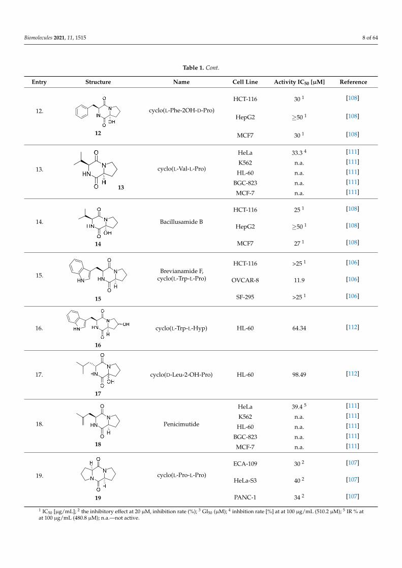
Bi- and policyclic diketopiperazines containing a proline fragment within the structureare of a great interest to medical chemists as potential cytotoxic and antineoplastic agents.However, in most cases the cytotoxic effect of simple proline-containing bicyclic DKPsis quite limited. Ye [105] reported that cyclo (L-Phe-L-Hyp) (1), isolated from mangroveStreptomyces sp. Q24, inhibits the proliferation of human glioma U87-MG and U251 cellsin at IC50 = 5.8 and 18.6 [µM], respectively (Table 1, entry 1). Cyclo (L-Phe-L-Hyp) (1),when tested on the adenocarcinoma HCT-116, the ovarian carcinoma OVCAR-8, and theglioblastoma SF-295 cell lines, did not exhibit any significant cytotoxic effect up to theconcentration of 25 µg/mL (Table 1, entry 1) [106]. Cyclo (L-Leu-L-Hyp) (2) exhibits cy-totoxic effect on U87-MG and U251 cells lines at IC50 = 14.5 and 29.4 [µM], respectively,while its close structural analog cyclo (L-Leu-L-Pro) (3) at IC50 = 1.3 and 19.8 [µM], respec-tively (Table 1, entries 2,3) [105]. Lin [107] reported that cyclo (L-Leu-L-Pro) (3), isolatedfrom Streptomyces xiamenensis MCCC 1A01570, evaluated for cytotoxicity against threecancer cell lines of ECA-109 (esophageal carcinoma), HeLa-S3 (cervix carcinoma) andPANC-1 (pancreatic carcinoma) exhibited moderate inhibition effect at 20 µM varyingfrom 14% (PANC-1) to 55% (ECA-109) (Table 1, entry 3). Shaala reported [108] that cyclo(L-Leu-L-Pro) (3), isolated from tunicate-derived actinomycete Streptomyces sp. moder-ately inhibits the proliferation of HCT-116, HepG2 (hepatocellular carcinoma) and MCF-7(breast cancer) cell lines, with values of 16, ≥50, and 30 µg/mL, respectively (Table 1,entry 3). Its diastereoisomer, cyclo (D-Leu-L-Pro) (4), evaluated for cytotoxicity againstECA-109, HeLa-S3, and PANC-1 exhibited moderate inhibition effect at 20 µM varyingfrom 44% (ECA-109) to 55% (PANC-1) [107] (Table 1, entry 4). The cytotoxic effect of cyclo(L-IIe-L-Pro) (5) was reported in two articles [107,108]. The compound 5 showed limitedcell growth at 20 µM when tested on ECA-109, HeLa-S3 and PANC-1 cell lines, from 45%(HeLa-S3) to 56% (PANC-1) (Table 1, entry 5) [107], while inhibited the proliferation ofHCT-116, HepG2 and MCF-7 cell lines, with values of 22, ≥50, and 27 µg/mL, respectively(Table 1, entry 5) [108]. Its hydroxylated analog, cyclo (L-IIe-L-Hyp) (6), when tested onECA-109, HeLa-S3 and PANC-1 cell lines, inhibited the cell growth at 20 µM from 42%(PANC-1) to 54% (ECA-109) (Table 1, entry 6) [107]. Cyclo (4-S-hydroxy-D-Pro-D-Ile) (7)was isolated from Australian marine sponge Stelletta sp. and its cytotoxicity was testedon human tumour cell lines H460 (lung carcinoma), SF-268 (glioblastoma), MCF-7, HT-29(colon adenocarcinoma) and a normal mammalian cell line CHO-K1, derived from hamsterovary. Compound 7 exhibited weak cytotoxic effect on all tested cell line with GI50 (µM)values varied from 204 (MCF-7) to > 295 (SF-268, CHO-K1) (Table 1, entry 7) [109]. Cyclo(L-Phe-L-Pro) (8) exhibited marked cytotoxicity, when tested on HCT-116, OVCAR-8 andSF-295, with IC50 µg/mL values of 21.4, 18.3, and 16.0, respectively [106]. The cytotox-icity observed effect of 8 was stronger than in the case of its hydroxylated derivarive 1(Table 1, entries 1 and 8) [106]. The compound 8 also decreased the cell growth at 20 µMwhen tested on ECA-109, HeLa-S3, and PANC-1 cell lines, from 36% (HeLa-S3) to 50%(PANC-1) (Table 1, entry 8) [107]. Two stereoisomers of 8: cyclo (L-Phe-D-Pro) (9) and cyclo(D-Phe-D-Pro) (10), as well as their 3-hydroxy analog, Penicicillatide B (11) (Table 1, entries

Biomolecules 2021, 11, 1515 6 of 64
9–11), were isolated from the marine-derived fungus, Penicillium sp. and tested on thecytotoxic effect on three cancer cell lines: HCT-116, HepG2 and MCF7 [110]. Of the threecell lines tested, HCT116 proved to be the most sensitive to compounds 9–11, with theIC50 [µM] values varied from 23.0 for Penicillatide B (11) to 94.0 for cyclo (D-Phe-D-Pro)(10). Cyclo (D-Phe-D-Pro) (10), a diastereoisomer of cyclo (L-Phe-D-Pro) (9), derived fromD-Pro instead of L-Pro, exhibited about three times weaker effect then on HCT116 then9 (IC50 = 94.0 vs. 38.9 µM), which proves that the configuration of the constituent aminoacids may have a significant influence on the cytotoxic effect of the tested compound. Cyclo(L-Phe-2-OH-D-Pro) (12), a hydroxylated analog of cyclo (L-Phe-D-Pro) (9) was tested onthree cancer cell lines: HCT-116, HepG2 and MCF7 (Table 1, entry 12) [108] and inhibitedthe proliferation with IC50 values of 30, ≥50, and 30 µg/mL, respectively. Wang reportedthat cyclo (L-Val-L-Pro) (13) (Table 1, entry 13), could inhibit HeLa cells with an inhibitionrate of 33.3% at 100 µg/mL [111], while its hydroxylated analog Bacillusamide B (14),(Table 1, entry 14), inhibited the proliferation of HCT-116, HepG2 and MCF7 with IC50values 25, ≥50, and 27 µg/mL, respectively [108]. Brevianamide F, cyclo (L-Trp-L-Pro)(15) exhibited marked cytotoxic effect on OVCAR-8 cell line (IC50 = 11.9 [µg/mL]) (Table 1,entry 15) [106], while its hydroxylated analog cyclo (L-Trp-L-Hyp) (16) showed moderatecytotoxic activity with IC50 = 64.34 [µM]) on HL-60 (acute promyelocytic leukemia) cellline (Table 1, entry 16) [112]. Cyclo (D-Leu-2-OH-Pro) (17) showed a rather weak cytotoxiceffect with on HL-60 with IC50 = 98.49 [µM]) (Table 1, entry 16) [112], while Penicimu-tide (18) could inhibit HeLa cells with inhibition rate of 39.4% at 100 µg/mL (Table 1,entry 16) [111]. The simplest tricyclic proline-based DKP consists of two proline subunits,cyclo (L-Pro-L-Pro) (19) was evaluated for cytotoxicity against ECA-109, HeLa-S3, andPANC-1 (pancreatic carcinoma) exhibited moderate inhibition effect at 20 µM varyingfrom 20% (ECA-109) to 40% (HeLa-S3) (Table 1, entry 19) [107]. Finally, Vázquez-Rivierareported [113] that the mixture of cyclo (L-Tyr-L-Pro) (20), cyclo (L-Val-L-Pro) (13), andcyclo(L-Phe-L-Pro) (8), isolated from Pseudomonas aeruginosa PAO1, initiated the cell deathin HeLa and Caco-2 (colorectal adenocarcinoma) cell cultures with IC50 values of 0.53 and0.66 mg/mL, respectively.
Table 1. Bi- and tricyclic proline-based DKP with simple side chains.
Entry Structure Name Cell Line Activity IC50 [µM] Reference
1.
Biomolecules 2021, 11, x FOR PEER REVIEW 6 of 65
OVCAR‐8 and SF‐295, with IC50 μg/mL values of 21.4, 18.3, and 16.0, respectively [106].
The cytotoxicity observed effect of 8 was stronger than in the case of its hydroxylated
derivarive 1 (Table 1, entries 1 and 8) [106]. The compound 8 also decreased the cell
growth at 20 μM when tested on ECA‐109, HeLa‐S3, and PANC‐1 cell lines, from 36%
(HeLa‐S3) to 50% (PANC‐1) (Table 1, entry 8) [107]. Two stereoisomers of 8: cyclo (L‐Phe‐
D‐Pro) (9) and cyclo (D‐Phe‐D‐Pro) (10), as well as their 3‐hydroxy analog, Penicicillatide
B (11) (Table 1, entries 9–11), were isolated from the marine‐derived fungus, Penicillium
sp. and tested on the cytotoxic effect on three cancer cell lines: HCT‐116, HepG2 and MCF7
[110]. Of the three cell lines tested, HCT116 proved to be the most sensitive to compounds
9–11, with the IC50 [μM] values varied from 23.0 for Penicillatide B (11) to 94.0 for cyclo
(D‐Phe‐D‐Pro) (10). Cyclo (D‐Phe‐D‐Pro) (10), a diastereoisomer of cyclo (L‐Phe‐D‐Pro)
(9), derived from D‐Pro instead of L‐Pro, exhibited about three times weaker effect then
on HCT116 then 9 (IC50 = 94.0 vs. 38.9 μM), which proves that the configuration of the
constituent amino acids may have a significant influence on the cytotoxic effect of the
tested compound. Cyclo (L‐Phe‐2‐OH‐D‐Pro) (12), a hydroxylated analog of cyclo (L‐Phe‐
D‐Pro) (9) was tested on three cancer cell lines: HCT‐116, HepG2 and MCF7 (Table 1, entry
12) [108] and inhibited the proliferation with IC50 values of 30, ≥50, and 30 μg/mL,
respectively. Wang reported that cyclo (L‐Val‐L‐Pro) (13) (Table 1, entry 13), could inhibit
HeLa cells with an inhibition rate of 33.3% at 100 μg/mL [111], while its hydroxylated
analog Bacillusamide B (14), (Table 1, entry 14), inhibited the proliferation of HCT‐116,
HepG2 and MCF7 with IC50 values 25, ≥50, and 27 μg/mL, respectively [108].
Brevianamide F, cyclo (L‐Trp‐L‐Pro) (15) exhibited marked cytotoxic effect on OVCAR‐8
cell line (IC50 = 11.9 [μg/mL]) (Table 1, entry 15) [106], while its hydroxylated analog cyclo
(L‐Trp‐L‐Hyp) (16) showed moderate cytotoxic activity with IC50 = 64.34 [μM]) on HL‐60
(acute promyelocytic leukemia) cell line (Table 1, entry 16) [112]. Cyclo (D‐Leu‐2‐OH‐Pro)
(17) showed a rather weak cytotoxic effect with on HL‐60 with IC50 = 98.49 [μM]) (Table 1,
entry 16) [112], while Penicimutide (18) could inhibit HeLa cells with inhibition rate of
39.4% at 100 μg/mL (Table 1, entry 16) [111]. The simplest tricyclic proline‐based DKP
consists of two proline subunits, cyclo (L‐Pro‐L‐Pro) (19) was evaluated for cytotoxicity
against ECA‐109, HeLa‐S3, and PANC‐1 (pancreatic carcinoma) exhibited moderate
inhibition effect at 20 μM varying from 20% (ECA‐109) to 40% (HeLa‐S3) (Table 1, entry
19) [107]. Finally, Vázquez‐Riviera reported [113] that the mixture of cyclo (L‐Tyr‐L‐Pro)
(20), cyclo (L‐Val‐L‐Pro) (13), and cyclo(L‐Phe‐L‐Pro) (8), isolated from Pseudomonas
aeruginosa PAO1, initiated the cell death in HeLa and Caco‐2 (colorectal adenocarcinoma)
cell cultures with IC50 values of 0.53 and 0.66 mg/mL, respectively.
Table 1. Bi‐ and tricyclic proline‐based DKP with simple side chains.
Entry Structure Name Cell Line Activity IC50 [μM] Reference
1.
1
cyclo(L‐Phe‐L‐Hyp)
U87‐MG 5.8 ± 1.7 [105]
U251 18.6 ± 0.1 [105]
HCT‐116 >25 1 [106]
OVCAR‐8 >25 1 [106]
SF‐295 >25 1 [106]
1
cyclo(L-Phe-L-Hyp)
U87-MG 5.8 ± 1.7 [105]
U251 18.6 ± 0.1 [105]
HCT-116 >25 1 [106]
OVCAR-8 >25 1 [106]
SF-295 >25 1 [106]
2.
Biomolecules 2021, 11, x FOR PEER REVIEW 7 of 65
2.
2
cyclo(L‐Leu‐L‐Hyp) U87‐MG
U251
14.5 ± 1.6
29.4 ± 1.3
[105]
[105]
3.
3
cyclo(L‐Leu‐L‐Pro)
U87‐MG 1.3 ± 0.1 [105]
U251 19.8 ± 0.8 [105]
ECA‐109 55 2 [107]
HeLa‐S3 41 2 [107]
PANC‐1 14 2 [107]
HCT‐116 16 1 [108]
HepG2 ≥50 1 [108]
MCF7 30 1 [108]
4.
4
cyclo(D‐Leu‐L‐Pro)
ECA‐109 44 2 [107]
HeLa‐S3 52 2 [107]
PANC‐1 55 2 [107]
5.
5
cyclo(L‐Ile‐L‐Pro)
ECA‐109 502 [107]
HeLa‐S3 45 2 [107]
PANC‐1 56 2 [107]
HCT‐116 22 1 [108]
HepG2 ≥50 1 [108]
MCF7 27 1 [108]
6.
6
cyclo(L‐Ile‐L‐Hyp)
ECA‐109 54 2 [107]
HeLa‐S3 47 2 [107]
PANC‐1 42 2 [107]
7.
7
cyclo(4‐S‐hydroxy‐D‐
Pro‐D‐Ile)
SF‐268 MCF‐7
H460
HT‐29 CHO‐
K1
>295 3
204 3
234 3
270 3
>295 3
[109]
[109]
[109]
[109]
8.
8
cyclo(L‐Phe‐L‐Pro)
HCT‐116 21.4 1 [106]
OVCAR‐8 18.3 1 [106]
SF‐295 16.0 1 [106]
ECA‐109
42 2 [107]
HeLa‐S3 36 2 [107]
2
cyclo(L-Leu-L-Hyp)U87-MG 14.5 ± 1.6 [105]
U251 29.4 ± 1.3[105]
3.
Biomolecules 2021, 11, x FOR PEER REVIEW 7 of 65
2.
2
cyclo(L‐Leu‐L‐Hyp) U87‐MG
U251
14.5 ± 1.6
29.4 ± 1.3
[105]
[105]
3.
3
cyclo(L‐Leu‐L‐Pro)
U87‐MG 1.3 ± 0.1 [105]
U251 19.8 ± 0.8 [105]
ECA‐109 55 2 [107]
HeLa‐S3 41 2 [107]
PANC‐1 14 2 [107]
HCT‐116 16 1 [108]
HepG2 ≥50 1 [108]
MCF7 30 1 [108]
4.
4
cyclo(D‐Leu‐L‐Pro)
ECA‐109 44 2 [107]
HeLa‐S3 52 2 [107]
PANC‐1 55 2 [107]
5.
5
cyclo(L‐Ile‐L‐Pro)
ECA‐109 502 [107]
HeLa‐S3 45 2 [107]
PANC‐1 56 2 [107]
HCT‐116 22 1 [108]
HepG2 ≥50 1 [108]
MCF7 27 1 [108]
6.
6
cyclo(L‐Ile‐L‐Hyp)
ECA‐109 54 2 [107]
HeLa‐S3 47 2 [107]
PANC‐1 42 2 [107]
7.
7
cyclo(4‐S‐hydroxy‐D‐
Pro‐D‐Ile)
SF‐268 MCF‐7
H460
HT‐29 CHO‐
K1
>295 3
204 3
234 3
270 3
>295 3
[109]
[109]
[109]
[109]
8.
8
cyclo(L‐Phe‐L‐Pro)
HCT‐116 21.4 1 [106]
OVCAR‐8 18.3 1 [106]
SF‐295 16.0 1 [106]
ECA‐109
42 2 [107]
HeLa‐S3 36 2 [107]
3
cyclo(L-Leu-L-Pro)
U87-MG 1.3 ± 0.1 [105]U251 19.8 ± 0.8 [105]
ECA-109 55 2 [107]HeLa-S3 41 2 [107]PANC-1 14 2 [107]HCT-116 16 1 [108]HepG2 ≥50 1 [108]MCF7 30 1 [108]

Biomolecules 2021, 11, 1515 7 of 64
Table 1. Cont.
Entry Structure Name Cell Line Activity IC50 [µM] Reference
4.
Biomolecules 2021, 11, x FOR PEER REVIEW 7 of 65
2.
2
cyclo(L‐Leu‐L‐Hyp) U87‐MG
U251
14.5 ± 1.6
29.4 ± 1.3
[105]
[105]
3.
3
cyclo(L‐Leu‐L‐Pro)
U87‐MG 1.3 ± 0.1 [105]
U251 19.8 ± 0.8 [105]
ECA‐109 55 2 [107]
HeLa‐S3 41 2 [107]
PANC‐1 14 2 [107]
HCT‐116 16 1 [108]
HepG2 ≥50 1 [108]
MCF7 30 1 [108]
4.
4
cyclo(D‐Leu‐L‐Pro)
ECA‐109 44 2 [107]
HeLa‐S3 52 2 [107]
PANC‐1 55 2 [107]
5.
5
cyclo(L‐Ile‐L‐Pro)
ECA‐109 502 [107]
HeLa‐S3 45 2 [107]
PANC‐1 56 2 [107]
HCT‐116 22 1 [108]
HepG2 ≥50 1 [108]
MCF7 27 1 [108]
6.
6
cyclo(L‐Ile‐L‐Hyp)
ECA‐109 54 2 [107]
HeLa‐S3 47 2 [107]
PANC‐1 42 2 [107]
7.
7
cyclo(4‐S‐hydroxy‐D‐
Pro‐D‐Ile)
SF‐268 MCF‐7
H460
HT‐29 CHO‐
K1
>295 3
204 3
234 3
270 3
>295 3
[109]
[109]
[109]
[109]
8.
8
cyclo(L‐Phe‐L‐Pro)
HCT‐116 21.4 1 [106]
OVCAR‐8 18.3 1 [106]
SF‐295 16.0 1 [106]
ECA‐109
42 2 [107]
HeLa‐S3 36 2 [107]
4
cyclo(D-Leu-L-Pro)
ECA-109 44 2 [107]
HeLa-S3 52 2 [107]
PANC-1 55 2 [107]
5.
Biomolecules 2021, 11, x FOR PEER REVIEW 7 of 65
2.
2
cyclo(L‐Leu‐L‐Hyp) U87‐MG
U251
14.5 ± 1.6
29.4 ± 1.3
[105]
[105]
3.
3
cyclo(L‐Leu‐L‐Pro)
U87‐MG 1.3 ± 0.1 [105]
U251 19.8 ± 0.8 [105]
ECA‐109 55 2 [107]
HeLa‐S3 41 2 [107]
PANC‐1 14 2 [107]
HCT‐116 16 1 [108]
HepG2 ≥50 1 [108]
MCF7 30 1 [108]
4.
4
cyclo(D‐Leu‐L‐Pro)
ECA‐109 44 2 [107]
HeLa‐S3 52 2 [107]
PANC‐1 55 2 [107]
5.
5
cyclo(L‐Ile‐L‐Pro)
ECA‐109 502 [107]
HeLa‐S3 45 2 [107]
PANC‐1 56 2 [107]
HCT‐116 22 1 [108]
HepG2 ≥50 1 [108]
MCF7 27 1 [108]
6.
6
cyclo(L‐Ile‐L‐Hyp)
ECA‐109 54 2 [107]
HeLa‐S3 47 2 [107]
PANC‐1 42 2 [107]
7.
7
cyclo(4‐S‐hydroxy‐D‐
Pro‐D‐Ile)
SF‐268 MCF‐7
H460
HT‐29 CHO‐
K1
>295 3
204 3
234 3
270 3
>295 3
[109]
[109]
[109]
[109]
8.
8
cyclo(L‐Phe‐L‐Pro)
HCT‐116 21.4 1 [106]
OVCAR‐8 18.3 1 [106]
SF‐295 16.0 1 [106]
ECA‐109
42 2 [107]
HeLa‐S3 36 2 [107]
5
cyclo(L-Ile-L-Pro)
ECA-109 50 2 [107]
HeLa-S3 45 2 [107]
PANC-1 56 2 [107]
HCT-116 22 1 [108]
HepG2 ≥50 1 [108]
MCF7 27 1 [108]
6.
Biomolecules 2021, 11, x FOR PEER REVIEW 7 of 65
2.
2
cyclo(L‐Leu‐L‐Hyp) U87‐MG
U251
14.5 ± 1.6
29.4 ± 1.3
[105]
[105]
3.
3
cyclo(L‐Leu‐L‐Pro)
U87‐MG 1.3 ± 0.1 [105]
U251 19.8 ± 0.8 [105]
ECA‐109 55 2 [107]
HeLa‐S3 41 2 [107]
PANC‐1 14 2 [107]
HCT‐116 16 1 [108]
HepG2 ≥50 1 [108]
MCF7 30 1 [108]
4.
4
cyclo(D‐Leu‐L‐Pro)
ECA‐109 44 2 [107]
HeLa‐S3 52 2 [107]
PANC‐1 55 2 [107]
5.
5
cyclo(L‐Ile‐L‐Pro)
ECA‐109 502 [107]
HeLa‐S3 45 2 [107]
PANC‐1 56 2 [107]
HCT‐116 22 1 [108]
HepG2 ≥50 1 [108]
MCF7 27 1 [108]
6.
6
cyclo(L‐Ile‐L‐Hyp)
ECA‐109 54 2 [107]
HeLa‐S3 47 2 [107]
PANC‐1 42 2 [107]
7.
7
cyclo(4‐S‐hydroxy‐D‐
Pro‐D‐Ile)
SF‐268 MCF‐7
H460
HT‐29 CHO‐
K1
>295 3
204 3
234 3
270 3
>295 3
[109]
[109]
[109]
[109]
8.
8
cyclo(L‐Phe‐L‐Pro)
HCT‐116 21.4 1 [106]
OVCAR‐8 18.3 1 [106]
SF‐295 16.0 1 [106]
ECA‐109
42 2 [107]
HeLa‐S3 36 2 [107]
6
cyclo(L-Ile-L-Hyp)
ECA-109 54 2 [107]
HeLa-S3 47 2 [107]
PANC-1 42 2 [107]
7.
Biomolecules 2021, 11, x FOR PEER REVIEW 7 of 65
2.
2
cyclo(L‐Leu‐L‐Hyp) U87‐MG
U251
14.5 ± 1.6
29.4 ± 1.3
[105]
[105]
3.
3
cyclo(L‐Leu‐L‐Pro)
U87‐MG 1.3 ± 0.1 [105]
U251 19.8 ± 0.8 [105]
ECA‐109 55 2 [107]
HeLa‐S3 41 2 [107]
PANC‐1 14 2 [107]
HCT‐116 16 1 [108]
HepG2 ≥50 1 [108]
MCF7 30 1 [108]
4.
4
cyclo(D‐Leu‐L‐Pro)
ECA‐109 44 2 [107]
HeLa‐S3 52 2 [107]
PANC‐1 55 2 [107]
5.
5
cyclo(L‐Ile‐L‐Pro)
ECA‐109 502 [107]
HeLa‐S3 45 2 [107]
PANC‐1 56 2 [107]
HCT‐116 22 1 [108]
HepG2 ≥50 1 [108]
MCF7 27 1 [108]
6.
6
cyclo(L‐Ile‐L‐Hyp)
ECA‐109 54 2 [107]
HeLa‐S3 47 2 [107]
PANC‐1 42 2 [107]
7.
7
cyclo(4‐S‐hydroxy‐D‐
Pro‐D‐Ile)
SF‐268 MCF‐7
H460
HT‐29 CHO‐
K1
>295 3
204 3
234 3
270 3
>295 3
[109]
[109]
[109]
[109]
8.
8
cyclo(L‐Phe‐L‐Pro)
HCT‐116 21.4 1 [106]
OVCAR‐8 18.3 1 [106]
SF‐295 16.0 1 [106]
ECA‐109
42 2 [107]
HeLa‐S3 36 2 [107]
7
cyclo(4-S-hydroxy-D-Pro-D-Ile)
SF-268 >295 3 [109]
MCF-7 204 3 [109]
H460 234 3 [109]
HT-29 270 3 [109]
CHO-K1 >295 3 [109]
8.
Biomolecules 2021, 11, x FOR PEER REVIEW 7 of 65
2.
2
cyclo(L‐Leu‐L‐Hyp) U87‐MG
U251
14.5 ± 1.6
29.4 ± 1.3
[105]
[105]
3.
3
cyclo(L‐Leu‐L‐Pro)
U87‐MG 1.3 ± 0.1 [105]
U251 19.8 ± 0.8 [105]
ECA‐109 55 2 [107]
HeLa‐S3 41 2 [107]
PANC‐1 14 2 [107]
HCT‐116 16 1 [108]
HepG2 ≥50 1 [108]
MCF7 30 1 [108]
4.
4
cyclo(D‐Leu‐L‐Pro)
ECA‐109 44 2 [107]
HeLa‐S3 52 2 [107]
PANC‐1 55 2 [107]
5.
5
cyclo(L‐Ile‐L‐Pro)
ECA‐109 502 [107]
HeLa‐S3 45 2 [107]
PANC‐1 56 2 [107]
HCT‐116 22 1 [108]
HepG2 ≥50 1 [108]
MCF7 27 1 [108]
6.
6
cyclo(L‐Ile‐L‐Hyp)
ECA‐109 54 2 [107]
HeLa‐S3 47 2 [107]
PANC‐1 42 2 [107]
7.
7
cyclo(4‐S‐hydroxy‐D‐
Pro‐D‐Ile)
SF‐268 MCF‐7
H460
HT‐29 CHO‐
K1
>295 3
204 3
234 3
270 3
>295 3
[109]
[109]
[109]
[109]
8.
8
cyclo(L‐Phe‐L‐Pro)
HCT‐116 21.4 1 [106]
OVCAR‐8 18.3 1 [106]
SF‐295 16.0 1 [106]
ECA‐109
42 2 [107]
HeLa‐S3 36 2 [107]
8
cyclo(L-Phe-L-Pro)
HCT-116 21.4 1 [106]OVCAR-8 18.3 1 [106]
SF-295 16.0 1 [106]ECA-109 42 2 [107]HeLa-S3 36 2 [107]PANC-1 50 2 [107]
9.
Biomolecules 2021, 11, x FOR PEER REVIEW 8 of 65
PANC‐1 50 2 [107]
9. HN
N
O
OH
9
cyclo(L‐Phe‐D‐Pro)
HCT‐116 38.9 [110]
HepG2 ≥50 [110]
MCF‐7 102.0 [110]
10. HN
N
O
OH
10
cyclo(D‐Phe‐D‐Pro)
HCT‐116 94.0 [110]
HepG2 ≥50 [110]
MCF‐7 114.0 [110]
11. HN
N
O
OH
HO
11
Penicillatide B
HCT‐116 23.0 [110]
HepG2 ≥50 [110]
MCF‐7 ≥50 [110]
12.
12
cyclo(L‐Phe‐2OH‐D‐
Pro)
HCT‐116 30 1 [108]
HepG2 ≥50 1 [108]
MCF7 30 1 [108]
13.
13
cyclo(L‐Val‐L‐Pro)
HeLa 33.3 4 [111]
K562 n.a. [111]
HL‐60 n.a. [111]
BGC‐823 n.a. [111]
MCF‐7 n.a. [111]
14.
14
Bacillusamide B
HCT‐116 25 1 [108]
HepG2 ≥50 1 [108]
MCF7 27 1 [108]
15. HN
N
O
OH
HN
15
Brevianamide F,
cyclo(L‐Trp‐L‐Pro)
HCT‐116 >25 1 [106]
OVCAR‐8 11.9 [106]
SF‐295 >25 1 [106]
9
cyclo(L-Phe-D-Pro)
HCT-116 38.9 [110]
HepG2 ≥50 [110]
MCF-7 102.0 [110]
10.
Biomolecules 2021, 11, x FOR PEER REVIEW 8 of 65
PANC‐1 50 2 [107]
9. HN
N
O
OH
9
cyclo(L‐Phe‐D‐Pro)
HCT‐116 38.9 [110]
HepG2 ≥50 [110]
MCF‐7 102.0 [110]
10. HN
N
O
OH
10
cyclo(D‐Phe‐D‐Pro)
HCT‐116 94.0 [110]
HepG2 ≥50 [110]
MCF‐7 114.0 [110]
11. HN
N
O
OH
HO
11
Penicillatide B
HCT‐116 23.0 [110]
HepG2 ≥50 [110]
MCF‐7 ≥50 [110]
12.
12
cyclo(L‐Phe‐2OH‐D‐
Pro)
HCT‐116 30 1 [108]
HepG2 ≥50 1 [108]
MCF7 30 1 [108]
13.
13
cyclo(L‐Val‐L‐Pro)
HeLa 33.3 4 [111]
K562 n.a. [111]
HL‐60 n.a. [111]
BGC‐823 n.a. [111]
MCF‐7 n.a. [111]
14.
14
Bacillusamide B
HCT‐116 25 1 [108]
HepG2 ≥50 1 [108]
MCF7 27 1 [108]
15. HN
N
O
OH
HN
15
Brevianamide F,
cyclo(L‐Trp‐L‐Pro)
HCT‐116 >25 1 [106]
OVCAR‐8 11.9 [106]
SF‐295 >25 1 [106]
10
cyclo(D-Phe-D-Pro)
HCT-116 94.0 [110]
HepG2 ≥50 [110]
MCF-7 114.0 [110]
11.
Biomolecules 2021, 11, x FOR PEER REVIEW 8 of 65
PANC‐1 50 2 [107]
9. HN
N
O
OH
9
cyclo(L‐Phe‐D‐Pro)
HCT‐116 38.9 [110]
HepG2 ≥50 [110]
MCF‐7 102.0 [110]
10. HN
N
O
OH
10
cyclo(D‐Phe‐D‐Pro)
HCT‐116 94.0 [110]
HepG2 ≥50 [110]
MCF‐7 114.0 [110]
11. HN
N
O
OH
HO
11
Penicillatide B
HCT‐116 23.0 [110]
HepG2 ≥50 [110]
MCF‐7 ≥50 [110]
12.
12
cyclo(L‐Phe‐2OH‐D‐
Pro)
HCT‐116 30 1 [108]
HepG2 ≥50 1 [108]
MCF7 30 1 [108]
13.
13
cyclo(L‐Val‐L‐Pro)
HeLa 33.3 4 [111]
K562 n.a. [111]
HL‐60 n.a. [111]
BGC‐823 n.a. [111]
MCF‐7 n.a. [111]
14.
14
Bacillusamide B
HCT‐116 25 1 [108]
HepG2 ≥50 1 [108]
MCF7 27 1 [108]
15. HN
N
O
OH
HN
15
Brevianamide F,
cyclo(L‐Trp‐L‐Pro)
HCT‐116 >25 1 [106]
OVCAR‐8 11.9 [106]
SF‐295 >25 1 [106]
11
Penicillatide B
HCT-116 23.0 [110]
HepG2 ≥50 [110]
MCF-7 ≥50 [110]
Biomolecules 2021, 11, 1515 8 of 64
Table 1. Cont.
Entry Structure Name Cell Line Activity IC50 [µM] Reference
12.
Biomolecules 2021, 11, x FOR PEER REVIEW 8 of 65
PANC‐1 50 2 [107]
9. HN
N
O
OH
9
cyclo(L‐Phe‐D‐Pro)
HCT‐116 38.9 [110]
HepG2 ≥50 [110]
MCF‐7 102.0 [110]
10. HN
N
O
OH
10
cyclo(D‐Phe‐D‐Pro)
HCT‐116 94.0 [110]
HepG2 ≥50 [110]
MCF‐7 114.0 [110]
11. HN
N
O
OH
HO
11
Penicillatide B
HCT‐116 23.0 [110]
HepG2 ≥50 [110]
MCF‐7 ≥50 [110]
12.
12
cyclo(L‐Phe‐2OH‐D‐
Pro)
HCT‐116 30 1 [108]
HepG2 ≥50 1 [108]
MCF7 30 1 [108]
13.
13
cyclo(L‐Val‐L‐Pro)
HeLa 33.3 4 [111]
K562 n.a. [111]
HL‐60 n.a. [111]
BGC‐823 n.a. [111]
MCF‐7 n.a. [111]
14.
14
Bacillusamide B
HCT‐116 25 1 [108]
HepG2 ≥50 1 [108]
MCF7 27 1 [108]
15. HN
N
O
OH
HN
15
Brevianamide F,
cyclo(L‐Trp‐L‐Pro)
HCT‐116 >25 1 [106]
OVCAR‐8 11.9 [106]
SF‐295 >25 1 [106]
12
cyclo(L-Phe-2OH-D-Pro)
HCT-116 30 1 [108]
HepG2 ≥50 1 [108]
MCF7 30 1 [108]
13.
Biomolecules 2021, 11, x FOR PEER REVIEW 8 of 65
PANC‐1 50 2 [107]
9. HN
N
O
OH
9
cyclo(L‐Phe‐D‐Pro)
HCT‐116 38.9 [110]
HepG2 ≥50 [110]
MCF‐7 102.0 [110]
10. HN
N
O
OH
10
cyclo(D‐Phe‐D‐Pro)
HCT‐116 94.0 [110]
HepG2 ≥50 [110]
MCF‐7 114.0 [110]
11. HN
N
O
OH
HO
11
Penicillatide B
HCT‐116 23.0 [110]
HepG2 ≥50 [110]
MCF‐7 ≥50 [110]
12.
12
cyclo(L‐Phe‐2OH‐D‐
Pro)
HCT‐116 30 1 [108]
HepG2 ≥50 1 [108]
MCF7 30 1 [108]
13.
13
cyclo(L‐Val‐L‐Pro)
HeLa 33.3 4 [111]
K562 n.a. [111]
HL‐60 n.a. [111]
BGC‐823 n.a. [111]
MCF‐7 n.a. [111]
14.
14
Bacillusamide B
HCT‐116 25 1 [108]
HepG2 ≥50 1 [108]
MCF7 27 1 [108]
15. HN
N
O
OH
HN
15
Brevianamide F,
cyclo(L‐Trp‐L‐Pro)
HCT‐116 >25 1 [106]
OVCAR‐8 11.9 [106]
SF‐295 >25 1 [106]
13
cyclo(L-Val-L-Pro)
HeLa 33.3 4 [111]
K562 n.a. [111]
HL-60 n.a. [111]
BGC-823 n.a. [111]
MCF-7 n.a. [111]
14.
Biomolecules 2021, 11, x FOR PEER REVIEW 8 of 65
PANC‐1 50 2 [107]
9. HN
N
O
OH
9
cyclo(L‐Phe‐D‐Pro)
HCT‐116 38.9 [110]
HepG2 ≥50 [110]
MCF‐7 102.0 [110]
10. HN
N
O
OH
10
cyclo(D‐Phe‐D‐Pro)
HCT‐116 94.0 [110]
HepG2 ≥50 [110]
MCF‐7 114.0 [110]
11. HN
N
O
OH
HO
11
Penicillatide B
HCT‐116 23.0 [110]
HepG2 ≥50 [110]
MCF‐7 ≥50 [110]
12.
12
cyclo(L‐Phe‐2OH‐D‐
Pro)
HCT‐116 30 1 [108]
HepG2 ≥50 1 [108]
MCF7 30 1 [108]
13.
13
cyclo(L‐Val‐L‐Pro)
HeLa 33.3 4 [111]
K562 n.a. [111]
HL‐60 n.a. [111]
BGC‐823 n.a. [111]
MCF‐7 n.a. [111]
14.
14
Bacillusamide B
HCT‐116 25 1 [108]
HepG2 ≥50 1 [108]
MCF7 27 1 [108]
15. HN
N
O
OH
HN
15
Brevianamide F,
cyclo(L‐Trp‐L‐Pro)
HCT‐116 >25 1 [106]
OVCAR‐8 11.9 [106]
SF‐295 >25 1 [106]
14
Bacillusamide B
HCT-116 25 1 [108]
HepG2 ≥50 1 [108]
MCF7 27 1 [108]
15.
Biomolecules 2021, 11, x FOR PEER REVIEW 8 of 65
PANC‐1 50 2 [107]
9. HN
N
O
OH
9
cyclo(L‐Phe‐D‐Pro)
HCT‐116 38.9 [110]
HepG2 ≥50 [110]
MCF‐7 102.0 [110]
10. HN
N
O
OH
10
cyclo(D‐Phe‐D‐Pro)
HCT‐116 94.0 [110]
HepG2 ≥50 [110]
MCF‐7 114.0 [110]
11. HN
N
O
OH
HO
11
Penicillatide B
HCT‐116 23.0 [110]
HepG2 ≥50 [110]
MCF‐7 ≥50 [110]
12.
12
cyclo(L‐Phe‐2OH‐D‐
Pro)
HCT‐116 30 1 [108]
HepG2 ≥50 1 [108]
MCF7 30 1 [108]
13.
13
cyclo(L‐Val‐L‐Pro)
HeLa 33.3 4 [111]
K562 n.a. [111]
HL‐60 n.a. [111]
BGC‐823 n.a. [111]
MCF‐7 n.a. [111]
14.
14
Bacillusamide B
HCT‐116 25 1 [108]
HepG2 ≥50 1 [108]
MCF7 27 1 [108]
15. HN
N
O
OH
HN
15
Brevianamide F,
cyclo(L‐Trp‐L‐Pro)
HCT‐116 >25 1 [106]
OVCAR‐8 11.9 [106]
SF‐295 >25 1 [106] 15
Brevianamide F,cyclo(L-Trp-L-Pro)
HCT-116 >25 1 [106]
OVCAR-8 11.9 [106]
SF-295 >25 1 [106]
16.
Biomolecules 2021, 11, x FOR PEER REVIEW 9 of 65
16.
16
cyclo(L‐Trp‐L‐Hyp) HL‐60 64.34 [112]
17.
17
cyclo(D‐Leu‐2‐OH‐
Pro) HL‐60 98.49 [112]
18. N
HN
H
O
O
18
Penicimutide
HeLa 39.4 5 [111]
K562 n.a. [111]
HL‐60 n.a. [111]
BGC‐823 n.a. [111]
MCF‐7 n.a. [111]
19.
19
cyclo(L‐Pro‐L‐Pro)
ECA‐109 30 2 [107]
HeLa‐S3 40 2 [107]
PANC‐1 34 2 [107]
1 IC50 [μg/mL]; 2 the inhibitory effect at 20 μM, inhibition rate (%); 3 GI50 (μM); 4 inhbition rate [%] at at 100 μg/mL (510.2
μM); 5 IR % at at 100 μg/mL (480.8 μM); n.a.—not active.
4.1.2. Bicyclic Proline‐Based DKP Modified with Indole‐Based Side Chains
A number of bicyclic proline‐based DKPs, bearing modified indole groups in the side
chains were obtained from marine organisms. Tryprostatin A (21) and Tryptostatin B (22),
isolated from marine fungal strain of Aspergillus fumigatus BM939, exhibited moderate
inhibition effect when tested on H520 (squamous cell carcinoma), MCF7 and PC‐3
(prostate adenocarcinoma) (Table 2, entries 1,2) [114]. The chemical modifications of
Tryprostatins structures led to the discovery of diastereoisomer of Tryptostatin B (22),
called ds2‐TryB (23), possessing D‐proline instead of L‐proline moiety within its structure
(Table 2, entry 3) [114,115]. Ds2‐TryB (23) exhibited a very potent inhibitory effect on
breast cancer resistance protein (BCRP), which was accompanied by a strong cytotoxic
effect observed on the panel of 19 cancer cell lines, derived from both solid and blood
tumors [116], while Tryprostatin A (21) and Tryptostatin B (22) exhibited an only
moderate cytotoxic effect on H520, MCF7, and PC‐3 cell lines at concentrations of 100 μM.
The percent cell survival observed for ds2‐TryB (20) at 100 μM varied from 0% (MCF7) to
0.2% (PC‐3), and growth inhibition (GI50) in μM was established as 11.9 (H520), 17.0
(MCF7), and 12.3 (PC‐3) (Table 2, entry 3) [115]. Piscarinine A (24) and Piscarinine B (25),
isolated from the fungal strain of Penicillium piscarium VKM F‐691, possess tri‐ or
tetracyclic indole‐based heterocycle in the side chain of DKP structure, as well as
unsaturated, double bond in the proline ring (Table 2, entries 4,5) [116]. Initial results
suggested that Piscarinine A (24) and Piscarinine B (25) exhibited a moderate cytotoxic
effect on L929 (murine fibroblasts) and HeLa cell lines with IC50 values larger than 50
mg/mL [116]. Further research revealed that out of 36 tumor cell lines tested [117], LNCAP
(prostate carcinoma) cell line seems to be the most susceptible to compounds 24 and 25,
16
cyclo(L-Trp-L-Hyp) HL-60 64.34 [112]
17.
Biomolecules 2021, 11, x FOR PEER REVIEW 9 of 65
16.
16
cyclo(L‐Trp‐L‐Hyp) HL‐60 64.34 [112]
17.
17
cyclo(D‐Leu‐2‐OH‐
Pro) HL‐60 98.49 [112]
18. N
HN
H
O
O
18
Penicimutide
HeLa 39.4 5 [111]
K562 n.a. [111]
HL‐60 n.a. [111]
BGC‐823 n.a. [111]
MCF‐7 n.a. [111]
19.
19
cyclo(L‐Pro‐L‐Pro)
ECA‐109 30 2 [107]
HeLa‐S3 40 2 [107]
PANC‐1 34 2 [107]
1 IC50 [μg/mL]; 2 the inhibitory effect at 20 μM, inhibition rate (%); 3 GI50 (μM); 4 inhbition rate [%] at at 100 μg/mL (510.2
μM); 5 IR % at at 100 μg/mL (480.8 μM); n.a.—not active.
4.1.2. Bicyclic Proline‐Based DKP Modified with Indole‐Based Side Chains
A number of bicyclic proline‐based DKPs, bearing modified indole groups in the side
chains were obtained from marine organisms. Tryprostatin A (21) and Tryptostatin B (22),
isolated from marine fungal strain of Aspergillus fumigatus BM939, exhibited moderate
inhibition effect when tested on H520 (squamous cell carcinoma), MCF7 and PC‐3
(prostate adenocarcinoma) (Table 2, entries 1,2) [114]. The chemical modifications of
Tryprostatins structures led to the discovery of diastereoisomer of Tryptostatin B (22),
called ds2‐TryB (23), possessing D‐proline instead of L‐proline moiety within its structure
(Table 2, entry 3) [114,115]. Ds2‐TryB (23) exhibited a very potent inhibitory effect on
breast cancer resistance protein (BCRP), which was accompanied by a strong cytotoxic
effect observed on the panel of 19 cancer cell lines, derived from both solid and blood
tumors [116], while Tryprostatin A (21) and Tryptostatin B (22) exhibited an only
moderate cytotoxic effect on H520, MCF7, and PC‐3 cell lines at concentrations of 100 μM.
The percent cell survival observed for ds2‐TryB (20) at 100 μM varied from 0% (MCF7) to
0.2% (PC‐3), and growth inhibition (GI50) in μM was established as 11.9 (H520), 17.0
(MCF7), and 12.3 (PC‐3) (Table 2, entry 3) [115]. Piscarinine A (24) and Piscarinine B (25),
isolated from the fungal strain of Penicillium piscarium VKM F‐691, possess tri‐ or
tetracyclic indole‐based heterocycle in the side chain of DKP structure, as well as
unsaturated, double bond in the proline ring (Table 2, entries 4,5) [116]. Initial results
suggested that Piscarinine A (24) and Piscarinine B (25) exhibited a moderate cytotoxic
effect on L929 (murine fibroblasts) and HeLa cell lines with IC50 values larger than 50
mg/mL [116]. Further research revealed that out of 36 tumor cell lines tested [117], LNCAP
(prostate carcinoma) cell line seems to be the most susceptible to compounds 24 and 25,
17
cyclo(D-Leu-2-OH-Pro) HL-60 98.49 [112]
18.
Biomolecules 2021, 11, x FOR PEER REVIEW 9 of 65
16.
16
cyclo(L‐Trp‐L‐Hyp) HL‐60 64.34 [112]
17.
17
cyclo(D‐Leu‐2‐OH‐
Pro) HL‐60 98.49 [112]
18. N
HN
H
O
O
18
Penicimutide
HeLa 39.4 5 [111]
K562 n.a. [111]
HL‐60 n.a. [111]
BGC‐823 n.a. [111]
MCF‐7 n.a. [111]
19.
19
cyclo(L‐Pro‐L‐Pro)
ECA‐109 30 2 [107]
HeLa‐S3 40 2 [107]
PANC‐1 34 2 [107]
1 IC50 [μg/mL]; 2 the inhibitory effect at 20 μM, inhibition rate (%); 3 GI50 (μM); 4 inhbition rate [%] at at 100 μg/mL (510.2
μM); 5 IR % at at 100 μg/mL (480.8 μM); n.a.—not active.
4.1.2. Bicyclic Proline‐Based DKP Modified with Indole‐Based Side Chains
A number of bicyclic proline‐based DKPs, bearing modified indole groups in the side
chains were obtained from marine organisms. Tryprostatin A (21) and Tryptostatin B (22),
isolated from marine fungal strain of Aspergillus fumigatus BM939, exhibited moderate
inhibition effect when tested on H520 (squamous cell carcinoma), MCF7 and PC‐3
(prostate adenocarcinoma) (Table 2, entries 1,2) [114]. The chemical modifications of
Tryprostatins structures led to the discovery of diastereoisomer of Tryptostatin B (22),
called ds2‐TryB (23), possessing D‐proline instead of L‐proline moiety within its structure
(Table 2, entry 3) [114,115]. Ds2‐TryB (23) exhibited a very potent inhibitory effect on
breast cancer resistance protein (BCRP), which was accompanied by a strong cytotoxic
effect observed on the panel of 19 cancer cell lines, derived from both solid and blood
tumors [116], while Tryprostatin A (21) and Tryptostatin B (22) exhibited an only
moderate cytotoxic effect on H520, MCF7, and PC‐3 cell lines at concentrations of 100 μM.
The percent cell survival observed for ds2‐TryB (20) at 100 μM varied from 0% (MCF7) to
0.2% (PC‐3), and growth inhibition (GI50) in μM was established as 11.9 (H520), 17.0
(MCF7), and 12.3 (PC‐3) (Table 2, entry 3) [115]. Piscarinine A (24) and Piscarinine B (25),
isolated from the fungal strain of Penicillium piscarium VKM F‐691, possess tri‐ or
tetracyclic indole‐based heterocycle in the side chain of DKP structure, as well as
unsaturated, double bond in the proline ring (Table 2, entries 4,5) [116]. Initial results
suggested that Piscarinine A (24) and Piscarinine B (25) exhibited a moderate cytotoxic
effect on L929 (murine fibroblasts) and HeLa cell lines with IC50 values larger than 50
mg/mL [116]. Further research revealed that out of 36 tumor cell lines tested [117], LNCAP
(prostate carcinoma) cell line seems to be the most susceptible to compounds 24 and 25,
18
Penicimutide
HeLa 39.4 5 [111]
K562 n.a. [111]
HL-60 n.a. [111]
BGC-823 n.a. [111]
MCF-7 n.a. [111]
19.
Biomolecules 2021, 11, x FOR PEER REVIEW 9 of 65
16.
16
cyclo(L‐Trp‐L‐Hyp) HL‐60 64.34 [112]
17.
17
cyclo(D‐Leu‐2‐OH‐
Pro) HL‐60 98.49 [112]
18. N
HN
H
O
O
18
Penicimutide
HeLa 39.4 5 [111]
K562 n.a. [111]
HL‐60 n.a. [111]
BGC‐823 n.a. [111]
MCF‐7 n.a. [111]
19.
19
cyclo(L‐Pro‐L‐Pro)
ECA‐109 30 2 [107]
HeLa‐S3 40 2 [107]
PANC‐1 34 2 [107]
1 IC50 [μg/mL]; 2 the inhibitory effect at 20 μM, inhibition rate (%); 3 GI50 (μM); 4 inhbition rate [%] at at 100 μg/mL (510.2
μM); 5 IR % at at 100 μg/mL (480.8 μM); n.a.—not active.
4.1.2. Bicyclic Proline‐Based DKP Modified with Indole‐Based Side Chains
A number of bicyclic proline‐based DKPs, bearing modified indole groups in the side
chains were obtained from marine organisms. Tryprostatin A (21) and Tryptostatin B (22),
isolated from marine fungal strain of Aspergillus fumigatus BM939, exhibited moderate
inhibition effect when tested on H520 (squamous cell carcinoma), MCF7 and PC‐3
(prostate adenocarcinoma) (Table 2, entries 1,2) [114]. The chemical modifications of
Tryprostatins structures led to the discovery of diastereoisomer of Tryptostatin B (22),
called ds2‐TryB (23), possessing D‐proline instead of L‐proline moiety within its structure
(Table 2, entry 3) [114,115]. Ds2‐TryB (23) exhibited a very potent inhibitory effect on
breast cancer resistance protein (BCRP), which was accompanied by a strong cytotoxic
effect observed on the panel of 19 cancer cell lines, derived from both solid and blood
tumors [116], while Tryprostatin A (21) and Tryptostatin B (22) exhibited an only
moderate cytotoxic effect on H520, MCF7, and PC‐3 cell lines at concentrations of 100 μM.
The percent cell survival observed for ds2‐TryB (20) at 100 μM varied from 0% (MCF7) to
0.2% (PC‐3), and growth inhibition (GI50) in μM was established as 11.9 (H520), 17.0
(MCF7), and 12.3 (PC‐3) (Table 2, entry 3) [115]. Piscarinine A (24) and Piscarinine B (25),
isolated from the fungal strain of Penicillium piscarium VKM F‐691, possess tri‐ or
tetracyclic indole‐based heterocycle in the side chain of DKP structure, as well as
unsaturated, double bond in the proline ring (Table 2, entries 4,5) [116]. Initial results
suggested that Piscarinine A (24) and Piscarinine B (25) exhibited a moderate cytotoxic
effect on L929 (murine fibroblasts) and HeLa cell lines with IC50 values larger than 50
mg/mL [116]. Further research revealed that out of 36 tumor cell lines tested [117], LNCAP
(prostate carcinoma) cell line seems to be the most susceptible to compounds 24 and 25,
19
cyclo(L-Pro-L-Pro)
ECA-109 30 2 [107]
HeLa-S3 40 2 [107]
PANC-1 34 2 [107]
1 IC50 [µg/mL]; 2 the inhibitory effect at 20 µM, inhibition rate (%); 3 GI50 (µM); 4 inhbition rate [%] at at 100 µg/mL (510.2 µM); 5 IR % atat 100 µg/mL (480.8 µM); n.a.—not active.

Biomolecules 2021, 11, 1515 9 of 64
4.1.2. Bicyclic Proline-Based DKP Modified with Indole-Based Side Chains
A number of bicyclic proline-based DKPs, bearing modified indole groups in theside chains were obtained from marine organisms. Tryprostatin A (21) and TryptostatinB (22), isolated from marine fungal strain of Aspergillus fumigatus BM939, exhibited mod-erate inhibition effect when tested on H520 (squamous cell carcinoma), MCF7 and PC-3(prostate adenocarcinoma) (Table 2, entries 1,2) [114]. The chemical modifications ofTryprostatins structures led to the discovery of diastereoisomer of Tryptostatin B (22),called ds2-TryB (23), possessing D-proline instead of L-proline moiety within its structure(Table 2, entry 3) [114,115]. Ds2-TryB (23) exhibited a very potent inhibitory effect on breastcancer resistance protein (BCRP), which was accompanied by a strong cytotoxic effect ob-served on the panel of 19 cancer cell lines, derived from both solid and blood tumors [116],while Tryprostatin A (21) and Tryptostatin B (22) exhibited an only moderate cytotoxiceffect on H520, MCF7, and PC-3 cell lines at concentrations of 100 µM. The percent cellsurvival observed for ds2-TryB (20) at 100 µM varied from 0% (MCF7) to 0.2% (PC-3),and growth inhibition (GI50) in µM was established as 11.9 (H520), 17.0 (MCF7), and 12.3(PC-3) (Table 2, entry 3) [115]. Piscarinine A (24) and Piscarinine B (25), isolated from thefungal strain of Penicillium piscarium VKM F-691, possess tri- or tetracyclic indole-basedheterocycle in the side chain of DKP structure, as well as unsaturated, double bond in theproline ring (Table 2, entries 4,5) [116]. Initial results suggested that Piscarinine A (24) andPiscarinine B (25) exhibited a moderate cytotoxic effect on L929 (murine fibroblasts) andHeLa cell lines with IC50 values larger than 50 mg/mL [116]. Further research revealed thatout of 36 tumor cell lines tested [117], LNCAP (prostate carcinoma) cell line seems to be themost susceptible to compounds 24 and 25, with IC50 of 2.195 and 1.914 µg/mL, respectively(Table 2, entries 4,5). Notoamide C (26), prenylated indole alkaloids isolated from a marine-derived fungus, Aspergillus sp., showed moderate cytotoxicity against HeLa and L1210(murine lymphocytic leukemia) cells with IC50 values of 50 and 22 µg/mL, respectively(Table 2, entry 6) [118]. Notoamide M (27) and its ethyl ether—17-O-ethylnotoamide M(28), isolated from co-culture of marine-derived fungi Aspergillus sulphureus and Isaria feline,significantly decreased colony formation of 22Rv1 (prostate carcinoma) cell line at con-centrations of 10 µM by 55 and 25%, respectively (Table 2, entries 7–8) [119]. Finally,Brevianamide W (29), Brevianamide Q (30), Brevianamide R (31), Brevianamide K (32),Brevianamide E (33) were isolated from deep sea derived fungus Aspergillus versicolorCXCTD-06-6a and their cytotoxic effect was tested on P388 (murine leukemia), BEL-7402(hepatocellular carcinoma) and MOLT-41 (acute lymphoblastic leukemia), but none of themshowed cytotoxicity against the tested cell lines (Table 2, entries 9–13) [120].
Table 2. Bicyclic proline-based DKP modified with indole-based side chains.
Entry Structure Name Cell Line Cytotoxic Effect Reference
1
Biomolecules 2021, 11, x FOR PEER REVIEW 10 of 65
with IC50 of 2.195 and 1.914 μg/mL, respectively (Table 2, entries 4,5). Notoamide C (26),
prenylated indole alkaloids isolated from a marine‐derived fungus, Aspergillus sp.,
showed moderate cytotoxicity against HeLa and L1210 (murine lymphocytic leukemia)
cells with IC50 values of 50 and 22 μg/mL, respectively (Table 2, entry 6) [118]. Notoamide
M (27) and its ethyl ether—17‐O‐ethylnotoamide M (28), isolated from co‐culture of
marine‐derived fungi Aspergillus sulphureus and Isaria feline, significantly decreased
colony formation of 22Rv1 (prostate carcinoma) cell line at concentrations of 10 μM by 55
and 25%, respectively (Table 2, entries 7–8) [119]. Finally, Brevianamide W (29),
Brevianamide Q (30), Brevianamide R (31), Brevianamide K (32), Brevianamide E (33)
were isolated from deep sea derived fungus Aspergillus versicolor CXCTD‐06‐6a and their
cytotoxic effect was tested on P388 (murine leukemia), BEL‐7402 (hepatocellular
carcinoma) and MOLT‐41 (acute lymphoblastic leukemia), but none of them showed
cytotoxicity against the tested cell lines (Table 2, entries 9–13) [120].
Table 2. Bicyclic proline‐based DKP modified with indole‐based side chains.
Entry Structure Name Cell Line Cytotoxic Effect Reference
1
21
Tryprostatin A (Try A)
H520 80.1 ± 4.1 1, 79.4 ± 4.2 2 [114]
MCF7 >100 1, 95.0 ± 4.7 2 [114]
PC‐3 99.2 ± 4.2 1, 95.6 ± 5.0 2 [114]
2
22
Tryprostatin B
(Try B)
H520 77.6 ± 3.6 1, 60.5 ± 3.5 2 [114]
MCF7 88.2 ± 5.8 1, 66.7 ± 5.3 2 [114]
PC‐3 95.5 ± 2.8 1, 68.9 ± 6.6 2 [114]
3
23
ds2‐TryB
H520 88.3 ± 8.4 1, 0.1 ± 0.1 2 [114]
MCF7 73.6 ± 5.3 1, 0.0 ± 0.0 2 [114]
PC‐3 59.3 ± 3.9 1, 0.2 ± 0.0 2 [114]
H520 11.9 3 [115]
MCF7 17.0 3 [115]
PC‐3 12.3 3 [115]
4
24
Piscarinin A
L929 >50 4 [116]
HeLa >50 4 [116]
LNCAP 2.195 5 [117]
5
25
Piscarinin B
L929 >50 4 [116]
HeLa >50 4 [116]
LNCAP 1.914 5 [117]
6
26
Notoamide C HeLa L1210
50 5 [118]
22 5 [118]
21
Tryprostatin A(Try A)
H520 80.1 ± 4.1 1, 79.4 ± 4.2 2 [114]
MCF7 >100 1, 95.0 ± 4.7 2 [114]
PC-3 99.2 ± 4.2 1, 95.6 ± 5.0 2 [114]
2
Biomolecules 2021, 11, x FOR PEER REVIEW 10 of 65
with IC50 of 2.195 and 1.914 μg/mL, respectively (Table 2, entries 4,5). Notoamide C (26),
prenylated indole alkaloids isolated from a marine‐derived fungus, Aspergillus sp.,
showed moderate cytotoxicity against HeLa and L1210 (murine lymphocytic leukemia)
cells with IC50 values of 50 and 22 μg/mL, respectively (Table 2, entry 6) [118]. Notoamide
M (27) and its ethyl ether—17‐O‐ethylnotoamide M (28), isolated from co‐culture of
marine‐derived fungi Aspergillus sulphureus and Isaria feline, significantly decreased
colony formation of 22Rv1 (prostate carcinoma) cell line at concentrations of 10 μM by 55
and 25%, respectively (Table 2, entries 7–8) [119]. Finally, Brevianamide W (29),
Brevianamide Q (30), Brevianamide R (31), Brevianamide K (32), Brevianamide E (33)
were isolated from deep sea derived fungus Aspergillus versicolor CXCTD‐06‐6a and their
cytotoxic effect was tested on P388 (murine leukemia), BEL‐7402 (hepatocellular
carcinoma) and MOLT‐41 (acute lymphoblastic leukemia), but none of them showed
cytotoxicity against the tested cell lines (Table 2, entries 9–13) [120].
Table 2. Bicyclic proline‐based DKP modified with indole‐based side chains.
Entry Structure Name Cell Line Cytotoxic Effect Reference
1
21
Tryprostatin A (Try A)
H520 80.1 ± 4.1 1, 79.4 ± 4.2 2 [114]
MCF7 >100 1, 95.0 ± 4.7 2 [114]
PC‐3 99.2 ± 4.2 1, 95.6 ± 5.0 2 [114]
2
22
Tryprostatin B
(Try B)
H520 77.6 ± 3.6 1, 60.5 ± 3.5 2 [114]
MCF7 88.2 ± 5.8 1, 66.7 ± 5.3 2 [114]
PC‐3 95.5 ± 2.8 1, 68.9 ± 6.6 2 [114]
3
23
ds2‐TryB
H520 88.3 ± 8.4 1, 0.1 ± 0.1 2 [114]
MCF7 73.6 ± 5.3 1, 0.0 ± 0.0 2 [114]
PC‐3 59.3 ± 3.9 1, 0.2 ± 0.0 2 [114]
H520 11.9 3 [115]
MCF7 17.0 3 [115]
PC‐3 12.3 3 [115]
4
24
Piscarinin A
L929 >50 4 [116]
HeLa >50 4 [116]
LNCAP 2.195 5 [117]
5
25
Piscarinin B
L929 >50 4 [116]
HeLa >50 4 [116]
LNCAP 1.914 5 [117]
6
26
Notoamide C HeLa L1210
50 5 [118]
22 5 [118]
22
Tryprostatin B(Try B)
H520 77.6 ± 3.6 1, 60.5 ± 3.5 2 [114]
MCF7 88.2 ± 5.8 1, 66.7 ± 5.3 2 [114]
PC-3 95.5 ± 2.8 1, 68.9 ± 6.6 2 [114]
3
Biomolecules 2021, 11, x FOR PEER REVIEW 10 of 65
with IC50 of 2.195 and 1.914 μg/mL, respectively (Table 2, entries 4,5). Notoamide C (26),
prenylated indole alkaloids isolated from a marine‐derived fungus, Aspergillus sp.,
showed moderate cytotoxicity against HeLa and L1210 (murine lymphocytic leukemia)
cells with IC50 values of 50 and 22 μg/mL, respectively (Table 2, entry 6) [118]. Notoamide
M (27) and its ethyl ether—17‐O‐ethylnotoamide M (28), isolated from co‐culture of
marine‐derived fungi Aspergillus sulphureus and Isaria feline, significantly decreased
colony formation of 22Rv1 (prostate carcinoma) cell line at concentrations of 10 μM by 55
and 25%, respectively (Table 2, entries 7–8) [119]. Finally, Brevianamide W (29),
Brevianamide Q (30), Brevianamide R (31), Brevianamide K (32), Brevianamide E (33)
were isolated from deep sea derived fungus Aspergillus versicolor CXCTD‐06‐6a and their
cytotoxic effect was tested on P388 (murine leukemia), BEL‐7402 (hepatocellular
carcinoma) and MOLT‐41 (acute lymphoblastic leukemia), but none of them showed
cytotoxicity against the tested cell lines (Table 2, entries 9–13) [120].
Table 2. Bicyclic proline‐based DKP modified with indole‐based side chains.
Entry Structure Name Cell Line Cytotoxic Effect Reference
1
21
Tryprostatin A (Try A)
H520 80.1 ± 4.1 1, 79.4 ± 4.2 2 [114]
MCF7 >100 1, 95.0 ± 4.7 2 [114]
PC‐3 99.2 ± 4.2 1, 95.6 ± 5.0 2 [114]
2
22
Tryprostatin B
(Try B)
H520 77.6 ± 3.6 1, 60.5 ± 3.5 2 [114]
MCF7 88.2 ± 5.8 1, 66.7 ± 5.3 2 [114]
PC‐3 95.5 ± 2.8 1, 68.9 ± 6.6 2 [114]
3
23
ds2‐TryB
H520 88.3 ± 8.4 1, 0.1 ± 0.1 2 [114]
MCF7 73.6 ± 5.3 1, 0.0 ± 0.0 2 [114]
PC‐3 59.3 ± 3.9 1, 0.2 ± 0.0 2 [114]
H520 11.9 3 [115]
MCF7 17.0 3 [115]
PC‐3 12.3 3 [115]
4
24
Piscarinin A
L929 >50 4 [116]
HeLa >50 4 [116]
LNCAP 2.195 5 [117]
5
25
Piscarinin B
L929 >50 4 [116]
HeLa >50 4 [116]
LNCAP 1.914 5 [117]
6
26
Notoamide C HeLa L1210
50 5 [118]
22 5 [118]
23
ds2-TryB
H520 88.3 ± 8.4 1, 0.1 ± 0.1 2 [114]MCF7 73.6 ± 5.3 1, 0.0 ± 0.0 2 [114]PC-3 59.3 ± 3.9 1, 0.2 ± 0.0 2 [114]H520 11.9 3 [115]MCF7 17.0 3 [115]PC-3 12.3 3 [115]

Biomolecules 2021, 11, 1515 10 of 64
Table 2. Cont.
Entry Structure Name Cell Line Cytotoxic Effect Reference
4
Biomolecules 2021, 11, x FOR PEER REVIEW 10 of 65
with IC50 of 2.195 and 1.914 μg/mL, respectively (Table 2, entries 4,5). Notoamide C (26),
prenylated indole alkaloids isolated from a marine‐derived fungus, Aspergillus sp.,
showed moderate cytotoxicity against HeLa and L1210 (murine lymphocytic leukemia)
cells with IC50 values of 50 and 22 μg/mL, respectively (Table 2, entry 6) [118]. Notoamide
M (27) and its ethyl ether—17‐O‐ethylnotoamide M (28), isolated from co‐culture of
marine‐derived fungi Aspergillus sulphureus and Isaria feline, significantly decreased
colony formation of 22Rv1 (prostate carcinoma) cell line at concentrations of 10 μM by 55
and 25%, respectively (Table 2, entries 7–8) [119]. Finally, Brevianamide W (29),
Brevianamide Q (30), Brevianamide R (31), Brevianamide K (32), Brevianamide E (33)
were isolated from deep sea derived fungus Aspergillus versicolor CXCTD‐06‐6a and their
cytotoxic effect was tested on P388 (murine leukemia), BEL‐7402 (hepatocellular
carcinoma) and MOLT‐41 (acute lymphoblastic leukemia), but none of them showed
cytotoxicity against the tested cell lines (Table 2, entries 9–13) [120].
Table 2. Bicyclic proline‐based DKP modified with indole‐based side chains.
Entry Structure Name Cell Line Cytotoxic Effect Reference
1
21
Tryprostatin A (Try A)
H520 80.1 ± 4.1 1, 79.4 ± 4.2 2 [114]
MCF7 >100 1, 95.0 ± 4.7 2 [114]
PC‐3 99.2 ± 4.2 1, 95.6 ± 5.0 2 [114]
2
22
Tryprostatin B
(Try B)
H520 77.6 ± 3.6 1, 60.5 ± 3.5 2 [114]
MCF7 88.2 ± 5.8 1, 66.7 ± 5.3 2 [114]
PC‐3 95.5 ± 2.8 1, 68.9 ± 6.6 2 [114]
3
23
ds2‐TryB
H520 88.3 ± 8.4 1, 0.1 ± 0.1 2 [114]
MCF7 73.6 ± 5.3 1, 0.0 ± 0.0 2 [114]
PC‐3 59.3 ± 3.9 1, 0.2 ± 0.0 2 [114]
H520 11.9 3 [115]
MCF7 17.0 3 [115]
PC‐3 12.3 3 [115]
4
24
Piscarinin A
L929 >50 4 [116]
HeLa >50 4 [116]
LNCAP 2.195 5 [117]
5
25
Piscarinin B
L929 >50 4 [116]
HeLa >50 4 [116]
LNCAP 1.914 5 [117]
6
26
Notoamide C HeLa L1210
50 5 [118]
22 5 [118]
24
Piscarinin A
L929 >50 4 [116]
HeLa >50 4 [116]
LNCAP 2.195 5 [117]
5
Biomolecules 2021, 11, x FOR PEER REVIEW 10 of 65
with IC50 of 2.195 and 1.914 μg/mL, respectively (Table 2, entries 4,5). Notoamide C (26),
prenylated indole alkaloids isolated from a marine‐derived fungus, Aspergillus sp.,
showed moderate cytotoxicity against HeLa and L1210 (murine lymphocytic leukemia)
cells with IC50 values of 50 and 22 μg/mL, respectively (Table 2, entry 6) [118]. Notoamide
M (27) and its ethyl ether—17‐O‐ethylnotoamide M (28), isolated from co‐culture of
marine‐derived fungi Aspergillus sulphureus and Isaria feline, significantly decreased
colony formation of 22Rv1 (prostate carcinoma) cell line at concentrations of 10 μM by 55
and 25%, respectively (Table 2, entries 7–8) [119]. Finally, Brevianamide W (29),
Brevianamide Q (30), Brevianamide R (31), Brevianamide K (32), Brevianamide E (33)
were isolated from deep sea derived fungus Aspergillus versicolor CXCTD‐06‐6a and their
cytotoxic effect was tested on P388 (murine leukemia), BEL‐7402 (hepatocellular
carcinoma) and MOLT‐41 (acute lymphoblastic leukemia), but none of them showed
cytotoxicity against the tested cell lines (Table 2, entries 9–13) [120].
Table 2. Bicyclic proline‐based DKP modified with indole‐based side chains.
Entry Structure Name Cell Line Cytotoxic Effect Reference
1
21
Tryprostatin A (Try A)
H520 80.1 ± 4.1 1, 79.4 ± 4.2 2 [114]
MCF7 >100 1, 95.0 ± 4.7 2 [114]
PC‐3 99.2 ± 4.2 1, 95.6 ± 5.0 2 [114]
2
22
Tryprostatin B
(Try B)
H520 77.6 ± 3.6 1, 60.5 ± 3.5 2 [114]
MCF7 88.2 ± 5.8 1, 66.7 ± 5.3 2 [114]
PC‐3 95.5 ± 2.8 1, 68.9 ± 6.6 2 [114]
3
23
ds2‐TryB
H520 88.3 ± 8.4 1, 0.1 ± 0.1 2 [114]
MCF7 73.6 ± 5.3 1, 0.0 ± 0.0 2 [114]
PC‐3 59.3 ± 3.9 1, 0.2 ± 0.0 2 [114]
H520 11.9 3 [115]
MCF7 17.0 3 [115]
PC‐3 12.3 3 [115]
4
24
Piscarinin A
L929 >50 4 [116]
HeLa >50 4 [116]
LNCAP 2.195 5 [117]
5
25
Piscarinin B
L929 >50 4 [116]
HeLa >50 4 [116]
LNCAP 1.914 5 [117]
6
26
Notoamide C HeLa L1210
50 5 [118]
22 5 [118]
25
Piscarinin B
L929 >50 4 [116]
HeLa >50 4 [116]
LNCAP 1.914 5 [117]
6
Biomolecules 2021, 11, x FOR PEER REVIEW 10 of 65
with IC50 of 2.195 and 1.914 μg/mL, respectively (Table 2, entries 4,5). Notoamide C (26),
prenylated indole alkaloids isolated from a marine‐derived fungus, Aspergillus sp.,
showed moderate cytotoxicity against HeLa and L1210 (murine lymphocytic leukemia)
cells with IC50 values of 50 and 22 μg/mL, respectively (Table 2, entry 6) [118]. Notoamide
M (27) and its ethyl ether—17‐O‐ethylnotoamide M (28), isolated from co‐culture of
marine‐derived fungi Aspergillus sulphureus and Isaria feline, significantly decreased
colony formation of 22Rv1 (prostate carcinoma) cell line at concentrations of 10 μM by 55
and 25%, respectively (Table 2, entries 7–8) [119]. Finally, Brevianamide W (29),
Brevianamide Q (30), Brevianamide R (31), Brevianamide K (32), Brevianamide E (33)
were isolated from deep sea derived fungus Aspergillus versicolor CXCTD‐06‐6a and their
cytotoxic effect was tested on P388 (murine leukemia), BEL‐7402 (hepatocellular
carcinoma) and MOLT‐41 (acute lymphoblastic leukemia), but none of them showed
cytotoxicity against the tested cell lines (Table 2, entries 9–13) [120].
Table 2. Bicyclic proline‐based DKP modified with indole‐based side chains.
Entry Structure Name Cell Line Cytotoxic Effect Reference
1
21
Tryprostatin A (Try A)
H520 80.1 ± 4.1 1, 79.4 ± 4.2 2 [114]
MCF7 >100 1, 95.0 ± 4.7 2 [114]
PC‐3 99.2 ± 4.2 1, 95.6 ± 5.0 2 [114]
2
22
Tryprostatin B
(Try B)
H520 77.6 ± 3.6 1, 60.5 ± 3.5 2 [114]
MCF7 88.2 ± 5.8 1, 66.7 ± 5.3 2 [114]
PC‐3 95.5 ± 2.8 1, 68.9 ± 6.6 2 [114]
3
23
ds2‐TryB
H520 88.3 ± 8.4 1, 0.1 ± 0.1 2 [114]
MCF7 73.6 ± 5.3 1, 0.0 ± 0.0 2 [114]
PC‐3 59.3 ± 3.9 1, 0.2 ± 0.0 2 [114]
H520 11.9 3 [115]
MCF7 17.0 3 [115]
PC‐3 12.3 3 [115]
4
24
Piscarinin A
L929 >50 4 [116]
HeLa >50 4 [116]
LNCAP 2.195 5 [117]
5
25
Piscarinin B
L929 >50 4 [116]
HeLa >50 4 [116]
LNCAP 1.914 5 [117]
6
26
Notoamide C HeLa L1210
50 5 [118]
22 5 [118]
26
Notoamide CHeLa 50 5 [118]
L1210 22 5 [118]
7
Biomolecules 2021, 11, x FOR PEER REVIEW 11 of 65
7 HN
N
O
OOH
HN
O
O
27
Notoamide M 22Rv1 55 6 [119]
8 HN
N
O
OO
HN
O
O
28
17‐O‐ethylnotoamide M 22Rv1 25 6 [119]
9
29
Brevianamide W
P388 n.a. [120]
BEL‐7402 n.a. [120]
MOLT n.a. [120]
10
30
Brevianamide Q
P388 n.a. [120]
BEL‐7402 n.a. [120]
MOLT n.a. [120]
11
31
Brevianamide R
P388 n.a. [120]
BEL‐7402 n.a. [120]
MOLT n.a. [120]
12
32
Brevianamide K
P388 n.a. [120]
BEL‐7402 n.a. [120]
MOLT n.a. [120]
13
33
Brevianamide E
P388 n.a. [120]
BEL‐7402 n.a. [120]
MOLT n.a. [120]
1 cell growth inhibition at 10 μM, percent cell survival; 2 cell growth inhibition at 100 μM, percent cell survival; 3 growth
inhibition (GI50) in μM; 4 IC50 [mg/mL]; 5 IC50 [μg/mL]; 6 decreased colony formation at concentrations of 10 μM; n.a.—not
active.
4.1.3. Tetracyclic Proline‐Based DKP
Drimentidine G (34), tetracyclic, proline‐based DKP possessed isopreonoid group,
was isolated from Streptomyces sp. CHQ‐64 [121] and tested for its cytotoxic effect on five
cell lines: HCT‐8 (ileocecal/colorectal adenocarcinoma), Bel‐7402, BGC‐823 (human
papillomavirus‐related endocervical adenocarcinoma), A549 (lung carcinoma), A2780
(ovarian carcinoma). Compound 34 exhibited promising cytotoxic effect with IC50 values
of 2.81 ± 0.09, 1.38 ± 0.27, >10, 1.01 ± 0.04 and 2.54 ± 0.18 μM, respectively (Table 3, entry
1). In contrary to 34, its lactam‐methylated analogs, Drimentidine F (35) and Drimentidine
H (36) did not exhibit promising activity up to 10 μM (Table 3, entries 2,3) [121,122].
27
Notoamide M 22Rv1 55 6 [119]
8
Biomolecules 2021, 11, x FOR PEER REVIEW 11 of 65
7 HN
N
O
OOH
HN
O
O
27
Notoamide M 22Rv1 55 6 [119]
8 HN
N
O
OO
HN
O
O
28
17‐O‐ethylnotoamide M 22Rv1 25 6 [119]
9
29
Brevianamide W
P388 n.a. [120]
BEL‐7402 n.a. [120]
MOLT n.a. [120]
10
30
Brevianamide Q
P388 n.a. [120]
BEL‐7402 n.a. [120]
MOLT n.a. [120]
11
31
Brevianamide R
P388 n.a. [120]
BEL‐7402 n.a. [120]
MOLT n.a. [120]
12
32
Brevianamide K
P388 n.a. [120]
BEL‐7402 n.a. [120]
MOLT n.a. [120]
13
33
Brevianamide E
P388 n.a. [120]
BEL‐7402 n.a. [120]
MOLT n.a. [120]
1 cell growth inhibition at 10 μM, percent cell survival; 2 cell growth inhibition at 100 μM, percent cell survival; 3 growth
inhibition (GI50) in μM; 4 IC50 [mg/mL]; 5 IC50 [μg/mL]; 6 decreased colony formation at concentrations of 10 μM; n.a.—not
active.
4.1.3. Tetracyclic Proline‐Based DKP
Drimentidine G (34), tetracyclic, proline‐based DKP possessed isopreonoid group,
was isolated from Streptomyces sp. CHQ‐64 [121] and tested for its cytotoxic effect on five
cell lines: HCT‐8 (ileocecal/colorectal adenocarcinoma), Bel‐7402, BGC‐823 (human
papillomavirus‐related endocervical adenocarcinoma), A549 (lung carcinoma), A2780
(ovarian carcinoma). Compound 34 exhibited promising cytotoxic effect with IC50 values
of 2.81 ± 0.09, 1.38 ± 0.27, >10, 1.01 ± 0.04 and 2.54 ± 0.18 μM, respectively (Table 3, entry
1). In contrary to 34, its lactam‐methylated analogs, Drimentidine F (35) and Drimentidine
H (36) did not exhibit promising activity up to 10 μM (Table 3, entries 2,3) [121,122].
28
17-O-ethylnotoamide M 22Rv1 25 6 [119]
9
Biomolecules 2021, 11, x FOR PEER REVIEW 11 of 65
7 HN
N
O
OOH
HN
O
O
27
Notoamide M 22Rv1 55 6 [119]
8 HN
N
O
OO
HN
O
O
28
17‐O‐ethylnotoamide M 22Rv1 25 6 [119]
9
29
Brevianamide W
P388 n.a. [120]
BEL‐7402 n.a. [120]
MOLT n.a. [120]
10
30
Brevianamide Q
P388 n.a. [120]
BEL‐7402 n.a. [120]
MOLT n.a. [120]
11
31
Brevianamide R
P388 n.a. [120]
BEL‐7402 n.a. [120]
MOLT n.a. [120]
12
32
Brevianamide K
P388 n.a. [120]
BEL‐7402 n.a. [120]
MOLT n.a. [120]
13
33
Brevianamide E
P388 n.a. [120]
BEL‐7402 n.a. [120]
MOLT n.a. [120]
1 cell growth inhibition at 10 μM, percent cell survival; 2 cell growth inhibition at 100 μM, percent cell survival; 3 growth
inhibition (GI50) in μM; 4 IC50 [mg/mL]; 5 IC50 [μg/mL]; 6 decreased colony formation at concentrations of 10 μM; n.a.—not
active.
4.1.3. Tetracyclic Proline‐Based DKP
Drimentidine G (34), tetracyclic, proline‐based DKP possessed isopreonoid group,
was isolated from Streptomyces sp. CHQ‐64 [121] and tested for its cytotoxic effect on five
cell lines: HCT‐8 (ileocecal/colorectal adenocarcinoma), Bel‐7402, BGC‐823 (human
papillomavirus‐related endocervical adenocarcinoma), A549 (lung carcinoma), A2780
(ovarian carcinoma). Compound 34 exhibited promising cytotoxic effect with IC50 values
of 2.81 ± 0.09, 1.38 ± 0.27, >10, 1.01 ± 0.04 and 2.54 ± 0.18 μM, respectively (Table 3, entry
1). In contrary to 34, its lactam‐methylated analogs, Drimentidine F (35) and Drimentidine
H (36) did not exhibit promising activity up to 10 μM (Table 3, entries 2,3) [121,122].
29
Brevianamide W
P388 n.a. [120]
BEL-7402 n.a. [120]
MOLT n.a. [120]
10
Biomolecules 2021, 11, x FOR PEER REVIEW 11 of 65
7 HN
N
O
OOH
HN
O
O
27
Notoamide M 22Rv1 55 6 [119]
8 HN
N
O
OO
HN
O
O
28
17‐O‐ethylnotoamide M 22Rv1 25 6 [119]
9
29
Brevianamide W
P388 n.a. [120]
BEL‐7402 n.a. [120]
MOLT n.a. [120]
10
30
Brevianamide Q
P388 n.a. [120]
BEL‐7402 n.a. [120]
MOLT n.a. [120]
11
31
Brevianamide R
P388 n.a. [120]
BEL‐7402 n.a. [120]
MOLT n.a. [120]
12
32
Brevianamide K
P388 n.a. [120]
BEL‐7402 n.a. [120]
MOLT n.a. [120]
13
33
Brevianamide E
P388 n.a. [120]
BEL‐7402 n.a. [120]
MOLT n.a. [120]
1 cell growth inhibition at 10 μM, percent cell survival; 2 cell growth inhibition at 100 μM, percent cell survival; 3 growth
inhibition (GI50) in μM; 4 IC50 [mg/mL]; 5 IC50 [μg/mL]; 6 decreased colony formation at concentrations of 10 μM; n.a.—not
active.
4.1.3. Tetracyclic Proline‐Based DKP
Drimentidine G (34), tetracyclic, proline‐based DKP possessed isopreonoid group,
was isolated from Streptomyces sp. CHQ‐64 [121] and tested for its cytotoxic effect on five
cell lines: HCT‐8 (ileocecal/colorectal adenocarcinoma), Bel‐7402, BGC‐823 (human
papillomavirus‐related endocervical adenocarcinoma), A549 (lung carcinoma), A2780
(ovarian carcinoma). Compound 34 exhibited promising cytotoxic effect with IC50 values
of 2.81 ± 0.09, 1.38 ± 0.27, >10, 1.01 ± 0.04 and 2.54 ± 0.18 μM, respectively (Table 3, entry
1). In contrary to 34, its lactam‐methylated analogs, Drimentidine F (35) and Drimentidine
H (36) did not exhibit promising activity up to 10 μM (Table 3, entries 2,3) [121,122].
30
Brevianamide Q
P388 n.a. [120]
BEL-7402 n.a. [120]
MOLT n.a. [120]
11
Biomolecules 2021, 11, x FOR PEER REVIEW 11 of 65
7 HN
N
O
OOH
HN
O
O
27
Notoamide M 22Rv1 55 6 [119]
8 HN
N
O
OO
HN
O
O
28
17‐O‐ethylnotoamide M 22Rv1 25 6 [119]
9
29
Brevianamide W
P388 n.a. [120]
BEL‐7402 n.a. [120]
MOLT n.a. [120]
10
30
Brevianamide Q
P388 n.a. [120]
BEL‐7402 n.a. [120]
MOLT n.a. [120]
11
31
Brevianamide R
P388 n.a. [120]
BEL‐7402 n.a. [120]
MOLT n.a. [120]
12
32
Brevianamide K
P388 n.a. [120]
BEL‐7402 n.a. [120]
MOLT n.a. [120]
13
33
Brevianamide E
P388 n.a. [120]
BEL‐7402 n.a. [120]
MOLT n.a. [120]
1 cell growth inhibition at 10 μM, percent cell survival; 2 cell growth inhibition at 100 μM, percent cell survival; 3 growth
inhibition (GI50) in μM; 4 IC50 [mg/mL]; 5 IC50 [μg/mL]; 6 decreased colony formation at concentrations of 10 μM; n.a.—not
active.
4.1.3. Tetracyclic Proline‐Based DKP
Drimentidine G (34), tetracyclic, proline‐based DKP possessed isopreonoid group,
was isolated from Streptomyces sp. CHQ‐64 [121] and tested for its cytotoxic effect on five
cell lines: HCT‐8 (ileocecal/colorectal adenocarcinoma), Bel‐7402, BGC‐823 (human
papillomavirus‐related endocervical adenocarcinoma), A549 (lung carcinoma), A2780
(ovarian carcinoma). Compound 34 exhibited promising cytotoxic effect with IC50 values
of 2.81 ± 0.09, 1.38 ± 0.27, >10, 1.01 ± 0.04 and 2.54 ± 0.18 μM, respectively (Table 3, entry
1). In contrary to 34, its lactam‐methylated analogs, Drimentidine F (35) and Drimentidine
H (36) did not exhibit promising activity up to 10 μM (Table 3, entries 2,3) [121,122].
31
Brevianamide R
P388 n.a. [120]
BEL-7402 n.a. [120]
MOLT n.a. [120]
12
Biomolecules 2021, 11, x FOR PEER REVIEW 11 of 65
7 HN
N
O
OOH
HN
O
O
27
Notoamide M 22Rv1 55 6 [119]
8 HN
N
O
OO
HN
O
O
28
17‐O‐ethylnotoamide M 22Rv1 25 6 [119]
9
29
Brevianamide W
P388 n.a. [120]
BEL‐7402 n.a. [120]
MOLT n.a. [120]
10
30
Brevianamide Q
P388 n.a. [120]
BEL‐7402 n.a. [120]
MOLT n.a. [120]
11
31
Brevianamide R
P388 n.a. [120]
BEL‐7402 n.a. [120]
MOLT n.a. [120]
12
32
Brevianamide K
P388 n.a. [120]
BEL‐7402 n.a. [120]
MOLT n.a. [120]
13
33
Brevianamide E
P388 n.a. [120]
BEL‐7402 n.a. [120]
MOLT n.a. [120]
1 cell growth inhibition at 10 μM, percent cell survival; 2 cell growth inhibition at 100 μM, percent cell survival; 3 growth
inhibition (GI50) in μM; 4 IC50 [mg/mL]; 5 IC50 [μg/mL]; 6 decreased colony formation at concentrations of 10 μM; n.a.—not
active.
4.1.3. Tetracyclic Proline‐Based DKP
Drimentidine G (34), tetracyclic, proline‐based DKP possessed isopreonoid group,
was isolated from Streptomyces sp. CHQ‐64 [121] and tested for its cytotoxic effect on five
cell lines: HCT‐8 (ileocecal/colorectal adenocarcinoma), Bel‐7402, BGC‐823 (human
papillomavirus‐related endocervical adenocarcinoma), A549 (lung carcinoma), A2780
(ovarian carcinoma). Compound 34 exhibited promising cytotoxic effect with IC50 values
of 2.81 ± 0.09, 1.38 ± 0.27, >10, 1.01 ± 0.04 and 2.54 ± 0.18 μM, respectively (Table 3, entry
1). In contrary to 34, its lactam‐methylated analogs, Drimentidine F (35) and Drimentidine
H (36) did not exhibit promising activity up to 10 μM (Table 3, entries 2,3) [121,122].
32
Brevianamide K
P388 n.a. [120]
BEL-7402 n.a. [120]
MOLT n.a. [120]
13
Biomolecules 2021, 11, x FOR PEER REVIEW 11 of 65
7 HN
N
O
OOH
HN
O
O
27
Notoamide M 22Rv1 55 6 [119]
8 HN
N
O
OO
HN
O
O
28
17‐O‐ethylnotoamide M 22Rv1 25 6 [119]
9
29
Brevianamide W
P388 n.a. [120]
BEL‐7402 n.a. [120]
MOLT n.a. [120]
10
30
Brevianamide Q
P388 n.a. [120]
BEL‐7402 n.a. [120]
MOLT n.a. [120]
11
31
Brevianamide R
P388 n.a. [120]
BEL‐7402 n.a. [120]
MOLT n.a. [120]
12
32
Brevianamide K
P388 n.a. [120]
BEL‐7402 n.a. [120]
MOLT n.a. [120]
13
33
Brevianamide E
P388 n.a. [120]
BEL‐7402 n.a. [120]
MOLT n.a. [120]
1 cell growth inhibition at 10 μM, percent cell survival; 2 cell growth inhibition at 100 μM, percent cell survival; 3 growth
inhibition (GI50) in μM; 4 IC50 [mg/mL]; 5 IC50 [μg/mL]; 6 decreased colony formation at concentrations of 10 μM; n.a.—not
active.
4.1.3. Tetracyclic Proline‐Based DKP
Drimentidine G (34), tetracyclic, proline‐based DKP possessed isopreonoid group,
was isolated from Streptomyces sp. CHQ‐64 [121] and tested for its cytotoxic effect on five
cell lines: HCT‐8 (ileocecal/colorectal adenocarcinoma), Bel‐7402, BGC‐823 (human
papillomavirus‐related endocervical adenocarcinoma), A549 (lung carcinoma), A2780
(ovarian carcinoma). Compound 34 exhibited promising cytotoxic effect with IC50 values
of 2.81 ± 0.09, 1.38 ± 0.27, >10, 1.01 ± 0.04 and 2.54 ± 0.18 μM, respectively (Table 3, entry
1). In contrary to 34, its lactam‐methylated analogs, Drimentidine F (35) and Drimentidine
H (36) did not exhibit promising activity up to 10 μM (Table 3, entries 2,3) [121,122].
33
Brevianamide E
P388 n.a. [120]
BEL-7402 n.a. [120]
MOLT n.a. [120]
1 cell growth inhibition at 10 µM, percent cell survival; 2 cell growth inhibition at 100 µM, percent cell survival; 3 growth inhibition (GI50) inµM; 4 IC50 [mg/mL]; 5 IC50 [µg/mL]; 6 decreased colony formation at concentrations of 10 µM; n.a.—not active.

Biomolecules 2021, 11, 1515 11 of 64
4.1.3. Tetracyclic Proline-Based DKP
Drimentidine G (34), tetracyclic, proline-based DKP possessed isopreonoid group,was isolated from Streptomyces sp. CHQ-64 [121] and tested for its cytotoxic effect onfive cell lines: HCT-8 (ileocecal/colorectal adenocarcinoma), Bel-7402, BGC-823 (humanpapillomavirus-related endocervical adenocarcinoma), A549 (lung carcinoma), A2780(ovarian carcinoma). Compound 34 exhibited promising cytotoxic effect with IC50 valuesof 2.81 ± 0.09, 1.38 ± 0.27, >10, 1.01 ± 0.04 and 2.54 ± 0.18 µM, respectively (Table 3, entry1). In contrary to 34, its lactam-methylated analogs, Drimentidine F (35) and DrimentidineH (36) did not exhibit promising activity up to 10 µM (Table 3, entries 2,3) [121,122].
Table 3. Tetracyclic proline-based DKP.
Entry Structure Name Cell Line IC50 [µM] Reference
Tetracyclic
1.
Biomolecules 2021, 11, x FOR PEER REVIEW 12 of 65
Table 3. Tetracyclic proline‐based DKP.
Entry Structure Name Cell Line IC50 [μM] Reference
Tetracyclic
1.
34
Drimentine G
HCT‐8
Bel‐7402 BGC‐
823 A549
A2780
2.81 ± 0.09 [121]
1.38 ± 0.27 [121]
>10 [121]
1.01 ± 0.04 [121]
2.54 ± 0.18 [121]
2.
35
Drimentine F
HCT‐8
Bel‐7402 BGC‐
823 A549
A2780
>10 [121]
>10 [121]
>10 [121]
>10 [121]
>10 [121]
3.
36
Drimentine H
HCT‐8
Bel‐7402
BGC‐823
A549
A2780
>10 [122]
>10 [122]
>10 [122]
>10 [122]
>10 [122]
4.
37
Okaramine C L5178Y 14.7 [123]
5.
38
Okaramine G L5178Y 12.8 [123]
6.
39–41
Okaramine S (39) (R1 = R2
= prenyl)
Okaramine T (40) (R1 =
prenyl, R2 = H)
Okaramine U (41) (R1 =
R2 = H)
HL‐60
K‐562
(for 39)
0.78
22.4 [124]
34
Drimentine G
2.81 ± 0.09 [121]
1.38 ± 0.27 [121]
A2780 >10 [121]
1.01 ± 0.04 [121]
2.54 ± 0.18 [121]
2.
Biomolecules 2021, 11, x FOR PEER REVIEW 12 of 65
Table 3. Tetracyclic proline‐based DKP.
Entry Structure Name Cell Line IC50 [μM] Reference
Tetracyclic
1.
34
Drimentine G
HCT‐8
Bel‐7402 BGC‐
823 A549
A2780
2.81 ± 0.09 [121]
1.38 ± 0.27 [121]
>10 [121]
1.01 ± 0.04 [121]
2.54 ± 0.18 [121]
2.
35
Drimentine F
HCT‐8
Bel‐7402 BGC‐
823 A549
A2780
>10 [121]
>10 [121]
>10 [121]
>10 [121]
>10 [121]
3.
36
Drimentine H
HCT‐8
Bel‐7402
BGC‐823
A549
A2780
>10 [122]
>10 [122]
>10 [122]
>10 [122]
>10 [122]
4.
37
Okaramine C L5178Y 14.7 [123]
5.
38
Okaramine G L5178Y 12.8 [123]
6.
39–41
Okaramine S (39) (R1 = R2
= prenyl)
Okaramine T (40) (R1 =
prenyl, R2 = H)
Okaramine U (41) (R1 =
R2 = H)
HL‐60
K‐562
(for 39)
0.78
22.4 [124]
35
Drimentine F
BGC-823 >10 [121]
A549 >10 [121]
A2780 >10 [121]
3.
Biomolecules 2021, 11, x FOR PEER REVIEW 12 of 65
Table 3. Tetracyclic proline‐based DKP.
Entry Structure Name Cell Line IC50 [μM] Reference
Tetracyclic
1.
34
Drimentine G
HCT‐8
Bel‐7402 BGC‐
823 A549
A2780
2.81 ± 0.09 [121]
1.38 ± 0.27 [121]
>10 [121]
1.01 ± 0.04 [121]
2.54 ± 0.18 [121]
2.
35
Drimentine F
HCT‐8
Bel‐7402 BGC‐
823 A549
A2780
>10 [121]
>10 [121]
>10 [121]
>10 [121]
>10 [121]
3.
36
Drimentine H
HCT‐8
Bel‐7402
BGC‐823
A549
A2780
>10 [122]
>10 [122]
>10 [122]
>10 [122]
>10 [122]
4.
37
Okaramine C L5178Y 14.7 [123]
5.
38
Okaramine G L5178Y 12.8 [123]
6.
39–41
Okaramine S (39) (R1 = R2
= prenyl)
Okaramine T (40) (R1 =
prenyl, R2 = H)
Okaramine U (41) (R1 =
R2 = H)
HL‐60
K‐562
(for 39)
0.78
22.4 [124]
36
Drimentine H
Bel-7402 >10 [122]
BGC-823 >10 [122]
A549 >10 [122]
A2780 >10 [122]
4.
Biomolecules 2021, 11, x FOR PEER REVIEW 12 of 65
Table 3. Tetracyclic proline‐based DKP.
Entry Structure Name Cell Line IC50 [μM] Reference
Tetracyclic
1.
34
Drimentine G
HCT‐8
Bel‐7402 BGC‐
823 A549
A2780
2.81 ± 0.09 [121]
1.38 ± 0.27 [121]
>10 [121]
1.01 ± 0.04 [121]
2.54 ± 0.18 [121]
2.
35
Drimentine F
HCT‐8
Bel‐7402 BGC‐
823 A549
A2780
>10 [121]
>10 [121]
>10 [121]
>10 [121]
>10 [121]
3.
36
Drimentine H
HCT‐8
Bel‐7402
BGC‐823
A549
A2780
>10 [122]
>10 [122]
>10 [122]
>10 [122]
>10 [122]
4.
37
Okaramine C L5178Y 14.7 [123]
5.
38
Okaramine G L5178Y 12.8 [123]
6.
39–41
Okaramine S (39) (R1 = R2
= prenyl)
Okaramine T (40) (R1 =
prenyl, R2 = H)
Okaramine U (41) (R1 =
R2 = H)
HL‐60
K‐562
(for 39)
0.78
22.4 [124]
37
Okaramine C L5178Y 14.7 [123]
5.
Biomolecules 2021, 11, x FOR PEER REVIEW 12 of 65
Table 3. Tetracyclic proline‐based DKP.
Entry Structure Name Cell Line IC50 [μM] Reference
Tetracyclic
1.
34
Drimentine G
HCT‐8
Bel‐7402 BGC‐
823 A549
A2780
2.81 ± 0.09 [121]
1.38 ± 0.27 [121]
>10 [121]
1.01 ± 0.04 [121]
2.54 ± 0.18 [121]
2.
35
Drimentine F
HCT‐8
Bel‐7402 BGC‐
823 A549
A2780
>10 [121]
>10 [121]
>10 [121]
>10 [121]
>10 [121]
3.
36
Drimentine H
HCT‐8
Bel‐7402
BGC‐823
A549
A2780
>10 [122]
>10 [122]
>10 [122]
>10 [122]
>10 [122]
4.
37
Okaramine C L5178Y 14.7 [123]
5.
38
Okaramine G L5178Y 12.8 [123]
6.
39–41
Okaramine S (39) (R1 = R2
= prenyl)
Okaramine T (40) (R1 =
prenyl, R2 = H)
Okaramine U (41) (R1 =
R2 = H)
HL‐60
K‐562
(for 39)
0.78
22.4 [124]
38
Okaramine G L5178Y 12.8 [123]

Biomolecules 2021, 11, 1515 12 of 64
Table 3. Cont.
Entry Structure Name Cell Line IC50 [µM] Reference
Tetracyclic
6.
Biomolecules 2021, 11, x FOR PEER REVIEW 12 of 65
Table 3. Tetracyclic proline‐based DKP.
Entry Structure Name Cell Line IC50 [μM] Reference
Tetracyclic
1.
34
Drimentine G
HCT‐8
Bel‐7402 BGC‐
823 A549
A2780
2.81 ± 0.09 [121]
1.38 ± 0.27 [121]
>10 [121]
1.01 ± 0.04 [121]
2.54 ± 0.18 [121]
2.
35
Drimentine F
HCT‐8
Bel‐7402 BGC‐
823 A549
A2780
>10 [121]
>10 [121]
>10 [121]
>10 [121]
>10 [121]
3.
36
Drimentine H
HCT‐8
Bel‐7402
BGC‐823
A549
A2780
>10 [122]
>10 [122]
>10 [122]
>10 [122]
>10 [122]
4.
37
Okaramine C L5178Y 14.7 [123]
5.
38
Okaramine G L5178Y 12.8 [123]
6.
39–41
Okaramine S (39) (R1 = R2
= prenyl)
Okaramine T (40) (R1 =
prenyl, R2 = H)
Okaramine U (41) (R1 =
R2 = H)
HL‐60
K‐562
(for 39)
0.78
22.4 [124]
39–41
Okaramine S (39) (R1 = R2 = prenyl)0.78
22.4
[124]Okaramine T (40) (R1 = prenyl, R2 = H) (for 39)
Okaramine U (41) (R1 = R2 = H)
7
Biomolecules 2021, 11, x FOR PEER REVIEW 13 of 65
7
42
Roquefortine F
A‐549 14.0 [125]
HL‐60 33.6 [125]
BEL‐7402 13.0 [125]
MOLT‐4 21.2 [125]
8.
43
Roquefortine G
A‐549 42.5 [125]
HL‐60 36.6 [125]
BEL‐7402 >50 [125]
MOLT‐4 >50 [125]
9.
44
Fructigenine A
HeLa
BGC‐823 MCF‐
7 K562
HL‐60
>100, 64.5 1 [126]
>100, 26.6 1 [126]
>100, 63.1 1 [126]
>100, 40.2 1 [126]
>100, 47.7 1 [126]
10.
45
Fructigenine B
HeLa
BGC‐823 MCF‐
7 K562
HL‐60
>100, 26.6 1 [126]
>100, 35.4 1 [126]
>100, 39.6 1 [126]
>100,49.9 1 [126]
>100, 34.2 1 [126]
11.
46
Rugulosuvine A
HeLa
BGC‐823 MCF‐
7 K562
HL‐60
>100, 52.2 1 [126]
>100, 52.9 1 [126]
>100, 62.2 1 [126]
>100, 75.6 1 [126]
>100, 81.1 1
[126]
12.
HeLa
BGC‐823 MCF‐
7 K562
HL‐60
7.0, 85.1 1 [126]
8.3, 88.0 1 [126]
7.3, 84.4 1 [126]
42
Roquefortine F
A-549 14.0 [125]
HL-60 33.6 [125]
BEL-7402 13.0 [125]
MOLT-4 21.2 [125]
8.
Biomolecules 2021, 11, x FOR PEER REVIEW 13 of 65
7
42
Roquefortine F
A‐549 14.0 [125]
HL‐60 33.6 [125]
BEL‐7402 13.0 [125]
MOLT‐4 21.2 [125]
8.
43
Roquefortine G
A‐549 42.5 [125]
HL‐60 36.6 [125]
BEL‐7402 >50 [125]
MOLT‐4 >50 [125]
9.
44
Fructigenine A
HeLa
BGC‐823 MCF‐
7 K562
HL‐60
>100, 64.5 1 [126]
>100, 26.6 1 [126]
>100, 63.1 1 [126]
>100, 40.2 1 [126]
>100, 47.7 1 [126]
10.
45
Fructigenine B
HeLa
BGC‐823 MCF‐
7 K562
HL‐60
>100, 26.6 1 [126]
>100, 35.4 1 [126]
>100, 39.6 1 [126]
>100,49.9 1 [126]
>100, 34.2 1 [126]
11.
46
Rugulosuvine A
HeLa
BGC‐823 MCF‐
7 K562
HL‐60
>100, 52.2 1 [126]
>100, 52.9 1 [126]
>100, 62.2 1 [126]
>100, 75.6 1 [126]
>100, 81.1 1
[126]
12.
HeLa
BGC‐823 MCF‐
7 K562
HL‐60
7.0, 85.1 1 [126]
8.3, 88.0 1 [126]
7.3, 84.4 1 [126]
43
Roquefortine G
A-549 42.5 [125]
HL-60 36.6 [125]
BEL-7402 >50 [125]
MOLT-4 >50 [125]
9.
Biomolecules 2021, 11, x FOR PEER REVIEW 13 of 65
7
42
Roquefortine F
A‐549 14.0 [125]
HL‐60 33.6 [125]
BEL‐7402 13.0 [125]
MOLT‐4 21.2 [125]
8.
43
Roquefortine G
A‐549 42.5 [125]
HL‐60 36.6 [125]
BEL‐7402 >50 [125]
MOLT‐4 >50 [125]
9.
44
Fructigenine A
HeLa
BGC‐823 MCF‐
7 K562
HL‐60
>100, 64.5 1 [126]
>100, 26.6 1 [126]
>100, 63.1 1 [126]
>100, 40.2 1 [126]
>100, 47.7 1 [126]
10.
45
Fructigenine B
HeLa
BGC‐823 MCF‐
7 K562
HL‐60
>100, 26.6 1 [126]
>100, 35.4 1 [126]
>100, 39.6 1 [126]
>100,49.9 1 [126]
>100, 34.2 1 [126]
11.
46
Rugulosuvine A
HeLa
BGC‐823 MCF‐
7 K562
HL‐60
>100, 52.2 1 [126]
>100, 52.9 1 [126]
>100, 62.2 1 [126]
>100, 75.6 1 [126]
>100, 81.1 1
[126]
12.
HeLa
BGC‐823 MCF‐
7 K562
HL‐60
7.0, 85.1 1 [126]
8.3, 88.0 1 [126]
7.3, 84.4 1 [126]
44
Fructigenine A
MCF-7 >100, 63.1 1 [126]
K562 >100, 40.2 1 [126]
HL-60 >100, 47.7 1 [126]
10.
Biomolecules 2021, 11, x FOR PEER REVIEW 13 of 65
7
42
Roquefortine F
A‐549 14.0 [125]
HL‐60 33.6 [125]
BEL‐7402 13.0 [125]
MOLT‐4 21.2 [125]
8.
43
Roquefortine G
A‐549 42.5 [125]
HL‐60 36.6 [125]
BEL‐7402 >50 [125]
MOLT‐4 >50 [125]
9.
44
Fructigenine A
HeLa
BGC‐823 MCF‐
7 K562
HL‐60
>100, 64.5 1 [126]
>100, 26.6 1 [126]
>100, 63.1 1 [126]
>100, 40.2 1 [126]
>100, 47.7 1 [126]
10.
45
Fructigenine B
HeLa
BGC‐823 MCF‐
7 K562
HL‐60
>100, 26.6 1 [126]
>100, 35.4 1 [126]
>100, 39.6 1 [126]
>100,49.9 1 [126]
>100, 34.2 1 [126]
11.
46
Rugulosuvine A
HeLa
BGC‐823 MCF‐
7 K562
HL‐60
>100, 52.2 1 [126]
>100, 52.9 1 [126]
>100, 62.2 1 [126]
>100, 75.6 1 [126]
>100, 81.1 1
[126]
12.
HeLa
BGC‐823 MCF‐
7 K562
HL‐60
7.0, 85.1 1 [126]
8.3, 88.0 1 [126]
7.3, 84.4 1 [126]
45
Fructigenine B
HeLa >100, 26.6 1 [126]
BGC-823 >100, 35.4 1 [126]
MCF-7 >100, 39.6 1 [126]
K562 >100,49.9 1 [126]
HL-60 >100, 34.2 1 [126]
11.
Biomolecules 2021, 11, x FOR PEER REVIEW 13 of 65
7
42
Roquefortine F
A‐549 14.0 [125]
HL‐60 33.6 [125]
BEL‐7402 13.0 [125]
MOLT‐4 21.2 [125]
8.
43
Roquefortine G
A‐549 42.5 [125]
HL‐60 36.6 [125]
BEL‐7402 >50 [125]
MOLT‐4 >50 [125]
9.
44
Fructigenine A
HeLa
BGC‐823 MCF‐
7 K562
HL‐60
>100, 64.5 1 [126]
>100, 26.6 1 [126]
>100, 63.1 1 [126]
>100, 40.2 1 [126]
>100, 47.7 1 [126]
10.
45
Fructigenine B
HeLa
BGC‐823 MCF‐
7 K562
HL‐60
>100, 26.6 1 [126]
>100, 35.4 1 [126]
>100, 39.6 1 [126]
>100,49.9 1 [126]
>100, 34.2 1 [126]
11.
46
Rugulosuvine A
HeLa
BGC‐823 MCF‐
7 K562
HL‐60
>100, 52.2 1 [126]
>100, 52.9 1 [126]
>100, 62.2 1 [126]
>100, 75.6 1 [126]
>100, 81.1 1
[126]
12.
HeLa
BGC‐823 MCF‐
7 K562
HL‐60
7.0, 85.1 1 [126]
8.3, 88.0 1 [126]
7.3, 84.4 1 [126]
46
Rugulosuvine A
HeLa >100, 52.2 1 [126]
BGC-823 >100, 52.9 1 [126]
MCF-7 >100, 62.2 1 [126]
K562 >100, 75.6 1 [126]
HL-60 >100, 81.1 1 [126]

Biomolecules 2021, 11, 1515 13 of 64
Table 3. Cont.
Entry Structure Name Cell Line IC50 [µM] Reference
Tetracyclic
12.
Biomolecules 2021, 11, x FOR PEER REVIEW 14 of 65
Penicimutanin A (47)
10.7, 87.4 1 [126]
6.1, 85.2 1 [126]
13.
Penicimutanin C (48)
HeLa
BGC‐823 MCF‐
7 K562
HL‐60
8.6, 88.1 1 [126]
8.7, 83.9 1 [126]
6.0, 80.5 1 [126]
11.9, 87.7 1 [126]
5.0, 87.3 1 [126]
14.
49
Eurotiumin A
SF‐268 >100 [127]
HepG2 >100 [127]
15.
50
Eurotiumin B
SF‐268 >100 [127]
HepG2 >100 [127]
1 IR% values at 100 μg/mL.
Yu reported the isolation of tetracyclic okaramine C (37) and okaramine G (38) from
coprophilous fungus Aphanoascus fulvescens and tested them for cytotoxic effect on L5178Y
(mouse lymphoma) cell line (Table 3, entries 4,5) [123]. Compounds 37 and 38 showed
significant cytotoxicity with IC50 values of 14.7 and 12.8 μM, respectively. Cai described
novel Okaramines S‐U, isolated from Aspergillus taichungensis ZHN‐7‐07, diprenylated
Okaramine S (39), monoprenylated Okaramine T (40) and Okaramine U (41) deprived of
prenyl groups (Table 3, entry 6) [124]. In the cytotoxic evaluation, only diprenylated
Okaramine S (39) exhibited a promising effect on HL‐60 and K562 cell lines with IC50
values of 0.78 and 22.4 μM, respectively. Roquefortine F (42) and Roquefortine G (43) were
isolated from a deep ocean sediment derived fungus Penicillium sp. and tested on four cell
lines: A‐549, HL‐60, BEL‐7402, MOLT‐4 (Table 3, entries 7,8) [125]. Roquefortine F (42), the
more cytotoxic derivative, showed moderate effect with IC50 values of 14.0, 33.6, 13.0, and
Penicimutanin A (47)
BGC-823 8.3, 88.0 1 [126]
MCF-7 7.3, 84.4 1 [126]
K562 10.7, 87.4 1 [126]
HL-60 6.1, 85.2 1 [126]
13.
Biomolecules 2021, 11, x FOR PEER REVIEW 14 of 65
Penicimutanin A (47)
10.7, 87.4 1 [126]
6.1, 85.2 1 [126]
13.
Penicimutanin C (48)
HeLa
BGC‐823 MCF‐
7 K562
HL‐60
8.6, 88.1 1 [126]
8.7, 83.9 1 [126]
6.0, 80.5 1 [126]
11.9, 87.7 1 [126]
5.0, 87.3 1 [126]
14.
49
Eurotiumin A
SF‐268 >100 [127]
HepG2 >100 [127]
15.
50
Eurotiumin B
SF‐268 >100 [127]
HepG2 >100 [127]
1 IR% values at 100 μg/mL.
Yu reported the isolation of tetracyclic okaramine C (37) and okaramine G (38) from
coprophilous fungus Aphanoascus fulvescens and tested them for cytotoxic effect on L5178Y
(mouse lymphoma) cell line (Table 3, entries 4,5) [123]. Compounds 37 and 38 showed
significant cytotoxicity with IC50 values of 14.7 and 12.8 μM, respectively. Cai described
novel Okaramines S‐U, isolated from Aspergillus taichungensis ZHN‐7‐07, diprenylated
Okaramine S (39), monoprenylated Okaramine T (40) and Okaramine U (41) deprived of
prenyl groups (Table 3, entry 6) [124]. In the cytotoxic evaluation, only diprenylated
Okaramine S (39) exhibited a promising effect on HL‐60 and K562 cell lines with IC50
values of 0.78 and 22.4 μM, respectively. Roquefortine F (42) and Roquefortine G (43) were
isolated from a deep ocean sediment derived fungus Penicillium sp. and tested on four cell
lines: A‐549, HL‐60, BEL‐7402, MOLT‐4 (Table 3, entries 7,8) [125]. Roquefortine F (42), the
more cytotoxic derivative, showed moderate effect with IC50 values of 14.0, 33.6, 13.0, and
Penicimutanin C (48)
HeLa 8.6, 88.1 1 [126]
BGC-823 8.7, 83.9 1 [126]
MCF-7 6.0, 80.5 1 [126]
K562 11.9, 87.7 1 [126]
HL-60 5.0, 87.3 1 [126]
14.
Biomolecules 2021, 11, x FOR PEER REVIEW 14 of 65
Penicimutanin A (47)
10.7, 87.4 1 [126]
6.1, 85.2 1 [126]
13.
Penicimutanin C (48)
HeLa
BGC‐823 MCF‐
7 K562
HL‐60
8.6, 88.1 1 [126]
8.7, 83.9 1 [126]
6.0, 80.5 1 [126]
11.9, 87.7 1 [126]
5.0, 87.3 1 [126]
14.
49
Eurotiumin A
SF‐268 >100 [127]
HepG2 >100 [127]
15.
50
Eurotiumin B
SF‐268 >100 [127]
HepG2 >100 [127]
1 IR% values at 100 μg/mL.
Yu reported the isolation of tetracyclic okaramine C (37) and okaramine G (38) from
coprophilous fungus Aphanoascus fulvescens and tested them for cytotoxic effect on L5178Y
(mouse lymphoma) cell line (Table 3, entries 4,5) [123]. Compounds 37 and 38 showed
significant cytotoxicity with IC50 values of 14.7 and 12.8 μM, respectively. Cai described
novel Okaramines S‐U, isolated from Aspergillus taichungensis ZHN‐7‐07, diprenylated
Okaramine S (39), monoprenylated Okaramine T (40) and Okaramine U (41) deprived of
prenyl groups (Table 3, entry 6) [124]. In the cytotoxic evaluation, only diprenylated
Okaramine S (39) exhibited a promising effect on HL‐60 and K562 cell lines with IC50
values of 0.78 and 22.4 μM, respectively. Roquefortine F (42) and Roquefortine G (43) were
isolated from a deep ocean sediment derived fungus Penicillium sp. and tested on four cell
lines: A‐549, HL‐60, BEL‐7402, MOLT‐4 (Table 3, entries 7,8) [125]. Roquefortine F (42), the
more cytotoxic derivative, showed moderate effect with IC50 values of 14.0, 33.6, 13.0, and
49
Eurotiumin A
SF-268 >100 [127]
HepG2 >100 [127]
15.
Biomolecules 2021, 11, x FOR PEER REVIEW 14 of 65
Penicimutanin A (47)
10.7, 87.4 1 [126]
6.1, 85.2 1 [126]
13.
Penicimutanin C (48)
HeLa
BGC‐823 MCF‐
7 K562
HL‐60
8.6, 88.1 1 [126]
8.7, 83.9 1 [126]
6.0, 80.5 1 [126]
11.9, 87.7 1 [126]
5.0, 87.3 1 [126]
14.
49
Eurotiumin A
SF‐268 >100 [127]
HepG2 >100 [127]
15.
50
Eurotiumin B
SF‐268 >100 [127]
HepG2 >100 [127]
1 IR% values at 100 μg/mL.
Yu reported the isolation of tetracyclic okaramine C (37) and okaramine G (38) from
coprophilous fungus Aphanoascus fulvescens and tested them for cytotoxic effect on L5178Y
(mouse lymphoma) cell line (Table 3, entries 4,5) [123]. Compounds 37 and 38 showed
significant cytotoxicity with IC50 values of 14.7 and 12.8 μM, respectively. Cai described
novel Okaramines S‐U, isolated from Aspergillus taichungensis ZHN‐7‐07, diprenylated
Okaramine S (39), monoprenylated Okaramine T (40) and Okaramine U (41) deprived of
prenyl groups (Table 3, entry 6) [124]. In the cytotoxic evaluation, only diprenylated
Okaramine S (39) exhibited a promising effect on HL‐60 and K562 cell lines with IC50
values of 0.78 and 22.4 μM, respectively. Roquefortine F (42) and Roquefortine G (43) were
isolated from a deep ocean sediment derived fungus Penicillium sp. and tested on four cell
lines: A‐549, HL‐60, BEL‐7402, MOLT‐4 (Table 3, entries 7,8) [125]. Roquefortine F (42), the
more cytotoxic derivative, showed moderate effect with IC50 values of 14.0, 33.6, 13.0, and
50
Eurotiumin B
SF-268 >100 [127]
HepG2 >100 [127]
1 IR% values at 100 µg/mL.
Yu reported the isolation of tetracyclic okaramine C (37) and okaramine G (38) fromcoprophilous fungus Aphanoascus fulvescens and tested them for cytotoxic effect on L5178Y(mouse lymphoma) cell line (Table 3, entries 4,5) [123]. Compounds 37 and 38 showedsignificant cytotoxicity with IC50 values of 14.7 and 12.8 µM, respectively. Cai describednovel Okaramines S-U, isolated from Aspergillus taichungensis ZHN-7-07, diprenylatedOkaramine S (39), monoprenylated Okaramine T (40) and Okaramine U (41) deprivedof prenyl groups (Table 3, entry 6) [124]. In the cytotoxic evaluation, only diprenylatedOkaramine S (39) exhibited a promising effect on HL-60 and K562 cell lines with IC50values of 0.78 and 22.4 µM, respectively. Roquefortine F (42) and Roquefortine G (43)were isolated from a deep ocean sediment derived fungus Penicillium sp. and tested onfour cell lines: A-549, HL-60, BEL-7402, MOLT-4 (Table 3, entries 7,8) [125]. RoquefortineF (42), the more cytotoxic derivative, showed moderate effect with IC50 values of 14.0,

Biomolecules 2021, 11, 1515 14 of 64
33.6, 13.0, and 21.2 µM, respectively, while Roquefortine G (43) with IC50 values of 14.0,33.6, 13.0, 21.2 µM, respectively, 42.5, 36.6, >50, and >50 µM, respectively. Fructigenine A(44), Fructigenine B (45), Rugulosuvine A (46) as well as N-glycosylated Penicimutanin A(47) and Penicimutanin C (48) were isolated from a neomycin-resistant mutant 3-f-31 ofPenicillium purpurogenum G59 [126]. Compounds 44–48 were tested for cytotoxicty usingfour cancer cell lines: HeLa, BGC-823, MCF-7, K562 and HL-60. While Fructigenine A (44),Fructigenine B (45), Rugulosuvine A (46) exhibited rather weak cytotoxic activity on testedcell lines (IC50 > 100 µM) (Table 3, entries 9–11), Penicimutanin A (47) and PenicimutaninC (48), bearing aglycone attached to the indole ring, showed marked inhibition effect withvalues IC50 [µM] of 10.7 (K562), 6.1 (HL-60), 7.0 (HeLa), 8.3 (BGC-823), and 7.3 (MCF-7)for Penicimutanin A (47) and 11.9 (K562), 5.0 (HL-60), 8.6 (HeLa), 8.7 (BGC-823), and6.0 (MCF-7) for Penicimutanin C (48) (Table 3, entries 17,18). Two diastereomeric Eurotiu-min A (49) and Eurotiumin B (50) were isolated from marine-derived fungus Eurotiumsp. SCSIO F452 and tested for cytotoxicity [127], but did not exhibit a promising effect onSF-268 and HepG2 cell lines up to 100 µM (Table 3, entries 14,15).
4.1.4. Penta- and Hexacyclic Proline-Based DKP
The pentacyclic proline-based DKP are mainly based on series of compounds calledthe Spirotryprostatins and Cyclotryprostatins, analogs of bicyclic Tryprostatins 21–23(Table 2, entries 1,2). Cui reported the isolation of novel mammalian cell cycle inhibitors,Spirotryprostatin A (51) and Spirotryprostatin B (52), produced by Aspergillus fumigatusstrain [128,129]. These compounds, possessing a spiro indole ring system attached to thetricyclic DKP scaffold, inhibited the mammalian cell cycle at G2/M phase with IC50 [µM]values of 197.5 and 14.0, respectively (Table 4, entries 1,2) [128,129]. Spirotryprostatin B(52) also showed cytotoxic activity on K562 (chronic myelogenous leukemia) and HL-60cell lines with the MIC values of 35 µg/mL and 10 µg/mL [129] and IC50 [µM] value of14.0 for 3Y1 cell line (rat fibroblasts) [130]. Recently, Spirotryprostatin L (53) and its analog54 [131] were isolated from marine-derived fungus Penicillium brasilianum. The authorsobserved the selective cytotoxicity of 53 and 54 against HL-60 cell line with the IC50 valuesof 6.0 and 7.9 µM, respectively (Table 4, entries 3,4).
Table 4. Penta- and hexacyclic proline-based DKP.
Entry Structure Name Cell Line IC50 [µM] Reference
Pentacyclic
1.
Biomolecules 2021, 11, x FOR PEER REVIEW 15 of 65
21.2 μM, respectively, while Roquefortine G (43) with IC50 values of 14.0, 33.6, 13.0, 21.2
μM, respectively, 42.5, 36.6, >50, and >50 μM, respectively. Fructigenine A (44),
Fructigenine B (45), Rugulosuvine A (46) as well as N‐glycosylated Penicimutanin A (47)
and Penicimutanin C (48) were isolated from a neomycin‐resistant mutant 3‐f‐31 of
Penicillium purpurogenum G59 [126]. Compounds 44–48 were tested for cytotoxicty using
four cancer cell lines: HeLa, BGC‐823, MCF‐7, K562 and HL‐60. While Fructigenine A (44),
Fructigenine B (45), Rugulosuvine A (46) exhibited rather weak cytotoxic activity on tested
cell lines (IC50 > 100 μM) (Table 3, entries 9–11), Penicimutanin A (47) and Penicimutanin
C (48), bearing aglycone attached to the indole ring, showed marked inhibition effect with
values IC50 [μM] of 10.7 (K562), 6.1 (HL‐60), 7.0 (HeLa), 8.3 (BGC‐823), and 7.3 (MCF‐7)
for Penicimutanin A (47) and 11.9 (K562), 5.0 (HL‐60), 8.6 (HeLa), 8.7 (BGC‐823), and 6.0
(MCF‐7) for Penicimutanin C (48) (Table 3, entries 17,18). Two diastereomeric Eurotiumin
A (49) and Eurotiumin B (50) were isolated from marine‐derived fungus Eurotium sp.
SCSIO F452 and tested for cytotoxicity [127], but did not exhibit a promising effect on SF‐
268 and HepG2 cell lines up to 100 μM (Table 3, entries 14,15).
4.1.4. Penta‐ and Hexacyclic Proline‐Based DKP
The pentacyclic proline‐based DKP are mainly based on series of compounds called
the Spirotryprostatins and Cyclotryprostatins, analogs of bicyclic Tryprostatins 21–23
(Table 2, entries 1,2). Cui reported the isolation of novel mammalian cell cycle inhibitors,
Spirotryprostatin A (51) and Spirotryprostatin B (52), produced by Aspergillus fumigatus
strain [128,129]. These compounds, possessing a spiro indole ring system attached to the
tricyclic DKP scaffold, inhibited the mammalian cell cycle at G2/M phase with IC50 [μM]
values of 197.5 and 14.0, respectively (Table 4, entries 1,2) [128,129]. Spirotryprostatin B
(52) also showed cytotoxic activity on K562 (chronic myelogenous leukemia) and HL‐60
cell lines with the MIC values of 35 μg/mL and 10 μg/mL [129] and IC50 [μM] value of 14.0
for 3Y1 cell line (rat fibroblasts) [130]. Recently, Spirotryprostatin L (53) and its analog 54
[131] were isolated from marine‐derived fungus Penicillium brasilianum. The authors
observed the selective cytotoxicity of 53 and 54 against HL‐60 cell line with the IC50 values
of 6.0 and 7.9 μM, respectively (Table 4, entries 3,4).
Table 4. Penta‐ and hexacyclic proline‐based DKP.
Entry Structure Name Cell Line IC50 [μM] Reference
Pentacyclic
1. N
N
O
OH
HHN
O
H
O
51
Spirotryprostatin A tsFT210 197.5 [128]
2.
52
Spirotryprostatin B
tsFT210 14.0, 12.5 1 [128,129]
K562 35 2 [129]
HL‐60 10 2 [129]
3Y1 14.0 [130]
51
Spirotryprostatin A tsFT210 197.5 [128]
2.
Biomolecules 2021, 11, x FOR PEER REVIEW 15 of 65
21.2 μM, respectively, while Roquefortine G (43) with IC50 values of 14.0, 33.6, 13.0, 21.2
μM, respectively, 42.5, 36.6, >50, and >50 μM, respectively. Fructigenine A (44),
Fructigenine B (45), Rugulosuvine A (46) as well as N‐glycosylated Penicimutanin A (47)
and Penicimutanin C (48) were isolated from a neomycin‐resistant mutant 3‐f‐31 of
Penicillium purpurogenum G59 [126]. Compounds 44–48 were tested for cytotoxicty using
four cancer cell lines: HeLa, BGC‐823, MCF‐7, K562 and HL‐60. While Fructigenine A (44),
Fructigenine B (45), Rugulosuvine A (46) exhibited rather weak cytotoxic activity on tested
cell lines (IC50 > 100 μM) (Table 3, entries 9–11), Penicimutanin A (47) and Penicimutanin
C (48), bearing aglycone attached to the indole ring, showed marked inhibition effect with
values IC50 [μM] of 10.7 (K562), 6.1 (HL‐60), 7.0 (HeLa), 8.3 (BGC‐823), and 7.3 (MCF‐7)
for Penicimutanin A (47) and 11.9 (K562), 5.0 (HL‐60), 8.6 (HeLa), 8.7 (BGC‐823), and 6.0
(MCF‐7) for Penicimutanin C (48) (Table 3, entries 17,18). Two diastereomeric Eurotiumin
A (49) and Eurotiumin B (50) were isolated from marine‐derived fungus Eurotium sp.
SCSIO F452 and tested for cytotoxicity [127], but did not exhibit a promising effect on SF‐
268 and HepG2 cell lines up to 100 μM (Table 3, entries 14,15).
4.1.4. Penta‐ and Hexacyclic Proline‐Based DKP
The pentacyclic proline‐based DKP are mainly based on series of compounds called
the Spirotryprostatins and Cyclotryprostatins, analogs of bicyclic Tryprostatins 21–23
(Table 2, entries 1,2). Cui reported the isolation of novel mammalian cell cycle inhibitors,
Spirotryprostatin A (51) and Spirotryprostatin B (52), produced by Aspergillus fumigatus
strain [128,129]. These compounds, possessing a spiro indole ring system attached to the
tricyclic DKP scaffold, inhibited the mammalian cell cycle at G2/M phase with IC50 [μM]
values of 197.5 and 14.0, respectively (Table 4, entries 1,2) [128,129]. Spirotryprostatin B
(52) also showed cytotoxic activity on K562 (chronic myelogenous leukemia) and HL‐60
cell lines with the MIC values of 35 μg/mL and 10 μg/mL [129] and IC50 [μM] value of 14.0
for 3Y1 cell line (rat fibroblasts) [130]. Recently, Spirotryprostatin L (53) and its analog 54
[131] were isolated from marine‐derived fungus Penicillium brasilianum. The authors
observed the selective cytotoxicity of 53 and 54 against HL‐60 cell line with the IC50 values
of 6.0 and 7.9 μM, respectively (Table 4, entries 3,4).
Table 4. Penta‐ and hexacyclic proline‐based DKP.
Entry Structure Name Cell Line IC50 [μM] Reference
Pentacyclic
1. N
N
O
OH
HHN
O
H
O
51
Spirotryprostatin A tsFT210 197.5 [128]
2.
52
Spirotryprostatin B
tsFT210 14.0, 12.5 1 [128,129]
K562 35 2 [129]
HL‐60 10 2 [129]
3Y1 14.0 [130] 52
Spirotryprostatin B
tsFT210 14.0, 12.5 1 [128,129]
K562 35 2 [129]
HL-60 10 2 [129]
3Y1 14.0 [130]

Biomolecules 2021, 11, 1515 15 of 64
Table 4. Cont.
Entry Structure Name Cell Line IC50 [µM] Reference
3.
Biomolecules 2021, 11, x FOR PEER REVIEW 16 of 65
3.
53
Spirotryprostatin L HL‐60 6.0 [131]
4.
54
‐ HL‐60 7.9 [131]
5.
55
Cyclotryprostatin B MCF‐7 5.1 [131]
6.
56
Cyclotryprostatin F MCF‐7 7.6 [131]
7.
57
Cyclotryprostatin G MCF‐7 10.8 [131]
8.
58
Cyclotryprostatin E
A549 n.a. [132]
A375 n.a. [132]
HeLa n.a. [132]
9.
59
Versicamide G
HeLa n.a. [133]
HCT‐116 n.a. [133]
HL‐60 n.a. [133]
K562 n.a. [133]
53
Spirotryprostatin L HL-60 6.0 [131]
4.
Biomolecules 2021, 11, x FOR PEER REVIEW 16 of 65
3.
53
Spirotryprostatin L HL‐60 6.0 [131]
4.
54
‐ HL‐60 7.9 [131]
5.
55
Cyclotryprostatin B MCF‐7 5.1 [131]
6.
56
Cyclotryprostatin F MCF‐7 7.6 [131]
7.
57
Cyclotryprostatin G MCF‐7 10.8 [131]
8.
58
Cyclotryprostatin E
A549 n.a. [132]
A375 n.a. [132]
HeLa n.a. [132]
9.
59
Versicamide G
HeLa n.a. [133]
HCT‐116 n.a. [133]
HL‐60 n.a. [133]
K562 n.a. [133]
54
- HL-60 7.9 [131]
5.
Biomolecules 2021, 11, x FOR PEER REVIEW 16 of 65
3.
53
Spirotryprostatin L HL‐60 6.0 [131]
4.
54
‐ HL‐60 7.9 [131]
5.
55
Cyclotryprostatin B MCF‐7 5.1 [131]
6.
56
Cyclotryprostatin F MCF‐7 7.6 [131]
7.
57
Cyclotryprostatin G MCF‐7 10.8 [131]
8.
58
Cyclotryprostatin E
A549 n.a. [132]
A375 n.a. [132]
HeLa n.a. [132]
9.
59
Versicamide G
HeLa n.a. [133]
HCT‐116 n.a. [133]
HL‐60 n.a. [133]
K562 n.a. [133]
55
Cyclotryprostatin B MCF-7 5.1 [131]
6.
Biomolecules 2021, 11, x FOR PEER REVIEW 16 of 65
3.
53
Spirotryprostatin L HL‐60 6.0 [131]
4.
54
‐ HL‐60 7.9 [131]
5.
55
Cyclotryprostatin B MCF‐7 5.1 [131]
6.
56
Cyclotryprostatin F MCF‐7 7.6 [131]
7.
57
Cyclotryprostatin G MCF‐7 10.8 [131]
8.
58
Cyclotryprostatin E
A549 n.a. [132]
A375 n.a. [132]
HeLa n.a. [132]
9.
59
Versicamide G
HeLa n.a. [133]
HCT‐116 n.a. [133]
HL‐60 n.a. [133]
K562 n.a. [133]
56
Cyclotryprostatin F MCF-7 7.6 [131]
7.
Biomolecules 2021, 11, x FOR PEER REVIEW 16 of 65
3.
53
Spirotryprostatin L HL‐60 6.0 [131]
4.
54
‐ HL‐60 7.9 [131]
5.
55
Cyclotryprostatin B MCF‐7 5.1 [131]
6.
56
Cyclotryprostatin F MCF‐7 7.6 [131]
7.
57
Cyclotryprostatin G MCF‐7 10.8 [131]
8.
58
Cyclotryprostatin E
A549 n.a. [132]
A375 n.a. [132]
HeLa n.a. [132]
9.
59
Versicamide G
HeLa n.a. [133]
HCT‐116 n.a. [133]
HL‐60 n.a. [133]
K562 n.a. [133]
57
Cyclotryprostatin G MCF-7 10.8 [131]
8.
Biomolecules 2021, 11, x FOR PEER REVIEW 16 of 65
3.
53
Spirotryprostatin L HL‐60 6.0 [131]
4.
54
‐ HL‐60 7.9 [131]
5.
55
Cyclotryprostatin B MCF‐7 5.1 [131]
6.
56
Cyclotryprostatin F MCF‐7 7.6 [131]
7.
57
Cyclotryprostatin G MCF‐7 10.8 [131]
8.
58
Cyclotryprostatin E
A549 n.a. [132]
A375 n.a. [132]
HeLa n.a. [132]
9.
59
Versicamide G
HeLa n.a. [133]
HCT‐116 n.a. [133]
HL‐60 n.a. [133]
K562 n.a. [133]
58
Cyclotryprostatin E
A549 n.a. [132]
A375 n.a. [132]
HeLa n.a. [132]
9.
Biomolecules 2021, 11, x FOR PEER REVIEW 16 of 65
3.
53
Spirotryprostatin L HL‐60 6.0 [131]
4.
54
‐ HL‐60 7.9 [131]
5.
55
Cyclotryprostatin B MCF‐7 5.1 [131]
6.
56
Cyclotryprostatin F MCF‐7 7.6 [131]
7.
57
Cyclotryprostatin G MCF‐7 10.8 [131]
8.
58
Cyclotryprostatin E
A549 n.a. [132]
A375 n.a. [132]
HeLa n.a. [132]
9.
59
Versicamide G
HeLa n.a. [133]
HCT‐116 n.a. [133]
HL‐60 n.a. [133]
K562 n.a. [133]
59
Versicamide G
HeLa n.a. [133]
HCT-116 n.a. [133]
HL-60 n.a. [133]
K562 n.a. [133]

Biomolecules 2021, 11, 1515 16 of 64
Table 4. Cont.
Entry Structure Name Cell Line IC50 [µM] Reference
Hexacyclic
10.
Biomolecules 2021, 11, x FOR PEER REVIEW 17 of 65
Hexacyclic
10.
N
N
O
O
HN
O
60
Versicamide A
HeLa n.a. [133]
HCT‐116 n.a. [133]
HL‐60 n.a. [133]
K562 n.a. [133]
11.
N
N
O
O
HN
O
O
61
Versicamide B
HeLa n.a. [133]
HCT‐116 n.a. [133]
HL‐60 n.a. [133]
K562 n.a. [133]
12.
N
N
O
O
HN
O
O OH
62
Versicamide C
HeLa n.a. [133]
HCT‐116 n.a. [133]
HL‐60 n.a. [133]
K562 n.a. [133]
13.
N
N
O
O
N
O
OH
63
Versicamide D
HeLa n.a. [133]
HCT‐116 n.a. [133]
HL‐60 n.a. [133]
K562 n.a. [133]
14.
N
N
O
O
N+
O
OH
-O
64
Versicamide E
HeLa n.a. [133]
HCT‐116 n.a. [133]
HL‐60 n.a. [133]
K562 n.a. [133]
15.
N
N
O
O
N
O
O OH
OH
65
Versicamide F
HeLa n.a. [133]
HCT‐116 n.a. [133]
HL‐60 n.a. [133]
K562 n.a. [133]
60
Versicamide A
HeLa n.a. [133]
HCT-116 n.a. [133]
HL-60 n.a. [133]
K562 n.a. [133]
11.
Biomolecules 2021, 11, x FOR PEER REVIEW 17 of 65
Hexacyclic
10.
N
N
O
O
HN
O
60
Versicamide A
HeLa n.a. [133]
HCT‐116 n.a. [133]
HL‐60 n.a. [133]
K562 n.a. [133]
11.
N
N
O
O
HN
O
O
61
Versicamide B
HeLa n.a. [133]
HCT‐116 n.a. [133]
HL‐60 n.a. [133]
K562 n.a. [133]
12.
N
N
O
O
HN
O
O OH
62
Versicamide C
HeLa n.a. [133]
HCT‐116 n.a. [133]
HL‐60 n.a. [133]
K562 n.a. [133]
13.
N
N
O
O
N
O
OH
63
Versicamide D
HeLa n.a. [133]
HCT‐116 n.a. [133]
HL‐60 n.a. [133]
K562 n.a. [133]
14.
N
N
O
O
N+
O
OH
-O
64
Versicamide E
HeLa n.a. [133]
HCT‐116 n.a. [133]
HL‐60 n.a. [133]
K562 n.a. [133]
15.
N
N
O
O
N
O
O OH
OH
65
Versicamide F
HeLa n.a. [133]
HCT‐116 n.a. [133]
HL‐60 n.a. [133]
K562 n.a. [133]
61
Versicamide B
HeLa n.a. [133]
HCT-116 n.a. [133]
HL-60 n.a. [133]
K562 n.a. [133]
12.
Biomolecules 2021, 11, x FOR PEER REVIEW 17 of 65
Hexacyclic
10.
N
N
O
O
HN
O
60
Versicamide A
HeLa n.a. [133]
HCT‐116 n.a. [133]
HL‐60 n.a. [133]
K562 n.a. [133]
11.
N
N
O
O
HN
O
O
61
Versicamide B
HeLa n.a. [133]
HCT‐116 n.a. [133]
HL‐60 n.a. [133]
K562 n.a. [133]
12.
N
N
O
O
HN
O
O OH
62
Versicamide C
HeLa n.a. [133]
HCT‐116 n.a. [133]
HL‐60 n.a. [133]
K562 n.a. [133]
13.
N
N
O
O
N
O
OH
63
Versicamide D
HeLa n.a. [133]
HCT‐116 n.a. [133]
HL‐60 n.a. [133]
K562 n.a. [133]
14.
N
N
O
O
N+
O
OH
-O
64
Versicamide E
HeLa n.a. [133]
HCT‐116 n.a. [133]
HL‐60 n.a. [133]
K562 n.a. [133]
15.
N
N
O
O
N
O
O OH
OH
65
Versicamide F
HeLa n.a. [133]
HCT‐116 n.a. [133]
HL‐60 n.a. [133]
K562 n.a. [133]
62
Versicamide C
HeLa n.a. [133]
HCT-116 n.a. [133]
HL-60 n.a. [133]
K562 n.a. [133]
13.
Biomolecules 2021, 11, x FOR PEER REVIEW 17 of 65
Hexacyclic
10.
N
N
O
O
HN
O
60
Versicamide A
HeLa n.a. [133]
HCT‐116 n.a. [133]
HL‐60 n.a. [133]
K562 n.a. [133]
11.
N
N
O
O
HN
O
O
61
Versicamide B
HeLa n.a. [133]
HCT‐116 n.a. [133]
HL‐60 n.a. [133]
K562 n.a. [133]
12.
N
N
O
O
HN
O
O OH
62
Versicamide C
HeLa n.a. [133]
HCT‐116 n.a. [133]
HL‐60 n.a. [133]
K562 n.a. [133]
13.
N
N
O
O
N
O
OH
63
Versicamide D
HeLa n.a. [133]
HCT‐116 n.a. [133]
HL‐60 n.a. [133]
K562 n.a. [133]
14.
N
N
O
O
N+
O
OH
-O
64
Versicamide E
HeLa n.a. [133]
HCT‐116 n.a. [133]
HL‐60 n.a. [133]
K562 n.a. [133]
15.
N
N
O
O
N
O
O OH
OH
65
Versicamide F
HeLa n.a. [133]
HCT‐116 n.a. [133]
HL‐60 n.a. [133]
K562 n.a. [133]
63
Versicamide D
HeLa n.a. [133]
HCT-116 n.a. [133]
HL-60 n.a. [133]
K562 n.a. [133]
14.
Biomolecules 2021, 11, x FOR PEER REVIEW 17 of 65
Hexacyclic
10.
N
N
O
O
HN
O
60
Versicamide A
HeLa n.a. [133]
HCT‐116 n.a. [133]
HL‐60 n.a. [133]
K562 n.a. [133]
11.
N
N
O
O
HN
O
O
61
Versicamide B
HeLa n.a. [133]
HCT‐116 n.a. [133]
HL‐60 n.a. [133]
K562 n.a. [133]
12.
N
N
O
O
HN
O
O OH
62
Versicamide C
HeLa n.a. [133]
HCT‐116 n.a. [133]
HL‐60 n.a. [133]
K562 n.a. [133]
13.
N
N
O
O
N
O
OH
63
Versicamide D
HeLa n.a. [133]
HCT‐116 n.a. [133]
HL‐60 n.a. [133]
K562 n.a. [133]
14.
N
N
O
O
N+
O
OH
-O
64
Versicamide E
HeLa n.a. [133]
HCT‐116 n.a. [133]
HL‐60 n.a. [133]
K562 n.a. [133]
15.
N
N
O
O
N
O
O OH
OH
65
Versicamide F
HeLa n.a. [133]
HCT‐116 n.a. [133]
HL‐60 n.a. [133]
K562 n.a. [133]
64
Versicamide E
HeLa n.a. [133]
HCT-116 n.a. [133]
HL-60 n.a. [133]
K562 n.a. [133]
15.
Biomolecules 2021, 11, x FOR PEER REVIEW 17 of 65
Hexacyclic
10.
N
N
O
O
HN
O
60
Versicamide A
HeLa n.a. [133]
HCT‐116 n.a. [133]
HL‐60 n.a. [133]
K562 n.a. [133]
11.
N
N
O
O
HN
O
O
61
Versicamide B
HeLa n.a. [133]
HCT‐116 n.a. [133]
HL‐60 n.a. [133]
K562 n.a. [133]
12.
N
N
O
O
HN
O
O OH
62
Versicamide C
HeLa n.a. [133]
HCT‐116 n.a. [133]
HL‐60 n.a. [133]
K562 n.a. [133]
13.
N
N
O
O
N
O
OH
63
Versicamide D
HeLa n.a. [133]
HCT‐116 n.a. [133]
HL‐60 n.a. [133]
K562 n.a. [133]
14.
N
N
O
O
N+
O
OH
-O
64
Versicamide E
HeLa n.a. [133]
HCT‐116 n.a. [133]
HL‐60 n.a. [133]
K562 n.a. [133]
15.
N
N
O
O
N
O
O OH
OH
65
Versicamide F
HeLa n.a. [133]
HCT‐116 n.a. [133]
HL‐60 n.a. [133]
K562 n.a. [133] 65
Versicamide F
HeLa n.a. [133]
HCT-116 n.a. [133]
HL-60 n.a. [133]
K562 n.a. [133]

Biomolecules 2021, 11, 1515 17 of 64
Table 4. Cont.
Entry Structure Name Cell Line IC50 [µM] Reference
16.
Biomolecules 2021, 11, x FOR PEER REVIEW 18 of 65
16.
N
N
O
O
NO
O
66
Versicamide H
HeLa 19.4 [133]
HCT‐116 17.7 [133]
HL‐60 8.7 [133]
K562 22.4 [133]
17.
HN
N
O
OH
O
HN
O
OH
67
Asperversiamide I HeLa 7.3 [134]
18.
N
N
O
ONHO
HOH
OH
68
Speramide B
PC3 >40 [135]
DU145 >40 [135]
LNCaP >40 [135]
1 complete inhibition of cell cycle progression [μg/mL]; 2 MIC (Minimum Inhibitory Concentration) values for cytotoxicity;
n.a.—non active.
Cyclotryprostatin B (55), Cyclotryptostatin F (56), and Cyclotryptostatin G (57) were
isolated from Penicillium brasilianum together with Spirotryprostatins 53,54 (Table 4,
entries 1–3) [131]. These compunds exhibited relatively selective cytotoxic effect when
tested on breast cancer cell line MCF‐7, with IC50 values of 5.1, 7.6, and 10.8 μM,
respectively. In contrary to Cyclotryprostatins B,F and G (55–57), Cyclotryprostatin E (58),
isolated from Aspergillus sydowii SCSIO 00305 [132] did not show any cytotoxic effect when
tested on A549, A375 (human melanoma) and HeLa cell lines (Table 4, entry 8), which
may indicate that the presence of the prenyl group in the structure of the compound is
necessary for the appearance of cytotoxic activity. Pentacyclic Versicamide G (59), bearing
11‐membered lactam ring, was isolated from marine‐derived fungus Aspergillus versicolor,
but was not active against HeLa, HCT‐116, HL‐60 and K562 cell lines (Table 4, entry 9)
[133]. Together with Versicamide G (59), six new hexacyclic Versicamides A‐F (60–65)
(Table 4, entries 10–15) were isolated and tested for cytotoxicity against HeLa, HCT‐116,
HL‐60 and K562 but significant effect was not observed. Versicamide G (59) was further
treated with methyl iodide, in the presence of sodium carbonate in tetrahydrofurane,
which led to the formation of Versicamide H (66) (Table 4, entry 16). In contrary to
Versicamides A‐G (59–65), compound 66 surprisingly exhibited moderate cytotoxic effect
with IC50 [μM] values of 19.4 (HeLa), 17.7 (HCT‐116), 8.7 (HL‐60) and 22.4 (K562). When
tested on the panel of 18 selected protein kinases, Versicamide H (66) showed effective
activity on c‐Kit (a transmembrane protein that functions as a tyrosine kinase receptor)
yielding an inhibitory rate of 60% at a final concentration of 10 μM [133]. In 2019, Li
reported isolation an identification of novel hexacylic asperversiamides I−P from a soil‐
derived fungus Aspergillus versicolor [134], and observed that one compound,
Asperversiamide I (67), exhibited marked cytotoxic activity against HeLa cell line with
66
Versicamide H
HeLa 19.4 [133]
HCT-116 17.7 [133]
HL-60 8.7 [133]
K562 22.4 [133]
17.
Biomolecules 2021, 11, x FOR PEER REVIEW 18 of 65
16.
N
N
O
O
NO
O
66
Versicamide H
HeLa 19.4 [133]
HCT‐116 17.7 [133]
HL‐60 8.7 [133]
K562 22.4 [133]
17.
HN
N
O
OH
O
HN
O
OH
67
Asperversiamide I HeLa 7.3 [134]
18.
N
N
O
ONHO
HOH
OH
68
Speramide B
PC3 >40 [135]
DU145 >40 [135]
LNCaP >40 [135]
1 complete inhibition of cell cycle progression [μg/mL]; 2 MIC (Minimum Inhibitory Concentration) values for cytotoxicity;
n.a.—non active.
Cyclotryprostatin B (55), Cyclotryptostatin F (56), and Cyclotryptostatin G (57) were
isolated from Penicillium brasilianum together with Spirotryprostatins 53,54 (Table 4,
entries 1–3) [131]. These compunds exhibited relatively selective cytotoxic effect when
tested on breast cancer cell line MCF‐7, with IC50 values of 5.1, 7.6, and 10.8 μM,
respectively. In contrary to Cyclotryprostatins B,F and G (55–57), Cyclotryprostatin E (58),
isolated from Aspergillus sydowii SCSIO 00305 [132] did not show any cytotoxic effect when
tested on A549, A375 (human melanoma) and HeLa cell lines (Table 4, entry 8), which
may indicate that the presence of the prenyl group in the structure of the compound is
necessary for the appearance of cytotoxic activity. Pentacyclic Versicamide G (59), bearing
11‐membered lactam ring, was isolated from marine‐derived fungus Aspergillus versicolor,
but was not active against HeLa, HCT‐116, HL‐60 and K562 cell lines (Table 4, entry 9)
[133]. Together with Versicamide G (59), six new hexacyclic Versicamides A‐F (60–65)
(Table 4, entries 10–15) were isolated and tested for cytotoxicity against HeLa, HCT‐116,
HL‐60 and K562 but significant effect was not observed. Versicamide G (59) was further
treated with methyl iodide, in the presence of sodium carbonate in tetrahydrofurane,
which led to the formation of Versicamide H (66) (Table 4, entry 16). In contrary to
Versicamides A‐G (59–65), compound 66 surprisingly exhibited moderate cytotoxic effect
with IC50 [μM] values of 19.4 (HeLa), 17.7 (HCT‐116), 8.7 (HL‐60) and 22.4 (K562). When
tested on the panel of 18 selected protein kinases, Versicamide H (66) showed effective
activity on c‐Kit (a transmembrane protein that functions as a tyrosine kinase receptor)
yielding an inhibitory rate of 60% at a final concentration of 10 μM [133]. In 2019, Li
reported isolation an identification of novel hexacylic asperversiamides I−P from a soil‐
derived fungus Aspergillus versicolor [134], and observed that one compound,
Asperversiamide I (67), exhibited marked cytotoxic activity against HeLa cell line with
67
Asperversiamide I HeLa 7.3 [134]
18.
Biomolecules 2021, 11, x FOR PEER REVIEW 18 of 65
16.
N
N
O
O
NO
O
66
Versicamide H
HeLa 19.4 [133]
HCT‐116 17.7 [133]
HL‐60 8.7 [133]
K562 22.4 [133]
17.
HN
N
O
OH
O
HN
O
OH
67
Asperversiamide I HeLa 7.3 [134]
18.
N
N
O
ONHO
HOH
OH
68
Speramide B
PC3 >40 [135]
DU145 >40 [135]
LNCaP >40 [135]
1 complete inhibition of cell cycle progression [μg/mL]; 2 MIC (Minimum Inhibitory Concentration) values for cytotoxicity;
n.a.—non active.
Cyclotryprostatin B (55), Cyclotryptostatin F (56), and Cyclotryptostatin G (57) were
isolated from Penicillium brasilianum together with Spirotryprostatins 53,54 (Table 4,
entries 1–3) [131]. These compunds exhibited relatively selective cytotoxic effect when
tested on breast cancer cell line MCF‐7, with IC50 values of 5.1, 7.6, and 10.8 μM,
respectively. In contrary to Cyclotryprostatins B,F and G (55–57), Cyclotryprostatin E (58),
isolated from Aspergillus sydowii SCSIO 00305 [132] did not show any cytotoxic effect when
tested on A549, A375 (human melanoma) and HeLa cell lines (Table 4, entry 8), which
may indicate that the presence of the prenyl group in the structure of the compound is
necessary for the appearance of cytotoxic activity. Pentacyclic Versicamide G (59), bearing
11‐membered lactam ring, was isolated from marine‐derived fungus Aspergillus versicolor,
but was not active against HeLa, HCT‐116, HL‐60 and K562 cell lines (Table 4, entry 9)
[133]. Together with Versicamide G (59), six new hexacyclic Versicamides A‐F (60–65)
(Table 4, entries 10–15) were isolated and tested for cytotoxicity against HeLa, HCT‐116,
HL‐60 and K562 but significant effect was not observed. Versicamide G (59) was further
treated with methyl iodide, in the presence of sodium carbonate in tetrahydrofurane,
which led to the formation of Versicamide H (66) (Table 4, entry 16). In contrary to
Versicamides A‐G (59–65), compound 66 surprisingly exhibited moderate cytotoxic effect
with IC50 [μM] values of 19.4 (HeLa), 17.7 (HCT‐116), 8.7 (HL‐60) and 22.4 (K562). When
tested on the panel of 18 selected protein kinases, Versicamide H (66) showed effective
activity on c‐Kit (a transmembrane protein that functions as a tyrosine kinase receptor)
yielding an inhibitory rate of 60% at a final concentration of 10 μM [133]. In 2019, Li
reported isolation an identification of novel hexacylic asperversiamides I−P from a soil‐
derived fungus Aspergillus versicolor [134], and observed that one compound,
Asperversiamide I (67), exhibited marked cytotoxic activity against HeLa cell line with
68
Speramide B
PC3 >40 [135]
DU145 >40 [135]
LNCaP >40 [135]
1 complete inhibition of cell cycle progression [µg/mL]; 2 MIC (Minimum Inhibitory Concentration) values for cytotoxicity; n.a.—nonactive.
Cyclotryprostatin B (55), Cyclotryptostatin F (56), and Cyclotryptostatin G (57) wereisolated from Penicillium brasilianum together with Spirotryprostatins 53,54 (Table 4, entries1–3) [131]. These compunds exhibited relatively selective cytotoxic effect when tested onbreast cancer cell line MCF-7, with IC50 values of 5.1, 7.6, and 10.8 µM, respectively. Incontrary to Cyclotryprostatins B, F and G (55–57), Cyclotryprostatin E (58), isolated fromAspergillus sydowii SCSIO 00305 [132] did not show any cytotoxic effect when tested onA549, A375 (human melanoma) and HeLa cell lines (Table 4, entry 8), which may indicatethat the presence of the prenyl group in the structure of the compound is necessary forthe appearance of cytotoxic activity. Pentacyclic Versicamide G (59), bearing 11-memberedlactam ring, was isolated from marine-derived fungus Aspergillus versicolor, but was notactive against HeLa, HCT-116, HL-60 and K562 cell lines (Table 4, entry 9) [133]. Togetherwith Versicamide G (59), six new hexacyclic Versicamides A–F (60–65) (Table 4, entries10–15) were isolated and tested for cytotoxicity against HeLa, HCT-116, HL-60 and K562but significant effect was not observed. Versicamide G (59) was further treated withmethyl iodide, in the presence of sodium carbonate in tetrahydrofurane, which led to theformation of Versicamide H (66) (Table 4, entry 16). In contrary to Versicamides A-G (59–65),compound 66 surprisingly exhibited moderate cytotoxic effect with IC50 [µM] values of 19.4(HeLa), 17.7 (HCT-116), 8.7 (HL-60) and 22.4 (K562). When tested on the panel of 18 selectedprotein kinases, Versicamide H (66) showed effective activity on c-Kit (a transmembraneprotein that functions as a tyrosine kinase receptor) yielding an inhibitory rate of 60% at afinal concentration of 10 µM [133]. In 2019, Li reported isolation an identification of novelhexacylic asperversiamides I−P from a soil-derived fungus Aspergillus versicolor [134], and

Biomolecules 2021, 11, 1515 18 of 64
observed that one compound, Asperversiamide I (67), exhibited marked cytotoxic activityagainst HeLa cell line with IC50 = 7.3 µM (Table 4, entry 17). Finally, Speramide B (68)(Table 4, entry 18), a new prenylated indole alkaloid isolated from the freshwater-derivedfungus Aspergillus ochraceus KM007 [135] was tested for cytotoxicity on PC3, DU145 (humanprostate carcinoma) and LNCaP cell lines, but did not exhibit any effect up to IC50 = 40 µM.
4.1.5. Hepta-, Polycyclic and Dimeric Proline-Based DKP
Heptacyclic Speramide A (69), isolated together with Speramide B (68), from fungusAspergillus ochraceus KM007 [135] was also tested for cytotoxicity on PC3, DU145 andLNCaP, but also did not exhibit any effect up to IC50 = 40 µM (Table 5, entry 1). Hepta-cyclic Stephacidin A (70) was isolated from Aspergillus ochraceus WC76466 strain [136] andtested on ten cancer and one reference non-cancerous cell lines: PC3 (prostate, testosterone-independent), LNCaP (prostate testosterone-sensitive), A2780 (ovarian parental), A2780/DDP(ovarian mutp53/bcl2+), A2780/Tax (ovarian taxol-resistant), HCT116 (colon parental),HCT116/mdr+ (overexpress mdr+), HCT116/topo (colon resistant to etoposide), MCF-7 (breast estradiol-sensitive), SKBR3 (breast estradiol-independent), and reference LX-1(non-cancerous, human hepatic stellate cell line). Compound 70 exhibited a strong cy-totoxic effect with IC50 [µM] values varied from 1.00 (LNCaP) to 13.10 (HCT116/topo)(Table 5, entry 2). Drimentine I (71), isolated from Streptomyces sp. CHQ-64 strain, wasevaluated in vitro for its cytotoxicity against two human tumor cell lines (A549 and HeLa)and exhibited weak activity against human cervical carcinoma cell line HeLa, with IC50values of 16.73 µM (Table 5, entry 3) [137]. Gartryprostatin A (72) and Gartryprostatin B(73) were isolated and identified as secondary metabolites of Aspergillus sp. GZWMJZ-258,an endophyte of the medicinal and edible plant Garcinia multiflora [138]. Compounds 72and 73 were tested for cytotoxic effect on four cancer cell lines: MV4-11, K562, HL-60, andA549 and exhibited selective cytotoxic effect against leukemic MV4-11 cell line with IC50values of 7.2 µM and 10.0 µM, respectively (Table 5, entries 4,5). (+)-Avrainvillamide (74),isolated from the fermentation broth of Aspergillus ochraceus [87], turned out to be a potentinhibitor of tumor-associated protein—nucleoplasmin [139,140] overexpressed in manyhuman tumors, exhibiting strong cytotoxic effect against HeLa (IC90 [µg/mL] = 1.1) [87],T-47D (breast cancer, GI50 [µM] = 0.33), and LNCaP (GI50 [µM] = 0.42) cell lines [139](Table 5, entry 6). Its enatiomer 75 exhibited a weaker, but still potent cytotoxic effect, whentested on T-47D and LNCaP cell lines with GI50 [µM] values of = 0.91 and 1.4, respectively(Table 5, entry 7) [140]. (+)-Avrainvillamide (74) was further investigated as a potentialantileukemic agent [140] and tested on five acute myeloid leukemia (AML) cell lines: NB4,HL-60, MV4-11, OCI-AML3, and Molm-13 giving IC50 [µM] values from 1.1 (NB4) to 0.078(Molm-13). Waikikiamide A (76) and Waikikiamide B (77) were isolated from a Hawaiianmarine fungal strain Aspergillus sp. FM242. Their cytotoxic effect was evaluated on fourcancer cell lines HT1080, (fibrosarcoma), PC3, Jurkat (acute T cell leukemia), and A2780S(human ovarian cancer) [141].
Table 5. Hepta-, polycyclic and dimeric proline-based DKP.
Entry Structure Name Cell Line IC50 [µM] Reference
Heptacyclic
1
Biomolecules 2021, 11, x FOR PEER REVIEW 19 of 65
IC50 = 7.3 μM (Table 4, entry 17). Finally, Speramide B (68) (Table 4, entry 18), a new
prenylated indole alkaloid isolated from the freshwater‐derived fungus Aspergillus
ochraceus KM007 [135] was tested for cytotoxicity on PC3, DU145 (human prostate
carcinoma) and LNCaP cell lines, but did not exhibit any effect up to IC50 = 40 μM.
4.1.5. Hepta‐, Polycyclic and Dimeric Proline‐Based DKP
Heptacyclic Speramide A (69), isolated together with Speramide B (68), from fungus
Aspergillus ochraceus KM007 [135] was also tested for cytotoxicity on PC3, DU145 and
LNCaP, but also did not exhibit any effect up to IC50 = 40 μM (Table 5, entry 1). Heptacyclic
Stephacidin A (70) was isolated from Aspergillus ochraceus WC76466 strain [136] and tested
on ten cancer and one reference non‐cancerous cell lines: PC3 (prostate, testosterone‐
independent), LNCaP (prostate testosterone‐sensitive), A2780 (ovarian parental),
A2780/DDP (ovarian mutp53/bcl2+), A2780/Tax (ovarian taxol‐resistant), HCT116 (colon
parental), HCT116/mdr+ (overexpress mdr+), HCT116/topo (colon resistant to etoposide),
MCF‐7 (breast estradiol‐sensitive), SKBR3 (breast estradiol‐independent), and reference
LX‐1 (non‐cancerous, human hepatic stellate cell line). Compound 70 exhibited a strong
cytotoxic effect with IC50 [μM] values varied from 1.00 (LNCaP) to 13.10 (HCT116/topo)
(Table 5, entry 2). Drimentine I (71), isolated from Streptomyces sp. CHQ‐64 strain, was
evaluated in vitro for its cytotoxicity against two human tumor cell lines (A549 and HeLa)
and exhibited weak activity against human cervical carcinoma cell line HeLa, with IC50
values of 16.73 μM (Table 5, entry 3) [137]. Gartryprostatin A (72) and Gartryprostatin B
(73) were isolated and identified as secondary metabolites of Aspergillus sp. GZWMJZ‐
258, an endophyte of the medicinal and edible plant Garcinia multiflora [138]. Compounds
72 and 73 were tested for cytotoxic effect on four cancer cell lines: MV4‐11, K562, HL‐60,
and A549 and exhibited selective cytotoxic effect against leukemic MV4‐11 cell line with
IC50 values of 7.2 μM and 10.0 μM, respectively (Table 5, entries 4,5). (+)‐Avrainvillamide
(74), isolated from the fermentation broth of Aspergillus ochraceus [87], turned out to be a
potent inhibitor of tumor‐associated protein—nucleoplasmin [139,140] overexpressed in
many human tumors, exhibiting strong cytotoxic effect against HeLa (IC90 [μg/mL] = 1.1)
[87], T‐47D (breast cancer, GI50 [μM] = 0.33), and LNCaP (GI50 [μM] = 0.42) cell lines [139]
(Table 5, entry 6). Its enatiomer 75 exhibited a weaker, but still potent cytotoxic effect,
when tested on T‐47D and LNCaP cell lines with GI50 [μM] values of = 0.91 and 1.4,
respectively (Table 5, entry 7) [140]. (+)‐Avrainvillamide (74) was further investigated as
a potential antileukemic agent [140] and tested on five acute myeloid leukemia (AML) cell
lines: NB4, HL‐60, MV4‐11, OCI‐AML3, and Molm‐13 giving IC50 [μM] values from 1.1
(NB4) to 0.078 (Molm‐13). Waikikiamide A (76) and Waikikiamide B (77) were isolated
from a Hawaiian marine fungal strain Aspergillus sp. FM242. Their cytotoxic effect was
evaluated on four cancer cell lines HT1080, (fibrosarcoma), PC3, Jurkat (acute T cell
leukemia), and A2780S (human ovarian cancer) [141].
Table 5. Hepta‐, polycyclic and dimeric proline‐based DKP.
Entry Structure Name Cell Line IC50 [μM] Reference
Heptacyclic
1 N HN
O
OHHN
O
O
Speramide A (69)
PC3 >40 [135]
DU145 >40 [135]
LNCaP >40 [135]
Speramide A (69)
PC3 >40 [135]
DU145 >40 [135]
LNCaP >40 [135]

Biomolecules 2021, 11, 1515 19 of 64
Table 5. Cont.
Entry Structure Name Cell Line IC50 [µM] Reference
2
Biomolecules 2021, 11, x FOR PEER REVIEW 20 of 65
2 N
H
O
NH
ONH
O
Stephacidin A (70)
PC3 2.1 [136]
LNCaP 1 [136]
A2780 4 [136]
A2780/DDP 6.8 [136]
A2780/Tax 3.6 [136]
HCT116 2.1 [136]
HCT116/mdr+ 6.7 [136]
HCT116/topo 13.1 [136]
MCF‐7 4.2 [136]
SKBR3 2.15 [136]
LX‐1 4.22 [136]
3 N
N
NH
HO
OH
H
71
Drimentine I
HeLa 16.73 [137]
A549 n.a. [137]
4 N
N
O
ONHO H
HO
OH
72
Gartryprostatin A
MV4‐11 7.2 [138]
K562 > 10, 19.7 1 [138]
HL‐60 > 10, 20.6 1 [138]
A549 > 10, 15.2 1 [138]
5 N
N
O
ONHO OH
HO
OH
73
Gartryprostatin B
MV4‐11 10 [138]
K562 > 10, 18.5 1 [138]
HL‐60 > 10, 23.2 1 [138]
A549 > 10, 18.2 1 [138]
6 N
O
NH
OHN+
-O
O
74
(+)‐Avrainvillamide
HeLa 1.1 2 [87]
T‐47D 0.33 3 [139]
LNCaP 0.423 [139]
NB4 1.1 [140]
HL‐60 0.643 [140]
MV4‐11 0.116 [140]
OCI‐AML3 0.112 [140]
Molm‐13 0.078 [140]
Stephacidin A (70)
PC3 2.1 [136]
LNCaP 1 [136]
A2780 4 [136]
A2780/DDP 6.8 [136]
A2780/Tax 3.6 [136]
HCT116 2.1 [136]
HCT116/mdr+ 6.7 [136]
HCT116/topo 13.1 [136]
MCF-7 4.2 [136]
SKBR3 2.15 [136]
LX-1 4.22 [136]
3
Biomolecules 2021, 11, x FOR PEER REVIEW 20 of 65
2 N
H
O
NH
ONH
O
Stephacidin A (70)
PC3 2.1 [136]
LNCaP 1 [136]
A2780 4 [136]
A2780/DDP 6.8 [136]
A2780/Tax 3.6 [136]
HCT116 2.1 [136]
HCT116/mdr+ 6.7 [136]
HCT116/topo 13.1 [136]
MCF‐7 4.2 [136]
SKBR3 2.15 [136]
LX‐1 4.22 [136]
3 N
N
NH
HO
OH
H
71
Drimentine I
HeLa 16.73 [137]
A549 n.a. [137]
4 N
N
O
ONHO H
HO
OH
72
Gartryprostatin A
MV4‐11 7.2 [138]
K562 > 10, 19.7 1 [138]
HL‐60 > 10, 20.6 1 [138]
A549 > 10, 15.2 1 [138]
5 N
N
O
ONHO OH
HO
OH
73
Gartryprostatin B
MV4‐11 10 [138]
K562 > 10, 18.5 1 [138]
HL‐60 > 10, 23.2 1 [138]
A549 > 10, 18.2 1 [138]
6 N
O
NH
OHN+
-O
O
74
(+)‐Avrainvillamide
HeLa 1.1 2 [87]
T‐47D 0.33 3 [139]
LNCaP 0.423 [139]
NB4 1.1 [140]
HL‐60 0.643 [140]
MV4‐11 0.116 [140]
OCI‐AML3 0.112 [140]
Molm‐13 0.078 [140]
71
Drimentine I
HeLa 16.73 [137]
A549 n.a. [137]
4
Biomolecules 2021, 11, x FOR PEER REVIEW 20 of 65
2 N
H
O
NH
ONH
O
Stephacidin A (70)
PC3 2.1 [136]
LNCaP 1 [136]
A2780 4 [136]
A2780/DDP 6.8 [136]
A2780/Tax 3.6 [136]
HCT116 2.1 [136]
HCT116/mdr+ 6.7 [136]
HCT116/topo 13.1 [136]
MCF‐7 4.2 [136]
SKBR3 2.15 [136]
LX‐1 4.22 [136]
3 N
N
NH
HO
OH
H
71
Drimentine I
HeLa 16.73 [137]
A549 n.a. [137]
4 N
N
O
ONHO H
HO
OH
72
Gartryprostatin A
MV4‐11 7.2 [138]
K562 > 10, 19.7 1 [138]
HL‐60 > 10, 20.6 1 [138]
A549 > 10, 15.2 1 [138]
5 N
N
O
ONHO OH
HO
OH
73
Gartryprostatin B
MV4‐11 10 [138]
K562 > 10, 18.5 1 [138]
HL‐60 > 10, 23.2 1 [138]
A549 > 10, 18.2 1 [138]
6 N
O
NH
OHN+
-O
O
74
(+)‐Avrainvillamide
HeLa 1.1 2 [87]
T‐47D 0.33 3 [139]
LNCaP 0.423 [139]
NB4 1.1 [140]
HL‐60 0.643 [140]
MV4‐11 0.116 [140]
OCI‐AML3 0.112 [140]
Molm‐13 0.078 [140]
72
Gartryprostatin A
MV4-11 7.2 [138]
K562 > 10, 19.7 1 [138]
HL-60 > 10, 20.6 1 [138]
A549 > 10, 15.2 1 [138]
5
Biomolecules 2021, 11, x FOR PEER REVIEW 20 of 65
2 N
H
O
NH
ONH
O
Stephacidin A (70)
PC3 2.1 [136]
LNCaP 1 [136]
A2780 4 [136]
A2780/DDP 6.8 [136]
A2780/Tax 3.6 [136]
HCT116 2.1 [136]
HCT116/mdr+ 6.7 [136]
HCT116/topo 13.1 [136]
MCF‐7 4.2 [136]
SKBR3 2.15 [136]
LX‐1 4.22 [136]
3 N
N
NH
HO
OH
H
71
Drimentine I
HeLa 16.73 [137]
A549 n.a. [137]
4 N
N
O
ONHO H
HO
OH
72
Gartryprostatin A
MV4‐11 7.2 [138]
K562 > 10, 19.7 1 [138]
HL‐60 > 10, 20.6 1 [138]
A549 > 10, 15.2 1 [138]
5 N
N
O
ONHO OH
HO
OH
73
Gartryprostatin B
MV4‐11 10 [138]
K562 > 10, 18.5 1 [138]
HL‐60 > 10, 23.2 1 [138]
A549 > 10, 18.2 1 [138]
6 N
O
NH
OHN+
-O
O
74
(+)‐Avrainvillamide
HeLa 1.1 2 [87]
T‐47D 0.33 3 [139]
LNCaP 0.423 [139]
NB4 1.1 [140]
HL‐60 0.643 [140]
MV4‐11 0.116 [140]
OCI‐AML3 0.112 [140]
Molm‐13 0.078 [140]
73
Gartryprostatin B
MV4-11 10 [138]
K562 > 10, 18.5 1 [138]
HL-60 > 10, 23.2 1 [138]
A549 > 10, 18.2 1 [138]
6
Biomolecules 2021, 11, x FOR PEER REVIEW 20 of 65
2 N
H
O
NH
ONH
O
Stephacidin A (70)
PC3 2.1 [136]
LNCaP 1 [136]
A2780 4 [136]
A2780/DDP 6.8 [136]
A2780/Tax 3.6 [136]
HCT116 2.1 [136]
HCT116/mdr+ 6.7 [136]
HCT116/topo 13.1 [136]
MCF‐7 4.2 [136]
SKBR3 2.15 [136]
LX‐1 4.22 [136]
3 N
N
NH
HO
OH
H
71
Drimentine I
HeLa 16.73 [137]
A549 n.a. [137]
4 N
N
O
ONHO H
HO
OH
72
Gartryprostatin A
MV4‐11 7.2 [138]
K562 > 10, 19.7 1 [138]
HL‐60 > 10, 20.6 1 [138]
A549 > 10, 15.2 1 [138]
5 N
N
O
ONHO OH
HO
OH
73
Gartryprostatin B
MV4‐11 10 [138]
K562 > 10, 18.5 1 [138]
HL‐60 > 10, 23.2 1 [138]
A549 > 10, 18.2 1 [138]
6 N
O
NH
OHN+
-O
O
74
(+)‐Avrainvillamide
HeLa 1.1 2 [87]
T‐47D 0.33 3 [139]
LNCaP 0.423 [139]
NB4 1.1 [140]
HL‐60 0.643 [140]
MV4‐11 0.116 [140]
OCI‐AML3 0.112 [140]
Molm‐13 0.078 [140]
74
(+)-Avrainvillamide
HeLa 1.1 2 [87]
T-47D 0.33 3 [139]
LNCaP 0.423 [139]
NB4 1.1 [140]
HL-60 0.643 [140]
MV4-11 0.116 [140]
OCI-AML3 0.112 [140]
Molm-13 0.078 [140]
7
Biomolecules 2021, 11, x FOR PEER REVIEW 21 of 65
7 N
O
HN
OHN+
O-
O
75
(‐)‐Avrainvillamide
T‐47D 0.91 3 [139]
LNCaP 1.4 3 [139]
Polycyclic
8
N
O
NH
O
HN+
O-
OH
O
OH
O
O
O
76
Waikikiamide A
HT1080 0.519 [141]
PC3 1.855 [141]
Jurkat 0.62 [141]
A2780S 0.78 [141]
Dimeric
9
N
O
NH
O
HNOH
OO
OO
NH
O
ON
HO
O
Waikikiamide B (77)
HT1080 1.135 [141]
PC3 1.805 [141]
Jurkat 1.79 [141]
A2780S 1.127 [141]
10
Stephacidin B (78)
PC3 0.37 [136]
LNCaP 0.06 [136]
A2780 0.33 [136]
A2780/DDP 0.43 [136]
A2780/Tax 0.26 [136]
HCT116 0.46 [136]
HCT116/mdr+ 0.46 [136]
HCT116/topo 0.42 [136]
MCF‐7 0.27 [136]
SKBR3 0.32 [136]
LX‐1 0.38 [136]
11
AGS n.a. [142]
SH‐SY5Y n.a. [142]
TF‐1 n.a. [142]
75
(−)-AvrainvillamideT-47D 0.91 3 [139]
LNCaP 1.4 3 [139]

Biomolecules 2021, 11, 1515 20 of 64
Table 5. Cont.
Entry Structure Name Cell Line IC50 [µM] Reference
Polycyclic
8
Biomolecules 2021, 11, x FOR PEER REVIEW 21 of 65
7 N
O
HN
OHN+
O-
O
75
(‐)‐Avrainvillamide
T‐47D 0.91 3 [139]
LNCaP 1.4 3 [139]
Polycyclic
8
N
O
NH
O
HN+
O-
OH
O
OH
O
O
O
76
Waikikiamide A
HT1080 0.519 [141]
PC3 1.855 [141]
Jurkat 0.62 [141]
A2780S 0.78 [141]
Dimeric
9
N
O
NH
O
HNOH
OO
OO
NH
O
ON
HO
O
Waikikiamide B (77)
HT1080 1.135 [141]
PC3 1.805 [141]
Jurkat 1.79 [141]
A2780S 1.127 [141]
10
Stephacidin B (78)
PC3 0.37 [136]
LNCaP 0.06 [136]
A2780 0.33 [136]
A2780/DDP 0.43 [136]
A2780/Tax 0.26 [136]
HCT116 0.46 [136]
HCT116/mdr+ 0.46 [136]
HCT116/topo 0.42 [136]
MCF‐7 0.27 [136]
SKBR3 0.32 [136]
LX‐1 0.38 [136]
11
AGS n.a. [142]
SH‐SY5Y n.a. [142]
TF‐1 n.a. [142]
76
Waikikiamide A
HT1080 0.519 [141]
PC3 1.855 [141]
Jurkat 0.62 [141]
A2780S 0.78 [141]
Dimeric
9
Biomolecules 2021, 11, x FOR PEER REVIEW 21 of 65
7 N
O
HN
OHN+
O-
O
75
(‐)‐Avrainvillamide
T‐47D 0.91 3 [139]
LNCaP 1.4 3 [139]
Polycyclic
8
N
O
NH
O
HN+
O-
OH
O
OH
O
O
O
76
Waikikiamide A
HT1080 0.519 [141]
PC3 1.855 [141]
Jurkat 0.62 [141]
A2780S 0.78 [141]
Dimeric
9
N
O
NH
O
HNOH
OO
OO
NH
O
ON
HO
O
Waikikiamide B (77)
HT1080 1.135 [141]
PC3 1.805 [141]
Jurkat 1.79 [141]
A2780S 1.127 [141]
10
Stephacidin B (78)
PC3 0.37 [136]
LNCaP 0.06 [136]
A2780 0.33 [136]
A2780/DDP 0.43 [136]
A2780/Tax 0.26 [136]
HCT116 0.46 [136]
HCT116/mdr+ 0.46 [136]
HCT116/topo 0.42 [136]
MCF‐7 0.27 [136]
SKBR3 0.32 [136]
LX‐1 0.38 [136]
11
AGS n.a. [142]
SH‐SY5Y n.a. [142]
TF‐1 n.a. [142]
Waikikiamide B (77)
HT1080 1.135 [141]
PC3 1.805 [141]
Jurkat 1.79 [141]
A2780S 1.127 [141]
10
Biomolecules 2021, 11, x FOR PEER REVIEW 21 of 65
7 N
O
HN
OHN+
O-
O
75
(‐)‐Avrainvillamide
T‐47D 0.91 3 [139]
LNCaP 1.4 3 [139]
Polycyclic
8
N
O
NH
O
HN+
O-
OH
O
OH
O
O
O
76
Waikikiamide A
HT1080 0.519 [141]
PC3 1.855 [141]
Jurkat 0.62 [141]
A2780S 0.78 [141]
Dimeric
9
N
O
NH
O
HNOH
OO
OO
NH
O
ON
HO
O
Waikikiamide B (77)
HT1080 1.135 [141]
PC3 1.805 [141]
Jurkat 1.79 [141]
A2780S 1.127 [141]
10
Stephacidin B (78)
PC3 0.37 [136]
LNCaP 0.06 [136]
A2780 0.33 [136]
A2780/DDP 0.43 [136]
A2780/Tax 0.26 [136]
HCT116 0.46 [136]
HCT116/mdr+ 0.46 [136]
HCT116/topo 0.42 [136]
MCF‐7 0.27 [136]
SKBR3 0.32 [136]
LX‐1 0.38 [136]
11
AGS n.a. [142]
SH‐SY5Y n.a. [142]
TF‐1 n.a. [142]
Stephacidin B (78)
PC3 0.37 [136]
LNCaP 0.06 [136]
A2780 0.33 [136]
A2780/DDP 0.43 [136]
A2780/Tax 0.26 [136]
HCT116 0.46 [136]
HCT116/mdr+ 0.46 [136]
HCT116/topo 0.42 [136]
MCF-7 0.27 [136]
SKBR3 0.32 [136]
LX-1 0.38 [136]
11
Biomolecules 2021, 11, x FOR PEER REVIEW 22 of 65
Naseseazine A (79)
HT‐29 n.a. [142]
12
Naseseazine B (80)
AGS n.a. [142]
SH‐SY5Y n.a. [142]
TF‐1 n.a. [142]
HT‐29 n.a. [142]
13
Asperflocin (81)
HT‐29 >20 [143]
A375 10.29 ± 2.37 [143]
MCF‐7 >20 [143]
HepG2 >20 [143]
14
WIN 64821 (82)
HT‐29 >20 [143]
A375 >20 [143]
MCF‐7 >20 [143]
HepG2 >20 [143]
1 Inhibition rate at 10 μM; 2 IC90 [μg/mL]; 3 GI50 [μM]; n.a.—not active.
The more potent Waikikiamide A (76) exhibited activity with IC50 [μM] values from
0.519 (HT1080) to 1.855 (PC3) (Table 5, entry 8), while Waikikiamide B (77) from 1.127
(A2780S) to 1.805 (PC3) (Table 5, entry 9). Dimeric Stephacidin B (78) was isolated together
with Stephacidin A (70) from Aspergillus ochraceus WC76466 strain [136] and tested on a
similar panel of ten cancer and one reference non‐cancerous cell lines: PC3, LNCaP,
A2780, A2780/DDP, A2780/Tax, HCT116, HCT116/mdr+, HCT116/topo, MCF‐7, SKBR3
and LX‐1. Compound 78 exhibited stronger cytotoxic effect then compound 70 with IC50
[μM] values varied from 0.06 (LNCaP) to 0.46 (HCT116, HCT116/mdr+) (Table 5, entry
10). Nandaseseazine A (79) Naseseazine B (80) were obtained from Streptomyces sp. (CMB‐
MQ030) isolated from a Fijian marine sediment and tested for cytotoxicity on four cancer
cell lines: AGS (gastric adenocarcinoma), SH‐SY5Y (neuroblastoma), TF‐1
(erythroleukemia) and HT‐29 (colorectal adenocarcinoma) but were found to be rather
Naseseazine A (79)
AGS n.a. [142]
SH-SY5Y n.a. [142]
TF-1 n.a. [142]
HT-29 n.a. [142]

Biomolecules 2021, 11, 1515 21 of 64
Table 5. Cont.
Entry Structure Name Cell Line IC50 [µM] Reference
12
Biomolecules 2021, 11, x FOR PEER REVIEW 22 of 65
Naseseazine A (79)
HT‐29 n.a. [142]
12
Naseseazine B (80)
AGS n.a. [142]
SH‐SY5Y n.a. [142]
TF‐1 n.a. [142]
HT‐29 n.a. [142]
13
Asperflocin (81)
HT‐29 >20 [143]
A375 10.29 ± 2.37 [143]
MCF‐7 >20 [143]
HepG2 >20 [143]
14
WIN 64821 (82)
HT‐29 >20 [143]
A375 >20 [143]
MCF‐7 >20 [143]
HepG2 >20 [143]
1 Inhibition rate at 10 μM; 2 IC90 [μg/mL]; 3 GI50 [μM]; n.a.—not active.
The more potent Waikikiamide A (76) exhibited activity with IC50 [μM] values from
0.519 (HT1080) to 1.855 (PC3) (Table 5, entry 8), while Waikikiamide B (77) from 1.127
(A2780S) to 1.805 (PC3) (Table 5, entry 9). Dimeric Stephacidin B (78) was isolated together
with Stephacidin A (70) from Aspergillus ochraceus WC76466 strain [136] and tested on a
similar panel of ten cancer and one reference non‐cancerous cell lines: PC3, LNCaP,
A2780, A2780/DDP, A2780/Tax, HCT116, HCT116/mdr+, HCT116/topo, MCF‐7, SKBR3
and LX‐1. Compound 78 exhibited stronger cytotoxic effect then compound 70 with IC50
[μM] values varied from 0.06 (LNCaP) to 0.46 (HCT116, HCT116/mdr+) (Table 5, entry
10). Nandaseseazine A (79) Naseseazine B (80) were obtained from Streptomyces sp. (CMB‐
MQ030) isolated from a Fijian marine sediment and tested for cytotoxicity on four cancer
cell lines: AGS (gastric adenocarcinoma), SH‐SY5Y (neuroblastoma), TF‐1
(erythroleukemia) and HT‐29 (colorectal adenocarcinoma) but were found to be rather
Naseseazine B (80)
AGS n.a. [142]
SH-SY5Y n.a. [142]
TF-1 n.a. [142]
HT-29 n.a. [142]
13
Biomolecules 2021, 11, x FOR PEER REVIEW 22 of 65
Naseseazine A (79)
HT‐29 n.a. [142]
12
Naseseazine B (80)
AGS n.a. [142]
SH‐SY5Y n.a. [142]
TF‐1 n.a. [142]
HT‐29 n.a. [142]
13
Asperflocin (81)
HT‐29 >20 [143]
A375 10.29 ± 2.37 [143]
MCF‐7 >20 [143]
HepG2 >20 [143]
14
WIN 64821 (82)
HT‐29 >20 [143]
A375 >20 [143]
MCF‐7 >20 [143]
HepG2 >20 [143]
1 Inhibition rate at 10 μM; 2 IC90 [μg/mL]; 3 GI50 [μM]; n.a.—not active.
The more potent Waikikiamide A (76) exhibited activity with IC50 [μM] values from
0.519 (HT1080) to 1.855 (PC3) (Table 5, entry 8), while Waikikiamide B (77) from 1.127
(A2780S) to 1.805 (PC3) (Table 5, entry 9). Dimeric Stephacidin B (78) was isolated together
with Stephacidin A (70) from Aspergillus ochraceus WC76466 strain [136] and tested on a
similar panel of ten cancer and one reference non‐cancerous cell lines: PC3, LNCaP,
A2780, A2780/DDP, A2780/Tax, HCT116, HCT116/mdr+, HCT116/topo, MCF‐7, SKBR3
and LX‐1. Compound 78 exhibited stronger cytotoxic effect then compound 70 with IC50
[μM] values varied from 0.06 (LNCaP) to 0.46 (HCT116, HCT116/mdr+) (Table 5, entry
10). Nandaseseazine A (79) Naseseazine B (80) were obtained from Streptomyces sp. (CMB‐
MQ030) isolated from a Fijian marine sediment and tested for cytotoxicity on four cancer
cell lines: AGS (gastric adenocarcinoma), SH‐SY5Y (neuroblastoma), TF‐1
(erythroleukemia) and HT‐29 (colorectal adenocarcinoma) but were found to be rather
Asperflocin (81)
HT-29 >20 [143]
A375 10.29 ± 2.37 [143]
MCF-7 >20 [143]
HepG2 >20 [143]
14
Biomolecules 2021, 11, x FOR PEER REVIEW 22 of 65
Naseseazine A (79)
HT‐29 n.a. [142]
12
Naseseazine B (80)
AGS n.a. [142]
SH‐SY5Y n.a. [142]
TF‐1 n.a. [142]
HT‐29 n.a. [142]
13
Asperflocin (81)
HT‐29 >20 [143]
A375 10.29 ± 2.37 [143]
MCF‐7 >20 [143]
HepG2 >20 [143]
14
WIN 64821 (82)
HT‐29 >20 [143]
A375 >20 [143]
MCF‐7 >20 [143]
HepG2 >20 [143]
1 Inhibition rate at 10 μM; 2 IC90 [μg/mL]; 3 GI50 [μM]; n.a.—not active.
The more potent Waikikiamide A (76) exhibited activity with IC50 [μM] values from
0.519 (HT1080) to 1.855 (PC3) (Table 5, entry 8), while Waikikiamide B (77) from 1.127
(A2780S) to 1.805 (PC3) (Table 5, entry 9). Dimeric Stephacidin B (78) was isolated together
with Stephacidin A (70) from Aspergillus ochraceus WC76466 strain [136] and tested on a
similar panel of ten cancer and one reference non‐cancerous cell lines: PC3, LNCaP,
A2780, A2780/DDP, A2780/Tax, HCT116, HCT116/mdr+, HCT116/topo, MCF‐7, SKBR3
and LX‐1. Compound 78 exhibited stronger cytotoxic effect then compound 70 with IC50
[μM] values varied from 0.06 (LNCaP) to 0.46 (HCT116, HCT116/mdr+) (Table 5, entry
10). Nandaseseazine A (79) Naseseazine B (80) were obtained from Streptomyces sp. (CMB‐
MQ030) isolated from a Fijian marine sediment and tested for cytotoxicity on four cancer
cell lines: AGS (gastric adenocarcinoma), SH‐SY5Y (neuroblastoma), TF‐1
(erythroleukemia) and HT‐29 (colorectal adenocarcinoma) but were found to be rather
WIN 64821 (82)
HT-29 >20 [143]
A375 >20 [143]
MCF-7 >20 [143]
HepG2 >20 [143]
1 Inhibition rate at 10 µM; 2 IC90 [µg/mL]; 3 GI50 [µM]; n.a.—not active.
The more potent Waikikiamide A (76) exhibited activity with IC50 [µM] valuesfrom 0.519 (HT1080) to 1.855 (PC3) (Table 5, entry 8), while Waikikiamide B (77) from1.127 (A2780S) to 1.805 (PC3) (Table 5, entry 9). Dimeric Stephacidin B (78) was isolatedtogether with Stephacidin A (70) from Aspergillus ochraceus WC76466 strain [136] and testedon a similar panel of ten cancer and one reference non-cancerous cell lines: PC3, LNCaP,A2780, A2780/DDP, A2780/Tax, HCT116, HCT116/mdr+, HCT116/topo, MCF-7, SKBR3and LX-1. Compound 78 exhibited stronger cytotoxic effect then compound 70 with IC50[µM] values varied from 0.06 (LNCaP) to 0.46 (HCT116, HCT116/mdr+) (Table 5, entry 10).Nandaseseazine A (79) Naseseazine B (80) were obtained from Streptomyces sp. (CMB-MQ030) isolated from a Fijian marine sediment and tested for cytotoxicity on four cancer celllines: AGS (gastric adenocarcinoma), SH-SY5Y (neuroblastoma), TF-1 (erythroleukemia) andHT-29 (colorectal adenocarcinoma) but were found to be rather non-toxic compounds [142](Table 5, entries 11,12). Asperflocin (81), an asymmetric diketopiperazine dimer from thesponge-associated fungus Aspergillus versicolor 16F-11, and its diastereomer WIN 64821 (82)were evaluated as potential anticancer agents on the panel of four cancer cell lines; HT-29,A375, MCF-7, and HepG2, but only Asperflocin (81) exhibited moderate selectivity againstA375 cell line with IC50 [µM] value of 10.29 ± 2.37 (Table 5, entries 13,14) [143].

Biomolecules 2021, 11, 1515 22 of 64
4.1.6. Sulfur-Containing Proline-Based DKPs
There are numerous examples of various proline-based diketopiperazine alkaloidswhich have in their structure from one to four sulfur atoms in the form of sulfide or poly-sulfide bridges or thiomethoxy groups. The influence of the presence of sulfur atoms aswell as the type of sulfur-containing functional groups on biological properties of proline-based DKPs is best seen on the example of the tricyclic gliotoxin (83) and its analogs [144].Gliotoxin (83), possessing tricyclic structure with disulfide bond, as well as its acetylatedderivative—Acetylgliotoxin (84) are strong cytotoxic agents when tested on cancer celllines: SF-268, MCF-7, NCI-H460, and HepG-2 with IC50 [µM] varied from 0.08 (MCF-7) to0.25 (SF-268, NCI-H460) for 83 and from 0.22 (MCF-7) to 0.49 (HepG-2) (Table 6, entries 1,2).6-deoxy-5a,6-didehydrogliotoxin (85) also possessing disulfide bond within its structure,also exhibited significantly but slightly weaker cytotoxicity with IC50 [µM] varied from0.68 (MCF-7) to 1.52 (HepG-2) [144] (Table 6, entry 3). Bisdethiobis(methylthio)gliotoxin(86), 6-acetylbisdethiobis(methylthio)gliotoxin (87) and Dichotocejpin A (88), bearing two(86,87) or one (88) thiomethoxy groups instead of disulfide bond, exhibited markedlyweaker (87,88) or no (86) cytotoxic effect on tested cell lines [144] (Table 6, entries 4–6).Structural analog of Gliotoxin (83) devoid of sulfur groups, Dichotocejpin B (89) lack-ing any activity on above cell lines tested (Table 6, entry 7). From these results it canbe generally assumed that the compounds with the disulfide bridge show the greatestcytotoxic effect, the compounds with thiomethoxyl groups instead of the disulfide bridgeshow a weaker effect, and the compounds devoid of sulfur groups have the weakestcytotoxic effect in the whole series of compounds. Sun [145] reported the isolation andidentification of gliotoxin and its analogs from a marine-derived fungus, Penicillium sp.including 83, 85, 86, bis(dethio)-10a-methylthio-3a-deoxy-3,3a-didehydrogliotoxin (90),bis(dethio)bis-(methylthio)-5a,6-didehydrogliotoxin (91), 5a,6-didehydrogliotoxin (92) andGliotoxin G (93) and tested them for cytotoxic effect on P388 cell line (Table 6, entries1,3,4,8–11). He found that Gliotoxin G (93), possessing tetrasulfide bond, was the mostactive of the tested compounds, exceeding its cytotoxicity of the reference compoundgliotoxin (83) (IC50 [µM] = 0.020 vs. 0.024, Table 6, entries 1,11). On the other hand,bis(dethio)-10a-methylthio-3a-deoxy-3,3a-didehydrogliotoxin (90) showed the weakest cy-totoxic effect on the line tested with IC50 [µM] value of 3.4 (Table 6, entry 8). A seriesof gliotoxin derivatives including 83, 84, 86, 87 together with reduced gliotoxin (94) and6-deoxy-bis(dethio)bis-(methylthio)-5a,6-didehydrogliotoxin (95) were isolated from themarine fungus Neosartorya pseudofischeri [146] and tested for cytotoxic effect on three celllines: HEK293 (human embryonic kidney), HCT-116, and RKO (a poorly differentiatedcolon carcinoma cell line). Compounds 87 and 95 proved to be inactive (Table 6, entries5,13), compound 86 exhibited moderate inhibition effect with IC50 [µM] of 16.39 (HEK293),8.59 (HCT-116), and 10.32 (RKO) (Table 6, entry 4). Compounds 83, 84 and 94 showedthe strongest cytotoxic effect (Table 6, entries 1,2,12), and reduced gliotoxin (94) showedslightly more potent cytotoxicity and selectivity than the reference compound gliotoxin(83) (IC50 [µM] = 1.58 vs. 1.26 for HEK293, 1.24 vs. 0.46 for HCT-116 and 0.8 vs. 0.41 forRKO). Compounds 83 and 91 were isolated by Zhao from Aspergillus fumigatus strain [147],who reported the IC50 [µg/mL] values for tsFT210 cell line of 0.15 and 89, respectively(Table 6, entries 1,9). Wang observed the cytotoxic effect of 83, 86 and 91 on U-937 (histi-ocytic lymphoma) and PC-3 cell lines [148] and reported IC50 [µM] values for U-937 cellline of 0.20 (83), 0.52 (86) and >100 (91) (Table 6, entries 1,4,9). The PC-3 cell line turnedout to be more resistant to the tested compounds, for which the IC values were 0.39 (83),15.8 (86) and >100 (91), respectively [148] (Table 6, entries 1,4,9). Zhao reported [149] that3-dehydroxymethylbisdethio-3,10a-bis(methylthio)gliotoxin (96) and two other gliotoxinanalogs 86 and 91 could inhibit B16 cancer cell line with inhibitory rate of 86%, 82%, 78%,respectively, at the concentration of 500 µg/mL (Table 6, entries 4,9,14). Coculture of a minedrainage-derived Sphingomonas bacterial strain, KMK-001, and a mine drainage-derivedAspergillus fumigatus fungal strain, KMC-901, resulted in isolation of two novel analogsof gliotoxin (83), glionitrin A (97) [150] glionitrin B (98) [151], possessing aromatic nitro

Biomolecules 2021, 11, 1515 23 of 64
group within their structures (Table 6, entries 15,16). Glionitrin A (97) was tested forcytotoxic effect against six human cancer cell lines, it showed submicromolar inhibitionof cell proliferation in the HCT-116, A549, AGS (gastric adenocarcinoma), and DU145 celllines (IC50 = 0.82, 0.55, 0.45, and 0.24 µM, respectively). Glionitrin A (97) showed relativelyweaker inhibition of the cell proliferation of the MCF-7 and HepG2 cell lines (IC50 = 2.0and 2.3 µM, respectively) [150]. In contrast to glionitrin A (97), glionitrin B (98) was notcytotoxic against the human prostate cancer cell line DU145 [151]. However, compound98 caused suppression of DU145 cell invasion, producing 46% inhibition at 60 µM [151].Phomazine B (99), a novel gliotoxin (83) analog was isolated from the marine-derivedfungus Phoma sp. OUCMDZ-1847 [152]. Compound 99 showed a rather moderate/weakcytotoxic effect when tested on HL-60, HCT-116, A549, K562, MGC-803 (human gastriccancer) cell lines with the highest cytotoxicity on MGC-803 (IC50 = 8.5 µM) (Table 6, entries17). As gliotoxin (83) has promising cytotoxic properties and a broad spectrum of activityagainst various types and types of cancer, its structure was also an inspiration for thedesign and synthesis of a number of bicyclic analogs with a disulfide bridge that showedsignificant cytotoxic effects [153].
Table 6. Sulfur-containing proline-based DKPs.
Entry Structure Name Cell Line IC50 [µM] Reference
Tricyclic
1.
Biomolecules 2021, 11, x FOR PEER REVIEW 24 of 65
0.39 (83), 15.8 (86) and >100 (91), respectively [148] (Table 6, entries 1,4,9). Zhao reported
[149] that 3‐dehydroxymethylbisdethio‐3,10a‐bis(methylthio)gliotoxin (96) and two other
gliotoxin analogs 86 and 91 could inhibit B16 cancer cell line with inhibitory rate of 86%,
82%, 78%, respectively, at the concentration of 500 μg/mL (Table 6, entries 4,9,14).
Coculture of a mine drainage‐derived Sphingomonas bacterial strain, KMK‐001, and a mine
drainage‐derived Aspergillus fumigatus fungal strain, KMC‐901, resulted in isolation of
two novel analogs of gliotoxin (83), glionitrin A (97) [150] glionitrin B (98) [151], possessing
aromatic nitro group within their structures (Table 6, entries 15,16). Glionitrin A (97) was
tested for cytotoxic effect against six human cancer cell lines, it showed submicromolar
inhibition of cell proliferation in the HCT‐116, A549, AGS (gastric adenocarcinoma), and
DU145 cell lines (IC50 = 0.82, 0.55, 0.45, and 0.24 μM, respectively). Glionitrin A (97)
showed relatively weaker inhibition of the cell proliferation of the MCF‐7 and HepG2 cell
lines (IC50 = 2.0 and 2.3 μM, respectively) [150]. In contrast to glionitrin A (97), glionitrin
B (98) was not cytotoxic against the human prostate cancer cell line DU145 [151]. However,
compound 98 caused suppression of DU145 cell invasion, producing 46% inhibition at 60
μM [151]. Phomazine B (99), a novel gliotoxin (83) analog was isolated from the marine‐
derived fungus Phoma sp. OUCMDZ‐1847 [152]. Compound 99 showed a rather
moderate/weak cytotoxic effect when tested on HL‐60, HCT‐116, A549, K562, MGC‐803
(human gastric cancer) cell lines with the highest cytotoxicity on MGC‐803 (IC50 = 8.5 μM)
(Table 6, entries 17). As gliotoxin (83) has promising cytotoxic properties and a broad
spectrum of activity against various types and types of cancer, its structure was also an
inspiration for the design and synthesis of a number of bicyclic analogs with a disulfide
bridge that showed significant cytotoxic effects [153].
Table 6. Sulfur‐containing proline‐based DKPs.
Entry Structure Name Cell Line IC50 [μM] Reference
Tricyclic
1.
HOH
SS
HO
O
O
N
N
83
Gliotoxin
SF‐268 MCF‐7
NCI‐H460
HepG‐2
P388
HEK293
HCT‐116
RKO
tsFT210
U937
PC‐3
0.24 ± 0.10
0.08 ± 0.0
0.24 ± 0.01
0.21 ± 0.01
0.024
1.58 ± 0.03
1.24 ± 0.38
0.80 ± 0.20
0.15 1
0.20 ± 0.03
0.39 ± 0.03
[144]
[144]
[144]
[144]
[145]
[146]
[146]
[146]
[147]
[148]
[148]
2.
OH
SS
HO
O
O
N
N
O
84
Acetylgliotoxin
SF‐268 MCF‐7
NCI‐H460
HepG‐2
HEK293
HCT‐116
RKO
0.25 ± 0.03
0.22 ± 0.04
0.32 ± 0.02
0.49 ± 0.07
4.49 ± 0.24
0.89 ± 0.04
1.24 ± 0.18
[144]
[144]
[144]
[144]
[146]
[146]
[146]
83
Gliotoxin
SF-268 0.24 ± 0.10 [144]MCF-7 0.08 ± 0.0 [144]
NCI-H460 0.24 ± 0.01 [144]HepG-2 0.21 ± 0.01 [144]
P388 0.024 [145]HEK293 1.58 ± 0.03 [146]HCT-116 1.24 ± 0.38 [146]
RKO 0.80 ± 0.20 [146]tsFT210 0.15 1 [147]
U937 0.20 ± 0.03 [148]PC-3 0.39 ± 0.03 [148]
2.
Biomolecules 2021, 11, x FOR PEER REVIEW 24 of 65
0.39 (83), 15.8 (86) and >100 (91), respectively [148] (Table 6, entries 1,4,9). Zhao reported
[149] that 3‐dehydroxymethylbisdethio‐3,10a‐bis(methylthio)gliotoxin (96) and two other
gliotoxin analogs 86 and 91 could inhibit B16 cancer cell line with inhibitory rate of 86%,
82%, 78%, respectively, at the concentration of 500 μg/mL (Table 6, entries 4,9,14).
Coculture of a mine drainage‐derived Sphingomonas bacterial strain, KMK‐001, and a mine
drainage‐derived Aspergillus fumigatus fungal strain, KMC‐901, resulted in isolation of
two novel analogs of gliotoxin (83), glionitrin A (97) [150] glionitrin B (98) [151], possessing
aromatic nitro group within their structures (Table 6, entries 15,16). Glionitrin A (97) was
tested for cytotoxic effect against six human cancer cell lines, it showed submicromolar
inhibition of cell proliferation in the HCT‐116, A549, AGS (gastric adenocarcinoma), and
DU145 cell lines (IC50 = 0.82, 0.55, 0.45, and 0.24 μM, respectively). Glionitrin A (97)
showed relatively weaker inhibition of the cell proliferation of the MCF‐7 and HepG2 cell
lines (IC50 = 2.0 and 2.3 μM, respectively) [150]. In contrast to glionitrin A (97), glionitrin
B (98) was not cytotoxic against the human prostate cancer cell line DU145 [151]. However,
compound 98 caused suppression of DU145 cell invasion, producing 46% inhibition at 60
μM [151]. Phomazine B (99), a novel gliotoxin (83) analog was isolated from the marine‐
derived fungus Phoma sp. OUCMDZ‐1847 [152]. Compound 99 showed a rather
moderate/weak cytotoxic effect when tested on HL‐60, HCT‐116, A549, K562, MGC‐803
(human gastric cancer) cell lines with the highest cytotoxicity on MGC‐803 (IC50 = 8.5 μM)
(Table 6, entries 17). As gliotoxin (83) has promising cytotoxic properties and a broad
spectrum of activity against various types and types of cancer, its structure was also an
inspiration for the design and synthesis of a number of bicyclic analogs with a disulfide
bridge that showed significant cytotoxic effects [153].
Table 6. Sulfur‐containing proline‐based DKPs.
Entry Structure Name Cell Line IC50 [μM] Reference
Tricyclic
1.
HOH
SS
HO
O
O
N
N
83
Gliotoxin
SF‐268 MCF‐7
NCI‐H460
HepG‐2
P388
HEK293
HCT‐116
RKO
tsFT210
U937
PC‐3
0.24 ± 0.10
0.08 ± 0.0
0.24 ± 0.01
0.21 ± 0.01
0.024
1.58 ± 0.03
1.24 ± 0.38
0.80 ± 0.20
0.15 1
0.20 ± 0.03
0.39 ± 0.03
[144]
[144]
[144]
[144]
[145]
[146]
[146]
[146]
[147]
[148]
[148]
2.
OH
SS
HO
O
O
N
N
O
84
Acetylgliotoxin
SF‐268 MCF‐7
NCI‐H460
HepG‐2
HEK293
HCT‐116
RKO
0.25 ± 0.03
0.22 ± 0.04
0.32 ± 0.02
0.49 ± 0.07
4.49 ± 0.24
0.89 ± 0.04
1.24 ± 0.18
[144]
[144]
[144]
[144]
[146]
[146]
[146] 84
Acetylgliotoxin
SF-268 0.25 ± 0.03 [144]
MCF-7 0.22 ± 0.04 [144]
NCI-H460 0.32 ± 0.02 [144]
HepG-2 0.49 ± 0.07 [144]
HEK293 4.49 ± 0.24 [146]
HCT-116 0.89 ± 0.04 [146]
RKO 1.24 ± 0.18 [146]
3.
Biomolecules 2021, 11, x FOR PEER REVIEW 25 of 65
3. SS
HO
O
O
N
N
85
6‐deoxy‐5a,6‐
didehydrogliotoxin
SF‐268 MCF‐7
NCI‐H460
HepG‐2
P388
1.35 ± 0.05
0.68 ± 0.02
1.27 ± 0.04
1.52 ± 0.03
0.058
[144]
[144]
[144]
[144]
[145]
4.
HOHS
S
HO
O
O
N
N
86
bisdethiobis(methylthi
o)gliotoxin
SF‐268 MCF‐7
NCI‐H460
HepG‐2
P388
HEK293
HCT‐116
RKO
U937
PC‐3
B16
>100
>100
>100
>100
0.11
16.39 ± 0.38
8.59 ± 0.96
10.32 ± 0.04
0.52 ± 0.07
15.87 ± 1.38
82 2
[144]
[144]
[144]
[144]
[145]
[146]
[146]
[146]
[148]
[148]
[149]
5.
OHS
S
HO
O
O
N
N
O
87
6‐
acetylbisdethiobis(met
hylthio)gliotoxin
SF‐268 MCF‐7
NCI‐H460
HepG‐2
HEK293
HCT‐116
RKO
34.0 ± 3.6
3.1 ± 0.10
5.4 ± 0.60
7.0 ± 0.17
>50
>50
>50
[144]
[144]
[144]
[144]
[146]
[146]
[146]
6.
S
HO
O
O
N
N
88
Dichotocejpin A
SF‐268 MCF‐7
NCI‐H460
HepG‐2
35.7 ± 2.1
29.5 ± 2.3
>100
28.9 ± 3.0
[144]
[144]
[144]
[144]
7.
OH
O
O
N
N
OH
O
89
Dichotocejpin B
SF‐268 MCF‐7
NCI‐H460
HepG‐2
>100
>100
>100
>100
[144]
[144]
[144]
[144]
85
6-deoxy-5a,6-didehydrogliotoxin
SF-268 1.35 ± 0.05 [144]
MCF-7 0.68 ± 0.02 [144]
NCI-H460 1.27 ± 0.04 [144]
HepG-2 1.52 ± 0.03 [144]
P388 0.058 [145]

Biomolecules 2021, 11, 1515 24 of 64
Table 6. Cont.
Entry Structure Name Cell Line IC50 [µM] Reference
4.
Biomolecules 2021, 11, x FOR PEER REVIEW 25 of 65
3. SS
HO
O
O
N
N
85
6‐deoxy‐5a,6‐
didehydrogliotoxin
SF‐268 MCF‐7
NCI‐H460
HepG‐2
P388
1.35 ± 0.05
0.68 ± 0.02
1.27 ± 0.04
1.52 ± 0.03
0.058
[144]
[144]
[144]
[144]
[145]
4.
HOHS
S
HO
O
O
N
N
86
bisdethiobis(methylthi
o)gliotoxin
SF‐268 MCF‐7
NCI‐H460
HepG‐2
P388
HEK293
HCT‐116
RKO
U937
PC‐3
B16
>100
>100
>100
>100
0.11
16.39 ± 0.38
8.59 ± 0.96
10.32 ± 0.04
0.52 ± 0.07
15.87 ± 1.38
82 2
[144]
[144]
[144]
[144]
[145]
[146]
[146]
[146]
[148]
[148]
[149]
5.
OHS
S
HO
O
O
N
N
O
87
6‐
acetylbisdethiobis(met
hylthio)gliotoxin
SF‐268 MCF‐7
NCI‐H460
HepG‐2
HEK293
HCT‐116
RKO
34.0 ± 3.6
3.1 ± 0.10
5.4 ± 0.60
7.0 ± 0.17
>50
>50
>50
[144]
[144]
[144]
[144]
[146]
[146]
[146]
6.
S
HO
O
O
N
N
88
Dichotocejpin A
SF‐268 MCF‐7
NCI‐H460
HepG‐2
35.7 ± 2.1
29.5 ± 2.3
>100
28.9 ± 3.0
[144]
[144]
[144]
[144]
7.
OH
O
O
N
N
OH
O
89
Dichotocejpin B
SF‐268 MCF‐7
NCI‐H460
HepG‐2
>100
>100
>100
>100
[144]
[144]
[144]
[144]
86
bisdethiobis(methylthio)gliotoxin
SF-268 >100 [144]MCF-7 >100 [144]
NCI-H460 >100 [144]HepG-2 >100 [144]
P388 0.11 [145]HEK293 16.39 ± 0.38 [146]HCT-116 8.59 ± 0.96 [146]
RKO 10.32 ± 0.04 [146]U937 0.52 ± 0.07 [148]PC-3 15.87 ± 1.38 [148]B16 82 2 [149]
5.
Biomolecules 2021, 11, x FOR PEER REVIEW 25 of 65
3. SS
HO
O
O
N
N
85
6‐deoxy‐5a,6‐
didehydrogliotoxin
SF‐268 MCF‐7
NCI‐H460
HepG‐2
P388
1.35 ± 0.05
0.68 ± 0.02
1.27 ± 0.04
1.52 ± 0.03
0.058
[144]
[144]
[144]
[144]
[145]
4.
HOHS
S
HO
O
O
N
N
86
bisdethiobis(methylthi
o)gliotoxin
SF‐268 MCF‐7
NCI‐H460
HepG‐2
P388
HEK293
HCT‐116
RKO
U937
PC‐3
B16
>100
>100
>100
>100
0.11
16.39 ± 0.38
8.59 ± 0.96
10.32 ± 0.04
0.52 ± 0.07
15.87 ± 1.38
82 2
[144]
[144]
[144]
[144]
[145]
[146]
[146]
[146]
[148]
[148]
[149]
5.
OHS
S
HO
O
O
N
N
O
87
6‐
acetylbisdethiobis(met
hylthio)gliotoxin
SF‐268 MCF‐7
NCI‐H460
HepG‐2
HEK293
HCT‐116
RKO
34.0 ± 3.6
3.1 ± 0.10
5.4 ± 0.60
7.0 ± 0.17
>50
>50
>50
[144]
[144]
[144]
[144]
[146]
[146]
[146]
6.
S
HO
O
O
N
N
88
Dichotocejpin A
SF‐268 MCF‐7
NCI‐H460
HepG‐2
35.7 ± 2.1
29.5 ± 2.3
>100
28.9 ± 3.0
[144]
[144]
[144]
[144]
7.
OH
O
O
N
N
OH
O
89
Dichotocejpin B
SF‐268 MCF‐7
NCI‐H460
HepG‐2
>100
>100
>100
>100
[144]
[144]
[144]
[144]
87
6-acetylbisdethiobis(methylthio)gliotoxin
SF-268 34.0 ± 3.6 [144]
MCF-7 3.1 ± 0.10 [144]
NCI-H460 5.4 ± 0.60 [144]
HepG-2 7.0 ± 0.17 [144]
HEK293 >50 [146]
HCT-116 >50 [146]
RKO >50 [146]
6.
Biomolecules 2021, 11, x FOR PEER REVIEW 25 of 65
3. SS
HO
O
O
N
N
85
6‐deoxy‐5a,6‐
didehydrogliotoxin
SF‐268 MCF‐7
NCI‐H460
HepG‐2
P388
1.35 ± 0.05
0.68 ± 0.02
1.27 ± 0.04
1.52 ± 0.03
0.058
[144]
[144]
[144]
[144]
[145]
4.
HOHS
S
HO
O
O
N
N
86
bisdethiobis(methylthi
o)gliotoxin
SF‐268 MCF‐7
NCI‐H460
HepG‐2
P388
HEK293
HCT‐116
RKO
U937
PC‐3
B16
>100
>100
>100
>100
0.11
16.39 ± 0.38
8.59 ± 0.96
10.32 ± 0.04
0.52 ± 0.07
15.87 ± 1.38
82 2
[144]
[144]
[144]
[144]
[145]
[146]
[146]
[146]
[148]
[148]
[149]
5.
OHS
S
HO
O
O
N
N
O
87
6‐
acetylbisdethiobis(met
hylthio)gliotoxin
SF‐268 MCF‐7
NCI‐H460
HepG‐2
HEK293
HCT‐116
RKO
34.0 ± 3.6
3.1 ± 0.10
5.4 ± 0.60
7.0 ± 0.17
>50
>50
>50
[144]
[144]
[144]
[144]
[146]
[146]
[146]
6.
S
HO
O
O
N
N
88
Dichotocejpin A
SF‐268 MCF‐7
NCI‐H460
HepG‐2
35.7 ± 2.1
29.5 ± 2.3
>100
28.9 ± 3.0
[144]
[144]
[144]
[144]
7.
OH
O
O
N
N
OH
O
89
Dichotocejpin B
SF‐268 MCF‐7
NCI‐H460
HepG‐2
>100
>100
>100
>100
[144]
[144]
[144]
[144]
88
Dichotocejpin A
SF-268 35.7 ± 2.1 [144]
MCF-7 29.5 ± 2.3 [144]
NCI-H460 >100 [144]
HepG-2 28.9 ± 3.0 [144]
7.
Biomolecules 2021, 11, x FOR PEER REVIEW 25 of 65
3. SS
HO
O
O
N
N
85
6‐deoxy‐5a,6‐
didehydrogliotoxin
SF‐268 MCF‐7
NCI‐H460
HepG‐2
P388
1.35 ± 0.05
0.68 ± 0.02
1.27 ± 0.04
1.52 ± 0.03
0.058
[144]
[144]
[144]
[144]
[145]
4.
HOHS
S
HO
O
O
N
N
86
bisdethiobis(methylthi
o)gliotoxin
SF‐268 MCF‐7
NCI‐H460
HepG‐2
P388
HEK293
HCT‐116
RKO
U937
PC‐3
B16
>100
>100
>100
>100
0.11
16.39 ± 0.38
8.59 ± 0.96
10.32 ± 0.04
0.52 ± 0.07
15.87 ± 1.38
82 2
[144]
[144]
[144]
[144]
[145]
[146]
[146]
[146]
[148]
[148]
[149]
5.
OHS
S
HO
O
O
N
N
O
87
6‐
acetylbisdethiobis(met
hylthio)gliotoxin
SF‐268 MCF‐7
NCI‐H460
HepG‐2
HEK293
HCT‐116
RKO
34.0 ± 3.6
3.1 ± 0.10
5.4 ± 0.60
7.0 ± 0.17
>50
>50
>50
[144]
[144]
[144]
[144]
[146]
[146]
[146]
6.
S
HO
O
O
N
N
88
Dichotocejpin A
SF‐268 MCF‐7
NCI‐H460
HepG‐2
35.7 ± 2.1
29.5 ± 2.3
>100
28.9 ± 3.0
[144]
[144]
[144]
[144]
7.
OH
O
O
N
N
OH
O
89
Dichotocejpin B
SF‐268 MCF‐7
NCI‐H460
HepG‐2
>100
>100
>100
>100
[144]
[144]
[144]
[144]
89
Dichotocejpin B
SF-268 >100 [144]
MCF-7 >100 [144]
NCI-H460 >100 [144]
HepG-2 >100 [144]
8.
Biomolecules 2021, 11, x FOR PEER REVIEW 26 of 65
8.
O
O
N
N
S
HHO
90
bis(dethio)‐10a‐
methylthio‐3a‐deoxy‐
3,3a‐
didehydrogliotoxin
P388 3.4 [145]
9. S
S
HO
O
O
N
N
HO
91
bis(dethio)bis‐
(methylthio)‐5a,6‐
didehydrogliotoxin
P388
tsFT210
U937
PC‐3
B16
0.11
89 1
>100
>100
78 2
[145]
[147]
[148]
[148]
[149]
10. SS
HO
O
O
N
N
HO
92
5a,6‐
didehydrogliotoxin P388 0.056 [145]
11.
HOH
HO
O
O
N
N
S
S
SS
93
Gliotoxin G P388 0.020 [145]
12.
HOHHS
SH
HO
O
O
N
N
94
reduced Gliotoxin
HEK293
HCT‐116
RKO
1.26 ± 0.04
0.43 ± 0.04
0.41 ± 0.07
[146]
[146]
[146]
13.
S
S
HO
O
O
N
N
95
6‐deoxy‐bis(dethio)bis‐
(methylthio)‐5a,6‐
didehydrogliotoxin
HEK293
HCT‐116
RKO
>50
>50
>50
[146]
[146]
[146]
90
bis(dethio)-10a-methylthio-3a-deoxy-3,3a-didehydrogliotoxin P388 3.4 [145]
9.
Biomolecules 2021, 11, x FOR PEER REVIEW 26 of 65
8.
O
O
N
N
S
HHO
90
bis(dethio)‐10a‐
methylthio‐3a‐deoxy‐
3,3a‐
didehydrogliotoxin
P388 3.4 [145]
9. S
S
HO
O
O
N
N
HO
91
bis(dethio)bis‐
(methylthio)‐5a,6‐
didehydrogliotoxin
P388
tsFT210
U937
PC‐3
B16
0.11
89 1
>100
>100
78 2
[145]
[147]
[148]
[148]
[149]
10. SS
HO
O
O
N
N
HO
92
5a,6‐
didehydrogliotoxin P388 0.056 [145]
11.
HOH
HO
O
O
N
N
S
S
SS
93
Gliotoxin G P388 0.020 [145]
12.
HOHHS
SH
HO
O
O
N
N
94
reduced Gliotoxin
HEK293
HCT‐116
RKO
1.26 ± 0.04
0.43 ± 0.04
0.41 ± 0.07
[146]
[146]
[146]
13.
S
S
HO
O
O
N
N
95
6‐deoxy‐bis(dethio)bis‐
(methylthio)‐5a,6‐
didehydrogliotoxin
HEK293
HCT‐116
RKO
>50
>50
>50
[146]
[146]
[146]
91
bis(dethio)bis-(methylthio)-5a,6-
didehydrogliotoxin
P388 0.11 [145]
tsFT210 89 1 [147]
U937 >100 [148]
PC-3 >100 [148]
B16 78 2 [149]

Biomolecules 2021, 11, 1515 25 of 64
Table 6. Cont.
Entry Structure Name Cell Line IC50 [µM] Reference
10.
Biomolecules 2021, 11, x FOR PEER REVIEW 26 of 65
8.
O
O
N
N
S
HHO
90
bis(dethio)‐10a‐
methylthio‐3a‐deoxy‐
3,3a‐
didehydrogliotoxin
P388 3.4 [145]
9. S
S
HO
O
O
N
N
HO
91
bis(dethio)bis‐
(methylthio)‐5a,6‐
didehydrogliotoxin
P388
tsFT210
U937
PC‐3
B16
0.11
89 1
>100
>100
78 2
[145]
[147]
[148]
[148]
[149]
10. SS
HO
O
O
N
N
HO
92
5a,6‐
didehydrogliotoxin P388 0.056 [145]
11.
HOH
HO
O
O
N
N
S
S
SS
93
Gliotoxin G P388 0.020 [145]
12.
HOHHS
SH
HO
O
O
N
N
94
reduced Gliotoxin
HEK293
HCT‐116
RKO
1.26 ± 0.04
0.43 ± 0.04
0.41 ± 0.07
[146]
[146]
[146]
13.
S
S
HO
O
O
N
N
95
6‐deoxy‐bis(dethio)bis‐
(methylthio)‐5a,6‐
didehydrogliotoxin
HEK293
HCT‐116
RKO
>50
>50
>50
[146]
[146]
[146]
92
5a,6-didehydrogliotoxin P388 0.056 [145]
11.
Biomolecules 2021, 11, x FOR PEER REVIEW 26 of 65
8.
O
O
N
N
S
HHO
90
bis(dethio)‐10a‐
methylthio‐3a‐deoxy‐
3,3a‐
didehydrogliotoxin
P388 3.4 [145]
9. S
S
HO
O
O
N
N
HO
91
bis(dethio)bis‐
(methylthio)‐5a,6‐
didehydrogliotoxin
P388
tsFT210
U937
PC‐3
B16
0.11
89 1
>100
>100
78 2
[145]
[147]
[148]
[148]
[149]
10. SS
HO
O
O
N
N
HO
92
5a,6‐
didehydrogliotoxin P388 0.056 [145]
11.
HOH
HO
O
O
N
N
S
S
SS
93
Gliotoxin G P388 0.020 [145]
12.
HOHHS
SH
HO
O
O
N
N
94
reduced Gliotoxin
HEK293
HCT‐116
RKO
1.26 ± 0.04
0.43 ± 0.04
0.41 ± 0.07
[146]
[146]
[146]
13.
S
S
HO
O
O
N
N
95
6‐deoxy‐bis(dethio)bis‐
(methylthio)‐5a,6‐
didehydrogliotoxin
HEK293
HCT‐116
RKO
>50
>50
>50
[146]
[146]
[146]
93
Gliotoxin G P388 0.020 [145]
12.
Biomolecules 2021, 11, x FOR PEER REVIEW 26 of 65
8.
O
O
N
N
S
HHO
90
bis(dethio)‐10a‐
methylthio‐3a‐deoxy‐
3,3a‐
didehydrogliotoxin
P388 3.4 [145]
9. S
S
HO
O
O
N
N
HO
91
bis(dethio)bis‐
(methylthio)‐5a,6‐
didehydrogliotoxin
P388
tsFT210
U937
PC‐3
B16
0.11
89 1
>100
>100
78 2
[145]
[147]
[148]
[148]
[149]
10. SS
HO
O
O
N
N
HO
92
5a,6‐
didehydrogliotoxin P388 0.056 [145]
11.
HOH
HO
O
O
N
N
S
S
SS
93
Gliotoxin G P388 0.020 [145]
12.
HOHHS
SH
HO
O
O
N
N
94
reduced Gliotoxin
HEK293
HCT‐116
RKO
1.26 ± 0.04
0.43 ± 0.04
0.41 ± 0.07
[146]
[146]
[146]
13.
S
S
HO
O
O
N
N
95
6‐deoxy‐bis(dethio)bis‐
(methylthio)‐5a,6‐
didehydrogliotoxin
HEK293
HCT‐116
RKO
>50
>50
>50
[146]
[146]
[146]
94
reduced Gliotoxin
HEK293 1.26 ± 0.04 [146]
HCT-116 0.43 ± 0.04 [146]
RKO 0.41 ± 0.07 [146]
13.
Biomolecules 2021, 11, x FOR PEER REVIEW 26 of 65
8.
O
O
N
N
S
HHO
90
bis(dethio)‐10a‐
methylthio‐3a‐deoxy‐
3,3a‐
didehydrogliotoxin
P388 3.4 [145]
9. S
S
HO
O
O
N
N
HO
91
bis(dethio)bis‐
(methylthio)‐5a,6‐
didehydrogliotoxin
P388
tsFT210
U937
PC‐3
B16
0.11
89 1
>100
>100
78 2
[145]
[147]
[148]
[148]
[149]
10. SS
HO
O
O
N
N
HO
92
5a,6‐
didehydrogliotoxin P388 0.056 [145]
11.
HOH
HO
O
O
N
N
S
S
SS
93
Gliotoxin G P388 0.020 [145]
12.
HOHHS
SH
HO
O
O
N
N
94
reduced Gliotoxin
HEK293
HCT‐116
RKO
1.26 ± 0.04
0.43 ± 0.04
0.41 ± 0.07
[146]
[146]
[146]
13.
S
S
HO
O
O
N
N
95
6‐deoxy‐bis(dethio)bis‐
(methylthio)‐5a,6‐
didehydrogliotoxin
HEK293
HCT‐116
RKO
>50
>50
>50
[146]
[146]
[146]
95
6-deoxy-bis(dethio)bis-(methylthio)-5a,6-
didehydrogliotoxin
HEK293 >50 [146]
HCT-116 >50 [146]
RKO >50 [146]
14.
Biomolecules 2021, 11, x FOR PEER REVIEW 27 of 65
14.
96
3‐
dehydroxymethylbisde
thio‐3,10a‐
bis(methylthio)gliotoxi
n
B16 86 2 [149]
15.
97
Glionitrin A
HCT‐116 A549
AGS DU145
MCF‐7 HepG2
0.82
0.55
0.45
0.24
2.0
2.3
[150]
[150]
[150]
[150]
[150]
[150]
16.
98
Glionitrin B DU145 n.a. [151]
17.
99
Phomazine B
HL‐60
HCT‐116
A549
K562
MGC‐803
>10
>10
>10
>10
8.5
[152]
[152]
[152]
[152]
[152]
tetracyclic
18. N
N
O
O NH
SSHO
H
HN
100
Luteoalbusin A
SF‐268 MCF‐7
NCI‐H460
HepG‐2
0.46 ± 0.05
0.23 ± 0.03
1.15 ± 0.03
0.91 ± 0.03
[154]
[154]
[154]
[154]
19. N
N
O
O NH
S
SHO
H
HN
S
101
Luteoalbusin B
SF‐268 MCF‐7
NCI‐H460
HepG‐2
0.59 ± 0.03
0.25 ± 0.00
1.31 ± 0.12
1.29 ± 0.16
[154]
[154]
[154]
[154]
96
3-dehydroxymethylbisdethio-3,10a-bis(methylthio)gliotoxin B16 86 2 [149]
15.
Biomolecules 2021, 11, x FOR PEER REVIEW 27 of 65
14.
96
3‐
dehydroxymethylbisde
thio‐3,10a‐
bis(methylthio)gliotoxi
n
B16 86 2 [149]
15.
97
Glionitrin A
HCT‐116 A549
AGS DU145
MCF‐7 HepG2
0.82
0.55
0.45
0.24
2.0
2.3
[150]
[150]
[150]
[150]
[150]
[150]
16.
98
Glionitrin B DU145 n.a. [151]
17.
99
Phomazine B
HL‐60
HCT‐116
A549
K562
MGC‐803
>10
>10
>10
>10
8.5
[152]
[152]
[152]
[152]
[152]
tetracyclic
18. N
N
O
O NH
SSHO
H
HN
100
Luteoalbusin A
SF‐268 MCF‐7
NCI‐H460
HepG‐2
0.46 ± 0.05
0.23 ± 0.03
1.15 ± 0.03
0.91 ± 0.03
[154]
[154]
[154]
[154]
19. N
N
O
O NH
S
SHO
H
HN
S
101
Luteoalbusin B
SF‐268 MCF‐7
NCI‐H460
HepG‐2
0.59 ± 0.03
0.25 ± 0.00
1.31 ± 0.12
1.29 ± 0.16
[154]
[154]
[154]
[154]
97
Glionitrin A
HCT-116 0.82 [150]
A549 0.55 [150]
AGS 0.45 [150]
DU145 0.24 [150]
MCF-7 2.0 [150]
HepG2 2.3 [150]
16.
Biomolecules 2021, 11, x FOR PEER REVIEW 27 of 65
14.
96
3‐
dehydroxymethylbisde
thio‐3,10a‐
bis(methylthio)gliotoxi
n
B16 86 2 [149]
15.
97
Glionitrin A
HCT‐116 A549
AGS DU145
MCF‐7 HepG2
0.82
0.55
0.45
0.24
2.0
2.3
[150]
[150]
[150]
[150]
[150]
[150]
16.
98
Glionitrin B DU145 n.a. [151]
17.
99
Phomazine B
HL‐60
HCT‐116
A549
K562
MGC‐803
>10
>10
>10
>10
8.5
[152]
[152]
[152]
[152]
[152]
tetracyclic
18. N
N
O
O NH
SSHO
H
HN
100
Luteoalbusin A
SF‐268 MCF‐7
NCI‐H460
HepG‐2
0.46 ± 0.05
0.23 ± 0.03
1.15 ± 0.03
0.91 ± 0.03
[154]
[154]
[154]
[154]
19. N
N
O
O NH
S
SHO
H
HN
S
101
Luteoalbusin B
SF‐268 MCF‐7
NCI‐H460
HepG‐2
0.59 ± 0.03
0.25 ± 0.00
1.31 ± 0.12
1.29 ± 0.16
[154]
[154]
[154]
[154]
98
Glionitrin B DU145 n.a. [151]

Biomolecules 2021, 11, 1515 26 of 64
Table 6. Cont.
Entry Structure Name Cell Line IC50 [µM] Reference
17.
Biomolecules 2021, 11, x FOR PEER REVIEW 27 of 65
14.
96
3‐
dehydroxymethylbisde
thio‐3,10a‐
bis(methylthio)gliotoxi
n
B16 86 2 [149]
15.
97
Glionitrin A
HCT‐116 A549
AGS DU145
MCF‐7 HepG2
0.82
0.55
0.45
0.24
2.0
2.3
[150]
[150]
[150]
[150]
[150]
[150]
16.
98
Glionitrin B DU145 n.a. [151]
17.
99
Phomazine B
HL‐60
HCT‐116
A549
K562
MGC‐803
>10
>10
>10
>10
8.5
[152]
[152]
[152]
[152]
[152]
tetracyclic
18. N
N
O
O NH
SSHO
H
HN
100
Luteoalbusin A
SF‐268 MCF‐7
NCI‐H460
HepG‐2
0.46 ± 0.05
0.23 ± 0.03
1.15 ± 0.03
0.91 ± 0.03
[154]
[154]
[154]
[154]
19. N
N
O
O NH
S
SHO
H
HN
S
101
Luteoalbusin B
SF‐268 MCF‐7
NCI‐H460
HepG‐2
0.59 ± 0.03
0.25 ± 0.00
1.31 ± 0.12
1.29 ± 0.16
[154]
[154]
[154]
[154]
99
Phomazine B
HL-60 >10 [152]
HCT-116 >10 [152]
A549 >10 [152]
K562 >10 [152]
MGC-803 8.5 [152]
tetracyclic
18.
Biomolecules 2021, 11, x FOR PEER REVIEW 27 of 65
14.
96
3‐
dehydroxymethylbisde
thio‐3,10a‐
bis(methylthio)gliotoxi
n
B16 86 2 [149]
15.
97
Glionitrin A
HCT‐116 A549
AGS DU145
MCF‐7 HepG2
0.82
0.55
0.45
0.24
2.0
2.3
[150]
[150]
[150]
[150]
[150]
[150]
16.
98
Glionitrin B DU145 n.a. [151]
17.
99
Phomazine B
HL‐60
HCT‐116
A549
K562
MGC‐803
>10
>10
>10
>10
8.5
[152]
[152]
[152]
[152]
[152]
tetracyclic
18. N
N
O
O NH
SSHO
H
HN
100
Luteoalbusin A
SF‐268 MCF‐7
NCI‐H460
HepG‐2
0.46 ± 0.05
0.23 ± 0.03
1.15 ± 0.03
0.91 ± 0.03
[154]
[154]
[154]
[154]
19. N
N
O
O NH
S
SHO
H
HN
S
101
Luteoalbusin B
SF‐268 MCF‐7
NCI‐H460
HepG‐2
0.59 ± 0.03
0.25 ± 0.00
1.31 ± 0.12
1.29 ± 0.16
[154]
[154]
[154]
[154]
100
Luteoalbusin A
SF-268 0.46 ± 0.05 [154]
MCF-7 0.23 ± 0.03 [154]
NCI-H460 1.15 ± 0.03 [154]
HepG-2 0.91 ± 0.03 [154]
19.
Biomolecules 2021, 11, x FOR PEER REVIEW 27 of 65
14.
96
3‐
dehydroxymethylbisde
thio‐3,10a‐
bis(methylthio)gliotoxi
n
B16 86 2 [149]
15.
97
Glionitrin A
HCT‐116 A549
AGS DU145
MCF‐7 HepG2
0.82
0.55
0.45
0.24
2.0
2.3
[150]
[150]
[150]
[150]
[150]
[150]
16.
98
Glionitrin B DU145 n.a. [151]
17.
99
Phomazine B
HL‐60
HCT‐116
A549
K562
MGC‐803
>10
>10
>10
>10
8.5
[152]
[152]
[152]
[152]
[152]
tetracyclic
18. N
N
O
O NH
SSHO
H
HN
100
Luteoalbusin A
SF‐268 MCF‐7
NCI‐H460
HepG‐2
0.46 ± 0.05
0.23 ± 0.03
1.15 ± 0.03
0.91 ± 0.03
[154]
[154]
[154]
[154]
19. N
N
O
O NH
S
SHO
H
HN
S
101
Luteoalbusin B
SF‐268 MCF‐7
NCI‐H460
HepG‐2
0.59 ± 0.03
0.25 ± 0.00
1.31 ± 0.12
1.29 ± 0.16
[154]
[154]
[154]
[154]
101
Luteoalbusin B
SF-268 0.59 ± 0.03 [154]
MCF-7 0.25 ± 0.00 [154]
NCI-H460 1.31 ± 0.12 [154]
HepG-2 1.29 ± 0.16 [154]
20.
Biomolecules 2021, 11, x FOR PEER REVIEW 28 of 65
20. N
N
O
O NH
S
SHO
H
HN
S
HO
102
T988A
SF‐268 MCF‐7
NCI‐H460
HepG‐2
1.04 ± 0.03
0.91 ± 0.03
5.60 ± 0.58
3.52 ± 0.74
[154]
[154]
[154]
[154]
21. N
N
O
O NH
SS
H
HN
HO
103
Gliocladine C
SF‐268 MCF‐7
NCI‐H460
HepG‐2
0.73 ± 0.05
0.23 ± 0.03
6.57 ± 0.81
0.53 ± 0.04
[154]
[154]
[154]
[154]
22. N
N
O
O NH
S
S
H
HN
S
HO
104
Gliocladine D
SF‐268 MCF‐7
NCI‐H460
HepG‐2
2.49 ± 0.07
0.65 ± 0.07
17.78 ± 0.27
2.03 ± 0.07
[154]
[154]
[154]
[154]
23. N
N
O
O N
OHOH
H
Cl
O
O
S
S
105
Sporidesmin A E006AA‐hT 2.5 [155]
pentacyclic
24. N
N
O
O
HO
OHO
SH
H
S O
OH
H
106
Brocazine A
Du145 HeLa
HepG2 MCF‐7
NCI‐H460
SGC‐7901
SW1990
SW480 U251
4.2
6.8
6.4
5.5
4.9
2.6
6.0
2.0
5.2
[156]
[156]
[156]
[156]
[156]
[156]
[156]
[156]
[156]
25. N
N
O
O
HO
OH
SH
H
S O
OH
H
107
Brocazine B
Du145 HeLa
HepG2 MCF‐7
NCI‐H460
SGC‐7901
SW1990
SW480 U251
3.6
5.3
5.5
6.1
4.0
2.4
6.4
1.2
3.5
[156]
[156]
[156]
[156]
[156]
[156]
[156]
[156]
[156]
102
T988A
SF-268 1.04 ± 0.03 [154]
MCF-7 0.91 ± 0.03 [154]
NCI-H460 5.60 ± 0.58 [154]
HepG-2 3.52 ± 0.74 [154]
21.
Biomolecules 2021, 11, x FOR PEER REVIEW 28 of 65
20. N
N
O
O NH
S
SHO
H
HN
S
HO
102
T988A
SF‐268 MCF‐7
NCI‐H460
HepG‐2
1.04 ± 0.03
0.91 ± 0.03
5.60 ± 0.58
3.52 ± 0.74
[154]
[154]
[154]
[154]
21. N
N
O
O NH
SS
H
HN
HO
103
Gliocladine C
SF‐268 MCF‐7
NCI‐H460
HepG‐2
0.73 ± 0.05
0.23 ± 0.03
6.57 ± 0.81
0.53 ± 0.04
[154]
[154]
[154]
[154]
22. N
N
O
O NH
S
S
H
HN
S
HO
104
Gliocladine D
SF‐268 MCF‐7
NCI‐H460
HepG‐2
2.49 ± 0.07
0.65 ± 0.07
17.78 ± 0.27
2.03 ± 0.07
[154]
[154]
[154]
[154]
23. N
N
O
O N
OHOH
H
Cl
O
O
S
S
105
Sporidesmin A E006AA‐hT 2.5 [155]
pentacyclic
24. N
N
O
O
HO
OHO
SH
H
S O
OH
H
106
Brocazine A
Du145 HeLa
HepG2 MCF‐7
NCI‐H460
SGC‐7901
SW1990
SW480 U251
4.2
6.8
6.4
5.5
4.9
2.6
6.0
2.0
5.2
[156]
[156]
[156]
[156]
[156]
[156]
[156]
[156]
[156]
25. N
N
O
O
HO
OH
SH
H
S O
OH
H
107
Brocazine B
Du145 HeLa
HepG2 MCF‐7
NCI‐H460
SGC‐7901
SW1990
SW480 U251
3.6
5.3
5.5
6.1
4.0
2.4
6.4
1.2
3.5
[156]
[156]
[156]
[156]
[156]
[156]
[156]
[156]
[156]
103
Gliocladine C
SF-268 0.73 ± 0.05 [154]
MCF-7 0.23 ± 0.03 [154]
NCI-H460 6.57 ± 0.81 [154]
HepG-2 0.53 ± 0.04 [154]
22.
Biomolecules 2021, 11, x FOR PEER REVIEW 28 of 65
20. N
N
O
O NH
S
SHO
H
HN
S
HO
102
T988A
SF‐268 MCF‐7
NCI‐H460
HepG‐2
1.04 ± 0.03
0.91 ± 0.03
5.60 ± 0.58
3.52 ± 0.74
[154]
[154]
[154]
[154]
21. N
N
O
O NH
SS
H
HN
HO
103
Gliocladine C
SF‐268 MCF‐7
NCI‐H460
HepG‐2
0.73 ± 0.05
0.23 ± 0.03
6.57 ± 0.81
0.53 ± 0.04
[154]
[154]
[154]
[154]
22. N
N
O
O NH
S
S
H
HN
S
HO
104
Gliocladine D
SF‐268 MCF‐7
NCI‐H460
HepG‐2
2.49 ± 0.07
0.65 ± 0.07
17.78 ± 0.27
2.03 ± 0.07
[154]
[154]
[154]
[154]
23. N
N
O
O N
OHOH
H
Cl
O
O
S
S
105
Sporidesmin A E006AA‐hT 2.5 [155]
pentacyclic
24. N
N
O
O
HO
OHO
SH
H
S O
OH
H
106
Brocazine A
Du145 HeLa
HepG2 MCF‐7
NCI‐H460
SGC‐7901
SW1990
SW480 U251
4.2
6.8
6.4
5.5
4.9
2.6
6.0
2.0
5.2
[156]
[156]
[156]
[156]
[156]
[156]
[156]
[156]
[156]
25. N
N
O
O
HO
OH
SH
H
S O
OH
H
107
Brocazine B
Du145 HeLa
HepG2 MCF‐7
NCI‐H460
SGC‐7901
SW1990
SW480 U251
3.6
5.3
5.5
6.1
4.0
2.4
6.4
1.2
3.5
[156]
[156]
[156]
[156]
[156]
[156]
[156]
[156]
[156]
104
Gliocladine D
SF-268 2.49 ± 0.07 [154]
MCF-7 0.65 ± 0.07 [154]
NCI-H460 17.78 ± 0.27 [154]
HepG-2 2.03 ± 0.07 [154]
23.
Biomolecules 2021, 11, x FOR PEER REVIEW 28 of 65
20. N
N
O
O NH
S
SHO
H
HN
S
HO
102
T988A
SF‐268 MCF‐7
NCI‐H460
HepG‐2
1.04 ± 0.03
0.91 ± 0.03
5.60 ± 0.58
3.52 ± 0.74
[154]
[154]
[154]
[154]
21. N
N
O
O NH
SS
H
HN
HO
103
Gliocladine C
SF‐268 MCF‐7
NCI‐H460
HepG‐2
0.73 ± 0.05
0.23 ± 0.03
6.57 ± 0.81
0.53 ± 0.04
[154]
[154]
[154]
[154]
22. N
N
O
O NH
S
S
H
HN
S
HO
104
Gliocladine D
SF‐268 MCF‐7
NCI‐H460
HepG‐2
2.49 ± 0.07
0.65 ± 0.07
17.78 ± 0.27
2.03 ± 0.07
[154]
[154]
[154]
[154]
23. N
N
O
O N
OHOH
H
Cl
O
O
S
S
105
Sporidesmin A E006AA‐hT 2.5 [155]
pentacyclic
24. N
N
O
O
HO
OHO
SH
H
S O
OH
H
106
Brocazine A
Du145 HeLa
HepG2 MCF‐7
NCI‐H460
SGC‐7901
SW1990
SW480 U251
4.2
6.8
6.4
5.5
4.9
2.6
6.0
2.0
5.2
[156]
[156]
[156]
[156]
[156]
[156]
[156]
[156]
[156]
25. N
N
O
O
HO
OH
SH
H
S O
OH
H
107
Brocazine B
Du145 HeLa
HepG2 MCF‐7
NCI‐H460
SGC‐7901
SW1990
SW480 U251
3.6
5.3
5.5
6.1
4.0
2.4
6.4
1.2
3.5
[156]
[156]
[156]
[156]
[156]
[156]
[156]
[156]
[156]
105
Sporidesmin A E006AA-hT 2.5 [155]

Biomolecules 2021, 11, 1515 27 of 64
Table 6. Cont.
Entry Structure Name Cell Line IC50 [µM] Reference
pentacyclic
24.
Biomolecules 2021, 11, x FOR PEER REVIEW 28 of 65
20. N
N
O
O NH
S
SHO
H
HN
S
HO
102
T988A
SF‐268 MCF‐7
NCI‐H460
HepG‐2
1.04 ± 0.03
0.91 ± 0.03
5.60 ± 0.58
3.52 ± 0.74
[154]
[154]
[154]
[154]
21. N
N
O
O NH
SS
H
HN
HO
103
Gliocladine C
SF‐268 MCF‐7
NCI‐H460
HepG‐2
0.73 ± 0.05
0.23 ± 0.03
6.57 ± 0.81
0.53 ± 0.04
[154]
[154]
[154]
[154]
22. N
N
O
O NH
S
S
H
HN
S
HO
104
Gliocladine D
SF‐268 MCF‐7
NCI‐H460
HepG‐2
2.49 ± 0.07
0.65 ± 0.07
17.78 ± 0.27
2.03 ± 0.07
[154]
[154]
[154]
[154]
23. N
N
O
O N
OHOH
H
Cl
O
O
S
S
105
Sporidesmin A E006AA‐hT 2.5 [155]
pentacyclic
24. N
N
O
O
HO
OHO
SH
H
S O
OH
H
106
Brocazine A
Du145 HeLa
HepG2 MCF‐7
NCI‐H460
SGC‐7901
SW1990
SW480 U251
4.2
6.8
6.4
5.5
4.9
2.6
6.0
2.0
5.2
[156]
[156]
[156]
[156]
[156]
[156]
[156]
[156]
[156]
25. N
N
O
O
HO
OH
SH
H
S O
OH
H
107
Brocazine B
Du145 HeLa
HepG2 MCF‐7
NCI‐H460
SGC‐7901
SW1990
SW480 U251
3.6
5.3
5.5
6.1
4.0
2.4
6.4
1.2
3.5
[156]
[156]
[156]
[156]
[156]
[156]
[156]
[156]
[156]
106
Brocazine A
Du145 4.2 [156]HeLa 6.8 [156]
HepG2 6.4 [156]MCF-7 5.5 [156]
NCI-H460 4.9 [156]SGC-7901 2.6 [156]SW1990 6.0 [156]SW480 2.0 [156]U251 5.2 [156]
25.
Biomolecules 2021, 11, x FOR PEER REVIEW 28 of 65
20. N
N
O
O NH
S
SHO
H
HN
S
HO
102
T988A
SF‐268 MCF‐7
NCI‐H460
HepG‐2
1.04 ± 0.03
0.91 ± 0.03
5.60 ± 0.58
3.52 ± 0.74
[154]
[154]
[154]
[154]
21. N
N
O
O NH
SS
H
HN
HO
103
Gliocladine C
SF‐268 MCF‐7
NCI‐H460
HepG‐2
0.73 ± 0.05
0.23 ± 0.03
6.57 ± 0.81
0.53 ± 0.04
[154]
[154]
[154]
[154]
22. N
N
O
O NH
S
S
H
HN
S
HO
104
Gliocladine D
SF‐268 MCF‐7
NCI‐H460
HepG‐2
2.49 ± 0.07
0.65 ± 0.07
17.78 ± 0.27
2.03 ± 0.07
[154]
[154]
[154]
[154]
23. N
N
O
O N
OHOH
H
Cl
O
O
S
S
105
Sporidesmin A E006AA‐hT 2.5 [155]
pentacyclic
24. N
N
O
O
HO
OHO
SH
H
S O
OH
H
106
Brocazine A
Du145 HeLa
HepG2 MCF‐7
NCI‐H460
SGC‐7901
SW1990
SW480 U251
4.2
6.8
6.4
5.5
4.9
2.6
6.0
2.0
5.2
[156]
[156]
[156]
[156]
[156]
[156]
[156]
[156]
[156]
25. N
N
O
O
HO
OH
SH
H
S O
OH
H
107
Brocazine B
Du145 HeLa
HepG2 MCF‐7
NCI‐H460
SGC‐7901
SW1990
SW480 U251
3.6
5.3
5.5
6.1
4.0
2.4
6.4
1.2
3.5
[156]
[156]
[156]
[156]
[156]
[156]
[156]
[156]
[156]
107
Brocazine B
Du145 3.6 [156]HeLa 5.3 [156]
HepG2 5.5 [156]MCF-7 6.1 [156]
NCI-H460 4.0 [156]SGC-7901 2.4 [156]SW1990 6.4 [156]SW480 1.2 [156]U251 3.5 [156]
26.
Biomolecules 2021, 11, x FOR PEER REVIEW 29 of 65
26. N
N
O
O
HO
OH
SH
H
S OH
OH
H
HO
108
Brocazine C
Du145 HeLa
HepG2 MCF‐7
NCI‐H460
SGC‐7901
SW1990
SW480 U251
>20
>20
>20
>20
>20
>20
>20
>20
>20
[156]
[156]
[156]
[156]
[156]
[156]
[156]
[156]
[156]
27. N
N
O
O
HO
OH
SH
H
S O
OH
H
O
OH
109
Brocazine D
Du145 HeLa
HepG2 MCF‐7
NCI‐H460
SGC‐7901
SW1990
SW480 U251
>20
>20
>20
>20
>20
>20
>20
>20
>20
[156]
[156]
[156]
[156]
[156]
[156]
[156]
[156]
[156]
28. N
N
O
O
HO
OH
SH
H
S OH
HOH
H
110
Brocazine E
Du145 HeLa
HepG2 MCF‐7
NCI‐H460
SGC‐7901
SW1990
SW480 U251
11.2
4.3
5.6
9.0
12.4
3.3
2.1
n.t.
6.1
[156]
[156]
[156]
[156]
[156]
[156]
[156]
[156]
[156]
29. N
N
O
O
HO
OH
SH
H
S OH
OH
H
111
Brocazine F
Du145 HeLa
HepG2 MCF‐7
NCI‐H460
SGC‐7901
SW1990
SW480 U251
1.7
6.9
2.9
3.0
0.89
8.0
5.9
n.t.
5.3
[156]
[156]
[156]
[156]
[156]
[156]
[156]
[156]
[156]
31.
112
Epicorazine A
HL‐60
HCT‐116
A549
K562
MGC‐803
0.05
0.33
2.3
1.5
2.7
[152]
[152]
[152]
[152]
[152]
32
113
Epicorazine B
HL‐60
HCT‐116
A549
K562
MGC‐803
1.6
>10
>10
4.6
5.2
[152]
[152]
[152]
[152]
[152]
108
Brocazine C
Du145 >20 [156]HeLa >20 [156]
HepG2 >20 [156]MCF-7 >20 [156]
NCI-H460 >20 [156]SGC-7901 >20 [156]SW1990 >20 [156]SW480 >20 [156]U251 >20 [156]
27.
Biomolecules 2021, 11, x FOR PEER REVIEW 29 of 65
26. N
N
O
O
HO
OH
SH
H
S OH
OH
H
HO
108
Brocazine C
Du145 HeLa
HepG2 MCF‐7
NCI‐H460
SGC‐7901
SW1990
SW480 U251
>20
>20
>20
>20
>20
>20
>20
>20
>20
[156]
[156]
[156]
[156]
[156]
[156]
[156]
[156]
[156]
27. N
N
O
O
HO
OH
SH
H
S O
OH
H
O
OH
109
Brocazine D
Du145 HeLa
HepG2 MCF‐7
NCI‐H460
SGC‐7901
SW1990
SW480 U251
>20
>20
>20
>20
>20
>20
>20
>20
>20
[156]
[156]
[156]
[156]
[156]
[156]
[156]
[156]
[156]
28. N
N
O
O
HO
OH
SH
H
S OH
HOH
H
110
Brocazine E
Du145 HeLa
HepG2 MCF‐7
NCI‐H460
SGC‐7901
SW1990
SW480 U251
11.2
4.3
5.6
9.0
12.4
3.3
2.1
n.t.
6.1
[156]
[156]
[156]
[156]
[156]
[156]
[156]
[156]
[156]
29. N
N
O
O
HO
OH
SH
H
S OH
OH
H
111
Brocazine F
Du145 HeLa
HepG2 MCF‐7
NCI‐H460
SGC‐7901
SW1990
SW480 U251
1.7
6.9
2.9
3.0
0.89
8.0
5.9
n.t.
5.3
[156]
[156]
[156]
[156]
[156]
[156]
[156]
[156]
[156]
31.
112
Epicorazine A
HL‐60
HCT‐116
A549
K562
MGC‐803
0.05
0.33
2.3
1.5
2.7
[152]
[152]
[152]
[152]
[152]
32
113
Epicorazine B
HL‐60
HCT‐116
A549
K562
MGC‐803
1.6
>10
>10
4.6
5.2
[152]
[152]
[152]
[152]
[152]
109
Brocazine D
Du145 >20 [156]HeLa >20 [156]
HepG2 >20 [156]MCF-7 >20 [156]
NCI-H460 >20 [156]SGC-7901 >20 [156]SW1990 >20 [156]SW480 >20 [156]U251 >20 [156]
28.
Biomolecules 2021, 11, x FOR PEER REVIEW 29 of 65
26. N
N
O
O
HO
OH
SH
H
S OH
OH
H
HO
108
Brocazine C
Du145 HeLa
HepG2 MCF‐7
NCI‐H460
SGC‐7901
SW1990
SW480 U251
>20
>20
>20
>20
>20
>20
>20
>20
>20
[156]
[156]
[156]
[156]
[156]
[156]
[156]
[156]
[156]
27. N
N
O
O
HO
OH
SH
H
S O
OH
H
O
OH
109
Brocazine D
Du145 HeLa
HepG2 MCF‐7
NCI‐H460
SGC‐7901
SW1990
SW480 U251
>20
>20
>20
>20
>20
>20
>20
>20
>20
[156]
[156]
[156]
[156]
[156]
[156]
[156]
[156]
[156]
28. N
N
O
O
HO
OH
SH
H
S OH
HOH
H
110
Brocazine E
Du145 HeLa
HepG2 MCF‐7
NCI‐H460
SGC‐7901
SW1990
SW480 U251
11.2
4.3
5.6
9.0
12.4
3.3
2.1
n.t.
6.1
[156]
[156]
[156]
[156]
[156]
[156]
[156]
[156]
[156]
29. N
N
O
O
HO
OH
SH
H
S OH
OH
H
111
Brocazine F
Du145 HeLa
HepG2 MCF‐7
NCI‐H460
SGC‐7901
SW1990
SW480 U251
1.7
6.9
2.9
3.0
0.89
8.0
5.9
n.t.
5.3
[156]
[156]
[156]
[156]
[156]
[156]
[156]
[156]
[156]
31.
112
Epicorazine A
HL‐60
HCT‐116
A549
K562
MGC‐803
0.05
0.33
2.3
1.5
2.7
[152]
[152]
[152]
[152]
[152]
32
113
Epicorazine B
HL‐60
HCT‐116
A549
K562
MGC‐803
1.6
>10
>10
4.6
5.2
[152]
[152]
[152]
[152]
[152]
110
Brocazine E
Du145 11.2 [156]HeLa 4.3 [156]
HepG2 5.6 [156]MCF-7 9.0 [156]
NCI-H460 12.4 [156]SGC-7901 3.3 [156]SW1990 2.1 [156]SW480 n.t. [156]U251 6.1 [156]
29.
Biomolecules 2021, 11, x FOR PEER REVIEW 29 of 65
26. N
N
O
O
HO
OH
SH
H
S OH
OH
H
HO
108
Brocazine C
Du145 HeLa
HepG2 MCF‐7
NCI‐H460
SGC‐7901
SW1990
SW480 U251
>20
>20
>20
>20
>20
>20
>20
>20
>20
[156]
[156]
[156]
[156]
[156]
[156]
[156]
[156]
[156]
27. N
N
O
O
HO
OH
SH
H
S O
OH
H
O
OH
109
Brocazine D
Du145 HeLa
HepG2 MCF‐7
NCI‐H460
SGC‐7901
SW1990
SW480 U251
>20
>20
>20
>20
>20
>20
>20
>20
>20
[156]
[156]
[156]
[156]
[156]
[156]
[156]
[156]
[156]
28. N
N
O
O
HO
OH
SH
H
S OH
HOH
H
110
Brocazine E
Du145 HeLa
HepG2 MCF‐7
NCI‐H460
SGC‐7901
SW1990
SW480 U251
11.2
4.3
5.6
9.0
12.4
3.3
2.1
n.t.
6.1
[156]
[156]
[156]
[156]
[156]
[156]
[156]
[156]
[156]
29. N
N
O
O
HO
OH
SH
H
S OH
OH
H
111
Brocazine F
Du145 HeLa
HepG2 MCF‐7
NCI‐H460
SGC‐7901
SW1990
SW480 U251
1.7
6.9
2.9
3.0
0.89
8.0
5.9
n.t.
5.3
[156]
[156]
[156]
[156]
[156]
[156]
[156]
[156]
[156]
31.
112
Epicorazine A
HL‐60
HCT‐116
A549
K562
MGC‐803
0.05
0.33
2.3
1.5
2.7
[152]
[152]
[152]
[152]
[152]
32
113
Epicorazine B
HL‐60
HCT‐116
A549
K562
MGC‐803
1.6
>10
>10
4.6
5.2
[152]
[152]
[152]
[152]
[152]
111
Brocazine F
Du145 1.7 [156]HeLa 6.9 [156]
HepG2 2.9 [156]MCF-7 3.0 [156]
NCI-H460 0.89 [156]SGC-7901 8.0 [156]SW1990 5.9 [156]SW480 n.t. [156]U251 5.3 [156]

Biomolecules 2021, 11, 1515 28 of 64
Table 6. Cont.
Entry Structure Name Cell Line IC50 [µM] Reference
31.
Biomolecules 2021, 11, x FOR PEER REVIEW 29 of 65
26. N
N
O
O
HO
OH
SH
H
S OH
OH
H
HO
108
Brocazine C
Du145 HeLa
HepG2 MCF‐7
NCI‐H460
SGC‐7901
SW1990
SW480 U251
>20
>20
>20
>20
>20
>20
>20
>20
>20
[156]
[156]
[156]
[156]
[156]
[156]
[156]
[156]
[156]
27. N
N
O
O
HO
OH
SH
H
S O
OH
H
O
OH
109
Brocazine D
Du145 HeLa
HepG2 MCF‐7
NCI‐H460
SGC‐7901
SW1990
SW480 U251
>20
>20
>20
>20
>20
>20
>20
>20
>20
[156]
[156]
[156]
[156]
[156]
[156]
[156]
[156]
[156]
28. N
N
O
O
HO
OH
SH
H
S OH
HOH
H
110
Brocazine E
Du145 HeLa
HepG2 MCF‐7
NCI‐H460
SGC‐7901
SW1990
SW480 U251
11.2
4.3
5.6
9.0
12.4
3.3
2.1
n.t.
6.1
[156]
[156]
[156]
[156]
[156]
[156]
[156]
[156]
[156]
29. N
N
O
O
HO
OH
SH
H
S OH
OH
H
111
Brocazine F
Du145 HeLa
HepG2 MCF‐7
NCI‐H460
SGC‐7901
SW1990
SW480 U251
1.7
6.9
2.9
3.0
0.89
8.0
5.9
n.t.
5.3
[156]
[156]
[156]
[156]
[156]
[156]
[156]
[156]
[156]
31.
112
Epicorazine A
HL‐60
HCT‐116
A549
K562
MGC‐803
0.05
0.33
2.3
1.5
2.7
[152]
[152]
[152]
[152]
[152]
32
113
Epicorazine B
HL‐60
HCT‐116
A549
K562
MGC‐803
1.6
>10
>10
4.6
5.2
[152]
[152]
[152]
[152]
[152]
112
Epicorazine A
HL-60 0.05 [152]
HCT-116 0.33 [152]
A549 2.3 [152]
K562 1.5 [152]
MGC-803 2.7 [152]
32
Biomolecules 2021, 11, x FOR PEER REVIEW 29 of 65
26. N
N
O
O
HO
OH
SH
H
S OH
OH
H
HO
108
Brocazine C
Du145 HeLa
HepG2 MCF‐7
NCI‐H460
SGC‐7901
SW1990
SW480 U251
>20
>20
>20
>20
>20
>20
>20
>20
>20
[156]
[156]
[156]
[156]
[156]
[156]
[156]
[156]
[156]
27. N
N
O
O
HO
OH
SH
H
S O
OH
H
O
OH
109
Brocazine D
Du145 HeLa
HepG2 MCF‐7
NCI‐H460
SGC‐7901
SW1990
SW480 U251
>20
>20
>20
>20
>20
>20
>20
>20
>20
[156]
[156]
[156]
[156]
[156]
[156]
[156]
[156]
[156]
28. N
N
O
O
HO
OH
SH
H
S OH
HOH
H
110
Brocazine E
Du145 HeLa
HepG2 MCF‐7
NCI‐H460
SGC‐7901
SW1990
SW480 U251
11.2
4.3
5.6
9.0
12.4
3.3
2.1
n.t.
6.1
[156]
[156]
[156]
[156]
[156]
[156]
[156]
[156]
[156]
29. N
N
O
O
HO
OH
SH
H
S OH
OH
H
111
Brocazine F
Du145 HeLa
HepG2 MCF‐7
NCI‐H460
SGC‐7901
SW1990
SW480 U251
1.7
6.9
2.9
3.0
0.89
8.0
5.9
n.t.
5.3
[156]
[156]
[156]
[156]
[156]
[156]
[156]
[156]
[156]
31.
112
Epicorazine A
HL‐60
HCT‐116
A549
K562
MGC‐803
0.05
0.33
2.3
1.5
2.7
[152]
[152]
[152]
[152]
[152]
32
113
Epicorazine B
HL‐60
HCT‐116
A549
K562
MGC‐803
1.6
>10
>10
4.6
5.2
[152]
[152]
[152]
[152]
[152]
113
Epicorazine B
HL-60 1.6 [152]
HCT-116 >10 [152]
A549 >10 [152]
K562 4.6 [152]
MGC-803 5.2 [152]
33.
Biomolecules 2021, 11, x FOR PEER REVIEW 30 of 65
33.
114
Epicorazine C
HL‐60
HCT‐116
A549
K562
MGC‐803
3.5
7.1
>10
>10
3.4
[152]
[152]
[152]
[152]
[152]
34.
115
Exserohilone A
HL‐60
HCT‐116
A549
K562
MGC‐803
3.5
4.0
>10
>10
4.2
[152]
[152]
[152]
[152]
[152]
35.
116
Brocazine G A2780
A2780 cisR
0.664
0.661
[157]
[157]
36.
117
Spirobrocazine A A2780
A2780 cisR
n.a.
n.a.
[157]
[157]
37.
118
Spirobrocazine B A2780
A2780 cisR
n.a.
n.a.
[157]
[157]
38.
119
5′‐hydroxy‐6′‐ene‐
epicoccin G Huh.7.5 n.a. [158]
39.
120
7‐methoxy‐7′‐
hydroxyepicoccin G Huh.7.5 n.a. [158]
114
Epicorazine C
HL-60 3.5 [152]
HCT-116 7.1 [152]
A549 >10 [152]
K562 >10 [152]
MGC-803 3.4 [152]
34.
Biomolecules 2021, 11, x FOR PEER REVIEW 30 of 65
33.
114
Epicorazine C
HL‐60
HCT‐116
A549
K562
MGC‐803
3.5
7.1
>10
>10
3.4
[152]
[152]
[152]
[152]
[152]
34.
115
Exserohilone A
HL‐60
HCT‐116
A549
K562
MGC‐803
3.5
4.0
>10
>10
4.2
[152]
[152]
[152]
[152]
[152]
35.
116
Brocazine G A2780
A2780 cisR
0.664
0.661
[157]
[157]
36.
117
Spirobrocazine A A2780
A2780 cisR
n.a.
n.a.
[157]
[157]
37.
118
Spirobrocazine B A2780
A2780 cisR
n.a.
n.a.
[157]
[157]
38.
119
5′‐hydroxy‐6′‐ene‐
epicoccin G Huh.7.5 n.a. [158]
39.
120
7‐methoxy‐7′‐
hydroxyepicoccin G Huh.7.5 n.a. [158]
115
Exserohilone A
HL-60 3.5 [152]
HCT-116 4.0 [152]
A549 >10 [152]
K562 >10 [152]
MGC-803 4.2 [152]
35.
Biomolecules 2021, 11, x FOR PEER REVIEW 30 of 65
33.
114
Epicorazine C
HL‐60
HCT‐116
A549
K562
MGC‐803
3.5
7.1
>10
>10
3.4
[152]
[152]
[152]
[152]
[152]
34.
115
Exserohilone A
HL‐60
HCT‐116
A549
K562
MGC‐803
3.5
4.0
>10
>10
4.2
[152]
[152]
[152]
[152]
[152]
35.
116
Brocazine G A2780
A2780 cisR
0.664
0.661
[157]
[157]
36.
117
Spirobrocazine A A2780
A2780 cisR
n.a.
n.a.
[157]
[157]
37.
118
Spirobrocazine B A2780
A2780 cisR
n.a.
n.a.
[157]
[157]
38.
119
5′‐hydroxy‐6′‐ene‐
epicoccin G Huh.7.5 n.a. [158]
39.
120
7‐methoxy‐7′‐
hydroxyepicoccin G Huh.7.5 n.a. [158]
116
Brocazine G
A2780 0.664 [157]
A2780 cisR 0.661 [157]
36.
Biomolecules 2021, 11, x FOR PEER REVIEW 30 of 65
33.
114
Epicorazine C
HL‐60
HCT‐116
A549
K562
MGC‐803
3.5
7.1
>10
>10
3.4
[152]
[152]
[152]
[152]
[152]
34.
115
Exserohilone A
HL‐60
HCT‐116
A549
K562
MGC‐803
3.5
4.0
>10
>10
4.2
[152]
[152]
[152]
[152]
[152]
35.
116
Brocazine G A2780
A2780 cisR
0.664
0.661
[157]
[157]
36.
117
Spirobrocazine A A2780
A2780 cisR
n.a.
n.a.
[157]
[157]
37.
118
Spirobrocazine B A2780
A2780 cisR
n.a.
n.a.
[157]
[157]
38.
119
5′‐hydroxy‐6′‐ene‐
epicoccin G Huh.7.5 n.a. [158]
39.
120
7‐methoxy‐7′‐
hydroxyepicoccin G Huh.7.5 n.a. [158]
117
Spirobrocazine A
A2780 n.a. [157]
A2780 cisR n.a. [157]
37.
Biomolecules 2021, 11, x FOR PEER REVIEW 30 of 65
33.
114
Epicorazine C
HL‐60
HCT‐116
A549
K562
MGC‐803
3.5
7.1
>10
>10
3.4
[152]
[152]
[152]
[152]
[152]
34.
115
Exserohilone A
HL‐60
HCT‐116
A549
K562
MGC‐803
3.5
4.0
>10
>10
4.2
[152]
[152]
[152]
[152]
[152]
35.
116
Brocazine G A2780
A2780 cisR
0.664
0.661
[157]
[157]
36.
117
Spirobrocazine A A2780
A2780 cisR
n.a.
n.a.
[157]
[157]
37.
118
Spirobrocazine B A2780
A2780 cisR
n.a.
n.a.
[157]
[157]
38.
119
5′‐hydroxy‐6′‐ene‐
epicoccin G Huh.7.5 n.a. [158]
39.
120
7‐methoxy‐7′‐
hydroxyepicoccin G Huh.7.5 n.a. [158]
118
Spirobrocazine B
A2780 n.a. [157]
A2780 cisR n.a. [157]

Biomolecules 2021, 11, 1515 29 of 64
Table 6. Cont.
Entry Structure Name Cell Line IC50 [µM] Reference
38.
Biomolecules 2021, 11, x FOR PEER REVIEW 30 of 65
33.
114
Epicorazine C
HL‐60
HCT‐116
A549
K562
MGC‐803
3.5
7.1
>10
>10
3.4
[152]
[152]
[152]
[152]
[152]
34.
115
Exserohilone A
HL‐60
HCT‐116
A549
K562
MGC‐803
3.5
4.0
>10
>10
4.2
[152]
[152]
[152]
[152]
[152]
35.
116
Brocazine G A2780
A2780 cisR
0.664
0.661
[157]
[157]
36.
117
Spirobrocazine A A2780
A2780 cisR
n.a.
n.a.
[157]
[157]
37.
118
Spirobrocazine B A2780
A2780 cisR
n.a.
n.a.
[157]
[157]
38.
119
5′‐hydroxy‐6′‐ene‐
epicoccin G Huh.7.5 n.a. [158]
39.
120
7‐methoxy‐7′‐
hydroxyepicoccin G Huh.7.5 n.a. [158]
119
5′-hydroxy-6′-ene-epicoccin G Huh.7.5 n.a. [158]
39.
Biomolecules 2021, 11, x FOR PEER REVIEW 30 of 65
33.
114
Epicorazine C
HL‐60
HCT‐116
A549
K562
MGC‐803
3.5
7.1
>10
>10
3.4
[152]
[152]
[152]
[152]
[152]
34.
115
Exserohilone A
HL‐60
HCT‐116
A549
K562
MGC‐803
3.5
4.0
>10
>10
4.2
[152]
[152]
[152]
[152]
[152]
35.
116
Brocazine G A2780
A2780 cisR
0.664
0.661
[157]
[157]
36.
117
Spirobrocazine A A2780
A2780 cisR
n.a.
n.a.
[157]
[157]
37.
118
Spirobrocazine B A2780
A2780 cisR
n.a.
n.a.
[157]
[157]
38.
119
5′‐hydroxy‐6′‐ene‐
epicoccin G Huh.7.5 n.a. [158]
39.
120
7‐methoxy‐7′‐
hydroxyepicoccin G Huh.7.5 n.a. [158]
120
7-methoxy-7′-hydroxyepicoccin G Huh.7.5 n.a. [158]
40.
Biomolecules 2021, 11, x FOR PEER REVIEW 31 of 65
40.
121
8′‐acetoxyepicoccin D Huh.7.5 n.a. [158]
41.
122
7′‐demethoxyrostratin
C Huh.7.5 9.52 [158]
42.
123
Epicoccin E Huh.7.5 n.a. [158]
43.
124
Epicoccin G Huh.7.5 n.a. [158]
44.
125
Rostracin C Huh.7.5 4.88 [158]
45.
126
‐
KB
BC‐1
NCI‐H187
>30.9
29.2
22.9
[159]
[159]
[159]
121
8′-acetoxyepicoccin D Huh.7.5 n.a. [158]
41.
Biomolecules 2021, 11, x FOR PEER REVIEW 31 of 65
40.
121
8′‐acetoxyepicoccin D Huh.7.5 n.a. [158]
41.
122
7′‐demethoxyrostratin
C Huh.7.5 9.52 [158]
42.
123
Epicoccin E Huh.7.5 n.a. [158]
43.
124
Epicoccin G Huh.7.5 n.a. [158]
44.
125
Rostracin C Huh.7.5 4.88 [158]
45.
126
‐
KB
BC‐1
NCI‐H187
>30.9
29.2
22.9
[159]
[159]
[159]
122
7′-demethoxyrostratin C Huh.7.5 9.52 [158]
42.
Biomolecules 2021, 11, x FOR PEER REVIEW 31 of 65
40.
121
8′‐acetoxyepicoccin D Huh.7.5 n.a. [158]
41.
122
7′‐demethoxyrostratin
C Huh.7.5 9.52 [158]
42.
123
Epicoccin E Huh.7.5 n.a. [158]
43.
124
Epicoccin G Huh.7.5 n.a. [158]
44.
125
Rostracin C Huh.7.5 4.88 [158]
45.
126
‐
KB
BC‐1
NCI‐H187
>30.9
29.2
22.9
[159]
[159]
[159]
123
Epicoccin E Huh.7.5 n.a. [158]
43.
Biomolecules 2021, 11, x FOR PEER REVIEW 31 of 65
40.
121
8′‐acetoxyepicoccin D Huh.7.5 n.a. [158]
41.
122
7′‐demethoxyrostratin
C Huh.7.5 9.52 [158]
42.
123
Epicoccin E Huh.7.5 n.a. [158]
43.
124
Epicoccin G Huh.7.5 n.a. [158]
44.
125
Rostracin C Huh.7.5 4.88 [158]
45.
126
‐
KB
BC‐1
NCI‐H187
>30.9
29.2
22.9
[159]
[159]
[159]
124
Epicoccin G Huh.7.5 n.a. [158]
44.
Biomolecules 2021, 11, x FOR PEER REVIEW 31 of 65
40.
121
8′‐acetoxyepicoccin D Huh.7.5 n.a. [158]
41.
122
7′‐demethoxyrostratin
C Huh.7.5 9.52 [158]
42.
123
Epicoccin E Huh.7.5 n.a. [158]
43.
124
Epicoccin G Huh.7.5 n.a. [158]
44.
125
Rostracin C Huh.7.5 4.88 [158]
45.
126
‐
KB
BC‐1
NCI‐H187
>30.9
29.2
22.9
[159]
[159]
[159]
125
Rostracin C Huh.7.5 4.88 [158]

Biomolecules 2021, 11, 1515 30 of 64
Table 6. Cont.
Entry Structure Name Cell Line IC50 [µM] Reference
45.
Biomolecules 2021, 11, x FOR PEER REVIEW 31 of 65
40.
121
8′‐acetoxyepicoccin D Huh.7.5 n.a. [158]
41.
122
7′‐demethoxyrostratin
C Huh.7.5 9.52 [158]
42.
123
Epicoccin E Huh.7.5 n.a. [158]
43.
124
Epicoccin G Huh.7.5 n.a. [158]
44.
125
Rostracin C Huh.7.5 4.88 [158]
45.
126
‐
KB
BC‐1
NCI‐H187
>30.9
29.2
22.9
[159]
[159]
[159]
126
-
KB >30.9 [159]
BC-1 29.2 [159]
NCI-H187 22.9 [159]
46.
Biomolecules 2021, 11, x FOR PEER REVIEW 32 of 65
46.
127
‐
KB
BC‐1
NCI‐H187
>31.0
>31.0
20.3
[159]
[159]
[159]
47.
128
‐
KB
BC‐1
NCI‐H187
>46.0
>46.0
>46.0
[159]
[159]
[159]
dimeric
48.
129
Cristazine HeLa
A431
0.5
~0.5
[160]
[161]
49.
130
Chaetocin
HL‐60
U937
KG‐1a
0.153 ± 0.22
0.096 ± 0.18
0.103 ± 0.34
[162]
[162]
[162]
50.
131
Verticillin A
HT‐29
H460
SF‐268
MCF‐7
MDAMB‐435
MDA‐MB‐231
OVCAR3
AGS
HeLa
OVSAHO
OVCAR4
OVCAR8
0.02
0.20
0.25
0.37
0.070, 0.018
0.023
0.036
0.070
0.319
0.060
0.047
0.045
[163]
[163]
[163]
[163]
[163,164]
[164]
[164]
[165]
[165]
[166]
[166]
[166]
127
-
KB >31.0 [159]
BC-1 >31.0 [159]
NCI-H187 20.3 [159]
47.
Biomolecules 2021, 11, x FOR PEER REVIEW 32 of 65
46.
127
‐
KB
BC‐1
NCI‐H187
>31.0
>31.0
20.3
[159]
[159]
[159]
47.
128
‐
KB
BC‐1
NCI‐H187
>46.0
>46.0
>46.0
[159]
[159]
[159]
dimeric
48.
129
Cristazine HeLa
A431
0.5
~0.5
[160]
[161]
49.
130
Chaetocin
HL‐60
U937
KG‐1a
0.153 ± 0.22
0.096 ± 0.18
0.103 ± 0.34
[162]
[162]
[162]
50.
131
Verticillin A
HT‐29
H460
SF‐268
MCF‐7
MDAMB‐435
MDA‐MB‐231
OVCAR3
AGS
HeLa
OVSAHO
OVCAR4
OVCAR8
0.02
0.20
0.25
0.37
0.070, 0.018
0.023
0.036
0.070
0.319
0.060
0.047
0.045
[163]
[163]
[163]
[163]
[163,164]
[164]
[164]
[165]
[165]
[166]
[166]
[166]
128
-
KB >46.0 [159]
BC-1 >46.0 [159]
NCI-H187 >46.0 [159]
dimeric
48.
Biomolecules 2021, 11, x FOR PEER REVIEW 32 of 65
46.
127
‐
KB
BC‐1
NCI‐H187
>31.0
>31.0
20.3
[159]
[159]
[159]
47.
128
‐
KB
BC‐1
NCI‐H187
>46.0
>46.0
>46.0
[159]
[159]
[159]
dimeric
48.
129
Cristazine HeLa
A431
0.5
~0.5
[160]
[161]
49.
130
Chaetocin
HL‐60
U937
KG‐1a
0.153 ± 0.22
0.096 ± 0.18
0.103 ± 0.34
[162]
[162]
[162]
50.
131
Verticillin A
HT‐29
H460
SF‐268
MCF‐7
MDAMB‐435
MDA‐MB‐231
OVCAR3
AGS
HeLa
OVSAHO
OVCAR4
OVCAR8
0.02
0.20
0.25
0.37
0.070, 0.018
0.023
0.036
0.070
0.319
0.060
0.047
0.045
[163]
[163]
[163]
[163]
[163,164]
[164]
[164]
[165]
[165]
[166]
[166]
[166]
129
Cristazine
HeLa 0.5 [160]
A431 ~0.5 [161]
49.
Biomolecules 2021, 11, x FOR PEER REVIEW 32 of 65
46.
127
‐
KB
BC‐1
NCI‐H187
>31.0
>31.0
20.3
[159]
[159]
[159]
47.
128
‐
KB
BC‐1
NCI‐H187
>46.0
>46.0
>46.0
[159]
[159]
[159]
dimeric
48.
129
Cristazine HeLa
A431
0.5
~0.5
[160]
[161]
49.
130
Chaetocin
HL‐60
U937
KG‐1a
0.153 ± 0.22
0.096 ± 0.18
0.103 ± 0.34
[162]
[162]
[162]
50.
131
Verticillin A
HT‐29
H460
SF‐268
MCF‐7
MDAMB‐435
MDA‐MB‐231
OVCAR3
AGS
HeLa
OVSAHO
OVCAR4
OVCAR8
0.02
0.20
0.25
0.37
0.070, 0.018
0.023
0.036
0.070
0.319
0.060
0.047
0.045
[163]
[163]
[163]
[163]
[163,164]
[164]
[164]
[165]
[165]
[166]
[166]
[166]
130
Chaetocin
HL-60 0.153 ± 0.22 [162]
U937 0.096 ± 0.18 [162]
KG-1a 0.103 ± 0.34 [162]

Biomolecules 2021, 11, 1515 31 of 64
Table 6. Cont.
Entry Structure Name Cell Line IC50 [µM] Reference
50.
Biomolecules 2021, 11, x FOR PEER REVIEW 32 of 65
46.
127
‐
KB
BC‐1
NCI‐H187
>31.0
>31.0
20.3
[159]
[159]
[159]
47.
128
‐
KB
BC‐1
NCI‐H187
>46.0
>46.0
>46.0
[159]
[159]
[159]
dimeric
48.
129
Cristazine HeLa
A431
0.5
~0.5
[160]
[161]
49.
130
Chaetocin
HL‐60
U937
KG‐1a
0.153 ± 0.22
0.096 ± 0.18
0.103 ± 0.34
[162]
[162]
[162]
50.
131
Verticillin A
HT‐29
H460
SF‐268
MCF‐7
MDAMB‐435
MDA‐MB‐231
OVCAR3
AGS
HeLa
OVSAHO
OVCAR4
OVCAR8
0.02
0.20
0.25
0.37
0.070, 0.018
0.023
0.036
0.070
0.319
0.060
0.047
0.045
[163]
[163]
[163]
[163]
[163,164]
[164]
[164]
[165]
[165]
[166]
[166]
[166]
131
Verticillin A
HT-29 0.02 [163]
H460 0.20 [163]
SF-268 0.25 [163]
MCF-7 0.37 [163]
MDAMB-435 0.070, 0.018 [163,164]
MDA-MB-231 0.023 [164]
OVCAR3 0.036 [164]
AGS 0.070 [165]
HeLa 0.319 [165]
OVSAHO 0.060 [166]
OVCAR4 0.047 [166]
OVCAR8 0.045 [166]
51.
Biomolecules 2021, 11, x FOR PEER REVIEW 33 of 65
51.
132
Verticillin H
HT‐29
H460
SF‐268
MCF‐7
MDAMB‐435
MDA‐MB‐231
OVCAR3
0.04
0.30
0.33
0.49
0.10, 0.044
0.031
0.229
[163]
[163]
[163]
[163]
[163,164]
[164]
[164]
52. N
N
O
ONH
H
S
S
N
HN
N
O
O
SS
H
OHHO
133
Sch 52900
HT‐29
H460
SF‐268
MCF‐7
MDAMB‐435
0.01
0.29
0.37
0.58
0.48
[163]
[163]
[163]
[163]
[163]
53. N
N
O
ONH
H
S
S
N
HN
N
O
O
SS
H
OHHO
134
Sch 52901
HT‐29
H460
SF‐268
MCF‐7
MDAMB‐435
0.19
1.20
0.75
1.11
0.03
[163]
[163]
[163]
[163]
[163]
54. N
N
O
ONH
H
S
S
N
HN
N
O
O
SS
H
OH
135
Gliocladicillin A
HT‐29
H460
SF‐268
MCF‐7
MDAMB‐435
n.t.
0.03
0.09
0.09
n.t.
[163]
[163]
[163]
[163]
[163]
55. N
N
O
ONH
H
S
S
N
HN
N
O
O
SS
H
OHHO
OH
136
Gliocladicillin C
HT‐29
H460
SF‐268
MCF‐7
MDAMB‐435
0.03
0.52
0.38
0.61
0.08
[163]
[163]
[163]
[163]
[163]
56. N
N
O
ONH
H
S
S
N
HN
N
O
O
SS
H
HO
137
11′‐Deoxyverticillin
HT‐29
H460
SF‐268
MCF‐7
MDAMB‐435
HCT‐116
n.t.
0.01
0.04
0.03
n.t.
0.0301
[163]
[163]
[163]
[163]
[163]
[167]
132
Verticillin H
HT-29 0.04 [163]
H460 0.30 [163]
SF-268 0.33 [163]
MCF-7 0.49 [163]
MDAMB-435 0.10, 0.044 [163,164]
MDA-MB-231 0.031 [164]
OVCAR3 0.229 [164]
52.
Biomolecules 2021, 11, x FOR PEER REVIEW 33 of 65
51.
132
Verticillin H
HT‐29
H460
SF‐268
MCF‐7
MDAMB‐435
MDA‐MB‐231
OVCAR3
0.04
0.30
0.33
0.49
0.10, 0.044
0.031
0.229
[163]
[163]
[163]
[163]
[163,164]
[164]
[164]
52. N
N
O
ONH
H
S
S
N
HN
N
O
O
SS
H
OHHO
133
Sch 52900
HT‐29
H460
SF‐268
MCF‐7
MDAMB‐435
0.01
0.29
0.37
0.58
0.48
[163]
[163]
[163]
[163]
[163]
53. N
N
O
ONH
H
S
S
N
HN
N
O
O
SS
H
OHHO
134
Sch 52901
HT‐29
H460
SF‐268
MCF‐7
MDAMB‐435
0.19
1.20
0.75
1.11
0.03
[163]
[163]
[163]
[163]
[163]
54. N
N
O
ONH
H
S
S
N
HN
N
O
O
SS
H
OH
135
Gliocladicillin A
HT‐29
H460
SF‐268
MCF‐7
MDAMB‐435
n.t.
0.03
0.09
0.09
n.t.
[163]
[163]
[163]
[163]
[163]
55. N
N
O
ONH
H
S
S
N
HN
N
O
O
SS
H
OHHO
OH
136
Gliocladicillin C
HT‐29
H460
SF‐268
MCF‐7
MDAMB‐435
0.03
0.52
0.38
0.61
0.08
[163]
[163]
[163]
[163]
[163]
56. N
N
O
ONH
H
S
S
N
HN
N
O
O
SS
H
HO
137
11′‐Deoxyverticillin
HT‐29
H460
SF‐268
MCF‐7
MDAMB‐435
HCT‐116
n.t.
0.01
0.04
0.03
n.t.
0.0301
[163]
[163]
[163]
[163]
[163]
[167]
133
Sch 52900
HT-29 0.01 [163]
H460 0.29 [163]
SF-268 0.37 [163]
MCF-7 0.58 [163]
MDAMB-435 0.48 [163]
53.
Biomolecules 2021, 11, x FOR PEER REVIEW 33 of 65
51.
132
Verticillin H
HT‐29
H460
SF‐268
MCF‐7
MDAMB‐435
MDA‐MB‐231
OVCAR3
0.04
0.30
0.33
0.49
0.10, 0.044
0.031
0.229
[163]
[163]
[163]
[163]
[163,164]
[164]
[164]
52. N
N
O
ONH
H
S
S
N
HN
N
O
O
SS
H
OHHO
133
Sch 52900
HT‐29
H460
SF‐268
MCF‐7
MDAMB‐435
0.01
0.29
0.37
0.58
0.48
[163]
[163]
[163]
[163]
[163]
53. N
N
O
ONH
H
S
S
N
HN
N
O
O
SS
H
OHHO
134
Sch 52901
HT‐29
H460
SF‐268
MCF‐7
MDAMB‐435
0.19
1.20
0.75
1.11
0.03
[163]
[163]
[163]
[163]
[163]
54. N
N
O
ONH
H
S
S
N
HN
N
O
O
SS
H
OH
135
Gliocladicillin A
HT‐29
H460
SF‐268
MCF‐7
MDAMB‐435
n.t.
0.03
0.09
0.09
n.t.
[163]
[163]
[163]
[163]
[163]
55. N
N
O
ONH
H
S
S
N
HN
N
O
O
SS
H
OHHO
OH
136
Gliocladicillin C
HT‐29
H460
SF‐268
MCF‐7
MDAMB‐435
0.03
0.52
0.38
0.61
0.08
[163]
[163]
[163]
[163]
[163]
56. N
N
O
ONH
H
S
S
N
HN
N
O
O
SS
H
HO
137
11′‐Deoxyverticillin
HT‐29
H460
SF‐268
MCF‐7
MDAMB‐435
HCT‐116
n.t.
0.01
0.04
0.03
n.t.
0.0301
[163]
[163]
[163]
[163]
[163]
[167]
134
Sch 52901
HT-29 0.19 [163]
H460 1.20 [163]
SF-268 0.75 [163]
MCF-7 1.11 [163]
MDAMB-435 0.03 [163]
54.
Biomolecules 2021, 11, x FOR PEER REVIEW 33 of 65
51.
132
Verticillin H
HT‐29
H460
SF‐268
MCF‐7
MDAMB‐435
MDA‐MB‐231
OVCAR3
0.04
0.30
0.33
0.49
0.10, 0.044
0.031
0.229
[163]
[163]
[163]
[163]
[163,164]
[164]
[164]
52. N
N
O
ONH
H
S
S
N
HN
N
O
O
SS
H
OHHO
133
Sch 52900
HT‐29
H460
SF‐268
MCF‐7
MDAMB‐435
0.01
0.29
0.37
0.58
0.48
[163]
[163]
[163]
[163]
[163]
53. N
N
O
ONH
H
S
S
N
HN
N
O
O
SS
H
OHHO
134
Sch 52901
HT‐29
H460
SF‐268
MCF‐7
MDAMB‐435
0.19
1.20
0.75
1.11
0.03
[163]
[163]
[163]
[163]
[163]
54. N
N
O
ONH
H
S
S
N
HN
N
O
O
SS
H
OH
135
Gliocladicillin A
HT‐29
H460
SF‐268
MCF‐7
MDAMB‐435
n.t.
0.03
0.09
0.09
n.t.
[163]
[163]
[163]
[163]
[163]
55. N
N
O
ONH
H
S
S
N
HN
N
O
O
SS
H
OHHO
OH
136
Gliocladicillin C
HT‐29
H460
SF‐268
MCF‐7
MDAMB‐435
0.03
0.52
0.38
0.61
0.08
[163]
[163]
[163]
[163]
[163]
56. N
N
O
ONH
H
S
S
N
HN
N
O
O
SS
H
HO
137
11′‐Deoxyverticillin
HT‐29
H460
SF‐268
MCF‐7
MDAMB‐435
HCT‐116
n.t.
0.01
0.04
0.03
n.t.
0.0301
[163]
[163]
[163]
[163]
[163]
[167]
135
Gliocladicillin A
HT-29 n.t. [163]
H460 0.03 [163]
SF-268 0.09 [163]
MCF-7 0.09 [163]
MDAMB-435 n.t. [163]
55.
Biomolecules 2021, 11, x FOR PEER REVIEW 33 of 65
51.
132
Verticillin H
HT‐29
H460
SF‐268
MCF‐7
MDAMB‐435
MDA‐MB‐231
OVCAR3
0.04
0.30
0.33
0.49
0.10, 0.044
0.031
0.229
[163]
[163]
[163]
[163]
[163,164]
[164]
[164]
52. N
N
O
ONH
H
S
S
N
HN
N
O
O
SS
H
OHHO
133
Sch 52900
HT‐29
H460
SF‐268
MCF‐7
MDAMB‐435
0.01
0.29
0.37
0.58
0.48
[163]
[163]
[163]
[163]
[163]
53. N
N
O
ONH
H
S
S
N
HN
N
O
O
SS
H
OHHO
134
Sch 52901
HT‐29
H460
SF‐268
MCF‐7
MDAMB‐435
0.19
1.20
0.75
1.11
0.03
[163]
[163]
[163]
[163]
[163]
54. N
N
O
ONH
H
S
S
N
HN
N
O
O
SS
H
OH
135
Gliocladicillin A
HT‐29
H460
SF‐268
MCF‐7
MDAMB‐435
n.t.
0.03
0.09
0.09
n.t.
[163]
[163]
[163]
[163]
[163]
55. N
N
O
ONH
H
S
S
N
HN
N
O
O
SS
H
OHHO
OH
136
Gliocladicillin C
HT‐29
H460
SF‐268
MCF‐7
MDAMB‐435
0.03
0.52
0.38
0.61
0.08
[163]
[163]
[163]
[163]
[163]
56. N
N
O
ONH
H
S
S
N
HN
N
O
O
SS
H
HO
137
11′‐Deoxyverticillin
HT‐29
H460
SF‐268
MCF‐7
MDAMB‐435
HCT‐116
n.t.
0.01
0.04
0.03
n.t.
0.0301
[163]
[163]
[163]
[163]
[163]
[167]
136
Gliocladicillin C
HT-29 0.03 [163]
H460 0.52 [163]
SF-268 0.38 [163]
MCF-7 0.61 [163]
MDAMB-435 0.08 [163]

Biomolecules 2021, 11, 1515 32 of 64
Table 6. Cont.
Entry Structure Name Cell Line IC50 [µM] Reference
56.
Biomolecules 2021, 11, x FOR PEER REVIEW 33 of 65
51.
132
Verticillin H
HT‐29
H460
SF‐268
MCF‐7
MDAMB‐435
MDA‐MB‐231
OVCAR3
0.04
0.30
0.33
0.49
0.10, 0.044
0.031
0.229
[163]
[163]
[163]
[163]
[163,164]
[164]
[164]
52. N
N
O
ONH
H
S
S
N
HN
N
O
O
SS
H
OHHO
133
Sch 52900
HT‐29
H460
SF‐268
MCF‐7
MDAMB‐435
0.01
0.29
0.37
0.58
0.48
[163]
[163]
[163]
[163]
[163]
53. N
N
O
ONH
H
S
S
N
HN
N
O
O
SS
H
OHHO
134
Sch 52901
HT‐29
H460
SF‐268
MCF‐7
MDAMB‐435
0.19
1.20
0.75
1.11
0.03
[163]
[163]
[163]
[163]
[163]
54. N
N
O
ONH
H
S
S
N
HN
N
O
O
SS
H
OH
135
Gliocladicillin A
HT‐29
H460
SF‐268
MCF‐7
MDAMB‐435
n.t.
0.03
0.09
0.09
n.t.
[163]
[163]
[163]
[163]
[163]
55. N
N
O
ONH
H
S
S
N
HN
N
O
O
SS
H
OHHO
OH
136
Gliocladicillin C
HT‐29
H460
SF‐268
MCF‐7
MDAMB‐435
0.03
0.52
0.38
0.61
0.08
[163]
[163]
[163]
[163]
[163]
56. N
N
O
ONH
H
S
S
N
HN
N
O
O
SS
H
HO
137
11′‐Deoxyverticillin
HT‐29
H460
SF‐268
MCF‐7
MDAMB‐435
HCT‐116
n.t.
0.01
0.04
0.03
n.t.
0.0301
[163]
[163]
[163]
[163]
[163]
[167] 137
11′-Deoxyverticillin
HT-29 n.t. [163]
H460 0.01 [163]
SF-268 0.04 [163]
MCF-7 0.03 [163]
MDAMB-435 n.t. [163]
HCT-116 0.030 1 [167]
57.
Biomolecules 2021, 11, x FOR PEER REVIEW 34 of 65
57. N
N
O
ONH
H
S
S
N
HN
N
O
O
SS
H
138
11,11′‐
dideoxyverticillin
HCT‐116
MDAMB‐468
MCF‐7
MDA‐MB‐435
MDA‐MB‐231
0.030 1
0.281 ± 0.022
0.158 ± 0.070
0.223 ± 0.099
0.138 ± 0.025
[167]
[168]
[168]
[168]
[168]
1 IC50 [μg/mL]; 2 inhibitory rate at the concentration of 500 μg/mL; n.a.—not active; n.t.—not tested.
A series of five, tetracyclic, proline‐based DKPs, containing disulphide and trisulphide
bridges, were isolated by Wang from deep sea‐derived fungus Acrostalagmus luteoalbus SCSIO
F457 [154]. Luteoalbusin A (100), Luteoalbusin B (101), T988A (102), Gliocladine C (103) and
Gliocladine D (104) were tested for cytotoxicity on four cancer cell lines: SF‐268, MCF‐7, NCI‐
H460, and HepG‐2, exhibiting significant effect, where MCF‐7 cell line proved to be the most
sensitive and NCI‐H460 line the most resistant to the compounds tested (Table 6, entries 18–
22). The highest cytotoxicity was observed for Luteoalbusin A (100) with IC50 [μM] values of
0.46 (SF‐268), 0.23 (MCF‐7), 1.15 (NCI‐H460), and 0.91 (HepG‐2), while the weakest
cytotoxicity was for Gliocladine D (104) with IC50 [μM] values of 2.49 (SF‐268), 0.65 (MCF‐7),
17.78 (NCI‐H460), and 2.03 (HepG‐2). Tetracyclic, chlorinated Sporidesmin A (105) was
isolated from a Freshwater Delitschia sp. [155] and was evaluated against the African American
prostate cancer cell line (E006AA‐hT) under hypoxic conditions giving IC50 [μM] value of 2.5
(Table 6, entry 23). A series of six, pentacyclic, proline‐based DKPs, containing disulphide
bridge were isolated by Meng from Penicillium brocae MA‐231, an endophytic fungus derived
from the marine mangrove plant Avicennia marina [156]. Brocazines A‐F (106–111) were
evaluated for possible cytotoxic effect using nine cancer cell lines: Du145, HeLa, HepG2, MCF‐
7, NCI‐H460, SGC‐7901 (gastric cancer), SW1990 (pancreatic adenocarcinoma), SW480 (colon
adenocarcinoma), and U251. The authors reported that two compounds, Brocazine C (108)
and Brocazine D (109), did not exhibit significant cytotoxic activity against the tested cancer
cell lines with IC50 values greater than 20 μM (Table 6, entries 26, 27). Brocazine A (106)
exhibited the cytotoxic effect in the range of 2.0 (SW480)—6.8 (HeLa) (Table 6, entry 24),
Brocazine B (107) in the range of 1.2 (SW480)—6.4 (SW1990) (Table 6, entry 25), Brocazine E
(110) in the range of 2.1 (SW1990)—12.4 (NCI‐H460) (Table 6, entry 28) and Brocazine F (111)
in the range of 0.89 (NCI‐H460)—8.0 (SGC‐7901) (Table 6, entry 29). Pentacyclic, proline‐based
DKPs, possessing disulphide bridge or thiomethoxy groups were isolated by Kong from the
marine‐derived fungus Phoma sp. OUCMDZ‐1847 [152]. Epicorazine A (112), Epicorazine B
(113), Epicorazine C (114), and Exserohilone A (115) were tested for cytotoxic effect against
five cancer cell lines: HL‐60, HCT‐116, A549, K562, and MGC‐803 (Table 6, entries 31–34). The
authors found that Epicorazine A (112) turned out to be the most cytotoxic from the
compounds tested with IC50 values of 0.05 (HL‐60), 0.33 (HCT‐116), 2.3 (A549), 1.5 (K562), and
2.7 μM (MGC‐803) (Table 6, entry 31), and HL‐60 cell line was the most susceptible from the
lines used in the research. Brocazine G (116), Spirobrocazine A (117), and Spirobrocazine B
(118) were isolated from the mangrove‐derived endophytic fungus Penicillium brocae MA‐231
and tested for cytotoxicity against sensitive and cisplatin‐resistant human ovarian cancer cell
lines A2780 and A2780 CisR (Table 6, entries 35–37) [157], whereas Spirobrocazine A (117) and
Spirobrocazine B (118) did not exhibit any activity, Brocazine G (116) showed strong activity
not only to A2780 but also to A2780 CisR cells, with IC50 values of 0.664 and 0.661 μM,
respectively, stronger than that of cisplatin used as a positive control. 5′‐Hydroxy‐6′‐ene‐
epicoccin G (119), 7‐methoxy‐7′‐hydroxyepicoccin G (120), 8′‐acetoxyepicoccin D (121), 7′‐
demethoxyrostratin C (122), Epicoccin E (123), Epicoccin G (124), and Rostracin C (124) were
isolated by Chi from the deep sea‐derived fungus Epicoccum nigrum SD‐388 and tested for
cytotoxicity against Huh7.5 liver tumor cells (Table 6, entries 38–44) [158]. Compounds 119–
121, 123, and 124 turn out to be completely inactive, while 7′‐demethoxyrostratin C (122) and
Rostracin C (125) showed marked activity with IC50 values of 9.52 and 4.00 μM, respectively,
138
11,11′-dideoxyverticillin
HCT-116 0.030 1 [167]
MDAMB-468 0.281 ± 0.022 [168]
MCF-7 0.158 ± 0.070 [168]
MDA-MB-435 0.223 ± 0.099 [168]
MDA-MB-231 0.138 ± 0.025 [168]
1 IC50 [µg/mL]; 2 inhibitory rate at the concentration of 500 µg/mL; n.a.—not active; n.t.—not tested.
A series of five, tetracyclic, proline-based DKPs, containing disulphide and trisulphidebridges, were isolated by Wang from deep sea-derived fungus Acrostalagmus luteoalbusSCSIO F457 [154]. Luteoalbusin A (100), Luteoalbusin B (101), T988A (102), GliocladineC (103) and Gliocladine D (104) were tested for cytotoxicity on four cancer cell lines: SF-268, MCF-7, NCI-H460, and HepG-2, exhibiting significant effect, where MCF-7 cell lineproved to be the most sensitive and NCI-H460 line the most resistant to the compoundstested (Table 6, entries 18–22). The highest cytotoxicity was observed for LuteoalbusinA (100) with IC50 [µM] values of 0.46 (SF-268), 0.23 (MCF-7), 1.15 (NCI-H460), and 0.91(HepG-2), while the weakest cytotoxicity was for Gliocladine D (104) with IC50 [µM]values of 2.49 (SF-268), 0.65 (MCF-7), 17.78 (NCI-H460), and 2.03 (HepG-2). Tetracyclic,chlorinated Sporidesmin A (105) was isolated from a Freshwater Delitschia sp. [155] andwas evaluated against the African American prostate cancer cell line (E006AA-hT) underhypoxic conditions giving IC50 [µM] value of 2.5 (Table 6, entry 23). A series of six,pentacyclic, proline-based DKPs, containing disulphide bridge were isolated by Meng fromPenicillium brocae MA-231, an endophytic fungus derived from the marine mangrove plantAvicennia marina [156]. Brocazines A-F (106–111) were evaluated for possible cytotoxiceffect using nine cancer cell lines: Du145, HeLa, HepG2, MCF-7, NCI-H460, SGC-7901(gastric cancer), SW1990 (pancreatic adenocarcinoma), SW480 (colon adenocarcinoma),and U251. The authors reported that two compounds, Brocazine C (108) and Brocazine D(109), did not exhibit significant cytotoxic activity against the tested cancer cell lines withIC50 values greater than 20 µM (Table 6, entries 26, 27). Brocazine A (106) exhibited thecytotoxic effect in the range of 2.0 (SW480)—6.8 (HeLa) (Table 6, entry 24), Brocazine B(107) in the range of 1.2 (SW480)—6.4 (SW1990) (Table 6, entry 25), Brocazine E (110) in therange of 2.1 (SW1990)—12.4 (NCI-H460) (Table 6, entry 28) and Brocazine F (111) in therange of 0.89 (NCI-H460)—8.0 (SGC-7901) (Table 6, entry 29). Pentacyclic, proline-basedDKPs, possessing disulphide bridge or thiomethoxy groups were isolated by Kong from themarine-derived fungus Phoma sp. OUCMDZ-1847 [152]. Epicorazine A (112), EpicorazineB (113), Epicorazine C (114), and Exserohilone A (115) were tested for cytotoxic effectagainst five cancer cell lines: HL-60, HCT-116, A549, K562, and MGC-803 (Table 6, entries31–34). The authors found that Epicorazine A (112) turned out to be the most cytotoxicfrom the compounds tested with IC50 values of 0.05 (HL-60), 0.33 (HCT-116), 2.3 (A549),1.5 (K562), and 2.7 µM (MGC-803) (Table 6, entry 31), and HL-60 cell line was the mostsusceptible from the lines used in the research. Brocazine G (116), Spirobrocazine A (117),and Spirobrocazine B (118) were isolated from the mangrove-derived endophytic fungusPenicillium brocae MA-231 and tested for cytotoxicity against sensitive and cisplatin-resistant

Biomolecules 2021, 11, 1515 33 of 64
human ovarian cancer cell lines A2780 and A2780 CisR (Table 6, entries 35–37) [157],whereas Spirobrocazine A (117) and Spirobrocazine B (118) did not exhibit any activity,Brocazine G (116) showed strong activity not only to A2780 but also to A2780 CisR cells,with IC50 values of 0.664 and 0.661 µM, respectively, stronger than that of cisplatin used asa positive control. 5′-Hydroxy-6′-ene-epicoccin G (119), 7-methoxy-7′-hydroxyepicoccinG (120), 8′-acetoxyepicoccin D (121), 7′-demethoxyrostratin C (122), Epicoccin E (123),Epicoccin G (124), and Rostracin C (124) were isolated by Chi from the deep sea-derivedfungus Epicoccum nigrum SD-388 and tested for cytotoxicity against Huh7.5 liver tumor cells(Table 6, entries 38–44) [158]. Compounds 119–121, 123, and 124 turn out to be completelyinactive, while 7′-demethoxyrostratin C (122) and Rostracin C (125) showed marked activitywith IC50 values of 9.52 and 4.00 µM, respectively, but were also toxic to human normalliver LO2 cell line. The authors suggested that disulfide bridge is likely required forcytotoxic activity. Chinworrungsee reported isolation and identification of three novelpentacyclic compounds 126–128 obtained from seed fungus Menisporopsis theobromae BCC3975, which were tested against three cancer cell lines: KB (papilloma), BC-1 and NCI-H187(lung carcinoma) (Table 6, entries 45–47) [159]. Compounds 126–128 exhibited rather weakcytotoxic effect with the highest IC50 values for 126 of 22.9 (NCI-H187) and 29.2 µM (BC-1)and for 127 of 20.3 µM (NCI-H187). A number of dimeric, sulfur-containing DKPs wereisolated from fungal sources including Cristazine (129), found in the mudflat-sediment-derived fungus Chaetomium cristatum [160]. Cristazine (129), exhibited cytotoxic effecton HeLa and A451 with IC50 values of 0.5 µM, respectively, induced apoptosis via thedeath receptor pathway [160,161] (Table 6, entry 48). Chaetocin (130) is a dimeric sulfur-containing DKPs, which exhibited a wide range of cytotoxic effect on both solid and bloodtumors [169,170]. Lai observed that Chaetocin (130) induced differentiation and showssynergistic cytotoxicity with other epigenetic drugs in acute myeloid leukemia cells HL-60,U937 and KG-1a [162]. He reported IC50 values of 0.153 (HL-60), 0.096 (U937) and 0.103(KG-1a) (Table 6, entry 49). A series of sulfur-containing, dimeric DKP were isolatedfrom Bionectriaceae cultures MSX 64546 and MSX 59553, including Verticillin A (131),Verticillin H (132), Sch 52900 (133), Sch 52901 (134), Gliocladicillin A (135), GliocladicillinC (136), and 11′-Deoxyverticillin (137) [163]. All compounds (131–138) were evaluatedfor cytotoxicity against a panel of human cancer cell lines (HT-29, H460, SF-268, MCF-7and MDAMB-435), displaying IC50 values ranging from 1.2 mM to 10 nM (Table 6, entries50–57). Verticillin A (131), Verticillin H (132), and their ester derivatives also exhibitedstrong cytotoxic effect on MDA-MB-231 and OVCAR3 cell lines (Table 6, entries 50–51), inthe few cases, some of the ester derivatives showed higher cytotoxicity than the parentcompounds [164]. Further studies proved the effectiveness of Verticillin A (131) in humangastric (AGS) [165], cervical (HeLa) [165], and ovarian cancer [166] cells lines (Table 6,entry 50). 11′-Deoxyverticillin (137) and 11,11′-dideoxyverticillin (138) were also veryactive against HCT-116 human colon carcinoma (IC50 = 0.030 µM/mL) (Table 6, entries56–57) [167] and 11,11′-dideoxyverticillin (138) also showed strong cytotoxic effect againsthuman breast adenocarcinoma MDAMB-468, MCF-7, MDA-MB-435, and MDA-MB-231cell lines with IC50 values of 0.281, 0.158, 0.223, and 0.138 µM, respectively [168] (Table 6,entry 57).
Except of the direct cytotoxic effect, there are some other reports suggesting usefulnessof proline-based DKPs in the treatment of cancer and supporting anticancer therapy.Yu reported [171] that several pentacyclic, hexacyclic, or dimeric proline-based DKPscould be potent inhbitors of BRD4 protein (Bromodomain-containing protein 4), very oftenexpressed in various types of tumors. Fumitremorgin C (139) and their analogs are selectiveand potent inhibitors of the multidrug-resistance protein (BCRP/ABCG2) that mediatedresistance to chemotherapeutics [172]. Octacyclic proline-based DKP, Nocardioazine A (140)proved to be a noncytotoxic inhibitor of the membrane protein efflux pump P-glycoprotein,reversing doxorubicin resistance in multidrug resistant colon cancer cells [173]. Gliotoxinanalogs could also act as non-histone arginine methyltransferase inhibitors [174]. Finally,

Biomolecules 2021, 11, 1515 34 of 64
Leptosins, proline 2,5-DKP derivatives, isolated from marine fungus Leptoshaeria, play arole of antitumor agents inhibiting DNA topoisomerases [175].
4.2. Other Bio-Activities
In the following sub-sections, key issues are only discussed briefly since a growingpotential in terms of broad spectrum bio-activities of proline-based DKPs have beenobserved only recently.
4.2.1. Neuroprotection
Neurodegenerative diseases, such as Alzheimer’s disease (AD), Parkinson’s disease(PD), and amyotrophic lateral sclerosis (ALS) are age-dependent multifactorial patholo-gies [19] with various etiopathogenetic sources, but sharing common pathogenic mecha-nisms: neuroinflammation, oxidative and ER stress; all characterized by neuronal deathand degeneration leading to a progressive functional decline. There is no currently avail-able treatment to cure any of those diseases with only symptomatic or slowing down theprogress of mental regression is offered.
Therefore, there is a demand for the new class of pharmaceutics providing the neuro-protection and preventing neurons damage. Neurodegenerative diseases could be treatedDKP, and their analogs, as they can cross the BBB, because of their unique cyclic structure,and inhibit neuronal degeneration due to their good pharmacokinetic profile and long-lasting neuroprotection. There are mainly two classes of DKP: 1., the TRH-related and2., unsaturated compounds, proposed recently as potential drug candidates possessingremarkable neuroprotective profile.
TRH-Related DKP
Naturally occurring hormone, thyrotropin-releasing hormone (TRH), L-pyroglutamyl-L-histidyl-L-prolineamide, was the first characterised as playing neuromodulatory rolewithin the central nervous system (CNS) with noticing its rapid enzymatic degradation,what is a big disadvantage for clinical use [176,177]. Therefore, during the last two decadesresearchers have focused on the cyclic derivatization of the linear TRH peptides, naturallyoccurring and synthetic [19] with the most investigated cyclo(His-Pro), (CHP), producedby the metabolism of TRH as regulatory agent mediating brain activities [176,177]. CHPis ubiquitous in the cerebrospinal fluid (CSF), blood, and several other body fluids [178]playing important roles in pain awareness, body core temperature, food intake, and modu-lating prolactin secretion, acting as an endocrine effector. There is a correlation betweenthe decrease CHP level and increased age of patients reaching a value in adulthood of300 pg/mL in adult age, while for the preterm babies it is 1909 pg/mL. CHP and CHP-likecompounds, when increased the presence in CSF, they can improve cognitive function andenhance neurological recovery after trauma due to their molecular mechanisms of neuro-protection to be related to apoptosis and necrosis; DNA repair; oxidative stress; ER stressand unfolded protein response; mitochondrial energy metabolism; together with immunitypathways. Additionally, CHP was found to protect neurons from cytotoxicity induced bysalsolinol, a metabolite of L-DOPA linked to Parkinson. CHP was proven to inhibit thepro-inflammator NF-kappaB pathway and its isomers were tested against neuropathologi-cal conditions showing an ameliorating potential via elevating METAP2 (responsible forpromoting cell proliferation in SH-SY5Y neuroblastoma cells and for exhibiting immune-modulatory activity) expression decreased by Abeta 1–42 [179]. Moreover, the CHP isomerexhibited noncytotoxic and non-mutagenic features in cultured human blood cells andDKP derivatives are promising therapeutics against neurodegeneration-related patholo-gies [19,179]. They are multipotent anti-Alzheimer drug candidates [179,180]. They alsoare promising in the treatment of other neurodegenerative diseases, such as amyotrophiclateral sclerosis (ALS, mentioned previously) [179]. The TRH-related DKPs are reported toprevent or reduce both necrotic and apoptotic cell death, they were tested in diverse in vitromodels to present significantly improved cognitive and motor outcomes and to reduce

Biomolecules 2021, 11, 1515 35 of 64
lesion volumes following traumatic brain injury. From all the bio-evaluations reportedin the literature, it could be concluded that the proline residue is a structural elementcontributing significantly to the DKPs biopotency [19].
Unsaturated DKPs
DKPs consisting of unsaturated units possess the ability to enhance somatosensory-motor function and long-term histological outcome to exhibit a significant radical scaveng-ing activity. The naturally occurring and synthetic DHP derivatives are comprehensivelydescribed by Pinnen and colleagues in their mini-review [19]. They concluded that in thoseDKP scaffolds, the unsaturated motifs play a pivotal role in the protective and reparativeproperties of these DKPs, suggesting that the increase of bioactivity is strictly related to thepresence of unsaturated groups in the DKP and such design can contribute significantly tothe development of the potential therapeutic intervention in neurodegenerative diseases.
Other DKPs
The cyclic dipeptides not structurally related to TRH or with unsaturated fragmenthave also been shown to exhibit neuroprotective and nootropic properties. The first exam-ple is cyclo(Leu-Gly), the cyclic analog of H-Pro-Leu-Gly-NH2, the C-terminal neurohy-pophyseal tripeptide of oxytocin, reducing the blockade of memory induced by puromycin,a protein synthesis inhibiting drug which can impair memory processes. The secondexample is cyclo(Pro-Gly) that was found to reduce motor neuron death induced by gluta-mate, probably due to the presence of neuroprotective Gly in the structure and potentiallyslowing down the progression of ALS [181].
Cyclo(Pro-Gly) is the only, apart from cyclo(His-Pro), cyclic dipeptide in the centralnervous system. It can be derived from glycine-proline-glutamate. It has anxiolytic activ-ity [181]. It is an endogenous prototype of Piracetam (derivative of gamma-aminobutyricacid), a nootropic drug. Cyclo(Pro-Gly) and its analog cyclo-L-glycyl-L-2-allylproline areneuroprotective after ischaemic brain injury [182]. Moreover, it has the antinociceptiveeffect that is mediated through the interaction with opioid system, with fewer side ef-fects [183]. Cyclic glycyl proline compounds are promising in the treatment of Pitt HopkinsSyndrome, a rare, genetic neurological disorder [184], for which there is no cure so far. More-over, cyclo(Pro-Gly) is a nature nutrient of the blackcurrant anthocyanins. It can improvethe function of insulin-like growth factor-1, in Parkinson disease [185,186]. In the latter case,cyclo-L-glycyl-L-2-allylproline has relevance as well [187]. Interestingly, cyclo(Gly-Pro)is a major bioactive component of raksi, an ethnic beverage, considered as a remedy forevery possible disorder related to high-altitude sicknesses. This cyclo-dipeptide, reportedearlier as antibiotic, is also a potential antioxidant, reported as promising anti-inflammatory,pain reliever, neuroprotective, and antinociceptive [27,188,189]. Cyclo(L-His-L-Pro) andcyclo(L-Trp-L-Pro) are anti-depressants and learning motivation improvers [190].
The derivatives of gliotoxin, without a disulfide bridge, are a new promising classof opioid antagonists with neuroprotective properties. Other DKP-based compounds areinhibitors of opioid receptors as well. DKPs show clinical implication in neurologicalrecovery after a traumatic brain or spinal cord injuries [191–193].
2,6-DKPs display a broad anticonvulsant activity in epilepsy, a major neurologicaldisorder, exhibiting a broad spectrum of seizure-suppressing effect. Notably, traditionalantiseizure drugs lead to severe side effects and drug–drug interactions caused by enzymeinduction. Keeping in mind the drug-resistance as growing problem, DKPs are a goodpromise for the need of either more effective, safer, or ‘disease modifying drugs’ that inhibitor reverse the progression of the sickness [96,194].
Mode of Actions
Due to their heterocyclic structure, DKPs possess high biostability against the proteol-ysis and bioavailability for the active intestinal absorption and crossing the blood-brainbarrier (BBB), both challenging targets for natural linear peptides. DKPs can reduce ox-

Biomolecules 2021, 11, 1515 36 of 64
idative and ER stress as well as inflammation, the main culprits of several neurologicaldisorders. Some of DKPs can act in the bacterial quorum sensing (QS), therefore they caninfluence the gut microbiome regulation, quite important in any clinical dysregulations,including neuropathology. This section is divided into four components, each presentingthe individual aspect of the DKP properties.
The Blood-Brain Barrier Transporters
The drugs delivery to the specific target sites is a challenge. Biological barriers such asblood brain barrier (BBB) prevent the passage of nearly all drugs to the brain [195]. Mostof the DKPs have an ability to cross the BBB [74,196]. More specifically, the ring structureallows permeability, as well as protects against proteolytic degradation. Thus, DKPs are per-fect candidates for new therapeutics to treat brain diseases by oral administration [19,197].The DKP-derived template has been also investigated as a brain shuttle for the delivery ofmedicinal agents with limited ability to cross the BBB, bypassing the limited bioavailabilityof several drugs [19,196].
Antioxidant Activity
DKPs are a source of natural antioxidants [198–200] due to their significant radicalscavenging ability. The spiro-anthronopyranoid DKPs, namely variecolortides, isolatedfrom the mycelia of the halotolerant fungal strain Aspergillus variecolor, exhibit antioxi-dant activity [4]. The DKPs with the DOPA catechol entity [201], such as cyclo(His-Pro),CHP [176] were isolated from Bacillus sp. N strain [202], as an example. CHP reducesoxidative and endoplasmic reticulum stress by increasing antioxidant capacity in the po-tential treatment of neurodegenerative diseases [179]. The treatment with CHP isomersprovided significant positive oxidative alterations via reducing oxidant status and sup-porting antioxidant capacity in the cellular AD model by protecting rat insulinoma cells(RINm5F) from streptozotocin (STZ)-induced in vitro cytotoxicity by minimizing of nitricoxide (NO) production and lipid peroxidation [179]. The proline structural element inCHP is responsible for its neuroprotective, nootropic, and cognitive-enhancing proper-ties due to its specific conformation [203,204]. The DKP compounds enhance memoryfunction and the incorporation of the proline in the 2,3- or 2,5-DKP skeleton presents acrucial aspect of neuroprotective action of theses cyclic peptides [19]. They are multipotentanti-Alzheimer drug candidates [179,180]. They also are promising in the treatment ofother neurodegenerative diseases, such as ALS [179].
DKP and Gut Microbiota
DKP and their analogs have been isolated from Gram-positive and Gram-negativemarine bacteria and identified as a promising source of bioactive compounds with potentialpharmacologic activity. These cyclic dipeptides are involved in the bacterial quorum sens-ing (QS), a cell-to-cell communication system based on the production, secretion, and de-tection of signals, QS effectors, that contain a family-defining cyclic dipeptide core/scaffoldand are produced by proteobacterial species as well as by humans [205]. Therefore, theycould play a crucial role in gut dysbiosis, a primary factor behind various gastrointestinaldisorders causing increased intestinal and BBB permeability via microbiota-gut-brain axis.Such uncontrolled accumulation of misfolded proteins can facilitate the axonal damageand neuronal demyelination in the pathogenesis of neurodegenerative disorders suchas Parkinson’s disease, Alzheimer’s disease, multiple sclerosis, and amyotrophic lateralsclerosis [206].
Protein–Protein Interaction Mechanism
The role of DKP derivatives can be especially important in relation to ‘difficult targets’such as protein–protein interactions. More specifically, DKP scaffolds can play a leadrole as antiprion agents [195]. Prion diseases, allo known as transmissible spongiformencephalopathies, are fatal infectious and neurodegenerative disorders, while effective

Biomolecules 2021, 11, 1515 37 of 64
drugs are lacking. Both prion and Alzheimer’s diseases are protein misfolding disorders.The interplay of prion and Alzheimer’s diseases, which cause dementia in over 40 milionpeople all over the world, revealed an urgent need for antiprion treatment. The progressionof the disease is caused by the conformational change of the prion protein, from alpha-helical to beta-sheeted form, forming amyloid aggregates. A planar conformation of DKPcore is the main determinant for this kind of bio-activity [207]. DKPs can modulate protein–protein interactions. They can optimize the desired anti-aggregating properties [195].
4.2.2. Metalloproteinase Inhibitors
DKPs, such as cyclo(His-Pro), are important agents in terms of matrix metallopro-teinases (and collagenase inhibitors), zinc-based enzymes, engaged in the degradation andremodeling of the extracellular matrix, mainly with regard to cancer or arthritis [32,208],but also multiple sclerosis.
4.2.3. Antibacterial Activity
Novel effective antibacterial agents are extremely needed to fight the increasing seri-ous problem of drug-resistant bacterial infections, claiming the lives of millions of peopleworldwide [209]. It is the alarming global problem and serious challenge. Short peptides,due to their unique features, are considered as antibiotics of the future [11]. DKPs have greatpotential in this area due to their specific features (Table 7), mentioned earlier. In particular,proline-based DKP, a known example of DKP classic antibiotic, is bicyclomycin, which isproduced by Streptomyces sapporonensis and S. aizunensis against diarrhea [210–213]. It isalso a rho (a member of the RecA-type ATPase) inhibitor [213]. Cyclo(L-Leu-L-Pro) wasfound to inhibit the growth of Vancomycin-resistant Enterococcus faecalis strains with MICvalue of 12.5 µg/mL, an important finding as these types of strains are sometimes untreat-able by any current antibiotic or antibiotic combinations [214]. The combination of cyclo(L-Leu-L-Pro) and cyclo(L-Phe-L-Pro) was synergistic with MIC values of 0.25 to 1 µg/mLand was found to be active also against Micrococcus luteus, Escherichia coli, Candida albicans,Staphylococcus aureus, and Cryptococcus neoformans with MIC values of 0.25 to 0.5 µg/mL [32,215].Other antibiotics, such as avrainvillamide, speramide A, fumitremorgin C, tryprostatin B, spirotryp-rostatin, are active against multidrug-resistant bacteria [32,41]. Cyclo(L-Trp-L-Pro), known asbrevianamide F, isolated from marine fungi Aspergillus fumigatus and Aspergillus sp. andfrom the bacterium Streptomyces sp., has activity against Gram-positive bacteria [32,216].Brevianamide S, dimer cyclo(Pro-Trp)-cyclo(Pro-Trp), with antimycobacterial effect, shouldnot be overlooked. Verpacamides, e.g., cyclo(L-Arg-L-Pro), have both antimicrobial andanticancer activity [217]. In another report, seven DKPs from the fermentation brothof Aspergillus fumigatus isolate, apart from cyclo(L-Leu-L-Pro) and cyclo(L-Phe-L-Pro), cy-clo(L-Gly-L-Pro), cyclo(L-Pro-L-Pro), cyclo(L-Pro-L-Val), cyclo(L-Leu-L-trans-4-OH-Pro),and cyclo(L-Phe-L-trans-4-OH-Pro), were found to exhibit weak antibacterial activity in-hibiting the growth of Staphylococcus aureus and Micrococcus luteus [218]. Additionally, cy-clo(L-Pro-L-Pro) isolated from Lucilia sericata demonstrated antibacterial activities againstPseudomonas aeruginosa and Micrococcus luteus [219]. Five DKPs possessing the D-configurationfor all amino acids, cyclo(D-Phe-D-Pro), cyclo(D-Leu-D-Pro), cyclo(D-Pro-D-Val), cyclo(D-Ile-D-Pro), and cyclo(D-Phe-trans-4-OH-D-Pro), isolated from bacteria in larvae of mollusks,exhibited strong antibiotic activity against Vibrio anguillarum. Structure-activity relation-ship studies revealed that at least one D-amino acid was required for antibacterial activityand that even better MIC was obtained in the case of D-, D-enantiomers [220]. Finally,the in vitro synergistic antibacterial activity of six proline-based DKPs, cyclo(D-Pro-L-Phe), cyclo(L-Pro-L-Met), (cyclo(D-Pro-L-Leu), cyclo(L-Pro-L-Phe), cyclo(L-Pro-L-Tyr), andcyclo(L-Pro-D-Tyr), and cyclo(D-Pro-L-Tyr)) was investigated in combination with twodifferent antibiotics. The results showed synergistic interaction with imipenem, whereas acombination of certain cyclic dipeptides with ceftazidime was additive, suggesting a po-tential role in delaying the development of resistance. The cytotoxicity of cyclic dipeptides

Biomolecules 2021, 11, 1515 38 of 64
was evaluated against VERO cell line (African Green Monkey kidney cell line), and nocytotoxicity was recorded for concentrations up to 100 µg/mL [202,221].
Table 7. Antibacterial DKPs.
Entry Structure Name Bacterial Strains MIC [µg/mL] Reference
1.
1
1
2
3
4
5
6
7
8
N HN
OOH
HN
OO
cyclo(L-Leu-L-Pro) Enterococcus faecalis (VRE)Micrococcus luteus,
Staphylococcus aureus
12.5 [214]
2.9 mmol/L [218]
2.
1
1
2
3
4
5
6
7
8
N HN
OOH
HN
OO
cyclo(L-Phe-L-Pro)Micrococcus luteus,
Staphylococcus aureusStaphylococcus aureus
2.9 mmol/L [218]
10 mm zone ofinhibition
[222]
3.
1
1
2
3
4
5
6
7
8
N HN
OOH
HN
OO
Combination ofcyclo(L-Leu-L-Pro) and
cyclo(L-Phe-L-Pro)
Enterococcus faecalis (VRE)Micrococcus luteus,
Escherichia coli, Candidaalbicans, Staphylococcus
aureus, Cryptococcusneoformans
0.25–1 [215]
0.25–0.5 [215]
4.
1
1
2
3
4
5
6
7
8
N HN
OOH
HN
OO
cyclo(L-Pro-L-Pro)
Pseudomonas aeruginosa 150 AU/mL [219]
Micrococcus luteus 100 AU/mL [219]
5.
1
1
2
3
4
5
6
7
8
N HN
OOH
HN
OO
Avrainvillamide
Staphylococcus aureus 12.5 [87]
Streptococcus pyogenes 12.5 [87]
Enterococcus faecalis 25 [87]
6.
1
1
2
3
4
5
6
7
8
N HN
OOH
HN
OO
Speramide A Pseudomonas aeruginosa 0.36 [41]
7.
1
1
2
3
4
5
6
7
8
N HN
OOH
HN
OO
Fumitremorgin C Staphylococcus aureusBacillus subtilis 2.1–3.3 [41]
8.
2
9
10
11
Tryprostatin B Staphylococcus aureusBacillus subtilis 2.1–3.3 [41]

Biomolecules 2021, 11, 1515 39 of 64
Table 7. Cont.
Entry Structure Name Bacterial Strains MIC [µg/mL] Reference
9.
2
9
10
11
Brevianamide F,cyclo(L-Trp-L-Pro)
Micrococcus luteusStaphylococcus aureus
Reported as zonesof inhibition of 14
mm[216]
10.
2
9
10
11
cyclo(L-Phe-D-Pro)
Vibrio anguillarum 0.10 [220]
Staphylococcus aureus 14 mm zone ofinhibition
[108]
11.
2
9
10
11
cyclo(D-Phe-D-Pro) Vibrio anguillarum 0.03 [220]
Among antibacterial agents, DKP alkaloid—actinozine A, cyclo(2-OH-Pro-L-Leu), butalso cyclo(2-OH-D-Pro-L-Leu), cyclo(D-Pro-L-Phe), and cyclo(L-Pro-L-Phe) can be men-tioned as well [222,223].
In addition, cyclo(L-Pro-L-Leu) isolated from Aspergillus aculeatus has antibacterialactivity, mainly against Escherichia coli [224].
Moreover, indole DKP alkaloids, such as spirodesmin A and verruculogen, shouldnot be overlooked [57].
Quorum-Sensing Agonists and Antagonists
In regard to quorum sensing, proline- and hydroxyproline-based DKPs were foundto influence cell–cell signaling, offering alternative ways of biofilm control by interferingwith microbial communication [225–229]. Cyclo(L-Leu-L-Pro) and cyclo(L-Pro-L-Tyr) foundin Pseudomonas putida, were able to activate the biosensor strain Agrobacterium tumefaciensNT1 [221,230]. Holden et al. reported the activation of quorum sensing in Chromobacteriumviolaceum by cyclo(L-Pro-L-Val) [39]. In another study, cyclo(L-Phe-L-Pro) produced byVibrio vulnificus was found to activate the quorum sensing on an E. coli biosensor strain bymodulating the expression of the gene encoding OmpU protein [231]. Finally, cyclo(L-Phe-L-Pro) and cyclo(L-Pro-L-Tyr) produced by Lactobacillus reuteri, were reported to interferewith quorum sensing in the Gram-positive S. Aureus [41,221,227,232–234].
4.2.4. Antifungal Activity
Many proline-based DKPs have been characterized for antifungal indications. For in-stance, cyclo(L-Gly-L-Pro), cyclo(L-Arg-D-Pro), cyclo(L-Arg-L-Pro), cyclo(L-His-L-Pro), andcyclo(L-Pro-L-Tyr) were reported to inhibit the growth of Saccharomyces cerevisae [235]. Ad-ditionally, Kumar et al. reported the isolation of proline-containing DKPs, namely cyclo(D-Pro-L-Leu), cyclo(L-Pro-L-Met), cyclo(L-Pro-L-Phe), cyclo(D-Pro-L-Phe), cyclo(D-Phe-L-Pro),cyclo(D-Pro-D-Phe) and cyclo(L-Pro-D-Phe) isomers, as well as cyclo(L-Pro-L-Tyr), and cy-clo(L-Pro-D-Tyr) from a culture filtrate of a Bacillus bacterial strain associated with an ento-mopathogenic nematode. These cyclic dipeptides showed significantly higher activity thanthe commercial fungicide bavistin against Fusarium oxysporum, Rhizoctonia solani, andPencillium expansum [227,236,237]. Cyclo(L-Pro-L-Leu), cyclo(D-Pro-L-Leu), and cyclo(D-Pro-L-Tyr) displayed superior antifungal activity against Aspergillus flavus, Candida albicans,and F. oxysporum [41]. Cyclo(L-Met-L-Pro), isolated from P. aeruginosa, and cyclo(L-Leu-L-Pro) is

Biomolecules 2021, 11, 1515 40 of 64
an antifungal agent isolated from Streptomyces sp. and the bacterium Achromobacter xylosoxidanswith activity against diverse fungi [32]. Another example of antifungal against is cyclo(L-Phe-trans-4-OH-L-Pro) [40]. Moreover, DKP derivatives, such as cis-cyclo(Pro-3-chloro-Tyr)and trans-cyclo(Pro-3-chloro-Tyr) from marine sediments in Aegean and Ionian seas can bementioned too [18].
Moreover, DKPs, especially those containing proline, as chitinase inhibitors, canact as insecticides, fungicides, and antimalarials. DKPs are chitinase inhibitors. Chitin,polymer of N-acetylglucosamine, is a key element of diverse pathogens (fungal, nematodal,and insect). Chitinases, hydrolyzing chitin, play an essential function in the life cycleof pathogens and pathogenesis [32,238–240]. Cyclo(L-Arg-D-Pro) and cyclo(L-His-L-Pro),produced by the marine bacteria Pseudomonas, inhibit the growth of Saccharomyces cerevisiaethrough mimicking an intermediate structure in the pathological cycle of chitin [241]. Itis an appealing lead compound drug [74]. In this context, cyclo(L-Phe-L-Pro) and cyclo(L-Phe-trans-4-OH-L-Pro) are antifungal compounds as well [40].
4.2.5. Antiviral Activity
DKPs containing proline motif exhibit diverse antiviral activities. In this context, thereare intriguing indole DPK alkaloids, which are widely distributed secondary metabolitesof microorganisms in fungi Aspergillus, Penicillium, Pestalotiopsis, and Chromocleista, andhave diverse bio-activities [57]. Marine-derived proline-based DKP alkaloids, such ashaenamindole, produced by Penicillium sp. [14,242], raistrickindole from P. raistrickii [113],showing activity against the hepatitis C virus, are good examples. Furthermore, rubrum-lines from E. rubrum [14], the indolyl DKP derivatives, and aspamide DKPs, isolated fromthe Aspergillus versicolor, endophyte from the sea crab, Chiromantes haematocheir, revealinganti-influenza A activity [243] are worthwile too. The latter may be helpful in the treatmentof Severe Acute Respiratory Syndrome Coronavirus 2 (SARS-CoV-2) [243]. Moreover,eutypellazines, thiodiketopiperazine-type alkaloids from Eutypella sp. present an inhibitoryeffect in relation to the human immunodeficiency virus (HIV) with low cytotoxicity [244].Epicoccins isolated from the Epicoccum nigrum are another example of proline-containingDKP derivatives causing HIV inhibitory effect [245]. Notably, DKPs are good anti-HIVdrugs with high selectivity and few side effects. They inhibit HIV infection and replica-tion [8]. DKPs, as chemokine receptor antagonists, act against multidrug-resistant strainsof HIV [41]. Other examples of antiviral DKPs are cis-cyclo(L-Leu-L-Pro), cis-cyclo(L-Phe-L-Pro), and cis-cyclo(L-Val-L-Pro) acting mainly as inhibitors to the proliferation ofinfluenza A virus (H3N2), but also against plant and human pathogenic fungi [246]. Cy-clo(L-Tyr-L-Pro) isolated from the Red Sea Spongia officinalis, reveals a strong effect as ahepatitis C virus protease inhibitor [247]. It should be mentioned that haenamindole hasweak inhibitory activity against the hepatitis C virus with the IC50 value of 76.3 µM [14].Cyclo(L-Phe-trans-4-OH-L-Pro), the same as cyclo(L-Pro-L-Leu), cyclo(4-trans-hydroxy-L-Pro-L-Leu), and cyclo(4-trans-hydroxy-L-Pro-L-Phe) isolated from Streptomyces gougerotiiGT and Microbulbifer variabilis exhibited a significant reduction effect on the dengue virustype 2 replication [248].
Furthermore, we can highlight that gliotoxin, mycotoxin containing proline-basedDKP core, is an either antiviral or anticancer, antimicrobial, antifungal, and immunomodu-latory agent.
Clomycin, albonoursin, ambewelamides, and phenylahistin were reported as agentswith antibacterial, antiviral, and immunosuppressive properties [249]. DKP derivativescontaining proline moiety play a role of inhibitors of viral haemorrhagic septicemia virus(VHSV), a deadly salmonid rhabdovirus of economic importance in the aquaculture indus-try [250].
Nucleoside Natural Products
Nucleosides, as the parts of DNA/RNA, are important elements for cells and theirmetabolic pathways, e.g., energy donors, second messengers, enzyme cofactors. Nucle-

Biomolecules 2021, 11, 1515 41 of 64
oside natural products are secondary metabolites of microbes. They offer wide rangeactivity, not only antibacterial, antifungal, antiviral, but also antitumor. In this con-text, proline-containing DKPs are promising for drug discovery, especially antibioticdevelopment [251,252].
4.2.6. Anthelmintic Activity
Drimentines have anthelmintic activity against various parasites [253–255]. DKPsdemonstrate an antimalarial effect [256–259]. Naseseazine C, isolated from a marine sedi-ment derived Streptomyces sp., as well as DKP from Metarhizium sp. fungi, have moderateactivity against malaria parasites [260,261]. Gancidin W, isolated from Streptomyces sp.,showed low-toxicity antimalarial effect [262]. Furthermore, the DKP alkaloids, cyclo(L-Pro-L-Leu), cyclo(L-Pro-L-Phe), and tryprostatin B are active against amastigote-like forms ofLeishmania amazonensis and intracellular amastigotes of Leishmania infantum [263]. Theyinhibit Leishmania donovani, a fly-borne protozoan that causes visceral leishmaniasis. It is note-worthy that antileishmanial DKP alkaloids from the endophytic fungus Trichosporum sp.also exhibit antimalarial, antifungal, and antibacterial activities [264].
Fumitremorgin B and verruculogen are potential growth inhibitors of the parasitesLeishmania donovani as well as Trypanosoma brucei brucei [57].
4.2.7. Antitoxin Activity
Cyclo(L-Leu-L-Pro) and cyclo(L-Ala-L-Pro) are inhibitors of production of aflatoxin,carcinogenic toxin, produced by Aspergillus sp. [265,266]. Notably, proline moiety is criticalto inhibitory activity. The cis-trans proline isomerism of the N-alkylamide bond in the DKPsis involved in the receptor-mediated bio-activity [267,268]. The relationship between thisactivity and molecular configuration can be crucial in defining the inhibition mechanism ofDKPs [265].
4.2.8. Antagonists of G Protein–Coupled Receptors
G protein-coupled receptors (GPCRs) are the largest family in the genome, implicatedin different diseases, such as cancer, Alzheimer’s disease, depression, type 2 diabetesmellitus, obesity etc., and represent therapeutic targets.
Interestingly, the chemokine receptor 5 (CCR5) antagonists, on the basis of 2,5-DKPscaffold, which was developed for the anti-HIV drug Aplaviroc, belong to a GPCRclass [32,82]. The potential of DKPs in relation to classical GPCRs is described below.
Inhibitors of GABAergic Receptors
GABA (gamma-aminobutyric acid) antagonists are drugs inhibiting the action ofGABA inhibitory neurotransmitters in the central nervous system. Cyclo(His-Pro) is acyclic dipeptide endogenous to the brain due to its structural similarity to tyreotropin-releasing hormone [269]. Its aza-analogs are a promising scaffold in the discovery ofnext-generation GABA inhibitors [270]. Piperazinediones reveal anxiolytic activity throughthe modulation of GABAergic transmission [271].
Antagonists of Serotoninergic 5-HT1A Receptors
Proline-based arylpiperazine 2,5-DKPs bind with high affinity and selectivity to the5-HT1A receptor, revealing an improved anxiolytic profile [32,272,273].
Antagonists of Oxytocin Receptor
Oxytocin, parathyroid hormone, and alpha-melanotropin have relevance in the occur-rence of preterm birth. DKP core facilitates the design and optimization of novel agentsto either complement or antagonize the action of the parent hormone [17,23,274–278].DKP-based retosiban, and epelsiban, more potent than early non-DKP-based atosiban,was previously developed as an oral drug for the prevention of premature labor [278–280].However, proline-based DKPs as potent and selective antagonists provide effective ap-

Biomolecules 2021, 11, 1515 42 of 64
proaches [278,280] in the treatment of sexual dysfunction, e.g., premature ejaculation, eitherthe treatment or prevention of benign prostate hyperplasia is also worthwhile [32,281,282].
4.2.9. PDE-5 Inhibitors
DKPs can act as efficient inhibitors of phosphodiesterase-5 (PDE-5) useful in thetreatment of sexual/erectile dysfunction [78,283,284] as a revolutionary approach. A well-known example is tadalafil (and its analogs), which is a highly potent and highly selectivedrug [79,80,285–287] with limited adverse effects [288] and a prolonged action [289,290].Notably, tadalafil has the longest half-life of the PDE5 inhibitors.
4.2.10. PAF Inhibitors: Anti-Inflammatory Activity and Beyond
PAF, a D-glycerol-derived phospholipid, a potent endogenous mediator of inflamma-tion, generated via diverse cells, evokes its bio-activity through interaction with specific G-protein coupled receptors, platelets, neutrophils, and other inflammatory cells [32,291–293].PAF has relevance in diverse dysfunctions, such as mainly anaphylaxis and inflammation,but also asthma, acute respiratory distress syndrome, respiratory infections, bronchitis,cardiovascular disease, myocardial infarction, arthritis, pain, shock, sepsis, autoimmunediseases, multiple organ dysfunction syndrome, inflammatory bowel disease, ischemia,ophthalmic inflammation, psoriasis, neoplastic diseases, ulcerative colitis, allergies, Crohn’sdisease, cystic fibrosis, emphy Sema, or gastrointestinal ulceration. In addition, PAF medi-ates platelet aggregation, while PAF antagonists protect amyloid-beta damaged neuronsfrom microglia mediated death [32]. Interestingly, DKPs can play a role of PAF inhibitorsuseful in the treatment of all these dysfunctions [32,294]. More specifically, cyclo(His-Pro),the same as cyclo(Pro-Gly), cyclo(Pro-Ala), cyclo(Pro-Ile), cyclo(Pro-Leu), cyclo(Pro-Ser),cyclo(Pro-Glu), cyclo(Pro-Gln), cyclo(Pro-Cys), cyclo(Pro-Met), cyclo(Pro-Phe), cyclo(Pro-Trp), cyclo(Pro-Tyr), cyclo(Leu-Pro), cyclo(Val-Pro), cyclo(Leu-hydroxy-Pro), and cyclo(Gly-Pro) are a good example of anti-inflammatory agents [74,179,181,295–297]. The latter alsohas anti-nociceptive effect [298], while cyclo(Leu-Pro) has anti-stress, cytoprotective andgenoprotective properties, apart from antimicrobial, antifouling, antimutagenic, carcino-genic activities [299]. The majority of mentioned proline-based DKPs, are isolated fromPseudomonas sp., and inhibit pro-inflammatory cytokines, alleviating crystal-induced renalinjury. In other words, they possess protective effect in renal nephropathy. With thisbackground, they are non-cytotoxic, and promising in the development of drugs to treat di-verse chronic inflammatory diseases, with cyclo(Val-Pro) as a new lead [297]. Furthermore,cyclo(Pro-Tyr) is an example of promising anti-platelet drugs against influenza A [300,301].On the other hand, DKP alkaloids from Aspergillus sp., aspechinulins, have antiviral activityand either an anti-inflammatory or antibacterial effect [178,302].
In the case of asthma, we should mention theophylline, a natural substance also usedin the treatment of other respiratory disorders, such as chronic obstructive pulmonarydisease [303].
Cyclo(Pro-Ile), cyclo(Pro-Leu), cyclo(Pro-Trp) exhibited inhibition of 12-O-tetradecanoylphorbol-13-acetate (TPA) inducing ear edema. They can be useful in the treatment of inflammatorydisorders such as arthritis, rheumatism, myalgia, and allergic dermatitis [21].
4.2.11. Cardio-Metabolic Disorders
There are several reports in the literature describing the relevance of proline-basedDKPs in cardio-metabolic dysfunctions. Firstly, the calcium-channel-blocking effect ofDKPs was proposed [304] with cyclo(Trp-Pro) blocking channels, and confirmed later [192].In addition, verrucologens from fungus Penicillium verruculosum are neurotoxins that canblock Ca2+ activated K+ channels [305]. The ability of blocking sodium channels by DKPsshould not be neglected [306]. Next, the potential of DKPs in the treatment of cardiologicaldysfunctions with an antiarrhythmic effect of cyclo(Trp-Pro) was presented [307]. Theyhave relevance in the treatment of hypertension, and congestive heart failure [306]. Cyclicglycine-proline, a natural nutrient, normalizes blood pressure in obese rats [186]. Cyclo(L-

Biomolecules 2021, 11, 1515 43 of 64
His-L-Pro), mentioned earlier, also plays a cardiovascular role. It exhibits endocrine andelectrophysiological effects [176]. Notably, ~430 million people worldwide suffer fromdiabetes, and it will reach the seventh cause of death by 2030 [308,309]. Current drugsare not sufficient because of the side effects, such as weight gain, liver damage, andallergic reactions [309]. Antihyperglycaemic activity of DKPs is well-documented in theliterature [310,311]. Proline-based DKP alkaloids, from marine fungi, can provide a newplatform for the discovery of a drug [312]. Cyclo(His-Pro) decreases the level of bloodglucose [176].
Pancreatic lipase is an important enzyme in relation to the digestion of dietary triglyc-erides. Thus, its inhibition is a promising target in the context of obesity treatment. Cyclo(D-8-acetoxyl-Pro-L-Leu), isolated from the Streptomyces sp. revealed preferable anti-lipaseactivity, and could be well bound with the catalytic pocket of the pancreatic lipase [14,313].
Interestingly, cyclo(L-His-L-Pro) inhibits mainly appetite but also the desire for alco-hol [21,314].
4.2.12. Hemo-Regulation
N-subsituted DKPs dimeric forms, including linker of di-proline-based DKP enti-ties and 1,4-phenylenedimethyl, show hemo-regulatory activities, and may be useful tostimulate hematopoiesis [21,315].
4.2.13. FSH Receptor Antagonists
Among the glycoprotein hormones used in infertility treatment, follicle-stimulatinghormone (FSH) is the major value driver for therapeutic intervention. Without the FSHtreatment, there is no ovarian hyperstimulation. DKPs can modulate FSH receptor andwere considered as lead structures [8,316,317].
4.2.14. Immunosupressants
Some DKPs possess immunosuppressive properties. In particular, chetomin andgliotoxin are of relevance to the inhibition of macrophage phagocytosis, mitogen-activatedT cell proliferation [283,318]. Other known examples are tardioxopiperazines, from theAscomycete, and cristatin A, from the plant Lepidagathis cristata [32].
4.2.15. Heat Shock Response
Asparaprolines, asparagus-derived proline-containing 3-alkyldiketopiperazines, mainlycyclo(L-Phe-L-Pro), cyclo(L-Tyr-L-Pro), and cyclo(L-Leu-L-Pro), exhibit heat shock protein70 mRNA induction activity. In more details, the heat shock response, a highly conservedmechanism in organisms, play important role in resetting of the circadian clock in relationto extreme proteotoxic insults (heat, oxidative stress, ultraviolet radiation, heavy metals,toxins, and bacterial infection). The response is mediated at the transcriptional level viaheat shock elements, in the upstream area of genes encoding heat shock proteins. Chap-erons, members of heat shock protein 70, cause cytoprotective activity through increasedcell viability and promotion of protein damage repair. Thus, thanks to antiapoptotic andanti-inflammatory properties, heat shock proteins are therapeutic targets in relation tointer alia regulation of circadian rhythm and sleep, antiulcer drugs, prevention/treatmentof diseases caused by protein misfolding such as neurodegenerative disorders or cysticfibrosis [319].
4.2.16. Photoprotective Agents
DKP derivatives have skin photoprotective potential as safe and more eco-friendlyagents [320].
4.2.17. Activators of Dioxygen in Oxidative Processes
Proline-based DKPs, including aromaric derivatives, act as catalytic mediators forchemoselective aerobic oxidation processes, such as sulfides oxidation, alkenes epoxidation,

Biomolecules 2021, 11, 1515 44 of 64
or the oxidative coupling of phenols [321]. Cyclo(Pro-Pro) is an archaic precursor in theevolution of life, rendering its potential role in the activation of dioxygen of tremendousimportance. Proline ‘could condense to afford DKPs under potentially prebiotic alkalineaqueous conditions with high yield’ [5,38].
4.2.18. Biological Herbicides and Plant Growth Regulation
Synthetic herbicides can lead to increased human health risks, environmental pollu-tion, and the evolution of resistant weeds. Thus, bio-herbicides from natural sources arerequired. DKPs are promising nature-inspired herbicides due to their environmental friend-liness, safety, high selectivity, and probable new target sites [322–324]. Proline-derivedDKPs have specific herbicide activities [32]. Notably, proline is linked to diverse plantstresses as defense against toxicity [325]. It is used by a plethora of organisms in protec-tion against the cellular imbalance resulting from environmental stress [326]. The prolineimproves the formation of reactive oxygen species, signaling, cellular apoptosis [327–329].The maculosin-like peptides cyclo(L-Tyr-L-Pro), but also cyclo(L-Phe-L-Pro) producedby Alternaria alternata were investigated as potent safe, and environmentally friendlyagents [32,91]. Thus, proline-based maculosin and its analogs are a specific herbicide [21].
DKPs containing acylhydrazone exhibit insecticidal activity to Plutella xylostella andCulex pipiens pallens [330]. DKP alkaloids are important secondary metabolites of microbes.Indole DKP alkaloids are the condensation products of tryptophan with a second aminoacid-like L-proline [105,312,331].
Okaramines can play the role of insecticide [32].DKPs within a 4-hydroxyproline, such as cyclo(L-Phe-L-4R-Hyp), cyclo(L-Leu-L-4R-
Hyp) and cyclo(L-Ala-L-4R-Hyp), isolated from Alternaria alternata, are active against thepathogen fungus Plasmopara viticola, but non-toxic for the vine plant [21].
Cyclo(D-Pro-L-Val), cyclo(L-Pro-L-Ile), cyclo(L-Pro-L-Phe) are useful in the treatmentof tree diseases, which devastate pine forests worldwide [28].
The hydroxyproline analog cyclo(L-Pro-L-trans-OH-Pro), isolated from a Ruegeriastrain of bacteria, acts towards plant-growth regulation [32]. Furthermore, brevicompanine,proline containing DKP-derived compound from fungus Penicillium brevicompactum. playsa role of plant growth promoter [332].
4.2.19. Biotechnological ApplicationsBone Tissue Engineering
It is a rapidly growing field with the objective of generating a bio-functional tissue,which is able to treat diseased or damaged tissues. The number of orthopedic surgeryprocedures is projected to ~28 million by 2022 [333]. Therefore, alternative scaffold fabrica-tion techniques are extremely needed in organizing the final, better structure-mechanicalproperties, and biological response of the implanted biomaterials [334]. DKPs are potentialscaffolds for bone regeneration. They can be used in various bone-related applications,inter alia bone defect, and fracture healing, osteoporosis, osteopenia, and many more [333].DKPs induce osteogenic differentiation, and support osteogenesis with no cytotoxicity.DKP-like compounds (e.g., protuboxepins) are promising inhibitors of induced osteoblasticdifferentiation. They can offer benefits for fibrodysplasia ossificans progressiva. It is a raredisease causing progressive and widespread heterotopic ossification in soft tissues (muscle,tendons, and ligaments), but surgery is prohibited due to severe heterotopic ossificationinduced by injury to soft tissues. On the other hand, no drugs have been approved so far.Therefore, DKP-derived agents give a glimmer of hope [335].
Vaccinology
Vaccines are among most successful medical interventions. Vaccines recruit the body’simmune system to protect the host from infections or cancer. Modern vaccine formula-tions are typically comprised of antigen (immunogen) and adjuvant (immune stimulatingcomponent) [336]. The antigens are fragments of the targeted pathogen against which an

Biomolecules 2021, 11, 1515 45 of 64
immune system builds the response. Peptide epitopes are minimal antigens and can bedivided into two groups: T-cell epitopes, which trigger either cellular or assisting/helperimmunity, and B-cell epitopes, which induce humoral immunity [337,338]. Protein antigenstypically include several of these epitopes [337,339]. Both peptides and proteins are ofteninadequately immunogenic, thus adjuvant are required to boost the immunogenicity ofvaccine antigen(s) [340]. A wide variety of molecules have been investigated as potentialadjuvants for vaccine delivery, including peptides [341–349].
DKPs have not been used as vaccine antigens to date. However, they are struc-turally rigid and easy of modification, thus, could act as peptidomimetic antigens in thefuture [55,350]. B-Cell epitopes are predominantly discontinuous, and the quality of theantibody response depends on the antigen structural conformation. Therefore, the con-formational properties and customization ability of DKP might be employed for antigendesign [11,351]. DKP can facilitates orienting amino acid’s side chains at ‘stereo-correct’distances and angles to superimpose those of native epitopes. Such antigens could addition-ally be tailored to resist enzymatic degradation [352], thus adapted for oral delivery, andcan bear additional targeting moieties for improved delivery to lymph nodes. Further, therigid structures of DKP-based peptidomimetic antigens could improve immune recognitionof epitope’s parent protein compared to the highly flexible standard peptide antigens.
In the vaccine design DKP can be employed not only as peptidomimetic antigens. Forexample, DKPs have been recently used as transfection agents in mRNA-based vaccineformulations [353]. The formulation was based on a phospholipid-based lipid nanoparticles(LNPs) loaded with mRNA that encoded a pathogenic antigen, intended to transfect hostcells. Upon immunization with such vaccine, host cell should express the antigen, whichis then recognized by immune cells initiating the immune response. To achieve it, thevaccine formulation must escape endosomes, where mRNA could degrade if exposedto enzymes. Therefore, charge altering DKPs were used as endosome escape agents inthis mRNA-LNP formulation to generate proton sponge effect enabling translation ofmRNA cargo [353]. The DKP-bearing vaccine was highly immunogenic and triggereda long-lasting Th1-responses, with increased IFN-γ+ CD4+ and CD8+ T-cell counts andgenerated high IgG titers.
Natively derived DKPs were recently employed as immunomodulatory agents; to alle-viate adverse events or disorders arising from inflammatory immune responses. Aspartyl-alanyl DKP fragment in human serum albumin was found to trigger immunomodulatoryeffect, via RIP-1-dependent suppression of IFN-γ expression from T-cells, rendering themanergic [354]. A self-administrable, anti-inflammatory intranasal product based on thisDKP fragment was launched (Ampion®) [355], and currently being investigated for allevi-ating of severe symptoms in COVID-19 patients (Phase-1, NCT04839965).
DNA Delivery
DKP-based cell penetrating peptidomimetics enable efficient cellular uptake and DNAdelivery, via specific noncovalent interactions [74,356]. Incorporation of cyclic peptidesimproves tolerability against proteases [45]. These mimetics contain lysine and asparticacid [356]. However, proline-containing cell-penetrating peptides present the greatestpenetrating activity, among amino acids, in relation to effective transport of DNA [357,358].
Fluorescent DKPs
They are excellent future scaffolds to form optical agents useful in inter alia smartmolecular bio-imaging or activatable fluorescent pro-drugs, while other applications willappear in the coming years. More specifically, drug discovery programs for cell reprogram-ming or mechanistic studies to characterize cells in tissue microenvironments and clinicalapplications are the main areas where highly specific probes are needed. They can be help-ful in building dual probes with a multimodal character, compatible with diverse imagingmodalities, as well as probes of theranostics to deliver imaging reporters, or therapeuticloads. Fluorescent DKPs will play an important role in the development of new smart
Biomolecules 2021, 11, 1515 46 of 64
therapies for personalized medicine. Moreover, proline-containing DKPs, providing extrabio- and conformational features, have potential. We can mention nano-chromophoreson based on the self-assembled proline-based cyclo-dipeptides or fluorophore-bearingoligomers of DKPs for the intracellular DNA delivery [359]. Notably, the self-assembled,well-ordered structure of DKPs leads to much better fluorescent properties [360].
Inhalable Powder Formulations
DKPs are utilized in the development of innovative forms of delivery systems, suchas technosphere dry powder formulation for drugs administration into the pulmonarysystem. The formulation of insulin-loaded fumaryl DKP microparticles suitable for inhaleddelivery (Afrezza) is a good example [74,361]. It is an interesting, new option for subcu-taneous injection, which is an inconvenience for patients, and lead to many side effectssuch as fat atrophy or fat hyperplasia at the injection site, and unsatisfactory blood glucosecontrol [362]. In comparison with other non-injection routes (oral preparation, transdermalpatch, pulmonary inhalation, or nasal mucosal administration) it is better due to its physio-logical advantage. More specifically, alveoli have a large surface area, high permeability,low toxicity, and large circulation perfusion, good lung deposition inhalation, which couldrapidly decrease the blood glucose level without immune stimulation, and the drug canbe quickly absorbed into the blood circulation, preventing both drug decomposition bydigestive intestine enzyme and ‘first pass effect’ of the liver [74,361,362].
Pheromonal Scaffold
Diatoms, an important ecological group of phytoplankton, an extremely diverse groupof microalgae, offer possibilities for biotechnological applications, e.g., sustainable pest con-trol in agriculture [363,364]. The proline derived DKP, cyclo(L-Pro-L-Pro), has been recentlyreported as the first molecular structure of sex-inducing pheromone from marine diatom,Seminavis robusta sp., with moderate to good bioactivity, and low to no phytotoxicity [365].Interestingly, diatom-based DKPs have relevance in biofuel production [366].
5. Supramolecular Structuring of DKPs
Cyclo-dipeptides, as important biomolecules, provide promising minimalistic scaf-folds for self-assemblies in terms of molecular recognition, aqueous processability, ther-mal stability, biocompatibility, structural, and functional versatility towards developinginnovative therapeutic modalities, with increased efficacy and reduced side effects. Aself-assembly is a process of linking molecules via noncovalent (supramolecular) interac-tions into (bio)functional systems (assemblies, called supermolecules). It can be observedin living organisms, from bacteria to humans. It is useful in arranging bio-machinery,including the DNA double helix, ribosomes, the quaternary structure of enzymes as wellas bio-entities, such as cell membranes, the helical structure of collagen, or cytoskele-ton [201,367]. Thus, from the supramolecular point of view, non-covalent interactionsprovide special systems which help to understand bio-processes and create innovativebio-materials that are able to promote 3D cell growth with increased proliferation and differ-entiation [368]. In this field, DKPs have an inherent tendency to take part in intermolecularinteractions driven by specific hydrogen bonding patterns leading to highly organized arti-ficial nanostructures. In particular, DKPs contain four main H-bonding sites, well-orientedH-bond donors, and acceptors, enabling the formation of gelators. In addition, weak forces,such as π· · ·π stacking, hydrophobic effect, electrostatic interactions, act as catalysts bytemplating unique arrangements toward easier gellation [369]. Notably, peptide-basedhydrogels are a new great promise for diverse bio-nano-applications, inter alia in tissueengineering or therapeutics delivery [369]. DKP-based supramolecular hydrogelators arepromising in anti-infectious vaccination, also in cancer immunotherapy, mainly becausethey enhance either the vaccines potency or delivery. They can be excellent novel, safer,and more potent adjuvants. In this context, they have high stability, while the costs ofproduction are low. Moreover, they can be produced and modified in large quantities in

Biomolecules 2021, 11, 1515 47 of 64
an easy way [370]. DKP-based low molecular weight gelators also find applications inencapsulation of drugs [74,370]. Self-assembled functional proline containing DKP-basedbio-nano-materials have superior structural rigidity and in vivo stability provided by theDKP ring. On the other hand, they display dynamic features such as morphological flexi-bility [369]. They can cross the blood-brain barrier, and reach difficult pathological targets,offering versatility of desirable biophysical properties. They have broad-spectrum prospec-tives in bio-medicine, such as high strength biomaterials, isothermal, and injectable gelationin (stimuli-responsive) the delivery of drugs and other bioactive molecules (as well asgene), or 3D bio-printing, smart bio-nano-architectonics, microfluidic devices, bio-sensing,and point-of-care diagnostics, hierarchical biomimetics [367,370]. DKP nanostructures canencapsulate biomolecules, fluorescent dyes and deliver them inside cells. These thera-nostics have enhanced efficacy [368]. In addition, we should mention proline-rich cyclicpeptides which due to size and complexity, can provide useful scaffolds for modulatingmore challenging biological targets, such as protein–protein interactions and allostericbinding sites [371]. The proline-based DKP scaffold is highly promising and should inspireresearchers to develop other appealing smart self-assembled bio-applications toward aneffective clinical translation [372]. In particular, targeting domains to promote receptorbinding and to achieve cytoplasmic/nuclear engaging drug delivery applications, can beincluded [368]. Therefore, better understanding of the nature of self-assembly behavior isrequired [368]. The design of DKP-based self-assembled materials still is a challenge.
From this point of view, a deep insight into the structural landscape of these supramolec-ular materials can help to understand the modulation mechanism of DKP self-assemblies.The nature of self-assembly depends on the information encoded in the molecular buildingmotifs called synthons [367]. The term ‘molecular synthon’ was introduced by Coreyas ‘structural unit within molecules which can be assembled by known or conceivablesynthetic operations’ [373], and developed to ‘supramolecular synthon’ by Desiraju as‘structural units formed with intermolecular interactions’ [374]. Both the self-assemblyprocess and applications of the final supramolecular systems (materials) can be modulatedby a suitable recognition and design of synthons [367]. Synthons should be robust enoughto exchange from one network to another. The proline-based DKP synthons are a bottom-up approach of great importance, providing inspiring self-assembly building blocks todevelop bio-functional architectures with appealing, innovative applications of advanced,next generation theranostics, biomimetics, biomaterials, as well as to tune their features viaa proper choice of amino acid and stereochemistry of DKP-synthons [32].
Supramolecular interactions forming building motifs (synthons) are increasinglyregarded as useful in the design of ideal ligands, inside the protein pocket [375]. However,they have potential in the design of bio-functional materials, with desired and controllableproperties, too.
Supramolecular interactions, despite their importance in bio-systems, have beenmostly behind the scenes because of the difficulty of correct detection. Nevertheless, recentadvances in structural biology, and better resolution of 3D structures, open a new avenueto deeper insight into supramolecular bio-complexes leading to better knowledge on thebio-supramolecular interactions engaged in the synthons formation.
In this context, supramoleculas studies on peptides, simple biomolecules, have pri-mary importance. In the course of our ongoing project, focused on the supramolecularperspective of peptide-based systems [6,11,12,52,53,376–403], a thorough screening of thestructural databases revealed an appealing sub-family of proline-based DKPs and itsanalogs. Here, we provide a brief overview of structural databases and library of familyof proline-based DKP structures as well as key non-covalent interaction motifs in foundcompounds, included in the Supplementary Materials (Tables S1–S3). Notably, the samemotifs are observed in DKP-based bio-complexes (Figure 2). These findings can be helpfulin the design of both more effective drugs and smart supramolecular bio-materials.

Biomolecules 2021, 11, 1515 48 of 64
Biomolecules 2021, 11, x FOR PEER REVIEW 50 of 65
From this point of view, a deep insight into the structural landscape of these
supramolecular materials can help to understand the modulation mechanism of DKP self‐
assemblies. The nature of self‐assembly depends on the information encoded in the
molecular building motifs called synthons [367]. The term ‘molecular synthon‘ was
introduced by Corey as ‘structural unit within molecules which can be assembled by
known or conceivable synthetic operations’ [373], and developed to ‘supramolecular
synthon’ by Desiraju as ‘structural units formed with intermolecular interactions’ [374].
Both the self‐assembly process and applications of the final supramolecular systems
(materials) can be modulated by a suitable recognition and design of synthons [367].
Synthons should be robust enough to exchange from one network to another. The proline‐
based DKP synthons are a bottom‐up approach of great importance, providing inspiring
self‐assembly building blocks to develop bio‐functional architectures with appealing,
innovative applications of advanced, next generation theranostics, biomimetics,
biomaterials, as well as to tune their features via a proper choice of amino acid and
stereochemistry of DKP‐synthons [32].
Supramolecular interactions forming building motifs (synthons) are increasingly
regarded as useful in the design of ideal ligands, inside the protein pocket [375]. However,
they have potential in the design of bio‐functional materials, with desired and controllable
properties, too.
Supramolecular interactions, despite their importance in bio‐systems, have been
mostly behind the scenes because of the difficulty of correct detection. Nevertheless,
recent advances in structural biology, and better resolution of 3D structures, open a new
avenue to deeper insight into supramolecular bio‐complexes leading to better knowledge
on the bio‐supramolecular interactions engaged in the synthons formation.
In this context, supramoleculas studies on peptides, simple biomolecules, have
primary importance. In the course of our ongoing project, focused on the supramolecular
perspective of peptide‐based systems [6,11,12,52,53,376–403], a thorough screening of the
structural databases revealed an appealing sub‐family of proline‐based DKPs and its
analogs. Here, we provide a brief overview of structural databases and library of family
of proline‐based DKP structures as well as key non‐covalent interaction motifs in found
compounds, included in the Supplementary Materials (Tables S1–S3). Notably, the same
motifs are observed in DKP‐based bio‐complexes (Figure 2). These findings can be helpful
in the design of both more effective drugs and smart supramolecular bio‐materials.
Figure 2. Crystal structure of cytochrome P450 NasF5053 Q65I‐A86G mutant variant from
Streptomyces sp. NRRL F‐5053 in the cyclo(L‐Trp‐L‐Pro)‐bound state; RCSB PDB ref. code: 6VZA.pdb
[234]. On the left: The molecular view of supramolecular interactions, showing, e.g., synthon formed
by (DKP)C = O H‐N H‐bonding interaction.
Figure 2. Crystal structure of cytochrome P450 NasF5053 Q65I-A86G mutant variant fromStreptomyces sp. NRRL F-5053 in the cyclo(L-Trp-L-Pro)-bound state; RCSB PDB ref. code:6VZA.pdb [234]. On the left: The molecular view of supramolecular interactions, showing, e.g.,synthon formed by (DKP)C = O H-N H-bonding interaction.
6. Conclusions
To sum up, cyclic dipeptides offer appealing structural and biological diversity. Mostrecently, more and more interesting DKPs and their derivatives have been isolated fromnatural sources and investigated in relation to novel, impressive bio-functionalities. Theproline-based DKPs are valuable molecular and supramolecular scaffolds, ‘programmed’by nature, in synthetic biology and protein engineering, toward tuning either the desirablefeatures of modern theranostics, biomimetics, biomaterials, or interactions via a properchoice of additional amino acid and stereochemistry of DKP-synthons, which may befurther chemically modified to increase their bio-activity spectrum. Here, we summarizean overview on both the bio-landscape and supramolecular structuring of proline-basedDKPs and their derivatives on the basis of the latest scientific and patent literature aswell as structural databases. Proline-based cyclo-dipeptides are extremely compact andstable bio-molecules, making noncovalent self-assemblies extra rigid, biomimetic and smartmaterials to address complex biological problems, towards innovative biomedical andbiotechnological applications for personalized medicine. Taken all together, we hope thatthis guide will deepen the knowledge on proline-containing DKPs and catalyse furtherintriguing studies in the field of drug discovery utilizing proline-DKP as a versatile scaffoldfor the molecular and supramolecular design of innovative smart therapeutics and bio-functional self-assembled (nano)materials that have been challenging so far.
Supplementary Materials: The following are available online at https://www.mdpi.com/article/10.3390/biom11101515/s1. Table S1: Crystal structures containing DKP moiety, retrieved fromthe Cambridge Structure Databases. Table S2: Graphical schemes of selected proline-DKP-basedsupramolecular synthons. Table S3: Library of supramolecular synthons, and interactions engaged inthe synthons formation, found in proline-based DKP compounds and their derivatives, according tothe Cambridge Structure Databases.
Author Contributions: Authors have equally contribution to this manuscript. In addition, A.M.wrote part 4.1.—all sections concerning anticancer activities; Z.M.Z.—part 4.2.1; M.S., I.T., A.O.S.—part 4.2.19—vaccinology; K.P., S.A.E.-M., E.H.M.M.—part 4.2.3, 4.2.4; S.E., M.A.—part 2—biosynthesisof DKPs; J.B.—Abstract, Introduction, part 2—occurrence of DKPs, part 3, part 4.2.2, 4.2.5—4.2.18,part 4.2.19 without vaccinology, part 5, conclusions, Suppl. Mat. A.M., M.S., A.O.S., K.P.—correctedtext; answers for Referees after reviews: A.M.; W.M.W., J.B. All authors have read and agreed to thepublished version of the manuscript.
Funding: This research did not receive any specific grant from funding agencies in the public,commercial, or not-for-profit sectors.

Biomolecules 2021, 11, 1515 49 of 64
Institutional Review Board Statement: Not applicable.
Informed Consent Statement: Not applicable.
Data Availability Statement: Not applicable.
Conflicts of Interest: The authors have no conflicts of interest to declare.
References1. Sammes, P.G. Naturally occurring 2,5-dioxopiperazines and related compounds. Fortschr. Chem. Org. Naturst. 1975, 32, 51–118.2. De Rosa, S.; Mitova, M.; Tommonaro, G. Marine bacteria associated with sponge as source of cyclic peptides. Biomol. Eng. 2003,
20, 311–316. [CrossRef]3. Bugni, T.S.; Ireland, C.M. Marine-derived fungi: A chemically and biologically diverse group of microorganisms. Nat. Prod. Rep.
2004, 21, 143–163. [CrossRef] [PubMed]4. Huang, R.; Zhou, X.; Xu, T.; Yang, X.; Liu, Y. Diketopiperazines from marine organisms. Chem. Biodivers. 2010, 7, 2809–2829.
[CrossRef]5. Ying, J.; Lin, R.; Xu, P.; Wu, Y.; Liu, Y.; Zhao, Y. Prebiotic Formation of Cyclic dipeptides Under Potentially Early Earth Conditions.
Sci. Rep. 2018, 8, 936–944. [CrossRef]6. Bojarska, J.; Wolf, W.M. Ultra-short cyclo-peptides as bio-inspired therapeutics: Proline-based 2,5-diketopiperazines (DKP).
Proceedings 2021, 79, 10. [CrossRef]7. Fischer, E. Untersuchungen über aminosäuren, polypeptide und proteine. Ber. Dtsch. Chem. Ges. 1906, 39, 530–610. [CrossRef]8. O’Neill, J.C.; Blackwell, H.E. Solid-phase and microwave-assisted syntheses of 2,5-DKPs: Small molecules with great potential.
Comb. Chem. High Throughput Screen. 2007, 10, 857–876. [CrossRef] [PubMed]9. Borgman, P.; Lopez, R.D.; Lane, A.L. The expanding spectrum of DKP natural product biosynthetic pathways containing
cyclodipeptide synthases. Org. Biomol. Chem. 2019, 17, 2305–2314. [CrossRef] [PubMed]10. Canu, N.; Moutiez, M.; Belin, P.; Gondry, M. Cyclodipeptide synthases: A promising biotechnological tool for the synthesis of
diverse 2,5-DKPs. Nat. Prod. Rep. 2020, 37, 312–321. [CrossRef]11. Apostolopoulos, V.; Bojarska, J.; Chai, T.T.; Elnagdy, S.; Kaczmarek, K.; Matsoukas, J.; New, R.; Parang, K.; Lopez, O.P.; Parhiz, H.;
et al. A Global Review on Short Peptides: Frontiers and Perspectives. Molecules 2021, 26, 430. [CrossRef]12. Bojarska, J.; Remko, M.; Breza, M.; Madura, I.; Fruzinski, A.; Wolf, W.M. A Proline-Based Tectons and Supramolecular Synthons
for Drug Design 2.0: A Case Study of ACEI. Pharmaceuticals 2020, 13, 338. [CrossRef]13. Otsuka, Y.; Arita, H.; Sakaji, M.; Yamamoto, K.; Kashiwagi, T.; Shimamura, T.; Ukeda, H. Investigation of the formation
mechanism of proline-containing cyclic dipeptide from the linear peptide. Biosci. Biotechnol. Biochem. 2019, 83, 2355–2363.[CrossRef] [PubMed]
14. Song, Z.; Hou, Y.; Yang, Q.; Li, X.; Wu, S. Structures and Biological Activities of Diketopiperazines from Marine Organisms: AReview. Mar. Drugs 2021, 19, 403. [CrossRef] [PubMed]
15. Abderhalden, E.; Komm, E. The formation of diketopiperazines from polypeptides under various conditions. Z. Physiol. Chem.1924, 139, 147–152. [CrossRef]
16. Falorni, M.; Giacomelli, G.; Porcheddu, A.; Taddei, M. Solution-Phase Synthesis of Mixed Amide Libraries by SimultaneousAddition of Functionalities (SPSAF) to a Diketopiperazine Tetracarboxylic Acid Scaffold Monitored by GC Analysis of IsobutylAlcohol. Eur. J. Org. Chem. 2000, 2000, 1669–1675. [CrossRef]
17. Martins, M.B.; Carvalho, I. Diketopiperazines: Biological activity and synthesis. Tetrahedron 2007, 63, 9923–9932. [CrossRef]18. Harizani, M.; Katsini, E.; Georgantea, P.; Roussis, V.; Ioannou, E. New chlorinated 2,5DKPs from marine-derived bacteria isolated
from sediments of the eastern Mediterranean sea. Molecules 2020, 25, 1509. [CrossRef]19. Cornacchia, C.; Cacciatore, I.; Baldassarre, L.; Mollica, A.; Feliciani, F.; Pinnen, F. 2,5 Diketopiperazines as neuroprotective agents.
Mini Rev. Med. Chem. 2012, 12, 2–12. [CrossRef]20. Prasad, C. Bioactive cyclic dipeptides. Peptides 1995, 16, 151–164. [CrossRef]21. Wang, Y.; Wang, P.; Ma, H.; Zhu, W. Developments around the bioactive diketopiperazines: A patent review. Expert Opin. Ther. Pat.
2013, 23, 1415–1433. [CrossRef] [PubMed]22. Mishra, A.K.; Choi, J.; Choi, S.J.; Baek, K.H. Cyclodipeptides: An overview of their biosynthesis and biological activity. Molecules
2017, 22, 1796. [CrossRef] [PubMed]23. Borthwick, A.D.; Davies, D.E.; Exall, A.M.; Livermore, D.G.; Sollis, S.L.; Nerozzi, F.; Allen, M.J.; Perren, M.; Shabbir, S.S.; Woollard,
P.M.; et al. 2,5-DKPs as potent, selective, and orally bioavailable oxytocin antagonists. Synthesis, chirality, and pharmacokinetics.J. Med. Chem. 2005, 48, 6956–6969. [CrossRef] [PubMed]
24. Sollis, S.L. Short and Novel Stereospecific Synthesis of Trisubstituted 2,5-Diketopiperazines. J. Org. Chem. 2005, 70, 4735–4740.[CrossRef] [PubMed]
25. Zeng, Y.; Li, Q.; Hanzlik, R.P.; Aube, J. Synthesis of a Small Library of Diketopiperazines as Potential Inhibitors of Calpain.Bioorgan. Med. Chem. Lett. 2005, 15, 3034–3038. [CrossRef] [PubMed]
26. Hayashi, Y.; Orikasa, S.; Tanaka, K.; Kanoh, K.; Kiso, Y. Total synthesis of anti-microtubule DKP derivatives: Phenylahistin andaurantiamine. J. Org. Chem. 2000, 65, 8402–8405. [CrossRef]

Biomolecules 2021, 11, 1515 50 of 64
27. Sun, S.J.; Liu, Y.C.; Weng, C.H.; Sun, S.W.; Li, F.; Li, H.; Zhu, H. Cyclic dipeptides mediating quorum sensing and their biologicaleffects in Hypsizygus Marmoreus. Biomolecules 2020, 10, 298. [CrossRef]
28. Park, A.R.; Jeong, S.I.; Jeon, H.W.; Kim, J.; Kim, N.; Ha, M.T.; Mannaa, M.; Kim, J.; Lee, C.W.; Min, B.S.; et al. A Diketopiper-azine, Cyclo(L-Pro-L-Ile), Derived from Bacillus thuringiensis JCK-1233 Controls Pine Wilt Disease by Elicitation of ModerateHypersensitive Reaction. Front. Plant Sci. 2020, 11, 1023–1037. [CrossRef]
29. Kogut, E.F.; Thoen, J.C.; Lipton, M.A. Examination and enhancement of enantioselective autoinduction in cyanohydrin formationby cyclo[(R)-His-(R)-Phe]. J. Org. Chem. 1998, 63, 4604–4610. [CrossRef]
30. Oku, J.I.; Inoue, S. Asymmetric cyanohydrin synthesis catalysed by a synthetic cyclic dipeptide. J. Chem. Soc. Chem. Commun.1981, 5, 229–230. [CrossRef]
31. Huang, R.M.; Yi, X.X.; Zhou, Y.; Su, X.; Pengs, Y.; Gao, C.H. An Update on 2,5-Diketopiperazines from Marine Organisms.Mar. Drugs 2014, 12, 6213–6235. [CrossRef] [PubMed]
32. Borthwick, A.D. 2,5-Diketopiperazines: Synthesis, reactions, medicinal chemistry, and bioactive natural products. Chem. Rev.2012, 112, 3641–3716. [CrossRef]
33. Ressurreição, A.S.M.; Delatouche, R.; Gennari, C.; Piarulli, U. Bifunctional 2,5-diketopiperazines as rigid three-dimensionalscaffolds in receptors and peptidomimetics. Eur. J. Org. Chem. 2011, 2, 217–228. [CrossRef]
34. Chanda, K.; Chou, C.-T.; Lai, J.J.; Lin, S.F.; Yellol, G.S.; Sun, C.M. Traceless synthesis of diketopiperazine fused tetrahydro-β-carbolines on soluble polymer support. Mol. Divers. 2011, 15, 569–581. [CrossRef]
35. Ressurreicao, A.S.M.; Bordessa, A.; Civera, M.; Belvisi, L.; Gennari, C.; Piarulli, U.J. Synthesis and Conformational Studies ofPeptidomimetics Containing a New Bifunctional Diketopiperazine Scaffold Acting as a β-Hairpin Inducer. Org. Chem. 2008, 73,652–660. [CrossRef]
36. Liu, J.; Brahimi, F.; Saragovi, H.U.; Burgess, K.J. Bivalent Diketopiperazine-Based Tropomysin Receptor Kinase C (TrkC)Antagonists. Med. Chem. 2010, 53, 5044–5048. [CrossRef]
37. Ferlay, J.; Ervik, M.; Lam, F.; Colombet, M.; Mery, L.; Piñeros, M.; Znaor, A.; Soerjomataram, I.; Bray, F. Global Cancer Observatory:Cancer Today; International Agency for Research on Cancer: Lyon, France, 2020.
38. Elkahoui, S.; Abdel Rahim, H.; Tabbene, O.; Shaaban, M.; Limam, F.; Laatsch, H. Cyclo(His,Leu): A new microbial diketopiper-azine from a terrestrial Bacillus subtilis strain B38. Nat. Prod. Res. 2013, 27, 108–116. [CrossRef]
39. Holden, M.T.; Ram Chhabra, S.; de Nys, R.; Stead, P.; Bainton, N.J.; Hill, P.J.; Manefield, M.; Kumar, N.; Labatte, M.; England, D.;et al. Quorum-sensing cross talk: Isolation and chemical characterization of cyclic dipeptides from Pseudomonas aeruginosa andother Gram-negative bacteria. Mol. Microbiol. 1999, 33, 1254–1266. [CrossRef] [PubMed]
40. Ström, K.; Sjögren, M.; Broberg, A.; Schnürer, J. Lactobacillus plantarum MiLAB 393 produces the antifungal cyclic dipeptidescyclo(L-Phe-L-Pro) and cyclo(L-Phe-trans-4-OH-L-Pro) and 3-phenyllactic acid. Appl. Environ. Microbiol. 2002, 68, 4322–4327.[CrossRef]
41. Zhao, P.; Xue, Y.; Li, J.; Li, X.; Zu, X.; Zhao, Z.; Quan, C.; Gao, W.; Feng, S. Non-lipopeptide fungi-derived peptide antibioticsdeveloped since 2000. Biotechnol. Lett. 2019, 41, 651–673. [CrossRef] [PubMed]
42. Lin, A.; Fang, Y.; Zhu, T.; Gu, Q.; Zhu, W. A new diketopiperazine alkaloid isolated from an algicolous Aspergillus flavus strain.Pharmazie 2008, 63, 323–325.
43. Stierle, A.C.; Cardellina, J.H.; Strobel, G.A. Maculosin, a host-specific phytotoxin for spotted knapweed from Alternaria alternate.Proc. Natl. Acad. Sci. USA 1988, 85, 8008–8011. [CrossRef] [PubMed]
44. Kazlauskas, R.; Murphy, P.T.; Wells, P.J. A diketopiperazine derived from trichloroleucine from the sponge Dysidea herbacea.Tetrahedron Lett. 1978, 49, 4945–4948. [CrossRef]
45. Prasad, C. Cyclo(His-Pro): Its distribution, origin and function in the human. Neurosci. Biobehav. Rev. 1988, 12, 19–22. [CrossRef]46. Ginz, M.; Engelhardt, U.H. Identification of proline-based diketopiperazines in roasted coffee. J. Agr. Food Chem. 2000, 48,
3528–3532. [CrossRef]47. Chen, M.Z.; Dewis, M.L.; Kraut, K.; Merritt, D.; Reiber, L.; Trinnaman, L.; Da Costa, N.C. 2,5-Diketopiperazines (cyclic dipeptides)
in beef: Identification, synthesis, and sensory evaluation. J. Food Sci. 2009, 74, C100–C105. [CrossRef] [PubMed]48. Gautschi, M.; Schmid, J.P.; Peppard, T.L.; Ryan, T.P.; Tuorto, R.M.; Yang, X. Chemical characterization of diketopiperazines in beer.
J. Agr. Food Chem. 1997, 45, 3183–3189. [CrossRef]49. Li, Y.; Li, F.; Zhu, Y.; Li, X.; Zhou, Z.; Liu, C.; Zhang, W.; Tang, M. DFT study onreaction mechanisms of cyclic dipeptide generation.
Struct. Chem. 2016, 27, 1165–1173. [CrossRef]50. Crowley, S.; Mahony, J.; van Sinderen, D. Current perspectives on antifungal lactic acid bacteria as natural bio-preservatives.
Trends Food Sci. Technol. 2013, 33, 93–109. [CrossRef]51. Cui, C.; Kakeya, H.; Osada, H. Novel mammalian cell cycle inhibitors, cyclotropstatins A-D, produced by Aspergillus fumigatus,
which inhibit mammalian cell cycle at G2/M phase. Tetrahedron 1997, 53, 59–72. [CrossRef]52. Bojarska, J.; Maniukiewicz, W.; Sieron, L.; Remko, M. An orthorhombic polymorph of a cyclization product of perindopril.
Acta Crystallogr. C 2013, 69, 630–633. [CrossRef] [PubMed]53. Bojarska, J.; Maniukiewicz, W.; Główka, M.L.; Sieron, L.; Remko, M. Crystal structure of perindopril cyclization product.
J. Chil. Chem. Soc. 2013, 58, 1530–1532. [CrossRef]54. Fischer, P.M. Diketopiperazines in peptide and combinatorial chemistry. J. Peptide Sci. 2003, 9, 9–35. [CrossRef] [PubMed]

Biomolecules 2021, 11, 1515 51 of 64
55. Zhang, Z.; Conant, C.R.; El-Baba, T.J.; Raab, S.A.; Fuller, D.R.; Hales, D.A.; Clemmer, D.E. Diketopiperazine formation fromFPGnK (n = 1-9) peptides: Rates of structural rearrangements and mechanisms. J. Phys. Chem. B 2021, 29, 8107–8116. [CrossRef]
56. Tareq, F.S.; Kim, J.H.; Lee, M.A.; Lee, H.S.; Lee, J.S.; Lee, Y.J.; Shin, H.J. Antimicrobial gageomacrolactins characterized from thefermentation of the marine-derived bacterium Bacillus subtilis under optimum growth conditions. J. Agric. Food. Chem. 2013, 61,3428–3434. [CrossRef] [PubMed]
57. Jia, B.; Ma, Y.; Liu, B.; Chen, P.; Hu, Y.; Zhang, R. Synthesis, Antimicrobial Activity, Structure-Activity Relationship, and MolecularDocking Studies of Indole Diketopiperazine Alkaloids. Front. Chem. 2019, 7, 837–850. [CrossRef]
58. Nielsen, M.R.; Wollenberg, R.D.; Westphal, K.R.; Sondergaard, T.E.; Wimmer, R.; Gardiner, D.M.; Sørensen, J.L. Heterologousexpression of intact biosynthetic gene clusters in Fusarium graminearum. Fungal Genet. Biol. 2019, 132, 103248. [CrossRef]
59. Giessen, T.W.; Marahiel, M.A. Rational and combinatorial tailoring of bioactive cyclic dipeptides. Front. Microbiol. 2015, 6,785–796. [CrossRef] [PubMed]
60. Giessen, T.W.; Von Tesmar, A.M.; Marahiel, M.A. Insights into the generation of structural diversity in a tRNA-dependentpathway for highly modified bioactive cyclic dipeptides. Chem. Biol. 2013, 20, 828–838. [CrossRef] [PubMed]
61. Schofield, M.M.; Sherman, D.H. Meta-omic characterization of prokaryotic gene clusters for natural product biosynthesis.Curr. Opin. Biotechnol. 2013, 24, 1151–1158. [CrossRef]
62. Fischbach, M.A.; Walsh, C.T.; Clardy, J. The evolution of gene collectives: How natural selection drives chemical innovation.Proc. Natl. Acad. Sci. USA 2008, 105, 4601–4608. [CrossRef]
63. Dasgupta, A.; Chowdhury, N.; De, R.K. Metabolic pathway engineering: Perspectives and applications. Comput. Methods ProgramsBiomed. 2020, 192, 105436. [CrossRef] [PubMed]
64. Boettcher, M.; McManus, M.T. Choosing the right tool for the job: RNAi, TALEN, or CRISPR. Mol. Cell. 2015, 58, 575–585.[CrossRef] [PubMed]
65. Leitão, A.L.; Costa, M.C.; Enguita, F.J. Applications of genome editing by programmable nucleases to the metabolic engineeringof secondary metabolites. J. Biotechnol. 2017, 241, 50–60. [CrossRef]
66. Shi, T.Q.; Liu, G.N.; Ji, R.Y.; Shi, K.; Song, P.; Ren, L.J.; Huang, H.; Ji, X.J. CRISPR/Cas9-based genome editing of the filamentousfungi: The state of the art. Appl. Microbiol. Biotechnol. 2017, 101, 7435–7443. [CrossRef] [PubMed]
67. Jiang, C.; Lv, G.; Tu, Y.; Cheng, X.; Duan, Y.; Zeng, B.; He, B. Applications of CRISPR/Cas9 in the synthesis of secondarymetabolites in filamentous fungi. Front. Microbiol. 2021, 12, 638096–638111. [CrossRef] [PubMed]
68. Liu, R.; Chen, L.; Jiang, Y.; Zhou, Z.; Zou, G. Efficient genome editing in filamentous fungus Trichoderma reesei using theCRISPR/Cas9 system. Cell Discov. 2015, 1, 15007–15011. [CrossRef]
69. Nodvig, C.S.; Nielsen, J.B.; Kogle, M.E.; Mortensen, U.H. A CRISPR-Cas9 System for genetic engineering of filamentous fungi.PLoS ONE 2015, 10, e0133085. [CrossRef]
70. Hu, Y.; Stumpfe, D.; Bajorath, J. Computational exploration of molecular scaffolds in medicinal chemistry. J. Med. Chem. 2016, 59,4062–4076. [CrossRef]
71. Bemis, G.W.; Murcko, M.A. The properties of known drugs. Molecular frameworks. J. Med. Chem. 1996, 39, 2887–2893. [CrossRef]72. Bajorath, J. Computational scaffold hopping: Cornerstone for the future of drug design. Future Med. Chem. 2017, 9, 629–631.
[CrossRef] [PubMed]73. Ertl, P. Intuitive ordering of scffolds and scaffold similarity searching using scaffold keys. J. Chem. Inf. Model. 2014, 54, 1617–1622.
[CrossRef] [PubMed]74. Balachandra, C.; Padhi, D.; Govindaraju, T. Cyclic dipeptide: A privileged molecular scaffold to derive structural diversity and
functional utility. ChemMedChem 2021, 16, 1–31. [CrossRef] [PubMed]75. Ma, Y.M.; Liang, X.A.; Kong, Y.; Jia, B. Structural diversity and biological activities of indole diketopiperazine alkaloids from
fungi. J. Agric. Food Chem. 2016, 64, 6659–6671. [CrossRef]76. Sano, S.; Nakao, M. Chemistry of 2,5-diketopiperazine and its bis-lactim ether: A brief review. Heterocycles 2015, 91, 1349–1375.
[CrossRef]77. Corey, R.B. The crystal structure of giketopiperazine. J. Am. Chem. Soc. 1938, 60, 1598–1604. [CrossRef]78. Sung, B.J.; Hwang, K.Y.; Jeon, Y.H.; Lee, J.I.; Heo, Y.S.; Kim, J.H.; Moon, J.; Yoon, J.M.; Hyun, Y.L.; Kim, E.; et al. Structure of the
catalytic domain of human phosphodiesterase 5 with bound drug molecules. Nature 2003, 425, 98–102. [CrossRef]79. Daugan, A.; Grondin, P.; Ruault, C.; Le Monnier de Gouville, A.C.; Coste, H.; Kirilovsky, J.; Hyafil, F.; Labaudiniere, R. The
discovery of tadalafil: A novel and highly selective PDE5 inhibitor. 1: 5,6,11,11a-tetrahydro-1H-imidazo[10,50:1,6]pyrido[3,4-b]indole-1,3(2H)-dione analogues. J. Med. Chem. 2003, 46, 4525–4532. [CrossRef]
80. Daugan, A.; Grondin, P.; Ruault, C.; Le Monnier de Gouville, A.C.; Coste, H.; Linget, J.M.; Kirilovsky, J.; Hyafil, F.; Labaudiniere, R.The discovery of tadalafil: A novel and highly selective PDE5 inhibitor. 2: 2,3,6,7,12,12a-hexahydropyrazino[10,20:1,6]pyrido[3,4-b]indole-1,4-dione analogues. J. Med. Chem. 2003, 46, 4533–4542. [CrossRef]
81. Liddle, J.; Allen, M.J.; Borthwick, A.D.; Brooks, D.P.; Davies, D.E.; Edwards, R.M.; Exall, A.M.; Hamlett, C.; Irving, W.R.; Mason,A.M.; et al. The discovery of GSK221149A: A potent and selective oxytocin antagonist. Bioorgan. Med. Chem. Lett. 2008, 18, 90–94.[CrossRef] [PubMed]
82. Maeda, K.; Nakata, H.; Koh, Y.; Miyakawa, T.; Ogata, H.; Takaoka, Y. Spiro diketopiperazine-based CCR5 inhibitorwhichpreservesCC- chemokine/CCR5 interactions and exerts potent activitya gainst R5 human immuno deficiency virus type 1in vitro. J. Virol. 2004, 78, 8654–8662. [CrossRef]

Biomolecules 2021, 11, 1515 52 of 64
83. Gomes, N.G.M.; Lefranc, F.; Kijjoa, A.; Kiss, R. Can some marine-derived fungal metabolites become actual anticancer agents?Mar. Drugs 2015, 13, 3950–3991. [CrossRef]
84. Mohanlal, R.W.; Lloyd, K.; Huang, L. Plinabulin, a novel small molecule clinical stage IO agent with anti-cancer activity, toprevent chemo–induced neutropenia and immune related AEs. J. Clin. Oncol. 2018, 36, 126. [CrossRef]
85. Kanzaki, H.; Yanagisawa, S.; Kanoh, K.; Nitoda, T. A novel potent cell cycle inhibitor dehydrophenylahisti enzymatic synthesisand inhibitory activity towards eaurchin embryo. J. Antibiot. 2002, 55, 1042–1047. [CrossRef] [PubMed]
86. Gu, B.; He, S.; Yan, X.; Zhang, L. Tentative biosynthetic pathways of some microbial diketopiperazines. Appl. Microbiol. Biotechnol.2013, 97, 8439–8453. [CrossRef] [PubMed]
87. Sugie, Y.; Hirai, H.; Inagaki, T.; Ishiguro, M.; Kim, Y.-J.; Kojima, Y.; Sakakibara, T.; Sakemi, S.; Sugiura, A.; Suzuki, Y.; et al. A newantibiotic CJ-17,665 from Aspergillus ochraceus. J. Antibiot. 2001, 54, 911–916. [CrossRef]
88. Pardo, J.; Urban, C.; Galvez, E.M.; Ekert, P.G.; Müller, U.; Kwon-Chung, J.; Lobigs, M.; Müllbacher, A.; Wallich, R.; Borner, C.;et al. The mitochondrial protein Bak is pivotal for gliotoxin-induced apoptosis and a critical host factor of Aspergillus fumigatusvirulence in mice. J. Cell Biol. 2006, 174, 509–519. [CrossRef]
89. Waring, P.; Eichner, R.D.; Müllbacher, A.; Sjaarda, A. Gliotoxin induces apoptosis in macrophages unrelated to its antiphagocyticproperties. J. Biol. Chem. 1988, 263, 18493–18499. [CrossRef]
90. King, R.R.; Calhoun, L.A. The thaxtomin phytotoxins: Sources, synthesis, biosynthesis, biotransformation and biological activity.Phytochemistry 2009, 70, 833–841. [CrossRef]
91. Borthwick, A.; Da Costa, N.C. 2,5-DKPs in food and beverages: Taste and bioactivity. Crit. Rev. Food Sci. Nutr. 2017, 718–742.[CrossRef]
92. Milne, P.J.; Kilian, G. The properties, formation, and biological activity of 2,5-DKPs. Comprehensive Natural Products II. ReferenceModule in Chemistry. Mol. Sci. Chem. Eng. Chem. Biol. 2010, 5, 657–698.
93. Ono, S.; Yamafuji, T.; Yamamoto, H.; Egawa, H.; Furuta, Y.; Kaga, H. Novel 2,3-DKP Derivative or Salt Thereof. PatentWO1996016947, 24 November 1995.
94. Goulding, C.E.; Pollard, C.B. Synthesis of 2,3-DKP. J. Am. Chem. Soc. 1948, 70, 1967–1968. [CrossRef]95. Deveau, A.M.; Labroli, M.A.; Dieckhaus, C.M.; Barthen, M.T.; Smith, K.S.; Macdonald, T.L. The synthesis of amino-acid
functionalized beta-carbolines as topoisomerase II inhibitors. Bioorgan. Med. Chem. Lett. 2001, 11, 1251–1255. [CrossRef]96. Dawidowski, M.; Turło, J. Multicomponent synthesis and anticonvulsant activity of monocyclic 2,6-DKP derivatives.
Med. Chem. Res. 2014, 23, 2007–2018. [CrossRef] [PubMed]97. Fytas, C.; Zoidis, G.; Fytas, G. A facile and effective synthesis of lipophilic 2,6-DKP analogues. Tetrahedron 2008, 64, 6749–6754.
[CrossRef]98. Fytas, C.; Zoidis, G.; Tzoutzas, N.; Taylor, M.C.; Fytas, G.; Kelly, J.M. Novel lipophilic acetohydroxamic acid derivatives based
on conformationally constrained spiro carbocyclic 2,6-DKP scaffolds with potent trypanoicidal activity. J. Med. Chem. 2011, 54,5250–5254. [CrossRef] [PubMed]
99. Brady, R.M.; Zhang, M.; Gable, R.; Norton, R.S.; Baell, J.B. De novo design and synthesis of a conotoxin KIIIA peptidomimetic.Bioorgan. Med. Chem. Let. 2013, 23, 4892–4895. [CrossRef] [PubMed]
100. Hayashi, H.; Murao, S.; Arai, M. New dihydroquinoline toxic to artemia salina produced by Penicillium sp. NTC-47. Chem. Express1991, 6, 989–992.
101. Deffieux, G.; Filleau, M.J.; Baute, R. New antibiotics from the fungus Epicoccum nigrum. III. Epicorazine B: Structure elucidationand absolute configuration. J. Antibiot. 1978, 31, 1106–1109. [CrossRef]
102. Seigle Murandi, F.; Krivobok, S.; Steiman, R.; Marzin, D. Production, mutagenicity, and immunotoxicity of gliotoxin.J. Agric. Food Chem. 1990, 38, 1854–1856. [CrossRef]
103. Takahashi, C.; Numata, A.; Ito, Y.; Matsumura, E.; Araki, H.; Iwaki, H.; Kushida, K. Leptosins, antitumor metabolites of a fungusisolated from a marine alga. J. Chem. Soc. Perkin Trans. 1994, 1, 1859–1864. [CrossRef]
104. Cavelier, F.; Marchand, D.; Mbassi, P.; Martinez, J.; Marraud, M. Conformational studies ofproline-, thiaproline-anddimethylsilaproline-containing diketopiperazines. J. Pept. Sci. 2006, 12, 621–625. [CrossRef]
105. Ye, X.; Chai, W.; Lian, X.Y.; Zhang, Z. Novel propanamide analogue and antiproliferative diketopiperazines from mangroveStreptomyces sp. Q24. Nat. Prod. Res. 2017, 31, 1390–1396. [CrossRef] [PubMed]
106. Ferreira, E.G.; Torres, M.C.M.; da Silva, A.B.; Colares, L.L.F.; Pires, K.; Lotufo, T.M.C.; Silveira, E.R.; Pessoa, O.D.L.; Costa-Lotufo,L.V.; Jimenez, P.C. Prospecting anticancer compounds in Actinomycetes recovered from the sediments of Saint Peter and SaintPaul’s Archipelago, Brazil. Chem. Biodivers. 2016, 13, 1149–1157. [CrossRef] [PubMed]
107. Lin, W.X.; Xie, C.L.; Zhou, M.; Xia, M.L.; Zhou, T.T.; Chen, H.F.; Yang, X.W.; Yang, Q. Chemical constituents from the deepsea-derived Streptomyces xiamenensis MCCC 1A01570 and their effects on RXRα transcriptional regulation. Nat. Prod. Res. 2020,34, 1461–1464. [CrossRef] [PubMed]
108. Shaala, L.A.; Youssef, D.T.A.; Badr, J.M.; Harakeh, S.M. Bioactive 2(1H)-pyrazinones and diketopiperazine alkaloids from atunicate-derived Actinomycete Streptomyces sp. Molecules 2016, 21, 1116. [CrossRef]
109. Ovenden, S.P.B.; Nielson, J.L.; Liptrot, C.H.; Willis, R.H.; Tapiolas, D.M.; Wright, A.D.; Motti, C.A. A new diketopiperazine,cyclo(4-S-hydroxy-R-proline-R-isoleucine), from an australian specimen of the Sponge Stelletta sp. Mar. Drugs 2011, 9, 2469–2478.[CrossRef] [PubMed]

Biomolecules 2021, 11, 1515 53 of 64
110. Youssef, D.T.A.; Alahdal, A.M. Cytotoxic and antimicrobial compounds from the marine-derived fungus, Penicillium Spec.Molecules 2018, 23, 394. [CrossRef]
111. Wang, N.; Cui, C.B.; Li, C.W. A new cyclic dipeptide penicimutide: The activated production of cyclic dipeptides by introductionof neomycin-resistance in the marine-derived fungus Penicillium purpurogenum G59. Arch. Pharm. Res. 2016, 39, 762–770.[CrossRef]
112. Li, B.; Chen, G.; Bai, J.; Jin, Y.K.; Pei, Y.H. A bisamide and four diketopiperazines from a marine-derived Streptomyces sp.J. Asian Nat. Prod. Res. 2011, 13, 1146–1150. [CrossRef]
113. Vázquez-Riviera, D.; González, O.; Guzmán-Rodríguez, J.; Díaz-Pérez, A.L.; Ochoa-Zarzosa, A.; López-Bucio, J.; Meza-Carmen,V.; Campos-García, J. Cytotoxicity of cyclodipeptides from Pseudomonas aeruginosa PAO1 leads to apoptosis in human cancercell lines. BioMed Res. Int. 2015, 2015, 197608. [CrossRef]
114. Zhao, S.; Smith, K.S.; Deveau, A.M.; Dieckhaus, C.M.; Johnson, M.A.; Macdonald, T.L.; Cook, J.M. Biological activity of thetryprostatins and their diastereomers on human carcinoma cell lines. J. Med. Chem. 2002, 45, 1559–1562. [CrossRef]
115. Jain, H.D.; Zhang, C.; Zhou, S.; Zhou, H.; Ma, J.; Liu, X.; Liao, X.; Deveau, A.M.; Dieckhaus, C.M.; Johnson, M.A.; et al.Synthesis and structure–activity relationship studies on tryprostatin A, an inhibitor of breast cancer resistance protein.Bioorgan. Med. Chem. Lett. 2008, 16, 4626–4651. [CrossRef] [PubMed]
116. Kozlovsky, A.; Vinokurova, N.G.; Adanin, V.M.; Gräfe, U. Piscarinines, new polycyclic diketopiperazine alkaloids fromPenicillium piscarium NKM F-691. Nat. Prod. Lett. 2000, 14, 333–340. [CrossRef]
117. Zhelifonova, V.P.; Maier, A.; Kozlovskii, A.G. Effect of various factors on the biosynthesis of piscarinines, secondary metabolitesof the fungus Penicillium piscarium Westling. Appl. Biochem. Microbiol. 2008, 44, 608–612. [CrossRef]
118. Kato, H.; Yoshida, T.; Tokue, T.; Nojiri, Y.; Hirota, H.; Ohta, T.; Williams, R.M.; Tsukamoto, S. Notoamides A–D: Prenylated indolealkaloids isolated from a marine-derived fungus, Aspergillus sp. Angew. Chem. Int. Ed. 2007, 46, 2254–2256. [CrossRef]
119. Afiyatullow, S.S.; Zhuravleva, O.I.; Antonov, A.S.; Berdyshev, D.V.; Pivkin, M.V.; Denisenko, V.A.; Popov, R.S.; Gerasimenko, A.V.;von Amsberg, G.; Dyshlovoy, S.A.; et al. Prenylated indole alkaloids from co-culture of marine-derived fungi Aspergillus sulphureusand Isaria felina. J. Antibiot. 2018, 71, 846–853. [CrossRef]
120. Xianglan, K.; Shengxin, C.; Tianjiao, Z.; Qianqun, G.; Dehai, L.; Yepeng, L. Secondary metabolites of a deep sea derived fungusAspergillus versicolor CXCTD-06-6a and their bioactivity. J. Ocean Univ. China 2014, 13, 691–695.
121. Che, Q.; Zhu, T.; Qi, X.; Mándi, A.M.; Kurtán, T.; Mo, X.; Li, J.; Gu, Q.; Li, D. Hybrid isoprenoids from a reeds rhizosphere soilderived actinomycete Streptomyces sp. CHQ-64. Org. Lett. 2012, 14, 3438–3441. [CrossRef]
122. Che, Q.; Zhu, T.; Keysers, R.A.; Liu, X.; Li, J.; Gu, Q.; Li, D. Polycyclic hybrid isoprenoids from a reed rhizosphere soil derivedStreptomyces sp. CHQ-64. J. Nat. Prod. 2013, 76, 759–763. [CrossRef]
123. Yu, X.; Müller, W.E.G.; Guo, Z.; Lin, W.; Zou, K.; Liu, Z.; Proksch, P. Indole alkaloids from the coprophilous fungusAphanoascus fulvescens. Fitoterapia 2019, 136, 104168. [CrossRef]
124. Cai, S.; Sun, S.; Peng, J.; Konh, X.; Zhou, H.; Zhu, T.; Gu, Q.; Li, D. Okaramines S-U, three new indole diketopiperazine alkaloidsfrom Aspergillus taichungensis ZHN-7-07. Tetrahedron 2015, 71, 3715–3719. [CrossRef]
125. Du, L.; Zhu, T.; Cai, S.; Wang, F.; Xiao, X.; Gu, Q. New alkaloids and diterpenes from a deep ocean sediment derived fungusPenicillium sp. Tetrahedron 2009, 65, 1033–1039. [CrossRef]
126. Wang, N.; Dong, Y.; Yang, Y.; Xu, R.; Li, C.W.; Cui, C.B. Penicimutanin C, a new alkaloidal compound, isolated from a neomycin-resistant mutant 3-f-31 of Penicillium purpurogenum G59. Chem. Biodivers. 2020, 17, e2000241. [CrossRef] [PubMed]
127. Zhong, W.M.; Wang, J.F.; Shi, X.F.; Wei, X.Y.; Chen, Y.C.; Zeng, Q.; Xiang, Y.; Chen, X.Y.; Tian, X.P.; Xiao, Z.H.; et al. EurotiuminsA–E, five new alkaloids from the marine-derived fungus Eurotium sp. SCSIO F452. Mar. Drugs 2018, 16, 136. [CrossRef]
128. Cui, C.-B.; Kakeya, H.; Osada, H. Novel mammalian cell cycle inhibitors, Spirotryprostatins A and B, produced byAspergillus fumigatus, which inhibit mammalian cell cycle at G2/M phase. Tetrahedron 1996, 52, 12651–12666. [CrossRef]
129. Cui, C.B.; Kakeya, H.; Osada, H. Spirotryprostatin B, a novel mammalian cell cycle inhibitor produced by Aspergillus fumigatus.J. Antibiot. 1996, 49, 832–835. [CrossRef] [PubMed]
130. Sebahar, P.R.; Osada, H.; Usui, T.; Williams, R.M. Asymmetric, stereocontrolled total synthesis of (+) and (−)-spirotryprostatin Bvia a diastereoselective azomethine ylide [1,3]-dipolar cycloaddition reaction. Tetrahedron 2002, 58, 6311–6322. [CrossRef]
131. Zhang, Y.H.; Geng, C.; Zhang, X.W.; Zhu, H.J.; Shao, C.L.; Cao, F.; Wang, C.Y. Discovery of bioactive indole-diketopiperazinesfrom the marine-derived fungus Penicillium brasilianum aided by genomic information. Mar. Drugs 2019, 17, 514. [CrossRef][PubMed]
132. He, F.; Sun, Y.L.; Liu, K.S.; Zhang, X.Y.; Qian, P.Y.; Wang, Y.F.; Qi, S.H. Indole alkaloids from marine-derived fungusAspergillus sydowii SCSIO 00305. J. Antibiot. 2012, 65, 109–111. [CrossRef]
133. Peng, J.; Gao, H.; Li, J.; Ai, J.; Geng, M.; Zhang, G.; Zhu, T.; Gu, Q.; Li, D. Prenylated indole diketopiperazines from themarine-derived fungus Aspergillus versicolor. J. Org. Chem. 2014, 79, 7895–7904. [CrossRef] [PubMed]
134. Li, H.; Xu, D.; Sun, W.; Yang, B.; Li, F.; Liu, M.; Wang, J.; Xue, Y.; Hu, Z.; Zhang, Y. HPLC-DAD-directed isolation of linearly fusedprenylated indole alkaloids from a soil-derived Aspergillus versicolor. J. Nat. Prod. 2019, 82, 2181–2188. [CrossRef]
135. Chang, Y.W.; Yuan, C.M.; Zhang, J.; Liu, S.; Cao, P.; Hua, H.M.; Di, Y.T.; Hao, X.-J. Speramides A–B, two new prenylated indolealkaloids from the freshwater-derived fungus Aspergillus ochraceus KM007. Tetrahedron Lett. 2016, 57, 4952–4955. [CrossRef]

Biomolecules 2021, 11, 1515 54 of 64
136. Qian-Cutrone, J.; Huang, S.; Shu, Y.Z.; Vyas, D.; Fairchild, C.; Menendez, A.; Krampitz, K.; Dalterio, R.; Klohr, S.E.; Gao,Q. Stephacidin A and B: Two structurally novel, selective inhibitors of the testosterone-dependent prostate LNCaP cells.J. Am. Chem.Soc. 2002, 124, 14556–14557. [CrossRef] [PubMed]
137. Che, Q.; Li, J.; Li, D.; Gu, Q.; Zhu, T. Structure and absolute configuration of drimentine I, an alkaloid from Streptomyces sp.CHQ-64. J. Antibiot. 2016, 69, 467–469. [CrossRef]
138. He, W.; Xu, Y.; Fu, P.; Zuo, M.; Liu, W.; Jiang, Y.; Wang, L.; Zhu, W. Cytotoxic indolyl diketopiperazines from the Aspergillus sp.GZWMJZ-258, endophytic with the medicinal and edible plant Garcinia multiflora. J. Agric. Food Chem. 2019, 67, 10660–10666.[CrossRef]
139. Wulff, J.E.; Siegrist, R.; Myers, A.G. The natural product Avrainvillamide binds to the oncoprotein nucleophosmin.J. Am. Chem. Soc. 2007, 129, 14444–14451. [CrossRef] [PubMed]
140. Andresen, V.; Eriksen, B.S.; Mukherjee, H.; Sulen, A.; Popa, M.; Sørnes, S.; Reikvam, H.; Chan, K.P.; Hovland, R.; McCormack,E.; et al. Anti-proliferative activity of the NPM1 interacting natural product avrainvillamide in acute myeloid leukemia.Cell Death Dis. 2016, 7, e2497. [CrossRef] [PubMed]
141. Wang, F.; Sarotti, A.M.; Jiang, G.; Huguet-Tapia, C.H.; Zheng, S.L.; Wu, X.; Li, C.; Ding, Y.; Cao, S. Waikikiamides A−C: Complexdiketopiperazine dimer and diketopiperazine−polyketide hybrids from a hawaiian marine fungal strain Aspergillus sp. FM242.Org. Lett. 2020, 22, 4408–4412. [CrossRef]
142. Raju, E.; Piggott, A.M.; Conte, M.; Aalbersberg, W.G.L.; Feussner, K.; Capon, R.J. Naseseazines A and B: A new dimericdiketopiperazine framework from a marine-derived actinomycete, Streptomyces sp. Org. Lett. 2009, 11, 3862–3865. [CrossRef][PubMed]
143. Gu, B.B.; Gui, Y.H.; Liu, L.; Su, Z.Y.; Jiao, W.H.; Li, L.; Sun, F.; Wang, S.P.; Yang, F.; Lin, H.-W. A new asymmetric diketopiperazinedimer from the sponge-associated fungus Aspergillus versicolor 16F-11. Magn. Reson. Chem. 2019, 57, 49–54. [CrossRef]
144. Fan, Z.; Sun, Z.H.; Liu, Z.; Chen, Y.C.; Liu, H.X.; Li, H.H.; Zhang, W.M. Dichotocejpins A–C: New diketopiperazines from adeep-sea-derived fungus Dichotomomyces cejpii FS110. Mar. Drugs 2016, 14, 164. [CrossRef]
145. Sun, Y.; Takada, K.; Takemoto, Y.; Yohida, M.; Nogi, Y.; Okada, S.; Matsunaga, S. Gliotoxin analogues from a marine-derivedfungus, Penicillium sp., and their cytotoxic and histone methyltransferase inhibitory activities. J. Nat. Prod. 2012, 75, 111–114.[CrossRef]
146. Liang, W.L.; Le, X.; Li, H.J.; Yang, X.L.; Chen, J.X.; Xu, J.; Liu, H.L.; Wang, L.Y.; Wang, K.T.; Hu, K.C.; et al. Exploringthe chemodiversity and biological activities of the secondary metabolites from the marine fungus Neosartorya pseudofischeri.Mar. Drugs 2014, 12, 5657–5676. [CrossRef] [PubMed]
147. Zhao, W.Y.; Zhu, T.J.; Han, X.X.; Fan, G.T.; Liu, H.B.; Zhu, W.M.; Gu, Q.Q. A new gliotoxin analogue from a marine-derivedfungus Aspergillus fumigatus Fres. Nat. Prod. Lett. 2009, 23, 203–207. [CrossRef]
148. Wang, Y.; Li, Z.-L.; Bai, J.; Zhang, L.-M.; Wu, X.; Zhang, L.; Pei, Y.-H.; Jing, Y.-K.; Hua, H.-M. 2,5-Diketopiperazines from themarine-derived fungus Aspergillus fumigatus YK-7. Chem. Biodivers. 2012, 9, 385–393. [CrossRef] [PubMed]
149. Zhao, Q.H.; Yang, Z.D.; Shu, Z.M.; Wang, Y.G.; Wang, M.G. Secondary Metabolites and Biological Activities of Talaromyces sp.LGT-2, an Endophytic Fungus from Tripterygium wilfordii. Iran. J. Pharm. Res. 2016, 15, 453–457. [PubMed]
150. Park, H.B.; Kwon, H.C.; Lee, C.H.; Yang, H.O. Glionitrin A, an antibiotic-antitumor metabolite derived from competitiveinteraction between abandoned mine microbes. J. Nat. Prod. 2009, 72, 248–252. [CrossRef] [PubMed]
151. Park, H.B.; Kim, Y.J.; Park, J.S.; Yang, H.O.; Lee, K.R.; Kwon, H.C. Glionitrin B, a cancer invasion inhibitory diketopiperazineproduced by microbial coculture. J. Nat. Prod. 2011, 74, 2309–2312. [CrossRef]
152. Kong, F.; Wang, Y.; Liu, P.; Dong, T.; Zhu, W. Thiodiketopiperazines from the marine-derived fungus Phoma sp. OUCMDZ-1847.J. Nat. Prod. 2014, 77, 132–137. [CrossRef]
153. Baumann, M.; Dieskau, A.; Loertscher, B.M.; Walton, M.C.; Nam, S.; Xie, J.; Horne, D.; Overman, L.E. Tricyclic analoguesof epidithiodioxopiperazine alkaloids with promising in vitro and in vivo antitumor activity. Chem. Sci. 2015, 6, 4451–4457.[CrossRef] [PubMed]
154. Wang, F.Z.; Huang, Z.; Shi, X.F.; Chen, Y.C.; Zhang, W.M.; Tian, X.P.; Li, J.; Zhang, S. Cytotoxic indole diketopiperazines from thedeep sea-derived fungus Acrostalagmus luteoalbus SCSIO F457. Bioorgan. Med. Chem. Lett. 2012, 22, 7265–7267. [CrossRef]
155. Rivera-Chávez, J.; El-Elimat, T.; Gallagher, J.M.; Graf, T.N.; Fourier, J.; Panigrahi, G.K.; Deep, G.; Bunch, R.L.; Raja, H.A.; Oberlies,N.H. Delitpyrones: α-Pyrone derivatives from a freshwater Delitschia sp. Planta Med. 2019, 85, 62–71. [CrossRef] [PubMed]
156. Meng, L.H.; Li, X.M.; Lv, C.T.; Huang, C.G.; Wang, B.G. Brocazines A−F, cytotoxic bisthiodiketopiperazine derivatives fromPenicillium brocae MA-231, an endophytic fungus derived from the marine mangrove plant Avicennia marina. J. Nat. Prod. 2014, 77,1921–1927. [CrossRef] [PubMed]
157. Meng, L.H.; Wang, C.Y.; Mándi, A.; Li, X.M.; Hu, X.Y.; Kassack, M.U.; Kurta, T.; Wang, B.G. Three diketopiperazine alkaloids withspirocyclic skeletons and one bisthiodiketopiperazine derivative from the mangrove-derived endophytic fungus Penicillium brocaeMA-231. Org. Lett. 2016, 18, 5304–5307. [CrossRef] [PubMed]
158. Chi, L.P.; Li, X.M.; Li, L.; Li, C.; Wang, B.G. Cytotoxic thiodiketopiperazine derivatives from the deep sea-derived fungusEpicoccum nigrum SD-388. Mar. Drugs 2020, 18, 160. [CrossRef]
159. Chinworrungsee, M.; Kittakoop, P.; Saenboonrueng, J.; Kongsaeree, P.; Thebtaranonth, Y. Bioactive compounds from the seedfungus Menisporopsis theobromae BCC 3975. J. Nat. Prod. 2006, 69, 1404–1410. [CrossRef]

Biomolecules 2021, 11, 1515 55 of 64
160. Yun, K.; Khong, T.T.; Leutou, A.S.; Kim, G.D.; Hong, J.; Lee, c.H.; Son, B.W. Cristazine, a new cytotoxic dioxopiperazine alkaloidfrom the mudflat-sediment-derived fungus Chaetomium cristatum. Chem. Pharm. Bull. 2016, 64, 59–62. [CrossRef]
161. Jo, M.J.; Patil, M.P.; Jung, H.I.; Seo, Y.B.; Lim, H.K.; Son, B.W.; Kim, G.D. Cristazine, a novel dioxopiperazine alkaloid, inducesapoptosis via the death receptor pathway in A431 cells. Drug Dev. Res. 2019, 80, 504–512. [CrossRef]
162. Lai, Y.S.; Chen, J.Y.; Tsai, H.J.; Chen, T.Y.; Hung, W.C. The SUV39H1 inhibitor chaetocin induces differentiation and showssynergistic cytotoxicity with other epigenetic drugs in acute myeloid leukemia cells. Blood Cancer J. 2015, 5, e313–e319. [CrossRef]
163. Figueroa, M.; Graf, T.N.; Ayers, S.; Adcock, A.F.; Kroll, D.J.; Yang, J.; Swanson, S.M.; Munoz-Acuna, U.; Carcache de Blanco, E.J.;Agrawal, R.; et al. Cytotoxic epipolythiodioxopiperazine alkaloids from filamentous fungi of the Bionectriaceae. J. Antibiot. 2012,65, 559–564. [CrossRef]
164. Amrine, C.S.M.; Huntsman, A.C.; Doyle, M.G.; Burdette, J.E.; Pearce, C.J.; Fuchs, J.R.; Oberlies, N.H. Semisynthetic derivatives ofthe Verticillin class of natural products through acylation of the C11 hydroxy group. ACS Med. Chem. Lett. 2021, 12, 625–630.[CrossRef] [PubMed]
165. Lu, J.; Li, X.; Tu, K.; Guan, Y.; Fung, K.P.; Liu, F. Verticillin A suppresses HGF-induced migration and invasion via repression ofthe c-Met/FAK/Src pathway in human gastric and cervical cancer cells. OncoTargets Ther. 2019, 12, 5823–5833. [CrossRef]
166. Salvi, A.; Amrine, C.S.M.; Austin, J.R.; Kilpatrick, K.; Russo, A.; Lanvit, D.; Calderon-Gierszal, E.; Mattes, Z.; Pearce, C.J.; Grinstaff,M.W.; et al. Verticillin A causes apoptosis and reduces tumor burden in high-grade serous ovarian cancer by inducing DNAdamage. Mol. Cancer Ther. 2020, 19, 89–100. [CrossRef] [PubMed]
167. Son, B.W.; Jensen, P.R.; Kauffman, C.A.; Fenical, W. New cytotoxic epidithiodioxopiperazhes related to Verticillin A from a marineisolate of the fungus Penicillium. Nat. Prod. Lett. 1999, 13, 213–222. [CrossRef]
168. Zhang, Y.X.; Chen, Y.; Guo, X.N.; Zhang, X.W.; Zhao, W.M.; Zhong, L.; Zhou, J.; Xia, Y.; Lin, L.P.; Dinga, J. 11,11’-Dideoxy-verticillin: A natural compound possessing growth factor receptor tyrosine kinase-inhibitory effect with anti-tumor activity.Anticancer Drugs 2005, 16, 515–524. [CrossRef] [PubMed]
169. Isham, C.R.; Tibodeau, J.D.; Jin, W.; Xu, R.; Timm, M.M.; Bible, K.C. Chaetocin: A promising new antimyeloma agent with in vitroand in vivo activity mediated via imposition of oxidative stress. Blood 2007, 109, 2579–2588. [CrossRef]
170. Isham, C.R.; Tibodeau, J.D.; Bossou, A.R.; Merchan, J.R.; Bible, K.C. The anticancer effects of chaetocin are independent ofprogrammed cell death and hypoxia, and are associated with inhibition of endothelial cell proliferation. Br. J. Cancer 2012, 106,314–323. [CrossRef]
171. Yu, L.; Ding, W.; Wang, Q.; Ma, Z.; Xu, X.; Zhao, X.; Chen, Z. Induction of cryptic bioactive 2,5-diketopiperazines in fungusPenicillium sp. DT-F29 by microbial co-culture. Tetrahedron 2017, 73, 907–914. [CrossRef]
172. Allen, J.D.; van Loevezijn, A.; Lakhai, J.M.; van der Valk, M.; van Tellingen, O.; Reid, G.; Schellens, J.H.M.; Koomen, G.J.; Schinkel,A.H. Potent and specific inhibition of the breast cancer resistance protein multidrug transporter in vitro and in mouse intestineby a novel analogue of Fumitremorgin C. Mol. Cancer Ther. 2002, 1, 417–425.
173. Raju, R.; Piggott, A.M.; Huang, X.C.; Capon, R.J. Nocardioazines: A novel bridged diketopiperazine scaffold from a marine-derived bacterium inhibits P-glycoprotein. Org. Lett. 2011, 13, 2770–2773. [CrossRef]
174. Sohtome, Y.; Shimazu, T.; Barjau, J.; Fujishiro, S.; Akakabe, M.; Terayama, N.; Dodo, K.; Ito, A.; Yoshida, M.; Shinkai, Y.; et al.Unveiling epidithiodiketopiperazine as a non-histone arginine methyltransferase inhibitor by chemical protein methylomeanalyses. ChemComm 2018, 54, 9202–9205. [CrossRef]
175. Yanagihara, M.; Sasaki-Takahasi, N.; Sugahara, T.; Yamamoto, S.; Shinomi, M.; Yamashita, L.; Hayashida, M.; Yamanoha, B.;Numata, A.; Yamori, T.; et al. Leptosins isolated from marine fungus Leptoshaeria species inhibit DNA topoisomerases I and/or IIand induce apoptosis by inactivation of Akt/protein kinase B. Cancer Sci. 2005, 96, 816–824. [CrossRef]
176. Minelli, A.; Bellezza, I.; Grotelli, S.l.; Galli, F. Focus on cyclo(His-Pro): History and perspectives as antioxidant peptide.Amino Acids 2008, 35, 283–289. [CrossRef]
177. Peterkofsky, A.; Battaini, F. The biological activities of the neuropeptide histydyl-proline diketopiperaine. Neuropeptides 1980, 1,105–118. [CrossRef]
178. Bellezza, I.; Peirce, M.J.; Minelli, A. Cyclic dipeptides: From bugs to brain. Trends Mol. Med. 2014, 20, 551–558. [CrossRef]179. Turkez, H.; Cacciatore, I.; Arslan, M.E.; Fornasari, E.; Marinelli, L.; Di Stefano, A.; Mardinoglu, A. Histidyl-proline diketopiper-
azine isomers as multipotent anti-Alzheimer drug candidates. Biomolecules 2020, 10, 737. [CrossRef] [PubMed]180. Marinelli, L.; Fornasari, E.; Di Stefano, A.; Turkez, H.; Arslan, M.E.; Eusepi, P.; Ciulla, M.; Cacciatore, I. (R)-α-Lipoyl-Gly-L-Pro-L-
Glu dimethyl ester as dual acting agent for the treatment of Alzheimer’s disease. Neuropeptides 2017, 66, 52–58. [CrossRef]181. Bellezza, I.; Peirce, M.J.; Minelli, A. Cyclic Peptides in Neurological Disorders: The Case of Cyclo(His-Pro); Elsevier Inc.: Amsterdam,
The Netherlands, 2019.182. Guan, J. Insulin-like growth factor-1 and its derivatives: Potential pharmaceutical application for ischemic brain injury.
Recent Pat. CNS Drug Discov. 2008, 3, 112–127. [CrossRef] [PubMed]183. Melo, I.S.; Candeia-Medeiros, N.; Ferro, J.N.S.; Cavalcante-Arau´jo, P.M.; Oliveira, T.L.; Santos, C.E.A.; Cardoso-Sousa, L.; Aguiar,
E.M.G.; Wutke Oliveira, S.; Castro, O.W.; et al. Restoration of Cyclo-Gly-Pro-induced salivary hyposecretion and submandibularcomposition by naloxone in mice. PLoS ONE 2020, 15, e0229761. [CrossRef] [PubMed]
184. Cogram, P.; Pilcher, J.; Glass, L.I. Bicyclic Compounds and Methods for Their Use in Treating Pitt Hopkins Syndrome. PatentWO/2021/080646, 24 April 2021.

Biomolecules 2021, 11, 1515 56 of 64
185. Fan, D.; Alamri, Y.; Liu, K.; MacAskill, M.; Harris, P.; Brimble, M.; Dalrymple-Alford, J.; Prickett, T.; Menzies, O.; Laurenson, A.;et al. Supplementation of blackcurrant anthocyanins increased cyclic-proline in the cerebrospinal fluid of Parkinson patients:Potential treatment to improve insulin-like growth factor-1 function. Nutrients 2018, 10, 714. [CrossRef] [PubMed]
186. Li, F.; Liu, K.; Gray, C.; Harris, P.; Reynolds, C.M.; Vickers, M.H. Guan, Cyclic glycine-proline normalizes systolic blood pressurein high-fat diet-induded obese male rats. J. Nutr. Metab. Cardiovasc. Dis. 2020, 30, 339–346. [CrossRef]
187. Brimble, M.A.; Guan, J. Cyclic G-2allylproline in Treatment of Parkinson’s Disease. U.S. Patent 7,776,876, 17 August 2010.188. Majumder, S.; Ghosh, A.; Chakraborty, S.; Saha, S.; Malay Bhattacharya, M. Metabolomics affirms traditional alcoholic beverage
raksi as a remdy for high-altitude sickness. J. Ethnic Foods 2021, 8, 17–27. [CrossRef]189. Dubois, P.; Correia, I.; Le Chevalier, F.; Dubois, S.; Jacques, I.; Canu, N.; Moutiez, M.; Thai, R.; Gondry, M.; Lequin, O.; et al.
Reprogramming Escherichia coli for the production of prenylated indole diketopiperazine alkaloids. Sci. Rep. 2019, 9, 9208–9220.[CrossRef]
190. Manabe, S.; Machida, H.; Aihara, Y.; Yasanuga, M.; Itoa, Y.; Matsumura, Y. Development of a diketopiperazine-forming dipeptidylGly-Pro spacer for preparation of an antibody-drug conjugate. Med. Chem. Commun. 2013, 4, 792–796. [CrossRef]
191. Hossain, M.A. Novel class of natural product-based opioid antagonists. In Proceedings of the AAAS Annual Meeting, Austin,TX, USA, 17 February 2018.
192. Kilian, G.; Jamie, H.; Brauns, S.C.A.; Dyason, K.; Milne, P.J. Biological activity of selected tyrosine-containing 2,5-diketopiperazines.Pharmazie 2005, 60, 305–309.
193. Balboni, G.; Guerrini, R.; Salvadori, S.; Tomatis, R.; Bryant, S.D.; Bianchi, C.; Attila, M.; Lazarus, L.H. Opioid diketopiperaines:Synthesis and activity of a prototypic class of opioid antagonists. Biol. Chem. 1997, 378, 19–29. [CrossRef]
194. Copmans, D.; Crawford, A.; DeWitte, P.; Esguerra, C.; Kildgaard, S.; Ostenfeld, L. Treatment of Epilepsy with Plinabulin, Halimideor Diketopiperazine Derivatives. Patent W02019043012, 7 March 2019.
195. Blazquez-Sanchez, M.T.; de Matos, A.M.; Rauter, A.P. Exploring anti-prion glyco-based and aromatic scaffolds: A chemicalstrategy for the quality of life. Molecules 2017, 22, 864. [CrossRef]
196. Teixidó, M.; Zurita, E.; Malakoutikhah, M.; Tarragó, T.; Giralt, E. Diketopiperazines as a tool for the study of transport across theblood-brain barrier (BBB) and their potential use as BBB-Shuttles. J. Am. Chem. Soc. 2007, 129, 11802–11813. [CrossRef] [PubMed]
197. Virgone-Carlotta, A.; Dufour, E.; Bacot, S.; Ahmadi, M.; Cornou, M.; Moni, L.; Garcia, J.; Chierici, S.; Garin, D.; Marti-Batlle,D.; et al. New diketopiperazines as vectors for peptide protection and brain delivery: Synthesis and biological evaluation.J. Label. Compd. Radiopharm. 2016, 59, 517–530. [CrossRef] [PubMed]
198. Sun, S.; Ma, K.; Tao, Q.; Han, J.; Bao, L.; Liu, L.; Liu, H. Diketopiperazines and 2H-pyran-2-ones with antioxidant activity from therice fermented with Aspegillus luchuensis. Fitoterapia 2018, 125, 266–272. [CrossRef]
199. Zhong, W.; Wang, J.; Wei, X.; Fu, T.; Chen, Y.; Zeng, Q.; Huang, Z.; Huang, X.; Zhang, W.; Zhang, S.; et al. Three pairs of newspirocyclic alkaloid enantiomers from the marine-derived fungus Eurotium sp. SCSIO F452. Front. Chem. 2019, 7, 350–360.[CrossRef]
200. Santoso, B.; Supardjan, A.M. Radical Scavenging Activity of 3,6-bis[(4-hydroxy-3-methoxyphenyl)metiliden]piperazine-2,5-dione and 3,6-bis[(4-hydroxy-3,5-dimethylphenyl)metiliden]piperazine-2,5-dione on 2,2-diphenyl-1-picrylhydrazyl (DPPH).In Proceedings of the Seminar Nasional Eight Star Performance Pharmacist, Faculty of Pharmacy, Gadjah Mada University,Yogyakarta, Indonesia, 27 December 2010.
201. Manchineella, S.; Voshavar, C.; Govindaraju, T. Scavenging Antioxidant Cyclic Dipeptides and Silk Fibroin Biomaterials.Eur. J. Org. Chem. 2017, 2017, 4363–4369. [CrossRef]
202. Kumar, S.N.; Lankalapalli, R.S.; Kumar, B.S. In vitro antibacterial screening of six proline-based cyclic dipeptides in combinationwith β-lactam antibiotics against medically important bacteria. Appl. Biochem. Biotechnol. 2014, 173, 116–128. [CrossRef]
203. Gudasheva, T.A.; Voronina, T.A.; Ostrovstaya, R.U.; Rozantsev, G.G.; Vasilevich, N.I.; Trofimov, S.S.; Kravshenko, E.V.; Skoldinov,A.R.; Seredenin, S.B. Synthesis and antiamnesic activity of a series of N-acylprolyl-containing dipeptides. Eur. J. Med. Chem. 1996,31, 151–157. [CrossRef]
204. Romanova, G.A.; Mirzoav, T.K.; Barskov, I.V.; Victorov, I.V.; Gudasheva, T.A.; Ostrovskaya, R.U. Antiamnesic effect of acylprolyl-containing dipeptide (GVS-111) in compression-induced damage to frontal cortex. Bull. Exp. Biol. Med. 2000, 130, 846–848.[CrossRef] [PubMed]
205. Gu, Q.; Fu, L.; Wang, Y.; Lin, J. Identification and characterization of extracellular cyclic dipeptides as quorum-sensingsignal molecules from Shewanella baltica, the specific spoilage organism of Pseudoscianena crocea during 4 ◦C storage.J. Agric. Food Chem. 2013, 61, 11645–11652. [CrossRef] [PubMed]
206. Sarkar, S.R.; Banerjee, S. Gut microbiota in neurodegenerative disorders. J. Neuroimmun. 2019, 328, 98–104. [CrossRef]207. Bolognesi, M.L.; Ai Tran, H.N.; Staderini, M.; Monaco, A.; López-Cobeñas, A.; Bongarzone, S.; Biarnés, X.; López-Alvarado, P.;
Cabezas, N.; Caramelli, M.; et al. Discovery of a class of diketopiperazines as antiprion compounds. ChemMedChem 2010, 5,1324–1334. [CrossRef]
208. Minelli, A.; Grottelli, S.; Mierla, A.; Pinnen, F.; Cacciatore, I. Cyclo(His_pro) exerts anti-inflammatory effects by modulatingNF-kB and Nrf2 signalling. Intern. J. Biochem. Cell Biol. 2012, 44, 525–535. [CrossRef]
209. O’Neill, J. Tackling Drug-Resistant infections Globally: Final Report and Recommendations. Rev. Antimicrob. Resist. 2016, 23, 590.210. Kamiya, T.; Maeno, S.; Hashimoto, M.; Mine, Y. Bicyclomycin, a new antibiotic. II. Structural elucidation and acyl derivatives.
J. Antibiot. 1972, 25, 576–581. [CrossRef]

Biomolecules 2021, 11, 1515 57 of 64
211. Williams, R.M.; Durham, C.A. Bicyclomycin: Synthetic, mechanistic, and biological studies. Chem. Rev. 1988, 88, 511–540.[CrossRef]
212. De Carvalho, M.P.; Abraham, W.-R. Antimicrobial and biofilm inhibiting diketopiperazines. Curr. Med. Chem. 2012, 19, 3564–3577.[CrossRef] [PubMed]
213. Li, H.; Lee, B.C.; Kim, T.S.; Bae, K.S.; Hong, J.; Ho Choi, S.; Bao, B.; Jung, J.H. Bioactive Cyclic Dipeptides from a MarineSponge-Associated Bacterium, Psychrobacter sp. Biomol. Ther. 2008, 16, 356–363. [CrossRef]
214. Rhee, K.H. Isolation and characterization of Streptomyces sp. KH-614 producing anti-VRE (vancomycin-resistant enterococci)antibiotics. J. Gen. Appl. Microbiol. 2002, 48, 321–327. [CrossRef] [PubMed]
215. Rhee, K.H. Cyclic dipeptides exhibit synergistic, broad spectrum antimicrobial effects and have anti-mutagenic properties.Int. J. Antimicrob. Agents 2004, 24, 423–427. [CrossRef] [PubMed]
216. Ben Ameur Mehdi, R.; Shaaban, K.A.; Rebai, I.K.; Smaoui, S.; Bejar, S.; Mellouli, L. Five naturally bioactive molecules includingtwo rhamnopyranoside derivatives isolated from the Streptomyces sp. Strain TN58. Nat. Prod. Res. 2009, 23, 1095–1107. [CrossRef]
217. Vergne, C.; Boury-Esnault, N.; Perez, T.; Martin, M.T.; Adeline, M.T.; Tran Huu Dau, E.; Al-Mourabit, A. Verpacamides A-D,a sequence of C11N5 diketopiperazines relating cyclo(Pro-Pro) to cyclo(Pro-Arg), from the marine sponge Axinella vaceleti:Possible biogenetic precursors of pyrrole2-aminoimidazole alkaloids. Org. Lett. 2006, 8, 2421–2424. [CrossRef]
218. Furtadoa, N.A.J.C.; Pupoa, M.T.; Carvalhoa, I.; Campoa, V.L.; Duarteb, M.C.T.; Bastos, J.K. Diketopiperazines produced by anAspergillus fumigatus Brazilian strain. J. Braz. Chem. Soc. 2005, 16, 1448–1453. [CrossRef]
219. Huberman, L.; Gollop, N.; Mumcuoglu, K.Y.; Breuer, E.; Bhusare, S.R.; Shai, Y.; Galun, R. Antibacterial substances of lowmolecular weight isolated from the blowfly, Lucilia sericata. Med. Vet. Entomol. 2007, 21, 127–131. [CrossRef]
220. Fdhila, F.; Vazquez, V.; Sanchez, J.L.; Riguera, R. DD-Diketopiperazines: Antibiotics active against Vibrio anguillarum isolatedfrom marine bacteria associated with cultures of Pecten maximus. J. Nat. Prod. 2003, 66, 1299–1301. [CrossRef] [PubMed]
221. Zhao, K.; Xing, R.; Yan, X. Cyclic dipeptides: Biological activities and self-assembled materials. Peptide Sci. 2021, 113, 24202–24215.[CrossRef]
222. Shaala, L.A.; Youssef, D.T.A.; Badr, J.M.; Harakeh, S.M.; Genta-Jouve, G. Bioactive DKPs and nucleoside derivatives from asponge-derived Streptomyces species. Mar. Drugs 2019, 16, 584. [CrossRef]
223. Carrieri, R.; Borriello, G.; Piccirillo, G.; Lahoz, E.; Sorrentino, R.; Cermola, M.; Bolletti Censi, S.; Grauso, L.; Mangoni, A.; Vinale, F.Antibiotic activity of a Paraphaeosphaeria sporulosa—Produced diketopiperazine against Salmonella enterica. J. Fungi 2020, 6, 83.[CrossRef] [PubMed]
224. Haoran, M.; Fuqian, W.; Xiaoqi, J.; Jie, J.; Song, H.; Lu, C.; Geng, Z. A new DKP from an endophytic fungus Aspergillus aculeatusFO27. Nat. Prod. Res. 2021, 35, 2370–2375.
225. Withers, H.; Swift, S.; Williams, P. Quorum sensing as an integral component of gene regulatory networks in Gram-negativebacteria. Curr. Opin. Microbiol. 2001, 4, 186–193. [CrossRef]
226. Abraham, W.R. Controlling Gram-negative pathogenic bacteria by interfering with their biofilm formation. Drug Des. Rev. 2005,2, 13–33. [CrossRef]
227. Yu, X.; Li, L.; Sun, S.; Chang, A.; Dai, X.; Li, H.; Wang, Y.; Zhu, H. A cyclic dipeptide from marine fungus Penicillium chrysogenumDXY-1 exhibits anti-quorum sensing activity. ACS Omega 2021, 6, 7693–7700. [CrossRef]
228. Verbeke, F.; De Craemer, S.; Debunne, N.; Janssens, Y.; Wynendaele, E.; Van de Wiele, C.; De Spiegeleer, B. Peptides as QuorumSensing Molecules: Measurement Techniques and Obtained Levels In vitro and In vivo. Front. Neurosci. 2017, 11, 183–201.[CrossRef]
229. Buroni, S.; Scoffone, V.C.; Fumagalli, M.; Makarov, V.; Cagnone, M.; Trespidi, G.; De Rossi, E.; Forneris, F.; Riccardi, G.; Chiarelli,L.R. Investigating the Mechanism of Action of Diketopiperazines Inhibitors of the Burkholderia cenocepacia Quorum SensingSynthase CepI: A Site-Directed Mutagenesis Study. Front. Pharmacol. 2018, 9, 836–845. [CrossRef] [PubMed]
230. Degrassi, G.; Aguilar, C.; Bosco, M.; Zahariev, S.; Pongor, S.; Venturi, V. Plant growth-promoting Pseudomonas putida WCS358produces and secretes four cyclic dipeptides: Cross-talk with quorum sensing bacterial sensors. Curr. Microbiol. 2002, 45, 250–254.[CrossRef] [PubMed]
231. Park, D.K.; Lee, K.E.; Baek, C.H.; Kim, I.H.; Kwon, J.H.; Lee, W.K.; Lee, K.H.; Kim, B.S.; Choi, S.H.; Kim, K.S. Cyclo(Phe-Pro)modulates the expression of ompU in Vibrio spp. J. Bacteriol. 2006, 188, 2214–2221. [CrossRef] [PubMed]
232. Li, J.; Wang, W.; Xu, S.X.; Magarvey, N.A.; McCormick, J.K. Lactobacillus reuteri—Produced cyclic dipeptides quench agr-mediatedexpression of toxic shock syndrome toxin-1 in staphylococci. Proc. Natl. Acad. Sci. USA 2011, 108, 3360–3365. [CrossRef][PubMed]
233. Ortiz-Castro, R.; Díaz-Pérez, C.; Martínez-Trujillo, M.; del Río, R.E.; Campos-García, J.; López-Bucio, J. Transkingdom signalingbased on bacterial cyclodipeptides with auxin activity in plants. Proc. Natl. Acad. Sci. USA 2011, 108, 7253–7258. [CrossRef]
234. Sun, C.; Luo, Z.; Zhang, W.; Deng, Z.; Mobli, M.; Kobe, B.; Jia, X.; Qu, X. Molecular Basis of Regio- and Stereo-Specificity inBiosynthesis of Bacterial Heterodimeric Diketopiperazines. Nat. Commun. 2020, 11, 6251–6261. [CrossRef]
235. Houston, D.R.; Synstad, B.; Ejsink, V.G.H.; Stark, M.J.; Eggleston, I.; Van Aalten, D.M.F. Structure-Based Exploration of CyclicDipeptide Chitinase Inhibitors. J. Med. Chem. 2004, 47, 5713–5720. [CrossRef]
236. Kumar, N.; Mohandas, C.; Nambisan, B.; Kumar, D.R.; Lankalapalli, R.S. Isolation of proline-based cyclic dipeptides fromBacillus sp. N strain associated with rhabditid [corrected] entomopathogenic nematode and its antimicrobial properties.World J. Microbiol. Biotechnol. 2013, 29, 355–364. [CrossRef]

Biomolecules 2021, 11, 1515 58 of 64
237. Semon, B.A. Dietary cyclic dipeptides, apoptosis and psychiatric disorders: A hypothesis. Med. Hypotheses 2014, 82, 740–743.[CrossRef]
238. Andersen, O.A.; Dixon, M.J.; Eggleston, I.M.; van Aalten, D.M.F. Natural product family 18 chitinase inhibitors. Nat. Prod. Rep.2005, 22, 563–579. [CrossRef]
239. Kumar, S.N.; Nambisan, B. Antifungal activity of DKPs and stilbenes against plant pathogenic fungi in vitro. Appl. Biochem.Biotechnol. 2014, 172, 741–754. [CrossRef] [PubMed]
240. Xu, C.; Xu, K.; Yuan, X.L.; Ren, G.W.; Wang, X.Q.; Li, W.; Deng, N.; Wang, X.F.; Zhang, P. Characterization of DKP heterodimers aspotential chemical markers for discrimination of two dominant black aspergilli. Phytochemistry 2020, 176, 112399. [CrossRef]
241. Houston, D.; Eggleston, I.; Synstad, B.; Eijsink, V.; Van Aalten, D. The cyclic dipeptide CI-4 [cyclo(I-Arg-d-Pro)] inhibits family 18chitinases by sructural mimicry of a reaction intermediate. Biochem. J. 2002, 27, 23–27. [CrossRef] [PubMed]
242. Hawas, U.W.; El-Kassem, L.T.A. Anticancer and antiviral DKP produced by the red sea endophytic fungus Penicillium chrysogenum.Lett. Org. Chem. 2019, 16, 409–414. [CrossRef]
243. Ding, Y.; Zhu, X.; Hao, L.; Zhao, M.; Hua, Q.; An, F. Bioactive indolyl DKPs from the marine derived endophytic Aspergillusversicolor DY180635. Mar. Drugs 2020, 18, 338. [CrossRef] [PubMed]
244. Niu, S.; Liu, D.; Shao, Z.; Proksch, P.; Lin, W. Euytypellazines A-M, thiodiketopiperazine-type alkaloids from deep sea derivedfungus Eutypella sp. MCCC 3A00281. RSC Adv. 2017, 7, 33580–33590. [CrossRef]
245. Guo, H.; Sun, B.; Gao, H.; Chen, X.; Liu, S.; Yao, X.; Liu, X.; Che, Y. DKPs from the Cordyceps-colonizing fungus Epicoccum nigrum.J. Nat. Prod. 2009, 72, 2115–2119. [CrossRef] [PubMed]
246. Kwak, M.K.; Liu, R.; Kwon, J.O.; Kim, M.K.; Kim, A.H.; Kang, S.O. Cyclic dipeptides from lactic acid bacteria inhibit proliferationof the influenza A virus. J. Microbiol. 2013, 51, 836–843. [CrossRef]
247. Ahmed, E.F.; Rateb, M.E.; El-Kassem, L.T.A.; Hawas, U.W. Anti-HCV protease of DKP produced by the Red Sea sponge-associatedfungus Aspergillus versicolor. Appl. Biochem. Microbiol. 2017, 53, 101–106. [CrossRef]
248. Lin, C.K.; Wang, Y.T.; Hung, E.M.; Yang, Y.L.; Lee, J.C.; Sheu, J.H.; Liaw, C.C. Butyrolactones and DKPs from marine microbes:Inhibition effects on Dengue virus type 2 replication. Planta Med. 2017, 83, 158–163.
249. Gondry, M.; Sauguet, L.; Belin, P.; Thai, R.; Amouroux, R.; Tellier, C.; Tuphile, K.; Jacquet, M.; Braud, S.; Courcon, M.; et al.Cyclodipeptide synthases are a family of tRNA-dependent peptide bond-forming enzymes. Nat. Chem. Biol. 2009, 5, 414–420.[CrossRef] [PubMed]
250. Mas, V.; Falco, A.; Brocal, I.; Perez, L.; Coll, J.M.; Estepa, A. Identification of selective inhibitors of VHSV from biased combinatoriallibraries of N,N′-disubstituted 2,5-piperazinediones. Antiviral Res. 2006, 72, 107–115. [CrossRef]
251. Ichikawa, S.; Matsuda, A. Chemistry and structure-activity relationship of antibacterial nucleoside natural products.Nucleic Acids Symp. Ser. 2008, 52, 77–78. [CrossRef]
252. Hirano, S.; Ichikawa, S.; Matsuda, A. Design and synthesis of diketopiperazine and acyclic analogs related to the caprazamycinsand liposidomycins as potential antibacterial agents. Bioorgan. Med. Chem. 2008, 16, 428–436. [CrossRef]
253. Yao, T.; Liu, J.; Jin, E.; Liu, Z.; Li, H.; Che, Q.; Zhu, T.; Li, D.; Li, W. Expanding the structural diversity of drimentines by exploringthe promiscuity of two N-methyltransferases. Science 2020, 23, 101323–101346. [CrossRef]
254. Martínez-Luis, S.; Rodríguez, R.; Acevedo, L.; González, M.C.; Lira-Rocha, A.; Mata, R. Malbrancheamide, anew calmodulininhibitor from the fungus Malbranchea aurantiaca. Tetrahedron 2006, 62, 1817–1822. [CrossRef]
255. Walchshofer, N.; Sarciron, M.E.; Garnier, F.; Delatour, P.; Petavy, A.F. Paris. Anthelmintic activity of 3,6-dibenzyl-2,5-dioxopiperazine, cyclo(L-Phe-L-Phe). J. Amino Acids 1997, 12, 41–47. [CrossRef]
256. Perez-Picaso, L.; Olivo, H.F.; Argotte-Ramos, R.; Rodriguez-Gutierrez, M.; Rios, M.Y. Linear and cyclic dipeptides with antimalar-ial acitivity. Bioorgan. Med. Chem. Let. 2012, 22, 7048–7051. [CrossRef]
257. Ishiyama, A.; Hokari, R.; Nonaka, K.; Chiba, T.; Miura, H.; Otoguro, K.; Iwatsuki, M. Diatretol, an alpha-dioxo-diketopiperazine,is a potent in vitro and in vivo antimalarial. J. Antibiot. 2021, 74, 266–268. [CrossRef] [PubMed]
258. Beghyn, T.B.; Charton, J.; Leroux, F.; Laconde, G.; Bourin, A.; Cos, P.; Maes, L.; Deprez, B. Drug to Genome to Drug: Discovery ofNew Antiplasmodial Compounds. J. Med. Chem. 2011, 54, 3222–3240. [CrossRef] [PubMed]
259. Beghyn, T.B.; Charton, J.; Leroux, F.; Henninot, A.; Reboule, I.; Cos, P.; Maes, L.; Deprez, B. Drug-to-genome-to-drug, step 2:Reversing selectivity in a series of antiplasmodial compounds. J. Med. Chem. 2012, 55, 1274–1286. [CrossRef] [PubMed]
260. Buedenbender, L.; Grkovic, T.; Duffy, S.; Kurtboke, D.I.; Avery, V.M.; Carroll, A.R. Naseseazine C, a new anti-plasmodial dimericDKP drom a marine sediment derived Streptomyces sp. Tetrahedron Let. 2016, 57, 5893–5895. [CrossRef]
261. Leesombun, A.; Iijima, M.; Pagmadulam, B.; Orkhon, B.; Doi, H.; Issiki, K.; Sawa, R.; Nihei, C.; Nishikawa, Y. Metacytofilin haspotent anti-malarial activity. Parasitol. Intern. 2021, 81, 102267. [CrossRef]
262. Zin, N.M.; Baba, M.S.; Zainal-Abidin, A.H.; Latip, J.; Mazlan, N.W.; Edrada-Ebel, R. Gancidin W, a potential low-toxicityantimalarial agent isolated from an endophytic Streptomyces SUK10. Drug Des. Dev. Ther. 2017, 11, 351–363. [CrossRef] [PubMed]
263. Tunes, L.G.; Goncalves, V.N.; Bueno, D.N.; Zani, C.L.; Rosa, L.H.; Cota, B.B. Diketopiperazine alkaloids produced by theendophytic fungus Penicillium citrinum and evaluation of their antileishmanial activity. Afr. J. Microbiol. Res. 2019, 13, 562–567.
264. Metwaly, A.M.; Ghoneim, M.M.; Musa, A. Two new antileishmanial DKP alkaloids from the endophytic fungus Trichosporum sp.Pharma Chem. 2015, 7, 322–327.

Biomolecules 2021, 11, 1515 59 of 64
265. Yan, P.S.; Song, Y.; Sakuno, E.; Nakajima, H.; Nakagawa, H.; Yabe, K. Cyclo (L-leucyl-L-prolyl) produced by Achromobacterxylosoxidans inhibits aflatoxin production by Aspergillus parasiticus. Appl. Environ. Microbiol. 2004, 70, 7466–7473. [CrossRef][PubMed]
266. Iimura, K.; Furukawa, T.; Yamamoto, T.; Negishi, L.; Suzuki, M.; Sakuda, S. The mode of action of cyclo(L-Ala-L-Pro) in inhibitingaflatoxin production of Aspergillus flavus. Toxins 2017, 12, 219. [CrossRef]
267. Brandl, C.J.; Deber, C.M. Hypothesis about the function of membrane-buried proline residues in transport proteins. Proc. Natl.Acad. Sci. USA 1986, 83, 917–921. [CrossRef]
268. Graz, M.; Hunt, A.; Jamie, H.; Grant, G.; Milne, P. Antimicrobial activity of selected cyclic dipeptides. Pharmazie 1999, 54, 772–775.269. Imamura, M.; Prasad, C. Cyclo (His-Pro) potentiates GABA/ethanol-mediated chloride uptake by neurosynaptosomes. Peptides
2003, 24, 445–448. [CrossRef]270. Maujean, T.; Girard, N.; Ganesan, A.; Gulea, M.; Bonnet, D. Three cheers for nitrogen: Aza-DKPs, the aza analogues of
2,5-diketopiperazines. RSC Adv. 2020, 10, 43358–43370. [CrossRef]271. Kumar, D.; Gupta, S.K.; Ganeshpurkar, A.; Singh, R.; Kumar, D.; Das, N.; Krishnamurthy, S.; Singh, S.K. Biological profiling of
piperazinediones for the management of anxiety. Pharmacol. Biochem. Behav. 2019, 176, 63–71. [CrossRef] [PubMed]272. Seredenin, S.B.; Gudasheva, T.A.; Boiko, S.S.; Kovalev, G.I.; Voronin, M.|V.; Yarkova, M.A. Endogenous dipeptide cyclopro-
lylglycine shows selective anxiolytic activity in animals with manifest fear reaction. Bull. Exp. Biol. Med. 2002, 133, 360–362.[CrossRef]
273. Lopez-Rodrıguez, M.L.; Morcillo, M.J.; Fernandez, E.; Porras, E.; Orensanz, L.; Beneytez, M.E.; Manzanares, J.; Fuentes, J.A.Synthesis and structure-activity relationships of a new model of arylpiperazines. 5.1 Study of the physicochemical influence ofthe pharmacophore on 5-HT1A/r1-adrenergic receptor affinity: Synthesis of a new derivative with mixed 5-HT1A/D2 antagonistproperties. J. Med. Chem. 2001, 44, 186–197.
274. Wyatt, P.G.; Allen, M.J.; Borthwick, A.D.; Davies, D.E.; Exall, A.M.; Hatley, R.J.D.; Irving, W.R.; Livermore, D.G.; Miller, N.D.;Nerozzi, F.; et al. 2,5-Diketopiperazines as potent and selective oxytocin antagonists 1: Identification, stereochemistry and initialSAR. Bioorgan. Med. Chem. Lett. 2005, 15, 2579–2582. [CrossRef]
275. Liddle, J. Substituted Diketopiperazines and Their Use as Oxytocin Antagonists. PCT Int. Appl. WO2005000840; A1 20050106.European Patent Specification EP164787B1, 6 January 2005.
276. Brooks, D.P. Substituted Diketopiperazines for the Treatment of Benign Prostatic Hyperplasia. PCT Int. Appl. CODEN PIXXD2;WO 2005000311; A1 20050106, 6 January 2005.
277. Borthwick, A.D.; Hatley, R.J.; Hickey, D.M.B.; Liddey, J.; Livermore, D.G.H.; Mason, A.M.; Miller, N.D.; Nerozzi, F.; Sollis, S.L.;Szardenings, A.K.; et al. Substituted Diketopiperazines as Oxytocin Antagonists. United States Patent Application Publication12/ 412, 837, 30 October 2009.
278. Borthwick, A.D.; Liddle, J. The design of orally bioavailable 2, 5-diketopiperazine oxytocin antagonists: From concept to clinicalcandidate for premature labor. Med. Res. Rev. 2011, 31, 576–604. [CrossRef]
279. Waltenspühl, Y.; Schöppe, J.; Ehrenmann, J.; Kummer, L.; Plückthun, A. Crystal structure ofthe human oxytocin receptor. Sci. Adv.2020, 6, 1–11. [CrossRef]
280. Yang, Y.H.; Yang, D.S.; Li, X.J.; Mo, M.H.; Zhao, P.J. Antibacterial diketopiperazines from an endophytic fungus Bionectria sp.Y1085. J. Antibiot. 2019, 72, 752–758. [CrossRef]
281. Borthwick, A.D. Oral oxytocin antagonists. J. Med. Chem. 2010, 53, 6525–6538. [CrossRef]282. Raja, U.; Garcia, E.; The, C.; Sun, D.; Collis, D.; Ghilchik, M.; King, P.; Korbonits, M.; Smyth, D. Inhibitory actions of DKPs within
the thyroid gland, and their system-wide presence. Endocr. Abstr. 2019. [CrossRef]283. Gomes, N.G.M.; Pereira, R.B.; Andrade, P.B.; Valentao, P. Double the chemistry, double the fun: Structural diversity and biological
activity of marine-derived diketopiperazine dimers. Mar. Drugs 2019, 17, 551. [CrossRef] [PubMed]284. Coward, R.M.; Carson, C.C. Tadalafil in the treatment of erectile dysfunction, Ther. Clin. Risk Manag. 2008, 4, 1315–1330.
[CrossRef] [PubMed]285. Gong, B.; Ma, M.; Xie, W.; Yang, X.; Huang, Y.; Sun, T.; Luo, Y.; Huang, J. Direct comparison of tadalafil with sildenafil for the
treatment of erectile dysfunction: A systematic review and meta-analysis. Int. Urol. Nephrol. 2017, 49, 1731–1740. [CrossRef]286. Peng, Z.; Yang, L.; Dong, Q.; Wei, Q.; Liu, L.; Yang, B. Efficacy and safety of Tadalafil once-a-day versus Tadalafil on-demand in
patients with erectile dysfunction: A systematic review and meta-analyses. Urol. Int. 2017, 99, 343–352. [CrossRef] [PubMed]287. Washington, S.L., 3rd; Shindel, A.W. A once-daily dose of tadalafil for erectile dysfunction: Compliance and efficacy.
Drug Des. Dev. 2010, 4, 159–171.288. Ventimiglia, E.; Capogrosso, P.; Montorsi, F.; Salonia, A. The safety of phosphodiesterase type 5 inhibitors for erectile dysfunction.
Expert Opin. Drug Saf. 2016, 15, 141–152. [CrossRef] [PubMed]289. Curran, M.; Keating, G. Tadalafil. Drugs 2003, 63, 2203–2212. [CrossRef] [PubMed]290. Mieczkowski, A.; Speina, E.; Trzybinski, D.; Winiewska-Szajewska, M.; Winska, P.; Borsuk, E.M.; Podsiadła-Białoskórska, M.;
Przygodzki, T.; Drabikowski, K.; Stanczyk, L.; et al. Diketopiperazine-Based, Flexible Tadalafil Analogues: Synthesis, CrystalStructures and Biological Activity Profile. Molecules 2021, 26, 794. [CrossRef] [PubMed]
291. Afroz, S.; Ara, A.; Arif, M. Pak. Platelet activating factor antagonists. J. Pharm. Sci. 1998, 11, 5–12.292. Bate, C.; Kempster, S.; Williams, A. Platelet-activating factor antagonists protec amyloid-beta damaged neurons from microglia-
mediated death. Neuropharmacology 2006, 51, 173–181. [CrossRef]

Biomolecules 2021, 11, 1515 60 of 64
293. Randazzo, A.; Bifulco, G.; Giannini, C.; Bucci, M.; Debitus, C.; Cirino, G.; Gomez-Paloma, L. (Halipeptins A and B: Two novelpotent anti-inflammatory cyclic depsipeptides from the Vanuatu marine sponge Haliclona species. J. Am. Chem. Soc. 2001, 123,10870–10876. [CrossRef] [PubMed]
294. Bar-Or, D.; Curtis, C.G.; Nagaraja, K.R.R.; Thomas, G. Method of Using DKPs and Composition Containing Them. U.S. Patent2012/0094918 A1, 11 April 2012.
295. Song, M.K.; Bischoff, D.S.; Song, A.M.; Uyemura, K.; Yamaguchi, D.T. Metabolic relationship between diabetes and Alzheimer’sdisease affected by Cyclo(His-Pro) plus zinc treatment. BBA Clin. 2017, 7, 41–54. [CrossRef] [PubMed]
296. Seiyaku, T. Remedy for Inflammatiory Disease Containing Diketopiperazine Derivative. Patent JP2000327575, 28 November 2000.297. Begum Ahil, S.; Hira, K.; Shaik, A.B.; Pal, P.P.; Kulkarni, O.P.; Araya, H.; Fujimoto, Y. L-Proline-based-cyclic dipeptides
from Pseudomonas sp. (ABS-36) inhibit pro-inflammatory cytokines and alleviate crystal-induced renal injury in mice. Int.Immunopharmacol. 2019, 73, 395–404. [CrossRef]
298. Ferro, J.N.S.; de Aquino, F.L.T.; de Brito, R.G.; dos Santos, P.L.; Quintas, J.S.S.; de Souza, L.C.; de Araujo, A.F.; Diaz, B.L.;Lucca-Junior, W.; Quintas-Junior, L.J.; et al. Cyclo(Gly-Pro), a cyclic dipeptide, attenuates nociceptive behavior and inflammatoryresponse in mice. Clin. Exp. Pharmacol. Physiol. 2015, 42, 1287–1295. [CrossRef]
299. Deepak, K.G.K.; Kumari, S.; Malla, R.R. Marine cyclic dipeptide cyclo(L-Leu-L-Pro) protects normal breast epithelial cells fromtBHP-induced oxidative damage by targeting CD151. Acrh. Breast Cancer 2021, 8, 162–173.
300. Lee, W.; Mi-Ae, K.; InWha, P.; Sam, J.; MinKyun, N.; Jong-Sup, B. Novel direct factor Xa inhibitory compounds from Tenebriomolitor with anti-platelet aggregation activity. Food Chem. Toxicol. 2017, 109, 19–27. [CrossRef]
301. Zhang, H.H.; Yu, W.Y.; Li, L.; Wu, F.; Chen, Q.; Yang, Y.; Yu, C.H. Protective effects of DKPs from Moslae Herba against influenzaA virus –induced pulmonary inflammation via inhibition of viral replication and platelets aggregation. J. Ethnopharmacol. 2018,215, 156–166. [CrossRef]
302. Liu, Z.; Chen, Y.; Li, S.; Hu, C.; Liu, H.; Zhang, W. Indole DKP alkaloids from the deep-sea derived fungus Aspergillus sp. FS445.Nat. Prod.Res. 2021, 12, 1–9.
303. Nuhoglu, Y.; Nuhoglu, C. Aminophylline for treating asthma and chronic obstructive pulmonary disease. Expert Rev. Respir. Med.2008, 2, 305–313. [CrossRef]
304. Milne, P.J.; Hunt, A.L.; Rostoll, K.; van der Walt, J.J.; Graz, C.J.M. The biological activity of selected cyclic dipeptides.J. Pharm. Pharmacol. 1998, 50, 1331–1335. [CrossRef]
305. Cole, R.; Kirksey, J.; Moore, J.; Blankenship, B.; Diener, U.; Davis, N. Tremorgenic toxin from Penicillium veruculosum.Appl. Microbiol. 1972, 24, 248–250. [CrossRef] [PubMed]
306. McCleland, K.; Milne, P.J.; Lucieto, F.R.; Frost, C.; Brauns, S.C.; Van De Venter, M.; Du Plessis, J.; Dyason, K. An investi-gation into the biological activity of the selected histidine-containing diketopiperazines cyclo(His-Phe) and cyclo(His-Tyr).J. Pharm. Pharmacol. 2004, 56, 1143–1153. [CrossRef]
307. Jamie, H.; Kilian, G.; Dyason, K.; Milne, P.J. The effect of the isomers of cyclo(Trp-Pro) on heart and ion-channel activity.J. Pharm. Pharmacol. 2002, 54, 1659–1665. [CrossRef]
308. Chen, L.; Magliano, D.J.; Zimmet, P.Z. The worldwide epidemiology of type 2 diabetes mellitus-present and future perspectives.Nat. Rev. Endocrinol. 2012, 8, 228–236. [CrossRef] [PubMed]
309. World Health Organization. Fact Sheet: Diabetes. 2020. Available online: https://www.who.int/news-room/fact-sheets/detail/diabetes (accessed on 20 April 2020).
310. Song, M.K.; Hwang, I.K.; Rosenthal, M.J.; Harris, D.M.; Yamaguchi, D.T.; Yip, I.; Go, V.L.W. Anti-hyperglycemic activity of zincplus cyclo (His-Pro) in genetically diabetic Goto-Kakizaki and aged rats. Exp. Biol. Med. 2003, 228, 1338–1345. [CrossRef]
311. Kwon, O.S.; Park, S.H.; Yun, B.S.; Pyun, Y.R.; Kim, C.J. Cyclo(dehydroala-L-Leu), an α-glucosidase inhibitor from Penicillium sp.F7Q6l4. J. Antibiot. 2000, 53, 954–958. [CrossRef] [PubMed]
312. Ye, G.; Huang, C.; Li, J.; Chen, T.; Tang, J.; Liu, W.; Long, Y. Isolation, Structural Characterization and Antidiabetic Activity ofNew Diketopiperazine Alkaloids from Mangrove Endophytic Fungus Aspergillus sp. 16-5c. Mar. Drugs 2021, 19, 402. [CrossRef]
313. Song, M.M.; Xie, Y.H.; Chen, W.H.; Hu, Y.W.; Zhao, K.; Liu, Y.H.; Huang, X.L.; Liu, Q.C.; Wang, J.F. Diketopiperazine andenterotoxin analogues from the mangrove derived-soil Streptomyces sp. SCSIO 41400 and their biological evaluation. Nat. Prod.Res. 2020, 1–8. [CrossRef]
314. Wilber, J.F. Histidyl-Proline Diketopiperazine (cyclo(His-Pro)), a CNS-Active Pharmacologic Agent. U.S. Patent 5418218;USOO54,8218A, 23 May 1995.
315. Smithkline Beecham Corp. Hemoregulatory Compounds. Patent WO9718214, 22 May 1997. C07487/04A61K.316. Guo, T.; Adang, A.E.; Dolle, R.E.; Dong, G.; Fitzpatrick, D.; Geng, P.; Ho, K.K.; Kultgen, S.G.; Liu, R.; McDonald, E.; et al. Small
molecule biaryl FSH receptor agonists. Part 1: Lead discovery via encoded combinatorial synthesis. Bioorgan. Med. Chem. Lett.2004, 14, 1713–1716. [CrossRef]
317. Guo, T.; Adang, A.E.; Dong, G.; Fitzpatrick, D.; Geng, P.; Ho, K.K.; Jibilian, C.H.; Kultgen, S.G.; Liu, R.; McDonald, E.; et al. Smallmolecule biaryl FSH receptor agonists. Part 2: Lead optimization via parallel synthesis. Bioorgan. Med. Chem. Lett. 2004, 14,1717–1720. [CrossRef] [PubMed]
318. Abad, A.; Fernandez-Molina, J.V.; Bikandi, J.; Ramirez, A.; Margareto, J.; Sendino, J.; Hernando, F.L.; Ponton, J.; Garaizar, J.;Rementeria, A. What makes Aspergillus fumigatus a successful pathogen? Genes and molecules involved in invasive aspergillosis.Rev. Iberoam. Micol. 2010, 27, 155–182. [CrossRef] [PubMed]

Biomolecules 2021, 11, 1515 61 of 64
319. Inoue, S.; Takanari, J.; Abe, K.; Nagayama, A.; Ikeya, Y.; Kohda, N. Isolation and structure determination of a heat shockprotein inducer, asparagus-derived proline-containing 3-alkyldiketopiperazines (Asparaprolines), from a standardized extract ofAsparagus officinalis stem. Nat. Prod. Commun. 2020, 15, 1–7. [CrossRef]
320. Campanini Maciel, O.M.; Napoleao Tavares, R.S.; Engracia Caluz, D.R.; Gaspar, L.R.; Debonsi, H.M. Photoprotective potential ofmetabolites isolated from algae-associated fungi Annulohypoxylon stygium. J. Photochem. Photobiol. B Biol. 2018, 178, 316–322.[CrossRef] [PubMed]
321. Petsi, M.; Zografos, A.L. 2,5-DKP catalysts as activators of dioxygen in oxidative processes. ACS Catal. 2020, 10, 7093–7099.[CrossRef]
322. Wang, Z.F.; Sun, Z.C.; Xiao, L.; Zhou, Y.M.; Du, F.Y. Herbicidal polyketides and diketopiperazine derivatives fromPenicillium viridicatum. J. Agricult. Food Chem. 2019, 67, 14102–14109. [CrossRef]
323. Wang, T.; Zhang, Y.R.; Liu, X.H.; Ge, S.; Zhu, Y.S. Strategy for the biosynthesis of short oligopeptides: Green and sustainablechemistry. Biomolecules 2019, 9, 733. [CrossRef]
324. Wang, L.; Wang, M.; Fu, Y.; Huang, P.; Kong, D.; Niu, G. Emgineered biosynthesis of thaxtomin phytotoxins. Crit. Rev. Biotechnol.2020, 40, 1163–1171. [CrossRef]
325. Esfandiari, E.; Shekari, F.; Shekari, F.; Esfandiari, M. The Effect of salt stress on antioxidant enzymes’ activity and lipid peroxidationon the wheat seedling. Not. Bot. Hortiagrobot. Cluj-Napoca 2007, 35, 48–56.
326. Hasan, M.; Ahmad-Hamdani, M.S.; Rosli, A.M.; Hamdan, H. Bioherbicides: An eco-freindly tool for sustainable weed manage-ment. Plants 2021, 10, 1212. [CrossRef]
327. Cecchini, N.M.; Monteoliva, M.I.; Alvarez, M.E. Proline dehydrogenase is a positive regulator of cell death in different kingdoms.Plant Signal. Behav. 2011, 6, 1195–1197. [CrossRef]
328. Zarse, K.; Schmeisser, S.; Groth, M.; Priebe, S.; Beuster, G.; Kuhlow, D.; Ristow, M. impaired insulin/IGF1 signaling extends lifespan by promoting mitochondrial l-proline catabolism to induce a transient ROS signal. Cell Metab. 2012, 15, 451–465. [CrossRef]
329. Liang, X.; Zhang, L.; Natarajan, S.K.; Becker, D.F. Proline mechanisms of stress survival. Antioxid. Redox Signal. 2013, 19, 998–1011.[CrossRef] [PubMed]
330. Xie, J.; Xu, W.; Song, H.; Liu, Y.; Zhang, J.; Wang, Q. Synthesis and antiviral/fungicidal/insecticidal activities study of novelchiral indole DKP derivatives containing acylhydrazone moiety. J. Agric. Food Chem. 2020, 68, 5555–5571. [CrossRef]
331. De Guzman, F.S.; Glober, J.B. New diketopiperazine metabolites from the sclerotia of Aspergillus ochrace us. J. Nat. Prod. 1992, 55,931–939. [CrossRef] [PubMed]
332. Kimura, Y.; Sawada, A.; Kuramata, M.; Kusano, M.; Fujioka, S.; Kawano, T.; Shimada, A. Brevicompanine C, cyclo(D-Ile-L-Trp),and cyclo(D-Leu-L-Trp), plant growth regulators from Penicillium brevi—Compactum. J. Nat. Prod. 2005, 68, 237–239. [CrossRef]
333. Panjla, A.; Qayoom, I.; Kumar, A.; Verma, S. Anionic diketopiperazine induces osteogenic differentiation and supports osteogene-sis in a 3D cryogel microenvironment. Chem. Commun. 2021, 57, 7422–7425. [CrossRef] [PubMed]
334. Collins, M.N.; Ren, G.; Young, K.S.; Rui, L.; Reis, P.; Oliveira, J.M. Scaffold Fabrication Technologies and Structure/FunctionProperties in Bone Tissue Engineering. Adv. Funct. Mater. 2021, 31, 2010609–2010631. [CrossRef]
335. Ohte, S.; Shiokawa, T.; Koyama, N.; Katagiri, T.; Imada, C.; Tomoda, H. A new diketopiperazine-like inhibitor of bone morpho-genetic protein-induced osteoblastic differentiation produced by marine-derived Aspergillus sp. BFM-0085. J. Antibiot. 2020, 73,554–558. [CrossRef]
336. Pollard, A.J. and Bijker, E.M. A guide to vaccinology: From basic principles to new developments. Nat. Rev. Immunol. 2021, 21,83–100. [CrossRef]
337. Purcell, A.W.; McCluskey, J.; Rossjohn, J. More than one reason to rethink the use of peptides in vaccine design. Nat. Rev. DrugDiscov. 2007, 6, 404–414. [CrossRef] [PubMed]
338. Skwarczynski, M.; Toth, I. Peptide-based synthetic vaccines. Chem. Sci. 2016, 7, 842–854. [CrossRef] [PubMed]339. Cresswell, P.; Ackerman, A.L.; Giodini, A.; Peaper, D.R.; Wearsch, P.A. Mechanisms of MHC class I-restricted antigen processing
and cross-presentation. Immunol. Rev. 2005, 207, 145–157. [CrossRef] [PubMed]340. Pulendran, B.; Arunachalam, P.S.; O’Hagan, D.T. Emerging concepts in the science of vaccine adjuvants. Nat. Rev. Drug Discov.
2021, 20, 454–475. [CrossRef] [PubMed]341. Azuar, A.; Li, Z.; Shibu, M.A.; Zhao, L.; Luo, Y.; Shalash, A.O.; Khalil, Z.G.; Capon, R.J.; Hussein, W.M.; Totoh, I.; et al.
Poly(hydrophobic amino acid)-Based Self-Adjuvanting Nanoparticles for Group A Streptococcus Vaccine Delivery. J. Med. Chem.2021, 64, 2648–2658. [CrossRef]
342. Skwarczynski, M.; Zhao, G.; Boer, J.C.; Ozberk, V.; Azuar, A.; Gonzalez Cruz, J.; Kumar Giddam, A.; Khalil, Z.G.; Pandey, M.;Shibu, M.A.; et al. Poly(amino acids) as a potent self-adjuvanting delivery system for peptide-based nanovaccines. Sci. Adv. 2020,6, eaax2285. [CrossRef] [PubMed]
343. Bartlett, S.; Skwarczynski, M.; Toth, I. Lipids as Activators of Innate Immunity in Peptide Vaccine Delivery. Curr. Med. Chem.2020, 27, 2887–2901. [CrossRef] [PubMed]
344. Bashiri, S.; Koirala, P.; Toth, I.; Skwarczynski, M. Carbohydrate Immune Adjuvants in Subunit Vaccines. Pharmaceutics 2020, 12,965. [CrossRef]
345. Yang, J.R.; Luo, Y.; Shibu, M.A.; Toth, I.; Skwarczynski, M. Cell-Penetrating Peptides: Efficient Vectors for Vaccine Delivery.Curr. Drug Deliv. 2019, 16, 430–443. [CrossRef] [PubMed]
346. Nevagi, R.J.; Skwarczynski, M.; Toth, I. Polymers for subunit vaccine delivery. Eur. Polym. J. 2019, 114, 397–410. [CrossRef]

Biomolecules 2021, 11, 1515 62 of 64
347. Zhao, L.; Skwarczynski, M.; Toth, I. Polyelectrolyte-Based Platforms for the Delivery of Peptides and Proteins. ACS Biomater. Sci. Eng.2019, 5, 4937–4950. [CrossRef]
348. Zhao, G.; Chandrudu, S.; Skwarczynski, M.; Toth, I. The application of self-assembled nanostructures in peptide-based subunitvaccine development. Eur. Polym. J. 2017, 93, 670–681. [CrossRef]
349. Marasini, N.; Ghaffar, K.A.; Skwarczynski, M.; Toth, I. Liposomes as a Vaccine Delivery System. In Micro- and Nanotechnology inVaccine Development; Skwarczynski, M., Toth, I., Eds.; William Andrew Inc.: Norwich, UK, 2017; pp. 221–239.
350. Loughlin, W.A.; Marshall, R.L.; Carreiro, A.; Elson, K.E. Solution-phase combinatorial synthesis and evaluation of piperazine-2,5-dione derivatives. Bioorg. Med. Chem. Lett. 2000, 10, 91–94. [CrossRef]
351. Robinson, J.A. Beta-hairpin peptidomimetics: Design, structures and biological activities. Acc. Chem. Res. 2008, 41, 1278–1288.[CrossRef]
352. Lyons, B.; Kwan, A.H.; Truscott, R. Spontaneous cyclization of polypeptides with a penultimate Asp, Asn or isoAsp at theN-terminus and implications for cleavage by aminopeptidase. FEBS J. 2014, 281, 2945–2955. [CrossRef]
353. Haabeth, O.A.W.; Lohmeyer, J.J.K.; Sallets, A.; Blake, T.R.; Sagiv-Barfi, I.; Czerwinski, D.K.; McCarthy, B.; Powell, A.E.; Wender,P.A.; Waymouth, R.M.; et al. An mRNA SARS-CoV-2 Vaccine Employing Charge-Altering Releasable Transporters with a TLR-9Agonist Induces Neutralizing Antibodies and T Cell Memory. ACS Cent. Sci. 2021, 7, 1191–1204. [CrossRef] [PubMed]
354. Shimonkevitz, R.; Thomas, G.; Sue Slone, D.; Craun, M.; Mains, C.; Bar-Or, D. A diketopiperazine fragment of human serumalbumin modulates T-lymphocyte cytokine production through rap1. J. Trauma 2008, 64, 35–41. [CrossRef]
355. Cole, B.; McGrath, B.; Salottolo, K. LMWF-5A for the Treatment of Severe Osteoarthritis of the Knee: Integrated Analysis of Safetyand Efficacy. Orthopedics 2017, 41, e77–e83. [CrossRef]
356. Madhu, C.; Voshavar, C.; Rajasekhar, K.; Govindaraju, T. Cyclic dipeptide based cel-penetrating peptidomimetics for effectiveDNA delivery. Org. Biomol. Chem. 2017, 15, 3170–3174. [CrossRef] [PubMed]
357. Yamashita, H.; Kato, T.; Oba, M.; Misawa, T.; Hattori, T.; Ohoka, N.; Tanaka, M.; Naito, M.; Kurihara, M.; Demizu, Y. Developmentof a cell-penetrating peptide that exhibits responsive changes in its secondary structure in the cellular environment. Sci. Rep.2016, 6, 33003–33012. [CrossRef]
358. Kato, T.; Yamashita, H.; Misawa, T.; Nishida, K.; Kurihara, M.; Tanaka, M.; Demizu, Y.; Oba, M. Plasmid DNA delivery byarginine-rich cell-penetrating peptides containing unnatural amino acids. Bioorgan. Med. Chem. 2016, 24, 2681–2687. [CrossRef][PubMed]
359. Mendive-Tapia, L.; Wang, J.; Vendrell, M. Fluorescent cyclic peptides for cell imaging. Pept. Sci. 2021, 113, e24181. [CrossRef]360. Jingee, Y.; Yang, L.; Xiaoai, W.; Dong, W.; Yawei, S.; Jiqian, W.; Hai, X. Self-assembly of cyclic dipeptides and their fluorescent
properties. Acta Chim. Sinica 2019, 77, 1279–1286.361. Xia, Y.; Su, Y.; Wang, Q.; Yang, C.; Tang, B.; Zhang, Y.; Tu, J.; Shen, J. Preparation, characterization, and pharmacodynamics of
insulinloaded fumaryl diketopiperazine microparticle dry powder inhalation. Drug Deliv. 2019, 26, 650–660. [CrossRef] [PubMed]362. Frid, A.H.; Hirsch, L.J.; Menchior, A.R.; Morel, D.R.; Strauss, K.W. Worldwide injection technique questionnaire study: Injecting
complications and the role of the professional. Mayo Clin. Proc. 2016, 91, 1224–12230. [CrossRef] [PubMed]363. Daboussi, F.; Leduc, S.; Maréchal, A.; Dubois, G.; Guyot, V.; Perez-Michaut, C.; Amato, A.; Falciatore, A.; Juillerat, A.; Beurdeley,
M.; et al. Genome engineering empowers the diatom Phaeodactylum tricornutum for biotechnology. Nat. Commun. 2014, 5,3831–3839. [CrossRef] [PubMed]
364. Bonneure, E.; De Baets, A.; De Decker, S.; Van den Berge, K.; Clement, L.; Vyverman, W.; Mangelinckx, S. Altering the SexPheromone Cyclo(L-Pro-L-Pro) of the Diatom Seminavis robusta towards a Chemical Probe. Int. J. Mol. Sci. 2021, 22, 1037.[CrossRef] [PubMed]
365. Frenkel, J.; Wess, C.; Vyverman, W.; Pohnert, G. Chiral separation of a DKP pheromone from marine diatoms using supercriticalfluid chromatography. J. Chromatogr. B 2014, 951, 58–61. [CrossRef]
366. Sittmann, J.; Bae, M.; Mevers, E.; Li, M.; Quinn, A.; Sriram, G.; Clardy, J.; Liu, Z. Bacterial DKPs stimulate diatom growth andlipid accumulation. Plant Physiol. 2021, 186, 1159–1170. [CrossRef] [PubMed]
367. Manchineella, S.; Govindaraju, T. Molecular self-assembly of cyclic dipeptide derivatives and their applications. ChemPlusChem2017, 82, 88–106. [CrossRef]
368. Chibh, S.; Mishra, J.; Kour, A.; Chauha, V.S.; Panda, J.J. Recent advances in the fabrication and bio-medical applications ofself-assembled dipeptide nanostructures. Nanomedicine 2021, 16, 139–163. [CrossRef]
369. You, Y.; Xing, R.; Zou, Q.; Shi, F.; Yan, X. High-tolerance crystalline hydrogels formed from self-assembling cyclic dipeptide.Beilstein J. Nanotechnol. 2019, 10, 1894–1901. [CrossRef] [PubMed]
370. Scarel, M.; Marchesan, S. Diketopiperazine gels: New horizons from the self-assembly of cyclic dipeptides. Molecules 2021, 26,3376. [CrossRef] [PubMed]
371. Chen, Y.; Tao, K.; Ji, W.; Makam, P.; Rencus-Lazar, S.; Gazit, E. Self-assembly of cyclic dipeptides: Platforms for functionalmaterials. Protein Pept. Lett. 2020, 27, 688–697. [CrossRef]
372. Petry, N.; Benakki, H.; Clot, E.; Retailleau, P.; Guenoun, F.; Asserar, F.; Sekkat, C.; Metro, T.X.; Martinez, J.; Lamaty, F. Amechanochemical approach to access the proline-proline diketopiperazine framework. Beilstein J. Org. Chem. 2017, 13, 2169–2178.[CrossRef]
373. Corey, E.J. General Methods for the Construction of Complex Molecules. Pure Appl. Chem. 1967, 14, 19–38. [CrossRef]

Biomolecules 2021, 11, 1515 63 of 64
374. Desiraju, G. Supramolecular Synthons in Crystal Engineering—A New Organic Synthesis. Angew. Chem. Int. Ed. Engl. 1995, 34,2311–2327. [CrossRef]
375. Spackman, P.R.; Yu, J.; Morton, C.J.; Parker, M.W.; Bond, C.S.; Spackman, M.A.; Jayatilaka, D.; Thomas, S.P. Bridging crystalengineering and drug discovery bu ytilizing intermolceluar interactions and molecular shapes in crystals. Anghewandte 2019, 58,16780–16784.
376. Bojarska, J.; New, R.; Borowiecki, P.; Remko, M.; Breza, M.; Madura, I.D.; Fruzinski, A.; Pietrzak, A.; Wolf, W.M. The First InsightInto the Supramolecular System of D,Lα-Difluoromethylornithine: A New Antiviral Perspective. Front. Chem. 2021, 9, 679776.[CrossRef]
377. Bojarska, J. Short Peptides: On the Trail of Future Stem Cell-Based Regenerative Therapies. Int. J. Nutr. Sci. 2021, 6, 1046.378. Bojarska, J.; Remko, M.; Breza, M.; Madura, I.D.; Kaczmarek, K.; Zabrocki, J.; Wolf, W.M. A supramolecular approach to
structure-based design with a focus on synthon hierarchy in ornithine-derived ligands: Review, synthesis, experimental and insilico studies. Molecules 2020, 25, 1135. [CrossRef]
379. Bojarska, J.; Remko, M.; Madura, I.D.; Kaczmarek, K.; Zabrocki, J.; Wolf, W.M. Synthesis, Experimental and In Silico Studies ofN-Fluorenylmethoxycarbonyl-O-Tert-Butyl-N-Methyltyrosine, Coupled with CSD Data: A Survey of Interactions in the CrystalStructures of Fmoc-Amino Acids. Acta Crystallogr. C 2020, 76, 328–345. [CrossRef]
380. Bojarska, J.; Remko, M.; Wojciechowski, J.; Madura, I.; Kaczmarek, K.; Zabrocki, J.; Zimecki, M.; Wolf, W.M. Cyclic Tetrapeptidesas Promising Scaffold for Innovative Therapeutic Agents: Synthesis, Crystallographic, Biological and In Silico Studies. Z.Kristallogr. 2020, 40, 1.
381. Bojarska, J. Amino Acids and Short Peptides as Anti-Aging “Superfood”. Int. J. Nutr. Sci. 2020, 5, 1039–1044.382. Bojarska, J.; Remko, M.; Madura, I.D.; Wojciechowski, J.M.; Olczak, A.; Kaczmarek, K.; Zabrocki, J.; Wolf, W.M. Supramolecular
Synthon Polymorphism in Modified Amino Acids. Structural, Conformational and Energy Landscapes of N-Benzoyl-2′-Hydroxy-3-Methylisovaline. J. Mol. Struct. 2019, 1190, 11–22. [CrossRef]
383. Bojarska, J.; Kaczmarek, K.; Zabrocki, J.; Wolf, W.M. Amino Acids: Molecules of Life. Int. J. Nutr. Sci. 2019, 4, 1035–1037.384. Bojarska, J.; Kaczmarek, K.; Zabrocki, J.; Wolf, W.M. Supramolecular Synthons as Related to Cooperativity in Biocomplexes:
Towards Design and Development of Oligopeptide-Based Modern Drugs and Cosmeceuticals. Nov. Approaches Drug Des. Dev.2019, 129, 1–27.
385. Bojarska, J.; Fruzinski, A.; Sieron, L.; Maniukiewicz, W. The First Insight into the Supramolecular Structures of Popular DrugRepaglinide: Focus on Intermolecular Interactions in Antidiabetic Agents. J. Mol. Struct. 2019, 1179, 411–420. [CrossRef]
386. Bojarska, J.; Wolf, W.; Zabrocki, J.; Kaczmarek, K.; Remko, M. New Synthons in Supramolecular Chemistry of Short BiologicallyActive Peptides. Acta Cryst. Sect A 2019, 75, e588. [CrossRef]
387. Bojarska, J.; Remko, M.; Fruzinski, A.; Maniukiewicz, W. The Experimental and Theoretical Landscape of a New AntiplateletDrug Ticagrelor: Insight into Supramolecular Architecture Directed by C-H· · · F, π· · ·π and C-H· · ·π Interactions. J. Mol. Struct.2018, 1154, 290–300. [CrossRef]
388. Bojarska, J.; Kaczmarek, K.; Zabrocki, J.; Wolf, W.M. Supramolecular Chemistry of Modified Amino Acids and Short Peptides. InAdvances in Organic Synthesis; Rahman, A., Ed.; Bentham Science Publishers Ltd.: Sharjah, United Arab Emirates, 2018; Volume 11,pp. 43–107.
389. Bojarska, J.; Fruzinski, A.; Maniukiewicz, W. Quantifying Intermolecular Interactions in Solid State Indapamide and OtherPopular Diuretic Drugs: Insights from Hirshfeld Surface Study. J. Mol. Struct. 2016, 1116, 22–29. [CrossRef]
390. Bojarska, J.; Maniukiewicz, W. Investigation of Intermolecular Interactions in Finasteride Drug Crystals in View of X-ray andHirshfeld Surface Analysis. J. Mol. Struct. 2015, 1099, 419–426. [CrossRef]
391. Bojarska, J.; Maniukiewicz, W.; Fruzinski, A.; Sieron, L.; Remko, M. Captopril and its Dimer Captopril Disulfide: ComparativeStructural and Conformational Studies. Acta Crystallogr. C 2015, 71, 199–203. [CrossRef] [PubMed]
392. Bojarska, J.; Maniukiewicz, W.; Fruzinski, A.; Jedrzejczyk, M.; Wojciechowski, J.; Krawczyk, H. Structural and Spectroscopic Char-acterization and Hirshfeld Surface Analysis of Major Component of Antibiotic Mupirocin—Pseudomonic Acid A. J. Mol. Struct.2014, 1076, 126–135. [CrossRef]
393. Bojarska, J.; Maniukiewicz, W.; Sieron, L. Three New Olanzapine Structures: The Acetic Acid Monosolvate, and the Propan-2-Oland Propan-2-One Hemisolvate Monohydrates. Acta Crystallogr. C 2013, 69, 781–786. [CrossRef] [PubMed]
394. Bojarska, J.; Maniukiewicz, W.; Sieron, L.; Fruzinski, A.; Kopczacki, P.; Walczynski, K.; Remko, M. Novel Pseudopolymorph of theActive Metabolite of Perindopril. Acta Crystallogr. C 2012, 68, o341–o343. [CrossRef] [PubMed]
395. Bojarska, J.; Maniukiewicz, W.; Sieron, L.; Kopczacki, P.; Walczynski, K.; Remko, M. Perindoprilat Monohydrate.Acta Crystallogr. C 2012, 68, o443–o446. [CrossRef] [PubMed]
396. Remko, M.; Bojarska, J.; Jezko, L.; Olczak, A.; Maniukiewicz, W. Molecular structure of antihypertensive drug perindopril, itsactive metabolite perindoprilat and impurity F. J. Mol. Struct. 2013, 1036, 292–297. [CrossRef]
397. Remko, M.; Bojarska, J.; Jezko, P.; Sieron, L.; Olczak, A.; Maniukiewicz, W. Crystal and Molecular Structure of PerindoprilErbumine Salt. J. Mol. Struct. 2011, 997, 103–109. [CrossRef]
398. Remko, M.; Bojarska, J.; Remková, A.; Maniukiewicz, W. (Molecular Structure and Acidity of Captopril, Zofenopril and TheirMetabolites Captopril Disulfide and Zofenoprilat. Comput. Theor. Chem. 2015, 1062, 50–55. [CrossRef]
399. Olczak, A.; Główka, M.L.; Szczesio, M.; Bojarska, J.; Duax, W.L.; Burkhart, B.M.; Wawrzak, Z. Nonstoichiometric Complex ofGramicidin D with KI at 0.80 Å Resolution. Acta Crystallogr. D Biol. Cryst. 2007, 63, 319–327. [CrossRef]

Biomolecules 2021, 11, 1515 64 of 64
400. Olczak, A.; Główka, M.L.; Szczesio, M.; Bojarska, J.; Wawrzak, Z.; Duax, W.L. The First Crystal Structure of a Gramicidin Complexwith Sodium: High-Resolution Study of a Nonstoichiometric Gramicidin D-NaI Complex. Acta Crystallogr. D Biol. Cryst. 2010, 66,874–880. [CrossRef] [PubMed]
401. Główka, M.; Olczak, A.; Bojarska, J.; Szczesio, M. Structural Puzzles of Complexed Gramicidins in Their Crystals. J. Pol. Chem. Soc.2007, 61, 161–187.
402. Główka, M.L.; Olczak, A.; Bojarska, J.; Szczesio, M.; Duax, W.L.; Burkhart, B.M.; Pangborn, W.A.; Langs, D.A.; Wawrzak,Z. Structure of Gramicidin D-RbCl Complex at Atomic Resolution from Low-Temperature Synchrotron Data: Interactions ofDouble-Stranded Gramicidin Channel Contents and Cations with Channel Wall. Acta Crystallogr. D Biol. Crystallogr. 2005, 61,433–441. [CrossRef] [PubMed]
403. Główka, M.; Olczak, A.; Bojarska, J.; Szczesio, M.; Duax, W.; Burkhart, B.; Pangborn, W.A.; Langs, D.A.; Li, N.; Wawrzak, Z.Ion Channels in Crystals of Gramicidin D Complex with RbCl. Atomic Resolution Low-Temperature Synchrotron X-ray Data.Acta Crystallogr. Sect. A Found. Crystallogr. 2004, 60, 165. [CrossRef]


![Reprint of “Conservation biological control and enemy diversity on a landscape scale” [Biol. Control 43 (2007) 294–309]](https://static.fdokumen.com/doc/165x107/63244fb95f71497ea904bf23/reprint-of-conservation-biological-control-and-enemy-diversity-on-a-landscape.jpg)