Nitric oxide regulates the repair of injured skeletal muscle
Upload
khangminh22Category
view
2download
0
NITRIC OXIDE SOCIETY
Sixth International ConferenceBiology, Chemistry, and Therapeutic Applications
of Nitric Oxide
Joint Meeting
Second International Conference NO and Cancer&
Tenth Annual Meetingof the Nitric Oxide Society Japan
14-18 June, 2010Kyoto, Japan
TABLE OF CONTENTS
Letter of Welcome ……………………………………………………………….S2
Conference Organization ………………………………………………………S3
Sponsors and Exhibitors ……………………………………………………….S4
Oral Abstracts ……………………………………………………………………S5
Poster Abstracts …………………………………………………………………S43
Additional Abstracts……………………………………………………………..S94
Author Index ……………………………………………………………………..S97

Letter of Welcome / Nitric Oxide 22 / S2 Conference Organization / Nitric Oxide 21 (2010) S3
<Conference Organization>
Conference ChairmenPresidentTakaaki Akaike (Professor, Kumamoto University) President of Nitric Oxide Society
Conference ChairsHiroaki Shimokawa (Professor, Tohoku University) Vice President of Nitric Oxide Society of JapanMasakazu Ichinose (Professor, Wakayama Med. University) Conference Chair, Nitric Oxide Society of Japan
Organizing CommitteeHiroyasu Esumi (National Cancer Center) Yukio Hirata (Tokyo Medical and Dental University) Tetsuo Nagano (The University of Tokyo) Hiroshi Ohshima (University of Shizuoka) Tomio Okamura (Shiga University of Medical Science) Toru Shimizu (Tohoku University) Yasuo Watanabe (Showa Pharmaceutical University) Hiroyasu Yasuda (Tohoku University)
International Scientific Advisory BoardAkinori Akaike (Japan) Takaaki Akaike (Japan) Joseph S. Beckman (USA)Timothy R. Billiar (USA) Hiroyasu Esumi (Japan) Martin Feelisch (USA) Ulrich Förstermann (Germany) Bruce A. Freeman (USA) Jon M. Fukuto (USA) John Garthwaite (UK) Mark T. Gladwin (USA) Andrew J. Gow (USA) Steven S. Gross (USA) Yasunobu Hirata (Japan) Yukio Hirata (Japan) Masakazu Ichinose (Japan) Victoria Kolb-Bachofen (Germany) Jack R. Lancaster, Jr. (USA) Stuart A. Lipton (USA) Bettie Sue Masters (USA) Bernd Mayer (Austria) Thomas Michel (USA) Salvador Moncada (UK) Claudia R. Morris (USA) Ferid Murad (USA)
Tetsuo Nagano (Japan) Yasuyuki Nomura (Japan) Hiroshi Ohshima (Japan) Tomio Okamura (Japan) Robert, K. Poole (UK) Toru Shimizu (Japan) Hiroaki Shimokawa (Japan) Jonathan S. Stamler (USA) Dennis J. Stuehr (USA) Hisanori Suzuki (Italy) Keiichiro Suzuki (Japan) Naoyuki Taniguchi (Japan) Steven R Tannenbaum (USA) Hideo Utsumi (Japan) Paul M. Vanhoutte (China) Yasuo Watanabe (Japan) David A. Wink (USA) Fumiyuki Yamakura (Japan) Hideo Yamasaki (Japan) Mitsuhiro Yokoyama (Japan)
NO and Cancer(Co-Organizer) Benjamin Bonavida (USA) Jean-François Jeannin (France)
Dear Colleagues and Friends:
It is our great pleasure to host the 6th International Conference on the Biology, Chemistry, and Therapeutic Applications of Nitric Oxide, to be held in Kyoto, Japan, on June 14-18, 2010. We are truly grateful and deeply honored to have the opportunity to organize this prestigious meeting in Japan. This gathering is the second time that we in Japan have been able to welcome researchers from around the world, the first being the 3rd International Conference held in Nara, Japan, in 2004. This Kyoto Conference is co-sponsored by the Nitric Oxide Society of Japan (NOSJ) and is being held jointly with the 2nd International Meeting on NO and Cancer. We are truly indebted to these organizations for their sponsorship.
Nitric oxide is now widely recognized as a master signaling molecule that regulates almost all cellular events in organisms. After the Nobel Prize in Physiology or Medicine was awarded to three leading scientists in 1998, the field of research on NO grew rapidly and continued to make steady progress during the past decade. New aspects of NO chemistry and biology include the diverse signal transductions, which depend not only on the chemistry of NO as a pure gas but also on rather complicated pathways mediated by different reactions of NO, i.e., oxidation, nitrosation, and nitration of various biological molecules. Research on the cell signaling mechanism of NO has achieved several breakthroughs, such that many researchers now pursue frontiers in basic research and clinical medicine including the topics of infection, cancer biology, metabolic syndromes, and even stem cell research.
This 6th International Conference promises to be an outstanding scientific event for all NO scientists throughout the world, with exciting and productive possibilities for attendees to share the most up-to-date findings on NO. Participants will no doubt appreciate the high academic standards of the Conference as well as be able to appreciate traditional Japanese culture, which can be best experienced in the ancient capital of Kyoto.
We look forward to seeing all of you in Kyoto in June 2010!
On behalf of the organizing committee,
Takaaki Akaike, M.D., Ph. D.President of Nitric Oxide Society
Conference Chairmen (Nitric Oxide Society)Takaaki Akaike (Professor, Kumamoto University) President of Nitric Oxide Society Hiroaki Shimokawa (Professor, Tohoku University) Vice President of Nitric Oxide Society of Japan Masakazu Ichinose (Professor, Wakayama Med. University) Conference Chair, Nitric Oxide Society of Japan

Letter of Welcome / Nitric Oxide 21 (2010) S2
Dear Colleagues and Friends:
It is our great pleasure to host the 6th International Conference on the Biology, Chemistry, and Therapeutic Applications of Nitric Oxide, to be held in Kyoto, Japan, on June 14-18, 2010. We are truly grateful and deeply honored to have the opportunity to organize this prestigious meeting in Japan. This gathering is the second time that we in Japan have been able to welcome researchers from around the world, the first being the 3rd International Conference held in Nara, Japan, in 2004. This Kyoto Conference is co-sponsored by the Nitric Oxide Society of Japan (NOSJ) and is being held jointly with the 2nd International Meeting on NO and Cancer. We are truly indebted to these organizations for their sponsorship.
Nitric oxide is now widely recognized as a master signaling molecule that regulates almost all cellular events in organisms. After the Nobel Prize in Physiology or Medicine was awarded to three leading scientists in 1998, the field of research on NO grew rapidly and continued to make steady progress during the past decade. New aspects of NO chemistry and biology include the diverse signal transductions, which depend not only on the chemistry of NO as a pure gas but also on rather complicated pathways mediated by different reactions of NO, i.e., oxidation, nitrosation, and nitration of various biological molecules. Research on the cell signaling mechanism of NO has achieved several breakthroughs, such that many researchers now pursue frontiers in basic research and clinical medicine including the topics of infection, cancer biology, metabolic syndromes, and even stem cell research.
This 6th International Conference promises to be an outstanding scientific event for all NO scientists throughout the world, with exciting and productive possibilities for attendees to share the most up-to-date findings on NO. Participants will no doubt appreciate the high academic standards of the Conference as well as be able to appreciate traditional Japanese culture, which can be best experienced in the ancient capital of Kyoto.
We look forward to seeing all of you in Kyoto in June 2010!
On behalf of the organizing committee,
Takaaki Akaike, M.D., Ph. D.President of Nitric Oxide Society
Conference Chairmen (Nitric Oxide Society)Takaaki Akaike (Professor, Kumamoto University) President of Nitric Oxide Society Hiroaki Shimokawa (Professor, Tohoku University) Vice President of Nitric Oxide Society of Japan Masakazu Ichinose (Professor, Wakayama Med. University) Conference Chair, Nitric Oxide Society of Japan
Conference Organization / Nitric Oxide 22 / S3
<Conference Organization>
Conference ChairmenPresidentTakaaki Akaike (Professor, Kumamoto University) President of Nitric Oxide Society
Conference ChairsHiroaki Shimokawa (Professor, Tohoku University) Vice President of Nitric Oxide Society of JapanMasakazu Ichinose (Professor, Wakayama Med. University) Conference Chair, Nitric Oxide Society of Japan
Organizing CommitteeHiroyasu Esumi (National Cancer Center) Yukio Hirata (Tokyo Medical and Dental University) Tetsuo Nagano (The University of Tokyo) Hiroshi Ohshima (University of Shizuoka) Tomio Okamura (Shiga University of Medical Science) Toru Shimizu (Tohoku University) Yasuo Watanabe (Showa Pharmaceutical University) Hiroyasu Yasuda (Tohoku University)
International Scientific Advisory BoardAkinori Akaike (Japan) Takaaki Akaike (Japan) Joseph S. Beckman (USA)Timothy R. Billiar (USA) Hiroyasu Esumi (Japan) Martin Feelisch (USA) Ulrich Förstermann (Germany) Bruce A. Freeman (USA) Jon M. Fukuto (USA) John Garthwaite (UK) Mark T. Gladwin (USA) Andrew J. Gow (USA) Steven S. Gross (USA) Yasunobu Hirata (Japan) Yukio Hirata (Japan) Masakazu Ichinose (Japan) Victoria Kolb-Bachofen (Germany) Jack R. Lancaster, Jr. (USA) Stuart A. Lipton (USA) Bettie Sue Masters (USA) Bernd Mayer (Austria) Thomas Michel (USA) Salvador Moncada (UK) Claudia R. Morris (USA) Ferid Murad (USA)
Tetsuo Nagano (Japan) Yasuyuki Nomura (Japan) Hiroshi Ohshima (Japan) Tomio Okamura (Japan) Robert, K. Poole (UK) Toru Shimizu (Japan) Hiroaki Shimokawa (Japan) Jonathan S. Stamler (USA) Dennis J. Stuehr (USA) Hisanori Suzuki (Italy) Keiichiro Suzuki (Japan) Naoyuki Taniguchi (Japan) Steven R Tannenbaum (USA) Hideo Utsumi (Japan) Paul M. Vanhoutte (China) Yasuo Watanabe (Japan) David A. Wink (USA) Fumiyuki Yamakura (Japan) Hideo Yamasaki (Japan) Mitsuhiro Yokoyama (Japan)
NO and Cancer(Co-Organizer) Benjamin Bonavida (USA) Jean-François Jeannin (France)

Sponsors and Exhibitors / Nitric Oxide 22 / S4
O1. Nitric Oxide, the gatekeeper of endothelial function
Paul M VanhouttePharmacology & Pharmacy, The University of Hong Kong, China
The dominant endothelium-derived relaxing factor (EDRF) is nitric oxide (NO) which is formed from l-arginine by the constitutive endothelial NO synthase (eNOS). NO diffuses to the underlying vascular smooth muscle and stimulates soluble guanylyl cyclase with the resulting production of cyclic GMP. The ability of the endothelial cell to release NO can be up-regulated by estrogens, exercise, diet (ω3-unsaturated fatty acids, polyphenols) and antioxidants, and down-regulated by oxidative stress and increased presence of oxidized low density lipoproteins (LDL). It is reduced chronically by aging, smoking, environmental pollution, hypertension, diabetes and atherosclerosis. NO not only directly affects vascular tone but also indirectly. Indeed, EDNO prevents the formation of endothelin-1 and strongly antagonizes the vasoconstrictor effect of the peptide. Likewise, NO exerts a long-term inhibition of the release of endothelium-derived contracting factors (EDCF). Thus a major role for endothelin-1 and EDCF in endothelial dysfunction is possible only when the endothelial cells lose the ability to generate enough EDNO.
O2. Nitric oxide and mitochondrial interactions: physiology and pathophysiology
Salvador Moncada Wolfson Institute for Biomedical Research, University College London, United Kingdom of Great Britain and Northern Ireland
At physiological concentrations nitric oxide (NO) inhibits mitochondrial cytochrome c oxidase in competition with oxygen. We have developed a technique based on visible light spectroscopy and used it to demonstrate that endogenous NO enhances reduction of the electron transport chain, thus enabling cells to maintain their VO2 at low oxygen concentrations. This favours the release of superoxide anion, which initiates the transcriptional activation of NF-κB as an early stress signalling response. We have also used this technique to demonstrate that NO is inactivated by cytochrome c oxidase in its oxidised state and have proposed that cessation of such inactivation at low oxygen concentrations may account for hypoxic vasodilatation.Many cells respond to a decrease in oxygen availability via stabilisation of hypoxia-inducible factor-1α (HIF-1α), whose accumulation is normally prevented by the action of prolyl hydroxylases. We have found that inhibition of mitochondrial respiration by low concentrations of NO leads to inhibition of HIF-
1α stabilisation. This prevents the cell from registering hypoxia at low oxygen concentrations, which would otherwise result in upregulation of defensive genes, including those for glycolysis and angiogenesis. Furthermore, inhibition of mitochondrial respiration in hypoxia leads to redistribution of available oxygen toward non-respiratory oxygen-dependent targets.In addition to its interaction with cytochrome c oxidase, NO can signal for mitochondrial biogenesis via a cyclic GMP-dependent mechanism. Furthermore, Increases in NO beyond physiological levels lead to persistent inhibition of other key enzymes in the mitochondria and this may account for NO-dependent initiation of cell pathology.
O3. Transduction of Nitric Oxide and Redox Signaling - Anti-Inflammatory Lipid Mediators
Bruce A. Freeman Department of Pharmacology & Chemical Biology, University of Pittsburgh, United States of America
The modification of cellular protein function by chemical reactions linked to metabolic and environmental stimuli significantly expands the functional proteome. This posttranslational protein modification (PTPM) process allows cells to dynamically regulate metabolism, growth, differentiation and immune responses. Notably, PTPM continues to emerge as a critical component of NO signaling. In addition to its initially-appreciated role in activating guanylate cyclase via heme-iron coordination, NO reacts with oxygen, superoxide and, indirectly, with hydrogen peroxide derived species to yield some of the most reactive molecules in biology. These reactions yield species such as peroxynitrite and nitrogen dioxide, thus accelerating the chemical reaction rate and breadth of reactions that transduce redox-dependent signaling. One class of byproducts of NO and nitrite-derived reactive species, electrophilic unsaturated fatty acids, induce PTPM by reacting with protein thiols and other nucleophilic amino acids such as histidine. In particular, multiple transcriptional regulatory mechanisms factors have prominent amino acids that are electrophile-reactive and functionally significant, thus providing cells with a capability for stress-related adaptive signaling reactions. This lecture will present new data regarding the mechanisms of formation of electrophilic fatty acids. The molecular targets, cell signaling responses and physiological actions induced by low concentrations of these species, both in vitro and in vivo, will also be discussed. This data will support the concept that NO and electrophile-mediated PTPM reactions link cell function with inflammatory and metabolic status.
Oral Abstracts
Plenary lectures
We are grateful for the generous supportof our sponsors and exhibitors
SponsorsAsahi Kasei Pharma Corporation
Astellas Pharma Inc. Astra Zeneca K.K.
BANYU PHARMACEUTICAL CO., LTD.Bayer Yakuhin, Ltd.
CHUGAI PHARMACEUTICAL CO., LTD.Daiichi Sankyo Co., Ltd.
Dainippon Sumitomo Phrma Co., Ltd.Eisai Co., Ltd.
Genzyme Japan K.K.Glaxo SmithKline
Kowa Pharmaceutical Co., Ltd.Kyowa Hakko Kirin Co., Ltd.
Mitsubishi Tanabe Pharma CorporationMOCHIDA PHARMACEUTICAL CO., LTD.Nippon Boehringer Ingelheim Co., Ltd.
Novartis Pharma. K.K.ONO PHARMACEUTICAL CO., LTD.
Otsuka Pharmaceutical Co., Ltd.Pfizer Japan Inc.
SANWA KAGAKU KENKYUSHO CO., LTD.Schering-Plough K.K.SHIONOGI&CO., LTD.
St. Jude Medical Japan Co., Ltd.Takeda Pharmaceutical Company Limited
The Naito Foundation TOA EIYO LTD.
ExhibitorsAgilent Technologies Japan. Ltd.
ALOKA CO., LTD.Dojindo Laboratories
FUKUDA DENSHIGOODMAN
Medtronic Japan Co., Ltd.NIHON KOHDEN CORPORATION
SANYO TRADING CO., LTD.St. Jude Medical Japan Co., Ltd.
TAIYO INSTRUMENTS INC. TAIYO NIPPON SANSO CorporationTEIJIN HOME HEALTHCARE LIMITED
As of April 1. 2010

Sponsors and Exhibitors / Nitric Oxide 21 (2010) S4
We are grateful for the generous supportof our sponsors and exhibitors
SponsorsAsahi Kasei Pharma Corporation
Astellas Pharma Inc. Astra Zeneca K.K.
BANYU PHARMACEUTICAL CO., LTD.Bayer Yakuhin, Ltd.
CHUGAI PHARMACEUTICAL CO., LTD.Daiichi Sankyo Co., Ltd.
Dainippon Sumitomo Phrma Co., Ltd.Eisai Co., Ltd.
Genzyme Japan K.K.Glaxo SmithKline
Kowa Pharmaceutical Co., Ltd.Kyowa Hakko Kirin Co., Ltd.
Mitsubishi Tanabe Pharma CorporationMOCHIDA PHARMACEUTICAL CO., LTD.Nippon Boehringer Ingelheim Co., Ltd.
Novartis Pharma. K.K.ONO PHARMACEUTICAL CO., LTD.
Otsuka Pharmaceutical Co., Ltd.Pfizer Japan Inc.
SANWA KAGAKU KENKYUSHO CO., LTD.Schering-Plough K.K.SHIONOGI&CO., LTD.
St. Jude Medical Japan Co., Ltd.Takeda Pharmaceutical Company Limited
The Naito Foundation TOA EIYO LTD.
ExhibitorsAgilent Technologies Japan. Ltd.
ALOKA CO., LTD.Dojindo Laboratories
FUKUDA DENSHIGOODMAN
Medtronic Japan Co., Ltd.NIHON KOHDEN CORPORATION
SANYO TRADING CO., LTD.St. Jude Medical Japan Co., Ltd.
TAIYO INSTRUMENTS INC. TAIYO NIPPON SANSO CorporationTEIJIN HOME HEALTHCARE LIMITED
As of April 1. 2010
O1. Nitric Oxide, the gatekeeper of endothelial function
Paul M VanhouttePharmacology & Pharmacy, The University of Hong Kong, China
The dominant endothelium-derived relaxing factor (EDRF) is nitric oxide (NO) which is formed from l-arginine by the constitutive endothelial NO synthase (eNOS). NO diffuses to the underlying vascular smooth muscle and stimulates soluble guanylyl cyclase with the resulting production of cyclic GMP. The ability of the endothelial cell to release NO can be up-regulated by estrogens, exercise, diet (ω3-unsaturated fatty acids, polyphenols) and antioxidants, and down-regulated by oxidative stress and increased presence of oxidized low density lipoproteins (LDL). It is reduced chronically by aging, smoking, environmental pollution, hypertension, diabetes and atherosclerosis. NO not only directly affects vascular tone but also indirectly. Indeed, EDNO prevents the formation of endothelin-1 and strongly antagonizes the vasoconstrictor effect of the peptide. Likewise, NO exerts a long-term inhibition of the release of endothelium-derived contracting factors (EDCF). Thus a major role for endothelin-1 and EDCF in endothelial dysfunction is possible only when the endothelial cells lose the ability to generate enough EDNO.
O2. Nitric oxide and mitochondrial interactions: physiology and pathophysiology
Salvador Moncada Wolfson Institute for Biomedical Research, University College London, United Kingdom of Great Britain and Northern Ireland
At physiological concentrations nitric oxide (NO) inhibits mitochondrial cytochrome c oxidase in competition with oxygen. We have developed a technique based on visible light spectroscopy and used it to demonstrate that endogenous NO enhances reduction of the electron transport chain, thus enabling cells to maintain their VO2 at low oxygen concentrations. This favours the release of superoxide anion, which initiates the transcriptional activation of NF-κB as an early stress signalling response. We have also used this technique to demonstrate that NO is inactivated by cytochrome c oxidase in its oxidised state and have proposed that cessation of such inactivation at low oxygen concentrations may account for hypoxic vasodilatation.Many cells respond to a decrease in oxygen availability via stabilisation of hypoxia-inducible factor-1α (HIF-1α), whose accumulation is normally prevented by the action of prolyl hydroxylases. We have found that inhibition of mitochondrial respiration by low concentrations of NO leads to inhibition of HIF-
1α stabilisation. This prevents the cell from registering hypoxia at low oxygen concentrations, which would otherwise result in upregulation of defensive genes, including those for glycolysis and angiogenesis. Furthermore, inhibition of mitochondrial respiration in hypoxia leads to redistribution of available oxygen toward non-respiratory oxygen-dependent targets.In addition to its interaction with cytochrome c oxidase, NO can signal for mitochondrial biogenesis via a cyclic GMP-dependent mechanism. Furthermore, Increases in NO beyond physiological levels lead to persistent inhibition of other key enzymes in the mitochondria and this may account for NO-dependent initiation of cell pathology.
O3. Transduction of Nitric Oxide and Redox Signaling - Anti-Inflammatory Lipid Mediators
Bruce A. Freeman Department of Pharmacology & Chemical Biology, University of Pittsburgh, United States of America
The modification of cellular protein function by chemical reactions linked to metabolic and environmental stimuli significantly expands the functional proteome. This posttranslational protein modification (PTPM) process allows cells to dynamically regulate metabolism, growth, differentiation and immune responses. Notably, PTPM continues to emerge as a critical component of NO signaling. In addition to its initially-appreciated role in activating guanylate cyclase via heme-iron coordination, NO reacts with oxygen, superoxide and, indirectly, with hydrogen peroxide derived species to yield some of the most reactive molecules in biology. These reactions yield species such as peroxynitrite and nitrogen dioxide, thus accelerating the chemical reaction rate and breadth of reactions that transduce redox-dependent signaling. One class of byproducts of NO and nitrite-derived reactive species, electrophilic unsaturated fatty acids, induce PTPM by reacting with protein thiols and other nucleophilic amino acids such as histidine. In particular, multiple transcriptional regulatory mechanisms factors have prominent amino acids that are electrophile-reactive and functionally significant, thus providing cells with a capability for stress-related adaptive signaling reactions. This lecture will present new data regarding the mechanisms of formation of electrophilic fatty acids. The molecular targets, cell signaling responses and physiological actions induced by low concentrations of these species, both in vitro and in vivo, will also be discussed. This data will support the concept that NO and electrophile-mediated PTPM reactions link cell function with inflammatory and metabolic status.
Oral Abstracts
Plenary lectures
1
Nitric Oxide 22 (2010) S5–S96
Contents lists available at ScienceDirect
Nitric Oxide
journal homepage: www.elsevier .com/locate /yniox
doi:10.1016/j.niox.2010.05.005
doi:10.1016/j.niox.2010.05.006
doi:10.1016/j.niox.2010.05.007

Chemistry/Imaging of NO and its related molecules
O4. The Chemical Biology of HNO: Comparison with other Nitrogen Oxides
Jon M Fukuto Department of Chemistry, Sonoma State University, United States of America
Nitroxyl (HNO) is the one-electron reduced and protonated congener of NO. Like other nitrogen oxides, HNO possesses novel and potentially important biological activity. Interestingly, the biological actions of HNO seem to be unique among all nitrogen oxides and independent of its possible conversion to NO. Due to its possible therapeutic application, the biological chemistry of HNO has become as area of extreme and recent interest. As with the other small molecule signaling agents, the biological targets of HNO appear to be thiols (thiol proteins) and metals (metalloproteins). Thus, the chemistry of these interactions will be discussed. Specifically, the chemistry of HNO will be compared and contrasted to other, related nitrogen oxides as a means of highlighting their distinct mechanisms of action.
O5. Photocontrollable NO and RNS donors
Hidehiko Nakagawa Graduate School of Pharmaceutical Sciences, Nagoya City University, Japan
In the last few decades, it has been revealed that nitric oxide and its related reactive nitrogen oxide species (RNS) play key roles in signal transduction not only in oxidative stress responses but also in physiological responses. In the course of those studies, it was recognized that the donors of NO and RNS were indispensable, and the donors generating the species of interest in a spatially and temporally controlled manner would be useful to dissect physiological, pathophysiological, and therapeutic effects of RNS including NO. Those donors are also expected to work at each level of spatial control such as tissues, cells, and organelles. In the light of this consideration, we developed several types of photocontrollable NO and RNS donors. As photoconotrollable NO donors, 2,6-dimethylnitrobenzene derivatives were synthesized, which produce NO based on a photoisomerization reaction of nitrobenzene to phenyl nitrite. They were confirmed to release NO in response to UVA-photoirradiation, and demonstrated NO-dependent cytotoxicity against cultured cancer cells. A two photon-operating NO donor is also developed, and its NO release was confirmed in response to 720 nm pulse laser irradiation. A Mitochondria-targetable NO donor is bearing a rhodamine moiety in addition to a nitrobenzene moiety, and performed NO release in cultured cells upon UVA-irradiation. We also developed photocontrollable donors for HNO (nitroxyl), which have unique properties for photoinduced reaction, and a photocontrollable donor for highly reactive oxygen species (hROS), which can control the production of peroxynitrite-like reactive species by on and off of photoirradiation.
ReferencesSuzuki, T., Nagae, O., Kato, T., Nakagawa, H., et al., J. Am. Chem. Soc., 127, 11720-11726 (2005).Adachi, Y., Nakagawa, H., et al., Chem. Commun., 5149-5151 (2008).Hishikawa, K., Nakagawa, H., et al., J. Am. Chem. Soc., 131, 7488-7489 (2009).
O7. Cysteine sulfenic acid is formed by ferri-heme mediated OAT from nitrite
Peter C Forda, Julie L Heineckea, Tigran S Kurtikyanb aChemistry and Biochemistry, University of California, Santa Barbara, United States of America
bMolecular Structure Research Centre (MSRC) NAS, Armenia
While investigating mechanisms by which nitrite, NO and related NOx react with iron porphyrins, we have demonstrated oxidation of cysteine (CysSH) to its sulfenic acid
CysSH + NO2- + FeIII(Por)(H2O)2 -- > FeII(Por)(NO) + CysS(O)H
where Por is a water soluble porphyrin. Analogous oxygen atom transfer (OAT) reactions mediated by a ferri-heme occur with other substrates including dimethyl sulfide (to give DMSO) and glutathione. Solution phase and matrix isolation spectroscopic studies argue for initial formation of the ferri-heme nitrito complex FeIII(Por)(H2O)(NO2
-) followed by reaction with the substrate to give the ferrous nitrosyl plus the OAT product. This reaction thus offers another pathway by which nitrite is converted to NO that may be released to the organism upon oxidation of the nitrosyl heme or perhaps by simple dissociation. OAT from nitrite also offers a rational mechanism by which protein amino acids can be oxidized. Given growing attention to possible roles of cysteine modifications in redox regulation, OAT from coordinated nitrite may also offer a mechanism by which the NOx cycles affect metabolic pathways. During these studies, we made the additional, unexpected observation that the ferroheme nitrosyl slowly decays back to the original ferric complex and that nitrous oxide is formed, possibly via the intermediacy of HNO. Such processes add to the skein of chemical transformations that need consideration in evaluating the physiological roles of the NO/NOx cycles.
References:Oxygen atom transfers from nitrite mediated by Fe(III) porphyrins. J. Amer. Chem. Soc. 2008, 130, 13830.Nitro complexes of iron(III) porphyrins. Oxo-transfer reactivity. Inorg. Chem. 2009, 48, 11236Mechanistic studies of nitrite reactions relevant to mammalian physiology, Coord. Chem. Rev., 2010, 254 ,235
O8. The Reaction of HNO Donors with Heme Proteins
Katrina Mirandaa, Claudia Torres-Martineza, Andrzej Weichsela,Asha Rajapakshea, Scott T. Masseya, Xiaohui Hua, Thomas W. Millerb,Jon M. Fukutob, William R. Montforta aChemistry & Biochemistry, The University of Arizona, United States of America
bUCLA School of Medicine, United States of America
Recent comparisons of the pharmacological effects of nitric oxide (NO) and nitroxyl (HNO) donors have demonstrated that responses to these redox-related nitrogen oxides are nearly universally dissimilar. These analyses have suggested the existence of mutually exclusive signaling pathways, as a result of discrete chemical interactions with critical biomolecules. Since the pharmacological responses to HNO are promising for clinical treatment of cardiovascular diseases, the mechanisms of action of HNO require further study. The primary targets of HNO are currently considered to be oxidized transition metals and thiols. Conversely, NO principally reacts with reduced metals, oxygen species and other free radicals. The association of HNO with ferrous heme proteins has historically been assumed to be transient. Given,
the importance of ferrous hemes to NO signaling, the interactions of HNO with deoxymyoglobin and soluble guanylyl cyclase, the principal physiological target for NO, have been investigated by spectroscopic techniques, structure analysis and activity studies.
O9. Chemical studies on protein modifications by nitronucleotides
Hirokazu Arimotoa, Yohei Saitoa, Eriko Kidaa, Takashi Tanoia,Tomohiro Sawab, Takaaki Akaikeb aGraduate School of Life Sciences, Tohoku University, JapanbKumamoto University, Japan
8-nitro-cGMP is a first endogenous derivative of cGMP. This compound has shown to possess cytoprotective effects via a protein modification, S-guanylation. In order to explore further the possibility of protein-S-adenylations, synthetic route to nitro adenosines and preparation of polyclonal antibodies are required. The detail of synthesis and some biochemical data are described.
NO signaling I
O10. S-nitrosylation of Proteins Updated
Jonathan Stamler Institute for Transformative Molecular Medicine, Case Western Reserve University, United States of America
S-nitrosylation, the covalent attachment of an NO group to the thiol side chain of cysteine, has emerged as an important mechanism for dynamic, posttranslational regulation of most or all classes of protein. S-nitrosylation thereby conveys a large part of the ubiquitous influence of NO on cellular signal transduction, and provides a prototypic example of redox-based physiological regulation. Accumulating evidence suggests that alterations in S-nitrosylation-regulated signaling contribute to human disease.
O11. S-nitrosylation of Surfactant Protein-D is a key determinant of inflammatory outcome within the lung.
Andrew J Gow Pharamacology & Toxicology, Rutgers University, United States of America
The biological chemistry of Nitric Oxide (NO) is complex in that there are multiple reactive targets and its reactivity is flux rate dependent. However, this very complexity undoubtedly lies at the heart of how NO is capable of controlling such a wide range of biological processes. Nowhere is this more apparent than in the lung where NO controls a wide range of functions ranging from bronchial and vascular tone, to innate immunity and development; and there are multiple reactive targets. Once such target is the pulmonary collectin, surfactant protein-D (SP-D). SP-D contains two critical cysteines that are central to its multimeric structure. Nitrosylation of these cysteine residues to produce S-nitrosoSP-D (SNO-SP-D) alters its interaction with both epithelial and monocytic cells to promote acute inflammation. Treatment of both RAW cells and primary alveolar macrophages with SP-D in the presence and absence of an activating ligand, LPS, shows that SNO-SP-D can favor the adoption of the classically activated pheontype, M1. However, SP-D in the absence of nitrosylation favors the adoption of the alternatively activated phenotype, M2. Macrophage phenotypic
differentiation is determined using RT-PCR, immunofluorescence, and cytokine expression. Examination of SP-D and NO interactions with macrophages demonstrates that the effects of SP-D are the result of the balance between SIRP-1α and calreticulin binding. To investigate the importance of SP-D and its nitrosylation in pathology we have examined the pulmonary injury model, intratracheal bleomycin administration. SP-D ablation increases susceptibility to bleomycin-mediated injury, while over expression promotes resistance. Furthermore inhibition of iNOS in this model results in reduction of SNO-SP-D formation and alters macrophage phenotype and function. From these results we have developed a model for iNOS and SP-D interaction in the regulation of acute and chronic inflammation within the lung.
O12. S-Nitrosylation/Redox Control of Protein Misfolding, Mitochondrial Fragmentation, and Neuronal Synaptic Damage in Neurodegenerative Diseases
Stuart A Lipton Neuroscience, Aging, & Stem Cell Research, Sanford-Burnham Medical Research Institute, United States of America
The relationship between misfolded proteins in Alzheimer's, Parkinson's (AD/PD) plus other diseases and excitotoxicity has remained obscure. Hyperactivation of extrasynaptic NMDA-type glutamate receptors (NMDARs) leads to excessive Ca influx and generation of free radicals, including nitric oxide (NO) and reactive oxygen species (ROS). Emerging evidence suggests a major role for protein S-nitrosylation (transfer of NO to a critical thiol group to regulate protein function via SNO-Protein formation). We found this reaction mimics the effect of rare genetic mutations causing disease. One such molecule affected is protein-disulfide isomerase (PDI), an enzyme responsible for normal protein folding. Redox stress precipitates S-nitrosylation of PDI (forming SNO-PDI), leading to misfolded proteins, neuronal cell injury and death in AD, PD and ALS. This discovery links protein misfolding to excitotoxicity and free radical formation. We showed that blockade of NMDAR activity can, in large measure, protect neurons from this type of injury if U
S6
doi:10.1016/j.niox.2010.05.008 doi:10.1016/j.niox.2010.05.010
doi:10.1016/j.niox.2010.05.009
Abstracts /Nitric Oxide 22 (2010) S5–S96

Chemistry/Imaging of NO and its related molecules
O4. The Chemical Biology of HNO: Comparison with other Nitrogen Oxides
Jon M Fukuto Department of Chemistry, Sonoma State University, United States of America
Nitroxyl (HNO) is the one-electron reduced and protonated congener of NO. Like other nitrogen oxides, HNO possesses novel and potentially important biological activity. Interestingly, the biological actions of HNO seem to be unique among all nitrogen oxides and independent of its possible conversion to NO. Due to its possible therapeutic application, the biological chemistry of HNO has become as area of extreme and recent interest. As with the other small molecule signaling agents, the biological targets of HNO appear to be thiols (thiol proteins) and metals (metalloproteins). Thus, the chemistry of these interactions will be discussed. Specifically, the chemistry of HNO will be compared and contrasted to other, related nitrogen oxides as a means of highlighting their distinct mechanisms of action.
O5. Photocontrollable NO and RNS donors
Hidehiko Nakagawa Graduate School of Pharmaceutical Sciences, Nagoya City University, Japan
In the last few decades, it has been revealed that nitric oxide and its related reactive nitrogen oxide species (RNS) play key roles in signal transduction not only in oxidative stress responses but also in physiological responses. In the course of those studies, it was recognized that the donors of NO and RNS were indispensable, and the donors generating the species of interest in a spatially and temporally controlled manner would be useful to dissect physiological, pathophysiological, and therapeutic effects of RNS including NO. Those donors are also expected to work at each level of spatial control such as tissues, cells, and organelles. In the light of this consideration, we developed several types of photocontrollable NO and RNS donors. As photoconotrollable NO donors, 2,6-dimethylnitrobenzene derivatives were synthesized, which produce NO based on a photoisomerization reaction of nitrobenzene to phenyl nitrite. They were confirmed to release NO in response to UVA-photoirradiation, and demonstrated NO-dependent cytotoxicity against cultured cancer cells. A two photon-operating NO donor is also developed, and its NO release was confirmed in response to 720 nm pulse laser irradiation. A Mitochondria-targetable NO donor is bearing a rhodamine moiety in addition to a nitrobenzene moiety, and performed NO release in cultured cells upon UVA-irradiation. We also developed photocontrollable donors for HNO (nitroxyl), which have unique properties for photoinduced reaction, and a photocontrollable donor for highly reactive oxygen species (hROS), which can control the production of peroxynitrite-like reactive species by on and off of photoirradiation.
ReferencesSuzuki, T., Nagae, O., Kato, T., Nakagawa, H., et al., J. Am. Chem. Soc., 127, 11720-11726 (2005).Adachi, Y., Nakagawa, H., et al., Chem. Commun., 5149-5151 (2008).Hishikawa, K., Nakagawa, H., et al., J. Am. Chem. Soc., 131, 7488-7489 (2009).
O7. Cysteine sulfenic acid is formed by ferri-heme mediated OAT from nitrite
Peter C Forda, Julie L Heineckea, Tigran S Kurtikyanb aChemistry and Biochemistry, University of California, Santa Barbara, United States of America
bMolecular Structure Research Centre (MSRC) NAS, Armenia
While investigating mechanisms by which nitrite, NO and related NOx react with iron porphyrins, we have demonstrated oxidation of cysteine (CysSH) to its sulfenic acid
CysSH + NO2- + FeIII(Por)(H2O)2 -- > FeII(Por)(NO) + CysS(O)H
where Por is a water soluble porphyrin. Analogous oxygen atom transfer (OAT) reactions mediated by a ferri-heme occur with other substrates including dimethyl sulfide (to give DMSO) and glutathione. Solution phase and matrix isolation spectroscopic studies argue for initial formation of the ferri-heme nitrito complex FeIII(Por)(H2O)(NO2
-) followed by reaction with the substrate to give the ferrous nitrosyl plus the OAT product. This reaction thus offers another pathway by which nitrite is converted to NO that may be released to the organism upon oxidation of the nitrosyl heme or perhaps by simple dissociation. OAT from nitrite also offers a rational mechanism by which protein amino acids can be oxidized. Given growing attention to possible roles of cysteine modifications in redox regulation, OAT from coordinated nitrite may also offer a mechanism by which the NOx cycles affect metabolic pathways. During these studies, we made the additional, unexpected observation that the ferroheme nitrosyl slowly decays back to the original ferric complex and that nitrous oxide is formed, possibly via the intermediacy of HNO. Such processes add to the skein of chemical transformations that need consideration in evaluating the physiological roles of the NO/NOx cycles.
References:Oxygen atom transfers from nitrite mediated by Fe(III) porphyrins. J. Amer. Chem. Soc. 2008, 130, 13830.Nitro complexes of iron(III) porphyrins. Oxo-transfer reactivity. Inorg. Chem. 2009, 48, 11236Mechanistic studies of nitrite reactions relevant to mammalian physiology, Coord. Chem. Rev., 2010, 254 ,235
O8. The Reaction of HNO Donors with Heme Proteins
Katrina Mirandaa, Claudia Torres-Martineza, Andrzej Weichsela,Asha Rajapakshea, Scott T. Masseya, Xiaohui Hua, Thomas W. Millerb,Jon M. Fukutob, William R. Montforta aChemistry & Biochemistry, The University of Arizona, United States of America
bUCLA School of Medicine, United States of America
Recent comparisons of the pharmacological effects of nitric oxide (NO) and nitroxyl (HNO) donors have demonstrated that responses to these redox-related nitrogen oxides are nearly universally dissimilar. These analyses have suggested the existence of mutually exclusive signaling pathways, as a result of discrete chemical interactions with critical biomolecules. Since the pharmacological responses to HNO are promising for clinical treatment of cardiovascular diseases, the mechanisms of action of HNO require further study. The primary targets of HNO are currently considered to be oxidized transition metals and thiols. Conversely, NO principally reacts with reduced metals, oxygen species and other free radicals. The association of HNO with ferrous heme proteins has historically been assumed to be transient. Given,
the importance of ferrous hemes to NO signaling, the interactions of HNO with deoxymyoglobin and soluble guanylyl cyclase, the principal physiological target for NO, have been investigated by spectroscopic techniques, structure analysis and activity studies.
O9. Chemical studies on protein modifications by nitronucleotides
Hirokazu Arimotoa, Yohei Saitoa, Eriko Kidaa, Takashi Tanoia,Tomohiro Sawab, Takaaki Akaikeb aGraduate School of Life Sciences, Tohoku University, JapanbKumamoto University, Japan
8-nitro-cGMP is a first endogenous derivative of cGMP. This compound has shown to possess cytoprotective effects via a protein modification, S-guanylation. In order to explore further the possibility of protein-S-adenylations, synthetic route to nitro adenosines and preparation of polyclonal antibodies are required. The detail of synthesis and some biochemical data are described.
NO signaling I
O10. S-nitrosylation of Proteins Updated
Jonathan Stamler Institute for Transformative Molecular Medicine, Case Western Reserve University, United States of America
S-nitrosylation, the covalent attachment of an NO group to the thiol side chain of cysteine, has emerged as an important mechanism for dynamic, posttranslational regulation of most or all classes of protein. S-nitrosylation thereby conveys a large part of the ubiquitous influence of NO on cellular signal transduction, and provides a prototypic example of redox-based physiological regulation. Accumulating evidence suggests that alterations in S-nitrosylation-regulated signaling contribute to human disease.
O11. S-nitrosylation of Surfactant Protein-D is a key determinant of inflammatory outcome within the lung.
Andrew J Gow Pharamacology & Toxicology, Rutgers University, United States of America
The biological chemistry of Nitric Oxide (NO) is complex in that there are multiple reactive targets and its reactivity is flux rate dependent. However, this very complexity undoubtedly lies at the heart of how NO is capable of controlling such a wide range of biological processes. Nowhere is this more apparent than in the lung where NO controls a wide range of functions ranging from bronchial and vascular tone, to innate immunity and development; and there are multiple reactive targets. Once such target is the pulmonary collectin, surfactant protein-D (SP-D). SP-D contains two critical cysteines that are central to its multimeric structure. Nitrosylation of these cysteine residues to produce S-nitrosoSP-D (SNO-SP-D) alters its interaction with both epithelial and monocytic cells to promote acute inflammation. Treatment of both RAW cells and primary alveolar macrophages with SP-D in the presence and absence of an activating ligand, LPS, shows that SNO-SP-D can favor the adoption of the classically activated pheontype, M1. However, SP-D in the absence of nitrosylation favors the adoption of the alternatively activated phenotype, M2. Macrophage phenotypic
differentiation is determined using RT-PCR, immunofluorescence, and cytokine expression. Examination of SP-D and NO interactions with macrophages demonstrates that the effects of SP-D are the result of the balance between SIRP-1α and calreticulin binding. To investigate the importance of SP-D and its nitrosylation in pathology we have examined the pulmonary injury model, intratracheal bleomycin administration. SP-D ablation increases susceptibility to bleomycin-mediated injury, while over expression promotes resistance. Furthermore inhibition of iNOS in this model results in reduction of SNO-SP-D formation and alters macrophage phenotype and function. From these results we have developed a model for iNOS and SP-D interaction in the regulation of acute and chronic inflammation within the lung.
O12. S-Nitrosylation/Redox Control of Protein Misfolding, Mitochondrial Fragmentation, and Neuronal Synaptic Damage in Neurodegenerative Diseases
Stuart A Lipton Neuroscience, Aging, & Stem Cell Research, Sanford-Burnham Medical Research Institute, United States of America
The relationship between misfolded proteins in Alzheimer's, Parkinson's (AD/PD) plus other diseases and excitotoxicity has remained obscure. Hyperactivation of extrasynaptic NMDA-type glutamate receptors (NMDARs) leads to excessive Ca influx and generation of free radicals, including nitric oxide (NO) and reactive oxygen species (ROS). Emerging evidence suggests a major role for protein S-nitrosylation (transfer of NO to a critical thiol group to regulate protein function via SNO-Protein formation). We found this reaction mimics the effect of rare genetic mutations causing disease. One such molecule affected is protein-disulfide isomerase (PDI), an enzyme responsible for normal protein folding. Redox stress precipitates S-nitrosylation of PDI (forming SNO-PDI), leading to misfolded proteins, neuronal cell injury and death in AD, PD and ALS. This discovery links protein misfolding to excitotoxicity and free radical formation. We showed that blockade of NMDAR activity can, in large measure, protect neurons from this type of injury if U
S7
doi:10.1016/j.niox.2010.05.011
doi:10.1016/j.niox.2010.05.012
doi:10.1016/j.niox.2010.05.013
doi:10.1016/j.niox.2010.05.014doi:10.1016/j.niox.2010.05.015
Abstracts /Nitric Oxide 22 (2010) S5–S96
ncompetitive/ F ast O ff-rate (UFO)-type antagonists like Memantine are employed because they block excessive extrasynaptic NMDAR activity without disrupting normal synaptic activity.
Another protein that is S-nitrosylated in AD, and possibly other diseases is dynamin-related protein 1 (Drp1), a mitochondrial fission protein. We found that NO, produced in response to oligomeric beta-amyloid peptide and NMDAR hyperactivation, triggers excessive mitochondrial fission, synaptic loss, and neuronal damage via S-nitrosylation of Drp1- (forming SNO-Drp1). SNO-Drp1 is increased in brains of human AD patients and may thus contribute to the pathogenesis of neurodegeneration. Taken together, our findings suggest that aberrant nitrosylation events contribute to protein misfolding and excessive mitochondrial fragmentation in neurodegenerative conditions, contributing to synaptic damage and neuronal cell death.
O13. S-Nitrosylation from GSNOR Deficiency Impairs DNA Repair and Promotes Liver Cancer
Limin Liua, Wei Weia, Bin Lia, Martha A Hanesb, Sanjay Kakara,Xin Chena aDepartment of Microbiology & Immunology, University of California San Francisco, United States of America
bUniversity of Texas Health Science Center, San Antonio, United States of America
Human hepatocellular carcinoma (HCC) is a leading cause of cancer deaths worldwide. HCC is associated with elevated expression of inducible nitric oxide synthase (iNOS), but the role of nitric oxide in the pathogenesis of HCC remains unknown. We have found that the abundance and activity of S-nitrosoglutathione reductase (GSNOR), a protein critical for control of protein S-nitrosylation, are significantly decreased in ~50% of patients with hepatocellular carcinoma. GSNOR-deficient mice are very susceptible to spontaneous and carcinogen-induced HCC. During inflammatory responses, the livers of GSNOR-deficient mice exhibit substantial S-nitrosylation and proteasomal degradation of the key DNA repair protein O6-alkylguanine-DNA alkyltransferase. As a result, repair of carcinogenic O6-alkylguanines in GSNOR-deficient mice is significantly impaired. Predisposition to HCC, S-nitrosylation and depletion of alkylguanine-DNA alkyltransferase, as well as
accumulation of O6-alkylguanines are all abolished in mice that are deficient in both GSNOR and iNOS. Thus, our data suggest that GSNOR deficiency, through dysregulated S-nitrosylation and possibly inactivation of a DNA repair system, promotes hepatocellular carcinoma. These findings suggest that patients with GSNOR deficiency and concurrent iNOS overexpression in the liver may be at an increased risk of HCC, and inhibition of iNOS-derived S-nitrosylation in these patients may provide a therapeutic strategy to prevent liver cancer.
O14. Nitric Oxide Controls Cellular Heme Insertion By Specific Protein S-Nitrosation
Ritu Chakravarti, Dennis J Stuehr PATHOBIOLOGY, CLEVELAND CLINIC FOUNDATION, United States of America
Heme proteins have widespread roles in biology. The inherent activity of heme necessitates a tight regulation of its cellular biosynthesis and availability, however, little is known about the mechanisms that govern heme transport and heme insertion into protein targets. Recently, we found that nitric oxide (NO) blocks mammalian cells from inserting heme into a range of B-type heme proteins including cytochrome P450s, hemoglobin, the NO synthases, and catalase (Waheed et al, FRBM, manuscript under revision). This led us to explore what proteins may be involved in cellular heme insertion, using inducible NO synthase (iNOS) as a model target. Surprisingly, we found that glyceraldehyde 3-phosphate dehydrogenase (GAPDH) is a key player in the process. We found that GAPDH can bind heme with high affinity, and that the heme binding property of GAPDH regulates heme insertion into iNOS. Moreover, NO inhibits cellular heme insertion by promoting the S-nitrosation of GAPDH, thereby altering GAPDH heme binding properties. Point mutation or a pharmacologic agent that prevents S-nitrosation of GAPDH made its heme binding and cellular heme insertion immune to the effect of NO. Our results suggest a model whereby GAPDH delivers heme to soluble protein targets in cells through a process that can be regulated by specific S-nitrosation. Our finding expands the non-traditional roles for GAPDH and uncovers a potential mechanism for regulating cellular heme insertion.
Physiology/pathophysiology of NO (Respiratory system)
O15. REGULATION OF ALLERGIC AIRWAY INFLAMMATION BY NITRIC OXIDE THROUGH MODULATION OF NF-κB AND HIF-1.
Albert van der Vliet, Nels Olson, David I Kasahara, Milena Hristova, Yvonne Janssen-Heininger College of Medicine, Department of Pathology, University of Vermont, United States of America
Elevated production of nitric oxide (NO) via induction of nitric oxide synthase-2 (NOS2) is a common feature of inflammatory lung diseases such as allergic asthma. Yet, conditions of severe asthma are also associated with a relative deficiency in airway S-nitrosothiols, which mediate many of the biological actions of NO. Previous studies have indicated that S-nitrosothiols can suppress inflammation by S-nitrosylation of the pro-inflammatory transcription factor NF-κB. We therefore postulated that supplementation of airway S-nitrosothiols might have therapeutic benefit, and this possibility was explored in a mouse model of allergic inflammation using intratracheal administration of
S-nitrosoglutathione (GSNO) prior to allergen challenge. As anticipated, GSNO administration resulted in reduced NF-κB activity in allergic mice, and also suppressed activation of HIF-1, another important mediator of inflammation. However, no significant changes in airways eosinophilia or indices of mucus metaplasia were observed, suggesting that these inhibitory actions were not sufficient to significantly suppress overall inflammation. The potential contribution of endogenous NO from NOS2 in regulating NF-κB and/or HIF-1 activation during allergic airway inflammation was explored using NOS2-deficient mice, and indicated that NOS2 is involved in regulating NF-κB during allergic inflammation but did not affect HIF-1 activation. Moreover, in spite of small increases in airway neutrophils and trends towards increased markers of mucus metaplasia, other markers of allergic inflammation were similar in NOS2-deficient mice compared to wild-type mice. Collectively, our results indicate that administration of GSNO can alter pro-inflammatory signaling by NF-κB or HIF-1 during allergic airway inflammation, but it is still debatable whether S-nitrosothiols or
related NO-releasing agents are useful in therapeutic management of acute or chronic airway inflammation.
O16. Inhaled nitric oxide decreased endothelial nitric oxide synthase expression and activity in newborn rat lungs
SY DUONG QUYa, THONG HUA-HUYa, TANG XIAO KUIa,HOA PHAM-THIb, JULIAN PANSIOTb, PAUL OLIVIERb, BAUD OLIVIERb,JEAN-CHRISTOPHE MERCIERc, ANH TUAN DINH-XUANa aPhysio-pathologie et Biologie Respiratoire, Facult de Mdecine - Paris 5, France
bINSERM Avenir R05230HS, Francec3Service des Urgences Pdiatriques, Hpital Robert Debr, Paris, France
Rational. Inhaled nitric oxide (iNO) is commonly used in the treatment of very ill preterm newborns. Previous studies showed that exogenous NO could affect endothelial NO synthase (NOS-3) activity and expression in vascular endothelial cell cultures or adult rat models. However, the effects of iNO on NOS-3 expression in newborn rat lungs have not yet been described. Objective. We assessed the effects of iNO on NOS-3 expression and activity in newborn rat lungs.Materials and Methods. Rat pups, postnatal days (P) 0 to P7, and their dams were placed in a chamber containing NO at 5ppm (iNO-5ppm) or 20ppm (iNO-20ppm) (< 1ppm NO2). NO and NO2 levels were continuously monitored using iNOvent (Datex-Ohmeda, Madison, USA). Control litters were kept in room air condition. Rat pups were sacrificed at P7 and P14. Pulmonary NOS-3 protein expressions were evaluated by immunohistochemistry (IHC) and Western Blot (WB). The amounts of NOS-3 mRNA were quantified by qRT-PCR.Results. At P7, NOS-3 expressions in total lung lysates (WB), in bronchial and arterial sections (IHC) were significantly decreased in iNO-20ppm versus control groups but did not significantly differ between animals inhaling NO (5ppm) and control groups. At P14, NOS-3 expression was comparable in all three groups of animals. Results of qRT-PCR confirmed the significant difference of NOS-3 mRNA at P7 between iNO-20ppm and control groups. Activity of NOS-3 and concentration of cGMP were decreased in iNO-20ppm at P7 (P<0.05) and it was normalized at P14. There was an imbalance of SOD and 3-Nitrotyrosine activities in iNO-20ppm at P7.Conclusion. Inhalation of NO at 20ppm early after birth decreased NOS-3 expression at transcriptional level and activity, that might explain the rebound phenomenon observed in some patients treated with inhaled NO.
O17. Nitric oxide synthases in infants and children with pulmonary hypertension and congenital heart disease
Thomas Hoehna, Brigitte Stillerb, Allan R McPhadenc,Roger M Wadsworthd aDepartment of General Pediatrics, University Hospital Duesseldorf, Germany
bUniversity Hospital Freiburg and Deutsches, Herzzentrum Berlin, GermanycGlasgow Royal Infirmary, United Kingdom of Great Britain and Northern Ireland
dUniversity of Strathclyde, United Kingdom of Great Britain and Northern Ireland
Background: Nitric oxide is an important regulator of vascular tone in the pulmonary circulation. Surgical correction of congenital heart disease limits pulmonary hypertension to a brief period. Objectives: The study has measured expression of endothelial (eNOS), inducible (iNOS), and neuronal nitric oxide synthase
(nNOS) in the lungs from biopsies of infants with pulmonary hypertension secondary to cardiac abnormalities (n=26), compared to a control group who did not have pulmonary or cardiac disease (n=8).Methods: eNOS, iNOS and nNOS were identified by immunohistochemistry and quantified in specific cell types.Measurements and main results: Significant increases of eNOS and iNOS staining were found in pulmonary vascular endothelial cells of patients with congenital heart disease compared to control infants. These changes were confined to endothelial cells and not present in other cell types. Patients who strongly expressed eNOS also had strong expression of iNOS. Conclusions: Upregulation of eNOS and iNOS occurs at an early stage of pulmonary hypertension, and may be a compensatory mechanism limiting the rise in pulmonary artery pressure.
O18. S-Nitrosoglutathione reductase inhibition in airway epithelium may be good for asthma, but may contribute to lung cancer risk
Nadzeya V. Marozkinaa, David R. Jonesa, Sean Yemena, Gerald Teague a, Sally Wenzelb, Suzy Comhairc, Serpil Erzurumc, Benjamin Gastona aPediatrics, University of Virginia School of Medicine, United States of America
bUniversity of Pittsburgh Medical Center, United States of AmericacCleveland Clinic, United States of America
Background. S-Nitrosoglutathione (GSNO) causes cGMP-independent airway smooth muscle relaxation and prevents tachyphylaxis to β2 agonists. However, GSNO can be pro-oncogenic by S-nitrosylating wild type Ras. A denitrosyating enzyme, GSNO reductase (GSNOR), depletes the asthmatic airway of GSNO but, beneficially, denitrosylates H-Ras. Here, we have studied the molecular determinants, and the role in human lung disease, of airway GSNOR expression.
Methods. Cultured human airway epithelial cells (cfbe41o-) were studied by immunoblot (IB). NO exposure was in a sealed, humidified chamber (37oC). Normal and asthmatic human airway epithelial biopsies from 3 Severe Asthma Research Program sites, and lung cancer specimens on an array (56 patients), were studied by immunohistochemistry (IHC). For IB, we used both our murine anti-human PAb and rabbit anti-human PAb (Protein Tech); for IHC, we used the rabbit PAb.
Results. Most human airway epithelia strongly expressed GSNOR; but negative controls did not. Monocytic, but not polymorphonuclear cells, also expressed GSNOR. Erythrocytes did as well, but blood experiments suggested this to be a false-positive. Many asthmatic epithelia and basement membranes expressed GSNOR. However, two thirds of the carcinomas -squamous cell (previously reported), adeno- or bronchoalveolar cell - did not express GSNOR. GSNOR has a GC-rich Specificity protein (Sp) binding site in its promoter; and the Sp binding inhibitor, mithramycin A (MMA), inhibited its expression. Though nitrosative stress, such as that in cigarette smoke, can alter Sp binding, neither acute nor chronic NO exposure (30 ppm x 4hr or 5 d) altered expression in vitro.
Conclusions. Asthma is heterogeneous. GSNOR may contribute to asthma pathophysiology in overexpressing epithelia, and inhibitors (analogous to MMA, but not including NO) may be of benefit. However, GSNOR inhibition may be a poor asthma therapy target if it is anti-oncogenic in the lung.
S8
doi:10.1016/j.niox.2010.05.016
doi:10.1016/j.niox.2010.05.017
doi:10.1016/j.niox.2010.05.018
Abstracts /Nitric Oxide 22 (2010) S5–S96

ncompetitive/ F ast O ff-rate (UFO)-type antagonists like Memantine are employed because they block excessive extrasynaptic NMDAR activity without disrupting normal synaptic activity.
Another protein that is S-nitrosylated in AD, and possibly other diseases is dynamin-related protein 1 (Drp1), a mitochondrial fission protein. We found that NO, produced in response to oligomeric beta-amyloid peptide and NMDAR hyperactivation, triggers excessive mitochondrial fission, synaptic loss, and neuronal damage via S-nitrosylation of Drp1- (forming SNO-Drp1). SNO-Drp1 is increased in brains of human AD patients and may thus contribute to the pathogenesis of neurodegeneration. Taken together, our findings suggest that aberrant nitrosylation events contribute to protein misfolding and excessive mitochondrial fragmentation in neurodegenerative conditions, contributing to synaptic damage and neuronal cell death.
O13. S-Nitrosylation from GSNOR Deficiency Impairs DNA Repair and Promotes Liver Cancer
Limin Liua, Wei Weia, Bin Lia, Martha A Hanesb, Sanjay Kakara,Xin Chena aDepartment of Microbiology & Immunology, University of California San Francisco, United States of America
bUniversity of Texas Health Science Center, San Antonio, United States of America
Human hepatocellular carcinoma (HCC) is a leading cause of cancer deaths worldwide. HCC is associated with elevated expression of inducible nitric oxide synthase (iNOS), but the role of nitric oxide in the pathogenesis of HCC remains unknown. We have found that the abundance and activity of S-nitrosoglutathione reductase (GSNOR), a protein critical for control of protein S-nitrosylation, are significantly decreased in ~50% of patients with hepatocellular carcinoma. GSNOR-deficient mice are very susceptible to spontaneous and carcinogen-induced HCC. During inflammatory responses, the livers of GSNOR-deficient mice exhibit substantial S-nitrosylation and proteasomal degradation of the key DNA repair protein O6-alkylguanine-DNA alkyltransferase. As a result, repair of carcinogenic O6-alkylguanines in GSNOR-deficient mice is significantly impaired. Predisposition to HCC, S-nitrosylation and depletion of alkylguanine-DNA alkyltransferase, as well as
accumulation of O6-alkylguanines are all abolished in mice that are deficient in both GSNOR and iNOS. Thus, our data suggest that GSNOR deficiency, through dysregulated S-nitrosylation and possibly inactivation of a DNA repair system, promotes hepatocellular carcinoma. These findings suggest that patients with GSNOR deficiency and concurrent iNOS overexpression in the liver may be at an increased risk of HCC, and inhibition of iNOS-derived S-nitrosylation in these patients may provide a therapeutic strategy to prevent liver cancer.
O14. Nitric Oxide Controls Cellular Heme Insertion By Specific Protein S-Nitrosation
Ritu Chakravarti, Dennis J Stuehr PATHOBIOLOGY, CLEVELAND CLINIC FOUNDATION, United States of America
Heme proteins have widespread roles in biology. The inherent activity of heme necessitates a tight regulation of its cellular biosynthesis and availability, however, little is known about the mechanisms that govern heme transport and heme insertion into protein targets. Recently, we found that nitric oxide (NO) blocks mammalian cells from inserting heme into a range of B-type heme proteins including cytochrome P450s, hemoglobin, the NO synthases, and catalase (Waheed et al, FRBM, manuscript under revision). This led us to explore what proteins may be involved in cellular heme insertion, using inducible NO synthase (iNOS) as a model target. Surprisingly, we found that glyceraldehyde 3-phosphate dehydrogenase (GAPDH) is a key player in the process. We found that GAPDH can bind heme with high affinity, and that the heme binding property of GAPDH regulates heme insertion into iNOS. Moreover, NO inhibits cellular heme insertion by promoting the S-nitrosation of GAPDH, thereby altering GAPDH heme binding properties. Point mutation or a pharmacologic agent that prevents S-nitrosation of GAPDH made its heme binding and cellular heme insertion immune to the effect of NO. Our results suggest a model whereby GAPDH delivers heme to soluble protein targets in cells through a process that can be regulated by specific S-nitrosation. Our finding expands the non-traditional roles for GAPDH and uncovers a potential mechanism for regulating cellular heme insertion.
Physiology/pathophysiology of NO (Respiratory system)
O15. REGULATION OF ALLERGIC AIRWAY INFLAMMATION BY NITRIC OXIDE THROUGH MODULATION OF NF-κB AND HIF-1.
Albert van der Vliet, Nels Olson, David I Kasahara, Milena Hristova, Yvonne Janssen-Heininger College of Medicine, Department of Pathology, University of Vermont, United States of America
Elevated production of nitric oxide (NO) via induction of nitric oxide synthase-2 (NOS2) is a common feature of inflammatory lung diseases such as allergic asthma. Yet, conditions of severe asthma are also associated with a relative deficiency in airway S-nitrosothiols, which mediate many of the biological actions of NO. Previous studies have indicated that S-nitrosothiols can suppress inflammation by S-nitrosylation of the pro-inflammatory transcription factor NF-κB. We therefore postulated that supplementation of airway S-nitrosothiols might have therapeutic benefit, and this possibility was explored in a mouse model of allergic inflammation using intratracheal administration of
S-nitrosoglutathione (GSNO) prior to allergen challenge. As anticipated, GSNO administration resulted in reduced NF-κB activity in allergic mice, and also suppressed activation of HIF-1, another important mediator of inflammation. However, no significant changes in airways eosinophilia or indices of mucus metaplasia were observed, suggesting that these inhibitory actions were not sufficient to significantly suppress overall inflammation. The potential contribution of endogenous NO from NOS2 in regulating NF-κB and/or HIF-1 activation during allergic airway inflammation was explored using NOS2-deficient mice, and indicated that NOS2 is involved in regulating NF-κB during allergic inflammation but did not affect HIF-1 activation. Moreover, in spite of small increases in airway neutrophils and trends towards increased markers of mucus metaplasia, other markers of allergic inflammation were similar in NOS2-deficient mice compared to wild-type mice. Collectively, our results indicate that administration of GSNO can alter pro-inflammatory signaling by NF-κB or HIF-1 during allergic airway inflammation, but it is still debatable whether S-nitrosothiols or
related NO-releasing agents are useful in therapeutic management of acute or chronic airway inflammation.
O16. Inhaled nitric oxide decreased endothelial nitric oxide synthase expression and activity in newborn rat lungs
SY DUONG QUYa, THONG HUA-HUYa, TANG XIAO KUIa,HOA PHAM-THIb, JULIAN PANSIOTb, PAUL OLIVIERb, BAUD OLIVIERb,JEAN-CHRISTOPHE MERCIERc, ANH TUAN DINH-XUANa aPhysio-pathologie et Biologie Respiratoire, Facult de Mdecine - Paris 5, France
bINSERM Avenir R05230HS, Francec3Service des Urgences Pdiatriques, Hpital Robert Debr, Paris, France
Rational. Inhaled nitric oxide (iNO) is commonly used in the treatment of very ill preterm newborns. Previous studies showed that exogenous NO could affect endothelial NO synthase (NOS-3) activity and expression in vascular endothelial cell cultures or adult rat models. However, the effects of iNO on NOS-3 expression in newborn rat lungs have not yet been described. Objective. We assessed the effects of iNO on NOS-3 expression and activity in newborn rat lungs.Materials and Methods. Rat pups, postnatal days (P) 0 to P7, and their dams were placed in a chamber containing NO at 5ppm (iNO-5ppm) or 20ppm (iNO-20ppm) (< 1ppm NO2). NO and NO2 levels were continuously monitored using iNOvent (Datex-Ohmeda, Madison, USA). Control litters were kept in room air condition. Rat pups were sacrificed at P7 and P14. Pulmonary NOS-3 protein expressions were evaluated by immunohistochemistry (IHC) and Western Blot (WB). The amounts of NOS-3 mRNA were quantified by qRT-PCR.Results. At P7, NOS-3 expressions in total lung lysates (WB), in bronchial and arterial sections (IHC) were significantly decreased in iNO-20ppm versus control groups but did not significantly differ between animals inhaling NO (5ppm) and control groups. At P14, NOS-3 expression was comparable in all three groups of animals. Results of qRT-PCR confirmed the significant difference of NOS-3 mRNA at P7 between iNO-20ppm and control groups. Activity of NOS-3 and concentration of cGMP were decreased in iNO-20ppm at P7 (P<0.05) and it was normalized at P14. There was an imbalance of SOD and 3-Nitrotyrosine activities in iNO-20ppm at P7.Conclusion. Inhalation of NO at 20ppm early after birth decreased NOS-3 expression at transcriptional level and activity, that might explain the rebound phenomenon observed in some patients treated with inhaled NO.
O17. Nitric oxide synthases in infants and children with pulmonary hypertension and congenital heart disease
Thomas Hoehna, Brigitte Stillerb, Allan R McPhadenc,Roger M Wadsworthd aDepartment of General Pediatrics, University Hospital Duesseldorf, Germany
bUniversity Hospital Freiburg and Deutsches, Herzzentrum Berlin, GermanycGlasgow Royal Infirmary, United Kingdom of Great Britain and Northern Ireland
dUniversity of Strathclyde, United Kingdom of Great Britain and Northern Ireland
Background: Nitric oxide is an important regulator of vascular tone in the pulmonary circulation. Surgical correction of congenital heart disease limits pulmonary hypertension to a brief period. Objectives: The study has measured expression of endothelial (eNOS), inducible (iNOS), and neuronal nitric oxide synthase
(nNOS) in the lungs from biopsies of infants with pulmonary hypertension secondary to cardiac abnormalities (n=26), compared to a control group who did not have pulmonary or cardiac disease (n=8).Methods: eNOS, iNOS and nNOS were identified by immunohistochemistry and quantified in specific cell types.Measurements and main results: Significant increases of eNOS and iNOS staining were found in pulmonary vascular endothelial cells of patients with congenital heart disease compared to control infants. These changes were confined to endothelial cells and not present in other cell types. Patients who strongly expressed eNOS also had strong expression of iNOS. Conclusions: Upregulation of eNOS and iNOS occurs at an early stage of pulmonary hypertension, and may be a compensatory mechanism limiting the rise in pulmonary artery pressure.
O18. S-Nitrosoglutathione reductase inhibition in airway epithelium may be good for asthma, but may contribute to lung cancer risk
Nadzeya V. Marozkinaa, David R. Jonesa, Sean Yemena, Gerald Teague a, Sally Wenzelb, Suzy Comhairc, Serpil Erzurumc, Benjamin Gastona aPediatrics, University of Virginia School of Medicine, United States of America
bUniversity of Pittsburgh Medical Center, United States of AmericacCleveland Clinic, United States of America
Background. S-Nitrosoglutathione (GSNO) causes cGMP-independent airway smooth muscle relaxation and prevents tachyphylaxis to β2 agonists. However, GSNO can be pro-oncogenic by S-nitrosylating wild type Ras. A denitrosyating enzyme, GSNO reductase (GSNOR), depletes the asthmatic airway of GSNO but, beneficially, denitrosylates H-Ras. Here, we have studied the molecular determinants, and the role in human lung disease, of airway GSNOR expression.
Methods. Cultured human airway epithelial cells (cfbe41o-) were studied by immunoblot (IB). NO exposure was in a sealed, humidified chamber (37oC). Normal and asthmatic human airway epithelial biopsies from 3 Severe Asthma Research Program sites, and lung cancer specimens on an array (56 patients), were studied by immunohistochemistry (IHC). For IB, we used both our murine anti-human PAb and rabbit anti-human PAb (Protein Tech); for IHC, we used the rabbit PAb.
Results. Most human airway epithelia strongly expressed GSNOR; but negative controls did not. Monocytic, but not polymorphonuclear cells, also expressed GSNOR. Erythrocytes did as well, but blood experiments suggested this to be a false-positive. Many asthmatic epithelia and basement membranes expressed GSNOR. However, two thirds of the carcinomas -squamous cell (previously reported), adeno- or bronchoalveolar cell - did not express GSNOR. GSNOR has a GC-rich Specificity protein (Sp) binding site in its promoter; and the Sp binding inhibitor, mithramycin A (MMA), inhibited its expression. Though nitrosative stress, such as that in cigarette smoke, can alter Sp binding, neither acute nor chronic NO exposure (30 ppm x 4hr or 5 d) altered expression in vitro.
Conclusions. Asthma is heterogeneous. GSNOR may contribute to asthma pathophysiology in overexpressing epithelia, and inhibitors (analogous to MMA, but not including NO) may be of benefit. However, GSNOR inhibition may be a poor asthma therapy target if it is anti-oncogenic in the lung.
S9
doi:10.1016/j.niox.2010.05.019
doi:10.1016/j.niox.2010.05.020
doi:10.1016/j.niox.2010.05.021
Abstracts /Nitric Oxide 22 (2010) S5–S96

NO in plants and microbes I
O19. Nitrite and NO in plant stress physiology
Hideo Yamasaki Faculty of Science, University of the Ryukyus, Japan
Abiotic and biotic stimuli beyond the optimal range for survival act as stressors on living organisms, causing metabolic dysfunction that can lead to death. A growing number of reports demonstrating higher stress tolerance in transgenic over wild-type plants provide strong evidence for verifying specific gene(s) in stress tolerance mechanisms. Plants are distinguished from animals by the close degree to which their growth and development is tied to the surrounding environment. A major difficulty in screening for stress tolerant plants under controlled environmental conditions is replicating the multiplicity of biotic and abiotic elements experienced in the natural environment that may modulate or depress the plant response. Recent progress on stress physiology in medical sciences has implicated nitric oxide (NO) in fundamental stress response roles. It is now evident that NO is enzymatically produced for regulatory purposes in plants, fungi, and bacteria. Plant nitrate reductase (NR), which catalyzes the reduction of nitrate to nitrite in the nitrate assimilation pathway, was the first plant enzyme conclusively shown to have NO producing ability in plant systems. Because plant NR is a key enzyme involved in crop productivity, it has been extensively studied by plant biologists but most of them focused exclusively on its beneficial role. The discovery of in vitro production of three toxic molecules (i.e. NO, O2
-, ONOO-) by NR provide evidence of the potentially dangerous nature of nitrate assimilation and unravel an unknown harmful feature of an old enzyme. From these findings, a new concept germinated in our minds; as plant antioxidant systems have evolved to overcome oxygen toxicity due to oxygenic photosynthesis, plants may also posses multiple mechanisms to suppress the toxicity of reactive nitrogen species (RNS) which are potentially overproduced during the nitrate assimilation process.
O20. Regulatory mechanisms of NO and ROS generation and role in plant immunity
Hirofumi Yoshiokaa, Keisuke Masea, Miki Yoshiokaa,Michie Kobayashi b, Shuta Asaia aGraduate School of Bioagricultural Sciences, Nagoya University, JapanbNational Institute of Agrobiological Sciences, Japan
Rapid production of NO and ROS has been implicated in the regulation of innate immunity in plants. We have isolated a potato calcium-dependent protein kinase 5 (StCDPK5) that activates an NADPH oxidase StRBOHB (Respiratory Burst Oxidase Homolog B) by direct phosphorylation of Ser82 and Ser97 in the N-terminal region. We confirmed that StCDPK5 phosphorylates the N-terminal regions of StRBOHA to D, and heterologous expression of StCDPK5 and StRBOHs in Nicotiana benthamiana results in ROS burst. There are many reports about complementary, synergistic and overlapping functions of NO and ROS in the defense responses. NOA1 (NO ASSOCIATED1; GTPase) and NADPH oxidase are believed to participate in NO and ROS bursts, respectively. We found that
two MAPK cascades, MEK2-SIPK and cytokinesis-related MEK1-NTF6, are involved in the induction of NbRBOHB, an inducible form of the NADPH oxidase at the transcriptional level in N. benthamiana. On the other hand, NOA1-mediated NO burst is regulated by the MEK2-SIPK cascade. In addition, we indentified RibA as a gene related to MAPK-mediated cell death by screening 5,000 genes using a normalized cDNA library and virus-induced gene silencing. RibA encodes a bifunctional enzyme, GTP cyclohydrolase II/3,4-dihydroxy-2-butanone 4-phosphate synthase, which participates in biosynthesis of flavin. Levels of endogenous riboflavin and its derivatives, FMN and FAD, which are important prosthetic groups for several enzymes participating in redox reactions, decreased in RibA-silenced N. benthamiana. Silencing RibA compromised not only hypersensitive response (HR) cell death, but also NO and ROS production induced by INF1 elicitor and a constitutively active form of MEK2, and also induced high susceptibility to plant pathogens. These results indicate that flavin biosynthesis participates in regulating NO and ROS production and HR cell death. We discuss roles of NO and ROS in defense against pathogens.
O21. Nitric oxide functions in the plant hypersensitive disease resistance response
Massimo Delledonne Department of Biotechnology, University of Verona, Italy
Nitric oxide (NO) is a highly reactive molecule that rapidly diffuses and permeates cell membranes. In animals, NO is implicated in a number of diverse physiological processes such as neurotransmission, vascular smooth muscle relaxation, and platelet inhibition. During the last few years NO has been detected also in several plant species, and the increasing number of reports on its function in plants have implicated NO as an important effector of growth, development, and defense.
Attempted infection of plants by an avirulent pathogen elicits a battery of defense responses often accompanied by the collapse of challenged host cells. This hypersensitive reaction (HR), triggers the cell death program in infected cells thus delimiting the infected zone and avoiding the multiplication and spread of the pathogen. The rapid accumulation of reactive oxygen species (ROS) and NO is one of the earliest events in the HR. Both NO and ROS are necessary to trigger host cell death; they are also components of a highly amplified and integrated defense system that triggers the local expression of resistance genes. NO also functions independently of ROS in the induction of various defence genes including pathogenesis-related proteins and enzymes of phenylpropanoid metabolism involved in the production of lignin, antibiotics and the secondary signal salicylic acid. NO signaling functions depend on its reactivity and ROS are key modulators of NO in triggering cell death, although through mechanisms different from those commonly observed in animals.I will present the signaling functions of NO during the plant disease resistance response, focusing on the recent discoveries of NO-dependent nitrosylation of proteins
NO synthase enzymology
O22. Intricacies of Nitric Oxide Synthase Structure-Function
Bettie Sue Mastersa, Linda J Romana, Pavel Martaseka, Changjian Fengb aBiochemistry, University of Texas Health Science Center San Antonio, United States of America
bThe University of New Mexico College of Pharmacy, United States of America
The nitric oxide synthases (NOSs) catalyze the formation of nitric oxide (NO) from L-arginine. They are bidomain enzymes, consisting of a heme-containing oxygenase domain and a flavin-containing reductase domain, bisected by a calmodulin (CaM) binding site. Occupancy of this site is required for NO formation. We have used various structural and biochemical techniques to probe the 3-D structure of these enzymes as well as to investigate the mechanism of NO production and its regulation by several elements intrinsic to the proteins themselves, specifically the autoregulatory element (AR) in the FMN-containing domain and the C-terminal tail (CT). We hypothesized that these elements, particularly the AR, functioned in part by stabilizing either the open or closed conformation of the FMN domain, depending on CaM binding and NADPH oxidation states. This model is supported by analysis of deletion as well as site-directed mutation of the regulatory elements. CO photolysis kinetics studies on wild-type and the AR-deletion mutant of nNOS strongly suggested that the AR may stabilize the open or output state once it is formed (Feng et. al., 2008, FEBS Letters, 582, 2768). In addition, the interaction of CaM with the NOSs was studied. Recently, Xia et al. (2009, J. Biol. Chem., 284, 30708) published the structure of human iNOS CaM-bound FMN domain, showing that the only interactions between the FMN domain and CaM involve R536 of iNOS, which makes a salt bridge with E47 of CaM and forms H-bonds with N42 of CaM and S562, F565 and A564 in the FMN domain. Mutations of R530, the homologous residue in the murine iNOS reductase domain, and R753 in the nNOS reductase domain yield enzymes active in electron transfer to cytochrome c and potassium ferricyanide. NADPH oxidation in the absence of acceptor, however, is elevated over that of wild-type, indicating an increase in reduced oxygen species production by these mutants. Supported by NIH GM052419 to BSSM and LJR.
O23. Nω-Hydroxyarginine for Repair of Uncoupled eNOS
Steven S Grossa, Mark Crabtreeb, Michael Goligorskyc, Jun Chenc aPharmacology, Weill Cornell Medical College, United States of AmericabUniversity of Oxford, John Radcliffe Hospital, United Kingdom of Great Britain and Northern Ireland
cNew York Medical College, United States of America
Nω-hydroxyarginine (NOHA) is an isolatable intermediate in the conversion of arginine to NO and citrulline by the NOS enzyme family. Production of NO from either NOHA or arginine requires tetrahydrobiopterin (BH4) as a tightly bound redox active NOS cofactor. However, unlike arginine, NOHA can also yield NO via reaction with superoxide anion, by a mechanism that does not necessarily require NOS. Based on this capacity for superoxide dependent NO release, we evaluated whether NOHA could be used to effectively restore NO production by eNOS in the setting of superoxide overproduction. Notably, whereas BH4 and dihydrobiopterin (BH2) each bind eNOS with an equal high affinity, BH2-eNOS produces superoxide rather than NO. Superoxide overproduction by such uncoupled eNOS (i.e., where reductive dioxygen activation is divorced from NO production), has been implicated in diverse chronic vascular diseases. Since NO and superoxide react at a near diffusion limited rate to form
peroxynitrite, a species that readily oxidizes BH4, cogeneration of NO and superoxide by a mixed population of BH2- and BH4-bound eNOS may trigger a progressive cascade of BH4 oxidation and endothelial NO insufficiency. Results will show that NOHA treatment can effectively protect against BH4 oxidation, protein tyrosine-nitration and eNOS uncoupling in an endothelial cell model of oxidative stress and prevents the loss of NO mediated vasorelaxation in vivo, in aortae from a murine genetic model of chronic endothelial dysfunction.
O24. Mechanisms that regulate catalysis by the NO Synthases
Dennis Stuehr Pathobiology, Lerner Research Institute, Cleveland Clinic, United States of America
The NO synthases have both unique and common structural and catalytic features relative to other redox enzymes. Mechanisms that regulate NOS catalysis are also novel in some cases. Given the fundamental importance of NOS enzymes in biology, health, and disease, and their potential to provide new information on enzyme structure-function relationships, it is important to know the hows and whys of NOS enzyme regulation. Our lab has established that the catalytic behavior and activity of any NOS enzyme is largely determined by the settings of just three enzyme kinetic parameters, which are the rate of NOS heme reduction (kr), rate of NO dissociation from the NOS ferric heme (kd), and rate of oxidation of the NOS ferrous heme-NO complex (kox). We are studying how the values of these three kinetic parameters are set and regulated in NOS enzymes from a protein structural, kinetic, and electronic viewpoint. The talk will describe our work on the various mechanisms that regulate the kr parameter (heme reduction) in NOS enzymes, and will focus on the integrated molecular mechanism by which calmodulin regulates the kr parameter in nNOS and eNOS.
O25. The lifetime of iNOS is regulated by the SPRY domain-containing SOCS box protein family linking iNOS to the elongin BC-Cul5-Rbx2 E3 ubiquitin ligase complex
Tadashi Nishiyaa, Satoshi Maekawaa, Masahiro Fujimurob,Kouetsu Ogasawarac, Takashi Ueharad, Soichi Miwaa aGraduate School of Medicine, Hokkaido University, JapanbUniversity of Yamanashi, JapancTohoku University, JapandOkayama University, Japan
The inducible isoform of nitric oxide synthase (iNOS) is always active when it forms homodimers, and it continues to produce NO until it is degraded. Therefore, iNOS-produced NO is thought to cause host damage and severe hypotension that may trigger septic shock. The mechanism regulating the lifetime of iNOS remains largely unknown. In this study, we performed yeast two-hybrid screening using amino acids 1-500 of human iNOS as bait, and identified a SPRY domain-containing SOCS box protein-2 (SSB-2) as a potential binding partner of iNOS. We found that the putative SSB recognition motif (Asp-Ile-Asn-Asn-Asn) is present in the N-terminus of human iNOS (amino acids 23-27) and that the iNOS mutant, in which the Asn-27 was replaced by Ala, did not bind to SSB-2. This motif is conserved in various animals, but is not present in the neuronal NOS (nNOS) and endothelial NOS (eNOS). In fact, SSB-2 did not bind to either nNOS or eNOS, suggesting that SSB-2 binding is iNOS specific. The SSB family has four members (SSB-1 to SSB-
S10
doi:10.1016/j.niox.2010.05.022
doi:10.1016/j.niox.2010.05.023
doi:10.1016/j.niox.2010.05.024
Abstracts /Nitric Oxide 22 (2010) S5–S96

NO in plants and microbes I
O19. Nitrite and NO in plant stress physiology
Hideo Yamasaki Faculty of Science, University of the Ryukyus, Japan
Abiotic and biotic stimuli beyond the optimal range for survival act as stressors on living organisms, causing metabolic dysfunction that can lead to death. A growing number of reports demonstrating higher stress tolerance in transgenic over wild-type plants provide strong evidence for verifying specific gene(s) in stress tolerance mechanisms. Plants are distinguished from animals by the close degree to which their growth and development is tied to the surrounding environment. A major difficulty in screening for stress tolerant plants under controlled environmental conditions is replicating the multiplicity of biotic and abiotic elements experienced in the natural environment that may modulate or depress the plant response. Recent progress on stress physiology in medical sciences has implicated nitric oxide (NO) in fundamental stress response roles. It is now evident that NO is enzymatically produced for regulatory purposes in plants, fungi, and bacteria. Plant nitrate reductase (NR), which catalyzes the reduction of nitrate to nitrite in the nitrate assimilation pathway, was the first plant enzyme conclusively shown to have NO producing ability in plant systems. Because plant NR is a key enzyme involved in crop productivity, it has been extensively studied by plant biologists but most of them focused exclusively on its beneficial role. The discovery of in vitro production of three toxic molecules (i.e. NO, O2
-, ONOO-) by NR provide evidence of the potentially dangerous nature of nitrate assimilation and unravel an unknown harmful feature of an old enzyme. From these findings, a new concept germinated in our minds; as plant antioxidant systems have evolved to overcome oxygen toxicity due to oxygenic photosynthesis, plants may also posses multiple mechanisms to suppress the toxicity of reactive nitrogen species (RNS) which are potentially overproduced during the nitrate assimilation process.
O20. Regulatory mechanisms of NO and ROS generation and role in plant immunity
Hirofumi Yoshiokaa, Keisuke Masea, Miki Yoshiokaa,Michie Kobayashi b, Shuta Asaia aGraduate School of Bioagricultural Sciences, Nagoya University, JapanbNational Institute of Agrobiological Sciences, Japan
Rapid production of NO and ROS has been implicated in the regulation of innate immunity in plants. We have isolated a potato calcium-dependent protein kinase 5 (StCDPK5) that activates an NADPH oxidase StRBOHB (Respiratory Burst Oxidase Homolog B) by direct phosphorylation of Ser82 and Ser97 in the N-terminal region. We confirmed that StCDPK5 phosphorylates the N-terminal regions of StRBOHA to D, and heterologous expression of StCDPK5 and StRBOHs in Nicotiana benthamiana results in ROS burst. There are many reports about complementary, synergistic and overlapping functions of NO and ROS in the defense responses. NOA1 (NO ASSOCIATED1; GTPase) and NADPH oxidase are believed to participate in NO and ROS bursts, respectively. We found that
two MAPK cascades, MEK2-SIPK and cytokinesis-related MEK1-NTF6, are involved in the induction of NbRBOHB, an inducible form of the NADPH oxidase at the transcriptional level in N. benthamiana. On the other hand, NOA1-mediated NO burst is regulated by the MEK2-SIPK cascade. In addition, we indentified RibA as a gene related to MAPK-mediated cell death by screening 5,000 genes using a normalized cDNA library and virus-induced gene silencing. RibA encodes a bifunctional enzyme, GTP cyclohydrolase II/3,4-dihydroxy-2-butanone 4-phosphate synthase, which participates in biosynthesis of flavin. Levels of endogenous riboflavin and its derivatives, FMN and FAD, which are important prosthetic groups for several enzymes participating in redox reactions, decreased in RibA-silenced N. benthamiana. Silencing RibA compromised not only hypersensitive response (HR) cell death, but also NO and ROS production induced by INF1 elicitor and a constitutively active form of MEK2, and also induced high susceptibility to plant pathogens. These results indicate that flavin biosynthesis participates in regulating NO and ROS production and HR cell death. We discuss roles of NO and ROS in defense against pathogens.
O21. Nitric oxide functions in the plant hypersensitive disease resistance response
Massimo Delledonne Department of Biotechnology, University of Verona, Italy
Nitric oxide (NO) is a highly reactive molecule that rapidly diffuses and permeates cell membranes. In animals, NO is implicated in a number of diverse physiological processes such as neurotransmission, vascular smooth muscle relaxation, and platelet inhibition. During the last few years NO has been detected also in several plant species, and the increasing number of reports on its function in plants have implicated NO as an important effector of growth, development, and defense.
Attempted infection of plants by an avirulent pathogen elicits a battery of defense responses often accompanied by the collapse of challenged host cells. This hypersensitive reaction (HR), triggers the cell death program in infected cells thus delimiting the infected zone and avoiding the multiplication and spread of the pathogen. The rapid accumulation of reactive oxygen species (ROS) and NO is one of the earliest events in the HR. Both NO and ROS are necessary to trigger host cell death; they are also components of a highly amplified and integrated defense system that triggers the local expression of resistance genes. NO also functions independently of ROS in the induction of various defence genes including pathogenesis-related proteins and enzymes of phenylpropanoid metabolism involved in the production of lignin, antibiotics and the secondary signal salicylic acid. NO signaling functions depend on its reactivity and ROS are key modulators of NO in triggering cell death, although through mechanisms different from those commonly observed in animals.I will present the signaling functions of NO during the plant disease resistance response, focusing on the recent discoveries of NO-dependent nitrosylation of proteins
NO synthase enzymology
O22. Intricacies of Nitric Oxide Synthase Structure-Function
Bettie Sue Mastersa, Linda J Romana, Pavel Martaseka, Changjian Fengb aBiochemistry, University of Texas Health Science Center San Antonio, United States of America
bThe University of New Mexico College of Pharmacy, United States of America
The nitric oxide synthases (NOSs) catalyze the formation of nitric oxide (NO) from L-arginine. They are bidomain enzymes, consisting of a heme-containing oxygenase domain and a flavin-containing reductase domain, bisected by a calmodulin (CaM) binding site. Occupancy of this site is required for NO formation. We have used various structural and biochemical techniques to probe the 3-D structure of these enzymes as well as to investigate the mechanism of NO production and its regulation by several elements intrinsic to the proteins themselves, specifically the autoregulatory element (AR) in the FMN-containing domain and the C-terminal tail (CT). We hypothesized that these elements, particularly the AR, functioned in part by stabilizing either the open or closed conformation of the FMN domain, depending on CaM binding and NADPH oxidation states. This model is supported by analysis of deletion as well as site-directed mutation of the regulatory elements. CO photolysis kinetics studies on wild-type and the AR-deletion mutant of nNOS strongly suggested that the AR may stabilize the open or output state once it is formed (Feng et. al., 2008, FEBS Letters, 582, 2768). In addition, the interaction of CaM with the NOSs was studied. Recently, Xia et al. (2009, J. Biol. Chem., 284, 30708) published the structure of human iNOS CaM-bound FMN domain, showing that the only interactions between the FMN domain and CaM involve R536 of iNOS, which makes a salt bridge with E47 of CaM and forms H-bonds with N42 of CaM and S562, F565 and A564 in the FMN domain. Mutations of R530, the homologous residue in the murine iNOS reductase domain, and R753 in the nNOS reductase domain yield enzymes active in electron transfer to cytochrome c and potassium ferricyanide. NADPH oxidation in the absence of acceptor, however, is elevated over that of wild-type, indicating an increase in reduced oxygen species production by these mutants. Supported by NIH GM052419 to BSSM and LJR.
O23. Nω-Hydroxyarginine for Repair of Uncoupled eNOS
Steven S Grossa, Mark Crabtreeb, Michael Goligorskyc, Jun Chenc aPharmacology, Weill Cornell Medical College, United States of AmericabUniversity of Oxford, John Radcliffe Hospital, United Kingdom of Great Britain and Northern Ireland
cNew York Medical College, United States of America
Nω-hydroxyarginine (NOHA) is an isolatable intermediate in the conversion of arginine to NO and citrulline by the NOS enzyme family. Production of NO from either NOHA or arginine requires tetrahydrobiopterin (BH4) as a tightly bound redox active NOS cofactor. However, unlike arginine, NOHA can also yield NO via reaction with superoxide anion, by a mechanism that does not necessarily require NOS. Based on this capacity for superoxide dependent NO release, we evaluated whether NOHA could be used to effectively restore NO production by eNOS in the setting of superoxide overproduction. Notably, whereas BH4 and dihydrobiopterin (BH2) each bind eNOS with an equal high affinity, BH2-eNOS produces superoxide rather than NO. Superoxide overproduction by such uncoupled eNOS (i.e., where reductive dioxygen activation is divorced from NO production), has been implicated in diverse chronic vascular diseases. Since NO and superoxide react at a near diffusion limited rate to form
peroxynitrite, a species that readily oxidizes BH4, cogeneration of NO and superoxide by a mixed population of BH2- and BH4-bound eNOS may trigger a progressive cascade of BH4 oxidation and endothelial NO insufficiency. Results will show that NOHA treatment can effectively protect against BH4 oxidation, protein tyrosine-nitration and eNOS uncoupling in an endothelial cell model of oxidative stress and prevents the loss of NO mediated vasorelaxation in vivo, in aortae from a murine genetic model of chronic endothelial dysfunction.
O24. Mechanisms that regulate catalysis by the NO Synthases
Dennis Stuehr Pathobiology, Lerner Research Institute, Cleveland Clinic, United States of America
The NO synthases have both unique and common structural and catalytic features relative to other redox enzymes. Mechanisms that regulate NOS catalysis are also novel in some cases. Given the fundamental importance of NOS enzymes in biology, health, and disease, and their potential to provide new information on enzyme structure-function relationships, it is important to know the hows and whys of NOS enzyme regulation. Our lab has established that the catalytic behavior and activity of any NOS enzyme is largely determined by the settings of just three enzyme kinetic parameters, which are the rate of NOS heme reduction (kr), rate of NO dissociation from the NOS ferric heme (kd), and rate of oxidation of the NOS ferrous heme-NO complex (kox). We are studying how the values of these three kinetic parameters are set and regulated in NOS enzymes from a protein structural, kinetic, and electronic viewpoint. The talk will describe our work on the various mechanisms that regulate the kr parameter (heme reduction) in NOS enzymes, and will focus on the integrated molecular mechanism by which calmodulin regulates the kr parameter in nNOS and eNOS.
O25. The lifetime of iNOS is regulated by the SPRY domain-containing SOCS box protein family linking iNOS to the elongin BC-Cul5-Rbx2 E3 ubiquitin ligase complex
Tadashi Nishiyaa, Satoshi Maekawaa, Masahiro Fujimurob,Kouetsu Ogasawarac, Takashi Ueharad, Soichi Miwaa aGraduate School of Medicine, Hokkaido University, JapanbUniversity of Yamanashi, JapancTohoku University, JapandOkayama University, Japan
The inducible isoform of nitric oxide synthase (iNOS) is always active when it forms homodimers, and it continues to produce NO until it is degraded. Therefore, iNOS-produced NO is thought to cause host damage and severe hypotension that may trigger septic shock. The mechanism regulating the lifetime of iNOS remains largely unknown. In this study, we performed yeast two-hybrid screening using amino acids 1-500 of human iNOS as bait, and identified a SPRY domain-containing SOCS box protein-2 (SSB-2) as a potential binding partner of iNOS. We found that the putative SSB recognition motif (Asp-Ile-Asn-Asn-Asn) is present in the N-terminus of human iNOS (amino acids 23-27) and that the iNOS mutant, in which the Asn-27 was replaced by Ala, did not bind to SSB-2. This motif is conserved in various animals, but is not present in the neuronal NOS (nNOS) and endothelial NOS (eNOS). In fact, SSB-2 did not bind to either nNOS or eNOS, suggesting that SSB-2 binding is iNOS specific. The SSB family has four members (SSB-1 to SSB-
S11
doi:10.1016/j.niox.2010.05.025
doi:10.1016/j.niox.2010.05.026
doi:10.1016/j.niox.2010.05.027
doi:10.1016/j.niox.2010.05.028
Abstracts /Nitric Oxide 22 (2010) S5–S96

4), and we found that SSB-1, SSB-2, and SSB-4 bind to iNOS, while SSB-3 does not. Since SSBs bind to elongin C via the SOCS box, SSBs might act as an adapter protein that bridges the substrate with elongin BC-Cul5-Rbx2 E3 ubiquitin ligase complex. Consistently, elongin C and Cul5 were co-immunoprecipitated with iNOS when SSB-1, SSB-2, or SSB-4 was expressed. Furthermore, iNOS was highly ubiquitinated and rapidly degraded in the presence of these SSBs. Finally, the inhibition of the association between iNOS and SSBs by the overexpression of iNOS(1-124) fragments significantly extended the iNOS lifetime, produced more NO, and enhanced NO-induced cell death. These findings not only define the biochemical and physiological roles of the N-terminal region of iNOS in its lifetime but also suggest that the SSB family is the master regulator of the iNOS lifetime and might be a potential therapeutic target in sepsis.
O26. FMN fluorescence in iNOS constructs reveals a series of conformational states involved in the reductase catalytic cycle
John C. Salernoa, D K Ghoshb, K Rayb ,M Adradosb, N Nahmb, H Lic,T I Poulosc, J Lakowiczd aBiology, Kennesaw State University, United States of AmericabDuke University and VA Medical Center, United States of AmericacUniversity of California, Irvine, United States of AmericadUniversity of Maryland, Baltimore, United States of America
Conformational changes are important in the control of NOS activity, but are also an obligatory feature of the catalytic cycle. In our tethered shuttle model for NOS reductase function, the FMN domain moves between NADPH dehydrogenase and oxygenase catalytic centers. Crystal structures of NOS and homologs correspond to an input state with FMN in close contact with FAD. We designed and produced two domain output state constructs showing CaM dependent FMN domain association with the oxygenase domain. FMN fluorescence is sensitive to enzyme conformation and CaM binding. Here we show that iNOS oxyFMN construct is seven times as fluorescent as iNOS holoenzyme. This striking difference is rationalized by the observation of a series of characteristic states in the two constructs, which we assign to FMN in different environments. OxyFMN and holoenzyme share an open conformation with a lifetime of 4.2 nsec. The majority state in holoenzyme has a short lifetime (120 ps), probably because of FAD/FMN interactions. In oxyFMN about 30% of the FMN is in a state with a lifetime of 0.9 nsec, which we attribute to quenching by heme in
the output state. Occupancy of the output state together with our previous EPR and kinetic results yield a heme edge to FMN distance estimate of 12-15 A. These results indicate that FMN fluorescence is a valuable tool to study conformational states involved in the NOS reductase catalytic cycle, providing a probe capable of resolving the key states in the reductase catalytic cycle.
O27. Inducible Nitric Oxide Synthase Mechanism and Allostery Modulated by N-NO-Pterin and S-NO-Cys
Robin J Rosenfelda, Joseph Bonaventurab, Blair R Szymczynaa,Michael J MacCossc, Andrew S Arvaia, John R Yates, IIIa,John A Tainera, Elizabeth D Getzoffa aMolecular Biology, The Scripps Research Institute, United States of America
bDuke University Marine Lab, United States of AmericacUniversity of Washington, United States of America
Inducible nitric oxide synthase (iNOS) produces high levels of nitric oxide (NO) as a potent mediator of cellular cytotoxicity and signaling. Yet, the mechanisms by which iNOS regulates product redox state and release in response to in vivo conditions are poorly understood. Here we define two specific nitrosation sites on iNOS by using spectroscopy, mass spectrometry and x-ray crystallography. We observed pterin-dependent transnitrosation reactions when iNOS was treated with nitrosothiols/thiols by UV/vis spectroscopy and auto-S-nitrosylation during enzymatic turnover by chemiluminescence. Selective S-nitrosylation of the ZnS4 site bridging the dimer interface promoted a dimer-destabilizing order-to-disorder transition. The nitrosated iNOS crystal structure reveals an N-NO modification on the activated, solvent-exposed, tetrahydrobiopterin, which is positioned to transnitrosate a partner. We propose glutathione (GSH) as one possible transnitrosation partner, based upon the high intracellular GSH concentration and the roles of nitrosoglutathione (GSNO) in NO transport and signaling. Our computational docking results predicted a GSH binding site near the N-NO pterin and we detected GSH binding to iNOS with saturation transfer difference nuclear magnetic resonance spectroscopy. Consequently, we propose that iNOS may directly participate in GSNO synthesis and be allosterically regulated by nitrosation. These observations resolve previous paradoxes regarding this uncommon pterin cofactor in NOS, and suggest mechanisms for regulation of iNOS activity and product release via N-NO pterin and S-NO-Cys modifications. iNOS self-nitrosation may limit NO production or transnitrosate partners, depending upon cellular conditions.
Nitrite and nitrate
O28. Recent insights into the biological signaling properties of sodium nitrite
Mark T Gladwin Vascular Medicine Institute, University of Pittsburgh PA and Division of Pulmonary Allergy and Critical Care Medicine, University of Pittsburgh, United States of America
Nitrite is now considered a biological reservoir of nitric oxide NO present in plasma red cells and organ systems that is reduced to NO during physiological and pathological hypoxia Current studies by multiple research groups indicate that dietary and NOS-dependent nitrite formation may contribute to critical physiological functions such as blood pressure control hypoxic vasodilation mitochondrial respiration and the cellular resilience to ischemic stress Recent studies suggest that a number of cellular enzymes regulate nitrite reduction to NO at different oxygen tensions with organ system specificity The role of molybdenum containing enzymes and heme-globin superfamily proteins hemoglobin myoglobin neuroglobin cytoglobin and the plant and drosophila hemoglobin are the subject of active current study, and recent advances in this area of investigation will be reviewed Studies of cytochrome C oxidase neuroglobin and plant hemoglobins have identified a role for heme coordination in the control of nitrite reduction to NO six-to-five coordinate regulation of nitrite binding and reduction It is proposed that the nitrate nitrite NO pathway represents a fundamentally conserved pathway for energetics and signaling in biology
O29. Sodium Nitrite therapy attenuates Lung Injury post Chlorine Gas Exposure: Impact of administration modality
Rakesh P Patel, Amit Yadav, Andrey Samal, Naseem Anjum,Kokila Vedagiri, Jaideep Honavar, Angela Brandon, Joanne Balanay, Stephen Doran, Giuseppe Squadrito, Michelle Fanucchi,Edward Postlethwait, Sadis MatalonPathology, University of Alabama at Birmingham, United States of America
Chlorine gas (Cl2) toxicity can occur during industrial accidents and in chemical warfare leading to acute lung injury (ALI) that continues post exposure and which is characterized by inflammatory stress that resembles ischemia-reperfusion injury. Currently, there is a lack of targeted therapies to prevent Cl2 induced injury which are effective post Cl2 exposure and amenable to administration in a mass casualty situation. Recent studies have shown that nitrite (NO2
-) is reduced to NO during hypoxia leading to the protection against ischemic diseases. In this study we hypothesized that nitrite administered after Cl2 exposure will mitigate acute lung injury. Male Sprague-Dawley rats were exposed to chlorine gas (400ppm) for 30 min in environmental chambers and then returned to room air. 0-30 min after Cl2 gas cessation, saline or nitrite (0.01-10mg/Kg) was administered intramuscularly (IM) or intraperitoneally (IP) as a single bolus injection or injection every 2h for 6h and ALI then assessed at 6 or 24 h by measuring inflammatory cell and protein infiltration into broncheoalveolar lavage fluid (BALF), histopathologic evaluation and assessment of cell death in inflation fixed lung tissues. IP injection of nitrite decreased ALI as indicated by lower BALF protein, but not cell infiltrates, and decreased TUNEL positive cells in secondary bronchi. Moreover, lungs treated with nitrite were markedly improved with respect to airway morphology and the presence of ciliated epithelia compared to Cl2 treated rats. IM nitrite decreased BALF neutrophil levels but had no effect on protein indicating a mechanistic profile that was distinct compared to IP nitrite. Furthermore, plasma nitrite levels were similar with IP and IM administration. Collectively these data
suggest that nitrite represents a novel post-exposure therapeutic to prevent post Cl2 gas exposure induced toxicity and reveal a role for the mode of nitrite administration on pulmonary protective mechanisms observed.
O30. Metal-Catalyzed Nitric Oxide-Nitroso Interconversions and Biological Signaling
Jack R. Lancaster, Jr. Center for Free Radical Biology, University of Alabama Birmingham, United States of America
The nitroso group (-N=O) is a ubiquitous species that is produced in biological environments exposed to nitric oxide (nitrogen monoxide, NO). It is a covalent adduct that is bound to three nucleophilic species of oxygen, nitrogen, or sulfur. The nitroso adduct of hydroxide is nitrite, the adduct of amine is nitrosamine, and the adduct of thiol is nitrosothiol. Biologically, nitrite has attracted great attention recently because of its demonstrated efficacious actions in a variety of clinically important conditions, with a common feature of hypoxia/ischemia. Nitrosamines are known carcinogens that have been studied for decades. Nitrosothiols have been proposed to be important posttranslational protein modifications involved in signaling. Nitroso formation from NO must involve one-electron chemistry, because the unpaired electron on NO is replaced by a covalent bond with two electrons. There are two chemically plausible mechanisms for nitroso formation from NO, (1) radical-radical combination where the NO radical reacts with a radical nucleophile or (2) one-electron oxidation of either NO or nucleophile. Both processes involve one-electron oxidation and the two most commonly proposed species which accept this electron are oxygen and transition metals (primarily iron).We have focused attention on the mechanism(s) of formation of nitroso species, especially nitrosothiols (RSNO), and have examined the possible involvement of both oxygen and transition metals.
We have recently found a major role for cellular iron in RSNO formation, involving a specific pool of iron called the chelatable iron pool (CIP). This mechanism is, surprisingly, oxygen-independent. These findings have major implications for nitrosothiols as signaling entities and also for chemical mechanisms of NO/nitroso interconversions.
O31. Arterio-venous difference in plasma nitrite concentration is not indicated at steady-state in anesthetized animals
Takaharu Ishibashi, Naoki Nishizawa, Mihoko Nakamoto-Nomura,Fusae Abe, Junko Yoshida, Matomo Nishio Department of Pharmacology School of Nursing/School of Medicine, Kanazawa Medical University, Japan
As different rates of disappearance (elimination constant; kel) of nitrite between arterial and venous plasma after sampling have been indicated, a possible contribution of the factor to an apparent arterio-venous difference in plasma nitrite concentration was examined. Arterial and venous blood samples were taken from anesthetized rabbits and changes in plasma nitrite concentrations were analyzed. Effort was taken to shorten the time from sampling to plasma separation. This time duration was precisely measured and was corrected for data analysis. As was expected, degree of arterio-venous difference was time-dependent. When these data were fit to first-order kinetic equation, it was suggested that kel of venous blood was significantly larger than that of arterial blood.
S12
doi:10.1016/j.niox.2010.05.029
doi:10.1016/j.niox.2010.05.030
Abstracts /Nitric Oxide 22 (2010) S5–S96
4), and we found that SSB-1, SSB-2, and SSB-4 bind to iNOS, while SSB-3 does not. Since SSBs bind to elongin C via the SOCS box, SSBs might act as an adapter protein that bridges the substrate with elongin BC-Cul5-Rbx2 E3 ubiquitin ligase complex. Consistently, elongin C and Cul5 were co-immunoprecipitated with iNOS when SSB-1, SSB-2, or SSB-4 was expressed. Furthermore, iNOS was highly ubiquitinated and rapidly degraded in the presence of these SSBs. Finally, the inhibition of the association between iNOS and SSBs by the overexpression of iNOS(1-124) fragments significantly extended the iNOS lifetime, produced more NO, and enhanced NO-induced cell death. These findings not only define the biochemical and physiological roles of the N-terminal region of iNOS in its lifetime but also suggest that the SSB family is the master regulator of the iNOS lifetime and might be a potential therapeutic target in sepsis.
O26. FMN fluorescence in iNOS constructs reveals a series of conformational states involved in the reductase catalytic cycle
John C. Salernoa, D K Ghoshb, K Rayb ,M Adradosb, N Nahmb, H Lic,T I Poulosc, J Lakowiczd aBiology, Kennesaw State University, United States of AmericabDuke University and VA Medical Center, United States of AmericacUniversity of California, Irvine, United States of AmericadUniversity of Maryland, Baltimore, United States of America
Conformational changes are important in the control of NOS activity, but are also an obligatory feature of the catalytic cycle. In our tethered shuttle model for NOS reductase function, the FMN domain moves between NADPH dehydrogenase and oxygenase catalytic centers. Crystal structures of NOS and homologs correspond to an input state with FMN in close contact with FAD. We designed and produced two domain output state constructs showing CaM dependent FMN domain association with the oxygenase domain. FMN fluorescence is sensitive to enzyme conformation and CaM binding. Here we show that iNOS oxyFMN construct is seven times as fluorescent as iNOS holoenzyme. This striking difference is rationalized by the observation of a series of characteristic states in the two constructs, which we assign to FMN in different environments. OxyFMN and holoenzyme share an open conformation with a lifetime of 4.2 nsec. The majority state in holoenzyme has a short lifetime (120 ps), probably because of FAD/FMN interactions. In oxyFMN about 30% of the FMN is in a state with a lifetime of 0.9 nsec, which we attribute to quenching by heme in
the output state. Occupancy of the output state together with our previous EPR and kinetic results yield a heme edge to FMN distance estimate of 12-15 A. These results indicate that FMN fluorescence is a valuable tool to study conformational states involved in the NOS reductase catalytic cycle, providing a probe capable of resolving the key states in the reductase catalytic cycle.
O27. Inducible Nitric Oxide Synthase Mechanism and Allostery Modulated by N-NO-Pterin and S-NO-Cys
Robin J Rosenfelda, Joseph Bonaventurab, Blair R Szymczynaa,Michael J MacCossc, Andrew S Arvaia, John R Yates, IIIa,John A Tainera, Elizabeth D Getzoffa aMolecular Biology, The Scripps Research Institute, United States of America
bDuke University Marine Lab, United States of AmericacUniversity of Washington, United States of America
Inducible nitric oxide synthase (iNOS) produces high levels of nitric oxide (NO) as a potent mediator of cellular cytotoxicity and signaling. Yet, the mechanisms by which iNOS regulates product redox state and release in response to in vivo conditions are poorly understood. Here we define two specific nitrosation sites on iNOS by using spectroscopy, mass spectrometry and x-ray crystallography. We observed pterin-dependent transnitrosation reactions when iNOS was treated with nitrosothiols/thiols by UV/vis spectroscopy and auto-S-nitrosylation during enzymatic turnover by chemiluminescence. Selective S-nitrosylation of the ZnS4 site bridging the dimer interface promoted a dimer-destabilizing order-to-disorder transition. The nitrosated iNOS crystal structure reveals an N-NO modification on the activated, solvent-exposed, tetrahydrobiopterin, which is positioned to transnitrosate a partner. We propose glutathione (GSH) as one possible transnitrosation partner, based upon the high intracellular GSH concentration and the roles of nitrosoglutathione (GSNO) in NO transport and signaling. Our computational docking results predicted a GSH binding site near the N-NO pterin and we detected GSH binding to iNOS with saturation transfer difference nuclear magnetic resonance spectroscopy. Consequently, we propose that iNOS may directly participate in GSNO synthesis and be allosterically regulated by nitrosation. These observations resolve previous paradoxes regarding this uncommon pterin cofactor in NOS, and suggest mechanisms for regulation of iNOS activity and product release via N-NO pterin and S-NO-Cys modifications. iNOS self-nitrosation may limit NO production or transnitrosate partners, depending upon cellular conditions.
Nitrite and nitrate
O28. Recent insights into the biological signaling properties of sodium nitrite
Mark T Gladwin Vascular Medicine Institute, University of Pittsburgh PA and Division of Pulmonary Allergy and Critical Care Medicine, University of Pittsburgh, United States of America
Nitrite is now considered a biological reservoir of nitric oxide NO present in plasma red cells and organ systems that is reduced to NO during physiological and pathological hypoxia Current studies by multiple research groups indicate that dietary and NOS-dependent nitrite formation may contribute to critical physiological functions such as blood pressure control hypoxic vasodilation mitochondrial respiration and the cellular resilience to ischemic stress Recent studies suggest that a number of cellular enzymes regulate nitrite reduction to NO at different oxygen tensions with organ system specificity The role of molybdenum containing enzymes and heme-globin superfamily proteins hemoglobin myoglobin neuroglobin cytoglobin and the plant and drosophila hemoglobin are the subject of active current study, and recent advances in this area of investigation will be reviewed Studies of cytochrome C oxidase neuroglobin and plant hemoglobins have identified a role for heme coordination in the control of nitrite reduction to NO six-to-five coordinate regulation of nitrite binding and reduction It is proposed that the nitrate nitrite NO pathway represents a fundamentally conserved pathway for energetics and signaling in biology
O29. Sodium Nitrite therapy attenuates Lung Injury post Chlorine Gas Exposure: Impact of administration modality
Rakesh P Patel, Amit Yadav, Andrey Samal, Naseem Anjum,Kokila Vedagiri, Jaideep Honavar, Angela Brandon, Joanne Balanay, Stephen Doran, Giuseppe Squadrito, Michelle Fanucchi,Edward Postlethwait, Sadis MatalonPathology, University of Alabama at Birmingham, United States of America
Chlorine gas (Cl2) toxicity can occur during industrial accidents and in chemical warfare leading to acute lung injury (ALI) that continues post exposure and which is characterized by inflammatory stress that resembles ischemia-reperfusion injury. Currently, there is a lack of targeted therapies to prevent Cl2 induced injury which are effective post Cl2 exposure and amenable to administration in a mass casualty situation. Recent studies have shown that nitrite (NO2
-) is reduced to NO during hypoxia leading to the protection against ischemic diseases. In this study we hypothesized that nitrite administered after Cl2 exposure will mitigate acute lung injury. Male Sprague-Dawley rats were exposed to chlorine gas (400ppm) for 30 min in environmental chambers and then returned to room air. 0-30 min after Cl2 gas cessation, saline or nitrite (0.01-10mg/Kg) was administered intramuscularly (IM) or intraperitoneally (IP) as a single bolus injection or injection every 2h for 6h and ALI then assessed at 6 or 24 h by measuring inflammatory cell and protein infiltration into broncheoalveolar lavage fluid (BALF), histopathologic evaluation and assessment of cell death in inflation fixed lung tissues. IP injection of nitrite decreased ALI as indicated by lower BALF protein, but not cell infiltrates, and decreased TUNEL positive cells in secondary bronchi. Moreover, lungs treated with nitrite were markedly improved with respect to airway morphology and the presence of ciliated epithelia compared to Cl2 treated rats. IM nitrite decreased BALF neutrophil levels but had no effect on protein indicating a mechanistic profile that was distinct compared to IP nitrite. Furthermore, plasma nitrite levels were similar with IP and IM administration. Collectively these data
suggest that nitrite represents a novel post-exposure therapeutic to prevent post Cl2 gas exposure induced toxicity and reveal a role for the mode of nitrite administration on pulmonary protective mechanisms observed.
O30. Metal-Catalyzed Nitric Oxide-Nitroso Interconversions and Biological Signaling
Jack R. Lancaster, Jr. Center for Free Radical Biology, University of Alabama Birmingham, United States of America
The nitroso group (-N=O) is a ubiquitous species that is produced in biological environments exposed to nitric oxide (nitrogen monoxide, NO). It is a covalent adduct that is bound to three nucleophilic species of oxygen, nitrogen, or sulfur. The nitroso adduct of hydroxide is nitrite, the adduct of amine is nitrosamine, and the adduct of thiol is nitrosothiol. Biologically, nitrite has attracted great attention recently because of its demonstrated efficacious actions in a variety of clinically important conditions, with a common feature of hypoxia/ischemia. Nitrosamines are known carcinogens that have been studied for decades. Nitrosothiols have been proposed to be important posttranslational protein modifications involved in signaling. Nitroso formation from NO must involve one-electron chemistry, because the unpaired electron on NO is replaced by a covalent bond with two electrons. There are two chemically plausible mechanisms for nitroso formation from NO, (1) radical-radical combination where the NO radical reacts with a radical nucleophile or (2) one-electron oxidation of either NO or nucleophile. Both processes involve one-electron oxidation and the two most commonly proposed species which accept this electron are oxygen and transition metals (primarily iron).We have focused attention on the mechanism(s) of formation of nitroso species, especially nitrosothiols (RSNO), and have examined the possible involvement of both oxygen and transition metals.
We have recently found a major role for cellular iron in RSNO formation, involving a specific pool of iron called the chelatable iron pool (CIP). This mechanism is, surprisingly, oxygen-independent. These findings have major implications for nitrosothiols as signaling entities and also for chemical mechanisms of NO/nitroso interconversions.
O31. Arterio-venous difference in plasma nitrite concentration is not indicated at steady-state in anesthetized animals
Takaharu Ishibashi, Naoki Nishizawa, Mihoko Nakamoto-Nomura,Fusae Abe, Junko Yoshida, Matomo Nishio Department of Pharmacology School of Nursing/School of Medicine, Kanazawa Medical University, Japan
As different rates of disappearance (elimination constant; kel) of nitrite between arterial and venous plasma after sampling have been indicated, a possible contribution of the factor to an apparent arterio-venous difference in plasma nitrite concentration was examined. Arterial and venous blood samples were taken from anesthetized rabbits and changes in plasma nitrite concentrations were analyzed. Effort was taken to shorten the time from sampling to plasma separation. This time duration was precisely measured and was corrected for data analysis. As was expected, degree of arterio-venous difference was time-dependent. When these data were fit to first-order kinetic equation, it was suggested that kel of venous blood was significantly larger than that of arterial blood.
S13
doi:10.1016/j.niox.2010.05.031
doi:10.1016/j.niox.2010.05.032
doi:10.1016/j.niox.2010.05.033
doi:10.1016/j.niox.2010.05.034
Abstracts /Nitric Oxide 22 (2010) S5–S96
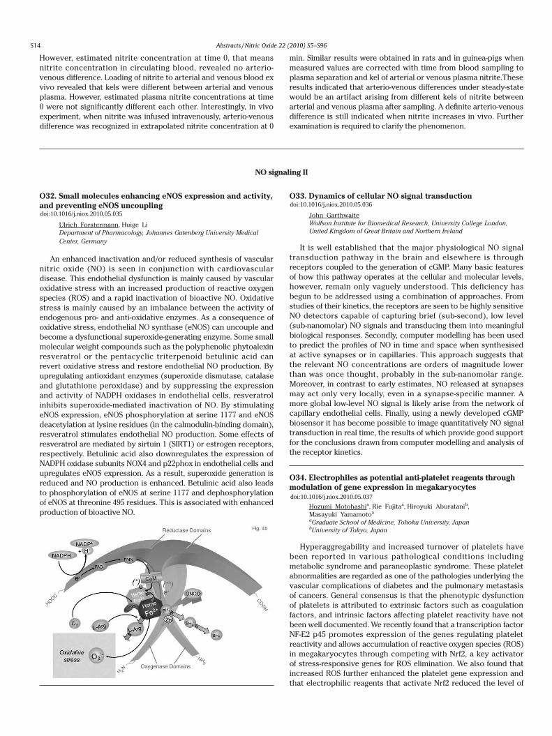
However, estimated nitrite concentration at time 0, that means nitrite concentration in circulating blood, revealed no arterio-venous difference. Loading of nitrite to arterial and venous blood ex vivo revealed that kels were different between arterial and venous plasma. However, estimated plasma nitrite concentrations at time 0 were not significantly different each other. Interestingly, in vivo experiment, when nitrite was infused intravenously, arterio-venous difference was recognized in extrapolated nitrite concentration at 0
min. Similar results were obtained in rats and in guinea-pigs when measured values are corrected with time from blood sampling to plasma separation and kel of arterial or venous plasma nitrite.These results indicated that arterio-venous differences under steady-state would be an artifact arising from different kels of nitrite between arterial and venous plasma after sampling. A definite arterio-venous difference is still indicated when nitrite increases in vivo. Further examination is required to clarify the phenomenon.
NO signaling II
O32. Small molecules enhancing eNOS expression and activity, and preventing eNOS uncoupling
Ulrich Forstermann, Huige Li Department of Pharmacology, Johannes Gutenberg University Medical Center, Germany
An enhanced inactivation and/or reduced synthesis of vascular nitric oxide (NO) is seen in conjunction with cardiovascular disease. This endothelial dysfunction is mainly caused by vascular oxidative stress with an increased production of reactive oxygen species (ROS) and a rapid inactivation of bioactive NO. Oxidative stress is mainly caused by an imbalance between the activity of endogenous pro- and anti-oxidative enzymes. As a consequence of oxidative stress, endothelial NO synthase (eNOS) can uncouple and become a dysfunctional superoxide-generating enzyme. Some small molecular weight compounds such as the polyphenolic phytoalexin resveratrol or the pentacyclic triterpenoid betulinic acid can revert oxidative stress and restore endothelial NO production. By upregulating antioxidant enzymes (superoxide dismutase, catalase and glutathione peroxidase) and by suppressing the expression and activity of NADPH oxidases in endothelial cells, resveratrol inhibits superoxide-mediated inactivation of NO. By stimulating eNOS expression, eNOS phosphorylation at serine 1177 and eNOS deacetylation at lysine residues (in the calmodulin-binding domain), resveratrol stimulates endothelial NO production. Some effects of resveratrol are mediated by sirtuin 1 (SIRT1) or estrogen receptors, respectively. Betulinic acid also downregulates the expression of NADPH oxidase subunits NOX4 and p22phox in endothelial cells and upregulates eNOS expression. As a result, superoxide generation is reduced and NO production is enhanced. Betulinic acid also leads to phosphorylation of eNOS at serine 1177 and dephosphorylation of eNOS at threonine 495 residues. This is associated with enhanced production of bioactive NO.
O33. Dynamics of cellular NO signal transduction
John Garthwaite Wolfson Institute for Biomedical Research, University College London, United Kingdom of Great Britain and Northern Ireland
It is well established that the major physiological NO signal transduction pathway in the brain and elsewhere is through receptors coupled to the generation of cGMP. Many basic features of how this pathway operates at the cellular and molecular levels, however, remain only vaguely understood. This deficiency has begun to be addressed using a combination of approaches. From studies of their kinetics, the receptors are seen to be highly sensitive NO detectors capable of capturing brief (sub-second), low level (sub-nanomolar) NO signals and transducing them into meaningful biological responses. Secondly, computer modelling has been used to predict the profiles of NO in time and space when synthesised at active synapses or in capillaries. This approach suggests that the relevant NO concentrations are orders of magnitude lower than was once thought, probably in the sub-nanomolar range. Moreover, in contrast to early estimates, NO released at synapses may act only very locally, even in a synapse-specific manner. A more global low-level NO signal is likely arise from the network of capillary endothelial cells. Finally, using a newly developed cGMP biosensor it has become possible to image quantitatively NO signal transduction in real time, the results of which provide good support for the conclusions drawn from computer modelling and analysis of the receptor kinetics.
O34. Electrophiles as potential anti-platelet reagents through modulation of gene expression in megakaryocytes
Hozumi Motohashia, Rie Fujitaa, Hiroyuki Aburatanib,Masayuki Yamamotoa aGraduate School of Medicine, Tohoku University, JapanbUniversity of Tokyo, Japan
Hyperaggregability and increased turnover of platelets have been reported in various pathological conditions including metabolic syndrome and paraneoplastic syndrome. These platelet abnormalities are regarded as one of the pathologies underlying the vascular complications of diabetes and the pulmonary metastasis of cancers. General consensus is that the phenotypic dysfunction of platelets is attributed to extrinsic factors such as coagulation factors, and intrinsic factors affecting platelet reactivity have not been well documented. We recently found that a transcription factor NF-E2 p45 promotes expression of the genes regulating platelet reactivity and allows accumulation of reactive oxygen species (ROS) in megakaryocytes through competing with Nrf2, a key activator of stress-responsive genes for ROS elimination. We also found that increased ROS further enhanced the platelet gene expression and that electrophilic reagents that activate Nrf2 reduced the level of
intracellular ROS and limited expression of the platelet genes. We further showed that platelet reactivity to thrombin correlates with the gene expression levels of megakaryocytes, suggesting that platelets produced from megakaryocytes with higher expression of platelet genes acquire increased reactivity. Thus platelets produced from megakaryocytes in an oxidative environment seem to acquire hyperaggregability due to elevated expression of platelet genes. These results also suggest that electrophiles activating Nrf2 serves as new anti-platelet reagents targeting megakaryocytes for decreasing expression of the platelet genes.
O35. Allosteric activators modulate the Fe-His bond in soluble guanylate cyclase and in Clostridium botulinum NO-sensor
Michel Negreriea, Byung-Kuk Yooa, Isabelle Lamarrea,Fabrice Rappaportb, Pierre Niochec, Jean-Louis Martina aLaboratoire d'Optique et Biosciences, Ecole Polytechnique, FrancebInstitut de Biologie Physico-Chimie, Paris, FrancecCentre Universitaire des Saints-Pres, Paris, France
Soluble guanylate cyclase (sGC), the mammalian nitric oxide (NO) endogenous receptor, can also be activated by carbon monoxide (CO) in synergy with artificial allosteric effectors (like BAY-412272 and YC-1). The molecular mechanism of this synergistic activation is still unknown. We investigated the interaction between sGC and these activators by time-resolved absorption spectroscopy. The transient spectra obtained immediately (1-500 picosecond time-range) after CO photo-dissociation are characteristic of the state of the heme before dissociation. This allows to unambiguously identify the change of coordination state of the heme iron induced by allosteric effectors. There are two distinct populations of carboxyl
heme in the presence of activators. Together with a 6-coordinate CO-heme, we observed a 5-coordinate CO-heme complex induced by activators and CO rebinding to the transient 4-coordinate heme after dissociation. Therefore, these activators induce the cleavage of the Fe-His bond for a proportion of sGC, as does NO. The occupancy of the substrate site of sGC α-subunit by the analog GTP-γ-S further increases this effect. These activators have a similar effect on the sGC truncated heme domain and on the homologous NO-sensors from Clostridium botulinum and Nostoc punctiform, albeit in a less extent due to lower affinity. The cleavage of the Fe-His bond in the simultaneous presence of CO and effectors rationalizes the synergistic sGC activation. We recorded in a broad time-range, from picoseconds to milliseconds, the dynamics of CO geminate rebinding after dissociation from sGC and from C. botulinum. The CO geminate rebinding, both in picosecond and in nanosecond time scales, is much faster in the presence of allosteric activators, providing explanation for the induced change of CO affinity.
O36. Evolution of Rapid and Slow NO Pulses During Signaling
John C. Salerno Biology, Kennesaw State University, United States of America
Diffusion of nitric oxide from a pulsed signal produces a spatial distribution that evolves in time and is much different than that produced by a steady state generator of NO. The shape and extent of this distribution depend on the geometry of the system and on the time course of the production of NO by NOS enzymes in the generating volume. Sharp (about 100 sec) pulses of NO produced by nNOS at synapses can be modeled in cylindrical symmetry with generating and quenching/receiving disks separated by a synaptic cleft of macromolecular dimensions. In this special case, short pulses consisting of at most a few hundred molecules of NO are efficiently received by soluble guanylate cyclase molecules in the receiving disk. The resulting NO distribution is cylindrically symmetrical and has an NO valley or shadow behind the receiver. The small amount of NO produced in an nNOS pulse produces minimal crosstalk at adjacent synapses. Signals from eNOS are relatively long-lasting. In vessels of arteriole dimensions, simulations show that about 1 sec of activity is sufficient to approach steady state limits of NO concentration in the smooth muscle layer. Quenching by sheer-confined RBCs limits the concentration of NO outside of the endothelial layer, and during most of the evolution of the gradient the majority of the NO produced by eNOS diffuses inward down the sharp gradient set up by the internal quenching volume.
Physiology/pathophysiology of NO (Cardiovascular system)
O37. Nitrosonium and the paradox of pulmonary arterial hypertension
Benjamin Gaston Pediatrics, University of Virginia, United States of America
Inhaled NO can acutely decrease pulmonary arterial (PA) vasoconstriction, but doses over 100 times levels endogenously present in the airways are needed. Data conflict on the role of pulmonary endothelial NOS (eNOS) expression in human PA hypertension (PAH); and PAH is not a striking feature of eNOS deficient mice. Our data show that chronic, excessive NO delivery to the PA bed in the form of nitrosonium (NO+) bound covalently to thiolate causes pulmonary vascular remodeling by upregulating
hypoxia- associated PA endothelial genes (JCI 2007;117:2592). This excessive NO+ dumping in the PA bed can occur with 1) excessive NO+ offloading from erythrocytes in the systemic periphery in chronic hypoxemia; 2) increased flow of erythrocytes through the PA bed (high flow states); and 3) chronic inflammation. Thus, acute NO exposure causes vasodilatation; but chronic NO+ exposure causes compensatory, hypoxia-mimetic remodeling and chronic PAH. Our more recent data suggest that eNOS upregulation by eNOS activates S-nitrosoglutathione reductase (GSNOR), which both protects the PA endothelium from excessive NO+ exposure and prevents NO+-mediated eNOS inactivation. However, females with decreased GSNOR response may be at risk for PAH caused by unchecked eNOS upregulation. These data help explain contradictory data regarding
S14
doi:10.1016/j.niox.2010.05.035doi:10.1016/j.niox.2010.05.036
doi:10.1016/j.niox.2010.05.037
Abstracts /Nitric Oxide 22 (2010) S5–S96

However, estimated nitrite concentration at time 0, that means nitrite concentration in circulating blood, revealed no arterio-venous difference. Loading of nitrite to arterial and venous blood ex vivo revealed that kels were different between arterial and venous plasma. However, estimated plasma nitrite concentrations at time 0 were not significantly different each other. Interestingly, in vivo experiment, when nitrite was infused intravenously, arterio-venous difference was recognized in extrapolated nitrite concentration at 0
min. Similar results were obtained in rats and in guinea-pigs when measured values are corrected with time from blood sampling to plasma separation and kel of arterial or venous plasma nitrite.These results indicated that arterio-venous differences under steady-state would be an artifact arising from different kels of nitrite between arterial and venous plasma after sampling. A definite arterio-venous difference is still indicated when nitrite increases in vivo. Further examination is required to clarify the phenomenon.
NO signaling II
O32. Small molecules enhancing eNOS expression and activity, and preventing eNOS uncoupling
Ulrich Forstermann, Huige Li Department of Pharmacology, Johannes Gutenberg University Medical Center, Germany
An enhanced inactivation and/or reduced synthesis of vascular nitric oxide (NO) is seen in conjunction with cardiovascular disease. This endothelial dysfunction is mainly caused by vascular oxidative stress with an increased production of reactive oxygen species (ROS) and a rapid inactivation of bioactive NO. Oxidative stress is mainly caused by an imbalance between the activity of endogenous pro- and anti-oxidative enzymes. As a consequence of oxidative stress, endothelial NO synthase (eNOS) can uncouple and become a dysfunctional superoxide-generating enzyme. Some small molecular weight compounds such as the polyphenolic phytoalexin resveratrol or the pentacyclic triterpenoid betulinic acid can revert oxidative stress and restore endothelial NO production. By upregulating antioxidant enzymes (superoxide dismutase, catalase and glutathione peroxidase) and by suppressing the expression and activity of NADPH oxidases in endothelial cells, resveratrol inhibits superoxide-mediated inactivation of NO. By stimulating eNOS expression, eNOS phosphorylation at serine 1177 and eNOS deacetylation at lysine residues (in the calmodulin-binding domain), resveratrol stimulates endothelial NO production. Some effects of resveratrol are mediated by sirtuin 1 (SIRT1) or estrogen receptors, respectively. Betulinic acid also downregulates the expression of NADPH oxidase subunits NOX4 and p22phox in endothelial cells and upregulates eNOS expression. As a result, superoxide generation is reduced and NO production is enhanced. Betulinic acid also leads to phosphorylation of eNOS at serine 1177 and dephosphorylation of eNOS at threonine 495 residues. This is associated with enhanced production of bioactive NO.
O33. Dynamics of cellular NO signal transduction
John Garthwaite Wolfson Institute for Biomedical Research, University College London, United Kingdom of Great Britain and Northern Ireland
It is well established that the major physiological NO signal transduction pathway in the brain and elsewhere is through receptors coupled to the generation of cGMP. Many basic features of how this pathway operates at the cellular and molecular levels, however, remain only vaguely understood. This deficiency has begun to be addressed using a combination of approaches. From studies of their kinetics, the receptors are seen to be highly sensitive NO detectors capable of capturing brief (sub-second), low level (sub-nanomolar) NO signals and transducing them into meaningful biological responses. Secondly, computer modelling has been used to predict the profiles of NO in time and space when synthesised at active synapses or in capillaries. This approach suggests that the relevant NO concentrations are orders of magnitude lower than was once thought, probably in the sub-nanomolar range. Moreover, in contrast to early estimates, NO released at synapses may act only very locally, even in a synapse-specific manner. A more global low-level NO signal is likely arise from the network of capillary endothelial cells. Finally, using a newly developed cGMP biosensor it has become possible to image quantitatively NO signal transduction in real time, the results of which provide good support for the conclusions drawn from computer modelling and analysis of the receptor kinetics.
O34. Electrophiles as potential anti-platelet reagents through modulation of gene expression in megakaryocytes
Hozumi Motohashia, Rie Fujitaa, Hiroyuki Aburatanib,Masayuki Yamamotoa aGraduate School of Medicine, Tohoku University, JapanbUniversity of Tokyo, Japan
Hyperaggregability and increased turnover of platelets have been reported in various pathological conditions including metabolic syndrome and paraneoplastic syndrome. These platelet abnormalities are regarded as one of the pathologies underlying the vascular complications of diabetes and the pulmonary metastasis of cancers. General consensus is that the phenotypic dysfunction of platelets is attributed to extrinsic factors such as coagulation factors, and intrinsic factors affecting platelet reactivity have not been well documented. We recently found that a transcription factor NF-E2 p45 promotes expression of the genes regulating platelet reactivity and allows accumulation of reactive oxygen species (ROS) in megakaryocytes through competing with Nrf2, a key activator of stress-responsive genes for ROS elimination. We also found that increased ROS further enhanced the platelet gene expression and that electrophilic reagents that activate Nrf2 reduced the level of
intracellular ROS and limited expression of the platelet genes. We further showed that platelet reactivity to thrombin correlates with the gene expression levels of megakaryocytes, suggesting that platelets produced from megakaryocytes with higher expression of platelet genes acquire increased reactivity. Thus platelets produced from megakaryocytes in an oxidative environment seem to acquire hyperaggregability due to elevated expression of platelet genes. These results also suggest that electrophiles activating Nrf2 serves as new anti-platelet reagents targeting megakaryocytes for decreasing expression of the platelet genes.
O35. Allosteric activators modulate the Fe-His bond in soluble guanylate cyclase and in Clostridium botulinum NO-sensor
Michel Negreriea, Byung-Kuk Yooa, Isabelle Lamarrea,Fabrice Rappaportb, Pierre Niochec, Jean-Louis Martina aLaboratoire d'Optique et Biosciences, Ecole Polytechnique, FrancebInstitut de Biologie Physico-Chimie, Paris, FrancecCentre Universitaire des Saints-Pres, Paris, France
Soluble guanylate cyclase (sGC), the mammalian nitric oxide (NO) endogenous receptor, can also be activated by carbon monoxide (CO) in synergy with artificial allosteric effectors (like BAY-412272 and YC-1). The molecular mechanism of this synergistic activation is still unknown. We investigated the interaction between sGC and these activators by time-resolved absorption spectroscopy. The transient spectra obtained immediately (1-500 picosecond time-range) after CO photo-dissociation are characteristic of the state of the heme before dissociation. This allows to unambiguously identify the change of coordination state of the heme iron induced by allosteric effectors. There are two distinct populations of carboxyl
heme in the presence of activators. Together with a 6-coordinate CO-heme, we observed a 5-coordinate CO-heme complex induced by activators and CO rebinding to the transient 4-coordinate heme after dissociation. Therefore, these activators induce the cleavage of the Fe-His bond for a proportion of sGC, as does NO. The occupancy of the substrate site of sGC α-subunit by the analog GTP-γ-S further increases this effect. These activators have a similar effect on the sGC truncated heme domain and on the homologous NO-sensors from Clostridium botulinum and Nostoc punctiform, albeit in a less extent due to lower affinity. The cleavage of the Fe-His bond in the simultaneous presence of CO and effectors rationalizes the synergistic sGC activation. We recorded in a broad time-range, from picoseconds to milliseconds, the dynamics of CO geminate rebinding after dissociation from sGC and from C. botulinum. The CO geminate rebinding, both in picosecond and in nanosecond time scales, is much faster in the presence of allosteric activators, providing explanation for the induced change of CO affinity.
O36. Evolution of Rapid and Slow NO Pulses During Signaling
John C. Salerno Biology, Kennesaw State University, United States of America
Diffusion of nitric oxide from a pulsed signal produces a spatial distribution that evolves in time and is much different than that produced by a steady state generator of NO. The shape and extent of this distribution depend on the geometry of the system and on the time course of the production of NO by NOS enzymes in the generating volume. Sharp (about 100 sec) pulses of NO produced by nNOS at synapses can be modeled in cylindrical symmetry with generating and quenching/receiving disks separated by a synaptic cleft of macromolecular dimensions. In this special case, short pulses consisting of at most a few hundred molecules of NO are efficiently received by soluble guanylate cyclase molecules in the receiving disk. The resulting NO distribution is cylindrically symmetrical and has an NO valley or shadow behind the receiver. The small amount of NO produced in an nNOS pulse produces minimal crosstalk at adjacent synapses. Signals from eNOS are relatively long-lasting. In vessels of arteriole dimensions, simulations show that about 1 sec of activity is sufficient to approach steady state limits of NO concentration in the smooth muscle layer. Quenching by sheer-confined RBCs limits the concentration of NO outside of the endothelial layer, and during most of the evolution of the gradient the majority of the NO produced by eNOS diffuses inward down the sharp gradient set up by the internal quenching volume.
Physiology/pathophysiology of NO (Cardiovascular system)
O37. Nitrosonium and the paradox of pulmonary arterial hypertension
Benjamin Gaston Pediatrics, University of Virginia, United States of America
Inhaled NO can acutely decrease pulmonary arterial (PA) vasoconstriction, but doses over 100 times levels endogenously present in the airways are needed. Data conflict on the role of pulmonary endothelial NOS (eNOS) expression in human PA hypertension (PAH); and PAH is not a striking feature of eNOS deficient mice. Our data show that chronic, excessive NO delivery to the PA bed in the form of nitrosonium (NO+) bound covalently to thiolate causes pulmonary vascular remodeling by upregulating
hypoxia- associated PA endothelial genes (JCI 2007;117:2592). This excessive NO+ dumping in the PA bed can occur with 1) excessive NO+ offloading from erythrocytes in the systemic periphery in chronic hypoxemia; 2) increased flow of erythrocytes through the PA bed (high flow states); and 3) chronic inflammation. Thus, acute NO exposure causes vasodilatation; but chronic NO+ exposure causes compensatory, hypoxia-mimetic remodeling and chronic PAH. Our more recent data suggest that eNOS upregulation by eNOS activates S-nitrosoglutathione reductase (GSNOR), which both protects the PA endothelium from excessive NO+ exposure and prevents NO+-mediated eNOS inactivation. However, females with decreased GSNOR response may be at risk for PAH caused by unchecked eNOS upregulation. These data help explain contradictory data regarding
S15
doi:10.1016/j.niox.2010.05.038
doi:10.1016/j.niox.2010.05.039
doi:10.1016/j.niox.2010.05.040
Abstracts /Nitric Oxide 22 (2010) S5–S96

the role of NO and eNOS in acute and chronic PAH.
O38. Critical roles of ROS and p38 MAPK for eNOS activation in laminar shear stress
Santiago Lamasa, Rosa M Bretna, Oscar Busnadiegoa,Natalia Romerob, Fernando Rodrguez-Pascuala,Almudena Porrasc, Rafael Radib aBiologia Celular, Consejo Superior de Investigaciones Cientficas-CBMSO, Spain
bUniversidad de la Repblica, UruguaycFacultad de Farmacia-UCM, Spain
Laminar shear stress (LSS) is a protective hemodynamic regulator of endothelial function, and limits the development of vascular wall diseases related to oxidative stress. LSS triggers the production of reactive oxygen and nitrogen species, which are in part responsible for promoting an antioxidant response. LSS activates several endothelial signaling pathways including the activation of MAPKs and eNOS. These studies explore the mode of activation of these key endothelial signaling targets. Using the cone/plate model for LSS (12 dyn/cm2) in bovine aortic endothelial cells (BAEC) we found that LSS rapidly promotes production of superoxide and hydrogen peroxide (H2O2) after 30-60 minutes. At physiological concentrations (15-25 μM) H2O2 significantly activated both eNOS and p38 MAPK. Importantly, fluxes of H2O2 (0.1-1000 nM/min) elicited similar responses in p38 and eNOS activation, using phosphospecific antibodies. The highly specific p38 MAPK inhibitors SB203580 and PD169316 blocked H2O2-promoted and LSS-induced eNOS phosphorylation and reduced NO levels, as determined by chemiluminescence. In murine embryonic fibroblasts (MEFs) of mice deficient in p38 MAPKα, basal levels of eNOS were markedly decreased. The activation of eNOS in response to the upstream p38 MAPK activator MKK6 as well as to H2O2 was also strikingly reduced in MEFs isolated from p38α KO mice. In contrast, DEA-NO failed to activate p38 MAPK in BAEC; similarly lung endothelial cells from eNOS-deficient mice exposed to LSS showed comparable levels of p38 MAPK phosphorylation. These findings indicate that p38 MAPK lies upstream of LSS-promoted eNOS activation. We propose a model in which LSS induces signaling by low levels of hydrogen peroxide, which in turn activate p38 MAPK and then stimulate eNOS, leading to increased NO levels and protection of endothelial function.
O39. Caveolin-1 downregulation prevents eNOS uncoupling by angiotensin II through inhibition of NADPH oxidase assembly and activation in caveolae of endothelial cells.
Irina I. Lobysheva, Geraldine Rath, Belaid Sekkali, Olivier Feron, Chantal Dessy, Bernard Gallez, Jean-Luc Balligand Medical School, Unit of Pharmacology & Therapeutics, Universit Catholique de Louvain, Belgium
Concurrent activation of O2_. production from NADPH oxidase
and NO from eNOS by Angiotensin II (AII) in endothelial cells (ECs) aggravates endothelial dysfunction through cross-reaction between the radicals. We examined the influence of subcellular co-localization of eNOS and NADPH oxidase in caveolae and caveolin-1 (cav1) abundance on the eNOS activation in response to AII in vitro and in vivo.
AII increased NO and O2_. production (to 158±12% and 209±5% of
control) measured by EPR spin-trapping at the membrane of ECs. Stimulated NO production was sensitive to inhibition of NADPH oxidase assembly and siRNA downregulation of non-receptor tyrosine kinase cAbl. Reciprocally, L-NAME, a NOS inhibitor, partly inhibited O2
_. stimulated by AII (by 47±11%), indicating eNOS
uncoupling, as confirmed by increase eNOS monomer/dimer ratio (to 133±8%). To resolve the role of subcellular compartmentation, AII stimulated EC fractions were separated by isopycnic ultracentrifugation. AII stimulated cAbl, p47phox, a NADPH oxidase subunit, redistribution with eNOS to cav1-enriched fractions. Downregulation of cav1 by siRNA (to 50%) while preserving eNOS confinement, inhibited AII-stimulated p47phox translocation, and NADPH oxidase activity in cav1-enriched fractions and reversed eNOS uncoupling. To verify the effect in vivo, heterozygote cav1+/- mice (and WT) were treated with AII by osmotic minipumps (2 weeks). Blood pressure, recorded by implanted telemetry and blood Hb-NO level measured by EPR were preserved in cav1+/- mice while AII produced hypertension and decreased Hb-NO in WT (to 55±15%, p<0.05). We conclude that, co-localization of key proteins in caveolae activates eNOS through ROS-sensitive cAbl kinase in response to AII but also eNOS uncoupling. Moderate downregulation of cav1 prevents NADPH oxidase assembly and eNOS uncoupling, underlining the possibility to treat endothelial dysfunction.
O40. Regular exercise training prevents calcific aortic valve disease in mice:potential importance of the valvular endothelial cells for the management of valvular heart disease
Yasuharu Matsumotoa, Hiroaki Shimokawaa, Volker Adamsb,Gerhard Schulerb, Axel Linkeb aDepartment of Cardiovascular Medicine, Tohoku University, JapanbUniversity of Leipzig, Germany
Background: Currently, no effective therapy to prevent degenerative calcific (aortic valve) AV disease exits. Importantly, the surface of AV leaflets is covered with endothelial cells that are critical for the transduction of mechanical and biological signals. Regular exercise training (ET) increases nitric oxide bioavailability and slows the progression of atherosclerotic lesions, which may be expected to improve degenerative AV disease. Methods and Results: Four-week-old LDL receptor deficient (LDLR-/-) mice (n=94) were randomly divided into four groups: Group 1 (N), normal diet plus sedentary; group 2 (Chol), cholesterol (chol)-diet plus sedentary; group 3 (Reg), chol-diet plus regular ET for 16 wks (Treadmil, 60min/d, 5d/wk); group 4 (Occa), chol-diet plus occasional ET for 16wks (1d/wk). Histological analysis at 20-wk-old showed that AV thickness increased significantly in chol-group compared to N-group. Of note, regular ET but not occasional ET significantly reduced AV thickness as compared tochol-group ( N;31.3±3.0, Chol; 50.1±3.4, Reg;30.4±1.2, Occa; 48.9±3.2μm, P<0.001). Interestingly, immunohistochemistory revealed that chol-diet disrupted and regular ET preserved endothelial integrity on the AV surface (N; 95.7±1.0, Chol; 73.7±2.2,
Reg; 95.8±2.3%, P<0.001). In addition, accumulation of macrophages and oxidized-LDL, in-situ superoxide, myofibroblasts/ osteoblasts phenotypes and mineralization at the AVs were markedly increased in chol-group and significantly decreased by regular ET. PCR revealed mRNA for Runx2 (osteogenic transcription factor) was increased in chol-group and significantly diminished by regular ET (P<0.05). Conclusions: In mice, regular ET preserved valvular endothelial integrity and thus may prevent subsequent inflammation and calcification leading to AV sclerosis. The valvular endothelial cell could be an important target for preventing degenerative AV disease particularly at an early stage of disease.
O41. Role of mitochondrial aldehyde dehydrogenase in nitroglycerin bioactivation and cardiac ischemia/reperfusion injury.
Matteo Beretta, Verena Wenzl, Gerald Woelkart,Kurt Schmidt, Antonius C.F. Gorren, Bernd Mayer Department of Pharmacology and Toxicology, Karl-Franzens University Graz, Austria
The East Asian variant of mitochondrial aldehyde dehydrogenase (ALDH2) exhibits reduced dehydrogenase and nitroglycerin (GTN) denitration activities. The activator Alda-1 was shown to increase 11-fold and 2-fold the dehydrogenase activity of this mutant and of wild type enzyme, respectively [1], but not GTN bioactivation [2].
The reaction mechanism underlying GTN metabolism is still partially unknown, but our data on the East Asian variant, where the general base E268 is dislocated from the catalytic center, and on the E268Q mutant suggest the co-existence of 2- and 3-electron reduction pathways for GTN. Nitric oxide (NO) formation is probably due to the 3-electron pathway and accounts for approx. 10% and 50% of total GTN turnover in the wild type enzyme and in the E268Q mutant, respectively. The 2-electron pathway probably involves cysteine oxidation that reduces O2 to superoxide, limiting NO bioavailability. Therefore, the lower reactivity of the catalytic cysteine due to the lack of the general base E268 could explain reduced superoxide and increased NO formation by these mutants [2-3].
Activation of ALDH2 by Alda-1 was shown to correlate with reduced ischemic heart damage in rodents [1]. To further investigate this issue, we compared cardiac function of hearts isolated from ALDH2 wild type and knock-out animals after 20 min of global ischemia. Our data showed reduced maximal velocity of contraction (-48±5%) and of relaxation (-42±7%), reduced left ventricular developed pressure (-39±4%), increased left ventricular end-diastolic pressure (2-fold) and unchanged heart rates in KO animals after 30 min of reperfusion. These
results demonstrate that ALDH2 is cardioprotective in ischemia/reperfusion injury.
References[1] C.H. Chen et al. Science 2008, 321:1493-1495.[2] M. Beretta et al. J. Biol. Chem. 2010, 285:943-952.[3] M.V. Wenzl et al. J. Biol. Chem. 2009, 284:19878-19886.
O42. Aldehyde Dehydrogenase-2 catalyzed bioactivation of Pentaerythrityl Tetranitrate
Martina Griesberger, Gerald Woelkart, Alexander Kollau,Verena Wenzl, Matteo Beretta, Antonius C.F. Gorren,Kurt Schmidt,Bernd Mayer Department of Pharmacology and Toxicology, Karl-Franzens University Graz, Austria
Organic nitrates represent a class of very effective anti-ischemic drugs, but a limitation of their clinical use is the development of nitrate tolerance. A remarkable exception is the long-acting drug pentaerythrityl tetranitrate (PETN), which was shown to cause no tolerance. Vasodilation through NO-mediated activation of vascular soluble guanylate cyclase (sGC) requires bioactivation of the nitrate. The mitochondrial isoform of aldehyde dehydrogenase (ALDH2) was shown to be involved in bioactivation of nitroglycerin (GTN) and PETN. The present work was carried out to study the molecular mechanisms underlying the pharmacological differences between GTN and PETN at the level of purified ALDH2.
In the presence of 25 μg of ALDH2 PETN activated sGC with an EC50 of 3.61 ± 0.35 μM and a maximum at 30 μM PETN. This effect was enhanced by superoxide dismutase (SOD) resulting in an EC50 of 0.64 ± 0.08 μM and maximal sGC activation. As measured with a Clark-type electrode ALDH2 catalyzed NO formation from 10 μM PETN in the presence of SOD with a peak concentration of 0.51 ± 0.06 μM in the presence and 0.22 ± 0.02 μM in the absence of the reducing agent dithiothreitol. Co-incubation of 2 μM [14C]GTN with ALDH2 and increasing concentrations of PETN resulted in only small decreases in 1,2-glyceroldinitrate formation as measured by radio thin layer chromatography indicating that PETN does not compete with GTN metabolism. ALDH2 activity was rapidly inactivated by GTN but not by PETN.
Low and high affinity pathways of PETN may explain the apparent differences between PETN metabolism and bioactivation, respectively. The reaction of PETN with ALDH2 leads to activation of sGC with markedly higher values in the presence of SOD suggesting formation of superoxide as a co-product of PETN metabolism. The lack of vascular tolerance to PETN may be due to significantly lower rates of ALDH2 inactivation as compared to GTN.
Second International Conference Nitric Oxide and Cancer
O43. Pathways associate Nitric Oxide Synthase and Cycloxygenase-2 that Lead to Poor Prognosis in Breast Cancer
David A Winka, Glynn Sharona,b, Switzer Christopherb,Ridnour Lisab, Ambs Stefana aRadiation Biology Branch, National Cancer Institute, United States of America
bRadiation Biology Branch, United States of America
Epidemiological studies have found that inflammatory proteins iNOS and COX-2 are poor prognostic indicators for many cancers. We have been investigating the chemical mechanism of chemistry
of NO and other reactive species associated with biological mechanisms in cancer. These studies have shown that specific concentrations of NO determine the pro or anti-tumorigenic behavior. When prolonged exposure to μM amounts of NO, there is an increase in the phosphorylation of p53 and cystostasis. However, when cells are exposed to 100 nM of NO, there is increase in protumorigenic molecular pathways such as MAPK, Akt-P and HIF1α. Several of these pathways are also activated by PGE2. In ER negative breast cancer patients we found that if either iNOS and COX-2 was highly expressed there was a decrease in survival. When both were present there was a dramatic decreased survival. From
S16
doi:10.1016/j.niox.2010.05.041
doi:10.1016/j.niox.2010.05.042
doi:10.1016/j.niox.2010.05.043
Abstracts /Nitric Oxide 22 (2010) S5–S96

the role of NO and eNOS in acute and chronic PAH.
O38. Critical roles of ROS and p38 MAPK for eNOS activation in laminar shear stress
Santiago Lamasa, Rosa M Bretna, Oscar Busnadiegoa,Natalia Romerob, Fernando Rodrguez-Pascuala,Almudena Porrasc, Rafael Radib aBiologia Celular, Consejo Superior de Investigaciones Cientficas-CBMSO, Spain
bUniversidad de la Repblica, UruguaycFacultad de Farmacia-UCM, Spain
Laminar shear stress (LSS) is a protective hemodynamic regulator of endothelial function, and limits the development of vascular wall diseases related to oxidative stress. LSS triggers the production of reactive oxygen and nitrogen species, which are in part responsible for promoting an antioxidant response. LSS activates several endothelial signaling pathways including the activation of MAPKs and eNOS. These studies explore the mode of activation of these key endothelial signaling targets. Using the cone/plate model for LSS (12 dyn/cm2) in bovine aortic endothelial cells (BAEC) we found that LSS rapidly promotes production of superoxide and hydrogen peroxide (H2O2) after 30-60 minutes. At physiological concentrations (15-25 μM) H2O2 significantly activated both eNOS and p38 MAPK. Importantly, fluxes of H2O2 (0.1-1000 nM/min) elicited similar responses in p38 and eNOS activation, using phosphospecific antibodies. The highly specific p38 MAPK inhibitors SB203580 and PD169316 blocked H2O2-promoted and LSS-induced eNOS phosphorylation and reduced NO levels, as determined by chemiluminescence. In murine embryonic fibroblasts (MEFs) of mice deficient in p38 MAPKα, basal levels of eNOS were markedly decreased. The activation of eNOS in response to the upstream p38 MAPK activator MKK6 as well as to H2O2 was also strikingly reduced in MEFs isolated from p38α KO mice. In contrast, DEA-NO failed to activate p38 MAPK in BAEC; similarly lung endothelial cells from eNOS-deficient mice exposed to LSS showed comparable levels of p38 MAPK phosphorylation. These findings indicate that p38 MAPK lies upstream of LSS-promoted eNOS activation. We propose a model in which LSS induces signaling by low levels of hydrogen peroxide, which in turn activate p38 MAPK and then stimulate eNOS, leading to increased NO levels and protection of endothelial function.
O39. Caveolin-1 downregulation prevents eNOS uncoupling by angiotensin II through inhibition of NADPH oxidase assembly and activation in caveolae of endothelial cells.
Irina I. Lobysheva, Geraldine Rath, Belaid Sekkali, Olivier Feron, Chantal Dessy, Bernard Gallez, Jean-Luc Balligand Medical School, Unit of Pharmacology & Therapeutics, Universit Catholique de Louvain, Belgium
Concurrent activation of O2_. production from NADPH oxidase
and NO from eNOS by Angiotensin II (AII) in endothelial cells (ECs) aggravates endothelial dysfunction through cross-reaction between the radicals. We examined the influence of subcellular co-localization of eNOS and NADPH oxidase in caveolae and caveolin-1 (cav1) abundance on the eNOS activation in response to AII in vitro and in vivo.
AII increased NO and O2_. production (to 158±12% and 209±5% of
control) measured by EPR spin-trapping at the membrane of ECs. Stimulated NO production was sensitive to inhibition of NADPH oxidase assembly and siRNA downregulation of non-receptor tyrosine kinase cAbl. Reciprocally, L-NAME, a NOS inhibitor, partly inhibited O2
_. stimulated by AII (by 47±11%), indicating eNOS
uncoupling, as confirmed by increase eNOS monomer/dimer ratio (to 133±8%). To resolve the role of subcellular compartmentation, AII stimulated EC fractions were separated by isopycnic ultracentrifugation. AII stimulated cAbl, p47phox, a NADPH oxidase subunit, redistribution with eNOS to cav1-enriched fractions. Downregulation of cav1 by siRNA (to 50%) while preserving eNOS confinement, inhibited AII-stimulated p47phox translocation, and NADPH oxidase activity in cav1-enriched fractions and reversed eNOS uncoupling. To verify the effect in vivo, heterozygote cav1+/- mice (and WT) were treated with AII by osmotic minipumps (2 weeks). Blood pressure, recorded by implanted telemetry and blood Hb-NO level measured by EPR were preserved in cav1+/- mice while AII produced hypertension and decreased Hb-NO in WT (to 55±15%, p<0.05). We conclude that, co-localization of key proteins in caveolae activates eNOS through ROS-sensitive cAbl kinase in response to AII but also eNOS uncoupling. Moderate downregulation of cav1 prevents NADPH oxidase assembly and eNOS uncoupling, underlining the possibility to treat endothelial dysfunction.
O40. Regular exercise training prevents calcific aortic valve disease in mice:potential importance of the valvular endothelial cells for the management of valvular heart disease
Yasuharu Matsumotoa, Hiroaki Shimokawaa, Volker Adamsb,Gerhard Schulerb, Axel Linkeb aDepartment of Cardiovascular Medicine, Tohoku University, JapanbUniversity of Leipzig, Germany
Background: Currently, no effective therapy to prevent degenerative calcific (aortic valve) AV disease exits. Importantly, the surface of AV leaflets is covered with endothelial cells that are critical for the transduction of mechanical and biological signals. Regular exercise training (ET) increases nitric oxide bioavailability and slows the progression of atherosclerotic lesions, which may be expected to improve degenerative AV disease. Methods and Results: Four-week-old LDL receptor deficient (LDLR-/-) mice (n=94) were randomly divided into four groups: Group 1 (N), normal diet plus sedentary; group 2 (Chol), cholesterol (chol)-diet plus sedentary; group 3 (Reg), chol-diet plus regular ET for 16 wks (Treadmil, 60min/d, 5d/wk); group 4 (Occa), chol-diet plus occasional ET for 16wks (1d/wk). Histological analysis at 20-wk-old showed that AV thickness increased significantly in chol-group compared to N-group. Of note, regular ET but not occasional ET significantly reduced AV thickness as compared tochol-group ( N;31.3±3.0, Chol; 50.1±3.4, Reg;30.4±1.2, Occa; 48.9±3.2μm, P<0.001). Interestingly, immunohistochemistory revealed that chol-diet disrupted and regular ET preserved endothelial integrity on the AV surface (N; 95.7±1.0, Chol; 73.7±2.2,
Reg; 95.8±2.3%, P<0.001). In addition, accumulation of macrophages and oxidized-LDL, in-situ superoxide, myofibroblasts/ osteoblasts phenotypes and mineralization at the AVs were markedly increased in chol-group and significantly decreased by regular ET. PCR revealed mRNA for Runx2 (osteogenic transcription factor) was increased in chol-group and significantly diminished by regular ET (P<0.05). Conclusions: In mice, regular ET preserved valvular endothelial integrity and thus may prevent subsequent inflammation and calcification leading to AV sclerosis. The valvular endothelial cell could be an important target for preventing degenerative AV disease particularly at an early stage of disease.
O41. Role of mitochondrial aldehyde dehydrogenase in nitroglycerin bioactivation and cardiac ischemia/reperfusion injury.
Matteo Beretta, Verena Wenzl, Gerald Woelkart,Kurt Schmidt, Antonius C.F. Gorren, Bernd Mayer Department of Pharmacology and Toxicology, Karl-Franzens University Graz, Austria
The East Asian variant of mitochondrial aldehyde dehydrogenase (ALDH2) exhibits reduced dehydrogenase and nitroglycerin (GTN) denitration activities. The activator Alda-1 was shown to increase 11-fold and 2-fold the dehydrogenase activity of this mutant and of wild type enzyme, respectively [1], but not GTN bioactivation [2].
The reaction mechanism underlying GTN metabolism is still partially unknown, but our data on the East Asian variant, where the general base E268 is dislocated from the catalytic center, and on the E268Q mutant suggest the co-existence of 2- and 3-electron reduction pathways for GTN. Nitric oxide (NO) formation is probably due to the 3-electron pathway and accounts for approx. 10% and 50% of total GTN turnover in the wild type enzyme and in the E268Q mutant, respectively. The 2-electron pathway probably involves cysteine oxidation that reduces O2 to superoxide, limiting NO bioavailability. Therefore, the lower reactivity of the catalytic cysteine due to the lack of the general base E268 could explain reduced superoxide and increased NO formation by these mutants [2-3].
Activation of ALDH2 by Alda-1 was shown to correlate with reduced ischemic heart damage in rodents [1]. To further investigate this issue, we compared cardiac function of hearts isolated from ALDH2 wild type and knock-out animals after 20 min of global ischemia. Our data showed reduced maximal velocity of contraction (-48±5%) and of relaxation (-42±7%), reduced left ventricular developed pressure (-39±4%), increased left ventricular end-diastolic pressure (2-fold) and unchanged heart rates in KO animals after 30 min of reperfusion. These
results demonstrate that ALDH2 is cardioprotective in ischemia/reperfusion injury.
References[1] C.H. Chen et al. Science 2008, 321:1493-1495.[2] M. Beretta et al. J. Biol. Chem. 2010, 285:943-952.[3] M.V. Wenzl et al. J. Biol. Chem. 2009, 284:19878-19886.
O42. Aldehyde Dehydrogenase-2 catalyzed bioactivation of Pentaerythrityl Tetranitrate
Martina Griesberger, Gerald Woelkart, Alexander Kollau,Verena Wenzl, Matteo Beretta, Antonius C.F. Gorren,Kurt Schmidt,Bernd Mayer Department of Pharmacology and Toxicology, Karl-Franzens University Graz, Austria
Organic nitrates represent a class of very effective anti-ischemic drugs, but a limitation of their clinical use is the development of nitrate tolerance. A remarkable exception is the long-acting drug pentaerythrityl tetranitrate (PETN), which was shown to cause no tolerance. Vasodilation through NO-mediated activation of vascular soluble guanylate cyclase (sGC) requires bioactivation of the nitrate. The mitochondrial isoform of aldehyde dehydrogenase (ALDH2) was shown to be involved in bioactivation of nitroglycerin (GTN) and PETN. The present work was carried out to study the molecular mechanisms underlying the pharmacological differences between GTN and PETN at the level of purified ALDH2.
In the presence of 25 μg of ALDH2 PETN activated sGC with an EC50 of 3.61 ± 0.35 μM and a maximum at 30 μM PETN. This effect was enhanced by superoxide dismutase (SOD) resulting in an EC50 of 0.64 ± 0.08 μM and maximal sGC activation. As measured with a Clark-type electrode ALDH2 catalyzed NO formation from 10 μM PETN in the presence of SOD with a peak concentration of 0.51 ± 0.06 μM in the presence and 0.22 ± 0.02 μM in the absence of the reducing agent dithiothreitol. Co-incubation of 2 μM [14C]GTN with ALDH2 and increasing concentrations of PETN resulted in only small decreases in 1,2-glyceroldinitrate formation as measured by radio thin layer chromatography indicating that PETN does not compete with GTN metabolism. ALDH2 activity was rapidly inactivated by GTN but not by PETN.
Low and high affinity pathways of PETN may explain the apparent differences between PETN metabolism and bioactivation, respectively. The reaction of PETN with ALDH2 leads to activation of sGC with markedly higher values in the presence of SOD suggesting formation of superoxide as a co-product of PETN metabolism. The lack of vascular tolerance to PETN may be due to significantly lower rates of ALDH2 inactivation as compared to GTN.
Second International Conference Nitric Oxide and Cancer
O43. Pathways associate Nitric Oxide Synthase and Cycloxygenase-2 that Lead to Poor Prognosis in Breast Cancer
David A Winka, Glynn Sharona,b, Switzer Christopherb,Ridnour Lisab, Ambs Stefana aRadiation Biology Branch, National Cancer Institute, United States of America
bRadiation Biology Branch, United States of America
Epidemiological studies have found that inflammatory proteins iNOS and COX-2 are poor prognostic indicators for many cancers. We have been investigating the chemical mechanism of chemistry
of NO and other reactive species associated with biological mechanisms in cancer. These studies have shown that specific concentrations of NO determine the pro or anti-tumorigenic behavior. When prolonged exposure to μM amounts of NO, there is an increase in the phosphorylation of p53 and cystostasis. However, when cells are exposed to 100 nM of NO, there is increase in protumorigenic molecular pathways such as MAPK, Akt-P and HIF1α. Several of these pathways are also activated by PGE2. In ER negative breast cancer patients we found that if either iNOS and COX-2 was highly expressed there was a decrease in survival. When both were present there was a dramatic decreased survival. From
S17
doi:10.1016/j.niox.2010.05.044
doi:10.1016/j.niox.2010.05.045
doi:10.1016/j.niox.2010.05.046
Abstracts /Nitric Oxide 22 (2010) S5–S96

this epidemiological data we have been able to develop cellular models to determine if we can find compounds that will reverse these mechanisms that result in poor phenotypes. We have found a class of thiol-based compounds that activate a tumor suppressor protein reversing the molecular pathways associated with iNOS and COX-2 in ER negative breast cancer. This presentation will focus on the understanding the chemical biology of nitric oxide and how it increases cancer risk and describe potential new molecular targets for the treatment of cancer.
O44. Dual role of NO donors in the reversal of tumor cell resistance and EMT: dysregulation of the NF-κB/YY1/Snail/RKIP circuitry
Benjamin Bonavidaa, Stavroula Baritakia, Sarah Huerta-Yepezb,Kam Yeungc aMicrobiology, Immunology & Molecular Genetics, David Geffen School of Medicine, University of California at Los Angeles, United States of America
bUnidad de Investigacion en Enfermedades Oncologicas, Mexico City, Mexico
cUniversity of Toledo, Ohio, United States of America
Tumor cells respond initially to both conventional chemotherapy and immunotherapy. However, a subset of patients experiences relapse and recurrences and no longer responds to further treatments. In addition, many tumors metastasize at different anatomical sites. Therefore, there is an urgent need to develop new therapeutics to overcome both resistance and metastasis. Nitric oxide (NO) has been reported to exert contrasting anti-tumor effects, namely, progression and resistance to apoptotic stimuli with low levels of NO as well as regression and sensitivity with high levels of NO. We have reported that high levels of the NO donor, DETANONOate, sensitizes resistant tumor cells to apoptosis by both chemotherapy and immunotherapy. NO-mediated sensitization was mediated via inhibition of NF-κB and downstream inhibition of Yin Yang 1 (YY1) and Snail and induction of Raf-1 kinase inhibitor protein (RKIP). The direct role of RKIP in sensitization was demonstrated by both overexpression and knockdown. The epithelial to mesenchymal transition (EMT), which initiates the metastatic cascade, has been shown to be regulated by NF-κB and downstream Snail. Further, RKIP was shown to be a metastasis suppressor in model systems. Therefore, we postulated that NO donors, which inhibit NF-κB and Snail and induces RKIP, will also be involved in the inhibition of EMT. Treatment of tumor cells with the NO donor DETANONOate resulted in the inhibition of EMT as assessed by induction of E-cadherin and inhibition of vimentin and fibronectin and invasion. Further, the in vitro findings were validated in mice-bearing tumor xenografts. These findings demonstrate that treatment of resistant tumors with the NO donor DETANONOate reverses the resistance as well as inhibits EMT via dysregulation of the NF-κB/YY1/Snail/RKIP circuitry. In addition, these studies suggest the dual therapeutic effect of NO donors in the reversal of resistance and inhibition of metastasis.
O45. Nitric oxide-mediated inhibition of hypoxia-induced immune escape in cancer
Charles H Graham, Thomas Hamilton, Ivraym Barsoum,Nianping Hu, Xin Li, Janet Elliott, D. Robert Siemens Anatomy and Cell Biology, Queen's University, Canada
An important aspect of malignant progression is the acquired ability of tumour cells to avoid recognition and killing by cytotoxic cells of the immune system (e.g. NK and cytotoxic T cells). We recently showed that hypoxia contributes to cancer immune
escape by inducing in tumour cells the shedding of ligands (MICA) for activating receptors (NKG2D) present on cytotoxic immune effectors and that hypoxia-mediated shedding of MICA requires metalloproteinase activity. We also demonstrated that this mechanism of hypoxia-mediated immune escape and shedding of MICA involves inhibition of endogenous NO signalling in the tumour cells. We now show that hypoxia-mediated shedding of MICA and resistance to lysis in MDA-MB-231 breast cancer cells requires HIF-1 expression and the activity of the pro-protein convertase furin. Knockdown of the α subunit of HIF-1 by siRNA resulted in inhibition of hypoxia-induced MICA shedding. Similarly, inhibition of furin activity with a furin inhibitor or knockdown of furin expression interfered with hypoxia-induced MICA shedding and with resistance to NK cell-mediated lysis. Furthermore, our findings indicate that ADAM 10 is required for the shedding of MICA, as siRNA-mediated knockdown of ADAM 10 led to inhibition of MICA shedding. These results reveal a hypoxia-activated multistep pathway of immune escape in tumours. They also support the findings of our recent Phase-2 clinical trial in which low-dose nitroglycerin therapy of men with recurrent prostate cancer showed a significant reduction in the doubling time of prostate-specific antigen compared to internal and non-randomized untreated controls. We are now investigating whether the beneficial effects of nitroglycerin in prostate cancer patients are linked to immuno-sensitization of tumour cells. (Supported by the Canadian Institutes of Health Research).
O46. An important function of Nitric Oxide in the interrelation between inflammation and epigenetic regulation
Daniela Kremera, Wolfgang Schulzb,Victoria Kolb-Bachofenb aResearch Group Immunbiology, Heinrich Heine University Dsseldorf, Germany
bHeinrich Heine University Dsseldorf, Germany
Chronic inflammation is characterized by the presence of proinflammatory cytokines, which lead to the expression of the inducible nitric oxide synthase and high-output nitric oxide synthesis. Nitric Oxide exerts important biological function via interaction with heme and sulfhydroyl groups, which both are present in DNA-methyltransferases. We here have asked, whether Dnmts may represent a target for NO either endogenously produced via iNOS during inflammatory conditions or by exogenously added NO via donor molecules and thus alter the methylation status of the genome. We used the cell line A549iNOS (human lung-epithelial cell line) stably transfected with a 16kb fragment of the iNOS-promotor in front of luciferase and treated with the methylation inhibitor 2-aza-5-deoxycytidine and/or trichostatin A and/or activated with proinflammatory cytokines or the NO-Donor DetaNO. We find that the presence of NO increases whole genome DNA methylation which among other effects shuts down iNOS expression. The increases in DNA methylation are due to an increased Dnmt activity in treated cells, but also addition of NO to recombinant Dnmt1 in an in vitro assay will enhanced enzyme activity. Further confirmation in the importance of cellular imprinting comes from studying NFkB activity, where it is known that its DNA binding sites are susceptible to methylation. We find that either iNOS expression or addition of exogeneous NO via donors will shut down NFkB binding activity previously enhanced by treatments rsulting in increased NFkB activity through inhibition of DNA methylation. In conclusion, NO increases DNA methylation by directly interacting with Dnmts and/or the methyl-group donating cosubstrate thereby increases the enzyme activity. This finding may provide a missing link between chronic inflammation and tumorgenesis.
O47. New NO-donors for chemotherapy: structure and properties of nitrosyl iron complexes with functional S-ligands as a synthetic models of [2Fe-2S] protein sites
Nataliya A. Sanina Russian Academy of Science, Institute of Problems of Chemical Physics, Russian Federation
In modern studies, the search for hybrid substances containing two medicinal pharmacophores in the molecule and able to act as a double drug is one of the approaches to the improvement of drugs effectiveness. Some hybrid drugs are able to interact with multiple targets as a unified molecule, while others should be destroyed, with active individual components attacking the target. The hybrid molecules can be potentially used in chemotherapy.
We propose to consider nitrosyl [2Fe-2S] complexes, which are synthetic models of natural NO reservoirs in cells, as hybrid medicines, provided that functional thiolyls will be used as sulfur-containing ligands. Mercaptothiols have been known as reversible inhibitors for synthesis of cellular DNA, and they are widely used in biochemical and medical experiments for inhibiting the growth of malignant tumors of different genesis and for the protection of cell genome. NO groups act as the second pharmacological component of such hybrid, as a key signal molecule controlling the neoplasms formation.
In this work the cytotoxic efficacy of nitrosyl [2Fe-2S] complexes against the human tumor cell lines (ovarian carcinoma, erythroblastic myeloleukemia, carcinoma of large intestine, carcinoma of mammary gland, prostate carcinoma, mmortalized kidney cells and breast carcinoma) have been studied. Differential sensitivity of human tumor cells of different genesis to nitrosyl [2Fe-2S] complexes of various structural types have been founded. The induction of apoptosis and expression of alkyguanintransferase of selected compounds on human tumor cells in culture have been studied. High antitumor activity of nitrosyl [2Fe-2S] complexes in vivo has been shown on the experimental models of animals (melanoma B16, adenocarcinoma Ca755 and LL carcinoma (LLC)).
The work has been financially supported by the RAS Program "Fundamental Science for Medicine".
O48. A role of nitric oxide in the conversion of human colonic adenoma cell line accelerated by chronic inflammation
Futoshi Okada Division of Pathological Biochemistry, Faculty of Medicine, Tottori University, Japan
Inflammation is one of the risk factors for carcinogenesis, especially that of the colon. To determine the contribution of inflammation and its related mediators to the colon carcinogenesis, we had established a model of chronic-inflammation-based conversion of human colonic adenoma cells, using a nude mouse. First, we used a culture cell line from a colonic polyp in a patient with familial adenomatous polyposis (Jpn J Cancer Res 82: 138-141, 1991). One of the variant cell lines, FPCK-1-1, was non-tumorigenic when the cells were injected subcutaneously into the mice in a cell suspension of up to 5 x 10⁶ cells. However, implantation of 1 x 10⁵ FPCK-1-1 cells attached to a plastic plate induced acute and chronic inflammation, and formed progressively growing tumors that were histologically determined as moderately differentiated adenocarcinoma in the mice; their acquired tumorigenicity was stable and did not further need such foreign-body-induced inflammation. The tumor arising from the adenoma cells implanted attached to a plastic plate was surrounded by highly proliferating fibrous stroma. Histological examination revealed that the fibrous stroma produced reactive nitrogen oxides. Then we examined the
tumor-preventive effect of aminoguanidine, which is known to be an inducible nitric oxide synthase inhibitor. Peroral administration of aminoguanidine (1% in drinking water) significantly prolonged latency periods and inhibited growth of tumor cells following reduction of reactive nitrogen oxides in situ. These results suggested that inflammation-associated stroma and its derived reactive nitrogen oxides do play a role in the conversion of colonic adenoma cells to adenocarcinoma cells and stimulate growth of colon adenocarcinoma cells. To control reactive nitrogen oxides in the inflammatory sites is one of the strategies for prevention and treatment of inflammation-associated colon carcinogenesis.
O49. Nitroglycerin facilitates tumor drug delivery via augmentation of vascular permeability involving NO generation in the hypoxic tumor tissue
Hiroshi Maeda, Jun Fang, Hideaki Nakamura Faculty of Pharmaceutical Sciences, Sojo University, Japan
In general, most solid tumors show enhanced vascular permeability for biocompatible macromolecular drugs in tumor tissue. This fact indeed resulted in tumor selective drug delivery. Furthermore, those macromolecules extravasated in the tumor tissue remain uncleared from the tumor tissue for many days. This phenomenon is now called enhanced permeability and retention (EPR) effect of macromolecules in solid tumor, which is now considered as a golden principle for antitumor drug designing to achieve selective drug delivery. We now demonstrated that the topical application of nitroglycerin (NG) ointment resulted in increased vascular flow in tumor tissue, and concomitantly increased drug delivery to tumor up to 2-3 fold of that without NG, using Evans blue-albumin as a model drug in all experimental mouse tumors (S-180, C38 etc). It was also demonstrated that NG was converted to nitrite in tumor (S-180) bearing mice as measured by Griess reagent, which was then reduced to nitric oxide (NO) in the tumor tissue as measured by NO specific fluorescent agent DAF. When tissue oxygen tension oxgen tension of normal and the tumor tissues were measured, only the hypoxic tumor tissue showed increased oxgen tension to normoxic state, which is a similar phenomenon observed in angina pectoris.When the therapeutic effect of this NG application to cancer chemotherapy was investigated in combination with either anthracycline or high MW micellar candidate drug of Zn protoporphyrin, the therapeutic effect of both drugs was also improved to a significant extent. It was also noted reproducibly that NG alone was found tumor suppressive in vivo tumor model. In conclusion, NG improves vascular blood flow dynamics in tumor tissues, and concomitantly tumor selective drug delivery based on EPR effect. It is also intriguing that these results have analogy between hypoxic cardiac infarct and cancer tissues. NG thus benefits in cancer chemotherapy by improving drug delivery to solid tumor.
O50. DETA/NONOATE-MEDIATED CHEMOSENSITIZATION TO CISPLATIN-INDUCED APOPTOSIS OF COLON CANCER: THE ROLE OF HIF-1-ALPHA AND VEGF
Sergio Huertaa, Xiaohuan Gaoa,Guillermina Baay-Guzmanc,Altagracia Maldonado-Valenzuelac, Benjamin Bonavidab,Sara Huerta-Yepezc aSurgery, University of Texas Southwestern, United States of AmericabDepartment of Microbiology, Immunology and Molecular Genetics, Jonsson Comprehensive Cancer Center, David Geffen School of Medicine, University of California, Los Angeles, CA, United States of America
cUnidad de Investigacion en Enfermedades Oncologicas. Hospital Infantil de Mexico, Federico Gomez, Mexico
Metastatic colon cancer (CRC) cells SW620 are resistant to cisplatin-mediated apoptosis and HT-29 cells are resistant to ionizing
S18
doi:10.1016/j.niox.2010.05.047
doi:10.1016/j.niox.2010.05.048
doi:10.1016/j.niox.2010.05.049
Abstracts /Nitric Oxide 22 (2010) S5–S96

this epidemiological data we have been able to develop cellular models to determine if we can find compounds that will reverse these mechanisms that result in poor phenotypes. We have found a class of thiol-based compounds that activate a tumor suppressor protein reversing the molecular pathways associated with iNOS and COX-2 in ER negative breast cancer. This presentation will focus on the understanding the chemical biology of nitric oxide and how it increases cancer risk and describe potential new molecular targets for the treatment of cancer.
O44. Dual role of NO donors in the reversal of tumor cell resistance and EMT: dysregulation of the NF-κB/YY1/Snail/RKIP circuitry
Benjamin Bonavidaa, Stavroula Baritakia, Sarah Huerta-Yepezb,Kam Yeungc aMicrobiology, Immunology & Molecular Genetics, David Geffen School of Medicine, University of California at Los Angeles, United States of America
bUnidad de Investigacion en Enfermedades Oncologicas, Mexico City, Mexico
cUniversity of Toledo, Ohio, United States of America
Tumor cells respond initially to both conventional chemotherapy and immunotherapy. However, a subset of patients experiences relapse and recurrences and no longer responds to further treatments. In addition, many tumors metastasize at different anatomical sites. Therefore, there is an urgent need to develop new therapeutics to overcome both resistance and metastasis. Nitric oxide (NO) has been reported to exert contrasting anti-tumor effects, namely, progression and resistance to apoptotic stimuli with low levels of NO as well as regression and sensitivity with high levels of NO. We have reported that high levels of the NO donor, DETANONOate, sensitizes resistant tumor cells to apoptosis by both chemotherapy and immunotherapy. NO-mediated sensitization was mediated via inhibition of NF-κB and downstream inhibition of Yin Yang 1 (YY1) and Snail and induction of Raf-1 kinase inhibitor protein (RKIP). The direct role of RKIP in sensitization was demonstrated by both overexpression and knockdown. The epithelial to mesenchymal transition (EMT), which initiates the metastatic cascade, has been shown to be regulated by NF-κB and downstream Snail. Further, RKIP was shown to be a metastasis suppressor in model systems. Therefore, we postulated that NO donors, which inhibit NF-κB and Snail and induces RKIP, will also be involved in the inhibition of EMT. Treatment of tumor cells with the NO donor DETANONOate resulted in the inhibition of EMT as assessed by induction of E-cadherin and inhibition of vimentin and fibronectin and invasion. Further, the in vitro findings were validated in mice-bearing tumor xenografts. These findings demonstrate that treatment of resistant tumors with the NO donor DETANONOate reverses the resistance as well as inhibits EMT via dysregulation of the NF-κB/YY1/Snail/RKIP circuitry. In addition, these studies suggest the dual therapeutic effect of NO donors in the reversal of resistance and inhibition of metastasis.
O45. Nitric oxide-mediated inhibition of hypoxia-induced immune escape in cancer
Charles H Graham, Thomas Hamilton, Ivraym Barsoum,Nianping Hu, Xin Li, Janet Elliott, D. Robert Siemens Anatomy and Cell Biology, Queen's University, Canada
An important aspect of malignant progression is the acquired ability of tumour cells to avoid recognition and killing by cytotoxic cells of the immune system (e.g. NK and cytotoxic T cells). We recently showed that hypoxia contributes to cancer immune
escape by inducing in tumour cells the shedding of ligands (MICA) for activating receptors (NKG2D) present on cytotoxic immune effectors and that hypoxia-mediated shedding of MICA requires metalloproteinase activity. We also demonstrated that this mechanism of hypoxia-mediated immune escape and shedding of MICA involves inhibition of endogenous NO signalling in the tumour cells. We now show that hypoxia-mediated shedding of MICA and resistance to lysis in MDA-MB-231 breast cancer cells requires HIF-1 expression and the activity of the pro-protein convertase furin. Knockdown of the α subunit of HIF-1 by siRNA resulted in inhibition of hypoxia-induced MICA shedding. Similarly, inhibition of furin activity with a furin inhibitor or knockdown of furin expression interfered with hypoxia-induced MICA shedding and with resistance to NK cell-mediated lysis. Furthermore, our findings indicate that ADAM 10 is required for the shedding of MICA, as siRNA-mediated knockdown of ADAM 10 led to inhibition of MICA shedding. These results reveal a hypoxia-activated multistep pathway of immune escape in tumours. They also support the findings of our recent Phase-2 clinical trial in which low-dose nitroglycerin therapy of men with recurrent prostate cancer showed a significant reduction in the doubling time of prostate-specific antigen compared to internal and non-randomized untreated controls. We are now investigating whether the beneficial effects of nitroglycerin in prostate cancer patients are linked to immuno-sensitization of tumour cells. (Supported by the Canadian Institutes of Health Research).
O46. An important function of Nitric Oxide in the interrelation between inflammation and epigenetic regulation
Daniela Kremera, Wolfgang Schulzb,Victoria Kolb-Bachofenb aResearch Group Immunbiology, Heinrich Heine University Dsseldorf, Germany
bHeinrich Heine University Dsseldorf, Germany
Chronic inflammation is characterized by the presence of proinflammatory cytokines, which lead to the expression of the inducible nitric oxide synthase and high-output nitric oxide synthesis. Nitric Oxide exerts important biological function via interaction with heme and sulfhydroyl groups, which both are present in DNA-methyltransferases. We here have asked, whether Dnmts may represent a target for NO either endogenously produced via iNOS during inflammatory conditions or by exogenously added NO via donor molecules and thus alter the methylation status of the genome. We used the cell line A549iNOS (human lung-epithelial cell line) stably transfected with a 16kb fragment of the iNOS-promotor in front of luciferase and treated with the methylation inhibitor 2-aza-5-deoxycytidine and/or trichostatin A and/or activated with proinflammatory cytokines or the NO-Donor DetaNO. We find that the presence of NO increases whole genome DNA methylation which among other effects shuts down iNOS expression. The increases in DNA methylation are due to an increased Dnmt activity in treated cells, but also addition of NO to recombinant Dnmt1 in an in vitro assay will enhanced enzyme activity. Further confirmation in the importance of cellular imprinting comes from studying NFkB activity, where it is known that its DNA binding sites are susceptible to methylation. We find that either iNOS expression or addition of exogeneous NO via donors will shut down NFkB binding activity previously enhanced by treatments rsulting in increased NFkB activity through inhibition of DNA methylation. In conclusion, NO increases DNA methylation by directly interacting with Dnmts and/or the methyl-group donating cosubstrate thereby increases the enzyme activity. This finding may provide a missing link between chronic inflammation and tumorgenesis.
O47. New NO-donors for chemotherapy: structure and properties of nitrosyl iron complexes with functional S-ligands as a synthetic models of [2Fe-2S] protein sites
Nataliya A. Sanina Russian Academy of Science, Institute of Problems of Chemical Physics, Russian Federation
In modern studies, the search for hybrid substances containing two medicinal pharmacophores in the molecule and able to act as a double drug is one of the approaches to the improvement of drugs effectiveness. Some hybrid drugs are able to interact with multiple targets as a unified molecule, while others should be destroyed, with active individual components attacking the target. The hybrid molecules can be potentially used in chemotherapy.
We propose to consider nitrosyl [2Fe-2S] complexes, which are synthetic models of natural NO reservoirs in cells, as hybrid medicines, provided that functional thiolyls will be used as sulfur-containing ligands. Mercaptothiols have been known as reversible inhibitors for synthesis of cellular DNA, and they are widely used in biochemical and medical experiments for inhibiting the growth of malignant tumors of different genesis and for the protection of cell genome. NO groups act as the second pharmacological component of such hybrid, as a key signal molecule controlling the neoplasms formation.
In this work the cytotoxic efficacy of nitrosyl [2Fe-2S] complexes against the human tumor cell lines (ovarian carcinoma, erythroblastic myeloleukemia, carcinoma of large intestine, carcinoma of mammary gland, prostate carcinoma, mmortalized kidney cells and breast carcinoma) have been studied. Differential sensitivity of human tumor cells of different genesis to nitrosyl [2Fe-2S] complexes of various structural types have been founded. The induction of apoptosis and expression of alkyguanintransferase of selected compounds on human tumor cells in culture have been studied. High antitumor activity of nitrosyl [2Fe-2S] complexes in vivo has been shown on the experimental models of animals (melanoma B16, adenocarcinoma Ca755 and LL carcinoma (LLC)).
The work has been financially supported by the RAS Program "Fundamental Science for Medicine".
O48. A role of nitric oxide in the conversion of human colonic adenoma cell line accelerated by chronic inflammation
Futoshi Okada Division of Pathological Biochemistry, Faculty of Medicine, Tottori University, Japan
Inflammation is one of the risk factors for carcinogenesis, especially that of the colon. To determine the contribution of inflammation and its related mediators to the colon carcinogenesis, we had established a model of chronic-inflammation-based conversion of human colonic adenoma cells, using a nude mouse. First, we used a culture cell line from a colonic polyp in a patient with familial adenomatous polyposis (Jpn J Cancer Res 82: 138-141, 1991). One of the variant cell lines, FPCK-1-1, was non-tumorigenic when the cells were injected subcutaneously into the mice in a cell suspension of up to 5 x 10⁶ cells. However, implantation of 1 x 10⁵ FPCK-1-1 cells attached to a plastic plate induced acute and chronic inflammation, and formed progressively growing tumors that were histologically determined as moderately differentiated adenocarcinoma in the mice; their acquired tumorigenicity was stable and did not further need such foreign-body-induced inflammation. The tumor arising from the adenoma cells implanted attached to a plastic plate was surrounded by highly proliferating fibrous stroma. Histological examination revealed that the fibrous stroma produced reactive nitrogen oxides. Then we examined the
tumor-preventive effect of aminoguanidine, which is known to be an inducible nitric oxide synthase inhibitor. Peroral administration of aminoguanidine (1% in drinking water) significantly prolonged latency periods and inhibited growth of tumor cells following reduction of reactive nitrogen oxides in situ. These results suggested that inflammation-associated stroma and its derived reactive nitrogen oxides do play a role in the conversion of colonic adenoma cells to adenocarcinoma cells and stimulate growth of colon adenocarcinoma cells. To control reactive nitrogen oxides in the inflammatory sites is one of the strategies for prevention and treatment of inflammation-associated colon carcinogenesis.
O49. Nitroglycerin facilitates tumor drug delivery via augmentation of vascular permeability involving NO generation in the hypoxic tumor tissue
Hiroshi Maeda, Jun Fang, Hideaki Nakamura Faculty of Pharmaceutical Sciences, Sojo University, Japan
In general, most solid tumors show enhanced vascular permeability for biocompatible macromolecular drugs in tumor tissue. This fact indeed resulted in tumor selective drug delivery. Furthermore, those macromolecules extravasated in the tumor tissue remain uncleared from the tumor tissue for many days. This phenomenon is now called enhanced permeability and retention (EPR) effect of macromolecules in solid tumor, which is now considered as a golden principle for antitumor drug designing to achieve selective drug delivery. We now demonstrated that the topical application of nitroglycerin (NG) ointment resulted in increased vascular flow in tumor tissue, and concomitantly increased drug delivery to tumor up to 2-3 fold of that without NG, using Evans blue-albumin as a model drug in all experimental mouse tumors (S-180, C38 etc). It was also demonstrated that NG was converted to nitrite in tumor (S-180) bearing mice as measured by Griess reagent, which was then reduced to nitric oxide (NO) in the tumor tissue as measured by NO specific fluorescent agent DAF. When tissue oxygen tension oxgen tension of normal and the tumor tissues were measured, only the hypoxic tumor tissue showed increased oxgen tension to normoxic state, which is a similar phenomenon observed in angina pectoris.When the therapeutic effect of this NG application to cancer chemotherapy was investigated in combination with either anthracycline or high MW micellar candidate drug of Zn protoporphyrin, the therapeutic effect of both drugs was also improved to a significant extent. It was also noted reproducibly that NG alone was found tumor suppressive in vivo tumor model. In conclusion, NG improves vascular blood flow dynamics in tumor tissues, and concomitantly tumor selective drug delivery based on EPR effect. It is also intriguing that these results have analogy between hypoxic cardiac infarct and cancer tissues. NG thus benefits in cancer chemotherapy by improving drug delivery to solid tumor.
O50. DETA/NONOATE-MEDIATED CHEMOSENSITIZATION TO CISPLATIN-INDUCED APOPTOSIS OF COLON CANCER: THE ROLE OF HIF-1-ALPHA AND VEGF
Sergio Huertaa, Xiaohuan Gaoa,Guillermina Baay-Guzmanc,Altagracia Maldonado-Valenzuelac, Benjamin Bonavidab,Sara Huerta-Yepezc aSurgery, University of Texas Southwestern, United States of AmericabDepartment of Microbiology, Immunology and Molecular Genetics, Jonsson Comprehensive Cancer Center, David Geffen School of Medicine, University of California, Los Angeles, CA, United States of America
cUnidad de Investigacion en Enfermedades Oncologicas. Hospital Infantil de Mexico, Federico Gomez, Mexico
Metastatic colon cancer (CRC) cells SW620 are resistant to cisplatin-mediated apoptosis and HT-29 cells are resistant to ionizing
S19
doi:10.1016/j.niox.2010.05.050
doi:10.1016/j.niox.2010.05.051
doi:10.1016/j.niox.2010.05.052
doi:10.1016/j.niox.2010.05.053
Abstracts /Nitric Oxide 22 (2010) S5–S96

radiation (IR). Nitric oxide (NO) is a novel cancer therapeutic. Treatment with DETANONOate sensitized SW620 metastatic CRC cells to CDDP-induced apoptosis and HT-29 cells to IR. Nude mice bearing SW620 xenografts when treated with CDDP and DETANONOate demonstrated a 36-percent reduction in tumor load vs. control. In vitro, DETANONOate attenuated hypoxia inducing factor-1-alpha (HIF) to the cytotoxic effects of IR in HT-29 cells. In vivo, four 5-wk old athymic female Balb nude mice were inoculated with SW620 colon cancer cells. Once tumors formed, mice were randomized to one of four treatments: 1) control, 2) DETA, 3) cisplatin, 4) cisplatin + DETA. Tumor load was measured weekly and at the end of treatment. Mice were sacrificed for final tumor load measurement following 5 weeks of treatment. Following treatment all tumors were collected and subjected to immunohistochemistry
with antibodies specific for HIF and VEGF. Our results demonstrated a substantial decrease in the levels of HIF in DETA-treated vs. control mice (3.25 +/- 0.9 vs. 12.0 +/- 2.3 % positive cells; p < 0.01); and DETA plus CDDP-treated vs. control mice (1.5 +/- 0.6 vs. 12.0 +/- 2.3 % positive cells; p < 0.001). This response was accompanied with a similar reduction in VEGF in DETA-treated and DETA + CDDP-treated mice (7.8 +/- 2.0 and 6.0 +/- 1.5 vs. 16.5 +/- 2.5 % postive cells; p's < 0.05 and < 0.001; respectively). Our results demonstrate that the reduction in tumor load in CRC xenografts resistant to CDDP is mediated, at least in part; by tissue hypoxia mediated mechanisms. In vitro, DETA + IR resulted in an attenuation of HIF in highly radioresistant cells. NO-donors are potential chemoradiosensitizing agents in colorectal cancers with a phenotype expressing high levels of hypoxia induced elements.
Infection/inflammation/immunity
O51. Expression and activity of iNOS in human cells: Inducing stimuli and consequences for health and disease
Victoria Kolb-Bachofen, Fitzner Nicole, Liebmann Joerg,Kramer Daniela Med. Faculty, University of Duesseldorf, Germany
Early during the beginnings of research on the inducible isotype of the NOS it became aware that there is very little NO formed in iNOS expressing human cells in contrast to cells from mice or rats, where iNOS-generated NO is readily seen. Currently, it is generally accepted that most cells in the human body will indeed express iNOS following a proinflammatory stimulus and it has been shown for to be expressed in a large number of diseases with acute or chronic inflammation. However, a truly high-output NO synthesis from the human enzyme has not been seen so far and the protein concentration in human cells appears to be low as compared to rodent cells. Moreover, in some diseases there appears to be no or inadequate low NO synthesis despite iNOS expression, as has been shown by us for psoriasis and by others for asthma. Here, a survey will be given as to the strongest iNOS induction stimuli, to the factors that limit iNOS induction and expression and to the consequences of iNOS-generated NO in cells and/or tissues of human origin. Data demonstrate that the time course of induction, the control of expression, the amount of NO formed and the consequences of NO formation are different in humans and do not readily allow a comparison to the respective events in cells of rodent origin. The consequences for human diseases will be discussed.
O52. Immuno-regulatory Roles of iNOS in the Setting of Trauma
Timothy R. Billiar Surgery, University of Pittsburgh, United States of America
Trauma is among the leading causes of mortality and morbidity world wide. Severe injury results induce a profound immunoinflammatory response that follows a typical sequence. Initially this is manifested by a systemic inflammatory response which is driven by the magnitude of the insult and can contribute to early organ dysfunction and death. Over time, the predominant response is one of immunosuppression resulting in a susceptibility to severe infections. This can lead to delayed organ failure and death. We and others have previously shown that the inducible nitric oxide synthase (iNOS) contributes to the early activation of
inflammatory pathways in models of systemic injury. Furthermore, iNOS contributes to early organ damage and dysfunction. Little is known about the role of iNOS in contributing to the delayed immunosuppression. Recently, we have shown that immune function following injury is preserved at late time points iNOS deficient mice, as well as wild type mice treated with iNOS inhibitors. This suggests that iNOS not only contributes to the early inflammatory response, but also to the sustained immuno depression that occurs following severe injury. Therefore, targeting iNOS may represent a viable strategy to limit both the early and late immune consequences in the severely injured patient.
O53. High mobility group A1 protein mediates human nitric oxide synthase 2 gene expression
Rina Takamiyaa, Rebecca M Baronc, Shaw-Fang Yetc,Matthew D Laynec, Takashi Angataa,b, Naoyuki Taniguchia,Mark A Perrellac aDepartment of Disease Glycomics, The Institute of Scientific and Industrial Research, Osaka University, Japan
bGraduate School of Medicine, Osaka University, JapancBrigham and Women’s Hospital, and Harvard Medical School, United State of America
The inducible form of nitric oxide synthase (NOS2) plays an important role in sepsis incurred as a result of infection with Gram-negative bacteria. The high mobility group (HMG) proteins are a
family of architectural proteins that have been recognized to play a role in the regulation of gene transcription, and an important member of this family is the HMGA1 protein. HMGA1 binds to AT-rich regions in the minor groove of DNA, and HMGA1 facilitates the assembly of functional nucleoprotein complexes by inducing changes in DNA structure. We have previously demonstrated a role for HMGA1 in regulation of the murine NOS2 promoter, yet the role of HMGA1 in human (h) NOS2 expression under inflammatory conditions is not known. Objective: To determine whether HMGA1 contributes to hNOS2 gene regulation. Methods: The hNOS2 promoter-reporter construct (-8296/+168) and deletion constructs as well as a dominant-negative (dn) HMGA1 expression plasmid were transfected into human type II alveolar epithelial cells (A549). After transfectionion, cells were treated with a cytokine
mixture (LPS, IFNγ, and IL-1β), and assayed for luciferase activity. To further elucidate which AT-rich regions of the hNOS2 promoter were capable of binding HMGA1, electrophoretic mobility shift assays (EMSAs) were performed using recombinant HMGA1 peptide. Results: Overexpression of dnHMGA1 suppressed cytokine induction of the full-length hNOS2 promoter. The ability of dnHMGA1 to suppress hNOS2 promoter activity remained in construct -3658. Deletion of region -3506 to -3375 completely abolished suppression of hNOS2 promoter by dnHMGA1. EMSAs confirmed increased HMGA1-DNA binding in the -3506 to -3375 hNOS2 promoter region. Conclusion: Binding of HMGA1 in region -3506 to -3375 of the hNOS2 promoter, a region not previously known to be involved in hNOS2 regulation, contributed to induction of hNOS2 promoter in conjunction with upstream enhancer regions.
Physiology/pathophysiology of NO (Neuronal and other systems)
O54. Nitric oxide signaling in protection of midbrain dopaminergic neurons
Hiroshi Katsuki Graduate School of Pharmaceutical Sciences, Kumamoto University, Japan
Excessive production of nitric oxide (NO) by microglia is at least in part responsible for the pathogenesis of various neurodegenerative disorders including Parkinson disease, but at the same time NO may also play a role as a signaling molecule. Here I discuss two examples of neuroprotective aspects of nitric oxide-mediated signaing in the midbrain. (1) Activation of microglia by interferon-γ followed by lipopolysaccharide caused dopaminergic cell death in rat midbrain slice cultures, which was dependent on excessive NO production. Inhibition of soluble guanylyl cyclase or protein kinase G exacerbated dopaminergic cell death. Results of pharmacological examinations suggested that NO-cyclic GMP signaling pathway promotes the induction of heme oxygenase-1 specifically in dopaminergic neurons, which acted as an endogenous protective system to limit inflammatory degeneration of this cell population. (2) Stimulation of retinoic acid receptors (RARs) protects midbrain dopaminergic neurons, presumably via up-regulation of brain-derived neurotrophic factor (BDNF) expression. Indeed, rat midbrain slice cultures treated with an RAR agonist Am80 showed increased tissue levels of BDNF. We also found that RAR stimulation increased expression of neuronal nitric oxide synthase (nNOS) in dopaminergic neurons as well as other neuronal population. The effect of Am80 on BDNF expression was attenuated by inhibitors of NOS, soluble guanylyl cyclase and protein kinase G. Hence, by recruiting cyclic GMP and protein kinase G, nNOS-derived nitric oxide plays an essential role in RAR signaling leading to BDNF up-regulation in midbrain dopaminergic neurons. Overall, these two examples demonstrate the neuroprotective aspects of nitric oxide signaling in midbrain dopaminergic neurons, which may help understand the pathogenic mechanisms of Parkinson disease and its therapeutic interventions.
O55. Parallel Evolution of NO Signaling and NO Synthetic Pathways: From Denitrification to Memory Mechanisms
Leonid L Moroz Neuroscience & Whitney lab for Marine Biosciences, University of Florida, United States of America
The origin of NO signaling is traceable back to the origin of life in the early Archean Era (~3.8 Gya) from primordial nitrogen fixation
& abiotic NO synthesis. I will illuminate comparative aspects of NO synthesis focusing upon >30 of newly discovered NOSs in invertebrates (sponges, cnidarians, placozoans, molluscs, worms, arthropods, etc) and non-animal groups. The observed diversity of NOSs and the reconstruction of NOS phylogeny confirm the large scale of parallel evolution within this family, with multiple gene loss and gene gain following eukaryotic radiation more than 1 Gya. The inducible-like NOS is the most basal prototype of all NOSs. Surprisingly, neuronal-like NOSs in different invertebrates might have evolved multiple times from this prototype. We shown that the most ancestral functions of NO in animals are control of feeding and innate immunity. Up to 8 other enzymatic and non-enzymatic pathways for NO synthesis from nitrites can act in parallel with classical NOSs or sometimes functionally replace it. As an alternative source of NO in neurons, nitrites might yield concentrations even greater than those generated enzymatically (with endogenous concentrations of nitrites up to 5 mM as directly measured in single cells using capillary electrophoresis). Many NO synthetic mechanisms can co-exist within the same cell or cell population. These pathways can be experimentally separated and quantified in single cells by direct analysis of nitrergic neurons in model memory forming circuits of gastropod molluscs. As a result, we both identified individual NOS-containing neurons and characterized their functional role as primarily interneurons facilitating long-term memory formation and classical conditioning. The molecular analysis of memory and learning mechanisms in a variety of invertebrate preparations (most notably in gastropod molluscs) further illustrate recruiting of ancient and evolutionarily conserved pathways for novel functions.
O56. Nitric oxide production by bone marrow stromal cells regulates hematopoietic progenitor cell survival through adhesive interactions
Dima JOUNI, Yanyan Zhang, Aline Betems, Corinne Hurtaud,Monika Wittner, Virginie Joulin, Fawzia Louache Institut gustave Roussy, Inserm U1009, France
Nitric oxide (NO) is a small gaseous molecule with diverse roles including the regulation of cell proliferation, differentiation, apoptosis, adhesion and migration. Recently, we have shown that NO donors induce CXCR4 expression in human CD34 positive cells suggesting that NO production may regulate the migration and adhesion of hematopoietic progenitors. To determine the relevance of these findings and define the role of NO in the biology
S20
doi:10.1016/j.niox.2010.05.054
doi:10.1016/j.niox.2010.05.055
doi:10.1016/j.niox.2010.05.056
Abstracts /Nitric Oxide 22 (2010) S5–S96

radiation (IR). Nitric oxide (NO) is a novel cancer therapeutic. Treatment with DETANONOate sensitized SW620 metastatic CRC cells to CDDP-induced apoptosis and HT-29 cells to IR. Nude mice bearing SW620 xenografts when treated with CDDP and DETANONOate demonstrated a 36-percent reduction in tumor load vs. control. In vitro, DETANONOate attenuated hypoxia inducing factor-1-alpha (HIF) to the cytotoxic effects of IR in HT-29 cells. In vivo, four 5-wk old athymic female Balb nude mice were inoculated with SW620 colon cancer cells. Once tumors formed, mice were randomized to one of four treatments: 1) control, 2) DETA, 3) cisplatin, 4) cisplatin + DETA. Tumor load was measured weekly and at the end of treatment. Mice were sacrificed for final tumor load measurement following 5 weeks of treatment. Following treatment all tumors were collected and subjected to immunohistochemistry
with antibodies specific for HIF and VEGF. Our results demonstrated a substantial decrease in the levels of HIF in DETA-treated vs. control mice (3.25 +/- 0.9 vs. 12.0 +/- 2.3 % positive cells; p < 0.01); and DETA plus CDDP-treated vs. control mice (1.5 +/- 0.6 vs. 12.0 +/- 2.3 % positive cells; p < 0.001). This response was accompanied with a similar reduction in VEGF in DETA-treated and DETA + CDDP-treated mice (7.8 +/- 2.0 and 6.0 +/- 1.5 vs. 16.5 +/- 2.5 % postive cells; p's < 0.05 and < 0.001; respectively). Our results demonstrate that the reduction in tumor load in CRC xenografts resistant to CDDP is mediated, at least in part; by tissue hypoxia mediated mechanisms. In vitro, DETA + IR resulted in an attenuation of HIF in highly radioresistant cells. NO-donors are potential chemoradiosensitizing agents in colorectal cancers with a phenotype expressing high levels of hypoxia induced elements.
Infection/inflammation/immunity
O51. Expression and activity of iNOS in human cells: Inducing stimuli and consequences for health and disease
Victoria Kolb-Bachofen, Fitzner Nicole, Liebmann Joerg,Kramer Daniela Med. Faculty, University of Duesseldorf, Germany
Early during the beginnings of research on the inducible isotype of the NOS it became aware that there is very little NO formed in iNOS expressing human cells in contrast to cells from mice or rats, where iNOS-generated NO is readily seen. Currently, it is generally accepted that most cells in the human body will indeed express iNOS following a proinflammatory stimulus and it has been shown for to be expressed in a large number of diseases with acute or chronic inflammation. However, a truly high-output NO synthesis from the human enzyme has not been seen so far and the protein concentration in human cells appears to be low as compared to rodent cells. Moreover, in some diseases there appears to be no or inadequate low NO synthesis despite iNOS expression, as has been shown by us for psoriasis and by others for asthma. Here, a survey will be given as to the strongest iNOS induction stimuli, to the factors that limit iNOS induction and expression and to the consequences of iNOS-generated NO in cells and/or tissues of human origin. Data demonstrate that the time course of induction, the control of expression, the amount of NO formed and the consequences of NO formation are different in humans and do not readily allow a comparison to the respective events in cells of rodent origin. The consequences for human diseases will be discussed.
O52. Immuno-regulatory Roles of iNOS in the Setting of Trauma
Timothy R. Billiar Surgery, University of Pittsburgh, United States of America
Trauma is among the leading causes of mortality and morbidity world wide. Severe injury results induce a profound immunoinflammatory response that follows a typical sequence. Initially this is manifested by a systemic inflammatory response which is driven by the magnitude of the insult and can contribute to early organ dysfunction and death. Over time, the predominant response is one of immunosuppression resulting in a susceptibility to severe infections. This can lead to delayed organ failure and death. We and others have previously shown that the inducible nitric oxide synthase (iNOS) contributes to the early activation of
inflammatory pathways in models of systemic injury. Furthermore, iNOS contributes to early organ damage and dysfunction. Little is known about the role of iNOS in contributing to the delayed immunosuppression. Recently, we have shown that immune function following injury is preserved at late time points iNOS deficient mice, as well as wild type mice treated with iNOS inhibitors. This suggests that iNOS not only contributes to the early inflammatory response, but also to the sustained immuno depression that occurs following severe injury. Therefore, targeting iNOS may represent a viable strategy to limit both the early and late immune consequences in the severely injured patient.
O53. High mobility group A1 protein mediates human nitric oxide synthase 2 gene expression
Rina Takamiyaa, Rebecca M Baronc, Shaw-Fang Yetc,Matthew D Laynec, Takashi Angataa,b, Naoyuki Taniguchia,Mark A Perrellac aDepartment of Disease Glycomics, The Institute of Scientific and Industrial Research, Osaka University, Japan
bGraduate School of Medicine, Osaka University, JapancBrigham and Women’s Hospital, and Harvard Medical School, United State of America
The inducible form of nitric oxide synthase (NOS2) plays an important role in sepsis incurred as a result of infection with Gram-negative bacteria. The high mobility group (HMG) proteins are a
family of architectural proteins that have been recognized to play a role in the regulation of gene transcription, and an important member of this family is the HMGA1 protein. HMGA1 binds to AT-rich regions in the minor groove of DNA, and HMGA1 facilitates the assembly of functional nucleoprotein complexes by inducing changes in DNA structure. We have previously demonstrated a role for HMGA1 in regulation of the murine NOS2 promoter, yet the role of HMGA1 in human (h) NOS2 expression under inflammatory conditions is not known. Objective: To determine whether HMGA1 contributes to hNOS2 gene regulation. Methods: The hNOS2 promoter-reporter construct (-8296/+168) and deletion constructs as well as a dominant-negative (dn) HMGA1 expression plasmid were transfected into human type II alveolar epithelial cells (A549). After transfectionion, cells were treated with a cytokine
mixture (LPS, IFNγ, and IL-1β), and assayed for luciferase activity. To further elucidate which AT-rich regions of the hNOS2 promoter were capable of binding HMGA1, electrophoretic mobility shift assays (EMSAs) were performed using recombinant HMGA1 peptide. Results: Overexpression of dnHMGA1 suppressed cytokine induction of the full-length hNOS2 promoter. The ability of dnHMGA1 to suppress hNOS2 promoter activity remained in construct -3658. Deletion of region -3506 to -3375 completely abolished suppression of hNOS2 promoter by dnHMGA1. EMSAs confirmed increased HMGA1-DNA binding in the -3506 to -3375 hNOS2 promoter region. Conclusion: Binding of HMGA1 in region -3506 to -3375 of the hNOS2 promoter, a region not previously known to be involved in hNOS2 regulation, contributed to induction of hNOS2 promoter in conjunction with upstream enhancer regions.
Physiology/pathophysiology of NO (Neuronal and other systems)
O54. Nitric oxide signaling in protection of midbrain dopaminergic neurons
Hiroshi Katsuki Graduate School of Pharmaceutical Sciences, Kumamoto University, Japan
Excessive production of nitric oxide (NO) by microglia is at least in part responsible for the pathogenesis of various neurodegenerative disorders including Parkinson disease, but at the same time NO may also play a role as a signaling molecule. Here I discuss two examples of neuroprotective aspects of nitric oxide-mediated signaing in the midbrain. (1) Activation of microglia by interferon-γ followed by lipopolysaccharide caused dopaminergic cell death in rat midbrain slice cultures, which was dependent on excessive NO production. Inhibition of soluble guanylyl cyclase or protein kinase G exacerbated dopaminergic cell death. Results of pharmacological examinations suggested that NO-cyclic GMP signaling pathway promotes the induction of heme oxygenase-1 specifically in dopaminergic neurons, which acted as an endogenous protective system to limit inflammatory degeneration of this cell population. (2) Stimulation of retinoic acid receptors (RARs) protects midbrain dopaminergic neurons, presumably via up-regulation of brain-derived neurotrophic factor (BDNF) expression. Indeed, rat midbrain slice cultures treated with an RAR agonist Am80 showed increased tissue levels of BDNF. We also found that RAR stimulation increased expression of neuronal nitric oxide synthase (nNOS) in dopaminergic neurons as well as other neuronal population. The effect of Am80 on BDNF expression was attenuated by inhibitors of NOS, soluble guanylyl cyclase and protein kinase G. Hence, by recruiting cyclic GMP and protein kinase G, nNOS-derived nitric oxide plays an essential role in RAR signaling leading to BDNF up-regulation in midbrain dopaminergic neurons. Overall, these two examples demonstrate the neuroprotective aspects of nitric oxide signaling in midbrain dopaminergic neurons, which may help understand the pathogenic mechanisms of Parkinson disease and its therapeutic interventions.
O55. Parallel Evolution of NO Signaling and NO Synthetic Pathways: From Denitrification to Memory Mechanisms
Leonid L Moroz Neuroscience & Whitney lab for Marine Biosciences, University of Florida, United States of America
The origin of NO signaling is traceable back to the origin of life in the early Archean Era (~3.8 Gya) from primordial nitrogen fixation
& abiotic NO synthesis. I will illuminate comparative aspects of NO synthesis focusing upon >30 of newly discovered NOSs in invertebrates (sponges, cnidarians, placozoans, molluscs, worms, arthropods, etc) and non-animal groups. The observed diversity of NOSs and the reconstruction of NOS phylogeny confirm the large scale of parallel evolution within this family, with multiple gene loss and gene gain following eukaryotic radiation more than 1 Gya. The inducible-like NOS is the most basal prototype of all NOSs. Surprisingly, neuronal-like NOSs in different invertebrates might have evolved multiple times from this prototype. We shown that the most ancestral functions of NO in animals are control of feeding and innate immunity. Up to 8 other enzymatic and non-enzymatic pathways for NO synthesis from nitrites can act in parallel with classical NOSs or sometimes functionally replace it. As an alternative source of NO in neurons, nitrites might yield concentrations even greater than those generated enzymatically (with endogenous concentrations of nitrites up to 5 mM as directly measured in single cells using capillary electrophoresis). Many NO synthetic mechanisms can co-exist within the same cell or cell population. These pathways can be experimentally separated and quantified in single cells by direct analysis of nitrergic neurons in model memory forming circuits of gastropod molluscs. As a result, we both identified individual NOS-containing neurons and characterized their functional role as primarily interneurons facilitating long-term memory formation and classical conditioning. The molecular analysis of memory and learning mechanisms in a variety of invertebrate preparations (most notably in gastropod molluscs) further illustrate recruiting of ancient and evolutionarily conserved pathways for novel functions.
O56. Nitric oxide production by bone marrow stromal cells regulates hematopoietic progenitor cell survival through adhesive interactions
Dima JOUNI, Yanyan Zhang, Aline Betems, Corinne Hurtaud,Monika Wittner, Virginie Joulin, Fawzia Louache Institut gustave Roussy, Inserm U1009, France
Nitric oxide (NO) is a small gaseous molecule with diverse roles including the regulation of cell proliferation, differentiation, apoptosis, adhesion and migration. Recently, we have shown that NO donors induce CXCR4 expression in human CD34 positive cells suggesting that NO production may regulate the migration and adhesion of hematopoietic progenitors. To determine the relevance of these findings and define the role of NO in the biology
S21
doi:10.1016/j.niox.2010.05.057
doi:10.1016/j.niox.2010.05.058
doi:10.1016/j.niox.2010.05.059
Abstracts /Nitric Oxide 22 (2010) S5–S96

of hematopoietic progenitor cells (HPCs), we studied the effect of NO produced by MS5 Stromal cell line ,which is known to support hematopoiesis,on CD34+ cells that are enriched by HPCs. Cord blood CD34 + cells were cultured on MS5 cells in presence or absence of NOS inhibitor (L-NAME )for three days, then hematopoietic progenitor numbers were determined by culture in a semi-solid medium Treatment with L-NAME reduced colony number by 33, 42 and 54 % with 10, 100 and 500 micro molar L-NAME concentrations, respectively. This effect is NO specific since the diminution of progenitor number was reverted by adding NO donor in the culture medium. To understand how NO can regulate
hematopoiesis, we investigated its effect on CXCR4 and AML1 (Runx1) expression. We found that inhibition of NO production by stromal cells results in diminution of CXCR4 and AML1 RNA messenger levels. To determine if the observed NO effects on hematopoiesis are related to adhesion, cultures were performed in transwell plates separating CD34+ cells from MS5 cells. When CD34+ cells were separated from stromal cells, L-NAME treatment did neither affect progenitor number nor AML1 expression. These results indicate that NO is produced by bone marrow stromal cells and regulates the survival of HPCs through a contact dependent pathway.
NO in plants and microbes II
O57. Haemoglobin-mediated NO detoxification and S-nitrosothiol depletion by pathogenic bacteria
Robert K. Poole Molecular Biology and Biotechnology, The University of Sheffield, United Kingdom of Great Britain and Northern Ireland
Bacteria exhibit remarkable adaptation and tolerance in the face of stresses. Amongst those stresses to have reached greatest prominence in microbiological research in recent years are the effects of NO, related nitrosative stresses and the effects of other gaseous ligands, notably CO. The best understood mechanisms of NO tolerance and detoxification, and those most relevant to the survival of microbial pathogens in animal and plant hosts, are mediated by haemoglobins. All three currently recognised classes (flavohaemoglobins, non-flavo-, single-domain proteins and truncated globins) contribute to NO tolerance in various pathogens, represented here by enterobacteria (Escherichia coli, Salmonella), the foodborne pathogen Campylobacter jejuni, and Mycobacterium species. The flavohaemoglobin Hmp of enterobacteria is remarkable for its robust denitrosylase activity and up-regulation by NO and nitrosative stresses. The complexity of the transcriptional response (by Fnr, NsrR and other transcription factors) reflects the essentiality of Hmp regulation, since the protein generates oxygen radicals in the absence of NO, exacerbating the effects of oxidative stress and the toxicity of peroxynitrite. The distinct chemistries of S-nitrosoglutathione, NO and peroxynitrite are reflected in the spectrum of transcriptional responses, as probed by microarray analyses of chemostat-grown cells, which confirms that S-nitrosoglutathione, but not NO, nitrosates cellular targets, whereas NO targets metalloproteins, including Fnr and other metal-containing transcription factors. The flavohaemoglobins of E. coli and Salmonella (and the functionally analogous NO reductase of Neisseria meningitidis) diminish, via removal of NO, the NO-dependent formation of S-nitrosothiols in macrophages. This discovery may represent a novel mechanism of host cell injury by pathogenic bacteria.
This work was supported by the BBSRC (UK).
O58. Nitrated cGMP: a new player in guard cell signaling
Sumio Iwaia, Takahiro Joudoia,Yudai Shichiria,Nobuto Kamizonoa, Takaaki Akaikeb, Tomoiro Sawab aFaculty of Agriculture, Kagoshima University, JapanbKumamoto University, Japan
Stomata are small pores on the surface of leaves and stems, surrounded by a pair of guard cells that regulated the flow of gasses in and out of leaves. Guard cells sense and rapidly respond to
several signals such as light, humidity, carbon dioxide, pathogens and to hormones such as abscisic acid, auxin and ethylene. Guard cells have been used for years as a model system for the study of signal transduction processes. Many signaling components are involved in the guard cell signaling, among them NO has acquired particular interest as a novel signaling component. Despite the wealth of information gathered for last decade, a little have been known about mechanisms by which NO exert effects. NO/cGMP cascade is main route in animal NO-signaling. However, it is still debatable whether NO/cGMP cascade function in plant cell. Sawa et al (2007) revealed that NO-dependent guanine nitration of cGMP does occur and nitrated derivative cGMP, 8-nitro-cGMP, was produced in mammalian cells under physiological condition. This new derivative activated cGMP-dependent protein kinase and induced vasodilatation as native cGMP. We assessed the possibility that nitrated cGMP (8-nitro-cGMP) function in guard cell signaling. Immunocytochemical analyses showed that and abscisic acid and NO induced synthesis of 8-nitro-cGMP in guard cell but not in epidermal pavement cell. 8-nitro-cGMP induced stomatal closure, while a membrane-permeating cGMP, 8-bromo-cGMP, did not. On the other hands, 8-bromo-cGMP induced stomatal opening in the dark, but 8-nitro-cGMP did not. cGMP and its nitrated derivative play a different role for guard cell signaling.; cGMP for stomatal opening and nitrated derivative for stomatal closure. Pharmacological assay showed that 8-nitro-cGMP is upstream of cADPR and operate in parallel with Phospholipase D. This study revealed that 8-nitro-cGMP is novel guard cell signaling molecule and NO/nitrated cGMP cascade operates in guard signaling.
O59. Nitric Oxide protects Gram-positive bacteria against a wide spectrum of antimicrobials
Ivan Gusarov Biochemistry, NYU School of Medicine, United States of America
It was demonstrated that bacterial NO-synthases (bNOS) can synthesize NO from arginine both in vitro and in vivo. However, little was known about the physiological role of NO in bacteria. We show that NO is generated by bNOS in response to antibiotic treatment and increases the resistance of Gram-positive bacteria to a broad spectrum of antimicrobials. Numerous bacteriotoxic compounds induce formation of reactive oxygen species which damage the DNA and ultimately kill bacteria. We discovered that NO protects bacteria against the oxidative stress imposed by these antimicrobials and as a result alleviates their toxicity. NO-mediated protection is achieved by the modulation of the catalase and thioredoxine reductase activities and by the induction of superoxide dismutase transcription. Moreover, we found that some antimicrobials can be detoxified directly by interaction with NO. Such multileveled NO-mediated protection enables bacteria to survive and share habitats with antibiotic-producing microorganisms. Our results suggest that the inhibition of bNOS activity may increase the effectiveness of antimicrobial therapy.
O60. Oxidative stress-induced NO synthesis and its physiological role in yeast Saccharomyces cerevisiae
Hiroshi Takagi, Akira Nishimura Graduate School of Biological Sciences, Nara Institute of Science and Technology, Japan
The budding yeast Saccharomyces cerevisiae Σ1278b has the MPR1 gene encoding the N-acetyltransferase Mpr1 that acetylates the proline metabolism intermediate Δ1-pyrroline-5-carboxylate (P5C)/glutamate-γ-semialdehyde (GSA) in vitro. In addition, Mpr1 protects yeast cells from various oxidative stresses by regulating levels of intracellular reactive oxygen species. However, the relationship between P5C/GSA acetylation and the antioxidative mechanism involving Mpr1 remains unclear. Here, we report the synthesis of oxidative stress-induced nitric oxide (NO) via P5C/GSA acetylation catalyzed by Mpr1. Gene disruption analysis revealed that Mpr1 converts P5C/GSA into N-acetyl-GSA for arginine synthesis in the mitochondria, indicating that Mpr1 mediates the proline and arginine metabolic pathways via the acetylation of P5C/GSA. Under oxidative stress conditions, the transcription of MPR1 and PUT1 encoding the proline oxidase Put1 was strongly induced, which increased the intracellular arginine level. Interestingly, NO was clearly detected in yeast cells after exposure to high temperature as well as with exposure to excess arginine, suggesting that NO production was dependent on increased arginine and was catalyzed by NO synthase-like activity. We also found that two deletion mutants (Δmpr1 and Δput1) were more sensitive to high-temperature stress than was the wild-type strain, and that direct arginine or NO treatment suppressed the stress-induced arginine/NO synthesis deficiency in these mutants. These results suggest that arginine-dependent NO synthesis confers oxidative stress tolerance on yeast cells. We propose an antioxidative mechanism in S. cerevisiae Σ1278b that is involved in stress-induced arginine/NO synthesis requiring Mpr1 and Put1.
Oxidative stress and signaling functions of reactive oxygen species(Second International Symposium on Signaling Functions of Reactive Oxygen Species,
Grants-in-Aid for Scientific Research on Innovative Areas, MEXT, Japan)
O61. Insights into the role of oxidative protein modifications in regulating cellular bioenergetics
Bradford G Hilla, Brian P Drankab, John C Chathamc,Victor M Darley-Usmarc aDepartment of Cardiovascular Medicine, University of Louisville, United States of America
bMedical College of Wisconsin, United States of AmericacUniversity of Alabama-Birmingham, United States of America
Reactive oxygen and nitrogen species (ROS/RNS) are part of normal metabolism and play prominent roles in cardiovascular (patho)physiology. These reactive species and their secondary products (e.g., oxidized lipids) can covalently modify proteins, thereby modulating enzyme function or eliciting redox signaling. Mitochondria, in particular, are both sources and targets of reactive species and are themselves critical regulators of myocardial and vascular injury. Using high throughput energetic analyses, we examined how reactive species affect glycolysis and mitochondrial metabolism in cardiovascular cells. In cardiac myocytes, oxidized lipids such as 4-hydroxynonenal (HNE) increased both ATP- and non-ATP-linked mitochondrial oxygen consumption by up to 300% and also increased glycolytic flux. These changes were associated with an increase in intracellular protein-bound HNE. Vascular cells, however, demonstrated a decrease in oxygen consumption upon HNE addition, with little effect on glycolysis. In both cell types, HNE decreased the bioenergetic reserve capacity, leading
to bioenergetic failure and cell death. Reversible types of oxidative protein modifications (e.g., S-glutathiolation) also decreased the cellular bioenergetic reserve, but these changes to oxygen consumption were fully reversible upon addition of a reducing agent. Interestingly, reversible S-glutathiolation reactions induced by nitric oxide treatment in isolated mitochondria prevented protein modification by oxidized lipids, supporting the hypothesis that protein glutathiolation may be a protective modification under pathological conditions. Collectively, these studies suggest that the protein modifications shown to occur during cardiovascular disease have bone fide effects on energy generating pathways in the cell and that maintenance of a bioenergetic reserve capacity is critical for cell survival under conditions of oxidative stress.
O62. Regulatory mechanisms of ROS-induced apoptosis and activation of the stress-responsive kinase ASK1 by the deubiquitinating enzyme USP9X
Atsushi Matsuzawa, Hidenori Ichijo Graduate School of Pharmaceutical Sciences, The University of Tokyo, Japan
Ubiquitination is an important posttranslational modification that regulates various cellular processes, including signal transduction. However, physiological roles of ubiquitination in the regulation of mitogen-activatied protein (MAP) kinase pathways
S22
doi:10.1016/j.niox.2010.05.060
doi:10.1016/j.niox.2010.05.061
Abstracts /Nitric Oxide 22 (2010) S5–S96
of hematopoietic progenitor cells (HPCs), we studied the effect of NO produced by MS5 Stromal cell line ,which is known to support hematopoiesis,on CD34+ cells that are enriched by HPCs. Cord blood CD34 + cells were cultured on MS5 cells in presence or absence of NOS inhibitor (L-NAME )for three days, then hematopoietic progenitor numbers were determined by culture in a semi-solid medium Treatment with L-NAME reduced colony number by 33, 42 and 54 % with 10, 100 and 500 micro molar L-NAME concentrations, respectively. This effect is NO specific since the diminution of progenitor number was reverted by adding NO donor in the culture medium. To understand how NO can regulate
hematopoiesis, we investigated its effect on CXCR4 and AML1 (Runx1) expression. We found that inhibition of NO production by stromal cells results in diminution of CXCR4 and AML1 RNA messenger levels. To determine if the observed NO effects on hematopoiesis are related to adhesion, cultures were performed in transwell plates separating CD34+ cells from MS5 cells. When CD34+ cells were separated from stromal cells, L-NAME treatment did neither affect progenitor number nor AML1 expression. These results indicate that NO is produced by bone marrow stromal cells and regulates the survival of HPCs through a contact dependent pathway.
NO in plants and microbes II
O57. Haemoglobin-mediated NO detoxification and S-nitrosothiol depletion by pathogenic bacteria
Robert K. Poole Molecular Biology and Biotechnology, The University of Sheffield, United Kingdom of Great Britain and Northern Ireland
Bacteria exhibit remarkable adaptation and tolerance in the face of stresses. Amongst those stresses to have reached greatest prominence in microbiological research in recent years are the effects of NO, related nitrosative stresses and the effects of other gaseous ligands, notably CO. The best understood mechanisms of NO tolerance and detoxification, and those most relevant to the survival of microbial pathogens in animal and plant hosts, are mediated by haemoglobins. All three currently recognised classes (flavohaemoglobins, non-flavo-, single-domain proteins and truncated globins) contribute to NO tolerance in various pathogens, represented here by enterobacteria (Escherichia coli, Salmonella), the foodborne pathogen Campylobacter jejuni, and Mycobacterium species. The flavohaemoglobin Hmp of enterobacteria is remarkable for its robust denitrosylase activity and up-regulation by NO and nitrosative stresses. The complexity of the transcriptional response (by Fnr, NsrR and other transcription factors) reflects the essentiality of Hmp regulation, since the protein generates oxygen radicals in the absence of NO, exacerbating the effects of oxidative stress and the toxicity of peroxynitrite. The distinct chemistries of S-nitrosoglutathione, NO and peroxynitrite are reflected in the spectrum of transcriptional responses, as probed by microarray analyses of chemostat-grown cells, which confirms that S-nitrosoglutathione, but not NO, nitrosates cellular targets, whereas NO targets metalloproteins, including Fnr and other metal-containing transcription factors. The flavohaemoglobins of E. coli and Salmonella (and the functionally analogous NO reductase of Neisseria meningitidis) diminish, via removal of NO, the NO-dependent formation of S-nitrosothiols in macrophages. This discovery may represent a novel mechanism of host cell injury by pathogenic bacteria.
This work was supported by the BBSRC (UK).
O58. Nitrated cGMP: a new player in guard cell signaling
Sumio Iwaia, Takahiro Joudoia,Yudai Shichiria,Nobuto Kamizonoa, Takaaki Akaikeb, Tomoiro Sawab aFaculty of Agriculture, Kagoshima University, JapanbKumamoto University, Japan
Stomata are small pores on the surface of leaves and stems, surrounded by a pair of guard cells that regulated the flow of gasses in and out of leaves. Guard cells sense and rapidly respond to
several signals such as light, humidity, carbon dioxide, pathogens and to hormones such as abscisic acid, auxin and ethylene. Guard cells have been used for years as a model system for the study of signal transduction processes. Many signaling components are involved in the guard cell signaling, among them NO has acquired particular interest as a novel signaling component. Despite the wealth of information gathered for last decade, a little have been known about mechanisms by which NO exert effects. NO/cGMP cascade is main route in animal NO-signaling. However, it is still debatable whether NO/cGMP cascade function in plant cell. Sawa et al (2007) revealed that NO-dependent guanine nitration of cGMP does occur and nitrated derivative cGMP, 8-nitro-cGMP, was produced in mammalian cells under physiological condition. This new derivative activated cGMP-dependent protein kinase and induced vasodilatation as native cGMP. We assessed the possibility that nitrated cGMP (8-nitro-cGMP) function in guard cell signaling. Immunocytochemical analyses showed that and abscisic acid and NO induced synthesis of 8-nitro-cGMP in guard cell but not in epidermal pavement cell. 8-nitro-cGMP induced stomatal closure, while a membrane-permeating cGMP, 8-bromo-cGMP, did not. On the other hands, 8-bromo-cGMP induced stomatal opening in the dark, but 8-nitro-cGMP did not. cGMP and its nitrated derivative play a different role for guard cell signaling.; cGMP for stomatal opening and nitrated derivative for stomatal closure. Pharmacological assay showed that 8-nitro-cGMP is upstream of cADPR and operate in parallel with Phospholipase D. This study revealed that 8-nitro-cGMP is novel guard cell signaling molecule and NO/nitrated cGMP cascade operates in guard signaling.
O59. Nitric Oxide protects Gram-positive bacteria against a wide spectrum of antimicrobials
Ivan Gusarov Biochemistry, NYU School of Medicine, United States of America
It was demonstrated that bacterial NO-synthases (bNOS) can synthesize NO from arginine both in vitro and in vivo. However, little was known about the physiological role of NO in bacteria. We show that NO is generated by bNOS in response to antibiotic treatment and increases the resistance of Gram-positive bacteria to a broad spectrum of antimicrobials. Numerous bacteriotoxic compounds induce formation of reactive oxygen species which damage the DNA and ultimately kill bacteria. We discovered that NO protects bacteria against the oxidative stress imposed by these antimicrobials and as a result alleviates their toxicity. NO-mediated protection is achieved by the modulation of the catalase and thioredoxine reductase activities and by the induction of superoxide dismutase transcription. Moreover, we found that some antimicrobials can be detoxified directly by interaction with NO. Such multileveled NO-mediated protection enables bacteria to survive and share habitats with antibiotic-producing microorganisms. Our results suggest that the inhibition of bNOS activity may increase the effectiveness of antimicrobial therapy.
O60. Oxidative stress-induced NO synthesis and its physiological role in yeast Saccharomyces cerevisiae
Hiroshi Takagi, Akira Nishimura Graduate School of Biological Sciences, Nara Institute of Science and Technology, Japan
The budding yeast Saccharomyces cerevisiae Σ1278b has the MPR1 gene encoding the N-acetyltransferase Mpr1 that acetylates the proline metabolism intermediate Δ1-pyrroline-5-carboxylate (P5C)/glutamate-γ-semialdehyde (GSA) in vitro. In addition, Mpr1 protects yeast cells from various oxidative stresses by regulating levels of intracellular reactive oxygen species. However, the relationship between P5C/GSA acetylation and the antioxidative mechanism involving Mpr1 remains unclear. Here, we report the synthesis of oxidative stress-induced nitric oxide (NO) via P5C/GSA acetylation catalyzed by Mpr1. Gene disruption analysis revealed that Mpr1 converts P5C/GSA into N-acetyl-GSA for arginine synthesis in the mitochondria, indicating that Mpr1 mediates the proline and arginine metabolic pathways via the acetylation of P5C/GSA. Under oxidative stress conditions, the transcription of MPR1 and PUT1 encoding the proline oxidase Put1 was strongly induced, which increased the intracellular arginine level. Interestingly, NO was clearly detected in yeast cells after exposure to high temperature as well as with exposure to excess arginine, suggesting that NO production was dependent on increased arginine and was catalyzed by NO synthase-like activity. We also found that two deletion mutants (Δmpr1 and Δput1) were more sensitive to high-temperature stress than was the wild-type strain, and that direct arginine or NO treatment suppressed the stress-induced arginine/NO synthesis deficiency in these mutants. These results suggest that arginine-dependent NO synthesis confers oxidative stress tolerance on yeast cells. We propose an antioxidative mechanism in S. cerevisiae Σ1278b that is involved in stress-induced arginine/NO synthesis requiring Mpr1 and Put1.
Oxidative stress and signaling functions of reactive oxygen species(Second International Symposium on Signaling Functions of Reactive Oxygen Species,
Grants-in-Aid for Scientific Research on Innovative Areas, MEXT, Japan)
O61. Insights into the role of oxidative protein modifications in regulating cellular bioenergetics
Bradford G Hilla, Brian P Drankab, John C Chathamc,Victor M Darley-Usmarc aDepartment of Cardiovascular Medicine, University of Louisville, United States of America
bMedical College of Wisconsin, United States of AmericacUniversity of Alabama-Birmingham, United States of America
Reactive oxygen and nitrogen species (ROS/RNS) are part of normal metabolism and play prominent roles in cardiovascular (patho)physiology. These reactive species and their secondary products (e.g., oxidized lipids) can covalently modify proteins, thereby modulating enzyme function or eliciting redox signaling. Mitochondria, in particular, are both sources and targets of reactive species and are themselves critical regulators of myocardial and vascular injury. Using high throughput energetic analyses, we examined how reactive species affect glycolysis and mitochondrial metabolism in cardiovascular cells. In cardiac myocytes, oxidized lipids such as 4-hydroxynonenal (HNE) increased both ATP- and non-ATP-linked mitochondrial oxygen consumption by up to 300% and also increased glycolytic flux. These changes were associated with an increase in intracellular protein-bound HNE. Vascular cells, however, demonstrated a decrease in oxygen consumption upon HNE addition, with little effect on glycolysis. In both cell types, HNE decreased the bioenergetic reserve capacity, leading
to bioenergetic failure and cell death. Reversible types of oxidative protein modifications (e.g., S-glutathiolation) also decreased the cellular bioenergetic reserve, but these changes to oxygen consumption were fully reversible upon addition of a reducing agent. Interestingly, reversible S-glutathiolation reactions induced by nitric oxide treatment in isolated mitochondria prevented protein modification by oxidized lipids, supporting the hypothesis that protein glutathiolation may be a protective modification under pathological conditions. Collectively, these studies suggest that the protein modifications shown to occur during cardiovascular disease have bone fide effects on energy generating pathways in the cell and that maintenance of a bioenergetic reserve capacity is critical for cell survival under conditions of oxidative stress.
O62. Regulatory mechanisms of ROS-induced apoptosis and activation of the stress-responsive kinase ASK1 by the deubiquitinating enzyme USP9X
Atsushi Matsuzawa, Hidenori Ichijo Graduate School of Pharmaceutical Sciences, The University of Tokyo, Japan
Ubiquitination is an important posttranslational modification that regulates various cellular processes, including signal transduction. However, physiological roles of ubiquitination in the regulation of mitogen-activatied protein (MAP) kinase pathways
S23
doi:10.1016/j.niox.2010.05.062
doi:10.1016/j.niox.2010.05.063
doi:10.1016/j.niox.2010.05.064
doi:10.1016/j.niox.2010.05.065
Abstracts /Nitric Oxide 22 (2010) S5–S96

are poorly understood. Recently, we identified the deubiquitinating enzyme, ubiquitin -specific peptidase 9, X-linked (USP9X) as a binding partner of the stress-responsive MAP3 kinase, apoptosis signal-regulating kinase 1 (ASK1) that mediates oxidative stress-induced cell death through activation of the c-Jun N-terminal kinase (JNK) and p38 MAPK pathways. In the recognition of ubiquitin by deubiquitinating enzymes, the importance of a tandem glycine-glycine sequence in the ubiquitin C terminus has been suggested. Interestingly, ASK1 contains six amino acids identical to the ubiquitin C terminus (LRLRGG), and the GG sequence of ASK1 was required for the USP9X-ASK1 interaction. We also found that oxidative stress induced ubiquitination and subsequent degradation of activated ASK1, and that USP9X interacted with oxidative stress-activated ASK1 and prevented it from undergoing ubiquitin-dependent degradation through its deubiquitination, leading to prolonged activation of ASK1. These data indicate that ASK1 activity in response to oxidative stress is modulated by not only the qualitative regulation by phosphorylation/dephosphorylation but also the quantitative regulation by ubiquitination/deubiquitination. In USP9X-deficient cells, oxidative stress-induced JNK activation and subsequent cell death were reduced. These results demonstrate that USP9X-dependent stabilization of activated ASK1 plays a crucial role in oxidative stress-induced cell death.
O63. Xanthine Oxidoreductase Catalyzed Nitric Oxide and Reactive Oxygen Species Formation: Importance of Oxygen Concentration
Margaret Tarpey Anesthesiology, University of Pittsburgh, United States of America
Xanthine oxidoreductase (XOR) has long been recognized to contribute to enhanced formation of reactive oxygen species, superoxide (O2
-) and hydrogen peroxide (H2O2), under pathophysiologic conditions. Additionally, the enzyme can catalyze the reduction of nitrite anion to NO. Recent studies in our laboratory have evaluated the influence of O2 concentration on the relative proportion of O2
- and H2O2 formation, as well as the capacity of O2 to modulate nitrite reduction to NO. At 21% O2, O2
- accounted for about 28% of O2 consumption from purified enzyme. As O2 levels were lowered, there was an increase in the relative formation of H2O2, such that at 1% O2, H2O2 accounted for 90% of O2 consumption. There was no significant impact of pH on relative proportions of reactive species formation. Similar effects of O2 concentration were also observed with cell-associated XOR, confirming H2O2 as the predominant reactive oxygen species formed under inflammatory conditions. The impact of O2 concentration on XOR-catalyzed nitrite reduction was also explored. Under anoxic conditions, the enzyme efficiently reduced nitrite to NO, using either xanthine or NADH as electron donors. As O2 tensions increased, there were concentration-dependent delays in the formation of NO, correlating to electron withdrawal through the FAD site, such that NO production was delayed until all O2 was first consumed. Above 3% O2, nitrite reduction to NO was not detected. These data suggest that availability of O2 critically determines the pathways by which XOR participates in inflammatory signaling processes.
O64. Role of the Redox-Sensitive Transcription Factor Nrf2 in Cellular Adaptive Survival Response to Nitrosative Stress
Young-Joon Surh College of Pharmacy, Seoul National University, Republic of Korea
The induction of heme oxygenase-1 (HO-1) gene expression represents the first line of cellular defence against oxidative/
nitrosative stress and other noxious stimuli. Nuclear factor E2-related factor-2 (Nrf2) has been identified as a major transcription factor responsible for regulating HO-1 gene expression. Our previous studies have demonstrated that nitric oxide or peroxynitrite induces HO-1 expression through nitrosylation of Keap1 and subsequent activation of Nrf2 signaling. In the subsequent study, we investigated the potential role of carbon monoxide (CO), one of the by-products of the HO-1 reaction, in adaptive survival response to peroxynitrite-induced PC12 cell death. Upon treatment of rat pheochromocytoma (PC12) cells with the peroxynitrite generator 3-morphoinosydonimine (SIN-1), the cellular glutathione (GSH) level decreased initially, but gradually restored to the basal level. This was accompanied by increased expression of the catalytic subunit of glutamate cysteine ligase (GCLC), the rate-limiting enzyme in GSH biosynthesis. The SIN-1-induced GCLC upregulation was preceded by induction of HO-1 and subsequent CO production. Inhibition of HO activity by ZnPP IX or the siRNA knock down of HO-1 gene expression abrogated the upregulation of GCLC expression and the subsequent GSH restoration induced by SIN-1. In contrast, additional exposure to the CO-releasing molecule (CO-RM) restored the GSH level previously reduced by inhibition of CO production using ZnPP IX. Furthermore, CO-RM treatment upregulated the GCLC expression through activation of Nrf2. The CO-RM-induced activation of Nrf2 was under the control of the phosphotidylinositol 3-kinase (PI3K)/Akt signaling pathway. In conclusion, CO produced by HO-1 rescues PC12 cells from nitrosative stress through induction of GCLC which is mediated by activation of PI3K/Akt and subsequently Nrf2 signaling.
O65. Nitrite is a regulator of hypoxic mitochondrial function
Sruti Shiva Dept. of Pharmacology & Chemical Biology; Vascular Medicine Institute, University of Pittsburgh, United States of America
The molecule nitrite (NO2-), present in the blood and tissues
at nanomolar to micromolar levels, was long considered to be a physiologically inert molecule. However, it is now accepted that this small anion is an endocrine storage form of the ubiquitous signaling molecule nitric oxide (NO) that is bioactivated during hypoxia to mediate a number of physiological responses including hypoxic vasodilation, initiation of angiogenesis, and cytoprotection after ischemia/reperfusion (IR) injury. The subcellular mechanisms by which nitrite mediates these effects in tissue are currently being elucidated. Work from our lab has shown that the mitochondrion is a major subcellular target for nitrite. Here we show evidence that nitrite modulates mitochondrial function by at least three distinct mechanisms. In hypoxic conditions, nitrite is reduced to NO through its reaction with myoglobin, and this NO reversibly nitrosylates the heme iron of complex IV (cytochrome c oxidase). This heme-nitrosylation results in the partial inhibition of respiration which potentially modulates tissue oxygen gradients physiologically. In pathological IR, nitrite modifies critical thiol residues on complex I by S-nitrosation, resulting in the dampening of electron flow through the respiratory chain, a decrease in mitochondrial reactive oxygen species generation upon reperfusion, and the prevention of cytochrome c release. This mechanism, which leads to the prevention of oxidative damage and apoptosis, is a major pathway for nitrite-mediated cytoprotection after IR. Most recent work from our lab demonstrates that chronic nitrite treatment activates the mitochondrial biogenesis transcriptional pathway, stimulating the production of new mitochondria within the cell. These data demonstrate that mitochondria are a target for nitrite in vivo, and suggest that differential modulation of mitochondrial function may be a major mechanism by which nitrite mediates hypoxic signaling.
O66. Hepatocyte-specific c-Flip-deficient mice uncover a causal link between oxidative stress and tissue repair
Hiroyasu Nakano School of Medicine/Department of Immunology, Juntendo University, Japan
Oxidative stress may link apoptosis to inflammation, tissue repair, and carcinogenesis, however, the molecular mechanisms are not fully understood. A very recent study using a wound model of zebrafish has shown that reactive oxygen species (ROS) play a crucial role in recruitment of neutrophils into a wound site, and conversely, downregulation of ROS impairs recruitment of neutrophils. We recently reported that c-FLIP (cellular FLICE inhibitory protein) plays a crucial role in protection of cells from apoptosis and oxidative stress. However, an anti-apoptotic role for c-FLIP in vivo is not fully investigated. To test whether c-FLIP plays an essential role in protection of hepatocytes from apoptosis,
we generated hepatocyte-specific and interferon-inducible c-Flip-deficient mice. Hepatocyte-specific c-Flip-deficient mice were highly susceptible to anti-Fas antibody-, tumor necrosis factor (TNF)α-, or concanavalin A-induced hepatitis. Moreover, interferon-inducible c-Flip-deficient mice succumbed within 72 hr after poly I:C injection along with complete ablation of c-FLIP and severe oxidative stress. This indicates that c-FLIP plays a crucial role in protection of hepatocytes from apoptosis and oxidative stress.
To identify a candidate that links oxidative stress to tissue repair, we performed genome-wide transcriptome analysis using liver from interferon-inducible c-Flip-deficient mice after poly I:C injection, and murine embryonic fibroblasts stimulated with hydrogen peroxide. Among various genes upregulated, we identified a candidate (we tentatively referred to this gene as oxidative stress-inducible factor (oif)) that may link oxidative stress to tissue repair. I will present a role for OIF in tissue repair.
Pharmacology/therapeutic applications
O67. CONSTITUTIVE NITRIC OXIDE SYNTHASE ACTIVATION IS A SIGNIFICANT ROUTE FOR NITROGLYCERIN-MEDIATED VASODILATION
Ronald P Masona, Krisztian Stadlera, Denise C Fernandesb, Leonardo Y Tanakab, Laurindo R M Franciscob, Vasquez-Vivar Jeannettec aLaboratory of Toxicology and Pharmacolgy, NIEHS/NIH, United States of America
bVascular Biology Laboratory, Heart Institute, University of Sao Paulo School of Medicine, Brazil
cDepartment of Biophysics, Medical College of Wisconsin, Milwaukee, WI, United States of America
The physiological effects of nitroglycerin as a potent vasodilator have long been documented. However, the molecular mechanisms by which nitroglycerin exerts its biological functions are still a matter of intense debate. Enzymatic pathways converting nitroglycerin to vasoactive compounds have been identified, but none of them seems to fully account for the reported clinical observations. Here, we demonstrate that nitroglycerin triggers constitutive nitric oxide synthase (NOS) activation, which is a major source of NO responsible for low-dose (1-10 nM) nitroglycerin-induced vasorelaxation. Our studies in cell cultures, isolated vessels, and whole animals identified endothelial NOS
activation as a fundamental requirement for nitroglycerin action at pharmacologically relevant concentrations in wild-type animals. When human aortic endothelial cells were exposed to nitroglycerin (10 nM) for 30 minutes, nitrite accumulation in the media was super stoichiometric, which excludes metabolism of nitroglycerin as the major source of NO in these cells.
O68. L-Arginine: A Promising Nitric Oxide-Based Therapy for Vaso-Occlusive Pain Episodes in Sickle Cell Disease (SCD)
Claudia R Morris, Michael Ansari, Lisa Lavrisha,Nancy Sweeters, Frans A Kuypers, Elliott P Vichinsky Emergency Medicine, Children's Hospital & Research Center Oakland, United States of America
Vaso-occlusive painful episodes (VOE) are the leading cause of hospitalizations and ER visits in SCD, and are associated with an increased mortality rate. There is no effective therapy that targets the underlying mechanisms of VOE. Symptomatic relief with analgesics is the only available treatment. Low nitric oxide (NO) contributes to vaso-occlusive events in SCD. We found that an arginine deficiency and low NO bioavailability occurs during VOE. Since arginine is the obligate substrate for NO production, and an acute deficiency is associated with VOE, we hypothesized that arginine may be a beneficial treatment for SCD pain. 56 SCD patients hospitalized for VOE were randomization in this double-blinded, placebo controlled trial. Average age was 13.9 years (range 3.6-19 years), and 52% were female. Patients received L-arginine (0.1 gram/kg TID IV or PO, N=28) or placebo (N=28) for 5 days or until discharge from the hospital. A significant reduction in total parental narcotic use over the course of the hospital stay by 56% was observed in the treatment arm compared to placebo (mean±SEM:1.8±0.4mg/kg vs. 4.1±0.8mg/kg, p=0.01). Average length of hospitalization was 4.5±0.4 days; there was no significant difference between the 2 groups (4.1±0.3 vs. 4.8±0.5 days, p = 0.27; arginine vs. placebo). One patient experienced clinical deterioration requiring emergent transfusion and a transfer to the intensive care unit (ICU) in the placebo arm. No clinical deterioration or ICU transfers occurred in the arginine arm. No drug-related adverse events were observed. Arginine therapy represents a novel nutritional intervention for VOE. A reduction of narcotic use by >50% is remarkable, as this is the first successful intervention for
S24
doi:10.1016/j.niox.2010.05.066
doi:10.1016/j.niox.2010.05.067
doi:10.1016/j.niox.2010.05.068
Abstracts /Nitric Oxide 22 (2010) S5–S96

are poorly understood. Recently, we identified the deubiquitinating enzyme, ubiquitin -specific peptidase 9, X-linked (USP9X) as a binding partner of the stress-responsive MAP3 kinase, apoptosis signal-regulating kinase 1 (ASK1) that mediates oxidative stress-induced cell death through activation of the c-Jun N-terminal kinase (JNK) and p38 MAPK pathways. In the recognition of ubiquitin by deubiquitinating enzymes, the importance of a tandem glycine-glycine sequence in the ubiquitin C terminus has been suggested. Interestingly, ASK1 contains six amino acids identical to the ubiquitin C terminus (LRLRGG), and the GG sequence of ASK1 was required for the USP9X-ASK1 interaction. We also found that oxidative stress induced ubiquitination and subsequent degradation of activated ASK1, and that USP9X interacted with oxidative stress-activated ASK1 and prevented it from undergoing ubiquitin-dependent degradation through its deubiquitination, leading to prolonged activation of ASK1. These data indicate that ASK1 activity in response to oxidative stress is modulated by not only the qualitative regulation by phosphorylation/dephosphorylation but also the quantitative regulation by ubiquitination/deubiquitination. In USP9X-deficient cells, oxidative stress-induced JNK activation and subsequent cell death were reduced. These results demonstrate that USP9X-dependent stabilization of activated ASK1 plays a crucial role in oxidative stress-induced cell death.
O63. Xanthine Oxidoreductase Catalyzed Nitric Oxide and Reactive Oxygen Species Formation: Importance of Oxygen Concentration
Margaret Tarpey Anesthesiology, University of Pittsburgh, United States of America
Xanthine oxidoreductase (XOR) has long been recognized to contribute to enhanced formation of reactive oxygen species, superoxide (O2
-) and hydrogen peroxide (H2O2), under pathophysiologic conditions. Additionally, the enzyme can catalyze the reduction of nitrite anion to NO. Recent studies in our laboratory have evaluated the influence of O2 concentration on the relative proportion of O2
- and H2O2 formation, as well as the capacity of O2 to modulate nitrite reduction to NO. At 21% O2, O2
- accounted for about 28% of O2 consumption from purified enzyme. As O2 levels were lowered, there was an increase in the relative formation of H2O2, such that at 1% O2, H2O2 accounted for 90% of O2 consumption. There was no significant impact of pH on relative proportions of reactive species formation. Similar effects of O2 concentration were also observed with cell-associated XOR, confirming H2O2 as the predominant reactive oxygen species formed under inflammatory conditions. The impact of O2 concentration on XOR-catalyzed nitrite reduction was also explored. Under anoxic conditions, the enzyme efficiently reduced nitrite to NO, using either xanthine or NADH as electron donors. As O2 tensions increased, there were concentration-dependent delays in the formation of NO, correlating to electron withdrawal through the FAD site, such that NO production was delayed until all O2 was first consumed. Above 3% O2, nitrite reduction to NO was not detected. These data suggest that availability of O2 critically determines the pathways by which XOR participates in inflammatory signaling processes.
O64. Role of the Redox-Sensitive Transcription Factor Nrf2 in Cellular Adaptive Survival Response to Nitrosative Stress
Young-Joon Surh College of Pharmacy, Seoul National University, Republic of Korea
The induction of heme oxygenase-1 (HO-1) gene expression represents the first line of cellular defence against oxidative/
nitrosative stress and other noxious stimuli. Nuclear factor E2-related factor-2 (Nrf2) has been identified as a major transcription factor responsible for regulating HO-1 gene expression. Our previous studies have demonstrated that nitric oxide or peroxynitrite induces HO-1 expression through nitrosylation of Keap1 and subsequent activation of Nrf2 signaling. In the subsequent study, we investigated the potential role of carbon monoxide (CO), one of the by-products of the HO-1 reaction, in adaptive survival response to peroxynitrite-induced PC12 cell death. Upon treatment of rat pheochromocytoma (PC12) cells with the peroxynitrite generator 3-morphoinosydonimine (SIN-1), the cellular glutathione (GSH) level decreased initially, but gradually restored to the basal level. This was accompanied by increased expression of the catalytic subunit of glutamate cysteine ligase (GCLC), the rate-limiting enzyme in GSH biosynthesis. The SIN-1-induced GCLC upregulation was preceded by induction of HO-1 and subsequent CO production. Inhibition of HO activity by ZnPP IX or the siRNA knock down of HO-1 gene expression abrogated the upregulation of GCLC expression and the subsequent GSH restoration induced by SIN-1. In contrast, additional exposure to the CO-releasing molecule (CO-RM) restored the GSH level previously reduced by inhibition of CO production using ZnPP IX. Furthermore, CO-RM treatment upregulated the GCLC expression through activation of Nrf2. The CO-RM-induced activation of Nrf2 was under the control of the phosphotidylinositol 3-kinase (PI3K)/Akt signaling pathway. In conclusion, CO produced by HO-1 rescues PC12 cells from nitrosative stress through induction of GCLC which is mediated by activation of PI3K/Akt and subsequently Nrf2 signaling.
O65. Nitrite is a regulator of hypoxic mitochondrial function
Sruti Shiva Dept. of Pharmacology & Chemical Biology; Vascular Medicine Institute, University of Pittsburgh, United States of America
The molecule nitrite (NO2-), present in the blood and tissues
at nanomolar to micromolar levels, was long considered to be a physiologically inert molecule. However, it is now accepted that this small anion is an endocrine storage form of the ubiquitous signaling molecule nitric oxide (NO) that is bioactivated during hypoxia to mediate a number of physiological responses including hypoxic vasodilation, initiation of angiogenesis, and cytoprotection after ischemia/reperfusion (IR) injury. The subcellular mechanisms by which nitrite mediates these effects in tissue are currently being elucidated. Work from our lab has shown that the mitochondrion is a major subcellular target for nitrite. Here we show evidence that nitrite modulates mitochondrial function by at least three distinct mechanisms. In hypoxic conditions, nitrite is reduced to NO through its reaction with myoglobin, and this NO reversibly nitrosylates the heme iron of complex IV (cytochrome c oxidase). This heme-nitrosylation results in the partial inhibition of respiration which potentially modulates tissue oxygen gradients physiologically. In pathological IR, nitrite modifies critical thiol residues on complex I by S-nitrosation, resulting in the dampening of electron flow through the respiratory chain, a decrease in mitochondrial reactive oxygen species generation upon reperfusion, and the prevention of cytochrome c release. This mechanism, which leads to the prevention of oxidative damage and apoptosis, is a major pathway for nitrite-mediated cytoprotection after IR. Most recent work from our lab demonstrates that chronic nitrite treatment activates the mitochondrial biogenesis transcriptional pathway, stimulating the production of new mitochondria within the cell. These data demonstrate that mitochondria are a target for nitrite in vivo, and suggest that differential modulation of mitochondrial function may be a major mechanism by which nitrite mediates hypoxic signaling.
O66. Hepatocyte-specific c-Flip-deficient mice uncover a causal link between oxidative stress and tissue repair
Hiroyasu Nakano School of Medicine/Department of Immunology, Juntendo University, Japan
Oxidative stress may link apoptosis to inflammation, tissue repair, and carcinogenesis, however, the molecular mechanisms are not fully understood. A very recent study using a wound model of zebrafish has shown that reactive oxygen species (ROS) play a crucial role in recruitment of neutrophils into a wound site, and conversely, downregulation of ROS impairs recruitment of neutrophils. We recently reported that c-FLIP (cellular FLICE inhibitory protein) plays a crucial role in protection of cells from apoptosis and oxidative stress. However, an anti-apoptotic role for c-FLIP in vivo is not fully investigated. To test whether c-FLIP plays an essential role in protection of hepatocytes from apoptosis,
we generated hepatocyte-specific and interferon-inducible c-Flip-deficient mice. Hepatocyte-specific c-Flip-deficient mice were highly susceptible to anti-Fas antibody-, tumor necrosis factor (TNF)α-, or concanavalin A-induced hepatitis. Moreover, interferon-inducible c-Flip-deficient mice succumbed within 72 hr after poly I:C injection along with complete ablation of c-FLIP and severe oxidative stress. This indicates that c-FLIP plays a crucial role in protection of hepatocytes from apoptosis and oxidative stress.
To identify a candidate that links oxidative stress to tissue repair, we performed genome-wide transcriptome analysis using liver from interferon-inducible c-Flip-deficient mice after poly I:C injection, and murine embryonic fibroblasts stimulated with hydrogen peroxide. Among various genes upregulated, we identified a candidate (we tentatively referred to this gene as oxidative stress-inducible factor (oif)) that may link oxidative stress to tissue repair. I will present a role for OIF in tissue repair.
Pharmacology/therapeutic applications
O67. CONSTITUTIVE NITRIC OXIDE SYNTHASE ACTIVATION IS A SIGNIFICANT ROUTE FOR NITROGLYCERIN-MEDIATED VASODILATION
Ronald P Masona, Krisztian Stadlera, Denise C Fernandesb, Leonardo Y Tanakab, Laurindo R M Franciscob, Vasquez-Vivar Jeannettec aLaboratory of Toxicology and Pharmacolgy, NIEHS/NIH, United States of America
bVascular Biology Laboratory, Heart Institute, University of Sao Paulo School of Medicine, Brazil
cDepartment of Biophysics, Medical College of Wisconsin, Milwaukee, WI, United States of America
The physiological effects of nitroglycerin as a potent vasodilator have long been documented. However, the molecular mechanisms by which nitroglycerin exerts its biological functions are still a matter of intense debate. Enzymatic pathways converting nitroglycerin to vasoactive compounds have been identified, but none of them seems to fully account for the reported clinical observations. Here, we demonstrate that nitroglycerin triggers constitutive nitric oxide synthase (NOS) activation, which is a major source of NO responsible for low-dose (1-10 nM) nitroglycerin-induced vasorelaxation. Our studies in cell cultures, isolated vessels, and whole animals identified endothelial NOS
activation as a fundamental requirement for nitroglycerin action at pharmacologically relevant concentrations in wild-type animals. When human aortic endothelial cells were exposed to nitroglycerin (10 nM) for 30 minutes, nitrite accumulation in the media was super stoichiometric, which excludes metabolism of nitroglycerin as the major source of NO in these cells.
O68. L-Arginine: A Promising Nitric Oxide-Based Therapy for Vaso-Occlusive Pain Episodes in Sickle Cell Disease (SCD)
Claudia R Morris, Michael Ansari, Lisa Lavrisha,Nancy Sweeters, Frans A Kuypers, Elliott P Vichinsky Emergency Medicine, Children's Hospital & Research Center Oakland, United States of America
Vaso-occlusive painful episodes (VOE) are the leading cause of hospitalizations and ER visits in SCD, and are associated with an increased mortality rate. There is no effective therapy that targets the underlying mechanisms of VOE. Symptomatic relief with analgesics is the only available treatment. Low nitric oxide (NO) contributes to vaso-occlusive events in SCD. We found that an arginine deficiency and low NO bioavailability occurs during VOE. Since arginine is the obligate substrate for NO production, and an acute deficiency is associated with VOE, we hypothesized that arginine may be a beneficial treatment for SCD pain. 56 SCD patients hospitalized for VOE were randomization in this double-blinded, placebo controlled trial. Average age was 13.9 years (range 3.6-19 years), and 52% were female. Patients received L-arginine (0.1 gram/kg TID IV or PO, N=28) or placebo (N=28) for 5 days or until discharge from the hospital. A significant reduction in total parental narcotic use over the course of the hospital stay by 56% was observed in the treatment arm compared to placebo (mean±SEM:1.8±0.4mg/kg vs. 4.1±0.8mg/kg, p=0.01). Average length of hospitalization was 4.5±0.4 days; there was no significant difference between the 2 groups (4.1±0.3 vs. 4.8±0.5 days, p = 0.27; arginine vs. placebo). One patient experienced clinical deterioration requiring emergent transfusion and a transfer to the intensive care unit (ICU) in the placebo arm. No clinical deterioration or ICU transfers occurred in the arginine arm. No drug-related adverse events were observed. Arginine therapy represents a novel nutritional intervention for VOE. A reduction of narcotic use by >50% is remarkable, as this is the first successful intervention for
S25
doi:10.1016/j.niox.2010.05.069
doi:10.1016/j.niox.2010.05.070
doi:10.1016/j.niox.2010.05.071
Abstracts /Nitric Oxide 22 (2010) S5–S96

SCD-related pain that targets the underlying mechanism of VOE through a promising NO-based therapy. Arginine is a safe and inexpensive intervention with narcotic-sparing effects that should be considered as an adjunct to standard therapy for VOE
O69. A new method for long-term generation of ultra-pure nitric oxide-containing gas mixtures via UVA-induced photo-decomposition of nitrite ions in aqueous solutions
Christian Oplandera, Marcel Buschina, Ernst E. van Faassenb,Matthias Bornc, Norbert Palluaa, Christoph V. Suscheka aUniversity Hospital of the RWTH aachen, Department of Plastic Surgery, Hand Surgery, Burn Center, Germany
bInterface Physics, Faculty of Sciences, Utrecht University, NetherlandscPhilips Technology Research Laboratories, Aachen, Germany
Exogenous gaseous nitric oxide (gNO) is an FDA approved drug for treatment of Persistent Pulmonary Hypertension in the full term infant and exerts potent antimicrobial and healing efficacy in the treatment of non healing wounds and fungal skin infections. Therefore, exogenous medical-grade gNO has a therapeutic potential in a number of human disorders, e.g. as a vasodilatation agent in cutaneous vasculature, or as an effective reductant of wound bacterial load. Unfortunately, substantial disadvantages of current gNO-based therapies are the high therapy costs, high storage costs of the gas cylinders, and the contamination of the NO gases with toxic NO2 radicals. Here we describe a new, very simple, and inexpensive procedure for the on demand generation of maximally pure NO-containing gas mixtures at therapeutic-relevant concentrations that bases on UVA-induced and redox-assisted decomposition of nitrite ions in aqueous solutions. NO formation via photolysis (UVA) of nitrite is accompanied by a OH radical-dependent production of NO2 that beside its toxic character additionally strongly reduces the NO yield by consuming NO in its reaction to N2O3. During the UVA-induced photodecomposition process both, inhibition of NO2 formation or NO2 depletion by antioxidants hinders the NO-consuming reaction with NO2 and ensured a maximal purity and maximal yield of NO containing gas mixtures suitable for medical applications like inhalation or gas application of wounds. By optimal adaptation of physical setting parameters like temperature and UVA irradiance or by using optimal concentrations of nitrite and the antioxidants, NO-containing gas mixtures with 1 to 1000 ppm NO can be produced at constant concentrations for more than 12 hours in inert gases like N2 but also when using compressed air as flushing gases to exhaust the NO from the reaction mixture.
O70. Cross-talk beteween nitric oxide synthase: a decade after
Hisanori Suzukia, Marco Colasantib aDepartment of Life and Reproduction Science, University of Verona, ItalybUniversity of Roma 3, Italy
In 2000 we proposed a new vision on the biological role of nitric oxide (NO), emphasizing its possible role played at the early phase of inflammatory response. Briefly, according to the in vitro experiments, we first pointed out the potent inhibitory action of NO on NF-kappaB activation induced by pro-inflammatory molecules such as LPS, TNF-alfa and IL-1beta and then a rapid inhibitory action of these compounds no the catalytic activity of neuronal/endothelial NO synthase (n/eNOS) by enhancing their tyrosine-phosphorylation.
Accordingly, we argued that any treatment or any compound counteracting the drop in NO contents at the early phase of inflammatory response may, by down-regulating the expression of
inflammation-related genes including inducible NOS, represent a new strategy to prevent or treat inflammation-correlated diseases.
An increasing body of literature indicates that the drop in NO contents characterizes the early phase of e number of inflammatory diseases and that this is due not only to the inhibition of n/eNOS activity but also to eNOS uncoupling. In vitro and in vivo treatment to induce the increase in the NO production has been successful in the alleviation of inflammatory damage in a number of pathology models. Moreover, this concept seems not only to be alive in many tissues/organs such as heart, central nervous system, livers, etc. but also to give a hint to explain complex inflammation-correlated phenomena such as remodelling, underlying further the possible treatment of these disease by modulating the amounts of constitutive NO contents.
Colasanti, M. and Suzuki, H. (2000) TIPS 21, 249-52.Mariotto, S., et al. (2004) Curr Pharmaceut Design 10, 1627-45.Conti A., et al. (2007) Brain Res Rev. 54, 205-185.Podesser BK, and Hallstrom S. (2007) Br J Pharmacol. 151, 930-40
O71. Evaluation of S-nitroso human serum albumin (S-NO-HSA), a novel NO donor, as a cardioprotective drug during regional myocardial ischemia and reperfusion.
Alessio Rungatschera, Seth Hallstrmb, Hisanori Suzukic,Daniele Linardia, Maddalena Tessaria, Bruno Podesserd,Giuseppe Faggiana aCardiovascular Surgery, University of Verona, ItalybInstitute of Physiological Chemistry, Centre for Physiological Medicine, University of Graz, Austria
cDivision of Biochemistry, University of Verona, ItalydLudwig Boltzmann Cluster for Cardiovascular Research, University of Vienna, Italy
Background: The early period of reperfusion after myocardial ischemia is critical for the endothelial dysfunction and the impairment of nitric oxide synthesis plays a critical role. We investigated the effect of S-NO-HSA in a regional myocardial ischemia-reperfusion animal model simulating clinical setting.
Methods: 30 male Wistar rats underwent reversible occlusion of the left anterior descending artery for 30 minutes. The reperfusion was variable in three groups of animal considered (G1 and G3: 120 minutes; G3: 24 hours). The animals of each of these groups were randomly treated with S-NO-HSA infusion (0.2 μmol/kg/h) or HSA (Human Serum Albumin) infusion as control. The infusion started 15 minutes after the beginning of regional ischemia in G1 and 15 minutes before ischemia in G2, G3 and lasted until the first 30 minutes of reperfusion. Hemodynamic measurements were performed during ischemia and reperfusion by a P/V catheter. The activity of eNOS and iNOS and the levels of NF-kB and STAT1 and high energy phosphates were measured in the ischemic-reperfused tissue.
Results: Hemodynamic parameters related to systolic function were significantly higher in treated animals after 120 minutes of reperfusion but not after 24 hours of reperfusion. Pre-treated groups (G2 and G3) had better systolic function also after ischemia. Diastolic function parameters were significantly improved after ischemia only in pre-treated groups and in all treated rats after 120 minutes of reperfusion. There were no differences in the eNOS enzymatic activity in the treated group compared with control. The iNOS activity, NF-kB, STAT1 and high energy phosphates levels were lower in the treated group after 24 hours of reperfusion.
Conclusions: S-NO-HSA improves the hemodynamic parameters during the early phase of myocardial ischemia-reperfusion. This novel NO-donor may help to maintain normal NO homeostasis and thus achieving a better myocardial protection.
Pathological aspects of peroxynitrite and NO
O72. Understanding the balance between Superoxide Dismutase, Superoxide and Nitric Oxide
Joe S Beckman Linus Pauling Institute, Oregon State University, United States of America
Peroxynitrite is well accepted now to be a powerful oxidant formed by the diffusion-limited reaction between nitric oxide and superoxide. Its biological significance has become confused from in vitro studies cogenerating superoxide and nitric oxide. These studies suggested that peroxynitrite was only formed when equal fluxes were generated. In fact, what was happening at higher fluxes of nitric oxide was that the excess nitric oxide reacted with nitrogen dioxide produced by peroxynitrite homolysis to form N2O3, which in turn oxidized another peroxynitrite to form two nitrogen dioxides. Through this mechanism, a modest flux of superoxide forming peroxynitrite can react with additional nitric oxide to become a major source of nitrosation of thiols. As the concentration of nitric oxide increases further, nitration becomes predominant. Remarkably, the large concentrations of copper, zinc superoxide dismutase present in the cytosol does not substantially affect the formation of peroxynitrite when the steady state concentration of nitric oxide is maintained. This is because the concentrations of SOD are in the range of 1-40 uM, whereas superoxide concentrations will be a million to a billion fold smaller due to efficient scavenging of superoxide. Under these conditions, reoxidation of reduced SOD1 by molecular oxygen to form superoxide allows nitric oxide to effectively complete with SOD-catalyzed dismutation to form peroxynitrite even when SOD is present at 100 times higher concentrations. Recently, Lei et al. (Biochem J. 399: 455-61, 2006) showed that endogenous tyrosine nitration in liver induced by acute acetaminophen toxicity was largely absent in Cu,Zn SOD1 knockout mice. Activated alveolar macrophages produce peroxynitrite by adding SOD extracellularly and measuring tyrosine nitration. These studies demonstrate that superoxide dismutase can be an intimate partner with superoxide in determining the dark side of nitric oxide in pathology.
O73. ANSID: A Proteomic Approach for Aromatic Nitration Site IDentification
Tal Nuriela, Emily Mercera,b, Yuliang Maa, Steven S Grossa aPharmacology, Weill Cornell Graduate School of Medical Sciences, United States of America
bUniversity of Surrey, United Kingdom of Great Britain and Northern Ireland
Nitration of tyrosine and tryptophan residues in proteins is commonly observed in the setting of inflammatory, neurodegenerative and autoimmune diseases. It has been proposed that this protein modification contributes to disease pathogenesis and to the physiological process of aging. To more fully understand the effect of protein nitration in both health and disease, it is important to broadly identify the proteins that undergo nitration in vivo, as well as to specify the amino acid sites that get modified. To date, unbiased identification of nitrated proteins has primarily employed 2D-gel electrophoresis followed by Western Blotting with an anti-nitrotyrosine antibody. This method, however, can suffer from non-confident protein identifications and is unable to discover the specific residues that are nitrated. To overcome these shortcomings, we have developed an in-solution, affinity-tag approach for unbiased and high-throughput discovery of nitrotyrosine and nitrotryptophan sites in proteins. This approach relies on a chemical-tagging strategy for selective substitution of a biotin-moiety at sites of protein nitration, to facilitate purification
of nitrated proteins, followed by trypsinolysis and identification of peptides and nitration sites using nanoflow LC-MS/MS. Application of this approach to healthy and diseased tissues is anticipated to allow identification of endogenous protein nitration sites and advance our understanding of the role of protein nitration in disease pathogenesis and normal physiology.
O74. Nitric oxide blocks cellular heme insertion into a broad range of heme proteins
ARNAB GHOSH, SYED MOHSIN WAHEED, RITU CHAKRAVARTI,ASHIS BISWAS, MOHAMMAD M. HAQUE, DENNIS J. STUEHR Department of Pathobiology/NC22, Lerner Research Institute, Cleveland Clinic, Cleveland, OH-44195, Lerner Research Institute, Cleveland Clinic, United States of America
Although heme insertion into proteins enables their function in bioenergetics, metabolism, and signaling, the mechanisms and regulation of cellular heme insertion is not fully understood. We developed a means to study cellular heme insertion into apo-protein targets over a three hour time period, and then investigated how nitric oxide (NO) released from a chemical donor (NOC-18) might influence heme (protoporphyrin nine) insertion into seven targets that represent a range of protein structure, heme ligation, and function (three NO synthases, two cytochrome P450s, catalase, and hemoglobin). NO blocked cellular heme insertion into all seven apo-protein targets. The inhibition occurred at relatively low (nM/min) fluxes of NO that were similar to those released by activated macrophages. The NO inhibition of heme insertion was reversible, and did not involve changes in intracellular heme level, NO activation of guanylate cyclase, or NO inhibition of mitochondrial ATP production. These aspects and the broad range of protein targets affected suggest that NO can act as a global inhibitor of cellular heme insertion, probably by inhibiting a common step in the process.Withdraw
S26
doi:10.1016/j.niox.2010.05.072
doi:10.1016/j.niox.2010.05.073
doi:10.1016/j.niox.2010.05.074
Abstracts /Nitric Oxide 22 (2010) S5–S96

SCD-related pain that targets the underlying mechanism of VOE through a promising NO-based therapy. Arginine is a safe and inexpensive intervention with narcotic-sparing effects that should be considered as an adjunct to standard therapy for VOE
O69. A new method for long-term generation of ultra-pure nitric oxide-containing gas mixtures via UVA-induced photo-decomposition of nitrite ions in aqueous solutions
Christian Oplandera, Marcel Buschina, Ernst E. van Faassenb,Matthias Bornc, Norbert Palluaa, Christoph V. Suscheka aUniversity Hospital of the RWTH aachen, Department of Plastic Surgery, Hand Surgery, Burn Center, Germany
bInterface Physics, Faculty of Sciences, Utrecht University, NetherlandscPhilips Technology Research Laboratories, Aachen, Germany
Exogenous gaseous nitric oxide (gNO) is an FDA approved drug for treatment of Persistent Pulmonary Hypertension in the full term infant and exerts potent antimicrobial and healing efficacy in the treatment of non healing wounds and fungal skin infections. Therefore, exogenous medical-grade gNO has a therapeutic potential in a number of human disorders, e.g. as a vasodilatation agent in cutaneous vasculature, or as an effective reductant of wound bacterial load. Unfortunately, substantial disadvantages of current gNO-based therapies are the high therapy costs, high storage costs of the gas cylinders, and the contamination of the NO gases with toxic NO2 radicals. Here we describe a new, very simple, and inexpensive procedure for the on demand generation of maximally pure NO-containing gas mixtures at therapeutic-relevant concentrations that bases on UVA-induced and redox-assisted decomposition of nitrite ions in aqueous solutions. NO formation via photolysis (UVA) of nitrite is accompanied by a OH radical-dependent production of NO2 that beside its toxic character additionally strongly reduces the NO yield by consuming NO in its reaction to N2O3. During the UVA-induced photodecomposition process both, inhibition of NO2 formation or NO2 depletion by antioxidants hinders the NO-consuming reaction with NO2 and ensured a maximal purity and maximal yield of NO containing gas mixtures suitable for medical applications like inhalation or gas application of wounds. By optimal adaptation of physical setting parameters like temperature and UVA irradiance or by using optimal concentrations of nitrite and the antioxidants, NO-containing gas mixtures with 1 to 1000 ppm NO can be produced at constant concentrations for more than 12 hours in inert gases like N2 but also when using compressed air as flushing gases to exhaust the NO from the reaction mixture.
O70. Cross-talk beteween nitric oxide synthase: a decade after
Hisanori Suzukia, Marco Colasantib aDepartment of Life and Reproduction Science, University of Verona, ItalybUniversity of Roma 3, Italy
In 2000 we proposed a new vision on the biological role of nitric oxide (NO), emphasizing its possible role played at the early phase of inflammatory response. Briefly, according to the in vitro experiments, we first pointed out the potent inhibitory action of NO on NF-kappaB activation induced by pro-inflammatory molecules such as LPS, TNF-alfa and IL-1beta and then a rapid inhibitory action of these compounds no the catalytic activity of neuronal/endothelial NO synthase (n/eNOS) by enhancing their tyrosine-phosphorylation.
Accordingly, we argued that any treatment or any compound counteracting the drop in NO contents at the early phase of inflammatory response may, by down-regulating the expression of
inflammation-related genes including inducible NOS, represent a new strategy to prevent or treat inflammation-correlated diseases.
An increasing body of literature indicates that the drop in NO contents characterizes the early phase of e number of inflammatory diseases and that this is due not only to the inhibition of n/eNOS activity but also to eNOS uncoupling. In vitro and in vivo treatment to induce the increase in the NO production has been successful in the alleviation of inflammatory damage in a number of pathology models. Moreover, this concept seems not only to be alive in many tissues/organs such as heart, central nervous system, livers, etc. but also to give a hint to explain complex inflammation-correlated phenomena such as remodelling, underlying further the possible treatment of these disease by modulating the amounts of constitutive NO contents.
Colasanti, M. and Suzuki, H. (2000) TIPS 21, 249-52.Mariotto, S., et al. (2004) Curr Pharmaceut Design 10, 1627-45.Conti A., et al. (2007) Brain Res Rev. 54, 205-185.Podesser BK, and Hallstrom S. (2007) Br J Pharmacol. 151, 930-40
O71. Evaluation of S-nitroso human serum albumin (S-NO-HSA), a novel NO donor, as a cardioprotective drug during regional myocardial ischemia and reperfusion.
Alessio Rungatschera, Seth Hallstrmb, Hisanori Suzukic,Daniele Linardia, Maddalena Tessaria, Bruno Podesserd,Giuseppe Faggiana aCardiovascular Surgery, University of Verona, ItalybInstitute of Physiological Chemistry, Centre for Physiological Medicine, University of Graz, Austria
cDivision of Biochemistry, University of Verona, ItalydLudwig Boltzmann Cluster for Cardiovascular Research, University of Vienna, Italy
Background: The early period of reperfusion after myocardial ischemia is critical for the endothelial dysfunction and the impairment of nitric oxide synthesis plays a critical role. We investigated the effect of S-NO-HSA in a regional myocardial ischemia-reperfusion animal model simulating clinical setting.
Methods: 30 male Wistar rats underwent reversible occlusion of the left anterior descending artery for 30 minutes. The reperfusion was variable in three groups of animal considered (G1 and G3: 120 minutes; G3: 24 hours). The animals of each of these groups were randomly treated with S-NO-HSA infusion (0.2 μmol/kg/h) or HSA (Human Serum Albumin) infusion as control. The infusion started 15 minutes after the beginning of regional ischemia in G1 and 15 minutes before ischemia in G2, G3 and lasted until the first 30 minutes of reperfusion. Hemodynamic measurements were performed during ischemia and reperfusion by a P/V catheter. The activity of eNOS and iNOS and the levels of NF-kB and STAT1 and high energy phosphates were measured in the ischemic-reperfused tissue.
Results: Hemodynamic parameters related to systolic function were significantly higher in treated animals after 120 minutes of reperfusion but not after 24 hours of reperfusion. Pre-treated groups (G2 and G3) had better systolic function also after ischemia. Diastolic function parameters were significantly improved after ischemia only in pre-treated groups and in all treated rats after 120 minutes of reperfusion. There were no differences in the eNOS enzymatic activity in the treated group compared with control. The iNOS activity, NF-kB, STAT1 and high energy phosphates levels were lower in the treated group after 24 hours of reperfusion.
Conclusions: S-NO-HSA improves the hemodynamic parameters during the early phase of myocardial ischemia-reperfusion. This novel NO-donor may help to maintain normal NO homeostasis and thus achieving a better myocardial protection.
Pathological aspects of peroxynitrite and NO
O72. Understanding the balance between Superoxide Dismutase, Superoxide and Nitric Oxide
Joe S Beckman Linus Pauling Institute, Oregon State University, United States of America
Peroxynitrite is well accepted now to be a powerful oxidant formed by the diffusion-limited reaction between nitric oxide and superoxide. Its biological significance has become confused from in vitro studies cogenerating superoxide and nitric oxide. These studies suggested that peroxynitrite was only formed when equal fluxes were generated. In fact, what was happening at higher fluxes of nitric oxide was that the excess nitric oxide reacted with nitrogen dioxide produced by peroxynitrite homolysis to form N2O3, which in turn oxidized another peroxynitrite to form two nitrogen dioxides. Through this mechanism, a modest flux of superoxide forming peroxynitrite can react with additional nitric oxide to become a major source of nitrosation of thiols. As the concentration of nitric oxide increases further, nitration becomes predominant. Remarkably, the large concentrations of copper, zinc superoxide dismutase present in the cytosol does not substantially affect the formation of peroxynitrite when the steady state concentration of nitric oxide is maintained. This is because the concentrations of SOD are in the range of 1-40 uM, whereas superoxide concentrations will be a million to a billion fold smaller due to efficient scavenging of superoxide. Under these conditions, reoxidation of reduced SOD1 by molecular oxygen to form superoxide allows nitric oxide to effectively complete with SOD-catalyzed dismutation to form peroxynitrite even when SOD is present at 100 times higher concentrations. Recently, Lei et al. (Biochem J. 399: 455-61, 2006) showed that endogenous tyrosine nitration in liver induced by acute acetaminophen toxicity was largely absent in Cu,Zn SOD1 knockout mice. Activated alveolar macrophages produce peroxynitrite by adding SOD extracellularly and measuring tyrosine nitration. These studies demonstrate that superoxide dismutase can be an intimate partner with superoxide in determining the dark side of nitric oxide in pathology.
O73. ANSID: A Proteomic Approach for Aromatic Nitration Site IDentification
Tal Nuriela, Emily Mercera,b, Yuliang Maa, Steven S Grossa aPharmacology, Weill Cornell Graduate School of Medical Sciences, United States of America
bUniversity of Surrey, United Kingdom of Great Britain and Northern Ireland
Nitration of tyrosine and tryptophan residues in proteins is commonly observed in the setting of inflammatory, neurodegenerative and autoimmune diseases. It has been proposed that this protein modification contributes to disease pathogenesis and to the physiological process of aging. To more fully understand the effect of protein nitration in both health and disease, it is important to broadly identify the proteins that undergo nitration in vivo, as well as to specify the amino acid sites that get modified. To date, unbiased identification of nitrated proteins has primarily employed 2D-gel electrophoresis followed by Western Blotting with an anti-nitrotyrosine antibody. This method, however, can suffer from non-confident protein identifications and is unable to discover the specific residues that are nitrated. To overcome these shortcomings, we have developed an in-solution, affinity-tag approach for unbiased and high-throughput discovery of nitrotyrosine and nitrotryptophan sites in proteins. This approach relies on a chemical-tagging strategy for selective substitution of a biotin-moiety at sites of protein nitration, to facilitate purification of nitrated proteins, followed by trypsinolysis and identification of peptides and nitration sites using nanoflow LC-MS/MS. Application of this approach to healthy and diseased tissues is anticipated to allow identification of endogenous protein nitration sites and advance our understanding of the role of protein nitration in disease pathogenesis and normal physiology.
S27
doi:10.1016/j.niox.2010.05.075
doi:10.1016/j.niox.2010.05.076
Abstracts /Nitric Oxide 22 (2010) S5–S96
Biology and clinical application of gas mediators (NO/CO/H2S)
O75. Biological effects of hydrogen sulfide: comparison with nitric oxide
Csaba Szabo Anesthesiology, University of Texas Medical Branch, United States of America
The biological actions of the gaseous biological mediator hydrogen sulfide (H2S) include vasodilatation, promotion of angiogenesis, cytoprotection and inhibition of mitochondrial respiration. Many of these effects are similar to the effects of nitric oxide (NO). In the current talk, the biological effects of H2S will be reviewed, and compared to those of NO. The vasodilatory effect of H2S, for most part, does not involve cGMP-mediated mechanisms, but it involves activation of KATP channels, as well as metabolic alterations in the vascular smooth muscle. Similar to NO, H2S exerts pro-angiogenic effects, which are mediated by MAP kinases, heat shock proteins and KATP channels. Similar to NO, H2S is continuously produced in the circulation, as evidenced by significant levels of its metabolites in the plasma and the presence of H2S gas in the exhaled air. Similar to inhibition of endogenous NO production, inhibition of endogenous H2S production exerts vasoconstrictor effects, elevating blood pressure. It also impairs angiogenesis. Similar to NO, administration of H2S can exert cytoprotective, anti-inflammatory and protective effects in experimental models of ischemia-reperfusion injury and critical illness. Both NO and H2S play a role in the relaxation of non-vascular smooth muscles as well; both mediators have been implicated in the physiology of penile erection. Both with NO, and with H2S, the 'double-edged sword' paradigm exists: low local concentrations and high local concentrations are often exerting opposing biological effects. Both in the case of NO and in the case of H2S, in various pathophysiological conditions, the expression of the enzymes that produce the mediator can change, and these alterations are regulated by local factors, including glucocorticoids. The biology of H2S therefore, shows many similarities with that of NO, and is expected to further expand in coming years.
O76. Surviving cardiac arrest with hydrogen sulfide
Fumito Ichinose Anesthesia, Critical Care, and Pain Medicine, Massachusetts General Hospital, Harvard Medical School, United States of America
Background: Sudden cardiac arrest (CA) is one of the leading causes of death worldwide. We sought to evaluate the impact of hydrogen sulfide (H2S) on the outcome after CA and cardiopulmonary resuscitation (CPR) in mouse.Methods and Results: Mice were subjected to 8 min of normothermic CA and resuscitated with chest compression and mechanical ventilation. Seven minutes after the onset of CA, mice received sodium sulfide (Na2S, 0.55 mg/kg i.v.) or vehicle 1 min before CPR. There was no difference in the rate of return of spontaneous circulation (ROSC), CPR time to ROSC, and left ventricular (LV) function at ROSC between groups. Administration of Na2S 1 min before CPR markedly improved survival rate at 24h after CPR (15/15) compared to vehicle (10/26, P=0.0001 vs Na2S). Administration of Na2S prevented CA/CPR-induced oxidative stress and ameliorated LV and neurological dysfunction 24h after CPR. Delayed administration of Na2S at 10 min after CPR did not improve outcomes after CA/CPR. Cardioprotective effects of Na2S were confirmed in isolated-perfused mouse hearts subjected to global ischemia and reperfusion. Cardiomyocyte-specific overexpression of cystathionine γ-lyase (CGL, an enzyme
that produces H2S) markedly improved outcomes of CA/CPR. Na2S increased phosphorylation of NOS3 in LV and brain cortex, increased serum nitrite/nitrate levels, and attenuated CA-induced mitochondrial injury and cell death. NOS3 deficiency abrogated the protective effects of Na2S on the outcome of CA/CPR. Conclusions: These results suggest that administration of Na2S at the time of CPR improves outcome after cardiac arrest possibly via an NOS3-dependent signaling pathway.
O77. Simultaneous inhalation of nitric oxide and hydrogen reduces infarct size in the mouse model of myocardial ischemia-reperfusion injury
Toshihiro Shinboa, Yuri Satoa, Ryuji Hataishib, Kenichi Kokuboa, Minoru Hirosea, Hirosuke Kobayashia aSchool of Allied Health Science, Kitasato University, JapanbKitasato University, Japan
Background and Aims: Recent studies have explored that inhaled nitric oxide (NO) can be used to decrease infarct size in cardiac ischemia-reperfusion injury. However, the use of NO may lead the production of peroxynitrite which may injure the ischemic myocardium. Since hydrogen (H2) specifically quenches peroxynitrite, we can expect H2 to reduce the adverse effect of NO and enhance the beneficial effect of NO by simultaneous inhalation of NO and H2. The aim of the present study was to determine whether simultaneous inhalation of NO and H2 reduces the infarct size during myocardial ischemia-reperfusion.
Methods: Ten-week-old male C57BL/6J mice were mechanically ventilated under anesthesia, and myocardial ischemia was induced by transient occlusion of the left anterior descending coronary artery. After 60 min of ischemia, we reperfused the coronary artery with blood and closed the thorax. Five minutes before the reperfusion, NO (80 ppm) gas or NO (80 ppm) and H2 (2%) gases were added to the inspiratory gas at FIO2 =0.3 for 35 min. We removed the heart 24 hr after the reperfusion and evaluated the areas of the infarct (AOI), non-infarct, non-ischemia, and at risk (AAR) using Evans blue dye and 2,3,5-triphenyltetrazolium chloride.
Results: The ratios of AAR to total area of left ventricle of heart sections were the same independent of the presence or absence of the gases, indicating that we can achieve the same ischemia for all experiments. AOI/AAR significantly decreased by inhalation of NO gas and NO and H2 gases in comparison to that without any additional gases. Furthermore, simultaneous inhalation of NO and H2 significantly reduced AOI/AAR compared with inhalation of NO gas.
Conclusion: Simultaneous inhalation of NO and H2 reduced infarct size of ischemic myocardium. The simultaneous inhalation will be a promising strategy for protecting against ischemia-reperfusion injury.
O78. A Tracer Analysis Study on Redistribution and Oxidization of Endogenous Carbon Monoxide in Human Body.
Makoto Sawano Department of Emergency Medicine and Critical Care, Saitama Medical Center, Saitama Medical University, Japan
Background: Past studies suggested that a portion of carbon monoxide (CO) would move from blood haemoglobin to tissue cells, and that cytochrome c oxidase in mitochondria oxidize CO to carbon dioxide (CO2). However, no study demonstrated these redistribution and oxidization of CO under physiological condition. The objective of the study is to trace the redistribution
and the oxidization of CO in living human body by detecting 13CO2 production after the inhalation of 13CO and the infusion of 13CO-haemoglobin.
Methods: In experiment 1, we asked a healthy subject to inhale 50ppm 13CO gas until his CO-haemoglobin concentration achieved 10%. In experiment 2, we infused 500ml of 13CO saturated autologous blood. In experiment 3, we circulated heparinized human blood in a cardio-pulmonary bypass circuit, and supplied 50ppm 13CO gas to the oxygenator. In all experiments, we sequentially sampled exhaled or output gas and measured 13CO2/12CO2 ratios.
Results: In experiment 1, exhaled gas 13CO2/12CO2 ratio increased significantly between 4 to 31 hours of the 13CO inhalation. In experiment 2, the ratio increased significantly between 4 to 28 hours of the 13CO-haemoglobin infusion. However, in experiment 3, the output gas 13CO2/12CO2 ratio showed no significant increase within 36 hours of the 13CO input.
Conclusion: Experiment 1 and 2 demonstrated the oxidization of CO in living human body under physiological conditions. The identical increase of exhaled gas 13CO2 shown in experiments 1
and 2, which was not shown in experiment 3, confirmed that the oxidization does not occur in the circulating blood or in the airway epithelium, and indicated the redistribution of CO from blood carboxyhaemoglobin to tissue cells.
Physiology/pathophysiology of NO (Gastrointestinal and renal systems)
O79. Exogenous luminal nitric oxide exposure accelerates columnar transformation of rat esophagus
Katsunori Iijima, Hiroyuki Endo, Kiyotaka Asanuma,Tooru Shimosegawa Division of Gastroenterology, Graduate School of Medicine, Tohoku University, Japan
Barrett's esophagus (BE) is a metaplastic change in which normal squamous epithelium is replaced with columnar epithelium, and is considered as a premalignant condition associated with an increased risk of developing esophageal adenocarcinoma. Exposure of the esophageal mucosa to refluxed gastroduodenal contents is recognized to be an important risk factor for BE. At the human gastro-esophageal junction, nitric oxide is generated luminally through the entero-salivary re-circulation of dietary nitrate and in cases with gastroesophageal reflux, the site of luminal nitric oxide generation could shift to the distal esophagus. The aim of this study is to investigate whether exogenous luminal nitric oxide could promote the development of BE in rats. Sodium nitrite plus ascorbic acid were administered to a rat surgical model of BE, in which the gastroduodenal contents were refluxed into the esophagus to generate exogenous luminal nitric oxide in the esophagus by the acid-catalyzed chemical reaction between the two reagents. The emergence of BE was evaluated histologically in the early phase (several weeks) after the surgery with or without exogenous nitric oxide administration. To elucidate the histogenesis of BE, CDX2, MUC2, and MUC6 expression were investigated immunohistochemically. Co-administration of sodium nitrite plus ascorbic acid significantly accelerated the timing of emergence and increased the area of BE compared with controls. Administration of either reagent alone did not show any promotive effects on BE formation. Immunohistochemically, the columnar epithelium thus induced was similar to the specialized intestinal metaplasia in human BE. The results of this animal model study suggest that exogenous luminal nitric oxide could be involved in the pathogenesis of the columnar transformation of the esophagus. Further studies in human are warranted.
O80. Effect of oral administration of S-nitrosoglutathione (GSNO) and S-nitroso-N-acetylcysteine (SNAC) on gastric blood flow and plasma nitric oxide metabolites
Marcelo G de Oliveiraa, Gabriela FP de Souzab, Gerson JN Ferrazb,Licio A Vellosob aInstitute of Chemistry, University of Campinas, BrazilbUniversity of Campinas, Brazil
The aim of this study was to investigate the pharmacokinetics of oral administration of two S-nitrosothiols, GSNO and SNAC, in gastric blood flow (GBF) and plasma nitrite and nitrate (NOx) concentration. GBF was measured in Sprague-Dawley rats by using ex vivo chamber preparation and laser-Doppler flowmetry. Basal gastric flow was measured for 10 min, GSNO and SNAC solutions were applied on gastric mucosa for 10 min, replaced by buffer solution for 20 min more, and gastric blood flow was recorded continuously during the experiments. GBF increased 100% over basal flow 7 min after GSNO or SNAC application, and returned to basal level 20 min after GSNO/SNAC solutions withdraw. For plasma nitric oxide investigations, Swiss mice were fasted for 8 h and received SNAC solutions by gavage. Plasma NOx concentrations were measured 2 h after oral administration of SNAC solution, using a NO analyzer (Sievers, Boulder, CO, USA), based on the vanadium chloride method. Plasma NOx remained unchanged for the doses of 1.4 and 14 mg/kg, and was 5-fold increased for the dose of 70 mg/kg (p=0.03). These data suggest that oral administration is a valid and fast route for obtaining systemic actions of GSNO and SNAC.
O81. CAROTENOIDS INHIBIT HELICOBACTER PYLORI-INDUCED EXPRESSION OF INDUCIBLE NITRIC OXIDE SYNTHASE IN HUMAN GASTRIC EPITHELIAL CELLS
Hyeyoung Kima, Sung Hee Jangb, Soon Ok Choc aFood and Nutrition, Yonsei University, Republic of KoreabDepartment of Food and Nutrition, Brain Korea 21 Project, College of Human Ecology, Yonsei University, Republic of Korea
cDepartment of Pharmacology, Brain Korea 21 Project, Yonsei University College of Medicine, Republic of Korea
Reactive oxygen species (ROS) is considered as an important regulator in pathogenesis of Helicobacter pylori (H. pylori)-
S28
doi:10.1016/j.niox.2010.05.077
doi:10.1016/j.niox.2010.05.078
doi:10.1016/j.niox.2010.05.079
doi:10.1016/j.niox.2010.05.080
Abstracts /Nitric Oxide 22 (2010) S5–S96

Biology and clinical application of gas mediators (NO/CO/H2S)
O75. Biological effects of hydrogen sulfide: comparison with nitric oxide
Csaba Szabo Anesthesiology, University of Texas Medical Branch, United States of America
The biological actions of the gaseous biological mediator hydrogen sulfide (H2S) include vasodilatation, promotion of angiogenesis, cytoprotection and inhibition of mitochondrial respiration. Many of these effects are similar to the effects of nitric oxide (NO). In the current talk, the biological effects of H2S will be reviewed, and compared to those of NO. The vasodilatory effect of H2S, for most part, does not involve cGMP-mediated mechanisms, but it involves activation of KATP channels, as well as metabolic alterations in the vascular smooth muscle. Similar to NO, H2S exerts pro-angiogenic effects, which are mediated by MAP kinases, heat shock proteins and KATP channels. Similar to NO, H2S is continuously produced in the circulation, as evidenced by significant levels of its metabolites in the plasma and the presence of H2S gas in the exhaled air. Similar to inhibition of endogenous NO production, inhibition of endogenous H2S production exerts vasoconstrictor effects, elevating blood pressure. It also impairs angiogenesis. Similar to NO, administration of H2S can exert cytoprotective, anti-inflammatory and protective effects in experimental models of ischemia-reperfusion injury and critical illness. Both NO and H2S play a role in the relaxation of non-vascular smooth muscles as well; both mediators have been implicated in the physiology of penile erection. Both with NO, and with H2S, the 'double-edged sword' paradigm exists: low local concentrations and high local concentrations are often exerting opposing biological effects. Both in the case of NO and in the case of H2S, in various pathophysiological conditions, the expression of the enzymes that produce the mediator can change, and these alterations are regulated by local factors, including glucocorticoids. The biology of H2S therefore, shows many similarities with that of NO, and is expected to further expand in coming years.
O76. Surviving cardiac arrest with hydrogen sulfide
Fumito Ichinose Anesthesia, Critical Care, and Pain Medicine, Massachusetts General Hospital, Harvard Medical School, United States of America
Background: Sudden cardiac arrest (CA) is one of the leading causes of death worldwide. We sought to evaluate the impact of hydrogen sulfide (H2S) on the outcome after CA and cardiopulmonary resuscitation (CPR) in mouse.Methods and Results: Mice were subjected to 8 min of normothermic CA and resuscitated with chest compression and mechanical ventilation. Seven minutes after the onset of CA, mice received sodium sulfide (Na2S, 0.55 mg/kg i.v.) or vehicle 1 min before CPR. There was no difference in the rate of return of spontaneous circulation (ROSC), CPR time to ROSC, and left ventricular (LV) function at ROSC between groups. Administration of Na2S 1 min before CPR markedly improved survival rate at 24h after CPR (15/15) compared to vehicle (10/26, P=0.0001 vs Na2S). Administration of Na2S prevented CA/CPR-induced oxidative stress and ameliorated LV and neurological dysfunction 24h after CPR. Delayed administration of Na2S at 10 min after CPR did not improve outcomes after CA/CPR. Cardioprotective effects of Na2S were confirmed in isolated-perfused mouse hearts subjected to global ischemia and reperfusion. Cardiomyocyte-specific overexpression of cystathionine γ-lyase (CGL, an enzyme
that produces H2S) markedly improved outcomes of CA/CPR. Na2S increased phosphorylation of NOS3 in LV and brain cortex, increased serum nitrite/nitrate levels, and attenuated CA-induced mitochondrial injury and cell death. NOS3 deficiency abrogated the protective effects of Na2S on the outcome of CA/CPR. Conclusions: These results suggest that administration of Na2S at the time of CPR improves outcome after cardiac arrest possibly via an NOS3-dependent signaling pathway.
O77. Simultaneous inhalation of nitric oxide and hydrogen reduces infarct size in the mouse model of myocardial ischemia-reperfusion injury
Toshihiro Shinboa, Yuri Satoa, Ryuji Hataishib, Kenichi Kokuboa, Minoru Hirosea, Hirosuke Kobayashia aSchool of Allied Health Science, Kitasato University, JapanbKitasato University, Japan
Background and Aims: Recent studies have explored that inhaled nitric oxide (NO) can be used to decrease infarct size in cardiac ischemia-reperfusion injury. However, the use of NO may lead the production of peroxynitrite which may injure the ischemic myocardium. Since hydrogen (H2) specifically quenches peroxynitrite, we can expect H2 to reduce the adverse effect of NO and enhance the beneficial effect of NO by simultaneous inhalation of NO and H2. The aim of the present study was to determine whether simultaneous inhalation of NO and H2 reduces the infarct size during myocardial ischemia-reperfusion.
Methods: Ten-week-old male C57BL/6J mice were mechanically ventilated under anesthesia, and myocardial ischemia was induced by transient occlusion of the left anterior descending coronary artery. After 60 min of ischemia, we reperfused the coronary artery with blood and closed the thorax. Five minutes before the reperfusion, NO (80 ppm) gas or NO (80 ppm) and H2 (2%) gases were added to the inspiratory gas at FIO2 =0.3 for 35 min. We removed the heart 24 hr after the reperfusion and evaluated the areas of the infarct (AOI), non-infarct, non-ischemia, and at risk (AAR) using Evans blue dye and 2,3,5-triphenyltetrazolium chloride.
Results: The ratios of AAR to total area of left ventricle of heart sections were the same independent of the presence or absence of the gases, indicating that we can achieve the same ischemia for all experiments. AOI/AAR significantly decreased by inhalation of NO gas and NO and H2 gases in comparison to that without any additional gases. Furthermore, simultaneous inhalation of NO and H2 significantly reduced AOI/AAR compared with inhalation of NO gas.
Conclusion: Simultaneous inhalation of NO and H2 reduced infarct size of ischemic myocardium. The simultaneous inhalation will be a promising strategy for protecting against ischemia-reperfusion injury.
O78. A Tracer Analysis Study on Redistribution and Oxidization of Endogenous Carbon Monoxide in Human Body.
Makoto Sawano Department of Emergency Medicine and Critical Care, Saitama Medical Center, Saitama Medical University, Japan
Background: Past studies suggested that a portion of carbon monoxide (CO) would move from blood haemoglobin to tissue cells, and that cytochrome c oxidase in mitochondria oxidize CO to carbon dioxide (CO2). However, no study demonstrated these redistribution and oxidization of CO under physiological condition. The objective of the study is to trace the redistribution
and the oxidization of CO in living human body by detecting 13CO2 production after the inhalation of 13CO and the infusion of 13CO-haemoglobin.
Methods: In experiment 1, we asked a healthy subject to inhale 50ppm 13CO gas until his CO-haemoglobin concentration achieved 10%. In experiment 2, we infused 500ml of 13CO saturated autologous blood. In experiment 3, we circulated heparinized human blood in a cardio-pulmonary bypass circuit, and supplied 50ppm 13CO gas to the oxygenator. In all experiments, we sequentially sampled exhaled or output gas and measured 13CO2/12CO2 ratios.
Results: In experiment 1, exhaled gas 13CO2/12CO2 ratio increased significantly between 4 to 31 hours of the 13CO inhalation. In experiment 2, the ratio increased significantly between 4 to 28 hours of the 13CO-haemoglobin infusion. However, in experiment 3, the output gas 13CO2/12CO2 ratio showed no significant increase within 36 hours of the 13CO input.
Conclusion: Experiment 1 and 2 demonstrated the oxidization of CO in living human body under physiological conditions. The identical increase of exhaled gas 13CO2 shown in experiments 1
and 2, which was not shown in experiment 3, confirmed that the oxidization does not occur in the circulating blood or in the airway epithelium, and indicated the redistribution of CO from blood carboxyhaemoglobin to tissue cells.
Physiology/pathophysiology of NO (Gastrointestinal and renal systems)
O79. Exogenous luminal nitric oxide exposure accelerates columnar transformation of rat esophagus
Katsunori Iijima, Hiroyuki Endo, Kiyotaka Asanuma,Tooru Shimosegawa Division of Gastroenterology, Graduate School of Medicine, Tohoku University, Japan
Barrett's esophagus (BE) is a metaplastic change in which normal squamous epithelium is replaced with columnar epithelium, and is considered as a premalignant condition associated with an increased risk of developing esophageal adenocarcinoma. Exposure of the esophageal mucosa to refluxed gastroduodenal contents is recognized to be an important risk factor for BE. At the human gastro-esophageal junction, nitric oxide is generated luminally through the entero-salivary re-circulation of dietary nitrate and in cases with gastroesophageal reflux, the site of luminal nitric oxide generation could shift to the distal esophagus. The aim of this study is to investigate whether exogenous luminal nitric oxide could promote the development of BE in rats. Sodium nitrite plus ascorbic acid were administered to a rat surgical model of BE, in which the gastroduodenal contents were refluxed into the esophagus to generate exogenous luminal nitric oxide in the esophagus by the acid-catalyzed chemical reaction between the two reagents. The emergence of BE was evaluated histologically in the early phase (several weeks) after the surgery with or without exogenous nitric oxide administration. To elucidate the histogenesis of BE, CDX2, MUC2, and MUC6 expression were investigated immunohistochemically. Co-administration of sodium nitrite plus ascorbic acid significantly accelerated the timing of emergence and increased the area of BE compared with controls. Administration of either reagent alone did not show any promotive effects on BE formation. Immunohistochemically, the columnar epithelium thus induced was similar to the specialized intestinal metaplasia in human BE. The results of this animal model study suggest that exogenous luminal nitric oxide could be involved in the pathogenesis of the columnar transformation of the esophagus. Further studies in human are warranted.
O80. Effect of oral administration of S-nitrosoglutathione (GSNO) and S-nitroso-N-acetylcysteine (SNAC) on gastric blood flow and plasma nitric oxide metabolites
Marcelo G de Oliveiraa, Gabriela FP de Souzab, Gerson JN Ferrazb,Licio A Vellosob aInstitute of Chemistry, University of Campinas, BrazilbUniversity of Campinas, Brazil
The aim of this study was to investigate the pharmacokinetics of oral administration of two S-nitrosothiols, GSNO and SNAC, in gastric blood flow (GBF) and plasma nitrite and nitrate (NOx) concentration. GBF was measured in Sprague-Dawley rats by using ex vivo chamber preparation and laser-Doppler flowmetry. Basal gastric flow was measured for 10 min, GSNO and SNAC solutions were applied on gastric mucosa for 10 min, replaced by buffer solution for 20 min more, and gastric blood flow was recorded continuously during the experiments. GBF increased 100% over basal flow 7 min after GSNO or SNAC application, and returned to basal level 20 min after GSNO/SNAC solutions withdraw. For plasma nitric oxide investigations, Swiss mice were fasted for 8 h and received SNAC solutions by gavage. Plasma NOx concentrations were measured 2 h after oral administration of SNAC solution, using a NO analyzer (Sievers, Boulder, CO, USA), based on the vanadium chloride method. Plasma NOx remained unchanged for the doses of 1.4 and 14 mg/kg, and was 5-fold increased for the dose of 70 mg/kg (p=0.03). These data suggest that oral administration is a valid and fast route for obtaining systemic actions of GSNO and SNAC.
O81. CAROTENOIDS INHIBIT HELICOBACTER PYLORI-INDUCED EXPRESSION OF INDUCIBLE NITRIC OXIDE SYNTHASE IN HUMAN GASTRIC EPITHELIAL CELLS
Hyeyoung Kima, Sung Hee Jangb, Soon Ok Choc aFood and Nutrition, Yonsei University, Republic of KoreabDepartment of Food and Nutrition, Brain Korea 21 Project, College of Human Ecology, Yonsei University, Republic of Korea
cDepartment of Pharmacology, Brain Korea 21 Project, Yonsei University College of Medicine, Republic of Korea
Reactive oxygen species (ROS) is considered as an important regulator in pathogenesis of Helicobacter pylori (H. pylori)-
S29
doi:10.1016/j.niox.2010.05.081doi:10.1016/j.niox.2010.05.082
doi:10.1016/j.niox.2010.05.083
Abstracts /Nitric Oxide 22 (2010) S5–S96

associated gastric ulceration and carcinogenesis. Inducible nitric oxide synthase (iNOS) plays a critical role in H. pylori-induced gastric diseases. Previously we demonstrated that H. pylori in Korean isolates induced the activation of oxidant transcription factors NF-κB and AP-1 which mediates the expression of iNOS in gastric epithelial cells. Carotenoids including β-carotene show antioxidant activities and inhibit NF-κB-dependent gene expression in various cells. Present study aims to investigate whether carotenoids (e.g. β-carotene) inhibit H. pylori-induced expression of iNOS by suppressing the activation of NF-κB, AP-1 and mitogen-activated protein kinases (MAPK) in gastric epithelial AGS cells. HP99 (H. pylori in Korean isolates) was used to stimulate gastric epithelial AGS cells which were treated with or without carotenoids. Carotenoids inhibited H. pylori-induced increase in ROS level, the activation of NF-κB, AP-1 and MAPK (p38, JNK, ERK) and the expression of iNOS in AGS cells. Carotenoids inhibit oxidant-mediated activation of inflammatory signaling and suppresss the expression of iNOS in gastric epithelial cells. This study was supported by Basic Science Research Program through the National Research Foundation of Korea (NRF) funded by the Ministry of Education, Science and Technology (R11-2007-040-01002-0) (to H Kim) and the Brain Korea 21 Project, College of Human Ecology, Yonsei University.
O82. Exercise Training Upregulates Nitric Oxide Synthases in the Kidney of Rats with Chronic Heart Failure
Daisuke Ito, Osamu Ito, Nobuyoshi Mori, Chihiro Suda,Kiyotaka Hao, Cao Peng-yu, Yoshikazu Muroya,Kenta Takashima, Masahiro Kohzuki Graduate School of Medicine, Tohoku University, Japan
Background: Exercise training is known to have beneficial effects in patients with chronic heart failure (CHF). It is known that exercise has cardioprotective effects through an upregulation of endothelial nitric oxide synthase (eNOS) in heart. However, the effects of exercise on nitric oxide synthase in kidney of CHF are unclear. The aim of this study is to examine the effects of exercise on expression of eNOS and neuronal NOS (nNOS) in CHF model rats.
Methods and Results: Male Sprague-Dawley rats were randomly divided into four groups and were treated for 8 weeks; sham-operated group, sham-operated and treadmill exercised (exercise for last 4 weeks) group, CHF (8 weeks after the operation of myocardial infarction (MI) ) group, and CHF plus treadmill exercised group. Expression of eNOS and nNOS in thoracic aorta, left ventricular (LV) myocardium and kidney were analyzed by Western blot analysis. At 8 weeks after MI including last 4 weeks exercise, chronic exercise improved cardiac and renal function, as indicating echocardiographic data, plasma brain natriuretic peptide level and creatinine clearance. The expression of eNOS and nNOS proteins in aorta, LV myocardium and kidney were significantly decreased in CHF rats, while chronic exercise markedly increased those proteins.
Conclusions: These results indicate that chronic exercise exerts cardio- and renal-protective effects against MI injury in rats. The favourably affection of exercise in CHF models of rats may be mediated in part by eNOS and nNOS in LV myocardium and kidney.
Young Investigators Award
O83. Dihydrofolate reductase is required to maintain endothelial nitric oxide synthase coupling in vivo: Insights from methotrexate-treated BH4 deficient and GTPCH overexpressing mice
Mark J. Crabtreea, Ashley B. Halea, Catherine Swalesb,Keith M. Channona aDepartment of Cardiovascular Medicine, University of Oxford, United Kingdom of Great Britain and Northern Ireland
bNuffield Orthopaedic Centre, Oxford, United Kingdom of Great Britain and Northern Ireland
Tetrahyrobiopterin (BH4) is a required cofactor for the synthesis of nitric oxide (NO) by endothelial nitric oxide synthase (eNOS), and BH4 bioavailability within the endothelium is a critical factor in regulating the balance between NO and superoxide production by eNOS (eNOS coupling). BH4 levels are determined by the activity of GTP cyclohydrolase I (GTPCH), the rate-limiting enzyme in de novo BH4 biosynthesis. However, BH4 levels may also be influenced by oxidation, forming 7,8-dihydrobiopterin (BH2), which promotes eNOS uncoupling. Conversely, dihydrofolate reductase (DHFR) can regenerate BH4 from BH2, but the functional importance of DHFR in maintaining eNOS coupling remains unclear. Having previously demonstrated that DHFR activity is critical in regulating BH4:BH2 ratio and hence eNOS coupling in vitro, particularly at low biopterin levels, we sought to elucidate a mechanism by which DHFR may regulate eNOS coupling in vivo. We treated wild-type (wt), BH4 deficient (hph-1) and GTPCH-1 overexpressing (GCH-Tg) mice with methotrexate (MTX) to inhibit BH4 recycling by DHFR. Treatment
with MTX resulted in attenuated DHFR activity in lung homogenates. A striking elevation in BH2 and a diminished BH4:BH2 ratio in the aortas of wild type mice was observed. These effects of MTX were further exacerbated in hph-1, and prevented in GCH-Tg mice. A reduction in eNOS activity was also observed in MTX-treated hph-1, but not wild-type or GCH-Tg mouse lung tissue suggesting that inhibition of DHFR in BH4 deficient states leads to eNOS uncoupling. These changes, induced by MTX were independent of any changes in GTPCH activity. We also quantified biopterin levels in plasma from rheumatoid arthritis patients taking therapeutic levels of MTX. These patients exhibited a significant accumulation of BH2 and a decreased BH4:BH2 ratio in plasma. Taken together, these data reveal a key role for DHFR in eNOS coupling in vivo, particularly in conditions of low total biopterin availability.
O84. A Unique Increase in the Effect of Calmodulin on the nNOS Reductase Domain Induced by Active-Site Mutation
Simon Daff, Andrew Welland, Ben Gazur, Jillian Goodall, Caroline Miles School of Chemistry, Edinburgh University, United Kingdom of Great Britain and Northern Ireland
Regulation of NO synthesis by the constitutive isoforms of NO synthase is controlled by the reversible binding of calmodulin (CaM), which activates electron transfer through the enzyme. CaM binding acts on a number of steps , including: hydride transfer from NADPH to FAD, electron transfer to FMN and electron transfer to the heme. CaM acts by inducing conformational motion in the reductase
domain, relaxing the constrained unbound structure. We have previously shown that this locked conformation is stabilised by an interdomain salt-bridge involving Arg1229 (Welland, Biochemistry 47, 9771). Mutation of this residue induces an inverse CaM binding effect. Here we report the mutation of His1032 in nNOS reductase domain, a residue positioned within H-bonding range of Arg1229 and central to a putative proton transfer pathway from the FAD N5 atom (the hydride acceptor atom) to Arg1229. Mutation of His1032 to either Ser or Asn resulted in a unique and dramatic increase in CaM-dependence, exhibited in both steady-state cytochrome c reduction and in the rate of hydride transfer from NADPH to FAD. It appears that the His residue recruits Arg1229 during the hydride transfer process, facilitating conformational motion and coupling it to hydride transfer. The His1032 mutations appear to result in less conformational mobility and lower catalytic activity in the absence of CaM. For the H1032S mutant the effect of CaM on hydride transfer is 50 to 100-fold and on steady-state cytochrome c reduction is 15-fold. These values are up to 10-fold more than for the wild-type enzyme. The His1032 residue appears therefore to form a remarkable link between the chemistry of the FAD and the conformational motion of the protein.
O85. ATP decreases angiotensin type 1 receptor expression through S-nitrosylation of nuclear factor κB
Motohiro Nishida, Mariko Ogushi, Reiko Suda, Miyuki Toyotaka,Michio Nakaya, Hitoshi Kurose Graduate School of Pharmaceutical Sciences, Kyushu University, Japan
Expression of Angiotensin (Ang) II type 1 receptor (AT1R) is one of major factors that determine Ang II-induced cardiovascular functions. We here demonstrated that AT1R-induced increase in intracellular Ca2+ concentration ([Ca2+]i increase) is inhibited by ATP treatment in rat cardiac fibroblasts. The number of AT1R was decreased by ATP. ATP increased nuclear factor of activated T cells (NFAT) activity through P2Y2 receptor. NFAT activation increased the expression of inducible nitric oxide synthase (iNOS), and the suppression of AT1R-induced [Ca2+]i increase by ATP was canceled by the inhibition of iNOS. Expression of AT1R is regulated by NF-κB activity and iNOS-induced NO inhibits NF-κB activity through S-nitrosylation of p65 subunit of NF-κB at Cys38. The suppression of AT1R-induced [Ca2+]i increase by ATP was canceled by overexpression of p65 (C38S) mutant. Furthermore, p65 was coprecipitated with iNOS and inhibition of p65-iNOS interaction canceled the S-nitrosylation of p65 induced by ATP stimulation. These results suggest that locally generated NO leads to S-nitrosylation of p65 at Cys38 via interaction of p65 with iNOS proteins, and S-nitrosylation of p65 underlies heterologous down-regulation of AT1R by purinergic P2Y2 receptor stimulation in rat cardiac fibroblasts.
O86. Cyclophilin A is an inflammatory mediator that promotes atherosclerosis
Kimio Satoha, Patrizia Nigrob, Bradford C. Berkb, Hiroaki Shimokawaa aDepartment of Cardiovascular Medicine, Tohoku University Graduate School of Medicine, Japan
bUniversity of Rochester, United States of America
Cyclophilin A (CyPA) is a proinflammatory cytokine that is highly expressed in vascular smooth muscle cells (VSMC) and is secreted from VSMC in response to reactive oxygen species (ROS). Based on involvement of CyPA in ROS signaling, we hypothesized that CyPA expression would be proatherogenic. Using the ApoE-/- mouse model, we compared atherosclerosis development following genetic deletion of CyPA in mice fed a high fat diet for 16 wks. Atherosclerosis was assessed by oil-red O (ORO) staining was significantly greater in ApoE-/- mice compared with ApoE-/-CyPA-/- mice (% en face aorta; 19.3±7.5 vs. 8.2±2.0, P<0.01). Immunostaining of aortic cusp revealed significant reduction of Mac3+ cells in ApoE-/-CyPA-/- compared with ApoE-/- mice, supporting the role of CyPA in inflammatory cell recruitment. En face aortic TUNEL staining revealed significant reduction of endothelial cell (EC) apoptosis in ApoE-/-CyPA-/- compared with ApoE-/- mice (count/area; 6.8±3.2 vs. 23.1±5.5, P<0.01). Moreover, ApoE-/-CyPA-/- mice had reduced expression of vascular cellular adhesion molecule 1 (VCAM-1) and increased expression of eNOS compared to ApoE-/- mice, suggesting a key role for CyPA in EC inflammation. These features were not altered by reconstitution of bone marrow cells from ApoE-/- mice or ApoE-/-CyPA-/- mice, suggesting that vascular-derived CyPA plays a crucial role in progression of atherosclerosis. Mechanistic experiments revealed that monocyte chemoattractant protein 1 secretion is impaired in CyPA-/- VSMC. These data are consistent with the findings in humans where expression of CyPA, migration of inflammatory cells, and adhesion molecule expression are significantly augmented in ruptured atherosclerotic plaque areas of patients with acute coronary syndromes. CyPA is a novel mediator of atherogenesis by promoting EC damage, adhesion molecule expression, and inflammatory cell migration.
O87. Development of Hypoxia-sensitive Fluorescent Probes for Real-time Ischemia Imaging in vivo
Kazuki Kiyosea, Nakamura Tomomic, Kajimura Mayumic,Suematsu Makotoc, Nishimatsu Hiroakid, Yasunobu Hiratab, d,Tetsuo Naganoa, b aGraduate School of Pharmaceutical Sciences, The University of Tokyo, Japan
bCREST JST, JapancSchool of Medicine, Keio University, JapandThe University of Tokyo Hospital, Japan
Hypoxia, which is caused by an inadequate oxygen supply, is a feature of several disease state, including cancer, cardiopathy, vascular diseases, and many ischemic diseases. Ischemia is especially important, because it has been closely related to most of such diseases. Furthermore, ischemia is also related to the generation of nitric oxide and its related molecules. Therefore, hypoxia-specific molecular probes have been needed. However, there are few fluorescent probes for detecting hypoxia. We focused on hypoxia-selective bioreduction, which was demonstrated by various reductases, and tried to design and synthesize fluorescent probes which become fluorescent when they reduced by reductases under hypoxic conditions. Most of probes reported have their detection wavelength at UV-visible region. However, all probes have visible absorption and emission, so they are not suitable for in vivo imaging. Then, we focused on near-infrared region (NIR), which
S30
doi:10.1016/j.niox.2010.05.084
doi:10.1016/j.niox.2010.05.085
doi:10.1016/j.niox.2010.05.086
Abstracts /Nitric Oxide 22 (2010) S5–S96

associated gastric ulceration and carcinogenesis. Inducible nitric oxide synthase (iNOS) plays a critical role in H. pylori-induced gastric diseases. Previously we demonstrated that H. pylori in Korean isolates induced the activation of oxidant transcription factors NF-κB and AP-1 which mediates the expression of iNOS in gastric epithelial cells. Carotenoids including β-carotene show antioxidant activities and inhibit NF-κB-dependent gene expression in various cells. Present study aims to investigate whether carotenoids (e.g. β-carotene) inhibit H. pylori-induced expression of iNOS by suppressing the activation of NF-κB, AP-1 and mitogen-activated protein kinases (MAPK) in gastric epithelial AGS cells. HP99 (H. pylori in Korean isolates) was used to stimulate gastric epithelial AGS cells which were treated with or without carotenoids. Carotenoids inhibited H. pylori-induced increase in ROS level, the activation of NF-κB, AP-1 and MAPK (p38, JNK, ERK) and the expression of iNOS in AGS cells. Carotenoids inhibit oxidant-mediated activation of inflammatory signaling and suppresss the expression of iNOS in gastric epithelial cells. This study was supported by Basic Science Research Program through the National Research Foundation of Korea (NRF) funded by the Ministry of Education, Science and Technology (R11-2007-040-01002-0) (to H Kim) and the Brain Korea 21 Project, College of Human Ecology, Yonsei University.
O82. Exercise Training Upregulates Nitric Oxide Synthases in the Kidney of Rats with Chronic Heart Failure
Daisuke Ito, Osamu Ito, Nobuyoshi Mori, Chihiro Suda,Kiyotaka Hao, Cao Peng-yu, Yoshikazu Muroya,Kenta Takashima, Masahiro Kohzuki Graduate School of Medicine, Tohoku University, Japan
Background: Exercise training is known to have beneficial effects in patients with chronic heart failure (CHF). It is known that exercise has cardioprotective effects through an upregulation of endothelial nitric oxide synthase (eNOS) in heart. However, the effects of exercise on nitric oxide synthase in kidney of CHF are unclear. The aim of this study is to examine the effects of exercise on expression of eNOS and neuronal NOS (nNOS) in CHF model rats.
Methods and Results: Male Sprague-Dawley rats were randomly divided into four groups and were treated for 8 weeks; sham-operated group, sham-operated and treadmill exercised (exercise for last 4 weeks) group, CHF (8 weeks after the operation of myocardial infarction (MI) ) group, and CHF plus treadmill exercised group. Expression of eNOS and nNOS in thoracic aorta, left ventricular (LV) myocardium and kidney were analyzed by Western blot analysis. At 8 weeks after MI including last 4 weeks exercise, chronic exercise improved cardiac and renal function, as indicating echocardiographic data, plasma brain natriuretic peptide level and creatinine clearance. The expression of eNOS and nNOS proteins in aorta, LV myocardium and kidney were significantly decreased in CHF rats, while chronic exercise markedly increased those proteins.
Conclusions: These results indicate that chronic exercise exerts cardio- and renal-protective effects against MI injury in rats. The favourably affection of exercise in CHF models of rats may be mediated in part by eNOS and nNOS in LV myocardium and kidney.
Young Investigators Award
O83. Dihydrofolate reductase is required to maintain endothelial nitric oxide synthase coupling in vivo: Insights from methotrexate-treated BH4 deficient and GTPCH overexpressing mice
Mark J. Crabtreea, Ashley B. Halea, Catherine Swalesb,Keith M. Channona aDepartment of Cardiovascular Medicine, University of Oxford, United Kingdom of Great Britain and Northern Ireland
bNuffield Orthopaedic Centre, Oxford, United Kingdom of Great Britain and Northern Ireland
Tetrahyrobiopterin (BH4) is a required cofactor for the synthesis of nitric oxide (NO) by endothelial nitric oxide synthase (eNOS), and BH4 bioavailability within the endothelium is a critical factor in regulating the balance between NO and superoxide production by eNOS (eNOS coupling). BH4 levels are determined by the activity of GTP cyclohydrolase I (GTPCH), the rate-limiting enzyme in de novo BH4 biosynthesis. However, BH4 levels may also be influenced by oxidation, forming 7,8-dihydrobiopterin (BH2), which promotes eNOS uncoupling. Conversely, dihydrofolate reductase (DHFR) can regenerate BH4 from BH2, but the functional importance of DHFR in maintaining eNOS coupling remains unclear. Having previously demonstrated that DHFR activity is critical in regulating BH4:BH2 ratio and hence eNOS coupling in vitro, particularly at low biopterin levels, we sought to elucidate a mechanism by which DHFR may regulate eNOS coupling in vivo. We treated wild-type (wt), BH4 deficient (hph-1) and GTPCH-1 overexpressing (GCH-Tg) mice with methotrexate (MTX) to inhibit BH4 recycling by DHFR. Treatment
with MTX resulted in attenuated DHFR activity in lung homogenates. A striking elevation in BH2 and a diminished BH4:BH2 ratio in the aortas of wild type mice was observed. These effects of MTX were further exacerbated in hph-1, and prevented in GCH-Tg mice. A reduction in eNOS activity was also observed in MTX-treated hph-1, but not wild-type or GCH-Tg mouse lung tissue suggesting that inhibition of DHFR in BH4 deficient states leads to eNOS uncoupling. These changes, induced by MTX were independent of any changes in GTPCH activity. We also quantified biopterin levels in plasma from rheumatoid arthritis patients taking therapeutic levels of MTX. These patients exhibited a significant accumulation of BH2 and a decreased BH4:BH2 ratio in plasma. Taken together, these data reveal a key role for DHFR in eNOS coupling in vivo, particularly in conditions of low total biopterin availability.
O84. A Unique Increase in the Effect of Calmodulin on the nNOS Reductase Domain Induced by Active-Site Mutation
Simon Daff, Andrew Welland, Ben Gazur, Jillian Goodall, Caroline Miles School of Chemistry, Edinburgh University, United Kingdom of Great Britain and Northern Ireland
Regulation of NO synthesis by the constitutive isoforms of NO synthase is controlled by the reversible binding of calmodulin (CaM), which activates electron transfer through the enzyme. CaM binding acts on a number of steps , including: hydride transfer from NADPH to FAD, electron transfer to FMN and electron transfer to the heme. CaM acts by inducing conformational motion in the reductase
domain, relaxing the constrained unbound structure. We have previously shown that this locked conformation is stabilised by an interdomain salt-bridge involving Arg1229 (Welland, Biochemistry 47, 9771). Mutation of this residue induces an inverse CaM binding effect. Here we report the mutation of His1032 in nNOS reductase domain, a residue positioned within H-bonding range of Arg1229 and central to a putative proton transfer pathway from the FAD N5 atom (the hydride acceptor atom) to Arg1229. Mutation of His1032 to either Ser or Asn resulted in a unique and dramatic increase in CaM-dependence, exhibited in both steady-state cytochrome c reduction and in the rate of hydride transfer from NADPH to FAD. It appears that the His residue recruits Arg1229 during the hydride transfer process, facilitating conformational motion and coupling it to hydride transfer. The His1032 mutations appear to result in less conformational mobility and lower catalytic activity in the absence of CaM. For the H1032S mutant the effect of CaM on hydride transfer is 50 to 100-fold and on steady-state cytochrome c reduction is 15-fold. These values are up to 10-fold more than for the wild-type enzyme. The His1032 residue appears therefore to form a remarkable link between the chemistry of the FAD and the conformational motion of the protein.
O85. ATP decreases angiotensin type 1 receptor expression through S-nitrosylation of nuclear factor κB
Motohiro Nishida, Mariko Ogushi, Reiko Suda, Miyuki Toyotaka,Michio Nakaya, Hitoshi Kurose Graduate School of Pharmaceutical Sciences, Kyushu University, Japan
Expression of Angiotensin (Ang) II type 1 receptor (AT1R) is one of major factors that determine Ang II-induced cardiovascular functions. We here demonstrated that AT1R-induced increase in intracellular Ca2+ concentration ([Ca2+]i increase) is inhibited by ATP treatment in rat cardiac fibroblasts. The number of AT1R was decreased by ATP. ATP increased nuclear factor of activated T cells (NFAT) activity through P2Y2 receptor. NFAT activation increased the expression of inducible nitric oxide synthase (iNOS), and the suppression of AT1R-induced [Ca2+]i increase by ATP was canceled by the inhibition of iNOS. Expression of AT1R is regulated by NF-κB activity and iNOS-induced NO inhibits NF-κB activity through S-nitrosylation of p65 subunit of NF-κB at Cys38. The suppression of AT1R-induced [Ca2+]i increase by ATP was canceled by overexpression of p65 (C38S) mutant. Furthermore, p65 was coprecipitated with iNOS and inhibition of p65-iNOS interaction canceled the S-nitrosylation of p65 induced by ATP stimulation. These results suggest that locally generated NO leads to S-nitrosylation of p65 at Cys38 via interaction of p65 with iNOS proteins, and S-nitrosylation of p65 underlies heterologous down-regulation of AT1R by purinergic P2Y2 receptor stimulation in rat cardiac fibroblasts.
O86. Cyclophilin A is an inflammatory mediator that promotes atherosclerosis
Kimio Satoha, Patrizia Nigrob, Bradford C. Berkb, Hiroaki Shimokawaa aDepartment of Cardiovascular Medicine, Tohoku University Graduate School of Medicine, Japan
bUniversity of Rochester, United States of America
Cyclophilin A (CyPA) is a proinflammatory cytokine that is highly expressed in vascular smooth muscle cells (VSMC) and is secreted from VSMC in response to reactive oxygen species (ROS). Based on involvement of CyPA in ROS signaling, we hypothesized that CyPA expression would be proatherogenic. Using the ApoE-/- mouse model, we compared atherosclerosis development following genetic deletion of CyPA in mice fed a high fat diet for 16 wks. Atherosclerosis was assessed by oil-red O (ORO) staining was significantly greater in ApoE-/- mice compared with ApoE-/-CyPA-/- mice (% en face aorta; 19.3±7.5 vs. 8.2±2.0, P<0.01). Immunostaining of aortic cusp revealed significant reduction of Mac3+ cells in ApoE-/-CyPA-/- compared with ApoE-/- mice, supporting the role of CyPA in inflammatory cell recruitment. En face aortic TUNEL staining revealed significant reduction of endothelial cell (EC) apoptosis in ApoE-/-CyPA-/- compared with ApoE-/- mice (count/area; 6.8±3.2 vs. 23.1±5.5, P<0.01). Moreover, ApoE-/-CyPA-/- mice had reduced expression of vascular cellular adhesion molecule 1 (VCAM-1) and increased expression of eNOS compared to ApoE-/- mice, suggesting a key role for CyPA in EC inflammation. These features were not altered by reconstitution of bone marrow cells from ApoE-/- mice or ApoE-/-CyPA-/- mice, suggesting that vascular-derived CyPA plays a crucial role in progression of atherosclerosis. Mechanistic experiments revealed that monocyte chemoattractant protein 1 secretion is impaired in CyPA-/- VSMC. These data are consistent with the findings in humans where expression of CyPA, migration of inflammatory cells, and adhesion molecule expression are significantly augmented in ruptured atherosclerotic plaque areas of patients with acute coronary syndromes. CyPA is a novel mediator of atherogenesis by promoting EC damage, adhesion molecule expression, and inflammatory cell migration.
O87. Development of Hypoxia-sensitive Fluorescent Probes for Real-time Ischemia Imaging in vivo
Kazuki Kiyosea, Nakamura Tomomic, Kajimura Mayumic,Suematsu Makotoc, Nishimatsu Hiroakid, Yasunobu Hiratab, d,Tetsuo Naganoa, b aGraduate School of Pharmaceutical Sciences, The University of Tokyo, Japan
bCREST JST, JapancSchool of Medicine, Keio University, JapandThe University of Tokyo Hospital, Japan
Hypoxia, which is caused by an inadequate oxygen supply, is a feature of several disease state, including cancer, cardiopathy, vascular diseases, and many ischemic diseases. Ischemia is especially important, because it has been closely related to most of such diseases. Furthermore, ischemia is also related to the generation of nitric oxide and its related molecules. Therefore, hypoxia-specific molecular probes have been needed. However, there are few fluorescent probes for detecting hypoxia. We focused on hypoxia-selective bioreduction, which was demonstrated by various reductases, and tried to design and synthesize fluorescent probes which become fluorescent when they reduced by reductases under hypoxic conditions. Most of probes reported have their detection wavelength at UV-visible region. However, all probes have visible absorption and emission, so they are not suitable for in vivo imaging. Then, we focused on near-infrared region (NIR), which
S31
doi:10.1016/j.niox.2010.05.087
doi:10.1016/j.niox.2010.05.088
doi:10.1016/j.niox.2010.05.089
Abstracts /Nitric Oxide 22 (2010) S5–S96

is relatively poorly absorbed by biomolecules, hemoglobin as the principal absorber of visible light, water and lipids as the principal absorber of infrared light, and so it can penetrate deeply into tissues. This time, we developed NIR fluorescent probes for detecting hypoxia based on azo compound which was selectively reduced by reductases under hypoxic conditions, and FRET mechanism was used as fluorescence control method. Then, these probes turned to be fluorescent when they were reduced. We applied these probes to living cells and showed the possibility of live-cell hypoxia-selective fluorescence imaging using them. Furthermore, we applied them to living mice, and achieved real-time imaging of ischemia of their liver and kidney. It is known that ischemia of organs initiate the generation of NO and its related molecules, our method is very important to investigate the role of NO and other molecules in vivo.
O88. Hydrogen sulfide induces calcium-dependent activation of endothelial nitric oxide synthase
Toru Sugiyama, Michiya Kida, Takanobu Yoshimoto, Yukio Hirata Department of Clinical and Molecular Endocrinology, Tokyo Medical and Dental University Graduate School, Japan
Hydrogen sulfide (H2S) is a well-known and pungent toxic gas that has recently been shown to be synthesized in mammalian tissues by several different enzymes. In the past few years, H2S has been emerging as an important vasoactive mediator, which regulates vascular tone, cardiac work, and exerts cardiovascular protection. However, the cellular mechanisms by which H2S induces vasorelaxation are incompletely understood, as are the effects of H2S on endothelial nitric oxide synthase (eNOS). In the present study, we investigated whether H2S directly regulates eNOS activity and nitric oxide (NO) production in bovine aortic endothelial cells (BAEC). A H2S donor, NaHS significantly and dose-dependently (25 - 200 μM) increased NO production from BAEC with 2.4-fold increase (n = 6, p < 0.01) by 200 μM. The increase in NO production induced by NaHS was abrogated by pretreatment with an intracellular calcium chelator, BAPTA (20 μM), whereas it was not affected by pretreatment with a phosphatidylinositol 3-kinase inhibitor, wortmannin (100 nM). NaHS induced phosphorylation
of eNOS at the activating phosphoserine residue 1179 as well as dephosphorylation of eNOS at the inhibitory phosphoserine residue 116, but had no effect on phosphorylation of Akt. Thus, the present study demonstrated for the first time that H2S directly acts on endothelial cells to induce eNOS activation and NO production via calcium-dependent manner, which may explain one of the complex mechanisms of its antihypertensive effects.
O89. Soluble guanylate cyclase as a therapeutic target in pulmonary hypertension
Oleg V. Evgenov Department of Anesthesia, Critical Care and Pain Medicine, Massachusetts General Hospital, Harvard Medical School, United States of America
Endothelial dysfunction associated with impaired signaling through the nitric oxide (NO)-soluble guanylate cyclase (sGC) pathway is a common factor in many forms of cardiopulmonary disease, including pulmonary hypertension. Pharmacological stimulators of sGC sensitize the enzyme to low levels of bioavailable NO, and can also increase sGC activity in the absence of NO, causing vasorelaxation and reducing cell proliferation and platelet aggregation. Our experimental studies in an ovine model of acute pulmonary hypertension have demonstrated that an inhaled administration of sGC stimulators produces selective pulmonary vasodilation. This pulmonary vasodilator response is augmented when sGC stimulators are administered in combination with inhaled NO or a phosphodiesterase inhibitor. Subsequent preclinical studies in other models of acute and chronic pulmonary hypertension have generated a wealth of data, and sGC stimulation has made a successful transition from the laboratory into clinical research. Riociguat (BAY 63-2521) is currently undergoing phase III clinical trials in patients with chronic thromboembolic pulmonary hypertension and pulmonary arterial hypertension, having shown encouraging results in open-label uncontrolled studies. In addition to these recent clinical developments, preclinical studies continue to shed new light on the properties of this novel drug class including antifibrotic effects in the heart and the lungs and antithrombotic effects.
Sponsored Symposiums
Basic aspect of NO pharmacology and renin-angiotensin system
oxide and related species: implications for the bioactivity of organic nitrates
Bernd Mayera, Matteo Berettaa, M. Verena Wenzla,Michael Russwurmb, Doris Koeslingb, Kurt Schmidta aPharmacology and Toxicology, Karl-Franzens University Graz, AustriabRuhr University Bochum, Germany
Soluble guanylate cyclase (sGC) is well established as major target of nitric oxide (NO) in blood vessels and other tissues. NO binds with nanomolar affinity to heme iron bound at histidine-105 of the β-subunit of the enzyme, resulting in several hundred-fold stimulation of cGMP formation via formation of a penta-coordinated nitrosyl heme complex. Although certain drugs unrelated to NO were shown to activate heme-free sGC, NO is the sole endogenous species known so far to cause significant enzyme stimulation. The antianginal drug nitroglycerin (GTN) and other organic nitrates are thought to act via NO-mediated vasodilation of large blood
vessels, in particular coronary arteries and capacitance veins. In line with this concept, we found that mitochondrial aldehyde dehydrogenase-2 (ALDH2), the key enzyme of vascular GTN bioactivation, catalyzes direct 3-electron reduction of GTN to NO, resulting in pronounced GTN-triggered cGMP formation by purified ALDH2 co-incubated with purified sGC. However, since several laboratories independently failed to detect NO formation in blood vessels exposed to GTN, we studied the modulation of purified sGC by NO-related species that might be formed in the course of GTN bioactivation (NO+ and HNO). While NO+ delivered from dinitrosyl iron complexes led to pronounced, reversible inactivation of both basal and NO-stimulated sGC activity through S-nitrosation of critical cysteine residues in the α- and β-subunits, HNO released from Angelis salt did indeed cause maximal enzyme stimulation. However, this effect was observed only in the presence of superoxide dismutase, which oxidizes HNO to NO, or dithiothreitol, which unmasks a minor decomposition pathway of Angelis salt yielding NO. Thus, activation of sGC appears to be confined to
free NO radical and the failure to detect GTN-derived NO in blood vessels remains unexplained.
O91. Calcium/calmodulin-dependent protein kinases as potential targets of NO
Yasuo WatanabePharmacology, Showa Pharmaceutical University, Japan
The Ca2+ /CaM (calmodulin)-dependent kinases (CaMKs) play critical roles in processing transduction by enhancing intracellular Ca2+. Among them, multifunctional CaMKs, such as CaMKI and CaMKII, are present in most mammalian tissues but are particularly abundant in brain, where they phosphorylate and regulate numerous protein substrates. Binding of Ca2+/CaM to CaMKI and CaMKII allows autophosphorylation of CaMKII or phosphorylation of CaMKI by the upstream CaMKK. This phosphorylation increases their Ca2+/CaM-dependent activity. Thus, the regulation of CaMKs by conventional means requires binding of Ca2+/CaM to and phosphoylation of the enzymes to initiate the process. Our laboratory want to determine the direct effects of oxidative or nitrosative stress on the CaMKs enzyme activity. We recently reported that CaMKII is reversibly inactivated by its S-nitrosylation at Cys6 in cells [1]. In the present study, we generated evidence that CaMKI is sensitive to inhibition by direct S-glutathionylation as well as by exposure to agents that induce oxidative stress. Our previous data showed that CaMKI and CaMKII modulate neuronal NO synthase enzyme activity by adding phosphate groups to Ser741 and Ser847 residues on the enzyme, respectively [2,3].
Thus, our results indicate that the reversible regulation of CaMKs via their site- specific thiol modification is an important mechanism in processing calcium signal transduction in cells.
References[1] T. Song, et al., Biochem. J. 412 (2008) 223-231.[2] T. Song, et al., FEBS Lett. 570 (2004) 133-137.[3] K. Komeima, et al.,J. Biol. Chem. 275 (2000) 28139-28143.
O92. RAAS and ROS-dependent renal injury
Akira Nishiyama Department of Pharmacology, Kagawa University, Japan
The renin-angiotensin-system plays an important role in the pathogenesis of renal diseases. All of the renin-angiotensin-system components can be found in the kidney, and intrarenal production of angiotensin II (AngII) is regulated by multiple independent mechanisms. There is also increasing evidence implicating aldosterone as an important factor besides AngII in the pathogenesis of renal diseases. Although multiple pathways are involved in the renin-angiotensin-aldosterone-system (RAAS)-dependent renal injury, there is increasing evidence supporting the roles of reactive oxygen species (ROS), which contribute to renal functional aberrations and pro-inflammatory or pro-fibrotic tissue damages. We have demonstrated that AngII- and aldosterone-dependent renal injury is associated with increased ROS production and NAD(P)H oxidase activity. Our data have also shown that the reno-protective effects of RAAS inhibitors are associated with their anti-oxidative effects.
NO and lipids in health and disease
O94. Update on Benefits of Cholesterol Absorption Inhibition in Reducing Cardiovascular Disease
Claude Benedict Department of Medicine, University of Texas medical School, United States of America
Risk of cardiovascular events in patients with hypercholeste-rolemia, and especially the diabetic patient, is well known. Clinical trials have shown event reduction, with LDL lowering in these patients. Recent trials have shown that if LDL can be reduced by >50% from the starting levels, atherosclerotic disease may be reversed. Despite this many patients with increased LDL do not reach desired goals for LDL lowering with current lipid lowering therapy.
Clinically initial dose of statins produce the biggest reduction in LDL levels. With dose increase, there is only an additional ~6% reduction in LDL for each increment but with likely hood of significant increase in side effects. Statins inhibit cholesterol synthesis in
liver which accounts for ~50% of daily cholesterol production. The remainder is from intestinally absorbed cholesterol. Evidence indicates that intestinal cholesterol transporter can be up-regulated by statin monotherapy which may limit the efficacy of higher doses. Another important observation is the variable reduction in LDL with initial doses of statins. It is known that in some hyperlipidemic patients intestinally absorbed cholesterol is the main contributor to the cholesterol pool which may explain this variable response.
Use of Ezetimibe with statins , resulted in significant additional reductions in LDL, improvement in goal attainment with less side effects when compared to statin montheraphy. 5-10 mg of Rosuvastatin with 10mg of Ezetamibe produces an ~52-56% reduction in LDL which is nearly equivalent to the LDL reduction produced by 80 mg of atorvastatin or 20 mg of rosuvastatin. When Ezetimibe is used with a statin there is significant improvement in other lipoprotein parameters beyond LDL. Details of data on these studies with Ezetamibe will be discussed.
NO Forum
O95. Role of NO in the pathogenesis of asthma and COPD
Peter John BARNES National Heart & Lung Institiute, Imperial College London, United Kingdom of Great Britain and Northern Ireland
Both asthma and COPD involve chronic inflammation of the respiratory tract resulting in increased expression of iNOS. In
asthma there is increased iNOS expression in airway epithelial cells and macrophages, resulting in increased NO in exhaled breath. Exhaled NO is now used to monitor inflammation in asthmatic airways and the response to anti-inflammatory treatments, such as corticosteroids. NO is a potent vasodilator in the bronchial circulation this may increase plasma exudation and airway oedema. Indeed, there is a close correlation between increased airway
O90. Modulation of soluble guanylate cyclase activity by nitric
S32
doi:10.1016/j.niox.2010.05.090
doi:10.1016/j.niox.2010.05.091
doi:10.1016/j.niox.2010.05.092
Abstracts /Nitric Oxide 22 (2010) S5–S96

is relatively poorly absorbed by biomolecules, hemoglobin as the principal absorber of visible light, water and lipids as the principal absorber of infrared light, and so it can penetrate deeply into tissues. This time, we developed NIR fluorescent probes for detecting hypoxia based on azo compound which was selectively reduced by reductases under hypoxic conditions, and FRET mechanism was used as fluorescence control method. Then, these probes turned to be fluorescent when they were reduced. We applied these probes to living cells and showed the possibility of live-cell hypoxia-selective fluorescence imaging using them. Furthermore, we applied them to living mice, and achieved real-time imaging of ischemia of their liver and kidney. It is known that ischemia of organs initiate the generation of NO and its related molecules, our method is very important to investigate the role of NO and other molecules in vivo.
O88. Hydrogen sulfide induces calcium-dependent activation of endothelial nitric oxide synthase
Toru Sugiyama, Michiya Kida, Takanobu Yoshimoto, Yukio Hirata Department of Clinical and Molecular Endocrinology, Tokyo Medical and Dental University Graduate School, Japan
Hydrogen sulfide (H2S) is a well-known and pungent toxic gas that has recently been shown to be synthesized in mammalian tissues by several different enzymes. In the past few years, H2S has been emerging as an important vasoactive mediator, which regulates vascular tone, cardiac work, and exerts cardiovascular protection. However, the cellular mechanisms by which H2S induces vasorelaxation are incompletely understood, as are the effects of H2S on endothelial nitric oxide synthase (eNOS). In the present study, we investigated whether H2S directly regulates eNOS activity and nitric oxide (NO) production in bovine aortic endothelial cells (BAEC). A H2S donor, NaHS significantly and dose-dependently (25 - 200 μM) increased NO production from BAEC with 2.4-fold increase (n = 6, p < 0.01) by 200 μM. The increase in NO production induced by NaHS was abrogated by pretreatment with an intracellular calcium chelator, BAPTA (20 μM), whereas it was not affected by pretreatment with a phosphatidylinositol 3-kinase inhibitor, wortmannin (100 nM). NaHS induced phosphorylation
of eNOS at the activating phosphoserine residue 1179 as well as dephosphorylation of eNOS at the inhibitory phosphoserine residue 116, but had no effect on phosphorylation of Akt. Thus, the present study demonstrated for the first time that H2S directly acts on endothelial cells to induce eNOS activation and NO production via calcium-dependent manner, which may explain one of the complex mechanisms of its antihypertensive effects.
O89. Soluble guanylate cyclase as a therapeutic target in pulmonary hypertension
Oleg V. Evgenov Department of Anesthesia, Critical Care and Pain Medicine, Massachusetts General Hospital, Harvard Medical School, United States of America
Endothelial dysfunction associated with impaired signaling through the nitric oxide (NO)-soluble guanylate cyclase (sGC) pathway is a common factor in many forms of cardiopulmonary disease, including pulmonary hypertension. Pharmacological stimulators of sGC sensitize the enzyme to low levels of bioavailable NO, and can also increase sGC activity in the absence of NO, causing vasorelaxation and reducing cell proliferation and platelet aggregation. Our experimental studies in an ovine model of acute pulmonary hypertension have demonstrated that an inhaled administration of sGC stimulators produces selective pulmonary vasodilation. This pulmonary vasodilator response is augmented when sGC stimulators are administered in combination with inhaled NO or a phosphodiesterase inhibitor. Subsequent preclinical studies in other models of acute and chronic pulmonary hypertension have generated a wealth of data, and sGC stimulation has made a successful transition from the laboratory into clinical research. Riociguat (BAY 63-2521) is currently undergoing phase III clinical trials in patients with chronic thromboembolic pulmonary hypertension and pulmonary arterial hypertension, having shown encouraging results in open-label uncontrolled studies. In addition to these recent clinical developments, preclinical studies continue to shed new light on the properties of this novel drug class including antifibrotic effects in the heart and the lungs and antithrombotic effects.
Sponsored Symposiums
Basic aspect of NO pharmacology and renin-angiotensin system
O90. Modulation of soluble guanylate cyclase activity by nitric oxide and related species: implications for the bioactivity of organic nitrates
Bernd Mayera, Matteo Berettaa, M. Verena Wenzla,Michael Russwurmb, Doris Koeslingb, Kurt Schmidta aPharmacology and Toxicology, Karl-Franzens University Graz, AustriabRuhr University Bochum, Germany
Soluble guanylate cyclase (sGC) is well established as major target of nitric oxide (NO) in blood vessels and other tissues. NO binds with nanomolar affinity to heme iron bound at histidine-105 of the β-subunit of the enzyme, resulting in several hundred-fold stimulation of cGMP formation via formation of a penta-coordinated nitrosyl heme complex. Although certain drugs unrelated to NO were shown to activate heme-free sGC, NO is the sole endogenous species known so far to cause significant enzyme stimulation. The antianginal drug nitroglycerin (GTN) and other organic nitrates are thought to act via NO-mediated vasodilation of large blood
vessels, in particular coronary arteries and capacitance veins. In line with this concept, we found that mitochondrial aldehyde dehydrogenase-2 (ALDH2), the key enzyme of vascular GTN bioactivation, catalyzes direct 3-electron reduction of GTN to NO, resulting in pronounced GTN-triggered cGMP formation by purified ALDH2 co-incubated with purified sGC. However, since several laboratories independently failed to detect NO formation in blood vessels exposed to GTN, we studied the modulation of purified sGC by NO-related species that might be formed in the course of GTN bioactivation (NO+ and HNO). While NO+ delivered from dinitrosyl iron complexes led to pronounced, reversible inactivation of both basal and NO-stimulated sGC activity through S-nitrosation of critical cysteine residues in the α- and β-subunits, HNO released from Angelis salt did indeed cause maximal enzyme stimulation. However, this effect was observed only in the presence of superoxide dismutase, which oxidizes HNO to NO, or dithiothreitol, which unmasks a minor decomposition pathway of Angelis salt yielding NO. Thus, activation of sGC appears to be confined to
free NO radical and the failure to detect GTN-derived NO in blood vessels remains unexplained.
O91. Calcium/calmodulin-dependent protein kinases as potential targets of NO
Yasuo WatanabePharmacology, Showa Pharmaceutical University, Japan
The Ca2+ /CaM (calmodulin)-dependent kinases (CaMKs) play critical roles in processing transduction by enhancing intracellular Ca2+. Among them, multifunctional CaMKs, such as CaMKI and CaMKII, are present in most mammalian tissues but are particularly abundant in brain, where they phosphorylate and regulate numerous protein substrates. Binding of Ca2+/CaM to CaMKI and CaMKII allows autophosphorylation of CaMKII or phosphorylation of CaMKI by the upstream CaMKK. This phosphorylation increases their Ca2+/CaM-dependent activity. Thus, the regulation of CaMKs by conventional means requires binding of Ca2+/CaM to and phosphoylation of the enzymes to initiate the process. Our laboratory want to determine the direct effects of oxidative or nitrosative stress on the CaMKs enzyme activity. We recently reported that CaMKII is reversibly inactivated by its S-nitrosylation at Cys6 in cells [1]. In the present study, we generated evidence that CaMKI is sensitive to inhibition by direct S-glutathionylation as well as by exposure to agents that induce oxidative stress. Our previous data showed that CaMKI and CaMKII modulate neuronal NO synthase enzyme activity by adding phosphate groups to Ser741 and Ser847 residues on the enzyme, respectively [2,3].
Thus, our results indicate that the reversible regulation of CaMKs via their site- specific thiol modification is an important mechanism in processing calcium signal transduction in cells.
References[1] T. Song, et al., Biochem. J. 412 (2008) 223-231.[2] T. Song, et al., FEBS Lett. 570 (2004) 133-137.[3] K. Komeima, et al.,J. Biol. Chem. 275 (2000) 28139-28143.
O92. RAAS and ROS-dependent renal injury
Akira Nishiyama Department of Pharmacology, Kagawa University, Japan
The renin-angiotensin-system plays an important role in the pathogenesis of renal diseases. All of the renin-angiotensin-system components can be found in the kidney, and intrarenal production of angiotensin II (AngII) is regulated by multiple independent mechanisms. There is also increasing evidence implicating aldosterone as an important factor besides AngII in the pathogenesis of renal diseases. Although multiple pathways are involved in the renin-angiotensin-aldosterone-system (RAAS)-dependent renal injury, there is increasing evidence supporting the roles of reactive oxygen species (ROS), which contribute to renal functional aberrations and pro-inflammatory or pro-fibrotic tissue damages. We have demonstrated that AngII- and aldosterone-dependent renal injury is associated with increased ROS production and NAD(P)H oxidase activity. Our data have also shown that the reno-protective effects of RAAS inhibitors are associated with their anti-oxidative effects.
NO and lipids in health and disease
O94. Update on Benefits of Cholesterol Absorption Inhibition in Reducing Cardiovascular Disease
Claude Benedict Department of Medicine, University of Texas medical School, United States of America
Risk of cardiovascular events in patients with hypercholeste-rolemia, and especially the diabetic patient, is well known. Clinical trials have shown event reduction, with LDL lowering in these patients. Recent trials have shown that if LDL can be reduced by >50% from the starting levels, atherosclerotic disease may be reversed. Despite this many patients with increased LDL do not reach desired goals for LDL lowering with current lipid lowering therapy.
Clinically initial dose of statins produce the biggest reduction in LDL levels. With dose increase, there is only an additional ~6% reduction in LDL for each increment but with likely hood of significant increase in side effects. Statins inhibit cholesterol synthesis in
liver which accounts for ~50% of daily cholesterol production. The remainder is from intestinally absorbed cholesterol. Evidence indicates that intestinal cholesterol transporter can be up-regulated by statin monotherapy which may limit the efficacy of higher doses. Another important observation is the variable reduction in LDL with initial doses of statins. It is known that in some hyperlipidemic patients intestinally absorbed cholesterol is the main contributor to the cholesterol pool which may explain this variable response.
Use of Ezetimibe with statins , resulted in significant additional reductions in LDL, improvement in goal attainment with less side effects when compared to statin montheraphy. 5-10 mg of Rosuvastatin with 10mg of Ezetamibe produces an ~52-56% reduction in LDL which is nearly equivalent to the LDL reduction produced by 80 mg of atorvastatin or 20 mg of rosuvastatin. When Ezetimibe is used with a statin there is significant improvement in other lipoprotein parameters beyond LDL. Details of data on these studies with Ezetamibe will be discussed.
NO Forum
O95. Role of NO in the pathogenesis of asthma and COPD
Peter John BARNES National Heart & Lung Institiute, Imperial College London, United Kingdom of Great Britain and Northern Ireland
Both asthma and COPD involve chronic inflammation of the respiratory tract resulting in increased expression of iNOS. In
asthma there is increased iNOS expression in airway epithelial cells and macrophages, resulting in increased NO in exhaled breath. Exhaled NO is now used to monitor inflammation in asthmatic airways and the response to anti-inflammatory treatments, such as corticosteroids. NO is a potent vasodilator in the bronchial circulation this may increase plasma exudation and airway oedema. Indeed, there is a close correlation between increased airway
S33
doi:10.1016/j.niox.2010.05.093
doi:10.1016/j.niox.2010.05.094
doi:10.1016/j.niox.2010.05.095
doi:10.1016/j.niox.2010.05.096
Abstracts /Nitric Oxide 22 (2010) S5–S96
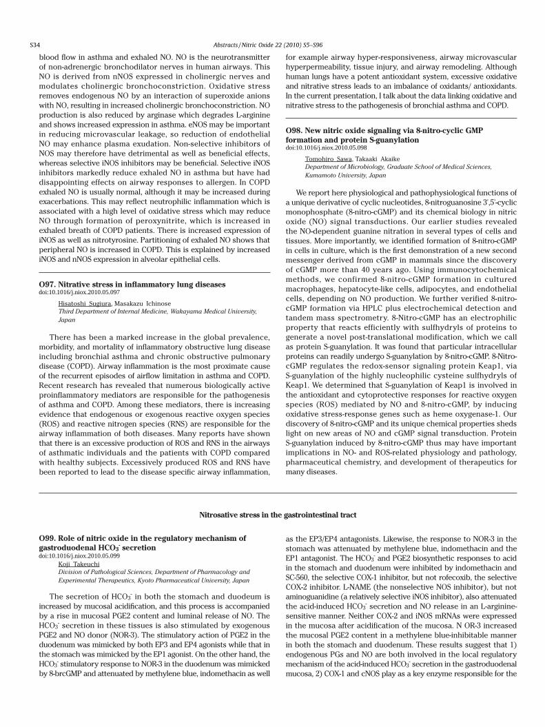
Nitrosative stress in the gastrointestinal tract
O99. Role of nitric oxide in the regulatory mechanism of gastroduodenal HCO3
- secretion
Koji Takeuchi Division of Pathological Sciences, Department of Pharmacology and Experimental Therapeutics, Kyoto Pharmaceutical University, Japan
The secretion of HCO3- in both the stomach and duodeum is
increased by mucosal acidification, and this process is accompanied by a rise in mucosal PGE2 content and luminal release of NO. The HCO3
- secretion in these tissues is also stimulated by exogenous PGE2 and NO donor (NOR-3). The stimulatory action of PGE2 in the duodenum was mimicked by both EP3 and EP4 agonists while that in the stomach was mimicked by the EP1 agonist. On the other hand, the HCO3
- stimulatory response to NOR-3 in the duodenum was mimicked by 8-brcGMP and attenuated by methylene blue, indomethacin as well
as the EP3/EP4 antagonists. Likewise, the response to NOR-3 in the stomach was attenuated by methylene blue, indomethacin and the EP1 antagonist. The HCO3
- and PGE2 biosynthetic responses to acid in the stomach and duodenum were inhibited by indomethacin and SC-560, the selective COX-1 inhibitor, but not rofecoxib, the selective COX-2 inhibitor. L-NAME (the nonselective NOS inhibitor), but not aminoguanidine (a relatively selective iNOS inhibitor), also attenuated the acid-induced HCO3
- secretion and NO release in an L-arginine-sensitive manner. Neither COX-2 and iNOS mRNAs were expressed in the mucosa after acidification of the mucosa. N OR-3 increased the mucosal PGE2 content in a methylene blue-inhibitable manner in both the stomach and duodenum. These results suggest that 1) endogenous PGs and NO are both involved in the local regulatory mechanism of the acid-induced HCO3
- secretion in the gastroduodenal mucosa, 2) COX-1 and cNOS play as a key enzyme responsible for the
blood flow in asthma and exhaled NO. NO is the neurotransmitter of non-adrenergic bronchodilator nerves in human airways. This NO is derived from nNOS expressed in cholinergic nerves and modulates cholinergic bronchoconstriction. Oxidative stress removes endogenous NO by an interaction of superoxide anions with NO, resulting in increased cholinergic bronchoconstriction. NO production is also reduced by arginase which degrades L-arginine and shows increased expression in asthma. eNOS may be important in reducing microvascular leakage, so reduction of endothelial NO may enhance plasma exudation. Non-selective inhibitors of NOS may therefore have detrimental as well as beneficial effects, whereas selective iNOS inhibitors may be beneficial. Selective iNOS inhibitors markedly reduce exhaled NO in asthma but have had disappointing effects on airway responses to allergen. In COPD exhaled NO is usually normal, although it may be increased during exacerbations. This may reflect neutrophilic inflammation which is associated with a high level of oxidative stress which may reduce NO through formation of peroxynitrite, which is increased in exhaled breath of COPD patients. There is increased expression of iNOS as well as nitrotyrosine. Partitioning of exhaled NO shows that peripheral NO is increased in COPD. This is explained by increased iNOS and nNOS expression in alveolar epithelial cells.
O97. Nitrative stress in inflammatory lung diseases
Hisatoshi Sugiura, Masakazu Ichinose Third Department of Internal Medicine, Wakayama Medical University, Japan
There has been a marked increase in the global prevalence, morbidity, and mortality of inflammatory obstructive lung disease including bronchial asthma and chronic obstructive pulmonary disease (COPD). Airway inflammation is the most proximate cause of the recurrent episodes of airflow limitation in asthma and COPD. Recent research has revealed that numerous biologically active proinflammatory mediators are responsible for the pathogenesis of asthma and COPD. Among these mediators, there is increasing evidence that endogenous or exogenous reactive oxygen species (ROS) and reactive nitrogen species (RNS) are responsible for the airway inflammation of both diseases. Many reports have shown that there is an excessive production of ROS and RNS in the airways of asthmatic individuals and the patients with COPD compared with healthy subjects. Excessively produced ROS and RNS have been reported to lead to the disease specific airway inflammation,
for example airway hyper-responsiveness, airway microvascular hyperpermeability, tissue injury, and airway remodeling. Although human lungs have a potent antioxidant system, excessive oxidative and nitrative stress leads to an imbalance of oxidants/ antioxidants. In the current presentation, I talk about the data linking oxidative and nitrative stress to the pathogenesis of bronchial asthma and COPD.
O98. New nitric oxide signaling via 8-nitro-cyclic GMP formation and protein S-guanylation
Tomohiro Sawa, Takaaki Akaike Department of Microbiology, Graduate School of Medical Sciences, Kumamoto University, Japan
We report here physiological and pathophysiological functions of a unique derivative of cyclic nucleotides, 8-nitroguanosine 3',5'-cyclic monophosphate (8-nitro-cGMP) and its chemical biology in nitric oxide (NO) signal transductions. Our earlier studies revealed the NO-dependent guanine nitration in several types of cells and tissues. More importantly, we identified formation of 8-nitro-cGMP in cells in culture, which is the first demonstration of a new second messenger derived from cGMP in mammals since the discovery of cGMP more than 40 years ago. Using immunocytochemical methods, we confirmed 8-nitro-cGMP formation in cultured macrophages, hepatocyte-like cells, adipocytes, and endothelial cells, depending on NO production. We further verified 8-nitro-cGMP formation via HPLC plus electrochemical detection and tandem mass spectrometry. 8-Nitro-cGMP has an electrophilic property that reacts efficiently with sulfhydryls of proteins to generate a novel post-translational modification, which we call as protein S-guanylation. It was found that particular intracellular proteins can readily undergo S-guanylation by 8-nitro-cGMP. 8-Nitro-cGMP regulates the redox-sensor signaling protein Keap1, via S-guanylation of the highly nucleophilic cysteine sulfhydryls of Keap1. We determined that S-guanylation of Keap1 is involved in the antioxidant and cytoprotective responses for reactive oxygen species (ROS) mediated by NO and 8-nitro-cGMP, by inducing oxidative stress-response genes such as heme oxygenase-1. Our discovery of 8-nitro-cGMP and its unique chemical properties sheds light on new areas of NO and cGMP signal transduction. Protein S-guanylation induced by 8-nitro-cGMP thus may have important implications in NO- and ROS-related physiology and pathology, pharmaceutical chemistry, and development of therapeutics for many diseases.
production of PGs and NO, respectively, during acidification, 3) NO released locally in response to acid increases HCO3
- secretion, at least partly, by stimulating PG generation, in a cGMP-dependent manner, 4) NO stimulates HCO3
- secretion mediated by PGE2 through the activation of EP1 receptors in the stomach and EP3/EP4 receptors in the duodenum.
O100. Nitric oxide involvement in gastric disorders
Hidekazu Suzuki, Sachiko Suzuki, Juntaro Matsuzaki,Hitoshi Tsugawa, Yoshimasa Saito, Kenro Hirata,Seiichiro Fukuhara, Masaharu Miyazawa, Masayuki Suzuki,Toshifumi Hibi School of Medicine, Keio University, Japan
The stomach is one of the most susceptible digestive organs to oxidative and nitrasative stress. In the setting of H. pylori infection (J. Gastroenterol. 42:1, 2007; Gastric Cancer 12:79, 2009), inducible nitric oxide synthase (iNOS) is thought to participate in not only the inflammatory response, but also in the regulation of gastric mucosal cell turnover. We previously reported that iNOS might play an important role in promoting apoptosis in the H. pylori-infected gastric mucosa, and that persistent inflammation without apoptosis in iNOS knockout mice with H. pylori infection may be linked to preneoplastic transformation (Free Rad. Biol. Med. 34:1621, 2003). Not only gastric mucosal injury, but also alterations of the gastric motility and loss of interstitial cells of Cajal (ICC) with a decrement of neuronal nitric oxide syntase (nNOS) expression have been reported in response to oxidative stress such as ischemia-reperfusion (I/R) (Neurogastroenterol. Motil. 2010). Also in diabetes, which evokes motility disorders known as diabetic gastroparesis, a loss of ICC were observed with the loss of heme oxygenase-1 up-regulation in response to oxidative stress (Gastroenterol. 135:2055-64, 2008). On the other hand, nitric oxide (NO) derived from inorganic nitrate, that could be metabolized to N-nitroso compounds (carcinogenic) or to nitrite and NO (protective). In the present symposium, NO and NOS dynamics in the pathogenesis of oxidative gastric disorders including whole layers of the stomach will be focused.
O101. Nitric oxide-modified proteins in Helicobacter pylori-associated inflamed gastric mucosa
Yuji Naito, Tomohisa Takagi, Hitomi Okada, Kazuhiko Uchiyama,Osamu Handa, Nobuaki Yagi, Satoshi Kokura, Toshikazu Yoshikawa Molecular Gastroenterology and Hepatology, Kyoto Prefectural University of Medicine, Japan
Induction of inducible nitric oxide synthase (iNOS) may be involved in carcinogenesis of the stomach, because nitric oxide (NO) derived from iNOS can exert DNA damage and post-transcriptional modification of target proteins. We previously reported that nitrotyrosine-modified proteins are increased in Helicobacter pylori (H.pylori)-associated inflamed gastric mucosa (J Gastroenterol Hepatol 2008). The expression of iNOS mRNA was significantly increased in patients with severe atrophic gastric mucosa compared with non-atrophic groups, with increased nitration of tyrosine residues of proteins determined by new monoclonal antibodies against nitro-tyrosine. In the present study, We analyzed using the non-inflamed gastric mucosa (H.pylori-negative) and the inflamed gastric mucosa (H.pylori-positive). Proteins extracted from the gastric mucosa were separated by SDS-PAGE, transferred to nitrocellulose membranes. Nitrotyrosine-modified proteins were detected using Western blotting with anti-3-nitrotyrosine antibody. To examine the existence of peroxiredoxin-6 (Prx-6) in the nitrotyrosine-modified proteins, the membrane, which was treated with anti-3-nitrotyrosine antibody, was stripped and reblotted with anti-Prx-6 antibody. The protein band which appeared in the molecular mass regions between 25 and 30 kDa was detected with both anti-3-nitrotyrosine antibody and anti-Prx-6 antibody. To collect the nitrotyrosine-modified proteins from the inflamed gastric mucosa, we performed immunoprecipitation with anti-3-nitrotyrosine antibody. Immunoprecipitated proteins were subjected to Western blotting with anti-Prx-6 antibody. The result shows Prx-6 was contained in the immunoprecipitated proteins. These results indicate that Prx-6 is modified by NO in the inflamed gastric mucosa.
Importance of ARB in cardiovascular medicine
O102. Next Generation Multifunctional ARBs: Beyond the Renin Angiotensin System
Theodore W Kurtz Laboratory Medicine, University of California, San Francisco, United States of America
Although inhibition of the renin angiotensin system (RAS) has proven useful for treatment of high blood pressure and reducing risk for cardiovascular (CV) disease, many patients continue to suffer from CV events despite taking an ARB, an angiotensin converting enzyme inhibitor (ACEi), or both on top of standard therapies for hypertension and dyslipidemia. In addition, many hypertensive patients are known to suffer from the metabolic syndrome which promotes diabetes and further increases the risk for CV disease. Accordingly, there is mounting interest in the identification of better antihypertensive drugs that do more than just inhibit the renin-angiotensin system and lower blood pressure. Recent studies have suggested that some ARBs may have an unusual ability to affect potential mechanisms of hypertension, diabetes, and cardiovascular disease beyond those involving activation of the renin angiotensin system. New ARBs are being developed that can also function as
nitric oxide donors, neprilysin inhibitors, PPAR gamma activators, or endothelin receptor antagonists. This presentation will: 1) address the potential importance of multi-functional ARBs that may reduce cardiovascular, renal, and metabolic risk through multiple mechanisms that go beyond just inhibition of the renin-angiotensin system; 2) discuss functional differences that exist among different ARBs within this broad class of compounds; and 3) introduce four classes of next generation ARBs intended to do more than simply inhibit the renin-angiotensin system and lower blood pressure.
O103. Strategy to prevent cardiovascular events in patients with metabolic syndrome: Insights from atherosclerosis research and hypertension management
Masataka Sata Department of Cardiovascular medicine, The University of Tokushima Graduate School, Japan
Recent evidence suggests that acute coronary syndrome (ACS) results from plaque rupture in most of the cases. Vulnerable plaques are characterized by thinning of fibrous cap, increased lipid content,
S34
doi:10.1016/j.niox.2010.05.097
doi:10.1016/j.niox.2010.05.098
doi:10.1016/j.niox.2010.05.099
Abstracts /Nitric Oxide 22 (2010) S5–S96

Nitrosative stress in the gastrointestinal tract
O99. Role of nitric oxide in the regulatory mechanism of gastroduodenal HCO3
- secretion
Koji Takeuchi Division of Pathological Sciences, Department of Pharmacology and Experimental Therapeutics, Kyoto Pharmaceutical University, Japan
The secretion of HCO3- in both the stomach and duodeum is
increased by mucosal acidification, and this process is accompanied by a rise in mucosal PGE2 content and luminal release of NO. The HCO3
- secretion in these tissues is also stimulated by exogenous PGE2 and NO donor (NOR-3). The stimulatory action of PGE2 in the duodenum was mimicked by both EP3 and EP4 agonists while that in the stomach was mimicked by the EP1 agonist. On the other hand, the HCO3
- stimulatory response to NOR-3 in the duodenum was mimicked by 8-brcGMP and attenuated by methylene blue, indomethacin as well
as the EP3/EP4 antagonists. Likewise, the response to NOR-3 in the stomach was attenuated by methylene blue, indomethacin and the EP1 antagonist. The HCO3
- and PGE2 biosynthetic responses to acid in the stomach and duodenum were inhibited by indomethacin and SC-560, the selective COX-1 inhibitor, but not rofecoxib, the selective COX-2 inhibitor. L-NAME (the nonselective NOS inhibitor), but not aminoguanidine (a relatively selective iNOS inhibitor), also attenuated the acid-induced HCO3
- secretion and NO release in an L-arginine-sensitive manner. Neither COX-2 and iNOS mRNAs were expressed in the mucosa after acidification of the mucosa. N OR-3 increased the mucosal PGE2 content in a methylene blue-inhibitable manner in both the stomach and duodenum. These results suggest that 1) endogenous PGs and NO are both involved in the local regulatory mechanism of the acid-induced HCO3
- secretion in the gastroduodenal mucosa, 2) COX-1 and cNOS play as a key enzyme responsible for the
blood flow in asthma and exhaled NO. NO is the neurotransmitter of non-adrenergic bronchodilator nerves in human airways. This NO is derived from nNOS expressed in cholinergic nerves and modulates cholinergic bronchoconstriction. Oxidative stress removes endogenous NO by an interaction of superoxide anions with NO, resulting in increased cholinergic bronchoconstriction. NO production is also reduced by arginase which degrades L-arginine and shows increased expression in asthma. eNOS may be important in reducing microvascular leakage, so reduction of endothelial NO may enhance plasma exudation. Non-selective inhibitors of NOS may therefore have detrimental as well as beneficial effects, whereas selective iNOS inhibitors may be beneficial. Selective iNOS inhibitors markedly reduce exhaled NO in asthma but have had disappointing effects on airway responses to allergen. In COPD exhaled NO is usually normal, although it may be increased during exacerbations. This may reflect neutrophilic inflammation which is associated with a high level of oxidative stress which may reduce NO through formation of peroxynitrite, which is increased in exhaled breath of COPD patients. There is increased expression of iNOS as well as nitrotyrosine. Partitioning of exhaled NO shows that peripheral NO is increased in COPD. This is explained by increased iNOS and nNOS expression in alveolar epithelial cells.
O97. Nitrative stress in inflammatory lung diseases
Hisatoshi Sugiura, Masakazu Ichinose Third Department of Internal Medicine, Wakayama Medical University, Japan
There has been a marked increase in the global prevalence, morbidity, and mortality of inflammatory obstructive lung disease including bronchial asthma and chronic obstructive pulmonary disease (COPD). Airway inflammation is the most proximate cause of the recurrent episodes of airflow limitation in asthma and COPD. Recent research has revealed that numerous biologically active proinflammatory mediators are responsible for the pathogenesis of asthma and COPD. Among these mediators, there is increasing evidence that endogenous or exogenous reactive oxygen species (ROS) and reactive nitrogen species (RNS) are responsible for the airway inflammation of both diseases. Many reports have shown that there is an excessive production of ROS and RNS in the airways of asthmatic individuals and the patients with COPD compared with healthy subjects. Excessively produced ROS and RNS have been reported to lead to the disease specific airway inflammation,
for example airway hyper-responsiveness, airway microvascular hyperpermeability, tissue injury, and airway remodeling. Although human lungs have a potent antioxidant system, excessive oxidative and nitrative stress leads to an imbalance of oxidants/ antioxidants. In the current presentation, I talk about the data linking oxidative and nitrative stress to the pathogenesis of bronchial asthma and COPD.
O98. New nitric oxide signaling via 8-nitro-cyclic GMP formation and protein S-guanylation
Tomohiro Sawa, Takaaki Akaike Department of Microbiology, Graduate School of Medical Sciences, Kumamoto University, Japan
We report here physiological and pathophysiological functions of a unique derivative of cyclic nucleotides, 8-nitroguanosine 3',5'-cyclic monophosphate (8-nitro-cGMP) and its chemical biology in nitric oxide (NO) signal transductions. Our earlier studies revealed the NO-dependent guanine nitration in several types of cells and tissues. More importantly, we identified formation of 8-nitro-cGMP in cells in culture, which is the first demonstration of a new second messenger derived from cGMP in mammals since the discovery of cGMP more than 40 years ago. Using immunocytochemical methods, we confirmed 8-nitro-cGMP formation in cultured macrophages, hepatocyte-like cells, adipocytes, and endothelial cells, depending on NO production. We further verified 8-nitro-cGMP formation via HPLC plus electrochemical detection and tandem mass spectrometry. 8-Nitro-cGMP has an electrophilic property that reacts efficiently with sulfhydryls of proteins to generate a novel post-translational modification, which we call as protein S-guanylation. It was found that particular intracellular proteins can readily undergo S-guanylation by 8-nitro-cGMP. 8-Nitro-cGMP regulates the redox-sensor signaling protein Keap1, via S-guanylation of the highly nucleophilic cysteine sulfhydryls of Keap1. We determined that S-guanylation of Keap1 is involved in the antioxidant and cytoprotective responses for reactive oxygen species (ROS) mediated by NO and 8-nitro-cGMP, by inducing oxidative stress-response genes such as heme oxygenase-1. Our discovery of 8-nitro-cGMP and its unique chemical properties sheds light on new areas of NO and cGMP signal transduction. Protein S-guanylation induced by 8-nitro-cGMP thus may have important implications in NO- and ROS-related physiology and pathology, pharmaceutical chemistry, and development of therapeutics for many diseases.
production of PGs and NO, respectively, during acidification, 3) NO released locally in response to acid increases HCO3
- secretion, at least partly, by stimulating PG generation, in a cGMP-dependent manner, 4) NO stimulates HCO3
- secretion mediated by PGE2 through the activation of EP1 receptors in the stomach and EP3/EP4 receptors in the duodenum.
O100. Nitric oxide involvement in gastric disorders
Hidekazu Suzuki, Sachiko Suzuki, Juntaro Matsuzaki,Hitoshi Tsugawa, Yoshimasa Saito, Kenro Hirata,Seiichiro Fukuhara, Masaharu Miyazawa, Masayuki Suzuki,Toshifumi Hibi School of Medicine, Keio University, Japan
The stomach is one of the most susceptible digestive organs to oxidative and nitrasative stress. In the setting of H. pylori infection (J. Gastroenterol. 42:1, 2007; Gastric Cancer 12:79, 2009), inducible nitric oxide synthase (iNOS) is thought to participate in not only the inflammatory response, but also in the regulation of gastric mucosal cell turnover. We previously reported that iNOS might play an important role in promoting apoptosis in the H. pylori-infected gastric mucosa, and that persistent inflammation without apoptosis in iNOS knockout mice with H. pylori infection may be linked to preneoplastic transformation (Free Rad. Biol. Med. 34:1621, 2003). Not only gastric mucosal injury, but also alterations of the gastric motility and loss of interstitial cells of Cajal (ICC) with a decrement of neuronal nitric oxide syntase (nNOS) expression have been reported in response to oxidative stress such as ischemia-reperfusion (I/R) (Neurogastroenterol. Motil. 2010). Also in diabetes, which evokes motility disorders known as diabetic gastroparesis, a loss of ICC were observed with the loss of heme oxygenase-1 up-regulation in response to oxidative stress (Gastroenterol. 135:2055-64, 2008). On the other hand, nitric oxide (NO) derived from inorganic nitrate, that could be metabolized to N-nitroso compounds (carcinogenic) or to nitrite and NO (protective). In the present symposium, NO and NOS dynamics in the pathogenesis of oxidative gastric disorders including whole layers of the stomach will be focused.
O101. Nitric oxide-modified proteins in Helicobacter pylori-associated inflamed gastric mucosa
Yuji Naito, Tomohisa Takagi, Hitomi Okada, Kazuhiko Uchiyama,Osamu Handa, Nobuaki Yagi, Satoshi Kokura, Toshikazu Yoshikawa Molecular Gastroenterology and Hepatology, Kyoto Prefectural University of Medicine, Japan
Induction of inducible nitric oxide synthase (iNOS) may be involved in carcinogenesis of the stomach, because nitric oxide (NO) derived from iNOS can exert DNA damage and post-transcriptional modification of target proteins. We previously reported that nitrotyrosine-modified proteins are increased in Helicobacter pylori (H.pylori)-associated inflamed gastric mucosa (J Gastroenterol Hepatol 2008). The expression of iNOS mRNA was significantly increased in patients with severe atrophic gastric mucosa compared with non-atrophic groups, with increased nitration of tyrosine residues of proteins determined by new monoclonal antibodies against nitro-tyrosine. In the present study, We analyzed using the non-inflamed gastric mucosa (H.pylori-negative) and the inflamed gastric mucosa (H.pylori-positive). Proteins extracted from the gastric mucosa were separated by SDS-PAGE, transferred to nitrocellulose membranes. Nitrotyrosine-modified proteins were detected using Western blotting with anti-3-nitrotyrosine antibody. To examine the existence of peroxiredoxin-6 (Prx-6) in the nitrotyrosine-modified proteins, the membrane, which was treated with anti-3-nitrotyrosine antibody, was stripped and reblotted with anti-Prx-6 antibody. The protein band which appeared in the molecular mass regions between 25 and 30 kDa was detected with both anti-3-nitrotyrosine antibody and anti-Prx-6 antibody. To collect the nitrotyrosine-modified proteins from the inflamed gastric mucosa, we performed immunoprecipitation with anti-3-nitrotyrosine antibody. Immunoprecipitated proteins were subjected to Western blotting with anti-Prx-6 antibody. The result shows Prx-6 was contained in the immunoprecipitated proteins. These results indicate that Prx-6 is modified by NO in the inflamed gastric mucosa.
Importance of ARB in cardiovascular medicine
O102. Next Generation Multifunctional ARBs: Beyond the Renin Angiotensin System
Theodore W Kurtz Laboratory Medicine, University of California, San Francisco, United States of America
Although inhibition of the renin angiotensin system (RAS) has proven useful for treatment of high blood pressure and reducing risk for cardiovascular (CV) disease, many patients continue to suffer from CV events despite taking an ARB, an angiotensin converting enzyme inhibitor (ACEi), or both on top of standard therapies for hypertension and dyslipidemia. In addition, many hypertensive patients are known to suffer from the metabolic syndrome which promotes diabetes and further increases the risk for CV disease. Accordingly, there is mounting interest in the identification of better antihypertensive drugs that do more than just inhibit the renin-angiotensin system and lower blood pressure. Recent studies have suggested that some ARBs may have an unusual ability to affect potential mechanisms of hypertension, diabetes, and cardiovascular disease beyond those involving activation of the renin angiotensin system. New ARBs are being developed that can also function as
nitric oxide donors, neprilysin inhibitors, PPAR gamma activators, or endothelin receptor antagonists. This presentation will: 1) address the potential importance of multi-functional ARBs that may reduce cardiovascular, renal, and metabolic risk through multiple mechanisms that go beyond just inhibition of the renin-angiotensin system; 2) discuss functional differences that exist among different ARBs within this broad class of compounds; and 3) introduce four classes of next generation ARBs intended to do more than simply inhibit the renin-angiotensin system and lower blood pressure.
O103. Strategy to prevent cardiovascular events in patients with metabolic syndrome: Insights from atherosclerosis research and hypertension management
Masataka Sata Department of Cardiovascular medicine, The University of Tokushima Graduate School, Japan
Recent evidence suggests that acute coronary syndrome (ACS) results from plaque rupture in most of the cases. Vulnerable plaques are characterized by thinning of fibrous cap, increased lipid content,
S35
doi:10.1016/j.niox.2010.05.100
doi:10.1016/j.niox.2010.05.101
doi:10.1016/j.niox.2010.05.102
doi:10.1016/j.niox.2010.05.103
Abstracts /Nitric Oxide 22 (2010) S5–S96

An Innovative Approach to Cardiovascular Risk Management via Direct Renin Inhibition
O104. Biochemical Aspects of a Direct Renin Inhibitor, Aliskiren: Messages from Renin Investigators
Fumiaki Suzuki Faculty of Applied Biological Sciences, Gifu University, Japan
A direct renin inhibitor, aliskiren, was developed by the structure based drug design by J.M. Wood et al. published in 2003. It is the first clinical available direct human renin inhibitor with the IC50 of 0.6 nM, the molecular weight of 609.8, highly soluble in water and biological fluids, and non-peptide medicine suitable for oral administration. That inhibitor has extremely high specificity and potency to human renin rather than other peptidase and species of renin. Since last October, the medicine has been also launched in Japan. Now, the large clinical trial program, called "ASPIRE HIGHER" for the beneficial effects of aliskiren on cardiovascular and renal desease, is globally in progress with more than 35,000 patients. In this presentation, recent papers related to aliskiren will be reviewed to discuss its benefit for human.
Interestingly, the half-life of aliskiren in the blood circulation after administration in human was reported around 40h that is longer than ohter antihypertensive agents. Aliskiren monotherapy provided the dose-dependent and prolonged reductions in DBP and SBP.
Plasma renin activity (PRA) is well known to be increased after administering ACEI, ARB, CCB and diuretic. Administration of aliskiren has been reported to reduce the PRA from the base line level with or without co-administration of either of them.
(Pro)renin receptor will be introduced as additional topics. G. Nguyen et al. reported the (pro)renin receptor in 2002. This receptor activated prorenin non-proteolytically. We showed more recently that aliskiren inhibited the renin activity of not only receptor-bound renin but also of prorenin bound to the receptor. D. Feldman et al. observed that aliskiren suppressed the glomerular expression of (pro)renin receptor in a rat model of diabetic nephropathy.
All together, aliskiren may contribute on keeping our quality of life at higher levels for longer times.
O105. Inhibition of the Renin Angiotensin System Markedly Reduces Hypercholesterolemia-Induced Atherosclerosis
Alan Daugherty Cardiovascular Research Center, University of Kentucky, United States of America
The renin angiotensin system is being increasingly recognized as a complex system with many bioactive peptides beyond the classically-defined octapeptide, AngII. The complex array of angiotensin peptides and receptors has the potential to exert both pro- and anti-atherogenic roles. The newly developed renin inhibitor, aliskiren, prevents the cleavage of angiotensinogen, is the unique precursor of angiotensin peptides. Therefore, sufficient concentrations of aliskiren leads to decrease in angiotensin peptides. To define the role of aliskiren on experimental atherosclerosis, male LDL receptor -/- mice were administered with either vehicle or aliskiren at doses of 2.5, 25 and 50 mg/kg/day delivered by osmotic minipumps for 3 months. All groups were fed a modified diet for the duration of drug administration. Systolic blood pressure was not significantly reduced at the lower dose of aliskiren, but was consistently reduced by both higher doses of aliskiren. Renin inhibition was not associated with changes in serum total cholesterol concentrations. However, all doses had highly significant reductions in atherosclerosis, with the higher doses nearly ablating the presence of lesions. To determine the location of the renin that promotes atherosclerosis, we performed bone marrow transplantation to generate chimeric mice with renin deficiency in bone marrow derived cells. Surprisingly, deficiency of renin in bone marrow derived cells had a dramatic effect in reducing atherosclerosis. This is consistent with the atherogenic effects of the renin angiotensin system being due to local generation of peptides. Although the mechanisms remains undefined, this and other studies, have demonstrated that renin inhibition is highly effective in reducing experimental atherosclerosis.
decreased smooth muscle cell content, and enhanced infiltration of inflammatory cells. However, the molecular mechanism of plaque destabilization is not fully understood. Thus, there is no established method to predict and prevent ACS. We have been studying the pathogenesis of plaque progression and destabilization, using animal models and clinical specimen. (1) ApoE-deficient mice showed exaggerated atherosclerotic lesions with aging. Accumulation of macrophages in adventitia was first detected prior to plaque formation. Proliferation of vasa vasorum was observed only after atherosclerotic lesion formation. Local delivery of an angiogenic growth factor promoted lesion formation with enhanced neovascularization in the adventitia. These results suggest that vasa vasorum development and inflammatory cell accumulation potentially accelerate plaque progression. (2) It was suggested
that the angiotensin II (Ang II)/Ang II type 1 receptor (AT1R) pathway plays a pivotal role in the pathogenesis of atherosclerosis. Genetic ablation or pharmacological blockade of AT1R led to a significant reduction and stabilization of atherosclerotic lesions. Ang II up-regulated MMP-9 expression in mononuclear cells from AT1R+/+ mice but not those from AT1R-/- mice. Ang II promoted atherosclerosis progression in the BM chimeric mice that had AT1aR in BM, regardless of the absence of AT1aR in the recipient vasculature. Our findings suggest that AT1R expressed not only in vessel wall but also in BM plays an important role in progression and destabilization of atherosclerotic plaque. In this session, I will discuss strategy to prevent cardiovascular events based on recent atherosclerosis research and hypertension management.
Role of NO in the effects of ACE-I/CCBs
O106. Converting Enzyme Inhibitors and Endothelial Dysfunction
Paul M VanhouttePharmacology & Pharmacy, The University of Hong Kong, China
The endothelial cells evoke endothelium-dependent relaxations of the underlying vascular smooth muscle because they release powerful nonprostanoid vasodilator substances. The best known endothelium-derived (ED) relaxing factor (RF) is nitric oxide (NO). Besides releasing NO, the endothelial cells can also cause endothelium-dependent hyperpolarizations (EDHF-mediated relaxation) of the underlying muscle cells. The release of relaxing factors can be trigerred by increases in shear stress, circulating hormones, thrombin, platelet products and autacoids (in particular bradykinin, the local release of which is increased by augmented shear stress) . The release of EDNO can be mediated by both pertussis toxin-sensitive Gi (e.g. serotonin) and insensitive Gq (e.g.bradykinin) G-proteins. However, EDHF-mediated responses do not involve Gi-proteins. In blood vessels lined with regenerated endothelium, there is a selective loss of the pertussis-toxin sensitive mechanisms of EDRF-release which favors the occurence of vasospasm, thrombosis, cellular growth and atherosclerosis. Angiotensin Converting Enzyme (ACE) inhibitors such as perindopril protect bradykinin from breakdown and thus augment the release of both NO and EDHF caused by increases in shear stress and locally produced bradykinin. They also directly interact with bradykinin-receptors to augment their sentivity. These endothelial actions of ACE-inhibitors help to explain why they lower peripheral resistance and arterial blood pressure [in particular in patients with low rennin and angiotensin II levels] and exert protective effects against cardiovascular disease.
O107. Role of Calcium Antagonists in Coronary Artery Disease-In view of their vasoprotective action-
Koichi NodeCardiovascular & Renal Medicine, Saga University, Japan
It has been speculated that vascular endothelial cells are involved in the pathophysiology of coronary spastic angina. More specifically, it has been shown that basal NO production and release are deficient in the coronary arteries of patients with coronary vasospasm, and that vascular contractility increases as a result. In addition, impaired NO production and release increases endogenous endothelin synthesis and may increase the responsiveness of vascular smooth muscle to a variety of vasoconstrictor substances, thereby stimulating vasoconstriction. On the other hand, the involvement of vascular smooth muscle has also been suggested. More specifically, increased local coronary artery contractility is the cause of coronary vasospasm, and both excessive vascular smooth muscle contraction and vascular endothelial dysfunction are considered to be involved in the increase in the contractility.Thus, it is also necessary to select a calcium antagonist that has a vasoprotective effect, because cardiovascular diseases, including angina pectoris, are the consequence of atherosclerosis progression due to oxidative stress and increased inflammatory reactions. We recently found that ADMA (an endogenous NO inhibitor substance) was inhibited in the group treated by Benidipine, a calcium antagonist developed in Japan. We have also previously reported a coronary-flow-increasing action of Benidipine due to NO production in the reduced coronary flow model, focusing our attention on the vasoprotective effect of Benidipine. In this lecture I will report on the usefulness of a calcium antagonist with action of NO production in addition to the pathophysiology of vascular insufficiency as a risk factor for cardiovascular diseases.
Morning Seminars
NO and peripheral artery disease
O108. Differences in the Endothelial Function and Morphological Modulation between the Canine Autogenous Vein and Arterial Grafts Endothelium and Intimal Thickening
Kimihiro Komori Division of Vascular Surgery, Department of Surgery, Nagoya University Graduate School of Medicine, Japan
Late graft failure is still a significant problem for vascular surgeons. Previous paper demonstrated the superiority of the patency in arterial grafts compared to those of vein grafts. In the present study, we demonstrated the differences of the functional and morphological modulation of experimental autogenous vein and arterial grafts.In canine vein grafts, the endothelium of the vein graft was once denuded, and thereafter recovered within 3 or 4 weeks. In contrast, in arterial grafts, the denudation of the endothelium was minimal, and no platelet adherence was observed. Instead, a nearly
normal intact endothelial cell surface had covered the intima within three days after grafting. These histological findings of the arterial grafts were thus quite different from those of the vein grafts.Different responses to flow changes between the vein and arterial grafts were observed. In the vein grafts, the pronounced intimal thickening was associated with the impairment of endothelium responses, while in the arterial grafts, no intimal thickening and intact endothelial function were observed. The intact endothelial function and no intimal thickening under the arterial grafts may explain the improved patency of autogenous arterial grafts in comparison to those of the vein grafts.Finally, we demonstrated that the systemic administration of L- arginin, a precursor of NO, inhibited vein graft intimal thickening with hypercholesterolemia and that poor runoff-induced intimal progression was also suppressed by the direct gene transfer of ecNOS.
S36
doi:10.1016/j.niox.2010.05.104
doi:10.1016/j.niox.2010.05.105
Abstracts /Nitric Oxide 22 (2010) S5–S96

An Innovative Approach to Cardiovascular Risk Management via Direct Renin Inhibition
O104. Biochemical Aspects of a Direct Renin Inhibitor, Aliskiren: Messages from Renin Investigators
Fumiaki Suzuki Faculty of Applied Biological Sciences, Gifu University, Japan
A direct renin inhibitor, aliskiren, was developed by the structure based drug design by J.M. Wood et al. published in 2003. It is the first clinical available direct human renin inhibitor with the IC50 of 0.6 nM, the molecular weight of 609.8, highly soluble in water and biological fluids, and non-peptide medicine suitable for oral administration. That inhibitor has extremely high specificity and potency to human renin rather than other peptidase and species of renin. Since last October, the medicine has been also launched in Japan. Now, the large clinical trial program, called "ASPIRE HIGHER" for the beneficial effects of aliskiren on cardiovascular and renal desease, is globally in progress with more than 35,000 patients. In this presentation, recent papers related to aliskiren will be reviewed to discuss its benefit for human.
Interestingly, the half-life of aliskiren in the blood circulation after administration in human was reported around 40h that is longer than ohter antihypertensive agents. Aliskiren monotherapy provided the dose-dependent and prolonged reductions in DBP and SBP.
Plasma renin activity (PRA) is well known to be increased after administering ACEI, ARB, CCB and diuretic. Administration of aliskiren has been reported to reduce the PRA from the base line level with or without co-administration of either of them.
(Pro)renin receptor will be introduced as additional topics. G. Nguyen et al. reported the (pro)renin receptor in 2002. This receptor activated prorenin non-proteolytically. We showed more recently that aliskiren inhibited the renin activity of not only receptor-bound renin but also of prorenin bound to the receptor. D. Feldman et al. observed that aliskiren suppressed the glomerular expression of (pro)renin receptor in a rat model of diabetic nephropathy.
All together, aliskiren may contribute on keeping our quality of life at higher levels for longer times.
O105. Inhibition of the Renin Angiotensin System Markedly Reduces Hypercholesterolemia-Induced Atherosclerosis
Alan Daugherty Cardiovascular Research Center, University of Kentucky, United States of America
The renin angiotensin system is being increasingly recognized as a complex system with many bioactive peptides beyond the classically-defined octapeptide, AngII. The complex array of angiotensin peptides and receptors has the potential to exert both pro- and anti-atherogenic roles. The newly developed renin inhibitor, aliskiren, prevents the cleavage of angiotensinogen, is the unique precursor of angiotensin peptides. Therefore, sufficient concentrations of aliskiren leads to decrease in angiotensin peptides. To define the role of aliskiren on experimental atherosclerosis, male LDL receptor -/- mice were administered with either vehicle or aliskiren at doses of 2.5, 25 and 50 mg/kg/day delivered by osmotic minipumps for 3 months. All groups were fed a modified diet for the duration of drug administration. Systolic blood pressure was not significantly reduced at the lower dose of aliskiren, but was consistently reduced by both higher doses of aliskiren. Renin inhibition was not associated with changes in serum total cholesterol concentrations. However, all doses had highly significant reductions in atherosclerosis, with the higher doses nearly ablating the presence of lesions. To determine the location of the renin that promotes atherosclerosis, we performed bone marrow transplantation to generate chimeric mice with renin deficiency in bone marrow derived cells. Surprisingly, deficiency of renin in bone marrow derived cells had a dramatic effect in reducing atherosclerosis. This is consistent with the atherogenic effects of the renin angiotensin system being due to local generation of peptides. Although the mechanisms remains undefined, this and other studies, have demonstrated that renin inhibition is highly effective in reducing experimental atherosclerosis.
decreased smooth muscle cell content, and enhanced infiltration of inflammatory cells. However, the molecular mechanism of plaque destabilization is not fully understood. Thus, there is no established method to predict and prevent ACS. We have been studying the pathogenesis of plaque progression and destabilization, using animal models and clinical specimen. (1) ApoE-deficient mice showed exaggerated atherosclerotic lesions with aging. Accumulation of macrophages in adventitia was first detected prior to plaque formation. Proliferation of vasa vasorum was observed only after atherosclerotic lesion formation. Local delivery of an angiogenic growth factor promoted lesion formation with enhanced neovascularization in the adventitia. These results suggest that vasa vasorum development and inflammatory cell accumulation potentially accelerate plaque progression. (2) It was suggested
that the angiotensin II (Ang II)/Ang II type 1 receptor (AT1R) pathway plays a pivotal role in the pathogenesis of atherosclerosis. Genetic ablation or pharmacological blockade of AT1R led to a significant reduction and stabilization of atherosclerotic lesions. Ang II up-regulated MMP-9 expression in mononuclear cells from AT1R+/+ mice but not those from AT1R-/- mice. Ang II promoted atherosclerosis progression in the BM chimeric mice that had AT1aR in BM, regardless of the absence of AT1aR in the recipient vasculature. Our findings suggest that AT1R expressed not only in vessel wall but also in BM plays an important role in progression and destabilization of atherosclerotic plaque. In this session, I will discuss strategy to prevent cardiovascular events based on recent atherosclerosis research and hypertension management.
Role of NO in the effects of ACE-I/CCBs
O106. Converting Enzyme Inhibitors and Endothelial Dysfunction
Paul M VanhouttePharmacology & Pharmacy, The University of Hong Kong, China
The endothelial cells evoke endothelium-dependent relaxations of the underlying vascular smooth muscle because they release powerful nonprostanoid vasodilator substances. The best known endothelium-derived (ED) relaxing factor (RF) is nitric oxide (NO). Besides releasing NO, the endothelial cells can also cause endothelium-dependent hyperpolarizations (EDHF-mediated relaxation) of the underlying muscle cells. The release of relaxing factors can be trigerred by increases in shear stress, circulating hormones, thrombin, platelet products and autacoids (in particular bradykinin, the local release of which is increased by augmented shear stress) . The release of EDNO can be mediated by both pertussis toxin-sensitive Gi (e.g. serotonin) and insensitive Gq (e.g.bradykinin) G-proteins. However, EDHF-mediated responses do not involve Gi-proteins. In blood vessels lined with regenerated endothelium, there is a selective loss of the pertussis-toxin sensitive mechanisms of EDRF-release which favors the occurence of vasospasm, thrombosis, cellular growth and atherosclerosis. Angiotensin Converting Enzyme (ACE) inhibitors such as perindopril protect bradykinin from breakdown and thus augment the release of both NO and EDHF caused by increases in shear stress and locally produced bradykinin. They also directly interact with bradykinin-receptors to augment their sentivity. These endothelial actions of ACE-inhibitors help to explain why they lower peripheral resistance and arterial blood pressure [in particular in patients with low rennin and angiotensin II levels] and exert protective effects against cardiovascular disease.
O107. Role of Calcium Antagonists in Coronary Artery Disease-In view of their vasoprotective action-
Koichi NodeCardiovascular & Renal Medicine, Saga University, Japan
It has been speculated that vascular endothelial cells are involved in the pathophysiology of coronary spastic angina. More specifically, it has been shown that basal NO production and release are deficient in the coronary arteries of patients with coronary vasospasm, and that vascular contractility increases as a result. In addition, impaired NO production and release increases endogenous endothelin synthesis and may increase the responsiveness of vascular smooth muscle to a variety of vasoconstrictor substances, thereby stimulating vasoconstriction. On the other hand, the involvement of vascular smooth muscle has also been suggested. More specifically, increased local coronary artery contractility is the cause of coronary vasospasm, and both excessive vascular smooth muscle contraction and vascular endothelial dysfunction are considered to be involved in the increase in the contractility.Thus, it is also necessary to select a calcium antagonist that has a vasoprotective effect, because cardiovascular diseases, including angina pectoris, are the consequence of atherosclerosis progression due to oxidative stress and increased inflammatory reactions. We recently found that ADMA (an endogenous NO inhibitor substance) was inhibited in the group treated by Benidipine, a calcium antagonist developed in Japan. We have also previously reported a coronary-flow-increasing action of Benidipine due to NO production in the reduced coronary flow model, focusing our attention on the vasoprotective effect of Benidipine. In this lecture I will report on the usefulness of a calcium antagonist with action of NO production in addition to the pathophysiology of vascular insufficiency as a risk factor for cardiovascular diseases.
Morning Seminars
NO and peripheral artery disease
O108. Differences in the Endothelial Function and Morphological Modulation between the Canine Autogenous Vein and Arterial Grafts Endothelium and Intimal Thickening
Kimihiro Komori Division of Vascular Surgery, Department of Surgery, Nagoya University Graduate School of Medicine, Japan
Late graft failure is still a significant problem for vascular surgeons. Previous paper demonstrated the superiority of the patency in arterial grafts compared to those of vein grafts. In the present study, we demonstrated the differences of the functional and morphological modulation of experimental autogenous vein and arterial grafts.In canine vein grafts, the endothelium of the vein graft was once denuded, and thereafter recovered within 3 or 4 weeks. In contrast, in arterial grafts, the denudation of the endothelium was minimal, and no platelet adherence was observed. Instead, a nearly
normal intact endothelial cell surface had covered the intima within three days after grafting. These histological findings of the arterial grafts were thus quite different from those of the vein grafts.Different responses to flow changes between the vein and arterial grafts were observed. In the vein grafts, the pronounced intimal thickening was associated with the impairment of endothelium responses, while in the arterial grafts, no intimal thickening and intact endothelial function were observed. The intact endothelial function and no intimal thickening under the arterial grafts may explain the improved patency of autogenous arterial grafts in comparison to those of the vein grafts.Finally, we demonstrated that the systemic administration of L- arginin, a precursor of NO, inhibited vein graft intimal thickening with hypercholesterolemia and that poor runoff-induced intimal progression was also suppressed by the direct gene transfer of ecNOS.
S37
doi:10.1016/j.niox.2010.05.106 doi:10.1016/j.niox.2010.05.107
doi:10.1016/j.niox.2010.05.108
Abstracts /Nitric Oxide 22 (2010) S5–S96

NO and nitrates
O109. Nitrate tolerance and endothelial hyperpolarization
Takeo Itoh Graduate School of Medical Sciences / Pharmacology, Nagoya City University, Japan
Organic nitrate is widely used in the management of such cardiovascular diseases as angina pectoris, acute myocardial infarction and congestive heart failure. Despite their beneficial hemodynamic and anti-ischemic effects, the usefulness of organic nitrates is limited by tolerance, which develops shortly after treatment starts. Furthermore, long-term nitroglycerine (NTG) therapy causes endothelial dysfunction in human coronary artery (Caramori et al., 1998; Gori et al., 2001). Endothelial cells induce relaxation through a release of vascular relaxing factors, such as nitric oxide (NO), prostacyclin and endothelial-derived hyperpolarizing factors (EDHFs). It was found that three days in vivo administration
of NTG induces dysfunction of endothelium-derived NO in rat and rabbit aorta (Munzel et al., 1995; Laursen et al., 1996; Berkenboom et al., 1999) and the synthesis of prostacyclin in rabbit aorta (Hink et al., 2003). We found that long-term in vivo administration of NTG downregulates the function of EDHF in rabbit mesenteric and coronary arteries. These results suggest that the function of most of endothelium-derived vasorelaxing factors in arteries undergo downregulation following chronic NTG administration.
It is suggested that endothelial cell hyperpolarization plays an essential role in the function of EDHF in some arteries. We recently found that chronic administered NTG in vivo downregulates acetylcholine-induced endothelial cell hyperpolarization under increased superoxide production in rabbit aortic valve. Here, I introduce the recent understanding of the mechanism underlying the dysfunction of endothelial cell hyperpolarization caused by long-term in vivo NTG administration.
NO and cardiovascular regulation
O110. Imbalance of central nitric oxide and reactive oxygen species in regulation of sympathetic nervous system activity as neural mechanisms of hypertension
Yoshitaka Hirooka Cardiovascular Medicine, Kyushu University, Japan
A balance of nitric oxide (NO) availability and reactive oxygen species (ROS) generation is a key factor in the pathogenesis of hypertension and target organ damage, beginning in the earliest stages. Considerable evidence indicates that the pivotal role of this imbalance in the pathogenesis of hypertension. It remains unclear, however, whether oxidative stress in the brain, particularly the autonomic nuclei (including the vasomotor center), has an important role in the occurrence and maintenance of hypertension
via activation of the sympathetic nervous system. A series of our studies using an in vivo gene transfer technique in conscious animals demonstrates that reduced NO availability and increased ROS generation leading to oxidative stress within the brain, particularly in the vasomotor centers. Correction of this imbalance decreases blood pressure and heart rate through the inhibition of the sympathetic nervous system in hypertensive rats. Activation of NAD(P)H oxidase is the major source of ROS generation in the brain, and it is also related to mitochondrial ROS generation. Interestingly, angiotensin receptor blockers and some other cardiovascular drugs have the beneficial effect on this imbalance. I will show the contribution of oxidative stress in the brain to the neural mechanisms that underlie hypertension, and discuss evidence that brain oxidative stress is a potential therapeutic target.
Luncheon Seminars
Reactive oxygen and steroid-resistance
O111. Oxidative stress and corticosteroid resistance
Peter John BARNES National Heart & Lung Institiute, Imperial College London, United Kingdom of Great Britain and Northern Ireland
Oxidative stress is increased in COPD as a result of cigarette smoking and increased numbers and activation of neutrophils and macrophages. Oxidative stress reduces the nuclear enzyme histone deacetylase(HDAC)2, which switches off activated inflammatory genes and mediates the antiinflammatory effects of corticosteroids. Steroids bind to glucocorticoid receptors (GR) which interact with coactivators that result in histone acetylation and recruit HDAC2 to the inflammatory gene complex. GR are acetylated after binding of steroids and bind to DNA to activate steroid-sensitive genes. However, most anti-inflammatory effects of steroids are mediated via suppression of proinflammatory transcription factors, such as NF-kB, and this involves deacetylation of acetylated GR by HDAC2. In COPD HDAC2 is reduced by oxidative stress so that acetylated
GR are increased and this reduces the ability of steroids to suppress NF-kB. There are two mechanisms whereby oxidative stress reduces HDAC2. The first involves interaction of superoxide anions and NO to form peroxynitrite, which nitrates tyrosine residues on HDAC2. Nitration of Tyr146 interferes with its catalytic activity, whereas Tyr253 nitration results in ubiquitination and destruction by the proteasome. The second involves activation of PI3 kinase-delta, which results in eventual phosphorylation of HDAC2 and its ubiquitination. The reduction in HDAC2 by oxidative stress may be reversed by low concentrations of theophylline, which we have shown to potently inhibit PI3Kd under conditions of oxidative stress. Similar effects are seen with selective PI3Kd-inhibitors and both treatments reverse corticosteroid resistance in COPD. This has been confirmed in smoking mice in vivo and in pilot studies of COPD patients treated with low doses of theophylline, Other drugs including nortriptyline, macrolides and Nrf2 activators (by increasing endogenous antioxidants) are also able to reverse oxidative stress-induced corticosteroid resistance.
NO and myocardial protection
O112. How to mediate cardioprotection in the ischemic heart -Lesson from basic research and clinical medicine-
Masafumi Kitakaze Cardiovascular Division of Medicine, National Cardiovascular Center, Japan
Chronic left ventricular function is believed to be a good predictor for cardiovascular events in patients with myocardial infarction. However, in PCI era, it remains unknown whether an improvement of EF between acute and chronic phases predicts less cardiovascular events after acute myocardial infarction (AMI). To clarify this issue, we investigated both EF improvement (the difference in EF between acute and chronic phases) and cardiovascular events in 577 AMI patients who underwent left veutriculography at both phases in J-WIND trial (Lancet 370:1483, 2007). In these AMI patients, EF was 41.9 ± 7.7 % before reperfusion therapy and increased to 44.1 ± 8.6 % at 7.1 ± 2.4 months after AMI. The areas under the receiver-operator characteristic curve with
the EF improvement to predict cardiovascular events was 0.76, and the EF improvement value of -5.4 provided the optimum cutoff, with sensitivity of 76.4 % and specificity of 88.3 %, suggesting that the EF improvement can predict cardiovascular events in AMI patients. Furthermore, multivariate regression analysis revealed that the factors associated with the EF improvement were AUC of CK, baseline EF, chronic oral nicorandil treatment, and hypertension. Among these 4 factors, oral nicorandil treatment was only an operable factor after AMI. Finally we evaluated the effect of oral nicorandil treatment on the EF improvement. The EF improvement was 3.39 % in patients with oral nicorandil treatment and 1.80 % in patients without nicorandil treatment. Moreover, we observed that oral nicorandil treatment enhances the EF improvement regardless of baseline EF and infarct size. These results suggest that an improvement of EF between acute and chronic phases in AMI patients is a good predictor for cardiovascular events, and oral nicorandil treatment provides beneficial impacts on EF improvement.
Recent progress in the treatment of PH
O113. Recent progress in the treatment of pulmonary hypertension
Hiroaki Shimokawa Department of Cardiovascular Medicine, Tohoku University Graduate School of Medicine, Japan
Pulmonary arterial hypertension (PAH) is a disease with poor prognosis characterized by progressive elevation of pulmonary arterial pressure and vascular resistance due to pulmonary artery hyperconstriction and remodeling. However, the precise mechanisms of PAH still remain to be elucidated. Although anticoagulant agents, vasodilators (e.g. prostaglandins, sildenafil, and bosentan), and lung transplantation are currently used for the treatment of PAH, more effective treatment needs to be developed. Rho-kinase causes vascular smooth muscle (VSMC) hyperconstriction and vascular remodeling through inhibition of myosin phosphatase and activation of its downstream effectors. In a series of experimental and clinical studies, we have demonstrated that Rho-kinase-mediated pathway
plays an important role in various cellular functions, not only in VSMC hyperconstriction but also in actin cytoskeleton organization, cell adhesion and motility, cytokinesis, and gene expressions, all of which may be involved in the pathogenesis of PH. We also have recently demonstrated that Rho-kinase is activated in animal models of PAH with different etiologies (monocrotaline and chronic hypoxia) and in patients with PAH. Indeed, we were able to demonstrate that intravenous fasudil, a selective Rho-kinase inhibitor, exerts acute pulmonary vasodilator effects in patients with severe PAH who were refractory to conventional therapies. In addition, we have recently confirmed that in patients with chronic thromboembolic PH (CTEPH), there are various forms of re-canalized pulmonary arteries by optical coherent tomography (OCT) and that percutaneous transluminal angioplasty of pulmonary artery (PTPA) is very effective to improve subjective symptoms and pulmonary hemodynamics. Taken together, our findings indicate that Rho-kinase inhibitors are a promising new class of drugs for PAH and that PTPA is useful for the treatment of CTEPH.
Current therapy for asthma and COPD
O115. Various Effects of Nitric Oxide (NO) on Airway Functions
Hisamichi Aizawa School of Medicine, Kurume University, Japan
NO plays a variety of role in regulating airway pathophysiology. NO relaxes airway smooth muscle as an inhibitory NANC (iNANC) transmitter, and it also acts as an inflammatory mediator. NANC relaxation induced by vagal nerve stimulation during infusion of serotonin can be classified into two components including NO, are involved in the relaxation. Inhibition of NO synthase by L-NAME or blockade of iNANC neurons by hexamethonium significantly increased airway responsiveness. However, addition of L-NAME did not further increase airway responsiveness in animals treated with hexamethonium. In the presence of atropine and
propranolol, inhaled capsaicin caused a marked bronchodilation during serotonin-induced sustained bronchoconstriction. The bronchodilation induced by capsaicin was significantly suppressed by hexamethonium and by L-NAME. These results suggest that NO released from NAN is important in modulating the airway responsiveness.By contrast, NO is known to be inflammatory mediator. In human transformed bronchial epithelial cells in vitro, NO donors increased IL-8 production dose-dependently. In addition, the combination of NO synthase (NOS) inhibitors attenuated the cytokine-induced IL-8 production. In guinea pigs in vivo, ozone exposure induced airway hyperresponsiveness to acetylcholine and increased neutrophils in bronchoalveolar lavage fluid (BALF), and these changes persisted for at least 5 h. Pretreatment with NOS inhibitors had no effect on airway hyperresponsiveness or neutrophil accumulation immediately after ozone, but significantly
S38
doi:10.1016/j.niox.2010.05.109
doi:10.1016/j.niox.2010.05.110
doi:10.1016/j.niox.2010.05.111
Abstracts /Nitric Oxide 22 (2010) S5–S96

NO and nitrates
O109. Nitrate tolerance and endothelial hyperpolarization
Takeo Itoh Graduate School of Medical Sciences / Pharmacology, Nagoya City University, Japan
Organic nitrate is widely used in the management of such cardiovascular diseases as angina pectoris, acute myocardial infarction and congestive heart failure. Despite their beneficial hemodynamic and anti-ischemic effects, the usefulness of organic nitrates is limited by tolerance, which develops shortly after treatment starts. Furthermore, long-term nitroglycerine (NTG) therapy causes endothelial dysfunction in human coronary artery (Caramori et al., 1998; Gori et al., 2001). Endothelial cells induce relaxation through a release of vascular relaxing factors, such as nitric oxide (NO), prostacyclin and endothelial-derived hyperpolarizing factors (EDHFs). It was found that three days in vivo administration
of NTG induces dysfunction of endothelium-derived NO in rat and rabbit aorta (Munzel et al., 1995; Laursen et al., 1996; Berkenboom et al., 1999) and the synthesis of prostacyclin in rabbit aorta (Hink et al., 2003). We found that long-term in vivo administration of NTG downregulates the function of EDHF in rabbit mesenteric and coronary arteries. These results suggest that the function of most of endothelium-derived vasorelaxing factors in arteries undergo downregulation following chronic NTG administration.
It is suggested that endothelial cell hyperpolarization plays an essential role in the function of EDHF in some arteries. We recently found that chronic administered NTG in vivo downregulates acetylcholine-induced endothelial cell hyperpolarization under increased superoxide production in rabbit aortic valve. Here, I introduce the recent understanding of the mechanism underlying the dysfunction of endothelial cell hyperpolarization caused by long-term in vivo NTG administration.
NO and cardiovascular regulation
O110. Imbalance of central nitric oxide and reactive oxygen species in regulation of sympathetic nervous system activity as neural mechanisms of hypertension
Yoshitaka Hirooka Cardiovascular Medicine, Kyushu University, Japan
A balance of nitric oxide (NO) availability and reactive oxygen species (ROS) generation is a key factor in the pathogenesis of hypertension and target organ damage, beginning in the earliest stages. Considerable evidence indicates that the pivotal role of this imbalance in the pathogenesis of hypertension. It remains unclear, however, whether oxidative stress in the brain, particularly the autonomic nuclei (including the vasomotor center), has an important role in the occurrence and maintenance of hypertension
via activation of the sympathetic nervous system. A series of our studies using an in vivo gene transfer technique in conscious animals demonstrates that reduced NO availability and increased ROS generation leading to oxidative stress within the brain, particularly in the vasomotor centers. Correction of this imbalance decreases blood pressure and heart rate through the inhibition of the sympathetic nervous system in hypertensive rats. Activation of NAD(P)H oxidase is the major source of ROS generation in the brain, and it is also related to mitochondrial ROS generation. Interestingly, angiotensin receptor blockers and some other cardiovascular drugs have the beneficial effect on this imbalance. I will show the contribution of oxidative stress in the brain to the neural mechanisms that underlie hypertension, and discuss evidence that brain oxidative stress is a potential therapeutic target.
Luncheon Seminars
Reactive oxygen and steroid-resistance
O111. Oxidative stress and corticosteroid resistance
Peter John BARNES National Heart & Lung Institiute, Imperial College London, United Kingdom of Great Britain and Northern Ireland
Oxidative stress is increased in COPD as a result of cigarette smoking and increased numbers and activation of neutrophils and macrophages. Oxidative stress reduces the nuclear enzyme histone deacetylase(HDAC)2, which switches off activated inflammatory genes and mediates the antiinflammatory effects of corticosteroids. Steroids bind to glucocorticoid receptors (GR) which interact with coactivators that result in histone acetylation and recruit HDAC2 to the inflammatory gene complex. GR are acetylated after binding of steroids and bind to DNA to activate steroid-sensitive genes. However, most anti-inflammatory effects of steroids are mediated via suppression of proinflammatory transcription factors, such as NF-kB, and this involves deacetylation of acetylated GR by HDAC2. In COPD HDAC2 is reduced by oxidative stress so that acetylated
GR are increased and this reduces the ability of steroids to suppress NF-kB. There are two mechanisms whereby oxidative stress reduces HDAC2. The first involves interaction of superoxide anions and NO to form peroxynitrite, which nitrates tyrosine residues on HDAC2. Nitration of Tyr146 interferes with its catalytic activity, whereas Tyr253 nitration results in ubiquitination and destruction by the proteasome. The second involves activation of PI3 kinase-delta, which results in eventual phosphorylation of HDAC2 and its ubiquitination. The reduction in HDAC2 by oxidative stress may be reversed by low concentrations of theophylline, which we have shown to potently inhibit PI3Kd under conditions of oxidative stress. Similar effects are seen with selective PI3Kd-inhibitors and both treatments reverse corticosteroid resistance in COPD. This has been confirmed in smoking mice in vivo and in pilot studies of COPD patients treated with low doses of theophylline, Other drugs including nortriptyline, macrolides and Nrf2 activators (by increasing endogenous antioxidants) are also able to reverse oxidative stress-induced corticosteroid resistance.
NO and myocardial protection
O112. How to mediate cardioprotection in the ischemic heart -Lesson from basic research and clinical medicine-
Masafumi Kitakaze Cardiovascular Division of Medicine, National Cardiovascular Center, Japan
Chronic left ventricular function is believed to be a good predictor for cardiovascular events in patients with myocardial infarction. However, in PCI era, it remains unknown whether an improvement of EF between acute and chronic phases predicts less cardiovascular events after acute myocardial infarction (AMI). To clarify this issue, we investigated both EF improvement (the difference in EF between acute and chronic phases) and cardiovascular events in 577 AMI patients who underwent left veutriculography at both phases in J-WIND trial (Lancet 370:1483, 2007). In these AMI patients, EF was 41.9 ± 7.7 % before reperfusion therapy and increased to 44.1 ± 8.6 % at 7.1 ± 2.4 months after AMI. The areas under the receiver-operator characteristic curve with
the EF improvement to predict cardiovascular events was 0.76, and the EF improvement value of -5.4 provided the optimum cutoff, with sensitivity of 76.4 % and specificity of 88.3 %, suggesting that the EF improvement can predict cardiovascular events in AMI patients. Furthermore, multivariate regression analysis revealed that the factors associated with the EF improvement were AUC of CK, baseline EF, chronic oral nicorandil treatment, and hypertension. Among these 4 factors, oral nicorandil treatment was only an operable factor after AMI. Finally we evaluated the effect of oral nicorandil treatment on the EF improvement. The EF improvement was 3.39 % in patients with oral nicorandil treatment and 1.80 % in patients without nicorandil treatment. Moreover, we observed that oral nicorandil treatment enhances the EF improvement regardless of baseline EF and infarct size. These results suggest that an improvement of EF between acute and chronic phases in AMI patients is a good predictor for cardiovascular events, and oral nicorandil treatment provides beneficial impacts on EF improvement.
Recent progress in the treatment of PH
O113. Recent progress in the treatment of pulmonary hypertension
Hiroaki Shimokawa Department of Cardiovascular Medicine, Tohoku University Graduate School of Medicine, Japan
Pulmonary arterial hypertension (PAH) is a disease with poor prognosis characterized by progressive elevation of pulmonary arterial pressure and vascular resistance due to pulmonary artery hyperconstriction and remodeling. However, the precise mechanisms of PAH still remain to be elucidated. Although anticoagulant agents, vasodilators (e.g. prostaglandins, sildenafil, and bosentan), and lung transplantation are currently used for the treatment of PAH, more effective treatment needs to be developed. Rho-kinase causes vascular smooth muscle (VSMC) hyperconstriction and vascular remodeling through inhibition of myosin phosphatase and activation of its downstream effectors. In a series of experimental and clinical studies, we have demonstrated that Rho-kinase-mediated pathway
plays an important role in various cellular functions, not only in VSMC hyperconstriction but also in actin cytoskeleton organization, cell adhesion and motility, cytokinesis, and gene expressions, all of which may be involved in the pathogenesis of PH. We also have recently demonstrated that Rho-kinase is activated in animal models of PAH with different etiologies (monocrotaline and chronic hypoxia) and in patients with PAH. Indeed, we were able to demonstrate that intravenous fasudil, a selective Rho-kinase inhibitor, exerts acute pulmonary vasodilator effects in patients with severe PAH who were refractory to conventional therapies. In addition, we have recently confirmed that in patients with chronic thromboembolic PH (CTEPH), there are various forms of re-canalized pulmonary arteries by optical coherent tomography (OCT) and that percutaneous transluminal angioplasty of pulmonary artery (PTPA) is very effective to improve subjective symptoms and pulmonary hemodynamics. Taken together, our findings indicate that Rho-kinase inhibitors are a promising new class of drugs for PAH and that PTPA is useful for the treatment of CTEPH.
Current therapy for asthma and COPD
O115. Various Effects of Nitric Oxide (NO) on Airway Functions
Hisamichi Aizawa School of Medicine, Kurume University, Japan
NO plays a variety of role in regulating airway pathophysiology. NO relaxes airway smooth muscle as an inhibitory NANC (iNANC) transmitter, and it also acts as an inflammatory mediator. NANC relaxation induced by vagal nerve stimulation during infusion of serotonin can be classified into two components including NO, are involved in the relaxation. Inhibition of NO synthase by L-NAME or blockade of iNANC neurons by hexamethonium significantly increased airway responsiveness. However, addition of L-NAME did not further increase airway responsiveness in animals treated with hexamethonium. In the presence of atropine and
propranolol, inhaled capsaicin caused a marked bronchodilation during serotonin-induced sustained bronchoconstriction. The bronchodilation induced by capsaicin was significantly suppressed by hexamethonium and by L-NAME. These results suggest that NO released from NAN is important in modulating the airway responsiveness.By contrast, NO is known to be inflammatory mediator. In human transformed bronchial epithelial cells in vitro, NO donors increased IL-8 production dose-dependently. In addition, the combination of NO synthase (NOS) inhibitors attenuated the cytokine-induced IL-8 production. In guinea pigs in vivo, ozone exposure induced airway hyperresponsiveness to acetylcholine and increased neutrophils in bronchoalveolar lavage fluid (BALF), and these changes persisted for at least 5 h. Pretreatment with NOS inhibitors had no effect on airway hyperresponsiveness or neutrophil accumulation immediately after ozone, but significantly
S39
doi:10.1016/j.niox.2010.05.112
doi:10.1016/j.niox.2010.05.113
doi:10.1016/j.niox.2010.05.114
Abstracts /Nitric Oxide 22 (2010) S5–S96

inhibited the changes 5 h after ozone. NOS inhibitors attenuated the increases of nitrite/nitrate levels in BALF and the IL-8 mRNA expression in epithelial cells and in neutrophils in guinea pig airways 5 h after ozone. These results suggest that endogenous NO may play an important role in the persistent airway inflammation and hyperresponsiveness after ozone exposure, presumably partly through the upregulation of IL-8.
O116. Long term management of asthma based on Japanese guidelines, namely JGL2009
Ken Ohta Division of Respiratory Medicine and Allergology, Department of Internal Medicine, Teikyo University, Japan
Bronchial asthma is a syndrome, if you will, characterized by chronic airway inflammation as well as reversible airway obstruction and airway hyperresponsiveness. In addition to inflammation, airway remodeling has been recognized as a result of injury and repair. In treating asthma, we have to realize the complexity of its pathogenesis and existence of various sites to be targeted with a various kinds of anti asthma drugs. According
to the new Japanese guideline JGL2009 based on EBM, inhaled corticosteroids (ICS) are the first line medication to suppress airway inflammation, and various types of drugs such as leukotriene receptor antagonists (LTRA), long acting β2-agonists (LABA), and slow release theophylline (SR-T) are employed as optional or additive medication. CS are known as the most potent anti-inflammatory drug and can inhibit eosinophilic inflammation and fraction of inhaled NO (FeNO) consistently. FeNO seems to be a good marker to distinguish asthma from other respiratory diseases causing chronic cough and/or airflow limitation for diagnosis, but it cannot always reflect the state of control since FeNO is suppressed significantly with CS regardless of the patient's control. Consequently, JGL2009 mentioned FeNO as a diagnostic parameter for recognizing airway inflammation in asthma. JGL2009 made a big revision in terms of steps and the control as a goal of the management. Steps are defined as treatment steps from mild (step1) to strong (step4). The goal of the long term management is to rach the state of control which is defined as no symptoms, no use of relievers, no limit of action in the daily life, normal pulmonary function (PEF and FEV1), normal daily or weekly variation of PEF, and no exacerbation. The national goal is to make asthma deaths zero by implementing JGL2009.
Importance of NO in humans
O117. Endothelial Function, an Emerging Risk Probe for Cardiovascular Diseases
Hidehiro Matsuoka Division of Cardiovascular Medicine Department of Internal Medicine, Kurume University School of Medicine, Japan
Traditional risk factors, such as hypertension, hyperlipidemia, or diabetes can only cover 50% incidence of atherosclerosis. Furthermore, the interventions are most successful if they are applied in the earliest phases of the atherosclerotic process. When the process is advanced, many of those lesions become irreversible. Cardiovascular events, such as stroke and myocardial infarction occur suddenly, in most cases without any warning signs, and lead to lethal and irreversible damages. Thus, the need for early detection of atherosclerosis in subjects at high risk is emerging. The measurement of endothelial function enable us to 1) identify subjects at high risk without traditional risk factors who need aggressive intervention as primary prevention, 2) detect preclinical atherosclerosis at the earliest, i.e. reversible, stage, 3) monitor the efficacy of therapeutic intervention directly in an individual manner. Collectively, the assessment of endothelial function provides us strategies for risk stratification in primary and secondary prevention for cardiovascular diseases. On this occasion, I would like to show you the rationale to evaluate endothelial function and some results of clinical studies conducted in our laboratory. In spite of recent remarkable progress in the field of vascular biology, the attempts for their clinical application are still insignificant. Our series of investigations may be examples providing a research tool which can link vascular biology and clinical practice. In addition, we may be able to manage our patients in a "tailor-made" manner by measuring endothelial function.
O118. Oxidative Stress and NO in Cardiovascular Diseases
Yukihito Higashi Division of Regenerative Medicine/Department of Cardiovascular Physiology, Hiroshima University, Japan
A healthy endothelium maintains vascular tone and structure by regulating the balance between vasodilation and vasoconstriction, growth inhibition and growth promotion, antithrombosis and prothrombosis, anti-inflammation and proinflammation, and also antioxidation and pro-oxidation. Nitric oxide (NO) plays an important role in the regulation of vascular tone, inhibition of platelet aggregation, and suppression of smooth muscle cell proliferation. Impaired endothelium-dependent vasodilation has been found in the forearm, coronary, and renal vasculature in cardiovascular diseases. Endothelial dysfunction is an early feature of atherosclerosis and vascular diseases, resulting in cardiovascular complications. Growing evidence has shown an interaction between oxidative stress and endothelial function. A balance between ambient levels of superoxide and released NO plays a critical role in the maintenance of normal endothelial function. One mechanism by which endothelium-dependent vasodilation is impaired is an increase in the oxidative stress that inactivates NO. Excess production of reactive oxygen species and an attenuated antioxidant system may contribute to endothelial dysfunction in cardiovascular diseases. I will focus on the role of oxidative stress and NO in cardiovascular diseases.
Evening Seminars
Diversities of NO: From enzymes to firefly flashing
O119. Nitric oxide synthases: sex, death, and cardiovascular signal transduction
Thomas Michel Cardiovascular Division, Department of Medicine, Harvard Medical School, Brigham and Women's Hospital, United States of America
Nitric oxide (NO) synthesized by nitric oxide synthases (NOS) or released from NO-donating drugs is a key determinant of vascular responses, including vasorelaxation, platelet aggregation, angiogenesis, penile erection, and endothelial metabolism. NOS-dependent signaling is deranged by oxidative stress and in vascular disease states. The endothelial NOS isoform (eNOS) is regulated by a series of post-translational modifications. Targeting of eNOS to plasmalemmal caveolae is a critical determinant of NO signaling. In resting endothelial cells, eNOS is robustly nitrosylated. Agonist activation of eNOS promotes denitrosylation and translocation of the enzyme from peripheral to internal membranes. Reversible eNOS S-nitrosylation provides a mechanism for the regulation of NO-dependent signaling by redox state. eNOS is phosphorylated by several protein kinases, including the AMP-activated protein kinase (AMPK), a ubiquitous protein kinase that is activated by ADP and by specific AMPK kinases, including the calcium-calmodulin kinase kinase-beta (CaMKK-beta). CaMKK-beta may be activated by reactive oxygen species such as hydrogen peroxide (H2O2). Low levels of H2O2 play signaling roles; higher levels of lead to oxidative stress. siRNA-mediated eNOS knockdown leads to increases in endothelial cell-derived H2O2, which can be detected using sensitive biosensors such as HyPer, associated with the activation of AMPK. AMPK activation is seen in tissues and endothelial cells isolated from eNOS-null mice. The GTPase Rac1 modulates the actin cytoskeleton and is a critical determinant of eNOS. Statins promote Rac1 activation in endothelial cells and in vessels from statin-treated mice. The role of Rac1 in eNOS activation reveals fundamental relationships between caveolae and the cytoskeleton. The interrelated signaling pathways that modulate eNOS have key implications for NO-dependent signaling in vascular disease.
O120. The role of nitric oxide in firefly sexual communication systems
Sara Lewis Department of Biology, Tufts University, United States of America
Nitric oxide (NO) plays diverse roles in invertebrate animals, functioning in insect development, neurotransmission, olfaction, vision, learning, feeding, and immunity. Here we describe a novel biological role for nitric oxide in the regulation of firefly flashing. In many of the ~2000 firefly species worldwide, sexual courtship depends on a remarkable communication system involving the production of precisely-timed bioluminescent flashes by both sexes. These flashes are initiated by neural activity that stimulates release of the neurotransmitter octopamine, and which ultimately leads to light production by the firefly lantern. The luciferin-luciferase reaction is restricted to photocyte peroxisomes, and light production resulting from the degradation of activated luciferin is absolutely dependent on molecular oxygen. Furthermore, the ability to rapidly switch light production on and off appears to
be correlated with distinctive anatomical features in the firefly abdominal lantern. Until recently, however, little was known about the pathways between octopamine release and light production. Several lines of evidence are presented that support a role for NOS in firefly flash control. In the lantern of adult Photuris fireflies, NOS is expressed in tracheal end cells that are interposed between octopaminergic nerve synapses and photocytes. Exogenous NO gas activates light production, while NO scavengers block octopamine-evoked flashes. NO inhibits respiration in isolated lantern mitochondria and this mitochondrial inhibition can be reversed by bright light. A model is proposed in which NO controls flashes by transiently inhibiting mitochondrial oxygen consumption and permitting direct peroxidation of activated luciferin. These results suggest that NOS expression within the firefly lantern may have been a key physiological adaptation that permitted the evolution of sexual signals based on rapidly modulated bioluminescence.
O121. New Insight from Genetically Manipulated Mice Lacking All NOSs
Masato Tsutsuia, Hiroaki Shimokawab, Nobuyuki Yanagiharac,Yutaka Otsujic aDepartment of Pharmacology, Faculty of Medicine, University of the Ryukyus, Japan
bTohoku University Graduate School of Medicine, JapancUniversity of Occupational and Environmental Health, Japan
The nitric oxide synthase (NOS) system consists of three isoforms: nNOS, iNOS, and eNOS. The roles of the NOS system in our body have been examined in pharmacological studies with non-selective NOS inhibitors. However, the NOS inhibitors possess multiple non-specific actions: as we have reported, arteriosclerotic lesion formation by long-term treatment with the NOS inhibitors is not caused by the simple inhibition of NOS activity. The roles of the NOS system in vivo have also been examined in studies with mice that lack each NOS isoform. However, in the singly NOS-/- mice, there appears to be a compensatory mechanism by other NOSs that are not genetically deleted. Indeed, although it is well established that eNOS exerts a potent anti-arteriosclerotic effect, singly eNOS-
/- mice do not manifest arteriosclerosis. Thus, even at the present time when NO research has developed remarkably, the authentic roles of the NOS system are still poorly understood. To address this important issue, we have developed mice lacking all NOS isoforms. Intriguingly, the triply n/i/eNOS-/- mice were viable and appeared normal, whereas their survival and fertility rates were markedly reduced as compared with wild-type mice. The phenotypes of the triply NOS-/- mice that we first noticed were polyuria, polydipsia, and renal unresponsiveness to vasopressin, characteristics consistent with nephrogenic diabetes insipidus. We subsequently revealed that the triply NOS-/- mice spontaneously develop myocardial infarction associated with multiple cardiovascular risk factors of metabolic origin. In addition, we have clarified diastolic heart failure, increased bone turnover, and anxiety-like behavior in the triply mutant mice. These results prove that the complete deletion of all NOS genes causes abnormalities in a variety of systems in mice in vivo, demonstrating a critical role of the whole endogenous NOS system in maintaining body homeostasis.
S40
doi:10.1016/j.niox.2010.05.115
doi:10.1016/j.niox.2010.05.116doi:10.1016/j.niox.2010.05.117
Abstracts /Nitric Oxide 22 (2010) S5–S96

inhibited the changes 5 h after ozone. NOS inhibitors attenuated the increases of nitrite/nitrate levels in BALF and the IL-8 mRNA expression in epithelial cells and in neutrophils in guinea pig airways 5 h after ozone. These results suggest that endogenous NO may play an important role in the persistent airway inflammation and hyperresponsiveness after ozone exposure, presumably partly through the upregulation of IL-8.
O116. Long term management of asthma based on Japanese guidelines, namely JGL2009
Ken Ohta Division of Respiratory Medicine and Allergology, Department of Internal Medicine, Teikyo University, Japan
Bronchial asthma is a syndrome, if you will, characterized by chronic airway inflammation as well as reversible airway obstruction and airway hyperresponsiveness. In addition to inflammation, airway remodeling has been recognized as a result of injury and repair. In treating asthma, we have to realize the complexity of its pathogenesis and existence of various sites to be targeted with a various kinds of anti asthma drugs. According
to the new Japanese guideline JGL2009 based on EBM, inhaled corticosteroids (ICS) are the first line medication to suppress airway inflammation, and various types of drugs such as leukotriene receptor antagonists (LTRA), long acting β2-agonists (LABA), and slow release theophylline (SR-T) are employed as optional or additive medication. CS are known as the most potent anti-inflammatory drug and can inhibit eosinophilic inflammation and fraction of inhaled NO (FeNO) consistently. FeNO seems to be a good marker to distinguish asthma from other respiratory diseases causing chronic cough and/or airflow limitation for diagnosis, but it cannot always reflect the state of control since FeNO is suppressed significantly with CS regardless of the patient's control. Consequently, JGL2009 mentioned FeNO as a diagnostic parameter for recognizing airway inflammation in asthma. JGL2009 made a big revision in terms of steps and the control as a goal of the management. Steps are defined as treatment steps from mild (step1) to strong (step4). The goal of the long term management is to rach the state of control which is defined as no symptoms, no use of relievers, no limit of action in the daily life, normal pulmonary function (PEF and FEV1), normal daily or weekly variation of PEF, and no exacerbation. The national goal is to make asthma deaths zero by implementing JGL2009.
Importance of NO in humans
O117. Endothelial Function, an Emerging Risk Probe for Cardiovascular Diseases
Hidehiro Matsuoka Division of Cardiovascular Medicine Department of Internal Medicine, Kurume University School of Medicine, Japan
Traditional risk factors, such as hypertension, hyperlipidemia, or diabetes can only cover 50% incidence of atherosclerosis. Furthermore, the interventions are most successful if they are applied in the earliest phases of the atherosclerotic process. When the process is advanced, many of those lesions become irreversible. Cardiovascular events, such as stroke and myocardial infarction occur suddenly, in most cases without any warning signs, and lead to lethal and irreversible damages. Thus, the need for early detection of atherosclerosis in subjects at high risk is emerging. The measurement of endothelial function enable us to 1) identify subjects at high risk without traditional risk factors who need aggressive intervention as primary prevention, 2) detect preclinical atherosclerosis at the earliest, i.e. reversible, stage, 3) monitor the efficacy of therapeutic intervention directly in an individual manner. Collectively, the assessment of endothelial function provides us strategies for risk stratification in primary and secondary prevention for cardiovascular diseases. On this occasion, I would like to show you the rationale to evaluate endothelial function and some results of clinical studies conducted in our laboratory. In spite of recent remarkable progress in the field of vascular biology, the attempts for their clinical application are still insignificant. Our series of investigations may be examples providing a research tool which can link vascular biology and clinical practice. In addition, we may be able to manage our patients in a "tailor-made" manner by measuring endothelial function.
O118. Oxidative Stress and NO in Cardiovascular Diseases
Yukihito Higashi Division of Regenerative Medicine/Department of Cardiovascular Physiology, Hiroshima University, Japan
A healthy endothelium maintains vascular tone and structure by regulating the balance between vasodilation and vasoconstriction, growth inhibition and growth promotion, antithrombosis and prothrombosis, anti-inflammation and proinflammation, and also antioxidation and pro-oxidation. Nitric oxide (NO) plays an important role in the regulation of vascular tone, inhibition of platelet aggregation, and suppression of smooth muscle cell proliferation. Impaired endothelium-dependent vasodilation has been found in the forearm, coronary, and renal vasculature in cardiovascular diseases. Endothelial dysfunction is an early feature of atherosclerosis and vascular diseases, resulting in cardiovascular complications. Growing evidence has shown an interaction between oxidative stress and endothelial function. A balance between ambient levels of superoxide and released NO plays a critical role in the maintenance of normal endothelial function. One mechanism by which endothelium-dependent vasodilation is impaired is an increase in the oxidative stress that inactivates NO. Excess production of reactive oxygen species and an attenuated antioxidant system may contribute to endothelial dysfunction in cardiovascular diseases. I will focus on the role of oxidative stress and NO in cardiovascular diseases.
Evening Seminars
Diversities of NO: From enzymes to firefly flashing
O119. Nitric oxide synthases: sex, death, and cardiovascular signal transduction
Thomas Michel Cardiovascular Division, Department of Medicine, Harvard Medical School, Brigham and Women's Hospital, United States of America
Nitric oxide (NO) synthesized by nitric oxide synthases (NOS) or released from NO-donating drugs is a key determinant of vascular responses, including vasorelaxation, platelet aggregation, angiogenesis, penile erection, and endothelial metabolism. NOS-dependent signaling is deranged by oxidative stress and in vascular disease states. The endothelial NOS isoform (eNOS) is regulated by a series of post-translational modifications. Targeting of eNOS to plasmalemmal caveolae is a critical determinant of NO signaling. In resting endothelial cells, eNOS is robustly nitrosylated. Agonist activation of eNOS promotes denitrosylation and translocation of the enzyme from peripheral to internal membranes. Reversible eNOS S-nitrosylation provides a mechanism for the regulation of NO-dependent signaling by redox state. eNOS is phosphorylated by several protein kinases, including the AMP-activated protein kinase (AMPK), a ubiquitous protein kinase that is activated by ADP and by specific AMPK kinases, including the calcium-calmodulin kinase kinase-beta (CaMKK-beta). CaMKK-beta may be activated by reactive oxygen species such as hydrogen peroxide (H2O2). Low levels of H2O2 play signaling roles; higher levels of lead to oxidative stress. siRNA-mediated eNOS knockdown leads to increases in endothelial cell-derived H2O2, which can be detected using sensitive biosensors such as HyPer, associated with the activation of AMPK. AMPK activation is seen in tissues and endothelial cells isolated from eNOS-null mice. The GTPase Rac1 modulates the actin cytoskeleton and is a critical determinant of eNOS. Statins promote Rac1 activation in endothelial cells and in vessels from statin-treated mice. The role of Rac1 in eNOS activation reveals fundamental relationships between caveolae and the cytoskeleton. The interrelated signaling pathways that modulate eNOS have key implications for NO-dependent signaling in vascular disease.
O120. The role of nitric oxide in firefly sexual communication systems
Sara Lewis Department of Biology, Tufts University, United States of America
Nitric oxide (NO) plays diverse roles in invertebrate animals, functioning in insect development, neurotransmission, olfaction, vision, learning, feeding, and immunity. Here we describe a novel biological role for nitric oxide in the regulation of firefly flashing. In many of the ~2000 firefly species worldwide, sexual courtship depends on a remarkable communication system involving the production of precisely-timed bioluminescent flashes by both sexes. These flashes are initiated by neural activity that stimulates release of the neurotransmitter octopamine, and which ultimately leads to light production by the firefly lantern. The luciferin-luciferase reaction is restricted to photocyte peroxisomes, and light production resulting from the degradation of activated luciferin is absolutely dependent on molecular oxygen. Furthermore, the ability to rapidly switch light production on and off appears to
be correlated with distinctive anatomical features in the firefly abdominal lantern. Until recently, however, little was known about the pathways between octopamine release and light production. Several lines of evidence are presented that support a role for NOS in firefly flash control. In the lantern of adult Photuris fireflies, NOS is expressed in tracheal end cells that are interposed between octopaminergic nerve synapses and photocytes. Exogenous NO gas activates light production, while NO scavengers block octopamine-evoked flashes. NO inhibits respiration in isolated lantern mitochondria and this mitochondrial inhibition can be reversed by bright light. A model is proposed in which NO controls flashes by transiently inhibiting mitochondrial oxygen consumption and permitting direct peroxidation of activated luciferin. These results suggest that NOS expression within the firefly lantern may have been a key physiological adaptation that permitted the evolution of sexual signals based on rapidly modulated bioluminescence.
O121. New Insight from Genetically Manipulated Mice Lacking All NOSs
Masato Tsutsuia, Hiroaki Shimokawab, Nobuyuki Yanagiharac,Yutaka Otsujic aDepartment of Pharmacology, Faculty of Medicine, University of the Ryukyus, Japan
bTohoku University Graduate School of Medicine, JapancUniversity of Occupational and Environmental Health, Japan
The nitric oxide synthase (NOS) system consists of three isoforms: nNOS, iNOS, and eNOS. The roles of the NOS system in our body have been examined in pharmacological studies with non-selective NOS inhibitors. However, the NOS inhibitors possess multiple non-specific actions: as we have reported, arteriosclerotic lesion formation by long-term treatment with the NOS inhibitors is not caused by the simple inhibition of NOS activity. The roles of the NOS system in vivo have also been examined in studies with mice that lack each NOS isoform. However, in the singly NOS-/- mice, there appears to be a compensatory mechanism by other NOSs that are not genetically deleted. Indeed, although it is well established that eNOS exerts a potent anti-arteriosclerotic effect, singly eNOS-
/- mice do not manifest arteriosclerosis. Thus, even at the present time when NO research has developed remarkably, the authentic roles of the NOS system are still poorly understood. To address this important issue, we have developed mice lacking all NOS isoforms. Intriguingly, the triply n/i/eNOS-/- mice were viable and appeared normal, whereas their survival and fertility rates were markedly reduced as compared with wild-type mice. The phenotypes of the triply NOS-/- mice that we first noticed were polyuria, polydipsia, and renal unresponsiveness to vasopressin, characteristics consistent with nephrogenic diabetes insipidus. We subsequently revealed that the triply NOS-/- mice spontaneously develop myocardial infarction associated with multiple cardiovascular risk factors of metabolic origin. In addition, we have clarified diastolic heart failure, increased bone turnover, and anxiety-like behavior in the triply mutant mice. These results prove that the complete deletion of all NOS genes causes abnormalities in a variety of systems in mice in vivo, demonstrating a critical role of the whole endogenous NOS system in maintaining body homeostasis.
S41
doi:10.1016/j.niox.2010.05.118
doi:10.1016/j.niox.2010.05.119
doi:10.1016/j.niox.2010.05.120
Abstracts /Nitric Oxide 22 (2010) S5–S96

Statin and plaque stablization
O122. Impact of intensive lipid lowering on stabilization of coronary plaque:Insight from the COSMOS trial
Masakazu Yamagishi Graduate School of Medicine, Kanazawa University, Japan
Although previous Japanese clinical trials demonstrated the effects of lipid lowering on plaque regression in acute coronary syndrome, few data exist regarding the impact of lipid lowering in stable coronary artery disease (CAD). The COSMOS trial was projected to investigate the effect of rosuvastatin (RSV) on plaque regression by IVUS imaging in hypercholesterolemic Japanese patients with stable CAD. The primary endpoint was the percent change in total atheroma volume (TAV) at 76 weeks compared with the baseline volume in 126 patients after treatment with rosuvastatin of 16.9 mg/day. LDL levels decreased by 38.6% and HDL increased by 19.8%, thus resulting in dramatic decrease in LDL/HDL. Under these conditions, TAV significantly reduced. We further investigated the effect of remodeling pattern on plaque regression induced by RSV. Remodeling index (RI) was calculated that the external elastic membrane (EEM) area at the most plaque-rich site was divided by the EEM area at the proximal reference site. The patients were classified into three groups: positive remodeling (PR, n=54) defined as RI ≧1.05, moderate remodeling (MR, n=49) as 0.95≦ RI <1.05 and negative remodeling (NR, n=23) as RI <0.95. Plaque area in PR group was significantly decreased versus MR group (-26.5±20.8%,p=0.02). Furthermore, the average of RI in all patients was significantly decreased (-7.4±14%,p<0.0001), and 40 patients in PR group at baseline changed from PR to MR group at the follow-up. These results elucidated RSV can produce the regressive effect on coronary plaque in Japanese hypercholesterolemic patients with stable CAD. The pathological positive remodeling in plaque was also improved by RSV which would contribute to stabilization of plaque.
O123. The regulatory effect of statin on the coronary plaque from the view point of molecular mechanism
Koichi Node Cardiovascular & Renal Medicine, Saga University, Japan
There is increasing evidence that statins reduce cardiovascular events such as coronar y ar ter y disease or stroke in hypercholesterolemic patients in both primary and secondary prevention. The striking benefit achieved with statin treatments in patients with a wide range of cholesterol levels cannot be attributed to their cholesterol lowering effect alone. Vascular endothelial growth factor (VEGF) is a potent angiogenic factor and plays an important pathophysiological role in the maintenance of tissue structure as well as regeneration after ischemic injury. Statin reduce vascular inflammation and induce angiogenesis. We examined whether statin stimulates VEGF expression in endothelial cells as well as the nature of its underlying mechanism. Statin induced mRNA expression and protein secretion of VEGF in endothelial cells that were reversed by pretreatment with mevalonate and geranylgeranylpyrophosphate but not by farnesylpyrophosphate. Adenovirus-mediated expression of the dominant-negative mutant of RhoA induced VEGF mRNA and protein. Statin increased hypoxia-inducible factor-1alpha (HIF-1alpha) protein level without changing its mRNA expression. Inhibition of RhoA had similar effects to statin on VEGF expression. Statin stimulates VEGF expression by RhoA downregulation and HIF-1alpha upregulation in endothelial cells. These data indicate a novel role for RhoA as a negative regulator of HIF-1alpha. These effects have potential in the treatments of coronary artery disease in various settings, such as prevention of its onset as well as its progression, or plaque rupture.
Poster Abstracts
Chemistry/Imaging of NO and its related molecules
P1. Acid-Promoting Transformation of Roussin's Red Ester [(NO)2Fe(μ-S-NAP)]2 (NAP = N-Acetyl-D-Penicillamine) into S-nitroso-N-Acetyl-D-Penicillamine
Chih-Chin Tsou, Wen-Feng Liaw* Chemistry, National Tsing Hua University, Taiwan
Dissolution of Roussin's red ester (RRE) [(NO)2Fe(μ-S-NAP)]2 (NAP = N-acetyl-D-penicillamine) in DMSO led to the formation of dinitrosyl iron complex (DNIC) [(NO)2Fe(S-NAP)(DMSO)] characterized by EPR. Addition of 2 equiv of tetramethylthiuram disulfide (DTC)2 into [(NO)2Fe(μ-S-NAP)]2 under the acidic DMSO (2,6-lutidine + HBF4) resulted in the formation of mononitrosyl iron complex (MNIC) [(NO)Fe(DTC)2] and S-nitroso-N-acetyl-D-penicillamine (SNAP) characterized by IR, EPR, 15N-NMR and X-ray diffraction, in contrast to reaction of (DTC)2 and [(NO)2Fe(μ-S-NAP)]2 in DMSO leading to the formation of [(NO)Fe(DTC)2], (NAP-S)2 and NO(g). These results signify the possible pathways for producing S-nitrosothiol derived from DNIC in molecular and cellular biology. This biomimetic study may be relevant to the S-nitrosylation of bovine serum albumin, enteroviral protease 2A, caspase-3, proteins in the immortalized vascular endothelial cell line EA.hy926 and murine macrophage cell line RAW 264.7 by DNIC or RRE.1-4
Reference:1. Boese, M.; Mordvintcev, P. I.; Vanin, A. F.; Busse, R.; Mulsch, A. J. Bio. Chem. 1995, 270, 29244.2. Badorff, C.; Fichtlscherer, B. Muelsch, A.; Zeiher, A. M.; Dimmeler, S. Nitric Oxide 2002, 6, 305. 3. Chen,Y.-J.; Ku, W.-C.; Feng, L.-T.; Tsai, M.-L.; Hsieh, C.-H.; Hsu, W.-H.; Liaw, W.-F.; Hung, C.-H.; Chen, Y.-J. J. Am. Chem. Soc. 2008, 130, 10929. 4. Bosworth, C. A.; Toledo, J. C. Jr.; Zmijewski, J. W.; Li, Q.; Lancaster, J. R. Jr. Proc. Natl. Acad. Sci. U. S. A. 2009, 106, 4671.
P2. NO-induced catalytic activation of mouse heme-regulated eukaryotic initiation factor 2α kinase (HRI) is attributable to a five-coordinated NO-heme complex and SNO formation
Jotaro Igarashi, Takehiko Sasaki, Jun Iwashita a, Toru Shimizu Institute of Multidisciplinary Research for Advanced Materials, Tohoku University, Japan
Eukaryotic initiation factor 2α (eIF2α) kinases catalyze phosphorylation of the α-subunit of eIF2 at Ser51 and terminate protein translation in response to stress. Heme-regulated inhibitor kinase (HRI), a member of the eIF2α kinase family, regulates globin synthesis in response to heme availability in reticulocytes. Under conditions of heme deficiency, HRI becomes active and phosphorylates eIF2α. HRI is additionally activated by other environmental and chemical stimuli, including nitric oxide (NO), oxidative stress, and sulfhydryl-reactive reagents. We have shown that one of the axial ligands of the Fe(III) complex is thiolate and that NO-induced activation is accompanied by formation of a 5-coordinate NO-heme complex within HRI. A micromolar level of NO is required for formation of this complex. This NO concentration is much higher than the level (in the picomolar range) required for activation of the well-studied soluble guanylate cyclase. We suggest that NO activation of HRI, if indeed relevant in vivo, should occur when robust iNOS expression is triggered by external stimuli. We also demonstrate that formation of SNO species on the protein surface, and these should significantly contribute to the enhancement of HRI catalytic activity in the absence of heme. Multiple phosphorylations and interaction with heat-shock protein 90 were also shown to contribute to enhancement of HRI catalytic activity. We discuss the dual role of NO in catalytic enhancement of HRI, with reference to NO binding to both heme and the protein surface under different physiological conditions.
Refs.:Igarashi et al., in preparation (2010); Igarashi et al., J. Biol. Chem. 283, 18782 (2008); Miksanova et al., Biochemistry 45, 9894 (2006); Igarashi et al., J. Biol. Chem. 279, 15752 (2004).
P3. In vitro inhibition of linoleic acid peroxidation by primaryS-nitrosothiols
Marcelo G de Oliveiraa, Fernanda I Simpliciob, Amedea B Seabrab aInstitute of Chemistry, University of Campinas, BrazilbUniversity of Campinas, Brazil
Nitric oxide (NO) is an effective chain-breaking antioxidant in the inhibition of lipid peroxidation and circulates in vivo mainly as primary S-nitrosothiols (RSNOs). In this work, the in vitro
S42
doi:10.1016/j.niox.2010.05.121 doi:10.1016/j.niox.2010.05.122
Abstracts /Nitric Oxide 22 (2010) S5–S96

Statin and plaque stablization
O122. Impact of intensive lipid lowering on stabilization of coronary plaque:Insight from the COSMOS trial
Masakazu Yamagishi Graduate School of Medicine, Kanazawa University, Japan
Although previous Japanese clinical trials demonstrated the effects of lipid lowering on plaque regression in acute coronary syndrome, few data exist regarding the impact of lipid lowering in stable coronary artery disease (CAD). The COSMOS trial was projected to investigate the effect of rosuvastatin (RSV) on plaque regression by IVUS imaging in hypercholesterolemic Japanese patients with stable CAD. The primary endpoint was the percent change in total atheroma volume (TAV) at 76 weeks compared with the baseline volume in 126 patients after treatment with rosuvastatin of 16.9 mg/day. LDL levels decreased by 38.6% and HDL increased by 19.8%, thus resulting in dramatic decrease in LDL/HDL. Under these conditions, TAV significantly reduced. We further investigated the effect of remodeling pattern on plaque regression induced by RSV. Remodeling index (RI) was calculated that the external elastic membrane (EEM) area at the most plaque-rich site was divided by the EEM area at the proximal reference site. The patients were classified into three groups: positive remodeling (PR, n=54) defined as RI ≧1.05, moderate remodeling (MR, n=49) as 0.95≦ RI <1.05 and negative remodeling (NR, n=23) as RI <0.95. Plaque area in PR group was significantly decreased versus MR group (-26.5±20.8%,p=0.02). Furthermore, the average of RI in all patients was significantly decreased (-7.4±14%,p<0.0001), and 40 patients in PR group at baseline changed from PR to MR group at the follow-up. These results elucidated RSV can produce the regressive effect on coronary plaque in Japanese hypercholesterolemic patients with stable CAD. The pathological positive remodeling in plaque was also improved by RSV which would contribute to stabilization of plaque.
O123. The regulatory effect of statin on the coronary plaque from the view point of molecular mechanism
Koichi Node Cardiovascular & Renal Medicine, Saga University, Japan
There is increasing evidence that statins reduce cardiovascular events such as coronar y ar ter y disease or stroke in hypercholesterolemic patients in both primary and secondary prevention. The striking benefit achieved with statin treatments in patients with a wide range of cholesterol levels cannot be attributed to their cholesterol lowering effect alone. Vascular endothelial growth factor (VEGF) is a potent angiogenic factor and plays an important pathophysiological role in the maintenance of tissue structure as well as regeneration after ischemic injury. Statin reduce vascular inflammation and induce angiogenesis. We examined whether statin stimulates VEGF expression in endothelial cells as well as the nature of its underlying mechanism. Statin induced mRNA expression and protein secretion of VEGF in endothelial cells that were reversed by pretreatment with mevalonate and geranylgeranylpyrophosphate but not by farnesylpyrophosphate. Adenovirus-mediated expression of the dominant-negative mutant of RhoA induced VEGF mRNA and protein. Statin increased hypoxia-inducible factor-1alpha (HIF-1alpha) protein level without changing its mRNA expression. Inhibition of RhoA had similar effects to statin on VEGF expression. Statin stimulates VEGF expression by RhoA downregulation and HIF-1alpha upregulation in endothelial cells. These data indicate a novel role for RhoA as a negative regulator of HIF-1alpha. These effects have potential in the treatments of coronary artery disease in various settings, such as prevention of its onset as well as its progression, or plaque rupture.
Poster Abstracts
Chemistry/Imaging of NO and its related molecules
P1. Acid-Promoting Transformation of Roussin's Red Ester [(NO)2Fe(μ-S-NAP)]2 (NAP = N-Acetyl-D-Penicillamine) into S-nitroso-N-Acetyl-D-Penicillamine
Chih-Chin Tsou, Wen-Feng Liaw* Chemistry, National Tsing Hua University, Taiwan
Dissolution of Roussin's red ester (RRE) [(NO)2Fe(μ-S-NAP)]2 (NAP = N-acetyl-D-penicillamine) in DMSO led to the formation of dinitrosyl iron complex (DNIC) [(NO)2Fe(S-NAP)(DMSO)] characterized by EPR. Addition of 2 equiv of tetramethylthiuram disulfide (DTC)2 into [(NO)2Fe(μ-S-NAP)]2 under the acidic DMSO (2,6-lutidine + HBF4) resulted in the formation of mononitrosyl iron complex (MNIC) [(NO)Fe(DTC)2] and S-nitroso-N-acetyl-D-penicillamine (SNAP) characterized by IR, EPR, 15N-NMR and X-ray diffraction, in contrast to reaction of (DTC)2 and [(NO)2Fe(μ-S-NAP)]2 in DMSO leading to the formation of [(NO)Fe(DTC)2], (NAP-S)2 and NO(g). These results signify the possible pathways for producing S-nitrosothiol derived from DNIC in molecular and cellular biology. This biomimetic study may be relevant to the S-nitrosylation of bovine serum albumin, enteroviral protease 2A, caspase-3, proteins in the immortalized vascular endothelial cell line EA.hy926 and murine macrophage cell line RAW 264.7 by DNIC or RRE.1-4
Reference:1. Boese, M.; Mordvintcev, P. I.; Vanin, A. F.; Busse, R.; Mulsch, A. J. Bio. Chem. 1995, 270, 29244.2. Badorff, C.; Fichtlscherer, B. Muelsch, A.; Zeiher, A. M.; Dimmeler, S. Nitric Oxide 2002, 6, 305. 3. Chen,Y.-J.; Ku, W.-C.; Feng, L.-T.; Tsai, M.-L.; Hsieh, C.-H.; Hsu, W.-H.; Liaw, W.-F.; Hung, C.-H.; Chen, Y.-J. J. Am. Chem. Soc. 2008, 130, 10929. 4. Bosworth, C. A.; Toledo, J. C. Jr.; Zmijewski, J. W.; Li, Q.; Lancaster, J. R. Jr. Proc. Natl. Acad. Sci. U. S. A. 2009, 106, 4671.
P2. NO-induced catalytic activation of mouse heme-regulated eukaryotic initiation factor 2α kinase (HRI) is attributable to a five-coordinated NO-heme complex and SNO formation
Jotaro Igarashi, Takehiko Sasaki, Jun Iwashita a, Toru Shimizu Institute of Multidisciplinary Research for Advanced Materials, Tohoku University, Japan
Eukaryotic initiation factor 2α (eIF2α) kinases catalyze phosphorylation of the α-subunit of eIF2 at Ser51 and terminate protein translation in response to stress. Heme-regulated inhibitor kinase (HRI), a member of the eIF2α kinase family, regulates globin synthesis in response to heme availability in reticulocytes. Under conditions of heme deficiency, HRI becomes active and phosphorylates eIF2α. HRI is additionally activated by other environmental and chemical stimuli, including nitric oxide (NO), oxidative stress, and sulfhydryl-reactive reagents. We have shown that one of the axial ligands of the Fe(III) complex is thiolate and that NO-induced activation is accompanied by formation of a 5-coordinate NO-heme complex within HRI. A micromolar level of NO is required for formation of this complex. This NO concentration is much higher than the level (in the picomolar range) required for activation of the well-studied soluble guanylate cyclase. We suggest that NO activation of HRI, if indeed relevant in vivo, should occur when robust iNOS expression is triggered by external stimuli. We also demonstrate that formation of SNO species on the protein surface, and these should significantly contribute to the enhancement of HRI catalytic activity in the absence of heme. Multiple phosphorylations and interaction with heat-shock protein 90 were also shown to contribute to enhancement of HRI catalytic activity. We discuss the dual role of NO in catalytic enhancement of HRI, with reference to NO binding to both heme and the protein surface under different physiological conditions.
Refs.:Igarashi et al., in preparation (2010); Igarashi et al., J. Biol. Chem. 283, 18782 (2008); Miksanova et al., Biochemistry 45, 9894 (2006); Igarashi et al., J. Biol. Chem. 279, 15752 (2004).
P3. In vitro inhibition of linoleic acid peroxidation by primaryS-nitrosothiols
Marcelo G de Oliveiraa, Fernanda I Simpliciob, Amedea B Seabrab aInstitute of Chemistry, University of Campinas, BrazilbUniversity of Campinas, Brazil
Nitric oxide (NO) is an effective chain-breaking antioxidant in the inhibition of lipid peroxidation and circulates in vivo mainly as primary S-nitrosothiols (RSNOs). In this work, the in vitro
S43
doi:10.1016/j.niox.2010.05.123 doi:10.1016/j.niox.2010.05.124
doi:10.1016/j.niox.2010.05.125
Abstracts /Nitric Oxide 22 (2010) S5–S96

peroxidation of linoleic acid-SDS comicelles (LA-SDS) catalyzed by soybean lipoxygenase (SLO) and Fe (II) ions was monitored in the presence and absence of three primary RSNOs: S-nitrosocysteine, S-nitroso-N-acetylcysteyne and S-nitrosoglutathione. Kinetic measurements based on the formation of conjugated double bonds and fluorescent oxidized LA-lysine adducts, showed that RSNOs are more potent antioxidants than their corresponding free thiols (RSHs) in equimolar conditions. These results are consistent with the blocking of LA-SDS peroxidation by RSNOs through the inactivation of peroxyl/alkoxyl radicals, leading to nitrogen-containing products of oxidized LA, which release free NO. These results indicate that endogenous RSNOs may play a major role in the blocking of lipid peroxidation in vivo, through the primary inactivation of alkoxyl/peroxyl radicals and also of preformed lipid hydroperoxides.
P5. Elucidation of metabolite of endogenous nitrated nucleotide using chemical probes.
Yohei Saitoa, Tomohiro Sawab, Takaaki Akaikeb, Hirokazu Arimotoa aGraduate School of Life Sciences, Tohoku University, JapanbKumamoto University, Japan
8-nitro-cGMP shows cytoprotective effect via a protein modification, S-guanylation. S-guanylation can be regarded as a novel post translational modification. It is very interesting to elucidate the metabolite of 8-nitro-cGMP. However, the metabolism of this compound is still unclear.
We synthesized a series of chemical probes based on 8-nitroguanosine.
Cancer cell line was treated with these probes and their metabolites were analyzed by LC-MS.
Efforts toward identification of 8-nitro-cGMP metabolite are also discussed.
S44
doi:10.1016/j.niox.2010.05.126
Abstracts /Nitric Oxide 22 (2010) S5–S96

peroxidation of linoleic acid-SDS comicelles (LA-SDS) catalyzed by soybean lipoxygenase (SLO) and Fe (II) ions was monitored in the presence and absence of three primary RSNOs: S-nitrosocysteine, S-nitroso-N-acetylcysteyne and S-nitrosoglutathione. Kinetic measurements based on the formation of conjugated double bonds and fluorescent oxidized LA-lysine adducts, showed that RSNOs are more potent antioxidants than their corresponding free thiols (RSHs) in equimolar conditions. These results are consistent with the blocking of LA-SDS peroxidation by RSNOs through the inactivation of peroxyl/alkoxyl radicals, leading to nitrogen-containing products of oxidized LA, which release free NO. These results indicate that endogenous RSNOs may play a major role in the blocking of lipid peroxidation in vivo, through the primary inactivation of alkoxyl/peroxyl radicals and also of preformed lipid hydroperoxides.
P4. Nitric oxide delivery system for chemical kinetic studies
Brian Skinn, Chang Hoon Lim, William M Deen Chemical Engineering, Massachusetts Institute of Technology, United States of America
The concentrations of NO generated by activated macrophages are on the order of 1 μM. Simulating inflammatory conditions in vitro requires a means for sustaining such concentrations in a controlled manner for hours to days. To achieve this, a 65 mL stirred reactor was equipped with a partially masked, porous PTFE membrane to allow for continuous NO delivery into the liquid-filled reactor by diffusion from a gas stream. Continuous replenishment of O2 is accomplished by diffusion from a loop of PDMS tubing. Simultaneous delivery of the two gases in this manner allows their concentrations to be maintained at physiological levels indefinitely, after a startup period of a few minutes. The concentrations can be altered predictably by varying the membrane areas and gas compositions. For characterization experiments, the reactor was equipped with an NO electrode and an O2 sensor, and the rate of NO2
- formation was determined by Griess assays. To assess the N2O3 activity in the liquid, some experiments employed 2 mM of morpholine (Mor) in a 50 mM phosphate buffer at pH 7.4, with N-nitrosomorpholine (NMor) formation determined by UV absorbance. Using 10% NO gas, the NO concentration in the reactor was about 3 μM and the rate of NO2
- accumulation was 56 ± 1 μM/hr (n=4). In each experiment the measured rate of NO2
- formation was within 8% of that predicted from the continuously monitored NO and O2 concentrations and the kinetics of autoxidation. Delivery experiments with Mor yielded constant rates of NO2
- and NMor formation of 50 ± 1 μM/hr and 4.3 ± 0.7 μM/hr, respectively (n=4), both again being consistent with reported rate constants. As
expected from kinetic and diffusional considerations, total nitrogen accumulation was unaffected by Mor. Further applications of the new reactor in studies of NO kinetics will be presented.
P5. Elucidation of metabolite of endogenous nitrated nucleotide using chemical probes.
Yohei Saitoa, Tomohiro Sawab, Takaaki Akaikeb, Hirokazu Arimotoa aGraduate School of Life Sciences, Tohoku University, JapanbKumamoto University, Japan
8-nitro-cGMP shows cytoprotective effect via a protein modification, S-guanylation. S-guanylation can be regarded as a novel post translational modification. It is very interesting to elucidate the metabolite of 8-nitro-cGMP. However, the metabolism of this compound is still unclear.
We synthesized a series of chemical probes based on 8-nitroguanosine.
Cancer cell line was treated with these probes and their metabolites were analyzed by LC-MS.
Efforts toward identification of 8-nitro-cGMP metabolite are also discussed.
P6. Physiological parameters of cellular dinitrosyliron complex formation and disappearance
Douglas D Thomas Medicinal Chemistry, University Of Illinois at Chicago, United States of America
Although dinitrosyliron complexes (DNIC) were first observed in humans in the 1960s and were later determined to be the result of endogenously produced nitric oxide (NO), there has never been a systematic kinetic analysis of DNIC formation and disappearance in mammalian cells. DNIC can be observed in biological systems because they are paramagnetic and have a characteristic axial EPR signal (g = 2.04). Using RAW 264.7 macrophages we studied the kinetics of DNIC formation and disappearance from endogenous (iNOS) and exogenous (NO donors) NO sources. Cellular DNIC formation paralleled iNOS upregulation and the onset of NO production. Maximal intracellular DNIC concentrations were observed at 12-18 hours and gradually decreased (>36 hours). Hypoxia (1% O2) decreased iNOS-derived NO production yet
P7. Formation of the Distinct Redox-Interrelated Forms of Nitric Oxide from Reaction of Dinitrosyl Iron Complexes (DNICs) and Substitution Ligands
Tsai-Te Lu, Chih-Hao Chen, Wen-Feng Liaw Chemistry, National Tsing Hua University, Taiwan
Release of the distinct No redox-interrelated forms (NO+, NO radical and HNO/NO-), derived from reaction of dinitrosyl iron complex (DNIC) [(NO)2Fe(C12H8N)2]- (1) (C12H8N = carbazolate) and the substitution ligands (S2CNMe2)2, [SC6H4-o-NHC(O)(C5H4N)]2 ((PyPepS)2) and P(C6H3-3-SiMe3-2-SH)3 ([P(SH)3]), respectively, was demonstrated. In contrast to the reaction of (PupepS)2 and DNIC 1 in a 1:1 stoichiometry inducing the release of NO radical and the formation of complex [PPN][Fe(PyPepS)2] (4), the incoming substitution liagdn (S2CNMe2)2 triggered the transformation of DNIC 1 into complex [(NO)Fe(S2CNMe2)2] (2) along with N-nitrosocarbazole (3). The subsequent nitrosation of N-acetyl-penicillamine (NAP) by N-nitrosocarbazole (3) producing S-nitroso-N-acetyl-penicillamine (SNAP) may signify the possible formation pathway of S-nitrosothiols from DNICs via transnitrosation of N-nitrosamines. Protonation of DNIC 1 by [P(SH)3] triggers the release of HNO and generation of complex [PPN][Fe(NO)P(C6H3-3-SiMe3-2-S)3] (5). In a simialr fashion, the nucleophilic attack of the chelating ligand P(C6H3-3-SiMe3-2-SNa)3 ([P(SNa)3]) on DNIC 1 resulted in the direct release of [NO]- captured by [(NO)Fe(SPh)3]- leading to [(NO)2Fe(SPh)2]-. These results illustrate one aspect of how the incoming substitution ligands ((S2CNMe2)2 vs (PyPepS)2 vs [P(SH)3/[P(SNa)3]) in cooperation with the carbazolate-coordinated ligands of DNIC 1 function to control the release of NO+, NO radical or [NO]- from DNIC 1 upon reaction of complex 1 and the substitution ligands. Also, these results signify that DNICs may act as an intermediary of NO in the redox signaling processes by providing the distinct redox-interrelated forms of No to interact with different NO-responsive targets in biological system.
P8. Nitrosation of 8-oxopurines and their transnitrosation ability to thiols
Toshinori Suzukia, Hiroshi Yamamotoa, Wolfgang Pfleidererb aSchool of Pharmacy, Shujitsu University, JapanbUniversitt Konstanz, Germany
We have recently reported the identification and characterization of a reaction product of uric acid with NO. In the present study, we report the reactions of several 8-oxopurines with NO and their transnitrosation ability to thiols. When 1,3-dimethyluric acid, a metabolite of theophylline, was treated with an NO donor, diethylamine NONOate, in a neutral solution and the reaction was analyzed by HPLC, 1,3-dimethyluric acid was consumed to yield a nitrosated derivative, which decomposed with a half-life of 18.0 min at pH 7.4 and 37 degrees Celucius. When 1,3,7-trimethyluric acid, a metabolite of caffeine, was treated with diethylamine NONOate, no consumption of 1,3,7-trimethyluric acid was observed. However, in the reaction of N-acetylcysteine with diethylamine NONOate, the yield of N-acetyl-S-nitrosocysteine increased by the addition of 1,3,7-trimethyluric acid as well as 1,3-dimethyluric acid. These results suggest that 1,3-dimethyluric and 1,3,7-trimethyluric acids are both nitrosated by diethylamine NONOate, although the half-life of the nitrosated 1,3,7-trimethyluric acid is too short to detect by HPLC. Consequently, these two acids can act as vehicles of nitric oxide.
P9. Molecular mechanism of NO-induced enhancement of the phosphodiesterase function of a heme-based oxygen-sensor enzyme, Ec DOS
Atsuko Honjo, Madoka Sekimoto, Hiroto Takahashi,Jotaro Igarashi, Toru Shimizu Inst. Multi. Res. Adv. Mat., Tohoku University, Japan
The heme-regulated phosphodiesterase of Escherichia coli (Ec DOS) is a gas-sensor enzyme that hydrolyzes cyclic dinucleotide-GMP, and is activated by NO, O2, or CO binding to Fe(II) heme. In contrast to other well-known heme-regulated gas-sensor enzymes or proteins, Ec DOS does not demonstrate specificity for a single gaseous ligand. In the well-known heme-containing gas-sensor proteins, binding of a specific gas to Fe(II) heme (e.g., NO in soluble guanylate cyclase, O2 in FixL, and CO in CooA) causes a change in the heme environment that is intramolecularly transduced to a distant domain to regulate enzyme function. Each of the abovementioned heme-bound gas-sensor enzymes can recognize only a single physiologically relevant gas. We found, however, that the phosphodiesterase activity of full-length, dithionite-reduced Ec DOS was upregulated upon binding of either NO, O2, or CO to the Fe(II) heme. Although the amino acid sequence and overall structure of the heme-containing PAS domains of Ec DOS and FixL are similar, Ec DOS is activated by all of NO, O2, and CO, whereas FixL is downregulated by O2 only. Thus, Ec DOS appears to be a novel heme-bound gas-sensor enzyme with the unprecedented ability to recognize more than one type of gas molecule.
Withdraw
Withdraw
S45
doi:10.1016/j.niox.2010.05.127
doi:10.1016/j.niox.2010.05.128
doi:10.1016/j.niox.2010.05.129
Abstracts /Nitric Oxide 22 (2010) S5–S96

P10. Synthesis of Peptide-Bound Dinitrosyl Iron Complexes (DNICs)
Zong-Sian Lina, Chih-Hsiang Lib, Ssu-Hui Wangb, Wen-Feng Liawb aDepartment of Chemistry, National Tsing Hua University, TaiwanbNational Tsing Hua University, Taiwan
Nitric oxide (NO) has been demonstrated to react with the [Fe-S] proteins to form the monomeric, EPR-active dinitrosyl iron complexes (DNICs).1 Characterization of DNICs in vitro has been known via electron paramagnetic resonance (EPR) signals at g = 2.03.2 De novo designed peptides can be constructed in a size scale intermediate between enzymes and biomimetic organometallic model compounds.3 Since de novo designed peptide sequences have been known to modulate the metal-peptide bonding interactions and tune the intrinsic properties of the metal centers.4 Study of chemical/physical properties of the de novo peptide-bound DNICs may signify the properties of the DNICs in the biological system. Here, an anionic water-soluble [Fe(SPh-NO2-COOH)2(NO)2]- (1) was synthesize and characterized by IR, UV-vis, EPR, and X-ray diffraction. Complex 1 can react with de novo peptides to form the peptide-bound DNICs. The peptide-bound DNICs were characterized by IR, UV-vis, EPR, XAS and ESI-mass. The study of interaction between the de novo designed peptides and Fe(NO)2 core in a systematic way will provide the information associated to the binding affinity between different amino acid resides and Fe(NO)2 motif. Also, understanding of sequences-reactivity relationship of peptide-bound DNICs may optimize the design of functional peptide-bound DNICs with the desired reactivity.
References1.(a) Beinert et al. J. Biol. Chem. 1997, 272, 20340. (b) Ding et al. Proc. Natl. Acad. Sci. U.S.A. 2000, 97, 5146.2.(a) Megson et al. Chem. Rev. 2002, 102, 1155. (b) Liaw et al. Inorg. Chem. 2004, 43, 5159.3.Ghosh et al. Inorg. Biochem. 2003, 96, 30. 4.DeGrado et al. Proc. Natl. Acad. Sci. U.S.A. 2000, 97, 6298.
P11. Formation of iron nitrosyl complexes of ferri- and ferrocytochrome C at its interaction with nitrosyl iron complex cysaconite
Tatiana N. Roudneva, Lidiya A. Syrtsova, Nataliya A. Sanina,Natal'ya I. Shkondina, Alexander I. Kotel'nikov, Sergey M. Aldoshin Inistitute of Problems of Chemical Physics, Russian Academy of Science, Russian Federation
Using nitrosyl iron complex cysaconite (CYS) as an example it was shown for the first time that NO donor can form iron nitrosyl complexes (NO-cyt c3+ and NO-cyt c2+) with ferri- and ferrocytochrome C (cyt c3+ and cyt c2+). Amounts of the complexes were detected by characteristic absorption spectrum after decomposition of absorption spectra of reactionary system into components. As CYS reduced cyt c3+, oxidized in statu nascendi by K3Fe(CN)6 CYS was used. It was found, that cyt c3+ acting as NO "trap" can serve as a pool for NO released with hydrolysis of CYS, and changes the rate of NO release into solution. At some ratios CYS/cyt c3+ nitrosyl complex becomes more prolonged NO donor (see Table). It was established, that the number of NO groups that can be released from CYS can be determined by spectroscopy of reaction between CYS and cyt c3+ in the presence of K3Fe(CN)6. It was found, that no more than one NO-group released into solution on spontaneous hydrolysis of CYS at pH 7.0, and at least three groups - from oxidized CYS.The rate constant of spontaneous hydrolysis of CYS without K3Fe(CN)6 and cyt c3+ was (1.8±0.2)×10-
4c-1; release of indicated number of NO-groups in the presence
of K3Fe(CN)6 following by formation of NO-cyt c3+, occurs during mixing with K3Fe(CN)6.The effective rate constant of NO-cyt c2+ formation from 2×10⁻⁴ M CYS and 2.6×10⁻⁵ M cyt c2+ at pH 3.0 is equal to (7.4±0.8)×10-4c-1. At higher pH rate values of NO-cyt c2+ formation from NO and cyt c2+ is low, and it cannot serve as a NO "trap".
Acknowledgement. The study was supported by the Program "Medical and Biomolecular Chemistry" of the RAS.
P12. HNO-releasing diazeniumdiolates as alcohol deterrent agents
Debra J Salmona, Hartmut Jaeschkeb, Katrina M Mirandaa aDepartment of Chemistry and Biochemistry, University of Arizona, United States of America
bUniversity of Kansas Medical Center, United States of America
Cyanamide (H2NC≡N) is used clinically as an alcohol deterrent drug. Bioactivation of cyanamide produces the metabolite nitroxyl (HNO), which inhibits aldehyde dehydrogenase (AlDH) via critical thiol modification. Unpleasant symptoms accompany the resulting increase in blood acetaldehyde levels, discouraging continued use of alcohol. That cyanamide also produces cyanide upon bioactiviaton has restricted clinical approval in the majority of countries. However, the efficacy and lower toxicity compared to other alcohol deterrent agents suggests that HNO donors may serve to improve pharmacotherapy of alcohol abuse.
Primary amine diazeniumdiolates (NONOates) having the general structure [RNHN(O)NO]- can be designed as either NO or HNO donors. Furthermore, these compounds are amenable to prodrug formation via protection of the O2 position. HNO-donating NONOates offer the potential to inhibit AlDH without inducing the side effects of cyanamide. The synthesis and characterization of a series of HNO-releasing NONOates and prodrugs will be presented. The pharmacological proficiency for AlDH inhibition indicates that HNO-donating NONOates can function as viable alternatives to cyanamide in the pharmacological treatment of alcoholism and alcohol abuse.
P13. NO-induced catalytic activation and heme-induced catalytic suppression of human heme-regulated eukaryotic initiation factor 2α kinase, HRI, associated with lung cancer
Yasuna Nakamura, Ayano Yamamura, Jotaro Igarashi, Toru Shimizu Institute of Multidisciplinary Research for Advanced Materials, Tohoku University, Japan
Heme-regulated eukaryotic initiation factor 2α (eIF2α) kinase (HRI) is activated in response to heme iron shortage. Our studies on mouse HRI allowed us to propose that heme regulation is induced by interaction between the N-terminal region and the C-terminal catalytic domain, accompanied by global tertiary structural changes and alterations in heme coordination. The addition of NO to mouse HRI markedly enhanced catalysis, probably because of (a) formation of a 5-coordinated NO-Fe(II) complex and (b) formation of -SNO species on the protein surface. A mutant form of human HRI has been described in lung cancer patients. However, no biochemical study of human HRI has been reported to date,
although both NO-induced catalytic activation and heme-induced catalytic suppression are clearly deserving of research attention. To elucidate the molecular mechanism of kinase action, NO-induced catalytic regulation, and heme sensing of the human HRI associated with lung cancer, we cloned and overexpressed human HRI in Escherichia coli and examined the effects of NO on enzyme activity and heme sensing. Site-directed mutations in the heme-binding (gas-sensing) site and at Ser-202, associated with lung cancer, were generated and characterized. We explore the role of NO and heme-sensing in catalysis by the human HRI associated with lung cancer.
Refs.:Igarashi et al. in preparation (2010); Igarashi et al., J. Biol. Chem. 283, 18782 (2008); Miksanova et al., Biochemistry 45, 9894 (2006); Igarashi et al., J. Biol. Chem. 279, 15752 (2004).
P14. Organic N-Nitrosoamines which release NO or its equivalents upon Visible Light Irradiation:Design, Synthesis and Application to Cells
Fumika Karaki, Yoji Kabasawa, Takahiro Yanagimoto,Nobuhiro Umeda, Yasuteru Urano, Tetsuo Nagano,Yuko Otani, Tomohiko Ohwada Graduate School of Pharmaceutical Sciences, The University of Tokyo, Japan
Although there have been demands for NO containing compounds which release NO or its equivalent species upon irradiation of visible light (visible-light-triggered caged-NO), previous studies have been limited to NO-metal compounds. Organic molecules, which behave as visible-light-triggered caged-NO, have great advantages for molecular design and application to biology. We demonstrate here that some of N-nitrosoamines exhibited such ability to release NO or its equivalents upon visible light irradiation. We found that N-nitrosoamines of 7-azabicyclo[2.2.1]heptanes (1) weaken the N-NO bonds because of their nitrogen pyramidal structures. A solution of compound (1) in 1.7% DMSO -98.3% PBS was irradiated with visible light (420 nm) and NO or NO+ equivalent could be detected as a fluorescence of DAF-2T. We found that N-nitroso derivatives of 7-azabicyclo[2.2.1]heptanes do release NO equivalents while monocyclic nitrosoamines do not. These features can be explained in terms of pyramidal N-NO bonds and consequent lower n-π* transition energy. We also found that one of the bicyclic nitrosoamines, substituted at the bridgehead position, was highly active under visible light irradiation but it was not so positive in the Griess assay. We applied some of the compounds to HeLa cells after preparation of AM esters. NO equivalents release from compound (1) was observed in the cells upon visible light irradiation (458 nm), which was detected by DCl-DA Cal-AM, a DAF-2-related fluorescent NO probe suitable for cell imaging.
P15. NO donors with 2,6-dimethylnitrobenzene moiety working with two-photon excitation
Kazuhiro Hishikawaa, Hidehiko Nakagawaa, Taeko Horinouchia,Toshiaki Furutab, Kiyoshi Fukuharac, Hiroki Tsumotoa,Takayoshi Suzukia, Naoki Miyataa,c aGraduate School of Pharmaceutical Science, Nagoya City University, JapanbToho University, JapancNational Institute of Health Sciences, Japan
NO releasers with the properties for temporal and spatial control in NO release have been candidates for superior physiologically active reagents and potential therapeutic agents. To develop such compounds, we have adopted photo-irradiation as a trigger for NO release, and have found and reported photoinduced NO release from 4-substituted-2,6-dimethylnitrobenzenes. However, the maximum absorption band for NO release from those 2,6-dimethylnitrobenzenes is in the UV-A range, which could be harmful to living cells. To overcome this potential problem, we adopted the two-photon excitation (TPE) technique, which offers the advantages not only of excitation at longer wavelength, but also of high spatial resolution.
We designed and synthesized novel two-photon NO donors based on two different concepts, one is Flu-DNB in which a 2,6-dimethylnitrobenzene (DNB) moiety was connected to a TPE moiety, fluorescein, a well known good TPE fluorophore, through a styryl and weakly conjugated amide linker. The others are TB2-DNB and related three compounds based on acceptor-donor-acceptor structure, which is popular to be suitable for TPE, in which DNB moiety (NO releasing and electron acceptor moiety) was connected to 1,4-dimethoxynitrobenzene moiety (electron donor moiety) through multiple bonds. NO release from these compounds in response to 100 W Hg lamp was first confirmed by Fe-MGD or Fe-DETC complex in ESR. This decomposition was considered to occur through a one-photon excitation pathway. Then, NO release by femtosecond IR pulse laser (a mode-locked titanium-sapphire laser) irradiation was also confirmed by the same method. This decomposition would occur through a TPE pathway. We also synthesized acetylated Flu-DNB to evaluate effects of NO release from Flu-DNB in cells, because Flu-DNB did not penetrate cell membrane. Acetylated Flu-DNB penetrated membrane and it suggests that acetylated Flu-DNB can be used as an NO releaser in cells.
P16. A photo-controllable releaser of hydroxyl radical-like species
Naoya Iedaa, Hidehiko Nakagawaa, Hiroki Tsumotoa,Takayoshi Suzukia, Kiyoshi Fukuharab, Naoki Miyataa,b aGraduated School of Pharmaceutical Science, Nagoya City University, Japan
bNational Institute of Health Sciences, Japan
Reactive oxygen species (ROS) play key roles in many pathophysiological processes, including carcinogenesis, inflammation, and ischemia-reperfusion injury, while high levels of ROS show cytotoxicity against cancerous tissues. Therefore, ROS releasers are not only useful as research reagents for investigation of pathophysiological functions at living systems suffering from ROS, but also potentially to be novel anticancer drugs.
Previously, we have reported photoinduced NO release from 2,6-dimethylnitrobenzene derivatives with light irradiation. Based on the previous finding, we designed and synthesized a novel hROS generator (1) bearing 2,6-dimethylnitrobenzene and phenol structure, which is expected to generate superoxide after NO release by photoirradiation. The term "hROS" stands for "highly reactive oxygen species" indicating ROS with stronger oxidizing
S46
doi:10.1016/j.niox.2010.05.130
doi:10.1016/j.niox.2010.05.131
doi:10.1016/j.niox.2010.05.132
doi:10.1016/j.niox.2010.05.133
Abstracts /Nitric Oxide 22 (2010) S5–S96

P10. Synthesis of Peptide-Bound Dinitrosyl Iron Complexes (DNICs)
Zong-Sian Lina, Chih-Hsiang Lib, Ssu-Hui Wangb, Wen-Feng Liawb aDepartment of Chemistry, National Tsing Hua University, TaiwanbNational Tsing Hua University, Taiwan
Nitric oxide (NO) has been demonstrated to react with the [Fe-S] proteins to form the monomeric, EPR-active dinitrosyl iron complexes (DNICs).1 Characterization of DNICs in vitro has been known via electron paramagnetic resonance (EPR) signals at g = 2.03.2 De novo designed peptides can be constructed in a size scale intermediate between enzymes and biomimetic organometallic model compounds.3 Since de novo designed peptide sequences have been known to modulate the metal-peptide bonding interactions and tune the intrinsic properties of the metal centers.4 Study of chemical/physical properties of the de novo peptide-bound DNICs may signify the properties of the DNICs in the biological system. Here, an anionic water-soluble [Fe(SPh-NO2-COOH)2(NO)2]- (1) was synthesize and characterized by IR, UV-vis, EPR, and X-ray diffraction. Complex 1 can react with de novo peptides to form the peptide-bound DNICs. The peptide-bound DNICs were characterized by IR, UV-vis, EPR, XAS and ESI-mass. The study of interaction between the de novo designed peptides and Fe(NO)2 core in a systematic way will provide the information associated to the binding affinity between different amino acid resides and Fe(NO)2 motif. Also, understanding of sequences-reactivity relationship of peptide-bound DNICs may optimize the design of functional peptide-bound DNICs with the desired reactivity.
References1.(a) Beinert et al. J. Biol. Chem. 1997, 272, 20340. (b) Ding et al. Proc. Natl. Acad. Sci. U.S.A. 2000, 97, 5146.2.(a) Megson et al. Chem. Rev. 2002, 102, 1155. (b) Liaw et al. Inorg. Chem. 2004, 43, 5159.3.Ghosh et al. Inorg. Biochem. 2003, 96, 30. 4.DeGrado et al. Proc. Natl. Acad. Sci. U.S.A. 2000, 97, 6298.
P11. Formation of iron nitrosyl complexes of ferri- and ferrocytochrome C at its interaction with nitrosyl iron complex cysaconite
Tatiana N. Roudneva, Lidiya A. Syrtsova, Nataliya A. Sanina,Natal'ya I. Shkondina, Alexander I. Kotel'nikov, Sergey M. Aldoshin Inistitute of Problems of Chemical Physics, Russian Academy of Science, Russian Federation
Using nitrosyl iron complex cysaconite (CYS) as an example it was shown for the first time that NO donor can form iron nitrosyl complexes (NO-cyt c3+ and NO-cyt c2+) with ferri- and ferrocytochrome C (cyt c3+ and cyt c2+). Amounts of the complexes were detected by characteristic absorption spectrum after decomposition of absorption spectra of reactionary system into components. As CYS reduced cyt c3+, oxidized in statu nascendi by K3Fe(CN)6 CYS was used. It was found, that cyt c3+ acting as NO "trap" can serve as a pool for NO released with hydrolysis of CYS, and changes the rate of NO release into solution. At some ratios CYS/cyt c3+ nitrosyl complex becomes more prolonged NO donor (see Table). It was established, that the number of NO groups that can be released from CYS can be determined by spectroscopy of reaction between CYS and cyt c3+ in the presence of K3Fe(CN)6. It was found, that no more than one NO-group released into solution on spontaneous hydrolysis of CYS at pH 7.0, and at least three groups - from oxidized CYS.The rate constant of spontaneous hydrolysis of CYS without K3Fe(CN)6 and cyt c3+ was (1.8±0.2)×10-
4c-1; release of indicated number of NO-groups in the presence
of K3Fe(CN)6 following by formation of NO-cyt c3+, occurs during mixing with K3Fe(CN)6.The effective rate constant of NO-cyt c2+ formation from 2×10⁻⁴ M CYS and 2.6×10⁻⁵ M cyt c2+ at pH 3.0 is equal to (7.4±0.8)×10-4c-1. At higher pH rate values of NO-cyt c2+ formation from NO and cyt c2+ is low, and it cannot serve as a NO "trap".
Acknowledgement. The study was supported by the Program "Medical and Biomolecular Chemistry" of the RAS.
P12. HNO-releasing diazeniumdiolates as alcohol deterrent agents
Debra J Salmona, Hartmut Jaeschkeb, Katrina M Mirandaa aDepartment of Chemistry and Biochemistry, University of Arizona, United States of America
bUniversity of Kansas Medical Center, United States of America
Cyanamide (H2NC≡N) is used clinically as an alcohol deterrent drug. Bioactivation of cyanamide produces the metabolite nitroxyl (HNO), which inhibits aldehyde dehydrogenase (AlDH) via critical thiol modification. Unpleasant symptoms accompany the resulting increase in blood acetaldehyde levels, discouraging continued use of alcohol. That cyanamide also produces cyanide upon bioactiviaton has restricted clinical approval in the majority of countries. However, the efficacy and lower toxicity compared to other alcohol deterrent agents suggests that HNO donors may serve to improve pharmacotherapy of alcohol abuse.
Primary amine diazeniumdiolates (NONOates) having the general structure [RNHN(O)NO]- can be designed as either NO or HNO donors. Furthermore, these compounds are amenable to prodrug formation via protection of the O2 position. HNO-donating NONOates offer the potential to inhibit AlDH without inducing the side effects of cyanamide. The synthesis and characterization of a series of HNO-releasing NONOates and prodrugs will be presented. The pharmacological proficiency for AlDH inhibition indicates that HNO-donating NONOates can function as viable alternatives to cyanamide in the pharmacological treatment of alcoholism and alcohol abuse.
P13. NO-induced catalytic activation and heme-induced catalytic suppression of human heme-regulated eukaryotic initiation factor 2α kinase, HRI, associated with lung cancer
Yasuna Nakamura, Ayano Yamamura, Jotaro Igarashi, Toru Shimizu Institute of Multidisciplinary Research for Advanced Materials, Tohoku University, Japan
Heme-regulated eukaryotic initiation factor 2α (eIF2α) kinase (HRI) is activated in response to heme iron shortage. Our studies on mouse HRI allowed us to propose that heme regulation is induced by interaction between the N-terminal region and the C-terminal catalytic domain, accompanied by global tertiary structural changes and alterations in heme coordination. The addition of NO to mouse HRI markedly enhanced catalysis, probably because of (a) formation of a 5-coordinated NO-Fe(II) complex and (b) formation of -SNO species on the protein surface. A mutant form of human HRI has been described in lung cancer patients. However, no biochemical study of human HRI has been reported to date,
although both NO-induced catalytic activation and heme-induced catalytic suppression are clearly deserving of research attention. To elucidate the molecular mechanism of kinase action, NO-induced catalytic regulation, and heme sensing of the human HRI associated with lung cancer, we cloned and overexpressed human HRI in Escherichia coli and examined the effects of NO on enzyme activity and heme sensing. Site-directed mutations in the heme-binding (gas-sensing) site and at Ser-202, associated with lung cancer, were generated and characterized. We explore the role of NO and heme-sensing in catalysis by the human HRI associated with lung cancer.
Refs.:Igarashi et al. in preparation (2010); Igarashi et al., J. Biol. Chem. 283, 18782 (2008); Miksanova et al., Biochemistry 45, 9894 (2006); Igarashi et al., J. Biol. Chem. 279, 15752 (2004).
P14. Organic N-Nitrosoamines which release NO or its equivalents upon Visible Light Irradiation:Design, Synthesis and Application to Cells
Fumika Karaki, Yoji Kabasawa, Takahiro Yanagimoto,Nobuhiro Umeda, Yasuteru Urano, Tetsuo Nagano,Yuko Otani, Tomohiko Ohwada Graduate School of Pharmaceutical Sciences, The University of Tokyo, Japan
Although there have been demands for NO containing compounds which release NO or its equivalent species upon irradiation of visible light (visible-light-triggered caged-NO), previous studies have been limited to NO-metal compounds. Organic molecules, which behave as visible-light-triggered caged-NO, have great advantages for molecular design and application to biology. We demonstrate here that some of N-nitrosoamines exhibited such ability to release NO or its equivalents upon visible light irradiation. We found that N-nitrosoamines of 7-azabicyclo[2.2.1]heptanes (1) weaken the N-NO bonds because of their nitrogen pyramidal structures. A solution of compound (1) in 1.7% DMSO -98.3% PBS was irradiated with visible light (420 nm) and NO or NO+ equivalent could be detected as a fluorescence of DAF-2T. We found that N-nitroso derivatives of 7-azabicyclo[2.2.1]heptanes do release NO equivalents while monocyclic nitrosoamines do not. These features can be explained in terms of pyramidal N-NO bonds and consequent lower n-π* transition energy. We also found that one of the bicyclic nitrosoamines, substituted at the bridgehead position, was highly active under visible light irradiation but it was not so positive in the Griess assay. We applied some of the compounds to HeLa cells after preparation of AM esters. NO equivalents release from compound (1) was observed in the cells upon visible light irradiation (458 nm), which was detected by DCl-DA Cal-AM, a DAF-2-related fluorescent NO probe suitable for cell imaging.
P15. NO donors with 2,6-dimethylnitrobenzene moiety working with two-photon excitation
Kazuhiro Hishikawaa, Hidehiko Nakagawaa, Taeko Horinouchia,Toshiaki Furutab, Kiyoshi Fukuharac, Hiroki Tsumotoa,Takayoshi Suzukia, Naoki Miyataa,c aGraduate School of Pharmaceutical Science, Nagoya City University, JapanbToho University, JapancNational Institute of Health Sciences, Japan
NO releasers with the properties for temporal and spatial control in NO release have been candidates for superior physiologically active reagents and potential therapeutic agents. To develop such compounds, we have adopted photo-irradiation as a trigger for NO release, and have found and reported photoinduced NO release from 4-substituted-2,6-dimethylnitrobenzenes. However, the maximum absorption band for NO release from those 2,6-dimethylnitrobenzenes is in the UV-A range, which could be harmful to living cells. To overcome this potential problem, we adopted the two-photon excitation (TPE) technique, which offers the advantages not only of excitation at longer wavelength, but also of high spatial resolution.
We designed and synthesized novel two-photon NO donors based on two different concepts, one is Flu-DNB in which a 2,6-dimethylnitrobenzene (DNB) moiety was connected to a TPE moiety, fluorescein, a well known good TPE fluorophore, through a styryl and weakly conjugated amide linker. The others are TB2-DNB and related three compounds based on acceptor-donor-acceptor structure, which is popular to be suitable for TPE, in which DNB moiety (NO releasing and electron acceptor moiety) was connected to 1,4-dimethoxynitrobenzene moiety (electron donor moiety) through multiple bonds. NO release from these compounds in response to 100 W Hg lamp was first confirmed by Fe-MGD or Fe-DETC complex in ESR. This decomposition was considered to occur through a one-photon excitation pathway. Then, NO release by femtosecond IR pulse laser (a mode-locked titanium-sapphire laser) irradiation was also confirmed by the same method. This decomposition would occur through a TPE pathway. We also synthesized acetylated Flu-DNB to evaluate effects of NO release from Flu-DNB in cells, because Flu-DNB did not penetrate cell membrane. Acetylated Flu-DNB penetrated membrane and it suggests that acetylated Flu-DNB can be used as an NO releaser in cells.
P16. A photo-controllable releaser of hydroxyl radical-like species
Naoya Iedaa, Hidehiko Nakagawaa, Hiroki Tsumotoa,Takayoshi Suzukia, Kiyoshi Fukuharab, Naoki Miyataa,b aGraduated School of Pharmaceutical Science, Nagoya City University, Japan
bNational Institute of Health Sciences, Japan
Reactive oxygen species (ROS) play key roles in many pathophysiological processes, including carcinogenesis, inflammation, and ischemia-reperfusion injury, while high levels of ROS show cytotoxicity against cancerous tissues. Therefore, ROS releasers are not only useful as research reagents for investigation of pathophysiological functions at living systems suffering from ROS, but also potentially to be novel anticancer drugs.
Previously, we have reported photoinduced NO release from 2,6-dimethylnitrobenzene derivatives with light irradiation. Based on the previous finding, we designed and synthesized a novel hROS generator (1) bearing 2,6-dimethylnitrobenzene and phenol structure, which is expected to generate superoxide after NO release by photoirradiation. The term "hROS" stands for "highly reactive oxygen species" indicating ROS with stronger oxidizing
S47
doi:10.1016/j.niox.2010.05.134
doi:10.1016/j.niox.2010.05.135
doi:10.1016/j.niox.2010.05.136
Abstracts /Nitric Oxide 22 (2010) S5–S96

power and cytotoxicity, such as hydroxyl radical and peroxynitrite (ONOO-). It is known that NO reacts with superoxide in diffusion control to form peroxynitrite. So, this compound is potentially becoming a new hROS generator activatable by photoirradiation. NO release from 1 in response to 330-380 nm light (100 W Hg lamp) was confirmed by Fe-MGD complex, which traps NO to give NO-Fe-MGD complex and show typical triplet signals at around 330 mT in 1 GHz ESR spectroscopy. Furthermore, hROS release from 1 was detected by fluorescent probe, HPF, which fluoresces upon reacting with hROS, and the fluorescence was suppressed in the presence of N-acetylcysteine as a ROS scavenger. To our best knowledge, this is the first example of a photo-controllable hROS generator.
P17. A Mitochondria-targeted photocontrollable NO donor
Taeko Horinouchia, Hidehiko Nakagawaa, Hiroki Tsumotoa,Takayoshi Suzukia, Kiyoshi Fukuharab, Naoki Miyataa,b aGraduate School of Medicine, Nagoya city University, JapanbNational Institute of Health Sciences, Japan
Exciting discoveries in biochemical researches have demonstrated that NO (nitric oxide) is involved in the maintenance and regulation of vital functions and is one of the most fascinating and studied compounds in biological chemistry. For further research on NO physiology and potential therapeutic application, photocontrollable NO donors are one of the appealing on/off tools to control NO treatment spatially and temporally. Since mitochondria is a key organelle for apoptotic signal transduction, and NO induces cellular apoptosis via mitochondria-dependent pathways, mitochondria would be a suitable target for NO donors to initiate apoptosis in cancer cells.Here we report a novel NO donor, RpNO, bearing both 2,6-dimethylnitrobenzene moiety for photocontrollable NO relaease and rhodamine moiety for targeting to mitochondria. Incorperation of rhodamine moiety facilitated the delivery of the compound into mitochondria due to its hydrophobic cationic property. We detected photoinduced generation of NO from RpNO upon 330-380 nm photoirradiation by using Fe-MGD complex as a spin trap for NO, which gives Fe-MGD-NO complex and shows a typical triplet signal at around 330 mT in 1-GHz ESR spectroscopy. Confocal microscopic analysis confirmed that RpNO was co-localized with Mitotracker green-FM, which is known as a mitochondria dye, in HCT116 colon cancer cells, indicating RpNO
was localized to mitochondria as expected. Photorelease of NO from RpNO in HCT116 cells was found by using DAF-FM-DA, a cell-membrane permeable NO specific fluorescence probe. To our best knowledge, this is the first demonstration of mitochondria-specific photocontrollable NO release by using a mitochondria-targeted NO donor.
P18. NO Release Reaction of Dinitrosyl-Molybdenum Complexes with 2-Pyrimidinethiolate Derivatives
Toshiaki Yonemura Department of Applied Chemistry, Kochi University, Japan
Nitric oxide (NO) has been identified as an important biological molecule, which has roles in neurotransmission, and in the immune responses to tumor cells and intracellular parasites. The regulation of NO in the creature has led to the NO-synthase or the controlled release of NO.We report the characterization and NO release reaction of some dinitrosyl-molybdenum complexes with thiolate ligands. Now we investigate the syntheses and some properties of dinitrosyl-molybdenum [Mo(L-N,S)2(NO)2]-type complexes {L = 2-pyrimidinethiolate (pymt), 4,6-dihydroxy-2-pyrimidinethiolate (dhpymt), 4,6-dimethyl-2-pyrimidinethiolate (dmpymt), 4,6-diamino-2-pyrimidine-thiolate (dapymt)}. Only a dmpymt complex has trans(N,S) configuration and other complexes have trans(S,S) ones. It was revealed that on the basis of their X-ray diffraction method and IR and 13C NMR spectral data. These complexes exhibited a remarkable time-course change under the room light, which decrease in the absorbance of the MLCT band in the UV-Vis absorption spectra. The reaction rate was decreased in this order (pymt > dmpymt > dapymt > dhpymt). In the IR absorption spectra, it is thought that disappearance of one of two NO stretching bands seen in a starting materials indicate the elimination of the coordinated one nitrosyl ligand. The reaction of dinitrosyl-molybdenum [Mo(didentate-N,S)2(NO)2]-type complexes with PPh3 was also examined. In the case of pymt and some derivatives complexes, the first example of the MoO4 bridged tetranuclear molybdenum-nitrosyl complexwas obtained. The 31P NMR spectral changes indicate that in the first step, PPh3 was oxidized by dissolved oxygen or NO radical to form μ-OH dinuclear complex and in the second step, Ph3PO oxidizes the dinuclear complex to form μ-MoO4 tetranuclear one.
P19. Chemical basis for mechanism of 8-nitroguanosine 3',5'-cyclic monophosphate formation in cells
Khandaker Ahtesham Ahmeda, Tomohiro Sawaa, Tomoaki Idab,Hideshi Iharab, Tatsuya Okamotoa, Shigemoto Fujiia, Takaaki Akaikea aDepartment of Microbiology, Graduate School of Medical Sciences, Kumamoto University, Japan
bOsaka Prefecture University, Japan
8-Nitroguanosine 3',5'-cyclic monophosphate (8-nitro-cGMP) is a novel second messenger of nitric oxide (NO) and its discovery sheds light on new areas of chemical biology of signal transduction by NO (Nat. Chem. Biol., 3: 727, 2007, Chem. Commun., 5984, 2008, J. Immunol., 182: 3746, 2009). Although 8-nitro-cGMP formation in cells was found to be dependent of NO production, role of reactive oxygen species (ROS) on 8-nitro-GMP formation remains to be
elucidated. Here, we investigated the chemical and biochemical mechanisms of 8-nitro-cGMP formation caused by NO and ROS. In vitro experiments showed that 8-nitro-cGMP formation was clearly identified when cGMP was reacted with authentic peroxynitrite as well as SIN-1, a reagent that generates both NO and superoxide radical simultaneously to form peroxynitrite. 8-Nitro-cGMP formation was also detected in the reaction containing nitrite anion plus hydrogen peroxide in the presence of myeloperoxidase. Significant amount of 8-nitro-cGMP formation was also determined in the reaction mixture of guanosine 5'-triphosphate and peroxynitrite followed by the catalytic cyclyzation with soluble guanylate cyclase (sGC). Cellular formation of 8-nitro-cGMP was demonstrated immunocytochemically in mouse macrophage cell line RAW 264.7 activated by lipopolysaccharide and interferon-gamma. 8-Nitro-cGMP formation was significantly inhibited by treatment of cells with superoxide dismutase but not with catalase. Pretreatment of activated cells with rotenone, a mitochondrial complex 1 inhibitor, as well as NADPH oxidase p47 phox specific siRNA remarkably suppressed 8-nitro-cGMP formation. These findings suggest the critical importance of superoxide radical derived from both mitochondria and NADPH oxidase, in addition to NO, in the regulation of 8-nitro-cGMP formation and signaling in activated macrophages.
P20. Organelle-specific evaluation of nuclear redox status implicating NO in inflammatory irritated cells
Mamiko Ikeda, Hidehiko Nakagawa, Shizuka Ban,Hiroki Tsumoto, Takayoshi Suzuki, Naoki Miyata Graduate School of Pharmaceutical Sciences, Nagoya City University, Japan
Damage to DNA in nuclei causes genetic losses and leads to mutagenesis and various disorders including aging or carcinogenesis. Oxidative stress is considered to contribute significantly to such DNA damages, and to induce single/double strand break or DNA base modification. Therefore, it is very important to evaluate oxidative stress in nuclei.
We have reported two site-specific spin-probes FAT and F-TriPPT, targeting membrane and mitochondria, respectively. These probes consist of three units, localizing function, fluorescent tag and radical moiety for ESR detection. Along with this concept, a nuclei-specific spin probe F-DisT was newly designed and synthesized. It has pyrrole-polyamide, a DNA minor groove binder, as a localizing function. The distribution of F-DisT was easily confirmed by confocal microscopy due to its fluorescent tag.
ESR experiments were conducted with mouse macrophage like cell line RAW264.7 with/without stimulation by LPS/IFN-gamma treatment for inflammatory irritation. Oxidative stress was estimated from the ESR signal decay rate (kobs) of F-DisT. In the stimulated cells, kobs increased significantly relative to that in control cells, suggesting that F-DisT would be directly oxidized by induced ROS concomitantly with reduction by biological reductants.
This increase of kobs was inhibited by addition of L-NNA (NOS inhibitor) or DPI (NADPH oxidase inhibitor). It indicates that NO and superoxide strongly affect the redox status in nuclei in inflammatory irritated cells. Since it was reported that NO was not involved in oxidative stress in membrane and mitochondria, these results revealed distinct characteristics of nuclear oxidative stress. It is implied that peroxynitrite, more powerful oxidant, was produced from NO and superoxide to perturb the redox balance in nuclei.
P21. Live cell imaging using fluorescent probe based on 8-nitroguanosine
Chiaki Itoa, Yohei Saitoa, Shigemoto Fujiib, Tatsuya Okamotob,Tomohiro Sawab, Takaaki Akaikeb, Hirokazu Arimotoa aGraduate School of Life Sciences, Tohoku University, JapanbKumamoto University, Japan
8-nitro-cGMP modifies proteins by S-guanylation. Targets of S-guanylated proteins,and metabolism of the modified proteins have not been known. Fluorescent probe based on 8-nitro-guanosine was prepared, and live cell imaging were conducted. Intracellular localization of this probe is described.
P22. Development and Application of a Near-Infrared Fluorescence Probe for In Vivo Imaging of Reactive Oxygen Species and Reactive Nitrogen Species
Daihi Oushikia, Hirotatsu Kojimaa,b, Takuya Teraia,b, Tetsuo Naganoa,b aGraduate School of Pharmaceutical Sciences, The University of Tokyo, Japan
bJapan Science and Technology Agency, CREST, Japan
Reactive oxygen species (ROS) and reactive nitrogen species (RNS) are important signaling molecules which regulate a wide range of physiological functions, but overproduction of ROS/RNS results in oxidative/nitrative stress and is involved in the pathogenesis of many diseases, including cardiovascular disease, cancer, and neurological disorders. Therefore, methods for visualizing ROS/RNS would be powerful tools to elucidate the molecular mechanisms that underlie such physiological and pathological conditions and might also be useful for diagnosis. Among them, fluorescence imaging methods are generally superior in terms of sensitivity, selectivity, and ease of use. However, existing probes based on fluorochromes emitting in the visible region have not been widely used for imaging in tissues or individuals, since they suffer from interference from hemoglobin absorption and significant background autofluorescence in the wavelength range. Here we describe a novel fluorescence probe, FOSCY-1, operating in the physiologically favorable near-infrared (650-900 nm) region. The probe consists of two differentially ROS/RNS-reactive cyanine dyes connected by a linker; reaction of the more susceptible dye with ROS/RNS releases intramolecular fluorescence quenching of the less susceptible dye. It should therefore be possible to follow changes in ROS/RNS levels by monitoring the fluorescence emission increase of the less susceptible dye. FOSCY-1 could detect a wide range of physiologically important ROS/RNS with immediate, concentration-dependent fluorescence enhancement in chemical and enzymatic system. We then successfully applied this probe to detect ROS produced by HL60 cells and porcine neutrophils and for imaging oxidative stress in a mouse model of peritonitis. On the basis of these results, we anticipate that FOSCY-1 will find wide application as a research tool.
P23. Nostotrebin, a novel electropolymerizable polyphenol from cyanobacteria applicable as an antiinterference barrier of nitric oxide selective microelectrodes
Jan Hrbac, Vladimir Halouzka, Petr Jakubec, Jan Vacek Physical Chemistry, Palacky University, Czech Republic
The sensitive and reliable techniques capable to measure the production of NO both in in vivo and in vitro biological models are important in NO research. Electrochemical detection of NO can provide high sensitivity and also a good selectivity combined with rapid response enabling realtime monitoring of NO production.
Withdraw
S48
doi:10.1016/j.niox.2010.05.137
doi:10.1016/j.niox.2010.05.138
doi:10.1016/j.niox.2010.05.139
Abstracts /Nitric Oxide 22 (2010) S5–S96

power and cytotoxicity, such as hydroxyl radical and peroxynitrite (ONOO-). It is known that NO reacts with superoxide in diffusion control to form peroxynitrite. So, this compound is potentially becoming a new hROS generator activatable by photoirradiation. NO release from 1 in response to 330-380 nm light (100 W Hg lamp) was confirmed by Fe-MGD complex, which traps NO to give NO-Fe-MGD complex and show typical triplet signals at around 330 mT in 1 GHz ESR spectroscopy. Furthermore, hROS release from 1 was detected by fluorescent probe, HPF, which fluoresces upon reacting with hROS, and the fluorescence was suppressed in the presence of N-acetylcysteine as a ROS scavenger. To our best knowledge, this is the first example of a photo-controllable hROS generator.
P17. A Mitochondria-targeted photocontrollable NO donor
Taeko Horinouchia, Hidehiko Nakagawaa, Hiroki Tsumotoa,Takayoshi Suzukia, Kiyoshi Fukuharab, Naoki Miyataa,b aGraduate School of Medicine, Nagoya city University, JapanbNational Institute of Health Sciences, Japan
Exciting discoveries in biochemical researches have demonstrated that NO (nitric oxide) is involved in the maintenance and regulation of vital functions and is one of the most fascinating and studied compounds in biological chemistry. For further research on NO physiology and potential therapeutic application, photocontrollable NO donors are one of the appealing on/off tools to control NO treatment spatially and temporally. Since mitochondria is a key organelle for apoptotic signal transduction, and NO induces cellular apoptosis via mitochondria-dependent pathways, mitochondria would be a suitable target for NO donors to initiate apoptosis in cancer cells.Here we report a novel NO donor, RpNO, bearing both 2,6-dimethylnitrobenzene moiety for photocontrollable NO relaease and rhodamine moiety for targeting to mitochondria. Incorperation of rhodamine moiety facilitated the delivery of the compound into mitochondria due to its hydrophobic cationic property. We detected photoinduced generation of NO from RpNO upon 330-380 nm photoirradiation by using Fe-MGD complex as a spin trap for NO, which gives Fe-MGD-NO complex and shows a typical triplet signal at around 330 mT in 1-GHz ESR spectroscopy. Confocal microscopic analysis confirmed that RpNO was co-localized with Mitotracker green-FM, which is known as a mitochondria dye, in HCT116 colon cancer cells, indicating RpNO
was localized to mitochondria as expected. Photorelease of NO from RpNO in HCT116 cells was found by using DAF-FM-DA, a cell-membrane permeable NO specific fluorescence probe. To our best knowledge, this is the first demonstration of mitochondria-specific photocontrollable NO release by using a mitochondria-targeted NO donor.
P18. NO Release Reaction of Dinitrosyl-Molybdenum Complexes with 2-Pyrimidinethiolate Derivatives
Toshiaki Yonemura Department of Applied Chemistry, Kochi University, Japan
Nitric oxide (NO) has been identified as an important biological molecule, which has roles in neurotransmission, and in the immune responses to tumor cells and intracellular parasites. The regulation of NO in the creature has led to the NO-synthase or the controlled release of NO.We report the characterization and NO release reaction of some dinitrosyl-molybdenum complexes with thiolate ligands. Now we investigate the syntheses and some properties of dinitrosyl-molybdenum [Mo(L-N,S)2(NO)2]-type complexes {L = 2-pyrimidinethiolate (pymt), 4,6-dihydroxy-2-pyrimidinethiolate (dhpymt), 4,6-dimethyl-2-pyrimidinethiolate (dmpymt), 4,6-diamino-2-pyrimidine-thiolate (dapymt)}. Only a dmpymt complex has trans(N,S) configuration and other complexes have trans(S,S) ones. It was revealed that on the basis of their X-ray diffraction method and IR and 13C NMR spectral data. These complexes exhibited a remarkable time-course change under the room light, which decrease in the absorbance of the MLCT band in the UV-Vis absorption spectra. The reaction rate was decreased in this order (pymt > dmpymt > dapymt > dhpymt). In the IR absorption spectra, it is thought that disappearance of one of two NO stretching bands seen in a starting materials indicate the elimination of the coordinated one nitrosyl ligand. The reaction of dinitrosyl-molybdenum [Mo(didentate-N,S)2(NO)2]-type complexes with PPh3 was also examined. In the case of pymt and some derivatives complexes, the first example of the MoO4 bridged tetranuclear molybdenum-nitrosyl complexwas obtained. The 31P NMR spectral changes indicate that in the first step, PPh3 was oxidized by dissolved oxygen or NO radical to form μ-OH dinuclear complex and in the second step, Ph3PO oxidizes the dinuclear complex to form μ-MoO4 tetranuclear one.
P19. Chemical basis for mechanism of 8-nitroguanosine 3',5'-cyclic monophosphate formation in cells
Khandaker Ahtesham Ahmeda, Tomohiro Sawaa, Tomoaki Idab,Hideshi Iharab, Tatsuya Okamotoa, Shigemoto Fujiia, Takaaki Akaikea aDepartment of Microbiology, Graduate School of Medical Sciences, Kumamoto University, Japan
bOsaka Prefecture University, Japan
8-Nitroguanosine 3',5'-cyclic monophosphate (8-nitro-cGMP) is a novel second messenger of nitric oxide (NO) and its discovery sheds light on new areas of chemical biology of signal transduction by NO (Nat. Chem. Biol., 3: 727, 2007, Chem. Commun., 5984, 2008, J. Immunol., 182: 3746, 2009). Although 8-nitro-cGMP formation in cells was found to be dependent of NO production, role of reactive oxygen species (ROS) on 8-nitro-GMP formation remains to be
elucidated. Here, we investigated the chemical and biochemical mechanisms of 8-nitro-cGMP formation caused by NO and ROS. In vitro experiments showed that 8-nitro-cGMP formation was clearly identified when cGMP was reacted with authentic peroxynitrite as well as SIN-1, a reagent that generates both NO and superoxide radical simultaneously to form peroxynitrite. 8-Nitro-cGMP formation was also detected in the reaction containing nitrite anion plus hydrogen peroxide in the presence of myeloperoxidase. Significant amount of 8-nitro-cGMP formation was also determined in the reaction mixture of guanosine 5'-triphosphate and peroxynitrite followed by the catalytic cyclyzation with soluble guanylate cyclase (sGC). Cellular formation of 8-nitro-cGMP was demonstrated immunocytochemically in mouse macrophage cell line RAW 264.7 activated by lipopolysaccharide and interferon-gamma. 8-Nitro-cGMP formation was significantly inhibited by treatment of cells with superoxide dismutase but not with catalase. Pretreatment of activated cells with rotenone, a mitochondrial complex 1 inhibitor, as well as NADPH oxidase p47 phox specific siRNA remarkably suppressed 8-nitro-cGMP formation. These findings suggest the critical importance of superoxide radical derived from both mitochondria and NADPH oxidase, in addition to NO, in the regulation of 8-nitro-cGMP formation and signaling in activated macrophages.
P20. Organelle-specific evaluation of nuclear redox status implicating NO in inflammatory irritated cells
Mamiko Ikeda, Hidehiko Nakagawa, Shizuka Ban,Hiroki Tsumoto, Takayoshi Suzuki, Naoki Miyata Graduate School of Pharmaceutical Sciences, Nagoya City University, Japan
Damage to DNA in nuclei causes genetic losses and leads to mutagenesis and various disorders including aging or carcinogenesis. Oxidative stress is considered to contribute significantly to such DNA damages, and to induce single/double strand break or DNA base modification. Therefore, it is very important to evaluate oxidative stress in nuclei.
We have reported two site-specific spin-probes FAT and F-TriPPT, targeting membrane and mitochondria, respectively. These probes consist of three units, localizing function, fluorescent tag and radical moiety for ESR detection. Along with this concept, a nuclei-specific spin probe F-DisT was newly designed and synthesized. It has pyrrole-polyamide, a DNA minor groove binder, as a localizing function. The distribution of F-DisT was easily confirmed by confocal microscopy due to its fluorescent tag.
ESR experiments were conducted with mouse macrophage like cell line RAW264.7 with/without stimulation by LPS/IFN-gamma treatment for inflammatory irritation. Oxidative stress was estimated from the ESR signal decay rate (kobs) of F-DisT. In the stimulated cells, kobs increased significantly relative to that in control cells, suggesting that F-DisT would be directly oxidized by induced ROS concomitantly with reduction by biological reductants.
This increase of kobs was inhibited by addition of L-NNA (NOS inhibitor) or DPI (NADPH oxidase inhibitor). It indicates that NO and superoxide strongly affect the redox status in nuclei in inflammatory irritated cells. Since it was reported that NO was not involved in oxidative stress in membrane and mitochondria,
these results revealed distinct characteristics of nuclear oxidative stress. It is implied that peroxynitrite, more powerful oxidant, was produced from NO and superoxide to perturb the redox balance in nuclei.
P21. Live cell imaging using fluorescent probe based on 8-nitroguanosine
Chiaki Itoa, Yohei Saitoa, Shigemoto Fujiib, Tatsuya Okamotob,Tomohiro Sawab, Takaaki Akaikeb, Hirokazu Arimotoa aGraduate School of Life Sciences, Tohoku University, JapanbKumamoto University, Japan
8-nitro-cGMP modifies proteins by S-guanylation. Targets of S-guanylated proteins,and metabolism of the modified proteins have not been known. Fluorescent probe based on 8-nitro-guanosine was prepared, and live cell imaging were conducted. Intracellular localization of this probe is described.
P22. Development and Application of a Near-Infrared Fluorescence Probe for In Vivo Imaging of Reactive Oxygen Species and Reactive Nitrogen Species
Daihi Oushikia, Hirotatsu Kojimaa,b, Takuya Teraia,b, Tetsuo Naganoa,b aGraduate School of Pharmaceutical Sciences, The University of Tokyo, Japan
bJapan Science and Technology Agency, CREST, Japan
Reactive oxygen species (ROS) and reactive nitrogen species (RNS) are important signaling molecules which regulate a wide range of physiological functions, but overproduction of ROS/RNS results in oxidative/nitrative stress and is involved in the pathogenesis of many diseases, including cardiovascular disease, cancer, and neurological disorders. Therefore, methods for visualizing ROS/RNS would be powerful tools to elucidate the molecular mechanisms that underlie such physiological and pathological conditions and might also be useful for diagnosis. Among them, fluorescence imaging methods are generally superior in terms of sensitivity, selectivity, and ease of use. However, existing probes based on fluorochromes emitting in the visible region have not been widely used for imaging in tissues or individuals, since they suffer from interference from hemoglobin absorption and significant background autofluorescence in the wavelength range. Here we describe a novel fluorescence probe, FOSCY-1, operating in the physiologically favorable near-infrared (650-900 nm) region. The probe consists of two differentially ROS/RNS-reactive cyanine dyes connected by a linker; reaction of the more susceptible dye with ROS/RNS releases intramolecular fluorescence quenching of the less susceptible dye. It should therefore be possible to follow changes in ROS/RNS levels by monitoring the fluorescence emission increase of the less susceptible dye. FOSCY-1 could detect a wide range of physiologically important ROS/RNS with immediate, concentration-dependent fluorescence enhancement in chemical and enzymatic system. We then successfully applied this probe to detect ROS produced by HL60 cells and porcine neutrophils and for imaging oxidative stress in a mouse model of peritonitis. On the basis of these results, we anticipate that FOSCY-1 will find wide application as a research tool.
S49
doi:10.1016/j.niox.2010.05.140
doi:10.1016/j.niox.2010.05.141
doi:10.1016/j.niox.2010.05.142
Abstracts /Nitric Oxide 22 (2010) S5–S96

NO signaling
P24. Cardioprotective mechanism of S-nitroso-N-acetylcysteine via overexpression of S-nitrosated betadrenoceptor-2 in the LDLr-/- mice.
Marta H. Krieger Biology Institute, University of Campinas, Brazil
Previous studies have shown the cardio protective effect of S-nitroso-N-acetylcysteine (SNAC) on dyslipidemic LDLr-/- mice. The present study was designed to investigate whether SNAC treatment produced this cardio protective effect via an antioxidant role, and to verify the possible anti-apoptotic role of β2-Adrenergic Receptors (β2-ARs) in the cardiac remodeling. Ventricular superoxide (O2.-) and hydrogen peroxide (H2O2) generation were measured by HPLC methods to allow quantification of Dihydroethidium (DHE) products. Histological sections were stained using terminal dUTP nick-end labeling (TUNEL) to identify nuclei with DNA degradation (apoptosis) and this was confirmed by procaspase-3 protein expression. The findings show that H2O2 production and cell apoptosis increased during left ventricular hypertrophy (LVH). SNAC treatment showed an anti-oxidant effect on cardiac remodeling by decreasing H2O2 and O2.- production (65% and 52%, respectively), this decrease was associated with a decrease in the ratio of p-Ser1177 eNOS/total eNOS. Left ventricle (LV) from SNAC treated mice revealed a 4-fold increase in β2-AR expression; β2-ARs-S-nitrosation (β2-AR-SNO) increased 61%, while apoptosis decreased 70%. We have thus demonstrated an anti-oxidant role of SNAC on cardio protection, which is associated with the mediation of β2-ARs overexpression and β2-AR-SNO via an anti-apoptotic pathway.
P25. Intracellular S-nitrosothiol (SNO) formation by extracellular NO sources
Akio Matsumotoa, Kentaro Ozawab, Ayako Matsumotoc,Haruaki Nakayaa aGraduate School of Medicine, Chiba University, JapanbKanazawa University, JapancOsaka University, Japan
NO plays a pivotal role in signaling as a messenger and a regulatory molecule on transduction, mainly through S-nitrosylation of thiols. In addition to endogenous NO source as a commencement of NO-signal, extracellular NO has a role in inter-cellular signal transduction from an NO-producing cell to adjacent non-NO-producing cells. This study focused on the changes of intracellular SNO levels over time following a treatment with extracellular NO species. The total SNO was measured by Cu(I)/Cys-Chemiluminescence system, with a detection limit above 500 fmol
NO.HEK293 cells treated with 100 μM SNO-glutathione (GSNO), a cell-impermeable SNO donor, in the culture medium (D-MEM without serum) gave SNO accumulation in cells within 30 min, followed by a peak around an hour. In contrast with GSNO, treatment with SNO-cysteine (CysNO) produced a rapid increase of SNO within a minute, supposedly because of the direct conveyance of CysNO through amino acid (AA) transporters (AAT). Although the half-life of CysNO is longer than 30 min in this system, intracellular SNO reached a peak within 15 min, followed by a slow decrease. Either GSNO or CysNO treatment reached the similar level of intracellular SNO following 45 min of incubation. An NO donor, DETA-NONOate, did not accumulate SNO in cells within an hour. To examine the involvement of cysteine transporter (Cys-T) in CysNO-translocation across the membrane, cells were treated in AA-free buffers (PBS or HBSS) as the Cys-T activity may be inhibited by an AA in D-MEM. The intracellular SNO level reached 20 times higher in PBS than in D-MEM. The activity of Cys-T is sodium dependent; however, the SNO-accumulation following a CysNO treatment was not completely abolished by the use of sodium-free buffer, suggesting the involvement of voltage-independent AATs on CysNO transport.These results suggest that cells possess regulatory systems on the plasma membrane to limit nitrosative stress besides the intracellular reduction system.
P26. Deficit in hydrogen peroxide-induced vasodilation in thoracic aorta from redox-dead Cys42Ser PKGIα knock-in mouse
Oleksandra Prysyazhna, Philip Eaton Cardiovascular Division, Department of Cardiology, King's College London, United Kingdom of Great Britain and Northern Ireland
We previously showed PKGIα forms an interprotein disulfide between its two subunits in response to oxidants such as hydrogen peroxide (H2O2) or nitrocysteine. This oxidative post-translational modification activates PKGIα independently of the classical NO-cGMP pathway. The oxidative activation of PKGIα may contribute to the endothelium-derived hyperpolarising factor (EDHF) phenomenon. To investigate this further we generated a global Cys42Ser PKGIα knock-in (KI) mouse line. Immunoblotting confirmed that hearts from KI mice express PKGIα at the same level as wild type (WT). However, when KI hearts were perfused in Langendorff mode and exposed to H2O2 (50μM, 10min) they did not form a disulfide dimer as anticipated, whereas WT hearts did. To examine the contribution of PKGIα disulfide dimerisation in oxidant-induced vasodilation, we compared the response of isolated vascular rings of thoracic aorta from WT or KI mice to H2O2. The Figure shows data from WT or KI rings of aorta that were
preconstricted with EC80 phenylephrine (1μM) and then serially exposed to increasing concentration of H2O2 (n=3, 10 rings per group). Clearly there is a deficit in the vasodilation induced by H2O2 in KI preparations compared to their littermate WT controls. KIs failed to reach the same maximal relaxation achieved by WTs. In contrast there was no difference in the dose response vasodilation profiles when WT or KI were exposed to acetylcholine or spermine NONOate. This confirms the importance of PKGIα disulfide formation in oxidant-induced vasodilation, although clearly there are also additional pathways that couple to vasodilation. We conclude that PKGIα disulfide formation is a significant component of oxidant-induced vasodilation, consistent with this being a cellular mechanism contributing to the EDHF phenomenon.
P27. SIN-1 cytotoxicity to PC12 cells is mediated by thiol-sensitive short-lived substances generated through SIN-1 decomposition in culture medium
Nobuo Watanabe, Kanako Konishi,Takao Arai Department of Applied Biological Science, Tokyo University of Science, Japan
3-morpholinosydnonimine (SIN-1) releases superoxide and nitric oxide in a 1 : 1 stoichiometry, and is therefore widely used on the assumption of peroxynitrite (ONOO-) generator in the study of oxidative/nitrosative stress in cultured cells. In this study, we report that unstable thiol-sensitive substances, generated from the reaction of SIN-1 with components in culture medium, play a crucial role in SIN-1 cytotoxicity in PC12 cells. Exposure of cells to culture medium obtained after almost complete SIN-1 decomposition at 37 degrees C for 2h demonstrated almost the same degree of cytotoxicity as did fresh SIN-1. The cytotoxicity of SIN-1-decomposed medium largely depended on serum, decayed with time, and could be completely abolished by the addition of thiols. Degradation of synthetic ONOO- in the culture medium did not reproduce the unstable cytotoxicity. Replacement of serum with bovine serum albumin (BSA) in culture medium significantly reproduced the cytotoxic nature of the SIN-1-decomposed medium, suggesting modified BSA plays a critical role. The cytotoxicity of fresh SIN-1 is dramatically suppressed in a basal medium (Hanks balanced salt), suggesting that the cytotoxicity of fresh SIN-1 also requires components of culture medium. In either fresh or decomposed SIN-1 cytotoxicity, the onset of cell death was preceded by inactivation of mitochondrial electron transport chain complexes. These results suggest that SIN-1 cytotoxicity in PC12 cells is mediated via the generation of cytotoxic substances in the medium during its decomposition, which induces dysfunctions of mitochondrial electron transport chain.
P28. Effects of phosphorylation on rate and affinity of CaM binding to eNOS
John C. Salerno, J McMurry, C A Chrestensen, B Forsberg,A Johansen, I. Scott, J. L. Wilson, E. W. Lee, A. M. Rahn Biology, Kennesaw State University, United States of America
Using interferometry based biosensors we measured binding and release of CaM to eNOS and nNOS. In both isoforms, binding of unphosphorylated, recombinant wild type holoenzyme is diffusion limited (Kon~3.3x10⁻⁶ sec-1 for eNOS) and within about three orders of magnitude of the Smoluchowski limit, suggesting that the orientation of CaM is directed by the charge arrays on the CaM binding site and the complementary surface on CaM. These results are consistent with previous results which produced somewhat faster rate constants for association of the CaM binding peptides alone (Meyer T., et al (1992) Science.). CaM release in the presence of Ca+2 is very slow (~10-4 sec-1), also consistent with previous results using other methods (e.g., Persechini, et al (1996) J. Biol. Chem.).
Phosphorylation of eNOS has been shown to be an important regulator of eNOS in many laboratories. PKC phosphorylation of T495, directly adjacent to the CaM binding site, greatly slows CaM binding, which is likely to be the primary effect of this modification on activity. Phosphorylation of other sites appears to have more complex effects.
P29. NITRIC OXIDE DEFINES THE LEFT-RIGHT ORIENTATION OF DEVELOPING AVIAN EMBRYO BY PERTURBING BMP4 PATHWAY
Jamila H Siamwala, Suvro Chatterjee AU KBC Research Institute, Anna University, India
Nitric oxide (NO), a free radical and intracellular messenger, is an important regulator of cardiovascular functions. NO has also been implicated in embryonic development. In cleaving embryo, establishment of the axes of polarity is important for the positioning of organs and proper orientation of embryo. In the initial phase of embryo development, the dorsal and ventral sides appear (the dorso-ventral axis) followed by the antero-posterior positioning (the cranio-caudal axis). By the interaction of these two axes the embryo attains a left or a right orientation. However, the role of NO in positioning of the embryo and establishing the left right axis is not known.The present study elucidates role of NO in cardiovascular development using chick embryo model. We
S50
doi:10.1016/j.niox.2010.05.143
doi:10.1016/j.niox.2010.05.144
doi:10.1016/j.niox.2010.05.145
Abstracts /Nitric Oxide 22 (2010) S5–S96
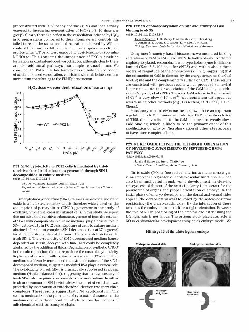
Laboratory-prepared amperometric NO selective sensors, composed of carbon fiber material covered with antiinterference barriers are widespread in NO research. In this work we report, that electrooxidation of Nostotrebin 6, i.e. 2,2-bis[4,5-bis(4-hydroxybenzyl)-2-(4-hydroxyphenyl)-cyclopent-4-ene-1,3-dione], a polyphenol isolated in gram quantities from cyanobacterial strain Nostoc sp. str. Lukesova 27-97, results in the formation of nonconducting films which make uniform, defect-free layers with permselective properties on solid electrodes, including carbon fibers. The properties of polynostotrebin modified electrodes to discriminate against selected biorelevant electroactive compounds of anionic (ascorbate, urate, nitrate), cationic
(dopamine) and neutral (acetaminophen) nature are evaluated for gold, platinum, glassy carbon, and carbon fiber electrodes. The electropolymerization proceeds on solid electrodes in neutral buffered aqueous medium. A single cyclic voltammetry cycle (0-1200 mV vs. Ag/AgCl, 100 mV/s) in 1.10-4 M nostotrebin solution produces polymer coating which is permeable to nitric oxide but effectively discriminates ascorbate with selectivity ratio greater than 1 : 3000 (determined by amperometry in stirred solution at 830 mV vs. Ag/AgCl). Nitrite, dopamine and acetaminophen amperometric responses are rejected by the factor of ca 1:100, relative to nitric oxide signal. This way, a rapid preparation of nitric oxide selective microelectrodes is achieved.
NO signaling
P24. Cardioprotective mechanism of S-nitroso-N-acetylcysteine via overexpression of S-nitrosated betadrenoceptor-2 in the LDLr-/- mice.
Marta H. Krieger Biology Institute, University of Campinas, Brazil
Previous studies have shown the cardio protective effect of S-nitroso-N-acetylcysteine (SNAC) on dyslipidemic LDLr-/- mice. The present study was designed to investigate whether SNAC treatment produced this cardio protective effect via an antioxidant role, and to verify the possible anti-apoptotic role of β2-Adrenergic Receptors (β2-ARs) in the cardiac remodeling. Ventricular superoxide (O2.-) and hydrogen peroxide (H2O2) generation were measured by HPLC methods to allow quantification of Dihydroethidium (DHE) products. Histological sections were stained using terminal dUTP nick-end labeling (TUNEL) to identify nuclei with DNA degradation (apoptosis) and this was confirmed by procaspase-3 protein expression. The findings show that H2O2 production and cell apoptosis increased during left ventricular hypertrophy (LVH). SNAC treatment showed an anti-oxidant effect on cardiac remodeling by decreasing H2O2 and O2.- production (65% and 52%, respectively), this decrease was associated with a decrease in the ratio of p-Ser1177 eNOS/total eNOS. Left ventricle (LV) from SNAC treated mice revealed a 4-fold increase in β2-AR expression; β2-ARs-S-nitrosation (β2-AR-SNO) increased 61%, while apoptosis decreased 70%. We have thus demonstrated an anti-oxidant role of SNAC on cardio protection, which is associated with the mediation of β2-ARs overexpression and β2-AR-SNO via an anti-apoptotic pathway.
P25. Intracellular S-nitrosothiol (SNO) formation by extracellular NO sources
Akio Matsumotoa, Kentaro Ozawab, Ayako Matsumotoc,Haruaki Nakayaa aGraduate School of Medicine, Chiba University, JapanbKanazawa University, JapancOsaka University, Japan
NO plays a pivotal role in signaling as a messenger and a regulatory molecule on transduction, mainly through S-nitrosylation of thiols. In addition to endogenous NO source as a commencement of NO-signal, extracellular NO has a role in inter-cellular signal transduction from an NO-producing cell to adjacent non-NO-producing cells. This study focused on the changes of intracellular SNO levels over time following a treatment with extracellular NO species. The total SNO was measured by Cu(I)/Cys-Chemiluminescence system, with a detection limit above 500 fmol
NO.HEK293 cells treated with 100 μM SNO-glutathione (GSNO), a cell-impermeable SNO donor, in the culture medium (D-MEM without serum) gave SNO accumulation in cells within 30 min, followed by a peak around an hour. In contrast with GSNO, treatment with SNO-cysteine (CysNO) produced a rapid increase of SNO within a minute, supposedly because of the direct conveyance of CysNO through amino acid (AA) transporters (AAT). Although the half-life of CysNO is longer than 30 min in this system, intracellular SNO reached a peak within 15 min, followed by a slow decrease. Either GSNO or CysNO treatment reached the similar level of intracellular SNO following 45 min of incubation. An NO donor, DETA-NONOate, did not accumulate SNO in cells within an hour. To examine the involvement of cysteine transporter (Cys-T) in CysNO-translocation across the membrane, cells were treated in AA-free buffers (PBS or HBSS) as the Cys-T activity may be inhibited by an AA in D-MEM. The intracellular SNO level reached 20 times higher in PBS than in D-MEM. The activity of Cys-T is sodium dependent; however, the SNO-accumulation following a CysNO treatment was not completely abolished by the use of sodium-free buffer, suggesting the involvement of voltage-independent AATs on CysNO transport.These results suggest that cells possess regulatory systems on the plasma membrane to limit nitrosative stress besides the intracellular reduction system.
P26. Deficit in hydrogen peroxide-induced vasodilation in thoracic aorta from redox-dead Cys42Ser PKGIα knock-in mouse
Oleksandra Prysyazhna, Philip Eaton Cardiovascular Division, Department of Cardiology, King's College London, United Kingdom of Great Britain and Northern Ireland
We previously showed PKGIα forms an interprotein disulfide between its two subunits in response to oxidants such as hydrogen peroxide (H2O2) or nitrocysteine. This oxidative post-translational modification activates PKGIα independently of the classical NO-cGMP pathway. The oxidative activation of PKGIα may contribute to the endothelium-derived hyperpolarising factor (EDHF) phenomenon. To investigate this further we generated a global Cys42Ser PKGIα knock-in (KI) mouse line. Immunoblotting confirmed that hearts from KI mice express PKGIα at the same level as wild type (WT). However, when KI hearts were perfused in Langendorff mode and exposed to H2O2 (50μM, 10min) they did not form a disulfide dimer as anticipated, whereas WT hearts did. To examine the contribution of PKGIα disulfide dimerisation in oxidant-induced vasodilation, we compared the response of isolated vascular rings of thoracic aorta from WT or KI mice to H2O2. The Figure shows data from WT or KI rings of aorta that were
preconstricted with EC80 phenylephrine (1μM) and then serially exposed to increasing concentration of H2O2 (n=3, 10 rings per group). Clearly there is a deficit in the vasodilation induced by H2O2 in KI preparations compared to their littermate WT controls. KIs failed to reach the same maximal relaxation achieved by WTs. In contrast there was no difference in the dose response vasodilation profiles when WT or KI were exposed to acetylcholine or spermine NONOate. This confirms the importance of PKGIα disulfide formation in oxidant-induced vasodilation, although clearly there are also additional pathways that couple to vasodilation. We conclude that PKGIα disulfide formation is a significant component of oxidant-induced vasodilation, consistent with this being a cellular mechanism contributing to the EDHF phenomenon.
P27. SIN-1 cytotoxicity to PC12 cells is mediated by thiol-sensitive short-lived substances generated through SIN-1 decomposition in culture medium
Nobuo Watanabe, Kanako Konishi,Takao Arai Department of Applied Biological Science, Tokyo University of Science, Japan
3-morpholinosydnonimine (SIN-1) releases superoxide and nitric oxide in a 1 : 1 stoichiometry, and is therefore widely used on the assumption of peroxynitrite (ONOO-) generator in the study of oxidative/nitrosative stress in cultured cells. In this study, we report that unstable thiol-sensitive substances, generated from the reaction of SIN-1 with components in culture medium, play a crucial role in SIN-1 cytotoxicity in PC12 cells. Exposure of cells to culture medium obtained after almost complete SIN-1 decomposition at 37 degrees C for 2h demonstrated almost the same degree of cytotoxicity as did fresh SIN-1. The cytotoxicity of SIN-1-decomposed medium largely depended on serum, decayed with time, and could be completely abolished by the addition of thiols. Degradation of synthetic ONOO- in the culture medium did not reproduce the unstable cytotoxicity. Replacement of serum with bovine serum albumin (BSA) in culture medium significantly reproduced the cytotoxic nature of the SIN-1-decomposed medium, suggesting modified BSA plays a critical role. The cytotoxicity of fresh SIN-1 is dramatically suppressed in a basal medium (Hanks balanced salt), suggesting that the cytotoxicity of fresh SIN-1 also requires components of culture medium. In either fresh or decomposed SIN-1 cytotoxicity, the onset of cell death was preceded by inactivation of mitochondrial electron transport chain complexes. These results suggest that SIN-1 cytotoxicity in PC12 cells is mediated via the generation of cytotoxic substances in the medium during its decomposition, which induces dysfunctions of mitochondrial electron transport chain.
P28. Effects of phosphorylation on rate and affinity of CaM binding to eNOS
John C. Salerno, J McMurry, C A Chrestensen, B Forsberg,A Johansen, I. Scott, J. L. Wilson, E. W. Lee, A. M. Rahn Biology, Kennesaw State University, United States of America
Using interferometry based biosensors we measured binding and release of CaM to eNOS and nNOS. In both isoforms, binding of unphosphorylated, recombinant wild type holoenzyme is diffusion limited (Kon~3.3x10⁻⁶ sec-1 for eNOS) and within about three orders of magnitude of the Smoluchowski limit, suggesting that the orientation of CaM is directed by the charge arrays on the CaM binding site and the complementary surface on CaM. These results are consistent with previous results which produced somewhat faster rate constants for association of the CaM binding peptides alone (Meyer T., et al (1992) Science.). CaM release in the presence of Ca+2 is very slow (~10-4 sec-1), also consistent with previous results using other methods (e.g., Persechini, et al (1996) J. Biol. Chem.).
Phosphorylation of eNOS has been shown to be an important regulator of eNOS in many laboratories. PKC phosphorylation of T495, directly adjacent to the CaM binding site, greatly slows CaM binding, which is likely to be the primary effect of this modification on activity. Phosphorylation of other sites appears to have more complex effects.
P29. NITRIC OXIDE DEFINES THE LEFT-RIGHT ORIENTATION OF DEVELOPING AVIAN EMBRYO BY PERTURBING BMP4 PATHWAY
Jamila H Siamwala, Suvro Chatterjee AU KBC Research Institute, Anna University, India
Nitric oxide (NO), a free radical and intracellular messenger, is an important regulator of cardiovascular functions. NO has also been implicated in embryonic development. In cleaving embryo, establishment of the axes of polarity is important for the positioning of organs and proper orientation of embryo. In the initial phase of embryo development, the dorsal and ventral sides appear (the dorso-ventral axis) followed by the antero-posterior positioning (the cranio-caudal axis). By the interaction of these two axes the embryo attains a left or a right orientation. However, the role of NO in positioning of the embryo and establishing the left right axis is not known.The present study elucidates role of NO in cardiovascular development using chick embryo model. We
S51
doi:10.1016/j.niox.2010.05.146
doi:10.1016/j.niox.2010.05.147
doi:10.1016/j.niox.2010.05.148
Abstracts /Nitric Oxide 22 (2010) S5–S96

observed that NO delivery in the air sac of embryo via a NO donor, DEAN (DietheylinetriamineNONOate) (500microM), alters the left-right axis and the orientation in 1/3rd of total number of embryos after 4 days of incubation. Wnt signaling controls Nodal expression in the Hensens node and may define the left-right axis of embryo when Shh is expressed on the right side of the node. BMP4 induced by the endodermal signal Shh, also plays a part in controlling the antero-posterior axis. Under DEAN treatment BMP4 protein expression shifted to left side of the embryos. Further investigation of mRNA and protein levels in 24h embryo showed that NO delivery modulates the expression of BMP4, Nodal, Shh, and Wnt3a, which are associated with the establishment and maintenance of dorso-ventral polarity. Our results indicate a role of NO in defining spatial orientation of developing embryo. NO works in synchronization with Shh, BMP4 and Nodal to establish the left-right positioning of the embryo. In this study we propose that NO gradient is the switch that controls left-right symmetry and assigns position to functional organs.
P30. Transnitrosation of Thiols from Water-soluble Aliphatic N-Nitrosamines: S-Nitrosation and Modulation of Protein Functions
Tomohiko Ohwada, Yoji Kabasawa, Takahiro Yanagimoto,Fumika Karaki, Yuko Otani, Noriko Makita, Taroh Iiri Graduate School of Pharmaceutical Sciences, The University of Tokyo, Japan
S-Nitrosothiols and heme nitrosyl species are nitric oxide (NO)-derived metabolites that provide an endogenous reservoir of NO, and also play roles in protein S-nitrosation, i.e., transnitrosation of thiols in proteins, thereby regulating protein functions and signal transduction pathways. Intriguingly, endogenous N-nitrosamines are present in similar abundance to S-nitrosothiols, and though they are thought to play similar physiological roles to S-nitrosothiols, their transnitrosation reactivities and their contribution to biological events are little understood. Herein we report aliphatic N-nitroso derivatives of 7-azabicyclo[2.2.1]heptanes, which do not act as NO donors themselves, but can transnitrosate thiols. Furthermore, in order to mimic the transnitrosation events in cells and to explore biological applications, we create water-soluble N-nitroso derivatives. Based on the correlation of the model S-transnitrosation reactivities and the inhibitory activities of these N-nitrosamines on the initial stage of internalization of β-adrenergic receptor (β-AR) upon stimulation of an agonist, we suggest a possible involvement of S-transnitrosation from aliphatic N-nitrosamines, leading to modulate the relevant protein functions.
Ref) Ohwada, T. et al, J. Am. Chem. Soc., 129, 736-737 (2007).
P31. Repression of classical nuclear export by S-nitrosylation of CRM1
Chang Chen National Laboratory of Biomacromolecules, Institute of Biophysics, Chinese Academy of Sciences, China
The karyopherin chromosomal region maintenance 1 (CRM1) is the major receptor for classical nuclear protein export. However, little is known about the regulation of CRM1 itself. Here, we report that cellular CRM1 became S-nitrosylated upon extensive exposure to endogenous or exogenous nitric oxide (NO). This abrogated the interaction of CRM1 with nuclear export signals (NESs) and repressed classical protein export. Analysis by mass spectrometry and involving the use of S-nitrosylation mimetic mutations indicated that modification at either of two specific cysteines of CRM1 was sufficient to abolish the CRM1-NES association. Moreover, ectopic overexpression of the corresponding S-nitrosylation-resistant CRM1 mutants rescued NO-induced repression of nuclear export. We also found that inactivation of CRM1 by NO facilitated the nuclear accumulation of the antioxidant response transcription factor Nrf2 and transcriptional activation of Nrf2-controlled genes. Together, these data demonstrate that CRM1 is negatively regulated by S-nitrosylation under nitrosative stress. We speculate that this is important for promoting a cytoprotective transcriptional response to nitrosative stress.
P32. S-nitrosylation regulates oxygen affinity of rainbow trout myoglobin
Signe Helbo, Angela Fago Department of Biological Sciences/Zoophysiology, Aarhus University, Denmark
S-nitrosylation is an in vivo post-translational modification that may modulate protein function. In human hemoglobin, S-nitrosylation increases oxygen affinity, which causes NO to be released together with oxygen during hypoxia, a mechanism that has been proposed to induce vasodilation and increase blood flow. It is known that S-nitrosylation in human myoglobin (Mb) may
function as a source of NO and that it in tuna Mb causes structural changes. However, it has not yet been examined if S-nitrosylation of Mb may affect oxygen affinity and play a role in oxygen-linked NO uptake and delivery. We have studied the oxygen affinity of rainbow trout Mb purified from heart and its dependence on various factors including temperature, pH, chloride and lactate. We found that Mb from rainbow trout has the lowest oxygen affinity of any tested Mb, indicating that this active species relies on oxygen delivery at high oxygen tensions to maintain steep diffusion gradients. This Mb shows absence of pH and anion effects and heat of oxygenation similar to that of other Mbs. Titration with 4-PDS indicates that rainbow trout Mb possesses three highly reactive cysteines, which can be partly S-nitrosylated by reaction with S-nitroso Cys. The oxygen affinity of this SNO-Mb is increased, suggesting that NO bound to reactive cysteines is released together with oxygen in hypoxic muscle tissues, which might be an important mechanism in regulating cardiac oxygen consumption and contractility during intense exercise in this fast swimmer.
P33. Nitric Oxide dependent regulation of the cellular hypoxic response
YEE LIU CHUAa, Emmanuel P. Dassab, Eric Dufourc, Howard T. Jacobsc, d, Cormac T. Taylore, Thilo Hagena aDEPARTMENT OF BIOCHEMISTRY, NATIONAL UNIVERSITY OF SINGAPORE, Singapore
bINSERM U676 and Universite Paris 7, Facult de Medecine Denis Diderot, France
cInstitute of Medical Technology and Tampere University Hospital, University of Tampere, Finland
dInstitute of Biomedical and Life Sciences, University of Glasgow, United Kingdom of Great Britain and Northern Ireland
eUCD Conway Institute UCD School of Medicine and Medical Science, Ireland
The transcription factor Hypoxia-Inducible Factor-1α(HIF-1α) is a master regulator of the cellular response to low oxygen. HIF-1α protein accumulates in hypoxia due to inhibition of prolyl hydroxylase enzymes, which under normoxic conditions use molecular oxygen to hydroxylate HIF-1α on two conserved proline residues, thus targeting the protein for 26S proteasome-dependent degradation. Nitric oxide(NO) has been reported to have dual effects on HIF-1α protein stability. At low concentrations it inhibits hypoxia dependent HIF-1α accumulation. At higher concentrations, it can induce HIF-1α protein expression even in the absence of hypoxia. We show here that the inhibition of HIF-1α stabilization by NO under hypoxic conditions is most likely due to inhibition of mitochondrial respiration via interaction with cytochrome c oxidase. Four other inhibitors that target various sites of the mitochondrial electron transport chain(ETC)had very similar effects on the HIF-1α protein half life in hypoxia. The effect of mitochondrial ETC inhibition is not due to hypoxia-induced reactive oxygen species(ROS), originating from complex III, as has been previously suggested, but due to inhibition of mitochondrial oxygen consumption. Thus, reestablishing mitochondrial oxygen consumption in the presence of a complex III inhibitor by using an artificial electron donor to complex IV or overexpressing Ciona intestinalis alternative oxidase results in HIF-1α protein stabilization. These findings provide evidence that NO and other mitochondrial ETC inhibitors regulate HIF-1α stabilization by controlling the intracellular oxygen concentration. Under normoxic conditions, NO, but not ROS, inhibit prolyl hydroxylase activity, which likely accounts for the NO-dependent HIF-1α stabilizing effect at normal oxygen concentrations. NO mediated regulation of HIF-1α is likely to play an important role in regulating HIF-dependent responses under physiological and pathological conditions.
P34. Protein S-nitrosylation : Physiologically Relevant Mechanism of Oxidation in Cell Signaling
KYAW H HLAINGa, MARIE V CLEMENTa, b, c aNUS Graduate School for Integrative Sciences and Engineering/ Biochemistry, National University of Singapore, Singapore
bNUS Graduate School for Integrative Sciences and Engineering, SingaporecDepartment of Biochemisty, Yong Loo Lin School of Medicine, Singapore
Reactive oxygen species (ROSs) were traditionally regarded as toxic and destructive to the cells. But over the past two decades, it is increasingly recognized that these radical species are produced at low level and are necessary for growth factors signaling. Among ROSs, superoxide and hydrogen peroxide have attracted most attention since their increased production was found to be associated with enhanced cell proliferation and malignant transformation. However, how superoxide and hydrogen peroxide modify protein function to effect growth advantage has been in debate. Previously, our group have reported that increased superoxide production, as a result of inhibition of cytosolic superoxide dismutase enzyme (SOD1), causes s-nitrosylation in a tumor suppressor protein, PTEN which was previously reported to form di-sulphide bond upon treatment with high concentration of hydrogen peroxide ( 1mM ). Using biotin switch technique (BST), we report here that in MEF cell line, SOD1 inhibition by diethyldithiocarbamate (DDC) caused generalized s-nitrosylation of proteins in addition to PTEN. As for hydrogen peroxide, at low concentration, total protein s-nitrosyation was also observed, but at 500uM of hydrogen peroxide treatment, s-nitrosylation was replaced by non-SNO oxidative modifications, which was demonstrated with BST using non-specific reducing agent, dithiothreitol. To demonstrate the relevance of our findings, we assessed the effect of platelet-derived growth factor (PDFG), epidermal growth factor (EGF) and 10% fetal bovine serum (FBS) on total protein s-nitrosylation. Results show that PDGF, EGFF and 10% FBS all induced generalized s-nitrosylation of proteins. Moreover, increasing concentration of PDGF did not lead to a concurrent increase in
S52
doi:10.1016/j.niox.2010.05.149
doi:10.1016/j.niox.2010.05.150
doi:10.1016/j.niox.2010.05.151
Abstracts /Nitric Oxide 22 (2010) S5–S96
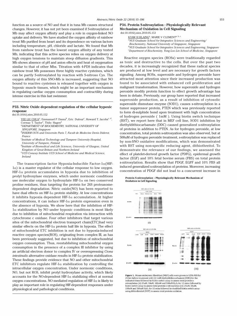
observed that NO delivery in the air sac of embryo via a NO donor, DEAN (DietheylinetriamineNONOate) (500microM), alters the left-right axis and the orientation in 1/3rd of total number of embryos after 4 days of incubation. Wnt signaling controls Nodal expression in the Hensens node and may define the left-right axis of embryo when Shh is expressed on the right side of the node. BMP4 induced by the endodermal signal Shh, also plays a part in controlling the antero-posterior axis. Under DEAN treatment BMP4 protein expression shifted to left side of the embryos. Further investigation of mRNA and protein levels in 24h embryo showed that NO delivery modulates the expression of BMP4, Nodal, Shh, and Wnt3a, which are associated with the establishment and maintenance of dorso-ventral polarity. Our results indicate a role of NO in defining spatial orientation of developing embryo. NO works in synchronization with Shh, BMP4 and Nodal to establish the left-right positioning of the embryo. In this study we propose that NO gradient is the switch that controls left-right symmetry and assigns position to functional organs.
P30. Transnitrosation of Thiols from Water-soluble Aliphatic N-Nitrosamines: S-Nitrosation and Modulation of Protein Functions
Tomohiko Ohwada, Yoji Kabasawa, Takahiro Yanagimoto,Fumika Karaki, Yuko Otani, Noriko Makita, Taroh Iiri Graduate School of Pharmaceutical Sciences, The University of Tokyo, Japan
S-Nitrosothiols and heme nitrosyl species are nitric oxide (NO)-derived metabolites that provide an endogenous reservoir of NO, and also play roles in protein S-nitrosation, i.e., transnitrosation of thiols in proteins, thereby regulating protein functions and signal transduction pathways. Intriguingly, endogenous N-nitrosamines are present in similar abundance to S-nitrosothiols, and though they are thought to play similar physiological roles to S-nitrosothiols, their transnitrosation reactivities and their contribution to biological events are little understood. Herein we report aliphatic N-nitroso derivatives of 7-azabicyclo[2.2.1]heptanes, which do not act as NO donors themselves, but can transnitrosate thiols. Furthermore, in order to mimic the transnitrosation events in cells and to explore biological applications, we create water-soluble N-nitroso derivatives. Based on the correlation of the model S-transnitrosation reactivities and the inhibitory activities of these N-nitrosamines on the initial stage of internalization of β-adrenergic receptor (β-AR) upon stimulation of an agonist, we suggest a possible involvement of S-transnitrosation from aliphatic N-nitrosamines, leading to modulate the relevant protein functions.
Ref) Ohwada, T. et al, J. Am. Chem. Soc., 129, 736-737 (2007).
P31. Repression of classical nuclear export by S-nitrosylation of CRM1
Chang Chen National Laboratory of Biomacromolecules, Institute of Biophysics, Chinese Academy of Sciences, China
The karyopherin chromosomal region maintenance 1 (CRM1) is the major receptor for classical nuclear protein export. However, little is known about the regulation of CRM1 itself. Here, we report that cellular CRM1 became S-nitrosylated upon extensive exposure to endogenous or exogenous nitric oxide (NO). This abrogated the interaction of CRM1 with nuclear export signals (NESs) and repressed classical protein export. Analysis by mass spectrometry and involving the use of S-nitrosylation mimetic mutations indicated that modification at either of two specific cysteines of CRM1 was sufficient to abolish the CRM1-NES association. Moreover, ectopic overexpression of the corresponding S-nitrosylation-resistant CRM1 mutants rescued NO-induced repression of nuclear export. We also found that inactivation of CRM1 by NO facilitated the nuclear accumulation of the antioxidant response transcription factor Nrf2 and transcriptional activation of Nrf2-controlled genes. Together, these data demonstrate that CRM1 is negatively regulated by S-nitrosylation under nitrosative stress. We speculate that this is important for promoting a cytoprotective transcriptional response to nitrosative stress.
P32. S-nitrosylation regulates oxygen affinity of rainbow trout myoglobin
Signe Helbo, Angela Fago Department of Biological Sciences/Zoophysiology, Aarhus University, Denmark
S-nitrosylation is an in vivo post-translational modification that may modulate protein function. In human hemoglobin, S-nitrosylation increases oxygen affinity, which causes NO to be released together with oxygen during hypoxia, a mechanism that has been proposed to induce vasodilation and increase blood flow. It is known that S-nitrosylation in human myoglobin (Mb) may
function as a source of NO and that it in tuna Mb causes structural changes. However, it has not yet been examined if S-nitrosylation of Mb may affect oxygen affinity and play a role in oxygen-linked NO uptake and delivery. We have studied the oxygen affinity of rainbow trout Mb purified from heart and its dependence on various factors including temperature, pH, chloride and lactate. We found that Mb from rainbow trout has the lowest oxygen affinity of any tested Mb, indicating that this active species relies on oxygen delivery at high oxygen tensions to maintain steep diffusion gradients. This Mb shows absence of pH and anion effects and heat of oxygenation similar to that of other Mbs. Titration with 4-PDS indicates that rainbow trout Mb possesses three highly reactive cysteines, which can be partly S-nitrosylated by reaction with S-nitroso Cys. The oxygen affinity of this SNO-Mb is increased, suggesting that NO bound to reactive cysteines is released together with oxygen in hypoxic muscle tissues, which might be an important mechanism in regulating cardiac oxygen consumption and contractility during intense exercise in this fast swimmer.
P33. Nitric Oxide dependent regulation of the cellular hypoxic response
YEE LIU CHUAa, Emmanuel P. Dassab, Eric Dufourc, Howard T. Jacobsc, d, Cormac T. Taylore, Thilo Hagena aDEPARTMENT OF BIOCHEMISTRY, NATIONAL UNIVERSITY OF SINGAPORE, Singapore
bINSERM U676 and Universite Paris 7, Facult de Medecine Denis Diderot, France
cInstitute of Medical Technology and Tampere University Hospital, University of Tampere, Finland
dInstitute of Biomedical and Life Sciences, University of Glasgow, United Kingdom of Great Britain and Northern Ireland
eUCD Conway Institute UCD School of Medicine and Medical Science, Ireland
The transcription factor Hypoxia-Inducible Factor-1α(HIF-1α) is a master regulator of the cellular response to low oxygen. HIF-1α protein accumulates in hypoxia due to inhibition of prolyl hydroxylase enzymes, which under normoxic conditions use molecular oxygen to hydroxylate HIF-1α on two conserved proline residues, thus targeting the protein for 26S proteasome-dependent degradation. Nitric oxide(NO) has been reported to have dual effects on HIF-1α protein stability. At low concentrations it inhibits hypoxia dependent HIF-1α accumulation. At higher concentrations, it can induce HIF-1α protein expression even in the absence of hypoxia. We show here that the inhibition of HIF-1α stabilization by NO under hypoxic conditions is most likely due to inhibition of mitochondrial respiration via interaction with cytochrome c oxidase. Four other inhibitors that target various sites of the mitochondrial electron transport chain(ETC)had very similar effects on the HIF-1α protein half life in hypoxia. The effect of mitochondrial ETC inhibition is not due to hypoxia-induced reactive oxygen species(ROS), originating from complex III, as has been previously suggested, but due to inhibition of mitochondrial oxygen consumption. Thus, reestablishing mitochondrial oxygen consumption in the presence of a complex III inhibitor by using an artificial electron donor to complex IV or overexpressing Ciona intestinalis alternative oxidase results in HIF-1α protein stabilization. These findings provide evidence that NO and other mitochondrial ETC inhibitors regulate HIF-1α stabilization by controlling the intracellular oxygen concentration. Under normoxic conditions, NO, but not ROS, inhibit prolyl hydroxylase activity, which likely accounts for the NO-dependent HIF-1α stabilizing effect at normal oxygen concentrations. NO mediated regulation of HIF-1α is likely to play an important role in regulating HIF-dependent responses under physiological and pathological conditions.
P34. Protein S-nitrosylation : Physiologically Relevant Mechanism of Oxidation in Cell Signaling
KYAW H HLAINGa, MARIE V CLEMENTa, b, c aNUS Graduate School for Integrative Sciences and Engineering/ Biochemistry, National University of Singapore, Singapore
bNUS Graduate School for Integrative Sciences and Engineering, SingaporecDepartment of Biochemisty, Yong Loo Lin School of Medicine, Singapore
Reactive oxygen species (ROSs) were traditionally regarded as toxic and destructive to the cells. But over the past two decades, it is increasingly recognized that these radical species are produced at low level and are necessary for growth factors signaling. Among ROSs, superoxide and hydrogen peroxide have attracted most attention since their increased production was found to be associated with enhanced cell proliferation and malignant transformation. However, how superoxide and hydrogen peroxide modify protein function to effect growth advantage has been in debate. Previously, our group have reported that increased superoxide production, as a result of inhibition of cytosolic superoxide dismutase enzyme (SOD1), causes s-nitrosylation in a tumor suppressor protein, PTEN which was previously reported to form di-sulphide bond upon treatment with high concentration of hydrogen peroxide ( 1mM ). Using biotin switch technique (BST), we report here that in MEF cell line, SOD1 inhibition by diethyldithiocarbamate (DDC) caused generalized s-nitrosylation of proteins in addition to PTEN. As for hydrogen peroxide, at low concentration, total protein s-nitrosyation was also observed, but at 500uM of hydrogen peroxide treatment, s-nitrosylation was replaced by non-SNO oxidative modifications, which was demonstrated with BST using non-specific reducing agent, dithiothreitol. To demonstrate the relevance of our findings, we assessed the effect of platelet-derived growth factor (PDFG), epidermal growth factor (EGF) and 10% fetal bovine serum (FBS) on total protein s-nitrosylation. Results show that PDGF, EGFF and 10% FBS all induced generalized s-nitrosylation of proteins. Moreover, increasing concentration of PDGF did not lead to a concurrent increase in
S53
doi:10.1016/j.niox.2010.05.152
doi:10.1016/j.niox.2010.05.153
Abstracts /Nitric Oxide 22 (2010) S5–S96

the level of total protein s-nitrosylation. Taken together, our data suggest that protein s-nitrosylation may represent a major oxidative modification caused by superoxide and hydrogen peroxide during growth factors signaling.
P35. Retinoic acid receptor stimulation activates NO/cyclic GMP-dependent MEK/ERK signaling pathway leading to upregulation of BDNF expression in midbrain dopaminergic neurons
Yuki Kurauchia, Akinori Hisatsunea, Yoichiro Isohamaa,Tomohiro Sawaa, Takaaki Akaikea, Koichi Shudob, Hiroshi Katsukia aDepartment of Chemico-Pharmacological Sciences, Graduate School of Pharmaceutical Sciences, Kumamoto University, Japan
bResearch Foundation Itsuu Laboratory, Japan
Brain-derived neurotrophic factor (BDNF) has important roles in the development and survival of dopaminergic neurons, and its loss might contribute to the pathogenesis of Parkinson disease. We have previously reported that stimulation of retinoic acid receptors (RARs) protects midbrain dopaminergic neurons, presumably via up-regulation of brain-derived neurotrophic factor (BDNF) expression. Here, we examined the signaling mechanisms linking RAR stimulation to BDNF expression. Rat midbrain slice cultures treated with an RAR agonist Am80 (30-300 nM) showed increased tissue levels of BDNF mRNA and protein as compared to cultures without treatment. Am80-induced increase in BDNF expression was observed in dopaminergic neurons, which was blocked by concomitant application of LE540 (1 μM), an RAR antagonist, and PD98059 (20 μM), a MEK1 inhibitor. On the other hand, HX630 (1 μM), an RXR agonist, and LY294002 (50 μM), a PI3-kinase inhibitor, did not have significant effect on BDNF expression. We also found that Am80 increased neuronal nitric oxide synthase (nNOS) expression in dopaminergic neurons even during ERK inhibition, and this increase was accompanied by 8-nitro-cyclic GMP formation. Notably, the effect of Am80 on BDNF expression was attenuated by concomitant application of L -NAME (1 mM), a general NOS inhibitor, ODQ (100 μM), a soluble guanylyl cyclase inhibitor, and KT5823 (10 μM), a cyclic GMP-dependent protein kinase (PKG) inhibitor. Am80-induced ERK phosphorylation in dopaminergic neurons was also attenuated by inhibition of soluble guanylyl cyclase and PKG. Moreover, 8-Br-cyclic GMP (100 μM) induced ERK phosphorylation and BDNF expression in dopaminergic neurons. These results suggest that, by recruiting cyclic GMP and PKG, nNOS-derived nitric oxide plays a novel and essential role in RAR signaling leading to ERK-dependent BDNF up-regulation in midbrain dopaminergic neurons.
P37. APP protein expression modulates S-nitrosylation of proteins in Alzheimer disease model transgenic mice
Aleksandra Wyslouch-Cieszynska, Monika Zareba-Koziol,Liliya Zhukova Department of Biophysics, Institute of Biochemistry and Biophysics, Poland
Alzheimer disease (AD) is an incurable, neurodegenerative disease that leads to dysfunction and loss of synapses. The pathogenesis of AD and the causes of its characteristic symptoms are not fully understood. They have been partly linked to imbalance in the production of reactive oxygen (ROS) and nitrogen species (RNS). Dysregulated S-nitrosylation of several proteins has been shown in AD model animals and AD patients [1,2].
A mass spectrometry based proteomic analysis of total S-nitrosomes will be presented for transgenic mice with neuronal expression of human amyloid precursor protein (APP) at different age and different stages of disease symptoms development.
1. Emerging roles of S-nitrosylation in protein misfolding and neurodegenerative diseases, Nakamura T, Lipton SA, Antioxid Redox Signal. 2008; 10(1),87-101 2. S-nitrosylation of Drp1 mediates beta-amyloid-related mitochondrial fission and neuronal injury, Cho DH, Nakamura T, Fang J, Cieplak P, Godzik A, Gu Z, Lipton SA, Science. 2009; 324(5923,102-5.
P38. Effect of NO on the transcriptional activity of NPAS2, a transcription factor associated with circadian rhythm
Katsuhiro Yoshii, Tomoyuki Takashima, Takeshi Ueha,Sumio Ishijima, Ikuko Sagami Graduate School of LIfe and Environmental Sciences, Kyoto Prefectural University, Japan
A neuronal PAS domain protein 2 (NPAS2) is a transcription factor regulating mammalian circadian rhythm and forms a heterodimer with BMAL1 like CLOCK that can induce the transcription of Per and Cry genes by binding to the E-box. NPAS2 consists of bHLH and two PAS domain (PAS A and PAS B) in N-terminal region and transactivation domain in C-terminal region. Both PAS domains of NPAS2 bind heme as a prosthetic group and CO binding to the PAS A heme inhibits the DNA binding activity, indicating that NPAS2 is a CO-regulated transcription factor and the heme domain acts as a gas sensor. Our previous studies of resonance Raman spectra of the isolated bHLH-PAS A domain demonstrated that His119 and His171 are axial ligands for both ferric and ferrous heme and that NO binding to the ferrous heme formed a 5-coordinated complex. It is also reported that NO affects stability of BMAL1 through S-nirosylation and produces light-like phase shifts of circadian
rhythms. However, the effect of NO on transcriptional activity of NPAS2 is not clear.
In the present study, we analyzed the effects of NO on functions of mouse NPAS2 by luciferase assay using mouse Per1 or Per2 promoter as a reporter gene in NIH3T3 cells. The addition of NO donor (SNAP) into the medium inhibited the trasnscriptional activity of NPAS2 for Per1 or Per2 expression in a dose-dependent manner (10-50 μM). The similar results were also obtained with another NO donor, S-nitrosoglutathione. NPAS2 protein expression level was examined using western blot analysis under the same conditions, indicating no effect of NO on the protein level. The addition of cGMP agonist, 8-Br-cGMP did not affect the transcriptional activity of NPAS2. These results suggest that circadian rhythm is regulated by NO signaling via NPAS2, but not cGMP-dependent pathways.
P39. Mechanisms for the Diverse Roles of Endothelial Nitric Oxide Synthases System between Conduit and Resistance Arteries in Mice
Junko Ohashia, Aya Takakia, Ayuko Sawadaa, Kazuki Nodaa,Maki Hosoyaa, Toyotaka Yadab, Hiroaki Shimokawaa aDepartment of Cardiovascular Medicine, Tohoku university, JapanbKawasaki medical school, Japan
Background: We have previously demonstrated in animals and humans that endothelium-derived hydrogen peroxide (H2O2) is an endothelium-derived hyperpolarizing factor (EDHF) and that endothelial NO synthase (eNOS)-derived superoxide anions is a major precursor of EDHF/H2O2. We have recently demonstrated in mice deficient of the whole NOSs system that endothelial NOSs system plays diverse roles depending on vessel size, mainly serving as a superoxide generating system to cause EDHF/H2O2 responses in resistance arteries (e.g. mesenteric arteries, MA) while serving as a NO generating system in conduit arteries (e.g. the aorta, Ao). In this study, we aimed to elucidate the mechanisms involved in the diverse roles of endothelial NOSs system. Methods and Results: We used male wild-type mice. EDHF-mediated relaxations to acetylcholine and vascular smooth muscle (VSM) relaxations to exogenous H2O2 were greater in MA than in Ao. However, the ratio of EDHF-mediated relaxations to VSM relaxations was greater in MA than in Ao, suggesting that both endothelial NOSs-related and VSM-related mechanisms are involved in the enhanced EDHF-mediated responses in MA. Dimer/monomer ratio of eNOS and BH4/BH2 ratio were comparable between Ao and MA, and modulation of BH4 synthesis had no effects on the EDHF responses. The extent of eNOS phosphorylation at Ser 1177 was significantly greater in Ao than in MA, whereas that at Thr 495 was comparable. STO-609, an inhibitor of CaMKKβ that lies upstream of PI3K/Akt/eNOS pathway, significantly decreased EDHF-mediated relaxations and hyperpolarizations in MA but not in Ao. Conclusions: These results suggest that the diverse roles of NOSs between conduit and resistance arteries are mediated, at least in part, by phosphorylation of eNOS at Ser 1177, for which CaMKKβ may be involved.
P40. Mechanism of protein S-nitrosylation in rat 3Y1 cells
Norihiro Ryuman, Nobuo Watanabe, Takao Arai Applied Biological Science, Tokyo University of Science, Japan
S-nitrosylated proteins are increased in pathological condition such as Alzheimer's and Parkinson's disease. In the mean time, S-nitrosylation/ S-denitrosylation regulates protein functions under physiological conditions. Several mechanisms have been
proposed for the formation of S-nitrosothiols in cellular proteins, possibly depending on cell types, nitrosylating agents, cell culture medium, and assay conditions. We investigated the mechanism of protein S-nitrosylation in rat 3Y1 cells using S-nitrosocysteine (CysNO), S-nitrosoglutathione (GSNO), NO donor (NOR3), NO/O.- donor (SIN1). In Dulbecco's Modified Eagle's Medium (DMEM) containing of 10% serum, none of the nitrosylating agents induced S-nitrosylation of cellular proteins. In contrast, in Hanks' balanced salts solution (HBSS), CysNO and GSNO induced cellular protein S-nitrosylation. Compared with CysNO, however, GSNO increased less S-nitrosylated proteins. Neither NOR3 nor SIN1 could induce S-nitrosylated proteins of 3Y1 cellls regardless of the medium. To assess the mechanism of protein S-nitrosylation by CysNO and GSNO, the stability of S-nitrosothiols was measured in HBSS and DMEM by using a NO electrode and with Saville-Griess assay. NO release from CysNO was much faster in HBSS than in DMEM while the levels of S-nitrosothiols decreased with time-dependent in either medium. In contrast, GSNO did not release NO and total amount of S-nitrosothiols decreased slightly in either medium. These results suggest that the most potent S-nitroylating agent for 3Y1 cells is CysNO. We are now examining whether the mechanism of S-nitrosylation due to CysNO itself or a large amount of NO released from CysNO.
P41. Nitric Oxide inhibits aquaporin-4 and -5 water channels through different signalings
Yoichiro Isohama, Akari Arimatsu, Makiko Kondoh,Akinori Hisatsune, Hiroshi Katsuki Graduate School of Pharmaceutical Sciences, Kumamoto University, Japan
Aquaporins (AQPs) are small, integral membrane proteins that facilitate water transport across cell membranes in response to osmotic gradients. Thirteen mammalian AQPs have been identified and localized to various epithelial, endothelial and other tissues. Phenotype studies of AQP knockout mice have demonstrated essential roles for AQP in mammalian physiology and pathophysiology, including urinary concentrating function in kidney, airway fluid secretion and brain edema formation. These studies suggest that AQP may be potential drug targets for not only new diuretic reagents, but also for novel therapy of inflammatory lung diseases and brain edema. However, the regulation of water transport through AQPs remains to be clear. In the present study, we have examined the effect of nitric oxide (NO) on water permeability mediated by AQP4 and AQP5.
In Xenopus oocytes expressing AQP4, GSNO, a NO-donor, decreased membrane water permeability, in a concentration-dependent manner (0.01-1 mM). Similar decrease in water permeability was observed by the treatment of SNAP and NOC-18. The decrease in water permeability by GSNO was sensitive to ODQ and KT582, suggesting that NO inhibits AQP4 via guanylyl cyclase and cyclic GMP pathway. GSNO and other NO-donors also decreased membrane water permeability in oocytes expressing AQP5. However, the inhibition of AQP5 by GSNO was insensitive to ODQ, and cGMP analogue did not affect AQP5-mediated water permeability. NO donors also caused S-nitrosylation of AQP5 assessed by biotin switch method. The C182A mutant of AQP5 increased the water permeability of the oocyte as well as wild-type. However, the C182A-dependent water permeability is unaffected by GSNO. It is, therefore, suggested that NO inhibits AQP5 via S-nitrosylation of this channel at C182.
Taken together, we assume that NO can inhibit both AQP4 and AQP5, and that the underlining mechanisms of inhibition by NO are different between these AQP isoforms.
S54
doi:10.1016/j.niox.2010.05.154
doi:10.1016/j.niox.2010.05.155
doi:10.1016/j.niox.2010.05.156
Abstracts /Nitric Oxide 22 (2010) S5–S96

the level of total protein s-nitrosylation. Taken together, our data suggest that protein s-nitrosylation may represent a major oxidative modification caused by superoxide and hydrogen peroxide during growth factors signaling.
P35. Retinoic acid receptor stimulation activates NO/cyclic GMP-dependent MEK/ERK signaling pathway leading to upregulation of BDNF expression in midbrain dopaminergic neurons
Yuki Kurauchia, Akinori Hisatsunea, Yoichiro Isohamaa,Tomohiro Sawaa, Takaaki Akaikea, Koichi Shudob, Hiroshi Katsukia aDepartment of Chemico-Pharmacological Sciences, Graduate School of Pharmaceutical Sciences, Kumamoto University, Japan
bResearch Foundation Itsuu Laboratory, Japan
Brain-derived neurotrophic factor (BDNF) has important roles in the development and survival of dopaminergic neurons, and its loss might contribute to the pathogenesis of Parkinson disease. We have previously reported that stimulation of retinoic acid receptors (RARs) protects midbrain dopaminergic neurons, presumably via up-regulation of brain-derived neurotrophic factor (BDNF) expression. Here, we examined the signaling mechanisms linking RAR stimulation to BDNF expression. Rat midbrain slice cultures treated with an RAR agonist Am80 (30-300 nM) showed increased tissue levels of BDNF mRNA and protein as compared to cultures without treatment. Am80-induced increase in BDNF expression was observed in dopaminergic neurons, which was blocked by concomitant application of LE540 (1 μM), an RAR antagonist, and PD98059 (20 μM), a MEK1 inhibitor. On the other hand, HX630 (1 μM), an RXR agonist, and LY294002 (50 μM), a PI3-kinase inhibitor, did not have significant effect on BDNF expression. We also found that Am80 increased neuronal nitric oxide synthase (nNOS) expression in dopaminergic neurons even during ERK inhibition, and this increase was accompanied by 8-nitro-cyclic GMP formation. Notably, the effect of Am80 on BDNF expression was attenuated by concomitant application of L -NAME (1 mM), a general NOS inhibitor, ODQ (100 μM), a soluble guanylyl cyclase inhibitor, and KT5823 (10 μM), a cyclic GMP-dependent protein kinase (PKG) inhibitor. Am80-induced ERK phosphorylation in dopaminergic neurons was also attenuated by inhibition of soluble guanylyl cyclase and PKG. Moreover, 8-Br-cyclic GMP (100 μM) induced ERK phosphorylation and BDNF expression in dopaminergic neurons. These results suggest that, by recruiting cyclic GMP and PKG, nNOS-derived nitric oxide plays a novel and essential role in RAR signaling leading to ERK-dependent BDNF up-regulation in midbrain dopaminergic neurons.
P36. Characterization of a Novel Posttranslational Modification in Neuronal Nitric Oxide Synthase by Small Ubiquitin-related Modifier-1; SUMO-1
Kouichi Itoh School of Kagawa Pharmacutical Sciences, Tokushima Bunri Univeersity, Japan
The three-dimensional structure of Small Ubiquitin-related Modifier (SUMO) is very similar to ubiquitin, but biological functions of SUMOylation were different from ubiquitination. SUMO proteins are covalently attached to lysine residues on target proteins by a specific conjugation pathway. Recently the functional implications of protein SUMOylation by SUMO were reported in neurophysiological and neuropathological conditions. Nitric oxide (NO) has paradoxically neuroprotective and neurotoxic effects in CNS. These effects of NO are due to the activity of neuronal NO synthase (nNOS), therefore further detailed understanding
of the regulatory mechanism of nNOS activity is important. The nNOS activity and its stability are modulated by post-translational modifications, phosphorylation and ubiquitination, but SUMOylation is still unknown. It was shown that nNOS had three high-probably SUMOylation consensus sites in the SUMOplot analysis. In this present study, we investigated whether nNOS is post-translationally modified by SUMO proteins. At least two nNOS-SUMO-1 conjugates were detected in HEK293T cells transfected with nNOS and SUMO-1. The two nNOS-SUMO-1 conjugates were identified by MS/MS analyses using MALDI-QIT/TOF. In co-transfection experiments in HEK293T cells, nNOS-SUMO-1 conjugates were increased by Ubc9 as the SUMO E2 ligase, and were decreased by SENP as SUMO-deconjugating protease. These findings indicated that nNOS was post-translationally modified by SUMO-1.
P37. APP protein expression modulates S-nitrosylation of proteins in Alzheimer disease model transgenic mice
Aleksandra Wyslouch-Cieszynska, Monika Zareba-Koziol,Liliya Zhukova Department of Biophysics, Institute of Biochemistry and Biophysics, Poland
Alzheimer disease (AD) is an incurable, neurodegenerative disease that leads to dysfunction and loss of synapses. The pathogenesis of AD and the causes of its characteristic symptoms are not fully understood. They have been partly linked to imbalance in the production of reactive oxygen (ROS) and nitrogen species (RNS). Dysregulated S-nitrosylation of several proteins has been shown in AD model animals and AD patients [1,2].
A mass spectrometry based proteomic analysis of total S-nitrosomes will be presented for transgenic mice with neuronal expression of human amyloid precursor protein (APP) at different age and different stages of disease symptoms development.
1. Emerging roles of S-nitrosylation in protein misfolding and neurodegenerative diseases, Nakamura T, Lipton SA, Antioxid Redox Signal. 2008; 10(1),87-101 2. S-nitrosylation of Drp1 mediates beta-amyloid-related mitochondrial fission and neuronal injury, Cho DH, Nakamura T, Fang J, Cieplak P, Godzik A, Gu Z, Lipton SA, Science. 2009; 324(5923,102-5.
P38. Effect of NO on the transcriptional activity of NPAS2, a transcription factor associated with circadian rhythm
Katsuhiro Yoshii, Tomoyuki Takashima, Takeshi Ueha,Sumio Ishijima, Ikuko Sagami Graduate School of LIfe and Environmental Sciences, Kyoto Prefectural University, Japan
A neuronal PAS domain protein 2 (NPAS2) is a transcription factor regulating mammalian circadian rhythm and forms a heterodimer with BMAL1 like CLOCK that can induce the transcription of Per and Cry genes by binding to the E-box. NPAS2 consists of bHLH and two PAS domain (PAS A and PAS B) in N-terminal region and transactivation domain in C-terminal region. Both PAS domains of NPAS2 bind heme as a prosthetic group and CO binding to the PAS A heme inhibits the DNA binding activity, indicating that NPAS2 is a CO-regulated transcription factor and the heme domain acts as a gas sensor. Our previous studies of resonance Raman spectra of the isolated bHLH-PAS A domain demonstrated that His119 and His171 are axial ligands for both ferric and ferrous heme and that NO binding to the ferrous heme formed a 5-coordinated complex. It is also reported that NO affects stability of BMAL1 through S-nirosylation and produces light-like phase shifts of circadian
rhythms. However, the effect of NO on transcriptional activity of NPAS2 is not clear.
In the present study, we analyzed the effects of NO on functions of mouse NPAS2 by luciferase assay using mouse Per1 or Per2 promoter as a reporter gene in NIH3T3 cells. The addition of NO donor (SNAP) into the medium inhibited the trasnscriptional activity of NPAS2 for Per1 or Per2 expression in a dose-dependent manner (10-50 μM). The similar results were also obtained with another NO donor, S-nitrosoglutathione. NPAS2 protein expression level was examined using western blot analysis under the same conditions, indicating no effect of NO on the protein level. The addition of cGMP agonist, 8-Br-cGMP did not affect the transcriptional activity of NPAS2. These results suggest that circadian rhythm is regulated by NO signaling via NPAS2, but not cGMP-dependent pathways.
P39. Mechanisms for the Diverse Roles of Endothelial Nitric Oxide Synthases System between Conduit and Resistance Arteries in Mice
Junko Ohashia, Aya Takakia, Ayuko Sawadaa, Kazuki Nodaa,Maki Hosoyaa, Toyotaka Yadab, Hiroaki Shimokawaa aDepartment of Cardiovascular Medicine, Tohoku university, JapanbKawasaki medical school, Japan
Background: We have previously demonstrated in animals and humans that endothelium-derived hydrogen peroxide (H2O2) is an endothelium-derived hyperpolarizing factor (EDHF) and that endothelial NO synthase (eNOS)-derived superoxide anions is a major precursor of EDHF/H2O2. We have recently demonstrated in mice deficient of the whole NOSs system that endothelial NOSs system plays diverse roles depending on vessel size, mainly serving as a superoxide generating system to cause EDHF/H2O2 responses in resistance arteries (e.g. mesenteric arteries, MA) while serving as a NO generating system in conduit arteries (e.g. the aorta, Ao). In this study, we aimed to elucidate the mechanisms involved in the diverse roles of endothelial NOSs system. Methods and Results: We used male wild-type mice. EDHF-mediated relaxations to acetylcholine and vascular smooth muscle (VSM) relaxations to exogenous H2O2 were greater in MA than in Ao. However, the ratio of EDHF-mediated relaxations to VSM relaxations was greater in MA than in Ao, suggesting that both endothelial NOSs-related and VSM-related mechanisms are involved in the enhanced EDHF-mediated responses in MA. Dimer/monomer ratio of eNOS and BH4/BH2 ratio were comparable between Ao and MA, and modulation of BH4 synthesis had no effects on the EDHF responses. The extent of eNOS phosphorylation at Ser 1177 was significantly greater in Ao than in MA, whereas that at Thr 495 was comparable. STO-609, an inhibitor of CaMKKβ that lies upstream of PI3K/Akt/eNOS pathway, significantly decreased EDHF-mediated relaxations and hyperpolarizations in MA but not in Ao. Conclusions: These results suggest that the diverse roles of NOSs between conduit and resistance arteries are mediated, at least in part, by phosphorylation of eNOS at Ser 1177, for which CaMKKβ may be involved.
P40. Mechanism of protein S-nitrosylation in rat 3Y1 cells
Norihiro Ryuman, Nobuo Watanabe, Takao Arai Applied Biological Science, Tokyo University of Science, Japan
S-nitrosylated proteins are increased in pathological condition such as Alzheimer's and Parkinson's disease. In the mean time, S-nitrosylation/ S-denitrosylation regulates protein functions under physiological conditions. Several mechanisms have been
proposed for the formation of S-nitrosothiols in cellular proteins, possibly depending on cell types, nitrosylating agents, cell culture medium, and assay conditions. We investigated the mechanism of protein S-nitrosylation in rat 3Y1 cells using S-nitrosocysteine (CysNO), S-nitrosoglutathione (GSNO), NO donor (NOR3), NO/O.- donor (SIN1). In Dulbecco's Modified Eagle's Medium (DMEM) containing of 10% serum, none of the nitrosylating agents induced S-nitrosylation of cellular proteins. In contrast, in Hanks' balanced salts solution (HBSS), CysNO and GSNO induced cellular protein S-nitrosylation. Compared with CysNO, however, GSNO increased less S-nitrosylated proteins. Neither NOR3 nor SIN1 could induce S-nitrosylated proteins of 3Y1 cellls regardless of the medium. To assess the mechanism of protein S-nitrosylation by CysNO and GSNO, the stability of S-nitrosothiols was measured in HBSS and DMEM by using a NO electrode and with Saville-Griess assay. NO release from CysNO was much faster in HBSS than in DMEM while the levels of S-nitrosothiols decreased with time-dependent in either medium. In contrast, GSNO did not release NO and total amount of S-nitrosothiols decreased slightly in either medium. These results suggest that the most potent S-nitroylating agent for 3Y1 cells is CysNO. We are now examining whether the mechanism of S-nitrosylation due to CysNO itself or a large amount of NO released from CysNO.
P41. Nitric Oxide inhibits aquaporin-4 and -5 water channels through different signalings
Yoichiro Isohama, Akari Arimatsu, Makiko Kondoh,Akinori Hisatsune, Hiroshi Katsuki Graduate School of Pharmaceutical Sciences, Kumamoto University, Japan
Aquaporins (AQPs) are small, integral membrane proteins that facilitate water transport across cell membranes in response to osmotic gradients. Thirteen mammalian AQPs have been identified and localized to various epithelial, endothelial and other tissues. Phenotype studies of AQP knockout mice have demonstrated essential roles for AQP in mammalian physiology and pathophysiology, including urinary concentrating function in kidney, airway fluid secretion and brain edema formation. These studies suggest that AQP may be potential drug targets for not only new diuretic reagents, but also for novel therapy of inflammatory lung diseases and brain edema. However, the regulation of water transport through AQPs remains to be clear. In the present study, we have examined the effect of nitric oxide (NO) on water permeability mediated by AQP4 and AQP5.
In Xenopus oocytes expressing AQP4, GSNO, a NO-donor, decreased membrane water permeability, in a concentration-dependent manner (0.01-1 mM). Similar decrease in water permeability was observed by the treatment of SNAP and NOC-18. The decrease in water permeability by GSNO was sensitive to ODQ and KT582, suggesting that NO inhibits AQP4 via guanylyl cyclase and cyclic GMP pathway. GSNO and other NO-donors also decreased membrane water permeability in oocytes expressing AQP5. However, the inhibition of AQP5 by GSNO was insensitive to ODQ, and cGMP analogue did not affect AQP5-mediated water permeability. NO donors also caused S-nitrosylation of AQP5 assessed by biotin switch method. The C182A mutant of AQP5 increased the water permeability of the oocyte as well as wild-type. However, the C182A-dependent water permeability is unaffected by GSNO. It is, therefore, suggested that NO inhibits AQP5 via S-nitrosylation of this channel at C182.
Taken together, we assume that NO can inhibit both AQP4 and AQP5, and that the underlining mechanisms of inhibition by NO are different between these AQP isoforms.
Withdraw
S55
doi:10.1016/j.niox.2010.05.157
doi:10.1016/j.niox.2010.05.158
doi:10.1016/j.niox.2010.05.159
Abstracts /Nitric Oxide 22 (2010) S5–S96

P42. Nitric oxide responsive transcription factor Nrf2 regulates Fpn1-mediated iron efflux and counteracts the inflammation-mediated Fpn1 mRNA suppression
Ken Itoha, Nobuhiko Haradaa ,Masaya Kanayamab ,Atsushi Maruyamaa, Aruto Yoshidab, Masayuki Yamamotoc aGraduate School of Medicine, Hirosaki University, JapanbKirin Holdings Co. Ltd., JapancTohoku Univeristy, Japan
Ferroportin 1 (Fpn1) is the sole iron exporter in mammals and regulates iron homeostasis. Although Fpn1 is posttranscriptionally regulated by iron-regulated hormone hepcidin and iron-responsive element (IRE) on its mRNA, mechanism of transcriptional regulation is largely unknown. In this study, we uncovered that an oxidative and nitrosative stress-responsive transcription factor Nrf2 regulates Fpn1 expression in macrophages as well as other heme and iron metabolizing genes including Nramp1. Nrf2 activation by electrophilic compounds upregulated Fpn1 and Nramp1 expression in bone marrow-derived and thioglycolate-induced peritoneal macrophages obtained from wild-type, but not from Nrf2 knockout mice. Nrf2 activation upregulated Fpn1 expression and enhanced transferrin-incorporated iron release from the murine macrophage cell line J774.1. In anemia of chronic disease, the proinflammatory cytokines such as IL-6, and LPS cause down-regulation of macrophage Fpn1 expression leading to iron sequestration by reticuloendothelial macrophages. We demonstrated that Nrf2 activation counteracted LPS-induced Fpn1 suppression in thioglycolate-induced peritoneal macrophages and human peripheral blood-derived macrophages. In vivo sulforaphane administration to mice rescued the suppression of splenic Fpn1 mRNA expression during Freund' complete adjuvant (FCA)-induced inflammation. Thus, Nrf2 may have the potential to therapeutically alleviate anemia of chronic disease by enhancing iron efflux from macrophages.
P43. Activation of EGFR coupled to reduction of PTP1B activity by nitrated oleic acid: an electrophile signaling
Noriko Iwamotoa, Takaaki Akaikeb, Bruce A Freemanc, Kumagai Yoshitoa aGraduate School of Comprehensive Human Sciences, University of Tsukuba, Japan
bKumamoto University, JapancUniversity of Pittsburgh, United States of America
Exposure of cells to electrophiles results in a covalent modification of proteins with thiolate ions to form thioether adducts. Some of these proteins are sensor proteins and the resulting modification affects cellular signal transduction pathways such as the EGFR system resulting in an inflammatory response. On the other hand, it has been suggested that such an interaction can involve the Nrf2/Keap1 system, thereby increasing the capacity for biotransformation of these reactive species to their polar metabolites, which are readily excreted into extracellular space. We reported previously that chemical modification of protein tyrosine phosphatase 1B (PTP1B) by 1,2-naphthoquinone, an exogenous electrophile, causes epidermal growth factor receptor (EGFR) phosphorylation, leading to subsequent activation of MEK/ERK/AP-1 signaling. In the present study, we examined effects of endogenous electrophiles on PTP1B/EGFR/ERK signaling. Incubation of purified PTP1B with 9- and 10-nitro-9-cis-0ctadecenoic acid (OA-NO2), 8-nitro-cGMP, 15-deoxy-delta (12,14)-PGJ2 (15d-PGJ2) and 4-hydroxy-2-nonenal resulted in a concentration-dependent reduction of its catalytic activity through covalent binding to thiol groups in either case. However, transactivation of EGFR coupled to reduction of cellular PTP activity during exposure of A549 cells was only observed with 15d-PGJ2 and OA-NO2, suggesting that there is
an electrophile-specific PTP1B/EGFR signaling in the cells. OA-NO2 also activated ERK pathway through presumably EGFR signaling. These results suggest that OA-NO2 affects PTP1B activity, thereby activating EGFR/ERK signaling.
P44. Combination Therapy with an Angiotensin Receptor Blocker and a Calcium Channel Blocker Improves EDHF-Mediated Responses in Diabetic Apolipoprotein E-Deficient Mice
MAKI HOSOYA, Ayuko Sawada, Junko Ohashi, Aya Takaki,Hiroaki Shimokawa Graduate School of Medicine, Tohoku University, Japan
Background: The endothelium modulates vascular tone by synthesizing and releasing several vasodilating factors, including vasodilator prostaglandins, nitric oxide (NO) and endothelium-derived hyperpolarizing factor (EDHF). In this study, we examined whether an angiotensin receptor blocker (ARB), a calcium channel blocker (CCB) and their combination improve EDHF-mediated responses in diabetic apolipoprotein E-deficient (ApoE-/-) mice. Methods and Results: We used 8-weeks-old male C57BL/6N (control) and streptozocin- induced diabetic ApoE-/- mice. Two weeks after the STZ injection, the diabetic ApoE-/- mice received oral administration of vehicle (Untreated), olmesartan (OLM, 30 mg/kg/day), azelnidipine (AZL, 10 mg/kg/day), their combination (OLM+AZL), or hydralazine (HYD 5 mg/kg/day) for 5 weeks. In the Untreated group, systolic blood pressure was significantly higher and both EDHF-mediated relaxations and endothelium-dependent hyperpolarizations were markedly reduced as compared with the control group. Although EDHF-mediated relaxations were not significantly improved in the HYD, OLM and AZL groups, they were significantly improved in the OLM+AZL group. This also was the case with phosphorylation of Akt and endothelial NO synthase (eNOS). In contrast, endothelium-independent relaxations to sodium nitroprusside or NS-1619 (a direct opener of KCa channels) were unaltered in any groups. Conclusions: These results indicate that the combination therapy with an ARB and a CCB improves the severely impaired EDHF-mediated responses in diabetic ApoE-/- mice, for which activation of endothelial Akt-eNOS pathway may be involved.
P45. Modulation of exocytosis by a novel nitrated second messenger, 8-nitroguanosine 3',5'-cyclic monophosphate
Hiroyasu Tsutsukia, Tomoaki Idaa, Nobuyuki Sasakawab,Mitsuki Hayashib, Konosuke Kumakurab, Tomohiro Sawac,Takaaki Akaikec, Hideshi Iharaa aDepartment of Biological Science, Graduate School of Science, Osaka Prefecture University, Japan
bLaboratory of Neurochemistry and Neuropharmacology, Sophia University, Japan
cDepartment of Microbiology, Graduate School of Medical Sciences, Kumamoto University, Japan
[Purpose] The main nitric oxide (NO) pathway is mediated by soluble guanylate cyclase (sGC), leading to the production of a second messenger, guanosine 3',5'-cyclic monophosphate (cGMP). Recently, it was reported that 8-nitroguanosine 3',5'-cyclic monophosphate (8-nitro-cGMP), a novel nitrated derivative of cGMP, was formed in NO-mediated signal transduction (Nat. Chem. Biol. 2007). 8-Nitro-cGMP not only activates cGMP-dependent protein kinase (PKG) as a cGMP analog, but it also has membrane permeability and redox activity to produce superoxide and S-guanylated protein. The aim of this study was to investigate the role of 8-nitro-cGMP in the nervous system as a novel cell-permeable
exocytosis modulator. In this study, we examined the formation of 8-nitro-cGMP in astrocytes and neurons and the effect of 8-nitro-cGMP on catecholamine release from bovine adrenal chromaffin cells.
[Methods] Primary astrocytes and cerebellar granule cells were prepared from Wistar rats, and bovine adrenal chromaffin cells were isolated from fresh adrenal glands. Intracellular 8-nitro-cGMP was detected by immunocytochemistry using an anti-8-nitro-cGMP monoclonal antibody. Catecholamine release from chromaffin cells was analyzed by amperometric techniques.
[Results & discussion] Immunocytochemistry revealed that 8-nitro-cGMP was produced in astrocytes and neurons. Amperometric techniques showed that 8-nitro-cGMP modulated the kinetics of catecholamine release from chromaffin cells. Although the effects of synthetic cGMP analog on the release were reversed by a PKG inhibitor, the effects of 8-nitro-cGMP were not. Superoxide scavenger did not affect the modulation of exocytosis kinetics mediated by 8-nitro-cGMP. These results suggested that 8-nitro-cGMP modulated exocytosis by a different PKG-dependent mechanism. Other unique chemical properties of 8-nitro-cGMP, e.g., protein S-guanylation, may be involved in the mechanism of exocytosis modulation.
P46. Establishment of a Public Chemical Library and Management Systems for Providing Chemical Screening Samples
Hirotatsu Kojima, Takayoshi Okabe, Tetsuo Nagano Chemical Biology Research Initiative, The University of Tokyo, Japan
Chemical screening can give useful research tools which regulate biological functions. However, Japanese academic researchers did not have accessibility to large-scale small molecule libraries, which are essential to develop such tools efficiently. To overcome this problem, we established a public chemical library which consists of about 200,000 compounds. We have lately started providing chemical samples to Japanese researchers at extremely low costs who need chemical screening samples and accept our policy. We expect that our library will contribute to finding new useful compounds for basic research and drug discovery related to nitric oxide. If you are interested, please visit our website at www.cbri.u-tokyo.ac.jp.
P47. Activation mechanism of cGMP-dependent protein kinase by a nitrated derivative of cGMP
Kohta Oyamaa, Ahmed Khandaker Ahteshama, Tomohiro Sawaa,Tatsuya Okamotoa, Shigemoto Fujiia, Philip Eatonb, Takaaki Akaikea aDepartment of Microbiology, Graduate School of Medical Sciences, Kumamoto University, Japan
bKing’s College London, United Kingdom of Great Britain and Northern Ireland
Nitric oxide (NO) is a gaseous free radical produced by NO synthases (NOS) in vivo. NO activates soluble guanylate cyclase, and its product guanosine 3',5'-cyclic monophosphate (cGMP) activates cGMP-dependent protein kinase (PKG). PKG is a serine/threonine-specific protein kinase that phosphorylates a number of biologically important targets and is implicated in the regulation of vascular smooth muscle relaxation, platelet function, sperm metabolism, cell division, and nucleic acid synthesis. We recently identified NO- and reactive oxygen species (ROS)-dependent formation of a nitrated cyclic nucleotide, 8-nitroguanosine 3',5'-cyclic monophosphate (8-nitro-cGMP), as a novel derivative of
cGMP. 8-Nitro-cGMP activates PKG and shows unique redox-active properties independent of cGMP. Formation of protein Cys-cGMP adducts by 8-nitro-cGMP was identified as a post-translational modification, which we call as protein S-guanylation. In this study, we further analyzed the molecular mechanism of PKG activation by 8-nitro-cGMP, with particular emphasis on the role of S-guanylation. Western blot analysis revealed the S-guanylation of recombinant PKG by 8-nitro-cGMP in a time- and dose-dependent manner. S-Guanylated Cys residue was found to be located around the cGMP binding site of PKG. Importantly, PKG-activation by 8-nitro-cGMP was fully sustained even after free 8-nitro-cGMP was removed from the reaction mixture by gel filtration; while PKG-activation by cGMP was almost completely abrogated after free cGMP was removed similarly. During severe infections with various pathogens, e.g. septicemia, an excessive production of NO is caused by inducible NOS, which may in turn lead to prolonged activation of PKG. We therefore suggest that persistent activation of PKG by 8-nitro-cGMP through S-guanylation may be involved in the pathogenesis of septic shock, characterized as extensive systemic vasorelaxation and circulatory failure.
P48. Proteomic identification for sulfhydryl modifications of mitochondrial proteins by 8-nitroguanosine 3',5'-cyclic monophosphate
Md. Mizanur Rahaman, Tomohiro Sawa, Tatsuya Okamoto,Shigemoto Fujii, Katsuhiko Ono, Takaaki Akaike Department of Microbiology, Graduate School Medical Sciences, Kumamoto University, Japan
Nitric oxide (NO) participates in a critical fashion in the regulation of diverse physiological functions such as vascular and neuronal signal transduction, host defense, and cell death regulation. Two major pathways of NO signaling involve production of the second messenger guanosine 3',5'-cyclic monophosphate (cGMP) and post translational modification (PTM) of redox-sensitive cysteine sulfhydryls of proteins. We recently clarified the physiological formation of 8-nitroguanosine 3',5'-cyclic monophosphate (8-nitro-cGMP) as a new second messenger derived from cGMP in mammals (Nat. Chem. Biol., 3, 727-735, 2007). 8-Nitro-cGMP is electrophilic and reacts efficiently with sulfhydryls of proteins to produce a novel PTM via cGMP adduction, a process that we named protein S-guanylation. 8-Nitro-cGMP may regulate electrophilic signaling on the basis of its electrophilicity through induction of S-guanylation of redox sensor proteins (J. Immunol., 182, 3746-3756, 2009 Amino Acids, in press, Bioconjugate Chem., in press). Here we investigated protein S-guanylation occurring in mitochondria. Western blotting analyses revealed that multiple proteins could be targets for S-guanylation in mitochondria. To identify protein targets as well as sites of modifications, we developed mass spectrometry-based proteomic approach. Trypsin-digested mitochondrial proteins were subjected for immunoaffinity enrichment of S-guanylated peptides. S-Guanylated peptides thus obtained were analyzed by liquid chromatography-electrospray ionization-quadrupole-time-of-flight tandem mass spectrometry. More than 70 proteins and 80 susceptible sites were identified as targets for S-guanylation in mitochondria treated with 8-nitro-cGMP in vitro. This proteomic approach may help understanding of signaling mechanisms mediated by 8-nitro-cGMP that may involve in regulation of antioxidant adaptive response as characterized for 8-nitro-cGMP.
S56
doi:10.1016/j.niox.2010.05.160
doi:10.1016/j.niox.2010.05.161
doi:10.1016/j.niox.2010.05.162
doi:10.1016/j.niox.2010.05.163
Abstracts /Nitric Oxide 22 (2010) S5–S96

P42. Nitric oxide responsive transcription factor Nrf2 regulates Fpn1-mediated iron efflux and counteracts the inflammation-mediated Fpn1 mRNA suppression
Ken Itoha, Nobuhiko Haradaa ,Masaya Kanayamab ,Atsushi Maruyamaa, Aruto Yoshidab, Masayuki Yamamotoc aGraduate School of Medicine, Hirosaki University, JapanbKirin Holdings Co. Ltd., JapancTohoku Univeristy, Japan
Ferroportin 1 (Fpn1) is the sole iron exporter in mammals and regulates iron homeostasis. Although Fpn1 is posttranscriptionally regulated by iron-regulated hormone hepcidin and iron-responsive element (IRE) on its mRNA, mechanism of transcriptional regulation is largely unknown. In this study, we uncovered that an oxidative and nitrosative stress-responsive transcription factor Nrf2 regulates Fpn1 expression in macrophages as well as other heme and iron metabolizing genes including Nramp1. Nrf2 activation by electrophilic compounds upregulated Fpn1 and Nramp1 expression in bone marrow-derived and thioglycolate-induced peritoneal macrophages obtained from wild-type, but not from Nrf2 knockout mice. Nrf2 activation upregulated Fpn1 expression and enhanced transferrin-incorporated iron release from the murine macrophage cell line J774.1. In anemia of chronic disease, the proinflammatory cytokines such as IL-6, and LPS cause down-regulation of macrophage Fpn1 expression leading to iron sequestration by reticuloendothelial macrophages. We demonstrated that Nrf2 activation counteracted LPS-induced Fpn1 suppression in thioglycolate-induced peritoneal macrophages and human peripheral blood-derived macrophages. In vivo sulforaphane administration to mice rescued the suppression of splenic Fpn1 mRNA expression during Freund' complete adjuvant (FCA)-induced inflammation. Thus, Nrf2 may have the potential to therapeutically alleviate anemia of chronic disease by enhancing iron efflux from macrophages.
P43. Activation of EGFR coupled to reduction of PTP1B activity by nitrated oleic acid: an electrophile signaling
Noriko Iwamotoa, Takaaki Akaikeb, Bruce A Freemanc, Kumagai Yoshitoa aGraduate School of Comprehensive Human Sciences, University of Tsukuba, Japan
bKumamoto University, JapancUniversity of Pittsburgh, United States of America
Exposure of cells to electrophiles results in a covalent modification of proteins with thiolate ions to form thioether adducts. Some of these proteins are sensor proteins and the resulting modification affects cellular signal transduction pathways such as the EGFR system resulting in an inflammatory response. On the other hand, it has been suggested that such an interaction can involve the Nrf2/Keap1 system, thereby increasing the capacity for biotransformation of these reactive species to their polar metabolites, which are readily excreted into extracellular space. We reported previously that chemical modification of protein tyrosine phosphatase 1B (PTP1B) by 1,2-naphthoquinone, an exogenous electrophile, causes epidermal growth factor receptor (EGFR) phosphorylation, leading to subsequent activation of MEK/ERK/AP-1 signaling. In the present study, we examined effects of endogenous electrophiles on PTP1B/EGFR/ERK signaling. Incubation of purified PTP1B with 9- and 10-nitro-9-cis-0ctadecenoic acid (OA-NO2), 8-nitro-cGMP, 15-deoxy-delta (12,14)-PGJ2 (15d-PGJ2) and 4-hydroxy-2-nonenal resulted in a concentration-dependent reduction of its catalytic activity through covalent binding to thiol groups in either case. However, transactivation of EGFR coupled to reduction of cellular PTP activity during exposure of A549 cells was only observed with 15d-PGJ2 and OA-NO2, suggesting that there is
an electrophile-specific PTP1B/EGFR signaling in the cells. OA-NO2 also activated ERK pathway through presumably EGFR signaling. These results suggest that OA-NO2 affects PTP1B activity, thereby activating EGFR/ERK signaling.
P44. Combination Therapy with an Angiotensin Receptor Blocker and a Calcium Channel Blocker Improves EDHF-Mediated Responses in Diabetic Apolipoprotein E-Deficient Mice
MAKI HOSOYA, Ayuko Sawada, Junko Ohashi, Aya Takaki,Hiroaki Shimokawa Graduate School of Medicine, Tohoku University, Japan
Background: The endothelium modulates vascular tone by synthesizing and releasing several vasodilating factors, including vasodilator prostaglandins, nitric oxide (NO) and endothelium-derived hyperpolarizing factor (EDHF). In this study, we examined whether an angiotensin receptor blocker (ARB), a calcium channel blocker (CCB) and their combination improve EDHF-mediated responses in diabetic apolipoprotein E-deficient (ApoE-/-) mice. Methods and Results: We used 8-weeks-old male C57BL/6N (control) and streptozocin- induced diabetic ApoE-/- mice. Two weeks after the STZ injection, the diabetic ApoE-/- mice received oral administration of vehicle (Untreated), olmesartan (OLM, 30 mg/kg/day), azelnidipine (AZL, 10 mg/kg/day), their combination (OLM+AZL), or hydralazine (HYD 5 mg/kg/day) for 5 weeks. In the Untreated group, systolic blood pressure was significantly higher and both EDHF-mediated relaxations and endothelium-dependent hyperpolarizations were markedly reduced as compared with the control group. Although EDHF-mediated relaxations were not significantly improved in the HYD, OLM and AZL groups, they were significantly improved in the OLM+AZL group. This also was the case with phosphorylation of Akt and endothelial NO synthase (eNOS). In contrast, endothelium-independent relaxations to sodium nitroprusside or NS-1619 (a direct opener of KCa channels) were unaltered in any groups. Conclusions: These results indicate that the combination therapy with an ARB and a CCB improves the severely impaired EDHF-mediated responses in diabetic ApoE-/- mice, for which activation of endothelial Akt-eNOS pathway may be involved.
P45. Modulation of exocytosis by a novel nitrated second messenger, 8-nitroguanosine 3',5'-cyclic monophosphate
Hiroyasu Tsutsukia, Tomoaki Idaa, Nobuyuki Sasakawab,Mitsuki Hayashib, Konosuke Kumakurab, Tomohiro Sawac,Takaaki Akaikec, Hideshi Iharaa aDepartment of Biological Science, Graduate School of Science, Osaka Prefecture University, Japan
bLaboratory of Neurochemistry and Neuropharmacology, Sophia University, Japan
cDepartment of Microbiology, Graduate School of Medical Sciences, Kumamoto University, Japan
[Purpose] The main nitric oxide (NO) pathway is mediated by soluble guanylate cyclase (sGC), leading to the production of a second messenger, guanosine 3',5'-cyclic monophosphate (cGMP). Recently, it was reported that 8-nitroguanosine 3',5'-cyclic monophosphate (8-nitro-cGMP), a novel nitrated derivative of cGMP, was formed in NO-mediated signal transduction (Nat. Chem. Biol. 2007). 8-Nitro-cGMP not only activates cGMP-dependent protein kinase (PKG) as a cGMP analog, but it also has membrane permeability and redox activity to produce superoxide and S-guanylated protein. The aim of this study was to investigate the role of 8-nitro-cGMP in the nervous system as a novel cell-permeable
exocytosis modulator. In this study, we examined the formation of 8-nitro-cGMP in astrocytes and neurons and the effect of 8-nitro-cGMP on catecholamine release from bovine adrenal chromaffin cells.
[Methods] Primary astrocytes and cerebellar granule cells were prepared from Wistar rats, and bovine adrenal chromaffin cells were isolated from fresh adrenal glands. Intracellular 8-nitro-cGMP was detected by immunocytochemistry using an anti-8-nitro-cGMP monoclonal antibody. Catecholamine release from chromaffin cells was analyzed by amperometric techniques.
[Results & discussion] Immunocytochemistry revealed that 8-nitro-cGMP was produced in astrocytes and neurons. Amperometric techniques showed that 8-nitro-cGMP modulated the kinetics of catecholamine release from chromaffin cells. Although the effects of synthetic cGMP analog on the release were reversed by a PKG inhibitor, the effects of 8-nitro-cGMP were not. Superoxide scavenger did not affect the modulation of exocytosis kinetics mediated by 8-nitro-cGMP. These results suggested that 8-nitro-cGMP modulated exocytosis by a different PKG-dependent mechanism. Other unique chemical properties of 8-nitro-cGMP, e.g., protein S-guanylation, may be involved in the mechanism of exocytosis modulation.
P46. Establishment of a Public Chemical Library and Management Systems for Providing Chemical Screening Samples
Hirotatsu Kojima, Takayoshi Okabe, Tetsuo Nagano Chemical Biology Research Initiative, The University of Tokyo, Japan
Chemical screening can give useful research tools which regulate biological functions. However, Japanese academic researchers did not have accessibility to large-scale small molecule libraries, which are essential to develop such tools efficiently. To overcome this problem, we established a public chemical library which consists of about 200,000 compounds. We have lately started providing chemical samples to Japanese researchers at extremely low costs who need chemical screening samples and accept our policy. We expect that our library will contribute to finding new useful compounds for basic research and drug discovery related to nitric oxide. If you are interested, please visit our website at www.cbri.u-tokyo.ac.jp.
P47. Activation mechanism of cGMP-dependent protein kinase by a nitrated derivative of cGMP
Kohta Oyamaa, Ahmed Khandaker Ahteshama, Tomohiro Sawaa,Tatsuya Okamotoa, Shigemoto Fujiia, Philip Eatonb, Takaaki Akaikea aDepartment of Microbiology, Graduate School of Medical Sciences, Kumamoto University, Japan
bKing’s College London, United Kingdom of Great Britain and Northern Ireland
Nitric oxide (NO) is a gaseous free radical produced by NO synthases (NOS) in vivo. NO activates soluble guanylate cyclase, and its product guanosine 3',5'-cyclic monophosphate (cGMP) activates cGMP-dependent protein kinase (PKG). PKG is a serine/threonine-specific protein kinase that phosphorylates a number of biologically important targets and is implicated in the regulation of vascular smooth muscle relaxation, platelet function, sperm metabolism, cell division, and nucleic acid synthesis. We recently identified NO- and reactive oxygen species (ROS)-dependent formation of a nitrated cyclic nucleotide, 8-nitroguanosine 3',5'-cyclic monophosphate (8-nitro-cGMP), as a novel derivative of
cGMP. 8-Nitro-cGMP activates PKG and shows unique redox-active properties independent of cGMP. Formation of protein Cys-cGMP adducts by 8-nitro-cGMP was identified as a post-translational modification, which we call as protein S-guanylation. In this study, we further analyzed the molecular mechanism of PKG activation by 8-nitro-cGMP, with particular emphasis on the role of S-guanylation. Western blot analysis revealed the S-guanylation of recombinant PKG by 8-nitro-cGMP in a time- and dose-dependent manner. S-Guanylated Cys residue was found to be located around the cGMP binding site of PKG. Importantly, PKG-activation by 8-nitro-cGMP was fully sustained even after free 8-nitro-cGMP was removed from the reaction mixture by gel filtration; while PKG-activation by cGMP was almost completely abrogated after free cGMP was removed similarly. During severe infections with various pathogens, e.g. septicemia, an excessive production of NO is caused by inducible NOS, which may in turn lead to prolonged activation of PKG. We therefore suggest that persistent activation of PKG by 8-nitro-cGMP through S-guanylation may be involved in the pathogenesis of septic shock, characterized as extensive systemic vasorelaxation and circulatory failure.
P48. Proteomic identification for sulfhydryl modifications of mitochondrial proteins by 8-nitroguanosine 3',5'-cyclic monophosphate
Md. Mizanur Rahaman, Tomohiro Sawa, Tatsuya Okamoto,Shigemoto Fujii, Katsuhiko Ono, Takaaki Akaike Department of Microbiology, Graduate School Medical Sciences, Kumamoto University, Japan
Nitric oxide (NO) participates in a critical fashion in the regulation of diverse physiological functions such as vascular and neuronal signal transduction, host defense, and cell death regulation. Two major pathways of NO signaling involve production of the second messenger guanosine 3',5'-cyclic monophosphate (cGMP) and post translational modification (PTM) of redox-sensitive cysteine sulfhydryls of proteins. We recently clarified the physiological formation of 8-nitroguanosine 3',5'-cyclic monophosphate (8-nitro-cGMP) as a new second messenger derived from cGMP in mammals (Nat. Chem. Biol., 3, 727-735, 2007). 8-Nitro-cGMP is electrophilic and reacts efficiently with sulfhydryls of proteins to produce a novel PTM via cGMP adduction, a process that we named protein S-guanylation. 8-Nitro-cGMP may regulate electrophilic signaling on the basis of its electrophilicity through induction of S-guanylation of redox sensor proteins (J. Immunol., 182, 3746-3756, 2009 Amino Acids, in press, Bioconjugate Chem., in press). Here we investigated protein S-guanylation occurring in mitochondria. Western blotting analyses revealed that multiple proteins could be targets for S-guanylation in mitochondria. To identify protein targets as well as sites of modifications, we developed mass spectrometry-based proteomic approach. Trypsin-digested mitochondrial proteins were subjected for immunoaffinity enrichment of S-guanylated peptides. S-Guanylated peptides thus obtained were analyzed by liquid chromatography-electrospray ionization-quadrupole-time-of-flight tandem mass spectrometry. More than 70 proteins and 80 susceptible sites were identified as targets for S-guanylation in mitochondria treated with 8-nitro-cGMP in vitro. This proteomic approach may help understanding of signaling mechanisms mediated by 8-nitro-cGMP that may involve in regulation of antioxidant adaptive response as characterized for 8-nitro-cGMP.
S57
doi:10.1016/j.niox.2010.05.164
doi:10.1016/j.niox.2010.05.165
doi:10.1016/j.niox.2010.05.166
Abstracts /Nitric Oxide 22 (2010) S5–S96

P49. Estrogen receptor-dependent or -independent suppression of NO production and 8-nitroguanosine formation by endocrine-disrupting chemicals
Jun Yoshitakea, Katsuaki Katob, Tomohiro Sawaa,Takaaki Akaikea, Tetsuhiko Yoshimurac,d aGraduate School of Medical Science, Kumamoto University, JapanbCancer Detection Center of Miyagi Cancer Society, JapancYamagata Promotional Organization for Industrial Technology, JapandDeceased, Other
The endocrine system is known to share intracellular signaling pathways with some cytokines of the immune system. It is possible that endocrine disrupting chemicals (EDCs) such as phenol-containing agents may also act on the inflammatory and immune responses for host defense. Some previous reports have suggested that steroid hormones function to regulate immunocompetent cells, but these results appear to differ by cell and tissue types. Here, we investigated the effect of EDCs on lipopolysaccharide (LPS)-induced NO production and NF-κB activation in the RAW264.7 mouse macrophage cell line. Five phenol-containing EDCs, namely bisphenol A (BPA), the alkyl phenols p-n-nonylphenol (NP) and p-n-octylphenol (OP), and the chlorinated phenols 2,4-dichrolophenol (DCP) and pentachlorophenol (PCP) were investigated. Our results revealed that these five EDCs were dose-dependently suppressed LPS-induced NO production. However, the estrogen receptor inhibitor, ICI182780 blocked suppression effect of BPA, NP and OP, but not PCP and DCP. ELISA-based quantification of the DNA-binding activity of free p65 NF-κB showed that LPS-induced NF-κB activation was significantly diminished by EDC treatment. Furthermore, immunocytochemical analysis of 8-nitroguanosine, a unique index of NO-mediated signaling, showed that 8-nitroguanosine formation increased in LPS-stimulated cells, but this increase was inhibited by the tested EDCs. These results demonstrate that EDCs suppress NO production and NF-κB activation in LPS-stimulated macrophages through ER-dependent (BPA, NP, OP) and -independent (PCP, DCP) pathways. The EDCs further inhibited 8-nitroguanosine formation, suggesting that they interfere with NO-mediated antioxidative cytoprotective signaling. Thus, EDCs might play important roles in the inflammatory response and host defense system against foreign pathogens and even oxidative stress.
P50. Role of Keap1 S-guanylation in the antioxidant adaptive response in C6 glioma cells
Shigemoto Fujiia, Tomohiro Sawaa, Hideshi Iharab, Tomoaki Idab, Tatsuya Okamotoa, Hozumi Motohashic, Masayuki Yamamotoc,Takaaki Akaikea aDepartment of Microbiology, Graduate School of Medical Sciences, Kumamoto University, Japan
bOsaka Prefecture University, JapancTohoku University, Japan
Chemical modification of biomolecules induced by nitric oxide (NO)-derived reactive nitrogen species is one of the mechanisms for NO signaling. A nitrated cyclic nucleotide, 8-nitroguanosine 3',5'-cyclic monophosphate (8-nitro-cGMP), that is formed via NO, has been found to cause a protein modification in cells called S-guanylation (Sawa et al., Nature Chem Biol, 3, 727, 2007). Here, we examined S-guanylation of the redox sensor protein Keap1 and its role in NO-related cell signaling in C6 rat glioma cells. We detected NO-dependent formation of 8-nitro-cGMP in C6 cells by immunocytochemical methods and quantified its accumulation by liquid chromatography (LC)-tandem mass spectrometry (MS/MS).
In S-nitroso-N-acetyl-D,L-penicillamine (SNAP)-treated cells, after a rapid, transient increase in cGMP, the amount of 8-nitro-cGMP increased linearly. More importantly, formation of 8-nitro-cGMP increased in lipopolysaccharide (LPS) plus cytokines stimulated C6 cells, correlated with the inducible NO synthase-derived NO formation. We also found that 8-nitro-cGMP caused S-guanylation of Keap1 in cells, which brought about nuclear translocation of a transcription factor Nrf2 and induction of cytoprotective enzymes including heme oxygenase-1 (HO-1), in SNAP- or LPS plus cytokines-treated cells. Indeed, treatment of C6 cells with 8-nitro-cGMP prevented cell death induced by hydrogen peroxide. A soluble guanylate cyclase inhibitor NS2028 attenuated formation of 8-nitro-cGMP, S-guanylation of Keap1, and induction of HO-1 in C6 cells stimulated with LPS plus cytokines. Further analyses utilizing matrix-assisted laser desorption/ionization time-of-flight (MALDI-TOF)-MS/MS revealed that 8-nitro-cGMP S-guanylated particular Cys residues of Keap1 in cells. These results unequivocally demonstrate that Keap1 S-guanylation is critically involved in the Nrf2-dependent adaptive response against oxidative stress.
P51. Superoxide generation from neuronal nitric oxide synthase splice variants and its potential involvement in NO-ROS signaling
Hideshi Iharaa, Tomoaki Idaa, Tomohiro Sawab, Yasuo Watanabec, Takaaki Akaikeb aGraduate School of Science, Osaka Prefecture University, JapanbKumamoto University, JapancShowa Pharmaceutical University, Japan
Neuronal nitric oxide synthase (nNOS)-μ is similar to that of nNOS-α, except that it has an additional 34 amino acids between the calmodulin (CaM)- and flavin-binding domains. Our previous study suggested that of various splice variants of nNOS, nNOS-μ has a unique function in the brain, but the exact role of nNOS-μ is not fully understood. In this study, we examined the production of superoxide by recombinant nNOS-μ and nNOS-α using EPR spectroscopy and cellular injury in PC12 cells stably expressing nNOS-α and nNOS-μ. We also analyzed the production of a second messenger, 8-nitro-cGMP, which is a downstream factor in NO-ROS signaling.
Superoxide production by purified nNOS-α and nNOS-μ was measured by EPR spectroscopy. Both nNOS-α and nNOS-μ produced superoxide in the absence of L-arginine. nNOS-μ generated superoxide at an amount that was 44% of that generated by nNOS-α. We examined the production of reactive oxygen species (ROS) in PC12 cells that stably expressed nNOS-α and nNOS-μ, as well as ROS involvement in neuronal apoptosis induced by MPP+. MPP+ treatment induced ROS production and apoptosis. The levels of ROS production and apoptosis were higher in nNOS-α-expressing cells than in nNOS-μ-expressing cells. We analyzed the formation of 8-nitro-cGMP in MPP+-treated PC12 cells. For immunochemical detection of 8-nitro-cGMP, monoclonal antibody specific for 8-nitro-cGMP was used. We observed strong immunoreactivity in MPP+- treated cells expressing nNOS-α, but almost no immunostaining was detected in the control and nNOS-μ expressing cells. Molecular evolutionary analysis indicates that nNOS-μ-specific region had fewer functional constraints than did the complete gene. These observations may thus clarify the substantial physiological functions of the nNOS-μ variant, which will improve understanding of nNOS-dependent NO-ROS signaling mechanisms in neuronal systems.
P52. Potential involvement of 8-nitro-cGMP in oxidative stress-induced inactivation of glyceraldehyde-3-phosphate dehydrogenase (GAPDH)
Takashi Aokia, Ono Katuhikob, Nakajima Hidemitsua, Yamaji Ryoichia, Sawa Tomohirob, Ihara Hideshia, Akaike Takaakib aGraduate School of Science, Osaka Prefecture University, JapanbKumamoto University, Japan
GAPDH has diverse activities unrelated to its glycolytic function. Mammalian GAPDHs have either 3 or 4 Cys, including the active site Cys. 8-Nitro-cGMP shows a redox-active and electrophilic properties independent of cGMP activity. The formation of Cys-cGMP adducts in proteins by 8-nitro-cGMP is termed as S-guanylation; this process is a posttranslational modification involved in some redox signaling. GAPDH may thus become a target for S-guanylation. In this study, we examined the S-guanylation of GAPDH in terms of potential regulation of GAPDH by 8-nitro-cGMP.
The effect of 8-nitro-cGMP on GAPDH and S-guanylation thereof was examined by the treatment of the recombinant GAPDH or cultured cells expressing GAPDH with authentic 8-nitro-cGMP. S-Guanylated GAPDH was detected by the
Western blotting. Enzyme activity was measured by monitoring the change in absorbance at 340 nm due to the formation of NADH. Superoxide production by GAPDH was measured by ESR spectroscopy.
GAPDH is highly susceptible to S-guanylation induced by 8-nitro-cGMP in the absence of substrate, GAP. Substitution of Cys to Ser at the active site prevented S-guanylation, suggesting that the active site Cys is a susceptible target of S-guanylation. S-Guanylated GAPDH exhibited lower enzyme activity. Although the enzyme was similarly inactivated by 8-nitro-cGMP even in the presence of substrate, GAP, S-guanylation of GAPDH was strongly inhibited. Similarly, lack of GAPDH S-gunaylation was evident for the cells in culture, even after treatment with a large amount of 8-nitro-cGMP. The GAPDH activity inhibited was, however, restored by the treatment with DTT, indicating that the inactivation may be due to oxidation of the enzyme by 8-nitro-cGMP rather than S-guanylation. In fact, superoxide generation was identified for the GAPDH and 8-nitro-cGMP reaction. These results thus suggest a potential involvement of 8-nitro-cGMP in oxidative modifications of GAPDH induced by NO under oxidative stress.
Physiology/pathophysiology of NO (Cardiovascular system)
P53. Mechanism underlying endothelium-dependent relaxation by a P2Y1 agonist in monkey cerebral artery
Ayman I. Geddawya, b, Takashi Shimosatoa, Masashi Tawaa,Takeshi Imamuraa, Tomio Okamuraa aGraduate School of Medicine/Department of Pharmacology, Shiga University of Medical Science, Japan
bMinia Faculty of Medicine, Egypt
Background: Nucleotides play important role in the physiological control of cerebral circulation, and may relate to the pathogenesis of cerebrovascular disorders in humans. We recently reported endothelium-dependent relaxation caused by nucleotides in the non-human primate cerebral artery where NO partially mediated the relaxation. In the present study, we investigated the endothelium-dependent, NO- and prostanoid-independent relaxation induced by 2MeSADP in monkey cerebral artery.
Method: Mechanical responses of isolated arteries to the agents were isometrically recorded.
Results: In endothelium-intact monkey cerebral arterial strips treated with NG-nitro-L-arginine plus indomethacin and partially contracted with prostaglandin F2α, 2MeSADP (1 nM-10 μM) induced concentration-dependent relaxation that was abolished by removal of endothelium but was not influenced by either carboxy PTIO, an NO scavenger, or 18α-Glycyrrhetinic acid, a gap junction blocker. The relaxation was inhibited by MRS2179, a selective P2Y1 receptor antagonist, and by U-73122, a phosphlipase C inhibitor. Charybdotoxin plus apamin, intermediate and small conductance calcium-activated potassium channel (Kca) blockers, markedly suppressed; whereas, iberiotoxin, a large conductance Kca blocker, partially attenuated the relaxation. The relaxation was also inhibited by ketoconazole, a CYP inhibitor, 14,15-EEZE, an EETs antagonist, or catalase, an enzyme which decomposes H2O2, but was not affected by DETCA, an inhibitor of CuZn-SOD.
Conclusion: In monkey cerebral artery, 2MeSADP elicits EDHF-type relaxation by stimulation of P2Y1 receptor. The relaxation is mediated via activation of Kca and the release of endothelial derived EETs and H2O2. These results may serve to understand the role of purine nucleotides in the brain function of primates including humans.
P54. Stimulation of Akt/eNOS signaling through Sigma-1 receptor ameliorates pressure overload-induced cardiac hypertrophy in ovariectomized rats
Kohji Fukunaga, Norifumi Shioda, Hideaki Tagashira,Shenuarin Md Bhuiyan Garduate School of Pharmaceutical Sciences/Pharmacology, Tohoku University, Japan
[Objective] Sigma-1 receptor (Sig-1R) is now considered as unique receptor having two transmembrane segments without homology with any mammalian G-protein coupled receptors. We first time confirmed dysregulation of Akt and endothelial nitric oxide synthase (eNOS) signaling through Sig-1R in cardiomyocytes under pathological conditions using pressure overload (PO)-induced cardiac hypertrophy in ovariectomized rats. [Methods] Wistar rats subjected to bilateral ovariectomy (OVX) were further treated with abdominal aortic stenosis between the right and left renal arteries. To confirm the cardioprotective role of Sig-1R in pathophysiological condition on the heart, we treated the rats with Sig-1R agonist (fluvoxamine, 0.5 and 1 mg/kg) and antagonist (NE-100, 1mg/kg) orally once a day for 4 weeks after the onset of aortic banding. [Results] The expression of Sig-1R and eNOS in the left ventricle (LV) decreased significantly 4 weeks after PO-induced hypertrophy with concomitant impaired Akt/eNOS signaling in OVX rats. Treatment with fluvoxamine significantly attenuated PO-induced myocardial hypertrophy with concomitant stimulation of Akt/eNOS signaling in the LV. Fluvoxamine also attenuated hypertrophy-induced impaired LV end diastolic pressure, LV developed pressure and LV contractility. The cardioprotective effect of fluvoxamine was nullified by treatment with Sig-1R antagonist. [Conclusion] We here reported, for the first time, the potential role of Akt./eNOS mediated by Sig-1R in the heart to attenuate PO-induced hypertrophy in ovariectomized rats. This may explain the mechanism underlying cardioprotective action of selective serotonin reuptake inhibitors (SSRIs) for cardiovascular diseases in menopausal women.
S58
doi:10.1016/j.niox.2010.05.167
doi:10.1016/j.niox.2010.05.168
doi:10.1016/j.niox.2010.05.169
Abstracts /Nitric Oxide 22 (2010) S5–S96

P49. Estrogen receptor-dependent or -independent suppression of NO production and 8-nitroguanosine formation by endocrine-disrupting chemicals
Jun Yoshitakea, Katsuaki Katob, Tomohiro Sawaa,Takaaki Akaikea, Tetsuhiko Yoshimurac,d aGraduate School of Medical Science, Kumamoto University, JapanbCancer Detection Center of Miyagi Cancer Society, JapancYamagata Promotional Organization for Industrial Technology, JapandDeceased, Other
The endocrine system is known to share intracellular signaling pathways with some cytokines of the immune system. It is possible that endocrine disrupting chemicals (EDCs) such as phenol-containing agents may also act on the inflammatory and immune responses for host defense. Some previous reports have suggested that steroid hormones function to regulate immunocompetent cells, but these results appear to differ by cell and tissue types. Here, we investigated the effect of EDCs on lipopolysaccharide (LPS)-induced NO production and NF-κB activation in the RAW264.7 mouse macrophage cell line. Five phenol-containing EDCs, namely bisphenol A (BPA), the alkyl phenols p-n-nonylphenol (NP) and p-n-octylphenol (OP), and the chlorinated phenols 2,4-dichrolophenol (DCP) and pentachlorophenol (PCP) were investigated. Our results revealed that these five EDCs were dose-dependently suppressed LPS-induced NO production. However, the estrogen receptor inhibitor, ICI182780 blocked suppression effect of BPA, NP and OP, but not PCP and DCP. ELISA-based quantification of the DNA-binding activity of free p65 NF-κB showed that LPS-induced NF-κB activation was significantly diminished by EDC treatment. Furthermore, immunocytochemical analysis of 8-nitroguanosine, a unique index of NO-mediated signaling, showed that 8-nitroguanosine formation increased in LPS-stimulated cells, but this increase was inhibited by the tested EDCs. These results demonstrate that EDCs suppress NO production and NF-κB activation in LPS-stimulated macrophages through ER-dependent (BPA, NP, OP) and -independent (PCP, DCP) pathways. The EDCs further inhibited 8-nitroguanosine formation, suggesting that they interfere with NO-mediated antioxidative cytoprotective signaling. Thus, EDCs might play important roles in the inflammatory response and host defense system against foreign pathogens and even oxidative stress.
P50. Role of Keap1 S-guanylation in the antioxidant adaptive response in C6 glioma cells
Shigemoto Fujiia, Tomohiro Sawaa, Hideshi Iharab, Tomoaki Idab, Tatsuya Okamotoa, Hozumi Motohashic, Masayuki Yamamotoc,Takaaki Akaikea aDepartment of Microbiology, Graduate School of Medical Sciences, Kumamoto University, Japan
bOsaka Prefecture University, JapancTohoku University, Japan
Chemical modification of biomolecules induced by nitric oxide (NO)-derived reactive nitrogen species is one of the mechanisms for NO signaling. A nitrated cyclic nucleotide, 8-nitroguanosine 3',5'-cyclic monophosphate (8-nitro-cGMP), that is formed via NO, has been found to cause a protein modification in cells called S-guanylation (Sawa et al., Nature Chem Biol, 3, 727, 2007). Here, we examined S-guanylation of the redox sensor protein Keap1 and its role in NO-related cell signaling in C6 rat glioma cells. We detected NO-dependent formation of 8-nitro-cGMP in C6 cells by immunocytochemical methods and quantified its accumulation by liquid chromatography (LC)-tandem mass spectrometry (MS/MS).
In S-nitroso-N-acetyl-D,L-penicillamine (SNAP)-treated cells, after a rapid, transient increase in cGMP, the amount of 8-nitro-cGMP increased linearly. More importantly, formation of 8-nitro-cGMP increased in lipopolysaccharide (LPS) plus cytokines stimulated C6 cells, correlated with the inducible NO synthase-derived NO formation. We also found that 8-nitro-cGMP caused S-guanylation of Keap1 in cells, which brought about nuclear translocation of a transcription factor Nrf2 and induction of cytoprotective enzymes including heme oxygenase-1 (HO-1), in SNAP- or LPS plus cytokines-treated cells. Indeed, treatment of C6 cells with 8-nitro-cGMP prevented cell death induced by hydrogen peroxide. A soluble guanylate cyclase inhibitor NS2028 attenuated formation of 8-nitro-cGMP, S-guanylation of Keap1, and induction of HO-1 in C6 cells stimulated with LPS plus cytokines. Further analyses utilizing matrix-assisted laser desorption/ionization time-of-flight (MALDI-TOF)-MS/MS revealed that 8-nitro-cGMP S-guanylated particular Cys residues of Keap1 in cells. These results unequivocally demonstrate that Keap1 S-guanylation is critically involved in the Nrf2-dependent adaptive response against oxidative stress.
P51. Superoxide generation from neuronal nitric oxide synthase splice variants and its potential involvement in NO-ROS signaling
Hideshi Iharaa, Tomoaki Idaa, Tomohiro Sawab, Yasuo Watanabec, Takaaki Akaikeb aGraduate School of Science, Osaka Prefecture University, JapanbKumamoto University, JapancShowa Pharmaceutical University, Japan
Neuronal nitric oxide synthase (nNOS)-μ is similar to that of nNOS-α, except that it has an additional 34 amino acids between the calmodulin (CaM)- and flavin-binding domains. Our previous study suggested that of various splice variants of nNOS, nNOS-μ has a unique function in the brain, but the exact role of nNOS-μ is not fully understood. In this study, we examined the production of superoxide by recombinant nNOS-μ and nNOS-α using EPR spectroscopy and cellular injury in PC12 cells stably expressing nNOS-α and nNOS-μ. We also analyzed the production of a second messenger, 8-nitro-cGMP, which is a downstream factor in NO-ROS signaling.
Superoxide production by purified nNOS-α and nNOS-μ was measured by EPR spectroscopy. Both nNOS-α and nNOS-μ produced superoxide in the absence of L-arginine. nNOS-μ generated superoxide at an amount that was 44% of that generated by nNOS-α. We examined the production of reactive oxygen species (ROS) in PC12 cells that stably expressed nNOS-α and nNOS-μ, as well as ROS involvement in neuronal apoptosis induced by MPP+. MPP+ treatment induced ROS production and apoptosis. The levels of ROS production and apoptosis were higher in nNOS-α-expressing cells than in nNOS-μ-expressing cells. We analyzed the formation of 8-nitro-cGMP in MPP+-treated PC12 cells. For immunochemical detection of 8-nitro-cGMP, monoclonal antibody specific for 8-nitro-cGMP was used. We observed strong immunoreactivity in MPP+- treated cells expressing nNOS-α, but almost no immunostaining was detected in the control and nNOS-μ expressing cells. Molecular evolutionary analysis indicates that nNOS-μ-specific region had fewer functional constraints than did the complete gene. These observations may thus clarify the substantial physiological functions of the nNOS-μ variant, which will improve understanding of nNOS-dependent NO-ROS signaling mechanisms in neuronal systems.
P52. Potential involvement of 8-nitro-cGMP in oxidative stress-induced inactivation of glyceraldehyde-3-phosphate dehydrogenase (GAPDH)
Takashi Aokia, Ono Katuhikob, Nakajima Hidemitsua, Yamaji Ryoichia, Sawa Tomohirob, Ihara Hideshia, Akaike Takaakib aGraduate School of Science, Osaka Prefecture University, JapanbKumamoto University, Japan
GAPDH has diverse activities unrelated to its glycolytic function. Mammalian GAPDHs have either 3 or 4 Cys, including the active site Cys. 8-Nitro-cGMP shows a redox-active and electrophilic properties independent of cGMP activity. The formation of Cys-cGMP adducts in proteins by 8-nitro-cGMP is termed as S-guanylation; this process is a posttranslational modification involved in some redox signaling. GAPDH may thus become a target for S-guanylation. In this study, we examined the S-guanylation of GAPDH in terms of potential regulation of GAPDH by 8-nitro-cGMP.
The effect of 8-nitro-cGMP on GAPDH and S-guanylation thereof was examined by the treatment of the recombinant GAPDH or cultured cells expressing GAPDH with authentic 8-nitro-cGMP. S-Guanylated GAPDH was detected by the
Western blotting. Enzyme activity was measured by monitoring the change in absorbance at 340 nm due to the formation of NADH. Superoxide production by GAPDH was measured by ESR spectroscopy.
GAPDH is highly susceptible to S-guanylation induced by 8-nitro-cGMP in the absence of substrate, GAP. Substitution of Cys to Ser at the active site prevented S-guanylation, suggesting that the active site Cys is a susceptible target of S-guanylation. S-Guanylated GAPDH exhibited lower enzyme activity. Although the enzyme was similarly inactivated by 8-nitro-cGMP even in the presence of substrate, GAP, S-guanylation of GAPDH was strongly inhibited. Similarly, lack of GAPDH S-gunaylation was evident for the cells in culture, even after treatment with a large amount of 8-nitro-cGMP. The GAPDH activity inhibited was, however, restored by the treatment with DTT, indicating that the inactivation may be due to oxidation of the enzyme by 8-nitro-cGMP rather than S-guanylation. In fact, superoxide generation was identified for the GAPDH and 8-nitro-cGMP reaction. These results thus suggest a potential involvement of 8-nitro-cGMP in oxidative modifications of GAPDH induced by NO under oxidative stress.
Physiology/pathophysiology of NO (Cardiovascular system)
P53. Mechanism underlying endothelium-dependent relaxation by a P2Y1 agonist in monkey cerebral artery
Ayman I. Geddawya, b, Takashi Shimosatoa, Masashi Tawaa,Takeshi Imamuraa, Tomio Okamuraa aGraduate School of Medicine/Department of Pharmacology, Shiga University of Medical Science, Japan
bMinia Faculty of Medicine, Egypt
Background: Nucleotides play important role in the physiological control of cerebral circulation, and may relate to the pathogenesis of cerebrovascular disorders in humans. We recently reported endothelium-dependent relaxation caused by nucleotides in the non-human primate cerebral artery where NO partially mediated the relaxation. In the present study, we investigated the endothelium-dependent, NO- and prostanoid-independent relaxation induced by 2MeSADP in monkey cerebral artery.
Method: Mechanical responses of isolated arteries to the agents were isometrically recorded.
Results: In endothelium-intact monkey cerebral arterial strips treated with NG-nitro-L-arginine plus indomethacin and partially contracted with prostaglandin F2α, 2MeSADP (1 nM-10 μM) induced concentration-dependent relaxation that was abolished by removal of endothelium but was not influenced by either carboxy PTIO, an NO scavenger, or 18α-Glycyrrhetinic acid, a gap junction blocker. The relaxation was inhibited by MRS2179, a selective P2Y1 receptor antagonist, and by U-73122, a phosphlipase C inhibitor. Charybdotoxin plus apamin, intermediate and small conductance calcium-activated potassium channel (Kca) blockers, markedly suppressed; whereas, iberiotoxin, a large conductance Kca blocker, partially attenuated the relaxation. The relaxation was also inhibited by ketoconazole, a CYP inhibitor, 14,15-EEZE, an EETs antagonist, or catalase, an enzyme which decomposes H2O2, but was not affected by DETCA, an inhibitor of CuZn-SOD.
Conclusion: In monkey cerebral artery, 2MeSADP elicits EDHF-type relaxation by stimulation of P2Y1 receptor. The relaxation is mediated via activation of Kca and the release of endothelial derived EETs and H2O2. These results may serve to understand the role of purine nucleotides in the brain function of primates including humans.
P54. Stimulation of Akt/eNOS signaling through Sigma-1 receptor ameliorates pressure overload-induced cardiac hypertrophy in ovariectomized rats
Kohji Fukunaga, Norifumi Shioda, Hideaki Tagashira,Shenuarin Md Bhuiyan Garduate School of Pharmaceutical Sciences/Pharmacology, Tohoku University, Japan
[Objective] Sigma-1 receptor (Sig-1R) is now considered as unique receptor having two transmembrane segments without homology with any mammalian G-protein coupled receptors. We first time confirmed dysregulation of Akt and endothelial nitric oxide synthase (eNOS) signaling through Sig-1R in cardiomyocytes under pathological conditions using pressure overload (PO)-induced cardiac hypertrophy in ovariectomized rats. [Methods] Wistar rats subjected to bilateral ovariectomy (OVX) were further treated with abdominal aortic stenosis between the right and left renal arteries. To confirm the cardioprotective role of Sig-1R in pathophysiological condition on the heart, we treated the rats with Sig-1R agonist (fluvoxamine, 0.5 and 1 mg/kg) and antagonist (NE-100, 1mg/kg) orally once a day for 4 weeks after the onset of aortic banding. [Results] The expression of Sig-1R and eNOS in the left ventricle (LV) decreased significantly 4 weeks after PO-induced hypertrophy with concomitant impaired Akt/eNOS signaling in OVX rats. Treatment with fluvoxamine significantly attenuated PO-induced myocardial hypertrophy with concomitant stimulation of Akt/eNOS signaling in the LV. Fluvoxamine also attenuated hypertrophy-induced impaired LV end diastolic pressure, LV developed pressure and LV contractility. The cardioprotective effect of fluvoxamine was nullified by treatment with Sig-1R antagonist. [Conclusion] We here reported, for the first time, the potential role of Akt./eNOS mediated by Sig-1R in the heart to attenuate PO-induced hypertrophy in ovariectomized rats. This may explain the mechanism underlying cardioprotective action of selective serotonin reuptake inhibitors (SSRIs) for cardiovascular diseases in menopausal women.
S59
doi:10.1016/j.niox.2010.05.170
doi:10.1016/j.niox.2010.05.171
doi:10.1016/j.niox.2010.05.172
Abstracts /Nitric Oxide 22 (2010) S5–S96

P55. Telmisartan inhibits sympathetic nerve activity through the inhibition of oxidative stress and increase in NO and GABA in the brain of hypertensive rats
Takuya Kishi, Yoshitaka Hirooka, Kenji Sunagawa Cardiovascular Medicine, Kyushu University Graduate School of Medical Sciences, Japan
Background: We have demonstrated that oxidative stress due to renin angiotensin system, nitric oxide (NO), and γ-amino butyric acid (GABA) in the rostral ventrolateral medulla (RVLM) play a major role in regulating sympathetic nerve activity (SNA) in hypertensive rats. Oxidative stress increase SNA, and NO decreases SNA through the increase in GABA. However, it has not been determined whether oxidative stress decreases GABA through NO or not. Moreover, it has also not determined whether oral administerd angiotensin II type 1 receptor (AT1R) blocker inhibits SNA through the inhibition of AT1R in the RVLM or not. The aim of this study was to determine whether oral administration of AT1R blocker, telmisartan, decreases SNA via release of NO and GABA in the RVLM of hypertensive rats. Methods and Results: We divided stroke-prone spontaneously hypertensive rats (SHRSP), as a hypertensive model with enhanced SNA, into 2 groups, telmisartan-group (TLM) and hydralazine-group (HYD). Telemetered mean blood pressure (MBP) was reduced in both groups to a similar level. SNA and heart rate (HR) were significantly decreased in TLM, whereas increased in HYD. Oxidative stress in the RVLM was significantly lower in TLM than in HYD. The release of GABA measure by microdialysis was significantly higher in TLM than in HYD (15.1±0.7 vs 8.2±1.1pmol/sample, n=5 for each, P<0.01). Microinjection of angiotensin II into the RVLM increased MBP and HR, and decreased GABA in the RVLM in HYD to a greater extent than in TLM. Microinjection of L-NMMA, a NOS inhibitor, into the RVLM increased MBP and HR, and decreased GABA in the RVLM in TLM to a greater extent than in HYD. Conclusion: Telmisartan decreases AT1R-dependent oxidative stress and increases NO and GABA in the RVLM, thereby decreases SNA as well as blood pressure in SHRSP.
P56. Nitroglycerin-mediated nitric oxide formation and inactivation of aldehyde dehydrogenase-2: Contribution of active site cysteine residues
Verena Wenzl, Matteo Beretta, Antonius C.F. Gorren, Martina Griesberger, Kurt Schmidt, Bernd Mayer Department of Pharmacology and Toxicology, Karl-Franzens University Graz, Austria
Mitochondrial aldehyde dehydrogenase (ALDH2) plays an essential role in bioactivating the antianginal drug nitroglycerin (GTN), resulting in formation of NO or a related activator of soluble guanylate cyclase. Among the three adjacent cysteine residues in the active site of ALDH2, Cys302 has been identified as being essential for GTN bioactivation. In the present study we investigated the role of Cys301 and Cys303, which are thought to be involved in the formation of a disulfide bridge during the reaction of ALDH2 with GTN, leading to partially irreversible enzyme inactivation.
In the absence of the reductant dithiothreitol (DTT) the formation of 1,2-gylceryl dinitrate (1,2-GDN) from GTN by wildtype (WT)- as well as C301S-, C303S- and C301S/C303S-ALDH2 corresponded to one turnover. While the presence of DTT led to a maximal turnover number of ~15 with ≧30 μM GTN for the WT enzyme, the amount of 1,2-GDN formation catalyzed by the mutants was only about doubled. In contrast to these findings, the reaction of GTN with C301S-, C303S- and C301S/C303S-ALDH2 led to equal or even more pronounced NO formation than with WT enzyme in the absence and presence of DTT. The
peak concentration of NO produced by the C301S/C303S double mutant in the presence of DTT was about 1.5-fold higher than the WT control (1.88±0.1 vs 1.2±0.1 μM NO for WT). ALDH2 inactivation measured as acetaldehyde oxidation in the presence of GTN and DTT was significantly increased by the lack of both adjacent cysteine residues (kinact = 3.0±0.1 x 10⁻³ AU x s-1 for C301S/C303S-ALDH2 vs kinact = 1.4±0.1 x 10⁻³ AU x s-1 for WT).
Taken together our results indicate that neither of the cysteine residues adjacent to the catalytically active Cys302 is important for GTN bioactivation, but they partially protect ALDH2 from inactivation by GTN.
P57. Reactive Nitrogen Oxide Species in Red Blood Cells Are Possible Cause for Hemolytic Anemia and Autoimmunity in SOD1-Deficient Mice
Junichi Fujii, Yoshihito Iuchi, Satoshi Tsunoda, Noriko Kibe,Saori Suzuki, Futoshi Okada Department of Biochemistry and Molecular Biology, Graduate School of Medical Science, Yamagata University, Japan
SOD1 is a pivotal enzyme that protects cells against oxidative stress. We recently found marked elevation of oxidative damage in red blood cells (RBC) of the SOD1-deficient mice. Anemia and autoantibody production against RBC were evident in aged mice, exhibiting similar characteristics to autoimmune hemolytic anemia (AIHA). Oxidative damage emerges severely in SOD1-deficient RBC because they are anuclear cells and, hence, lack compensatory induction of antioxidative enzymes. We generated mice that expressed human SOD1 (hSOD1) transgene in a RBC-specific manner, and bred the transgene onto SOD1-KO background (SOD1-KO;hSOD1-tg). Lifespan of RBC, oxidative damage, and content of RBC in SOD1-KO;hSOD1-tg mice were about equivalent to those of wild-type mice. The production of antibodies against lipid peroxidation products as well as autoantibodies against RBC were elevated in the SOD1-KO mice, but were all suppressed in the SOD1-KO;hSOD1-tg mice. We also found that oxidative stress was elevated in RBC of hereditary AIHA mouse strain, NZB, and that the death rate was significantly suppressed by transgenic expression of hSOD1 in RBC of the NZB mice. Elevated reactive nitrogen oxide species (RNOS) due to SOD1 deficiency were suspected to be a source for the oxidative stress. Superoxide/NO donor SIN-1 accelerated lipid peroxidation and methemoglobin formation in SOD1-deficient RBC in vitro. Although hemoglobin present at a concentration of 5 mM in RBC is a potential scavenger of NO, the data implicate RNOS with the accelerated oxidation of RBC and hemolysis, which eventually trigger autoimmune responses against oxidized RBC in SOD1-deficient mice. Thus, our data suggest possibility that NO in blood vessel may trigger autoimmune responses against RBC under conditions with low antioxidative capacity.
P58. Acute effect of simvastatin on Nitric Oxide synthesis in bovine aortic endothelial cells
Kazuaki Tange, Kouhei Kawashima, Yasuyuki Tokinaga, Kouji Ogawa, Yoshio Hatano Anesthesiology, Wakayama Medical University, Japan
Introduction: 3-Hydoxy-3-Methylglutaryl-CoA (HMG-CoA) reductase inhibitors, statins, induce an improvement in endothelial function that may be due to increase endothelial nitric oxide (NO) synthase (eNOS) expression and activity. Although recent study reported that statin can stimulate NO synthesis in an acute time frame, there is no direct evidence demonstrating statin-induced acute increase in endothelial NO production. The aim of these
studies was to investigate the acute effect of simvastatin on NO synthesis in cultured aortic endothelial cells. Methods: Bovine aortic endothelial cells (BAECs), passages 5-6, were used for experiments. The effects of simvastatin (10-6 M) on intracellular NO measurement, eNOS expression and eNOS m-RNA expression level were investigated by fluorometer using NO-sensitive diaminofluorescein (DAF)-FMDA, western blotting using anti-eNOS antibody and RT-PCR, respectively.Results: Simvastatin induced a rapid increase in the DAF-FMDA fluorescence intensity in BAECs. Five min exposure to simvastatin significantly increased the endothelial NO production. Exposure to simvastatin for 15 min induced an increase in eNOS expression in BAECs. However, simvastatin did not influence the eNOS m-RNA expression level measured by RT-PCR in BAECs.Conclusions: The current findings suggest that simvastatin can increase intracellular NO production and eNOS expression in BAECs, and its activation occurs very early after exposure to simvastatin. However eNOS m-RNA expression level is not altered by simvastatin. Transcriptional modification of eNOS may be involved in the statin-induced activation of eNOS- NO signaling pathways.
P59. Time course of constitutive nitric oxide synthase impairment after myocardial ischemia-reperfusion injury. An experimental model of regional ischemia-reperfusion.
Alessio Rungatschera, Elena Darrab, Alessandra Carcereri de Pratib, Kazuo Shojib, Maddalena Tessaria, Alessandro Mazzuccoa,Giuseppe Faggiana, Hisanori Suzukib aCardiovascular Surgery, University of Verona, ItalybUniversity of Verona, Division of Biochemistry, Italy
Objective: To investigate the effect of ischemia-reperfusion injury on the enzymatic activities of constitutive isoforms of nitric oxide synthase (cNOS) and their modifications over the early phase of regional myocardial ischemia-reperfusion. Endothelial dysfunction is common after ischemia-reperfusion and the change in the production of NO plays a critical role. The relationship between the ischemic period and the following reperfusion on the release of NO has not been completely ascertained nor the time course of endothelial dysfunction.
Design: Experimental animal model of regional myocardial ischemia-reperfusion reproducing clinical scenarios such as myocardial infarction, PTCA, off-pump CABG.
Subjects: Male Wistar rats weighting 350-400 g.Interventions: Temporary occlusion of left anterior descending
coronary artery for 30 minutes and variable time of reperfusion in five groups of rats (0, 5, 15, 30 and 60 minutes). Hemodynamic parameters were evaluated by a miniaturized conductance pressure-volume catheter. Ischemic area was identified, sized
and then separated from the non ischemic myocardium of the left ventricle. The activity of cNOS was measured in the ischemic-reperfused tissue and in the non ischemic part of the left ventricle as control.
Measurements and Main Results: The enzymatic activity of cNOS and specifically the endothelial isoforms (eNOS) decreased significantly after 30 minutes of ischemia and significantly more after 5 and 15 minutes of reperfusion. The nadir point of cNOS and eNOS activity was at 15 minutes of reperfusion. The contribution of the neuronal isoform (nNOS) to cNOS enzymatic activity was negligible in the myocardium.
Conclusions: The early period of reperfusion (initial 15 minutes) after myocardial ischemia is critical for the endothelial dysfunction. Consequently interventions aimed at reversing the impairment of NO synthesis in the early period could have important implications for cardiac function.
P60. Sodium nitroprusside-eluting stents as a new alternative for reducing platelet adhesion and smooth muscle cell proliferation
Marcelo G de Oliveira, Maira MSG Simoes, Fernando G de Oliveira Institute of Chemistry, University of Campinas, Brazil
Drug-eluting stents (DES) have decreased intra-stent restenosis and repeated interventions in angioplasty procedures by allowing the local delivery of antiproliferative drugs that suppress neointimal growth. Although clinically available Rapamycin and Paclitaxel-eluting stents have demonstrated great efficacy in the reduction of restenosis, their efficacies have not been uniform across all patient and lesions subsets, stimulating the development of DES with alternative anti-restenosis drugs. Sodium nitroprusside (SNP) is a nitric oxide (NO) donor widely used as a potent vasodilator agent. However, SNP potential as an anti-restenosis drug in local-release applications has not been evaluated yet. In this work, we have coated stents with a hydrophilic SNP-releasing polymeric matrix. Morphological analysis revealed that smooth and adherent coatings can be obtained. SNP-elution profile showed that the coated stents are able to release SNP for more than 10 h in body conditions. In vitro experiments with whole blood showed that the SNP-coating surface strongly inhibits platelet adhesion and aggregation. In vivo implantation of coated stents in the abdominal aorta of Wistar rats lead to a significant reduction in cell proliferation, as shown by histological and immunohistochemical analysis for Proliferating Cellular Nuclear Antigen (PCNA). Also, the coated stents were shown to withstand sterilization with ethylene oxide. These results indicate that this new formulation has potential for inhibiting intra-stent restenosis through a local action, while preserving endothelial cells.
P61. New findings on the role of red blood cells in NO-mediated vascular control mechanisms
Oguz K Baskurta, Pinar Ulkera, Herbert J Meiselmanb aFaculty of Medicine, Department of Physiology, Akdeniz University, TurkeybUniversity of California, United States of America
Red blood cells (RBC) are recognized as important modulators of endothelial function, and previous studies have demonstrated that hemoglobin has a potent ability to scavenge nitric oxide (NO). However, more recent studies have placed RBC in a more central role for NO-related vascular control mechanisms: RBC contribute to the delicate balance determining NO bioavailability in a given vasculature by increasing NO generation as well as scavenging. Adenosine-triphosphate released by RBC may
Withdraw
S60
doi:10.1016/j.niox.2010.05.173
doi:10.1016/j.niox.2010.05.174
doi:10.1016/j.niox.2010.05.175
doi:10.1016/j.niox.2010.05.176
Abstracts /Nitric Oxide 22 (2010) S5–S96

P55. Telmisartan inhibits sympathetic nerve activity through the inhibition of oxidative stress and increase in NO and GABA in the brain of hypertensive rats
Takuya Kishi, Yoshitaka Hirooka, Kenji Sunagawa Cardiovascular Medicine, Kyushu University Graduate School of Medical Sciences, Japan
Background: We have demonstrated that oxidative stress due to renin angiotensin system, nitric oxide (NO), and γ-amino butyric acid (GABA) in the rostral ventrolateral medulla (RVLM) play a major role in regulating sympathetic nerve activity (SNA) in hypertensive rats. Oxidative stress increase SNA, and NO decreases SNA through the increase in GABA. However, it has not been determined whether oxidative stress decreases GABA through NO or not. Moreover, it has also not determined whether oral administerd angiotensin II type 1 receptor (AT1R) blocker inhibits SNA through the inhibition of AT1R in the RVLM or not. The aim of this study was to determine whether oral administration of AT1R blocker, telmisartan, decreases SNA via release of NO and GABA in the RVLM of hypertensive rats. Methods and Results: We divided stroke-prone spontaneously hypertensive rats (SHRSP), as a hypertensive model with enhanced SNA, into 2 groups, telmisartan-group (TLM) and hydralazine-group (HYD). Telemetered mean blood pressure (MBP) was reduced in both groups to a similar level. SNA and heart rate (HR) were significantly decreased in TLM, whereas increased in HYD. Oxidative stress in the RVLM was significantly lower in TLM than in HYD. The release of GABA measure by microdialysis was significantly higher in TLM than in HYD (15.1±0.7 vs 8.2±1.1pmol/sample, n=5 for each, P<0.01). Microinjection of angiotensin II into the RVLM increased MBP and HR, and decreased GABA in the RVLM in HYD to a greater extent than in TLM. Microinjection of L-NMMA, a NOS inhibitor, into the RVLM increased MBP and HR, and decreased GABA in the RVLM in TLM to a greater extent than in HYD. Conclusion: Telmisartan decreases AT1R-dependent oxidative stress and increases NO and GABA in the RVLM, thereby decreases SNA as well as blood pressure in SHRSP.
P56. Nitroglycerin-mediated nitric oxide formation and inactivation of aldehyde dehydrogenase-2: Contribution of active site cysteine residues
Verena Wenzl, Matteo Beretta, Antonius C.F. Gorren, Martina Griesberger, Kurt Schmidt, Bernd Mayer Department of Pharmacology and Toxicology, Karl-Franzens University Graz, Austria
Mitochondrial aldehyde dehydrogenase (ALDH2) plays an essential role in bioactivating the antianginal drug nitroglycerin (GTN), resulting in formation of NO or a related activator of soluble guanylate cyclase. Among the three adjacent cysteine residues in the active site of ALDH2, Cys302 has been identified as being essential for GTN bioactivation. In the present study we investigated the role of Cys301 and Cys303, which are thought to be involved in the formation of a disulfide bridge during the reaction of ALDH2 with GTN, leading to partially irreversible enzyme inactivation.
In the absence of the reductant dithiothreitol (DTT) the formation of 1,2-gylceryl dinitrate (1,2-GDN) from GTN by wildtype (WT)- as well as C301S-, C303S- and C301S/C303S-ALDH2 corresponded to one turnover. While the presence of DTT led to a maximal turnover number of ~15 with ≧30 μM GTN for the WT enzyme, the amount of 1,2-GDN formation catalyzed by the mutants was only about doubled. In contrast to these findings, the reaction of GTN with C301S-, C303S- and C301S/C303S-ALDH2 led to equal or even more pronounced NO formation than with WT enzyme in the absence and presence of DTT. The
peak concentration of NO produced by the C301S/C303S double mutant in the presence of DTT was about 1.5-fold higher than the WT control (1.88±0.1 vs 1.2±0.1 μM NO for WT). ALDH2 inactivation measured as acetaldehyde oxidation in the presence of GTN and DTT was significantly increased by the lack of both adjacent cysteine residues (kinact = 3.0±0.1 x 10⁻³ AU x s-1 for C301S/C303S-ALDH2 vs kinact = 1.4±0.1 x 10⁻³ AU x s-1 for WT).
Taken together our results indicate that neither of the cysteine residues adjacent to the catalytically active Cys302 is important for GTN bioactivation, but they partially protect ALDH2 from inactivation by GTN.
P57. Reactive Nitrogen Oxide Species in Red Blood Cells Are Possible Cause for Hemolytic Anemia and Autoimmunity in SOD1-Deficient Mice
Junichi Fujii, Yoshihito Iuchi, Satoshi Tsunoda, Noriko Kibe,Saori Suzuki, Futoshi Okada Department of Biochemistry and Molecular Biology, Graduate School of Medical Science, Yamagata University, Japan
SOD1 is a pivotal enzyme that protects cells against oxidative stress. We recently found marked elevation of oxidative damage in red blood cells (RBC) of the SOD1-deficient mice. Anemia and autoantibody production against RBC were evident in aged mice, exhibiting similar characteristics to autoimmune hemolytic anemia (AIHA). Oxidative damage emerges severely in SOD1-deficient RBC because they are anuclear cells and, hence, lack compensatory induction of antioxidative enzymes. We generated mice that expressed human SOD1 (hSOD1) transgene in a RBC-specific manner, and bred the transgene onto SOD1-KO background (SOD1-KO;hSOD1-tg). Lifespan of RBC, oxidative damage, and content of RBC in SOD1-KO;hSOD1-tg mice were about equivalent to those of wild-type mice. The production of antibodies against lipid peroxidation products as well as autoantibodies against RBC were elevated in the SOD1-KO mice, but were all suppressed in the SOD1-KO;hSOD1-tg mice. We also found that oxidative stress was elevated in RBC of hereditary AIHA mouse strain, NZB, and that the death rate was significantly suppressed by transgenic expression of hSOD1 in RBC of the NZB mice. Elevated reactive nitrogen oxide species (RNOS) due to SOD1 deficiency were suspected to be a source for the oxidative stress. Superoxide/NO donor SIN-1 accelerated lipid peroxidation and methemoglobin formation in SOD1-deficient RBC in vitro. Although hemoglobin present at a concentration of 5 mM in RBC is a potential scavenger of NO, the data implicate RNOS with the accelerated oxidation of RBC and hemolysis, which eventually trigger autoimmune responses against oxidized RBC in SOD1-deficient mice. Thus, our data suggest possibility that NO in blood vessel may trigger autoimmune responses against RBC under conditions with low antioxidative capacity.
P58. Acute effect of simvastatin on Nitric Oxide synthesis in bovine aortic endothelial cells
Kazuaki Tange, Kouhei Kawashima, Yasuyuki Tokinaga, Kouji Ogawa, Yoshio Hatano Anesthesiology, Wakayama Medical University, Japan
Introduction: 3-Hydoxy-3-Methylglutaryl-CoA (HMG-CoA) reductase inhibitors, statins, induce an improvement in endothelial function that may be due to increase endothelial nitric oxide (NO) synthase (eNOS) expression and activity. Although recent study reported that statin can stimulate NO synthesis in an acute time frame, there is no direct evidence demonstrating statin-induced acute increase in endothelial NO production. The aim of these
studies was to investigate the acute effect of simvastatin on NO synthesis in cultured aortic endothelial cells. Methods: Bovine aortic endothelial cells (BAECs), passages 5-6, were used for experiments. The effects of simvastatin (10-6 M) on intracellular NO measurement, eNOS expression and eNOS m-RNA expression level were investigated by fluorometer using NO-sensitive diaminofluorescein (DAF)-FMDA, western blotting using anti-eNOS antibody and RT-PCR, respectively.Results: Simvastatin induced a rapid increase in the DAF-FMDA fluorescence intensity in BAECs. Five min exposure to simvastatin significantly increased the endothelial NO production. Exposure to simvastatin for 15 min induced an increase in eNOS expression in BAECs. However, simvastatin did not influence the eNOS m-RNA expression level measured by RT-PCR in BAECs.Conclusions: The current findings suggest that simvastatin can increase intracellular NO production and eNOS expression in BAECs, and its activation occurs very early after exposure to simvastatin. However eNOS m-RNA expression level is not altered by simvastatin. Transcriptional modification of eNOS may be involved in the statin-induced activation of eNOS- NO signaling pathways.
P59. Time course of constitutive nitric oxide synthase impairment after myocardial ischemia-reperfusion injury. An experimental model of regional ischemia-reperfusion.
Alessio Rungatschera, Elena Darrab, Alessandra Carcereri de Pratib, Kazuo Shojib, Maddalena Tessaria, Alessandro Mazzuccoa,Giuseppe Faggiana, Hisanori Suzukib aCardiovascular Surgery, University of Verona, ItalybUniversity of Verona, Division of Biochemistry, Italy
Objective: To investigate the effect of ischemia-reperfusion injury on the enzymatic activities of constitutive isoforms of nitric oxide synthase (cNOS) and their modifications over the early phase of regional myocardial ischemia-reperfusion. Endothelial dysfunction is common after ischemia-reperfusion and the change in the production of NO plays a critical role. The relationship between the ischemic period and the following reperfusion on the release of NO has not been completely ascertained nor the time course of endothelial dysfunction.
Design: Experimental animal model of regional myocardial ischemia-reperfusion reproducing clinical scenarios such as myocardial infarction, PTCA, off-pump CABG.
Subjects: Male Wistar rats weighting 350-400 g.
Interventions: Temporary occlusion of left anterior descending coronary artery for 30 minutes and variable time of reperfusion in five groups of rats (0, 5, 15, 30 and 60 minutes). Hemodynamic parameters were evaluated by a miniaturized conductance pressure-volume catheter. Ischemic area was identified, sized and then separated from the non ischemic myocardium of the left ventricle. The activity of cNOS was measured in the ischemic-reperfused tissue and in the non ischemic part of the left ventricle as control.
Measurements and Main Results: The enzymatic activity of cNOS and specifically the endothelial isoforms (eNOS) decreased significantly after 30 minutes of ischemia and significantly more after 5 and 15 minutes of reperfusion. The nadir point of cNOS and eNOS activity was at 15 minutes of reperfusion. The contribution of the neuronal isoform (nNOS) to cNOS enzymatic activity was negligible in the myocardium.
Conclusions: The early period of reperfusion (initial 15 minutes) after myocardial ischemia is critical for the endothelial dysfunction. Consequently interventions aimed at reversing the impairment of NO synthesis in the early period could have important implications for cardiac function.
S61
doi:10.1016/j.niox.2010.05.177
Abstracts /Nitric Oxide 22 (2010) S5–S96

P64. Ezetimibe but not fluvastatin reduces atherosclerosis in ApoE-deficient mice
Yoshiyuki Hattori, Teruo Jojima Endocrinology & Metabolism, Dokkyo University School of Medicine, Japan
Hypercholesterolemia accelerates atherosclerosis development, which is mainly regulated by endogenous cholesterol synthesis in the liver and dietary absorption in the small intestine. In the present study, we compared the effects of a HMG-CoA reductase inhibitor, fluvastatin, and a potent cholesterol absorption inhibitor, ezetimibe on progress of atherosclerosis in ApoE-deficient mice. ApoE-deficient mice were fed high-fat diet with or without fluvastatin (10 mg/(kg day)) or ezetimibe (5 mg/(kg day)) for 4 months. Treatment with ezetimibe significantly reduced plasma cholesterol by 63% and LDL cholesterol by 74%, while treatment with fluvastatin caused no effect on those levels. Consistently, a marked inhibitory effect of ezetimibe on the development of lipid-rich plaque was observed, as assessed by the vasodilator response to acetylcholine, accompanied by inhibition of gene expression of MCP-1, VCAM-1, ICAM-1, E-selectin. Inhibition of genen expression of NADPH oxidase components (gp91, p67, p47) was also observed in ezetimibe-treated mice. On the other hand, a modest inhibition of gene expression of MCP-1, VCAM-1, ICAM-1, E-selectin, and NADPH oxidase components was observed in fluvastatin-treated mice. Overall, ezetimibe significantly prevented atherosclerosis mainly
through lipid-lowering effects, while fluvastatin showed a very modest vascular protective actions via cholesterol-independent manner in ApoE-deficient mice.
P65. Fenofibrate suppresses microvascular inflammation and apoptosis through AMP-activated protein kinase activation
Yoshiyuki Hattori, Atsuko Tomizawa Endocrinology & Metabolism, Dokkyo University School of Medicine, Japan
The FIELD (Fenofibrate Intervention & Event Lowering in Diabetes) study demonstrated that treatment with fenofibrate in individuals with type 2 diabetes mellitus not only reduced non-fatal coronary events but also diminished the need for laser treatment for diabetic retinopathy and delayed the progression of diabetic nephropathy. However, the mechanism by which fenofibrate may have altered the microvasculature remains unclear. We thus investigated the effect of fenofibrate on human glomerular microvascular endothelial cells (HGMEC). Treatment of HGMEC with fenofibrate resulted in transient activation of AMP-activated protein kinase (AMPK), thereby inducing the phosphorylation of Akt and eNOS, leading to NO production. We compared AMPK activation induced by bezafibrate and WY14643 with that induced by fenofibrate in HGMEC, as well as HepG2 cells. Only fenofibrate activated AMPK in HGMEC. Fenofibrate also inhibited NF-κB activation by advanced glycation end-products (AGE), thereby suppressing the expression of various adhesion molecule genes in HGMEC. Suppression of fenofibrate-induced inhibition of NF-κB activation was observed in cells treated with AMPK siRNA or compound C. Furthermore, fenofibrate was observed to significantly suppress apoptosis of HGMEC in hyperglycemic culture medium. Treatment with compound C or L-NAME abolished the suppressive effect of fenofibrate on HGMEC apoptosis. Our findings suggest that fenofibrate might exert a protective effect on the microvasculature by suppressing inflammation and apoptosis through AMPK activation beyond its lipid-lowering actions.
P66. Possible involvement of nitrergic nerve function in the control of cerebral, ocular, and nasal blood flow in anesthetized rats
Kazuhide Ayajikia, Shuhei Kobuchia, Masashi Tawab, Tomio Okamurab aDepartment of Pharmacy, School of Pharmacy, Hyogo University of Health Sciences, Japan
bShiga University of Medical Science, Japan
Regulation of cerebral, ocular, and nasal blood flow (CBF, OBF and NBF, respectively) by autonomic nerves plays an important role in the pathogenesis of the vascular diseases. We previously reported that the electrical stimulation (ES) of the sphenopalatine ganglion (SPG), a parasympathetic ganglion, caused vasodilatation in monkey cerebral and retinal arteries.In the present study, we measured changes in CBF, OBF, and NBF and systemic blood pressure (SBP) in anesthetized rats treated with atropine (1 mg/kg) with a laser-Doppler flow meter and a blood pressure monitoring system, respectively. ES (10 V, 5-20 Hz, 30 sec) was delivered to the nerve bundles including distal nasociliary nerves from the trigeminal nerve and parasympathetic nerves from the SPG. ES increased SBP and developed increase of CBF, OBF, and NBF in the ipsilateral side. Hexamethonium (C6), a ganglion blocker (10 mg/kg), was injected in order to eliminate the influence of the pressor action due to sympathetic nerve activation on the blood flow. Treatment with C6 alone abolished the increase in SBP induced by ES, and partly inhibited the increase in CBF and NBF in response to ES whereas C6 did not affect the increase in OBF. Treatment with
a nitric oxide synthase (NOS) inhibitor, NG-nitro-L-arginine (10 mg/kg), partly inhibited the increase in CBF and OBF and abolished the increase in NBF in the presence of C6. Combined treatment with C6 and the NOS inhibitor abolished the increase in CBF caused by ES in the rat pretreated with capsaicin (125 mg/kg) whereas the pretreatment with capsaicin failed to inhibit the increase in OBF in the rat treated with C6 and the NOS inhibitor.These results suggest that cerebral, ocular and nasal blood flow are mainly regulated by nitric oxide, in the different degree, released from nitrergic nerve possibly derived from the ipsilateral SPG.
P67. Kinetic regularities of hemoglobin oxidation and hemolysis of erythrocytes under exposure to nitric oxide donors
Tatiana N. Roudneva, Nikolay I. Neshev, Boris L. Psikha,Nataliya A. Sanina, Tatiana N. Roudneva, Elena M. Sokolova Inistitute of Problems of Chemical Physics, Russian Academy of Science, Russian Federation
Kinetics of hemoglobin oxidation and subsequent cell lysis under action of sulfur nitrosyl iron complexes (SNIC) including different ligands has been studied. The complexes are capable of spontaneous hydrolytic decomposition with NO release. SNIC being added to the 0.2% erythrocyte suspension caused hemolysis of erythrocytes after certain induction time. The duration of induction period was used as individual characteristics of hemolytic activity of each complex. It appears hemolysis has been preceded by methemoglobin formation inside of cell. It arises from interaction of NO penetrating into cell with oxyhemoglobin:
HbFe2+...O2+NO → HbFe3++NO3- (1)
In addition, it was established that kinetics of methemoglobin formation followed first order equation. Therefore the first order rate constants were determined. Given rate constants were used as individual characteristics of NO-donating ability of each complex. Analysis of intercoupling between NO-donating ability and hemolytic activity of SNICs brought us to a significant conclusion: this dependence is extremal. Namely, with the increasing NO-donating ability of complex the hemolytic activity initially increases and then decreases to zero. Taking into account earlier results evidencing the hemolytic action of synthetic peroxynitrite we supposed that hemolytic effect of SNICs may be caused by peroxynitrite formed inside erythrocytes in reaction with superoxide anion radical.
NO+O2-→ONOO- (2)
The generation of superoxide in red blood cells as known is the result of the continuous oxyhemoglobin autoxidation. Consumption of oxyhemoglobin in reaction of (1) will decrease the formation of superoxide in reaction of (2). It may provide an explanation of extremal dependence observing by us here.
Acknowledgement. The study was supported by the Program "Medical and Biomolecular Chemistry" of the RAS.
P68. Effects of Endogenous Hydrogen Peroxide and Erythropoietin during Acute Coronary Occlusion in Canine Coronary Native Collateral Microcirculation in Vivo
Toyotaka Yadaa, Hiroaki Shimokawab, Osamu Hiramatsua,Yoshiro Shinozakic, Hidezo Moric, Masami Gotoa, Yasuo Ogasawaraa, Fumihiko Kajiyaa aMedical Engineering and Systems Cardiology, Kawasaki Medical School. Japan
bTohoku University Graduate School of Medicine, JapancTokai University School of Medicine, Japan
We examined the role of endothelium-derived hydrogen peroxide (H2O2) as an endothelium-derived hyperpolarizing
S62
through lipid-lowering effects, while fluvastatin showed a very modest vascular protective actions via cholesterol-independent manner in ApoE-deficient mice.
doi:10.1016/j.niox.2010.05.179
P60. Sodium nitroprusside-eluting stents as a new alternative for reducing platelet adhesion and smooth muscle cell proliferation
Marcelo G de Oliveira, Maira MSG Simoes, Fernando G de Oliveira Institute of Chemistry, University of Campinas, Brazil
Drug-eluting stents (DES) have decreased intra-stent restenosis and repeated interventions in angioplasty procedures by allowing the local delivery of antiproliferative drugs that suppress neointimal growth. Although clinically available Rapamycin and Paclitaxel-eluting stents have demonstrated great efficacy in the reduction of restenosis, their efficacies have not been uniform across all patient and lesions subsets, stimulating the development of DES with alternative anti-restenosis drugs. Sodium nitroprusside (SNP) is a nitric oxide (NO) donor widely used as a potent vasodilator agent. However, SNP potential as an anti-restenosis drug in local-release applications has not been evaluated yet. In this work, we have coated stents with a hydrophilic SNP-releasing polymeric matrix. Morphological analysis revealed that smooth and adherent coatings can be obtained. SNP-elution profile showed that the coated stents are able to release SNP for more than 10 h in body conditions. In vitro experiments with whole blood showed that the SNP-coating surface strongly inhibits platelet adhesion and aggregation. In vivo implantation of coated stents in the abdominal aorta of Wistar rats lead to a significant reduction in cell proliferation, as shown by histological and immunohistochemical analysis for Proliferating Cellular Nuclear Antigen (PCNA). Also, the coated stents were shown to withstand sterilization with ethylene oxide. These results indicate that this new formulation has potential for inhibiting intra-stent restenosis through a local action, while preserving endothelial cells.
doi:10.1016/j.niox.2010.05.178
Abstracts /Nitric Oxide 22 (2010) S5–S96

stimulate NO synthesis in endothelial cells via P2 receptors. A number of hemoglobin-related mechanisms that may participate in transporting NO between sites in the circulatory system under appropriate conditions have been demonstrated. Furthermore, RBC have been clearly shown to possess an active, enzymatic NO synthesizing mechanism which has properties similar to endothelial NO synthase (eNOS). RBC NOS generates NO using L-arginine as a substrate and the generated NO can be exported from RBC. We have recently found that exposure of RBC to physiological-ranges of shear stress activates NOS, via phosphorylation of NOS protein, in a manner similar to eNOS. Our results also show that NO export from RBC is enhanced during exposure to shear stress, and that activation of RBC NOS by shear stress depends on the presence of calcium in the extracellular medium. The relative contribution of NO from RBC to vascular control mechanisms still needs to be determined. However, it should also be noted that NO plays a significant role in the regulation of RBC mechanical properties that are a major determinant of blood flow resistance, especially in the microcirculation. We thus suggest that RBC NOS may be involved in the regulation of both vascular and hemorheological factors influencing tissue perfusion.
P62. The diverse role of insulin on endothelial senescence- The difference of continuous and intermittent high glucose -
Toshio Hayashia, Morihiko Maedaa, Koichiro Inaa, Hisako Matsui-Hiraia, Akihisa Iguchib aDepartment of Geriatric, Nagoya University Graduate School of Medicine, Japan
bAichi Syukutoku University, Japan
Background Impaired glucose tolerance, typically postprandial hyperglycemia is an independent coronary risk factor like diabetes mellitus and aging. Insulin is treatment tool, but may promote aging. Little is known about the effect of hyperglycemia and insulin on cellular senescence. Methods Effects of glucose and insulin were investigated on senescence of human aortic and umbilical venous endothelial cells. Senescence-associated-β-galactosidase(SA-β-gal), human telomerase activity, telomere length, NO and reactive oxygen species(ROS) were evaluated. Results Continuous high glucose increased SA-β-gal activity, decreased telomerase activity and shortened telomere length. This was associated with increased ROS, decreased NO and eNOS. Intermittent stimulus of high glucose (for 1 to 3 hours each, 2 to 4 times/day) increased SA-β-gal activity almost equal to that of continuous high glucose, however did not decrease telomerase activity. Physiological concentrations(10-12-10-10M) of insulin decreased SA-β-gal and preserved telomere length under continuous high-glucose with reduced ROS and increased NO. Insulin promotes endothelial senescence under normal or intermittent high glucose. Transfection of eNOSsiRNA eliminated high-glucose-induced senescence and anti-senescence effect of insulin but was without effect on its enhancement under normal and intermittent high glucose. P53 and VCAM-1 were increased by continuous and intermittent high glucose. Conclusion High glucose induced endothelial senescence is both stress induced and replicative senescence. Physiological concentrations of insulin delay cellular senescence by NO-dependent and telomere-related mechanism under continuous high glucose, whereas insulin under normal or intermittent high glucose promote it in NO-independent way. This unique effect of glucose and insulin offers important clues for the pathophysiology of endothelial senescence in diabetes.
P63. The interesting role of HMG-CoA reductase inhibitor on endothelial senescence induced by angiotensin II- The relation of telomere and nitric oxide -
Toshio Hayashia, Yasuhiro Sakakibaraa, Koichiro Inaa, Akihisa IguchibaDepartment of Geriatric, Nagoya University Graduate School of Medicine, Japan
bAichi Syukutoku University, Japan
Backgroud: Hypertension, dyslipidemia and aging are independent risk factors for atherosclerosis. AngiotensinII(ATII) is proposed as one of the responsible factor for atherogenesis and promoting aging by hypertension. On the other hands, HMG-CoA reductase inhibitor (statin) is shown to have anti-senescent effect on endothelial senescence induced by oxidized LDL. However, little is known about the relation of ATII, ATII receptros and statin on cellular senescence. Methods: The effects of ATII and ATII receptor blockers (ARB) and statin were investigated on senescence of human aortic and umbilical venous endothelial cells. Senescence-associated-β-galactosidase (SA-β-gal), human telomerase activity and telomere length were evaluated. Results: ATII increased SA-β-gal activity and decreased telomerase activity in 1 day, however the extent was smaller than the stimulus of high glucose. This was associated with slight increased reactive oxygen species (ROS) generation, and decreased NO production and endothelial NO synthase (eNOS) expression. ARB partially prevented the effect of ATII, however it was not NO dependent. Interestingly, statin, even low concentrations (10-9-10-7M), restored the ATII induced SA-β-gal activity increase, the decrease of telomerase activity and the shortening of telomere length. Transfection of siRNA targeting eNOS was investigated. P53 and vascular cell adhesion molecule-1 (VCAM-1) expression were increased by ATII, however were restored by physiological concentration of statin. Conclusion: ATII induced endothelial senescence has the factor of stress induced but not replicative senescence. Physiological concentrations of statin delay cellular senescence induced by ATII through NO-dependent mechanism and may retard atherosclerosis formation under ATII, This unique effect of statin offers significant clues for the regulation of endothelial cell senescence in hypertension.
P64. Ezetimibe but not fluvastatin reduces atherosclerosis in ApoE-deficient mice
Yoshiyuki Hattori, Teruo Jojima Endocrinology & Metabolism, Dokkyo University School of Medicine, Japan
Hypercholesterolemia accelerates atherosclerosis development, which is mainly regulated by endogenous cholesterol synthesis in the liver and dietary absorption in the small intestine. In the present study, we compared the effects of a HMG-CoA reductase inhibitor, fluvastatin, and a potent cholesterol absorption inhibitor, ezetimibe on progress of atherosclerosis in ApoE-deficient mice. ApoE-deficient mice were fed high-fat diet with or without fluvastatin (10 mg/(kg day)) or ezetimibe (5 mg/(kg day)) for 4 months. Treatment with ezetimibe significantly reduced plasma cholesterol by 63% and LDL cholesterol by 74%, while treatment with fluvastatin caused no effect on those levels. Consistently, a marked inhibitory effect of ezetimibe on the development of lipid-rich plaque was observed, as assessed by the vasodilator response to acetylcholine, accompanied by inhibition of gene expression of MCP-1, VCAM-1, ICAM-1, E-selectin. Inhibition of genen expression of NADPH oxidase components (gp91, p67, p47) was also observed in ezetimibe-treated mice. On the other hand, a modest inhibition of gene expression of MCP-1, VCAM-1, ICAM-1, E-selectin, and NADPH oxidase components was observed in fluvastatin-treated mice. Overall, ezetimibe significantly prevented atherosclerosis mainly
P65. Fenofibrate suppresses microvascular inflammation and apoptosis through AMP-activated protein kinase activation
Yoshiyuki Hattori, Atsuko Tomizawa Endocrinology & Metabolism, Dokkyo University School of Medicine, Japan
The FIELD (Fenofibrate Intervention & Event Lowering in Diabetes) study demonstrated that treatment with fenofibrate in individuals with type 2 diabetes mellitus not only reduced non-fatal coronary events but also diminished the need for laser treatment for diabetic retinopathy and delayed the progression of diabetic nephropathy. However, the mechanism by which fenofibrate may have altered the microvasculature remains unclear. We thus investigated the effect of fenofibrate on human glomerular microvascular endothelial cells (HGMEC). Treatment of HGMEC with fenofibrate resulted in transient activation of AMP-activated protein kinase (AMPK), thereby inducing the phosphorylation of Akt and eNOS, leading to NO production. We compared AMPK activation induced by bezafibrate and WY14643 with that induced by fenofibrate in HGMEC, as well as HepG2 cells. Only fenofibrate activated AMPK in HGMEC. Fenofibrate also inhibited NF-κB activation by advanced glycation end-products (AGE), thereby suppressing the expression of various adhesion molecule genes in HGMEC. Suppression of fenofibrate-induced inhibition of NF-κB activation was observed in cells treated with AMPK siRNA or compound C. Furthermore, fenofibrate was observed to significantly suppress apoptosis of HGMEC in hyperglycemic culture medium. Treatment with compound C or L-NAME abolished the suppressive effect of fenofibrate on HGMEC apoptosis. Our findings suggest that fenofibrate might exert a protective effect on the microvasculature by suppressing inflammation and apoptosis through AMPK activation beyond its lipid-lowering actions.
P66. Possible involvement of nitrergic nerve function in the control of cerebral, ocular, and nasal blood flow in anesthetized rats
Kazuhide Ayajikia, Shuhei Kobuchia, Masashi Tawab, Tomio Okamurab aDepartment of Pharmacy, School of Pharmacy, Hyogo University of Health Sciences, Japan
bShiga University of Medical Science, Japan
Regulation of cerebral, ocular, and nasal blood flow (CBF, OBF and NBF, respectively) by autonomic nerves plays an important role in the pathogenesis of the vascular diseases. We previously reported that the electrical stimulation (ES) of the sphenopalatine ganglion (SPG), a parasympathetic ganglion, caused vasodilatation in monkey cerebral and retinal arteries.In the present study, we measured changes in CBF, OBF, and NBF and systemic blood pressure (SBP) in anesthetized rats treated with atropine (1 mg/kg) with a laser-Doppler flow meter and a blood pressure monitoring system, respectively. ES (10 V, 5-20 Hz, 30 sec) was delivered to the nerve bundles including distal nasociliary nerves from the trigeminal nerve and parasympathetic nerves from the SPG. ES increased SBP and developed increase of CBF, OBF, and NBF in the ipsilateral side. Hexamethonium (C6), a ganglion blocker (10 mg/kg), was injected in order to eliminate the influence of the pressor action due to sympathetic nerve activation on the blood flow. Treatment with C6 alone abolished the increase in SBP induced by ES, and partly inhibited the increase in CBF and NBF in response to ES whereas C6 did not affect the increase in OBF. Treatment with
a nitric oxide synthase (NOS) inhibitor, NG-nitro-L-arginine (10 mg/kg), partly inhibited the increase in CBF and OBF and abolished the increase in NBF in the presence of C6. Combined treatment with C6 and the NOS inhibitor abolished the increase in CBF caused by ES in the rat pretreated with capsaicin (125 mg/kg) whereas the pretreatment with capsaicin failed to inhibit the increase in OBF in the rat treated with C6 and the NOS inhibitor.These results suggest that cerebral, ocular and nasal blood flow are mainly regulated by nitric oxide, in the different degree, released from nitrergic nerve possibly derived from the ipsilateral SPG.
P67. Kinetic regularities of hemoglobin oxidation and hemolysis of erythrocytes under exposure to nitric oxide donors
Tatiana N. Roudneva, Nikolay I. Neshev, Boris L. Psikha,Nataliya A. Sanina, Tatiana N. Roudneva, Elena M. Sokolova Inistitute of Problems of Chemical Physics, Russian Academy of Science, Russian Federation
Kinetics of hemoglobin oxidation and subsequent cell lysis under action of sulfur nitrosyl iron complexes (SNIC) including different ligands has been studied. The complexes are capable of spontaneous hydrolytic decomposition with NO release. SNIC being added to the 0.2% erythrocyte suspension caused hemolysis of erythrocytes after certain induction time. The duration of induction period was used as individual characteristics of hemolytic activity of each complex. It appears hemolysis has been preceded by methemoglobin formation inside of cell. It arises from interaction of NO penetrating into cell with oxyhemoglobin:
HbFe2+...O2+NO → HbFe3++NO3- (1)
In addition, it was established that kinetics of methemoglobin formation followed first order equation. Therefore the first order rate constants were determined. Given rate constants were used as individual characteristics of NO-donating ability of each complex. Analysis of intercoupling between NO-donating ability and hemolytic activity of SNICs brought us to a significant conclusion: this dependence is extremal. Namely, with the increasing NO-donating ability of complex the hemolytic activity initially increases and then decreases to zero. Taking into account earlier results evidencing the hemolytic action of synthetic peroxynitrite we supposed that hemolytic effect of SNICs may be caused by peroxynitrite formed inside erythrocytes in reaction with superoxide anion radical.
NO+O2-→ONOO- (2)
The generation of superoxide in red blood cells as known is the result of the continuous oxyhemoglobin autoxidation. Consumption of oxyhemoglobin in reaction of (1) will decrease the formation of superoxide in reaction of (2). It may provide an explanation of extremal dependence observing by us here.
Acknowledgement. The study was supported by the Program "Medical and Biomolecular Chemistry" of the RAS.
P68. Effects of Endogenous Hydrogen Peroxide and Erythropoietin during Acute Coronary Occlusion in Canine Coronary Native Collateral Microcirculation in Vivo
Toyotaka Yadaa, Hiroaki Shimokawab, Osamu Hiramatsua,Yoshiro Shinozakic, Hidezo Moric, Masami Gotoa, Yasuo Ogasawaraa, Fumihiko Kajiyaa aMedical Engineering and Systems Cardiology, Kawasaki Medical School. Japan
bTohoku University Graduate School of Medicine, JapancTokai University School of Medicine, Japan
We examined the role of endothelium-derived hydrogen peroxide (H2O2) as an endothelium-derived hyperpolarizing
Withdraw
Withdraw
S63
doi:10.1016/j.niox.2010.05.180
doi:10.1016/j.niox.2010.05.181
doi:10.1016/j.niox.2010.05.182
doi:10.1016/j.niox.2010.05.183
Abstracts /Nitric Oxide 22 (2010) S5–S96

factor during acute coronary occlusion with recombinant human erythropoietin (EPO) in collateral vasodilatation in vivo. Methods and Results: Experiments were performed after LAD occlusion (90 min) under the following 7 conditions (n=5 each); control, EPO, EPO+catalase (a decomposer of H2O2), EPO+L-NMMA (an inhibitor of nitric oxide synthases), EPO+L-NMMA+catalase, EPO+L-NMMA+tetraethylammonium (TEA, a KCa channels blocker) and EPO+wortmannin (a PI3K inhibitor). Canine subepicardial native collateral coronary small arteries (CSA, >100 μm) and arterioles (CA, <100 μm) were observed by an intravital microscope under cyclooxygenase blockade. Ischemia caused a collateral arteriolar vasodilatation of control, which was significantly inhibited by catalase. After EPO, the vasodilatation was significantly increased in both-sized arteries compared with control associated with increase in collateral blood flow (microsphere technique). EPO+catalase significantly decreased the vasodilatation in CA. The vasodilatation was markedly attenuated after EPO+L-NMMA+catalase, EPO+L-NMMA+TEA and EPO+wortmanninin both-sized arteries. In control, expression of Akt phosphorylation in the ischemic LAD area was significantly decreased compared with EPO. In EPO+L-NMMA+catalase or EPO+wortmannin, expression of Akt phosphorylation was further decreased compared with EPO or EPO+L-NMMA. Plasma 8-hydroxydeoxyguanosine from coronary sinus as a myocardial oxidative stress marker (HPLC-ECD) significantly decreased in EPO compared with control. Conclusions: H2O2 and EPO play an important role in native collateral vasodilatation during acute coronary occlusion in canine coronary microcirculation in vivo.
P69. Calorie restriction improves endothelial nitric oxide synthase overexpression and production of mitochondrial reactive oxygen species of cardiovasculatures in type II diabetic rats
Yukiko Minamiyamaa, Shigekazu Takemurab, Hiroshi Ichikawac, Shintaro Kodaib, Hiroji Shinkawab, Yuji Naitoa, Shigeru Okadad, Toshikazu Yoshikawaa aGastroenterology/Biological Safety Science, Kyoto Prefectural University of Medicine, Japan
bOsaka City University, JapancDoshisha University, JapandOkayama University, Japan
There is evidence to suggest that reactive oxygen species (ROS) are involved in diabetes mellitus. We examined the effect of calorie restriction on ROS production and endothelial NOS (eNOS) expression of the cardiovasculatures in type 2 diabetic Otsuka Long-Evans Tokushima Fatty (OLETF) rats. Non-diabetic control (LETO) rats and OLETF rats were killed at 42 weeks age. Some rats were on restricted diets (30% reduction from free intake) from age 29 to 42 weeks. ROS production was detected by chemiluminescense and in situ detection of MitoTracker in the aorta. SOD activity of the aorta was detected by SOD assay kit-WST. Apoptosis was detected using a cleaved caspase 3 monoclonal antibody. Calorie restriction improved diabetic symptoms such as HbA1c. ROS production and eNOS expression markedly increased in the aorta of OLETF rats. Cleaved caspase 3 was also increased. In contrast, SOD activity of the aorta decreased in OLETF rats. Calorie restriction improved these phenomena. These results suggested that eNOS overexpression was associated with endothelial oxidative stress and apoptosis. Calorie restriction may improve the cardiovascular risk factor in diabetes.
P70. Increased iNOS expression in aortae of Goto-Kakizaki Type 2 diabetes rats
Ichiro Wakabayashi, Yuji Takahashi, Tomoyuki NakanoDepartment of Environmental & Preventive Medicine, Hyogo College of Medicine, Japan
BACKGROUND: It remains unknown whether the iNOS pathway in blood vessel is affected by type 2 diabetes. We investigated iNOS expression in aortae of Goto-Kakizaki (G-K) diabetes rats, genetic non-obese type 2 diabetes rats, and control Wistar rats.METHODS: Aortic tissues and aortic smooth muscle cells (ASMCs) isolated from G-K and control rats were stimulated with lipopolysaccharide (LPS) or interleukin (IL)-1beta. NO production was evaluated by measuring NOx with Griess reagent. iNOS expression was evaluated by immunochemical and immunoblot analyses, and extracellular signal-regulated kinase (ERK) activation was evaluated by immunoblot analysis. RESULTS: Both NO production and iNOS expression stimulated with LPS or IL-1beta were significantly higher in aortae from G-K rats than in those from control rats. iNOS immunoreactivity was detected in the adventitia but not detected in the media of aortic tissues of control and G-K rats after stimulation with LPS or IL-1beta. When ASMCs were stimulated with IL-1beta for 24 h, iNOS expression was potently induced in the G-K rat group, while only a trace level of iNOS expression was detected in the control group. By stimulation with IL-1beta, the number of iNOS-immunoreactive ASMCs increased more prominently in the G-K group than in the control group. NO production in response to IL-1beta was also much greater in ASMCs of G-K rats than in those of control rats. iNOS expression and NO production in ASMCs of G-K and control rats were markedly inhibited in the presence of an ERK inhibitor, U0126 or PD98059. Both basal and IL-1beta -stimulated levels of ERK activity were significantly higher in the G-K group than in the control group.CONCLUSION: The results suggest that iNOS induction in aortic tissues and cultured ASMCs are enhanced in G-K diabetes rats and this enhancement in ASMCs is associated with increased ERK activity, while the adventitia is responsible for iNOS expression in aortic tissues of control and G-K rats.
P71. Dual effect of inducible nitric oxide synthase in hypoxic heart and lung
Santos Blancoa, Maria A Rusa, Maria A Peinadob, Maria L Del Moralb, aDepartment of Experimental Biology, University of Jaen, SpainbUniversity of Jaen, Spain
Nitric oxide (NO) is an important modulator of the cardiopulmonary system in physiological and pathological processes, including hypoxia/reoxygenation situations. The present study has been addressed to clarify the current controversy around the double protective/toxic role of NO derived from inducible NO synthase (iNOS) in the heart and lung of rats submitted to hypoxia/reoxygenation by using the iNOS inhibitor 1400W.
For this purpose, a follow-up study was carried out in Wistar rats submitted for 30 min to acute hypobaric hypoxia, with or without a prior treatment with 1400W [(N-(3-(Aminomethyl)benzyl) acetamidine), 10 mg/kg]. Animals were later studied at 0 h, 12 h, and 5 days of reoxygenation, analysing lipid peroxidation, apoptosis level as well as NO levels (NOx). This is the first time-course study which analyses the effects of the iNOS inhibition during hypoxia/reoxygenation in the cardiopulmonary system of adult rats.
The results showed that 1400W administration lowered NOx levels in both organs. Interestingly, lipid peroxidation and apoptosis level increased throughout the reoxygenation period
in the heart, but decreased in the lung.These findings point out that the effect of iNOS-derived NO
is organ-dependent. Particularly, iNOS may have a protective effect on myocardium against hypoxia/reoxygenation injuries, challenging the conventional wisdom that iNOS is deleterious during these conditions. On the other hand, this isoform seems to exert a negative effect in the hypoxic lung.
P72. Omega6-polyunsatulated fatty acids evoke endothelial dysfunction by inhibiting store-operated Ca2+ entries.
Kazuhiko Takeuchi, Jia-zhang Wei, Hiroshi Watanabe Clinical Pharmacology and Therapeutics, Hamamatsu University School of Medicine, Japan
Introduction: Free fatty acid (FFA) levels are higher in patients with metabolic syndrome and diabetes mellitus susceptible to cardiovascular diseases, and circadian rhythm of FFA levels has similar pattern to that of cardiovascular events, vasospastic angina, myocardial infarction and ischemic stroke; high in the morning and low in the evening. However, mechanical pathways through which FFAs contribute to vascular disorders are not completely understood. Thus, the present study was designed to elucidate effects of major FFAs in human plasma on endothelium-dependent vascular relaxation (EDR) and endothelial Ca2+ signaling. Methods: Primary porcine aortic endothelial cells (PAECs) were used for measurements of intracellular Ca2+/Mn2+ concentrations and NO production with fura-2/AM and DAF-FM/DA fluoroscopy, and of prostacyclin production by assessing 6-keto prostaglandin F1α with enzyme immunoassay. Results: In PAECs, two omega6-polyunsatulated fatty acids (ω6PUFAs), arachidonic (AA, 2μM) and linoleic (LA, 2μM) acids suppressed bradykinin (BK, 10nM)-induced prostacyclin and NO productions by 5% and 7% at 0.1μM, by 41% and 59% at 5μM respectively, and abolished prostacyclin and NO productions at 10μM. Moreover, the ω6PUFAs suppressed BK-induced endothelial Ca2+ responses and thapsigargin-stimulated non-selective cation (Mn2+) channels in dose-dependent manner. However, other FFAs (10μM), palmitic, stearic and oleic acids, did not affect BK-induced prostacyclin and NO productions, or Ca2+ responses in PAECs. Conclusion: Among major FFAs, only ω6PUFAs, AA and LA, affect store-operated Ca2+ entry followed by suppressing endothelial functions; NO and prostacyclin productions, thus which may trigger cardiovascular events.
P73. Effects of olmesartan on apelin/APJ and Akt/eNOS pathway in Dahl rats with end-stage heart failure
Hiroshi Takeshima, Naohiko Kobayashi, Wataru Koguchi,Hiromichi Fukushima, Mayuko Ishikawa, Tomoyuki Ohno,Toshihiko Ishimitsu Department of Hypertension and Cardiorenal Medicine, Dokkyo University School of Medicine, Japan
Apelin and its cognate G protein-coupled receptor APJ constitute a signaling pathway with a positive inotropic effect on cardiac function, and the apelin/APJ pathway appears to have opposing physiological role to the renin-angiotensin system. We investigated whether angiotensin II receptor blocker olmesartan could improve cardiac function associated with apelin/APJ and Akt/ endothelial nitric oxide synthase (eNOS) pathway in Dahl salt-sensitive hypertensive (DS) rats with end-stage heart failure using NOS inhibitor L-NG-nitroarginine methyl ester (L-NAME). High salt-loaded DS rats were treated with (1) vehicle, (2) olmesartan, and (3) olmesartan plus L-NAME for 7 weeks. Decreased end-systolic elastance (Ees) and percent fractional shortening in failing rats was
significantly ameliorated by olmesartan. Increased atherosclerosis and vascular remodeling and fibrosis factors such as procollagen type I and III and fibronectin expression in DS rats was inhibited by olmesartan. Downregulation of apelin and APJ expression, and phosphorylation of Akt and eNOS in failing rats was significantly increased by olmesartan. In addition, administration of L-NAME completely abrogated the olmesartan-mediated improvement of cardiac function and remodeling, and apelin/APJ expression and Akt/eNOS phosphorylation. These findings suggest that olmesartan may improve cardiac dysfunction and remodeling associated with apelin/APJ and Akt/eNOS pathway in DS rats with end-stage heart failure.
P74. Effect of combination therapy with bone-marrow mononuclear cells implantation and eicosapentaenoic acid on angiogenesis and cardiac performance in myocardial ischemia
Hiroshi Takeshima, Naohiko Kobayashi, Wataru Koguchi,Hiromichi Fukushima, Mayuko Ishikawa, Tomoyuki Ohno,Toshihiko Ishimitsu Department of Hypertension and Cardiorenal Medicine, Dokkyo University School of Medicine, Japan
Japan EPA Lipid Intervention Study (JELIS) was a large-scale clinical trial examining the effects of eicosapentaenoic acid (EPA) on coronary artery disease in hypercholesterolemic patients. The present study was to investigate whether combination therapy with bone marrow mononuclear cells (MNCs) and EPA shows to enhance angiogenesis and ameliorates cardiac function compared with MNCs alone in a canine model of chronic myocardial infarction. Immediately after aspiration of BM, a model of chronic myocardial ischemia was created by LAD ligation in adult beagles, and then BM-MNCs or medium alone (control) was injected into the LAD risk area. EPA was administered by orally (180 mg/kg/day) in combination therapy with BM-MNCs and EPA. Four weeks later, end-systolic elastance (Ees) was measured by the pressure-volume loop using conductance catheter, and pathological findings, and immunohistochemistry (ant-VWF, CD31, and alfa-SMA antibody) were measured for angiogenesis. At 4 weeks, MNCs group decreased infarct sizes, increased capillary density, and improved Ees, VEGF, and eNOS compared to control-group. Combination therapy with MNCs and EPA shows to enhance angiogenesis, VEGF, eNOS, and circulating CD34-positive cells by FACS, and ameliorates cardiac function compared with MNCs alone. These findings suggest that EPA enhances neovascularization and improves cardiac function, which may be mediated by in part by endothelial progenitor cells mobilization. Thus, these cardioprotective effects of EPA may be a key role for a large-scale clinical trial, JELIS.
P75. Deferoxamine, an iron chelator, promotes angiogenesis after ischemic hind limb through Akt-eNOS-dependent pathway
Soichiro Tajima, Yasumasa Ikeda, Yoshitaka Kihira,Keisuke Ishizawa, Kazuyoshi Kawazoe, Shuhei Tomita,Kazuo Minakuchi, Toshiaki Tamaki, Koichiro Tsuchiya Department of Pharmacology, Institute of Health Biosciences, The University of Tokushima Graduate School, Japan
Background: Deferoxamine (DFO), an iron chelator drug, is a bacterial siderophore produced by the actinobacter Streptomyces pilosusa and clinically used for excess iron accumulation diseases. At the same time, DFO has been reported to upregulate vascular endothelial growth factor (VEGF) and cyclooxygenase-2 (COX-2), which promotes angiogenesis and endothelial function. In this study, we investigate the effect of DFO on endothelial function and angiogenesis.Methods and results: In vitro study, DFO promoted
S64
doi:10.1016/j.niox.2010.05.184
doi:10.1016/j.niox.2010.05.185
doi:10.1016/j.niox.2010.05.186
Abstracts /Nitric Oxide 22 (2010) S5–S96

factor during acute coronary occlusion with recombinant human erythropoietin (EPO) in collateral vasodilatation in vivo. Methods and Results: Experiments were performed after LAD occlusion (90 min) under the following 7 conditions (n=5 each); control, EPO, EPO+catalase (a decomposer of H2O2), EPO+L-NMMA (an inhibitor of nitric oxide synthases), EPO+L-NMMA+catalase, EPO+L-NMMA+tetraethylammonium (TEA, a KCa channels blocker) and EPO+wortmannin (a PI3K inhibitor). Canine subepicardial native collateral coronary small arteries (CSA, >100 μm) and arterioles (CA, <100 μm) were observed by an intravital microscope under cyclooxygenase blockade. Ischemia caused a collateral arteriolar vasodilatation of control, which was significantly inhibited by catalase. After EPO, the vasodilatation was significantly increased in both-sized arteries compared with control associated with increase in collateral blood flow (microsphere technique). EPO+catalase significantly decreased the vasodilatation in CA. The vasodilatation was markedly attenuated after EPO+L-NMMA+catalase, EPO+L-NMMA+TEA and EPO+wortmanninin both-sized arteries. In control, expression of Akt phosphorylation in the ischemic LAD area was significantly decreased compared with EPO. In EPO+L-NMMA+catalase or EPO+wortmannin, expression of Akt phosphorylation was further decreased compared with EPO or EPO+L-NMMA. Plasma 8-hydroxydeoxyguanosine from coronary sinus as a myocardial oxidative stress marker (HPLC-ECD) significantly decreased in EPO compared with control. Conclusions: H2O2 and EPO play an important role in native collateral vasodilatation during acute coronary occlusion in canine coronary microcirculation in vivo.
P69. Calorie restriction improves endothelial nitric oxide synthase overexpression and production of mitochondrial reactive oxygen species of cardiovasculatures in type II diabetic rats
Yukiko Minamiyamaa, Shigekazu Takemurab, Hiroshi Ichikawac, Shintaro Kodaib, Hiroji Shinkawab, Yuji Naitoa, Shigeru Okadad, Toshikazu Yoshikawaa aGastroenterology/Biological Safety Science, Kyoto Prefectural University of Medicine, Japan
bOsaka City University, JapancDoshisha University, JapandOkayama University, Japan
There is evidence to suggest that reactive oxygen species (ROS) are involved in diabetes mellitus. We examined the effect of calorie restriction on ROS production and endothelial NOS (eNOS) expression of the cardiovasculatures in type 2 diabetic Otsuka Long-Evans Tokushima Fatty (OLETF) rats. Non-diabetic control (LETO) rats and OLETF rats were killed at 42 weeks age. Some rats were on restricted diets (30% reduction from free intake) from age 29 to 42 weeks. ROS production was detected by chemiluminescense and in situ detection of MitoTracker in the aorta. SOD activity of the aorta was detected by SOD assay kit-WST. Apoptosis was detected using a cleaved caspase 3 monoclonal antibody. Calorie restriction improved diabetic symptoms such as HbA1c. ROS production and eNOS expression markedly increased in the aorta of OLETF rats. Cleaved caspase 3 was also increased. In contrast, SOD activity of the aorta decreased in OLETF rats. Calorie restriction improved these phenomena. These results suggested that eNOS overexpression was associated with endothelial oxidative stress and apoptosis. Calorie restriction may improve the cardiovascular risk factor in diabetes.
P70. Increased iNOS expression in aortae of Goto-Kakizaki Type 2 diabetes rats
Ichiro Wakabayashi, Yuji Takahashi, Tomoyuki NakanoDepartment of Environmental & Preventive Medicine, Hyogo College of Medicine, Japan
BACKGROUND: It remains unknown whether the iNOS pathway in blood vessel is affected by type 2 diabetes. We investigated iNOS expression in aortae of Goto-Kakizaki (G-K) diabetes rats, genetic non-obese type 2 diabetes rats, and control Wistar rats.METHODS: Aortic tissues and aortic smooth muscle cells (ASMCs) isolated from G-K and control rats were stimulated with lipopolysaccharide (LPS) or interleukin (IL)-1beta. NO production was evaluated by measuring NOx with Griess reagent. iNOS expression was evaluated by immunochemical and immunoblot analyses, and extracellular signal-regulated kinase (ERK) activation was evaluated by immunoblot analysis. RESULTS: Both NO production and iNOS expression stimulated with LPS or IL-1beta were significantly higher in aortae from G-K rats than in those from control rats. iNOS immunoreactivity was detected in the adventitia but not detected in the media of aortic tissues of control and G-K rats after stimulation with LPS or IL-1beta. When ASMCs were stimulated with IL-1beta for 24 h, iNOS expression was potently induced in the G-K rat group, while only a trace level of iNOS expression was detected in the control group. By stimulation with IL-1beta, the number of iNOS-immunoreactive ASMCs increased more prominently in the G-K group than in the control group. NO production in response to IL-1beta was also much greater in ASMCs of G-K rats than in those of control rats. iNOS expression and NO production in ASMCs of G-K and control rats were markedly inhibited in the presence of an ERK inhibitor, U0126 or PD98059. Both basal and IL-1beta -stimulated levels of ERK activity were significantly higher in the G-K group than in the control group.CONCLUSION: The results suggest that iNOS induction in aortic tissues and cultured ASMCs are enhanced in G-K diabetes rats and this enhancement in ASMCs is associated with increased ERK activity, while the adventitia is responsible for iNOS expression in aortic tissues of control and G-K rats.
P71. Dual effect of inducible nitric oxide synthase in hypoxic heart and lung
Santos Blancoa, Maria A Rusa, Maria A Peinadob, Maria L Del Moralb, aDepartment of Experimental Biology, University of Jaen, SpainbUniversity of Jaen, Spain
Nitric oxide (NO) is an important modulator of the cardiopulmonary system in physiological and pathological processes, including hypoxia/reoxygenation situations. The present study has been addressed to clarify the current controversy around the double protective/toxic role of NO derived from inducible NO synthase (iNOS) in the heart and lung of rats submitted to hypoxia/reoxygenation by using the iNOS inhibitor 1400W.
For this purpose, a follow-up study was carried out in Wistar rats submitted for 30 min to acute hypobaric hypoxia, with or without a prior treatment with 1400W [(N-(3-(Aminomethyl)benzyl) acetamidine), 10 mg/kg]. Animals were later studied at 0 h, 12 h, and 5 days of reoxygenation, analysing lipid peroxidation, apoptosis level as well as NO levels (NOx). This is the first time-course study which analyses the effects of the iNOS inhibition during hypoxia/reoxygenation in the cardiopulmonary system of adult rats.
The results showed that 1400W administration lowered NOx levels in both organs. Interestingly, lipid peroxidation and apoptosis level increased throughout the reoxygenation period
in the heart, but decreased in the lung.These findings point out that the effect of iNOS-derived NO
is organ-dependent. Particularly, iNOS may have a protective effect on myocardium against hypoxia/reoxygenation injuries, challenging the conventional wisdom that iNOS is deleterious during these conditions. On the other hand, this isoform seems to exert a negative effect in the hypoxic lung.
P72. Omega6-polyunsatulated fatty acids evoke endothelial dysfunction by inhibiting store-operated Ca2+ entries.
Kazuhiko Takeuchi, Jia-zhang Wei, Hiroshi Watanabe Clinical Pharmacology and Therapeutics, Hamamatsu University School of Medicine, Japan
Introduction: Free fatty acid (FFA) levels are higher in patients with metabolic syndrome and diabetes mellitus susceptible to cardiovascular diseases, and circadian rhythm of FFA levels has similar pattern to that of cardiovascular events, vasospastic angina, myocardial infarction and ischemic stroke; high in the morning and low in the evening. However, mechanical pathways through which FFAs contribute to vascular disorders are not completely understood. Thus, the present study was designed to elucidate effects of major FFAs in human plasma on endothelium-dependent vascular relaxation (EDR) and endothelial Ca2+ signaling. Methods: Primary porcine aortic endothelial cells (PAECs) were used for measurements of intracellular Ca2+/Mn2+ concentrations and NO production with fura-2/AM and DAF-FM/DA fluoroscopy, and of prostacyclin production by assessing 6-keto prostaglandin F1α with enzyme immunoassay. Results: In PAECs, two omega6-polyunsatulated fatty acids (ω6PUFAs), arachidonic (AA, 2μM) and linoleic (LA, 2μM) acids suppressed bradykinin (BK, 10nM)-induced prostacyclin and NO productions by 5% and 7% at 0.1μM, by 41% and 59% at 5μM respectively, and abolished prostacyclin and NO productions at 10μM. Moreover, the ω6PUFAs suppressed BK-induced endothelial Ca2+ responses and thapsigargin-stimulated non-selective cation (Mn2+) channels in dose-dependent manner. However, other FFAs (10μM), palmitic, stearic and oleic acids, did not affect BK-induced prostacyclin and NO productions, or Ca2+ responses in PAECs. Conclusion: Among major FFAs, only ω6PUFAs, AA and LA, affect store-operated Ca2+ entry followed by suppressing endothelial functions; NO and prostacyclin productions, thus which may trigger cardiovascular events.
P73. Effects of olmesartan on apelin/APJ and Akt/eNOS pathway in Dahl rats with end-stage heart failure
Hiroshi Takeshima, Naohiko Kobayashi, Wataru Koguchi,Hiromichi Fukushima, Mayuko Ishikawa, Tomoyuki Ohno,Toshihiko Ishimitsu Department of Hypertension and Cardiorenal Medicine, Dokkyo University School of Medicine, Japan
Apelin and its cognate G protein-coupled receptor APJ constitute a signaling pathway with a positive inotropic effect on cardiac function, and the apelin/APJ pathway appears to have opposing physiological role to the renin-angiotensin system. We investigated whether angiotensin II receptor blocker olmesartan could improve cardiac function associated with apelin/APJ and Akt/ endothelial nitric oxide synthase (eNOS) pathway in Dahl salt-sensitive hypertensive (DS) rats with end-stage heart failure using NOS inhibitor L-NG-nitroarginine methyl ester (L-NAME). High salt-loaded DS rats were treated with (1) vehicle, (2) olmesartan, and (3) olmesartan plus L-NAME for 7 weeks. Decreased end-systolic elastance (Ees) and percent fractional shortening in failing rats was
significantly ameliorated by olmesartan. Increased atherosclerosis and vascular remodeling and fibrosis factors such as procollagen type I and III and fibronectin expression in DS rats was inhibited by olmesartan. Downregulation of apelin and APJ expression, and phosphorylation of Akt and eNOS in failing rats was significantly increased by olmesartan. In addition, administration of L-NAME completely abrogated the olmesartan-mediated improvement of cardiac function and remodeling, and apelin/APJ expression and Akt/eNOS phosphorylation. These findings suggest that olmesartan may improve cardiac dysfunction and remodeling associated with apelin/APJ and Akt/eNOS pathway in DS rats with end-stage heart failure.
P74. Effect of combination therapy with bone-marrow mononuclear cells implantation and eicosapentaenoic acid on angiogenesis and cardiac performance in myocardial ischemia
Hiroshi Takeshima, Naohiko Kobayashi, Wataru Koguchi,Hiromichi Fukushima, Mayuko Ishikawa, Tomoyuki Ohno,Toshihiko Ishimitsu Department of Hypertension and Cardiorenal Medicine, Dokkyo University School of Medicine, Japan
Japan EPA Lipid Intervention Study (JELIS) was a large-scale clinical trial examining the effects of eicosapentaenoic acid (EPA) on coronary artery disease in hypercholesterolemic patients. The present study was to investigate whether combination therapy with bone marrow mononuclear cells (MNCs) and EPA shows to enhance angiogenesis and ameliorates cardiac function compared with MNCs alone in a canine model of chronic myocardial infarction. Immediately after aspiration of BM, a model of chronic myocardial ischemia was created by LAD ligation in adult beagles, and then BM-MNCs or medium alone (control) was injected into the LAD risk area. EPA was administered by orally (180 mg/kg/day) in combination therapy with BM-MNCs and EPA. Four weeks later, end-systolic elastance (Ees) was measured by the pressure-volume loop using conductance catheter, and pathological findings, and immunohistochemistry (ant-VWF, CD31, and alfa-SMA antibody) were measured for angiogenesis. At 4 weeks, MNCs group decreased infarct sizes, increased capillary density, and improved Ees, VEGF, and eNOS compared to control-group. Combination therapy with MNCs and EPA shows to enhance angiogenesis, VEGF, eNOS, and circulating CD34-positive cells by FACS, and ameliorates cardiac function compared with MNCs alone. These findings suggest that EPA enhances neovascularization and improves cardiac function, which may be mediated by in part by endothelial progenitor cells mobilization. Thus, these cardioprotective effects of EPA may be a key role for a large-scale clinical trial, JELIS.
P75. Deferoxamine, an iron chelator, promotes angiogenesis after ischemic hind limb through Akt-eNOS-dependent pathway
Soichiro Tajima, Yasumasa Ikeda, Yoshitaka Kihira,Keisuke Ishizawa, Kazuyoshi Kawazoe, Shuhei Tomita,Kazuo Minakuchi, Toshiaki Tamaki, Koichiro Tsuchiya Department of Pharmacology, Institute of Health Biosciences, The University of Tokushima Graduate School, Japan
Background: Deferoxamine (DFO), an iron chelator drug, is a bacterial siderophore produced by the actinobacter Streptomyces pilosusa and clinically used for excess iron accumulation diseases. At the same time, DFO has been reported to upregulate vascular endothelial growth factor (VEGF) and cyclooxygenase-2 (COX-2), which promotes angiogenesis and endothelial function. In this study, we investigate the effect of DFO on endothelial function and angiogenesis.Methods and results: In vitro study, DFO promoted
S65
doi:10.1016/j.niox.2010.05.187
doi:10.1016/j.niox.2010.05.188
doi:10.1016/j.niox.2010.05.189
doi:10.1016/j.niox.2010.05.190
Abstracts /Nitric Oxide 22 (2010) S5–S96

the phosphorylation of Akt and endothelial nitric oxide synthesis (eNOS) in human aortic endothelial cells (HAEC). DFO-induced eNOS phosphorylation was inhibited by the phosphatidylinositol 3-kinase inhibitor LY294002. The tube formation, cell proliferation and migration were promoted by DFO treatment, which was significantly reduced by LY294002 in HAEC. In vivo study, C57BL/6J mice at the age of 8 weeks were subjected to unilateral hind limb surgery with or without DFO treatment. Laser Doppler analysis revealed that DFO promoted blood flow recovery in response
to hind limb ischemia. Capillary density in ischemic muscle was higher in DFO-treated mice compared to vehicle-treated mice. DFO-treated mice elevated Akt and eNOS phosphorylation in ischemic tissues compared to vehicle-treated mice. Furthermore, urinary NOx excretion was higher in DFO-treated mice. Conclusion: These results suggest that DFO activates endothelial cell function and promotes revascularization in response to ischemia through Akt-eNOS-dependent mechanism.
Physiology/pathophysiology of NO (Respiratory system)
P76. Peroxynitrite augments fibroblast-mediated tissue remodeling through NF-kB activation
Tomohiro Ichikawa, Hisatoshi Sugiura, Akira Koarai,Satoru Yanagisawa, Masae Kanda, Keiichiro Akamatsu,Tsunahiko Hirano, Kazuto Matsunaga, Yoshiaki Minakata,Masakazu Ichinose Third Department of Internal Medicine, Wakayama Medical University, Japan
[RATIONALE] Reactive nitrogen species (RNS) such as peroxynitrite cause cellular injury and tissue inflammation. Excessive production of RNS has been observed in the airways of patients with inflammatory airway diseases such as COPD and asthma. Airway remodeling is associated with severity of COPD and asthma. The aim of this study is to evaluate if RNS can affect fibroblast-mediated tissue remodeling including myofibroblast differentiation, extracellular matrix (ECM) protein production and matrix metallproteinases (MMPs) activation.
[METHODS] To accomplish this, human fetal lung fibroblasts (HFL-1) were used to assess α-smooth muscle actin (SMA) expression, ECM protein production and MMPs activity. In addition, the effect of peroxynitrite on NF-kB translocation into nucleus and TGF-β1 production, which have been reported to regulate myofibroblast differentiation and MMPs production, were assessed.
[RESULTS] Authentic peroxynitrite significantly augmented α-SMA expression (p<0.01), fibronectin (p<0.01) and collagen I (p<0.01) production compared with control. Peroxynitrite significantly augmented activity of MMP-2 (p<0.05) and MMP-9 (p<0.05) compared with control assessed by zymogram. We showed that authentic peroxynitrite significantly stimulated NF-kB translocation into nucleus and TGF-β1 production. NF-κB inhibitor and anti-TGF-β antibody inhibited α-SMA production and MMP activation, suggesting that peroxynitrite augmented tissue remodeling through NF-kB-TGF-β pathway.
[CONCLUSIONS] RNS can affect tissue remodeling through myofibroblast differentiation and MMPs activation via NF-κB-TGF-β pathway.
P77. Oxidative stress augments Toll-Like receptor 8 mediated neutrophil responses.
Satoru Yanagisawa, Akira Koarai, Hisatoshi Sugiura,Tomohiro Ichikawa, Masae Kanda, Keiichirou Akamatsu,Tsunahiko Hirano, Kazuto Matsunaga, Yoshiaki Minakata,Masakazu Ichinose 3rd Department of Internal Medicine, Wakayama Medical University, Japan
[Background and objectives] Innate immunity for viral infections consists of pattern recognition receptors, especially toll-like receptor (TLR) systems. TLR8 is one of such anti-viral receptors recognizing single strand RNA. Considering viral infections being a
main cause for the acute exacerbation of COPD, there is a possibility that oxidative stress, involved in COPD pathophysiology, augments neutrophil inflammatory cytokine release against viral infections. This study examined whether H2O2 potentiates neutrophil function to TLR8 stimulation.[Methods] Human peripheral blood neutrophils were isolated from whole blood and stimulated with TLR8 agonist R848 pretreated with or without H2O2. IL-8 production from neutrophils was measured by ELISA, and its transcriptional pathways were examined by western blotting or flow cytometry[Results] After R848 stimulation, neutrophils released IL-8 dose- depedently. This reaction was significantly potentiated by co-stimulation with H2O2 (EC50 of control vs H2O2=1.59 vs 2.74 μM, p<0.05) and N-acetyl cysteine pretreatment completely reversed this effect potentiation. H2O2 also enhanced the phosphorylation of NF-kB (p65) after R848 stimulation, which effect was inhibited by NF-kB inhibitor, MG132. On the other hand, the expression level of TLR8, or signaling molecules such as MyD88 and TRAF6 were not affected by H2O2.[Conclusion] These results suggested that oxidative stress augments neutrophil reactivity to the anti-viral receptor TLR8 stimulation via NF-kB phosphorylation. Modulation of this pathway may be useful for the prevention of COPD exacerbation.
P78. OXIDATIVE STRESS ENHANCES TLR3 RESPONSE TO DOUBLE-STRANDED RNA IN AIRWAY EPITHELIAL CELLS
Akira Koarai, Hisatoshi Sugiura, Satoru Yanagisawa,Tomohiro Ichikawa, Yoshiaki Minakata, Kazuto Matsunaga,Tsunahiko Hirano, Keiichiro Akamatsu, Masakazu Ichinose Third Department of Internal Medicine, Wakayama Medical University, School of Medicine, Japan
[Rationale and Objectives] Virus infections are a major cause of COPD exacerbations. Recently, toll-like receptor 3 (TLR3) has been demonstrated to react to double-stranded RNA (dsRNA) and to be involved in the immune responses after viral infections. In the
present study, we examined whether oxidative stress, which is an important amplifying mechanism in COPD, enhances the responses of TLR3 in airway epithelial cells. [Methods] The effect of H2O2 on the release of IL-8 from Beas-2B cells and primary human bronchial epithelial cells after stimulation with polyinosine-polycytidylic acid [poly(I:C)], a synthetic analog of viral dsRNA and a ligand for TLR3, and the signal transduction were also examined. [Results] H2O2 significantly potentiated the release of IL-8 from the epithelial cells after stimulation with 10μg/ml poly(I:C) . The H2O2-augmented IL-8 release was inhibited by treatment with N-acetylcysteine. H2O2
enhanced the translocation of nuclear factor-kB (NF-kB) (p65), not that of interferon regulatory factor-3 (IRF-3), to the nucleus after poly(I:C) stimulation, which effect was inhibited not by the silencing of IRF-3 but by MG132, a proteasome inhibitor, or dexamethasone. H2O2 potentiated the TLR3 expression on the airway epithelial cells treated with poly(I:C). [Conclusions] These data suggest that oxidative stress augments the response of TLR3 in airway epithelial cells via NF-kB and that this effect might be partly explained by the enhancement of TLR3 expression. Modulation of this pathway may be therapeutic target for exacerbations of COPD.
Physiology/pathophysiology of NO (Neuronal system)
P79. Role of Nitric Oxide in acid sensing ion channel mediated cell death.
Amal K. Bera Departmet of Biotechnology, Indian Institute of Technology Madras, India
Acid Sensing Ion Channels (ASICs) are implicated in variety of physiological and pathophysiological processes: synaptic transmission, learning and memory, pain perception, ischemia etc. Activation of calcium permeable ASIC1a in neuron is associated with ischemic cell death. Nitric oxide (NO) is known to potentiate ASIC currents in expression system as well as in cortical neuron. We have evaluated the role of endogenous NO in cell death at different pH, mediated by the activation of ASICs. At pH 6.1, death rates of ASIC1 expressing Neuro2A (N2A) cells are significantly higher in comparison to the cells that do not express ASICs. Amiloride, a blocker of ASICs protects the cell from acid-injury. Sodium nitroprusside, a potent NO donor not only increases the ASIC mediated currents but also increases cell death at low pH. L-Arg, the precursor of NO also potentiates ASICs in a pH dependent manner. L-Arg-induced NO production and potentiation of ASICs were observed at pHs 7.4, 7.2, 7.0 and 6.8. Lowering the pHe below 6.8 did not result in significant production of NO or potentiation of ASICs upon L-Arg stimulation. Our results suggest that potentiation of ASICs by NO and subsequent cell death in vivo depends on the severity of acidosis. During mild and moderate acidosis, NO promotes cell death by potentiating ASICs, whereas this potentiation subsides in severe acidosis due to inhibition of NO synthase.
P80. Generation of 6-nitrotryptophan residues in the proteins of PC12 cell lysate by a peroxynitrite treatment
Hiroaki Kawasakia, Keiichi Ikedac, Ayako Shigenagad, Takeshi Babae, Hideoki Ogawab, Kenji Takamorib, Fumiyuki Yamakurab,f aGraduate School of Medicine, The Institute for Environmental and Gender-specific Medicine, Juntendo University, Japan
bThe Institute for Environmental and Gender-specific Medicine, Juntendo University Graduate School of Medicine, Japan
cDepartment of Environmental and Industrial Health, Juntendo University School of Health and Sports Science, Japan
dInstitute of Health and Sports Science and Medicine, Juntendo University Graduate School, Japan
eDepartment of Chemistry, Juntendo University School of Medicine, JapanfDepartment of Chemistry, Juntendo University School of Health Care and Nursing, Japan
We have reported that peroxynitrite nitrated tryptophan residue and formed 6-nitrotryptophan in some isolated or recombinant proteins (Cu/Zn SOD, Lysozyme). In some neurodegenerative disease, such as Parkinson's syndrome and Alzheimer disease, the generation of reactive nitrogen species and 3-nitrotyrosin
was reported. However, formation of 6-nitrotryptophan in these diseases had not been reported. We tried to find 6-nitrotryptophan-containing proteins by using our newly developed antibody for 6-nitrotryptophan residues in peroxynitrite-treated crude lysate from PC12 cells as a model of protein nitration in neurodegenerative diseases. Western blot analysis for one- or two-dimensional gel electrophoresis of the peroxynitrite-treated lysate showed some strongly stained immunoreactive bands or spots for 6-nitrotryptophan residues, which were then subjected to trypsin digestion and LC-ESI-MS/MS analysis. We identified several peptides containing nitrotryptophan residues in those amino acid sequences and identified as L-lactate dehydrogenase A (LDH), malate dehydrogenase 1 (MDH), brain-specific alpha actinin, M2 pyruvate kinase, Translin-associated factor X, heat shock protein 90 alpha, respectively by MASCOT search with significant ion score (p<0.05). Furthermore, we also revealed that LDH, MDH, A-kinase anchor protein 12, 78kDa glucose regulated protein, Moesin and lactoylglutathione lyase as nitrotryptophan containing proteins, respectively, in the lysate of NGF-treated PC12 cell. We found 6-nitrotryptophan was the major product of the nitrated tryptophans through quantitative analysis of 6-nitrotryptophan in the proteinase K-digested PC12 cell lysate treated with peroxynitrite by a HPLC equipped with couloarray system (ESA, USA). The molar ratio of 3-nitortyrosine to 6-nitrotryptophan formed was found to be about 6:1. Searching of 6-nitrotryptophan-containing proteins in PC 12 cells under pathophysiological condition is now progressing in our laboratory.
P81. Potential Role of Heme Oxygenase-1 Induction as Adaptive Survival Response against Nitrosative Damage and Cell Death
Jung-Hee Janga, Mey-Hua Lib, Young-Joon Surhb aCollege of Oriental Medicine, Daegu Haany University, Republic of KoreabSeoul National University, Republic of Korea
Parkinson's disease (PD) is a neurodegenerative movement disorder characterized by selective loss of dopaminergic neurons in the substantia nigra. 6-Hydroxydopamine (6-OHDA) is catecholaminergic neurotoxin widely used to produce an experimental model of PD and has been reported to induce oxidative and nitrosative stress. In this study, we have investigated 6-OHDA-induced nitrosative damages and cellular self-defense against them. Treatment of C6 cells with 6-OHDA elevated expression of iNOS and subsequent production of NO. As NO can rapidly interact with superoxide anion to produce more potent peroxynitrite, 6-OHDA also increased peroxynitrite generation and nitrotyrosine formation. 6-OHDA-induced nitrosative stress ultimately led to apoptotic cell death, which was attenuated by
S66
doi:10.1016/j.niox.2010.05.191
doi:10.1016/j.niox.2010.05.192
doi:10.1016/j.niox.2010.05.193
Abstracts /Nitric Oxide 22 (2010) S5–S96

the phosphorylation of Akt and endothelial nitric oxide synthesis (eNOS) in human aortic endothelial cells (HAEC). DFO-induced eNOS phosphorylation was inhibited by the phosphatidylinositol 3-kinase inhibitor LY294002. The tube formation, cell proliferation and migration were promoted by DFO treatment, which was significantly reduced by LY294002 in HAEC. In vivo study, C57BL/6J mice at the age of 8 weeks were subjected to unilateral hind limb surgery with or without DFO treatment. Laser Doppler analysis revealed that DFO promoted blood flow recovery in response
to hind limb ischemia. Capillary density in ischemic muscle was higher in DFO-treated mice compared to vehicle-treated mice. DFO-treated mice elevated Akt and eNOS phosphorylation in ischemic tissues compared to vehicle-treated mice. Furthermore, urinary NOx excretion was higher in DFO-treated mice. Conclusion: These results suggest that DFO activates endothelial cell function and promotes revascularization in response to ischemia through Akt-eNOS-dependent mechanism.
Physiology/pathophysiology of NO (Respiratory system)
P76. Peroxynitrite augments fibroblast-mediated tissue remodeling through NF-kB activation
Tomohiro Ichikawa, Hisatoshi Sugiura, Akira Koarai,Satoru Yanagisawa, Masae Kanda, Keiichiro Akamatsu,Tsunahiko Hirano, Kazuto Matsunaga, Yoshiaki Minakata,Masakazu Ichinose Third Department of Internal Medicine, Wakayama Medical University, Japan
[RATIONALE] Reactive nitrogen species (RNS) such as peroxynitrite cause cellular injury and tissue inflammation. Excessive production of RNS has been observed in the airways of patients with inflammatory airway diseases such as COPD and asthma. Airway remodeling is associated with severity of COPD and asthma. The aim of this study is to evaluate if RNS can affect fibroblast-mediated tissue remodeling including myofibroblast differentiation, extracellular matrix (ECM) protein production and matrix metallproteinases (MMPs) activation.
[METHODS] To accomplish this, human fetal lung fibroblasts (HFL-1) were used to assess α-smooth muscle actin (SMA) expression, ECM protein production and MMPs activity. In addition, the effect of peroxynitrite on NF-kB translocation into nucleus and TGF-β1 production, which have been reported to regulate myofibroblast differentiation and MMPs production, were assessed.
[RESULTS] Authentic peroxynitrite significantly augmented α-SMA expression (p<0.01), fibronectin (p<0.01) and collagen I (p<0.01) production compared with control. Peroxynitrite significantly augmented activity of MMP-2 (p<0.05) and MMP-9 (p<0.05) compared with control assessed by zymogram. We showed that authentic peroxynitrite significantly stimulated NF-kB translocation into nucleus and TGF-β1 production. NF-κB inhibitor and anti-TGF-β antibody inhibited α-SMA production and MMP activation, suggesting that peroxynitrite augmented tissue remodeling through NF-kB-TGF-β pathway.
[CONCLUSIONS] RNS can affect tissue remodeling through myofibroblast differentiation and MMPs activation via NF-κB-TGF-β pathway.
P77. Oxidative stress augments Toll-Like receptor 8 mediated neutrophil responses.
Satoru Yanagisawa, Akira Koarai, Hisatoshi Sugiura,Tomohiro Ichikawa, Masae Kanda, Keiichirou Akamatsu,Tsunahiko Hirano, Kazuto Matsunaga, Yoshiaki Minakata,Masakazu Ichinose 3rd Department of Internal Medicine, Wakayama Medical University, Japan
[Background and objectives] Innate immunity for viral infections consists of pattern recognition receptors, especially toll-like receptor (TLR) systems. TLR8 is one of such anti-viral receptors recognizing single strand RNA. Considering viral infections being a
main cause for the acute exacerbation of COPD, there is a possibility that oxidative stress, involved in COPD pathophysiology, augments neutrophil inflammatory cytokine release against viral infections. This study examined whether H2O2 potentiates neutrophil function to TLR8 stimulation.[Methods] Human peripheral blood neutrophils were isolated from whole blood and stimulated with TLR8 agonist R848 pretreated with or without H2O2. IL-8 production from neutrophils was measured by ELISA, and its transcriptional pathways were examined by western blotting or flow cytometry[Results] After R848 stimulation, neutrophils released IL-8 dose- depedently. This reaction was significantly potentiated by co-stimulation with H2O2 (EC50 of control vs H2O2=1.59 vs 2.74 μM, p<0.05) and N-acetyl cysteine pretreatment completely reversed this effect potentiation. H2O2 also enhanced the phosphorylation of NF-kB (p65) after R848 stimulation, which effect was inhibited by NF-kB inhibitor, MG132. On the other hand, the expression level of TLR8, or signaling molecules such as MyD88 and TRAF6 were not affected by H2O2.[Conclusion] These results suggested that oxidative stress augments neutrophil reactivity to the anti-viral receptor TLR8 stimulation via NF-kB phosphorylation. Modulation of this pathway may be useful for the prevention of COPD exacerbation.
P78. OXIDATIVE STRESS ENHANCES TLR3 RESPONSE TO DOUBLE-STRANDED RNA IN AIRWAY EPITHELIAL CELLS
Akira Koarai, Hisatoshi Sugiura, Satoru Yanagisawa,Tomohiro Ichikawa, Yoshiaki Minakata, Kazuto Matsunaga,Tsunahiko Hirano, Keiichiro Akamatsu, Masakazu Ichinose Third Department of Internal Medicine, Wakayama Medical University, School of Medicine, Japan
[Rationale and Objectives] Virus infections are a major cause of COPD exacerbations. Recently, toll-like receptor 3 (TLR3) has been demonstrated to react to double-stranded RNA (dsRNA) and to be involved in the immune responses after viral infections. In the
present study, we examined whether oxidative stress, which is an important amplifying mechanism in COPD, enhances the responses of TLR3 in airway epithelial cells. [Methods] The effect of H2O2 on the release of IL-8 from Beas-2B cells and primary human bronchial epithelial cells after stimulation with polyinosine-polycytidylic acid [poly(I:C)], a synthetic analog of viral dsRNA and a ligand for TLR3, and the signal transduction were also examined. [Results] H2O2 significantly potentiated the release of IL-8 from the epithelial cells after stimulation with 10μg/ml poly(I:C) . The H2O2-augmented IL-8 release was inhibited by treatment with N-acetylcysteine. H2O2
enhanced the translocation of nuclear factor-kB (NF-kB) (p65), not that of interferon regulatory factor-3 (IRF-3), to the nucleus after poly(I:C) stimulation, which effect was inhibited not by the silencing of IRF-3 but by MG132, a proteasome inhibitor, or dexamethasone. H2O2 potentiated the TLR3 expression on the airway epithelial cells treated with poly(I:C). [Conclusions] These data suggest that oxidative stress augments the response of TLR3 in airway epithelial cells via NF-kB and that this effect might be partly explained by the enhancement of TLR3 expression. Modulation of this pathway may be therapeutic target for exacerbations of COPD.
Physiology/pathophysiology of NO (Neuronal system)
P79. Role of Nitric Oxide in acid sensing ion channel mediated cell death.
Amal K. Bera Departmet of Biotechnology, Indian Institute of Technology Madras, India
Acid Sensing Ion Channels (ASICs) are implicated in variety of physiological and pathophysiological processes: synaptic transmission, learning and memory, pain perception, ischemia etc. Activation of calcium permeable ASIC1a in neuron is associated with ischemic cell death. Nitric oxide (NO) is known to potentiate ASIC currents in expression system as well as in cortical neuron. We have evaluated the role of endogenous NO in cell death at different pH, mediated by the activation of ASICs. At pH 6.1, death rates of ASIC1 expressing Neuro2A (N2A) cells are significantly higher in comparison to the cells that do not express ASICs. Amiloride, a blocker of ASICs protects the cell from acid-injury. Sodium nitroprusside, a potent NO donor not only increases the ASIC mediated currents but also increases cell death at low pH. L-Arg, the precursor of NO also potentiates ASICs in a pH dependent manner. L-Arg-induced NO production and potentiation of ASICs were observed at pHs 7.4, 7.2, 7.0 and 6.8. Lowering the pHe below 6.8 did not result in significant production of NO or potentiation of ASICs upon L-Arg stimulation. Our results suggest that potentiation of ASICs by NO and subsequent cell death in vivo depends on the severity of acidosis. During mild and moderate acidosis, NO promotes cell death by potentiating ASICs, whereas this potentiation subsides in severe acidosis due to inhibition of NO synthase.
P80. Generation of 6-nitrotryptophan residues in the proteins of PC12 cell lysate by a peroxynitrite treatment
Hiroaki Kawasakia, Keiichi Ikedac, Ayako Shigenagad, Takeshi Babae, Hideoki Ogawab, Kenji Takamorib, Fumiyuki Yamakurab,f aGraduate School of Medicine, The Institute for Environmental and Gender-specific Medicine, Juntendo University, Japan
bThe Institute for Environmental and Gender-specific Medicine, Juntendo University Graduate School of Medicine, Japan
cDepartment of Environmental and Industrial Health, Juntendo University School of Health and Sports Science, Japan
dInstitute of Health and Sports Science and Medicine, Juntendo University Graduate School, Japan
eDepartment of Chemistry, Juntendo University School of Medicine, JapanfDepartment of Chemistry, Juntendo University School of Health Care and Nursing, Japan
We have reported that peroxynitrite nitrated tryptophan residue and formed 6-nitrotryptophan in some isolated or recombinant proteins (Cu/Zn SOD, Lysozyme). In some neurodegenerative disease, such as Parkinson's syndrome and Alzheimer disease, the generation of reactive nitrogen species and 3-nitrotyrosin
was reported. However, formation of 6-nitrotryptophan in these diseases had not been reported. We tried to find 6-nitrotryptophan-containing proteins by using our newly developed antibody for 6-nitrotryptophan residues in peroxynitrite-treated crude lysate from PC12 cells as a model of protein nitration in neurodegenerative diseases. Western blot analysis for one- or two-dimensional gel electrophoresis of the peroxynitrite-treated lysate showed some strongly stained immunoreactive bands or spots for 6-nitrotryptophan residues, which were then subjected to trypsin digestion and LC-ESI-MS/MS analysis. We identified several peptides containing nitrotryptophan residues in those amino acid sequences and identified as L-lactate dehydrogenase A (LDH), malate dehydrogenase 1 (MDH), brain-specific alpha actinin, M2 pyruvate kinase, Translin-associated factor X, heat shock protein 90 alpha, respectively by MASCOT search with significant ion score (p<0.05). Furthermore, we also revealed that LDH, MDH, A-kinase anchor protein 12, 78kDa glucose regulated protein, Moesin and lactoylglutathione lyase as nitrotryptophan containing proteins, respectively, in the lysate of NGF-treated PC12 cell. We found 6-nitrotryptophan was the major product of the nitrated tryptophans through quantitative analysis of 6-nitrotryptophan in the proteinase K-digested PC12 cell lysate treated with peroxynitrite by a HPLC equipped with couloarray system (ESA, USA). The molar ratio of 3-nitortyrosine to 6-nitrotryptophan formed was found to be about 6:1. Searching of 6-nitrotryptophan-containing proteins in PC 12 cells under pathophysiological condition is now progressing in our laboratory.
P81. Potential Role of Heme Oxygenase-1 Induction as Adaptive Survival Response against Nitrosative Damage and Cell Death
Jung-Hee Janga, Mey-Hua Lib, Young-Joon Surhb aCollege of Oriental Medicine, Daegu Haany University, Republic of KoreabSeoul National University, Republic of Korea
Parkinson's disease (PD) is a neurodegenerative movement disorder characterized by selective loss of dopaminergic neurons in the substantia nigra. 6-Hydroxydopamine (6-OHDA) is catecholaminergic neurotoxin widely used to produce an experimental model of PD and has been reported to induce oxidative and nitrosative stress. In this study, we have investigated 6-OHDA-induced nitrosative damages and cellular self-defense against them. Treatment of C6 cells with 6-OHDA elevated expression of iNOS and subsequent production of NO. As NO can rapidly interact with superoxide anion to produce more potent peroxynitrite, 6-OHDA also increased peroxynitrite generation and nitrotyrosine formation. 6-OHDA-induced nitrosative stress ultimately led to apoptotic cell death, which was attenuated by
S67
doi:10.1016/j.niox.2010.05.194
doi:10.1016/j.niox.2010.05.195 doi:10.1016/j.niox.2010.05.196
Abstracts /Nitric Oxide 22 (2010) S5–S96

peroxynitrite decomposition catalyst, FeTPPS and phytochemicals selectively suppressing nitrosative stress. Under the same experimental condition, 6-OHDA treatment resulted in an increased expression of HO-1 and 6-OHDA-induced cell death was effectively suppressed by the HO-1 inducer and aggravated by HO-1 inhibitor. Furthermore, 6-OHDA-mediated HO-1 induction was inhibited by FeTPPS suggesting that peroxynitrite-mediated HO-1 induction confers cellular adaptive survival response, which was mediated by activation of redox-sensitive transcription factor, Nrf2. To further elucidate the molecular mechanism of cytoprotection by HO-1, we have examined the role of CO, one of the by-products of the HO-1 reaction. Peroxynitrite generator SIN-1 initially decreased the cellular GSH level, which was gradually restored to the basal level via increased expression of GCL. Inhibition of HO activity by ZnPP or knockdown of HO-1 gene expression by siRNA abrogated the up-regulation of GCL expression and GSH restoration by SIN-1. In contrast, additional exposure to the CO-releasing molecule (CO-RM) restored the GSH level previously reduced by inhibition of CO by using ZnPP.
P82. Heme oxygenase, rather than arginase, regulates nitric oxide production from lipopolysaccharide-stimulated BV-2 microglial cells
Hiroshi Katsuki, Yukiko Komichi,Yuya Sonoyama Graduate School of Pharmaceutical Sciences, Kumamoto University, Japan
In various cell types, arginase and heme oxygenase (HO) have been reported to regulate NO production, by restricting availability of arginine and by inhibiting expression of inducible isoform of NO synthase (iNOS), respectively. Although NO production is well recognized to play an important role in pathogenesis of many neurodegenerative disorders in the central nervous system, relative contribution of arginase and HO to regulation of NO production in brain microglial cells is unknown. We examined expression of arginase-1, HO-1 and iNOS, as well as NO production in mouse microglial BV-2 cell line after activation with lipopolysaccharide (LPS). Stimulation of BV-2 cells with 100 ng/ml LPS induced expression of HO-1 concomitantly with expression of iNOS. Expression of arginase-1 was observed in unstimulated BV-2 cells, but unexpectedly, was not increased after stimulation with LPS. In addition, LPS-stimulated NO production was not affected by an arginase inhibitor Nω-hydroxy-nor-L-arginine (nor-NOHA; 100 μM). In contrast, NO production induced by LPS tended to be enhanced by an HO inhibitor Zn-protoporphyrin IX (Zn-PPIX; 3 μM) and was significantly inhibited by an HO activator Co-PPIX (3 μM). Expression level of iNOS after LPS treatment was lowered by both Zn-PPIX and Co-PPIX, and again, was not influenced by nor-NOHA. Zn-PPIX and nor-NOHA at tested concentrations did not alter LPS-induced decrease in cell viability. Taken together, these results suggest that HO-1 rather than arginase-1 is involved in regulation of NO production in microglial BV-2 cells, presumably via inhibitory actions of heme degradation product(s) on iNOS activity.
P83. Dilating Thoughts: Vessel and Blood-derived NO in Human Brain Signal Processing
Rasmus Aamanda,b,c, Joshua Skewesa,c, Arne Mllera,c, Angela Fagoa,b, Andreas Roepstorffa,c aDept. Biological Sciences and Center for Functional Integrative Neuroscience, Aarhus University, Denmark
bZoophysiology, DenmarkcCenter of Functionally Integrative Neuroscience, Denmark
Neurons are often regarded as the cells only responsible for the processing of incoming or outgoing signals in the brain. However
neurons are just one category of cells in a functioning brain, which actually largely consists of glial cells and vessels. Blood vessels are mostly considered to be passive elements in the brain merely responding to the activity of nearby neurons by increasing blood flow. However recent work has shown that vessels can communicate back to neurons via NO and induce depolarization (1) thereby potentially taking active part in signal processing in the brain. This is known as the hemo-neural hypothesis (2). We tested this hypothesis by taking advantage of our recent discovery of carbonic anhydrase's (CA's) ability to convert nitrite into NO (3).
As part of this research, we have shown that Diamox (acetazolamide) increases the generation of NO from CA (3). Therefore, we used Diamox to increase the cerebral blood flow of 12 human subjects, while recording their responses to visual stimuli and the activity of neurons in the brain by electroencephalographic recordings. The reactions of these subjects to visual stimuli were compared to those from 12 subjects who received saline. Diamox does not readily cross the brain barrier (4) and only 1/2 the normal dosage was used in this study to specifically target CA.
Our analysis of the data shows that subjects treated with Diamox have a significantly lower threshold for perceiving visual stimuli and that their visually-evoked potentials are different from those in subjects treated with saline. Moreover, their neuronal firing frequency also tends to decrease in comparison to the control group, pointing to a distinct effect of Diamox. Thus our preliminary analysis argues that NO derived from blood may modulate the information processing system of the brain.
1.Garthwaite G, et al. (2006)2.Moore CI and Cao R (2008)3.Aamand R, et al. (2009)4.Roth LJ, Schoolar JC, and Barlow CF (1959)
P84. β-Amyloid-induced Nitrosative Cell Death and It's Protection by Naturally Occurring Phytochemicals
Jung-Hee Jang, Chan Lee, Chang-Yul Kim College of Oriental Medicine, Daegu Haany University, Republic of Korea
β-Amyloid (Aβ) peptide is responsible for the formation of senile plaques, the neuropathological hallmarks of Alzheimer's Disease (AD) and has been reported to induce apoptotic cell death in neurons and glia via oxidative and nitrosative stress. Recently, attention has been focused on dietary and medicinal plants to protect against Aβ-induced oxidative and nitrosative cell death. Therefore, in this study, we have investigated the neuroprotective effect of epigallocatechin-3-gallate (EGCG), a potent antioxidant green tea polyphenol and [6]-gingerol, a pungent ingredient of ginger against Aβ-induced nitrosative cell death. BV2 microglical cells treated with Aβ underwent apoptotic cell death as determined by positive TUNEL staining, decreased mitochondrial membrane potential, and increased ratio of proapoptotic Bax to antiapoptotic Bcl-2 cells. Moreover, EGCG and [6]-gingerol effectively inhibited Aβ-induced cell death by suppressing expression of iNOS and subsequent production of NO as well as peroxynitrite. To elucidate a molecular mechanism underlying the neuroprotective effect of EGCG, we have examined the cellular metabolism of GSH. EGCG treatment fortified cellular GSH pool through elevated expression of GCL, the rate limiting enzyme in the GSH biosynthesis. In another experiment, Aβ cuased activation of NF-κB as an upstream regulator for iNOS expression, which seemed to be mediated by activation of ERK and p38 MAPK. [6]-Gingerol pretreatment effectively down-regulated DNA binding of NF-κB, phosphorylation of p65, the functionally active subunit of NF-κB, and degradation of IκBα, the cytoplasmic inhibitor of NF-κB by suppressing upstream
kinases. These results suggest that EGCG and [6]-gingerol may have preventive and therapeutic potential in the management of nitrosative damage and cell death in AD.
P85. Deleterious effects of oxidative and nitrosative stress on aconitase activity in brain mitochondria during diabetes
Alfredo Saavedra-Molinaa, Ruth Noriega-Cisnerosa,Elizabeth Caldern-Cortsa, Christian Corts-Rojoa,Mnica Clemente-Guerreroa, Rafael Salgado-Garcigliaa,Salvador Manzo-Avalosa, Roco Montoya-Prezb aInstituto de Investigaciones Qumico-Biolgicas, Universidad Michoacana de San Nicols de Hidalgo, Mexico
bFac. de Ciencias Medicas y Biolgicas, Mexico
OBJECTIVE Chronic hyperglycemia induces an augment on reductive equivalents, which are oxidized in the mitochondrial respiratory chain generating an increase in superoxide and oxidative stress. Under these oxidative conditions, the presence of mitochondrial nitric oxide (mtNO) can generate peroxynitrite, a potent oxidant. Aconitase posses a particular characteristic susceptible to oxidative damage, the center [4Fe-4S]2+ essential for its catalytic activity. In this study it was analyzed the roles of mtNO, superoxide radical and peroxynitrite generation under diabetes mellitus on aconitase activity. RESEARCH DESIGN AND METHODS We used streptozotocin-induced diabetes of different ages and brain mitochondria were isolated and oxidative and nitrosative assays were performed. Activity of aconitase was measured. RESULTS Brain mitochondria mtNO levels were 13 and 14 nmoles/mg/ml in control and diabetic rats, respectively, of 1 week diabetes treatment. These values diminished to 69% in the control rats, compared to a decrease of 57% in the diabetic rats of 7 week diabetic treatment. Aconitase activity values in brain mitochondria of 1 week treatment diminished from 100% an 85%. In diabetic rats with seven week streptozotocin-induced diabetes, aconitase activity decreased from 45% in the control a 20% in the diabetic rats. CONCLUSIONS These results are in accordance with our hypothesis that aging in diabetic rats affect both mitochondrial nitric oxide and aconitase activity with the possible participation of the synthesis of peroxynitrite, a potent nitrosative oxidant. Acknowledgements: The authors appreciate the partial economic support of the grant of CIC-UMSNH.
P86. Paradoxical role of nitric oxide in regulating HIF-1 in the cerebral cortex of rats submitted to a model of hypobaric hypoxia
Maria A Peinadoa, Estibaliz Ramirezb, Santos Blancob aDepartment of Experimental Biology, University of Jaen, SpainbUniversity of Jaen, Spain
Nitric oxide (NO) is a neurotransmitter that depending on its concentration can develop a dual neuroprotective/neurotoxic function. Thus, and due to its high reactivity, increased NO levels can cause alterations in protein function by means of protein nitration. On the other hand NO also plays key roles at physiological level as it acts as an inter- and intracellular messenger, triggering protective functions in the brain such as vasodilatation and participation in mechanisms of synaptic plasticity. The transcription factor complex hypoxia inducible factor 1 (HIF-1) controls the expression of most genes involved in adaptation to hypoxic conditions; in addition, many studies have shown that NO leads a main function in hypoxic processes, as it can regulate HIF-1alpha accumulation.
Considering all these facts, we proposed to analyze the response of NO and HIF-1alpha in the cerebral cortex of rats submitted to an experimental model of hypobaric hypoxia that reproduces hypoxic
conditions which have not been previously studied.In this study we investigate the expression of HIF-1alpha (by
means of ELISA), the amount of NO (measured as nitrate/nitrite and S-nitroso compounds), and the level of cell damage caused by NO (studying the protein nitration) in the brain cortex of controls and rats subjected to hypobaric hypoxia followed by several reoxygenation periods.
Our results show that hypobaric hypoxia involves certain changes in the NO/HIF-1alpha correlation. Hence, after hypoxia, the augmented HIF-1alpha levels expected are not found; on the contrary, diminished levels are shown. Regarding NO quantification data, a decrease during the reoxygenation periods was detected. On the other hand, higher protein nitration levels are observed in the hypoxic/reoxygenation group.
In short, in our model of hypobaric hypoxia, the NO plays a paradoxical role in regulating HIF-1alpha.
Supported by MICINN (SAF2008-03938).
P87. Nitric oxide induces tau protein oligomerization in SY5Y cells
Muneaki Takahashi, Nobuo Watanabe, Takao Arai Applied Biological Science, Tokyo University of Science, Japan
Tau, a microtubule-associated protein with the molecular weight of 37~46 kDa, promotes microtubule assembly through binding with tubulin and stabilizes the microtubule structures. Aberrantly phosphorylated, insoluble tau aggregates in the cytoplasm is a hallmark of Alzheimer's disease (AD). It has been suggested that oxidative/nitrosative stress is involved in the process of tau aggregation, wherein the formation of tau oligomers are suggested as a key initial event. The purpose of this study is to assess what effects chronic exposure of nitric oxide (NO) has on tau in neuronal SY5Y cells.
SY5Y cells were transfected with human tau and treated with the NO donor DETA-NONOate. A sub-lethal concentration of the NO donor treatment for 48 h increased the levels of S-nitrosylated cellular proteins, as determined by the biotin switch assay. Also, in the NO-treated cells, O2 consumption rate was decreased to 20% of the control level, and the uncoupler Carbonyl cyanide m-chlorophenyl hydrazone (CCCP) failed to restore the decreased O2 consumption rate. These results suggested that mitochondrial functions were impaired by the NO treatment. Treatment of tau-transfected SY5Y cells with DETA-NONOate for 2 days resulted in the formation of ~250 kDa tau oligomers in the soluble fraction. However, no tau aggregates were detected in the insoluble fraction regardless of the concentrations of the NO donor employed. Prorogued DETA-NONOate treatment, in which cell culture medium was changed to fresh medium containing the same concentration of DETA-NONOate every other day for 6 days, however, resulted in the disappearance of the 250 kDa tau oligomers in the soluble fraction still without tau aggregates in the insoluble fraction. These results suggest that a sub-lethal concentration of NO can induce soluble tau oligomerization but not insoluble aggregates.
P88. Identification of S-nitrosylated proteins induced by MPTP.
Kentaro OzawaGraduate School of Pharmaceutical Sciences, Kyoto University, Japan
Recently it has been shown S-nitrosylation is ivolved in the regulation of numerous signalling pathways in a wide range of pathophysiological condition including neurodegenertion diseases. To evaluate the importance of S-nitrosylation in pathogenesis
S68
doi:10.1016/j.niox.2010.05.197
doi:10.1016/j.niox.2010.05.198
doi:10.1016/j.niox.2010.05.199
Abstracts /Nitric Oxide 22 (2010) S5–S96

peroxynitrite decomposition catalyst, FeTPPS and phytochemicals selectively suppressing nitrosative stress. Under the same experimental condition, 6-OHDA treatment resulted in an increased expression of HO-1 and 6-OHDA-induced cell death was effectively suppressed by the HO-1 inducer and aggravated by HO-1 inhibitor. Furthermore, 6-OHDA-mediated HO-1 induction was inhibited by FeTPPS suggesting that peroxynitrite-mediated HO-1 induction confers cellular adaptive survival response, which was mediated by activation of redox-sensitive transcription factor, Nrf2. To further elucidate the molecular mechanism of cytoprotection by HO-1, we have examined the role of CO, one of the by-products of the HO-1 reaction. Peroxynitrite generator SIN-1 initially decreased the cellular GSH level, which was gradually restored to the basal level via increased expression of GCL. Inhibition of HO activity by ZnPP or knockdown of HO-1 gene expression by siRNA abrogated the up-regulation of GCL expression and GSH restoration by SIN-1. In contrast, additional exposure to the CO-releasing molecule (CO-RM) restored the GSH level previously reduced by inhibition of CO by using ZnPP.
P82. Heme oxygenase, rather than arginase, regulates nitric oxide production from lipopolysaccharide-stimulated BV-2 microglial cells
Hiroshi Katsuki, Yukiko Komichi,Yuya Sonoyama Graduate School of Pharmaceutical Sciences, Kumamoto University, Japan
In various cell types, arginase and heme oxygenase (HO) have been reported to regulate NO production, by restricting availability of arginine and by inhibiting expression of inducible isoform of NO synthase (iNOS), respectively. Although NO production is well recognized to play an important role in pathogenesis of many neurodegenerative disorders in the central nervous system, relative contribution of arginase and HO to regulation of NO production in brain microglial cells is unknown. We examined expression of arginase-1, HO-1 and iNOS, as well as NO production in mouse microglial BV-2 cell line after activation with lipopolysaccharide (LPS). Stimulation of BV-2 cells with 100 ng/ml LPS induced expression of HO-1 concomitantly with expression of iNOS. Expression of arginase-1 was observed in unstimulated BV-2 cells, but unexpectedly, was not increased after stimulation with LPS. In addition, LPS-stimulated NO production was not affected by an arginase inhibitor Nω-hydroxy-nor-L-arginine (nor-NOHA; 100 μM). In contrast, NO production induced by LPS tended to be enhanced by an HO inhibitor Zn-protoporphyrin IX (Zn-PPIX; 3 μM) and was significantly inhibited by an HO activator Co-PPIX (3 μM). Expression level of iNOS after LPS treatment was lowered by both Zn-PPIX and Co-PPIX, and again, was not influenced by nor-NOHA. Zn-PPIX and nor-NOHA at tested concentrations did not alter LPS-induced decrease in cell viability. Taken together, these results suggest that HO-1 rather than arginase-1 is involved in regulation of NO production in microglial BV-2 cells, presumably via inhibitory actions of heme degradation product(s) on iNOS activity.
P83. Dilating Thoughts: Vessel and Blood-derived NO in Human Brain Signal Processing
Rasmus Aamanda,b,c, Joshua Skewesa,c, Arne Mllera,c, Angela Fagoa,b, Andreas Roepstorffa,c aDept. Biological Sciences and Center for Functional Integrative Neuroscience, Aarhus University, Denmark
bZoophysiology, DenmarkcCenter of Functionally Integrative Neuroscience, Denmark
Neurons are often regarded as the cells only responsible for the processing of incoming or outgoing signals in the brain. However
neurons are just one category of cells in a functioning brain, which actually largely consists of glial cells and vessels. Blood vessels are mostly considered to be passive elements in the brain merely responding to the activity of nearby neurons by increasing blood flow. However recent work has shown that vessels can communicate back to neurons via NO and induce depolarization (1) thereby potentially taking active part in signal processing in the brain. This is known as the hemo-neural hypothesis (2). We tested this hypothesis by taking advantage of our recent discovery of carbonic anhydrase's (CA's) ability to convert nitrite into NO (3).
As part of this research, we have shown that Diamox (acetazolamide) increases the generation of NO from CA (3). Therefore, we used Diamox to increase the cerebral blood flow of 12 human subjects, while recording their responses to visual stimuli and the activity of neurons in the brain by electroencephalographic recordings. The reactions of these subjects to visual stimuli were compared to those from 12 subjects who received saline. Diamox does not readily cross the brain barrier (4) and only 1/2 the normal dosage was used in this study to specifically target CA.
Our analysis of the data shows that subjects treated with Diamox have a significantly lower threshold for perceiving visual stimuli and that their visually-evoked potentials are different from those in subjects treated with saline. Moreover, their neuronal firing frequency also tends to decrease in comparison to the control group, pointing to a distinct effect of Diamox. Thus our preliminary analysis argues that NO derived from blood may modulate the information processing system of the brain.
1.Garthwaite G, et al. (2006)2.Moore CI and Cao R (2008)3.Aamand R, et al. (2009)4.Roth LJ, Schoolar JC, and Barlow CF (1959)
P84. β-Amyloid-induced Nitrosative Cell Death and It's Protection by Naturally Occurring Phytochemicals
Jung-Hee Jang, Chan Lee, Chang-Yul Kim College of Oriental Medicine, Daegu Haany University, Republic of Korea
β-Amyloid (Aβ) peptide is responsible for the formation of senile plaques, the neuropathological hallmarks of Alzheimer's Disease (AD) and has been reported to induce apoptotic cell death in neurons and glia via oxidative and nitrosative stress. Recently, attention has been focused on dietary and medicinal plants to protect against Aβ-induced oxidative and nitrosative cell death. Therefore, in this study, we have investigated the neuroprotective effect of epigallocatechin-3-gallate (EGCG), a potent antioxidant green tea polyphenol and [6]-gingerol, a pungent ingredient of ginger against Aβ-induced nitrosative cell death. BV2 microglical cells treated with Aβ underwent apoptotic cell death as determined by positive TUNEL staining, decreased mitochondrial membrane potential, and increased ratio of proapoptotic Bax to antiapoptotic Bcl-2 cells. Moreover, EGCG and [6]-gingerol effectively inhibited Aβ-induced cell death by suppressing expression of iNOS and subsequent production of NO as well as peroxynitrite. To elucidate a molecular mechanism underlying the neuroprotective effect of EGCG, we have examined the cellular metabolism of GSH. EGCG treatment fortified cellular GSH pool through elevated expression of GCL, the rate limiting enzyme in the GSH biosynthesis. In another experiment, Aβ cuased activation of NF-κB as an upstream regulator for iNOS expression, which seemed to be mediated by activation of ERK and p38 MAPK. [6]-Gingerol pretreatment effectively down-regulated DNA binding of NF-κB, phosphorylation of p65, the functionally active subunit of NF-κB, and degradation of IκBα, the cytoplasmic inhibitor of NF-κB by suppressing upstream
kinases. These results suggest that EGCG and [6]-gingerol may have preventive and therapeutic potential in the management of nitrosative damage and cell death in AD.
P85. Deleterious effects of oxidative and nitrosative stress on aconitase activity in brain mitochondria during diabetes
Alfredo Saavedra-Molinaa, Ruth Noriega-Cisnerosa,Elizabeth Caldern-Cortsa, Christian Corts-Rojoa,Mnica Clemente-Guerreroa, Rafael Salgado-Garcigliaa,Salvador Manzo-Avalosa, Roco Montoya-Prezb aInstituto de Investigaciones Qumico-Biolgicas, Universidad Michoacana de San Nicols de Hidalgo, Mexico
bFac. de Ciencias Medicas y Biolgicas, Mexico
OBJECTIVE Chronic hyperglycemia induces an augment on reductive equivalents, which are oxidized in the mitochondrial respiratory chain generating an increase in superoxide and oxidative stress. Under these oxidative conditions, the presence of mitochondrial nitric oxide (mtNO) can generate peroxynitrite, a potent oxidant. Aconitase posses a particular characteristic susceptible to oxidative damage, the center [4Fe-4S]2+ essential for its catalytic activity. In this study it was analyzed the roles of mtNO, superoxide radical and peroxynitrite generation under diabetes mellitus on aconitase activity. RESEARCH DESIGN AND METHODS We used streptozotocin-induced diabetes of different ages and brain mitochondria were isolated and oxidative and nitrosative assays were performed. Activity of aconitase was measured. RESULTS Brain mitochondria mtNO levels were 13 and 14 nmoles/mg/ml in control and diabetic rats, respectively, of 1 week diabetes treatment. These values diminished to 69% in the control rats, compared to a decrease of 57% in the diabetic rats of 7 week diabetic treatment. Aconitase activity values in brain mitochondria of 1 week treatment diminished from 100% an 85%. In diabetic rats with seven week streptozotocin-induced diabetes, aconitase activity decreased from 45% in the control a 20% in the diabetic rats. CONCLUSIONS These results are in accordance with our hypothesis that aging in diabetic rats affect both mitochondrial nitric oxide and aconitase activity with the possible participation of the synthesis of peroxynitrite, a potent nitrosative oxidant. Acknowledgements: The authors appreciate the partial economic support of the grant of CIC-UMSNH.
P86. Paradoxical role of nitric oxide in regulating HIF-1 in the cerebral cortex of rats submitted to a model of hypobaric hypoxia
Maria A Peinadoa, Estibaliz Ramirezb, Santos Blancob aDepartment of Experimental Biology, University of Jaen, SpainbUniversity of Jaen, Spain
Nitric oxide (NO) is a neurotransmitter that depending on its concentration can develop a dual neuroprotective/neurotoxic function. Thus, and due to its high reactivity, increased NO levels can cause alterations in protein function by means of protein nitration. On the other hand NO also plays key roles at physiological level as it acts as an inter- and intracellular messenger, triggering protective functions in the brain such as vasodilatation and participation in mechanisms of synaptic plasticity. The transcription factor complex hypoxia inducible factor 1 (HIF-1) controls the expression of most genes involved in adaptation to hypoxic conditions; in addition, many studies have shown that NO leads a main function in hypoxic processes, as it can regulate HIF-1alpha accumulation.
Considering all these facts, we proposed to analyze the response of NO and HIF-1alpha in the cerebral cortex of rats submitted to an experimental model of hypobaric hypoxia that reproduces hypoxic
conditions which have not been previously studied.In this study we investigate the expression of HIF-1alpha (by
means of ELISA), the amount of NO (measured as nitrate/nitrite and S-nitroso compounds), and the level of cell damage caused by NO (studying the protein nitration) in the brain cortex of controls and rats subjected to hypobaric hypoxia followed by several reoxygenation periods.
Our results show that hypobaric hypoxia involves certain changes in the NO/HIF-1alpha correlation. Hence, after hypoxia, the augmented HIF-1alpha levels expected are not found; on the contrary, diminished levels are shown. Regarding NO quantification data, a decrease during the reoxygenation periods was detected. On the other hand, higher protein nitration levels are observed in the hypoxic/reoxygenation group.
In short, in our model of hypobaric hypoxia, the NO plays a paradoxical role in regulating HIF-1alpha.
Supported by MICINN (SAF2008-03938).
P87. Nitric oxide induces tau protein oligomerization in SY5Y cells
Muneaki Takahashi, Nobuo Watanabe, Takao Arai Applied Biological Science, Tokyo University of Science, Japan
Tau, a microtubule-associated protein with the molecular weight of 37~46 kDa, promotes microtubule assembly through binding with tubulin and stabilizes the microtubule structures. Aberrantly phosphorylated, insoluble tau aggregates in the cytoplasm is a hallmark of Alzheimer's disease (AD). It has been suggested that oxidative/nitrosative stress is involved in the process of tau aggregation, wherein the formation of tau oligomers are suggested as a key initial event. The purpose of this study is to assess what effects chronic exposure of nitric oxide (NO) has on tau in neuronal SY5Y cells.
SY5Y cells were transfected with human tau and treated with the NO donor DETA-NONOate. A sub-lethal concentration of the NO donor treatment for 48 h increased the levels of S-nitrosylated cellular proteins, as determined by the biotin switch assay. Also, in the NO-treated cells, O2 consumption rate was decreased to 20% of the control level, and the uncoupler Carbonyl cyanide m-chlorophenyl hydrazone (CCCP) failed to restore the decreased O2 consumption rate. These results suggested that mitochondrial functions were impaired by the NO treatment. Treatment of tau-transfected SY5Y cells with DETA-NONOate for 2 days resulted in the formation of ~250 kDa tau oligomers in the soluble fraction. However, no tau aggregates were detected in the insoluble fraction regardless of the concentrations of the NO donor employed. Prorogued DETA-NONOate treatment, in which cell culture medium was changed to fresh medium containing the same concentration of DETA-NONOate every other day for 6 days, however, resulted in the disappearance of the 250 kDa tau oligomers in the soluble fraction still without tau aggregates in the insoluble fraction. These results suggest that a sub-lethal concentration of NO can induce soluble tau oligomerization but not insoluble aggregates.
P88. Identification of S-nitrosylated proteins induced by MPTP.
Kentaro OzawaGraduate School of Pharmaceutical Sciences, Kyoto University, Japan
Recently it has been shown S-nitrosylation is ivolved in the regulation of numerous signalling pathways in a wide range of pathophysiological condition including neurodegenertion diseases. To evaluate the importance of S-nitrosylation in pathogenesis
S69
doi:10.1016/j.niox.2010.05.200
doi:10.1016/j.niox.2010.05.201
doi:10.1016/j.niox.2010.05.202
doi:10.1016/j.niox.2010.05.203
Abstracts /Nitric Oxide 22 (2010) S5–S96

of neurodegeneration diseases, we have developed proteomic methods after purifing S-nitrosyalted proteins. We treated he human neuroblastoma cell line SH-SY5Y with or without 1-methyl-4-phenyl-1,2,3,6-tetrahydropyridine (MPTP), which causes Parkinson's disease-like symptoms, and purified S-nitorosylated proteins by Biotin-Switch method. Then we isolated proteins by 2D gel electrophoresis and identified several proteins whose S-nitrosylation is increased by MPTP by mass spectometry. This research might be a clue to understanding pathogenesis mechanism of sporadic neurodegeneration, and therefore, might have significant therapeutic benefit.
P89. Regulatory mechanism of calcium-dependent glutamate release from astrocytes mediated by cytokines via the nitric oxide signal pathway
Tomoaki Idaa, Shigemoto Fujiib, Tomohiro Sawab, Akaike Takaakib,Ihara Hideshia aGraduate School of Science, Osaka Prefecture University, JapanbKumamoto University, Japan
[Background] Astrocytes are known to release neurotransmitters, including glutamate (Glu). We had previously described that cytokines enhance calcium-dependent Glu release from astrocytes, and this enhancement is mediated by NO produced by iNOS expression. Although many reports have indicated that the NO-cGMP signal pathway regulates neurotransmitter release from neurons, there has been little research regarding the involvement of the NO-cGMP signal pathway in Glu release from astrocytes. 8-nitro-cGMP which is produced at the downstream of an sGC activation is noted as a novel second messenger. The effect of 8-nitro-cGMP on Glu release from astrocytes is unknown. Therefore, in this study, we investigated the effects of the NO-cGMP signal pathway and 8-nitro-cGMP on Glu release from astrocytes.
[Methods] C6 cells of the rat glioma cell line, which is an astrocyte model were treated with the NO donor in the presence of several NO-cGMP signal pathway-related reagents and 8-nitro-cGMP. Glu release caused by the calcium influx was determined using HPLC.
[Results and Discussion] The inhibition of sGC attenuated the NO-enhanced Glu release, but the inhibition of PKG did not affect NO-enhanced Glu release. There was no significant difference in Glu release between the cGMP analog-treated cells and untreated cells. These results demonstrated that the activation of sGC caused NO-enhanced Glu release, but cGMP alone did not cause NO-enhanced Glu release. Calcium-dependent Glu release was enhanced by 8-nitro-cGMP. Furthermore, 8-nitro-cGMP attenuated the effect of sGC inhibitor. These results indicated that 8-nitro-cGMP plays a key role to clear up the contradictory regulatory mechanism of NO-enhanced Glu release, which cannot be explained on the basis of the NO-cGMP signal pathway alone. Thus, cytokines may participate in the communication between neurons and astrocytes via the production of NO and 8-nitro-cGMP, which serve as modulators.
P90. S-guanylation of synaptosomal-associated protein of 25 kDa (SNAP25)
Kouhei Kuniedaa, Tomoaki Idaa, Tomohiro Sawab, Takaaki Akaikeb, Makoto Itakurac, Masami Takahashic, Hideshi Iharaa aGraduate School of Science, Osaka Prefecture University, JapanbKumamoto University, JapancKitasato University, Japan
[Background] SNAP25 and syntaxin function as t-SNAREs on the presynaptic membrane. Exocytosis of the neurotransmitter is
triggered by the formation of a stable trimer complex of v-SNARE and t-SNAREs. Formation of the SNARE complex is a crucial step in neurotransmitter release, and this process is regulated by several factors. We detected 8-nitro-cGMP, which is formed in an NO-dependent manner, as a novel second messenger in the cells. In addition to functioning as a cGMP analog, 8-nitro-cGMP plays an important role in membrane permeability and protein S-guanylation. We found that 8-nitro-cGMP regulates exocytosis. In this study, we examined the S-guanylation of SNAP25 and the effect of 8-nitro-cGMP on the formation of the SNARE complex.
[Method] Rat synaptosome was incubated with 8-nitro-cGMP to perform S-guanylation reaction. S-guanylation of SNAP25 was detected by immunoprecipitation using anti-S-guanylated protein antibody, and western blotting was performed using the anti-SNAP25 antibody. The SNARE complex was detected using a low-temperature SDS-PAGE. The amount of SNAP25 and S-guanyalted SNAP25 in the SNARE complex was analyzed by immunoprecipitation using anti-syntaxin antibody, followed by western blotting using anti-SNAP25 antibody and anti-S-guanylated protein antibody.
[Result] SNAP25 was detected in the immunoprecipitation reaction with anti-S-guanylated protein antibody. It was shown that SNAP25 in the synaptosome could be S-guanylated by trearment with 8-nitro-cGMP. Low-temperature SDS-PAGE revealed that the amount of the SNARE complex in the synaptosome was increased after treatment with 8-nitro-cGMP. Coimmunoprecipitation studies with the anti-syntaxin antibody revealed that S-guanylated SNAP25 was concentrated in the complex together with a syntaxin. These results suggest that 8-nitro-cGMP causes S-guanylation of SNAP25 and increased formation of the SNARE complex.
P91. Detection of oxidized ER protein by nitrosative/oxidative stress with a specific antibody
Takashi UeharaGraduate School of Medicine, Dentistry and Pharmaceutical Sciences, Okayama University, Japan
Protein quality control is a critical system of intracellular homeostasis. Particularly, misfolded proteins produced in ER lumen are rapidly degraded by the ubiquitin-proteasome pathway. Free radicals including nitric oxide (NO) are known to induce the accumulation of misfolded proteins in cells. We have demonstrated that protein-disulfide isomerase (PDI), a thioredoxin-related ER chaperone, functions as a protective protein against apoptotic neuronal cell death triggered by hypoxia/ischemia. Furthermore, we found that PDI is S-nitrosylated and oxidized in cells by treatment with NO/glutamate but also in brains manifesting sporadic Parkinson's (PD) or Alzheimer's disease (AD). Oxidation of PDI attenuates the enzymatic chaperone and isomerase activities, leads to accumulation of poly-ubiquitinated proteins, and activated the unfolded protein response in primary cortical neurons. The aim of this study is to establish the assay system to detect oxidized PDI (PDI-SO3H) with a specific antibody. Initially, we screened human antibodies that bind to the synthetic peptide containing SO3H at C36/C383, and then succeeded to isolate several specific antibodies against PDI-SO3H. An antibody could detect the oxidized form of wild-type PDI (WT) treated with H2O2, NO, or rotenone. On the other hand, the intensities of mutants (C36/39S, C383/386S) and double mutant (C36/39/383/386S) were decreased by 50 % and ~100% compared with that of WT, respectively. From these findings, we established a specific antibody that recognizes PDI-SO3H (C36/C383) and this antibody might be useful for diagnosis of several sporadic neurodegenerative diseases such as PD and AD.
Physiology/pathophysiology of NO (Gastrointestinal system)
P92. Sivelestat suppresses iNOS gene expression through inhibi-tion of both its mRNA synthesis and stabilization in hepatocytes
Yoshiro Arakia, Miho Matsumiyaa, Takashi Matsuuraa, Masaki Kaiboria, Tadayoshi Okumuraa,b, Mikio Nishizawab, A-Hon Kwona aDepartment of Surgery, Kansai Medical University, JapanbRitsumeikan University, Japan
Recent evidence has indicated that sivelestat, a neutrophil elastase inhibitor, has liver-protective effects in a variety of liver injuries. Proinflammatory cytokines including tumor necrosis factor (TNF)-α, and interleukin (IL)-1β stimulate the induction of inducible nitric oxide synthase (iNOS) gene expression, leading to excess production of nitric oxide (NO) and resulting in liver damage. We hypothesized that inhibition of iNOS induction underlies the protective effects of sivelestat on the liver. The objective was to investigate whether sivelestat directly influences iNOS induction in cultured hepatocytes, which is used as a simple in vitro injury model, and if so, to determine the mechanism involved. Primary cultured rat hepatocytes were treated with IL-1β in the presence or absence of sivelestat. The induction of iNOS and its signal were analyzed. Sivelestat (1-5 mM) inhibited the induction of iNOS and the production of NO. Sivelestat also blocked the up-regulation of IL-1receptor I and activation of nuclear factor-κB, which are essential for iNOS induction. Transfection and iNOS antisense-transcript experiments revealed that sivelestat reduced the levels of iNOS mRNA at both the promoter activation and mRNA stabilization steps. However, delayed sivelestat addition experiments demonstrated that the destabilization of the iNOS mRNA contributed more significantly to the inhibitory effect of sivelestat than the reduction in iNOS mRNA synthesis. Sivelestat may provide useful therapeutic effects for the iNOS induction involved in liver injury.
P93. The Kampo medicine Daikenchuto reduces iNOS gene expression at transcriptional and post-transcriptional levels by iNOS natural antisense transcript
Mikio Nishizawaa, Hiromitsu Murasea, Takafumi Haraa, Emi Yoshigaib, Masaki Kaiboric, Yukinobu Ikeyad, A-Hon Kwonc, Tadayoshi Okumurac,e aDepartment of Biomedical Sciences, College of Life Sciences, Ritsumeikan University, Japan
bRitsumeikan Global Innovation Research Organization, Ritsumeikan University, Japan
cDepartment of Surgery, Kansai Medical University, JapandDepartment of Pharmacy, Ritsumeikan University, JapaneResearch Organization of Science and Technology, Ritsumeikan University, Japan
The inflammatory mediator nitric oxide (NO) is synthesized with inducible nitric oxide synthase (iNOS) in the liver against pathogenic bacteria and viruses. In contrast, excess production of NO may cause tissue injury and seems to be involved in dysfunction of the liver. The induction of iNOS was thought to be regulated mainly by the transcription factor NF-κB. Recently, we reported that a natural antisense transcript (NAT) of iNOS gene post-transcriptionally stabilizes iNOS mRNA and contributes to the regulation of iNOS induction.
Daikenchuto is one of Kampo medicines and consists of processed ginger (dried steamed rhizome of Zingiber officinale Roscoe), Zanthoxylum fruit (fruit of Zanthoxylum piperitum DC.), and ginseng (root of Panax ginseng C. A. Meyer). Daikenchuto is administered to patients to improve gastrointestinal motility and to increase secretion of gastrointestinal hormones.
Here, we investigated effects of Daikenchuto by analyzing iNOS gene expression in primary cultured rat hepatocytes. Hepatocytes were incubated with the pro-inflammatory cytokine interleukin
1β (IL-1β) and/or Daikenchuto (TJ-100, Tsumura & Co., Tokyo, Japan). When hepatocytes were incubated with Daikenchuto, levels of the iNOS mRNA, NAT, and protein decreased, suggesting that Daikenchuto suppressed the transcription of iNOS gene and reduced stability of iNOS mRNA.
Further analyses of the three ingredients demonstrated that each ingredient of Daikenchuto reduced levels of the iNOS mRNA, NAT, and protein. Taken together, several components of Daikenchuto ingredients may be involved in hepatoprotection by suppressing iNOS gene expression to regulate activation of iNOS promoter and stabilization of iNOS mRNA.
P94. Kampo medicine Inchin-ko-to inhibits iNOS induction in inflammatory cytokine-stimulated hepatocytes
Takashi Matsuuraa, Yoshiro Arakia, Miho Matsumiyaa, Masaki Kaiboria, A-Hon Kwona, Mikio Nishizawab, Tadayoshi Okumuraa,b aDepartment of Surgery, Kansai Medical University, JapanbRitsumeikan University, Japan
The herbal Kampo medicine, Inchin-ko-to (TJ-135, Tsumura & Co., Tokyo, Japan), is Japanese traditional medicine. TJ-135 is administrated to improve when jaundice is protracted in patients with acute or drug-induced hepatitis. TJ-135 seems to have choleretic and liver-protective effects. In the inflamed liver, proinflammatory cytokines including TNF-α and IL-1β stimulate the induction of iNOS gene expression. Over-production of NO by iNOS has been implicated as one of the factors in liver injury. In this report, we used IL-1β stimulated-cultured hepatocytes as a simple in in vitro injury model to determine liver-protective effects of Kampo medicines. The objective was to investigate whether TJ-135 directly influences iNOS induction in cultured hepatocytes, and if so, to determine the mechanism involved.
Primary cultured rat hepatocytes were treated with IL-1β (1 nM) in the presence or absence of TJ-135. The induction of iNOS and its signal were analyzed. IL-1β increased the production of NO in the culture medium at 4-10 h, and simultaneous addition of TJ-135 (0.5-4 mg/ml) prevented the increased levels of NO dose-dependently. TJ-135 exerted its maximal effects at the concentration of 2-4 mg/ml, and decreased NO production to almost basal levels. TJ-135 (2 mg/ml) also decreased the expression of iNOS protein and its mRNA. Electrophoretic mobility shift assay with nuclear extracts demonstrated that TJ-135 inhibited the activation of NF-κB, which is involved in the transactivation of iNOS promoter. Further, transfection experiments with iNOS promoter-luciferase constructs revealed that TJ-135 suppressed iNOS gene induction through inhibition of both its mRNA synthesis and stabilization. Delayed administration of TJ-135 at 3-4 h after IL-1 β addition also inhibited iNOS induction. TJ-135 may have therapeutic potential for various liver injuries.
P95. Perilla leaf extract suppresses the induction of inducible nitric oxide synthase (iNOS) in rat hepatocytes
Mikio Nishizawaa, Emi Yoshigaib, Mayumi Igaa, Megumi Satoc, Takafumi Haraa, Takehito Miurad, Tadayoshi Okumurae,f, Yukinobu Ikeyac aDepartment of Biomedical Sciences, College of Life Sciences, Ritsumeikan University, Japan
bRitsumeikan Global Innovation Research Organization, Ritsumeikan University, Japan
cDepartment of Pharmacy, Ritsumeikan University, JapandAmino Up Chemical Co., Ltd., JapaneDepartment of Surgery, Kansai Medical University, JapanfResearch Organization of Science and Technology, Ritsumeikan University, Japan
Perilla leaves have been used not only as a garnish with sushi,
S70
doi:10.1016/j.niox.2010.05.204
doi:10.1016/j.niox.2010.05.205
doi:10.1016/j.niox.2010.05.206
Abstracts /Nitric Oxide 22 (2010) S5–S96

of neurodegeneration diseases, we have developed proteomic methods after purifing S-nitrosyalted proteins. We treated he human neuroblastoma cell line SH-SY5Y with or without 1-methyl-4-phenyl-1,2,3,6-tetrahydropyridine (MPTP), which causes Parkinson's disease-like symptoms, and purified S-nitorosylated proteins by Biotin-Switch method. Then we isolated proteins by 2D gel electrophoresis and identified several proteins whose S-nitrosylation is increased by MPTP by mass spectometry. This research might be a clue to understanding pathogenesis mechanism of sporadic neurodegeneration, and therefore, might have significant therapeutic benefit.
P89. Regulatory mechanism of calcium-dependent glutamate release from astrocytes mediated by cytokines via the nitric oxide signal pathway
Tomoaki Idaa, Shigemoto Fujiib, Tomohiro Sawab, Akaike Takaakib,Ihara Hideshia aGraduate School of Science, Osaka Prefecture University, JapanbKumamoto University, Japan
[Background] Astrocytes are known to release neurotransmitters, including glutamate (Glu). We had previously described that cytokines enhance calcium-dependent Glu release from astrocytes, and this enhancement is mediated by NO produced by iNOS expression. Although many reports have indicated that the NO-cGMP signal pathway regulates neurotransmitter release from neurons, there has been little research regarding the involvement of the NO-cGMP signal pathway in Glu release from astrocytes. 8-nitro-cGMP which is produced at the downstream of an sGC activation is noted as a novel second messenger. The effect of 8-nitro-cGMP on Glu release from astrocytes is unknown. Therefore, in this study, we investigated the effects of the NO-cGMP signal pathway and 8-nitro-cGMP on Glu release from astrocytes.
[Methods] C6 cells of the rat glioma cell line, which is an astrocyte model were treated with the NO donor in the presence of several NO-cGMP signal pathway-related reagents and 8-nitro-cGMP. Glu release caused by the calcium influx was determined using HPLC.
[Results and Discussion] The inhibition of sGC attenuated the NO-enhanced Glu release, but the inhibition of PKG did not affect NO-enhanced Glu release. There was no significant difference in Glu release between the cGMP analog-treated cells and untreated cells. These results demonstrated that the activation of sGC caused NO-enhanced Glu release, but cGMP alone did not cause NO-enhanced Glu release. Calcium-dependent Glu release was enhanced by 8-nitro-cGMP. Furthermore, 8-nitro-cGMP attenuated the effect of sGC inhibitor. These results indicated that 8-nitro-cGMP plays a key role to clear up the contradictory regulatory mechanism of NO-enhanced Glu release, which cannot be explained on the basis of the NO-cGMP signal pathway alone. Thus, cytokines may participate in the communication between neurons and astrocytes via the production of NO and 8-nitro-cGMP, which serve as modulators.
P90. S-guanylation of synaptosomal-associated protein of 25 kDa (SNAP25)
Kouhei Kuniedaa, Tomoaki Idaa, Tomohiro Sawab, Takaaki Akaikeb, Makoto Itakurac, Masami Takahashic, Hideshi Iharaa aGraduate School of Science, Osaka Prefecture University, JapanbKumamoto University, JapancKitasato University, Japan
[Background] SNAP25 and syntaxin function as t-SNAREs on the presynaptic membrane. Exocytosis of the neurotransmitter is
triggered by the formation of a stable trimer complex of v-SNARE and t-SNAREs. Formation of the SNARE complex is a crucial step in neurotransmitter release, and this process is regulated by several factors. We detected 8-nitro-cGMP, which is formed in an NO-dependent manner, as a novel second messenger in the cells. In addition to functioning as a cGMP analog, 8-nitro-cGMP plays an important role in membrane permeability and protein S-guanylation. We found that 8-nitro-cGMP regulates exocytosis. In this study, we examined the S-guanylation of SNAP25 and the effect of 8-nitro-cGMP on the formation of the SNARE complex.
[Method] Rat synaptosome was incubated with 8-nitro-cGMP to perform S-guanylation reaction. S-guanylation of SNAP25 was detected by immunoprecipitation using anti-S-guanylated protein antibody, and western blotting was performed using the anti-SNAP25 antibody. The SNARE complex was detected using a low-temperature SDS-PAGE. The amount of SNAP25 and S-guanyalted SNAP25 in the SNARE complex was analyzed by immunoprecipitation using anti-syntaxin antibody, followed by western blotting using anti-SNAP25 antibody and anti-S-guanylated protein antibody.
[Result] SNAP25 was detected in the immunoprecipitation reaction with anti-S-guanylated protein antibody. It was shown that SNAP25 in the synaptosome could be S-guanylated by trearment with 8-nitro-cGMP. Low-temperature SDS-PAGE revealed that the amount of the SNARE complex in the synaptosome was increased after treatment with 8-nitro-cGMP. Coimmunoprecipitation studies with the anti-syntaxin antibody revealed that S-guanylated SNAP25 was concentrated in the complex together with a syntaxin. These results suggest that 8-nitro-cGMP causes S-guanylation of SNAP25 and increased formation of the SNARE complex.
P91. Detection of oxidized ER protein by nitrosative/oxidative stress with a specific antibody
Takashi UeharaGraduate School of Medicine, Dentistry and Pharmaceutical Sciences, Okayama University, Japan
Protein quality control is a critical system of intracellular homeostasis. Particularly, misfolded proteins produced in ER lumen are rapidly degraded by the ubiquitin-proteasome pathway. Free radicals including nitric oxide (NO) are known to induce the accumulation of misfolded proteins in cells. We have demonstrated that protein-disulfide isomerase (PDI), a thioredoxin-related ER chaperone, functions as a protective protein against apoptotic neuronal cell death triggered by hypoxia/ischemia. Furthermore, we found that PDI is S-nitrosylated and oxidized in cells by treatment with NO/glutamate but also in brains manifesting sporadic Parkinson's (PD) or Alzheimer's disease (AD). Oxidation of PDI attenuates the enzymatic chaperone and isomerase activities, leads to accumulation of poly-ubiquitinated proteins, and activated the unfolded protein response in primary cortical neurons. The aim of this study is to establish the assay system to detect oxidized PDI (PDI-SO3H) with a specific antibody. Initially, we screened human antibodies that bind to the synthetic peptide containing SO3H at C36/C383, and then succeeded to isolate several specific antibodies against PDI-SO3H. An antibody could detect the oxidized form of wild-type PDI (WT) treated with H2O2, NO, or rotenone. On the other hand, the intensities of mutants (C36/39S, C383/386S) and double mutant (C36/39/383/386S) were decreased by 50 % and ~100% compared with that of WT, respectively. From these findings, we established a specific antibody that recognizes PDI-SO3H (C36/C383) and this antibody might be useful for diagnosis of several sporadic neurodegenerative diseases such as PD and AD.
Physiology/pathophysiology of NO (Gastrointestinal system)
P92. Sivelestat suppresses iNOS gene expression through inhibi-tion of both its mRNA synthesis and stabilization in hepatocytes
Yoshiro Arakia, Miho Matsumiyaa, Takashi Matsuuraa, Masaki Kaiboria, Tadayoshi Okumuraa,b, Mikio Nishizawab, A-Hon Kwona aDepartment of Surgery, Kansai Medical University, JapanbRitsumeikan University, Japan
Recent evidence has indicated that sivelestat, a neutrophil elastase inhibitor, has liver-protective effects in a variety of liver injuries. Proinflammatory cytokines including tumor necrosis factor (TNF)-α, and interleukin (IL)-1β stimulate the induction of inducible nitric oxide synthase (iNOS) gene expression, leading to excess production of nitric oxide (NO) and resulting in liver damage. We hypothesized that inhibition of iNOS induction underlies the protective effects of sivelestat on the liver. The objective was to investigate whether sivelestat directly influences iNOS induction in cultured hepatocytes, which is used as a simple in vitro injury model, and if so, to determine the mechanism involved. Primary cultured rat hepatocytes were treated with IL-1β in the presence or absence of sivelestat. The induction of iNOS and its signal were analyzed. Sivelestat (1-5 mM) inhibited the induction of iNOS and the production of NO. Sivelestat also blocked the up-regulation of IL-1receptor I and activation of nuclear factor-κB, which are essential for iNOS induction. Transfection and iNOS antisense-transcript experiments revealed that sivelestat reduced the levels of iNOS mRNA at both the promoter activation and mRNA stabilization steps. However, delayed sivelestat addition experiments demonstrated that the destabilization of the iNOS mRNA contributed more significantly to the inhibitory effect of sivelestat than the reduction in iNOS mRNA synthesis. Sivelestat may provide useful therapeutic effects for the iNOS induction involved in liver injury.
P93. The Kampo medicine Daikenchuto reduces iNOS gene expression at transcriptional and post-transcriptional levels by iNOS natural antisense transcript
Mikio Nishizawaa, Hiromitsu Murasea, Takafumi Haraa, Emi Yoshigaib, Masaki Kaiboric, Yukinobu Ikeyad, A-Hon Kwonc, Tadayoshi Okumurac,e aDepartment of Biomedical Sciences, College of Life Sciences, Ritsumeikan University, Japan
bRitsumeikan Global Innovation Research Organization, Ritsumeikan University, Japan
cDepartment of Surgery, Kansai Medical University, JapandDepartment of Pharmacy, Ritsumeikan University, JapaneResearch Organization of Science and Technology, Ritsumeikan University, Japan
The inflammatory mediator nitric oxide (NO) is synthesized with inducible nitric oxide synthase (iNOS) in the liver against pathogenic bacteria and viruses. In contrast, excess production of NO may cause tissue injury and seems to be involved in dysfunction of the liver. The induction of iNOS was thought to be regulated mainly by the transcription factor NF-κB. Recently, we reported that a natural antisense transcript (NAT) of iNOS gene post-transcriptionally stabilizes iNOS mRNA and contributes to the regulation of iNOS induction.
Daikenchuto is one of Kampo medicines and consists of processed ginger (dried steamed rhizome of Zingiber officinale Roscoe), Zanthoxylum fruit (fruit of Zanthoxylum piperitum DC.), and ginseng (root of Panax ginseng C. A. Meyer). Daikenchuto is administered to patients to improve gastrointestinal motility and to increase secretion of gastrointestinal hormones.
Here, we investigated effects of Daikenchuto by analyzing iNOS gene expression in primary cultured rat hepatocytes. Hepatocytes were incubated with the pro-inflammatory cytokine interleukin
1β (IL-1β) and/or Daikenchuto (TJ-100, Tsumura & Co., Tokyo, Japan). When hepatocytes were incubated with Daikenchuto, levels of the iNOS mRNA, NAT, and protein decreased, suggesting that Daikenchuto suppressed the transcription of iNOS gene and reduced stability of iNOS mRNA.
Further analyses of the three ingredients demonstrated that each ingredient of Daikenchuto reduced levels of the iNOS mRNA, NAT, and protein. Taken together, several components of Daikenchuto ingredients may be involved in hepatoprotection by suppressing iNOS gene expression to regulate activation of iNOS promoter and stabilization of iNOS mRNA.
P94. Kampo medicine Inchin-ko-to inhibits iNOS induction in inflammatory cytokine-stimulated hepatocytes
Takashi Matsuuraa, Yoshiro Arakia, Miho Matsumiyaa, Masaki Kaiboria, A-Hon Kwona, Mikio Nishizawab, Tadayoshi Okumuraa,b aDepartment of Surgery, Kansai Medical University, JapanbRitsumeikan University, Japan
The herbal Kampo medicine, Inchin-ko-to (TJ-135, Tsumura & Co., Tokyo, Japan), is Japanese traditional medicine. TJ-135 is administrated to improve when jaundice is protracted in patients with acute or drug-induced hepatitis. TJ-135 seems to have choleretic and liver-protective effects. In the inflamed liver, proinflammatory cytokines including TNF-α and IL-1β stimulate the induction of iNOS gene expression. Over-production of NO by iNOS has been implicated as one of the factors in liver injury. In this report, we used IL-1β stimulated-cultured hepatocytes as a simple in in vitro injury model to determine liver-protective effects of Kampo medicines. The objective was to investigate whether TJ-135 directly influences iNOS induction in cultured hepatocytes, and if so, to determine the mechanism involved.
Primary cultured rat hepatocytes were treated with IL-1β (1 nM) in the presence or absence of TJ-135. The induction of iNOS and its signal were analyzed. IL-1β increased the production of NO in the culture medium at 4-10 h, and simultaneous addition of TJ-135 (0.5-4 mg/ml) prevented the increased levels of NO dose-dependently. TJ-135 exerted its maximal effects at the concentration of 2-4 mg/ml, and decreased NO production to almost basal levels. TJ-135 (2 mg/ml) also decreased the expression of iNOS protein and its mRNA. Electrophoretic mobility shift assay with nuclear extracts demonstrated that TJ-135 inhibited the activation of NF-κB, which is involved in the transactivation of iNOS promoter. Further, transfection experiments with iNOS promoter-luciferase constructs revealed that TJ-135 suppressed iNOS gene induction through inhibition of both its mRNA synthesis and stabilization. Delayed administration of TJ-135 at 3-4 h after IL-1 β addition also inhibited iNOS induction. TJ-135 may have therapeutic potential for various liver injuries.
P95. Perilla leaf extract suppresses the induction of inducible nitric oxide synthase (iNOS) in rat hepatocytes
Mikio Nishizawaa, Emi Yoshigaib, Mayumi Igaa, Megumi Satoc, Takafumi Haraa, Takehito Miurad, Tadayoshi Okumurae,f, Yukinobu Ikeyac aDepartment of Biomedical Sciences, College of Life Sciences, Ritsumeikan University, Japan
bRitsumeikan Global Innovation Research Organization, Ritsumeikan University, Japan
cDepartment of Pharmacy, Ritsumeikan University, JapandAmino Up Chemical Co., Ltd., JapaneDepartment of Surgery, Kansai Medical University, JapanfResearch Organization of Science and Technology, Ritsumeikan University, Japan
Perilla leaves have been used not only as a garnish with sushi,
S71
doi:10.1016/j.niox.2010.05.207
doi:10.1016/j.niox.2010.05.208
doi:10.1016/j.niox.2010.05.209
doi:10.1016/j.niox.2010.05.210
Abstracts /Nitric Oxide 22 (2010) S5–S96

but also as a component of several Kampo medicines that are effective for patients suffering allergic diseases, for example, bronchial asthma. Perilla leaf extract is recently reported to inhibit tumor promotion on mouse skin probably by suppressing tumor necrosis factor α (TNF-α). We expected that Perilla leaf extract had an anti-inflammatory effect by reducing the inflammatory mediator nitric oxide (NO).
In this study, we analyzed effects of Perilla leaf extract on the expression of inducible nitric oxide synthase (iNOS) and TNF-α genes in hepatocytes. A hot-water extract from Perilla frutescens f. viridis (Amino Up Chemical Co., Ltd., Sapporo, Japan) was added to the medium of primary cultured rat hepatocytes in the presence/absence of interleukin 1β (IL-1β), a pro-inflammatory cytokine. Then, the production of NO and lactose dehydrogenase (LDH) activity in the medium were measured. Total RNA and cell extracts from the hepatocytes were subjected to RT-PCR and Western blot analysis, respectively.
The Perilla leaf extract inhibited the IL-1β-induced production of NO in a dose-dependent manner without showing cytotoxicity. The Perilla leaf extract suppressed the levels of both iNOS protein and mRNA, as well as the levels of iNOS antisense transcript, which post-transcriptionally stabilizes iNOS mRNA and contributes to the regulation of iNOS induction. On the other hand, we confirmed that TNF-α expression was suppressed in IL-1β-simulated hepatocytes. Further analyses demonstrated that a hydrophobic fraction of the Perilla leaf extract reduced levels of the iNOS protein, mRNA, and antisense transcript. Taken together, several hydrophobic ingredients may be involved in the anti-inflammatory effect of Perilla leaf extract by regulating activation of iNOS promoter and stabilization of iNOS mRNA.
P96. Suppression of iNOS induction by Na+/H+exchanger inhibitor FR183998 - a simple in vitro injury model for screening of drugs showing liver-protective effects -
Tadayoshi Okumuraa,b, Masaki Kaiboria, Mikio Nishizawac,A-Hon Kwona aDepartment of Surgery, Kansai Medical University, JapanbResearch Organization of Science and Technology, Ritsumeikan University, Japan
cCollege of Life Sciences, Ritsumeikan University, Japan
In the inflamed liver, proinflammatory cytokines stimulate the induction of inducible nitric oxide synthase (iNOS) gene expression, followed by high levels of NO production. Excess production of NO by iNOS has been implicated as one of the factors in liver injury. In animal models of liver injury, the induction of iNOS and NO production is upregulated concomitantly with the production of cytokines in the liver, as we reported previously [Tsuchiya J.Hepatol 2004;40:94, Tsuji J.Hepatol 2005;42:94, Hijikawa Shock 2008;29:740]. In these reports, drugs showing liver-protective effects inhibited the induction of iNOS and NO production as well as the decreased production of various inflammatory mediators. Furthermore, in vitro experiments with primary cultured rat hepatocytes revealed that these drugs inhibited the induction of iNOS and NO production [Nakanishi J.Hepatol 2004;41:730, Yoshida Nitric Oxide 2008;18:105]. Thus, the prevention of NO production is considered to be one of the indicators of liver protection.
Selective inhibition of Na+/H+ exchanger (NHE) improves heart and brain injuries induced by ischemia-reperfusion (I/R). In this study, in vitro and in vivo studies were designed to investigate whether NHE inhibitor (FR183998) has protective effect in liver injury. We examined IL-1β-stimulated cultured hepatocytes as a simple in vitro injury model for in vivo animal models. We hypothesized that FR183998 would directly inhibit the induction of iNOS and NO production in this in vitro system, therefore showing liver-protective effects in in
vivo animal models (lethal acute liver failure and hepatic I/R injury).We found that FR183998 inhibited iNOS induction and NO
production in both in vitro cultured hepatocytes and in vivo animal models. In the latter, FR183998 reduced a variety of other inflammatory mediators, resulting in the prevention of fluminant liver failure and hepatic I/R injury.
P97. The role of neuronal and inducible NOS in the regulation of intestinal apoptosis in fasting and refeeding rat
Junta Ito, Hiroyuki Uchida, Kazuo Ohtake, Jun Kobayashi Department of Clinical Dietetics and Human Nutrition, Faculty of Pharmaceutical Science, Josai University, Japan
Back ground: Intestinal apoptosis induced by prolonged fasting is a causative factor of intestinal barrier dysfunction leading to bacterial translocation. Nitric oxide (NO) has been reported to be associated with intestinal apoptosis in health and disease. Our previous study showed that fasting induces intestinal apoptosis through the inducible NO synthase (iNOS)-mediated mechanism in rat. The aim of this study is to investigate the role of intestinal NOSs in the regulation of apoptosis in fasting and refeeding rat.
Methods: Male Wistar rats subcutaneously injected with saline (SA) or aminoguanidine (AG), a specific iNOS inhibitor, were fasted for 72 h, followed by refeeding for 6, 24, 48 and 72 h. Jejunal tissues were collected at each designated time point, then used for the histological evaluations including immunohistochemistry of iNOS expression, 5-BrdU expression for cell proliferation and TUNEL assay for apoptosis. Tissue levels of nitrite, oxidative product of NO, were measured using HPLC system. Transcriptional levels of iNOS and neuronal NOS (nNOS) were determined using RT-PCR method.
Results and Discussion: Fasting caused a significant jejunal mucosal atrophy with increased apoptosis and attenuated cell proliferation through the mechanism mediated by an increased jejunual iNOS transcription and protein expression with a decreased jejunal nNOS transcription. However, refeeding following 72 h fasting significantly restored mucosal atrophy by suppressing iNOS-induced apoptosis and enhancing nNOS transcription to basal levels. In addition, subcutaneously injected AG to refeeding rat further attenuated jejunal apoptosis than refeeding alone, resulting in restoration of mucosal atrophy accompanied with decreased jejunal nitrite levels.
Conclusion: These results suggest that a possible interaction between nNOS and iNOS might be closely involved in the fasting-induced intestinal apoptosis in rat.
P98. Involvement of Ras and AP-1 on iNOS expression in Helicobacter pylori-induced gastric epithelial AGS cells
Hyeyoung Kima,b, Soon Ok Choc, Joo Weon Limb, Kyung Hwan Kimc aFood and Nutrition, Yonsei University, Republic of KoreabDept. of Food and Nutrition, Brain Korea 21 Project, College of Human Ecology, Yonsei University, Republic of Korea
cDept. of Pharmacology, Brain Korea 21 Project for Medical Science, Yonsei University College of Medicine, Republic of Korea
Helicobacter pylori (H. pylori) is an important risk factor for chronic gastritis, peptic ulcer, and gastric cancer. The genetic differences of H. pylori isolates play a role in the clinical outcome of the infection. Inflammatory genes including inducible nitric oxide synthase (iNOS) are involved in H. pylori gastritis. Transcription factor AP-1 is composed of c-Fos and c-Jun and mediates inflammation and carcinogenesis. Ras acts as a regulator for AP-1 activation in various cells. We investigated whether H. pylori in a Korean isolate (HP99), a cagA+, vacA+ strain, induces the expression of c-Fos and c-Jun and activates AP-1 to induce iNOS and whether
HP99- induced expression of iNOS is mediated by Ras and AP-1 in gastric epithelial AGS cells, using transfection with mutant genes for Ras (ras N-17) and c-Jun (TAM-67). As a result, HP99 induced the expression of c-Fos and c-Jun and activated AP-1 as well as the expression of iNOS in AGS cells. Transfection with mutant genes for Ras or c-Jun suppressed HP99-induced expression of iNOS in AGS cells. In conclusion, H. pylori in a Korean isolate induces the expression of iNOS via AP-1 activation which may be mediated by Ras and the expression of c-Fos and c-Jun in gastric epithelial cells. This study was supported by the Korea Science and Engineering Foundation (KOSEF) grant funded by the Korea government (MOST) (R11-2007-040-01002-0) (to H Kim). H. Kim is grateful to the Brain Korea 21 Project, College of Human Ecology, Yonsei University.
P99. INVOLVEMENT OF NF-κB AND AP-1 ON INTERLEUKIN-8 EXPRESSION IN NITRIC OXIDE-STIMUTATED HUMAN GASTRIC EPITHELIAL CELLS
JEONG YEON SEOa,b, HYEYOUNG KIMb aDEPARTMENT OF FOOD AND NUTRITION, YONSEI UNIVERSITY, Republic of Korea
bDept. of Food and Nutrition, Brain Korea 21 Project, College of Human Ecology, Yonsei University, Republic of Korea
Transcription of iNOS is induced by a variety of stimuli, including lipopolysaccharide, cytokines, and bacteria. Large amount of NO produced by iNOS are harmful to the tissues by producing peroxynitrite and may cause gastric inflammation. iNOS activity was enhanced in gastric mucosa of patients with H.pylori-positive duodenal ulcers. Our previous study showed that H.pylori-induced apoptosis is related to large amount of NO produced by iNOS in gastric epithelial AGS cells. In other hands, expression of IL-8 is highly stimulated by H.pylori in gastric epithelial cells. Present study aims to investigate 1) whether NO directly induces IL-8 expression, and 2) whether NF-κB and AP-1 are involved in signal pathway of IL-8 expression in AGS cells treated with NO donors. AGS cells were incubated with NO donors, SIN-1 and NOC-18. Released amount of IL-8 and nitrite in the medium and DNA binding activities of NF-κB and AP-1 were measured by ELISA, colorimetric methods and electrophoretic mobility shift assay (EMSA), respectively. Luciferase report assay was carried out in AGS cells transfected with mutant
NF-κB, mutant AP-1, or mutant C/EBP plasmid and cultured in the presence or absence of NO donors. As a result, NO donors increased DNA binding activities of NF-κB and AP-1, and induced IL-8 production in AGS cells. Transfection with mutant genes of NF-κB and AP-1, not C/EBP, inhibited NO donor-induced IL-8 expression of AGS cells. In conclusion, IL-8 expression is induced by large amounts of NO in response to various stimuli, which may be mediated by activation of NF-κB and AP-1 in gastric epithelial cells.
This study was supported by Brain Korea 21 Project, Yonsei University College of Human Ecology, Seoul 120-749, Korea and the Korea Science and Engineering Foundation (KOSEF) grant funded by the Korea government (MOST) (R11-2007-040-01002-0) (to H Kim).
P100. Down-Regulation of Bcl-2, NF-kB Activation, and iNOS Expression Mediate Apoptosis in Helicobacter pylori-Infected Gastric Epithelial Cells
Hyeyoung Kima,b, Sang Hui Chuc, Joo Weon Limb aFood and Nutrition, Yonsei University, Republic of KoreabDepartment of Food and Nutrition, Brain Korea 21 Project, College of Human Ecology, Yonsei University, Republic of Korea
cNursing Policy and Research Institute, Biobehavioral Research Center, Yonsei University College of Nursing, Republic of Korea
Helicobacter pylori (H. pylori) causes gastric epithelial cell damage. Bcl-2 family is involved in the regulation of apoptosis. Previously we showed that NF-kB activation and iNOS expression in H. pylori-infected gastric epithelial cells. iNOS expression is related to apoptosis in some cells. The present study aims to investigate the relation of Bcl-2 expression, NF-kB activation, and iNOS expression in H. pylori-induced apoptotic cell death of gastric epithelial AGS cells. As a result, H. pylori induced decrease in Bcl-2, but increase in p53 and Bax in AGS cells. H. pylori-induced increment of apoptotic cells, decrease in Bcl-2 level and iNOS expression were inhibited in the cells in which NF-kB activation was inhibited. In conclusion, down-regulation of Bcl-2, NF-kB activation, and iNOS expression may be the underlying mechanism of H. pylori-induced apoptosis of gastric epithelial cells. (Authors appreciate Ms. Eun Ji Jun for good illustration and poster preparation. She is a student assistant of Dongmyung Girls High School, Korea. This study was supported by Brain Korea 21 Project, College of Human Ecology, Yonsei University.)
Physiology/pathophysiology of NO (Renal system)
P101. Possible involvement of albuminuria-elicited ADMA accumulation in accelerated atherosclerosis in chronic kidney disease
Yusuke Kaidaa, Ueda Seijia, Yamagishi Sho-ichib, Nakayama Yosukea, Ando Ryotaroa, Iwatani Ryujia, Fukami Keia, Okuda Seiyaa aDivision of Nephrology,Department of Medicine, Kurume university, JapanbDepartment of Physiology and Therapeutics of Diabetic Vascular Complications, Kurume university, Japan
Background. Albuminuria is widely recognized as a strong indicator of cardiovascular disease (CVD). There is an increasing body of evidence that endothelial dysfunction due to impaired NO generation is linked to albuminuria. In addition, it has been recently reported that there is a close relationship between proteinuria and ADMA in patients with chronic kidney disease (CKD). These observations led us to speculate that ADMA could link albuminuria to CVD in CKD patients. However, the underlying mechanisms for the possible association between ADMA and albuminuria remain to be elucidated.Methods. We investigated the relationship between ADMA levels and proteinuria in
adriamycin (ADR)-treated rats, an animal model of nephritic syndrome. We also examined the expression levels and activity of DDAH and PRMT in the kidney of this animal. Further, we examined the effects of albumin on ADMA-DDAH axis in cultured human renal proximal tubular epithelial cells (RPTEC). Results. A positive correlation between ADMA and proteinuria was observed in the ADR-treated rats (n=28, r2=0.46, p<0.01). Although the expression levels of DDAH were not changed 14 days after the treatment of ADR, the enzymatic activity of DDAH was significantly decreased in the kidney of ADR-treated rats. The expressin levels of PRMT significantly increased in the kidney of ADR-treated rats. In vitro, albumin time- and dose-dependently increased ADMA accumulation in cultured media of RPTEC. Albumin also decreased DDAH activity and increased the expression levels of PRMT in RPTEC. Albumin-elicited dysregulation of DDAH and PRMT was completely abolished by pretreatment of an anti-oxidant, N-acetyl cystein.Conclusion. These results suggest that albuminuria could increase ADMA accumulation via suppression of renal DDAH activity and PRMT overexpression, partly explaining a link between albuminuria and CVD.
S72
doi:10.1016/j.niox.2010.05.211
doi:10.1016/j.niox.2010.05.212
doi:10.1016/j.niox.2010.05.213
Abstracts /Nitric Oxide 22 (2010) S5–S96

but also as a component of several Kampo medicines that are effective for patients suffering allergic diseases, for example, bronchial asthma. Perilla leaf extract is recently reported to inhibit tumor promotion on mouse skin probably by suppressing tumor necrosis factor α (TNF-α). We expected that Perilla leaf extract had an anti-inflammatory effect by reducing the inflammatory mediator nitric oxide (NO).
In this study, we analyzed effects of Perilla leaf extract on the expression of inducible nitric oxide synthase (iNOS) and TNF-α genes in hepatocytes. A hot-water extract from Perilla frutescens f. viridis (Amino Up Chemical Co., Ltd., Sapporo, Japan) was added to the medium of primary cultured rat hepatocytes in the presence/absence of interleukin 1β (IL-1β), a pro-inflammatory cytokine. Then, the production of NO and lactose dehydrogenase (LDH) activity in the medium were measured. Total RNA and cell extracts from the hepatocytes were subjected to RT-PCR and Western blot analysis, respectively.
The Perilla leaf extract inhibited the IL-1β-induced production of NO in a dose-dependent manner without showing cytotoxicity. The Perilla leaf extract suppressed the levels of both iNOS protein and mRNA, as well as the levels of iNOS antisense transcript, which post-transcriptionally stabilizes iNOS mRNA and contributes to the regulation of iNOS induction. On the other hand, we confirmed that TNF-α expression was suppressed in IL-1β-simulated hepatocytes. Further analyses demonstrated that a hydrophobic fraction of the Perilla leaf extract reduced levels of the iNOS protein, mRNA, and antisense transcript. Taken together, several hydrophobic ingredients may be involved in the anti-inflammatory effect of Perilla leaf extract by regulating activation of iNOS promoter and stabilization of iNOS mRNA.
P96. Suppression of iNOS induction by Na+/H+exchanger inhibitor FR183998 - a simple in vitro injury model for screening of drugs showing liver-protective effects -
Tadayoshi Okumuraa,b, Masaki Kaiboria, Mikio Nishizawac,A-Hon Kwona aDepartment of Surgery, Kansai Medical University, JapanbResearch Organization of Science and Technology, Ritsumeikan University, Japan
cCollege of Life Sciences, Ritsumeikan University, Japan
In the inflamed liver, proinflammatory cytokines stimulate the induction of inducible nitric oxide synthase (iNOS) gene expression, followed by high levels of NO production. Excess production of NO by iNOS has been implicated as one of the factors in liver injury. In animal models of liver injury, the induction of iNOS and NO production is upregulated concomitantly with the production of cytokines in the liver, as we reported previously [Tsuchiya J.Hepatol 2004;40:94, Tsuji J.Hepatol 2005;42:94, Hijikawa Shock 2008;29:740]. In these reports, drugs showing liver-protective effects inhibited the induction of iNOS and NO production as well as the decreased production of various inflammatory mediators. Furthermore, in vitro experiments with primary cultured rat hepatocytes revealed that these drugs inhibited the induction of iNOS and NO production [Nakanishi J.Hepatol 2004;41:730, Yoshida Nitric Oxide 2008;18:105]. Thus, the prevention of NO production is considered to be one of the indicators of liver protection.
Selective inhibition of Na+/H+ exchanger (NHE) improves heart and brain injuries induced by ischemia-reperfusion (I/R). In this study, in vitro and in vivo studies were designed to investigate whether NHE inhibitor (FR183998) has protective effect in liver injury. We examined IL-1β-stimulated cultured hepatocytes as a simple in vitro injury model for in vivo animal models. We hypothesized that FR183998 would directly inhibit the induction of iNOS and NO production in this in vitro system, therefore showing liver-protective effects in in
vivo animal models (lethal acute liver failure and hepatic I/R injury).We found that FR183998 inhibited iNOS induction and NO
production in both in vitro cultured hepatocytes and in vivo animal models. In the latter, FR183998 reduced a variety of other inflammatory mediators, resulting in the prevention of fluminant liver failure and hepatic I/R injury.
P97. The role of neuronal and inducible NOS in the regulation of intestinal apoptosis in fasting and refeeding rat
Junta Ito, Hiroyuki Uchida, Kazuo Ohtake, Jun Kobayashi Department of Clinical Dietetics and Human Nutrition, Faculty of Pharmaceutical Science, Josai University, Japan
Back ground: Intestinal apoptosis induced by prolonged fasting is a causative factor of intestinal barrier dysfunction leading to bacterial translocation. Nitric oxide (NO) has been reported to be associated with intestinal apoptosis in health and disease. Our previous study showed that fasting induces intestinal apoptosis through the inducible NO synthase (iNOS)-mediated mechanism in rat. The aim of this study is to investigate the role of intestinal NOSs in the regulation of apoptosis in fasting and refeeding rat.
Methods: Male Wistar rats subcutaneously injected with saline (SA) or aminoguanidine (AG), a specific iNOS inhibitor, were fasted for 72 h, followed by refeeding for 6, 24, 48 and 72 h. Jejunal tissues were collected at each designated time point, then used for the histological evaluations including immunohistochemistry of iNOS expression, 5-BrdU expression for cell proliferation and TUNEL assay for apoptosis. Tissue levels of nitrite, oxidative product of NO, were measured using HPLC system. Transcriptional levels of iNOS and neuronal NOS (nNOS) were determined using RT-PCR method.
Results and Discussion: Fasting caused a significant jejunal mucosal atrophy with increased apoptosis and attenuated cell proliferation through the mechanism mediated by an increased jejunual iNOS transcription and protein expression with a decreased jejunal nNOS transcription. However, refeeding following 72 h fasting significantly restored mucosal atrophy by suppressing iNOS-induced apoptosis and enhancing nNOS transcription to basal levels. In addition, subcutaneously injected AG to refeeding rat further attenuated jejunal apoptosis than refeeding alone, resulting in restoration of mucosal atrophy accompanied with decreased jejunal nitrite levels.
Conclusion: These results suggest that a possible interaction between nNOS and iNOS might be closely involved in the fasting-induced intestinal apoptosis in rat.
P98. Involvement of Ras and AP-1 on iNOS expression in Helicobacter pylori-induced gastric epithelial AGS cells
Hyeyoung Kima,b, Soon Ok Choc, Joo Weon Limb, Kyung Hwan Kimc aFood and Nutrition, Yonsei University, Republic of KoreabDept. of Food and Nutrition, Brain Korea 21 Project, College of Human Ecology, Yonsei University, Republic of Korea
cDept. of Pharmacology, Brain Korea 21 Project for Medical Science, Yonsei University College of Medicine, Republic of Korea
Helicobacter pylori (H. pylori) is an important risk factor for chronic gastritis, peptic ulcer, and gastric cancer. The genetic differences of H. pylori isolates play a role in the clinical outcome of the infection. Inflammatory genes including inducible nitric oxide synthase (iNOS) are involved in H. pylori gastritis. Transcription factor AP-1 is composed of c-Fos and c-Jun and mediates inflammation and carcinogenesis. Ras acts as a regulator for AP-1 activation in various cells. We investigated whether H. pylori in a Korean isolate (HP99), a cagA+, vacA+ strain, induces the expression of c-Fos and c-Jun and activates AP-1 to induce iNOS and whether
HP99- induced expression of iNOS is mediated by Ras and AP-1 in gastric epithelial AGS cells, using transfection with mutant genes for Ras (ras N-17) and c-Jun (TAM-67). As a result, HP99 induced the expression of c-Fos and c-Jun and activated AP-1 as well as the expression of iNOS in AGS cells. Transfection with mutant genes for Ras or c-Jun suppressed HP99-induced expression of iNOS in AGS cells. In conclusion, H. pylori in a Korean isolate induces the expression of iNOS via AP-1 activation which may be mediated by Ras and the expression of c-Fos and c-Jun in gastric epithelial cells. This study was supported by the Korea Science and Engineering Foundation (KOSEF) grant funded by the Korea government (MOST) (R11-2007-040-01002-0) (to H Kim). H. Kim is grateful to the Brain Korea 21 Project, College of Human Ecology, Yonsei University.
P99. INVOLVEMENT OF NF-κB AND AP-1 ON INTERLEUKIN-8 EXPRESSION IN NITRIC OXIDE-STIMUTATED HUMAN GASTRIC EPITHELIAL CELLS
JEONG YEON SEOa,b, HYEYOUNG KIMb aDEPARTMENT OF FOOD AND NUTRITION, YONSEI UNIVERSITY, Republic of Korea
bDept. of Food and Nutrition, Brain Korea 21 Project, College of Human Ecology, Yonsei University, Republic of Korea
Transcription of iNOS is induced by a variety of stimuli, including lipopolysaccharide, cytokines, and bacteria. Large amount of NO produced by iNOS are harmful to the tissues by producing peroxynitrite and may cause gastric inflammation. iNOS activity was enhanced in gastric mucosa of patients with H.pylori-positive duodenal ulcers. Our previous study showed that H.pylori-induced apoptosis is related to large amount of NO produced by iNOS in gastric epithelial AGS cells. In other hands, expression of IL-8 is highly stimulated by H.pylori in gastric epithelial cells. Present study aims to investigate 1) whether NO directly induces IL-8 expression, and 2) whether NF-κB and AP-1 are involved in signal pathway of IL-8 expression in AGS cells treated with NO donors. AGS cells were incubated with NO donors, SIN-1 and NOC-18. Released amount of IL-8 and nitrite in the medium and DNA binding activities of NF-κB and AP-1 were measured by ELISA, colorimetric methods and electrophoretic mobility shift assay (EMSA), respectively. Luciferase report assay was carried out in AGS cells transfected with mutant
NF-κB, mutant AP-1, or mutant C/EBP plasmid and cultured in the presence or absence of NO donors. As a result, NO donors increased DNA binding activities of NF-κB and AP-1, and induced IL-8 production in AGS cells. Transfection with mutant genes of NF-κB and AP-1, not C/EBP, inhibited NO donor-induced IL-8 expression of AGS cells. In conclusion, IL-8 expression is induced by large amounts of NO in response to various stimuli, which may be mediated by activation of NF-κB and AP-1 in gastric epithelial cells.
This study was supported by Brain Korea 21 Project, Yonsei University College of Human Ecology, Seoul 120-749, Korea and the Korea Science and Engineering Foundation (KOSEF) grant funded by the Korea government (MOST) (R11-2007-040-01002-0) (to H Kim).
P100. Down-Regulation of Bcl-2, NF-kB Activation, and iNOS Expression Mediate Apoptosis in Helicobacter pylori-Infected Gastric Epithelial Cells
Hyeyoung Kima,b, Sang Hui Chuc, Joo Weon Limb aFood and Nutrition, Yonsei University, Republic of KoreabDepartment of Food and Nutrition, Brain Korea 21 Project, College of Human Ecology, Yonsei University, Republic of Korea
cNursing Policy and Research Institute, Biobehavioral Research Center, Yonsei University College of Nursing, Republic of Korea
Helicobacter pylori (H. pylori) causes gastric epithelial cell damage. Bcl-2 family is involved in the regulation of apoptosis. Previously we showed that NF-kB activation and iNOS expression in H. pylori-infected gastric epithelial cells. iNOS expression is related to apoptosis in some cells. The present study aims to investigate the relation of Bcl-2 expression, NF-kB activation, and iNOS expression in H. pylori-induced apoptotic cell death of gastric epithelial AGS cells. As a result, H. pylori induced decrease in Bcl-2, but increase in p53 and Bax in AGS cells. H. pylori-induced increment of apoptotic cells, decrease in Bcl-2 level and iNOS expression were inhibited in the cells in which NF-kB activation was inhibited. In conclusion, down-regulation of Bcl-2, NF-kB activation, and iNOS expression may be the underlying mechanism of H. pylori-induced apoptosis of gastric epithelial cells. (Authors appreciate Ms. Eun Ji Jun for good illustration and poster preparation. She is a student assistant of Dongmyung Girls High School, Korea. This study was supported by Brain Korea 21 Project, College of Human Ecology, Yonsei University.)
Physiology/pathophysiology of NO (Renal system)
P101. Possible involvement of albuminuria-elicited ADMA accumulation in accelerated atherosclerosis in chronic kidney disease
Yusuke Kaidaa, Ueda Seijia, Yamagishi Sho-ichib, Nakayama Yosukea, Ando Ryotaroa, Iwatani Ryujia, Fukami Keia, Okuda Seiyaa aDivision of Nephrology,Department of Medicine, Kurume university, JapanbDepartment of Physiology and Therapeutics of Diabetic Vascular Complications, Kurume university, Japan
Background. Albuminuria is widely recognized as a strong indicator of cardiovascular disease (CVD). There is an increasing body of evidence that endothelial dysfunction due to impaired NO generation is linked to albuminuria. In addition, it has been recently reported that there is a close relationship between proteinuria and ADMA in patients with chronic kidney disease (CKD). These observations led us to speculate that ADMA could link albuminuria to CVD in CKD patients. However, the underlying mechanisms for the possible association between ADMA and albuminuria remain to be elucidated.Methods. We investigated the relationship between ADMA levels and proteinuria in
adriamycin (ADR)-treated rats, an animal model of nephritic syndrome. We also examined the expression levels and activity of DDAH and PRMT in the kidney of this animal. Further, we examined the effects of albumin on ADMA-DDAH axis in cultured human renal proximal tubular epithelial cells (RPTEC). Results. A positive correlation between ADMA and proteinuria was observed in the ADR-treated rats (n=28, r2=0.46, p<0.01). Although the expression levels of DDAH were not changed 14 days after the treatment of ADR, the enzymatic activity of DDAH was significantly decreased in the kidney of ADR-treated rats. The expressin levels of PRMT significantly increased in the kidney of ADR-treated rats. In vitro, albumin time- and dose-dependently increased ADMA accumulation in cultured media of RPTEC. Albumin also decreased DDAH activity and increased the expression levels of PRMT in RPTEC. Albumin-elicited dysregulation of DDAH and PRMT was completely abolished by pretreatment of an anti-oxidant, N-acetyl cystein.Conclusion. These results suggest that albuminuria could increase ADMA accumulation via suppression of renal DDAH activity and PRMT overexpression, partly explaining a link between albuminuria and CVD.
S73
doi:10.1016/j.niox.2010.05.214doi:10.1016/j.niox.2010.05.215
doi:10.1016/j.niox.2010.05.216
Abstracts /Nitric Oxide 22 (2010) S5–S96

P102. Effect of apocynin and tempol on the expression of nitric oxide synthase and oxidative stress in the kidney of spontaneously hypertensive rats
Peng Yu Cao, Qi Guo, Daisuke Ito, Yoshikazu Muroya, Takefumi Mori, Sadayoshi Ito, Masahiro Kohzuki Graduate School of Medicine, Department of Internal Medicine and Rehabilitation Science, Tohoku University, Japan
Both oxidative stress and the expression of nitric oxide (NO) synthase (NOS) are higher in SHR than in WKY. However, the mechanism of the upregulation of the NOS expression in SHR has not been clarified. To clarify the relationship between oxidative stress and the NOS expression, the present study examined the effect of chronic treatment with apocynin and tempol on the NOS expression in the kidney of SHR and WKY. Five week-old, male SHR or WKY were randomly divided into three groups; a control group, an apocynin group or a tempol group, and treated with vehicle, apocynin (2 mmol/L) or tempol (2 mmol/L) in drinking water for 8 weeks. H2O2 and NOx in plasma and urine were measured by Amplex Red and Griess reagents. The expression of eNOS and nNOS proteins in the renal cortex, the outer medulla and the inner medulla was analyzed using Western blots. Systolic blood pressure (SBP), plasma and urine levels of H2O2 and NOx and the renal expression of eNOS and nNOS proteins were higher in SHR than in WKY. In SHR, SBP was significantly lower in the apocynin group and the tempol group than in the control group (207±3, 196±3 vs. 224±3 mmHg). Plasma and urine H2O2 were significantly lower in the apocynin group and higher in the tempol group than in the control group. Plasma and urine NOx were significantly lower in the apocynin group and higher in the tempol group than in the control group. The renal expression of eNOS and nNOS proteins was significantly lower in the apocynin group and higher in the tempol group than in the control group. In WKY, SBP, H2O2, NOx or the renal expression of NOS proteins were not significantly different among the groups. These results indicate that the production of H2O2 and NOx and the renal expression of eNOS and nNOS proteins in SHR were changed conversely by apocynin and tempol. Oxidative stress, especially H2O2 may upregulate the expression of NOS in the kidney of SHR.
P103. Effect of the administration of extracts of Eryngium carlinae on oxidative and nitrosative stress in kidney mitochondria in diabetic rats.
Alfredo Saavedra-Molina, Ruth Noriega-Cisneros, Omar Ortiz-vila, Melchor Arellano-Plaza, Edgar Esquivel-Gutirrez,Mnica Clemente-Guerrero, Salvador Manzo-Avalos,Rafael Salgado-Garciglia Instituto de Investigaciones Qumico-Biolgicas, Universidad Michoacana de San Nicols de Hidalgo, Mexico
Diabetes mellitus (DM) is a metabolic disorder of multiple etiology, characterized by chronic hyperglycemia, as a result of defects in the secretion or action of insulin or both. The diabetic nephropathy is a major vascular complication of DM. In addition to hyperglycemia, other risk factors for the development or progression of diabetic nephropathy have been identified, including hypertension and hyperlipidemia (Taneja et al. 2006). In the early diabetic nephropathy has seen an increase in the nitric oxide (NO) synthesis, which contributes to intraglomerular hemodynamic changes that cause hyperfiltration and microalbuminuria, in the late diabetic nephropathy is a decrease in NO synthesis, proteinuria, progressive renal failure, hypertension (Katyare and Satav, 2005). In addition, each time taking more importance in our society the use of traditional medicinal plants to treat diabetes and its complications. One of the most used plants in Mexican Folk Medicine to treat this condition is Eryngium carlinae, herbaceous perennial plant of the
Apiaceae, have been ascribed healing and diuretic properties. Based on the above, we decided to evaluate the effect of administration of ethanol extract of E. carlinae in serum and kidney mitochondria of diabetic rats. In streptozotocin (STZ) induced diabetic male rats, ethanolic extract (30 mg/kg) or vehicle were administered orally for 45 days and the effect on serum (glucose, uric acid, cholesterol, triglycerides) or mitochondria kidney (NO levels, TBARS levels, SOD activity, GSH content) were assessed. To date the most significant results show that in diabetes there is increased NO synthesis (25±2 umols/mg protein), this increase is reversed by the administrationof the extract (8.25±1 umols/mg protein), which we may indicate its possible use as an adjunct in the treatment of diabetic nephropathy. Acknowledgements: The authors appreciate the partial economic support of the grants: CIC-UMSNH (2.16).
P104. Olmesartan ameliorates the impairment of renal function in aortic regurgitation model rats
Akira Nishiyamaa, Takahisa Nomaa, Daisuke Nakanoa, Hirofumi Hitomia, Hiroyuki Koborib, Rafiq Kazia aDepartment of Pharmacology, Kagawa University, JapanbTulane University Health Sciences Center, United States of America
Introduction: We examined whether impaired cardiac function after aortic regurgitation (AR) induced renal injury via renal angiotensin II and oxidative stress. Methods and Results: Rats were subjected to AR and treated with vehicle or olmesartan (Olme; 15 mg/kg/day) for 6 months. AR induced left ventricular hypertrophy and cardiac dilatation. Olme treatment attenuated AR-induced decrease in cardiac function. AR rats showed significantly increased urinary albumin excretion (sham; 0.69±0.06 mg/dL, AR; 3.72±0.34 mg/dL, Olme; 0.70±0.08 mg/dL) and glomerular desmin staining. Glomerular nephrin and podocin mRNA expression was down-regulated in this model. Furthermore, AR rats showed significantly higher kidney angiotensin II (AngII) levels, glomerular dihydroethidium (DHE) staining and norepinephrine (NE) level. Olme treatment significantly prevented the albuminuria, podocyte injury, augmentation of intrarenal AngII levels, oxidative stress and renal NE level. Denervation before AR diminished the AR-induced albuminuria. Conclusion: These findings suggest that impaired cardiac function after AR leads to augmentation of intrarenal AngII and oxidative stress, possibly, through the activation of sympathetic nerves, which may contribute to the progression of podocyte injury and albuminuria.
P105. Reno-protective effect of cilnidipine in metabolic syndrome rats; possible involvement of N-type calcium channel in podocyte
Akira Nishiyamaa, Daisuke Nakanoa, Hirofumi Hitomia,Katsuhiko Asanumab, Yasuhiko Tominob, Toshiro Fujitac,Masakazu Kohnoa, Yu-Yan Fana aDepartment of Pharmacology, Kagawa University, JapanbJuntendo University School of Medicine, JapancUniversity of Tokyo Graduate School of Medicine, Japan
Introduction: Clinical studies have indicated the beneficial effect of an L/N-type calcium channel blocker (CCB), cilnidipine, on the progression of proteinuria in hypertensive patients compared with an L-type CCB, amlodipine. In the present study, we examined the effects of cilnidipine and amlodipine on the renal injury in spontaneously hypertensive rat/ND mcr-cp (SHR/ND) and their underlying mechanism. Methods: SHR/ND were treated with vehicle (n=10), cilnidipine (33 mg/kg/day, p.o.; n=11) or amlodipine (20 mg/kg/day, p.o.; n=9) for 20 weeks. Results: SHR/ND developed proteinuria in an age-dependent manner. Cilnidipine suppressed the proteinuria greater than amlodipine did. The immunohistochemical analysis
showed that N-type calcium channel and Wilm's tumor factor, a marker of podocyte, were co-expressed. SHR/ND had significantly greater desmin staining (WKY; 0.12±0.03%, SHR/ND; 1.36±0.31%), an indicator of podocyte injury. Cilnidipine significantly prevented the increase in desmin staining (0.63±0.26%, p<0.05) compared to amlodipine (2.23±0.27%). Cilnidipine also prevented the increase in renal angiotensin II content and superoxide production in SHR/ND. In contrast, amlodipine failed to change these parameters. Cultured murine podocytes expressed N-type calcium channel mRNA. Angiotensin II increased superoxide production (dihydroethidium fluorescence) in the cultured podocytes, which was decreased by RNA interference for N-type calcium channel. Conclusion: These data suggest that cilnidipine suppressed the development of proteinuria greater than amlodipine, at least in part, through inhibiting N-type calcium channel-dependent podocyte injury in SHR/ND. The inhibiting effects of cilnidipine on renal RAS and oxidative stress may also be involved in its beneficial effect in the metabolic syndrome subjects.
P106. Alterration in activity and expression of renal NOS and dimethylarginine dimethylaminohydrolase in type 2 diabetic rats
Miyuki Ohataa, Ying-Ling Laib, Sae Aoyamab, Noriyuki Miyoshib,Akio Fujiwarac, Hiroaki Kanazawab, Hiroshi Ohshimab aGraduate School of Nutritional and Environmental Sciences, University of Shizuoka, Japan
bUniversity of Shizuoka, JapancKumamoto University, Japan
Diabetic nephropathy is a common and serious vascular complication in diabetes mellitus, where the dysregulation of nitric oxide (NO) production has been reported to be an
important risk factor.Asymmetric dimethylarginine (ADMA) is a potential endogenous NO synthase inhibitor, and its elevated levels observed in individuals with hypertension, hypercholesterolemia, and diabetes have been associated with endothelial dysfunction. ADMA is metabolized by dimethylarginine dimethylaminohydrolase (DDAH). However, the alteration of DDAH activity and expression has not been investigated in relation to diabetes progression. Otsuka Long-Evans Tokushima Fatty (OLETF), a model for human type 2 diabetes, and control rats, Long-Evans Tokushima Otsuka (LETO), were given drinking water containing 20% sucrose from 10 weeks of age, in order to accelerate development of diabetes. Rats were sacrificed at 10, 14, 18, 22 and 27 weeks of age. Protein expression of the NOS and DDAH in the rat kidney were evaluated by immunohistochemistry and/or western blot analysis. DDAH and NOS activities were measured using radio-labeled substrates. The results of oral glucose tolerance tests (OGTT) indicated that OLETF rats developed severe diabetes consisting of hyperglycemia and impaired insulin secretion. Although protein expression levels of DDAH Iand II in the kidney were not different between OLETF and LETO rats, enzymatic activities of DDAH were significantly lower in OLETF rats. Moreover, renal NOS activities were significantly reduced, and western blot analyses also showed reduction of eNOS and induction of iNOS proteins in OLETF rats. During development of diabetes, plasma and kidney levels of ADMA tended to increase in both OLETF and LETO rats. Taken together, our results suggest that the DDAH activity is reduced during development of diabetes, resulting in increased levels of ADMA, which may contribute to the vascular complications, through inhibition of renal eNOS activities.
Physiology/pathophysiology of NO (Other systems)
P107. Arsenic mediated immunodisruption in chicken with special emphasis on nitric oxide production pathway under in vitro and in vivo test system
Asit Kumar Bera, Subhashree Das, Tanmoy Rana,Debasis Bhattacharya, Diganta Pan, Subhasish Bandyopadhyay, Sumanta De,Subrata Kumar DasEastern Regional Station, Indian Veterinary Research Institute, India
Nitric oxide (NO) production is regulated by nitric oxide synthase (NOS) pathway in response to infections and injurious agents. Considering the importance of NO in avian system and burning problem of arsenicosis amongst human and livestock population through out the globe the present study was conducted. This was dealt with role of inorganic arsenic on cell mediated immunity in avian system both under in vitro and in vivo condition. The results revealed that macrophage activity, NO production and MTT index in in vitro splenocyte culture were significantly higher (p<0.01) for the whole period of 72 hours. In vivo study on broiler chicken two different doses i.e. 4 mg/ L and 8 mg/L caused cytotoxic effect on 30th and 45th day. NBT index was reduced significantly (p<0.05) from 30th day onward. NO production was increased at 15 th day in 8 mg/L arsenic fed group where as both the dose group showed higher (p<0.01) production of nitrite at 30th and 45th day of age. In vitro study revealed that iNOS gene expression upregulated at 24 hrs of incubation followed by significant down regulation at 72 hrs interval in splenocyte culture. In vivo study also revealed that both the treatment (4 mg/ L and 8 mg/L arsenic in drinking water) started to downregulate iNOS expression at 15th day and continued
to 45th day (p<0.001). The study suggested that inorganic As causes immunodisruption in avian system inclding alteration in NO production which are detrimental for poultry health.
P108. Monocarboxylate transporter-1 is involved in activation of NF-κB and expression of NOX-2 in mouse chondrocytes exposed to interleukin-1β
Kentaro Yoshimuraa, Yoichi Miyamotoa, Rika Yasuharaa,Toshifumi Maruyamaa, Tomohito Akiyamaa, Atsushi Yamadaa, Masamichi Takamia, Tetsuo Suzawaa, Shoko Tsunawakib,Tetsuhiko Tachikawaa, Kazuyoshi Babaa, Kamijo Ryutaroa aDepartment of Biochemistry, School of Dentistry, Showa University, JapanbNational Research Institute for Child Health and Development, Japan
It is known that proinflammatory cytokines such as interleukin-1β (IL-1β) play pivotal roles in the degeneration of cartilage in the joint diseases such as rheumatoid arthritis and osteoarthritis. We previously reported that peroxynitrite, a reaction product of NO and O2
-, was one of the executioners of cell death in chondrocytes after exposure to IL-1β (Yasuhara, R., et al., Biochem. J. 389, 315-323, 2005). In this study, we observed that production of lactate increased in IL-1β-treated mouse chondrocytic ATDC5 cells prior to the onset of their death. IL-1β-induced cell death in ATDC5 cells was suppressed by introducing a siRNA for monocarboxylate transporter-1 (MCT-1), a lactate transporter distributed in plasma and mitochondrial inner membranes. MCT-1 knockdown also prevented the IL-1β-induced expression of phagocyte-type NADPH-oxidase (NOX-2), one of the enzymes specialized for production of
S74
doi:10.1016/j.niox.2010.05.217
doi:10.1016/j.niox.2010.05.218
doi:10.1016/j.niox.2010.05.219
doi:10.1016/j.niox.2010.05.220
Abstracts /Nitric Oxide 22 (2010) S5–S96

P102. Effect of apocynin and tempol on the expression of nitric oxide synthase and oxidative stress in the kidney of spontaneously hypertensive rats
Peng Yu Cao, Qi Guo, Daisuke Ito, Yoshikazu Muroya, Takefumi Mori, Sadayoshi Ito, Masahiro Kohzuki Graduate School of Medicine, Department of Internal Medicine and Rehabilitation Science, Tohoku University, Japan
Both oxidative stress and the expression of nitric oxide (NO) synthase (NOS) are higher in SHR than in WKY. However, the mechanism of the upregulation of the NOS expression in SHR has not been clarified. To clarify the relationship between oxidative stress and the NOS expression, the present study examined the effect of chronic treatment with apocynin and tempol on the NOS expression in the kidney of SHR and WKY. Five week-old, male SHR or WKY were randomly divided into three groups; a control group, an apocynin group or a tempol group, and treated with vehicle, apocynin (2 mmol/L) or tempol (2 mmol/L) in drinking water for 8 weeks. H2O2 and NOx in plasma and urine were measured by Amplex Red and Griess reagents. The expression of eNOS and nNOS proteins in the renal cortex, the outer medulla and the inner medulla was analyzed using Western blots. Systolic blood pressure (SBP), plasma and urine levels of H2O2 and NOx and the renal expression of eNOS and nNOS proteins were higher in SHR than in WKY. In SHR, SBP was significantly lower in the apocynin group and the tempol group than in the control group (207±3, 196±3 vs. 224±3 mmHg). Plasma and urine H2O2 were significantly lower in the apocynin group and higher in the tempol group than in the control group. Plasma and urine NOx were significantly lower in the apocynin group and higher in the tempol group than in the control group. The renal expression of eNOS and nNOS proteins was significantly lower in the apocynin group and higher in the tempol group than in the control group. In WKY, SBP, H2O2, NOx or the renal expression of NOS proteins were not significantly different among the groups. These results indicate that the production of H2O2 and NOx and the renal expression of eNOS and nNOS proteins in SHR were changed conversely by apocynin and tempol. Oxidative stress, especially H2O2 may upregulate the expression of NOS in the kidney of SHR.
P103. Effect of the administration of extracts of Eryngium carlinae on oxidative and nitrosative stress in kidney mitochondria in diabetic rats.
Alfredo Saavedra-Molina, Ruth Noriega-Cisneros, Omar Ortiz-vila, Melchor Arellano-Plaza, Edgar Esquivel-Gutirrez,Mnica Clemente-Guerrero, Salvador Manzo-Avalos,Rafael Salgado-Garciglia Instituto de Investigaciones Qumico-Biolgicas, Universidad Michoacana de San Nicols de Hidalgo, Mexico
Diabetes mellitus (DM) is a metabolic disorder of multiple etiology, characterized by chronic hyperglycemia, as a result of defects in the secretion or action of insulin or both. The diabetic nephropathy is a major vascular complication of DM. In addition to hyperglycemia, other risk factors for the development or progression of diabetic nephropathy have been identified, including hypertension and hyperlipidemia (Taneja et al. 2006). In the early diabetic nephropathy has seen an increase in the nitric oxide (NO) synthesis, which contributes to intraglomerular hemodynamic changes that cause hyperfiltration and microalbuminuria, in the late diabetic nephropathy is a decrease in NO synthesis, proteinuria, progressive renal failure, hypertension (Katyare and Satav, 2005). In addition, each time taking more importance in our society the use of traditional medicinal plants to treat diabetes and its complications. One of the most used plants in Mexican Folk Medicine to treat this condition is Eryngium carlinae, herbaceous perennial plant of the
Apiaceae, have been ascribed healing and diuretic properties. Based on the above, we decided to evaluate the effect of administration of ethanol extract of E. carlinae in serum and kidney mitochondria of diabetic rats. In streptozotocin (STZ) induced diabetic male rats, ethanolic extract (30 mg/kg) or vehicle were administered orally for 45 days and the effect on serum (glucose, uric acid, cholesterol, triglycerides) or mitochondria kidney (NO levels, TBARS levels, SOD activity, GSH content) were assessed. To date the most significant results show that in diabetes there is increased NO synthesis (25±2 umols/mg protein), this increase is reversed by the administrationof the extract (8.25±1 umols/mg protein), which we may indicate its possible use as an adjunct in the treatment of diabetic nephropathy. Acknowledgements: The authors appreciate the partial economic support of the grants: CIC-UMSNH (2.16).
P104. Olmesartan ameliorates the impairment of renal function in aortic regurgitation model rats
Akira Nishiyamaa, Takahisa Nomaa, Daisuke Nakanoa, Hirofumi Hitomia, Hiroyuki Koborib, Rafiq Kazia aDepartment of Pharmacology, Kagawa University, JapanbTulane University Health Sciences Center, United States of America
Introduction: We examined whether impaired cardiac function after aortic regurgitation (AR) induced renal injury via renal angiotensin II and oxidative stress. Methods and Results: Rats were subjected to AR and treated with vehicle or olmesartan (Olme; 15 mg/kg/day) for 6 months. AR induced left ventricular hypertrophy and cardiac dilatation. Olme treatment attenuated AR-induced decrease in cardiac function. AR rats showed significantly increased urinary albumin excretion (sham; 0.69±0.06 mg/dL, AR; 3.72±0.34 mg/dL, Olme; 0.70±0.08 mg/dL) and glomerular desmin staining. Glomerular nephrin and podocin mRNA expression was down-regulated in this model. Furthermore, AR rats showed significantly higher kidney angiotensin II (AngII) levels, glomerular dihydroethidium (DHE) staining and norepinephrine (NE) level. Olme treatment significantly prevented the albuminuria, podocyte injury, augmentation of intrarenal AngII levels, oxidative stress and renal NE level. Denervation before AR diminished the AR-induced albuminuria. Conclusion: These findings suggest that impaired cardiac function after AR leads to augmentation of intrarenal AngII and oxidative stress, possibly, through the activation of sympathetic nerves, which may contribute to the progression of podocyte injury and albuminuria.
P105. Reno-protective effect of cilnidipine in metabolic syndrome rats; possible involvement of N-type calcium channel in podocyte
Akira Nishiyamaa, Daisuke Nakanoa, Hirofumi Hitomia,Katsuhiko Asanumab, Yasuhiko Tominob, Toshiro Fujitac,Masakazu Kohnoa, Yu-Yan Fana aDepartment of Pharmacology, Kagawa University, JapanbJuntendo University School of Medicine, JapancUniversity of Tokyo Graduate School of Medicine, Japan
Introduction: Clinical studies have indicated the beneficial effect of an L/N-type calcium channel blocker (CCB), cilnidipine, on the progression of proteinuria in hypertensive patients compared with an L-type CCB, amlodipine. In the present study, we examined the effects of cilnidipine and amlodipine on the renal injury in spontaneously hypertensive rat/ND mcr-cp (SHR/ND) and their underlying mechanism. Methods: SHR/ND were treated with vehicle (n=10), cilnidipine (33 mg/kg/day, p.o.; n=11) or amlodipine (20 mg/kg/day, p.o.; n=9) for 20 weeks. Results: SHR/ND developed proteinuria in an age-dependent manner. Cilnidipine suppressed the proteinuria greater than amlodipine did. The immunohistochemical analysis
showed that N-type calcium channel and Wilm's tumor factor, a marker of podocyte, were co-expressed. SHR/ND had significantly greater desmin staining (WKY; 0.12±0.03%, SHR/ND; 1.36±0.31%), an indicator of podocyte injury. Cilnidipine significantly prevented the increase in desmin staining (0.63±0.26%, p<0.05) compared to amlodipine (2.23±0.27%). Cilnidipine also prevented the increase in renal angiotensin II content and superoxide production in SHR/ND. In contrast, amlodipine failed to change these parameters. Cultured murine podocytes expressed N-type calcium channel mRNA. Angiotensin II increased superoxide production (dihydroethidium fluorescence) in the cultured podocytes, which was decreased by RNA interference for N-type calcium channel. Conclusion: These data suggest that cilnidipine suppressed the development of proteinuria greater than amlodipine, at least in part, through inhibiting N-type calcium channel-dependent podocyte injury in SHR/ND. The inhibiting effects of cilnidipine on renal RAS and oxidative stress may also be involved in its beneficial effect in the metabolic syndrome subjects.
P106. Alterration in activity and expression of renal NOS and dimethylarginine dimethylaminohydrolase in type 2 diabetic rats
Miyuki Ohataa, Ying-Ling Laib, Sae Aoyamab, Noriyuki Miyoshib,Akio Fujiwarac, Hiroaki Kanazawab, Hiroshi Ohshimab aGraduate School of Nutritional and Environmental Sciences, University of Shizuoka, Japan
bUniversity of Shizuoka, JapancKumamoto University, Japan
Diabetic nephropathy is a common and serious vascular complication in diabetes mellitus, where the dysregulation of nitric oxide (NO) production has been reported to be an
important risk factor.Asymmetric dimethylarginine (ADMA) is a potential endogenous NO synthase inhibitor, and its elevated levels observed in individuals with hypertension, hypercholesterolemia, and diabetes have been associated with endothelial dysfunction. ADMA is metabolized by dimethylarginine dimethylaminohydrolase (DDAH). However, the alteration of DDAH activity and expression has not been investigated in relation to diabetes progression. Otsuka Long-Evans Tokushima Fatty (OLETF), a model for human type 2 diabetes, and control rats, Long-Evans Tokushima Otsuka (LETO), were given drinking water containing 20% sucrose from 10 weeks of age, in order to accelerate development of diabetes. Rats were sacrificed at 10, 14, 18, 22 and 27 weeks of age. Protein expression of the NOS and DDAH in the rat kidney were evaluated by immunohistochemistry and/or western blot analysis. DDAH and NOS activities were measured using radio-labeled substrates. The results of oral glucose tolerance tests (OGTT) indicated that OLETF rats developed severe diabetes consisting of hyperglycemia and impaired insulin secretion. Although protein expression levels of DDAH Iand II in the kidney were not different between OLETF and LETO rats, enzymatic activities of DDAH were significantly lower in OLETF rats. Moreover, renal NOS activities were significantly reduced, and western blot analyses also showed reduction of eNOS and induction of iNOS proteins in OLETF rats. During development of diabetes, plasma and kidney levels of ADMA tended to increase in both OLETF and LETO rats. Taken together, our results suggest that the DDAH activity is reduced during development of diabetes, resulting in increased levels of ADMA, which may contribute to the vascular complications, through inhibition of renal eNOS activities.
Physiology/pathophysiology of NO (Other systems)
P107. Arsenic mediated immunodisruption in chicken with special emphasis on nitric oxide production pathway under in vitro and in vivo test system
Asit Kumar Bera, Subhashree Das, Tanmoy Rana,Debasis Bhattacharya, Diganta Pan, Subhasish Bandyopadhyay, Sumanta De,Subrata Kumar DasEastern Regional Station, Indian Veterinary Research Institute, India
Nitric oxide (NO) production is regulated by nitric oxide synthase (NOS) pathway in response to infections and injurious agents. Considering the importance of NO in avian system and burning problem of arsenicosis amongst human and livestock population through out the globe the present study was conducted. This was dealt with role of inorganic arsenic on cell mediated immunity in avian system both under in vitro and in vivo condition. The results revealed that macrophage activity, NO production and MTT index in in vitro splenocyte culture were significantly higher (p<0.01) for the whole period of 72 hours. In vivo study on broiler chicken two different doses i.e. 4 mg/ L and 8 mg/L caused cytotoxic effect on 30th and 45th day. NBT index was reduced significantly (p<0.05) from 30th day onward. NO production was increased at 15 th day in 8 mg/L arsenic fed group where as both the dose group showed higher (p<0.01) production of nitrite at 30th and 45th day of age. In vitro study revealed that iNOS gene expression upregulated at 24 hrs of incubation followed by significant down regulation at 72 hrs interval in splenocyte culture. In vivo study also revealed that both the treatment (4 mg/ L and 8 mg/L arsenic in drinking water) started to downregulate iNOS expression at 15th day and continued
to 45th day (p<0.001). The study suggested that inorganic As causes immunodisruption in avian system inclding alteration in NO production which are detrimental for poultry health.
P108. Monocarboxylate transporter-1 is involved in activation of NF-κB and expression of NOX-2 in mouse chondrocytes exposed to interleukin-1β
Kentaro Yoshimuraa, Yoichi Miyamotoa, Rika Yasuharaa,Toshifumi Maruyamaa, Tomohito Akiyamaa, Atsushi Yamadaa, Masamichi Takamia, Tetsuo Suzawaa, Shoko Tsunawakib,Tetsuhiko Tachikawaa, Kazuyoshi Babaa, Kamijo Ryutaroa aDepartment of Biochemistry, School of Dentistry, Showa University, JapanbNational Research Institute for Child Health and Development, Japan
It is known that proinflammatory cytokines such as interleukin-1β (IL-1β) play pivotal roles in the degeneration of cartilage in the joint diseases such as rheumatoid arthritis and osteoarthritis. We previously reported that peroxynitrite, a reaction product of NO and O2
-, was one of the executioners of cell death in chondrocytes after exposure to IL-1β (Yasuhara, R., et al., Biochem. J. 389, 315-323, 2005). In this study, we observed that production of lactate increased in IL-1β-treated mouse chondrocytic ATDC5 cells prior to the onset of their death. IL-1β-induced cell death in ATDC5 cells was suppressed by introducing a siRNA for monocarboxylate transporter-1 (MCT-1), a lactate transporter distributed in plasma and mitochondrial inner membranes. MCT-1 knockdown also prevented the IL-1β-induced expression of phagocyte-type NADPH-oxidase (NOX-2), one of the enzymes specialized for production of
S75
doi:10.1016/j.niox.2010.05.221
doi:10.1016/j.niox.2010.05.222
doi:10.1016/j.niox.2010.05.223
Abstracts /Nitric Oxide 22 (2010) S5–S96

reactive oxygen species including O2-, whereas it did not affect that
of inducible NO synthase (NOS-2). Suppression of IL-1β-induced cell death by knockdown of NOX-2 indicated that NOX-2 is involved in the cell death. Phosphorylation and degradation of I-κBα from 5 to 20 min after addition of IL-1β was not affected by MCT-1 siRNA. In contrast, degradation of I-κBα and nuclear translocation of RelA observed in the control cells during 36- to 48-h exposure to IL-1β were not seen in the MCT-1-silenced cells. A scavenger of reactive oxygen species, N-acetylcysteine, as well as an NF-κB inhibitor, BAY 11-7082, nullified both the later-phase I-κBα degradation and NOX-2 expression. In addition, MCT-1 knockdown lowered the level of reactive oxygen species generated after 15-h exposure to IL-1β. These results suggest that MCT-1 contributes to the expression of NOX-2 via the later-phase activation of NF-κB in a reactive oxygen species-dependent manner in IL-1β-exposed ATDC5 cells.
P109. Does nitric oxide regulates lipoprotein lipase in white adipose tissue?
Pere Carullaa, Raquel Cabeznb, Nria Janb, David Ricart-Janb,Albert Casanovasb, Marta Campsb, Miquel Lloberab,Dolores Lpez-Tejerob aBiochemistry and Molecular Biology, University of Barcelona, SpainbUniversity of Barcelona, Spain
Lipoprotein lipase (LPL, EC 3.1.1.34) hydrolyzes circulating triacylglycerols, releasing fatty acids for tissue uptake. The relationship between LPL and several pathologies such atherosclerosis, diabetes, obesity and Alzheimer's disease shows the clinical importance of this enzyme. The physiologically-active form of LPL is located on the surface of the vascular endothelium. LPL regulation has been studied at transcriptional and translational levels and differs in each tissue depending on metabolic requirements. However, we have described in the rat that acute stress by immobilization (IMMO) rapidly increases LPL activity in plasma and decreases in white adipose tissue (WAT). This rapid response, that can not be explained by transcription neither translation, could be due to an increase of the release of LPL from the tissue to the blood. Nitrate levels also increase with IMMO, corresponding to a local increase of NO production. As NO is synthesized in the endothelium, where functional LPL is found, we suggest a possible link between NO and the downregulation of LPL in WAT. Several results from our group support this hypothesis: 1) heart LPL may be nitrated in vivo after lipopolysaccharide challenge in rats and in vitro when bovine LPL was treated with peroxynitrite, 2) a NO donor (spermine NONOate) does not release LPL activity in vitro from cultured murine adipocytes but it provokes an increase of LPL activity in the medium of a perfused epididymal WAT of rat, 3) active eNOS is present in WAT and a specific inhibitor of eNOS (L-NMMA) blocked the increase of NO in plasma and the decrease of LPL in WAT produced by IMMO. Theses results suggest that a local and transient increase of NO could interact with the adjacent LPL and produce a decrease of LPL activity and/or affinity for its endothelium anchoring. This process could be a new, fast mechanism of LPL regulation. More studies are required in order to understand the mechanism of interaction between NO and LPL.
P110. Calorie restriction: Effects of oxidative and nitrosative stress at the mitochondrial level.
Alfredo Saavedra-Molinaa, Sacnit Albarrn-Bravoa,Viridiana Prez-Gallardoa, Mnica Clemente-Guerreroa,Elizabeth Caldern-Cortsa, Christian Corts-Rojob,Salvador Manzo-Avalosa, Roco Montoya-Prezb aInstituto de Investigaciones Qumico-Biolgicas, Universidad Michoacana de San Nicols de Hidalgo, Mexico
bFac. de Ciencias Medicas y Biolgicas, Mexico
Calorie restriction (CR) is an experimental manipulation without malnutrition that is known to extend the lifespan of a number of organisms including yeast, worms, rodents and non-human primates. In addition, CR has been shown to reduce the incidence of aging-related disorders (for example, diabetes, cancer and cardiovascular disorders) in mammals. The mechanisms through which this occurs have been unclear. The aim of this project is to determine the effects of a prenatal and perinatal calorie restriction in rats on oxidative and nitrosative stress in liver mitochondria. Female Wistar rats of 200-300 g of body weight were used and were caged individually and maintained in a 12:12 (light:dark) cycle at a 22C. The control groups were fed ad libitum and restricted animals received daily 50% of control animals food intake (50% calorie restriction). After two weeks of treatment the female rats were caged with males for one week and mating was confirmed by the presence of spermatozoa in a vaginal smear. Pregnant rats were submitted to calorie restriction during the course of gestation and during lactation period, pups were calorie restricted too. At the age of one, three and five months the animals restricted were measured and weighed before the sacrifice by decapitation. The liver was immediately processed to isolate mitochondria. Results obtained showed variation depending the age-development of the rats. An increase on mitochondrial nitric oxide and lipoperoxidation in the perinatal and postnatal stages were observed, as well as peroxynitrite synthesis, respect control. Presence of antioxidants as glutathione and glutathione reductase activity were measured. Both antioxidants measurements were decreased both in pre and postnatal stages. These results suggest that CR has negative control over oxidative and nitrosative stress in the perinatal stage. Acknowledgements: The authors appreciate the partial economic support of the grant of CIC-UMSNH (2010).
P111. Nitric oxide promotes odontoblastic differentiation.
Yoichi Miyamotoa, Rika Yasuharab,Tetsuo Suzawaa, Xiaogu Wanga, Kentaro Yoshimuraa, Toshifumi Maruyamac, Tomohito Akiyamac, Masamichi Takamia, Atsushi Yamadaa, Tomohiro Sawad,Takaaki Akaiked, Tetsuhiko Tachikawab, Kazuyoshi Babac,Ryutaro Kamijoa
aDepartment of Biochemistry, School of Dentistry, Showa University, JapanbDepartment of Oral Pathology, Showa University School of Dentistry, Japan
cDepartment of Prosthodontics, Showa University School of Dentistry, Japan
dDepartment of Microbilogy, Graduate School of Medical Sciences, Kumamoto University, Japan
Elimination of infected lesions by tooth preparation is one of the common treatments for dental caries. Dental preparation sometimes causes transient congestion, edema and necrosis of pulp. It is also known that dental preparation induces the formation of reparative dentin. We hypothesized that nitric oxide (NO) is involved in these pathophysiological changes after dental preparation. The mRNA and protein expression of the inducible isoform of NO synthase (iNOS) was detected in murine pulp after dental preparation. We found that not only iNOS but also mRNAs for alkaline phosphatase and plasma membrane glycoprotein-1, both
known as enzymes related to mineralization, were expressed in the pulp after preparation. We therefore studied the effects of NO on the proliferation, mineralization and apoptosis of pulp cells in vitro. Pulp was removed from the lower incisors of seven-day-old mice, then cultured for 1 week to allow cell out growth. The cells were harvested by collagenase digestion and used for the experiments as pulp cells. An NO donor, NOC-18, suppressed the proliferation of pulp cells without inducing cell death, whereas it promoted the expression of alkaline phosphatase and the mineralization in cells cultured in the presence of β-glycerophosphate, ascorbic
acid, dexamethasone and KH2PO4. Under these conditions, NOC-18 induced the apoptosis of pulp cells. While it has been reported that several cytokines including bone morphogenetic protein-2 and fibroblast growth factor-2 influence the differentiation of pulp cells, an NO synthase inhibitor, L-NAME, suppressed alkaline phosphatase activity after incubation with these cytokines. L-NAME also suppressed mineral deposition by pulp cells. These results suggest that NO plays a part in odontoblast-like cell differentiation and the subsequent formation of reparative dentin.
NO in plants and microbes
P112. Plant defense-related activities of NO producing elicitor candidates on potato and Nicotiana benthamiana
Kazuhito Kawakita, Monjil M Shahjahan, Daigo Takemoto Graduate School of Bioagricultural Sciences, Nagoya University, Japan
In plants, nitric oxide (NO) plays crucial roles in the regulation of various physiological processes. NO has been also shown to be an important messenger in plant defense signaling against microbial pathogens. NO participates in the induction of the hypersensitive response, defense gene expression and production of antimicrobial compounds (phytoalexins). Current knowledge on the role of NO in plant defense responses is still limited and NO producing elicitor has not been reported yet. Compounds 1, 2 and 3 are the synthetic substances expected to have NO producing activity. NO and reactive oxygen species producing activities of these products were measured by using potato and tobacco (Nicotiana benthamiana) plants. Compounds 2 and 3 induced NO generation in potato suspension cultured cells, potato leaves and N. benthamiana leaves. NO scavenger carboxy-PTIO decreased the amounts of NO production by Compounds in the potato suspension cultured cells. O2
- generation by Compounds 2 and 3 was also observed in potato suspension cultured cells, while phytoalexin production was not detected in potato tubers. Compounds 1, 2 and 3 induced hypersensitive cell death in potato suspension cultured cells. High level of cell death by Compound 1 without producing O2
- and/or NO may indicate the toxic effect on plant cells. Compounds 2 and 3 induced the expression of NO production-related genes (NbNR and NbNOA1) in N. benthamiana leaves. NbNR encodes nitrate reductase and NbNOA1 is a homolog of AtNOA1, the gene for Arabidopsis thaliana nitric oxide-associated 1 identified as a putative regulator of NOS activity. Furthermore, hypersensitive reaction-related gene, StHSR203J was highly expressed in potato leaves. These results suggested that Compounds 2 and 3 could be the candidate of NO producing elicitors.
P113. NO-detoxifying flavohaemoglobins of the fungal pathogens Magnaporthe grisea and Botrytis cinerea
Robert K. Poole, Megan Lewis, See Pao Theen, Bleddyn Hughes,Julie Scholes Molecular Biology and Biotechnology, The University of Sheffield, United Kingdom of Great Britain and Northern Ireland
Magnaporthe grisea and Botrytis cinerea are important fungal pathogens infecting rice and fruit crops, respectively. Infected plants elicit a "hypersensitive response" (HR): the plant attempts to control infection with a form of programmed cell death, where levels of NO and hydrogen peroxide are increased in tissue around the infection locus. Therefore, plant pathogens are subject to nitrosative
and oxidative stresses. In bacteria, including the plant pathogen Erwinia chrysanthemi, NO is detoxified by the flavohaemoglobin Hmp; our aim was to explore the properties of M. grisea and B. cinerea flavohaemoglobins. The flavohaemoglobin of M. grisea (Mgfhb) resembled in sequence, structure and catalytic activities the canonical bacterial Hmp proteins. Mutants in which mgfhb was deleted produced fewer conidia than the wild-type strain suggesting a possible involvement of mgfhb in fungal conidia production. In the presence of NO stress, Δmgfhb mutants exhibited impaired spore germination and appressorium formation, consistent with a function for Mgfhb in NO detoxification. However, in the absence of NO stress, Δmgfhb mutants germinated and produced appressorium as well as the wild-type strain. Deletion of the mgfhb gene did not abolish the capacity of M. grisea to infect rice, suggesting that Hmp does not play a major role in pathogenicity. These data will be presented. The B. cinerea flavohaemoglobin, BcFHb1, displayed NO detoxification capacity and expression of the protein partially complemented E. coli hmp mutants in the presence of GSNO-mediated nitrosative stress.
This work was supported by The Leverhulme Trust (UK).
P114. Effects of NO on the structure and function of a novel heme-based oxygen-sensor enzyme, YddV, exhibiting diguanylate cyclase activity
Kenichi Kitanishia, Izumi Ishigamib, Takashi Ogurab, Jotaro Igarashia, Atsunari Tanakaa, Toru Shimizua aInstitute of Multidisciplinary Research for Advanced Materials, Tohoku University, Japan
bUniversity of Hyogo, Japan
Cycl ic -di -GMP [bis -(3 ' -5 ' ) -cycl ic dimeric guanosine monophosphate, c-di-GMP] is a second messenger important in the regulation of numerous bacterial physiological functions. We have cloned, over-expressed, and characterized a heme-bound globin-coupled diguanylate cyclase (DGC), termed YddV, from Escherichia coli. Optical absorption spectra of Fe(III)- and Fe(II)-bound full-length YddV revealed 5-coordinated high-spin complexes, whereas those of Fe(II)-NO, -CO, and -O2 bound proteins were 6-coordinated low-spin complexes, similar to those of myoglobin. Notably, Fe(II)-NO complexes of certain hemoproteins, including soluble guanylate cyclase (an NO sensor enzyme) are in the 5-coordinated high spin form. The Fe(III), Fe(II)-CO, and Fe(II)-O2 complexes displayed DGC activity toward GTP, generating c-di-GMP, whereas the Fe(II) and Fe(II)-NO complexes did not exhibit significant activity. Notably, YddV is unusual in the sense that gas-sensor proteins usually recognize only specific gases that bind to the Fe(II) heme in the sensing domain to regulate protein function. For example, the heme group of soluble guanylate cyclase binds only NO, and such binding markedly enhances the catalytic activity of the enzyme. However,
S76
doi:10.1016/j.niox.2010.05.224
doi:10.1016/j.niox.2010.05.225
doi:10.1016/j.niox.2010.05.226
Abstracts /Nitric Oxide 22 (2010) S5–S96

reactive oxygen species including O2-, whereas it did not affect that
of inducible NO synthase (NOS-2). Suppression of IL-1β-induced cell death by knockdown of NOX-2 indicated that NOX-2 is involved in the cell death. Phosphorylation and degradation of I-κBα from 5 to 20 min after addition of IL-1β was not affected by MCT-1 siRNA. In contrast, degradation of I-κBα and nuclear translocation of RelA observed in the control cells during 36- to 48-h exposure to IL-1β were not seen in the MCT-1-silenced cells. A scavenger of reactive oxygen species, N-acetylcysteine, as well as an NF-κB inhibitor, BAY 11-7082, nullified both the later-phase I-κBα degradation and NOX-2 expression. In addition, MCT-1 knockdown lowered the level of reactive oxygen species generated after 15-h exposure to IL-1β. These results suggest that MCT-1 contributes to the expression of NOX-2 via the later-phase activation of NF-κB in a reactive oxygen species-dependent manner in IL-1β-exposed ATDC5 cells.
P109. Does nitric oxide regulates lipoprotein lipase in white adipose tissue?
Pere Carullaa, Raquel Cabeznb, Nria Janb, David Ricart-Janb,Albert Casanovasb, Marta Campsb, Miquel Lloberab,Dolores Lpez-Tejerob aBiochemistry and Molecular Biology, University of Barcelona, SpainbUniversity of Barcelona, Spain
Lipoprotein lipase (LPL, EC 3.1.1.34) hydrolyzes circulating triacylglycerols, releasing fatty acids for tissue uptake. The relationship between LPL and several pathologies such atherosclerosis, diabetes, obesity and Alzheimer's disease shows the clinical importance of this enzyme. The physiologically-active form of LPL is located on the surface of the vascular endothelium. LPL regulation has been studied at transcriptional and translational levels and differs in each tissue depending on metabolic requirements. However, we have described in the rat that acute stress by immobilization (IMMO) rapidly increases LPL activity in plasma and decreases in white adipose tissue (WAT). This rapid response, that can not be explained by transcription neither translation, could be due to an increase of the release of LPL from the tissue to the blood. Nitrate levels also increase with IMMO, corresponding to a local increase of NO production. As NO is synthesized in the endothelium, where functional LPL is found, we suggest a possible link between NO and the downregulation of LPL in WAT. Several results from our group support this hypothesis: 1) heart LPL may be nitrated in vivo after lipopolysaccharide challenge in rats and in vitro when bovine LPL was treated with peroxynitrite, 2) a NO donor (spermine NONOate) does not release LPL activity in vitro from cultured murine adipocytes but it provokes an increase of LPL activity in the medium of a perfused epididymal WAT of rat, 3) active eNOS is present in WAT and a specific inhibitor of eNOS (L-NMMA) blocked the increase of NO in plasma and the decrease of LPL in WAT produced by IMMO. Theses results suggest that a local and transient increase of NO could interact with the adjacent LPL and produce a decrease of LPL activity and/or affinity for its endothelium anchoring. This process could be a new, fast mechanism of LPL regulation. More studies are required in order to understand the mechanism of interaction between NO and LPL.
P110. Calorie restriction: Effects of oxidative and nitrosative stress at the mitochondrial level.
Alfredo Saavedra-Molinaa, Sacnit Albarrn-Bravoa,Viridiana Prez-Gallardoa, Mnica Clemente-Guerreroa,Elizabeth Caldern-Cortsa, Christian Corts-Rojob,Salvador Manzo-Avalosa, Roco Montoya-Prezb aInstituto de Investigaciones Qumico-Biolgicas, Universidad Michoacana de San Nicols de Hidalgo, Mexico
bFac. de Ciencias Medicas y Biolgicas, Mexico
Calorie restriction (CR) is an experimental manipulation without malnutrition that is known to extend the lifespan of a number of organisms including yeast, worms, rodents and non-human primates. In addition, CR has been shown to reduce the incidence of aging-related disorders (for example, diabetes, cancer and cardiovascular disorders) in mammals. The mechanisms through which this occurs have been unclear. The aim of this project is to determine the effects of a prenatal and perinatal calorie restriction in rats on oxidative and nitrosative stress in liver mitochondria. Female Wistar rats of 200-300 g of body weight were used and were caged individually and maintained in a 12:12 (light:dark) cycle at a 22C. The control groups were fed ad libitum and restricted animals received daily 50% of control animals food intake (50% calorie restriction). After two weeks of treatment the female rats were caged with males for one week and mating was confirmed by the presence of spermatozoa in a vaginal smear. Pregnant rats were submitted to calorie restriction during the course of gestation and during lactation period, pups were calorie restricted too. At the age of one, three and five months the animals restricted were measured and weighed before the sacrifice by decapitation. The liver was immediately processed to isolate mitochondria. Results obtained showed variation depending the age-development of the rats. An increase on mitochondrial nitric oxide and lipoperoxidation in the perinatal and postnatal stages were observed, as well as peroxynitrite synthesis, respect control. Presence of antioxidants as glutathione and glutathione reductase activity were measured. Both antioxidants measurements were decreased both in pre and postnatal stages. These results suggest that CR has negative control over oxidative and nitrosative stress in the perinatal stage. Acknowledgements: The authors appreciate the partial economic support of the grant of CIC-UMSNH (2010).
P111. Nitric oxide promotes odontoblastic differentiation.
Yoichi Miyamotoa, Rika Yasuharab,Tetsuo Suzawaa, Xiaogu Wanga, Kentaro Yoshimuraa, Toshifumi Maruyamac, Tomohito Akiyamac, Masamichi Takamia, Atsushi Yamadaa, Tomohiro Sawad,Takaaki Akaiked, Tetsuhiko Tachikawab, Kazuyoshi Babac,Ryutaro Kamijoa
aDepartment of Biochemistry, School of Dentistry, Showa University, JapanbDepartment of Oral Pathology, Showa University School of Dentistry, Japan
cDepartment of Prosthodontics, Showa University School of Dentistry, Japan
dDepartment of Microbilogy, Graduate School of Medical Sciences, Kumamoto University, Japan
Elimination of infected lesions by tooth preparation is one of the common treatments for dental caries. Dental preparation sometimes causes transient congestion, edema and necrosis of pulp. It is also known that dental preparation induces the formation of reparative dentin. We hypothesized that nitric oxide (NO) is involved in these pathophysiological changes after dental preparation. The mRNA and protein expression of the inducible isoform of NO synthase (iNOS) was detected in murine pulp after dental preparation. We found that not only iNOS but also mRNAs for alkaline phosphatase and plasma membrane glycoprotein-1, both
known as enzymes related to mineralization, were expressed in the pulp after preparation. We therefore studied the effects of NO on the proliferation, mineralization and apoptosis of pulp cells in vitro. Pulp was removed from the lower incisors of seven-day-old mice, then cultured for 1 week to allow cell out growth. The cells were harvested by collagenase digestion and used for the experiments as pulp cells. An NO donor, NOC-18, suppressed the proliferation of pulp cells without inducing cell death, whereas it promoted the expression of alkaline phosphatase and the mineralization in cells cultured in the presence of β-glycerophosphate, ascorbic
acid, dexamethasone and KH2PO4. Under these conditions, NOC-18 induced the apoptosis of pulp cells. While it has been reported that several cytokines including bone morphogenetic protein-2 and fibroblast growth factor-2 influence the differentiation of pulp cells, an NO synthase inhibitor, L-NAME, suppressed alkaline phosphatase activity after incubation with these cytokines. L-NAME also suppressed mineral deposition by pulp cells. These results suggest that NO plays a part in odontoblast-like cell differentiation and the subsequent formation of reparative dentin.
NO in plants and microbes
P112. Plant defense-related activities of NO producing elicitor candidates on potato and Nicotiana benthamiana
Kazuhito Kawakita, Monjil M Shahjahan, Daigo Takemoto Graduate School of Bioagricultural Sciences, Nagoya University, Japan
In plants, nitric oxide (NO) plays crucial roles in the regulation of various physiological processes. NO has been also shown to be an important messenger in plant defense signaling against microbial pathogens. NO participates in the induction of the hypersensitive response, defense gene expression and production of antimicrobial compounds (phytoalexins). Current knowledge on the role of NO in plant defense responses is still limited and NO producing elicitor has not been reported yet. Compounds 1, 2 and 3 are the synthetic substances expected to have NO producing activity. NO and reactive oxygen species producing activities of these products were measured by using potato and tobacco (Nicotiana benthamiana) plants. Compounds 2 and 3 induced NO generation in potato suspension cultured cells, potato leaves and N. benthamiana leaves. NO scavenger carboxy-PTIO decreased the amounts of NO production by Compounds in the potato suspension cultured cells. O2
- generation by Compounds 2 and 3 was also observed in potato suspension cultured cells, while phytoalexin production was not detected in potato tubers. Compounds 1, 2 and 3 induced hypersensitive cell death in potato suspension cultured cells. High level of cell death by Compound 1 without producing O2
- and/or NO may indicate the toxic effect on plant cells. Compounds 2 and 3 induced the expression of NO production-related genes (NbNR and NbNOA1) in N. benthamiana leaves. NbNR encodes nitrate reductase and NbNOA1 is a homolog of AtNOA1, the gene for Arabidopsis thaliana nitric oxide-associated 1 identified as a putative regulator of NOS activity. Furthermore, hypersensitive reaction-related gene, StHSR203J was highly expressed in potato leaves. These results suggested that Compounds 2 and 3 could be the candidate of NO producing elicitors.
P113. NO-detoxifying flavohaemoglobins of the fungal pathogens Magnaporthe grisea and Botrytis cinerea
Robert K. Poole, Megan Lewis, See Pao Theen, Bleddyn Hughes,Julie Scholes Molecular Biology and Biotechnology, The University of Sheffield, United Kingdom of Great Britain and Northern Ireland
Magnaporthe grisea and Botrytis cinerea are important fungal pathogens infecting rice and fruit crops, respectively. Infected plants elicit a "hypersensitive response" (HR): the plant attempts to control infection with a form of programmed cell death, where levels of NO and hydrogen peroxide are increased in tissue around the infection locus. Therefore, plant pathogens are subject to nitrosative
and oxidative stresses. In bacteria, including the plant pathogen Erwinia chrysanthemi, NO is detoxified by the flavohaemoglobin Hmp; our aim was to explore the properties of M. grisea and B. cinerea flavohaemoglobins. The flavohaemoglobin of M. grisea (Mgfhb) resembled in sequence, structure and catalytic activities the canonical bacterial Hmp proteins. Mutants in which mgfhb was deleted produced fewer conidia than the wild-type strain suggesting a possible involvement of mgfhb in fungal conidia production. In the presence of NO stress, Δmgfhb mutants exhibited impaired spore germination and appressorium formation, consistent with a function for Mgfhb in NO detoxification. However, in the absence of NO stress, Δmgfhb mutants germinated and produced appressorium as well as the wild-type strain. Deletion of the mgfhb gene did not abolish the capacity of M. grisea to infect rice, suggesting that Hmp does not play a major role in pathogenicity. These data will be presented. The B. cinerea flavohaemoglobin, BcFHb1, displayed NO detoxification capacity and expression of the protein partially complemented E. coli hmp mutants in the presence of GSNO-mediated nitrosative stress.
This work was supported by The Leverhulme Trust (UK).
P114. Effects of NO on the structure and function of a novel heme-based oxygen-sensor enzyme, YddV, exhibiting diguanylate cyclase activity
Kenichi Kitanishia, Izumi Ishigamib, Takashi Ogurab, Jotaro Igarashia, Atsunari Tanakaa, Toru Shimizua aInstitute of Multidisciplinary Research for Advanced Materials, Tohoku University, Japan
bUniversity of Hyogo, Japan
Cycl ic -di -GMP [bis -(3 ' -5 ' ) -cycl ic dimeric guanosine monophosphate, c-di-GMP] is a second messenger important in the regulation of numerous bacterial physiological functions. We have cloned, over-expressed, and characterized a heme-bound globin-coupled diguanylate cyclase (DGC), termed YddV, from Escherichia coli. Optical absorption spectra of Fe(III)- and Fe(II)-bound full-length YddV revealed 5-coordinated high-spin complexes, whereas those of Fe(II)-NO, -CO, and -O2 bound proteins were 6-coordinated low-spin complexes, similar to those of myoglobin. Notably, Fe(II)-NO complexes of certain hemoproteins, including soluble guanylate cyclase (an NO sensor enzyme) are in the 5-coordinated high spin form. The Fe(III), Fe(II)-CO, and Fe(II)-O2 complexes displayed DGC activity toward GTP, generating c-di-GMP, whereas the Fe(II) and Fe(II)-NO complexes did not exhibit significant activity. Notably, YddV is unusual in the sense that gas-sensor proteins usually recognize only specific gases that bind to the Fe(II) heme in the sensing domain to regulate protein function. For example, the heme group of soluble guanylate cyclase binds only NO, and such binding markedly enhances the catalytic activity of the enzyme. However,
S77
doi:10.1016/j.niox.2010.05.227
doi:10.1016/j.niox.2010.05.228
doi:10.1016/j.niox.2010.05.229
Abstracts /Nitric Oxide 22 (2010) S5–S96

YddV binds all of NO, CO, and O2, yet catalysis is enhanced by binding of CO and O2 but strongly inhibited by NO. We discuss the molecular mechanism of gas binding, with special focus on NO-induced inhibition and the subsequent intramolecular signal transduction that regulates catalysis by this enzyme.Ref.) Kitanishi, et al., submitted for publication (2010).
P115. Important roles played by Tyr43 and Gln60 at the heme-distal side in gas binding of a heme-based oxygen-sensor enzyme, YddV, from Escherichia coli
Kyousuke Nakajima, Yuriko Kawamura, Kenichi Kitanishi,Jotaro Igarashi,Toru Shimizu Institute of Multidisciplinary Research for Advanced Materials, Tohoku University, Japan
YddV from Escherichia coli is a novel heme-based oxygen-sensor enzyme that synthesizes c-di-GMP from two molecules of GTP. The YddV protein contains an N-terminal heme-bound globin domain and a functional domain, containing the active site, in the C-terminal portion. The Fe(II)-O2 enzyme form exhibits diguanylate cyclase activity, whereas the Fe(II)-NO form does not. Thus, heme electronic state, heme coordination structure, or a particular protein conformation related to heme status, may be critical for catalytic regulation of YddV. Analysis of the amino acid sequence and protein structure in the heme surroundings of globin proteins such as HemAT suggested that Tyr43 and Gln60 should be components of a protein structure essential for effective gas recognition on the heme-distal side of YddV. In the present study, we overexpressed the isolated heme-bound globin domain of YddV in E. coli, and characterized the physicochemical properties of this protein segment. Mutations at Tyr43 and Gln60 markedly affected several protein physicochemical parameters, including the binding behavior of gaseous molecules, cyanide, and imidazole; autooxidation rate constants; electron spin resonance spectra; and circular dichroism readings. We discuss the roles of Tyr43 and Gln60 in gas recognition. These results are important for an understanding of both the catalytic regulation of the relevant enzyme subunit, and intersubunit signal transduction within the enzyme.
P116. The nitric oxide-responsive haemoglobins of Campylobacter jejuni
Holly K Smith, Mark Shepherd,Robert K Poole Department of Molecular Biology and Biotechnology, University of Sheffield, United Kingdom of Great Britain and Northern Ireland
C. jejuni experiences nitrosative stress from nitric oxide (NO) generated in the host and from nitrosating species such as S-nitrosoglutathione. The response to such stress is the activation of expression from a small regulon under the control of a NO-sensitive regulator, NssR. Two haemoglobins are expressed: Cgb is a single domain globin with a recently solved structure, to be presented, that takes part in the NO detoxification reaction. The second globin, Ctb, is a truncated protein, the role of which is unclear. It does not demonstrate NO-detoxifying properties but has a high affinity for oxygen, suggesting that it may be involved in oxygen transfer processes. To determine a role for Ctb, a ctb mutant was grown under high and low aeration conditions and stressed with NO donors. There were no significant differences in growth between the wild type and ctb mutant under these conditions, suggesting that, even at low oxygen concentrations, the ability to detoxify NO is not attenuated in the absence of Ctb. Microarray analysis was used to assess whether the ctb mutation elicits global transcriptional changes when compared to wild type cultured under high and low aeration conditions and stressed with NO. The results showed 94
and 40 genes significantly differentially expressed in the ctb mutant compared to wild type under high and low aeration conditions, respectively. Genes encoding subunits of the cb-type cytochrome c oxidase were down-regulated in both conditions, suggesting a link between terminal oxidase function and Ctb. The ctb mutation has no effect on the expression of Cgb or other members of the NssR regulon and suggests that Ctb is not intimately involved in the NO detoxification reaction.
P117. Peroxynitrite stress is exacerbated by increased Hmp levels in Salmonella Typhimurium
Samantha McLean, Lesley AH Bowman, Robert K Poole Molecular Biology and Biotechnology, University of Sheffield, United Kingdom of Great Britain and Northern Ireland
Salmonella Typhimurium elicits protective responses to oxidative and nitrosative stresses including NO, superoxide and peroxynitrite in order to survive within the host and allow proliferation within immune cells such as macrophages. Salmonella has a number of inducible proteins that are able to detoxify these highly reactive species, notably the flavohaemoglobin, Hmp. Aerobically, Hmp catalyses the reaction between oxygen and NO to produce relatively inert nitrate. In the absence of NO, superoxide is produced by Hmp and may form a variety of further oxidative species. Hmp is known to be under the regulation of the transcription factor NsrR, abolition of which causes an increase in the production of Hmp. In a previous study, this increase in Hmp levels conferred resistance to the nitrosating agent S-nitrosoglutathione, but perhaps surprisingly the organism became more sensitive to attack by macrophages.
We found that an nsrR knockout was hypersensitive to peroxynitrite stress, a sensitivity that could be overcome by subsequent deletion of the hmp gene. We confirmed that this sensitivity was due to an increased expression of Hmp and that pre-incubation with NO donors was able to reduce the sensitivity. RT-PCR suggests that peroxynitrite causes an oxidative stress on the organism, which is exacerbated by heightened levels of Hmp in the absence of NO. One gene upregulated in response to peroxynitrite was that of the catalase-peroxidase enzyme, katG. Previously known for its ability to detoxify oxidative stresses such as hydrogen peroxide, we observe that KatG is also able to enhance the breakdown of peroxynitrite suggesting the protective role of this enzyme may be wider than previously thought.
P118. Nitration of 3',5'-cyclic diguanylic acid, a bacterial signaling molecule
Katsuhiko Ono, Khandaker Ahtesham Ahmed,Tomohiro Sawa,Takaaki Akaike Department of Microbiology, Graduate School of Medical Sciences, Kumamoto University, Japan
3',5'-Cyclic diguanylic acid (c-di-GMP) is a second messenger that was identified exclusively in prokaryotes (Nature 325, 279, 1987). It acts not only for bacterial virulence and motility, but also as an immunomodulator and cell growth inhibitor in eukaryotes (J. Immunol., 178, 2171, 2007). In mammals, we recently identified 8-nitroguanosine 3',5'-cyclic monophosphate, a nitrated derivative of universal second messenger cyclic GMP, which have unique electrophilic and redox active property, resistance against phosphodiesterases and causes unique protein modification that involves in regulation of redox signal transduction (Nature Chem. Biol., 3, 687, 2007; J. Immunol., 182, 3746-3756, 2009; Amino Acids, in press, Bioconjugate Chem., in press). In this study, we analyzed formation of the nitrated derivative of c-di-GMP in vitro. c-di-GMP
(0.05 mM) was reacted with 0.5 mM peroxynitrite, a biological nitrating agent formed from nitric oxide and superoxide radical, in sodium phosphate buffer (100 mM, pH 7.4) with 25 mM sodium bicarbonate. Reaction product was analyzed by high-performance liquid chromatography (HPLC) with photodiode array detector (PDA) as well as liquid chromatography electrospray ionization quadrupole-time-of-flight tandem mass spectrometry. HPLC-PDA analyses showed the formation of new product that possesses absorbance maximum around 400 nm, suggesting the nitration of
guanine moiety in c-di-GMP. Mass spectrometric analyses revealed that the product has molecular weight of 736.08 that corresponds to the formation of a mono-nitrated c-di-GMP derivative. Formation of the nitrated c-di-GMP was also identified when c-di-GMP was reacted with nitrite and hydrogen peroxide in the presence of myeloperoxidase. These results indicate that c-di-GMP can be a target for nitration to potentially form a novel nitrated c-di-GMP derivative. Further study is warranted to identified this unique molecule and its functional role in the biological milieu.
NO synthase enzymology
P119. A proximal tryptophan in NOS regulates catalysis by modulation of the thiolate ligand basicity
Jerome Langa, Jrme Santolinib, Manon Couturea aDepartment of Biochemistry and Microbiology, Universite Laval, CanadabCommissariat à l’Énergie Atomique (CEA), Sarclay, France
The proximal ligand in heme-thiolate proteins is crucial for oxygen activation during catalysis. In nitric oxide synthases (NOSs), the side chain of a conserved tryptophan forms a hydrogen bond with the heme proximal ligand cysteine. Various spectroscopic and biochemical methods were utilized to probe and characterize tryptophan variants of a bacterial NOS from Staphylococcus aureus (W56F and W56H). Both variants are designed to disturb the proximal hydrogen bond network and alter the thiolate ligand basicity, i.e. increasing (W56F) or diminishing (W56H) the thiolate basicity. First, resonance Raman spectroscopy experiments with the diatomic ligands CO and NO were conducted to probe the modulation of the thiolate basicity in the variants[1]. Afterwards, miscellaneous experiments were carried out (Griess assay, HPLC, redox potential measurement, Continuous and Stopped-Flow) with the W56 variants to see the impact of the thiolate basicity on catalysis with respect to wild-type saNOS. Main results and conclusions will be discussed at the poster session.[1]. Lang, J., et al., Trp180 of endothelial NOS and Trp56 of bacterial saNOS modulate sigma bonding of the axial cysteine to the heme. J Inorg Biochem, 2009. 103(7): p. 1102-12.
P120. Stopped flow kinetic studies of electron transfers within the reductase domain of the wild type nNOS and its ΔG810 mutant
Huiying Lia, Joumana Jamalb, Thomas L. Poulosb aMolecular Biology and Biochemitry, University of California, Irvine, United States of America
bUCI, United States of America
Previous studies have demonstrated that the FMN binding loop in neuronal nitric oxide synthase (nNOS) has a direct impact on the redox properties of the FMN cofactor (Li et al., 2008, J. Biol. Chem. 283, 34762). The carbonyl of Gly810 in the FMN binding loop of nNOS accepts a hydrogen bond from the N5 atom of FMN, which stabilizes the blue neutral, protonated FMN semiquinone. As a result the blue semiquinone exhibits a high redox potential and is air stable. In nNOS the two-electron reduced, low potential FMN hydroquinone delivers electrons to the heme. In contrast, this Gly residue is missing in the FMN binding loop in P450BM3 and thus a backbone amide nitrogen becomes a H-bond donor to the N5 atom in FMN. This results in a low potential, red anionic FMN semiquinone in P450BM3 that functions as a direct electron donor to the heme. A Gly810 deletion mutant of nNOS (ΔG810) has been shown to behave just like P450BM3 in that it has a low
potential, red anionic FMN semiquinone. The nNOS ΔG810 mutant shows a cytochrome c reductase activity which is independent of calmodulin (CaM) binding, although its NO synthase activity is only one twentieth that of the wild type enzyme.
To determine the effects of the ΔG810 mutant on electron transfer between two flavin cofactors, FAD and FMN, we have studied the kinetics of the NADPH-driven flavin reduction using stopped flow spectrophotometry. The reduction rates of flavins are compared between the wild type and the ΔG810 mutant nNOS reductase domain constructs, in the presence and absence of CaM. Comparisons were also made between the ΔG810 mutant and P450BM3 reductases. The kinetic data will be used to interpret the enzymatic behavior in both the NO synthase and cytochrome c reductase activities of the nNOS ΔG810 mutant.
P121. Analogues of Tetrahydrobiopterin in Nitric Oxide Synthase Oxygen Activation
Ben Gazura, Simon Daffa, Craig McInnesb, Colin Gibsonb,Colin Sucklingb aDepartment of Chemistry, University of Edinburgh, United Kingdom of Great Britain and Northern Ireland
bUniversity of Strathclyde, United Kingdom of Great Britain and Northern Ireland
All isoforms of NO synthase require NADPH, FAD, FMN and tetrahydrobiopterin (H4B) for catalysis. H4B is a pteridine which acts as a single electron donor during turnover, and must be in its reduced tetrahydro- state to be active. The electron donating ability of H4B is vital in both oxygen activation and nitric oxide release. The inactive 4-amino- H4B is unable to support the production of NO and so serves as a good control in analogue studies. Synthesis of pteridines with less susceptibility to oxidation has been performed. Here the ability of three such analogues of H4B (WSG1002, WSG1007, WSG1060) to support turnover in the full length nNOS enzyme is reported. These compounds are blocked dihydropterines; that is with extensions in the 7 position, reduced to their tetrahydro-forms. They support NOS turnover at rates of 30 %, 20 % and 50 % of the H4B catalysed rate, for 1002, 1007 and 1060 respectively and display Kd values in the low micromolar range.
The ability of the analogues to donate electrons to the oxy-ferrous heme species in NOS was also shown by stopped-flow kinetics in the pre-steady state. Comparison of the rates of oxygen activation in the presence of the analogues with H4B will help to illuminate the mechanism of electron delivery from this cofactor. This subject, which is tied to the mechanism of NO synthesis is a controversial and fascinating aspect of NO biochemistry. The relationship between structure and activity in the pterins is important in our search for active H4B substitutes with greater oxidative stability and cell absorption properties for use in therapeutic applications.
S78
doi:10.1016/j.niox.2010.05.230
doi:10.1016/j.niox.2010.05.231
doi:10.1016/j.niox.2010.05.232
doi:10.1016/j.niox.2010.05.233
Abstracts /Nitric Oxide 22 (2010) S5–S96

YddV binds all of NO, CO, and O2, yet catalysis is enhanced by binding of CO and O2 but strongly inhibited by NO. We discuss the molecular mechanism of gas binding, with special focus on NO-induced inhibition and the subsequent intramolecular signal transduction that regulates catalysis by this enzyme.Ref.) Kitanishi, et al., submitted for publication (2010).
P115. Important roles played by Tyr43 and Gln60 at the heme-distal side in gas binding of a heme-based oxygen-sensor enzyme, YddV, from Escherichia coli
Kyousuke Nakajima, Yuriko Kawamura, Kenichi Kitanishi,Jotaro Igarashi,Toru Shimizu Institute of Multidisciplinary Research for Advanced Materials, Tohoku University, Japan
YddV from Escherichia coli is a novel heme-based oxygen-sensor enzyme that synthesizes c-di-GMP from two molecules of GTP. The YddV protein contains an N-terminal heme-bound globin domain and a functional domain, containing the active site, in the C-terminal portion. The Fe(II)-O2 enzyme form exhibits diguanylate cyclase activity, whereas the Fe(II)-NO form does not. Thus, heme electronic state, heme coordination structure, or a particular protein conformation related to heme status, may be critical for catalytic regulation of YddV. Analysis of the amino acid sequence and protein structure in the heme surroundings of globin proteins such as HemAT suggested that Tyr43 and Gln60 should be components of a protein structure essential for effective gas recognition on the heme-distal side of YddV. In the present study, we overexpressed the isolated heme-bound globin domain of YddV in E. coli, and characterized the physicochemical properties of this protein segment. Mutations at Tyr43 and Gln60 markedly affected several protein physicochemical parameters, including the binding behavior of gaseous molecules, cyanide, and imidazole; autooxidation rate constants; electron spin resonance spectra; and circular dichroism readings. We discuss the roles of Tyr43 and Gln60 in gas recognition. These results are important for an understanding of both the catalytic regulation of the relevant enzyme subunit, and intersubunit signal transduction within the enzyme.
P116. The nitric oxide-responsive haemoglobins of Campylobacter jejuni
Holly K Smith, Mark Shepherd,Robert K Poole Department of Molecular Biology and Biotechnology, University of Sheffield, United Kingdom of Great Britain and Northern Ireland
C. jejuni experiences nitrosative stress from nitric oxide (NO) generated in the host and from nitrosating species such as S-nitrosoglutathione. The response to such stress is the activation of expression from a small regulon under the control of a NO-sensitive regulator, NssR. Two haemoglobins are expressed: Cgb is a single domain globin with a recently solved structure, to be presented, that takes part in the NO detoxification reaction. The second globin, Ctb, is a truncated protein, the role of which is unclear. It does not demonstrate NO-detoxifying properties but has a high affinity for oxygen, suggesting that it may be involved in oxygen transfer processes. To determine a role for Ctb, a ctb mutant was grown under high and low aeration conditions and stressed with NO donors. There were no significant differences in growth between the wild type and ctb mutant under these conditions, suggesting that, even at low oxygen concentrations, the ability to detoxify NO is not attenuated in the absence of Ctb. Microarray analysis was used to assess whether the ctb mutation elicits global transcriptional changes when compared to wild type cultured under high and low aeration conditions and stressed with NO. The results showed 94
and 40 genes significantly differentially expressed in the ctb mutant compared to wild type under high and low aeration conditions, respectively. Genes encoding subunits of the cb-type cytochrome c oxidase were down-regulated in both conditions, suggesting a link between terminal oxidase function and Ctb. The ctb mutation has no effect on the expression of Cgb or other members of the NssR regulon and suggests that Ctb is not intimately involved in the NO detoxification reaction.
P117. Peroxynitrite stress is exacerbated by increased Hmp levels in Salmonella Typhimurium
Samantha McLean, Lesley AH Bowman, Robert K Poole Molecular Biology and Biotechnology, University of Sheffield, United Kingdom of Great Britain and Northern Ireland
Salmonella Typhimurium elicits protective responses to oxidative and nitrosative stresses including NO, superoxide and peroxynitrite in order to survive within the host and allow proliferation within immune cells such as macrophages. Salmonella has a number of inducible proteins that are able to detoxify these highly reactive species, notably the flavohaemoglobin, Hmp. Aerobically, Hmp catalyses the reaction between oxygen and NO to produce relatively inert nitrate. In the absence of NO, superoxide is produced by Hmp and may form a variety of further oxidative species. Hmp is known to be under the regulation of the transcription factor NsrR, abolition of which causes an increase in the production of Hmp. In a previous study, this increase in Hmp levels conferred resistance to the nitrosating agent S-nitrosoglutathione, but perhaps surprisingly the organism became more sensitive to attack by macrophages.
We found that an nsrR knockout was hypersensitive to peroxynitrite stress, a sensitivity that could be overcome by subsequent deletion of the hmp gene. We confirmed that this sensitivity was due to an increased expression of Hmp and that pre-incubation with NO donors was able to reduce the sensitivity. RT-PCR suggests that peroxynitrite causes an oxidative stress on the organism, which is exacerbated by heightened levels of Hmp in the absence of NO. One gene upregulated in response to peroxynitrite was that of the catalase-peroxidase enzyme, katG. Previously known for its ability to detoxify oxidative stresses such as hydrogen peroxide, we observe that KatG is also able to enhance the breakdown of peroxynitrite suggesting the protective role of this enzyme may be wider than previously thought.
P118. Nitration of 3',5'-cyclic diguanylic acid, a bacterial signaling molecule
Katsuhiko Ono, Khandaker Ahtesham Ahmed,Tomohiro Sawa,Takaaki Akaike Department of Microbiology, Graduate School of Medical Sciences, Kumamoto University, Japan
3',5'-Cyclic diguanylic acid (c-di-GMP) is a second messenger that was identified exclusively in prokaryotes (Nature 325, 279, 1987). It acts not only for bacterial virulence and motility, but also as an immunomodulator and cell growth inhibitor in eukaryotes (J. Immunol., 178, 2171, 2007). In mammals, we recently identified 8-nitroguanosine 3',5'-cyclic monophosphate, a nitrated derivative of universal second messenger cyclic GMP, which have unique electrophilic and redox active property, resistance against phosphodiesterases and causes unique protein modification that involves in regulation of redox signal transduction (Nature Chem. Biol., 3, 687, 2007; J. Immunol., 182, 3746-3756, 2009; Amino Acids, in press, Bioconjugate Chem., in press). In this study, we analyzed formation of the nitrated derivative of c-di-GMP in vitro. c-di-GMP
(0.05 mM) was reacted with 0.5 mM peroxynitrite, a biological nitrating agent formed from nitric oxide and superoxide radical, in sodium phosphate buffer (100 mM, pH 7.4) with 25 mM sodium bicarbonate. Reaction product was analyzed by high-performance liquid chromatography (HPLC) with photodiode array detector (PDA) as well as liquid chromatography electrospray ionization quadrupole-time-of-flight tandem mass spectrometry. HPLC-PDA analyses showed the formation of new product that possesses absorbance maximum around 400 nm, suggesting the nitration of
guanine moiety in c-di-GMP. Mass spectrometric analyses revealed that the product has molecular weight of 736.08 that corresponds to the formation of a mono-nitrated c-di-GMP derivative. Formation of the nitrated c-di-GMP was also identified when c-di-GMP was reacted with nitrite and hydrogen peroxide in the presence of myeloperoxidase. These results indicate that c-di-GMP can be a target for nitration to potentially form a novel nitrated c-di-GMP derivative. Further study is warranted to identified this unique molecule and its functional role in the biological milieu.
NO synthase enzymology
P119. A proximal tryptophan in NOS regulates catalysis by modulation of the thiolate ligand basicity
Jerome Langa, Jrme Santolinib, Manon Couturea aDepartment of Biochemistry and Microbiology, Universite Laval, CanadabCommissariat à l’Énergie Atomique (CEA), Sarclay, France
The proximal ligand in heme-thiolate proteins is crucial for oxygen activation during catalysis. In nitric oxide synthases (NOSs), the side chain of a conserved tryptophan forms a hydrogen bond with the heme proximal ligand cysteine. Various spectroscopic and biochemical methods were utilized to probe and characterize tryptophan variants of a bacterial NOS from Staphylococcus aureus (W56F and W56H). Both variants are designed to disturb the proximal hydrogen bond network and alter the thiolate ligand basicity, i.e. increasing (W56F) or diminishing (W56H) the thiolate basicity. First, resonance Raman spectroscopy experiments with the diatomic ligands CO and NO were conducted to probe the modulation of the thiolate basicity in the variants[1]. Afterwards, miscellaneous experiments were carried out (Griess assay, HPLC, redox potential measurement, Continuous and Stopped-Flow) with the W56 variants to see the impact of the thiolate basicity on catalysis with respect to wild-type saNOS. Main results and conclusions will be discussed at the poster session.[1]. Lang, J., et al., Trp180 of endothelial NOS and Trp56 of bacterial saNOS modulate sigma bonding of the axial cysteine to the heme. J Inorg Biochem, 2009. 103(7): p. 1102-12.
P120. Stopped flow kinetic studies of electron transfers within the reductase domain of the wild type nNOS and its ΔG810 mutant
Huiying Lia, Joumana Jamalb, Thomas L. Poulosb aMolecular Biology and Biochemitry, University of California, Irvine, United States of America
bUCI, United States of America
Previous studies have demonstrated that the FMN binding loop in neuronal nitric oxide synthase (nNOS) has a direct impact on the redox properties of the FMN cofactor (Li et al., 2008, J. Biol. Chem. 283, 34762). The carbonyl of Gly810 in the FMN binding loop of nNOS accepts a hydrogen bond from the N5 atom of FMN, which stabilizes the blue neutral, protonated FMN semiquinone. As a result the blue semiquinone exhibits a high redox potential and is air stable. In nNOS the two-electron reduced, low potential FMN hydroquinone delivers electrons to the heme. In contrast, this Gly residue is missing in the FMN binding loop in P450BM3 and thus a backbone amide nitrogen becomes a H-bond donor to the N5 atom in FMN. This results in a low potential, red anionic FMN semiquinone in P450BM3 that functions as a direct electron donor to the heme. A Gly810 deletion mutant of nNOS (ΔG810) has been shown to behave just like P450BM3 in that it has a low
potential, red anionic FMN semiquinone. The nNOS ΔG810 mutant shows a cytochrome c reductase activity which is independent of calmodulin (CaM) binding, although its NO synthase activity is only one twentieth that of the wild type enzyme.
To determine the effects of the ΔG810 mutant on electron transfer between two flavin cofactors, FAD and FMN, we have studied the kinetics of the NADPH-driven flavin reduction using stopped flow spectrophotometry. The reduction rates of flavins are compared between the wild type and the ΔG810 mutant nNOS reductase domain constructs, in the presence and absence of CaM. Comparisons were also made between the ΔG810 mutant and P450BM3 reductases. The kinetic data will be used to interpret the enzymatic behavior in both the NO synthase and cytochrome c reductase activities of the nNOS ΔG810 mutant.
P1 . Analogues of Tetrahydrobiopterin in Nitric Oxide Synthase Oxygen Activation
Ben Gazura, Simon Daffa, Craig McInnesb, Colin Gibsonb,Colin Sucklingb aDepartment of Chemistry, University of Edinburgh, United Kingdom of Great Britain and Northern Ireland
bUniversity of Strathclyde, United Kingdom of Great Britain and Northern Ireland
All isoforms of NO synthase require NADPH, FAD, FMN and tetrahydrobiopterin (H4B) for catalysis. H4B is a pteridine which acts as a single electron donor during turnover, and must be in its reduced tetrahydro- state to be active. The electron donating ability of H4B is vital in both oxygen activation and nitric oxide release. The inactive 4-amino- H4B is unable to support the production of NO and so serves as a good control in analogue studies. Synthesis of pteridines with less susceptibility to oxidation has been performed. Here the ability of three such analogues of H4B (WSG1002, WSG1007, WSG1060) to support turnover in the full length nNOS enzyme is reported. These compounds are blocked dihydropterines; that is with extensions in the 7 position, reduced to their tetrahydro-forms. They support NOS turnover at rates of 30 %, 20 % and 50 % of the H4B catalysed rate, for 1002, 1007 and 1060 respectively and display Kd values in the low micromolar range.
The ability of the analogues to donate electrons to the oxy-ferrous heme species in NOS was also shown by stopped-flow kinetics in the pre-steady state. Comparison of the rates of oxygen activation in the presence of the analogues with H4B will help to illuminate the mechanism of electron delivery from this cofactor. This subject, which is tied to the mechanism of NO synthesis is a controversial and fascinating aspect of NO biochemistry. The relationship between structure and activity in the pterins is important in our search for active H4B substitutes with greater oxidative stability and cell absorption properties for use in therapeutic applications.
21
S79
doi:10.1016/j.niox.2010.05.234
doi:10.1016/j.niox.2010.05.235
doi:10.1016/j.niox.2010.05.236
Abstracts /Nitric Oxide 22 (2010) S5–S96

P122. Secosterol-A, an oxidized cholesterol metabolite, may contribute toatherosclerosis and neurodegenerative diseases through inhibition of eNOS and nNOS
Ying Ling Lai, Susumu Tomono, Noriyuki Miyoshi, Hiroshi Oshima Graduate School of Nutritional and Environmental Sciences, University of Shizuoka, Japan
oxidized cholesterol metabolites, 3β-hydroxy-5-oxo-5,6-secocholestan-6-al (secosterol-A) and its aldolization product 3β-hydroxy-5β-hydroxy-B-norcholestane-6β-carboxaldehyde (secosterol-B), have recently been detected in human atherosclerotic tissues and in human brain specimens form Alzheimer's disease.
Cholesterol and oxidized cholesterol metabolites including 25-hydroxycholesterol, 5β,6β-epoxycholesterol, 7-ketocholesterol, secosterol-A and secosterol-B were examined for their effects on the activities of three NOS isoforms. Only secosterol-A was found to be a potent inhibitor against recombinant bovine eNOS and rat nNOS, but not against mouse iNOS, with IC50 values of 37 ± 4 μM and 34 ± 5 μM, respectively. Cholesterol and other oxidized cholesterol metabolites exhibited no inhibitory effects on three NOS isoforms. Enzyme kinetics studies showed that secosterol-A is noncompetitive or mixed-type inhibition for eNOS and bind more easily with the enzyme-substrate complex rather than the enzyme itself. As secosterol-A can modify proteins through the formation of a Schiff base with lysine residues, we compared amino acid sequences of three NOS isoforms. Although NOS isoforms contain many lysine restudies, in their molecules, 12 lysine residues were found to be commonly present in both bovine eNOS and rat nNOS, but not in mouse iNOS. Interestingly, half of these lysine residues are localized near or within the calmodulin- or tetrahydrobiopterin-binding region. We speculate that secosterol-A modifies their conformations of cofactor-binding regions, resulting in reduced activities of eNOS and nNOS.
Our results imply that the impairment of NO production mediated through inhibition of eNOS and nNOS by secosterol-A could contribute to the development of atherosclerosis and some neurodegenerative diseases.
P123. Interaction between neuronal nitric oxide synthase and NADPH analogues
Ikuko Sagami, Ryuhei Sanae,Hirofumi Kurokawa, Masayuki Oda,Sumio Ishijima Graduate School of Life and Environmental Sciences, Kyoto Prefectural University, Japan
Nitric oxide synthase (NOS) is regulated with calmodulin (CaM) binding that accelerates NADPH-dependent flavin reductions and transfer of electrons to heme for NO formation or external electron acceptors such as cytochrome c. NADPH binding to the CaM-free nNOS reductase domain inhibits electron transfer to cytochrome c, suggesting that the CaM-free enzyme with NADPH forms a conformationally "closed" complex. The binding of CaM is thought to induce a conformational rearrangement to facilitate electron transfer by releasing a NADPH-dependent conformational lock. To understand the conformational change of nNOS caused by
cofactor binding, the thermodynamics of NADP+ or 2',5'-ADP binding to the isolated reductase domain (Red) of nNOS was studied by isothermal titration calorimetry, and the results were compared to those of cytochrome P450 reductase (CPR). The NADP+ or 2',5'-ADP binding stoichiometry to Red was 1:1, supporting a one-site kinetic model. The binding constant and the heat capacity change value of Red for 2',5'-ADP were significantly smaller than those for NADP+. These indicate that the nicotinamide moiety of cofactor reduces the binding affinity and affects the conformational change in addition to the adenosine moiety. It also demonstrates that the manner of conformational change of Red caused by cofactor binding is different from CPR, in that the adenosine moiety of cofactor provides the primary element in the binding. In the case of the isolated RedCaM (Red plus CaM binding domain), the presence of CaM binding domain itself decreased binding affinity for 2',5'-ADP without Ca2+/CaM. The addition of Ca2+/CaM to the RedCaM protein induced 6.5-fold increase of the binding affinity for 2',5'-ADP, mostly by the contribution of entropy change (TΔS), suggesting that the fluctuation of RedCaM was induced by CaM binding.
P124. Analysis of the uncoupling reaction of neuronal nitric oxide synthase
Shingo Kasamatsua, Yasuo Watanabeb, Hideshi Iharaa aGraduate School of Science, Osaka Prefecture University, JapanbShowa Pharmaceutical University, Japan
[Background] Nitric oxide synthase (NOS) catalyzes the conversion of arginine (Arg) to citrulline (Cit) and NO (coupling reactions) and also produces reactive oxygen species (ROS) (uncoupling reactions). The uncoupling reactions are regulated by several factors. It has been proved that neuronal NOS (nNOS) activities are reduced through the phosphorylation at the Ser847 of the enzyme. To date, no information about the effects of the phosphorylation is available. Here, in order to elucidate the effects of phosphorylation on the uncoupling reactions, we prepared a Ser847D point mutant to mimic phosphorylation and examined the nNOS activities of the wild-type and the mutant.[Method] We produced the mutant nNOS gene (S847D mutant). The wild-type nNOS and the mutant were overexpressed in E. coli, thereafter purified using 2',5'-ADP-Sepharose. We used a spectrophotometric assay to determine the NO synthesis and NADPH oxidation activities of the wild-type and the mutant forms. The uncoupling efficiency was calculated as the difference between the amount of NADPH consumed and the amount of Cit produced. The amount of Cit produced was determined using HPLC. [Result] The NO synthesis activity was found to be 176 and 128 nmol mg-1min-1 in the wild-type and the mutant, respectively. The activity of the mutant was 27% lower than that of the wild-type. These results were consistent with the results of previous reports. The NADPH oxidation activity in the presence of Arg was 373 and 367 mg-1min-1 in the wild-type and the mutant, respectively. Further, in the absence of Arg, the activities were 643 and 846 mg-1min-1 in the wild-type and the mutant, respectively. The mutant activity was 32% higher than the wild-type activity. The uncoupling efficiency of the wild-type nNOS was 49% and that of the mutant was 62%. These results suggest that the phosphorylation at Ser847 of nNOS regulates the uncoupling efficiency.
Nitrite and nitrate
P125. The Roles of the {Fe(NO)2}9 and {Fe(NO)2}10 DNICs in Modulating Nitrite Binding Modes and Nitrite Activation Pathways
Fu-Te Tsai Department of Chemistry, National Tsing Hua University, Taiwan
Nitrosylation of [PPN]2[(ONO)2Fe(η2-ONO)2] (1) yields nitrite-containing {Fe(NO)}7 MNIC [PPN]2[(NO)Fe(ONO)3(η2-ONO)] (2). Addition of one equiv of PPh3 into complex 2 triggers oxygen atom transfer of the chelating nitrito under mild condition to yield {Fe(NO)2}9 DNIC [PPN][(ONO)2Fe(NO)2] (3). Compared to the O-atom abstraction of chelating nitrito of {Fe(NO)2}9 DNIC [(1-MeIm)2(η2-ONO)Fe(NO)2] (5), triggered by PPh3, to generate {Fe(NO)2}10 [(1-MeIm)(PPh3)Fe(NO)2] (6), glacial acetic acid protonation of N-bound nitro {Fe(NO)2}10 DNIC [PPN][(η1-NO2)(PPh3)Fe(NO)2] (7), obtained from addition of [PPN][NO2] into THF solution of complex 6, produced {Fe(NO)2}9 DNIC [PPN][(OAc)2Fe(NO)2] (8), nitric oxide and H2O. These results demonstrate that the distinct electronic structures of {Fe(NO)2}9/10motifs ({Fe(NO)2}9 vs. {Fe(NO)2}10) play crucial roles in modulating nitrite binding modes (O-bound chelating/monodentate nitrito as a π donor for {Fe(NO)2}9 DNICs vs. N-bound nitro as a π acceptor for {Fe(NO)2}10 DNICs), and in regulating nitrite activation pathways (O-atom abstraction by PPh3 leading to the intermediate with nitroxyl anion coordinated ligand vs. protonation accompanied by dehydration leading to the intermediate with nitrosonium cation coordinated ligand). That is, the redox shuttling between {Fe(NO)2}9 DNIC and {Fe(NO)2}10 DNIC modulates nitrite binding modes and then triggers nitrite activation to generate nitric oxide.
P126. NO generation from nitrite catalysed by carbonic anhydrase: Coupling end-products of metabolism with vasodilation
Angela Fagoa, Rasmus Aamanda, Thomas Dalsgaarda, Frank B. Jensenb, Ulf Simonsena aDept. Biological Sciences, Aarhus University, DenmarkbUniversity of Southern Denmark
The ubiquitous enzyme carbonic anhydrase (CA) that catalyses the reversible hydration of CO2 to bicarbonate and protons plays an essential role in CO2 transport, acid-base balance and in linking local acidosis to O2-unloading from the hemoglobin. Considering the high similarity between bicarbonate and nitrite ions, we examined whether CA may utilize nitrite as a substrate to produce NO to increase local blood flow to metabolically active tissues. We find that CA readily reacts with nitrite to generate NO particularly at low pH and that the NO produced in the reaction induces vasodilation in aortic rings (1). This reaction occurs under normoxic and hypoxic conditions and in various tissues at physiological levels of CA and nitrite. Addition of dorzolamide and acetazolamide, two specific inhibitors of the CO2 hydration, increase the production of vasoactive NO, which may explain the known vasodilating effects of these drugs (2) and indicate that CO2 and nitrite bind differently to the enzyme active site. Kinetic analyses show a faster reaction at high pH, suggesting that anionic nitrite participates more effectively in catalysis. Taken together, our results reveal a novel nitrous anhydrase enzymatic activity of CA that would function to link in vivo main end-products of energy metabolism (CO2/H+) to generation of vasoactive NO. The CA-mediated NO production may be of importance in matching blood flow to tissue metabolic activity, as occurring for instance in active areas of the brain and
exercising muscle, as well as in NO excretion at the lungs.
(1) Aamand R. et al. (2009) Am J. Physiol. Heart Circ. Physiol. 297, H2068-H2074(2) Swenson E.R. and Teppema L.J. (2007) J. Appl. Physiol. 102, 1305-1307.
P127. EGb761 protects motoneurons against avulsion-induced oxidative stress in rats
Xiao Chenga, Fo-Lin Liub, Li-Hua Zhoub aDepartment of Anatomy, Zhong Shan School of Medicine, Sun Yat-Sen University, China
bSun Yat-Sen University, China
Root avulsion of the brachial plexus causes oxidative stress reaction in spinal cord and induces dramatic spinal motoneuron death. The study was designed to investigate the protective effects of intraperitoneally injection EGb761 on neural damage following brachial root avulsion. Animals in Groups 1, 3, 5, 7, 9, 11, 13, 15, 17 ,19, 21, 23 and 25 received EGb761(50mg/kg.d) and those in Groups 2, 4, 6, 8, 10, 12, 14, 16 , 18, 20, 22, 24 and 26 received normal saline solution (i.p.), served as controls. Groups 1-8 were for determination of nitric oxide (NO) levels in the serum and injured spinal cord at 5d, 2w, 4w, and 6w time points. Groups 9-16 were for determination of constitutive nitric oxide synthase (cNOS) and inducible nitric oxide synthase (iNOS) levels in injured spinal cord at 5d, 2w, 4w, and 6w time points. Groups 17-26 were for determination of the number of neuronal nitric oxide synthase (nNOS)-positive and survival motoneurons in injured C7 ventral horn at 5d, 2w, 4w, 6w and 8w time points. The results showed that compared to control groups, EGb761 treatment group not only had significant decreased the level of NO in serum at 2w and 6w, but also had reduced the level of NO in spinal cord at 2w, 4w and 6w. The cNOS activity in spinal cord was also significant decreased at 2w and 4w while the iNOS activity in injured C6-T1 spinal segments reduced at 2w, 4w and 6w. All together the percentages of NADPH-d positive motoneuron in injured C7 segment were down-regulated and the numbers of surviving motoneurons in injured C7 ventral horn were increased at 2w, 4w, 6w and 8w. EGb761 administered intraperitoneally after root avulsion of the brachial plexus showed protective effects by decreasing the level of NO in spinal cord and serum,the activity of cNOS and iNOS and easing the delayed motoneurons death.
P128. Pathomorphology and Endocrinology of Nitrate Toxicity in Goats
Mouchira Mohamed Mohi El-Dina,Mahmoud Abd-El-Zaher Abd-El-Samiehb, Ateif Mohamed Husseinc aFaculty of Veterinary Medicine, Pathology and Clinical Pathology Department., South Valley University, Egypt
bAssuit University, EgyptcSouth Valley University, Qeuna , Egypt
Two groups (gps.) of goats, (n= 4, average of body weight = 18kg) with an age of 9-12 month, were used to determine the effect of potassium nitrate (KNO3) on the activity of the thyroid gland and other biological parameters. Gp.1 was orally given 20mg/kg body weight potassium nitrate at a dose of 1/40 LD50 daily for 9 successive months. Gp.2 was kept as control. Each two goats from (gps.1& 2) were sacrificed after 3& 9 months (short and long terms) from the start of treatment. Two blood samples were collected for hematological and biochemical parameters. Specimens from all visceral organs and thyroid gland were collected for
S80
doi:10.1016/j.niox.2010.05.238
doi:10.1016/j.niox.2010.05.239
doi:10.1016/j.niox.2010.05.237
Abstracts /Nitric Oxide 22 (2010) S5–S96

P122. Secosterol-A, an oxidized cholesterol metabolite, may contribute toatherosclerosis and neurodegenerative diseases through inhibition of eNOS and nNOS
Ying Ling Lai, Susumu Tomono, Noriyuki Miyoshi, Hiroshi Oshima Graduate School of Nutritional and Environmental Sciences, University of Shizuoka, Japan
oxidized cholesterol metabolites, 3β-hydroxy-5-oxo-5,6-secocholestan-6-al (secosterol-A) and its aldolization product 3β-hydroxy-5β-hydroxy-B-norcholestane-6β-carboxaldehyde (secosterol-B), have recently been detected in human atherosclerotic tissues and in human brain specimens form Alzheimer's disease.
Cholesterol and oxidized cholesterol metabolites including 25-hydroxycholesterol, 5β,6β-epoxycholesterol, 7-ketocholesterol, secosterol-A and secosterol-B were examined for their effects on the activities of three NOS isoforms. Only secosterol-A was found to be a potent inhibitor against recombinant bovine eNOS and rat nNOS, but not against mouse iNOS, with IC50 values of 37 ± 4 μM and 34 ± 5 μM, respectively. Cholesterol and other oxidized cholesterol metabolites exhibited no inhibitory effects on three NOS isoforms. Enzyme kinetics studies showed that secosterol-A is noncompetitive or mixed-type inhibition for eNOS and bind more easily with the enzyme-substrate complex rather than the enzyme itself. As secosterol-A can modify proteins through the formation of a Schiff base with lysine residues, we compared amino acid sequences of three NOS isoforms. Although NOS isoforms contain many lysine restudies, in their molecules, 12 lysine residues were found to be commonly present in both bovine eNOS and rat nNOS, but not in mouse iNOS. Interestingly, half of these lysine residues are localized near or within the calmodulin- or tetrahydrobiopterin-binding region. We speculate that secosterol-A modifies their conformations of cofactor-binding regions, resulting in reduced activities of eNOS and nNOS.
Our results imply that the impairment of NO production mediated through inhibition of eNOS and nNOS by secosterol-A could contribute to the development of atherosclerosis and some neurodegenerative diseases.
P123. Interaction between neuronal nitric oxide synthase and NADPH analogues
Ikuko Sagami, Ryuhei Sanae,Hirofumi Kurokawa, Masayuki Oda,Sumio Ishijima Graduate School of Life and Environmental Sciences, Kyoto Prefectural University, Japan
Nitric oxide synthase (NOS) is regulated with calmodulin (CaM) binding that accelerates NADPH-dependent flavin reductions and transfer of electrons to heme for NO formation or external electron acceptors such as cytochrome c. NADPH binding to the CaM-free nNOS reductase domain inhibits electron transfer to cytochrome c, suggesting that the CaM-free enzyme with NADPH forms a conformationally "closed" complex. The binding of CaM is thought to induce a conformational rearrangement to facilitate electron transfer by releasing a NADPH-dependent conformational lock. To understand the conformational change of nNOS caused by
cofactor binding, the thermodynamics of NADP+ or 2',5'-ADP binding to the isolated reductase domain (Red) of nNOS was studied by isothermal titration calorimetry, and the results were compared to those of cytochrome P450 reductase (CPR). The NADP+ or 2',5'-ADP binding stoichiometry to Red was 1:1, supporting a one-site kinetic model. The binding constant and the heat capacity change value of Red for 2',5'-ADP were significantly smaller than those for NADP+. These indicate that the nicotinamide moiety of cofactor reduces the binding affinity and affects the conformational change in addition to the adenosine moiety. It also demonstrates that the manner of conformational change of Red caused by cofactor binding is different from CPR, in that the adenosine moiety of cofactor provides the primary element in the binding. In the case of the isolated RedCaM (Red plus CaM binding domain), the presence of CaM binding domain itself decreased binding affinity for 2',5'-ADP without Ca2+/CaM. The addition of Ca2+/CaM to the RedCaM protein induced 6.5-fold increase of the binding affinity for 2',5'-ADP, mostly by the contribution of entropy change (TΔS), suggesting that the fluctuation of RedCaM was induced by CaM binding.
P124. Analysis of the uncoupling reaction of neuronal nitric oxide synthase
Shingo Kasamatsua, Yasuo Watanabeb, Hideshi Iharaa aGraduate School of Science, Osaka Prefecture University, JapanbShowa Pharmaceutical University, Japan
[Background] Nitric oxide synthase (NOS) catalyzes the conversion of arginine (Arg) to citrulline (Cit) and NO (coupling reactions) and also produces reactive oxygen species (ROS) (uncoupling reactions). The uncoupling reactions are regulated by several factors. It has been proved that neuronal NOS (nNOS) activities are reduced through the phosphorylation at the Ser847 of the enzyme. To date, no information about the effects of the phosphorylation is available. Here, in order to elucidate the effects of phosphorylation on the uncoupling reactions, we prepared a Ser847D point mutant to mimic phosphorylation and examined the nNOS activities of the wild-type and the mutant.[Method] We produced the mutant nNOS gene (S847D mutant). The wild-type nNOS and the mutant were overexpressed in E. coli, thereafter purified using 2',5'-ADP-Sepharose. We used a spectrophotometric assay to determine the NO synthesis and NADPH oxidation activities of the wild-type and the mutant forms. The uncoupling efficiency was calculated as the difference between the amount of NADPH consumed and the amount of Cit produced. The amount of Cit produced was determined using HPLC. [Result] The NO synthesis activity was found to be 176 and 128 nmol mg-1min-1 in the wild-type and the mutant, respectively. The activity of the mutant was 27% lower than that of the wild-type. These results were consistent with the results of previous reports. The NADPH oxidation activity in the presence of Arg was 373 and 367 mg-1min-1 in the wild-type and the mutant, respectively. Further, in the absence of Arg, the activities were 643 and 846 mg-1min-1 in the wild-type and the mutant, respectively. The mutant activity was 32% higher than the wild-type activity. The uncoupling efficiency of the wild-type nNOS was 49% and that of the mutant was 62%. These results suggest that the phosphorylation at Ser847 of nNOS regulates the uncoupling efficiency.
Nitrite and nitrate
P125. The Roles of the {Fe(NO) } and {Fe(NO) }29
210 DNICs in
Modulating Nitrite Binding Modes and Nitrite Activation Pathways
Fu-Te Tsai Department of Chemistry, National Tsing Hua University, Taiwan
Nitrosylation of [PPN]2[(ONO)2Fe(η2-ONO)2] (1) yields nitrite-containing {Fe(NO)}7 MNIC [PPN]2[(NO)Fe(ONO)3(η2-ONO)] (2). Addition of one equiv of PPh3 into complex 2 triggers oxygen atom transfer of the chelating nitrito under mild condition to yield {Fe(NO)2}9 DNIC [PPN][(ONO)2Fe(NO)2] (3). Compared to the O-atom abstraction of chelating nitrito of {Fe(NO)2}9 DNIC [(1-MeIm)2(η2-ONO)Fe(NO)2] (5), triggered by PPh3, to generate {Fe(NO)2}10 [(1-MeIm)(PPh3)Fe(NO)2] (6), glacial acetic acid protonation of N-bound nitro {Fe(NO)2}10 DNIC [PPN][(η1-NO2)(PPh3)Fe(NO)2] (7), obtained from addition of [PPN][NO2] into THF solution of complex 6, produced {Fe(NO)2}9 DNIC [PPN][(OAc)2Fe(NO)2] (8), nitric oxide and H2O. These results demonstrate that the distinct electronic structures of {Fe(NO)2}9/10motifs ({Fe(NO)2}9 vs. {Fe(NO)2}10) play crucial roles in modulating nitrite binding modes (O-bound chelating/monodentate nitrito as a π donor for {Fe(NO)2}9 DNICs vs. N-bound nitro as a π acceptor for {Fe(NO)2}10 DNICs), and in regulating nitrite activation pathways (O-atom abstraction by PPh3 leading to the intermediate with nitroxyl anion coordinated ligand vs. protonation accompanied by dehydration leading to the intermediate with nitrosonium cation coordinated ligand). That is, the redox shuttling between {Fe(NO)2}9 DNIC and {Fe(NO)2}10 DNIC modulates nitrite binding modes and then triggers nitrite activation to generate nitric oxide.
P126. NO generation from nitrite catalysed by carbonic anhydrase: Coupling end-products of metabolism with vasodilation
Angela Fagoa, Rasmus Aamanda, Thomas Dalsgaarda, Frank B. Jensenb, Ulf Simonsena aDept. Biological Sciences, Aarhus University, DenmarkbUniversity of Southern Denmark
The ubiquitous enzyme carbonic anhydrase (CA) that catalyses the reversible hydration of CO2 to bicarbonate and protons plays an essential role in CO2 transport, acid-base balance and in linking local acidosis to O2-unloading from the hemoglobin. Considering the high similarity between bicarbonate and nitrite ions, we examined whether CA may utilize nitrite as a substrate to produce NO to increase local blood flow to metabolically active tissues. We find that CA readily reacts with nitrite to generate NO particularly at low pH and that the NO produced in the reaction induces vasodilation in aortic rings (1). This reaction occurs under normoxic and hypoxic conditions and in various tissues at physiological levels of CA and nitrite. Addition of dorzolamide and acetazolamide, two specific inhibitors of the CO2 hydration, increase the production of vasoactive NO, which may explain the known vasodilating effects of these drugs (2) and indicate that CO2 and nitrite bind differently to the enzyme active site. Kinetic analyses show a faster reaction at high pH, suggesting that anionic nitrite participates more effectively in catalysis. Taken together, our results reveal a novel nitrous anhydrase enzymatic activity of CA that would function to link in vivo main end-products of energy metabolism (CO2/H+) to generation of vasoactive NO. The CA-mediated NO production may be of importance in matching blood flow to tissue metabolic activity, as occurring for instance in active areas of the brain and
exercising muscle, as well as in NO excretion at the lungs.
(1) Aamand R. et al. (2009) Am J. Physiol. Heart Circ. Physiol. 297, H2068-H2074(2) Swenson E.R. and Teppema L.J. (2007) J. Appl. Physiol. 102, 1305-1307.
P127. EGb761 protects motoneurons against avulsion-induced oxidative stress in rats
Xiao Chenga, Fo-Lin Liub, Li-Hua Zhoub aDepartment of Anatomy, Zhong Shan School of Medicine, Sun Yat-Sen University, China
bSun Yat-Sen University, China
Root avulsion of the brachial plexus causes oxidative stress reaction in spinal cord and induces dramatic spinal motoneuron death. The study was designed to investigate the protective effects of intraperitoneally injection EGb761 on neural damage following brachial root avulsion. Animals in Groups 1, 3, 5, 7, 9, 11, 13, 15, 17 ,19, 21, 23 and 25 received EGb761(50mg/kg.d) and those in Groups 2, 4, 6, 8, 10, 12, 14, 16 , 18, 20, 22, 24 and 26 received normal saline solution (i.p.), served as controls. Groups 1-8 were for determination of nitric oxide (NO) levels in the serum and injured spinal cord at 5d, 2w, 4w, and 6w time points. Groups 9-16 were for determination of constitutive nitric oxide synthase (cNOS) and inducible nitric oxide synthase (iNOS) levels in injured spinal cord at 5d, 2w, 4w, and 6w time points. Groups 17-26 were for determination of the number of neuronal nitric oxide synthase (nNOS)-positive and survival motoneurons in injured C7 ventral horn at 5d, 2w, 4w, 6w and 8w time points. The results showed that compared to control groups, EGb761 treatment group not only had significant decreased the level of NO in serum at 2w and 6w, but also had reduced the level of NO in spinal cord at 2w, 4w and 6w. The cNOS activity in spinal cord was also significant decreased at 2w and 4w while the iNOS activity in injured C6-T1 spinal segments reduced at 2w, 4w and 6w. All together the percentages of NADPH-d positive motoneuron in injured C7 segment were down-regulated and the numbers of surviving motoneurons in injured C7 ventral horn were increased at 2w, 4w, 6w and 8w. EGb761 administered intraperitoneally after root avulsion of the brachial plexus showed protective effects by decreasing the level of NO in spinal cord and serum,the activity of cNOS and iNOS and easing the delayed motoneurons death.
P128. Pathomorphology and Endocrinology of Nitrate Toxicity in Goats
Mouchira Mohamed Mohi El-Dina,Mahmoud Abd-El-Zaher Abd-El-Samiehb, Ateif Mohamed Husseinc aFaculty of Veterinary Medicine, Pathology and Clinical Pathology Department., South Valley University, Egypt
bAssuit University, EgyptcSouth Valley University, Qeuna , Egypt
Two groups (gps.) of goats, (n= 4, average of body weight = 18kg) with an age of 9-12 month, were used to determine the effect of potassium nitrate (KNO3) on the activity of the thyroid gland and other biological parameters. Gp.1 was orally given 20mg/kg body weight potassium nitrate at a dose of 1/40 LD50 daily for 9 successive months. Gp.2 was kept as control. Each two goats from (gps.1& 2) were sacrificed after 3& 9 months (short and long terms) from the start of treatment. Two blood samples were collected for hematological and biochemical parameters. Specimens from all visceral organs and thyroid gland were collected for
S81
doi:10.1016/j.niox.2010.05.240
doi:10.1016/j.niox.2010.05.241
doi:10.1016/j.niox.2010.05.242
doi:10.1016/j.niox.2010.05.243
Abstracts /Nitric Oxide 22 (2010) S5–S96

histopathological examination. The hematological results revealed a significant decrease in total RBCs and lymphocytes counts, Hb concentration and PCV. Meanwhile, a significant increase in total WBCs, neutrophil and monocyte counts throughout the experiment. The biochemical findings elucidated a significant decrease in triiodothyronine (T3) and thyroxine (T4) levels. On the other hand, a significant increase in serum glutamic pyruvic transaminase (sGPT) and serum glutamic oxaloacetic transaminase (sGOT) as well as blood urea nitrogen were evident. A highly significant decrease in serum nitric oxide was observed only in the first month, later on the serum nitric oxide was highly significantly increased until the end of the experiment. One goat, died 12 weeks post-treatment. Signs included tremors, unsteady gait and loss in body weight with blue mucous membrane. Enlargement in all organs and lymph nodes were observed. Histopathologically, inflammation and necrosis were found in all parenchymatous organs. Extensive depletion of lymphocytes from the white pulp was detected. Thyroid gland displayed vacuolated, necrotic and atrophic follicular cells. Cerebral edema was observed. It could be concluded that nitrate toxicity was accompanied by severe anemia, thyro-toxicosis and impaired liver and kidney functions.
P129. Flirting with anoxia: nitrite-dependent nitric oxide control of mitochondrial respiration
David Macherela, Hardy Rolletschekb,Ramaroson Andriantsitohainac, Hardy Benamara aUMR 1191 PMS, Universit d'Angers, FrancebIPK Gatersleben, GermanycUMR INSERM 771- CNRS 6214, France
For tissues combining high respiratory rate with limited oxygen diffusion, and which generally operate under hypoxia, the control of respiration is primordial to prevent anoxic conditions to settle.
We have developed an original oxygraphic method to monitor functioning of isolated plant mitochondria at low oxygen level, mimicking the situation in intact tissues. In this approach, the electrode reaction chamber is left open, allowing atmospheric oxygen to diffuse through the liquid surface. Steady-state oxygen levels then result from the equilibrium between mitochondrial oxygen consumption and oxygen diffusion from the atmosphere. Mitochondria functioning under self-generated hypoxic conditions can be monitored with this robust method for long periods (up to several hours). The approach nicely illustrated the reversible inhibitory effect of nitric oxide (NO) on cytochrome oxidase (COX) and brought to light the capacity of mitochondria to finely tune their surrounding oxygen levels through a nitrite-dependent nitric oxide regulation of oxygen consumption. At low O2, nitrite is
reduced into NO, likely at complex III, and in turn reversibly inhibits COX, provoking a rise to higher steady state levels of oxygen. Since NO can be re-oxidized chemically or by COX, the establishment of a nitrite-NO cycle prevents mitochondrial self-anoxia. Such a mechanism, which could be demonstrated with plant and animal mitochondria, is likely an evolutionarily conserved mechanism. It could have far-reaching implications for the regulation of oxygen homeostasis at the cellular level in tissues undergoing hypoxia.
Benamar A, Rolletschek H, Borisjuk L, Avelange-Macherel M-H, Curien G, Mostefai A, Andriantsitohaina R, Macherel D (2008) Nitrite-nitric oxide control of mitochondrial respiration at the frontier of anoxia. Biochim. Biophys. Acta Bioenergetics 1777, 1268-1275.
P130. Release of NO2- from nitro-compounds by biotic and
abiotic reactions
Naoko Watanabe, Yuki N Sakamoto, Hideo Yamasaki Faculty of Science, University of the Ryukyus, Japan
In many biological systems, nitric oxide (NO) is produced from L-arginine by the enzyme nitric oxide synthase (NOS). In addition to the L-arginine-dependent NO production, the radical can be produced from nitrite (NO2
-) by enzymatic as well as non-enzymatic mechanisms. It has been known that nitro-compounds such as nitroglycerin are capable of serving NO through either direct or indirect mechanisms. Recent studies have shown that the cells contain a variety of nitro molecules in proteins, nucleic acids and lipids. However, denitration mechanisms for those molecules have remained unclear. Using nitrophenol as a model compound, here we report nitrite release from nitro-compounds by biotic and abiotic reactions. We isolated soil bacteria that grew with 4-nitrophenol (4-NP) as a carbon source. 16s rDNA sequence suggested that the bacteria were closely associated with Pseudomonas putida. The bacteria produced nitrite from 4-NP in a culture medium and showed a low activity when 4-nitrocatechol was supplemented instead of 4-NP. However, nitrite was not produced with 2-nitrophenol and 2,4-dinitrophenol, suggesting the presence of high substrate specificity in the biodegradation of nitrophenols. We next investigated non-enzymatic nitrite release from nitrophenols with titanium dioxide (TiO2). Stoichiometric nitrite release was observed with TiO2 in the presence of hydrogen peroxide under UV illumination. There was no substrate specificity among the nitrophenols tested. The TiO2-dependent nitrite release from nitrophenols was observed even in the dark when the reaction temperature was high. These results suggest that nitrite release by biotic or enzymatic reactions may require specific structures, whereas that by abiotic or non-enzymatic reactions could be observed in any types of nitro-compounds. Reactive oxygen species such as hydroxyl radicals appear to be involved in the nitrite release from nitro-compounds.
Second International Conference NO and Cancer
P131. iNOS-mediated DNA damage induced by nanoparticles: 8-nitroguanine formation in cultured cells treated with carbon black
Yusuke Hirakua, Ryo Ishiyamaa, Yuta Matsunagaa, Yoshiteru Horibea, Ning Mab, Shosuke Kawanishic, Mariko Murataa aDepartment of Environmental and Molecular Medicine, Mie University Graduate School of Medicine, Japan
bFaculty of Health Science, Suzuka University of Medical Science, JapancFaculty of Pharmaceutical Sciences, Suzuka University of Medical Science, Japan
BACKGROUND: Recently, various nanomaterials are widely used in industry, but there is a growing concern about their harmful effects on human health. Carbon black (CB) is used in rubber products (predominantly in tyres), inks and paints. Inhalation exposure to CB induces malignant lung tumors in rats, and IARC has evaluated CB as a group 2B carcinogen (possibly carcinogenic to humans). Nanoparticles induce inflammatory responses in lung tissues, and reactive oxygen and nitrogen species generated under such conditions can cause DNA damage, leading to carcinogenesis. 8-Nitroguanine is a potentially mutagenic DNA lesion generated under inflammatory conditions. In this study, we examined 8-nitroguanine formation in CB-treated cultured cells.
METHODS: CB with primary diameter of 56 nm (CB56) or 95 nm (CB95) was suspended in cell culture medium, followed by sonication to disperse agglomerates and centrifugation to remove coarse particles. The supernatant was added to RAW 264.7 mouse macrophage cells, followed by incubation for indicated durations. Then we performed immunocytochemistry to examine 8-nitroguanine formation using a specific antibody produced by our group.
RESULTS: We confirmed that the diameter of CB agglomerates was less than 1 μm. Both CB56 and CB95 induced 8-nitroguanine formation in the nucleus of cultured cells, but showed different time courses. CB56 gradually increased 8-nitroguanine formation within 24 h, whereas CB95 apparently formed 8-nitroguanine at 8 h and its formation was decreased at 24 h. The addition of 1400W, an iNOS-specific inhibitor, completely inhibited CB-induced 8-nitroguanine formation.
CONCLUSION: These results suggest that macrophages phagocytose CB and then iNOS is expressed, resulting in nitrative DNA damage. NO generated from macrophages may cause DNA damage and carcinogenesis in adjacent airway and alveolar epithelial cells. Therefore, such DNA lesion may participate in carcinogenesis induced by nanoparticles.
P132. Arginine: Appropriate Dose and Delivery Environment Makes It an Anticancer Molecule, through Nitric Oxide Independent pathway.
Jyoti Shukla, V.S. Thakur, T.B. Poduval Immunology & Hyperthermia Section, Bhabha Atomic Research Center, India
The electrostatic attraction between the negatively charged cancer cell membrane and the positively charged anticancer peptides (ACPs) play a role in selective disruption of cancer cell membrane. Since arginine (Arg), a cationic amino acid and the substrate for Nitric oxide synthase (NOS), is the most prevalent in ACPs; we hypothesized that Arg when delivered in saline environment at the pharmacological concentration could become an anticancer molecule. The effects of both enantiomers
of Arg on various tumor cell lines and their therapeutic ability at pharmacological doses in saving the experimental tumor mice models was studied. Both the enantiomers of Arg at 10 mM concentration caused tumor cell clumping when treated in PBS. Arg delivered in PBS and not in medium up to 50 mM caused extensive tumor cell membrane damage leading to its death. Arg at 150 mM and above irrespective of chirality and incubation vehicle became an effective antitumor molecule against all the four cell lines tested. The inhibitor of NOS i.e. L-NAME, and its enantiomer D-NAME, inhibit the clumping of LSA tumor cells induced by Arg in PBS. The results suggest that both L-NAME and D-NAME prevented the anticancer effect of Arg through its chemical structure. The results also indicated that there was no increased production of NO in PBS group of Arg compared to the control group that demonstrates that NO is not contributing to the anticancer effect of Arg. L-Arg was not toxic to normal cells like erythrocytes, lymphocytes, NIH 3T3 cells when presented in PBS. Mice bearing solid tumor fibrosarcoma when delivered with Arg either in medium or PBS cured the mice. Lymphosarcoma ascetic tumor mice cured when Arg was delivered intraperitoneally in PBS. Our studies indicate that Arg can be used for loco-regional tumor therapy with minimal damage to normal cells and the mechanism of anticancer action of Arg is not metabolically driven but through its chemical structure, dose and delivery environment.
P133. The role of eNOS-Hsp90 interaction and Calpain 1 involvement in lymphangiogenesis in vitro.
Orawin Prangsaengtonga, Keiichi Koizumib, Hiroaki Sakuraib,Ikuo Saikib aDivision of Pathogenic Biochemistry/Institute of Natural Medicine, University of Toyama, Japan
bUniversity of Toyama, Japan
Lymphatic metastasis is a critical determinant of cancer prognosis. The recent study reported that eNOS involved in lymphangiogenesis (Lahdenranta J, et al., 2009). However, the mechanisms of eNOS and association proteins in lymphangiogenesis have remained unidentified.
In the present study, we investigated the involvement of eNOS, Hsp90, and calpain1 on lymphangiogenesis process. The gene chip analysis confirmed that eNOS was the most up-regulated gene in a conditionally immortalized rat lymphatic endothelial cell line (TR-LE cells) when compared with a rat retinal endothelial cell line (TRiBRB2 cells) during capillary formation. siRNA-targeted NOS3 significantly reduced capillary formation of human dermal lymphatic endothelial cells (HMVEC-dLy) than human dermal blood endothelial cells (HMVEC-d). We first show that the interaction
S82
doi:10.1016/j.niox.2010.05.244
doi:10.1016/j.niox.2010.05.245
Abstracts /Nitric Oxide 22 (2010) S5–S96
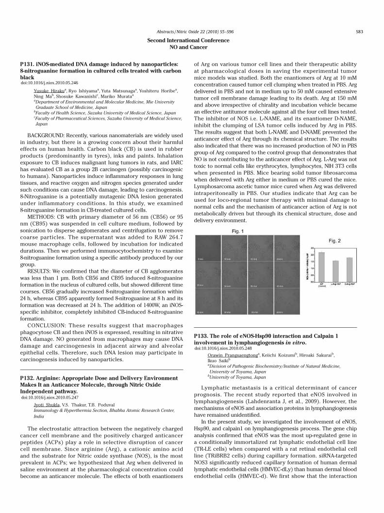
histopathological examination. The hematological results revealed a significant decrease in total RBCs and lymphocytes counts, Hb concentration and PCV. Meanwhile, a significant increase in total WBCs, neutrophil and monocyte counts throughout the experiment. The biochemical findings elucidated a significant decrease in triiodothyronine (T3) and thyroxine (T4) levels. On the other hand, a significant increase in serum glutamic pyruvic transaminase (sGPT) and serum glutamic oxaloacetic transaminase (sGOT) as well as blood urea nitrogen were evident. A highly significant decrease in serum nitric oxide was observed only in the first month, later on the serum nitric oxide was highly significantly increased until the end of the experiment. One goat, died 12 weeks post-treatment. Signs included tremors, unsteady gait and loss in body weight with blue mucous membrane. Enlargement in all organs and lymph nodes were observed. Histopathologically, inflammation and necrosis were found in all parenchymatous organs. Extensive depletion of lymphocytes from the white pulp was detected. Thyroid gland displayed vacuolated, necrotic and atrophic follicular cells. Cerebral edema was observed. It could be concluded that nitrate toxicity was accompanied by severe anemia, thyro-toxicosis and impaired liver and kidney functions.
P129. Flirting with anoxia: nitrite-dependent nitric oxide control of mitochondrial respiration
David Macherela, Hardy Rolletschekb,Ramaroson Andriantsitohainac, Hardy Benamara aUMR 1191 PMS, Universit d'Angers, FrancebIPK Gatersleben, GermanycUMR INSERM 771- CNRS 6214, France
For tissues combining high respiratory rate with limited oxygen diffusion, and which generally operate under hypoxia, the control of respiration is primordial to prevent anoxic conditions to settle.
We have developed an original oxygraphic method to monitor functioning of isolated plant mitochondria at low oxygen level, mimicking the situation in intact tissues. In this approach, the electrode reaction chamber is left open, allowing atmospheric oxygen to diffuse through the liquid surface. Steady-state oxygen levels then result from the equilibrium between mitochondrial oxygen consumption and oxygen diffusion from the atmosphere. Mitochondria functioning under self-generated hypoxic conditions can be monitored with this robust method for long periods (up to several hours). The approach nicely illustrated the reversible inhibitory effect of nitric oxide (NO) on cytochrome oxidase (COX) and brought to light the capacity of mitochondria to finely tune their surrounding oxygen levels through a nitrite-dependent nitric oxide regulation of oxygen consumption. At low O2, nitrite is
reduced into NO, likely at complex III, and in turn reversibly inhibits COX, provoking a rise to higher steady state levels of oxygen. Since NO can be re-oxidized chemically or by COX, the establishment of a nitrite-NO cycle prevents mitochondrial self-anoxia. Such a mechanism, which could be demonstrated with plant and animal mitochondria, is likely an evolutionarily conserved mechanism. It could have far-reaching implications for the regulation of oxygen homeostasis at the cellular level in tissues undergoing hypoxia.
Benamar A, Rolletschek H, Borisjuk L, Avelange-Macherel M-H, Curien G, Mostefai A, Andriantsitohaina R, Macherel D (2008) Nitrite-nitric oxide control of mitochondrial respiration at the frontier of anoxia. Biochim. Biophys. Acta Bioenergetics 1777, 1268-1275.
P130. Release of NO2- from nitro-compounds by biotic and
abiotic reactions
Naoko Watanabe, Yuki N Sakamoto, Hideo Yamasaki Faculty of Science, University of the Ryukyus, Japan
In many biological systems, nitric oxide (NO) is produced from L-arginine by the enzyme nitric oxide synthase (NOS). In addition to the L-arginine-dependent NO production, the radical can be produced from nitrite (NO2
-) by enzymatic as well as non-enzymatic mechanisms. It has been known that nitro-compounds such as nitroglycerin are capable of serving NO through either direct or indirect mechanisms. Recent studies have shown that the cells contain a variety of nitro molecules in proteins, nucleic acids and lipids. However, denitration mechanisms for those molecules have remained unclear. Using nitrophenol as a model compound, here we report nitrite release from nitro-compounds by biotic and abiotic reactions. We isolated soil bacteria that grew with 4-nitrophenol (4-NP) as a carbon source. 16s rDNA sequence suggested that the bacteria were closely associated with Pseudomonas putida. The bacteria produced nitrite from 4-NP in a culture medium and showed a low activity when 4-nitrocatechol was supplemented instead of 4-NP. However, nitrite was not produced with 2-nitrophenol and 2,4-dinitrophenol, suggesting the presence of high substrate specificity in the biodegradation of nitrophenols. We next investigated non-enzymatic nitrite release from nitrophenols with titanium dioxide (TiO2). Stoichiometric nitrite release was observed with TiO2 in the presence of hydrogen peroxide under UV illumination. There was no substrate specificity among the nitrophenols tested. The TiO2-dependent nitrite release from nitrophenols was observed even in the dark when the reaction temperature was high. These results suggest that nitrite release by biotic or enzymatic reactions may require specific structures, whereas that by abiotic or non-enzymatic reactions could be observed in any types of nitro-compounds. Reactive oxygen species such as hydroxyl radicals appear to be involved in the nitrite release from nitro-compounds.
Second International Conference NO and Cancer
P131. iNOS-mediated DNA damage induced by nanoparticles: 8-nitroguanine formation in cultured cells treated with carbon black
Yusuke Hirakua, Ryo Ishiyamaa, Yuta Matsunagaa, Yoshiteru Horibea, Ning Mab, Shosuke Kawanishic, Mariko Murataa aDepartment of Environmental and Molecular Medicine, Mie University Graduate School of Medicine, Japan
bFaculty of Health Science, Suzuka University of Medical Science, JapancFaculty of Pharmaceutical Sciences, Suzuka University of Medical Science, Japan
BACKGROUND: Recently, various nanomaterials are widely used in industry, but there is a growing concern about their harmful effects on human health. Carbon black (CB) is used in rubber products (predominantly in tyres), inks and paints. Inhalation exposure to CB induces malignant lung tumors in rats, and IARC has evaluated CB as a group 2B carcinogen (possibly carcinogenic to humans). Nanoparticles induce inflammatory responses in lung tissues, and reactive oxygen and nitrogen species generated under such conditions can cause DNA damage, leading to carcinogenesis. 8-Nitroguanine is a potentially mutagenic DNA lesion generated under inflammatory conditions. In this study, we examined 8-nitroguanine formation in CB-treated cultured cells.
METHODS: CB with primary diameter of 56 nm (CB56) or 95 nm (CB95) was suspended in cell culture medium, followed by sonication to disperse agglomerates and centrifugation to remove coarse particles. The supernatant was added to RAW 264.7 mouse macrophage cells, followed by incubation for indicated durations. Then we performed immunocytochemistry to examine 8-nitroguanine formation using a specific antibody produced by our group.
RESULTS: We confirmed that the diameter of CB agglomerates was less than 1 μm. Both CB56 and CB95 induced 8-nitroguanine formation in the nucleus of cultured cells, but showed different time courses. CB56 gradually increased 8-nitroguanine formation within 24 h, whereas CB95 apparently formed 8-nitroguanine at 8 h and its formation was decreased at 24 h. The addition of 1400W, an iNOS-specific inhibitor, completely inhibited CB-induced 8-nitroguanine formation.
CONCLUSION: These results suggest that macrophages phagocytose CB and then iNOS is expressed, resulting in nitrative DNA damage. NO generated from macrophages may cause DNA damage and carcinogenesis in adjacent airway and alveolar epithelial cells. Therefore, such DNA lesion may participate in carcinogenesis induced by nanoparticles.
P132\. Arginine: Appropriate Dose and Delivery Environment Makes It an Anticancer Molecule, through Nitric Oxide Independent pathway.
Jyoti Shukla, V.S. Thakur, T.B. Poduval Immunology & Hyperthermia Section, Bhabha Atomic Research Center, India
The electrostatic attraction between the negatively charged cancer cell membrane and the positively charged anticancer peptides (ACPs) play a role in selective disruption of cancer cell membrane. Since arginine (Arg), a cationic amino acid and the substrate for Nitric oxide synthase (NOS), is the most prevalent in ACPs; we hypothesized that Arg when delivered in saline environment at the pharmacological concentration could become an anticancer molecule. The effects of both enantiomers
of Arg on various tumor cell lines and their therapeutic ability at pharmacological doses in saving the experimental tumor mice models was studied. Both the enantiomers of Arg at 10 mM concentration caused tumor cell clumping when treated in PBS. Arg delivered in PBS and not in medium up to 50 mM caused extensive tumor cell membrane damage leading to its death. Arg at 150 mM and above irrespective of chirality and incubation vehicle became an effective antitumor molecule against all the four cell lines tested. The inhibitor of NOS i.e. L-NAME, and its enantiomer D-NAME, inhibit the clumping of LSA tumor cells induced by Arg in PBS. The results suggest that both L-NAME and D-NAME prevented the anticancer effect of Arg through its chemical structure. The results also indicated that there was no increased production of NO in PBS group of Arg compared to the control group that demonstrates that NO is not contributing to the anticancer effect of Arg. L-Arg was not toxic to normal cells like erythrocytes, lymphocytes, NIH 3T3 cells when presented in PBS. Mice bearing solid tumor fibrosarcoma when delivered with Arg either in medium or PBS cured the mice. Lymphosarcoma ascetic tumor mice cured when Arg was delivered intraperitoneally in PBS. Our studies indicate that Arg can be used for loco-regional tumor therapy with minimal damage to normal cells and the mechanism of anticancer action of Arg is not metabolically driven but through its chemical structure, dose and delivery environment.
P133. The role of eNOS-Hsp90 interaction and Calpain 1 involvement in lymphangiogenesis in vitro.
Orawin Prangsaengtonga, Keiichi Koizumib, Hiroaki Sakuraib,Ikuo Saikib aDivision of Pathogenic Biochemistry/Institute of Natural Medicine, University of Toyama, Japan
bUniversity of Toyama, Japan
Lymphatic metastasis is a critical determinant of cancer prognosis. The recent study reported that eNOS involved in lymphangiogenesis (Lahdenranta J, et al., 2009). However, the mechanisms of eNOS and association proteins in lymphangiogenesis have remained unidentified.
In the present study, we investigated the involvement of eNOS, Hsp90, and calpain1 on lymphangiogenesis process. The gene chip analysis confirmed that eNOS was the most up-regulated gene in a conditionally immortalized rat lymphatic endothelial cell line (TR-LE cells) when compared with a rat retinal endothelial cell line (TRiBRB2 cells) during capillary formation. siRNA-targeted NOS3 significantly reduced capillary formation of human dermal lymphatic endothelial cells (HMVEC-dLy) than human dermal blood endothelial cells (HMVEC-d). We first show that the interaction
S83
doi:10.1016/j.niox.2010.05.246
doi:10.1016/j.niox.2010.05.247
doi:10.1016/j.niox.2010.05.248
Abstracts /Nitric Oxide 22 (2010) S5–S96

of Hsp90 and eNOS correlated with tube formation pattern of HMVEC-dLy after seeded on Matrigel. This interaction including eNOS protein expression, and tube formation were significantly diminished in 17-DMAG, a Hsp90 inhibitor, pre-incubated cells for 24 h, while Hsp90 protein expression alone was not affected. Moreover, eNOS and Hsp90 protein expression was maintained in the early stage and dramatically decreased during late stage of lymphangiogenesis in vitro which the time of Hsp90 and eNOS interactions were declined. From the decreasing of Hsp90 and eNOS interaction in this stage, we hypothesized that calpain1 may degrade eNOS and cause to decreasing of lymphatic tube formation. The calpain1 inhibitor, PD151746 significantly increased tube formation length when compared with control. However, the mechanisms of calpain1 on lymphangiogenesis have been further investigated.
In conclusion, this study we found that eNOS was a highly important protein for lymphangiogenesis process and provides the new information about the involvement of eNOS and Hsp90 interaction and calpain 1 functions on lymphangiogenesis.
P134. δ-Tocotrienol suppresses iNOS protein expression and COX-2 enzyme activity
Sayori Wadaa, Yuzuru Matsushitaa, Yuji Naitob, Toshikazu Yoshikawab, Ohshima Hiroshic aFood Science and Nutritional Health, Kyoto Prefectural University, JapanbKyoto Prefectural University of Medicine, JapancUniversity of Shizuoka, Japan
[Aim] Tocotrienols is one class of natural compound of vitamin E, together with tocopherols. Previous studies showed that tocotrienols exert not only cardiovascular protective effects but also anti-cancer effects. We previously proved that δ-tocotrienol most potently inhibited cell proliferation in colon cancer cells. A positive correlation between inducible nitric oxide (iNOS) expression and grade of malignancy was found in human colorectal cancer. Cyclooxygenase-2 (COX-2) produced by stromal cells has been reported to promote tumorigenic progression of intestinal epithelial cells. We investigated whether δ-tocotrienol suppressed iNOS and/or COX-2 activity in stromal cells.
[Method] Mouse embryonic fibroblasts (MEFs) were stimulated with 1 μg/ml of LPS and 100 IU/ml of IFN-γ after pretreatment with or without various concentrations of δ-tocotrienol (0, 2.5, 5 μM). Protein expressions of iNOS and COX-2 were analyzed by Western-blotting, concentrations of nitrite were measured by Griess reagent, and concentrations of prostaglandin E2 (PGE2), which indicates COX-2 enzyme activity, was detected by ELISA. Cell viability was assessed by MTT assay.
[Results] Each concentration of δ-tocotrienol did not affect on cell viability. δ-tocotrienol treatment suppressed iNOS protein expression in dose-dependent manner. 5 μM of δ-tocotrienol significantly reduced nitrite concentrations (p<0.05). PGE2 concentrations was reduced by 2.5 μM of δ-tocotrienol (p<0.01) but not COX-2 proteins expression.
[Conclusion] δ-tocotrienol suppressed nitrite concentrations by suppressing iNOS protein expression. On the other hand, δ-tocotrienol reduced PGE2 concentrations without affecting COX-2 protein expression. It might indicate that δ-tocotrienol inhibited COX-2 enzyme activity. These data suggest that δ-tocotrienol appears to have a potential as anti-cancer agents reducing iNOS and COX-2 activity in stromal cells.
P135. Nitric oxide enhances cancer invasion through induction of AnnexinA8
Masayuki Tatemichi, Harumi Hata, Toshio Nakadate Hygiene and Preventive Medicine, Showa University, Japan
Background and Aim: We have reported that nitric oxide (NO) produced by inducible nitric oxide synthase (iNOS) enhanced cancer progression, particularly cancer invasion. Previously we reported that NO induced VEGF and MMPs expression. This study further tried to identify factors related to cancer progression by NO.
Methods: Mouse embriofibroblast cells (MEFs) with p53-
/-iNOS+/+and p53-/-iNOS-/- were cultured under the medium containing lipopolysaccharide (LPS) which induced spontaneous iNOS expression in p53-/- iNOS+/+ MEFs. p53-/-iNOS+/+, but not p53-
/-iNOS-/-, MEF cells showed anchorage independent growth (AIG). And then, we compared expression profiles between control cells and MEFs showing AIG using DNA chip and subtraction method.
Results: We found that Annexin A8 was overexpressed in MEFs showing AIG. AnnexinA8 was expressed in several human cancer cell lines. Epigenetics agents, 5-Aza-dC and histone deacetylase (HDAC) inhibitors enhanced Annexin A8 expression. Nitric oxide also enhanced Annexin A8 expression. The Annexin A8 expression levels were associated with hypomethylation status of CpG in the exon 1 region of Annexin A8 gene. Inhibition of Annexin A8 by siRNA reduced HIF-1alpha expression and invaded cell numbers by invasion assay.
Conclusion: Annexin A8 could be regulated by epigenetics and involved in cancer progression by NO through HIF-1alpha signaling.
P136. Nitrosylated human serum albumin (SNO-HSA) induces apoptosis in tumor cells
Marie Haraa, Naohisa Katayamaa,b,Keisuke Nakajoub, Yu Ishimaa, Shotaro Ikutab, Ayaka Suenagaa, Toru Maruyamaa, Toshiya Kaia,b,Masaki Otagiria,c aGraduate School of Pharmaceutical Sciences, Kumamoto University, JapanbPharmaceutical Research Center, Nipro, JapancSojo University, Japan
The cytotoxic activity of nitric oxide (NO) has recently been investigated for its potential use in anticancer therapies. In our previous study, poly S-nitrosylated human serum albumin (SNO-HSA) induced apoptosis in C26 cells in vitro and in vivo, demonstrating that SNO-HSA can be therapeutically applicable to cancer for the first time. In the present study, for the investigation of the anti-tumor activity of SNO-HSA against other tumor species, a rat tumor LY-80 cells (a variant Yoshida sarcoma) were used. Flow cytometric analysis using rhodamine 123 showed that SNO-HSA caused mitochondrial depolarization. Activation of caspase-3 and DNA fragmentation were dose-dependently observed in LY-80 cells by incubation with SNO-HSA. Inhibition of caspases completely abolished DNA fragmentation induced by SNO-HSA. SNO-HSA reduced cell viability of LY-80 cells correspondingly, and induced cytotoxicity in a concentration-dependent manner. The in vivo anti-tumor effect of SNO-HSA was investigated using LY-80 tumor-bearing rats. The growth of LY-80 tumors in rats was significantly inhibited by administration of SNO-HSA, compared with saline- and HSA-treatment. These results suggest that SNO-HSA induced apoptosis in tumor cells and has the potential for chemopreventive and/or chemotherapeutic activity.
Infection/inflammation/immunity
P137. Guanine nitration and oxidative stress responses during influenza virus pneumonia in mice
Shahzada Khan, Tatsuya Okamoto, Tomohiro Sawa, Shigemoto Fujii, Takaaki Akaike Department of Microbiology, Graduate School of Medical Sciences, Kumamoto University, Japan
Nitric oxide (NO) has been suggested to be involved in the pathogenesis of various diseases, including microbial infections, inflammatory diseases, and cancer. Guanine nitration is characterized as a nucleic acid modification induced by NO and reactive oxygen species during infections. Extensive 8-nitroguanine formation was identified in the lung of influenza virus-infected mice. We also recently found that cGMP, a second messenger of NO-signaling, was nitrated to form its unique nitrated derivative, 8-nitroguanosine 3', 5'-cyclic monophosphate (8-nitro-cGMP) in cells depending on the expression of inducible NO synthase (iNOS). In this study, to better understand pathophysiological relevance of guanine nitration, we investigated the formation of 8-nitro-cGMP in influenza virus pneumonia model in mice. We observed strong 8-nitro-cGMP immunostaining primarily in the cytosol of bronchial epithelial cells of influenza virus-infected wild-type mice but not in the iNOS-deficient mice. Interestingly, in parallel with 8-nitro-cGMP formation, strong induction of heme oxygenase-1 (HO-1), an antioxidant enzyme, was detected in the influenza virus-infected lung. HO-1 expression was well co-localized with that of 8-nitro-cGMP: infiltrated phagocytes and bronchial epithelial cells were strongly immunostained for HO-1. On the other hand, HO-1 was induced to lesser extent in the infected lung of iNOS-deficient mice than that of wild-type mice. Importantly, authentic 8-nitro-cGMP strongly induced HO-1 in cultured cells, suggesting potent signaling functions of 8-nitro-cGMP for HO-1 induction. The present data thus indicate that guanine nitration forming 8-nitro-cGMP may be involved in a unique signal transduction, which contributes to the oxidative stress responses during influenza virus pneumonia.
P138. The susceptibility of ventral horn motoneurons to roots avulsion injury was related to nNOS gene diversity
Lifeng Yana, Lihua Zhoub aZhong Shan School of Medicine, Sun Yat-sen University, ChinabSun Yat-sen University, China
OBJECTIVE: To evaluate the nNOS gene levels in different region of the injured spinal cord caused by brachial root avulsion in the rats. BACKGROUND: A number of previous studies have demonstrated that avulsion of the spinal ventral roots leads to loss of motoneurons in the injured spinal segments, and nNOS gene was closely related to the motoneuron death. METHODS: Sprague-Dawly rats were chosen as the experimental animals. The five (C5-8 and T1) spinal roots were avulsed. After surviving for 14 days the spinal segments from C6-8 of some animals were removed and divided into ipsilateral and contralateral half from midline, the ventral or dorsal horns in control or lesioned parts of the C6-8 spinal segments were taken out separately for RT-PCR and western blot detection. and the others were perfused and prepared for NADPH staining, nNOS immunofluorescence labling. RESULTS: At 14 days after avulsion, nNOS positive motoneurons and NADPH-d positive motoneurons were evident in lesioned ventral horn but did not appeared in control ventral horn. But nNOS expression pattern was no significantly difference between lesioned and control dorsal horns. At 14 days after the brachial root avulsion, the amount of nNOS mRNA and nNOS protein in C6-8 spinal segments of the spinal
cord were more than that in control ventral horn, the difference was statistically significant, and there were no difference between the lesioned dorsal horn and control dorsal horn. CONCLUSIONS: The present study revealed that (1) regional or spatial differences exist in the injured spinal cord after brachial root avulsion, and the difference is may be play a key role in loss of motoneurons, (2) avulsion can induce that motoneurons de novo nNOS mRNA and protein expressions and this change can not be detected in whole injured spinal cord and must be in the lesioned ventral horn tissue.
P139. Nitric- and oxidative mitochondria related cell death by cytokines and its prevention by estrogen in estrogen receptor expressed rheumatoid arthritis synovial fibroblasts
Shigeaki Suenagaa, Hiroko P Indoa, Kazuo Tomitaa, Kosei Ijiria, Setsuro Komiyaa, Takami Matsuyamaa, Hsiu-Chuan Yenb, Masahiro Higuchic, Hirofumi Matsuid, Toshihiko Ozawae, Hideyuki J Majimaa aDepartment of Oncology, Kagoshima University Graduate School of Medical and Dental Sciences, Japan
bChang Gung University, TaiwancUniversity of Arkansas for Medical Sciences, United States of AmericadUniversity of Tsukuba, JapaneYokohama College of Pharmacy, Japan
We examined the expressions of estrogen receptor (ER), the production and activity of reactive oxygen species (ROS), reactive nitrogen species (RNS), lipid peroxidation and apoptosis following a cytokine treatment in primary fibroblast cell lines which have been established from human synovial tissues of rheumatoid arthritis (RA) patients. Two cell lines, which expressed the most, and two cell lines, which expressed the least were selected among 9 cell lines, and were used for this study. Cells, incubated with cytokines (IL-1β, TNF-α, IFN-γ) with or without estrogen, were examined to observe generations of ROS, RNS, lipid peroxidation and apoptotic cell death. The results showed that cytokines stimulation on synovial fibroblasts increased the intracellular mitochondrial ROS and RNS generations, lipid peroxidation product, and followed by apoptosis. Combined treatment of estrogen plus cytokines for the high ERα expressed fibroblasts decreased the mitochondrial ROS and RNS generations, lipid peroxidation product and prevented subsequent apoptotic cell death, although the low expressed ERα cells did not show the substantial change. Examinations of the levels of iNOS mRNA confirmed that the expression was dependent on the RNS levels. These results suggest that both mitochondrial ROS and RNS are key players to RA pathogenesis and estrogen plays an important role in protecting cells against cytokine-induced cell death by controlling the generations of mitochondrial ROS, RNS and lipid peroxidation. These results also suggest that mitochondria, as the initial trigger, control the subsequent events to induce apoptosis in RA cells.
P140. Can iNOS be the target in blocking of cytotoxic signal activation in pathophysiology of sepsis?
Kyungho Chang, Yoshitsugu Yamada Department of Anesthesiology, University of Tokyo, Japan
[Background]Sepsis remains the most important cause of acute kidney injury (AKI) in critically ill patients and accounts for more than 50% cases of AKI in ICUs. However, the pathophysiology of sepsis has not been fully elucidated. While hemodynamic factors are important in the cause of septic AKI, non-hemodynamic ones, including direct cytotoxicity of inflammatory mediators, are supposed to play major roles as well. Although NO has been
S84
doi:10.1016/j.niox.2010.05.249
doi:10.1016/j.niox.2010.05.250
doi:10.1016/j.niox.2010.05.251
Abstracts /Nitric Oxide 22 (2010) S5–S96

of Hsp90 and eNOS correlated with tube formation pattern of HMVEC-dLy after seeded on Matrigel. This interaction including eNOS protein expression, and tube formation were significantly diminished in 17-DMAG, a Hsp90 inhibitor, pre-incubated cells for 24 h, while Hsp90 protein expression alone was not affected. Moreover, eNOS and Hsp90 protein expression was maintained in the early stage and dramatically decreased during late stage of lymphangiogenesis in vitro which the time of Hsp90 and eNOS interactions were declined. From the decreasing of Hsp90 and eNOS interaction in this stage, we hypothesized that calpain1 may degrade eNOS and cause to decreasing of lymphatic tube formation. The calpain1 inhibitor, PD151746 significantly increased tube formation length when compared with control. However, the mechanisms of calpain1 on lymphangiogenesis have been further investigated.
In conclusion, this study we found that eNOS was a highly important protein for lymphangiogenesis process and provides the new information about the involvement of eNOS and Hsp90 interaction and calpain 1 functions on lymphangiogenesis.
P134. δ-Tocotrienol suppresses iNOS protein expression and COX-2 enzyme activity
Sayori Wadaa, Yuzuru Matsushitaa, Yuji Naitob, Toshikazu Yoshikawab, Ohshima Hiroshic aFood Science and Nutritional Health, Kyoto Prefectural University, JapanbKyoto Prefectural University of Medicine, JapancUniversity of Shizuoka, Japan
[Aim] Tocotrienols is one class of natural compound of vitamin E, together with tocopherols. Previous studies showed that tocotrienols exert not only cardiovascular protective effects but also anti-cancer effects. We previously proved that δ-tocotrienol most potently inhibited cell proliferation in colon cancer cells. A positive correlation between inducible nitric oxide (iNOS) expression and grade of malignancy was found in human colorectal cancer. Cyclooxygenase-2 (COX-2) produced by stromal cells has been reported to promote tumorigenic progression of intestinal epithelial cells. We investigated whether δ-tocotrienol suppressed iNOS and/or COX-2 activity in stromal cells.
[Method] Mouse embryonic fibroblasts (MEFs) were stimulated with 1 μg/ml of LPS and 100 IU/ml of IFN-γ after pretreatment with or without various concentrations of δ-tocotrienol (0, 2.5, 5 μM). Protein expressions of iNOS and COX-2 were analyzed by Western-blotting, concentrations of nitrite were measured by Griess reagent, and concentrations of prostaglandin E2 (PGE2), which indicates COX-2 enzyme activity, was detected by ELISA. Cell viability was assessed by MTT assay.
[Results] Each concentration of δ-tocotrienol did not affect on cell viability. δ-tocotrienol treatment suppressed iNOS protein expression in dose-dependent manner. 5 μM of δ-tocotrienol significantly reduced nitrite concentrations (p<0.05). PGE2 concentrations was reduced by 2.5 μM of δ-tocotrienol (p<0.01) but not COX-2 proteins expression.
[Conclusion] δ-tocotrienol suppressed nitrite concentrations by suppressing iNOS protein expression. On the other hand, δ-tocotrienol reduced PGE2 concentrations without affecting COX-2 protein expression. It might indicate that δ-tocotrienol inhibited COX-2 enzyme activity. These data suggest that δ-tocotrienol appears to have a potential as anti-cancer agents reducing iNOS and COX-2 activity in stromal cells.
P135. Nitric oxide enhances cancer invasion through induction of AnnexinA8
Masayuki Tatemichi, Harumi Hata, Toshio Nakadate Hygiene and Preventive Medicine, Showa University, Japan
Background and Aim: We have reported that nitric oxide (NO) produced by inducible nitric oxide synthase (iNOS) enhanced cancer progression, particularly cancer invasion. Previously we reported that NO induced VEGF and MMPs expression. This study further tried to identify factors related to cancer progression by NO.
Methods: Mouse embriofibroblast cells (MEFs) with p53-
/-iNOS+/+and p53-/-iNOS-/- were cultured under the medium containing lipopolysaccharide (LPS) which induced spontaneous iNOS expression in p53-/- iNOS+/+ MEFs. p53-/-iNOS+/+, but not p53-
/-iNOS-/-, MEF cells showed anchorage independent growth (AIG). And then, we compared expression profiles between control cells and MEFs showing AIG using DNA chip and subtraction method.
Results: We found that Annexin A8 was overexpressed in MEFs showing AIG. AnnexinA8 was expressed in several human cancer cell lines. Epigenetics agents, 5-Aza-dC and histone deacetylase (HDAC) inhibitors enhanced Annexin A8 expression. Nitric oxide also enhanced Annexin A8 expression. The Annexin A8 expression levels were associated with hypomethylation status of CpG in the exon 1 region of Annexin A8 gene. Inhibition of Annexin A8 by siRNA reduced HIF-1alpha expression and invaded cell numbers by invasion assay.
Conclusion: Annexin A8 could be regulated by epigenetics and involved in cancer progression by NO through HIF-1alpha signaling.
P136. Nitrosylated human serum albumin (SNO-HSA) induces apoptosis in tumor cells
Marie Haraa, Naohisa Katayamaa,b,Keisuke Nakajoub, Yu Ishimaa, Shotaro Ikutab, Ayaka Suenagaa, Toru Maruyamaa, Toshiya Kaia,b,Masaki Otagiria,c aGraduate School of Pharmaceutical Sciences, Kumamoto University, JapanbPharmaceutical Research Center, Nipro, JapancSojo University, Japan
The cytotoxic activity of nitric oxide (NO) has recently been investigated for its potential use in anticancer therapies. In our previous study, poly S-nitrosylated human serum albumin (SNO-HSA) induced apoptosis in C26 cells in vitro and in vivo, demonstrating that SNO-HSA can be therapeutically applicable to cancer for the first time. In the present study, for the investigation of the anti-tumor activity of SNO-HSA against other tumor species, a rat tumor LY-80 cells (a variant Yoshida sarcoma) were used. Flow cytometric analysis using rhodamine 123 showed that SNO-HSA caused mitochondrial depolarization. Activation of caspase-3 and DNA fragmentation were dose-dependently observed in LY-80 cells by incubation with SNO-HSA. Inhibition of caspases completely abolished DNA fragmentation induced by SNO-HSA. SNO-HSA reduced cell viability of LY-80 cells correspondingly, and induced cytotoxicity in a concentration-dependent manner. The in vivo anti-tumor effect of SNO-HSA was investigated using LY-80 tumor-bearing rats. The growth of LY-80 tumors in rats was significantly inhibited by administration of SNO-HSA, compared with saline- and HSA-treatment. These results suggest that SNO-HSA induced apoptosis in tumor cells and has the potential for chemopreventive and/or chemotherapeutic activity.
Infection/inflammation/immunity
P137. Guanine nitration and oxidative stress responses during influenza virus pneumonia in mice
Shahzada Khan, Tatsuya Okamoto, Tomohiro Sawa, Shigemoto Fujii, Takaaki Akaike Department of Microbiology, Graduate School of Medical Sciences, Kumamoto University, Japan
Nitric oxide (NO) has been suggested to be involved in the pathogenesis of various diseases, including microbial infections, inflammatory diseases, and cancer. Guanine nitration is characterized as a nucleic acid modification induced by NO and reactive oxygen species during infections. Extensive 8-nitroguanine formation was identified in the lung of influenza virus-infected mice. We also recently found that cGMP, a second messenger of NO-signaling, was nitrated to form its unique nitrated derivative, 8-nitroguanosine 3', 5'-cyclic monophosphate (8-nitro-cGMP) in cells depending on the expression of inducible NO synthase (iNOS). In this study, to better understand pathophysiological relevance of guanine nitration, we investigated the formation of 8-nitro-cGMP in influenza virus pneumonia model in mice. We observed strong 8-nitro-cGMP immunostaining primarily in the cytosol of bronchial epithelial cells of influenza virus-infected wild-type mice but not in the iNOS-deficient mice. Interestingly, in parallel with 8-nitro-cGMP formation, strong induction of heme oxygenase-1 (HO-1), an antioxidant enzyme, was detected in the influenza virus-infected lung. HO-1 expression was well co-localized with that of 8-nitro-cGMP: infiltrated phagocytes and bronchial epithelial cells were strongly immunostained for HO-1. On the other hand, HO-1 was induced to lesser extent in the infected lung of iNOS-deficient mice than that of wild-type mice. Importantly, authentic 8-nitro-cGMP strongly induced HO-1 in cultured cells, suggesting potent signaling functions of 8-nitro-cGMP for HO-1 induction. The present data thus indicate that guanine nitration forming 8-nitro-cGMP may be involved in a unique signal transduction, which contributes to the oxidative stress responses during influenza virus pneumonia.
P138. The susceptibility of ventral horn motoneurons to roots avulsion injury was related to nNOS gene diversity
Lifeng Yana, Lihua Zhoub aZhong Shan School of Medicine, Sun Yat-sen University, ChinabSun Yat-sen University, China
OBJECTIVE: To evaluate the nNOS gene levels in different region of the injured spinal cord caused by brachial root avulsion in the rats. BACKGROUND: A number of previous studies have demonstrated that avulsion of the spinal ventral roots leads to loss of motoneurons in the injured spinal segments, and nNOS gene was closely related to the motoneuron death. METHODS: Sprague-Dawly rats were chosen as the experimental animals. The five (C5-8 and T1) spinal roots were avulsed. After surviving for 14 days the spinal segments from C6-8 of some animals were removed and divided into ipsilateral and contralateral half from midline, the ventral or dorsal horns in control or lesioned parts of the C6-8 spinal segments were taken out separately for RT-PCR and western blot detection. and the others were perfused and prepared for NADPH staining, nNOS immunofluorescence labling. RESULTS: At 14 days after avulsion, nNOS positive motoneurons and NADPH-d positive motoneurons were evident in lesioned ventral horn but did not appeared in control ventral horn. But nNOS expression pattern was no significantly difference between lesioned and control dorsal horns. At 14 days after the brachial root avulsion, the amount of nNOS mRNA and nNOS protein in C6-8 spinal segments of the spinal
cord were more than that in control ventral horn, the difference was statistically significant, and there were no difference between the lesioned dorsal horn and control dorsal horn. CONCLUSIONS: The present study revealed that (1) regional or spatial differences exist in the injured spinal cord after brachial root avulsion, and the difference is may be play a key role in loss of motoneurons, (2) avulsion can induce that motoneurons de novo nNOS mRNA and protein expressions and this change can not be detected in whole injured spinal cord and must be in the lesioned ventral horn tissue.
P139. Nitric- and oxidative mitochondria related cell death by cytokines and its prevention by estrogen in estrogen receptor expressed rheumatoid arthritis synovial fibroblasts
Shigeaki Suenagaa, Hiroko P Indoa, Kazuo Tomitaa, Kosei Ijiria, Setsuro Komiyaa, Takami Matsuyamaa, Hsiu-Chuan Yenb, Masahiro Higuchic, Hirofumi Matsuid, Toshihiko Ozawae, Hideyuki J Majimaa aDepartment of Oncology, Kagoshima University Graduate School of Medical and Dental Sciences, Japan
bChang Gung University, TaiwancUniversity of Arkansas for Medical Sciences, United States of AmericadUniversity of Tsukuba, JapaneYokohama College of Pharmacy, Japan
We examined the expressions of estrogen receptor (ER), the production and activity of reactive oxygen species (ROS), reactive nitrogen species (RNS), lipid peroxidation and apoptosis following a cytokine treatment in primary fibroblast cell lines which have been established from human synovial tissues of rheumatoid arthritis (RA) patients. Two cell lines, which expressed the most, and two cell lines, which expressed the least were selected among 9 cell lines, and were used for this study. Cells, incubated with cytokines (IL-1β, TNF-α, IFN-γ) with or without estrogen, were examined to observe generations of ROS, RNS, lipid peroxidation and apoptotic cell death. The results showed that cytokines stimulation on synovial fibroblasts increased the intracellular mitochondrial ROS and RNS generations, lipid peroxidation product, and followed by apoptosis. Combined treatment of estrogen plus cytokines for the high ERα expressed fibroblasts decreased the mitochondrial ROS and RNS generations, lipid peroxidation product and prevented subsequent apoptotic cell death, although the low expressed ERα cells did not show the substantial change. Examinations of the levels of iNOS mRNA confirmed that the expression was dependent on the RNS levels. These results suggest that both mitochondrial ROS and RNS are key players to RA pathogenesis and estrogen plays an important role in protecting cells against cytokine-induced cell death by controlling the generations of mitochondrial ROS, RNS and lipid peroxidation. These results also suggest that mitochondria, as the initial trigger, control the subsequent events to induce apoptosis in RA cells.
P140. Can iNOS be the target in blocking of cytotoxic signal activation in pathophysiology of sepsis?
Kyungho Chang, Yoshitsugu Yamada Department of Anesthesiology, University of Tokyo, Japan
[Background]Sepsis remains the most important cause of acute kidney injury (AKI) in critically ill patients and accounts for more than 50% cases of AKI in ICUs. However, the pathophysiology of sepsis has not been fully elucidated. While hemodynamic factors are important in the cause of septic AKI, non-hemodynamic ones, including direct cytotoxicity of inflammatory mediators, are supposed to play major roles as well. Although NO has been
S85
doi:10.1016/j.niox.2010.05.252
doi:10.1016/j.niox.2010.05.253
doi:10.1016/j.niox.2010.05.254
doi:10.1016/j.niox.2010.05.255
Abstracts /Nitric Oxide 22 (2010) S5–S96

implicated as one of major inflammatory mediators associated with dysfunction of vital organs, whether blockade of iNOS is beneficial in septic patients is still controversial.
[Methods]We analyzed cytotoxic signal pathway induced by combined inflammatory mediators (cytomix;LPS, TNFalpha and IFNgamma) in immortalized rat renal proximal tubular cells (IRPTC) and investigated what kinds of pharmacological agents are effective in attenuating the cytotoxic signal pathways.
[Results]Cytomix induced cell death in various cultured cells including IRPTC. Caspase-dependent apoptosis was confirmed in cytomix-induced cell death. This cytotoxicity consisted of NO-dependent and NO-independent pathway. Cytomix-induced cell death was attenuated by 1400W (iNOS inhibitor), dexamethasone, melatonin, or spironolactone in IRPTC. These reagents attenuated caspase-dependent apoptosis in cytomix-induced cell death. While 1400W completely inhibited NO production, the latter three reagents partially inhibited NO production. However, the cytoprotective effect exerted by 1400W did not last long.
[Discussion]Combined inflammatory mediators were cytotoxic and activated apoptosis signal in renal tubular cells. This cytotoxicity was attenuated by an iNOS inhibitor, dexamethasone, melatonin, or spironolactone. However, the effect of direct iNOS inhibition was less reliable than the other pharmacological reagents in terms of cytoprotection.
P141. ER stress pathway induced by Nitric Oxide regulates inflammatory diseases through cell death and cytokine induction
Motoyoshi Endoa, Yoichiro Nakayamaa, Hiroto Tsukanoa,Masataka Morib, Yuichi Oikea, Tomomi Gotoha aDepartment of Molecular Genetics, Faculty of Medical and Pharmaceutical Sciences, Kumamoto University, Japan
bSojo University, Japan
Nitric oxide (NO) is one of the triggers of endoplasmic reticulum (ER) stress pathway in inflammation. ER is involved in various biosynthetic events, including processing, modification, folding, and the assembly of newly synthesized secretory and cell membrane proteins. Therefore, its proper function is essential to cell survival. Various stresses disturb ER functions, and unfolded proteins are accumulated in the ER lumen. Then, the ER stress pathways are activated to protect the cells. These responses involve induction of ER chaperones including BiP. Therefore, ER stress pathway function as an intracellular stress sensor and protect cells from various stresses. However, when the ER functions are severely impaired, then apoptosis occurs to remove damaged cells. CHOP is a member of the C/EBP transcription factor family, is induced by ER stress response. NO is one of the major candidates by which the ER stress pathways are activated in inflammation. We found CHOP show bilateral characters in inflammation. When inflammation-inducing stimulus were mild, then ER function-protecting molecules including, ER chaperones, were induced earlier than CHOP, and apoptosis was escaped. In this condition, IL-1β pathway is activated through caspase-11 induction in a CHOP-dependent manner. On the other hand, when inflammation-inducing stimulus were severe, CHOP induction was rapid and high, then apoptosis was induced. In this way, the cellular responses to ER stress in inflammation are variable, depending on the difference in stimuli. We also have showed the relationship between various inflammatory diseases and CHOP induction. In fact, chop deficient mice were resistant to various inflammatory diseases, such as pneumonitis, pancreatitis, and atherosclerosis. From these results, ER stress pathway involving CHOP activated by NO in inflammation can be the new target for therapy.
P142. INVOLVEMENTI OF DDAH-ADMA AXIS IN ACCELERATED RENAL INJURY IN ACUTE ISCHEMIA-REPERFUSION INJURY
Yosuke Nakayamaa, Seiji Uedaa, Sho-ichi Yamagishib, Yusuke Kaidaa, Ryotaro Andoa, Ryuji Iwatania, Kei Fukamia, Seiya Okudaa aDivision of Nephrology,Department of Medicine, Kurume University, JapanbDepartment of Physiology and Therapeutics of Diabetic vascular Complications,Krume University, Japan
Background. Recent evidence suggests that injury to the renal vasculature may play an important role in the pathogenesis of ischemic acute kidney injury (AKI). Since nitric oxide (NO) is a vasodilator and known to play an important role in the maintenance of renal microvasculature and its flow and may exert a protective role against AKI, it is conceivable that asymmetric dimethylarginine (ADMA), an endogenous inhibitor of NO synthase, could enhance tubulointerstitial injury during AKI by decreasing NO bioavailability. However, the association between ADMA and AKI remain to be elucidated.Methods. To determine the role of ADMA in AKI, eight-week-old male C57BL/6J (wild) mice and DDAH-1, a key enzyme for ADMA degradation, transgenic (Tg) mice were used in this study. Ischemia- reperfusion (IR) injury were performed in wild mice with (n=8) or without (n=11) ADMA infusion (0.01mg/kg/min) and Tg mice (n=5). Sham operated mice (n=5) were used as control. Renal function and morphology of acute renal injury were compared after IR. Tissue or plasma levels of ADMA were measured by HPLC. ADMA-related related enzymes such as DDAH and PRMT, an enzyme for ADMA synthesis, were measured by western blot analysis.Results. Western blot analysis revealed significant decreases in renal DDAH expression levels and increases in PRMT expression levels during IR injury associated with increased ADMA levels in wild mice. Compared with IR-treated wild mice, ADMA infusion markedly enhanced tubular injury and increases in BUN levels, whereas these changes were significantly attenuated in IR-treated DDAH Tg mice.Conclusion. These results strongly suggest that the active involvements of ADMA-DDAH axis in the pathogenesis of IR injury. ADMA-DDAH axis could be a novel target for patients with AKI.
P143. cAMP mediated up-regulation and expression of enzymes involved in arginine metabolism in Human Gingival Fibroblasts
Farzaneh Daghigh, Ellen Cho, Scott Riggs, Sandra Taylor Coley Biochemistry and Molecular Biology, Philadelphia College of Osteopathic Medicine, United States of America
Availability of arginine during periodontitis may be regulated by enzymes involved in arginine and ornithine metabolisms in human gingival fibroblast (HGF). Thus, the expression of enzymes such as arginase I (ARGI), arginase II (ARG II), ornithine decarboxylase (ODC), and ornithine aminotransferase (OAT) appears to be essential in the regulation of the cellular immune response and the inflammatory process during periodontitis. Arginase may also play a role in the regulation of arginine availability for nitric oxide (NO) synthesis. Excessive NO is associated with tissue destruction and inflammation and its synthesis is increased in periodontitis. The inducible form of nitric oxide synthase (iNOS) is responsible for the overproduction of NO. Arginine is a common substrate of both iNOS and arginase enzymes. Up-regulation of arginase is speculated to decrease arginine availability for iNOS and it may down-regulate NO overproduction. Pro-inflammatory cytokines have been found to up-regulate iNOS in HGF but regulation of arginase enzymes, ODC, and OAT in this tissue type has yet to be determined. Presence of ARGI and ARGII in HGF may provide a source of ornithine for production of polyamines, and ultimately for collagen synthesis and cellular
proliferation via ODC and OAT. In this study, we have determined the transcription and expression of the enzymes important in homeostasis arginine and ornithine in HGF in absence and presence of T helper II cytokines and glucocorticoids. Our major findings are as followings: 1) ARGI, ARGII, ODC, and OAT are expressed in HGF constitutively, 2) cAMP and dexamethasone significantly induce these enzymes 3) Pro-inflammatory cytokines inhibit their expression and attenuated their enhanced expression via cAMP. These enzymes may also modulate the inflammatory response in periodontitis, limiting NO production by iNOS and exerting anti-inflammatory effects.
P144. Detection of protein-bound 3-nitrotyrosine in plasma from pediatric patients with fulminant ARDS and avian influenza infection
Tatsuya Okamotoa, Tomohiro Sawaa, Shigemoto Fujiia, Mie Tateyamaa, Shoji Kawachib, Thuy Thi Pungc, Lien Thanh Nguyenc, Kazuo Suzukib, Takaaki Akaikea aDepartment of Microbiology, Graduate School of Medical Sciences, Kumamoto University, Japan
bNational Institute of Infectious Disease, JapancNational Hospital of Pediatrics, Viet Nam
Nitric oxide (NO) and reactive oxygen species (ROS) have been suggested to be involved in the pathogenesis of various diseases, including microbial infections, inflammatory diseases, and cancer. 3-Nitrotyrosine (3-NT) produced by NO/ROS is
considered as a biomarker for oxidative stress. In fact, we have identified the extensive and NO-dependent formation of 3-NT in the lung of influenza virus-infected mice (PNAS, 100:685-690, 2003). Acute respiratory distress syndrome (ARDS) is characterized as an inflammatory lung disease, and associates with enhanced formation of NO and ROS. Immunological analyses suggested the formation of 3-NT in the lungs of patients with ARDS. However, biochemical and quantitative aspects of 3-NT formation in ARDS patients remains poorly understood. In this study, we investigated the levels of plasma protein-bound 3-NT from fulminant cases with ARDS patients by using a reverse phase-HPLC coupled with electrochemical detector (ECD). We analyzed 36 plasma specimens (27 cases) from pediatric patients with ARDS. Four specimens (2 cases) with highly pathogenic avian influenza A (H5N1)-positive and 32 specimens (25 cases) with H5N1-negative ARDS were enrolled. Plasma proteins were precipitated with nitrite-free ethanol/acetate buffer followed by digested with pronase. 3-NT separated with the HPLC was first reduced at -700 mV to form 3-aminotyrosine, followed by being oxidized with second electrode (+150 mV) to detect 3-NT electrochemically. Detection limit of this system was approximately 1 nM for 3-NT. 3-NT levels were standardized by free tyrosine, determined by ultraviolet detector, connected just after ECD. By this method, we could successfully detect 3-NT from human plasma of ARDS patients. This method is convenient, specific and sensitive for 3-NT quantification applicable for clinical specimens and hence may help further understanding of pathological roles of NO/ROS formation in ARDS.
Pharmacology/therapeutic applications
P145. Photo-induced NO Release from Nano-sized Sphere for New Cancer Therapy
Kazuhiro Yamaguchia, Naoki Kanayamaa,b, Yukio Nagasakia,b,c,d,e aGraduate School of Pure and Applied Sciences, Tsukuba University, JapanbTsukuba Research Center for Interdisciplinary Materials Science (TIMS), Japan
cCenter for Tsukuba Advanced Research Alliance (TARA), JapandGraduate School of Comprehensive Human Sciences, Tsukuba University, Japan
eSatellite Laboratory of International Center for Materials Nanoarchitechtonics(MANA), NIMS, Japan
Nitric oxide (NO) has been widely investigated in physiology and pathology because of its role in multiple processes. NO has been found to play key roles in inhibiting tumor cell adhesion, arresting the growth of tumor cells. Though NO possesses a significant potential as an anticancer agent, the efficient therapeutic strategies to deliver NO specifically to tumor site are one of the recent major targets. In this study, we newly developed a highly dispersible nano-sized sphere with NO photo-generative core (Fig. (a)). Particles with several tens nanometers in size are known to preferentially accumulate in solid tumors by the enhanced permeation and retention (EPR) effect. If a photochemical process is combined with NO release, spatiotemporal control of the NO can be anticipated. Therefore, a combination of EPR effect and photochemical reaction is expected to achieve the site-specific delivery of NO in vivo.
Poly(ethylene glycol) (PEG)-based amphiphilic block copolymer (PEG-b-PCNTP) carrying 4-nitro-3-(trifluoromethyl)phenyl (NTP) moiety was synthesized via conventional two step reaction (Fig (b)). NTP derivatives are known to generate NO under UV light irradiation (Fig (c)). PEG-b-PCNTP was confirmed to spontaneously form highly dispersible and sphere-shaped particles in water, and their diameter was estimated as ca. 40 nm. The NO photo-generative ability of
PEG-b-PCNTP particles was confirmed by both ESR spin trapping method and Griess method. As the NO produced from PEG-b-PCNTP particles by light irradiation would act as a cytotoxic agent, the photo-induced cytotoxicity to mouse colon-26 carcinoma cells was investigated. Contrast to low cytotoxicity of PEG-b-PCNTP particle, colon-26 cells viability was effectively decreased by irradiation of UV light after the contacted with PEG-b-PCNTP particle for 24 h. These results suggest that the photo-induced cytotoxic behavior of PEG-b-PCNTP particle is useful for site-specific cancer therapy.
P146. Evaluation of cytotoxicity and anticarcinogenic potential of Mentha piperita Linn leaf extracts
Ravindra M Samartha Department of Research, Bhopal Memorial Hospital & Research Centre (BMHRC), India
We demonstrated radioprotective and anticancer properties of Mentha piperita leaf extract in Swiss albino mice in earlier studies.
S86
doi:10.1016/j.niox.2010.05.256
doi:10.1016/j.niox.2010.05.257
doi:10.1016/j.niox.2010.05.258
Abstracts /Nitric Oxide 22 (2010) S5–S96

implicated as one of major inflammatory mediators associated with dysfunction of vital organs, whether blockade of iNOS is beneficial in septic patients is still controversial.
[Methods]We analyzed cytotoxic signal pathway induced by combined inflammatory mediators (cytomix;LPS, TNFalpha and IFNgamma) in immortalized rat renal proximal tubular cells (IRPTC) and investigated what kinds of pharmacological agents are effective in attenuating the cytotoxic signal pathways.
[Results]Cytomix induced cell death in various cultured cells including IRPTC. Caspase-dependent apoptosis was confirmed in cytomix-induced cell death. This cytotoxicity consisted of NO-dependent and NO-independent pathway. Cytomix-induced cell death was attenuated by 1400W (iNOS inhibitor), dexamethasone, melatonin, or spironolactone in IRPTC. These reagents attenuated caspase-dependent apoptosis in cytomix-induced cell death. While 1400W completely inhibited NO production, the latter three reagents partially inhibited NO production. However, the cytoprotective effect exerted by 1400W did not last long.
[Discussion]Combined inflammatory mediators were cytotoxic and activated apoptosis signal in renal tubular cells. This cytotoxicity was attenuated by an iNOS inhibitor, dexamethasone, melatonin, or spironolactone. However, the effect of direct iNOS inhibition was less reliable than the other pharmacological reagents in terms of cytoprotection.
P141. ER stress pathway induced by Nitric Oxide regulates inflammatory diseases through cell death and cytokine induction
Motoyoshi Endoa, Yoichiro Nakayamaa, Hiroto Tsukanoa,Masataka Morib, Yuichi Oikea, Tomomi Gotoha aDepartment of Molecular Genetics, Faculty of Medical and Pharmaceutical Sciences, Kumamoto University, Japan
bSojo University, Japan
Nitric oxide (NO) is one of the triggers of endoplasmic reticulum (ER) stress pathway in inflammation. ER is involved in various biosynthetic events, including processing, modification, folding, and the assembly of newly synthesized secretory and cell membrane proteins. Therefore, its proper function is essential to cell survival. Various stresses disturb ER functions, and unfolded proteins are accumulated in the ER lumen. Then, the ER stress pathways are activated to protect the cells. These responses involve induction of ER chaperones including BiP. Therefore, ER stress pathway function as an intracellular stress sensor and protect cells from various stresses. However, when the ER functions are severely impaired, then apoptosis occurs to remove damaged cells. CHOP is a member of the C/EBP transcription factor family, is induced by ER stress response. NO is one of the major candidates by which the ER stress pathways are activated in inflammation. We found CHOP show bilateral characters in inflammation. When inflammation-inducing stimulus were mild, then ER function-protecting molecules including, ER chaperones, were induced earlier than CHOP, and apoptosis was escaped. In this condition, IL-1β pathway is activated through caspase-11 induction in a CHOP-dependent manner. On the other hand, when inflammation-inducing stimulus were severe, CHOP induction was rapid and high, then apoptosis was induced. In this way, the cellular responses to ER stress in inflammation are variable, depending on the difference in stimuli. We also have showed the relationship between various inflammatory diseases and CHOP induction. In fact, chop deficient mice were resistant to various inflammatory diseases, such as pneumonitis, pancreatitis, and atherosclerosis. From these results, ER stress pathway involving CHOP activated by NO in inflammation can be the new target for therapy.
P142. INVOLVEMENTI OF DDAH-ADMA AXIS IN ACCELERATED RENAL INJURY IN ACUTE ISCHEMIA-REPERFUSION INJURY
Yosuke Nakayamaa, Seiji Uedaa, Sho-ichi Yamagishib, Yusuke Kaidaa, Ryotaro Andoa, Ryuji Iwatania, Kei Fukamia, Seiya Okudaa aDivision of Nephrology,Department of Medicine, Kurume University, JapanbDepartment of Physiology and Therapeutics of Diabetic vascular Complications,Krume University, Japan
Background. Recent evidence suggests that injury to the renal vasculature may play an important role in the pathogenesis of ischemic acute kidney injury (AKI). Since nitric oxide (NO) is a vasodilator and known to play an important role in the maintenance of renal microvasculature and its flow and may exert a protective role against AKI, it is conceivable that asymmetric dimethylarginine (ADMA), an endogenous inhibitor of NO synthase, could enhance tubulointerstitial injury during AKI by decreasing NO bioavailability. However, the association between ADMA and AKI remain to be elucidated.Methods. To determine the role of ADMA in AKI, eight-week-old male C57BL/6J (wild) mice and DDAH-1, a key enzyme for ADMA degradation, transgenic (Tg) mice were used in this study. Ischemia- reperfusion (IR) injury were performed in wild mice with (n=8) or without (n=11) ADMA infusion (0.01mg/kg/min) and Tg mice (n=5). Sham operated mice (n=5) were used as control. Renal function and morphology of acute renal injury were compared after IR. Tissue or plasma levels of ADMA were measured by HPLC. ADMA-related related enzymes such as DDAH and PRMT, an enzyme for ADMA synthesis, were measured by western blot analysis.Results. Western blot analysis revealed significant decreases in renal DDAH expression levels and increases in PRMT expression levels during IR injury associated with increased ADMA levels in wild mice. Compared with IR-treated wild mice, ADMA infusion markedly enhanced tubular injury and increases in BUN levels, whereas these changes were significantly attenuated in IR-treated DDAH Tg mice.Conclusion. These results strongly suggest that the active involvements of ADMA-DDAH axis in the pathogenesis of IR injury. ADMA-DDAH axis could be a novel target for patients with AKI.
P143. cAMP mediated up-regulation and expression of enzymes involved in arginine metabolism in Human Gingival Fibroblasts
Farzaneh Daghigh, Ellen Cho, Scott Riggs, Sandra Taylor Coley Biochemistry and Molecular Biology, Philadelphia College of Osteopathic Medicine, United States of America
Availability of arginine during periodontitis may be regulated by enzymes involved in arginine and ornithine metabolisms in human gingival fibroblast (HGF). Thus, the expression of enzymes such as arginase I (ARGI), arginase II (ARG II), ornithine decarboxylase (ODC), and ornithine aminotransferase (OAT) appears to be essential in the regulation of the cellular immune response and the inflammatory process during periodontitis. Arginase may also play a role in the regulation of arginine availability for nitric oxide (NO) synthesis. Excessive NO is associated with tissue destruction and inflammation and its synthesis is increased in periodontitis. The inducible form of nitric oxide synthase (iNOS) is responsible for the overproduction of NO. Arginine is a common substrate of both iNOS and arginase enzymes. Up-regulation of arginase is speculated to decrease arginine availability for iNOS and it may down-regulate NO overproduction. Pro-inflammatory cytokines have been found to up-regulate iNOS in HGF but regulation of arginase enzymes, ODC, and OAT in this tissue type has yet to be determined. Presence of ARGI and ARGII in HGF may provide a source of ornithine for production of polyamines, and ultimately for collagen synthesis and cellular
proliferation via ODC and OAT. In this study, we have determined the transcription and expression of the enzymes important in homeostasis arginine and ornithine in HGF in absence and presence of T helper II cytokines and glucocorticoids. Our major findings are as followings: 1) ARGI, ARGII, ODC, and OAT are expressed in HGF constitutively, 2) cAMP and dexamethasone significantly induce these enzymes 3) Pro-inflammatory cytokines inhibit their expression and attenuated their enhanced expression via cAMP. These enzymes may also modulate the inflammatory response in periodontitis, limiting NO production by iNOS and exerting anti-inflammatory effects.
P144. Detection of protein-bound 3-nitrotyrosine in plasma from pediatric patients with fulminant ARDS and avian influenza infection
Tatsuya Okamotoa, Tomohiro Sawaa, Shigemoto Fujiia, Mie Tateyamaa, Shoji Kawachib, Thuy Thi Pungc, Lien Thanh Nguyenc, Kazuo Suzukib, Takaaki Akaikea aDepartment of Microbiology, Graduate School of Medical Sciences, Kumamoto University, Japan
bNational Institute of Infectious Disease, JapancNational Hospital of Pediatrics, Viet Nam
Nitric oxide (NO) and reactive oxygen species (ROS) have been suggested to be involved in the pathogenesis of various diseases, including microbial infections, inflammatory diseases, and cancer. 3-Nitrotyrosine (3-NT) produced by NO/ROS is
considered as a biomarker for oxidative stress. In fact, we have identified the extensive and NO-dependent formation of 3-NT in the lung of influenza virus-infected mice (PNAS, 100:685-690, 2003). Acute respiratory distress syndrome (ARDS) is characterized as an inflammatory lung disease, and associates with enhanced formation of NO and ROS. Immunological analyses suggested the formation of 3-NT in the lungs of patients with ARDS. However, biochemical and quantitative aspects of 3-NT formation in ARDS patients remains poorly understood. In this study, we investigated the levels of plasma protein-bound 3-NT from fulminant cases with ARDS patients by using a reverse phase-HPLC coupled with electrochemical detector (ECD). We analyzed 36 plasma specimens (27 cases) from pediatric patients with ARDS. Four specimens (2 cases) with highly pathogenic avian influenza A (H5N1)-positive and 32 specimens (25 cases) with H5N1-negative ARDS were enrolled. Plasma proteins were precipitated with nitrite-free ethanol/acetate buffer followed by digested with pronase. 3-NT separated with the HPLC was first reduced at -700 mV to form 3-aminotyrosine, followed by being oxidized with second electrode (+150 mV) to detect 3-NT electrochemically. Detection limit of this system was approximately 1 nM for 3-NT. 3-NT levels were standardized by free tyrosine, determined by ultraviolet detector, connected just after ECD. By this method, we could successfully detect 3-NT from human plasma of ARDS patients. This method is convenient, specific and sensitive for 3-NT quantification applicable for clinical specimens and hence may help further understanding of pathological roles of NO/ROS formation in ARDS.
Pharmacology/therapeutic applications
P145. Photo-induced NO Release from Nano-sized Sphere for New Cancer Therapy
Kazuhiro Yamaguchia, Naoki Kanayamaa,b, Yukio Nagasakia,b,c,d,e aGraduate School of Pure and Applied Sciences, Tsukuba University, JapanbTsukuba Research Center for Interdisciplinary Materials Science (TIMS), Japan
cCenter for Tsukuba Advanced Research Alliance (TARA), JapandGraduate School of Comprehensive Human Sciences, Tsukuba University, Japan
eSatellite Laboratory of International Center for Materials Nanoarchitechtonics(MANA), NIMS, Japan
Nitric oxide (NO) has been widely investigated in physiology and pathology because of its role in multiple processes. NO has been found to play key roles in inhibiting tumor cell adhesion, arresting the growth of tumor cells. Though NO possesses a significant potential as an anticancer agent, the efficient therapeutic strategies to deliver NO specifically to tumor site are one of the recent major targets. In this study, we newly developed a highly dispersible nano-sized sphere with NO photo-generative core (Fig. (a)). Particles with several tens nanometers in size are known to preferentially accumulate in solid tumors by the enhanced permeation and retention (EPR) effect. If a photochemical process is combined with NO release, spatiotemporal control of the NO can be anticipated. Therefore, a combination of EPR effect and photochemical reaction is expected to achieve the site-specific delivery of NO in vivo.
Poly(ethylene glycol) (PEG)-based amphiphilic block copolymer (PEG-b-PCNTP) carrying 4-nitro-3-(trifluoromethyl)phenyl (NTP) moiety was synthesized via conventional two step reaction (Fig (b)). NTP derivatives are known to generate NO under UV light irradiation (Fig (c)). PEG-b-PCNTP was confirmed to spontaneously form highly dispersible and sphere-shaped particles in water, and their diameter was estimated as ca. 40 nm. The NO photo-generative ability of
PEG-b-PCNTP particles was confirmed by both ESR spin trapping method and Griess method. As the NO produced from PEG-b-PCNTP particles by light irradiation would act as a cytotoxic agent, the photo-induced cytotoxicity to mouse colon-26 carcinoma cells was investigated. Contrast to low cytotoxicity of PEG-b-PCNTP particle, colon-26 cells viability was effectively decreased by irradiation of UV light after the contacted with PEG-b-PCNTP particle for 24 h. These results suggest that the photo-induced cytotoxic behavior of PEG-b-PCNTP particle is useful for site-specific cancer therapy.
P146. Evaluation of cytotoxicity and anticarcinogenic potential of Mentha piperita Linn leaf extracts
Ravindra M Samartha Department of Research, Bhopal Memorial Hospital & Research Centre (BMHRC), India
We demonstrated radioprotective and anticancer properties of Mentha piperita leaf extract in Swiss albino mice in earlier studies.
S87
doi:10.1016/j.niox.2010.05.259
doi:10.1016/j.niox.2010.05.260
doi:10.1016/j.niox.2010.05.261
Abstracts /Nitric Oxide 22 (2010) S5–S96

Also, immunomodulatory effects of Mentha piperita leaf extract were evaluated by assessing the nitric oxide and b-glucosonidase activity. Mentha extract treatment showed an increase in NO release and β-glucosonidase activity within macrophages. Increased amount of NO production indicates activation of macrophages. The activated macrophages produce more enzymes inducing efficient killing mechanisms of macrophages. Thus, suggesting that the immunomodulatory action of Mentha extract could be used for prophylactic purposes and also in conditions demanding the stimulation of cellular response mechanisms to cope up more efficiently against a wide array of pathogens or toxicants. In the present study six extracts were prepared from Mentha piperita leaves in sequence using a standard Soxhletion protocol. Each extract was assessed for the measure of apoptosis through annexin-V-FITC/PI assay and apoptotic DNA ladder assay, active caspase-3, secreted cytokine levels and induction of oxidative stress by CM-H(2)DCFDA and glutathione reductase in cultured human lymphocytes. The number of apoptotic cells was incremental with increase in dose of Mentha extracts, however, water and methanol fractions showed less increase in apoptotic index compared to other fractions. The typical DNA ladder, with increasing pattern of DNA fragmentation was demonstrated. The caspase-3 activity in Mentha extracts-treated cells showed a trend similar to that observed in annexin-V-FITC/PI assay. Multiplex CBA assay for human inflammatory cytokines displayed an increase in response of inflammatory cytokines in lymphocyte culture supernatant treated with different fractions. In conclusion, these results reveal that different fractions of Mentha extract exhibits multiple and complex actions in cultured human lymphocytes in vitro.
P147. Nitric oxide donor alleviates detrimental effect of glucocorticoid on diaphragmatic myofibres of dystrophic mdx mice
Wataru Mizunoya, Upadhaya Ritika, Anderson E. Judy Department of Biological Sciences, University of Manitoba, Canada
Duchenne muscular dystrophy (DMD) leads to disruption of the sarcolemma and lethal muscle weakness. The current therapy for DMD is palliative glucocorticoid treatment, though it can induce myopathy or calcification. Treatment with nitric oxide (NO) synthase substrate, L-arginine, in vivo activates muscle precursor satellite cells and promotes muscle regeneration. Furthermore, our group recently developed a new drug named MyoNovin (MN) that combines NO donor and muscle relaxant compounds. We tested whether treatment with NO donor (MN or isosorbide dinitrate, ISDN) with or without glucocorticoid (prednisone, P) would increase the effectiveness of these drugs in reducing disease progression in dystrophic mice. Dystrophic mdx mice at the onset of dystrophy (3.5 wk-old) were randomly assigned to 6 groups (n = 8-9): untreated controls, and mice treated with MN (80 mg/kg po), ISDN (66 mg/kg po), P (1.0 mg/kg subcut.), MN+P and ISDN+P. Mice were treated daily for 18 days. On day 18, mice were injected with Evans blue dye (EBD, 100 mg/kg ip) to evaluate fiber permeability; tissues were collected on day 19. P significantly decreased body weight gain, ISDN significantly increased heart mass and MN significantly increased quadriceps mass. P increased the proportion of EBD-positive fibers and calcification in diaphragm, indicating an increase in dystrophy. Treatment with MN or ISDN plus P reduced the proportion of EBD-positive fibers and calcification compared to P. Macroscopically, the severity of diaphragm damage (EBD intensity) also showed dystrophy was increased by P and returned to control levels by MN+P. NO donors tended to increase the areal proportions of actively regenerating myotubes and normal myofibers, possibly as NO-induced fiber regeneration may accelerate removal of
damaged fibers. Results suggest that simultaneous administration of an NO-donor with glucocorticoid can suppress some detrimental effects of glucocorticoid in DMD.
P148. Inhibition of Inducible Nitric Oxide Synthase Enhances Physiological Revascularization and Reduces Pathological Neovascularization in a Mouse Model of Retinopathy of Prematurity
Michael Z. DeNiroa,b, Falah H. Al-Mohannab, Ali M. Al-Halafia,Osama Alsmadib, Futwan A. Al-Mohannab aResearch Department, King Khaled Eye Specialist Hospital, Saudi ArabiabKing Faisal Specialist Hospital and Research Centre, Saudi Arabia
Purpose: Ischemic retinopathy has been shown to up-regulate HIF-1, VEGF and iNOS expression. It has been postulated that one of the consequences of iNOS expression in the ischemic retina is the inhibition of angiogenesis. YC-1, a HIF-1 inhibitor, has been shown to inhibit iNOS expression in various tissues. Our aim was to investigate the efficacy of YC-1 in modulating iNOS expression, and examine the potentials of utilizing YC-1 as a HIF-1 and an iNOS inhibitor in targeting retinal pathologies.
Methods: An array of assays to evaluate the effects of YC-1 on retinal NV and iNOS expression were conducted in the ROP mouse model. In addition, Muller and neuroglial cells were utilized in our vitro studies. Moreover, we assessed the influence of YC-1 in the vascular repair promotion.
Results: YC-1 inhibited pathological retinal NV, while concomitantly promoted physiological retinal revascularization. Furthermore, YC-1 augmented vascular density by increasing the level of retinal immunoreactivities for CD31 and vWF. In addition, YC-1 inhibited HIF-1 alpha protein expression, as well as it down-regulated VEGF, EPO, ET-1, MMP-9, and iNOS message and protein levels. Moreover, our in vitro studies have revealed that YC-1 dose dependently inhibited iNOS expression at both mRNA and protein levels in hypoxic neuroglial and Muller cells.
Conclusions: We demonstrated that YC-1 may directly sculpt the microenvironment within the vascular plexus by exerting notable in vivo pleiotropic effects. YC-1 inhibited pathological retinal NV by exhibiting anti-angiogenic properties, which impaired ischemia-induced expression of HIF-1 and its downstream angiogenic molecules. Concomitantly, YC-1 promoted a robust de novo vessel growth in the avascular retina by impairing ischemia-induced iNOS expression. The effects of YC-1 allude to its potential use as a promising HIF-1 and iNOS inhibitor for the treatment of retinal NV.
P149. A possible role of acetate for NO production in hemodialysis patients
Yoshie Asaharaa, Kaisei Onob, Masahiro Ozakic, Teruyuki Kawabatac aKake Medical Science Education Center, Kurashiki University of Science and The Arts, Japan
bSumida Clinic, JapancOkayama University of Science, Japan
Hemodialysis treatment is essential for 28 million people of renal failure in Japan. In this study we focused on acetate intolerance, a complication of hemodialysis patients, where they have discomfort symptoms such as hypotension. Several kinds of sodium bicarbonate dialysates are used, and the dialysates often contain acetate to adjust the pH.
Acetic acid increases in the patient's blood during hemodialysis, and the complication may be due to the acetate in the dialysates because the withdrawal of acetate often releases the symptoms. However, the mechanism acetate intolerance is unclear.
Therefore we have a working hypothesis for the hypotension mechanism: the increase of acetate in hemodialysis patients induces NO radical that results in vascular relaxation and the following decrease of blood pressure.
In order to demonstrate the hypothesis, we measured NO radical in the blood of hemodialysis patients by electron spin resonance (ESR). Three different groups of patients were prepared: Group 1 dialyzed with KINDALY, Group 2 with BIFIL and Group3 with CALBOSTAR. Small amounts of the patient's blood were taken just before the hemodialysis, and it was again sampled after the treatment. The blood was frozen in a ESR standard tube, and the ESR spectra were recorded in 77 K at the following condition: microwave power, 20 mW, time constant, 0.03 s, and others. The spectra were 20 times accumulated to reduce the noise and the subtraction spectra between paired initial and final blood were analyzed with JEOL software.
After the accumulation of ESR spectra, characteristic NO-Hb spectra were not detected. Even in subtraction spectra between paired bloods, NO-Hb signal were not appeared.
In this study, we cannot detect an increase of NO concentration. Because ESR direct method is not sensitive for NO detection, we planned more sensitive methods such as western blotting and quantitative PCR for the elucidation of NO production in hemodialysis patients.
P150. Acetyl salicylic acid inhibits systemic leishmaniasis via nitric oxide pathway in susceptible Balb/c model infected with Leishmania major
Hossein Nahrevaniana, Maryam Jalalianb, Mahin Farahmanda,Ahmad Reza Esmaeili Rastaghia, Mehdi Assmarb, Mohammad Sayyahc, Marzyeh Aminia aParasitology, Pasteur Institute of Iran, IranbDepartment of Microbiology, Islamic Azad University of Lahijan, Gilan Province, Iran
cDepartment of Physiology & Pharmacology, Pasteur Institute of Iran, Tehran, Iran
Leishmaniasis is a zoonotic disease caused by Leishmania parasites ranging from lesion to severe cutaneous/visceral leishmaniasis. Nitric oxide as a free radical released during an inflammatory response and involved in the microbicidal activity of macrophages. Acetyl salysilic acid inhibits inflammatory reactions but little is known about its effects on CL therapy. The purpose of this study was to evaluate antileishmanial effects of ASA via NO pathway in susceptible Balb/c mice infected with L. major. Experimental leishmaniasis was initiated by injection of Leishmania promastigotes into mice. ASA was inoculated orally after lesion appearance using
gavages once a day up to 13 weeks. The development of lesion was determined weekly and animals were humanly killed and target tissues were removed, weighted and their impression smears prepared. Griess microassay was applied for measurement of NO in plasma and target organs. Results showed ASA increased NO production in plasma of both naive and leishmania groups. A sharp decline was observed in proliferation of amastigotes inside MQ. Contrary, ASA reduced lesion size inhibited leishmania visceralisation in spleen lymph node and decreased routine hepato/splenomegaly. However it had some negative side effect on survival rate and body weight. Results indicated some antileishmanial effects of ASA by alterations of NO as immunomodulatory factor in L. major infected Balb/c mice. ASA presented its ability to elevate NO concentration in plasma during systemic leishmaniasis in mice and it decreased parasite visceralization in target organs as well as declining its proliferation inside macrophages with less effect on lesion size. It presented no significant effects on hepato/splenomegaly and decrease survival rate and body weight. It is indicated ASA may be applied for inhibition of systemic leishmaniasis via nitric oxide pathway on Balb c mice infected with L. major, however more studies are required to clarify this concept.
P151. Vascular responses to 8-nitroguanosine 3',5'-cyclic monophosphate in non-diabetic and diabetic mice
Yoshiko Tokutomia,b, Keiichiro Kataokab, Khandaker Ahtesham Ahmedc, Tomohiro Sawac, Takaaki Akaikec, Shokei Kim-Mitsuyamab aGraduate School of Medical Sciences, Kumamoto University, JapanbGraduate School of Medical Sciences, Department of Pharmacology and Molecular Therapeutics, Kumamoto University, Japan
cGraduate School of Medical Sciences, Department of Microbiology, Kumamoto University, Japan
Objective: A variety of redox reactions can convert nitric oxide (NO) into reactive nitrogen oxide species (RNS) that may be involved in several physiological actions. To investigate the pharmacological activity of 8-nitroguanosine 3',5'-cyclic monophosphate (8-nitro-cGMP) formed RNS-dependently, we studied the effects of 8-nitro-cGMP on vascular reactivity of aortas from non-diabetic and diabetic mice. Methods: Vascular tension recording was performed in thoracic aortic rings from wild type (C57BL/6), non-diabetic db/+ and obese/diabetic db/db mice. Structural integrity for dimerized endothelial NO synthase (eNOS) was estimated by Western blot and superoxide production was evaluated by dihydroethidium fluorescence. Results: 8-Nitro-cGMP, at concentrations up to 10 μM, enhanced phenylephrine-induced precontraction in aortas from C57BL/6 and db/+ mice, but not from db/db mice. This enhancement was not observed with 8-bromo-cGMP. Pretreatment of aortas from C57BL/6 and db/+ mice with L-NAME (0.1 mM), superoxide dismutase (SOD, 100 U/ml), or a SOD mimetic, tiron (1 mM), abolished 8-nitro-cGMP-induced enhancement of the phenylephrine-precontraction. In 8-nitro-cGMP (10 μM)-treated C57BL/6 aorta, eNOS dimer/monomer ratio was significantly decreased and vascular superoxide production was significantly increased, suggesting that 8-nitro-cGMP-induced superoxide production via eNOS uncoupling may be attributable to an enhancement of the phenylephrine-precontraction. At higher concentrations (>10 μM), 8-nitro-cGMP produced relaxation of the phenylephrine-precontracted aortas from C57BL/6, db/+ and db/db mice. 8-Nitro-cGMP-induced relaxation in aortas from db/db mice was found to be resistant to a phosphodiesterase 5 inhibitor, zaprinast (1 μM). Conclusions: 8-Nitro-cGMP contributes to the regulation of vascular responses dose-dependently and eNOS-dependently via its redox activity and protein kinase G activation.
S88
doi:10.1016/j.niox.2010.05.262
doi:10.1016/j.niox.2010.05.263
Abstracts /Nitric Oxide 22 (2010) S5–S96

Also, immunomodulatory effects of Mentha piperita leaf extract were evaluated by assessing the nitric oxide and b-glucosonidase activity. Mentha extract treatment showed an increase in NO release and β-glucosonidase activity within macrophages. Increased amount of NO production indicates activation of macrophages. The activated macrophages produce more enzymes inducing efficient killing mechanisms of macrophages. Thus, suggesting that the immunomodulatory action of Mentha extract could be used for prophylactic purposes and also in conditions demanding the stimulation of cellular response mechanisms to cope up more efficiently against a wide array of pathogens or toxicants. In the present study six extracts were prepared from Mentha piperita leaves in sequence using a standard Soxhletion protocol. Each extract was assessed for the measure of apoptosis through annexin-V-FITC/PI assay and apoptotic DNA ladder assay, active caspase-3, secreted cytokine levels and induction of oxidative stress by CM-H(2)DCFDA and glutathione reductase in cultured human lymphocytes. The number of apoptotic cells was incremental with increase in dose of Mentha extracts, however, water and methanol fractions showed less increase in apoptotic index compared to other fractions. The typical DNA ladder, with increasing pattern of DNA fragmentation was demonstrated. The caspase-3 activity in Mentha extracts-treated cells showed a trend similar to that observed in annexin-V-FITC/PI assay. Multiplex CBA assay for human inflammatory cytokines displayed an increase in response of inflammatory cytokines in lymphocyte culture supernatant treated with different fractions. In conclusion, these results reveal that different fractions of Mentha extract exhibits multiple and complex actions in cultured human lymphocytes in vitro.
P147. Nitric oxide donor alleviates detrimental effect of glucocorticoid on diaphragmatic myofibres of dystrophic mdx mice
Wataru Mizunoya, Upadhaya Ritika, Anderson E. Judy Department of Biological Sciences, University of Manitoba, Canada
Duchenne muscular dystrophy (DMD) leads to disruption of the sarcolemma and lethal muscle weakness. The current therapy for DMD is palliative glucocorticoid treatment, though it can induce myopathy or calcification. Treatment with nitric oxide (NO) synthase substrate, L-arginine, in vivo activates muscle precursor satellite cells and promotes muscle regeneration. Furthermore, our group recently developed a new drug named MyoNovin (MN) that combines NO donor and muscle relaxant compounds. We tested whether treatment with NO donor (MN or isosorbide dinitrate, ISDN) with or without glucocorticoid (prednisone, P) would increase the effectiveness of these drugs in reducing disease progression in dystrophic mice. Dystrophic mdx mice at the onset of dystrophy (3.5 wk-old) were randomly assigned to 6 groups (n = 8-9): untreated controls, and mice treated with MN (80 mg/kg po), ISDN (66 mg/kg po), P (1.0 mg/kg subcut.), MN+P and ISDN+P. Mice were treated daily for 18 days. On day 18, mice were injected with Evans blue dye (EBD, 100 mg/kg ip) to evaluate fiber permeability; tissues were collected on day 19. P significantly decreased body weight gain, ISDN significantly increased heart mass and MN significantly increased quadriceps mass. P increased the proportion of EBD-positive fibers and calcification in diaphragm, indicating an increase in dystrophy. Treatment with MN or ISDN plus P reduced the proportion of EBD-positive fibers and calcification compared to P. Macroscopically, the severity of diaphragm damage (EBD intensity) also showed dystrophy was increased by P and returned to control levels by MN+P. NO donors tended to increase the areal proportions of actively regenerating myotubes and normal myofibers, possibly as NO-induced fiber regeneration may accelerate removal of
damaged fibers. Results suggest that simultaneous administration of an NO-donor with glucocorticoid can suppress some detrimental effects of glucocorticoid in DMD.
P148. Inhibition of Inducible Nitric Oxide Synthase Enhances Physiological Revascularization and Reduces Pathological Neovascularization in a Mouse Model of Retinopathy of Prematurity
Michael Z. DeNiroa,b, Falah H. Al-Mohannab, Ali M. Al-Halafia,Osama Alsmadib, Futwan A. Al-Mohannab aResearch Department, King Khaled Eye Specialist Hospital, Saudi ArabiabKing Faisal Specialist Hospital and Research Centre, Saudi Arabia
Purpose: Ischemic retinopathy has been shown to up-regulate HIF-1, VEGF and iNOS expression. It has been postulated that one of the consequences of iNOS expression in the ischemic retina is the inhibition of angiogenesis. YC-1, a HIF-1 inhibitor, has been shown to inhibit iNOS expression in various tissues. Our aim was to investigate the efficacy of YC-1 in modulating iNOS expression, and examine the potentials of utilizing YC-1 as a HIF-1 and an iNOS inhibitor in targeting retinal pathologies.
Methods: An array of assays to evaluate the effects of YC-1 on retinal NV and iNOS expression were conducted in the ROP mouse model. In addition, Muller and neuroglial cells were utilized in our vitro studies. Moreover, we assessed the influence of YC-1 in the vascular repair promotion.
Results: YC-1 inhibited pathological retinal NV, while concomitantly promoted physiological retinal revascularization. Furthermore, YC-1 augmented vascular density by increasing the level of retinal immunoreactivities for CD31 and vWF. In addition, YC-1 inhibited HIF-1 alpha protein expression, as well as it down-regulated VEGF, EPO, ET-1, MMP-9, and iNOS message and protein levels. Moreover, our in vitro studies have revealed that YC-1 dose dependently inhibited iNOS expression at both mRNA and protein levels in hypoxic neuroglial and Muller cells.
Conclusions: We demonstrated that YC-1 may directly sculpt the microenvironment within the vascular plexus by exerting notable in vivo pleiotropic effects. YC-1 inhibited pathological retinal NV by exhibiting anti-angiogenic properties, which impaired ischemia-induced expression of HIF-1 and its downstream angiogenic molecules. Concomitantly, YC-1 promoted a robust de novo vessel growth in the avascular retina by impairing ischemia-induced iNOS expression. The effects of YC-1 allude to its potential use as a promising HIF-1 and iNOS inhibitor for the treatment of retinal NV.
P149. A possible role of acetate for NO production in hemodialysis patients
Yoshie Asaharaa, Kaisei Onob, Masahiro Ozakic, Teruyuki Kawabatac aKake Medical Science Education Center, Kurashiki University of Science and The Arts, Japan
bSumida Clinic, JapancOkayama University of Science, Japan
Hemodialysis treatment is essential for 28 million people of renal failure in Japan. In this study we focused on acetate intolerance, a complication of hemodialysis patients, where they have discomfort symptoms such as hypotension. Several kinds of sodium bicarbonate dialysates are used, and the dialysates often contain acetate to adjust the pH.
Acetic acid increases in the patient's blood during hemodialysis, and the complication may be due to the acetate in the dialysates because the withdrawal of acetate often releases the symptoms. However, the mechanism acetate intolerance is unclear.
Therefore we have a working hypothesis for the hypotension mechanism: the increase of acetate in hemodialysis patients induces NO radical that results in vascular relaxation and the following decrease of blood pressure.
In order to demonstrate the hypothesis, we measured NO radical in the blood of hemodialysis patients by electron spin resonance (ESR). Three different groups of patients were prepared: Group 1 dialyzed with KINDALY, Group 2 with BIFIL and Group3 with CALBOSTAR. Small amounts of the patient's blood were taken just before the hemodialysis, and it was again sampled after the treatment. The blood was frozen in a ESR standard tube, and the ESR spectra were recorded in 77 K at the following condition: microwave power, 20 mW, time constant, 0.03 s, and others. The spectra were 20 times accumulated to reduce the noise and the subtraction spectra between paired initial and final blood were analyzed with JEOL software.
After the accumulation of ESR spectra, characteristic NO-Hb spectra were not detected. Even in subtraction spectra between paired bloods, NO-Hb signal were not appeared.
In this study, we cannot detect an increase of NO concentration. Because ESR direct method is not sensitive for NO detection, we planned more sensitive methods such as western blotting and quantitative PCR for the elucidation of NO production in hemodialysis patients.
P150. Acetyl salicylic acid inhibits systemic leishmaniasis via nitric oxide pathway in susceptible Balb/c model infected with Leishmania major
Hossein Nahrevaniana, Maryam Jalalianb, Mahin Farahmanda,Ahmad Reza Esmaeili Rastaghia, Mehdi Assmarb, Mohammad Sayyahc, Marzyeh Aminia aParasitology, Pasteur Institute of Iran, IranbDepartment of Microbiology, Islamic Azad University of Lahijan, Gilan Province, Iran
cDepartment of Physiology & Pharmacology, Pasteur Institute of Iran, Tehran, Iran
Leishmaniasis is a zoonotic disease caused by Leishmania parasites ranging from lesion to severe cutaneous/visceral leishmaniasis. Nitric oxide as a free radical released during an inflammatory response and involved in the microbicidal activity of macrophages. Acetyl salysilic acid inhibits inflammatory reactions but little is known about its effects on CL therapy. The purpose of this study was to evaluate antileishmanial effects of ASA via NO pathway in susceptible Balb/c mice infected with L. major. Experimental leishmaniasis was initiated by injection of Leishmania promastigotes into mice. ASA was inoculated orally after lesion appearance using
gavages once a day up to 13 weeks. The development of lesion was determined weekly and animals were humanly killed and target tissues were removed, weighted and their impression smears prepared. Griess microassay was applied for measurement of NO in plasma and target organs. Results showed ASA increased NO production in plasma of both naive and leishmania groups. A sharp decline was observed in proliferation of amastigotes inside MQ. Contrary, ASA reduced lesion size inhibited leishmania visceralisation in spleen lymph node and decreased routine hepato/splenomegaly. However it had some negative side effect on survival rate and body weight. Results indicated some antileishmanial effects of ASA by alterations of NO as immunomodulatory factor in L. major infected Balb/c mice. ASA presented its ability to elevate NO concentration in plasma during systemic leishmaniasis in mice and it decreased parasite visceralization in target organs as well as declining its proliferation inside macrophages with less effect on lesion size. It presented no significant effects on hepato/splenomegaly and decrease survival rate and body weight. It is indicated ASA may be applied for inhibition of systemic leishmaniasis via nitric oxide pathway on Balb c mice infected with L. major, however more studies are required to clarify this concept.
P151. Vascular responses to 8-nitroguanosine 3',5'-cyclic monophosphate in non-diabetic and diabetic mice
Yoshiko Tokutomia,b, Keiichiro Kataokab, Khandaker Ahtesham Ahmedc, Tomohiro Sawac, Takaaki Akaikec, Shokei Kim-Mitsuyamab aGraduate School of Medical Sciences, Kumamoto University, JapanbGraduate School of Medical Sciences, Department of Pharmacology and Molecular Therapeutics, Kumamoto University, Japan
cGraduate School of Medical Sciences, Department of Microbiology, Kumamoto University, Japan
Objective: A variety of redox reactions can convert nitric oxide (NO) into reactive nitrogen oxide species (RNS) that may be involved in several physiological actions. To investigate the pharmacological activity of 8-nitroguanosine 3',5'-cyclic monophosphate (8-nitro-cGMP) formed RNS-dependently, we studied the effects of 8-nitro-cGMP on vascular reactivity of aortas from non-diabetic and diabetic mice. Methods: Vascular tension recording was performed in thoracic aortic rings from wild type (C57BL/6), non-diabetic db/+ and obese/diabetic db/db mice. Structural integrity for dimerized endothelial NO synthase (eNOS) was estimated by Western blot and superoxide production was evaluated by dihydroethidium fluorescence. Results: 8-Nitro-cGMP, at concentrations up to 10 μM, enhanced phenylephrine-induced precontraction in aortas from C57BL/6 and db/+ mice, but not from db/db mice. This enhancement was not observed with 8-bromo-cGMP. Pretreatment of aortas from C57BL/6 and db/+ mice with L-NAME (0.1 mM), superoxide dismutase (SOD, 100 U/ml), or a SOD mimetic, tiron (1 mM), abolished 8-nitro-cGMP-induced enhancement of the phenylephrine-precontraction. In 8-nitro-cGMP (10 μM)-treated C57BL/6 aorta, eNOS dimer/monomer ratio was significantly decreased and vascular superoxide production was significantly increased, suggesting that 8-nitro-cGMP-induced superoxide production via eNOS uncoupling may be attributable to an enhancement of the phenylephrine-precontraction. At higher concentrations (>10 μM), 8-nitro-cGMP produced relaxation of the phenylephrine-precontracted aortas from C57BL/6, db/+ and db/db mice. 8-Nitro-cGMP-induced relaxation in aortas from db/db mice was found to be resistant to a phosphodiesterase 5 inhibitor, zaprinast (1 μM). Conclusions: 8-Nitro-cGMP contributes to the regulation of vascular responses dose-dependently and eNOS-dependently via its redox activity and protein kinase G activation.
S89
doi:10.1016/j.niox.2010.05.264
doi:10.1016/j.niox.2010.05.265
doi:10.1016/j.niox.2010.05.266
Abstracts /Nitric Oxide 22 (2010) S5–S96

P152. Genetically engineered mannosylated-human serum albumin as a versatile carrier for liver-selective therapeutics
Kaori Watanabea, Kenshiro Hirataa, Toru Maruyamaa,Hiroshi Watanabea, Keisuke Nakajoub, Yasunori Iwaoa, Yu Ishimaa, Masaki Otagiria,c aGraduate School of Pharmaceutical Sciences, Kumamoto University, Japan
bPharmaceutical Research Center, Nipro, JapancSojo University, Japan
Human serum albumin (HSA) is a non-glycosylated plasma protein except three genetic glycosylated variants, D63N, A320T and D494N. We genetically engineered mannosylated-recombinant HSA mutants (Man-rHSAs: D63N, A320T and D494N) and a triple mutant (TM-rHSA: D63N/A320T/D494N), which can be selectively delivered to the liver via mannose receptors (MR) localized on hepatic cell surface. They were prepared by inserting the consensus sequence for the N-glycoside binding motif using the above three genetically glycosylated variants as a template and a Pichia expression system, to produce a highly mannosylated protein. Periodic acid schiff staining and MALDI-TOF-MASS analysis clearly confirmed that Man-rHSAs contained oligosaccharide chains with degrees of mannosylation in order: TM-rHSA>D494N>>A320T=D63N. Pharmacokinetic analysis of 111In-Man-rHSAs in mice showed that they quickly disappear from the blood circulation, and are largely taken up by the liver rapidly in order: TM-rHSA>D494N>>A320T=D63N, consistent with the degree of mannosylation of these Man-rHSAs. TM-rHSA was taken up exclusively by nonparenchymal cells, mainly Kupffer cells. Competition experiments with a 50-fold excess of mannan, a well known ligand for MR, showed moderate inhibition of TM-rHSA uptake in vivo, suggesting that hepatic uptake of TM-rHSA was selectively mediated by MR on Kupffer cells. Finally, we prepared a S-nitrosylated TM-rHSA (SNO-TM-rHSA), which was S-nitrosylated via Cys34 in a mutant and examined its therapeutic potency using hepatic ischemia/reperfusion (IR) injury model rats. SNO-TM-rHSA effectively delivered nitric oxide (NO) to the liver and then exhibited a significant inhibitory effect against IR injury accompanied with the induction of heme oxygenase-1. It can thus be concluded that genetically engineered TM-rHSA has the potential for use as a versatile carrier for liver-selective therapeutics, such as NO.
P153. Phenethyl isothiocyanate attenuates nitric oxide production via inhibition of PI3K/Akt-induced IFN-γ secretion in LPS-activated peritoneal macrophages from ICR mice
Akira Murakamia, Ohkubo Tohrua, Washida Kazutob aGraduate School of Agriculture, Kyoto University, JapanbNara Prefectural Agricultural Experiment Station, Japan
Phenethyl isothiocyanate (PEITC) is a phytochemical occurring in many cruciferous vegetables and well known to have versatile physiological activities, including chemopreventive effects. On the other hand, its anti-inflammatory effects are poorly reported. Nitric oxide (NO) is associated with a wide variety of inflammatory diseases. In this study, we investigated the effects of PEITC on NO production in lipopolysaccharide (LPS)-activated peritoneal macrophages (pMφ) from ICR mice. The signaling pathway of LPS-induced NO production was examined using neutralizing antibodies [anti-interferon (IFN)-γ and anti-interleukin (IL-12)] and specific protein kinase inhibitors, as well as others. The activity of PEITC toward NOx production was assessed in mice that received LPS via intraperitoneal administration. The neutralizing antibody of anti-IFN-γ, but not anti-IL-12, suppressed LPS-induced NO production by 90%. LY294002, a specific inhibitor of phosphoinositide-3-kinase, suppressed Akt and IFN-γ mRNA expression up-regulated by LPS, while PEITC exhibited a similar inhibition profile. Furthermore,
oral administration of PEITC significantly suppressed the serum concentration of NOx in ICR mice. Our results suggest that PEITC suppresses LPS-induced NO production via inhibition of Akt activation and the resultant decrease in expression of IFN-γ. This is one of the first reports to demonstrate a marked anti-inflammatory effect of PEITC following its oral administration.
P154. Nitric oxide formation from NOS mediated by controlled ultrafast electron injection from a nanotrigger
Pavel Martaseka, Bogdan Tarusb, Jean-Christophe Lambryc, Mireille Blanchard-Desced, Eric Depreze, Anny Slama-Schwokb aFirst School of Medicine, Department of Pediatrics, Charles University in Prague, Czech Republic
bINRA UR892, Jouy en Josas, FrancecINSERM U696, LOB, Palaiseau, FrancedCNRS UMR 6510, Universit de Rennes I, FranceeLBPA, ENS Cachan, France
Achieving molecular recognition of NADPH binding sites is a compelling strategy to control many redox biological processes. We recently developed a photoactive NADPH substitute, called nanotrigger, synchronizing the initiation of enzymatic catalysis of the endothelial NO-synthase (eNOS) with a laser pulse (1). Moreover, the nanotrigger can be switched on with two-photon excitation, allowing imaging the protein via nanotrigger fluorescence (2-3). By NO spin trapping, photoactivation of the nanotrigger promoted nNOS catalysis, leading to the formation of the final product, NO (4). Spatial and temporal control of enzymatic activity by the nanotrigger would benefit from achieving molecular selectivity. Thus, we probe the ability of nanotrigger to discriminate with light between the NADPH sites of the eNOS to that belonging to dehydrofolate reductase (DHFR). Ultrafast kinetics showed that nanotrigger could not activate DHFR catalysis with a laser pulse in contrast with the observed trigger of eNOS catalysis leading to NO formation. The ability of the nanotrigger to activate eNOS combined with a low reactivity in unfavorable NADPH sites makes nanotrigger a highly promising tool for targeting eNOS in endothelial cells (AS-S acknowledges support from ANR-PCVI08-006-01; PM is supported by grants KAN200200651 and 0021620806 from GAAV and MSMT of Czech Republic). References: 1. Beaumont et al., (2007) J. Am. Chem. Soc., 129(7), 2178-2186 ; 2. Robin et al., (2007) Chem. Com. 13, 1334-1336. 3. Beaumont et al (2008) ChemPhysChem 9 (16), 2325-2331 ; 4. Beaumont et al., (2009) ChemBioChem 10(4), 690-701 ; 5. Lambry et al. (2010) J. Mol Recog., in press.
P155. Cardiac Shock Wave Therapy Ameliorates Left Ventricular Remodeling after Myocardial Ischemia-Reperfusion Injury in Pigs in Vivo -Involvement of NO-Mediated Mechanism-
Yoshitaka Ito, Kenta Ito, Takashi Shiroto, Ryuji Tsuburaya, Jun Yi Gao, Morihiko Takeda, Yoshihiro Fukumoto, Satoshi Yasuda,Hiroaki Shimokawa Graduate School of Medicine, Cardiovascular Medicine, Tohoku University, Japan
Objectives. Left ventricular (LV) remodeling after acute myocardial infarction (AMI) causes poor prognosis and impaired quality of life. We have previously demonstrated that low-energy extracorporeal cardiac SW therapy improves chronic myocardial ischemia in pigs and humans and also ameliorates LV remodeling in a pig model of AMI induced by permanent coronary ligation. However, in the current clinical setting, most of AMI patients receive a reperfusion therapy. In this study, we thus examined whether our SW therapy also ameliorates LV remodeling after myocardial ischemia-reperfusion (I/R) injury in pigs in vivo. Methods. Pigs were subjected to 90-min ischemia and reperfusion using a balloon catheter and were randomly assigned to 2 groups with or without the SW therapy to
the ischemic border zone (0.09 mJ/mm2, 200 pulses/spot, 9 spots/animal, 3 times in the first week) (n=15 each). Results. Four weeks after I/R, as compared with the control group, the SW group showed significantly ameliorated LV remodeling, in terms of LV enlargement (131±9 vs. 100±7 ml), reduced LVEF (28±2 vs. 36±3%), and elevated LVEDP (11±2 vs. 4±1 mmHg) (all P<0.05, n=8 each). The SW group also showed significantly increased regional myocardial blood flow (-0.06±0.11 vs. 0.36±0.13 ml/min/g, P<0.05), capillary density (1,233±31 vs. 1,560±60 /mm2, P<0.001), and endothelial nitric oxide synthase activity (0.24±0.03 vs. 0.41±0.05, P<0.05) in the ischemic border zone compared with the control group (n=7 each). Conclusions. These results suggest that our SW therapy also is effective to ameliorate LV remodeling after myocardial I/R injury in pigs in vivo, at least in part, by enhancing NO production.
P156. Metformin-Induced Expression of Urethral nNOS and AMPKalpha Improves Voiding Dysfunctions in Type 2 Goto-Kakizaki Diabetic Rats
Chang-Shin Parka, Long-Hu Jinb, Yong-Hyun Kwonc, Sang-Min Yoonb, Ju-Hee Kangc, Tack Leeb aDepartment of Pharmacology, Center for Advanced Medical Education, BK-21, MTRC, Inha University, Republic of Korea
bDepartment of Urology, Inha University, Republic of KoreacDepartment of Pharmacology, Inha University, Republic of Korea
Aims: In diabetes mellitus, lower urinary tract (LUT) dysfunction
is among the most common complications (DM). Our aim was to investigate the effects of metformin on the voiding functions in type 2 diabetes model, Goto-Kakizaki (GK) rats. Procedures: Sham group (female, n=6) was treated with drink water, and treated group (female, n=6) was given ~100 mg/kg/day of oral metformin for 4 weeks. Totally 20 week-old groups and age matched female Wistar rats were used as a sham control group (untreated group, n=6), because of free of non-voiding detrusor overactivity (DO) or overactive bladder, typical forms of LUT dysfunctions. Bladder and intra-abdominal catheter implants were surgically equipped, and cystometry was also performed without anaesthesia. Expression and/or activity of nNOS, AMPKalpha and CaMKKbeta from bladder and urethral tissues were determined with their phosphorylation levels by Western-blot analysis. Results: Among the characteristics of intravesical pressure rises (IVPRs) during filling phase, the values of DO peak (p<0.05) and DO baseline (p<0.05) induced highly in sham GK rats were significantly decreased in metformin-treated rats. In addition, metformin treatment decreased significantly the values of maxmum pressure without significant differences in compliance and residual urine volume. In NOS and AMPK pathways examined, metformin increased significantly the nNOS and AMPKalpha levels in urethral tissue only, but unchanged in bladder. Conclusion: We suggest that metformin can improve the type 2 DM-mediated voiding dysfunctions by facilitating emptying activity, rather than storage activity by decreasing voiding resistance mediated by nNOS and AMPKalpha-mediated urethral opening system.
Pathological aspects of peroxynitrite and NO
P157. The transcriptional response of Escherichia coli to peroxynitrite uncovers an enzyme with peroxynitrase activity
Lesley A. H. Bowman, Samantha McLean, Robert K. Poole Department of Molecular Biology and Biotechnology, University of Sheffield, United Kingdom of Great Britain and Northern Ireland
Peroxynitrite (ONOO-) is formed by the reaction of nitric oxide with superoxide and is regarded as a highly reactive species, thus, Escherichia coli encounters ONOO- as part of the host's innate immune response to infection. Many pathogens have evolved defence mechanisms against this species, allowing them to survive and thrive within the host.
We have undertaken the first transcriptomic analysis of ONOO- stress under tightly controlled chemostat conditions. Elevated expression of oxidative stress response genes (but not nitrosative stress response genes) has characterised this agent as a reactive oxygen species. Additionally, a substantial increase in cysteine
and, surprisingly, arginine biosynthetic genes was observed. Enhanced S-nitrosothiol levels following stress were measured via chemiluminescence. As thiols are functional groups of cysteine residues, increased expression of cysteine biosynthetic genes may compensate for their damage.
Transcriptional analysis also highlighted katG, encoding a catalase-peroxidase, as the highest up-regulated gene. A previous study has shown KatG from Mycobacterium tuberculosis to exhibit peroxynitrase activity, which could explain the observed expression levels. Therefore, His-tagged KatG was overexpressed and purified on a cobalt column. The catalase and peroxidase activities of the recombinant enzyme were in line with previously reported values, validating this method of cloning and purification. Evidence of accelerated ONOO- breakdown was also noted, but kinetic studies have revealed E. coli KatG to be relatively inefficient at catalysis when compared with other peroxynitrases. This work uncovers the targets of an exceptionally complex species and suggests possible detoxification systems.
S90
doi:10.1016/j.niox.2010.05.267
doi:10.1016/j.niox.2010.05.268
doi:10.1016/j.niox.2010.05.269
doi:10.1016/j.niox.2010.05.270
Abstracts /Nitric Oxide 22 (2010) S5–S96

P152. Genetically engineered mannosylated-human serum albumin as a versatile carrier for liver-selective therapeutics
Kaori Watanabea, Kenshiro Hirataa, Toru Maruyamaa,Hiroshi Watanabea, Keisuke Nakajoub, Yasunori Iwaoa, Yu Ishimaa, Masaki Otagiria,c aGraduate School of Pharmaceutical Sciences, Kumamoto University, Japan
bPharmaceutical Research Center, Nipro, JapancSojo University, Japan
Human serum albumin (HSA) is a non-glycosylated plasma protein except three genetic glycosylated variants, D63N, A320T and D494N. We genetically engineered mannosylated-recombinant HSA mutants (Man-rHSAs: D63N, A320T and D494N) and a triple mutant (TM-rHSA: D63N/A320T/D494N), which can be selectively delivered to the liver via mannose receptors (MR) localized on hepatic cell surface. They were prepared by inserting the consensus sequence for the N-glycoside binding motif using the above three genetically glycosylated variants as a template and a Pichia expression system, to produce a highly mannosylated protein. Periodic acid schiff staining and MALDI-TOF-MASS analysis clearly confirmed that Man-rHSAs contained oligosaccharide chains with degrees of mannosylation in order: TM-rHSA>D494N>>A320T=D63N. Pharmacokinetic analysis of 111In-Man-rHSAs in mice showed that they quickly disappear from the blood circulation, and are largely taken up by the liver rapidly in order: TM-rHSA>D494N>>A320T=D63N, consistent with the degree of mannosylation of these Man-rHSAs. TM-rHSA was taken up exclusively by nonparenchymal cells, mainly Kupffer cells. Competition experiments with a 50-fold excess of mannan, a well known ligand for MR, showed moderate inhibition of TM-rHSA uptake in vivo, suggesting that hepatic uptake of TM-rHSA was selectively mediated by MR on Kupffer cells. Finally, we prepared a S-nitrosylated TM-rHSA (SNO-TM-rHSA), which was S-nitrosylated via Cys34 in a mutant and examined its therapeutic potency using hepatic ischemia/reperfusion (IR) injury model rats. SNO-TM-rHSA effectively delivered nitric oxide (NO) to the liver and then exhibited a significant inhibitory effect against IR injury accompanied with the induction of heme oxygenase-1. It can thus be concluded that genetically engineered TM-rHSA has the potential for use as a versatile carrier for liver-selective therapeutics, such as NO.
P153. Phenethyl isothiocyanate attenuates nitric oxide production via inhibition of PI3K/Akt-induced IFN-γ secretion in LPS-activated peritoneal macrophages from ICR mice
Akira Murakamia, Ohkubo Tohrua, Washida Kazutob aGraduate School of Agriculture, Kyoto University, JapanbNara Prefectural Agricultural Experiment Station, Japan
Phenethyl isothiocyanate (PEITC) is a phytochemical occurring in many cruciferous vegetables and well known to have versatile physiological activities, including chemopreventive effects. On the other hand, its anti-inflammatory effects are poorly reported. Nitric oxide (NO) is associated with a wide variety of inflammatory diseases. In this study, we investigated the effects of PEITC on NO production in lipopolysaccharide (LPS)-activated peritoneal macrophages (pMφ) from ICR mice. The signaling pathway of LPS-induced NO production was examined using neutralizing antibodies [anti-interferon (IFN)-γ and anti-interleukin (IL-12)] and specific protein kinase inhibitors, as well as others. The activity of PEITC toward NOx production was assessed in mice that received LPS via intraperitoneal administration. The neutralizing antibody of anti-IFN-γ, but not anti-IL-12, suppressed LPS-induced NO production by 90%. LY294002, a specific inhibitor of phosphoinositide-3-kinase, suppressed Akt and IFN-γ mRNA expression up-regulated by LPS, while PEITC exhibited a similar inhibition profile. Furthermore,
oral administration of PEITC significantly suppressed the serum concentration of NOx in ICR mice. Our results suggest that PEITC suppresses LPS-induced NO production via inhibition of Akt activation and the resultant decrease in expression of IFN-γ. This is one of the first reports to demonstrate a marked anti-inflammatory effect of PEITC following its oral administration.
P154. Nitric oxide formation from NOS mediated by controlled ultrafast electron injection from a nanotrigger
Pavel Martaseka, Bogdan Tarusb, Jean-Christophe Lambryc, Mireille Blanchard-Desced, Eric Depreze, Anny Slama-Schwokb aFirst School of Medicine, Department of Pediatrics, Charles University in Prague, Czech Republic
bINRA UR892, Jouy en Josas, FrancecINSERM U696, LOB, Palaiseau, FrancedCNRS UMR 6510, Universit de Rennes I, FranceeLBPA, ENS Cachan, France
Achieving molecular recognition of NADPH binding sites is a compelling strategy to control many redox biological processes. We recently developed a photoactive NADPH substitute, called nanotrigger, synchronizing the initiation of enzymatic catalysis of the endothelial NO-synthase (eNOS) with a laser pulse (1). Moreover, the nanotrigger can be switched on with two-photon excitation, allowing imaging the protein via nanotrigger fluorescence (2-3). By NO spin trapping, photoactivation of the nanotrigger promoted nNOS catalysis, leading to the formation of the final product, NO (4). Spatial and temporal control of enzymatic activity by the nanotrigger would benefit from achieving molecular selectivity. Thus, we probe the ability of nanotrigger to discriminate with light between the NADPH sites of the eNOS to that belonging to dehydrofolate reductase (DHFR). Ultrafast kinetics showed that nanotrigger could not activate DHFR catalysis with a laser pulse in contrast with the observed trigger of eNOS catalysis leading to NO formation. The ability of the nanotrigger to activate eNOS combined with a low reactivity in unfavorable NADPH sites makes nanotrigger a highly promising tool for targeting eNOS in endothelial cells (AS-S acknowledges support from ANR-PCVI08-006-01; PM is supported by grants KAN200200651 and 0021620806 from GAAV and MSMT of Czech Republic). References: 1. Beaumont et al., (2007) J. Am. Chem. Soc., 129(7), 2178-2186 ; 2. Robin et al., (2007) Chem. Com. 13, 1334-1336. 3. Beaumont et al (2008) ChemPhysChem 9 (16), 2325-2331 ; 4. Beaumont et al., (2009) ChemBioChem 10(4), 690-701 ; 5. Lambry et al. (2010) J. Mol Recog., in press.
P155. Cardiac Shock Wave Therapy Ameliorates Left Ventricular Remodeling after Myocardial Ischemia-Reperfusion Injury in Pigs in Vivo -Involvement of NO-Mediated Mechanism-
Yoshitaka Ito, Kenta Ito, Takashi Shiroto, Ryuji Tsuburaya, Jun Yi Gao, Morihiko Takeda, Yoshihiro Fukumoto, Satoshi Yasuda,Hiroaki Shimokawa Graduate School of Medicine, Cardiovascular Medicine, Tohoku University, Japan
Objectives. Left ventricular (LV) remodeling after acute myocardial infarction (AMI) causes poor prognosis and impaired quality of life. We have previously demonstrated that low-energy extracorporeal cardiac SW therapy improves chronic myocardial ischemia in pigs and humans and also ameliorates LV remodeling in a pig model of AMI induced by permanent coronary ligation. However, in the current clinical setting, most of AMI patients receive a reperfusion therapy. In this study, we thus examined whether our SW therapy also ameliorates LV remodeling after myocardial ischemia-reperfusion (I/R) injury in pigs in vivo. Methods. Pigs were subjected to 90-min ischemia and reperfusion using a balloon catheter and were randomly assigned to 2 groups with or without the SW therapy to
the ischemic border zone (0.09 mJ/mm2, 200 pulses/spot, 9 spots/animal, 3 times in the first week) (n=15 each). Results. Four weeks after I/R, as compared with the control group, the SW group showed significantly ameliorated LV remodeling, in terms of LV enlargement (131±9 vs. 100±7 ml), reduced LVEF (28±2 vs. 36±3%), and elevated LVEDP (11±2 vs. 4±1 mmHg) (all P<0.05, n=8 each). The SW group also showed significantly increased regional myocardial blood flow (-0.06±0.11 vs. 0.36±0.13 ml/min/g, P<0.05), capillary density (1,233±31 vs. 1,560±60 /mm2, P<0.001), and endothelial nitric oxide synthase activity (0.24±0.03 vs. 0.41±0.05, P<0.05) in the ischemic border zone compared with the control group (n=7 each). Conclusions. These results suggest that our SW therapy also is effective to ameliorate LV remodeling after myocardial I/R injury in pigs in vivo, at least in part, by enhancing NO production.
P156. Metformin-Induced Expression of Urethral nNOS and AMPKalpha Improves Voiding Dysfunctions in Type 2 Goto-Kakizaki Diabetic Rats
Chang-Shin Parka, Long-Hu Jinb, Yong-Hyun Kwonc, Sang-Min Yoonb, Ju-Hee Kangc, Tack Leeb aDepartment of Pharmacology, Center for Advanced Medical Education, BK-21, MTRC, Inha University, Republic of Korea
bDepartment of Urology, Inha University, Republic of KoreacDepartment of Pharmacology, Inha University, Republic of Korea
Aims: In diabetes mellitus, lower urinary tract (LUT) dysfunction
is among the most common complications (DM). Our aim was to investigate the effects of metformin on the voiding functions in type 2 diabetes model, Goto-Kakizaki (GK) rats. Procedures: Sham group (female, n=6) was treated with drink water, and treated group (female, n=6) was given ~100 mg/kg/day of oral metformin for 4 weeks. Totally 20 week-old groups and age matched female Wistar rats were used as a sham control group (untreated group, n=6), because of free of non-voiding detrusor overactivity (DO) or overactive bladder, typical forms of LUT dysfunctions. Bladder and intra-abdominal catheter implants were surgically equipped, and cystometry was also performed without anaesthesia. Expression and/or activity of nNOS, AMPKalpha and CaMKKbeta from bladder and urethral tissues were determined with their phosphorylation levels by Western-blot analysis. Results: Among the characteristics of intravesical pressure rises (IVPRs) during filling phase, the values of DO peak (p<0.05) and DO baseline (p<0.05) induced highly in sham GK rats were significantly decreased in metformin-treated rats. In addition, metformin treatment decreased significantly the values of maxmum pressure without significant differences in compliance and residual urine volume. In NOS and AMPK pathways examined, metformin increased significantly the nNOS and AMPKalpha levels in urethral tissue only, but unchanged in bladder. Conclusion: We suggest that metformin can improve the type 2 DM-mediated voiding dysfunctions by facilitating emptying activity, rather than storage activity by decreasing voiding resistance mediated by nNOS and AMPKalpha-mediated urethral opening system.
Pathological aspects of peroxynitrite and NO
P157. The transcriptional response of Escherichia coli to peroxynitrite uncovers an enzyme with peroxynitrase activity
Lesley A. H. Bowman, Samantha McLean, Robert K. Poole Department of Molecular Biology and Biotechnology, University of Sheffield, United Kingdom of Great Britain and Northern Ireland
Peroxynitrite (ONOO-) is formed by the reaction of nitric oxide with superoxide and is regarded as a highly reactive species, thus, Escherichia coli encounters ONOO- as part of the host's innate immune response to infection. Many pathogens have evolved defence mechanisms against this species, allowing them to survive and thrive within the host.
We have undertaken the first transcriptomic analysis of ONOO- stress under tightly controlled chemostat conditions. Elevated expression of oxidative stress response genes (but not nitrosative stress response genes) has characterised this agent as a reactive oxygen species. Additionally, a substantial increase in cysteine
and, surprisingly, arginine biosynthetic genes was observed. Enhanced S-nitrosothiol levels following stress were measured via chemiluminescence. As thiols are functional groups of cysteine residues, increased expression of cysteine biosynthetic genes may compensate for their damage.
Transcriptional analysis also highlighted katG, encoding a catalase-peroxidase, as the highest up-regulated gene. A previous study has shown KatG from Mycobacterium tuberculosis to exhibit peroxynitrase activity, which could explain the observed expression levels. Therefore, His-tagged KatG was overexpressed and purified on a cobalt column. The catalase and peroxidase activities of the recombinant enzyme were in line with previously reported values, validating this method of cloning and purification. Evidence of accelerated ONOO- breakdown was also noted, but kinetic studies have revealed E. coli KatG to be relatively inefficient at catalysis when compared with other peroxynitrases. This work uncovers the targets of an exceptionally complex species and suggests possible detoxification systems.
S91
doi:10.1016/j.niox.2010.05.271
doi:10.1016/j.niox.2010.05.272
Abstracts /Nitric Oxide 22 (2010) S5–S96

Biology and clinical application of gas mediators (NO/CO/H2S)
P158. Effects of H S and NO on Conduit and Resistance 2
Arteries from Rats with Endotoxemia
CHIN-CHEN WUa, SHIU-JEN CHENb, CHEN-TZU CHIUa, MEI-HUEI LIAOa aDepartment of Pharmacology, National Defense Medical Center, TaiwanbKang-Ning Junior College of Medical Care and Management, Taiwan
Severe hypotension, an important characterization of circulatory failure in late sepsis, causes tissue hypoperfusion and leads to multiple organ failure. It can largely be accounted for NO overproduction. Recently, H2S has been reported that associated with inflammation and it may cause vasodilatation. Thus, we hypothesized that H2S and NO decreased conduit and resistance vessel tone and contributed to hypotension in sepsis. In this study, we examined effects of sodium hydrosulfide (NaHS, a donor of H2S) and acetylcholine (ACh, endothelium-dependent vasodilator) on thoracic aortas (TA) and mesenteric arteries (2nd branch, MA) from rats with sepsis, and evaluated whether H2S could modulate effect of NO. The vascular reactivity to norepinephrine (NE) was examined in vivo and ex vivo, whereas the response to NaHS and ACh of TA and MA were measured ex vivo. In addition, the changes of hemodynamics, blood glucose, hepatic and renal functions were also monitored. Our preliminary results showed that (i) rats with sepsis manifested hypotension, hypoglycemia, and organ dysfunction, (ii) the NaHS-induced contraction (at low concentrations) in aortic rings from septic rat was enhanced, (iii) the relaxation elicited by NaHS (at high concentrations) was not significantly different from control rats, (iv) PAG (an inhibitor of H2S formation) attenuated the NaHS-induced relaxation in TA and MA from both groups, whereas L-NAME (an inhibitor of NO formation) potentiated the NaHS-induced relaxation in TA from both groups, and (v) L-NAME attenuated NaHS-induced relaxation in MA from the SOP group only. In conclusion, our results suggest that NO, but not H2S, plays a major role in vascular hyporeactivity to NE in endotoxin-induced sepsis.
P159. Dialysis membrane releasing nitric oxide from dialysate side increases cyclic guanosine monophosphate in platelets and suppresses the activation of platelets on the surface
Kenichi Kokubo, Shunichiro Urabe, Hiroshi Tsukao,Toshihiro Shinbo, Minoru Hirose, Hirosuke Kobayashi School of Allied Health Sciences, Kitasato University, Japan
Introduction and Aims: Increased platelet reactivity is one of the probable causes of progression of vascular diseases that are accelerated in patients undergoing hemodialysis. In the blood vessels, nitric oxide (NO) generated by the nitric oxide synthase on the endothelial cells suppresses the aggregation and activation of platelets. Therefore, the biocompatibility of dialysis membranes can be improved with the use of a dialysis membrane with an NO-releasing surface. The aim of the present study was to examine whether dialysis membrane surface releasing NO from the dialysate can suppress platelet activation.
Methods: Porcine whole blood or platelet rich plasma (PRP) was circulated for 4 hrs through polymethylmethacrylate dialyzer. After the blood or PRP was circulated in the blood circuit and dialyzer for 1 hr, NO was added using an NO donor (sodium nitropruside) infused into the circulating dialysate. We determined the changes in the platelet activation status and platelet counts from blood samples and in the level of cyclic guanosine monophosphate (cGMP) in platelets from PRP samples.
Results: Platelet aggregation in response to ADP or collagen
in the blood coming in contact with an NO-releasing membrane surface was significantly decreased as compared to that in blood coming in contact with a non-NO-releasing surface. Decrease in the platelet counts was also significantly suppressed in the blood with NO, indicating that NO-releasing surface suppressed the consumption of platelets occurring as a result of the formation of aggregates and/or adhesion of platelets on the dialysis membrane. The level of cGMP in platelets increased in the platelets coming in contact with NO-releasing surface, indicating that NO released from membrane surface suppressed platelet activation via cGMP pathway.
Conclusions: NO was capable of suppressing platelet activation, which was induced by its contact with the dialysis membrane.
P160. Colonic myofibroblast enhance colonic epithelial cell restitution by carbon monoxide (CO) treatment via inhibition of microRNA-710 expression and increase of FGF15 expression.
Kazuhiko Uchiyama, Yuji Naito, Tomohisa Takagi, Katsura Mizushima, Natsuko Hayashi, Osamu Handa, Takeshi Ishikawa, Nobuaki Yagi, Satoshi Kokura, Toshikazu Yoshikawa Molecular Gastroenterology and Hepatology, Kyoto Prefectural University of Medicine, Japan
BACKGROUND It has been reported that carbon monoxide (CO) enhance colonic cellular restitution. In contrast, colonic myofibroblast (MF) has been reported to play an important role of colonic epithelial cell restitution via constitutive secretion of TGF-beta. In this study, we determined gene expression in colonic MF treated with CO using microRNA array and its effect to colonic cell restitution instead of TGF-beta. METHODS Mouse colonic MF (VUPF1) cells were treated with CO releasing molecule (CORM). The microRNA expression of 3 hours after CORM treatment was determined by microRNA array. For the wound healing assay of colonic epithelial cells, conditioned medium (CM) from MF (24 hours of incubation) with and without CORM treatment was harvested. These CM were added to mouse colonic epithelial cells (YAMC cells) and wound healing assay was performed. YAMC cells were cultured serum-free medium for 24 hours, then circle wound was scraped with a 10 ml extra long micro-pipette tip. YAMC cells were treated with CM for 12 hours, and the diameter of wound was measured. The microRNA array suggested that microRNA-710 was significantly reduced and the target gene of microRNA-710 is determined to fibroblast growth factor (FGF) 15. FGF15 in MF was silenced and YAMC cell wound healing assay was performed with FGF15 silenced CM from MF. RESULTS TGF-beta mRNA expression in MF did not increased with CO treatment. FGF15 mRNA in MF was significantly increased with CORM treatment. The CM from CO treated MF enhanced YAMC cell restitution compared with normal CM (wound remaining ratio; normal CM; 6.34±2.14%, CO treated CM; 2.12±0.38%). The CO treated CM which FGF15 expression was silenced extinguished the enhancement effect of YAMC cell restitution. CONCLUSION CO treatment to MF increased FGF15 expression via inhibition of mir-710 expression. CO treatment enhanced colonic epithelial cell restitution with the effect to not only epithelial cells but also colonic MF.
P161. Carbon monoxide (CO)-saturated solution enema amelio-rates trinitrobenzene sulfonic acid-induced colitis in rats
Tomohisa Takagi, Yuji Naito, Kazuhiko Uchiyama,Toshimitsu Okuda, Ikuhiro Hirata, Tatsushi Omatsu,Katsura Mizushima, Natsuko Hayashi, Osamu Handa,Takeshi Ishikawa, Nobuaki Yagi, Satoshi Kokura, Hiroshi Ichikawa, Toshikazu YoshikawaMolecular Gastroenterology and Hepatology, Kyoto Prefectural University of Medicine, Japan
Background: Despite carbon monoxide (CO) at high concentrations had been considered as a toxic gas, CO at low concentration has recently been shown the cytoprotective and anti-inflammatory effect against various animal models. Previously, it has been reported that the inhalation of CO inhibited the development of the intestinal inflammation in the murine colitis model. However, it is unclear whether the direct exposure of CO to the intestinal inflamed mucosa is effective or not. In the present study, we investigated the therapeutic efficacy of the rectal CO administration for rat trinitrobenzene sulfonic acid (TNBS)-induced colitis model. Materials and methods: Acute colitis was induced with trinitrobenzene sulfonic acid (TNBS) in male Wistar rats. CO-saturated solution was made by bubbled 50% CO gas into saline, and CO solution was intrarectally administrated twice a day after the induction of colitis. Rats were sacrificed at day 3 after the administration of TNBS. The distal colon was removed, and the ulcer lesions were measured. Thiobarbituric acid (TBA)-reactive substances and MPO activity were measured in the colonic mucosa as indices of lipid peroxidation and neutrophil infiltration, respectively. Moreover, we evaluated the expressions of HO-1, iNOS, CINC-1 mRNA/protein were evaluated. Results: The intracolonic administration of CO ameliorated TNBS-induced colonic ulceration accelerated the healing of the colonic ulceration. The increases in TBA-reactive substances and MPO activity after TNBS administration were both significantly inhibited by treatment with CO. Moreover, the rectal administration of CO significantly inhibited the increased expression of HO-1, iNOS, CINC-1 mRNA/protein after the induction of TNBS-induced colitis. Conclusion: The rectal administration of CO protected from the intestinal inflammation in rats. Based on these data, the beneficial effects of CO on the intestinal mucosal injury may be attributed to its anti-inflammatory properties.
P162. The difference of the fraction of exhaled nitric oxide (FeNO) levels measured by off-line methods or NIOXmino in adult Japanese asthmatics
Takahiro Tsuburaia, Naomi Tsurikisawaa, Noritaka Higashia, Sayaka Tatsunoa, Yuma Fukutomia, Hidenori Tanimotoa, Kiyoshi Sekiyaa, Chiyako Oshikataa, Mamoru Otomoa, Yuji Maedaa, Masami Taniguchia, Kunihiko Ikeharab, Kazuo Akiyamaa aClinical Research Center for Allergy and Rheumatology, National Hospital Organization, Sagamihara National Hospital, Japan
bIkehara Clinic, Japan
(Background) The fraction of exhaled nitric oxide (FeNO) is a useful marker of asthma control. The FeNO measurement with our offline methods or NIOXmino may be more affordable, but there have been no studies to show the difference of the FeNO levels with our offline method in adult asthmatics. (Methods) The study population comprised 39 inhaled-steroid-treated stable asthmatics at our outpatient clinic. We also measured FeNO levels by our offline method (SIEVERS and CEIS), by NIOXmino, and spirometory. (Results) The levels of FeNONIOXmino significantly correlated with the levels of FeNOsievers (r=0.935, p<0.001) or FeNOceis (r=0.908,
p<0.001). However, the levels of FeNONIOXmino were low compared with FeNOsievers (FeNONIOXmino = 0.848 x FeNOsievers) or FeNOceis (FeNONIOXmino = 0.672 x FeNOceis). (Conclusion) The levels of FeNO measured by various methods might be differences, so that it needs the conversion equation to compare the FeNO levels by these three methods.
P163. Toxicity of Carbon Monoxide-Releasing Molecules to Gram-negative Bacteria
Kelly S Davidgea, Jayne Louise Wilsonb, Guido Sanguinettib, Roberto Motterlinic, Brian Mannb, Alan Coxb, Robert Pooleb aMolecular Biology and Biotechnology, University of Sheffield, United Kingdom of Great Britain and Northern Ireland
bUniversity of Sheffield, United Kingdom of Great Britain and Northern Ireland
cItalian Institute of Technology, Italy
Carbon monoxide (CO) has recently been discovered as a gas mediator, in addition to nitric oxide and hydrogen sulfide. Contrary to its reputation as the "silent killer" gas, it can act as a vasodilator, neurotransmitter and in the response to inflammation, sepsis and oxidative stress. Whilst there are numerous published data on the roles of CO in mammals, its potential use as an anti-bacterial molecule is little studied.
CO, delivered by carbon monoxide-releasing molecules (CO-RMs), has therapeutic value in reducing sepsis-induced lethality and increasing phagocytosis. We report here that Escherichia coli and Neisseria meningitidis are susceptible to micromolar concentrations of CORM-3; unexpectedly, E. coli was sensitive to CORM-3 when provided at concentrations equimolar with oxygen. CORM-3, assayed as ruthenium, was taken up by E. coli.
Microarray analysis of CORM-3-treated E. coli revealed modification of gene expression, including genes involved in metal homeostasis and down-regulation of genes encoding key respiratory complexes. The two respiratory complexes responded differently, with cytochrome bo down-regulated, and increased expression and protein levels of cytochrome bd. Probabilistic modelling of transcriptomic data suggested the global transcription regulators ArcA, CRP, Fis, FNR, Fur, BaeR, CpxR and IHF as targets and potential CO sensors; deletion of ArcA led to changes in the response of two key genes towards CO, suggesting involvement of the ArcAB system in response to CO. CORM-3 was less effective as an inhibitor of respiration when cells were not growing; respiration was significantly inhibited by the addition of CORM-3 to growing cells.
The discovery that CORM-3 is an effective inhibitor of two pathogenic bacteria and a global regulator of gene expression in E. coli has important implications for the administration of CO-releasing agents in sepsis and inflammation.
S92
doi:10.1016/j.niox.2010.05.273
doi:10.1016/j.niox.2010.05.274
doi:10.1016/j.niox.2010.05.275
Abstracts /Nitric Oxide 22 (2010) S5–S96

Biology and clinical application of gas mediators (NO/CO/H2S)
P158. Effects of H2S and NO on Conduit and Resistance Arteries from Rats with Endotoxemia
CHIN-CHEN WUa, SHIU-JEN CHENb, CHEN-TZU CHIUa, MEI-HUEI LIAOa aDepartment of Pharmacology, National Defense Medical Center, TaiwanbKang-Ning Junior College of Medical Care and Management, Taiwan
Severe hypotension, an important characterization of circulatory failure in late sepsis, causes tissue hypoperfusion and leads to multiple organ failure. It can largely be accounted for NO overproduction. Recently, H2S has been reported that associated with inflammation and it may cause vasodilatation. Thus, we hypothesized that H2S and NO decreased conduit and resistance vessel tone and contributed to hypotension in sepsis. In this study, we examined effects of sodium hydrosulfide (NaHS, a donor of H2S) and acetylcholine (ACh, endothelium-dependent vasodilator) on thoracic aortas (TA) and mesenteric arteries (2nd branch, MA) from rats with sepsis, and evaluated whether H2S could modulate effect of NO. The vascular reactivity to norepinephrine (NE) was examined in vivo and ex vivo, whereas the response to NaHS and ACh of TA and MA were measured ex vivo. In addition, the changes of hemodynamics, blood glucose, hepatic and renal functions were also monitored. Our preliminary results showed that (i) rats with sepsis manifested hypotension, hypoglycemia, and organ dysfunction, (ii) the NaHS-induced contraction (at low concentrations) in aortic rings from septic rat was enhanced, (iii) the relaxation elicited by NaHS (at high concentrations) was not significantly different from control rats, (iv) PAG (an inhibitor of H2S formation) attenuated the NaHS-induced relaxation in TA and MA from both groups, whereas L-NAME (an inhibitor of NO formation) potentiated the NaHS-induced relaxation in TA from both groups, and (v) L-NAME attenuated NaHS-induced relaxation in MA from the SOP group only. In conclusion, our results suggest that NO, but not H2S, plays a major role in vascular hyporeactivity to NE in endotoxin-induced sepsis.
P159. Dialysis membrane releasing nitric oxide from dialysate side increases cyclic guanosine monophosphate in platelets and suppresses the activation of platelets on the surface
Kenichi Kokubo, Shunichiro Urabe, Hiroshi Tsukao,Toshihiro Shinbo, Minoru Hirose, Hirosuke Kobayashi School of Allied Health Sciences, Kitasato University, Japan
Introduction and Aims: Increased platelet reactivity is one of the probable causes of progression of vascular diseases that are accelerated in patients undergoing hemodialysis. In the blood vessels, nitric oxide (NO) generated by the nitric oxide synthase on the endothelial cells suppresses the aggregation and activation of platelets. Therefore, the biocompatibility of dialysis membranes can be improved with the use of a dialysis membrane with an NO-releasing surface. The aim of the present study was to examine whether dialysis membrane surface releasing NO from the dialysate can suppress platelet activation.
Methods: Porcine whole blood or platelet rich plasma (PRP) was circulated for 4 hrs through polymethylmethacrylate dialyzer. After the blood or PRP was circulated in the blood circuit and dialyzer for 1 hr, NO was added using an NO donor (sodium nitropruside) infused into the circulating dialysate. We determined the changes in the platelet activation status and platelet counts from blood samples and in the level of cyclic guanosine monophosphate (cGMP) in platelets from PRP samples.
Results: Platelet aggregation in response to ADP or collagen
in the blood coming in contact with an NO-releasing membrane surface was significantly decreased as compared to that in blood coming in contact with a non-NO-releasing surface. Decrease in the platelet counts was also significantly suppressed in the blood with NO, indicating that NO-releasing surface suppressed the consumption of platelets occurring as a result of the formation of aggregates and/or adhesion of platelets on the dialysis membrane. The level of cGMP in platelets increased in the platelets coming in contact with NO-releasing surface, indicating that NO released from membrane surface suppressed platelet activation via cGMP pathway.
Conclusions: NO was capable of suppressing platelet activation, which was induced by its contact with the dialysis membrane.
P160. Colonic myofibroblast enhance colonic epithelial cell restitution by carbon monoxide (CO) treatment via inhibition of microRNA-710 expression and increase of FGF15 expression.
Kazuhiko Uchiyama, Yuji Naito, Tomohisa Takagi, Katsura Mizushima, Natsuko Hayashi, Osamu Handa, Takeshi Ishikawa, Nobuaki Yagi, Satoshi Kokura, Toshikazu Yoshikawa Molecular Gastroenterology and Hepatology, Kyoto Prefectural University of Medicine, Japan
BACKGROUND It has been reported that carbon monoxide (CO) enhance colonic cellular restitution. In contrast, colonic myofibroblast (MF) has been reported to play an important role of colonic epithelial cell restitution via constitutive secretion of TGF-beta. In this study, we determined gene expression in colonic MF treated with CO using microRNA array and its effect to colonic cell restitution instead of TGF-beta. METHODS Mouse colonic MF (VUPF1) cells were treated with CO releasing molecule (CORM). The microRNA expression of 3 hours after CORM treatment was determined by microRNA array. For the wound healing assay of colonic epithelial cells, conditioned medium (CM) from MF (24 hours of incubation) with and without CORM treatment was harvested. These CM were added to mouse colonic epithelial cells (YAMC cells) and wound healing assay was performed. YAMC cells were cultured serum-free medium for 24 hours, then circle wound was scraped with a 10 ml extra long micro-pipette tip. YAMC cells were treated with CM for 12 hours, and the diameter of wound was measured. The microRNA array suggested that microRNA-710 was significantly reduced and the target gene of microRNA-710 is determined to fibroblast growth factor (FGF) 15. FGF15 in MF was silenced and YAMC cell wound healing assay was performed with FGF15 silenced CM from MF. RESULTS TGF-beta mRNA expression in MF did not increased with CO treatment. FGF15 mRNA in MF was significantly increased with CORM treatment. The CM from CO treated MF enhanced YAMC cell restitution compared with normal CM (wound remaining ratio; normal CM; 6.34±2.14%, CO treated CM; 2.12±0.38%). The CO treated CM which FGF15 expression was silenced extinguished the enhancement effect of YAMC cell restitution. CONCLUSION CO treatment to MF increased FGF15 expression via inhibition of mir-710 expression. CO treatment enhanced colonic epithelial cell restitution with the effect to not only epithelial cells but also colonic MF.
P161. Carbon monoxide (CO)-saturated solution enema amelio-rates trinitrobenzene sulfonic acid-induced colitis in rats
Tomohisa Takagi, Yuji Naito, Kazuhiko Uchiyama,Toshimitsu Okuda, Ikuhiro Hirata, Tatsushi Omatsu,Katsura Mizushima, Natsuko Hayashi, Osamu Handa,Takeshi Ishikawa, Nobuaki Yagi, Satoshi Kokura, Hiroshi Ichikawa, Toshikazu YoshikawaMolecular Gastroenterology and Hepatology, Kyoto Prefectural University of Medicine, Japan
Background: Despite carbon monoxide (CO) at high concentrations had been considered as a toxic gas, CO at low concentration has recently been shown the cytoprotective and anti-inflammatory effect against various animal models. Previously, it has been reported that the inhalation of CO inhibited the development of the intestinal inflammation in the murine colitis model. However, it is unclear whether the direct exposure of CO to the intestinal inflamed mucosa is effective or not. In the present study, we investigated the therapeutic efficacy of the rectal CO administration for rat trinitrobenzene sulfonic acid (TNBS)-induced colitis model. Materials and methods: Acute colitis was induced with trinitrobenzene sulfonic acid (TNBS) in male Wistar rats. CO-saturated solution was made by bubbled 50% CO gas into saline, and CO solution was intrarectally administrated twice a day after the induction of colitis. Rats were sacrificed at day 3 after the administration of TNBS. The distal colon was removed, and the ulcer lesions were measured. Thiobarbituric acid (TBA)-reactive substances and MPO activity were measured in the colonic mucosa as indices of lipid peroxidation and neutrophil infiltration, respectively. Moreover, we evaluated the expressions of HO-1, iNOS, CINC-1 mRNA/protein were evaluated. Results: The intracolonic administration of CO ameliorated TNBS-induced colonic ulceration accelerated the healing of the colonic ulceration. The increases in TBA-reactive substances and MPO activity after TNBS administration were both significantly inhibited by treatment with CO. Moreover, the rectal administration of CO significantly inhibited the increased expression of HO-1, iNOS, CINC-1 mRNA/protein after the induction of TNBS-induced colitis. Conclusion: The rectal administration of CO protected from the intestinal inflammation in rats. Based on these data, the beneficial effects of CO on the intestinal mucosal injury may be attributed to its anti-inflammatory properties.
P162. The difference of the fraction of exhaled nitric oxide (FeNO) levels measured by off-line methods or NIOXmino in adult Japanese asthmatics
Takahiro Tsuburaia, Naomi Tsurikisawaa, Noritaka Higashia, Sayaka Tatsunoa, Yuma Fukutomia, Hidenori Tanimotoa, Kiyoshi Sekiyaa, Chiyako Oshikataa, Mamoru Otomoa, Yuji Maedaa, Masami Taniguchia, Kunihiko Ikeharab, Kazuo Akiyamaa aClinical Research Center for Allergy and Rheumatology, National Hospital Organization, Sagamihara National Hospital, Japan
bIkehara Clinic, Japan
(Background) The fraction of exhaled nitric oxide (FeNO) is a useful marker of asthma control. The FeNO measurement with our offline methods or NIOXmino may be more affordable, but there have been no studies to show the difference of the FeNO levels with our offline method in adult asthmatics. (Methods) The study population comprised 39 inhaled-steroid-treated stable asthmatics at our outpatient clinic. We also measured FeNO levels by our offline method (SIEVERS and CEIS), by NIOXmino, and spirometory. (Results) The levels of FeNONIOXmino significantly correlated with the levels of FeNOsievers (r=0.935, p<0.001) or FeNOceis (r=0.908,
p<0.001). However, the levels of FeNONIOXmino were low compared with FeNOsievers (FeNONIOXmino = 0.848 x FeNOsievers) or FeNOceis (FeNONIOXmino = 0.672 x FeNOceis). (Conclusion) The levels of FeNO measured by various methods might be differences, so that it needs the conversion equation to compare the FeNO levels by these three methods.
P163. Toxicity of Carbon Monoxide-Releasing Molecules to Gram-negative Bacteria
Kelly S Davidgea, Jayne Louise Wilsonb, Guido Sanguinettib, Roberto Motterlinic, Brian Mannb, Alan Coxb, Robert Pooleb aMolecular Biology and Biotechnology, University of Sheffield, United Kingdom of Great Britain and Northern Ireland
bUniversity of Sheffield, United Kingdom of Great Britain and Northern Ireland
cItalian Institute of Technology, Italy
Carbon monoxide (CO) has recently been discovered as a gas mediator, in addition to nitric oxide and hydrogen sulfide. Contrary to its reputation as the "silent killer" gas, it can act as a vasodilator, neurotransmitter and in the response to inflammation, sepsis and oxidative stress. Whilst there are numerous published data on the roles of CO in mammals, its potential use as an anti-bacterial molecule is little studied.
CO, delivered by carbon monoxide-releasing molecules (CO-RMs), has therapeutic value in reducing sepsis-induced lethality and increasing phagocytosis. We report here that Escherichia coli and Neisseria meningitidis are susceptible to micromolar concentrations of CORM-3; unexpectedly, E. coli was sensitive to CORM-3 when provided at concentrations equimolar with oxygen. CORM-3, assayed as ruthenium, was taken up by E. coli.
Microarray analysis of CORM-3-treated E. coli revealed modification of gene expression, including genes involved in metal homeostasis and down-regulation of genes encoding key respiratory complexes. The two respiratory complexes responded differently, with cytochrome bo down-regulated, and increased expression and protein levels of cytochrome bd. Probabilistic modelling of transcriptomic data suggested the global transcription regulators ArcA, CRP, Fis, FNR, Fur, BaeR, CpxR and IHF as targets and potential CO sensors; deletion of ArcA led to changes in the response of two key genes towards CO, suggesting involvement of the ArcAB system in response to CO. CORM-3 was less effective as an inhibitor of respiration when cells were not growing; respiration was significantly inhibited by the addition of CORM-3 to growing cells.
The discovery that CORM-3 is an effective inhibitor of two pathogenic bacteria and a global regulator of gene expression in E. coli has important implications for the administration of CO-releasing agents in sepsis and inflammation.
S93
doi:10.1016/j.niox.2010.05.276
doi:10.1016/j.niox.2010.05.277
doi:10.1016/j.niox.2010.05.278
Abstracts /Nitric Oxide 22 (2010) S5–S96

O6. Real-time imaging of various ROS, RNS and related enzymatic activities in living cells by using precisely designed novel fluorescence probes
Yasuteru UranoGraduate School of Medicine, The University of Tokyo, Japan
Fluorescence imaging is the most powerful technique currently available for continuous observation of dynamic intracellular processes in living cells. Suitable fluorescence probes are naturally of critical importance for fluorescence imaging, but only a very limited range of biomolecules can currently be visualized because of the lack of flexible design strategies. Recently, we have established some rational design strategies based on the intramolecular photoinduced electron transfer for the development of a variety
Additional Abstracts
Chemistry/Imaging of NO and its related molecules
of fluorescence probes. These strategies are quite powerful and versatile, and indeed, based on these strategies, we have succeeded to develop a series of fluorescence probes for reactive oxygen and nitrogen species which can detect one kind of ROS or RNS specifically. For example, we have developed fluorescence probes for singlet oxygen, nitric oxide, highly reactive oxygen species(hROS), peroxynitrite, hypochlorite, and mitochondria-specific hROS. Further, very recently, we have succeeded in developing fluorescence probes for monitoring the activity of glutathione S-transferase(GST) in living cells. By utilizing this new probe, we could monitor the real enzymatic activity of GST in living cells. In this symposium, I will discuss about the design strategies of fluorescence probes as well as their application to visualize various cellular responses.
NO in plants and microbes I
O124. From genomics to cellular dynamics: Dissection of ROS-mediated guard cell ABA signaling
June M Kwak, Fabien Jammesa, Caroline Sirichandrab,Charlotte Songa, Shin Dongjina, Heng-Cheng Hua, Dan Gua, Jun Zhenga, Shintaro Munemasad, Sylvain Merlotb,c, Yoshiyuki Muratad
aCell Biology and Molecular Genetics, University of Maryland, United States of America
bCentre National de la Recherche Scientifique, FrancecCentre de Noumea, New CaledoniadOkayama University, Japan
The phytohormone abscisic acid (ABA) regulates diverse cellular processes including modulation of seed dormancy, seed maturation, stomatal movements, gene expression, and vegetative growth during plant development. Reactive oxygen species (ROS) and the gas nitric oxide (NO) are short-lived molecules and act as
as second messengers to mediate ABA signaling in stomatal guard cells. Previously, we showed that two NADPH oxidases AtrbohD and AtrbohF are responsible for ABA-triggered ROS production in guard cells, ABA-activation of plasma membrane calcium channels, and ABA-induced stomatal closure in Arabidopsis. Recently, we have identified two MAP kinase genes that are highly and preferentially expressed in guard cells and act downstream of ROS to positively regulate ABA- and calcium-activation of anion channels and ABA-induced stomatal closure. Furthermore, using a biochemical and proteomics approach, we demonstrated that the ABA-activated OST1 protein kinase that functions upstream of the NADPH oxidases by phosphorylating AtrbohF, which is responsible for the production of reactive oxygen species (ROS) in response to ABA in order to mediate ABA signaling in guard cells. Further progress will be also discussed.
Physiology/pathophysiology of NO (Cardiovascular system)
O125. The diverse role of insulin on endothelial senescence- The difference of continuous and intermittent high glucose -
Toshio Hayashi, Morihiko Maedaa, Koichiro Inaa, Hisako Matsui-Hiraia, Akihisa IguchibaDepartment of Geriatric, Nagoya University Graduate School of Medicine, Japan
bAichi Syukutoku University, Japan
Background Impaired glucose tolerance, typically postprandial
hyperglycemia is an independent coronary risk factor like diabetes mellitus and aging. Insulin is treatment tool, but may promote aging. Little is known about the effect of hyperglycemia and insulin on cellular senescence. Methods Effects of glucose and insulin were investigated on senescence of human aortic and umbilical venous endothelial cells. Senescence-associated-β-galactosidase(SA-β-gal), human telomerase activity, telomere length, NO and reactive oxygen species(ROS) were evaluated. Results Continuous high glucose increased SA-β-gal activity, decreased telomerase activity
and shortened telomere length. This was associated with increased ROS, decreased NO and eNOS. Intermittent stimulus of high glucose (for 1 to 3 hours each, 2 to 4 times/day) increased SA-β-gal activity almost equal to that of continuous high glucose, however did not decrease telomerase activity. Physiological concentrations(10-12
-10-10M) of insulin decreased SA-β-gal and preserved telomere length under continuous high-glucose with reduced ROS and increased NO. Insulin promotes endothelial senescence under normal or intermittent high glucose. Transfection of eNOSsiRNA eliminated high-glucose-induced senescence and anti-senescence effect of
insulin but was without effect on its enhancement under normal and intermittent high glucose. P53 and VCAM-1 were increased by continuous and intermittent high glucose. Conclusion High glucose induced endothelial senescence is both stress induced and replicative senescence. Physiological concentrations of insulin delay cellular senescence by NO-dependent and telomere-related mechanism under continuous high glucose, whereas insulin under normal or intermittent high glucose promote it in NO-independent way. This unique effect of glucose and insulin offers important clues for the pathophysiology of endothelial senescence in diabetes.
NO and lipids in health and disease
O93. Understanding the Pathological Roles of Nitric Oxide and Peroxynitrite
Joe S BeckmanLinus Pauling Institute, Oregon State University, United States of America
Nitric oxide has many important physiological roles modulating signal transduction, particularly linking endothelium regulation of vascular smooth muscle function. The majority of these are mediated through activation of soluble guanylate cyclase by nanomolar concentrations of nitric oxide. Surprisingly, nitric oxide is itself too unreactive to be directly toxic. Its role in disease is more likely a consequence of its secondary chemical reactions with other free radicals to form much stronger, toxic oxidants. The primary
example is the diffusion-limited reaction with superoxide to form peroxynitrite. The biological generation of peroxynitrite (ONOO-) was described twenty years ago and its physiological chemistry proved complex to unravel. This complexity has led to considerable controversy that can largely be resolved now in the light of new understanding of the chemistry. In essence, peroxynitrite is a binary weapon assembled when nitric oxide and superoxide are cogenerated. When nitric oxide is produced faster than superoxide, nitrosation of thiols whereas at higher rates of production, nitration of tyrosine will predominate. The molecular effects of tyrosine nitration of proteins have begun to be explored by new methods to produce recombinant proteins that can substitute nitrotyrosine at specific sites in a protein to probe the biological consequences.
NO Forum
O96. Nrf2-Keap1 System and Respiratory Diseases
Masayuki YamamotoGraduate School of Medicine, Tohoku University, Japan
Our bodies must counteract insults originating from the environment. The lungs are constantly exposed to high oxygen tensions and are affected by many different types of external environmental stresses. Transcription factor Nrf2 is essential for the coordinated induction of cellular defense enzymes. Detailed analysis of the regulatory mechanisms governing Nrf2 activity led to the identification of Keap1 that represses the Nrf2 activity. We have proposed the hinge and latch model, which describes the regulation of Nrf2 nuclear accumulation by a Keap1-dependent E3 ubiquitin ligase. We recently found cancer-related mutations of the Nrf2-Keap1 system. The cancer cell-related somatic mutations are concentrated in the Keap1-Nrf2 interface and act as activating mutations of Nrf2, and critically support the hinge and latch model of the Nrf2-Keap1 system function. In contrast, disruption of Nrf2 activity was found to exacerbate lung injury and pulmonary fibrosis caused by bleomycin, suggesting a direct link to induction of the Nrf2 battery of cytoprotective enzymes and alleviation of pulmonary injury. Because traditional methods of Keap1 disruption lead to juvenile mortality due to constitutive Nrf2 activation, we prepared conditional Keap1-
null mice. These mice offer the first in vivo examination of the role of Keap1 in the lung. The results show that constitutive Nrf2 activation leads to pathological changes in lung airways.
S94
doi:10.1016/j.niox.2010.05.279
doi:10.1016/j.niox.2010.05.280
doi:10.1016/j.niox.2010.05.281
Abstracts /Nitric Oxide 22 (2010) S5–S96

O6. Real-time imaging of various ROS, RNS and related enzymatic activities in living cells by using precisely designed novel fluorescence probes
Yasuteru UranoGraduate School of Medicine, The University of Tokyo, Japan
Fluorescence imaging is the most powerful technique currently available for continuous observation of dynamic intracellular processes in living cells. Suitable fluorescence probes are naturally of critical importance for fluorescence imaging, but only a very limited range of biomolecules can currently be visualized because of the lack of flexible design strategies. Recently, we have established some rational design strategies based on the intramolecular photoinduced electron transfer for the development of a variety
Additional Abstracts
Chemistry/Imaging of NO and its related molecules
of fluorescence probes. These strategies are quite powerful and versatile, and indeed, based on these strategies, we have succeeded to develop a series of fluorescence probes for reactive oxygen and nitrogen species which can detect one kind of ROS or RNS specifically. For example, we have developed fluorescence probes for singlet oxygen, nitric oxide, highly reactive oxygen species(hROS), peroxynitrite, hypochlorite, and mitochondria-specific hROS. Further, very recently, we have succeeded in developing fluorescence probes for monitoring the activity of glutathione S-transferase(GST) in living cells. By utilizing this new probe, we could monitor the real enzymatic activity of GST in living cells. In this symposium, I will discuss about the design strategies of fluorescence probes as well as their application to visualize various cellular responses.
NO in plants and microbes I
O124. From genomics to cellular dynamics: Dissection of ROS-mediated guard cell ABA signaling
June M Kwak, Fabien Jammesa, Caroline Sirichandrab,Charlotte Songa, Shin Dongjina, Heng-Cheng Hua, Dan Gua, Jun Zhenga, Shintaro Munemasad, Sylvain Merlotb,c, Yoshiyuki Muratad
aCell Biology and Molecular Genetics, University of Maryland, United States of America
bCentre National de la Recherche Scientifique, FrancecCentre de Noumea, New CaledoniadOkayama University, Japan
The phytohormone abscisic acid (ABA) regulates diverse cellular processes including modulation of seed dormancy, seed maturation, stomatal movements, gene expression, and vegetative growth during plant development. Reactive oxygen species (ROS) and the gas nitric oxide (NO) are short-lived molecules and act as
as second messengers to mediate ABA signaling in stomatal guard cells. Previously, we showed that two NADPH oxidases AtrbohD and AtrbohF are responsible for ABA-triggered ROS production in guard cells, ABA-activation of plasma membrane calcium channels, and ABA-induced stomatal closure in Arabidopsis. Recently, we have identified two MAP kinase genes that are highly and preferentially expressed in guard cells and act downstream of ROS to positively regulate ABA- and calcium-activation of anion channels and ABA-induced stomatal closure. Furthermore, using a biochemical and proteomics approach, we demonstrated that the ABA-activated OST1 protein kinase that functions upstream of the NADPH oxidases by phosphorylating AtrbohF, which is responsible for the production of reactive oxygen species (ROS) in response to ABA in order to mediate ABA signaling in guard cells. Further progress will be also discussed.
Physiology/pathophysiology of NO (Cardiovascular system)
O125. The diverse role of insulin on endothelial senescence- The difference of continuous and intermittent high glucose -
Toshio Hayashi, Morihiko Maedaa, Koichiro Inaa, Hisako Matsui-Hiraia, Akihisa IguchibaDepartment of Geriatric, Nagoya University Graduate School of Medicine, Japan
bAichi Syukutoku University, Japan
Background Impaired glucose tolerance, typically postprandial
hyperglycemia is an independent coronary risk factor like diabetes mellitus and aging. Insulin is treatment tool, but may promote aging. Little is known about the effect of hyperglycemia and insulin on cellular senescence. Methods Effects of glucose and insulin were investigated on senescence of human aortic and umbilical venous endothelial cells. Senescence-associated-β-galactosidase(SA-β-gal), human telomerase activity, telomere length, NO and reactive oxygen species(ROS) were evaluated. Results Continuous high glucose increased SA-β-gal activity, decreased telomerase activity
and shortened telomere length. This was associated with increased ROS, decreased NO and eNOS. Intermittent stimulus of high glucose (for 1 to 3 hours each, 2 to 4 times/day) increased SA-β-gal activity almost equal to that of continuous high glucose, however did not decrease telomerase activity. Physiological concentrations(10-12
-10-10M) of insulin decreased SA-β-gal and preserved telomere length under continuous high-glucose with reduced ROS and increased NO. Insulin promotes endothelial senescence under normal or intermittent high glucose. Transfection of eNOSsiRNA eliminated high-glucose-induced senescence and anti-senescence effect of
insulin but was without effect on its enhancement under normal and intermittent high glucose. P53 and VCAM-1 were increased by continuous and intermittent high glucose. Conclusion High glucose induced endothelial senescence is both stress induced and replicative senescence. Physiological concentrations of insulin delay cellular senescence by NO-dependent and telomere-related mechanism under continuous high glucose, whereas insulin under normal or intermittent high glucose promote it in NO-independent way. This unique effect of glucose and insulin offers important clues for the pathophysiology of endothelial senescence in diabetes.
NO and lipids in health and disease
O93. Understanding the Pathological Roles of Nitric Oxide and Peroxynitrite
Joe S BeckmanLinus Pauling Institute, Oregon State University, United States of America
Nitric oxide has many important physiological roles modulating signal transduction, particularly linking endothelium regulation of vascular smooth muscle function. The majority of these are mediated through activation of soluble guanylate cyclase by nanomolar concentrations of nitric oxide. Surprisingly, nitric oxide is itself too unreactive to be directly toxic. Its role in disease is more likely a consequence of its secondary chemical reactions with other free radicals to form much stronger, toxic oxidants. The primary
example is the diffusion-limited reaction with superoxide to form peroxynitrite. The biological generation of peroxynitrite (ONOO-) was described twenty years ago and its physiological chemistry proved complex to unravel. This complexity has led to considerable controversy that can largely be resolved now in the light of new understanding of the chemistry. In essence, peroxynitrite is a binary weapon assembled when nitric oxide and superoxide are cogenerated. When nitric oxide is produced faster than superoxide, nitrosation of thiols whereas at higher rates of production, nitration of tyrosine will predominate. The molecular effects of tyrosine nitration of proteins have begun to be explored by new methods to produce recombinant proteins that can substitute nitrotyrosine at specific sites in a protein to probe the biological consequences.
NO Forum
O96. Nrf2-Keap1 System and Respiratory Diseases
Masayuki YamamotoGraduate School of Medicine, Tohoku University, Japan
Our bodies must counteract insults originating from the environment. The lungs are constantly exposed to high oxygen tensions and are affected by many different types of external environmental stresses. Transcription factor Nrf2 is essential for the coordinated induction of cellular defense enzymes. Detailed analysis of the regulatory mechanisms governing Nrf2 activity led to the identification of Keap1 that represses the Nrf2 activity. We have proposed the hinge and latch model, which describes the regulation of Nrf2 nuclear accumulation by a Keap1-dependent E3 ubiquitin ligase. We recently found cancer-related mutations of the Nrf2-Keap1 system. The cancer cell-related somatic mutations are concentrated in the Keap1-Nrf2 interface and act as activating mutations of Nrf2, and critically support the hinge and latch model of the Nrf2-Keap1 system function. In contrast, disruption of Nrf2 activity was found to exacerbate lung injury and pulmonary fibrosis caused by bleomycin, suggesting a direct link to induction of the Nrf2 battery of cytoprotective enzymes and alleviation of pulmonary injury. Because traditional methods of Keap1 disruption lead to juvenile mortality due to constitutive Nrf2 activation, we prepared conditional Keap1-
null mice. These mice offer the first in vivo examination of the role of Keap1 in the lung. The results show that constitutive Nrf2 activation leads to pathological changes in lung airways.
S95
doi:10.1016/j.niox.2010.05.282
doi:10.1016/j.niox.2010.05.283
Abstracts /Nitric Oxide 22 (2010) S5–S96

Author IndexA
Aamand, R., P83, P126Abd-El-Samieh, A.M., P128Abe, F., O31Aburatani, H., O34Adams, V., O40Adrados, M., O26Ahmed, A.K., P19, P118, P151Ahtesham, K.A., P47Aizawa, H., O115Akaike, T., O9, O58, O98, P5,
P19, P21, P35, P43, P45, P47, P48, P49, P50, P51, P52, P89, P90, P111, P118, P137, P144, P151
Akamatsu, .K., P76, P77, P78Akiyama, .T., P108, P111Akiyama, .K., P162Al-Halafi, M.A., P148Al-Mohanna, H.F., P148Al-Mohanna, A.F., P148Albarrn-Bravo, S., P110Aldoshin, M.S., P11Alsmadi, O., P148Amini, M., P150Ando, R., P101, P142Andriantsitohaina, R., P129Angata, T., O53Anjum, N., O29Ansari, M., O68Aoki, T., P52Aoyama, S., P106Arai, T., P27, P40, P87Araki, Y., P92, P94Arellano-Plaza, M., P103Arimatsu, A., P41Arimoto, H., O9, P5, P21Arvai, S.A., O27Asahara, Y., P149Asai, S., O20Asanuma, K., O79Asanuma, K., P105Assmar, M., P150Ayajiki, K., P66
B
Baay-Guzman, G., O50Baba, T., P80Baba, K., P108, P111Balanay, J., O29Balligand, J., O39Ban, S., P20Bandyopadhyay, S., P107Baritaki, S., O44Barnes, P.J., O95, O111
Baron, M.R., O53Barsoum, I., O45Beckman, S.J., O72, O93 Benamar, .H., P129Benedict, .C., O94Bera, K.A., P79Bera, K.A., P107Beretta, M., O41, O42, O90,
P56Berk, C.B., O86Betems, A., O56Bhattacharya, D., P107Bhuiyan, Md.S., P54Billiar, R.T., O52Blanchard-Desce, M., P154Blanco, S., P71, P86Bonaventura, J., O27Bonavida, B., O44, O50Born, M., O69Bowman, A.L., P117, P157Brandon, A., O29Bretn, M.R., O38Buschin, M., O69Busnadiego, O., O38
C
Cabezn, R., P109Caldern-Corts, E., P85, P110Camps, M., P109Cao, Y.P., P102Carcereri de Prati, A., P59Carulla, P., P109Casanovas, A., P109Chakravarti, R., O14Chang, K., P140Channon, M.K., O83Chatham, C.J., O61Chatterjee, S., P29Chen, X., O13Chen, J., O23Chen, C.H., P7Chen, C., P31Chen, S.J., P158Cheng, X., P127Chiu, C.T., P158Cho, S.O., O81, P98Cho, E., P143Chrestensen, A.C., P28Christopher, S., O43Chu, S.H., P100Chua, L.Y., P33Clement, V.M., P34Clemente-Guerrero, M., P85,
P103, P110Colasanti, M., O70
Comhair, S., O18Corts-Rojo, C., P85, P110Couture, M., P119Cox, A., P163Crabtree, M., O23Crabtree, J.M., O83
D
Daff, S., O84, P121Daghigh, F., P143Dalsgaard, T., P126Daniela, K., O51Darley-Usmar, M.V., O61Darra, E., P59Das, S., P107Das, K.S., P107Dassa, P.E., P33Daugherty, A., O105Davidge, S.K., P163De, S., P107de Oliveira, G.M., O80, P3, P60de Oliveira, G.F., P60de Souza, F.G., O80Del Moral, L.M., P71Delledonne, M., O21DeNiro, Z.M., P148Deprez, E., P154Dessy, C., O39Dinh-xuan, A.T., O16Dongjin, S., O124Doran, S., O29Dranka, P.B., O61Dufour, E., P33Duong quy, S., O16
E
Eaton, P., P26, P47Elliott, J., O45Endo, H., O79Endo, M., P141Erzurum, S., O18Esmaeili Rastaghi, A.R., P150Esquivel-Gutirrez, E., P103Evgenov, V.O., O89
F
Faggian, G., O71, P59Fago, A., P32, P83, P126Fan, Y.Y., P105Fang, J., O49Fanucchi, M., O29Farahmand, M., P150Feng, C., O22Fernandes, C.D., O67Feron, O., O39
Ferraz, J.G., O80Ford, C.P., O7Forsberg, B., P28Forstermann, U., O32Francisco, R.L., O67Freeman, A.B., O3, P43Fujii, J., P57Fujii, S., P19, P21, P47, P48,
P50, P89, P137, P144Fujimuro, M., O25Fujita, R., O34Fujita, T., P105Fujiwara, A., P106Fukami, K., P101, P142Fukuhara, S., O100Fukuhara, K., P15, P16, P17Fukumoto, Y., P155Fukunaga, K., P54Fukushima, H., P73, P74Fukuto, M.J., O4, O8Fukutomi, Y., P162Furuta, T., P15
G
Gallez, B., O39Gao, X., O50Gao, J., P155Garthwaite, J., O33Gaston, B., O18, O37Gazur, B., O84, P121Geddawy, I.A., P53Getzoff, D.E., O27Ghosh, K.D., O26Gibson, C., P121Gladwin, T.M., O28Goligorsky, M., O23Goodall, J., O84Gorren, C.F.A., O41, O42, P56Goto, M., P68Gotoh, T., P141Gow, J.A., O11Graham, H.C., O45Griesberger, .M., O42, P56Gross, S.S., O23, O73Gu, D., O124Guo, Qi., P102Gusarov, I., O59
H
Hagen, T., P33Hale, B.A., O83Hallstrm, S., O71Hamilton, T., O45Handa, O., O101, P160, P161Hanes, A.M., O13
Vasculoprotection by EPA
O114. Secondary prevention strategies of coronary artery disease: The possibilities of EPA
Ken-ichi HirataCarduiovascular Medicine, Kobe University Graduate School of Medicine, Japan
Revascularization performed by percutaneous coronary intervention (PCI) with stent implantation is becoming common for the treatment of ischemic heart disease. However, preventing recurrence after revascularization is a problem in the cardiovascular field which needs to be addressed. In recent times, administration of statins to reduce low-density lipoprotein cholesterol (LDL-C) has been the generally-used treatment; however despite giving
strong control over LDL-C, it is not possible to suppress the onset of events completely and there is strong interest in how to manage the remaining risk. It has been demonstrated in the Japan EPA Lipid Intervention Study that combining eicosapentaenoic acid (EPA) with the statins inhibits cardiovascular events, and has been shown to be particularly effective in secondary prevention of coronary artery disease. EPA has been suggested to have anti-inflammatory effects and promote NO production, suppressing the induction of cellular ageing due to a decrease in NO production, allowing it to actively contribute to secondary prevention strategies for coronary artery disease. The question of how EPA should be employed clinically is included in the discussion.
S96
doi:10.1016/j.niox.2010.05.284
Abstracts /Nitric Oxide 22 (2010) S5–S96

Author IndexA
Aamand, R., P83, P126Abd-El-Samieh, A.M., P128Abe, F., O31Aburatani, H., O34Adams, V., O40Adrados, M., O26Ahmed, A.K., P19, P118, P151Ahtesham, K.A., P47Aizawa, H., O115Akaike, T., O9, O58, O98, P5,
P19, P21, P35, P43, P45, P47, P48, P49, P50, P51, P52, P89, P90, P111, P118, P137, P144, P151
Akamatsu, .K., P76, P77, P78Akiyama, .T., P108, P111Akiyama, .K., P162Al-Halafi, M.A., P148Al-Mohanna, H.F., P148Al-Mohanna, A.F., P148Albarrn-Bravo, S., P110Aldoshin, M.S., P11Alsmadi, O., P148Amini, M., P150Ando, R., P101, P142Andriantsitohaina, R., P129Angata, T., O53Anjum, N., O29Ansari, M., O68Aoki, T., P52Aoyama, S., P106Arai, T., P27, P40, P87Araki, Y., P92, P94Arellano-Plaza, M., P103Arimatsu, A., P41Arimoto, H., O9, P5, P21Arvai, S.A., O27Asahara, Y., P149Asai, S., O20Asanuma, K., O79Asanuma, K., P105Assmar, M., P150Ayajiki, K., P66
B
Baay-Guzman, G., O50Baba, T., P80Baba, K., P108, P111Balanay, J., O29Balligand, J., O39Ban, S., P20Bandyopadhyay, S., P107Baritaki, S., O44Barnes, P.J., O95, O111
Baron, M.R., O53Barsoum, I., O45Beckman, S.J., O72, O93 Benamar, .H., P129Benedict, .C., O94Bera, K.A., P79Bera, K.A., P107Beretta, M., O41, O42, O90,
P56Berk, C.B., O86Betems, A., O56Bhattacharya, D., P107Bhuiyan, Md.S., P54Billiar, R.T., O52Blanchard-Desce, M., P154Blanco, S., P71, P86Bonaventura, J., O27Bonavida, B., O44, O50Born, M., O69Bowman, A.L., P117, P157Brandon, A., O29Bretn, M.R., O38Buschin, M., O69Busnadiego, O., O38
C
Cabezn, R., P109Caldern-Corts, E., P85, P110Camps, M., P109Cao, Y.P., P102Carcereri de Prati, A., P59Carulla, P., P109Casanovas, A., P109Chakravarti, R., O14Chang, K., P140Channon, M.K., O83Chatham, C.J., O61Chatterjee, S., P29Chen, X., O13Chen, J., O23Chen, C.H., P7Chen, C., P31Chen, S.J., P158Cheng, X., P127Chiu, C.T., P158Cho, S.O., O81, P98Cho, E., P143Chrestensen, A.C., P28Christopher, S., O43Chu, S.H., P100Chua, L.Y., P33Clement, V.M., P34Clemente-Guerrero, M., P85,
P103, P110Colasanti, M., O70
Comhair, S., O18Corts-Rojo, C., P85, P110Couture, M., P119Cox, A., P163Crabtree, M., O23Crabtree, J.M., O83
D
Daff, S., O84, P121Daghigh, F., P143Dalsgaard, T., P126Daniela, K., O51Darley-Usmar, M.V., O61Darra, E., P59Das, S., P107Das, K.S., P107Dassa, P.E., P33Daugherty, A., O105Davidge, S.K., P163De, S., P107de Oliveira, G.M., O80, P3, P60de Oliveira, G.F., P60de Souza, F.G., O80Del Moral, L.M., P71Delledonne, M., O21DeNiro, Z.M., P148Deprez, E., P154Dessy, C., O39Dinh-xuan, A.T., O16Dongjin, S., O124Doran, S., O29Dranka, P.B., O61Dufour, E., P33Duong quy, S., O16
E
Eaton, P., P26, P47Elliott, J., O45Endo, H., O79Endo, M., P141Erzurum, S., O18Esmaeili Rastaghi, A.R., P150Esquivel-Gutirrez, E., P103Evgenov, V.O., O89
F
Faggian, G., O71, P59Fago, A., P32, P83, P126Fan, Y.Y., P105Fang, J., O49Fanucchi, M., O29Farahmand, M., P150Feng, C., O22Fernandes, C.D., O67Feron, O., O39
Ferraz, J.G., O80Ford, C.P., O7Forsberg, B., P28Forstermann, U., O32Francisco, R.L., O67Freeman, A.B., O3, P43Fujii, J., P57Fujii, S., P19, P21, P47, P48,
P50, P89, P137, P144Fujimuro, M., O25Fujita, R., O34Fujita, T., P105Fujiwara, A., P106Fukami, K., P101, P142Fukuhara, S., O100Fukuhara, K., P15, P16, P17Fukumoto, Y., P155Fukunaga, K., P54Fukushima, H., P73, P74Fukuto, M.J., O4, O8Fukutomi, Y., P162Furuta, T., P15
G
Gallez, B., O39Gao, X., O50Gao, J., P155Garthwaite, J., O33Gaston, B., O18, O37Gazur, B., O84, P121Geddawy, I.A., P53Getzoff, D.E., O27Ghosh, K.D., O26Gibson, C., P121Gladwin, T.M., O28Goligorsky, M., O23Goodall, J., O84Gorren, C.F.A., O41, O42, P56Goto, M., P68Gotoh, T., P141Gow, J.A., O11Graham, H.C., O45Griesberger, .M., O42, P56Gross, S.S., O23, O73Gu, D., O124Guo, Qi., P102Gusarov, I., O59
H
Hagen, T., P33Hale, B.A., O83Hallstrm, S., O71Hamilton, T., O45Handa, O., O101, P160, P161Hanes, A.M., O13
Vasculoprotection by EPA
O114. Secondary prevention strategies of coronary artery disease: The possibilities of EPA
Ken-ichi HirataCarduiovascular Medicine, Kobe University Graduate School of Medicine, Japan
Revascularization performed by percutaneous coronary intervention (PCI) with stent implantation is becoming common for the treatment of ischemic heart disease. However, preventing recurrence after revascularization is a problem in the cardiovascular field which needs to be addressed. In recent times, administration of statins to reduce low-density lipoprotein cholesterol (LDL-C) has been the generally-used treatment; however despite giving
strong control over LDL-C, it is not possible to suppress the onset of events completely and there is strong interest in how to manage the remaining risk. It has been demonstrated in the Japan EPA Lipid Intervention Study that combining eicosapentaenoic acid (EPA) with the statins inhibits cardiovascular events, and has been shown to be particularly effective in secondary prevention of coronary artery disease. EPA has been suggested to have anti-inflammatory effects and promote NO production, suppressing the induction of cellular ageing due to a decrease in NO production, allowing it to actively contribute to secondary prevention strategies for coronary artery disease. The question of how EPA should be employed clinically is included in the discussion.

Hao, K., O82Hara, T., P93, P95Hara, M., P136Harada, N., P42Hata, H., P135Hataishi, R., O77Hatano, Y., P58Hattori, Y., P64, P65Hayashi, M., P45Hayashi, T., O125Hayashi, N., P160, P161Heinecke, L.J., O7Helbo, S., P32Hibi, T., O100Higashi, Y., O118Higashi, N., P162Higuchi, M., P139Hill, G.B., O61Hiraku, Y., P131Hiramatsu, O., P68Hirano, T., P76, P77, P78Hirata, Y., O87Hirata, Y., O88Hirata, K., O100Hirata, K., O114Hirata, K., P152Hirata, I., P161Hirooka, Y., O110, P55Hirose, M., O77, P159Hisatsune, A., P35, P41Hishikawa, K., P15Hitomi, H., P104, P105Hlaing, H.K., P34Hoehn, T., O17Honavar, J., O29Honjo, A., P9Horibe, Y., P131Horinouchi, T., P15, P17Hosoya, M., P39, P44Hristova, M., O15Hu, H.C., O124Hu, X., O8Hu, N., O45Hua-huy, T., O16Huerta, S., O50Huerta-Yepez, S., O44, O50Hughes, B., P113Hurtaud, C., O56Hussein, M.A., P128
I
Ichijo, H., O62Ichikawa, H., P69Ichikawa, T., P76, P77, P78Ichikawa, H., P161Ichinose, F., O76Ichinose, M., O97, P76, P77,
P78Ida, T., P19, P45, P50, P51, P89,
P90Ieda, N., P16Iga, M., P95Igarashi, J., P2, P9, P13, P114,
P115Iguchi, A., O125Ihara, H., P19, P45, P50, P51,
P52, P89, P90, P124Iijima, K., O79
Iiri, T., P30Ijiri, K., P139Ikeda, M., P20Ikeda, Y., P75Ikeda, K., P80Ikehara, K., P162Ikeya, Y., P93, P95Ikuta, S., P136Imamura, T., P53Ina, K., O125Indo, P.H., P139Ishibashi, T., O31Ishigami, I., P114Ishijima, S., P38, P123Ishikawa, M., P73, P74Ishikawa, T., P160, P161Ishima, Y., P136, P152Ishimitsu, T., P73, P74Ishiyama, R., P131Ishizawa, K., P75Isohama, Y., P35, P41Itakura, M., P90Ito, D., O82, P102Ito, O., O82Ito, C., P21Ito, J., P97Ito, K., P155Ito, S., P102Ito, Y., P155Itoh, T., O109Itoh, K., P42Iuchi, Y., P57Iwai, S., O58Iwamoto, N., P43Iwao, Y., P152Iwashita, J., P2Iwatani, R., P101, P142
J
Jacobs, T.H., P33Jaeschke, H., P12Jalalian, M., P150Jamal, J., P120Jammes, F., O124Jan, N., P109Jang, S.H., O81Jang, J.H., P81, P84Janssen-Heininger, Y., O15Jeannette, V.V., O67Jensen, B.F., P126Jin, L.H., P156Joerg, L., O51Johansen, A., P28Jojima, T., P64Jones, R.D., O18Joudoi, T., O58Joulin, V., O56Jouni, D., O56Judy, E.A., P147
K
Kabasawa, Y., P14, P30Kai, T., P136Kaibori, M., P92, P93, P94, P96Kaida, Y., P101, P142Kajiya, F., P68Kakar, S., O13Kamijo, R., P108, P111
Kamizono, N., O58Kanayama, M., P42Kanayama, N., P145Kanazawa, H., P106Kanda, M., P76, P77Kang, J.H., P156Karaki, F., P14, P30Kasahara, I.D., O15Kasamatsu, S., P124Kataoka, K., P151Katayama, N., P136Kato, K., P49Katsuki, H., O54, P35, P41, P82Kawabata, T., P149Kawachi, S., P144Kawakita, K., P112Kawamura, Y., P115Kawanishi, S., P131Kawasaki, H., P80Kawashima, K., P58Kawazoe, K., P75Kazi, R., P104Khan, S., P137Kibe, N., P57Kida, E., O9Kida, M., O88Kihira, Y., P75Kim, C.Y., P84Kim, H., O81, P98, P99, P100Kim, K.H., P98Kim-Mitsuyama, S., P151Kishi, T., P55Kitakaze, M., O112Kitanishi, K., P114, P115Kiyose, K., O87Koarai, A., P76, P77, P78Kobayashi, M., O20Kobayashi, H., O77Kobayashi, N., P73, P74Kobayashi, J., P97Kobayashi, H., P159Kobori, H., P104Kobuchi, S., P66Kodai, S., P69Koesling, D., O90Koguchi, W., P73, P74Kohno, M., P105Kohzuki, M., O82, P102Koizumi, K., P133Kojima, H., P22, P46Kokubo, K., O77, P159Kokura, S., O101, P160, P161Kolb-Bachofen, V., O46, O51Kollau, A., O42Komichi, Y., P82Komiya, S., P139Komori, K., O108Kondoh, M., P41Konishi, K., P27Kotel'nikov, I.A., P11Kremer, D., O46Krieger, H.M., P24Kumagai, Y., P43Kumakura, K., P45Kunieda, K., P90Kurauchi, Y., P35Kurokawa, H., P123Kurose, H., O85
Kurtikyan, S.T., O7Kurtz, W.T., O102Kuypers, A.F., O68Kwak, M.J., O124Kwon, A.H., P92, P93, P94, P96Kwon, Y.H., P156
L
Lai, Y.L., P106, P122Lakowicz, J., O26Lamarre, I., O35Lamas, S., O38Lambry, J.C., P154Lancaster Jr, R.J., O30Lang, J., P119Lavrisha, L., O68Layne, D.M., O53Lee, W.E., P28Lee, C., P84Lee, T., P156Lesley, A.B., P117, P157Lewis, S., O120Lewis, M., P113Li, B., O13Li, H., O26Li, H., O32Li, X., O45Li, C.H., P10Li, M.H., P81Li, H., P120Liao, M.H., P158Liaw, W.F., P7, P10, P1Lim, J.W., P98, P100Lin, Z.S., P10Linardi, D., O71Linke, A., O40Lipton, A.S., O12Lisa, R., O43Liu, L., O13Liu, F.L., P127Llobera, M., P109Lobysheva, I.I., O39Louache, F., O56Lpez-Tejero, D., P109Lu, T.T., P7
M
Ma, Y., O73Ma, N., P131MacCoss, J.M., O27Macherel, D., P129Maeda, H., O49Maeda, M., O125Maeda, Y., P162Maekawa, S., O25Majima, J.H., P139Makita, N., P30Maldonado-Valenzuela, A.,
O50Mann, B., P163Manzo-Avalos, S., P85, P103,
P110Marozkina, V.N., O18Martasek, P., O22, P154Martin, J.L., O35Maruyama, A., P42Maruyama, T., P108, P111Maruyama, .T., P136, P152
Mase, K., O20Mason, P.R., O67Massey, T.S., O8Masters, B.S., O22Matalon, S., O29Matsui, H., P139Matsui-Hirai, H., O125Matsumiya, M., P92, P94Matsumoto, Y., O40Matsumoto, .A., P25Matsumoto, .A., P25Matsunaga, K., P76, P77, P78Matsunaga, Y., P131Matsuoka, H., O117Matsushita, Y., P134Matsuura, T., P92, P94Matsuyama, T., P139Matsuzaki, J., O100Matsuzawa, A., O62Mayer, B., O41, O42, O90, P56Mayumi, K., O87Mazzucco, A., P59McInnes, C., P121McLean, S., P117, P157McMurry, J., P28McPhaden, R.A., O17Mercer, E., O73Mercier, J.C., O16Merlot, S., O124Michel, T., O119Miles, C., O84Miller, W.T., O8Minakata, Y., P76, P77, P78Minakuchi, K., P75Minamiyama, Y., P69Miranda, K., O8Miranda, M.K., P12Miura, T., P95Miwa, S., O25Miyamoto, Y., P108, P111Miyata, N., P15, P16, P17, P20Miyazawa, M., O100Miyoshi, N., P106, P122Mizunoya, W., P147Mizushima, K., P160, P161Mller, A., P83Mohi El-Din, M.M., P128Moncada, S., O2Montfort, R.W., O8Montoya-Prez, R., P85, P110Mori, N., O82Mori, H., P68Mori, T., P102Mori, M., P141Moroz, L.L., O55Morris, R.C., O68Motohashi, H., O34, P50Motterlini, R., P163Munemasa, S., O124Murakami, A., P153Murase, H., P93Murata, M., P131Murata, Y., O124Muroya, Y., O82, P102
N
Nagano, T., O87, P14, P22, P46Nagasaki, Y., P145
Nahm, N., O26Nahrevanian, H., P150Naito, Y., O101, P69, P134,
P160, P161Nakadate, T., P135Nakagawa, H., O5, P15, P16,
P17, P20Nakajima, H., P52Nakajima, K., P115Nakajou, K., P136, P152Nakamoto-Nomura, M., O31Nakamura, H., O49Nakamura, T., O87Nakamura, Y., P13Nakano, D., P104, P105Nakano, H., O66Nakano, T., P70Nakaya, M., O85Nakaya, H., P25Nakayama, Y., P141Nakayama, Y., P101, P142Negrerie, M., O35Neshev, I.N., P67Nguyen, T.L., P144Nicole, F., O51Nigro, P., O86Nioche, P., O35Nishida, M., O85Nishimatsu,H., O87Nishimura, A., O60Nishio, M., O31Nishiya, T., O25Nishiyama, A., O92, P104, P105Nishizawa, N., O31Nishizawa, M., P92, P93, P94,
P95, P96Noda, K., P39Node, K., O107, O123Noma, T., P104Noriega-Cisneros, R., P85,
P103Nuriel, T., O73
O
Oda, M., P123Ogasawara, K., O25Ogasawara, Y., P68Ogawa, K., P58Ogawa, H., P80Ogura, T., P114Ogushi, M., O85Ohashi, J., P39, P44Ohata, M., P106Ohno, T., P73, P74Ohshima, H., P106Ohta, K., O116Ohtake, K., P97Ohwada, T., P14, P30Oike, Y., P141Okabe, T., P46Okada, F., O48, P57Okada, H., O101Okada, S., P69Okamoto, T., P19, P21, P47,
P48, P50, P137, P144Okamura, T., P53, P66Okuda, S., P101, P142Okuda, T., P161
Okumura, T., P92, P93, P94, P95, P96
Olivier, P., O16Olivier, B., O16Olson, N., O15Omatsu, T., P161Ono, K., P48, P52, P118Ono, K., P149Oplander, C., O69Ortiz-vila, O., P103Ohshima, H., P134Oshikata, C., P162Oshima, H., P122, P134Otagiri, M., P136, P152Otani, Y., P14, P30Otomo, M., P162Otsuji, Y., O121Oushiki, D., P22Oyama, K., P47Ozaki, M., P149Ozawa, K., P25, P88Ozawa, T., P139
P
Pallua, N., O69Pan, D., P107PANSIOT, J., O16Park, C.S., P156Patel, P.R., O29Peinado, A.M., P71, P86Peng-yu, C., O82Perrella, A.M., O53Pfleiderer, W., P8Pham-thi, H., O16Podesser, B., O71Poduval, T.B., P132Poole, K.R., O57, P113, P116,
P117, P157Poole, R., P163Porras, A., O38Postlethwait, E., O29Poulos, I.T., O26Poulos, L.T., P120Prangsaengtong, O., P133Prez-Gallardo, V., P110Prysyazhna, O., P26Psikha, L.B., P67Pung, T.T., P144
R
Radi, R., O38Rahaman, M.M., P48Rahn, M.A., P28Rajapakshe, A., O8Ramirez, E., P86Rana, T., P107Rappaport, F., O35Rath, G., O39Ray, K., O26Ricart-Jan, D., P109Riggs, S., P143Ritika, U., P147Rodrguez-Pascual, F., O38Roepstorff, A., P83Rolletschek, H., P129Roman, J.L., O22Romero, N., O38Rosenfeld, J.R., O27
Roudneva, N.T., P11, P67Rungatscher, A., O71, P59Rus, A.M., P71Russwurm, M., O90Ryuman, N., P40
S
Saavedra-Molina, A., P85, P103, P110
Sagami, I., P38, P123Saiki, I., P133Saito, Y., O9, P5, P21Saito, Y., O100Sakamoto, N.Y., P130Sakurai, H., P133Salerno, C.J., O26, O36, P28Salgado-Garciglia, R., P85,
P103Salmon, J.D., P12Samal, A., O29Samartha, M.R., P146Sanae, R., P123Sanguinetti, G., P163Sanina, A.N., O47, P11, P67Santolini, J., P119Sasakawa, N., P45Sasaki, T., P2Sata, M., O103Sato, Y., O77Sato, M., P95Satoh, K., O86Sawa, T., O9, O58, O98, P5,
P19, P21, P35, P45, P47, P48, P49, P50, P51, P52, P89, P90, P111, P118, P137, P144, P151
Sawada, A., P39, P44Sawano, M., O78Sayyah, M., P150Schmidt, K., O41, O42, O90,
P56Scholes, J., P113Schuler, G., O40Schulz, W., O46Scott, I., P28Seabra, B.A., P3Sekimoto, M., P9Sekiya, K., P162Sekkali, B., O39Seo, J.Y., P99Shahjahan, M.M., P112Sharon, G., O43Shepherd, M., P116Shichiri, Y., O58Shigenaga, A., P80Shimizu, T., P2, P9, P13, P114,
P115Shimokawa, H., O40, O86,
O113, O121, P39, P44, P68, P155
Shimosato, T., P53Shimosegawa, T., O79Shinbo, T., O77, P159Shinkawa, H., P69Shinozaki, Y., P68Shioda, N., P54Shiroto, T., P155Shiva, S., O65

Shkondina, I.N., P11Shoji, K., P59Shudo, K., P35Shukla, J., P132Siamwala, H.J., P29Siemens, D.R., O45Simoes, M.M., P60Simonsen, U., P126Simplicio, I.F., P3Sirichandra, C., O124Skewes, J., P83Slama-Schwok, A., P154Smith, K.H., P116Sokolova, M.E., P67Song, C., O124Sonoyama, Y., P82Squadrito, G., O29Stadler, K., O67Stamler, J., O10Stefan, A., O43Stiller, B., O17Stuehr, J.D., O14Stuehr, D., O24Suckling, C., P121Suda, C., O82Suda, R., O85Suematsu, M., O87Suenaga, A., P136Suenaga, S., P139Sugiura, H., O97, P76, P77, P78Sugiyama, T., O88Sunagawa, K., P55Surh, Y.J., O64, P81Suschek, V.C., O69Suzawa, T., P108, P111Suzuki, H., O70, O71, P59Suzuki, H., O100Suzuki, S., O100Suzuki, M., O100Suzuki, F., O104Suzuki, T., P8Suzuki, T., P15, P16, P17, P20Suzuki, S., P57Suzuki, K., P144Swales, C., O83Sweeters, N., O68Syrtsova, A.L., P11Szabo, C., O75Szymczyna, R.B., O27
T
Tachikawa, T., P108, P111Tagashira, H., P54Tainer, A.J., O27Tajima, S., P75
Takagi, H., O60Takagi, T., O101, P160, P161Takahashi, H., P9Takahashi, Y., P70Takahashi, M., P87Takahashi, M., P90Takaki, A., P39, P44Takami, M., P108, P111Takamiya, R., O53Takamori, K., P80Takashima, K., O82Takashima, T., P38Takeda, M., P155Takemoto, D., P112Takemura, S., P69Takeshima, H., P73, P74Takeuchi, K., O99Takeuchi, K., P72Tamaki, T., P75Tanaka, Y.L., O67Tanaka, A., P114Tange, K., P58Taniguchi, N., O53Taniguchi, M., P162Tanimoto, H., P162Tanoi, T., O9Tarpey, M., O63Tarus, B., P154Tatemichi, M., P135Tateyama, M., P144Tatsuno, S., P162Tawa, M., P53, P66Taylor, T.C., P33Taylor Coley, S., P143Teague, G., O18Terai, T., P22Tessari, M., O71, P59Thakur, V.S., P132Theen, S.P., P113Tohru, O., P153Tokinaga, Y., P58Tokutomi, Y., P151Tomino, Y., P105Tomita, S., P75Tomita, K., P139Tomizawa, A., P65Tomono, S., P122Torres-Martinez, C., O8Toyotaka, M., O85Tsai, F.T., P125Tsou, C.C., P1Tsuburai, T., P162Tsuburaya, R., P155Tsuchiya, K., P75Tsugawa, H., O100
Tsukano, H., P141Tsukao, H., P159Tsumoto, H., P15, P16, P17,
P20Tsunawaki, S., P108Tsunoda, S., P57Tsurikisawa, N., P162Tsutsui, M., O121Tsutsuki, H., P45
U
Uchida, H., P97Uchiyama, K., O101, P160,
P161Ueda, S., P101, P142Ueha, T., P38Uehara, T., O25, P91Umeda, N., P14Urabe, S., P159Urano, Y., O6, P14
V
van der Vliet, A., O15van Faassen, E.E., O69Vanhoutte, M.P., O1Vanhoutte, M.P., O106Vedagiri, K., O29Velloso, A.L., O80Vichinsky, P.E., O68
W
Wada, S., P134Wadsworth, M.R., O17Wakabayashi, I., P70Wang, S.H., P10Wang, X., P111Washida, K., P153Watanabe, Y., O91Watanabe, H., P72, P152Watanabe, K., P152Watanabe, N., P27, P40, P87Watanabe, N., P130Watanabe, Y., P51, P124Wei, W., O13Wei, J.Z., P72Weichsel, A., O8Welland, A., O84Wenzel, S., O18Wenzl, V., O41, O42, P56Wenzl, V.M., O90Wilson, L.J., P28Wilson, J.L., P163Wink, A.D., O43Wittner, M., O56Woelkart, G., O41, O42
Wu, C.C., P158Wyslouch-Cieszynska, A., P37
X
XIAO KUI, T., O16
Y
Yada, T., P39, P68Yadav, A., O29Yagi, N., O101, P160, P161Yamada, A., P108, P111Yamada, Y., P140Yamagishi, M., O122Yamagishi, S., P101, P142Yamaguchi, K., P145Yamaji, R., P52Yamakura, F., P80Yamamoto, H., P8Yamamoto, M., O34, O96, P42,
P50Yamamura, A., P13Yamasaki, H., O19, P130Yan, L., P138Yanagihara, N., O121Yanagimoto, T, P14, P30Yanagisawa, S., P76, P77, P78Yasuda, S., P155Yasuhara, R., P108, P111Yates III, R.J., O27Yemen, S., O18Yen, H.C., P139Yet, S.F., O53Yeung, K., O44Yonemura, T., P18Yoo, B.K., O35Yoon, S.M., P156Yoshida, J., O31Yoshida, A., P42Yoshigai, E., P93, P95Yoshii, K., P38Yoshikawa, T., O101, P69,
P134, P160, P161Yoshimoto, T., O88Yoshimura, T., P49Yoshimura, K., P108, P111Yoshioka, H., O20Yoshioka, M., O20Yoshitake, J., P49
Z
Zareba-Koziol, M., P37Zhang, Y., O56Zheng, J., O124Zhou, L.H., P127, P138Zhukova, L., P37
Copyright © 2022 FDOKUMEN