Nitric oxide inhibits viral replication in murine myocarditis
Upload
independentCategory
view
2download
0
The Role of Nitric Oxide in NociceptionZ. David Luo, MD, PhD,* and Dasa Cizkova, MVD, PhD†
Address* Department of Anesthesiology, University of California, San Diego, 9500 Gilman Drive, La Jolla, CA 92093-0818, USA. E-mail: [email protected]† Institute of Neurobiology, Slovak Academy of Sciences, Soltesovej 6, Kosice 040 01, Slovakia.
Current Review of Pain 2000, 4:459–466Current Science Inc. ISSN 1069–5850Copyright © 2000 by Current Science Inc.
Nitric oxide (NO) is a free radical gas that has been shown tobe produced in different cell types. The diverse distributionof this free radical leads to identification of NO as a potentbiological mediator that plays a role in diverse physiologicfunctions. These functions include synaptic transmission,hippocampal long-term potentiation, smooth muscle relax-ation, morphogenesis, gene expression regulation, anti-microbial activities, and inhibitory processes associated withaggressive and sexual behavior. Over production of NO,however, has been shown to be detrimental in differentpathologic conditions.
Nitric oxide is produced along with the byproductcitrulline from L-arginine by a family of enzymes called nitricoxide synthases (NOS). NO has a short half - l i fe(seconds) and is rapidly oxidized to the inactive, stable end-products, nitrite (NO2
-) and nitrate (NO3-). Because NO is
membrane permeable, cells cannot sequester and regulatelocal NO concentration. Thus, unlike conventional transmit-ters that are stored in synaptic vesicles, the actions of whichare mediated by binding to their receptors, and terminatedby either reuptake mechanisms or enzymatic degradation,NO is produced on demand, directly reacts with an intra-
cellular substrate and terminates after the chemical reaction.Therefore, the key to NO activity regulation in differentphysiologic and pathologic conditions is to control NObiosynthesis by tight regulation of NOS [1•].
Nitric oxide synthases exist as a family of at least threedistinct isoforms, the neuronal (nNOS, NOS1), inducible(iNOS, NOS2), and endothelial (eNOS, NOS3) forms.nNOS was originally found in neurons, but has also beenidentified in other tissues such as skeletal and cardiacmuscles. eNOS was initially identified in endothelial cells,and recent studies have suggested the existence of the eNOSgene in neurons, suggesting the participation of eNOS insynaptic transmission. The nNOS and eNOS isoforms areconstitutively expressed, and enzyme activation requiresstimulation of the calcium/calmodulin-dependent signalingpathway. In contrast, iNOS is not typically expressed inresting cells. iNOS expression is induced in large varieties oftissues and cells types by cytokines, microbial products, orlipopolysaccharide (LPS), and its activity is independent ofintracellular calcium levels [1•,2].
These NOS isoforms are encoded by three distinctgenes [1•,2]. Although it is constitutively expressed, thenNOS gene is transcriptionally and post-transcriptionallyregulated through alternative promoter usage and alterna-tive pre-mRNA splicing, respectively. Similarly, expressionof the eNOS gene is regulated at the transcription andmRNA stability levels [1•,2]. The prevailing mechanism ofiNOS induction is transcriptional regulation [1•,2]. Thebasal level of iNOS gene transcription can be synergisti-cally activated by pro-inflammatory cytokines, such asTNF-a and IL-1b, and interferon-g and LPS, probablythrough mechanisms mediated by promoter regions of thegene [1•]. Thus, NO generated by iNOS may play impor-tant roles in cytokine-related conditions such as inflam-mation and sepsis. Because gene transcription, pre-mRNAsplicing, and mRNA stability regulation involve transactingfactors, expression regulation of these factors underdefined conditions may be pivotal in NOS expression. Theregulation of NOS biosynthesis at different levels of geneexpression may allow for a fine control of NO productionin different tissues and physiologic and pathologic condi-tions. For detailed information regarding NOS expressionregulation, the readers are referred to a recent review [1•].
Role of Nitric Oxide in NociceptionEven though the biological consequences of NO produc-tion and its cellular targets are not fully understood, its
Pharmacologic, electrophysiologic, and immunohisto-chemical studies have suggested a role of nitric oxide (NO) in nociception processing. Recent studies have indicated that NO may modulate spinal and sensory neuron excitability through multiple mechanisms that may underlie its distinctive roles in different pain states. Differential regulation of a family of NO-producing enzymes, NO synthases, contributes mainly to the complexity underlying the role of NO in nociception. This review summarizes the latest advances in our understanding of the contribution of NO to pain transduction. Possible cellular mechanisms regarding the connection between NO production and the abnormal sensation derived from different stimuli and pathologic conditions are discussed.
460 Basic Science and Other Topics on Pain
potential physiologic and pathologic functions are beingdiscovered rapidly. A large body of evidence indicates thatnociceptive afferent activation, as in the case of peripheralnerve injury and inflammation, results in increased excit-ability of spinal neurons, a phenomenon known as centralsensitization. Pharmacologic studies indicate that centralsensitization is at least partially mediated by activation ofN-methyl-D-aspartate acid (NMDA) receptors, which couldlead to ultimate NO production. Activation of spinalNMDA receptors by enhanced presynaptic release ofneurotransmitter glutamate results in an increase ofintracellular calcium and stimulation of Ca2+/calmodulin-sensitive NOS. Inflammation-induced cytokines and LPScan also induce iNOS expression. The activation or induc-tion of NOS leads to production of NO that can either acton its downstream targets inside the neurons or diffuse outof the neurons and influence volumes of surroundingtissues. The biological effects of NO may be mediatedthrough direct interaction with its targets or the activationof soluble guanylate cyclase and subsequent production ofcyclic GMP (cGMP). cGMP then activates downstreamtargets including cGMP-dependent protein kinase, ionchannels, and receptors.
As discussed in detail later, a large body of evidence hassuggested that the NO-cGMP pathway is an importantcomponent in nociceptive information processing relatedto certain stimuli [3••,4•,5••,6]. However, some recentanimal studies have suggested that other pathways, ratherthan the NO-cGMP pathway, are also involved in NO-induced hyperalgesia [7,8].
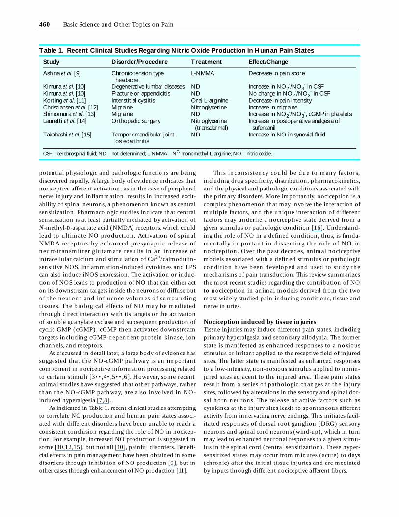
As indicated in Table 1, recent clinical studies attemptingto correlate NO production and human pain states associ-ated with different disorders have been unable to reach aconsistent conclusion regarding the role of NO in nocicep-tion. For example, increased NO production is suggested insome [10,12,15], but not all [10], painful disorders. Benefi-cial effects in pain management have been obtained in somedisorders through inhibition of NO production [9], but inother cases through enhancement of NO production [11].
This inconsistency could be due to many factors,including drug specificity, distribution, pharmacokinetics,and the physical and pathologic conditions associated withthe primary disorders. More importantly, nociception is acomplex phenomenon that may involve the interaction ofmultiple factors, and the unique interaction of differentfactors may underlie a nociceptive state derived from agiven stimulus or pathologic condition [16]. Understand-ing the role of NO in a defined condition, thus, is funda-mentally important in dissecting the role of NO innociception. Over the past decades, animal nociceptivemodels associated with a defined stimulus or pathologiccondition have been developed and used to study themechanisms of pain transduction. This review summarizesthe most recent studies regarding the contribution of NOto nociception in animal models derived from the twomost widely studied pain-inducing conditions, tissue andnerve injuries.
Nociception induced by tissue injuriesTissue injuries may induce different pain states, includingprimary hyperalgesia and secondary allodynia. The formerstate is manifested as enhanced responses to a noxiousstimulus or irritant applied to the receptive field of injuredsites. The latter state is manifested as enhanced responsesto a low-intensity, non-noxious stimulus applied to nonin-jured sites adjacent to the injured area. These pain statesresult from a series of pathologic changes at the injurysites, followed by alterations in the sensory and spinal dor-sal horn neurons. The release of active factors such ascytokines at the injury sites leads to spontaneous afferentactivity from innervating nerve endings. This initiates facil-itated responses of dorsal root ganglion (DRG) sensoryneurons and spinal cord neurons (wind-up), which in turnmay lead to enhanced neuronal responses to a given stimu-lus in the spinal cord (central sensitization). These hyper-sensitized states may occur from minutes (acute) to days(chronic) after the initial tissue injuries and are mediatedby inputs through different nociceptive afferent fibers.
Table 1. Recent Clinical Studies Regarding Nitric Oxide Production in Human Pain States
Study Disorder/Procedure Treatment Effect/Change
Ashina et al. [9] Chronic-tension type headache
L-NMMA Decrease in pain score
Kimura et al. [10] Degenerative lumbar diseases ND Increase in NO2-/NO3
- in CSFKimura et al. [10] Fracture or appendicitis ND No change in NO2
-/NO3- in CSF
Korting et al. [11] Interstitial cystitis Oral L-arginine Decrease in pain intensityChristiansen et al. [12] Migraine Nitroglycerine Increase in migraineShimomura et al. [13] Migraine ND Increase in NO2
-/NO3-, cGMP in platelets
Lauretti et al. [14] Orthopedic surgery Nitroglycerine (transdermal)
Increase in postoperative analgesia of sufentanil
Takahashi et al. [15] Temporomandibular joint osteoarthritis
ND Increase in NO in synovial fluid
CSF—cerebrospinal fluid; ND—not determined; L-NMMA—NG-monomethyl-L-arginine; NO—nitric oxide.

The Role of Nitric Oxide in Nociception • Luo and Cizkova 461
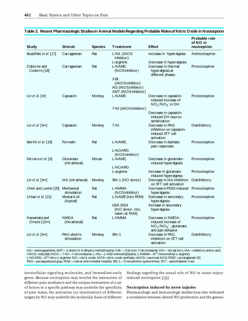
Even though NO is not likely a mediator in transductionof peripheral stimuli under physiologic conditions [4•], alarge body of pharmacologic and immunohistochemicalstudies have suggested that NO is involved in post-tissueinjury pain states. As indicated in Table 2, the most recentpharmacologic studies also point to the same direction.Inhibition of NO production by NOS inhibitors in mostcases results in suppression of hyperalgesia induced by tissueinjury or chemical stimulations. Thus, NO may modulate thehyperexcitability of dorsal horn neurons and play a pronoci-ceptive role in these pain states. This hypothesis is in agree-ment with the findings that NOS protein levels are increasedin various animal pain models (Table 3).
In addition, a recent study has provided strongevidences to support this hypothesis. It has been shownthat treatment with intrathecal NOS inhibitors, at concen-trations completely blocking intrathecal NMDA-inducedspinal release of NO2
-/NO3-, blocks NMDA-induced
increases of spinal cGMP and glutamate, and correspond-ing pain-related behavior. Similarly, treatment with acompetitive NMDA antagonist inhibits NMDA-inducedincreases of spinal NO2
-/NO3-, glutamate, and pain-related
behavior. Finally, intrathecal treatment with a cGMP inhib-itor inhibits NMDA-induced increase of spinal glutamateand pain-related behavior [22••]. Thus, activation ofspinal NMDA receptors is likely linked to an NO-cGMPpathway that may lead to further release of excitatoryneurotransmitters, resulting in a positive feedback regula-tion of neuronal hyperexcitability in the dorsal horn.
In addition to being a mediator of spinal excitatory neu-rons, NO could also modulate hyperexcitability ofspinal neurons by reducing spinal tonic inhibition. Thismechanism has been suggested by Willis et al. [4•,5••,27••]in a series of elegant studies. They have shown that intrader-mal capsaicin increases the release of spinal NO2
-/NO3- and
sensitizes a population of primate spinothalamic tract (STT)cells in the deep dorsal horn to peripheral mechanical stimu-lation. This spinal sensitization can be mimicked by treat-ment with an NO donor and prevented by an NOS inhibitor,suggesting a role of NO in the central sensitization [4•]. Asubsequent study has shown that the STT cell sensitizationthrough capsaicin or NO donor treatment results in a reduc-tion of spinal-descending inhibition on the STT cells inducedby stimulation in the periaqueductal gray [5••]. This disin-hibition is blocked by a pretreatment with an NOS inhibitor.In addition, the inhibitory effect of spinal glycine andgamma aminobutyric acid (GABA) agonists on the responsesof the STT cells to noxious stimulation is attenuated by treat-ment with an NO donor. These data suggest that the spinalinhibitory tone on the STT cells can be reduced by an NO-mediated pathway [5••]. Finally, they have shown that theinhibitory effects of locally delivered glycine and GABAagonists on responses of the STT cells to noxious stimulationcan be diminished by the administration of a membrane-permeable cGMP analogue. The disinhibition effects of theNO donor on the STT cell inhibition mediated by the inhibi-
tory amino acid receptors can be attenuated by a spinalpretreatment with a guanylate cyclase inhibitor [27••]. Thus,the disinhibition effects of NO on these dorsal horn cells aremediated through a cGMP-sensitive pathway. Together, thesestudies provide convincing evidence to indicate that modula-tion of inhibitory amino acid receptors in the spinal dorsalhorn by an NO-cGMP pathway is likely to contribute tocentral sensitization through a disinhibition mechanism[27••]. These in vivo findings are in contrast with the obser-vations that local application of NO donor or membrane-permeable cGMP blocks the activity of rat lamina II neurons,presumably a different type of neurons, whereas applicationof an NOS inhibitor leads to an increase in the activity of thedorsal horn neurons [28•].
Some studies have suggested that NO may not play apronociceptive role in pain states. For example, it has beenshown that NOS inhibition results in enhanced hyper-algesia induced by carrageenan [17]. A reduction, but notan increase, of NOS expression has been reported to beassociated with hyperalgesia induced by chronic inflam-mation [23]. These discrepancies might be explained bythe in vitro findings that expression of NOS in spinal cordneurons, which ultimately leads to NO production, isdifferentially regulated by activation of different afferentfibers, as well as by the intensity and duration of nocicep-tive inputs [28•]. Thus, it is likely that the unique localenvironment at the injury site, such as the severity andduration of inflammation, types of released cytokines, andso forth, could activate primary afferents with differentstrengths, thus resulting in differential regulation of NOSgene expression.
Another possibility leading to the discrepancies regardingthe role of NO in tissue injury-induced nociception is thatNO may be a messenger molecule of different types ofneurons, including excitatory neurons and inhibitory inter-neurons [29,30]. Depending on the type of neurons beingactivated following a specific lesion, the influence of alteredNO production in nociception could vary among animalpain models and human disorders. That is, NO-inducedactivation of excitatory neurons in a nociception pathwaycould result in hyperalgesia whereas NO-induced activationof inhibitory interneurons could cause hypoalgesia. This is inagreement with in vitro findings that NO may exert a dualrole in mediating neuronal activity. For example, NO canaugment spinal neuronal activity to inflammation [31] ordepress the responsiveness of spinal neurons to noxiousstimuli [32]. In addition, NO produced in one type ofneuron can diffuse to and influence surrounding neuronsthat may have opposite properties (such as excitatory versesinhibitory) and form synaptic contacts with the NO-produc-ing neurons. Thus, NO-induced activation of the surround-ing neurons may post positive or negative feedbackregulation on the primary NO-producing neurons.
In addition, the downstream targets of NO are not welldefined. It has been shown that NO activates diversifiedfunctional proteins including ion channels, receptors,
462 Basic Science and Other Topics on Pain
intracellular signaling molecules, and immediate-earlygenes. Because nociception may involve the interaction ofdifferent pain mediators and the unique interaction of a setof factors in a specific pathway may underlie the specificityof pain states, the activation (or inactivation) of differenttargets by NO may underlie the molecular basis of different
findings regarding the actual role of NO in tissue injury-induced nociception [16].
Nociception induced by nerve injuriesPharmacologic and immunologic studies have also indicateda correlation between altered NO production and the genera-
Table 2. Recent Pharmacologic Studies in Animal Models Regarding Probable Roles of Nitric Oxide in Nociception
Study Stimuli Species Treatment Effect
Probable role of NO in nociception
Budziñski et al. [17] Carrageenan Rat L-NIL (iNOS inhibitor)
Increase in hyperalgesia Antinociceptive
L-arginine Decrease in hyperalgesiaOsborne and
Coderre [18]Carrageenan Rat L-NAME
(NOS inhibitor)Decrease in thermal
hyperalgesia at different phases
Pronociceptive
3-Br. (nNOS inhibitor)
AG (iNOS inhibitor)AMT (iNOS inhibitor)
Lin et al. [4•] Capsaicin Monkey L-NAME Decrease in capsaicin-induced increase of NO2
-/NO3- in DH
Pronociceptive
7-NI (nNOS inhibitor)Decrease in capsaicin-
induced DH neuron sensitization
Lin et al. [5••] Capsaicin Monkey 7-NI Decrease in PAG inhibition on capsaicin-induced STT cell activation
Disinhibitory
Beirith et al. [19] Formalin Rat L-NAME, Decrease in biphasic pain responses
Pronociceptive
L-NOARG (NOS inhibitor)
Ferreira et al. [6] Glutamate (intrathecal)
Mouse L-NAME, Decrease in glutamate-induced hyperalgesia
Pronociceptive
L-NOARGL-arginine Increase in glutamate-
induced hyperalgesiaPronociceptive
Lin et al. [5••] IAA (intrathecal) Monkey SIN-1 (NO donor) Decrease in IAA inhibition on STT cell activation
Disinhibitory
Chen and Levine [20] Mechanical stimulation
Rat L-NMMA (NOS inhibitor)
Decrease in PGE2-induced hyperalgesia
Pronociceptive
Urban et al. [21] Mustard oil (topical)
Rat L-NAME (into RVM) Decrease in secondary hyperalgesia
Pronociceptive
GEA 5024 (NO donor, into naïve rat RVM)
Increase in secondary hyperalgesia
Kawamata and Omote [22••]
NMDA (intrathecal)
Rat L-NMMA Decrease in NMDA-induced increase of NO2
-/NO3- , glutamate
and pain behavior
Pronociceptive
Lin et al. [5••] PAG electric stimulation
Monkey SIN-1 Decrease in PAG inhibition on STT cell activation
Disinhibitory
AG—aminoguanidine; AMT—2-amino-5, 6-dihydro-methylthiazine; 3-Br.—3-bromo-7-nitroindazole; DH— dorsal horn; IAA—inhibitory amino acid; i-NOS—inducible NOS—; 7-NI—7-nitroindazole; L-NIL—L-N6(l-iminoethyl)lysine; L-NMMA—NG-monomethyl-L-arginine; L-NOARG—NG-nitro-L-arginine; NO—nitric oxide; NOS—nitric oxide synthase; nNOS—neuronal NOS; PGE2—prostaglandin E2; PAG—periaqueductal gray; RVM—rostral ventromedial medulla; SIN-1—3-morpholino-sydnonimine; STT—spinothalamic tract.

The Role of Nitric Oxide in Nociception • Luo and Cizkova 463
tion and/or maintenance of chronic pain associated withnerve injury. Immunolabelled nNOS positive cells and NOSactivity are increased in the DRG (Fig. 1) [33,34••] anddecreased in the spinal cord (Fig. 2) [33,35] of rats withneuropathic pain resulting from peripheral nerve injury.Administration of a nonselective NOS inhibitor L-NAMEinhibits the development of thermal hyperalgesia induced bychronic constriction injury [36], and tactile allodyniainduced by tight ligation of the L5/L6 spinal nerves [37] in anL-arginine reversible and dose-dependent manner. Thesedata suggest a functional role for NO in the processing and/or modulation of neuropathic pain.
Because NO production is tightly controlled by NOS,an important issue related to the role of NO in neuropathicpain (and other pain states) is the relative contribution ofNOS forms to the abnormal sensation derived from nerveinjury. Experimental data suggest that nNOS is the most
likely contributing candidate. Expression of eNOS is notsignificantly altered in the spinal cord (data not shown)and DRG (Fig. 1B), nor is iNOS expression detectable inthese tissues after the nerve ligation (data not shown) [35]when the neuropathic pain state is obvious. Even though arecent study has reported increased eNOS and iNOSexpression in constricted sciatic nerve [38], the exact originof the increases are not known and might have arisen fromactivated macrophages and Schwann cells by nerve injury-induced secondary inflammation [39].
If NO is indeed involved in the generation and/ormaintenance of nerve injury-induced neuropathic pain,one would expect to see a tight correlation between NOSexpression and neuropathic pain development. However,detailed studies to examine the linkage have been unableto show a cause-effect relationship. Even though nerveinjury (ligation) induces marked upregulation of nNOS in
Table 3. Expression of Nitric Oxide Synthase in Animal Models with Different Pain States
Study Stimuli/Pathology Species NOS Location Pain states
Dolan et al. [23] Chronic mastitis Sheep Decrease in nNOS-ir Bilateral lumbar and cervical SC, laminae I–III, X
Contralateral hyperalgesia
Leong et al. [24] Formalin Rat Increase in NOS-ir, within or near Fox positive neurons
Caudal spinal trigeminal nucleus
ND
Przewlocka et al. [25] Formalin Rat Increase in NOS-ir Lumbar SC laminae I–III, IV, X
Hyperalgesia (biphasic)
Rodella et al. [26] Noxious visceral stimulation
Rat Increase in NADPH-d staining
Brain areas ND
ir—immunoreactivity; NADPH-d—nicotinamide adenine dinucleotide phosphate diaphorase; ND—not determined; NOS—nitric oxide synthase; SC—spinal cord.
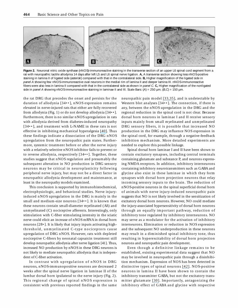
Figure 1. Nitric oxide synthase (NOS) expressions in the dorsal root ganglion (DRG) and changes of behavioral responses to mechanical stimulation in spinal nerve injured rats. Unilateral nerve injury was induced in Harlan Sprague-Dawley rats by tight ligation of the left L5/L6 spinal nerves and the paw withdrawal threshold to Von Frey filaments was tested up to 17 weeks. Total protein was extracted from pooled L5/L6 DRGs at designated time points, and neuronal NOS (nNOS) and endothelial NOS (eNOS) protein levels were examined by Western blot analysis. A, Nerve-ligated rats showed a gradual recovery from tactile allodynia in about 10 weeks after spinal nerve ligation. Data presented are the means ± SEM from at least 4 rats in each group. B, Representative Western blots showing expression levels of nNOS and eNOS in DRGs of nerve-ligated rats. Purified rat brain nNOS and endothelial cell extracts were used as positive controls (+) for nNOS and eNOS, respectively. C—contralateral side; Ip—ligation side.
464 Basic Science and Other Topics on Pain
the rat DRG that precedes the onset and persists for theduration of allodynia [34••], nNOS expression remainselevated in nerve-injured rats that either are fully recoveredfrom allodynia (Fig. 1) or do not develop allodynia [34••].Furthermore, there is no similar nNOS upregulation in ratswith allodynia derived from diabetes-induced neuropathy[34••], and treatment with L-NAME in these rats is noteffective in inhibiting mechanical hyperalgesia [40]. Thusthese findings indicate a dissociation of the DRG nNOSupregulation from the neuropathic pain states. Further-more, systemic treatment before or after the nerve injurywith a relatively selective nNOS inhibitor fails to prevent orto reverse allodynia, respectively [34••]. Together, thesestudies suggest that nNOS regulation and presumably thesubsequent alteration in NO production in DRG sensoryneurons may be critical in neuroplasticity followingperipheral nerve injury, but may not be a direct factor inneuropathic allodynia development and maintenance, atleast in the neuropathy models examined.
This conclusion is supported by immunohistochemical,electrophysiologic, and behavioral studies. Nerve injury-induced nNOS upregulation in the DRG is mainly in thesmall and medium-size neurons [34••]. It is known thatthese neurons contain small-diameter myelinated (Ad) andunmyelinated (C) nociceptive afferents. Interestingly, onlystimulation with C-fiber stimulating intensity in the sciaticnerve could elicit an increase of nNOS mRNA in dorsal hornneurons [28•]. It is likely that injury inputs activating high-threshold, unmyelinated C-type nociceptors causeupregulation of DRG nNOS. However, rats with depletednociceptive C-fibers by neonatal capsaicin treatment stilldevelop neuropathic allodynia after nerve ligation [41]. Thus,increased NO production by nNOS in these DRG neurons isnot likely to mediate neuropathic allodynia that is indepen-dent of C-fiber activation.
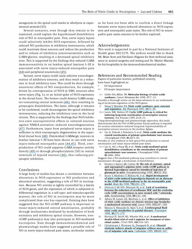
In contrast with upregulation of nNOS in DRGneurons, nNOS-immunopositive neurons are decreased 2weeks after the spinal nerve ligation in laminae II of thelumbar dorsal horn ipsilateral to the nerve injury (Fig. 2).This regional change of spinal nNOS expression isconsistent with previous reported findings in the same
neuropathic pain model [33,35], and is undetectable byWestern blot analyses [34••]. The connection, if there isany, between the nNOS upregulation in the DRG and theregional reduction in the spinal cord is not clear. Becausedorsal horn neurons in laminae I and II receive sensoryinputs mainly from small myelinated and unmyelinatedDRG sensory fibers, it is possible that increased NOproduction in the DRG may influence NOS expression inthe spinal cord, for example, through a negative-feedbackinhibition mechanism. More detailed experiments areneeded to explore this possible linkage.
Spinal dorsal horn laminae I and II have been shown tocontain excitatory synapses, including central terminalscontaining glutamate and substance P, and neurons express-ing NMDA receptors. In addition, inhibitory interneuronscontaining inhibitory neurotransmitters such as GABA andglycine also exist in these laminae in which they formsynapses with dorsal horn projection neurons that relayincoming sensory inputs to the brain. The reduction ofnNOS-positive neurons in the spinal superficial dorsal hornof animals with nerve injury-induced neuropathic painsuggests that NO is not likely involved in the sensitization ofexcitatory dorsal horn neurons. However, NO could mediatethe injury-associated hypersensitivity of dorsal horn neuronsthrough an equally important pathway, reduction ofinhibitory tone regulated by inhibitory interneurons. NOmay serve as a modulator for the activation of inhibitoryinterneurons. Elimination or reduction in NOS expressionand the subsequent NO underproduction in these neuronsmay result in a diminished spinal inhibitory tone, thusresulting in hyperexcitability of dorsal horn projectionneurons and neuropathic pain development.
Even though a definitive linkage remains to beestablished, existing experimental data suggest that NOmay be involved in neuropathic pain through a disinhibi-tion mechanism. Expression of NOS has been detected indistinctive types of spinal neurons [42]. NOS-positiveneurons in lamina II have been shown to contain theinhibitory transmitter GABA, but not the excitatory trans-mitter glutamate [30]. Importantly, antagonizing theinhibitory effect of GABA and glycine with respective
Figure 2. Neuronal nitric oxide synthase (nNOS)-immunoreactive staining in the transverse section of an upper L6 spinal cord segment from a rat with neuropathic tactile allodynia 14 days after left L5 and L6 spinal nerve ligation. A, A transverse section showing less nNOS-positive staining in lamina II of ligated side (asterisk) compared with that in the contralateral side. B, Higher magnification of the ligated side in panel A showing few nNOS-immunoreactive oval neurons in the medial rim of lamina II and deeper lamina III. nNOS-immunoreactive fibers were also less in lamina II compared with that in the contralateral side as shown in panel C. C, Higher magnification of the nonligated side in panel A showing nNOS-immunoreactive staining in laminae II and III. Scale Bars: (A) = 250 µm; (B,C) = 150 µm.
The Role of Nitric Oxide in Nociception • Luo and Cizkova 465
antagonists in the spinal cord results in allodynia in exper-imental animals [43].
Several scenarios, even though they remain to beexamined, could explain the hypothesized disinhibitoryrole of NO in neuropathic pain. First, nerve injury inputscould selectively inhibit NOS expression, followed byreduced NO production in inhibitory interneurons, whichcould inactivate these neurons and reduce the productionand/or release of inhibitory neurotransmitters such asGABA/glycine, resulting in a decreased spinal inhibitorytone. This is supported by the findings that reduced GABAimmunoreactivity in rat lumbar spinal laminae I–III isassociated with nerve injury-induced neuropathic pain[44] and peripheral neurectomy [45].
Second, nerve injury could cause selective neurodegen-eration of inhibitory neurons, and thus result in a reduc-tion in local inhibitory tone. This could be done throughneurotoxic effects of NO overproduction, for example,driven by overexpression of NOS in DRG neurons afternerve injury (Fig. 1), or an initial spike of NOS expressionin the dorsal horn. The former could destroy GABA recep-tor-containing central terminals [46], thus resulting inpresynaptic disinhibition. The latter, although it remainsto be confirmed, could downregulate the spinal inhibitoryinterneurons, reducing the inhibitory tone in the localcircuits. This is supported by the findings that NOS inhibi-tors exert neuroprotective effects in cultured neuronsagainst NMDA activation–induced neurodegeneration[47]. Furthermore, input from peripheral nerve injury issufficient to elicit transsynaptic degeneration in the super-ficial dorsal horn [48]. Diminished GABAergic neurons inlumbar laminae I–III have been shown in rats with nerveinjury-induced neuropathic pain [44,45]. Third, over-production of NO could suppress GABA receptor activitydirectly [49] or through phosphorylation [50] in centralterminals of injured neurons [46], thus reducing pre-synaptic inhibition.
ConclusionsA large body of studies has shown a correlation betweenalterations in NOS expression or NO production andabnormal sensation, suggesting a role of NO in nocicep-tion. Because NO activity is tightly controlled by a familyof NOS genes, and the expression of which is subjected todifferential regulation in a cell type- and stimulus-specificmanner, the role of NO in nociception may be morecomplicated than one has expected. Existing data havesuggested that the NO-cGMP pathway is important intissue injury-induced central sensitization, probablythrough mechanisms mediated by modulation of bothexcitatory and inhibitory spinal circuits. However, non-cGMP pathway(s) may also participate in NO-mediatednociception. Even though immunohistochemical andpharmacologic studies have suggested a possible role ofNO in nerve injury-induced pain states, molecular studies
so far have not been able to confirm a direct linkagebetween nerve injury-induced alterations in NOS expres-sion and neuropathic pain states. The role of NO in neuro-pathic pain states remains to be further explored.
AcknowledgementsThis work is supported in part by a National Institutes ofHealth grant DE13270. The authors would like to thankMr. Brian Scott and Emiliano Higuera for their technical assis-tance in animal surgeries and testing and Dr. Martin Marsalafor his hospitality in the immunohistochemical studies.
References and Recommended ReadingPapers of particular interest, published recently, have been highlighted as:• Of importance•• Of major importance
1.• Geller DA, Billiar TR: Molecular biology of nitric oxide synthases. Cancer Metastasis Rev 1998, 17:7–23.
Provides a thorough review on the latest advances in our understand-ing of molecular regulation of the NOS genes.
2. Wang Y, Marsden PA: Nitric oxide synthases: gene structure and regulation. Adv Pharmacol 1995, 34:71–90.
3.•• Lewin MR, Walters ET: Cyclic GMP pathway is critical for inducing long-term sensitization of nociceptive sensory neurons. Nat Neurosci 1999, 2:18–23.
Using electrophysiologic and pharmacologic approches, this study demonstrates that the NO-cGMP-protein kinase pathway is required for noxious stimulation-induced long-term hyperexcitability of nociceptive sensory neurons in the mollusc Aplysia.
4.• Lin Q, Palecek J, Paleckova V, et al.: Nitric oxide mediates the central sensitization of primate spinothalamic tract neurons. J Neurophysiol 1999, 81:1075–85.
Provides in vivo evidence to link spinal NO production to central sensitization and tissue injury-related pain states.
5.•• Lin Q, Wu J, Peng YB, et al.: Nitric oxide-mediated spinal disinhibition contributes to the sensitization of primate spinothalamic tract neurons. J Neurophysiol 1999, 81:1086–1094.
Suggests that a NO-mediated pathway may contribute to central sensitization through a mechanism of disinhibition.
6. Ferreira J, Santos AR, Calixto JB: The role of systemic, spinal and supraspinal L-arginine-nitric oxide-cGMP pathway in thermal hyperalgesia caused by intrathecal injection of glutamate in mice. Neuropharmacology 1999, 38:835–842.
7. Inoue T, Mashimo T, Shibata M, et al.: Rapid development of nitric oxide-induced hyperalgesia depends on an alternate to the cGMP-mediated pathway in the rat neuropathic pain model. Brain Res 1998, 792:263–270.
8. Ichinose F, Mi WD, Miyazaki M, et al.: Lack of correlation between the reduction of sevoflurane MAC and the cerebellar cyclic GMP concentrations in mice treated with 7-nitroinda-zole. Anesthesiology 1998, 89:143–148.
9. Ashina M, Lassen LH, Bendtsen L, et al.: Effect of inhibition of nitric oxide synthase on chronic tension-type headache: a randomised crossover trial. Lancet 1999, 353:287–289.
10. Kimura S, Watanabe K, Yajiri Y, et al.: Cerebrospinal fluid nitric oxide metabolites in painful diseases. Neuroreport 1999, 10:275–279.
11. Korting GE, Smith SD, Wheeler MA, et al.: A randomized double-blind trial of oral L-arginine for treatment of inter-stitial cystitis. J Urol 1999, 161:558–565.
12. Christiansen I, Thomsen LL, Daugaard D, et al.: Glyceryl trinitrate induces attacks of migraine without aura in suffer-ers of migraine with aura. Cephalalgia 1999, 19:660–667.

466 Basic Science and Other Topics on Pain
13. Shimomura T, Murakami F, Kotani K, et al.: Platelet nitric oxide metabolites in migraine. Cephalalgia 1999, 19:218–222.
14. Lauretti GR, de Oliveira R, Reis MP, et al.: Transdermal nitroglycerine enhances spinal sufentanil postoperative analgesia following orthopedic surgery. Anesthesiology 1999, 90:734–739.
15. Takahashi T, Kondoh T, Ohtani M, et al.: Association between arthroscopic diagnosis of temporomandibular joint osteo-arthritis and synovial fluid nitric oxide levels. Oral Surg Oral Med Oral Pathol Oral Radiol Endod 1999, 88:129–136.
16. Luo ZD: Molecular dissection of pain mediators. Pain Rev 2000, 7:37–64.
17. Budziñski M, Misterek K, Gumulka W, Dorociak A: Inhibition of inducible nitric oxide synthase in persistent pain. Life Sci 2000, 66:301–305.
18. Osborne MG, Coderre TJ: Effects of intrathecal administration of nitric oxide synthase inhibitors on carrageenan-induced thermal hyperalgesia. Br J Pharmacol 1999, 126:1840–1846.
19. Beirith A, Creczynski-Pasa TB, Bonetti VR, et al.: Antinociceptive properties and nitric oxide synthase inhibitory action of new ruthenium complexes. Eur J Pharmacol 1999, 369:289–297.
20. Chen X, Levine JD: NOS inhibitor antagonism of PGE2-induced mechanical sensitization of cutaneous C-fiber nociceptors in the rat. J Neurophysiol 1999, 81:963–966.
21. Urban MO, Coutinho SV, Gebhart GF: Involvement of excitatory amino acid receptors and nitric oxide in the rostral ventromedial medulla in modulating secondary hyperalgesia produced by mustard oil. Pain 1999, 81:45–55.
22.•• Kawamata T, Omote K: Activation of spinal N-methyl-D-aspar-tate receptors stimulates a nitric oxide/cyclic guanosine 3’,5’-monophosphate/glutamate release cascade in nocicep-tive signaling. Anesthesiology 1999, 91:1415–1424.
Provides strong pharmacologic evidence to support a tight correlation between NMDA-induced pain states and activation of the NO-cGMP pathway in the spinal cord.23. Dolan S, Field LC, Nolan AM: The role of nitric oxide
and prostaglandin signaling pathways in spinal nociceptive processing in chronic inflammation. Pain 2000, 86:311–320.
24. Leong S, Liu H, Yeo J: Nitric oxide synthase and glutamate receptor immunoreactivity in the rat spinal trigeminal neurons expressing Fos protein after formalin injection. Brain Res 2000, 855:107–115.
25. Przewlocka B, Mika J, Capone F, et al.: Intrathecal oxotremo-rine affects formalin-induced behavior and spinal nitric oxide synthase immunoreactivity in rats. Pharmacol Biochem Behav 1999, 62:531–536.
26. Rodella L, Rezzani R, Agostini C, Bianchi R: Expression of NADPH-diaphorase and colocalization with Fos in the brain neurons of the rat following visceral noxious stimulation. Brain Res 1999, 834:173–177.
27.•• Lin Q, Wu J, Peng YB, et al.: Inhibition of primate spino-thalamic tract neurons by spinal glycine and GABA is modulated by guanosine 3',5'-cyclic monophosphate. J Neurophysiol 1999, 81:1095–1103.
Demonstrates that a NO-cGMP pathway is involved in modulation of the spinal inhibitory tone and central sensitization.28.• Callsen-Cencic P, Hoheisel U, Kaske A, et al.: The controversy
about spinal neuronal nitric oxide synthase: under which conditions is it up- or downregulated? Cell Tissue Res 1999, 295:183–194.
This review article has summarized regulation of nNOS expression in spinal dorsal horn neurons under different physiologic and pathologic circumstances. In addition, data from in vitro experiments were used to explain possible mechanisms underlying the discrepan-cies of in vivo data.29. Bogdanov MB, Wurtman RJ: Possible involvement of nitric
oxide in NMDA-induced glutamate release in the rat striatum: an in vivo microdialysis study. Neurosci Lett 1997, 221:197–201.
30. Valtschanoff JG, Weinberg RJ, Rustioni A, Schmidt HH: Nitric oxide synthase and GABA colocalize in lamina II of rat spinal cord. Neurosci Lett 1992, 148:6–10.
31. Haley JE, Dickenson AH, Schachter M: Electrophysiological evidence for a role of nitric oxide in prolonged chemical nociception in the rat. Neuropharmacology 1992, 31:251–258.
32. Zhuo M, Meller ST, Gebhart GF: Endogenous nitric oxide is required for tonic cholinergic inhibition of spinal mechanical transmission. Pain 1993, 54:71–78.
33. Choi Y, Raja SN, Moore LC, Tobin JR: Neuropathic pain in rats is associated with altered nitric oxide synthase activity in neural tissue. J Neurol Sci 1996, 138:14–20.
34.•• Luo ZD, Chaplan SR, Scott BP, et al.: Neuronal nitric oxide synthase mRNA upregulation in rat sensory neurons after spinal nerve ligation: lack of a role in allodynia develop-ment. J Neurosci 1999, 19:9201–9208.
Provides evidence from molecular biology, genetics, immunohis-tochemistry, and behavioral pharmacology to indicate that increased nNOS expression in the DRG after peripheral nerve injury is not directly associated with neuropathic allodynia.35. Goff JR, Burkey AR, Goff DJ, Jasmin L: Reorganization of
the spinal dorsal horn in models of chronic pain: correlation with behaviour. Neuroscience 1998, 82:559–574.
36. Yamamoto T, Shimoyama N: Role of nitric oxide in the development of thermal hyperesthesia induced by sciatic nerve constriction injury in the rat. Anesthesiology 1995, 82:1266–1273.
37. Yoon YW, Sung B, Chung JM: Nitric oxide mediates behavioral signs of neuropathic pain in an experimental rat model. Neuroreport 1998, 9:367–372.
38. Levy D, Zochodne DW: Local nitric oxide synthase activity in a model of neuropathic pain. Eur J Neurosci 1998, 10:1846–1855.
39. Levy D, Höke A, Zochodne DW: Local expression of inducible nitric oxide synthase in an animal model of neuropathic pain. Neurosci Lett 1999, 260:207–209.
40. Fox A, Eastwood C, Gentry C, et al.: Critical evaluation of the streptozotocin model of painful diabetic neuropathy in the rat. Pain 1999, 81:307–316.
41. Okuse K, Chaplan SR, McMahon SB, et al.: Regulation of expression of the sensory neuron-specific sodium channel SNS in inflammatory and neuropathic pain. Mol Cell Neurosci 1997, 10:196–207.
42. Marsala J, Marsala M, Vanicky I, Taira Y: Localization of NADPHd-exhibiting neurons in the spinal cord of the rabbit. J Comp Neurol 1999, 406:263–284.
43. Onaka M, Minami T, Nishihara I, Ito S: Involvement of glutamate receptors in strychnine- and bicuculline-induced allodynia in conscious mice. Anesthesiology 1996, 84:1215–1222.
44. Ibuki T, Hama AT, Wang XT, et al.: Loss of GABA-immuno-reactivity in the spinal dorsal horn of rats with peripheral nerve injury and promotion of recovery by adrenal medullary grafts. Neuroscience 1997, 76:845–858.
45. Castro-Lopes JM, Tavares, Coimbra A: GABA decreases in the spinal cord dorsal horn after peripheral neurectomy. Brain Res 1993, 620:287–291.
46. Bhisitkul RB, Kocsis JD, Gordon TR, Waxman SG: Trophic influence of the distal nerve segment on GABAA receptor expression in axotomized adult sensory neurons. Exp Neurol 1990, 109:273–278.
47. Dawson VL, Dawson TM, London ED, et al.: Nitric oxide mediates glutamate neurotoxicity in primary cortical cultures. Proc Natl Acad Sci U S A 1991, 88:6368–6371.
48. Sugimoto T, Bennett GJ, Kajander KC: Transsynaptic degeneration in the superficial dorsal horn after sciatic nerve injury: effects of a chronic constriction injury, transection, and strychnine. Pain 1990, 42:205–213.
49. Fukami S, Uchida I, Mashimo T, et al.: Gamma subunit dependent modulation by nitric oxide (NO) in recombinant GABAA receptor. Neuroreport 1998, 9:1089–1092.
50. Moon C, Fraser SP, Djamgoz MB: Protein kinase and phosphatase modulation of quail brain GABA(A) and non-NMDA receptors co-expressed in Xenopus oocytes. Cell Signal 2000, 12:105–112.
Copyright © 2022 FDOKUMEN