Optimizing Mobile Application Performance through Network ...
Upload
independentCategory
view
0download
0
Inflammasome-activating nanoparticles as modular systems foroptimizing vaccine efficacy
Stacey L. Demento1, Stephanie C. Eisenbarth2,7, Harald G. Foellmer3, Craig Platt4, MichaelJ. Caplan5, W. Mark Saltzman1, Ira Mellman4, Michel Ledizet6, Erol Fikrig3,8, Richard A.Flavell2,8, and Tarek M. Fahmy1
1 Department of Biomedical Engineering, Yale University, New Haven, CT 06511, USA
2 Department of Immunobiology, Yale University, New Haven, CT 06511, USA
3 Section of Infectious Diseases, Department of Internal Medicine, Yale University, New Haven, CT 06511,USA
4 Department of Cell Biology, Yale University, New Haven, CT 06511, USA
5 Department of Cellular and Molecular Physiology, Yale University, New Haven, CT 06511, USA
7 Department of Laboratory Medicine, Yale University, New Haven, CT 06511, USA
6 L2 Diagnostics, LLC, 300 George Street, New Haven, CT 06511, USA
8 Howard Hughes Medical Institute
AbstractInnate immune system activation is a critical step in the initiation of an effective adaptive immuneresponse; therefore, activation of a class of innate pathogen receptors called pattern recognitionreceptors (PRR) is a central feature of many adjuvant systems. It has recently been shown that onemember of an intracellular PRR, the NLRP3 inflammasome, is activated by a number of classicaladjuvants including aluminum hydroxide and saponins [1,2]. Inflammasome activation in vitrorequires signaling of both the Toll-like receptor (TLR) and NLRP3 in antigen-presenting cells. Herewe present a class of nanomaterials endowed with these two signals for rapid optimization of vaccinedesign. We constructed this system using a simple approach that incorporates lipopolysaccharides(LPS) onto the surface of nanoparticles constructed from a biocompatible polyester, poly (lactic-co-glycolic acid) (PLGA), loaded with antigen. We demonstrate that LPS-modified particles arepreferentially internalized by dendritic cells compared to uncoated nanoparticles and the system,when administered to mice, elicits potent humoral and cellular immunity against a model antigen,ovalbumin. Wild type macrophages pulsed with LPS-modified nanoparticles resulted in productionof the proinflammatory cytokine IL-1β consistent with inflammasome activation. In comparison,NLRP3-deficient and caspase-1-deficient macrophages showed negligible production of IL-1β.Furthermore, when endocytosis and lysosomal destabilization were inhibited, inflammasome activitywas diminished, supporting the notion that nanoparticles rupture lysosomal compartments andbehave as ‘danger signals’[3]. The generality of this vaccination approach is tested by encapsulationof a recombinant West Nile envelope protein and demonstrated by protection against a murine modelof West Nile encephalitis. The design of such an antigen delivery mechanism with the ability to
Address correspondence to: Tarek Fahmy, PhD, Tel: 203-512-1699, FAX: 203-432-0030, E-mail: [email protected]'s Disclaimer: This is a PDF file of an unedited manuscript that has been accepted for publication. As a service to our customerswe are providing this early version of the manuscript. The manuscript will undergo copyediting, typesetting, and review of the resultingproof before it is published in its final citable form. Please note that during the production process errors may be discovered which couldaffect the content, and all legal disclaimers that apply to the journal pertain.
NIH Public AccessAuthor ManuscriptVaccine. Author manuscript; available in PMC 2010 May 18.
Published in final edited form as:Vaccine. 2009 May 18; 27(23): 3013–3021. doi:10.1016/j.vaccine.2009.03.034.
NIH
-PA Author Manuscript
NIH
-PA Author Manuscript
NIH
-PA Author Manuscript

stimulate two potent innate immune pathways represents a potent new approach to simultaneousantigen and adjuvant delivery.
KeywordsPLGA; inflammasome; West Nile virus; Nanoparticles
1. IntroductionPathogens are continually emerging and changing; therefore, there is a need for flexible vaccinedelivery systems. Vaccines have eliminated smallpox and nearly eliminated polio, two of theworst global infectious diseases. By contrast, vaccines for many other infectious diseases, suchas human immunodeficiency virus (HIV) and malaria, are poorly developed or simplyunavailable [4,5]. There are a number of significant scientific challenges that have limited thedevelopment of vaccines for deadly diseases. First, few if any approaches are available thatefficiently prime cell-mediated immunity by direct intracellular delivery of an antigen. Second,‘tunable’ adjuvants, that is, adjuvants that can be engineered to optimize the magnitude anddirection of an immune response are not available [6,7]. Third, most vaccine approaches areparenteral (i.e. subcutaneous or intramuscular injection) which has made it difficult to deployvaccines in underdeveloped countries where medical support systems, resources, and evenrefrigeration are limited. Therefore, there is a critical need for safe and stable vaccine systemsthat would address these factors.
Several key variables are assembled and integrated in the design of current vaccines [8,9]. Thefirst variable is the form of the antigen itself, which can be whole inactivated or attenuatedorganisms, purified proteins and peptides, or DNA encoded antigens. Purified antigens oftenbecome less immunogenic compared to whole pathogens or crude extracts, necessitating ameans to amplify the immune response against the purified subunit antigen. Thus, a secondnecessary component of a vaccine involves potentiating or stimulating the innate and adaptivearms of the immune system to the antigen subunit [8,9]. Immune potentiators may includebacterial products, toxins, or other molecules that augment specific immunity. Finally, to effectoptimal stimulation to a given antigen, a formulation is needed that delivers the correct amountof antigen in a repetitive or sustained fashion to the appropriate immune cells and to theappropriate compartments within those cells. A vaccine vehicle should facilitate delivery ofboth antigen and immune potentiator molecules preferably to target cells of the immune system.
Because of the historical emphasis on eliciting humoral immune responses, current adjuvants,represented predominately by aluminum adjuvants (alum), are optimized for effectiveinduction of Th2-biased responses and high antibody serum titers, but are less effective ateliciting a strong T cell-mediated immune response. While soluble antigen is poorly presentedon MHC class I to stimulate cytotoxic T lymphocytes, it has been demonstrated that internalizedantigen encapsulated in polymer particles can be effectively cross-presented on MHC Class I[10,11] yielding an effective CD8+ T cell response [12,13].
Aluminum adjuvants while having a limited capacity to adsorb many antigens [14,15] havebeen a mainstay in current vaccine formulations. One reason for this is that alum is generallysafe to administer and a good humoral immune potentiator. Its adjuvanicity has recently beenattributed to NLRP3 inflammasome activation [1,2]. The inflammasome is an intracellularmultiprotein complex that mediates caspase-1 cleavage of the inactive precursor of theproinflammatory cytokine, IL-1β, resulting in release of mature IL-1β [16]. Inflammasome-mediated cleavage of pro-IL-1B in vitro depends on signals that activate both TLR and NOD-like receptors (NLR), such as NLRP3. Activation of these innate immune system receptors is
Demento et al. Page 2
Vaccine. Author manuscript; available in PMC 2010 May 18.
NIH
-PA Author Manuscript
NIH
-PA Author Manuscript
NIH
-PA Author Manuscript

now recognized to be a prerequisite of effective adaptive immunity through provision of thenecessary combination of stimuli for naïve T cells.
We hypothesized that antigen-loaded nanoparticles constructed from poly(lactic-co-glycolic)(PLGA), a biodegradable and biocompatible, FDA-approved polymer and LPS would proveto be an effective vaccine vector via both TLR and inflammasome activation. Unlike traditionaladjuvants, PLGA particles allow for targeted delivery, protection, and sustained release ofantigen during vaccination. Here we show that LPS-modified nanoparticles effectively enterantigen-presenting cells (APCs) and elicit both humoral and cellular immunity againstencapsulated antigens in mice. We demonstrate the modularity of this system for vaccinedevelopment by encapsulation of a West Nile virus envelope protein antigen (rWNVE) andthe induction of protection in a murine model of West Nile Encephalitis.
2. Materials and Methods2.1 Materials
50:50 PLGA with an inherent viscosity of 0.59 dL/g, was purchased from Lactel Polymers,Inc. (Pelham, AL, USA). Polyvinyl alcohol (PVA) (Mw average 30–70 kD), LPS (Escherichiacoli strain 0111:B4), LPS-FITC, chicken egg ovalbumin (OVA), rhodamine B, and Nile redwere all obtained from Sigma-Aldrich. Methylene chloride was of chromatography grade andsupplied by Fisher Scientific. All other reagents were of reagent grade and used as received.Recombinant West Nile virus envelope protein antigen (rWNVE) was made in Drosophila S2cells as described previously [17].
2.2 Preparation of LPS-modified biodegradable nanoparticlesWe used a modified water-in-oil-in-water (W/O/W) emulsion method for preparation of LPS-modified PLGA particles. In the first emulsion (W/O), super-concentrated OVA (100 mg/ml )or rWNVE (20 mg/ml) in phosphate-buffered saline (PBS) was added drop-wise to a vortexingPLGA solution (2 ml) dissolved in methylene chloride. To facilitate nanoparticle detection byfluorescence, rhodamine B or Nile red (0.5 mg) was added to the polymer solution. Polymerand encapsulant were added drop-wise to 5% PVA in the second emulsion (W/O/W). Aftereach emulsion, the samples were sonicated for 30 s on ice using a Tekmar Sonic Distributorfitted with a model CV26 sonicator – amplitude set at 38%. The second emulsion was rapidlyadded to 0.3% PVA. This external phase underwent vigorous stirring for 3 h at constant roomtemperature to evaporate methylene chloride. LPS-modified particles were prepared with LPS(20 mg/ml in de-ionized (DI) water) added to the second emulsion containing 5% PVA.Particles were collected at 12,000 rpm for 15 min and washed with DI water three times. Theparticles were freeze dried and stored at −20°C for later use.
2.3 Characterization of LPS modified-biodegradable nanoparticlesOVA-loaded nanoparticles were visualized by scanning electron microscopy (SEM) and sizewas assessed by ImageJ software as previously described [18]. Protein encapsulation wasquantified using a Micro BCA Protein Assay (Pierce) after dissolving the particles in 0.05 NNaOH with 1% SDS. Release from nanoparticles was measured by performing the Micro BCAProtein Assay on supernatant from nanoparticles incubating in PBS at 37°C over 3 weeks. Toquantify the amount of LPS incorporated into the biodegradable particles were prepared LPS-modified nanoparticles with 5% fluorescently labeled LPS (LPS-FITC). The amount of LPS-FITC on particles was calculated from a standard obtained with LPS-FITC and blank particles.Endotoxin content of dissolved LPS-modified and unmodified nanoparticles was confirmedby the Limulus Amebocyte Lysate endotoxin test (Cambrex).
Demento et al. Page 3
Vaccine. Author manuscript; available in PMC 2010 May 18.
NIH
-PA Author Manuscript
NIH
-PA Author Manuscript
NIH
-PA Author Manuscript

2.4 Uptake studies of fluorescent LPS-modified nanoparticles by dendritic cellsUptake of particles by dendritic cells (DCs) was assessed by confocal microscopy and flowcytometry. DC2.4, a cell line that presents exogenous antigen on MHC Class I and II molecules,were incubated at 37°C until adherent on glass coverslips in RPMI with 10% fetal bovine serum(FBS) and 2% penicillin-streptomycin (P/S). Cells were primed with rhodamine B-loaded,LPS-modified (LPS/RhodB) and unmodified (-/RhodB) nanoparticles at desiredconcentrations for 2 h, and were fixed in 4% paraformaldehyde. Cells were permeablized with1X PBS with 0.1% Triton-X-100 and counterstained with DAPI (nuclear stain) and Phalloidin-FITC (cytoskeleton stain) to visualize cellular viability and morphology. After washing, cellswere imaged with a spinning-disk fluorescent confocal microscope (Zeiss).
For flow cytometry experiments DC2.4 were grown to confluence at 37°C in RPMI media with10% FBS and 2% P/S [11]. Cells were incubated for 2 h with various concentrations of LPS-modified (LPS/Nile red) and unmodified (−/Nile red) nanoparticles loaded with Nile red dye.Subsequently, cells were washed in phosphate buffered saline, fixed at 4°C in 1%paraformaldehyde, and analyzed by flow cytometry using a FACSCalibur system (BD). Nilered fluorescence was detected in the FL2 channel. Significance was analyzed by the student t-test using GraphPad, Prism, version 4.0b.
2.5 Assessment of inflammasome activationWild type (WT) (C57Bl/6), NLRP3-deficient, and caspase-1-deficient mice were administered3% thioglycollate via intraperitoneal injection. After 5 days, macrophages were isolated andtransferred to 24-well plates at 5 × 10⁵ cells/well in Dulbecco’s Modified Eagle Media with10% fetal calf serum, 1% penicillin/streptomycin, and 1% L-glutamine. Cells in triplicate wereincubated overnight with 50 ng/ml LPS (Invivogen) or media alone followed by 24 hour ofpriming with 250 μg of either LPS-modified, OVA-loaded particles; unmodified OVA-loadedparticles; or pulsed with ATP for 20 minutes, as a positive control. Cells were incubated with10 μM for 1 h prior to particle incubation with cytochalasin D (Sigma-Aldrich) or CA-074 Me(Peptide Institute, Osaka, Japan), to inhibit actin polymerization and the lysosomal protease,cathepsin B, respectively. A similar experiment was performed with bone marrow-deriveddendritic cells (BMDCs), that were isolated from C57Bl/6 mice by a method previouslydescribed [19]. BMDCs were given fresh RPMI media, every two days, supplemented with10% FBS, L-glutamine, MEM non-essential amino acids, HEPES buffer, gentamicin, β-mercaptoethanol, and 12.5 ng/ml rGM-CSF for 7 days post-isolation. Cells were then pre-stimulated overnight with LPS, as described above, followed by an incubation period with 250μg particles or Alhydrogel (1.3%) (Accurate Chemicals), preceeded by a pulse with inhibitors,when noted, at the concentrations listed previously. In all groups, supernatant was collected at24 hours post-particle incubation and analyzed for IL-1β by ELISA (R&D Systems). Allexperiments were repeated at least twice with reproducible results.
2.6 Antigen-specific CD8+ T cell stimulationDC2.4 were incubated at 37°C at 1×10⁵/well in 96-well plates until adherent. Cells were pulsedwith LPS-modified, OVA-loaded particles (LPS/OVA); unmodified, OVA-loaded particles(−/OVA); blank particles (−/−) with soluble OVA and soluble LPS; soluble OVA and solubleLPS; or soluble OVA for 2 h at 37°C in triplicate. The amount of soluble OVA and LPS wasequivalent to the level of these molecules in the particles. Cells were washed and co-culturedwith 2×10⁵ splenocytes/well from an OTI mouse – a strain that expresses a transgenic T cellreceptor specific for the OVA peptide SIINFEKL in MHC Class I. After 48 h, the supernatantwas analyzed for T cell stimulation by IFN-γ secretion using an ELISA kit (BD Pharmingen).This experiment was performed twice on separate occasions with the same findings.
Demento et al. Page 4
Vaccine. Author manuscript; available in PMC 2010 May 18.
NIH
-PA Author Manuscript
NIH
-PA Author Manuscript
NIH
-PA Author Manuscript

2.7 Animal vaccinationC57BL/6 mice at 6–8 weeks of age were purchased from Charles River Laboratories for allvaccination experiments. National Institute of Health guidelines for the care and use oflaboratory animals were observed. All animals were maintained under specific pathogen-freeconditions and routinely checked by the Yale University Animal Resource Center staff. Forsubcutaneous (s.c.) vaccination studies with OVA antigen-loaded particles, animals (n=5 pergroup) were injected with a single dose of 100 μg of OVA at the base of the tail with eitherOVA-loaded nanoparticles (−/OVA); LPS-modified, OVA-loaded nanoparticles (LPS/OVA);OVA in a 20mM Tris-HCl, 100mM NaCL buffer absorbed to 100 μL Alhydrogel; or OVAemulsified in Complete Freund’s Adjuvant according to the manufacturers diferections(Pierce). LPS-modified, ”empty” nanoparticles were used as a negative control. Blood andspleens were harvested at 2 weeks post-vaccination. Vaccination with the OVA antigen wererepeated twice.
For subcutaneous vaccinations with the recombinant West Nile viral antigen, rWNVE, mice(n=10) received a single-dose of antigen-loaded, LPS-modified particles (LPS/rWNVE) (20μg) in PBS at the base of the tail. Mice vaccinated intranasally (n=10) were administered 25μg of antigen encapsulated in LPS/rWNVE in 20 μl of PBS in small droplets to the tip of thenose with a micropipette, allowing the unanesthetized mice to inhale the dose. Mice vaccinatedorally (n=10) were given 150 μg of encapsulated antigen in LPS/rWNVE by oral gavage in300 μl of PBS. To demonstrate the protection of these particles during oral vaccination, 150μg of rWNVE was administered by oral gavage. Intranasally and orally vaccinated animalswere given an identical booster dose at 2 weeks post-initial vaccination. LPS/OVA particleswere administered s.c. as a control. Mice were injected intraperitoneally with 1000 PFU ofWN virus isolate 2741 at 2 weeks or 4 weeks post-initial vaccination for s.c. and oral/nasalvaccinations, respectively. Mice were monitored daily for morbidity and mortality for 21 dayspost-challenge. Survival results are the combined curves from two experiments (total n pergroup=20). Statistical analysis was performed by the Logrank (Mantel-Cox) test usingGraphPad, Prism, version 4.0b.
2.8 Analysis of serum antigen-specific antibodiesBlood was collected retro-orbitally at week 2 for OVA vaccinations and s.c. rWNVEvaccinations and at week 4 for nasal and oral rWNVE vaccinations. Samples were incubatedat 4°C overnight and centrifuged at 3000 rpm for 10 min. Serum was isolated and stored at−80°C for later analysis. Antigen-specific IgG titers were analyzed by ELISA. In brief, plateswere coated with 100 ng OVA or rWNVE per well at 4°C overnight and blocked using 1XPBS with 10% goat serum. Serum (unpooled) was diluted in blocking buffer and added to thewashed plate. After 2 h incubation at room temperature, plates were again washed andincubated with an excess of horseradish peroxidase-conjugated goat anti-mouse IgG (H+L)(Zymed) for 1 h. Enzymatic activity was assessed using TMB substrate. End-point antibodytiter was the average reciprocal dilution that corresponded to an absorbance two standarddeviations above the control (LPS/−) absorbance. Data shown are the average titer from a singleexperiment. Two identical experiments were performed with the same outcome.
2.9 Antigen-specific T cell stimulation in OVA-vaccinated miceMice were sacrificed 2 weeks post-vaccination. Splenocytes were isolated and pooled after redblood cell lysis with ACK lysis buffer (Quality Biological). Splenocytes were cultured in 96-well, round bottom plates at 2×10⁵ cells/well with 100 μg/ml OVA in RPMI + 10% FBSsupplemented with L-glutamine, MEM non-essential amino acids, HEPES buffer, gentamicin,and β-mercaptoethanol. Supernatant was collect at 48 h and analyzed for IFN-γ content byELISA (BD Pharmingen). As with serum IgG analysis, data shown are represntative of mulitpleexperiments.
Demento et al. Page 5
Vaccine. Author manuscript; available in PMC 2010 May 18.
NIH
-PA Author Manuscript
NIH
-PA Author Manuscript
NIH
-PA Author Manuscript

3. Results3.1 Characterization of LPS-modified nanoparticles
LPS-modified and unmodified OVA-loaded nanoparticles (Fig. 1A) exhibited a characteristicsustained release of encapsulated antigen at a statistically similar rate (Fig. 1B). Because ourstrategy for formulating the vaccine particle involved simultaneous encapsulation and surfacemodification, we examined how the addition of LPS might affect the encapsulation efficiencyof the model antigen, ovalbumin (OVA). The amount of encapsulated OVA in modified andunmodified PLGA nanoparticles was 44 ± 0.4 μg and 37 ± 0.1 μg per 1 mg of particles,respectively; therefore, LPS functionalization enhanced OVA encapsulation by about 16percent. Others have found similar effects on encapsulation efficiency of molecules with theaddition of fatty acids or lipids to PLGA during the encapsulation process[20,21]. A possiblemechanism for this effect might involve increased particle stability due to the presence ofamphipathic molecules such as LPS, facilitating enhancements in PLGA particle formationand encapsulation efficiency [22]. LPS-modified nanoparticles loaded with rWNVE contained17 ± 0.2 μg of antigen per 1 mg of particles. LPS-FITC was used as a means to quantify theamount of LPS incorporated on the surface. Total LPS incorporation was found to be roughly13 μg LPS per mg of particles and activity was confirmed by the Limulus Amebocyte Lysateendotoxin assay. While ovalbumin from Sigma does contain LPS, the endotoxin activity ofunmodified, OVA-loaded nanoparticles was far less (12.7 ± 5.2 EU/mg) compared to LPS-modified, OVA-loaded nanoparticles (641 ± 0.4 EU/mg). Particles displayed a size distributionin the range of 100–400 nm in diameter (Fig. 1C,D).
3.2 LPS–modified particles are efficiently internalized by dendritic cells compared tounmodified particles
To study the effect of LPS surface modification on the internalization of particles by dendriticcells, particles with and without LPS were incubated with DCs and internalization was assessedby microscopy and flow cytometry. LPS-modified nanoparticles were internalized moreeffectively than unmodified particles after 2 hours of incubation (Fig. 2A,B). In Fig. 2A,representative images showed DCs internalized a high amount of LPS-modified nanoparticles,whereas unmodified nanoparticles were less likely to be endocytosed. These findings areconsistent with previous reports of enhanced macrophage phagocytosis upon TLR stimulation[23]. Flow cytometry analysis of cells pulsed with nanoparticles at various concentrationssupported the preferential internalization of LPS-modified versus unmodified particles(P=0.09) (Fig. 2B).
3.3 LPS-modified particles stimulate the inflammasomeTo assess if nanoparticles stimulated the NLRP3 inflammasome pathway (Fig. 3A),macrophages were isolated from WT; NLRP3-deficient; and caspase-1-deficient mice, treatedwith modified and unmodified nanoparticles, and analyzed for IL-1β secretion (Fig. 3B). Wild-type macrophages, but neither NLRP3-deficient nor caspase-1-deficient macrophages,exhibited a significant level of IL-1β secretion post-incubation with LPS-modified particleswith and without prior LPS stimulation. Thus, the LPS-modified particles alone offered bothsignals necessary to activate the TLR and NLRP3 inflammasome pathways, unlike alum[1],which requires LPS stimulation in vitro and endogenous signals in vivo for inflammasomeactivation. Unmodified particles did not produce significant levels of the inflammatorycytokine in WT, NLRP3-deficient, or caspase-1-deficient mice even with pre-stimulation withsoluble LPS, likely because of poor uptake by APCs as demonstrated in Figure 2. This findinglikely explains why previous studies of the inflammatory properties of polystyrenenanoparticles have failed to find a role for the NLRP3 inflammasome [24].
Demento et al. Page 6
Vaccine. Author manuscript; available in PMC 2010 May 18.
NIH
-PA Author Manuscript
NIH
-PA Author Manuscript
NIH
-PA Author Manuscript

Recent data have shown that silica crystals and aluminum salts both disrupt lysosomes,propagating an endogenous ‘danger signal’ to the NLRP3 inflammasome [3]. We treated WTmacrophages and BMDCs with cytochalasin D, an inhibitor of actin polymerization, or CA-074Me, an inhibitor of cathepsin B (a lysosomal protease thought to play a role in lysosomaldestabilization), to see how inflammasome activity was affected. Both inhibitors abated, butdid not eliminate, the IL-1β response to LPS-modified nanoparticles (Fig. 3C,D). This partialresponse was also observed by Hornung et al [3]. As predicted, inflammasome activity wasnot hindered in cells treated with both inhibitors followed by ATP, a NLRP3 inflammasomestimulus that is not endocytosed and would, therefore, not be affected by actin polymerizationor lysosomal stabilization (Fig. 3C). Lastly, these data support that alum, like LPS/OVAnanoparticles, stimulates the inflammasome (Fig. 3D).
3.4 Dendritic cells primed with LPS-modified particles induce antigen-specific CD8+ T cellresponses
Next we evaluated the efficacy of this system in cross-presentation of antigenic epitopes andstimulation of a CD8+ T cell response. For this we studied OVA-specific T cell stimulation byDCs primed with LPS-modified and unmodified nanoparticles loaded with OVA. DC2.4 wereincubated with LPS/OVA nanoparticles, −/OVA nanoparticles, soluble OVA and LPS withblank nanoparticles, soluble OVA and LPS, and OVA alone; and then co-cultured withsplenocytes from an OTI mouse. While incubation with both modified and unmodifiednanoparticles yielded higher T cell responses than soluble antigen as assessed by IFN-γsecretion, it was clear that the LPS/OVA nanoparticles were more effective (Fig. 4A). DCsprimed with soluble OVA, or even soluble OVA and free LPS were significantly less effectivein stimulating antigen-specific lymphocytes. We note that equivalent amounts of OVA andLPS given with blank PLGA nanoparticles (−/−) were not nearly as effective as the integratedLPS-modified, OVA-loaded particles and that a 60-fold increase in soluble OVA was necessaryto generate an equivalent response to the one obtained with LPS-modified nanoparticles with50 μg/ml encapsulated OVA (Fig. 4B).
3.5 Early humoral and cellular immune response induction with LPS-modified nanoparticlesTo assess the efficacy of the particle system in the induction of early immune responses, weanalyzed serum antigen-specific IgG from mice vaccinated subcutaneously with a single doseof modified and unmodified nanoparticles at 2 weeks post-immunization (Fig. 5A). Asignificant increase in OVA-specific IgG was observed in mice given LPS-modified particlesby subcutaneous injection compared to unmodified particles. Similar titers of IgG wereobserved after vaccination with LPS-modified particles, OVA absorbed to Alum, and OVA inCFA.
Splenocytes from vaccinated animals were stimulated ex vivo with OVA. Antigen-stimulatedsplenocytes from mice administered LPS/OVA showed greater antigen-specific stimulation inthe form of IFN-γ secretion compared to animals vaccinated with -/OVA (Fig. 5B), suggestingthat modified particles induced a detectable cellular response in addition to a humoral response.Moreover, while IgG titers were indistinguishable between adjuvantation with LPS-modifiedparticles, alum, and CFA; only LPS-modified particles and CFA displayed a robust cellularresponse.
3.6 Vaccination with West Nile viral antigen-loaded LPS-modified nanoparticles providedprotection to viral challenge
Animals were vaccinated subcutaneously, intranasally, and orally with LPS/rWNVEnanoparticles and compared with a control group of mice injected subcutaneously with LPS/OVA nanoparticles. Animals administered with LPS/rWNVE subcutaneously had IgG titersabout 40-fold higher than the control mice (Fig. 6A) and over 20 fold higher than mice given
Demento et al. Page 7
Vaccine. Author manuscript; available in PMC 2010 May 18.
NIH
-PA Author Manuscript
NIH
-PA Author Manuscript
NIH
-PA Author Manuscript

soluble rWNVE subcutaneously (data not shown). Animals intranasally and orally vaccinatedwith LPS/rWNVE had, in comparison, much lower IgG titers than animals vaccinatedsubcutaneously, though significantly above the control. In a separate study (Supp. Fig. A) miceadministered LPS/rWNVE particles showed an elevated cellular response compared tounmodified particles and rWNVE absorbed to alum. Survival curves (Fig. 6B) show that thenanoparticles when injected subcutaneously confer almost complete protection to a viralchallenge after a single dose. Intranasal and oral vaccination with nanoparticles resulted in 80and 75 percent survival, respectively. Oral vaccination with rWNVE alone resulted in 60percent survival, indicating that particles may be effective vehicles for transporting antigenacross epithelial barriers and may provide antigen protection in the gut. All survival curvesfrom vaccinated mice were significantly different from the control (p=0.0003), by the Logrank(Mantel-Cox) test.
4. DiscussionAn approach for constructing vaccine delivery systems in which antigen, immune-potentiator,and delivery vehicle incorporated in a single nanodevice is attractive because it allows controlover variables that are important in optimizing an effective vaccine delivery system. Asdiscussed in this study, such systems may be engineered to induce inflammasome activation,much the same as aluminum adjuvants, but can also be designed to activate multiple arms ofthe innate immune system.
Inflammasome activation leading to the production of the potent inflammatory cytokineIL-1β is a manifestation of activated NOD-like receptor (NLR) signaling mediated by cytosolic‘danger’ signals. These NLR signals are still being identified but are now known to includealum, silica, and certain microbial products [1,24]. In vitro, TLR signaling is a prerequisite forIL-1β induction; therefore, we conjectured that integrating TLR and NLR ligands with antigenmay provide a highly effective vaccine vector. Results from this study argue for the successof this approach but also highlight a striking effect regarding the importance of combiningthese signals in a nanoparticulate assembly. LPS-modification effected an elevated rate ofnanoparticle internalization by antigen-presenting cells and subsequent T cell activation.Soluble LPS, ovalbumin, and blank PLGA particles (TLR signal, antigen, and NLR activatingligand) did not yield the elevated T cell responses observed with the LPS-modified, OVA-loaded nanoparticle complex. In addition, cellular and humoral responses were markedlyhigher after subcutaneous vaccination with LPS-modified particles encapsulating ovalbumincompared to unmodified particles. Even in the absence of an antigen boost, the animalsdisplayed antibody titers competitive with mice given OVA in alum or Complete Freund’sAdjuvant. Moreover, immunization with LPS-modified particles resulted in a cellular responsecomparable to that from CFA, and much higher than alum, the only FDA-approved adjuvantat present.
Our group previously developed a method for functionalizing PLGA particles with amphiphilicprotein-fatty acid conjugates [18]. We hypothesized that this method would apply to otheramphiphilic molecules, such as LPS, for targeting antigen presenting cells such as macrophagesand dendritic cells, creating simple vaccine delivery systems that combine antigen,immunostimulatory agent, and vehicle all in a single nanodevice. Other groups have devisedsimilar systems for incorporating TLR ligands such as CpG oligonucleotides [25–27], a ligandfor TLR9 or mono-phosphoryl lipid A (MPL) and RC529, derivatives of LPS [21,28] inbiodegradable particulates. Although CpG is a relatively non-cytotoxic alternative, TLR9 isan intracellular receptor [29] and unlike TLR4, is therefore unable to facilitate entry into thecell. While MPL and RC529 were devised to combat the toxicity of LPS, these molecules aregenerally considered less potent adjuvants than LPS. A study by Thompson, BS et al [30]concluded that mice given LPS as a vaccine adjuvant had 6 fold higher CD4+ T cell responses
Demento et al. Page 8
Vaccine. Author manuscript; available in PMC 2010 May 18.
NIH
-PA Author Manuscript
NIH
-PA Author Manuscript
NIH
-PA Author Manuscript

compared to mice given MPL and even more of a difference was observed with RC529.Similarly, a number of groups have reported that levels of cytokines relevant to successfulvaccination are much lower after treatment with MPL compared to the same amount of LPS[31,32]. Cytotoxicity analysis showed cellular viability at nanoparticle concentrations used inthis study (data not shown). Furthermore, no significant cytotoxicity difference between LPS-modified and unmodified particles was observed. Biodegradable particles surface-modifiedwith LPS largely evade the toxicity associated with the free form of LPS. This feature has beendemonstrated previously: incorporation of LPS into liposomes decreased its toxicity 10-foldcompared to its free form[33] and by 1000 fold in endotoxin activity [34]. Thus, this systemminimizes the negative effects of LPS while maintaining the potency of the whole molecule.
To the best of our knowledge, this is the first report that shows inflammasome activity withthe commonly utilized PLGA nanomaterials. This is also the first report to exploit the efficacyof particulate delivery systems as a vaccine vectors against West Nile (WN) virus, a flavivirus,currently without an available vaccine or specific therapy. Examples of flaviviruses includemany human pathogens of global epidemiological importance, such as the agents of denguefever, yellow fever, tick-borne encephalitis, West Nile (WN) meningoencephalitis, MurrayValley encephalitis, Japanese encephalitis, and St. Louis encephalitis. The envelope proteinsfrom these viruses are all related, suggesting that our results may be applicable to a variety offlaviviruses. Surface-modified, delivery vehicles offer the flexibility to generate an immuneresponse against single flaviviral antigens or a combination of antigens – a powerfulmethodology for the creation of new, effective vaccines against this group of infectiouspathogens.
After a single subcutaneous dose with surface-modified particles loaded with recombinantWest Nile virus antigen, 95 percent protection to viral infection was conferred to mice. Antigen-specific IgG titers were more than 20 times higher from antigen delivered in particulate form,compared to soluble antigen. Control of flavivirus infection is generally assumed to beprimarily mediated by neutralizing antibodies. Interestingly, while titers were relatively lowafter intranasal and oral vaccination, significant protection was observed. This finding mayhighlight the importance of the cellular immune response reported here with the model antigen,ovalbumin, which may also play a significant role in survival rates. This is consistent withprevious studies that show that CD8+ and CD4+ T cells both participate in the immune responseagainst a challenge with WN virus [35–37]. In those studies it was proposed that CD8+ T cellsare essential to fully eliminate the infecting virus and prevent viral persistence.
In summary, this study which demonstrates a technology for construction of vaccines supportsan underlying hypothesis: nanoparticles encapsulating antigen can be made to be more effectivevaccines by the proper choice and engineering of immune potentiators into the surface. Indeedthe proper combination of these potentiators can activate the inflammasome similar toconventional adjuvants such as alum. By coupling the nanoparticles with a simple immunemodulator, we were able to enhance the nanoparticles’ ability to elicit both humoral and cellularimmune responses as well as provide protection against a viral challenge by multiple routes ofadministration. Thus, the system described here is ideal for investigating the wide-variety ofemerging antigens and immune potentiators, allowing control over variables that are importantin optimizing an effective vaccine delivery system.
Supplementary MaterialRefer to Web version on PubMed Central for supplementary material.
Demento et al. Page 9
Vaccine. Author manuscript; available in PMC 2010 May 18.
NIH
-PA Author Manuscript
NIH
-PA Author Manuscript
NIH
-PA Author Manuscript
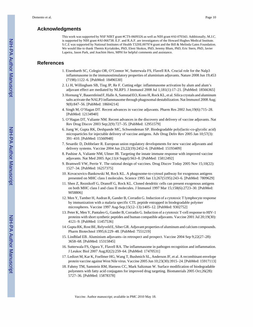
AcknowledgmentsThis work was supported by NSF NIRT grant #CTS-0609326 as well as NIH grant #AI-070343. Additionally, M.J.C.is supported by NIH grant #AI-066738. E.F. and R.A.F. are investigators of the Howard Hughes Medical Institute.S.C.E was supported by National Institutes of Health T32HL007974 grant and the Bill & Melinda Gates Foundation.We would like to thank Themis Kyriakides, PhD, Eleni Skokos, PhD, Jeremy Blum, PhD, Eric Stern, PhD, JavierLapeira, Jason Park, and Joachim Hero, MPH for helpful comments and assistance.
References1. Eisenbarth SC, Colegio OR, O’Connor W, Sutterwala FS, Flavell RA. Crucial role for the Nalp3
inflammasome in the immunostimulatory properties of aluminium adjuvants. Nature 2008 Jun 19;453(7198):1122–6. [PubMed: 18496530]
2. Li H, Willingham SB, Ting JP, Re F. Cutting edge: inflammasome activation by alum and alum’sadjuvant effect are mediated by NLRP3. J Immunol 2008 Jul 1;181(1):17–21. [PubMed: 18566365]
3. Hornung V, Bauernfeind F, Halle A, Samstad EO, Kono H, Rock KL, et al. Silica crystals and aluminumsalts activate the NALP3 inflammasome through phagosomal destabilization. Nat Immunol 2008 Aug;9(8):847–56. [PubMed: 18604214]
4. Singh M, O’Hagan DT. Recent advances in vaccine adjuvants. Pharm Res 2002 Jun;19(6):715–28.[PubMed: 12134940]
5. O’Hagan DT, Valiante NM. Recent advances in the discovery and delivery of vaccine adjuvants. NatRev Drug Discov 2003 Sep;2(9):727–35. [PubMed: 12951579]
6. Jiang W, Gupta RK, Deshpande MC, Schwendeman SP. Biodegradable poly(lactic-co-glycolic acid)microparticles for injectable delivery of vaccine antigens. Adv Drug Deliv Rev 2005 Jan 10;57(3):391–410. [PubMed: 15560948]
7. Sesardic D, Dobbelaer R. European union regulatory developments for new vaccine adjuvants anddelivery systems. Vaccine 2004 Jun 23;22(19):2452–6. [PubMed: 15193409]
8. Pashine A, Valiante NM, Ulmer JB. Targeting the innate immune response with improved vaccineadjuvants. Nat Med 2005 Apr;11(4 Suppl):S63–8. [PubMed: 15812492]
9. Bramwell VW, Perrie Y. The rational design of vaccines. Drug Discov Today 2005 Nov 15;10(22):1527–34. [PubMed: 16257375]
10. Kovacsovics-Bankowski M, Rock KL. A phagosome-to-cytosol pathway for exogenous antigenspresented on MHC class I molecules. Science 1995 Jan 13;267(5195):243–6. [PubMed: 7809629]
11. Shen Z, Reznikoff G, Dranoff G, Rock KL. Cloned dendritic cells can present exogenous antigenson both MHC class I and class II molecules. J Immunol 1997 Mar 15;158(6):2723–30. [PubMed:9058806]
12. Men Y, Tamber H, Audran R, Gander B, Corradin G. Induction of a cytotoxic T lymphocyte responseby immunization with a malaria specific CTL peptide entrapped in biodegradable polymermicrospheres. Vaccine 1997 Aug-Sep;15(12–13):1405–12. [PubMed: 9302752]
13. Peter K, Men Y, Pantaleo G, Gander B, Corradin G. Induction of a cytotoxic T-cell response to HIV-1proteins with short synthetic peptides and human compatible adjuvants. Vaccine 2001 Jul 20;19(30):4121–9. [PubMed: 11457536]
14. Gupta RK, Rost BE, Relyveld E, Siber GR. Adjuvant properties of aluminum and calcium compounds.Pharm Biotechnol 1995;6:229–48. [PubMed: 7551219]
15. Lindblad EB. Aluminium adjuvants--in retrospect and prospect. Vaccine 2004 Sep 9;22(27–28):3658–68. [PubMed: 15315845]
16. Sutterwala FS, Ogura Y, Flavell RA. The inflammasome in pathogen recognition and inflammation.J Leukoc Biol 2007 Aug;82(2):259–64. [PubMed: 17470531]
17. Ledizet M, Kar K, Foellmer HG, Wang T, Bushmich SL, Anderson JF, et al. A recombinant envelopeprotein vaccine against West Nile virus. Vaccine 2005 Jun 10;23(30):3915–24. [PubMed: 15917113]
18. Fahmy TM, Samstein RM, Harness CC, Mark Saltzman W. Surface modification of biodegradablepolyesters with fatty acid conjugates for improved drug targeting. Biomaterials 2005 Oct;26(28):5727–36. [PubMed: 15878378]
Demento et al. Page 10
Vaccine. Author manuscript; available in PMC 2010 May 18.
NIH
-PA Author Manuscript
NIH
-PA Author Manuscript
NIH
-PA Author Manuscript

19. Talmor M, Mirza A, Turley S, Mellman I, Hoffman LA, Steinman RM. Generation or large numbersof immature and mature dendritic cells from rat bone marrow cultures. Eur J Immunol 1998 Mar;28(3):811–7. [PubMed: 9541575]
20. Wiwattanapatapee R, Lomlim L, Saramunee K. Dendrimers conjugates for colonic delivery of 5-aminosalicylic acid. J Control Release 2003 Feb 14;88(1):1–9. [PubMed: 12586498]
21. Newman KD, Elamanchili P, Kwon GS, Samuel J. Uptake of poly(D,L-lactic-co-glycolic acid)microspheres by antigen-presenting cells in vivo. J Biomed Mater Res 2002 Jun 5;60(3):480–6.[PubMed: 11920673]
22. Thomasin C, Ho NT, Merkle HP, Gander B. Drug microencapsulation by PLA/PLGA coacervationin the light of thermodynamics. 1. Overview and theoretical considerations. J Pharm Sci 1998 Mar;87(3):259–68. [PubMed: 9523976]
23. Blander JM, Medzhitov R. Regulation of phagosome maturation by signals from toll-like receptors.Science 2004 May 14;304(5673):1014–8. [PubMed: 15143282]
24. Dostert C, Petrilli V, Van Bruggen R, Steele C, Mossman BT, Tschopp J. Innate immune activationthrough Nalp3 inflammasome sensing of asbestos and silica. Science 2008 May 2;320(5876):674–7. [PubMed: 18403674]
25. Jain S, Yap WT, Irvine DJ. Synthesis of protein-loaded hydrogel particles in an aqueous two-phasesystem for coincident antigen and CpG oligonucleotide delivery to antigen-presenting cells.Biomacromolecules 2005 Sep–Oct;6(5):2590–600. [PubMed: 16153096]
26. de Jong S, Chikh G, Sekirov L, Raney S, Semple S, Klimuk S, et al. Encapsulation in liposomalnanoparticles enhances the immunostimulatory, adjuvant and anti-tumor activity of subcutaneouslyadministered CpG ODN. Cancer Immunol Immunother 2007 Aug;56(8):1251–64. [PubMed:17242927]
27. Hunter SK, Andracki ME, Krieg AM. Biodegradable microspheres containing group B Streptococcusvaccine: immune response in mice. Am J Obstet Gynecol 2001 Nov;185(5):1174–9. [PubMed:11717653]
28. Kazzaz J, Singh M, Ugozzoli M, Chesko J, Soenawan E, O’Hagan DT. Encapsulation of the immunepotentiators MPL and RC529 in PLG microparticles enhances their potency. J Control Release 2006Feb 21;110(3):566–73. [PubMed: 16360956]
29. Miyake K. Innate immune sensing of pathogens and danger signals by cell surface Toll-like receptors.Semin Immunol 2007 Feb;19(1):3–10. [PubMed: 17275324]
30. Thompson BS, Chilton PM, Ward JR, Evans JT, Mitchell TC. The low-toxicity versions of LPS, MPLadjuvant and RC529, are efficient adjuvants for CD4+ T cells. J Leukoc Biol 2005 Dec;78(6):1273–80. [PubMed: 16204643]
31. Tiberio L, Fletcher L, Eldridge JH, Duncan DD. Host factors impacting the innate response in humansto the candidate adjuvants RC529 and monophosphoryl lipid A. Vaccine 2004 Mar 29;22(11–12):1515–23. [PubMed: 15063577]
32. Salkowski CA, Detore GR, Vogel SN. Lipopolysaccharide and monophosphoryl lipid A differentiallyregulate interleukin-12, gamma interferon, and interleukin-10 mRNA production in murinemacrophages. Infect Immun 1997 Aug;65(8):3239–47. [PubMed: 9234781]
33. Chhibber S, Wadhwa S, Yadav V. Protective role of liposome incorporated lipopolysaccharide antigenof Klebsiella pneumoniae in a rat model of lobar pneumonia. Jpn J Infect Dis 2004 Aug;57(4):150–5. [PubMed: 15329446]
34. Petrov AB, Semenov BF, Vartanyan YP, Zakirov MM, Torchilin VP, Trubetskoy VS, et al. Toxicityand immunogenicity of Neisseria meningitidis lipopolysaccharide incorporated into liposomes.Infect Immun 1992 Sep;60(9):3897–903. [PubMed: 1500196]
35. Purtha WE, Myers N, Mitaksov V, Sitati E, Connolly J, Fremont DH, et al. Antigen-specific cytotoxicT lymphocytes protect against lethal West Nile virus encephalitis. Eur J Immunol 2007 Jul;37(7):1845–54. [PubMed: 17559174]
36. Shrestha B, Diamond MS. Role of CD8+ T cells in control of West Nile virus infection. J Virol 2004Aug;78(15):8312–21. [PubMed: 15254203]
37. Sitati EM, Diamond MS. CD4+ T-cell responses are required for clearance of West Nile virus fromthe central nervous system. J Virol 2006 Dec;80(24):12060–9. [PubMed: 17035323]
Demento et al. Page 11
Vaccine. Author manuscript; available in PMC 2010 May 18.
NIH
-PA Author Manuscript
NIH
-PA Author Manuscript
NIH
-PA Author Manuscript

Figure 1.Characterization of nanoparticles. A) Schematic of biodegradable particles withlipopolysaccharide modification. B) Controlled release profile of OVA from LPS/OVA (●)and -/OVA (■) particles. Figure represents release from nanoparticles at 37°C in phosphate-buffered saline in triplicate over 3 weeks. C) Scanning Electron Microscopy (SEM) image ofLPS-modified nanoparticles. Scale bar is 2 μm. D) Size distribution profiles for LPS-modifiedand unmodified OVA-loaded nanoparticles.
Demento et al. Page 12
Vaccine. Author manuscript; available in PMC 2010 May 18.
NIH
-PA Author Manuscript
NIH
-PA Author Manuscript
NIH
-PA Author Manuscript

Figure 2.Evaluation of uptake efficiency. A) Dendritic cells were incubated with nanoparticlesencapsulating rhodamine B and analyzed by confocal microscopy (green: phalloidin-FITC,blue: DAPI, and red: rhodamine B). A representative image is shown. B) Flow cytometryanalysis of dendritic cells pulsed with Nile red-loaded nanoparticles: LPS/Nile red (■) and -/Nile red (□). Mean fluorescence in FL2 is shown.
Demento et al. Page 13
Vaccine. Author manuscript; available in PMC 2010 May 18.
NIH
-PA Author Manuscript
NIH
-PA Author Manuscript
NIH
-PA Author Manuscript

Figure 3.LPS-modified nanoparticles activated the NLRP3 inflammasome. A) Proposed mechanism ofactivation. Signal 1: LPS triggers Toll-like receptor 4 (TLR4) signaling, which ultimatelyactivates the transcription factor, NF-κB. Pro-IL-1β is upregulated and is cleaved by caspase-1in an inflammasome complex. Signal 2: Nanoparticles are phagocytosed by cells, disruptlysosomal compartments, and activate the NLRP3 receptor. NLRP3 recruits ASC, an adaptermolecule, which binds caspase-1. B) Macrophages from WT (□), NLRP3-deficient ( ) andcaspase-1-deficient (■) mice were isolated and incubated with LPS-modified and unmodifiedOVA-loaded particles with and without prior stimulation with soluble LPS. ATP with LPSpre-stimulation was used as a positive control. After 24 h, supernatant was analyzed forIL-1β by ELISA. C) WT macrophages and D) BMDCs were pre-stimulated with LPS and thentreated with cytochalasin D (cytoD), to inhibit internalization; CA-074 Me, to inhibit cathepsinB; or media alone. After incubation with particles or alum for 24 h or ATP for 20 min,supernatant was analyzed for IL-1β.
Demento et al. Page 14
Vaccine. Author manuscript; available in PMC 2010 May 18.
NIH
-PA Author Manuscript
NIH
-PA Author Manuscript
NIH
-PA Author Manuscript

Figure 4.In vitro vaccination with nanoparticles. A) Dendritic cells were pulsed with −/OVA, LPS/OVA,−/− with soluble OVA and LPS, soluble OVA and soluble LPS, or soluble OVA at an OVAconcentration of 50 μg/ml. Cells were then co-cultured with OT-1 transgenic splenocytes.Supernatant IFN-γ was quantified by ELISA after 48 hours. B) Dendritic cells were pulsedwith soluble OVA at high concentrations and then co-cultured with OT-1 splenocytes in orderto achieve similar levels of T cell activity. Dotted line represents IFN-γ levels from cells pulsedwith LPS/OVA from (A).
Demento et al. Page 15
Vaccine. Author manuscript; available in PMC 2010 May 18.
NIH
-PA Author Manuscript
NIH
-PA Author Manuscript
NIH
-PA Author Manuscript

Figure 5.OVA-specific immune responses 2 weeks after subcutaneous vaccination. Animals receiveda single vaccination by s.c. injection with particle formulations, alum, or CFA as adjuvants.A) Serum antibody titers were analyzed by ELISA. Data shown are mean OVA-specific IgGtiter. B) Splenocytes were stimulated ex vivo with OVA and supernatant was analyzed for IFN-γ secretion at 48 h by ELISA. For all groups: n=5. Experiments were repeated 2 more times(with n=4) with similar results.
Demento et al. Page 16
Vaccine. Author manuscript; available in PMC 2010 May 18.
NIH
-PA Author Manuscript
NIH
-PA Author Manuscript
NIH
-PA Author Manuscript

Figure 6.Antigen-specific IgG and survival in mice after vaccination with encapsulated West Nile virusenvelope protein antigen. Mice (n=20) were vaccinated with LPS/rWNVE subcutaneously(s.c.), orally, or intranasally (i.n.). As a negative control, mice were subcutaneouslyadministered LPS/OVA particles. A) Anti-rWNVE IgG titers in the serum pre-challenge werequantified by ELISA. B) Mice were challenged with 1000 PFU of isolate 2741 and monitoredfor mortality for 21 days. LPS/OVA (●); rWNVE alone ( ); LPS/rWNVE s.c. ( ); LPS/rWNVE oral ( ); and LPS/rWNVE nasal ( ;). Results are combined data from twoexperiments (n=10).
Demento et al. Page 17
Vaccine. Author manuscript; available in PMC 2010 May 18.
NIH
-PA Author Manuscript
NIH
-PA Author Manuscript
NIH
-PA Author Manuscript
Copyright © 2022 FDOKUMEN