Comparison of Intra-Peritoneal Instillation of Bupivacaine and ...
Amadori adducts activate nuclear factor-jB-relatedproinflammatory genes in cultured human peritoneal mesothelialcells
*,1Julian Nevado, 2Concepcion Peiro, 1Susana Vallejo, 1Mariam El-Assar, 2Nuria Lafuente,2Nuria Matesanz, 2Veronica Azcutia, 2Elena Cercas, 2Carlos F. Sanchez-Ferrer& 1,3Leocadio Rodrıguez-Manas
1Unidad de Investigacion, Hospital Universitario de Getafe, Ctra. de Toledo Km 12.5, Getafe, Madrid 28905, Spain;2Departamento de Farmacologıa y Terapeutica, Facultad de Medicina, Universidad Autonoma de Madrid, Madrid 28029, Spainand 3Servicio de Geriatrıa, Hospital Universitario de Getafe, Madrid 28905, Spain
1 Diabetes mellitus leads to a high incidence of several so-called complications, sharing similarpathophysiological features in several territories. Previous reports points at early nonenzymaticglycosylation products (Amadori adducts) as mediators of diabetic vascular complications. In thepresent study, we analysed a possible role for Amadori adducts as stimulators of proinflammatorypathways in human peritoneal mesothelial cells (HPMCs).
2 Cultured HPMCs isolated from 13 different patients (mean age 38.7716 years) were exposed todifferent Amadori adducts, that is, highly glycated haemoglobin (10 nM) and glycated bovine serumalbumin (0.25mgml�1), as well as to their respective low glycosylation controls. Amadori adducts, butnot their respective controls, elicited a marked increase of NF-kB activation, as determined byelectromobility shift assays and transient transfection experiments.
3 Additionally, Amadori adducts significantly increased the production of NF-kB-relatedproinflammatory molecules, including cytokines, such as TNF-a, IL-1b or IL-6, and enzymes, suchas cyclooxygenase-2 and inducible nitric oxide (NO) synthase, this latter leading to the release of NOby HPMCs.
4 The effects of Amadori adducts were mediated by different reactive oxygen and nitrosative species(e.g. superoxide anions, hydroxyl radicals, and peroxynitrite), as they were blunted by coincubationwith the appropriate scavengers. Furthermore, NO generated upon exposure to Amadori adductsfurther stimulated NF-kB activation, either directly or after combination with superoxide anions toform peroxynitrite.
5 We conclude that Amadori adducts can favour peritoneal inflammation by exacerbating changesin NO synthesis pathway and triggering NF-kB-related proinflammatory signals in human mesothelialcells.British Journal of Pharmacology (2005) 146, 268–279. doi:10.1038/sj.bjp.0706309;published online 4 July 2005
Keywords: Diabetes; inflammation; oxidative stress; Amadori adducts; nitric oxide; nuclear factor-kB; mesothelial cells
Abbreviations: AP-1, activator protein-1; AGEs, advanced glycation end products; CAPD, continuous ambulatory peritonealdialysis; DMTU, dimethylthiourea; DRB, 5,6-dichloro-1-b-D-ribofuranosylbenzimidazole; eNOS, constitutiveendothelial nitric oxide synthase; GAPDH, glyceraldehyde-3-phosphate dehydrogenase; HHb, highly glycatedoxyhaemoglobin; HPMCs, human peritoneal mesothelial cells; iNOS, inducible nitric oxide synthase; L-NAME,No-nitro-L-arginine methyl ester; NF-kB, nuclear factor-kB; NHb, oxyhaemoglobin glycated at normal levels;PDTC, pyrrolidine dithiocarbamate; ROS, reactive oxygen species; SNAP, S-nitroso-N-acetylpenicillamine; SNP,sodium nitroprusside; SOD, superoxide dismutase; Tempol, 4-hydroxy-tempo
Introduction
Diabetic patients account for approximately 40% of patients
undergoing dialysis in Western Europe and United States
(Stein et al., 2004). At the same time, the use of continuous
ambulatory peritoneal dialysis (CAPD) is increasing as a renal
replacement therapy in these patients. Although the influence
of diabetes on peritoneal membrane (PM) remains debated,
long-term hyperglycaemia has been suggested to be involved in
several changes affecting the functionality of the PM, either in
animal models or patients undergoing CAPD (Nakamoto
et al., 2002; Stoenoiu et al., 2002). Indeed, such alterations of
the peritoneum could be responsible, at least in part, for the
higher mortality rate observed in diabetic patients compared
to nondiabetic patients with peritoneal dialysis (Stein et al.,
2004).
In recent years, alterations of peritoneal mesothelial cells
have been proposed to be on the basis of PM dysfunction in
these patients (Yanez-Mo et al., 2003; Yao et al., 2003). PM
includes a monolayer of mesothelial cells, which acts as a
permeability barrier. These cells share many properties with*Author for correspondence; E-mail: [email protected]
British Journal of Pharmacology (2005) 146, 268–279 & 2005 Nature Publishing Group All rights reserved 0007–1188/05 $30.00
www.nature.com/bjp
vascular endothelial cells, including the ability to synthesise
nitric oxide (NO) and prostacyclin (Amore et al., 1997). Recent
studies suggest that some pathophysiological mechanisms
involved in diabetic microvascular complications may also be
participating in PM dysfunction. Among them, an increased
local release of growth factors (Mandl-Weber et al., 2002),
and proinflammatory mediators (Riese et al., 1999), as well
as deregulation of NO synthases (Devuyst et al., 2001) and
increased accumulation of advanced glycosylation end pro-
ducts (AGEs) (Park et al., 2000) within the peritoneum is
worth noting.
In the last years, several groups, including ours, have
postulated that other products of nonenzymatic protein
glycosylation, different to AGEs, can play an important role
in diabetic vascular complications. Amadori adducts are the
result of condensation reactions between glucose and reactive
protein amino groups, which yield Schiff bases that undergo
reversible rearrangement within days or weeks. Amadori
adducts can, in turn, undergo irreversible changes to form
AGEs after longer periods of time (Cerami et al., 1988). In the
vasculature, circulating Amadori adducts, like glycated hae-
moglobin or glycated albumin, produce reactive oxygen
species (ROS), which in turn inactivate NO, leading to
endothelial dysfunction (Angulo et al., 1996; Amore et al.,
1997; Peiro et al., 1998; Hattori et al., 1999; Vallejo et al.,
2000a; Peiro et al., 2001; Rodrıguez-Manas et al., 2003).
Amadori adducts can also activate proinflammatory redox-
regulated transcription factors, including NF-kB (Hattori
et al., 1999; 2002; Mandl-Weber et al., 2001; Peiro et al., 2003).
Alterations of the PM are associated with a local
proinflammatory environment that may be triggered by
different molecules, including the above-mentioned ROS,
NO, or NF-kB. In the present work, we aimed to study the
ability of Amadori adducts to induce a proinflammatory
response in human peritoneal mesothelial cells (HPMCs). For
this purpose, we tested the effects of both glycated haemo-
globin and glycated albumin on NF-kB activity, inducible NO
synthase (iNOS) and cyclooxygenase-2 (COX-2) gene expres-
sion and activity, as well as on the secretion and gene
expression of several proinflammatory cytokines such as IL-
1b, IL-6, and TNF-a by HPMCs. In addition, a putative role
for ROS and reactive nitrosative species was also analysed.
Methods
Materials
Culture plastic ware was obtained from Corning-Costar (New
York, NY, U.S.A.). M199 medium, L-glutamine, and strepto-
mycin/penicillin solutions were purchased from Biochrom KG,
Berlin, Germany. Phosphate-buffered saline (PBS), foetal calf
serum (FCS) and trypsin–EDTA were from Amresco (Solon,
Ohio, U.S.A.), Biological Industries (Beit-Hamek, Israel), and
GIBCO BRM (Paisley, U.K.), respectively. Human TNF-awas from Peprotech (London, U.K.) and IL-1b from R&D
systems (Minneapolis, MN, U.S.A.). Taq DNA polymerase
and dNTPs were from EGOGEN (Barcelona, Spain), while
1400W ([N-(3-aminomethyl) benzylacetamidine, 2 HCl]) was
from Calbiochem (Darmstadt, Germany). Unless otherwise
stated, all other reagents were purchased from Sigma Chemical
Co. (St Louis, MO, U.S.A.).
Preparation of Amadori adducts
Lyophilised human haemoglobins, nonenzymatically glyco-
sylated at either elevated or normal levels, containing 11.1%
(Catalogue no. G-1012) and 5.4% (Catalogue no. G-2012)
HbA1, respectively, were purchased from Sigma Chemical Co.
Before use, haemoglobins were prepared as described pre-
viously (Peiro et al., 2003). Briefly, haemoglobins were
dissolved in deionised water and subsequently reduced by
incubation with an excess of sodium dithionite. The haemo-
globin solutions were then extensively dialysed using a 0.25 A
pore diameter (approximately 12 kDa mol wt) dialysis
membrane (Viskings, Serva, Heidelberg, Germany) against
deionised water containing 10mg l�1 EDTA and continuously
bubbled with N2. Oxyhaemoglobins were then aliquoted and
stored at �701C until used.
A 50mgml�1 solution of bovine serum albumin (BSA)
(Sigma Chemical Co.) was glycosylated by incubation in PBS
(pH 7.4) containing 1M glucose under sterile and light-
protected conditions for 6 days at 371C as described previously
(Peiro et al., 1998). A control solution was prepared in parallel
using PBS without glucose. Glycosylation of serum albumin
was verified using the thiobarbituric acid assay (Ney et al.,
1981).
The absence of AGEs in the glycated oxyhaemoglobin and
albumin solutions was assessed by measuring fluorescence in a
Fluostar fluorometer (BMG Labtechnologies, Offenburg,
Germany) at excitation maximum of 370 nm and emission
maximum of 440 nm, which allows quantifying total AGEs
(Sell & Monnier, 1989). A standard curve (r¼ 0.99) was
carried out using AGE-modified BSA (0.5–5 mgml�1), pre-
pared following a previously described method (Bucala et al.,
1991). Glycated preparations did not contain significative
bacterial endotoxin contamination (p0.5U endotoxinml�1),
as measured with Pyrogents plus kit (Biowhittaker Europe
SPRL, Verners, Belgium).
Cell culture
HPMCs were isolated from omental tissue from 13/15 different
donors (free of any cardiovascular or peritoneal disease and
nontaking anti-inflammatory drugs or antioxidants) under-
going nonurgent, nonseptic abdominal surgery, using pre-
viously described methods (Chung-Welch et al., 1997).
HPMCs were routinely cultured in M199 containing 1 g l�1
of D-glucose and supplemented with 10% FCS, 100mgml�1
streptomycin, 100Uml�1 penicillin, and 2.5 mgml�1 amphoter-
icin. At confluence, HPMC were passaged using a 0.02%
EDTA–0.05% trypsin solution and split in a 1 : 2 ratio.
HPMCs characterisation was based on both cell morphology
(immediately prior to and at confluence, cells adopted the
polygonal cobblestone-like appearance characteristic of epithe-
lial cells and formed a monolayer) and indirect immunofluor-
escence staining of several human mesothelial markers
(Chung-Welch et al., 1997). In brief, HPMCs showed a diffuse
positive staining with an anti-von Willebrand factor antibody
(Dakopatts, Glostrup, Denmark) and a marked staining with
anti-cytokeratins 8 and 18, anti-E-cadherin, and anti-vimentin
antibodies (all of them from Sigma Chemical Co.). HPMCs
failed to express the endothelial marker PECAM-1 (CD31)
(see Supplemental data, Figure 1). Cell cultures between
passages two and eight were used.
J. Nevado et al NF-jB-inflammatory activation by Amadori adducts 269
British Journal of Pharmacology vol 146 (2)
Reporter plasmids
The reporter plasmid, p5�NF-kB-Luc (Stratagene, La Jolla,
CA, U.S.A.), and different luciferase-based reporter plasmids
corresponding to the 50-flanking regulatory regions of either
human iNOS (7.2 hiNOS-luc; Taylor et al., 1998), human
eNOS (1.33 heNOS-luc; Cieslik et al., 1999), short human
COX-2 and human COX-1 (phPES2 �327/þ 59, phPES1
�1010/þ 69, respectively; Inoue et al., 1995), or human IL-6
(p1168hu.IL6P-luc; Berghe et al., 1998) genes were used.
Transient transfection and luciferase assays
Transient transfection experiments were performed as we
described previously (Peiro et al., 2003). Briefly, HPMCs (105
cells) were grown in six-well plates to 80–90% confluence and
the culture medium (M-199) was then replaced by vehicle
medium, that is, serum-free medium supplemented with 0.1%
BSA. The transfection mixture was added to cell cultures for
further 18–20 h. The transfection mixture consisted of 2mg of
the above-mentioned plasmids incubated with 75ml of DMEM
and 7.5ml of Superfects (Quiagen Gmbh, Hilden, Germany) in
vehicle medium, following the manufacturer’s instructions.
Following treatment with the specified agents, HPMCs were
harvested and lysed with passive lysis buffer (1� ; Promega,
Madison, WI, U.S.A.), followed by one freeze/thaw cycle. The
extracts were centrifuged for 30 s at 13,000 r.p.m. at 41C, and
assayed with a luciferase reporter system (Promega, Madison,
WI, U.S.A.). Luciferase activity was expressed as relative
luciferase units (RLUs; Peiro et al., 2003).
Western blotting and protein content
Extraction of protein homogenates and Western blotting were
performed as described previously (Peiro et al., 2003). Briefly,
HPMCs were extracted in lysis buffer containing 10mmol l�1
Tris, pH 7.4, 1% SDS, 10mmol l�1 orthovanadate, 2mmol l�1
PMSF, and 12.5 mgml�1 of aprotinin. Total protein extracts
were diluted 3 : 1 in 4�Laemmli’s buffer and boiled for 5min
at 1001C. Proteins were equally loaded (10 mg lane�1), sepa-
rated on 12% SDS–PAGE gels, and transferred onto a
nitrocellulose membrane (Bio-Rad Laboratories, Madrid,
Spain). After blocking overnight at 41C in 0.2% Tween-20
and 5% nonfat dry milk, the membrane was incubated for 1 h
at room temperature with a monoclonal antibody against
either COX-2 or iNOS (dilution 1/1000; BD Biosciences,
Bedford, MA, U.S.A.), or an affinity-purified anti-nitro-
tyrosine polyclonal antibody (dilution 1/750; Alexis, Carlsbad,
CA, U.S.A.) followed by incubation for 45min with the
respective anti-mouse or anti-rabbit horseradish peroxidase-
conjugated secondary antibodies (dilution 1/10,000; Chemi-
con, Temecula, CA, U.S.A.). Immunoreactive bands were
detected using an enhanced chemiluminescence detection kit
(Amersham, Arlington Hills, IL, U.S.A.) and quantified using
Chemi-Imager 5.5 software from AlphaInnotec (San Leandro,
CA, U.S.A.).
Preparation of nuclear extracts and electrophoreticmobility shift assay (EMSA)
Nuclear extracts were prepared as previously described by
others (Schreiber et al., 1989). EMSAs were performed as
described previously (Peiro et al., 2003). Nuclear extracts (5 mgof protein) were incubated in the presence of 3 mg of poly-dIdC
together with a commercial double-stranded 32P-labelled
oligonucleotide encoding for the NF-kB consensus sequence:
50-AGTTGAGGGGACTTTCCCAGGC-30. DNA–protein
complexes were electrophoretically separated and subjected
to autoradiography. Bands were quantified by densitometry
using NIH Image software.
Determination of cytokine levels
Cytokine levels in confluent cell (105 cells) culture supernatants
were determined using human TNF-a, IL-1b, and IL-6 Instant
ELISAs (The Bender Medsystems, Vienna, Austria), by
generating a standard curve provided by the manufacturer
and normalised to protein content (1 mg). Protein content of
whole-cell extract and cell culture supernatants were deter-
mined using the BCA assay (Pierce, Rockford, IL, U.S.A.).
Measurement of nitrite plus nitrate
Confluent HPMCs (105 cells) were grown in six-well plates and
nitrite plus nitrate production (NOx), used as an indirect
quantification of NO, was measured in cell supernatants by an
ozone-chemiluminescence method (Fries et al., 2003), using an
NO detector (NOAs 280 analyzer, Sievers, Boulder, CO,
U.S.A.). A standard curve was generated by injections of
known concentrations of sodium nitrate. The levels of NOxwere normalised to protein content (1 mg).
Measurement of COX activity
Confluent HPMCs (105 cells) were grown in six-well plates and
COX activity was measured by the Cyclooxygenase Activity
Kit (Stressgen Biotech, Madison, WI, U.S.A.) using a specific
chemiluminescence substrate to detect the peroxidative activity
of COX enzymes in protein extract homogenates, as specified
by the manufactures. Light emission is directly proportional to
COX activity in the sample. Results are expressed as RLUs
normalised to protein content (1 mg).
RNA isolation and RT–Multiplex PCR (MPCR) assays
Total RNA from HPMC (105–106 cells) was obtained using
RNAquouss kit (Ambion Inc., Austin, TX, U.S.A.), following
the manufacturer’s instructions. RT and MPCR were
performed with appropriate kits (Maxim Biotech. Inc.,
San Francisco, CA, U.S.A.), using 1mg of cDNA for
each MPCR reaction. MPCR kit has been designed to direct
the simultaneous amplification of specific ORF regions of
human NOS genes and GAPDH (hNOSG-MPCR), human
COX NF-kB (NF-kB1, 2) and GAPDH genes (hTNF-M052G-
MPCR), or the proinflammatory cytokines IL-6, IL-1b, TNF-a, their respective receptors, and GAPDH genes (h-Inflamma-
tion-M053G-MPCR). Levels of mRNA were normalised to
GAPDH transcript.
Ethic considerations
The study was approved by the Clinical Research and Ethics
Committee of Hospital Universitario de Getafe, with oral
informed consent obtained from all donors.
270 J. Nevado et al NF-jB-inflammatory activation by Amadori adducts
British Journal of Pharmacology vol 146 (2)
Statistical analysis
Results are expressed as mean7s.e.m. Statistical analysis was
determined by ANOVA, followed by Fisher’s protected least-
significance test with the level of significance chosen at
Pp0.05. n denotes the number of experiments performed in
triplicate, using cell obtained from at least three different
donors.
Results
Activation of NF-kB by Amadori adducts in HPMCs
In a first instance, we checked the absence of AGEs in the
glycated preparations used in the present study by measuring
AGE-related fluorescence at excitation maximum of 370 nm
and emission maximum of 440 nm. The fluorescence values
obtained in the highly glycated haemoglobin solution (HHb;
10 nM) or the glycated BSA solution (gBSA; 0.25mgml�1
equivalent to approximately 4mM) were below the fluorescence
values obtained with the 0mgml�1 concentration of the AGE-
BSA in our standard curve (data not shown), therefore
confirming the absence of AGEs in the glycated preparations.
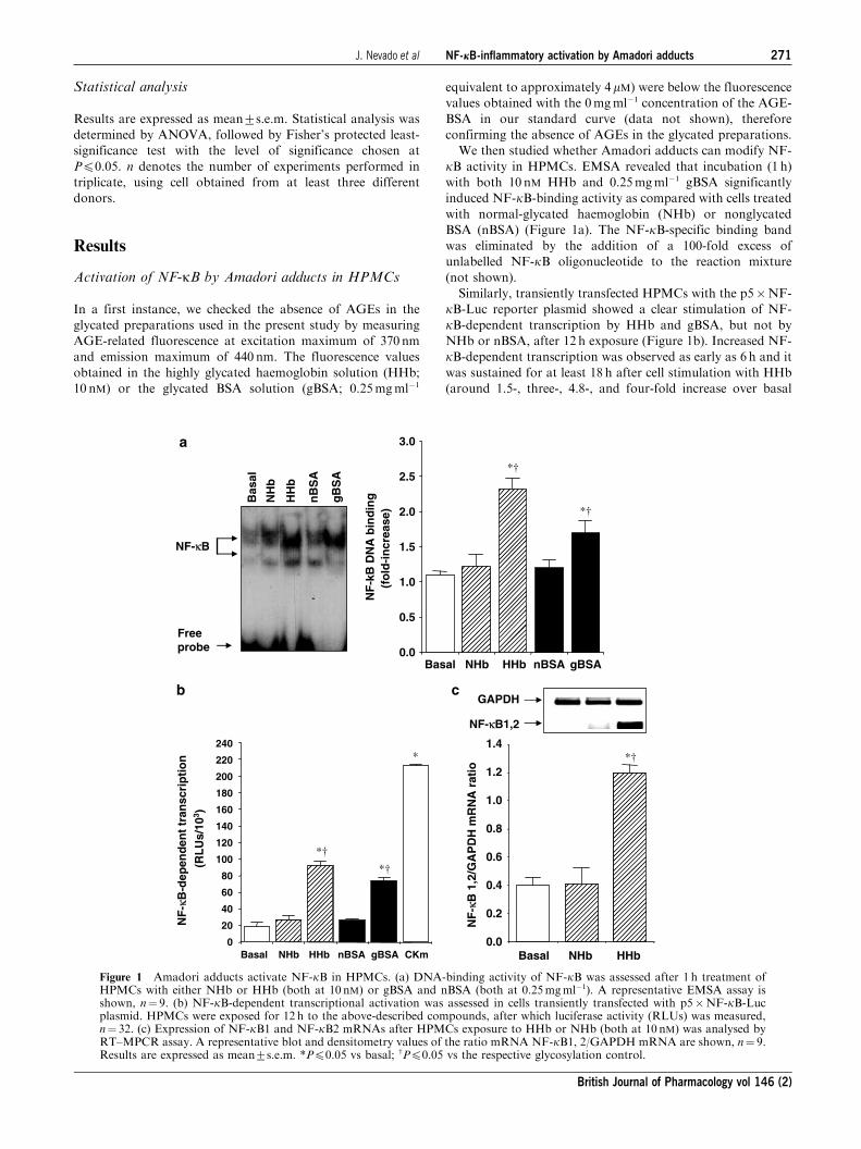
We then studied whether Amadori adducts can modify NF-
kB activity in HPMCs. EMSA revealed that incubation (1 h)
with both 10 nM HHb and 0.25mgml�1 gBSA significantly
induced NF-kB-binding activity as compared with cells treated
with normal-glycated haemoglobin (NHb) or nonglycated
BSA (nBSA) (Figure 1a). The NF-kB-specific binding band
was eliminated by the addition of a 100-fold excess of
unlabelled NF-kB oligonucleotide to the reaction mixture
(not shown).
Similarly, transiently transfected HPMCs with the p5�NF-
kB-Luc reporter plasmid showed a clear stimulation of NF-
kB-dependent transcription by HHb and gBSA, but not by
NHb or nBSA, after 12 h exposure (Figure 1b). Increased NF-
kB-dependent transcription was observed as early as 6 h and it
was sustained for at least 18 h after cell stimulation with HHb
(around 1.5-, three-, 4.8-, and four-fold increase over basal
NF
-kB
DN
A b
ind
ing
(fo
ld-i
ncr
ease
)
Basal NHb HHb nBSA gBSA0.0
0.5
1.0
1.5
2.0
2.5
3.0
*†
*†
NF
-κB
-dep
end
ent
tran
scri
pti
on
(RL
Us/
103 )
Basal NHb HHb0.0
0.2
0.4
0.6
0.8
1.0
1.2
1.4
NF
-κB
1,2
/GA
PD
H m
RN
A r
atio
Basal NHb HHb nBSA gBSA CKm0
20
40
60
80
100
120
140
160
180
200
220
240
*†
*†
*
Freeprobe
NF-κB
Bas
al
NH
b
HH
b
nB
SA
gB
SA
GAPDH
NF-κB1,2
*†
a
b c
Figure 1 Amadori adducts activate NF-kB in HPMCs. (a) DNA-binding activity of NF-kB was assessed after 1 h treatment ofHPMCs with either NHb or HHb (both at 10 nM) or gBSA and nBSA (both at 0.25mgml�1). A representative EMSA assay isshown, n¼ 9. (b) NF-kB-dependent transcriptional activation was assessed in cells transiently transfected with p5�NF-kB-Lucplasmid. HPMCs were exposed for 12 h to the above-described compounds, after which luciferase activity (RLUs) was measured,n¼ 32. (c) Expression of NF-kB1 and NF-kB2 mRNAs after HPMCs exposure to HHb or NHb (both at 10 nM) was analysed byRT–MPCR assay. A representative blot and densitometry values of the ratio mRNA NF-kB1, 2/GAPDH mRNA are shown, n¼ 9.Results are expressed as mean7s.e.m. *Pp0.05 vs basal; wPp0.05 vs the respective glycosylation control.
J. Nevado et al NF-jB-inflammatory activation by Amadori adducts 271
British Journal of Pharmacology vol 146 (2)
at 6 h, 8 h, 12 h, and 18 h, respectively, Pp0.05 by ANOVA)
(see Supplemental data, Figure 2). A cytokine mix (CKm)
consisting of IL-1b and TNF-a (both at 10 ngml�1) was used
as a positive control for the induction of NF-kB-dependenttranscription. We additionally analysed the effect of HHb on
mRNA levels of p50/105 (NF-kB1) and p49/100 (NF-kB2) inHPMCs (Figure 1c). The best-characterised form of NF-kB is
a heterodimer formed by a 50 kDa (p50/NF-kB1) and a
65 kDa (p65/RealA) protein. RT–MPCR assays revealed an
increased band corresponding to NF-kB1 and/or NF-kB2(143 bp) in HPMCs treated with 10 nM HHb for 6 h, but not
with vehicle or 10 nM NHb (Figure 1c) (see also Supplemental
data, Figure 3).
Amadori adducts stimulate several NF-kB-relatedproinflammatory genes in HPMCs
We further studied whether Amadori adducts may affect the
expression, activity, and cellular levels of several NF-kB-relatedproinflammatory markers. Thus, both HHb and gBSA, but not
their corresponding glycosylation controls, stimulated the
activity (more than three fold increase over basal) of two NF-
kB-related proinflammatory enzymes such as NOS and COX
(Table 1). HHb (10 nM) also induced a significant increase on
the basal secretion of several NF-kB-related proinflammatory
cytokines, such as IL-6 (4.972.6 vs 1.870.2pgml�1, Pp0.05),
TNF-a (118.0715.1 vs 55.075.5pgml�1, Pp0.05), and IL-1b(12.573.1 vs 4.373.2pgml�1, Pp0.05), in cell supernatants.
NHb failed to modify the basal production of these cytokines
(see also Supplemental data, Figure 4).
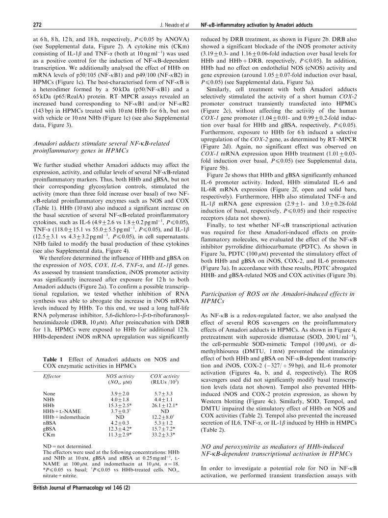
We therefore determined the influence of HHb and gBSA on
the expression of NOS, COX, IL-6, TNF-a, and IL-1b genes.
As assessed by transient transfection, iNOS promoter activity
was significantly increased after exposure for 12 h to both
Amadori adducts (Figure 2a). To confirm a possible transcrip-
tional regulation, we tested whether inhibition of RNA
synthesis was able to abrogate the increase in iNOS mRNA
levels induced by HHb. To this end, we used a long half-life
RNA polymerase inhibitor, 5,6-dichloro-1-b-D-ribofuranosyl-benzimidazole (DRB, 10mM). After preincubation with DRB
for 1 h, HPMCs were exposed to HHb for additional 12 h.
HHb-dependent iNOS mRNA upregulation was significantly
reduced by DRB treatment, as shown in Figure 2b. DRB also
showed a significant blockade of the iNOS promoter activity
(3.1970.3- and 1.1670.06-fold induction over basal levels for
HHb and HHbþDRB, respectively, Pp0.05). In addition,
HHb had no effect on endothelial NOS (eNOS) activity and
gene expression (around 1.0570.07-fold induction over basal,
Pp0.05) (see Supplemental data, Figure 5a).
Similarly, cell treatment with both Amadori adducts
selectively stimulated the activity of a short human COX-2
promoter construct transiently transfected into HPMCs
(Figure 2c), without affecting the activity of the human
COX-1 gene promoter (1.0470.01- and 0.9970.2-fold induc-
tion over basal for HHb and gBSA, respectively, Pp0.05).
Furthermore, exposure to HHb for 6 h induced a selective
upregulation of the COX-2 gene, as determined by RT–MPCR
(Figure 2d). Again, no significant effect was observed on
COX-1 mRNA expression upon HHb treatment (1.0170.03-
fold induction over basal, Pp0.05) (see Supplemental data,
Figure 5b).
Figure 2e shows that HHb and gBSA significantly enhanced
IL-6 promoter activity. Indeed, HHb stimulated IL-6 and
IL-6R mRNA expression (Figure 2f, open and solid bars,
respectively). Furthermore, HHb also stimulated TNF-a and
IL-1b mRNA gene expression (2.971- and 3.070.28-fold
induction of basal, respectively, Pp0.05) and their respective
receptors (data not shown).
Finally, to test whether NF-kB transcriptional activation
was required for these Amadori-induced effects on proin-
flammatory molecules, we evaluated the effect of the NF-kBinhibitor pyrrolidine dithiocarbamate (PDTC). As shown in
Figure 3a, PDTC (100mM) prevented the stimulatory effect of
both HHb and gBSA on iNOS, COX-2, and IL-6 promoters
(Figure 3a). In accordance with these results, PDTC abrogated
HHB- and gBSA-related NOS and COX activities (Figure 3b).
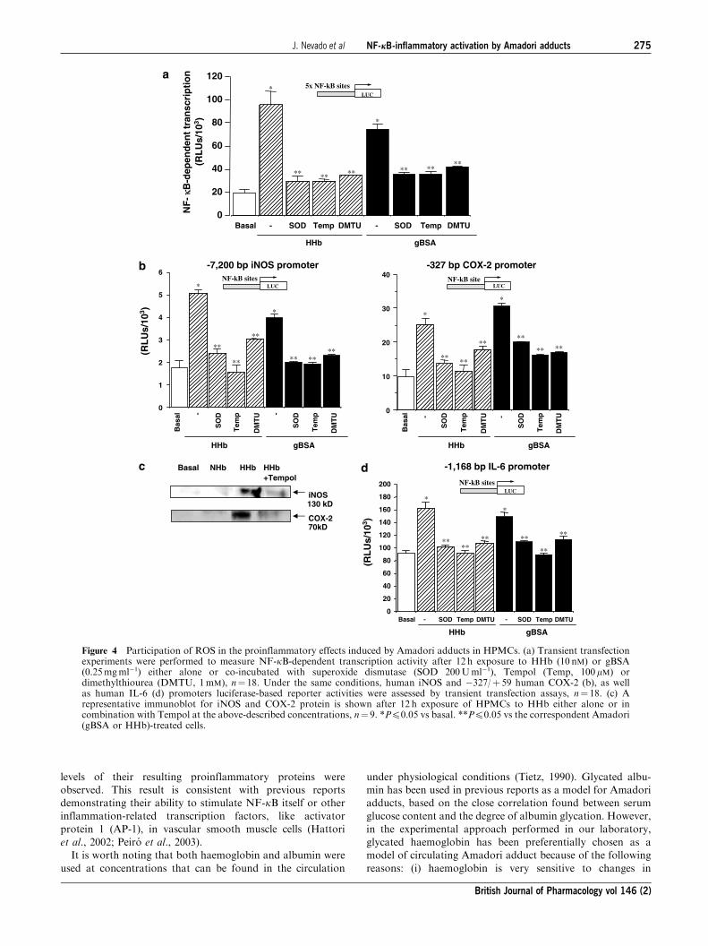
Participation of ROS on the Amadori-induced effects inHPMCs
As NF-kB is a redox-regulated factor, we also analysed the
effect of several ROS scavengers on the proinflammatory
effects of Amadori adducts in HPMCs. As shown in Figure 4,
pretreatment with superoxide dismutase (SOD, 200Uml�1),
the cell-permeable SOD-mimetic Tempol (100 mM), or di-
methylthiourea (DMTU, 1mM) prevented the stimulatory
effect of both HHb and gBSA on NF-kB-dependent transcrip-tion and iNOS, COX-2 (�327/þ 59 bp), and IL-6 promoter
activation (Figures 4a, b, and d, respectively). The ROS
scavengers used did not significantly modify basal transcrip-
tion levels (data not shown). Tempol also prevented HHb-
induced iNOS and COX-2 protein expression, as shown by
Western blotting (Figure 4c). Similarly, SOD, Tempol, and
DMTU impaired the stimulatory effect of HHb on NOS and
COX activities (Table 2). Tempol also prevented the increased
secretion of IL6, TNF-a, or IL-1b induced by HHb in HMPCs
(Table 2).
NO and peroxynitrite as mediators of HHb-inducedNF-kB-dependent transcriptional activation in HPMCs
In order to investigate a potential role for NO in NF-kBactivation, we performed transient transfection assays with
Table 1 Effect of Amadori adducts on NOS andCOX enzymatic activities in HPMCs
Effector NOS activity(NOx, mM)
COX activity(RLUs /103)
None 3.972.0 5.773.3NHb 4.071.8 4.471.1HHb 15.372.5* 26.1712.1*HHb+L-NAME 3.770.3w NDHHb+indomethacin ND 12.278.0w
nBSA 4.270.3 5.371.2gBSA 12.374.2* 15.777.2*CKm 11.372.9* 33.273.3*
ND¼not determined.The effectors were used at the following concentrations: HHband NHb at 10 nM, gBSA and nBSA at 0.25mgml�1, L-NAME at 100mM, and indomethacin at 10mM, n¼ 18.*Pp0.05 vs basal; wPp0.05 vs HHb-treated cells. NOx,nitrate+nitrite.
272 J. Nevado et al NF-jB-inflammatory activation by Amadori adducts
British Journal of Pharmacology vol 146 (2)
p5�NF-kB-Luc, in the presence of different iNOS inhibitors,
either specific (1400W) or nonspecific (L-NAME). As shown
in Figure 5a, both 1400W (10 mM) and L-NAME (100mM)
reduced HHb-stimulated NF-kB activity to an extent
similar to that obtained in the presence of the anti-
inflammatory glucocorticoid dexamethasone (Dex). In unsti-
mulated HPMCs, NOS inhibitors and Dex failed to modify
NF-kB activity by themselves (data not shown). PDTC
(100mM) was used as a control for blocking HHb-induced
NF-kB activation (see Supplemental data, Figure 6).
Furthermore, incubation of HPMCs with two exogenous
NO donors S-nitroso-N-acetylpenicillamine (SNAP) and
*†
0
5
10
15
20
25
30
*
*†
*†
(RL
Us/
103 )
Basal NHb HHb nBSA gBSA CKm0
20
40
60
80
100
120
*†*† *
(RL
Us/
103 )
0
2
4
6
8
Basal NHb HHb nBSA gBSA CKm
Basal NHb HHb nBSA gBSA CKm
1
3
5
7 *†
*†
*(R
LU
s/10
3 )
Basal NHb HHb0.0
0.2
0.4
0.6
0.8
1.0
1.2
1.4 *†
CO
X-2
/GA
DP
H m
RN
A r
atio
Basal HHb Basal HHb0.0
0.2
0.4
0.6
0.8
1.0
1.2
1.4
IL-6
or
IL-6
R/G
AD
PH
m
RN
A r
atio *
*
Basal NHb HHb HHb+DRB
CKm0.0
0.2
0.4
0.6
0.8
1.0
1.2
1.4
*
iNO
S/G
AD
PH
mR
NA
rat
io
**
GAPDHiNOS
Basal HHb
GAPDH
IL-6RIL-6
GAPDH
COX-2
Basal NHb HHb-327 bp COX-2 promoter
-1,168 bp IL-6 promoter
Basal NHb HHb HHb CKm+DRB
LUCNF-kB site
NF-kB sitesLUC
-7,200 bp iNOS promoter
NF-kB sitesLUC
a b
dc
e f
Figure 2 Stimulation by Amadori adducts of NF-kB-related proinflammatory gene expression. The effect of Amadori adducts oneither human iNOS (a), human �327/þ 59 COX-2 (c), and human IL-6 (e) promoters was studied using luciferase-based reporterplasmids in transiently transfected HPMCs. Cells were treated for 12 h with HHb and NHb (both at 10 nM), gBSA, and nBSA (bothat 0.25mgml�1) or a cytokine mixture (TNF-aþ IL-1b, 10 ngml�1 each), n¼ 32. The effect of a 6 h treatment with the above-described compounds on iNOS (b), COX-2 (d), and IL-6 (open bars) and IL-6 receptor (solid bars) (f) mRNA levels was alsodetermined by RT–MPCR assays. Representative blots are shown, n¼ 9. DRB: 5,6-dichloro-1-b-D-ribofuranosylbenzimidazole(10mM). Results are expressed as mean7s.e.m. *Pp0.05 vs basal; wPp0.05 vs the respective glycosylation control; **Pp0.05 vsHHb-treated cells.
J. Nevado et al NF-jB-inflammatory activation by Amadori adducts 273
British Journal of Pharmacology vol 146 (2)
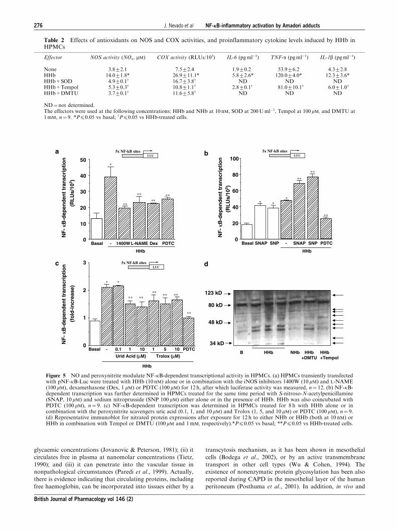
sodium nitroprusside (SNP) resulted in the stimulation of this
NF-kB-Luc reporter construct (Figure 5b). Both SNAP
(10mM) and SNP (100 mM) showed a cooperative effect with
HHb (Figure 5b), but not with NHb (not shown).
A role for peroxynitrite in HHb-induced NF-kB activation
was also analysed. Treatment of HPMCs with the peroxyni-
trite scavenger’s uric acid (1 and 10 mM) and Troloxs (1, 5,
and 10mM) partially blocked (40–55%) the stimulation of
NF-kB-dependent transcription elicited by HHb (Figure 5c).
The partial nitration of different proteins (up to 45%) in
HPMCs stimulated by HHb was confirmed by Western blot
experiments using a 3-nitrotyrosine antibody (Figure 5d, see
arrows). HHb-induced nitration was almost suppressed by
coincubation with Tempol, and in a lesser extent by DMTU
(Figure 5d).
Discussion
The use of CAPD is increasing as a renal replacement therapy
in diabetic patients undergoing dialysis. This therapeutic
option has more complications in diabetic patients than in
nondiabetic patients (Stein et al., 2004). Hyperglycaemia or
events linked to hyperglycaemia have risen as potentially
culprit mechanisms explaining this poor outcome (Nakamoto
et al., 2002; Stoenoiu et al., 2002). Hyperglycaemia is now
clearly identified as a pivotal factor in other diabetes-
associated complications, including the vascular ones. Among
the mechanisms by which hyperglycaemia can contribute to
diabetic vasculopathy, we and others have highlighted in
previous works a role for early products of nonenzymatic
protein glycosylation, the so-called Amadori adducts, in
promoting NO-related endothelial dysfunction and inflamma-
tion in the vascular wall (Angulo et al., 1996; Amore et al.,
1997; Peiro et al., 1998; Vallejo et al., 2000a; Peiro et al., 2001;
Hattori et al., 2002; Peiro et al., 2003; Rodrıguez-Manas et al.,
2003). We therefore aimed to analyse whether Amadori
adducts may play a role in hyperglycaemia-associated PM
dysfunction. We focused on the effects of Amadori adducts on
mesothelial cells, which share a common embryological
derivation with vascular endothelial cells (Hernando et al.,
1994), and have been proposed to be on the basis of peri-
toneal dysfunction (Yanez-Mo et al., 2003; Yao et al., 2003)
through the production of different growth factors and
proinflammatory markers (Riese et al., 1999; Mandl-Weber
et al., 2002).
We observed a significant stimulation of NF-kB in HPMCs
treated with Amadori adducts. As a result of this, increased
transcriptional activity of different proinflammatory genes,
such as IL-6, TNF-a, IL-1b, iNOS, and COX-2, and increased
**
**
Basal HHb HHb gBSA gBSA0
2
4
6
8
10
12
14
16
0
50
100
150
200
250
300
0
100
200
300
NOS activity COX activity
(RL
Us/
103 )
0
5
10
15
20
a
b
0
5
10
15
20
25
30
35
(RL
Us/
103 )
NO
x (µ
M)
PDTC - - -+ + Basal HHb HHb gBSA gBSA
- - -+ + Basal HHb HHb gBSA gBSA
- - -+ +
*
*
**
**
** **
* *
**
**
Basal HHb HHb gBSA gBSA
PDTC - - - - -+ +
**
*
*
*
*
****
Basal HHb HHb gBSA gBSA
PDTC - + +
-1,168 bp IL-6 promoterNF-kB sites
LUC
-327 bp COX-2 promoter
LUCNF-kB site
-7,200 bp iNOS promoterNF-kB sites
LUC
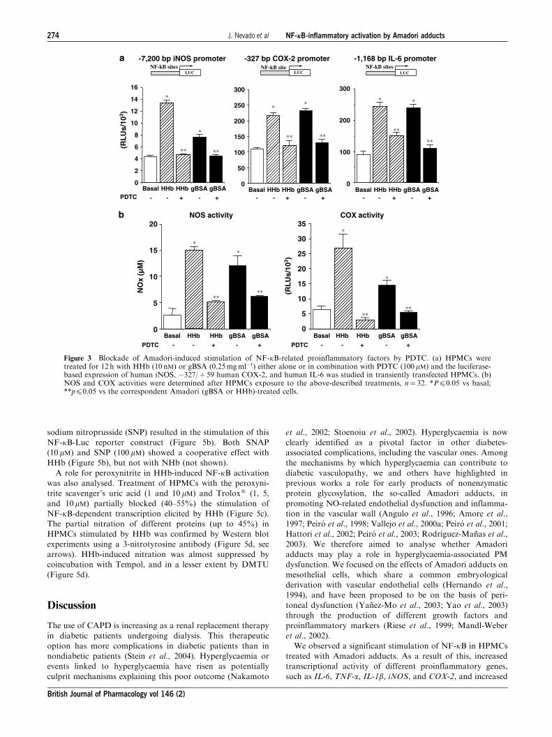
Figure 3 Blockade of Amadori-induced stimulation of NF-kB-related proinflammatory factors by PDTC. (a) HPMCs weretreated for 12 h with HHb (10 nM) or gBSA (0.25mgml�1) either alone or in combination with PDTC (100 mM) and the luciferase-based expression of human iNOS, �327/þ 59 human COX-2, and human IL-6 was studied in transiently transfected HPMCs. (b)NOS and COX activities were determined after HPMCs exposure to the above-described treatments, n¼ 32. *Pp0.05 vs basal;**pp0.05 vs the correspondent Amadori (gBSA or HHb)-treated cells.
274 J. Nevado et al NF-jB-inflammatory activation by Amadori adducts
British Journal of Pharmacology vol 146 (2)
levels of their resulting proinflammatory proteins were
observed. This result is consistent with previous reports
demonstrating their ability to stimulate NF-kB itself or other
inflammation-related transcription factors, like activator
protein 1 (AP-1), in vascular smooth muscle cells (Hattori
et al., 2002; Peiro et al., 2003).
It is worth noting that both haemoglobin and albumin were
used at concentrations that can be found in the circulation
under physiological conditions (Tietz, 1990). Glycated albu-
min has been used in previous reports as a model for Amadori
adducts, based on the close correlation found between serum
glucose content and the degree of albumin glycation. However,
in the experimental approach performed in our laboratory,
glycated haemoglobin has been preferentially chosen as a
model of circulating Amadori adduct because of the following
reasons: (i) haemoglobin is very sensitive to changes in
Basal NHb HHb
iNOS130 kD
COX-270kD
HHb+Tempol
Basal SOD Temp DMTU
gBSA
SOD Temp DMTU0
20
40
60
80
100
120
- -
HHb
0
1
2
3
4
5
6
Bas
al
SO
D
Tem
p
DM
TU
gBSA
SO
D
Tem
p
DM
TU
HHb
- - 0
10
20
30
40
SO
D
Tem
p
DM
TU
SO
D
Tem
p
DM
TU- -
gBSAHHb
Bas
al
Basal - SOD Temp DMTU - SOD Temp DMTU0
20
40
60
80
100
120
140
160
180
200
HHb gBSA
** ** ** ** ****
**
**
**
** ****
*
*
*
* *
*
*
*
** **
**
****
**
**
** **
**
**
**
(RL
Us/
103 )
(RL
Us/
103 )
-327 bp COX-2 promoter
LUCNF-kB site
-7,200 bp iNOS promoterNF-kB sites
LUC
-1,168 bp IL-6 promoter
NF-kB sitesLUC
5x NF-kB sitesLUC
NF
- κκB
-dep
end
ent
tran
scri
pti
on
(RL
Us/
103 )
a
b
c d
Figure 4 Participation of ROS in the proinflammatory effects induced by Amadori adducts in HPMCs. (a) Transient transfectionexperiments were performed to measure NF-kB-dependent transcription activity after 12 h exposure to HHb (10 nM) or gBSA(0.25mgml�1) either alone or co-incubated with superoxide dismutase (SOD 200Uml�1), Tempol (Temp, 100mM) ordimethylthiourea (DMTU, 1mM), n¼ 18. Under the same conditions, human iNOS and �327/þ 59 human COX-2 (b), as wellas human IL-6 (d) promoters luciferase-based reporter activities were assessed by transient transfection assays, n¼ 18. (c) Arepresentative immunoblot for iNOS and COX-2 protein is shown after 12 h exposure of HPMCs to HHb either alone or incombination with Tempol at the above-described concentrations, n¼ 9. *Pp0.05 vs basal. **Pp0.05 vs the correspondent Amadori(gBSA or HHb)-treated cells.
J. Nevado et al NF-jB-inflammatory activation by Amadori adducts 275
British Journal of Pharmacology vol 146 (2)
glycaemic concentrations (Jovanovic & Peterson, 1981); (ii) it
circulates free in plasma at nanomolar concentrations (Tietz,
1990); and (iii) it can penetrate into the vascular tissue in
nonpathological circumstances (Paredi et al., 1999). Actually,
there is evidence indicating that circulating proteins, including
free haemoglobin, can be incorporated into tissues either by a
transcytosis mechanism, as it has been shown in mesothelial
cells (Bodega et al., 2002), or by an active transmembrane
transport in other cell types (Wu & Cohen, 1994). The
existence of nonenzymatic protein glycosylation has been also
reported during CAPD in the mesothelial layer of the human
peritoneum (Posthuma et al., 2001). In addition, in vivo and
Table 2 Effects of antioxidants on NOS and COX activities, and proinflammatory cytokine levels induced by HHb inHPMCs
Effector NOS activity (NOx, mM) COX activity (RLUs/103) IL-6 (pgml�1) TNF-a (pgml�1) IL-1b (pgml�1)
None 3.872.1 7.572.4 1.970.2 53.976.2 4.372.8HHb 14.071.8* 26.9711.1* 5.872.6* 120.074.0* 12.373.6*HHb+SOD 4.970.1w 16.773.8w ND ND NDHHb+Tempol 5.370.3w 10.871.1w 2.870.1w 81.0710.1w 6.071.0w
HHb+DMTU 3.770.1w 11.675.8w ND ND ND
ND¼not determined.The effectors were used at the following concentrations: HHb and NHb at 10 nM, SOD at 200Uml�1, Tempol at 100mM, and DMTU at1mM, n¼ 9. *Pp0.05 vs basal; wPp0.05 vs HHb-treated cells.
Basal - 1400W L-NAME Dex PDTC0
10
20
30
40
50
HHb
NF
- κκB
-dep
end
ent
tran
scri
pti
on
(RL
Us/
103 )
Basal SNAP SNP - SNAP SNP PDTC0
20
40
60
80
100
HHb
Basal - 0.1 1 10 1 10 PDTC0
1
2
3
Urid Acid (µM) Trolox (µM)
HHb
NF
- κκB
-dep
end
ent
tran
scri
pti
on
(fo
ld-i
ncr
ease
)
**
**
****
*
**
**
**
**
**
**
**** **
**
* *
NF
- κκB
-dep
end
ent
tran
scri
pti
on
(RL
Us/
103 )
5x NF-kB sitesLUC
5x NF-kB sitesLUC
5x NF-kB sitesLUC
5
48 kD
80 kD
123 kD
B NHbHHb HHb+Tempol
34 kD
HHb+DMTU
a b
dc
*
Figure 5 NO and peroxynitrite modulate NF-kB-dependent transcriptional activity in HPMCs. (a) HPMCs transiently transfectedwith pNF-kB-Luc were treated with HHb (10 nM) alone or in combination with the iNOS inhibitors 1400W (10mM) and L-NAME(100 mM), dexamethasone (Dex, 1 mM) or PDTC (100 mM) for 12 h, after which luciferase activity was measured, n¼ 12. (b) NF-kB-dependent transcription was further determined in HPMCs treated for the same time period with S-nitroso-N-acetylpenicillamine(SNAP, 10 mM) and sodium nitroprusside (SNP 100 mM) either alone or in the presence of HHb. HHb was also coincubated withPDTC (100 mM), n¼ 9. (c) NF-kB-dependent transcription was determined in HPMCs treated for 8 h with HHb alone or incombination with the peroxynitrite scavengers uric acid (0.1, 1, and 10 mM) and Trolox (1, 5, and 10 mM) or PDTC (100 mM), n¼ 9.(d) Representative immunoblot for nitrated protein expressions after exposure for 12 h to either NHb or HHb (both at 10 nM) orHHb in combination with Tempol or DMTU (100 mM and 1mM, respectively).*Pp0.05 vs basal; **Pp0.05 vs HHb-treated cells.
276 J. Nevado et al NF-jB-inflammatory activation by Amadori adducts
British Journal of Pharmacology vol 146 (2)
in vitro kinetics data provide evidence for the formation of
early-glycated proteins in the peritoneal cavity during the time
course (10 h) of the routine peritoneal equilibration test
(Friedlander et al., 1996). It has also been suggested that
glycated proteins can move from plasma into the peritoneal
cavity (Friedlander et al., 1996).
Although Amadori adducts have been described as im-
portant precursors of AGEs (Makita et al., 1992), the
proinflammatory effects of the glycated solutions on HPMCs
shown in this study can be attributed to Amadori adducts, as
the presence of detectable AGEs in the solutions was
discarded. Thus, this seems to be a separate mechanism from
that induced by AGEs (Boulanger et al., 2002; Wu et al., 2002;
Rashid et al., 2004), as inferred by several recent studies
(Mandl-Weber et al., 2001; Hattori et al., 2002; Valencia et al.,
2004). In this regard, a possible role for metal ions or
endotoxin contamination was discarded mainly by the ability
of Amadori proteins to release ROS even in the presence of
EDTA (Vallejo et al., 2000b), or by a detection kit (see
Methods), respectively.
The ability of Amadori adducts for inducing proinflamma-
tory factors in HPMC cultures appears to be mediated by
different ROS. These results are consistent with previous
reports showing the ability of Amadori adducts to produce
ROS, mainly superoxide anions (Vallejo et al., 2000b; Yoo
et al., 2004). In this way, it is well accepted that increased
oxidative stress plays a key role in the development of vascular
inflammation and vasculopathy in diabetes (Spitaler & Graier,
2002). It seems therefore reasonable to propose an analogous
role for ROS in the putative alterations of peritoneum
membrane functionality associated to hyperglycaemia.
Concerning a role for NO in PM dysfunction, it is thought
to be involved in both structural and permeability alterations
(Chen et al., 2000; Mandl-Weber et al., 2002). Currently, a fact
widely debated is the definitive source of local NO at the
peritoneum. Our data agree with previous reports showing a
barely detectable basal iNOS activity, (Chen et al., 2000;
Davenport et al., 2004). However, upon treatment with
Amadori adducts, a clear stimulation of iNOS activity and
gene expression was observed. Thus we can conclude that, at
present, several extracellular stimuli are known to induce
iNOS in HPMCs, including cytokines (this work, and Chen
et al., 2000), the insoluble polysaccharide zymosan, which acts
as a peritonitis inducer (Yao et al., 2004), or, as shown in the
present study, Amadori adducts. Although eNOS stimulation
has also been involved in NO production by mesothelial cells
(Reimann et al., 2004), Amadori adducts did not stimulate
eNOS activity in HPMCs.
Finally, our results suggest a possible role for Amadori-
induced NO or NO-derived compounds in peritoneal
complications related to hyperglycaemia through the activa-
tion of NF-kB in HPMCs. This is in accordance with the fact
that NO and NO-derived compounds have emerged in the
last years as important modulators of gene expression through
their ability to modulate several transcription factors (Liaudet
et al., 2000; Cooke & Davidge, 2002). Indeed, modulation
of NF-kB activity by NO appears to have an important
role in the regulation of the inflammatory response (Liaudet
et al., 2000). Nevertheless, in a pro-oxidant environment,
NO can react with superoxide anions leading to peroxynitrite
production, which may in turn further stimulate NF-kB-dependent transcription in HPMCs. Our results indicate a
role for peroxynitrite as a mediator for NF-kB activation, in
HPMCs, as previously proposed in vascular cells (Cooke &
Davidge, 2002; Hattori et al., 2004). However, the fact that
a certain degree of NF-kB-dependent transcription still
occurred in the presence of peroxynitrite scavengers discards
peroxynitrite as the sole mediator of NF-kB activation by
glycated haemoglobin. At present, little is known about the
cellular signalling pathways activated by NO and peroxynitrite
in HPMCs.
In conclusion, we propose that Amadori adducts can alter
mesothelial cell functionality by increasing oxidative and
nitrosative stress and by activating NF-kB-related proinflam-
matory pathway, which may be on the basis of a low-grade
proinflammatory response within the peritoneum.
This work was supported by grants from Fondo de InvestigacionesSanitarias (01/0579 and 02/1246), Comunidad Autonoma de Madrid(GR/SAL/0899/2004), Ministerio de Educacion y Ciencia (SAF-2001-1328), and Instituto de Salud Carlos III (RGDM (G03/212).Dr J. Nevado is a recipient of a Research Contract from Instituto deSalud Carlos III (FIS 99/3077). We are grateful to Drs D. Geller,G. Haegeman, K.K. Wu, and T. Tanabe for kindly providingplasmids, and Dr M. Blazquez and N. Nin for critical review of themanuscript.
References
AMORE, A., CIRINA, P., MITOLA, S., PERUZZI, L., GIANOGLIO, B.,RABBONE, I., SACCHETTI, C., CERUTTI, F., GRILLO, C. &COPPO, R. (1997). Nonenzymatically glycated albumin (Amadoriadducts) enhances nitric oxide synthase activity and gene expressionin endothelial cells. Kidney Int., 51, 27–35.
ANGULO, J., SANCHEZ-FERRER, C.F., PEIRO, C., MARIN, J. &RODRIGUEZ-MANAS, L. (1996). Impairment of endothelium-dependent relaxations by increasing percentages of glycated humanhaemoglobin. Hypertension, 28, 583–592.
BERGHE, W.V., PLAISANCE, S., BOONE, E., DE BOSSCHER, K.,SCHIMITZ, M.L., FIERS, W. & HAEGEMAN, G. (1998). P38 andextracellular signal-regulated kinase mitogen-activated proteinkinase pathway are regulated for nuclear-factor kB p65 transactiva-tion mediated by tumor necrosis factor. J. Biol. Chem., 273,3285–3290.
BODEGA, F., ZACCHI, L. & AGOSTINI, E. (2002). Albumin transcy-tosis in mesothelium. Am. J. Physiol. Lung Cell. Mol. Physiol., 282,L3–L11.
BOULANGER, E., WAUTIER, M.P., WAUTIER, J.C., BOVAL, B.,PANIS, Y., WERNERT, N., DANZE, P.M. & DEQUIEDT, P.
(2002). AGEs bind to mesothelial cells via RAGE and stimulateVCAM-1 expression. Kidney Int., 61, 148–156.
BUCALA, R., TRACEY, K.J. & CERAMI, A. (1991). Advancedglycosylation products quench nitric oxide mediate defectiveendothelium-dependent vasodilatation in experimental diabetes.J. Clin. Invest., 87, 432–438.
CERAMI, A., VLASSARA, H. & BROWNLEE, M. (1988). Role ofadvanced glycosylation products in complications of diabetes.Diabetes Care, 1, 73–79.
CHEN, J.Y., CHIU, J.H., CHEN, T.W., YANG, W.C. & YANG, A.H.
(2000). Human peritoneal mesothelial cells produce nitric oxide:induction by cytokines. Periton. Dialysis Int., 20, 772–777.
CHUNG-WELCH, N., PATTON, W.F., SHEPRO, D. & CAMBRIA, R.P.
(1997). Two-stage isolation procedure for obtaining homogenouspopulations of microvascular endothelial and mesothelial cells fromhuman omentum. Microvas. Res., 54, 121–134.
J. Nevado et al NF-jB-inflammatory activation by Amadori adducts 277
British Journal of Pharmacology vol 146 (2)
CIESLIK, K., LEE, L.H., TANG, J.L. & WU, K.K. (1999). Transcrip-tional regulation of endothelial nitric oxide synthase by aninteraction between casein kinase 2 and protein phosphatase 2A.J. Biol. Chem., 274, 34669–34675.
COOKE, C.M. & DAVIDGE, S.T. (2002). Peroxynitrite increases iNOSthrough NF-kB and decreases prostacyclin synthase in endothelialcells. Am. J. Physiol. Cell. Physiol., 282, C395–C402.
DAVENPORT, A., FERNANDO, R.L., ROBSON, R. & VARGHESE, Z.(2004). Nitric oxide production by peritoneal mesothelial cells. Int.J. Artif. Organs, 27, 15–23.
DEVUYST, O., COMBET, S., CNOPS, Y. & STOENOIU, M. (2001).Regulation of NO synthase isoforms in the peritoneum: implica-tions for ultrafiltration failure in peritoneal dialysis. Nephrol. Dial.Transplant., 16, 675–678.
FRIEDLANDER, M.A., WU, Y.C., ELGAWISH, A. & MONNIER, V.M.
(1996). Early and advanced glycosylation end-products. Kinetics offormation and clearance in peritoneal dialysis. J. Clin. Invest., 97,728–735.
FRIES, D.M., PAXINOU, E., THEMISTOCLEOUS, M., SWANBERG, E.,GRIENDLING, K.K., SALVEMINI, D., SLOT, J.W., HEIJNEN, H.F.,HAZEN, S.L. & ISCHIROPOULUS, H. (2003). Expression of induciblenitric-oxide synthase and intracellular protein tyrosine nitration invascular smooth muscle cells. J. Biol. Chem., 25, 22901–22907.
HATTORI, Y., BANBA, N., GROSS, S.S. & KASAI, K. (1999). Glycatedserum albumin-induced nitric oxide production in vascular smoothmuscle cell by nuclear-factor kB-dependent transcriptional activa-tion of inducible nitric oxide synthase. Biochem. Biophys. Res.Commun., 259, 128–132.
HATTORI, Y., KASSAI, K. & GROSS, S.S. (2004). NO suppresses whileperoxynitrite sustains NF-kB: a paradigm to rationalize cytoprotectiveand cytotoxic actions attributed to NO. Cardiovasc. Res., 63, 31–40.
HATTORI, Y., SUZIKI, M., HATTORI, S. & KASAI, K. (2002). Vascularsmooth muscle cell activation by glycated albumin (Amadoriadducts). Hypertension, 39, 22–28.
HERNANDO, A., GARCIA-HONDUVILLA, N., BELLO, J.M., BUJAN,J. & NAVLET, J. (1994). Coatings for vascular prostheses:mesothelial cell express specific markers for muscle cells and havebiological activity similar to that of endothelial cells. Eur. J.Endovasc. Surg., 8, 531–536.
INOUE, H., YOKOYAMA, C., HARA, S., TONE, Y. & TANABE, T.
(1995). Transcriptional regulation of human prostaglandin-endo-peroxide synthase-2 gene by lipopolysaccharide and phorbol ester invascular endothelial cells. Involvement of both nuclear factor forinterleukin-6 expression site and cAMP response element. J. Biol.Chem., 270, 24965–24971.
JOVANOVIC, I. & PETERSON, C.M. (1981). The clinical utility ofglycated hemoglobin. Am. J. Med., 70, 331–338.
LIAUDET, L., GARCIA-SORIANO, F. & SZABO, C. (2000). Biology ofnitric oxide signalling. Crit. Care Med., 28, N37–N49.
MAKITA, Z., VLASSARA , H., RAYFIELD, E., CARTWRIGHT, K.,FRIEDMAN, E., RODBY, R., CERAMI, A. & BUCALA, R. (1992).Hemoglobin-AGE: a circulating marker of advanced glycosylation.Science, 258, 651–653.
MANDL-WEBER, S., COHEN, C.D., HASLINGER, B., KRETZLER, M.
& SITTER, T. (2002). Vascular endothelial growth factor productionand regulation in human peritoneal mesothelial cells. Kidney Int.,61, 570–578.
MANDL-WEBER, S., HASLINGER, B., SCHALKWIJK, C.G. & SITTER,T. (2001). Early glycated albumin, but not advanced glycatedalbumin, methylglyoxal, or 3-deoxyglucosonee increases theexpression of PAI-1 in human peritoneal mesothelial cells. Periton.Dialysis Int., 21, 487–494.
NAKAMOTO, H., IMAI, H., KAWANISHI, H., NAKAMOTO, M.,MINAKUCHI, J., KUMON, S., WATANABE S SHIOHIRA, Y., ISHII,T., KAWAHARA, T., TSUZAKI, K. & SUZUKI, H. (2002). Effect ofdiabetes on peritoneal function assessed by personal dialysiscapacity test in patients undergoing CAPD. Am. J. Kidney Dis.,40, 1045–1054.
NEY, K.A., COLLEY, K.J. & PIZZO, S.V. (1981). The standardization ofthe thiobarbituric acid assay for nonenzymatic glycosylation ofhuman serum albumin. Anal. Biochem., 118, 294–300.
PAREDI, P., BIERNACKI, W., INVERNIZZI, G., KHARITONOV, S.A.
& BARNES, P.J. (1999). Exhaled carbon monoxide levels elevated indiabetes and correlated with glucose concentration in blood: a newtest for monitoring the disease? Chest, 116, 1007–1011.
PARK, M.S., LEE, H.A., CHU, W.S., YANG, D.H. & HWANG, S.D.
(2000). Peritoneal accumulation of AGE and peritoneal membranepermeability. Periton. Dialysis Int., 20, 452–460, 12.
PEIRO, C., ANGULO, J., RODRIGUEZ-MANAS, L., LLERGO, J.L.,VALLEJO, S., CERCAS, E. & SANCHEZ-FERRER, C.F. (1998).Vascular smooth muscle cell hypertrophy induced by glycosylatedhuman oxyhaemoglobin. Br. J. Pharmacol., 125, 637–644.
PEIRO, C., LAFUENTE, N., MATESANZ CERCAS, E., LLERGO, J.L.,VALLEJO, S., RODRIGUEZ-MANAS, L. & SANCHEZ-FERRER,C.F. (2001). High glucose induces cell death of cultured humanaortic smooth muscle cells through the formation of hydrogenperoxide. Br. J. Pharmacol., 133, 967–974.
PEIRO, C., MATESANZ, N., NEVADO, J., LAFUENTE, N., CERCAS,E., AZCUTIA, V., VALLEJO, S., RODRIGUEZ-MANAS, L. &SANCHEZ-FERRER, C.F. (2003). Glycated human oxyhaemoglobinactivates nuclear factor-kB and activator protein-1 in culturedhuman aortic smooth muscle. Br. J. Pharmacol., 140, 681–690.
POSTHUMA, N., TER WEE, P.M., NIESSEN, H., DONKER, A.J.,VERNRUGH, H.A. & SCHALKWIJK, C.G. (2001). Amadorialbumin and advanced glycation end-product formation in perito-neal dialysis using icodextrin. Periton. Dialysis Int., 21, 43–51.
RASHID, G., BENCHETRIT, S., FISHMAN, D. & BERHEIM, J. (2004).Effect of advanced glycation end-products on gene expression andsynthesis of TNF-alpha and endothelial nitric oxide synthase byendothelial cell. Kidney Int., 66, 1099–1106.
REIMANN, D., DACHS, D., MEYE, C. & GROSS, P. (2004). Amino-acid-based peritoneal dialysis solution stimulates mesothelial nitricoxide production. Periton. Dial. Int., 24, 378–384.
RIESE, J., DENZEL, C., ZOWE, M., MEHER, C., HOHENBERGER, W.
& HAUPT, W. (1999). Secretion of IL-6, monocyte chemoattractantprotein-1, macrophage inflammatory protein-1-alpha, and TNF-alpha by cultured intact human peritoneum. Eur. Surg. Res., 31,281–288.
RODRIGUEZ-MANAS, L., ANGULO, J., VALLEJO, S., PEIRO, C.,SANCHEZ-FERRER, A., CERCAS, E. & SANCHEZ-FERRER, C.F.(2003). Early and intermediate Amadori glycated adducts, oxidativestress, and endothelial dysfunction in the streptozotocin-induceddiabetic rats vasculature. Diabetologia, 46, 556–566.
SCHREIBER, M.M., MATTHIAS, P., MULLER, M.M. & SCHAFFNER,W. (1989). Rapid detection of octamer binding proteins with ‘miniextracts’ prepared from a small number of cell. Nucleic Acid Res.,17, 6419.
SELL, D.R. & MONNIER, V.M. (1989). Structure elucidation of asenescence crosslink from human extracellular matrix. Implicationof pentoses in the aging process. J. Biol. Chem., 264, 21597–21602.
SPITALER, M.M. & GRAIER, W.F. (2002). Vascular targets of redoxsignalling in diabetes mellitus. Diabetologia, 45, 476–494.
STEIN, G., FUNFSTUCK, R. & SCHIEL, R. (2004). Diabetes mellitusand dialysis. Min. Urol. Nefrol., 56, 289–303.
STOENOIU, M.S., DE VRIESSE, A.S., MOULIN, P., FERON, O.,LAMEIRE, M. & DEVUYST, O. (2002). Experimental diabetesinduces functional and structural changes in the peritoneum. KidneyInt., 62, 668–678.
TAYLOR, B.S., De VERA, M.E., GANSTER, R.W., Wang, K., SHAPIRO,R.A., MORRIS JR., S.M., BILLIAR, T.R. & GELLER, D.A. (1998).Multiple NF-kB enhancer elements regulate cytokine induction ofhuman inducible nitric oxide synthase gene. J. Biol. Chem., 273,15148–15156.
TIETZ, N.W. (1990). Clinical Guide to Laboratory Tests. Philadelphia,PA: WB Saunders Co., pp. 284–285.
VALENCIA, J.V., MONE, M., KOEHNE, C., REDISKE, J. & HUGHES,T.E. (2004). Binding of receptor for advanced glycation end product(RAGE) ligands is not sufficient to induce inflammatory signals:lack of activity on endotoxin-free albumin-derived advancedglycation end products. Diabetologia, 47, 844–852.
VALLEJO, S., ANGULO, J., PEIRO, C., NEVADO, J., SANCHEZ-FERRER,A., PETIDIER, R., SANCHEZ-FERRER, C.F. & RODRIGUEZ-
MANAS, L. (2000a). Highly glycated oxyhemoglobin impairs nitricoxide relaxations in human mesenteric microvessels. Diabetologia, 43,83–90.
VALLEJO, S., ANGULO, J., PEIRO, C., SANCHEZ-FERRER, A., CERCAS,E., NEVADO, J., SANCHEZ-FERRER, C.F. & RODRIGUEZ-MANAS,L. (2000b). Correction of glycated oxyhemoglobin-induced impairmentof endothelium-dependent vasodilatation by glycacide. J. DiabetesComplicat., 14, 207–214.
278 J. Nevado et al NF-jB-inflammatory activation by Amadori adducts
British Journal of Pharmacology vol 146 (2)
WU, C.H., HUANG, C.M., LIN, C.H., HO, Y.S., CHEN, C.M. & LEE,H.M. (2002). Advanced glycosylation end products induceNF-kappaB dependent iNOS expression in RAW 264.7 cells. Mol.Cell. Endocrinol., 194, 9–17.
WU, V.Y. & COHEN, M.P. (1994). Receptor specific for Amadori-modified glycated albumin on murine endothelial cells. Biochem.Biophys. Res. Commun., 199, 1088.
YANEZ-MO, M., LARA-PEZZI, E., SELGAS, R., RAMIREZ-HUESCA, M.,DOMINGUEZ-JIMENEZ, C., JIMENEZ-HEFFERNAN, A., AGUI-
LERA, A., SANCHEZ-TOMERO, J.A., BAJO, M.A., ALVAREZ, V.,CASTRO, M.A., DEL PESO, G., CIRUJEDA, A., GAMALLO, C,SANCHEZ-MADRID, F. & LOPEZ-CABRERA, M. (2003). Peritonealdialysis and epithelial-to-mesenchymal transition of mesothelial cells.N. Engl. J. Med., 348, 403–412.
YAO, V., MCCAULEY, R., COOPER, D., PLATELL, C. & HALL, J.C.(2004). Zymosan induces nitric oxide production by peritonealmesothelial cells. ANZ J. Surg., 74, 266–269.
YAO, V., PLATELL, C. & HALL, J.C. (2003). Role of peritonealmesothelial cells in peritonitis. Br. J. Surg., 90, 1187–1194.
YOO, C., SONG, C., KIM, B., HONHG, H. & LEE, H. (2004). Glycatedalbumin induces superoxide generation in mesangial cells. Cell.Physiol. Biochem., 14, 361–368.
(Received March 24, 2005Revised May 3, 2005
Accepted May 17, 2005Published online 4 July 2005)
Supplementary Information accompanies the paper on British Journal of Pharmacology website (http://www.nature.com/bjp).
J. Nevado et al NF-jB-inflammatory activation by Amadori adducts 279
British Journal of Pharmacology vol 146 (2)
Copyright © 2022 FDOKUMEN


![Identification and quantitation of benzo[a]pyrene-DNA adducts formed in mouse skin](https://static.fdokumen.com/doc/165x107/6333eb3bb94d623842027004/identification-and-quantitation-of-benzoapyrene-dna-adducts-formed-in-mouse-skin.jpg)