
The inefficiency of incisions of ecteinascidin 743–DNA adducts by the UvrABC nuclease and the...
17
Research Paper The ine/ciency of incisions of ecteinascidin 743^DNA adducts by the UvrABC nuclease and the unique structural feature of the DNA adducts can be used to explain the repair-dependent toxicities of this antitumor agent Maha Zewail-Foote a , Ven-Shun Li b , Harold Kohn b; 1 , David Bearss c , Mary Guzman c , Laurence H. Hurley a; d ; * a Department of Chemistry and Biochemistry, The University of Texas at Austin, Austin, TX 78712, USA b Department of Chemistry, University of Houston, Houston, TX 77204, USA c Arizona Cancer Center, 1515 N. Campbell Avenue, Tucson, AZ 85724, USA d College of Pharmacy, The University of Texas at Austin, Austin, TX 78712, USA Received 9 April 2001; revisions requested 8 June 2001; revisions received 10 July 2001; accepted 12 July 2001 First published online 12 September 2001 Abstract Background : Ecteinascidin 743 (Et 743), a natural product derived from a marine tunicate, is a potent antitumor agent presently in phase II clinical trials. Et 743 binds in the minor groove of DNA and alkylates N2 of guanine via a unique mechanism involving catalytic activation. The sequence selectivity of Et 743 is governed by different patterns of hydrogen-bonding to DNA, which results in differential reversibility of the covalent adducts. As determined by nuclear magnetic resonance spectros- copy, the preferred sequences 5P-PuGC and 5P-PyGG are stabilized by a hydrogen-bonding network, while the non- preferred sequences 5P-NG(A/T) are much less stabilized due to the lack of a key hydrogen bond to the GC base pair on the 3P-side of the alkylated guanine. Results : Mammalian cell lines (XPB, XPD, XPF, XPG, and ERCC1) deficient in the nucleotide excision repair (NER) gene products show resistance to Et 743. The recognition and subsequent incision of Et 743^DNA adducts by the bacterial multisubunit endonuclease UvrABC were used to evaluate DNA repair-mediated toxicity as a rationale for the resistance of NER- defective cell lines and the antitumor activity of Et 743. The Et 743^DNA adducts are indeed recognized and incised by the UvrABC repair proteins; however, the pattern of incision indicated that the non-preferred, and less stable, sequences (i.e. 5P-NG(A/T)) modified with Et 743 are generally incised at a much higher efficiency than the preferred, more stable sequences (i.e. 5P- PuGC or 5P-PyGG). In addition, within the same Et 743 recognition sequence, the level of incision varies, indicating that flanking regions also contribute to the differential incision frequency. Conclusions : The inefficient repair incision by the UvrABC nuclease of Et 743^DNA adducts provides a basis for rationalizing the observed repair-dependent cytotoxicities of these DNA adducts, if other associated structural properties of Et 743^DNA adducts are taken into account. In particular, the wedge-shaped Et 743, which forces open the minor groove of DNA, introducing a major groove bend, and the extrahelical protrusion of the C- subunit of Et 743 provide unique characteristics alongside the hydrogen-bonding stabilization of a covalent DNA adduct, which we propose traps an intermediate in NER processing of Et 743^ DNA adducts. This trapped intermediate protein^Et 743^DNA adduct complex can be considered analogous to a poisoned topoisomerase I^ or topoisomerase II^DNA complex. In the absence of an intact NER nuclease complex, this toxic lesion is unable to form, and the Et 743^DNA adducts, although not 1074-5521 / 01 / $ ^ see front matter ß 2001 Elsevier Science Ltd. All rights reserved. PII:S1074-5521(01)00071-0 1 Present address : Division of Medicinal Chemistry and Natural Products, University of North Carolina at Chapel Hill, Chapel Hill, NC 27599, USA. * Corresponding author. Present addresses: The University of Arizona, Arizona Cancer Center, 1515 N. Campbell Avenue, Tucson, AZ 85724, USA, and The University of Arizona, College of Pharmacy, 1703 E. Mabel, Tucson, AZ 85721, USA. E-mail address : [email protected] (L.H. Hurley). Chemistry & Biology 8 (2001) 1033^1049 www.elsevier.com/locate/chembiol
Transcript of The inefficiency of incisions of ecteinascidin 743–DNA adducts by the UvrABC nuclease and the...
Research Paper
The ine¤ciency of incisions of ecteinascidin743^DNA adducts by the UvrABC nuclease and the unique
structural feature of the DNA adducts can be used toexplain the repair-dependent toxicities of this
antitumor agent
Maha Zewail-Foote a, Ven-Shun Li b, Harold Kohn b; 1, David Bearss c,Mary Guzman c, Laurence H. Hurley a; d; *
aDepartment of Chemistry and Biochemistry, The University of Texas at Austin, Austin, TX 78712, USAbDepartment of Chemistry, University of Houston, Houston, TX 77204, USAcArizona Cancer Center, 1515 N. Campbell Avenue, Tucson, AZ 85724, USA
dCollege of Pharmacy, The University of Texas at Austin, Austin, TX 78712, USA
Received 9 April 2001; revisions requested 8 June 2001; revisions received 10 July 2001; accepted 12 July 2001First published online 12 September 2001
Abstract
Background: Ecteinascidin 743 (Et 743), a natural productderived from a marine tunicate, is a potent antitumor agentpresently in phase II clinical trials. Et 743 binds in the minorgroove of DNA and alkylates N2 of guanine via a uniquemechanism involving catalytic activation. The sequence selectivityof Et 743 is governed by different patterns of hydrogen-bonding toDNA, which results in differential reversibility of the covalentadducts. As determined by nuclear magnetic resonance spectros-copy, the preferred sequences 5P-PuGC and 5P-PyGG arestabilized by a hydrogen-bonding network, while the non-preferred sequences 5P-NG(A/T) are much less stabilized due tothe lack of a key hydrogen bond to the GC base pair on the 3P-sideof the alkylated guanine.
Results : Mammalian cell lines (XPB, XPD, XPF, XPG, andERCC1) deficient in the nucleotide excision repair (NER) geneproducts show resistance to Et 743. The recognition andsubsequent incision of Et 743^DNA adducts by the bacterialmultisubunit endonuclease UvrABC were used to evaluate DNArepair-mediated toxicity as a rationale for the resistance of NER-defective cell lines and the antitumor activity of Et 743. The Et743^DNA adducts are indeed recognized and incised by theUvrABC repair proteins ; however, the pattern of incision
indicated that the non-preferred, and less stable, sequences (i.e.5P-NG(A/T)) modified with Et 743 are generally incised at a muchhigher efficiency than the preferred, more stable sequences (i.e. 5P-PuGC or 5P-PyGG). In addition, within the same Et 743recognition sequence, the level of incision varies, indicating thatflanking regions also contribute to the differential incisionfrequency.
Conclusions: The inefficient repair incision by the UvrABCnuclease of Et 743^DNA adducts provides a basis for rationalizingthe observed repair-dependent cytotoxicities of these DNAadducts, if other associated structural properties of Et 743^DNAadducts are taken into account. In particular, the wedge-shaped Et743, which forces open the minor groove of DNA, introducing amajor groove bend, and the extrahelical protrusion of the C-subunit of Et 743 provide unique characteristics alongside thehydrogen-bonding stabilization of a covalent DNA adduct, whichwe propose traps an intermediate in NER processing of Et 743^DNA adducts. This trapped intermediate protein^Et 743^DNAadduct complex can be considered analogous to a poisonedtopoisomerase I^ or topoisomerase II^DNA complex. In theabsence of an intact NER nuclease complex, this toxic lesion isunable to form, and the Et 743^DNA adducts, although not
1074-5521 / 01 / $ ^ see front matter ß 2001 Elsevier Science Ltd. All rights reserved.PII: S 1 0 7 4 - 5 5 2 1 ( 0 1 ) 0 0 0 7 1 - 0
1 Present address: Division of Medicinal Chemistry and Natural Products, University of North Carolina at Chapel Hill, Chapel Hill, NC 27599, USA.
* Corresponding author. Present addresses: The University of Arizona, Arizona Cancer Center, 1515 N. Campbell Avenue, Tucson, AZ 85724, USA,and The University of Arizona, College of Pharmacy, 1703 E. Mabel, Tucson, AZ 85721, USA.
E-mail address: [email protected] (L.H. Hurley).
CHBIOL 135 24-10-01 Cyaan Magenta Geel Zwart
Chemistry & Biology 8 (2001) 1033^1049
www.elsevier.com/locate/chembiol
repaired by the NER pathway, are less toxic to cells. Conversely,elevated levels of either of these nucleases should lead to enhancedEt 743 toxicity. ß 2001 Elsevier Science Ltd. All rights reserved.
Keywords: Alkylating agent; Anticancer drug; DNA repair ; Ec-teinascidin
1. Introduction
The ecteinascidins (Ets) are extremely potent antitumoragents isolated from the marine tunicate Ecteinascidia tur-binata [1]. Of the numerous Ets that have been isolated, Et743 is the ¢rst to advance to clinical trials, where it isparticularly active against soft tissue sarcomas [2^6]. Et743 is a carbinolamine-containing antitumor agent com-posed of three fused tetrahydroisoquinoline rings and isstructurally related to the DNA-reactive saframycins [7^9],naphthyridinomycin [10], and anthramycin [11] (Fig. 1A).The primary structural di¡erence between the saframycinsand Et 743 is that Et 743 contains an extra tetrahydroiso-quinoline ring, the C-subunit, that is absent in this otherantitumor antibiotic. While the A- and B-subunits of Et743 provide the sca¡old for DNA recognition and bond-ing, the C-subunit protrudes out of the minor groove,making only limited contacts with the DNA [12] (Fig.2A). Like the other carbinolamine antibiotics, the Etsbond in the minor groove of DNA in a sequence-depen-dent manner and alkylate the N2 position of guanine [13].Et 743^DNA adduct formation is mediated through anintramolecular acid-catalyzed dehydration of the carbinol-amine moiety, resulting in an iminium intermediate that isthe DNA-reactive species (Fig. 2B) [14].
Through gel mobility shift assays [13] and high-¢eldnuclear magnetic resonance (NMR) studies [12,15], thepreferred thermodynamic sequence of Et 743 has beenshown to be 5P-PuG*C and 5P-PyG*G (* indicates thecovalently modi¢ed guanine), while the non-preferred se-quences are 5P-NG*(A/T). The Et 743^DNA hydrogen-bonding network that stabilizes the adduct is a major fac-tor in determining sequence recognition. The formation ofa hydrogen bond between one of the oxygens of the meth-ylenedioxy ring of Et 743 and a proton in the exocyclic 2-amino group of the guanine located 3P to the alkylated siteon either strand (HB2 in Fig. 1B) largely determines if thebonding sequence yields a stable DNA adduct. Elimina-tion of this hydrogen bond acceptor by replacing the 3P Gor C with an A or T makes the sequence less stable [16].We have recently shown that the relative stabilities of theEt 743^DNA covalent adducts determine the rate of theEt 743^DNA covalent adduct reverse reaction, while therate of the forward reaction is independent of the £ankingsequence [16]. Hence, the sequence selectivity of Et 743 isdetermined by the relative kinetics of the covalent reversalreaction, which leads to the release of free Et 743 andrebonding at the same or di¡erent sequences (Fig. 2B).The net e¡ect of this process is the time-dependent chan-
neling of Et 743 to a subset of DNA sequences in whichthe Et 743^DNA adducts are thermodynamically stable.Thus, during a 6-h incubation, 20% of the initial covalentadduct at the less stable sequence migrates to the stablesequence (5P-PuG*C and 5P-PyG*G).
Et 743 has shown promising activity in phase I and IIclinical trials, particularly in soft tissue sarcomas, whichsets it apart from other minor groove DNA monoalkylat-ing agents [17,18]. One very unique feature of this agent isthat it is the ¢rst example of a minor groove alkylatingagent that bends DNA toward the major groove [19]. Inaddition, other factors, such as the sequence-dependenthydrogen-bonding interactions between Et 743 and theminor groove of DNA and the protrusion of the C-sub-unit from the minor groove, may also contribute to thebiological activity of Et 743 [15]. However, the biologicallyrelevant macromolecular targets have not been fully deter-mined. The ¢rst proposed target was topoisomerase I,which forms protein^DNA cross-links in the presence ofEt 743 [20,21]. More recently, evidence was presented thatshows that Et 743 can interfere with transcription activa-tion of the MDR1 gene [22]. The Pommier laboratory hasnow provided compelling evidence that eukaryotic XPG-defective cells are resistant to Et 743 [23], and the D'In-calci group has shown that XPF^ERCC1-defective cellsare also resistant to Et 743 [24]. In this contribution wehave tested a panel of eukaryotic cell lines defective in thenucleotide excision repair (NER) pathway for sensitivityto Et 743 and found that they are more resistant than theparent wild-type cell line. To gain further insight into thisunexpected result, we have used the bacterial UvrABCnuclease system to characterize how the NER pathwayrecognizes and repairs Et 743^DNA adducts that di¡erin their hydrogen-bonding-induced stability in the DNAhelix. Signi¢cantly, Et 743^DNA adducts at di¡erent se-quences, with inherently di¡erent stabilities, are repairedat di¡erent e¤ciencies. Speci¢cally, the sequences at whichEt 743 forms less stable, more dynamically £exible DNAadducts (e.g. at 5P-AGT) are the sites at which repair in-cision is the most e¤cient, while the more stable and con-formationally rigid Et 743^DNA lesions are not as readilyincised. In both cases this can lead to aberrant or un-coupled 3P or 5P incisions. This decrease in incision e¤-ciencies at the preferred DNA sequences (e.g. 5P-AGC)compared to the non-preferred sequences (e.g. 5P-AGT)may be explained by de¢ned di¡erences in DNA structureand dynamics induced by binding Et 743 at these di¡erentbonding sites. We propose that the ine¤cient incision re-pair is related to the repair-dependent toxicities previously
CHBIOL 135 24-10-01 Cyaan Magenta Geel Zwart
1034 Chemistry & Biology 8/11 (2001) 1033^1049

demonstrated in mammalian cells [23,24]. A model is pro-posed for how the bacterial UvrABC nuclease handles Et743^DNA adducts at the preferred and non-preferredbonding sequences, which rationalized the NER-depen-dent toxicity observed in mammalian cells. While the com-plexity of NER in bacteria and mammalian cells is clearlydi¡erent, with a larger patch and more proteins involvedin the mammalian NER, it is believed that both systemshave much in common.
2. Results
2.1. Mutant cell lines de¢cient in the transcriptionallycoupled NER pathway are more resistant to Et 743than to their isogenic parent cell line
Both the Pommier [23] and the D'Incalci [24] laborato-ries have reported that NER-defective cell lines (XPG^and XPF^ERCC1, respectively) are more resistant to Et
Fig. 1. A: Structures of Et 743, saframycin A, naphthyridinomycin, and anthramycin. Carbinolamines are indicated by a dashed line. A-, B-, and C-subunits of Et 743 and saframycin A are shaded blue, green, and orange, respectively. B: The hydrogen-bonding (HB1^HB4) interaction of Et 743 co-valently bound with DNA. HB1^HB3 make direct contacts with the 3-bp DNA target sequence. The direction of arrows refers to hydrogen bond donorto acceptor. In the case of the 5P-AGT sequence, HB2 is lost. The enlarged area shows the HB2 hydrogen-bonding between O18 and the exocyclic 2-amino group of guanine on the 3P-side of the covalent adduct site.
CHBIOL 135 24-10-01 Cyaan Magenta Geel Zwart
Research Paper Toxicity of ecteinascidin 743 M. Zewail-Foote et al. 1035

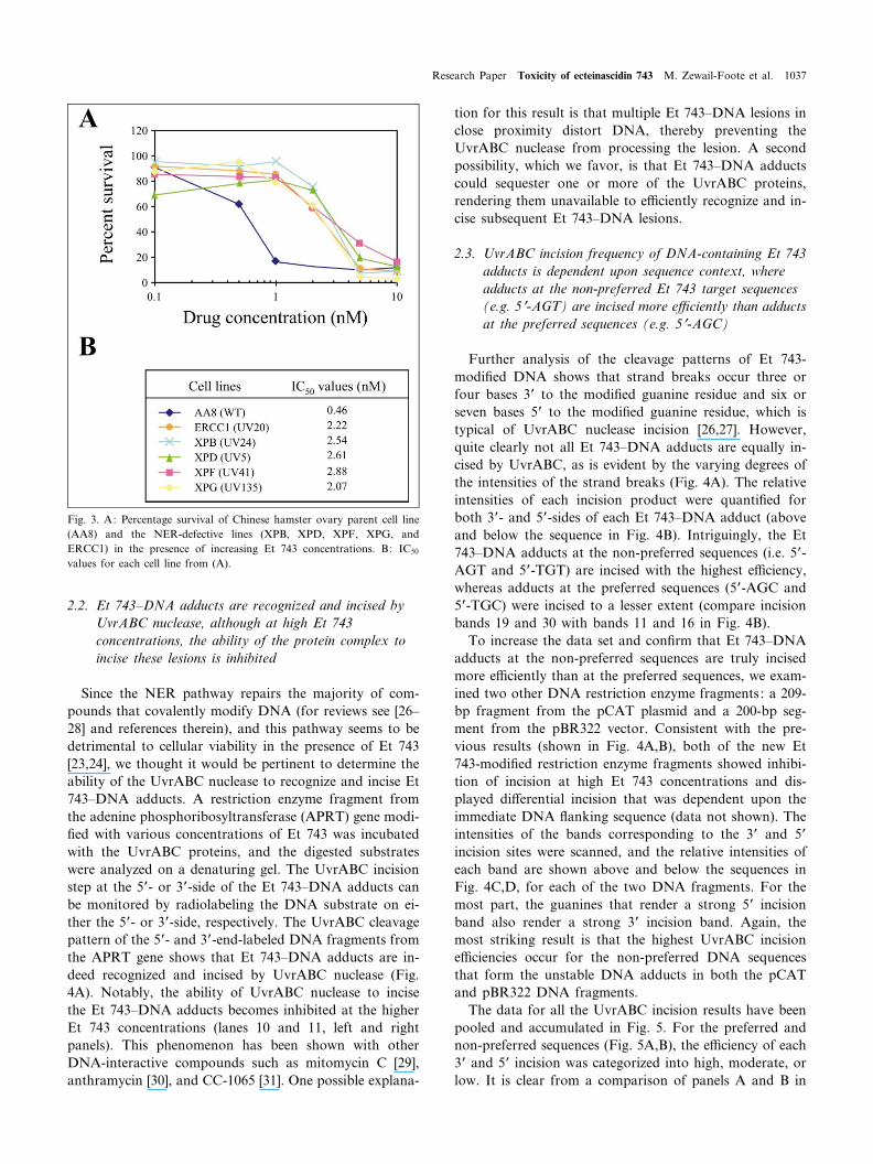
743 than to their isogenic parent cell lines. The results inFig. 3 demonstrate that all of the mammalian NER-defec-tive cell lines (XPB, XPD, XPF, XPG, and ERCC1) showabout a ¢ve-fold greater resistance to Et 743 than theChinese hamster ovary parent cell line (AA8). Of thesecell lines, ERCC1 and XPG are more resistant, thus con-
¢rming the previous results but extending this observationto all these genes involved in transcriptionally coupledevents. Of the other carbinolamine antibiotics shown inFig. 1B, only anthramycin has been tested for DNA re-pair. In this case, single- and double-strand breaks werefound to be excision repair-dependent [25].
Fig. 2. A: A molecular dynamics-generated structure of Et 743 at the preferred bonding site. The three Et 743 subunits A (green), B (yellow), and C(white) are positioned in the minor groove of DNA. B: Reversible reaction of Et 743 with DNA to form the Et 743^N2-guanine^DNA adduct. `B' is aDNA base hydrogen bond acceptor (adapted from [16]).
CHBIOL 135 24-10-01 Cyaan Magenta Geel Zwart
1036 Chemistry & Biology 8/11 (2001) 1033^1049
2.2. Et 743^DNA adducts are recognized and incised byUvrABC nuclease, although at high Et 743concentrations, the ability of the protein complex toincise these lesions is inhibited
Since the NER pathway repairs the majority of com-pounds that covalently modify DNA (for reviews see [26^28] and references therein), and this pathway seems to bedetrimental to cellular viability in the presence of Et 743[23,24], we thought it would be pertinent to determine theability of the UvrABC nuclease to recognize and incise Et743^DNA adducts. A restriction enzyme fragment fromthe adenine phosphoribosyltransferase (APRT) gene modi-¢ed with various concentrations of Et 743 was incubatedwith the UvrABC proteins, and the digested substrateswere analyzed on a denaturing gel. The UvrABC incisionstep at the 5P- or 3P-side of the Et 743^DNA adducts canbe monitored by radiolabeling the DNA substrate on ei-ther the 5P- or 3P-side, respectively. The UvrABC cleavagepattern of the 5P- and 3P-end-labeled DNA fragments fromthe APRT gene shows that Et 743^DNA adducts are in-deed recognized and incised by UvrABC nuclease (Fig.4A). Notably, the ability of UvrABC nuclease to incisethe Et 743^DNA adducts becomes inhibited at the higherEt 743 concentrations (lanes 10 and 11, left and rightpanels). This phenomenon has been shown with otherDNA-interactive compounds such as mitomycin C [29],anthramycin [30], and CC-1065 [31]. One possible explana-
tion for this result is that multiple Et 743^DNA lesions inclose proximity distort DNA, thereby preventing theUvrABC nuclease from processing the lesion. A secondpossibility, which we favor, is that Et 743^DNA adductscould sequester one or more of the UvrABC proteins,rendering them unavailable to e¤ciently recognize and in-cise subsequent Et 743^DNA lesions.
2.3. UvrABC incision frequency of DNA-containing Et 743adducts is dependent upon sequence context, whereadducts at the non-preferred Et 743 target sequences(e.g. 5P-AGT) are incised more e¤ciently than adductsat the preferred sequences (e.g. 5P-AGC)
Further analysis of the cleavage patterns of Et 743-modi¢ed DNA shows that strand breaks occur three orfour bases 3P to the modi¢ed guanine residue and six orseven bases 5P to the modi¢ed guanine residue, which istypical of UvrABC nuclease incision [26,27]. However,quite clearly not all Et 743^DNA adducts are equally in-cised by UvrABC, as is evident by the varying degrees ofthe intensities of the strand breaks (Fig. 4A). The relativeintensities of each incision product were quanti¢ed forboth 3P- and 5P-sides of each Et 743^DNA adduct (aboveand below the sequence in Fig. 4B). Intriguingly, the Et743^DNA adducts at the non-preferred sequences (i.e. 5P-AGT and 5P-TGT) are incised with the highest e¤ciency,whereas adducts at the preferred sequences (5P-AGC and5P-TGC) were incised to a lesser extent (compare incisionbands 19 and 30 with bands 11 and 16 in Fig. 4B).
To increase the data set and con¢rm that Et 743^DNAadducts at the non-preferred sequences are truly incisedmore e¤ciently than at the preferred sequences, we exam-ined two other DNA restriction enzyme fragments : a 209-bp fragment from the pCAT plasmid and a 200-bp seg-ment from the pBR322 vector. Consistent with the pre-vious results (shown in Fig. 4A,B), both of the new Et743-modi¢ed restriction enzyme fragments showed inhibi-tion of incision at high Et 743 concentrations and dis-played di¡erential incision that was dependent upon theimmediate DNA £anking sequence (data not shown). Theintensities of the bands corresponding to the 3P and 5Pincision sites were scanned, and the relative intensities ofeach band are shown above and below the sequences inFig. 4C,D, for each of the two DNA fragments. For themost part, the guanines that render a strong 5P incisionband also render a strong 3P incision band. Again, themost striking result is that the highest UvrABC incisione¤ciencies occur for the non-preferred DNA sequencesthat form the unstable DNA adducts in both the pCATand pBR322 DNA fragments.
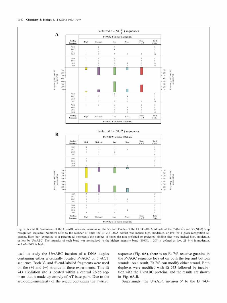
The data for all the UvrABC incision results have beenpooled and accumulated in Fig. 5. For the preferred andnon-preferred sequences (Fig. 5A,B), the e¤ciency of each3P and 5P incision was categorized into high, moderate, orlow. It is clear from a comparison of panels A and B in
Fig. 3. A: Percentage survival of Chinese hamster ovary parent cell line(AA8) and the NER-defective lines (XPB, XPD, XPF, XPG, andERCC1) in the presence of increasing Et 743 concentrations. B: IC50
values for each cell line from (A).
CHBIOL 135 24-10-01 Cyaan Magenta Geel Zwart
Research Paper Toxicity of ecteinascidin 743 M. Zewail-Foote et al. 1037

Fig. 5 that the Et 743^DNA adducts at non-preferredsequences that lack the ability to form the HB2 hydrogenbond (see Fig. 1B) are incised at a higher e¤ciency thanthose that can form the complete hydrogen bond network.Adducts at the preferred alkylation sites that form theHB2 hydrogen bond are incised with moderate to lowe¤ciency (Fig. 5A), while adducts at the non-preferredsequences display incision frequencies that are moreeven, cutting at high, moderate, or low e¤ciencies (Fig.5B). In addition, about 25% of the preferred target se-quences show no incision 5P or 3P to the Et 743^DNAadduct, whereas all the non-preferred sites showed at least3P or 5P incision. It is important to recognize that theactual percentage of the Et 743 adducts that are incisedby UvrABC are not adjusted for the extent of modi¢ca-tion of guanines to a given site. It is likely that the actualpercentage of Et 743-induced modi¢cation at the preferredsequences is even higher than that at the non-preferredtarget site, since the DNA adducts at these sites aremore stable at these sequences [16]. Thus, the actual extentof di¡erence between incision frequency at preferred andnon-preferred sequences is likely to be underrepresented.There is an apparent low frequency of uncoupled 3P or 5Pincisions for both preferred and non-preferred sequences
(usually less than 10%). We are hesitant to make too muchof the signi¢cance of this result because of its minor con-tribution and because the absolute assignment of the 3P or5P incisions can be sometimes confused by the aberrant 5Pcleavage bands observed in the site-directed Et 743^DNAadducts.
2.4. The £anking sequence outside the 3-bp target sequence(5P-NGN) may contribute to the e¤ciency of incisionof Et 743^DNA adducts by UvrABC nuclease
Further inspection of the data in Figs. 5 and 6 demon-strates that not all of the Et 743^DNA adducts at thesame 3-bp target sequence are incised at the same e¤-ciency, suggesting that more than just the 3-bp recognitionsequence is involved in determining the e¤ciency of inci-sion by the UvrABC nuclease. For example, the highestlevel of incision occurs for a 5P-TGT lesion (site 30 in Fig.4B) in the DNA fragment from the APRT gene, but Et743^DNA adducts at other 5P-TGT sequences surroundedby a di¡erent £anking sequence show di¡erent levels ofincision (compare incision sites 1, 2, 15, and 30 in Fig.4B). In another example, a 5P-AGT adduct site placednext to an A-tract was not incised e¤ciently (compare
Fig. 4. A: Autoradiogram of UvrABC nuclease incision of an Et 743-modi¢ed 5P- or 3P-end-labeled 188-bp DNA fragment from the Chinese hamsterovary APRT gene. Lanes 1^3, Maxam^Gilbert chemical sequencing reactions; lane 4 (control), DNA treated with 5 WM of Et 743; lane 5, unmodi¢edDNA treated with UvrABC; lanes 6^11, DNA modi¢ed with 0.25, 0.5, 1, 5, 15, and 20 WM of Et 743, respectively, in the presence of UvrABC. The Et743 target sequence corresponding to the drug modi¢cation-induced UvrABC nuclease incision bands is shown on the right side of each panel. B: Rela-tive intensities of UvrABC nuclease incision on the 3P- and 5P-sides of Et 743^DNA adducts in the 188-bp sequence from the APRT gene. The inten-sities were normalized to 100% for the most intense band in each experiment. Arrows highlight some of the Et 743-modi¢ed AGC and AGT sequencesthat are incised to varying degrees. C and D: Relative intensities of UvrABC nuclease incision on the 3P- and 5P-sides of Et 743^DNA adducts in the209-bp fragment from pCAT plasmid (C) and the 200-bp fragment from pBR322 vector (D). The intensities were normalized to 100% for the most in-tense band in each experiment. (b) indicates guanines that show no incision on either the 5P- or 3P-side of the Et 743^DNA adducts.
CHBIOL 135 24-10-01 Cyaan Magenta Geel Zwart
1038 Chemistry & Biology 8/11 (2001) 1033^1049
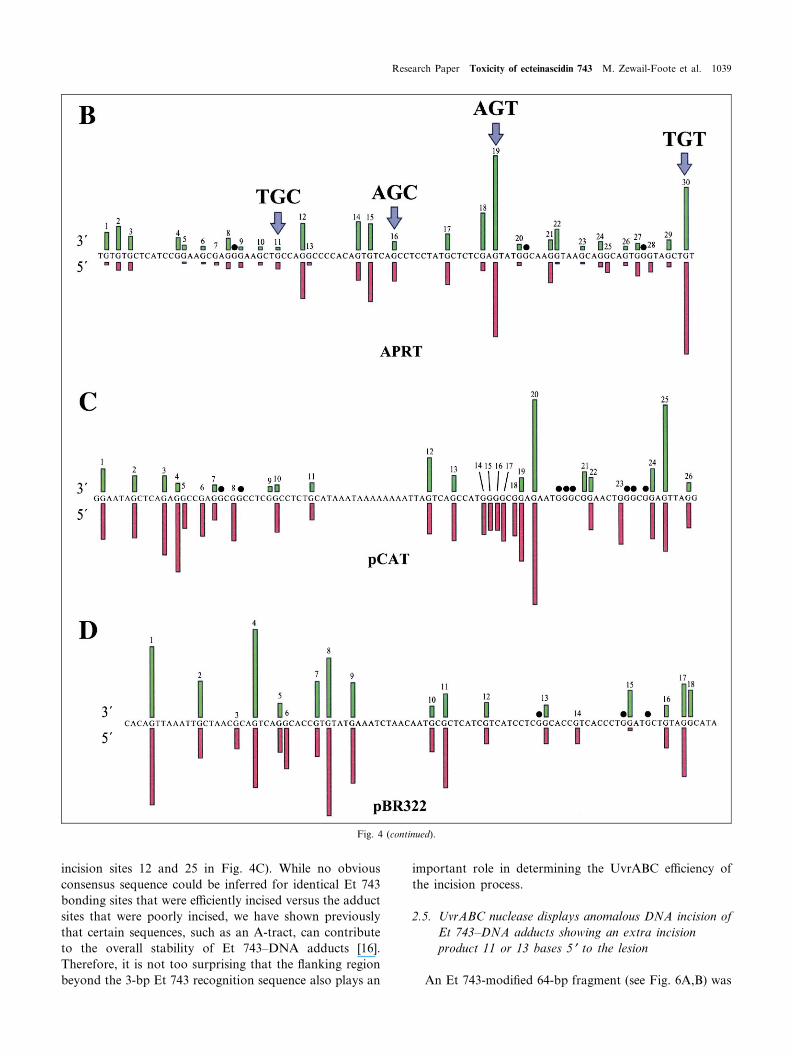
incision sites 12 and 25 in Fig. 4C). While no obviousconsensus sequence could be inferred for identical Et 743bonding sites that were e¤ciently incised versus the adductsites that were poorly incised, we have shown previouslythat certain sequences, such as an A-tract, can contributeto the overall stability of Et 743^DNA adducts [16].Therefore, it is not too surprising that the £anking regionbeyond the 3-bp Et 743 recognition sequence also plays an
important role in determining the UvrABC e¤ciency ofthe incision process.
2.5. UvrABC nuclease displays anomalous DNA incision ofEt 743^DNA adducts showing an extra incisionproduct 11 or 13 bases 5P to the lesion
An Et 743-modi¢ed 64-bp fragment (see Fig. 6A,B) was
Fig. 4 (continued).
CHBIOL 135 24-10-01 Cyaan Magenta Geel Zwart
Research Paper Toxicity of ecteinascidin 743 M. Zewail-Foote et al. 1039
used to study the UvrABC incision of a DNA duplexcontaining either a centrally located 5P-AGC or 5P-AGTsequence. Both 3P- and 5P-end-labeled fragments were usedon the (+) and (3) strands in these experiments. This Et743 alkylation site is located within a central 22-bp seg-ment that is made up entirely of AT base pairs. Due to theself-complementarity of the region containing the 5P-AGC
sequence (Fig. 6A), there is an Et 743-reactive guanine inthe 5P-AGC sequence located on both the top and bottomstrands. As a result, Et 743 can modify either strand. Bothduplexes were modi¢ed with Et 743 followed by incuba-tion with the UvrABC proteins, and the results are shownin Fig. 6A,B.
Surprisingly, the UvrABC incision 5P to the Et 743^
Fig. 5. A and B: Summaries of the UvrABC nuclease incisions on the 5P- and 3P-sides of the Et 743^DNA adducts at the 5P-(NGGC ) and 5P-(NGA
T ) 3-bprecognition sequence. Numbers refer to the number of times the Et 743^DNA adduct was incised high, moderate, or low for a given recognition se-quence. Each bar (expressed as a percentage) represents the number of times the non-preferred or preferred binding sites were incised high, moderate,or low by UvrABC. The intensity of each band was normalized to the highest intensity band (100%). 1^20% is de¢ned as low, 21^44% is moderate,and 45^100% is high.
CHBIOL 135 24-10-01 Cyaan Magenta Geel Zwart
1040 Chemistry & Biology 8/11 (2001) 1033^1049

Fig. 6. Autoradiogram showing the 5P and 3P incisions of the 64-bp DNA oligonucleotide containing the (A) 5P-AGC or (B) 5P-AGT sequence. DNAwas modi¢ed with 0, 0.5, 1, 2.5, 5, or 10 WM (lanes 3^8) of Et 743 followed by incubation with UvrABC. Arrows denote the sites of UvrABC incision.(*) indicates the 5P-AGC and 5P-AGT alkylation sites of Et 743. AG and TC refer to the Maxam^Gilbert sequencing lanes. The other sites of incisionarise from other Et 743^DNA alkylation sites. The full 64-bp sequence, with the bonding site highlighted and incision sites shown by arrows, whoselengths correspond to intensity of cleavage, is shown at the bottom of each panel.
CHBIOL 135 24-10-01 Cyaan Magenta Geel Zwart
Research Paper Toxicity of ecteinascidin 743 M. Zewail-Foote et al. 1041

DNA adduct is not the same for both strands. As ex-pected, the 5P incisions for the top and bottom strandsoccurred six bases from the adduct; however, 5P incisionsalso occurred 11 and 13 bp from the adduct on the topstrand (Fig. 6A, left panel). The 3P incision occurred fourbases away from the site of the Et 743^DNA lesion forboth strands (Fig. 6A, right panel). That the intensity ofthe 3P strand incisions is nearly equivalent for the products
on the top and bottom strands of the 5P-AGC adductargues that Et 743 alkylates both strands equally. Theextra incisions on the 5P-side also occurred for the se-quence containing the 5P-AGT adduct on the top strand(Fig. 6B, left panel), indicating that, in this case, theanomalous incision is independent of the 3-bp Et 743 tar-get sequence. It is surprising that the extra incisions occuronly on the top strand. The explanation must be that
Fig. 7. A: Schematic for the preparation of the Et 743 site-directed adduct. B: Reaction of the UvrABC nuclease with either a top or bottom strand5P- or 3P-end-labeled DNA sequence containing an Et 743^DNA site-directed adduct. Modi¢cation of DNA by Et 743 was as shown for Fig. 6. TheUvrABC incision sites are indicated by arrows. AG and TC refer to Maxam^Gilbert sequencing lanes. (*) indicates the 5P-AGC alkylation site. The cen-tral sequence shows the incision pattern for UvrABC nuclease.
CHBIOL 135 24-10-01 Cyaan Magenta Geel Zwart
1042 Chemistry & Biology 8/11 (2001) 1033^1049

although the 5P-AGC sequences on the top and bottomstrands are both contained within an A/T-rich region, the£anking sequences are not identical. Presumably UvrABCis able to recognize di¡erential structural or dynamic fea-tures between Et 743^DNA adducts on the top and bot-tom strands. Extra 5P incisions have also been reported forcisplatin [32], where two major incision products occurred8 and 15 bases from the site of the lesion. However, amore de¢ned system seemed necessary since the sequencesshown in Fig. 6A,B contained other Et 743 alkylation sitesin £anking regions, which when alkylated could poten-tially interfere with UvrABC incision at the 5P-AGC and5P-AGT adduct sites. Hence, we created a site-directed Et743^DNA adduct with the same sequence as that usedabove.
2.6. UvrABC incision of a DNA fragment containinga site-directed Et 743^DNA adduct in thepreferred 5P-AGC sequence shows anomalouscleavage
In all the previous experiments, there was potential foroverlap of UvrABC repair incision sites due to multiple Et743^DNA adducts, and consequently the results weresometimes di¤cult to interpret unambiguously. In a sub-sequent experiment, we attempted to create a more de¢nedsystem. Speci¢cally, we carried out parallel UvrABC nu-clease experiments on Et 743^DNA site-directed adductscontaining either the preferred 5P-AGC sequence or thenon-preferred 5P-AGT sequence. Since Et 743 reacts onlywith duplex DNA and not single-stranded DNA, the prep-aration and puri¢cation of Et 743^DNA adducts wererestricted to duplex DNA substrates. The strategy usedfor the preparation of 5P-AGC, the site-directed adduct,is shown in Fig. 7A. The Et 743^DNA site-directed adductwas created by ligating a 22-bp Et 743-modi¢ed duplexcontaining either a single 5P-AGC or 5P-AGT sequenceto a pair of double-stranded DNA oligonucleotides, usingdi¡erent sticky ends to unambiguously ligate one on eachside of the Et 743-modi¢ed duplex. This resulted in theconstruction of a 64-bp fragment containing the same se-quence used in the prior section. While the creation of anEt 743 site-directed adduct containing the preferred 5P-AGC sequence was successful, repeated attempts to gen-erate the Et 743^5P-AGT DNA adduct failed. This is pre-sumably because the Et 743^DNA adduct at the 5P-AGTsequence underwent covalent reversal during puri¢cationdue to its intrinsic instability [16]. Therefore, only the 64-bp site-directed Et 743^DNA adduct containing the 5P-AGC target sequence was used to study the pattern ofincision by UvrABC.
Fig. 7B shows the results of four experiments using bothstrands having either the 3P- or 5P-radiolabeled site-di-rected 5P-AGC adduct after incubation with the UvrABCnuclease. 5P incision occurs both 6 and 13 bases from the
covalent attachment site on the top strand, whereas on thebottom strand 5P incision only occurs six bases from theEt 743 adduct site. The 3P incision events occur equallywell on both strands and show only one incision productfour bases from the Et 743-damaged guanine. The resultspresented here substantiate our previous results (shown inFig. 6A), in which anomalous incision occurs only on thetop strand on the 5P-side, while the incision occurs on thebottom strand at the expected sites, i.e. six bases 5P andfour bases 3P to the Et 743-alkylated guanine, for a DNAduplex containing a 5P-AGC sequence.
3. Discussion
Et 743 is a structurally and mechanistically unique anti-tumor antibiotic that has shown very encouraging antitu-mor activity in phase I and II clinical trials, particularly insoft tissue sarcomas. Because of the implications of theinvolvement of NER in mediating the biological potency,and perhaps speci¢city, of this compound (see Fig. 3), weset out to study the recognition and incision of Et 743^DNA adducts using the Escherichia coli UvrABC nucle-ase. While the human NER system is more complex, in-volving a larger number of repair proteins, and the repairpatch is larger than in the bacterial system, the basicmechanism is similar in both systems. In the UvrABCsystem, repair is initiated by the UvrA protein, which di-merizes prior to formation of a heterotrimeric complexwith UvrB [33,34]. This UvrA2B complex has helicase-like properties and speci¢cally binds to damaged DNA[35,36]. Binding of the UvrA2B complex to duplex DNAresults in both bending of the DNA and an open DNAstructure [35,37,38]. After damage recognition, UvrA dis-sociates, while UvrB remains bound to the damaged DNAin a stable preincision complex [39,40]. Following thebinding of UvrC to this complex, incision is triggered onboth sides of the damaged site: four nucleotides 3P to thedamaged site and seven nucleotides 5P to the same site[37,41^43]. The repair is completed by UvrD, DNA poly-merase, and DNA ligase. Much remains to be learnedabout the structural biology of the discrimination betweennon-damaged and damaged DNA and the structures ofthe various complexes of UvrA2, UvrA2B^DNA, UvrB^DNA, and UvrBC^DNA. A crystal structure of UvrB hasrecently been published and a model of the UvrB^DNAcomplex has been proposed [36]. We propose a model forhow Et 743^DNA adducts are processed by bacterial andhuman cells to result in the repair-dependent toxicity tocells. This model takes into account both the unique struc-tural and dynamic features of the Et 743^DNA adductsand the resistance of mammalian cells to the toxic e¡ectsof Et 743, which are defective in NER repair proteinsinvolved in assembly of a repair complex competent toperform 3P and 5P incision steps.
CHBIOL 135 24-10-01 Cyaan Magenta Geel Zwart
Research Paper Toxicity of ecteinascidin 743 M. Zewail-Foote et al. 1043

3.1. The e¤ciency of UvrABC incision of Et 743^DNAadducts is dependent upon the stability of the boundadduct at the target sequence, and in some cases, the3P and 5P incisions are absent or they occur ataberrant sites
There is a clear di¡erence in the e¤ciency of theUvrABC incision of Et 743^DNA adducts that is depen-dent upon the precise Et 743 bonding sequence. The re-sults summarized in Fig. 5A,B show that Et 743^DNAadducts bound to 5P-AGT sequences are much more e¤-ciently incised by UvrABC nuclease than the same adductsbound to 5P-AGC sequences. In a previous study [16] wedemonstrated that the presence or absence of speci¢c hy-drogen-bonding interactions between donors and accep-tors on Et 743 and the 3-bp recognition sequence leadsto signi¢cant di¡erences in the stability and conformation-
al £exibility of the resulting DNA adducts. Of prime im-portance is the presence or absence of a hydrogen-bondinginteraction between O18 of the methylenedioxy ring andthe exocyclic 2-amino group of guanine on the 3P-side ofthe alkylated guanine, which may be on either the covalentor non-covalently modi¢ed strand (see Fig. 1B). Of lesssigni¢cance are the hydrogen-bonding interactions on the5P-side of the covalently modi¢ed guanine. In the absenceof a 3P GC base pair, which provides this crucial hydro-gen-bonding, Et 743^DNA adducts more easily undergocovalent reversal and show weaker base-pairing forms andalternative base-pairing conformers on the 5P-side [16].Furthermore, as anticipated, helicase-catalyzed unwindingof DNA using T-antigen or thermal denaturation resultsin enhanced instability of the Et 743^5P-AGT duplex ad-ducts over the corresponding 5P-AGC duplex adducts [16].Therefore, there is an inverse correlation between Et 743^
Fig. 8. Proposed model for how UvrABC nuclease may process Et 743^DNA adducts, leading to either a repaired lesion (pathway `a') or a toxic lesion(pathway `b') corresponding to the mammalian NER-dependent toxicity. In pathway `a', UvrA2B is able to recognize the Et 743 and unwind the duplexDNA, creating the bubble that positions the Et 743^DNA adduct in a region of single-strandness, which leads to covalent reversal of the Et 743^DNAadduct (aP) or to 3P and 5P incisions (aQ). For pathway `b', an optimally stabilized Et 743^DNA adduct is in a region of bent DNA, which is recognizedand bound by the UvrA2B complex. In this case the Et 743^DNA duplex adduct mimics the normally unwound form of modi¢ed DNA bound toUvrA2B, A2 dissociates, and then C binds to form the Et 743^BC complex. This substrate is ine¤ciently incised by UvrBC, which results in the absenceof 3P and 5P incisions. This leads to the toxic lesion mechanistically equivalent to a drug-poisoned topoisomerase I^ or topoisomerase II^DNA complex.
CHBIOL 135 24-10-01 Cyaan Magenta Geel Zwart
1044 Chemistry & Biology 8/11 (2001) 1033^1049
DNA adduct stability and UvrABC incision e¤ciency.This point is further exempli¢ed by a comparison of theUvrABC incision e¤ciency of Et 743^5P-AGT adducts indi¡erent sequence contexts. Et 743^5P-AGT adducts adja-cent to A-tracts are much less e¤ciently incised than thesame adducts without £anking A-tracts. In a previousstudy [16] we demonstrated that A-tracts stabilize Et743^DNA adducts to covalent reversal, which we haveattributed to dampening the conformational £exibility ofEt 743^5P-AGT adducts and strengthening the apparenthydrogen-bonding interactions.
Irrespective of the covalent bonding sequence, Et 743^DNA adducts appeared to show some uncoupled 3P and 5Pincisions (Fig. 5A,B), although the combination of the lowfrequency of these events and their sometimes ambiguousassignment makes this conclusion less than ¢rm. The ab-sence of both 3P and 5P incisions is found only for the 5P-AGC Et 743 bonding sequences. Thus it would appearthat for loss of both 3P and 5P incisions, the hydrogen-bonding donor/acceptor pair between Et 743 and the GCbase pair immediately to the 3P-side of the Et 743^DNAadduct is required. Last, aberrant UvrABC incisions oc-curring 11 or 13 bases to the 5P-side of the Et 743^DNAadducts are found for both 5P-AGT and 5P-AGC bondingsites.
3.2. Ine¤cient 3P and 5P incisions by NER complexes leadto cytotoxic e¡ects of Et 743^DNA adducts inhuman cells
In contrast to the E. coli UvrABC nuclease, where theUvrC and UvrB proteins give rise to the 5P and 3P inci-sions, in human NER the dual incisions at the 5P- and 3P-sides of the DNA adducts are carried out by two physi-cally separated nuclease activities : XPG^ and XPF^ERCC1, respectively. Human cells lacking either of thesenucleases are resistant to Et 743 [23,24]. Furthermore, theresults in Fig. 3 show that other defective proteins in-volved in the formation of the NER nuclease complexalso show resistance to Et 743. Therefore, any model ofhow Et 743 exerts its cytotoxic potency via the NER path-way must take into account how this NER nuclease com-plex might result in structural consequences that are det-rimental to cell survival.
3.3. In addition to a sequence-dependent hydrogen-bondingstabilization of the Et 743^DNA adduct, Et 743^DNAadducts have other unique structural e¡ects on DNA
NMR studies on covalent oligomer duplex adducts ofEt 743 and non-denaturing PAGE analysis of ligated olig-omers containing de¢ned Et 743^DNA adducts reveal im-portant novel characteristics of Et 743^DNA adducts.While Et 743 modi¢es duplex DNA within the minorgroove (Fig. 2A), it also bends DNA into the majorgroove [19]. This results from the wedge shape of Et
743, which forces open the minor groove of DNA andconcomitantly closes up the major groove. In addition tothe major groove bending and minor groove occupancy,the C-subunit of Et 743 protrudes outside the helical axisoccupied by duplex DNA (Fig. 2A). In summary, the co-valent attachment of Et 743 to DNA and £anking donor/acceptor hydrogen-bonding of DNA zipper up a locallywidened minor groove of DNA, which also has an extra-helical protrusion in an otherwise bent DNA. Somehowthese unique structural characteristics of Et 743^DNA ad-ducts must result in the reduced e¤ciencies of the se-quence-dependent UvrABC nuclease incision and the un-coupled or absent 3P and 5P UvrABC incisions observed inthis study, along with the NER-dependent cytotoxicitiesobserved in human cells. In Section 3.4 we propose amodel that takes into account all of these observationswith what is known about NER in E. coli and humancells.
3.4. Model for how the inhibited repair of Et 743^DNAadducts can lead to NER-mediated cytotoxicity
As previously described, UvrA2B complex detection ofdamaged DNA is proposed to lead to a 6-bp bubblearound the modi¢ed based and to bending of the DNA[35]. This then results in dissociation of UvrA2 from thecomplex, leaving a UvrB^DNA complex that is a stablepreincision complex. UvrC is then recruited, which resultsin endonuclease cleavage by UvrC 3P and 5P to the adductsite [44] (see pathways `a' and `aQ' in Fig. 8). Dependentupon the stability of the Et 743^N2-guanine DNA adductin the 6-bp bubble, the Et 743^N2-guanine covalent link-age may reverse before the 3P and 5P incisions occur (path-ways `a' and `aP' in Fig. 8). These pathways are most likelyin the unfavored sequences for Et 743^DNA adduct stabil-ity, where bubble formation is most likely, due to the lackof the additional H-bonding 5P to the covalently modi¢edguanine (see Fig. 1B). Neither pathway `a' scenario wouldbe predicted to give rise to the repair-dependent enhancedtoxicities observed in mammalian cells.
However, processing of Et 743^DNA lesions by path-way `b' (Fig. 8) is predicted to give rise to a toxic lesion.We propose that the unique properties of speci¢c Et 743^DNA adducts lead to malfunction of the UvrABC nucle-ase system, leading to ine¤cient incisions on both the 3P-and 5P-sides and, in some cases, possibly uncoupled 3P and5P incisions. Why does an encounter between the UvrABCnuclease system and the hydrogen-bonded stabilized Et743^DNA adduct lead to aberrant DNA repair?
First, Et 743^DNA adducts may be particularly di¤cultto process because of their resistance to the helicase activ-ity of the UvrA2B complex, primarily arising from thezipper-like e¡ects of the hydrogen-bonding stabilizationon both sides of the covalent adduct. This is analogousto resistance of stable Et 743^DNA adducts to T-antigenunwinding [16].
CHBIOL 135 24-10-01 Cyaan Magenta Geel Zwart
Research Paper Toxicity of ecteinascidin 743 M. Zewail-Foote et al. 1045
It is most likely to be those Et 743^DNA adducts thatare resistant to the helicase activity of UvrA2B that lead tothe excision-dependent cytotoxicity (i.e. Et 743^DNA ad-ducts at preferred sequences). In order for incision to oc-cur, UvrA must dissociate and the stable preincision com-plex of UvrB must be recognized by UvrC to form theactive UvrBC nuclease (pathway `b' in Fig. 8).
Second, we propose that the stable Et 743^DNA duplexadducts must have structural features that somehow mimicthe bent and open form of the normal UvrA2B heterotri-meric complex with DNA. Indeed, Et 743^DNA adductsare bent into the major groove through minor groove ex-pansion due to occupancy by the wedge-shaped Et 743.The extrahelical protrusion of the C-subunit of Et 743may also play a role in promoting the dissociation ofUvrA and, perhaps, stabilizing the UvrB preincision com-plex. However, in the absence of de¢ned structural inter-actions among the various protein^DNA components ofthe UvrA2B^, UvrB^, and UvrBC^DNA complexes, it isimpossible to be more speci¢c than just suggesting possiblestructural mimics (bending) and steric interactions (extra-helical protrusion) that could lead to aberrant DNA repairoutcomes. One could envisage a situation similar to that ofa drug-poisoned topoisomerase I or II complex where adrug molecule intercepts an intermediate in which neithera 3P nor a 5P incision is made but the UvrBC, or even theUvrABC, is trapped as a non-covalent protein^Et 743^DNA complex, which is more detrimental to cells thanthe original Et 743^DNA adduct. Just as with the drug-poisoned topoisomerase I or II complexes, where elevatedlevels of topoisomerases would lead to populations ofmore susceptible cells, a similar situation exists for ele-vated levels of NER proteins. Some of these possibilitiesare being explored.
4. Signi¢cance
The pattern of ine¤cient repair incision by the UvrABCnuclease of Et 743^DNA adducts provides a basis forrationalizing the observed repair-dependent cytotoxicitiesof these DNA adducts, if other associated structural prop-erties of Et 743^DNA adducts are taken into account. Onthe basis of promising activity in phase I clinical trials, Et743 has progressed into phase II clinical trials, where it hasshown remarkable activity in soft tissue sarcomas. This isthe ¢rst of the monoalkylating minor groove-interactiveagents to progress beyond phase I clinical trials. In pre-vious studies we have determined the structure of the Et743^DNA covalent adduct, its e¡ect on DNA structure,and the hydrogen-bonding interactions, which determineDNA sequence selectivity and enhanced stability on DNA.These properties set Et 743 apart from other minor groovealkylating compounds. Recent results from groups in Italy(D'Incalci lab) and at NCI (Pommier lab) provide insightinto the molecular targets for Et 743, which appear to be
proteins involved in the NER pathway. We have con-¢rmed this conclusion, but what is most signi¢cant isthat we have identi¢ed what is likely to be the structuraland biochemical basis for the NER-dependent cytotoxicityof the Et 743^DNA adducts. These results link the struc-tural biology of the Et 743^DNA adducts with the biolog-ical consequences of incomplete DNA repair. This result issomewhat reminiscent of the formation of cleavable com-plexes with topoisomerase I^ and topoisomerase II^DNAadducts.
5. Materials and methods
5.1. Materials
Ecteinascidin was generously supplied by PharmaMar, Madrid,Spain.
5.2. Cell lines and culture conditions
The Chinese hamster ovary parent cell line (AA8) and theNER-defective cell lines XPB, XPD, XPF, XPG, and ERCC1were purchased from American Type Culture Collection. All celllines were grown as a monolayer in Dulbecco's modi¢ed Eagle'smedium (Gibco, #10-013-CV) supplemented with 1% (v/v) penicil-lin (100 U/ml), streptomycin (100 Wg/ml), and 10% fetal bovineserum. Cells were grown at 37³C in a 5% CO2 humidi¢ed incu-bator. Trypsin^EDTA solution was used for detaching cells.
5.3. Cytotoxicity assay
A colorimetric assay based on the ability of viable cells toreduce the tetrazolium salt MTS (CellTiter 96 Non-RadioactiveCell Proliferation Assay, Promega Corp., Madison, WI, USA) toa blue formazan product was used to measure cytotoxicity fol-lowing exposure to Et 743. Cells were plated in a 0.1 ml mediumon day 0 in 96-well microtiter plates (Falcon, #3072). On day 1,10 Wl of serial dilutions of the test agent was added in replicatesof four to the plates. After incubation for 4 days at 37³C in ahumidi¢ed incubator, 2 Wl of a 20:1 mixture of 3-(4,5-dimethyl-thiazol-2-yl)-5-(3-carboxymethoxyphenyl)-2-(4-sulfophenyl)-2H-tetrazolium, inner salt MTS (2 mg/ml) and an electron couplingreagent, phenazine methosulfate (0.92 mg/ml in DPBS), wasadded to each well and incubated for 4 h at 37³C. Absorbancewas measured using a Model 7520 microplate reader (CambridgeTechnology, Inc.) at 490 nm. Data were expressed as the percent-age of survival of control calculated from the absorbance cor-rected for background absorbance. The surviving fraction of cellswas determined by dividing the mean absorbance values of thetest agents by the mean absorbance values of untreated control.
5.4. Et 743^DNA bonding
Speci¢c amounts of drug were added to radiolabeled DNA in10 mM Tris^HCl (pH 7.5) and 25 mM NaCl bu¡er to give thedesired ¢nal concentration. The solutions were incubated at roomtemperature for either 1 or 2 h. The unreacted drug was removedby phenol^chloroform extraction.
CHBIOL 135 24-10-01 Cyaan Magenta Geel Zwart
1046 Chemistry & Biology 8/11 (2001) 1033^1049

5.5. Puri¢cation of UvrABC proteins
UvrA, UvrB, and UvrC proteins were isolated from E. coliK12 strain CH292 carrying plasmids pUNC45 (UvrA),pUNC211 (UvrB), and pDR3274 (UvrC) [45]. The methods ofpuri¢cation were the same as described previously [46]. For theexperiments using the DNA fragments from the pCAT andpBR322 plasmids, the UvrABC proteins were purchased fromPharMingen (San Diego, CA, USA).
5.6. DNA fragment isolation and 32P-end-labeling
Plasmid pGEM inserted with APRT gene from Chinese ham-ster ovary cells was puri¢ed via cesium chloride density centrifu-gation and dialyzed extensively against TE bu¡er. The 284-bpfragment of exon III of the APRT gene was obtained by digestingplasmid DNA with restriction enzymes PstI and StuI. The bandcorresponding to the 284-bp fragment was isolated from 1.4%agarose gel and cleaned by passing through a NACS Prepaccolumn followed by ethanol precipitation. The 284-bp fragmentwas further cut with the restriction enzyme MseI to generate a188-bp fragment with a unique site for a single 3P-end-labeling.DNA labeling was accomplished in the presence of [K-32P]TTPand Klenow fragment (5 U) in 10 mM Tris^HCl (pH 8.0), 5 mMMgCl2, and 7.5 mM dithiothreitol (DTT) at 22³C. The 3P-end-labeled fragment was puri¢ed on a 5% polyacrylamide gel. For5P-end-labeling, the restriction enzyme HinfI was used to cut the284-bp fragment to create protruding 5P termini. The resultingfragment was treated with bacterial alkaline phosphatase at60³C for 1 h. The dephosphorylated fragment was extractedwith phenol^chloroform and puri¢ed by gel electrophoresis.The puri¢ed fragment was then labeled with [Q-32P]ATP in thepresence of T4 polynucleotide kinase. The labeled fragment wasprecipitated with ethanol and digested with MseI to a¡ord a 170-bp 5P-end-labeled fragment, which was then isolated and puri¢edas above.
For the isolation of a 209-bp fragment labeled at the 5P-endwith 32P, the pCAT plasmid (New England Biolabs) was ¢rstdigested with HindIII followed by treatment with shrimp alkalinephosphatase at 37³C for 2 h. The dephosphorylated fragment wasextracted with phenol^chloroform and then ethanol-precipitated.The precipitated DNA was labeled with [Q-32P]ATP in the pres-ence of T4 polynucleotide kinase. The labeled DNA was thenincubated with EcoRI and gel-puri¢ed to yield the 209-bp 5P-end-labeled fragment. For 3P-end-labeling, the restriction enzymeEcoRI was used to cut the pCAT plasmid. DNA labeling wasaccomplished in the presence of [K-32P]ATP and Klenow frag-ment. The 3P-end-labeled DNA was then incubated with HindIIIand puri¢ed on a 6% polyacrylamide gel. The same protocol wasused to generate a 200-bp fragment from the pBR322 plasmid(New England Biolabs) except the restriction enzymes NheI andBamHI were used.
5.7. Construction of the Et 743^DNA adducts
The 22-bp duplex DNA [(5P-TGGTTATATAAGCTATTA-TAAT-3P)(3P-ATATATTCGATAATATTAGCTC-5P)] contain-ing a single 5P-AGC alkylation site was modi¢ed with 20 WMEt 743 for 2 h. Unreacted drug was removed by phenol^chloro-
form extraction followed by ethanol precipitation. The Et 743-modi¢ed DNA was ligated to a radiolabeled duplex DNA, oneither the 5P- or 3P-side, by incubating with T4 DNA ligase (1 U/100 Wl) at 16³C for 12 h. After gel puri¢cation, the resulting Et743-modi¢ed duplex was ligated to another duplex, on either the3P- or 5P-side of the modi¢ed DNA fragment. The site-directedadduct was gel-puri¢ed and eluted in annealing bu¡er.
5.8. UvrABC nuclease reactions
The UvrABC nuclease reactions were carried out in a reactionmixture (30 Wl) containing 50 mM Tris^HCl (pH 7.5), 0.1 mMEDTA, 10 mM MgCl2, 1 mM ATP, 100 mM KCl, 1 mM DTT,60 nM UvrA, 120 nM UvrB, 60 nM UvrC, and substrate DNA.The mixtures were incubated at 37³C for 1 h and the reactionswere stopped by phenol^chloroform extraction followed by etha-nol precipitation in the presence of aqueous ammonium acetate(2.5 M). The precipitated DNA was recovered by centrifugationand washed with 80% ethanol.
For the DNA fragments isolated from the pCAT and pBR322plasmids, the UvrABC nuclease reactions were carried out in areaction mixture (20 Wl) containing 50 mM Tris^HCl (pH 7.5), 10mM MgCl2, 2 mM ATP, 50 mM KCl, 5 mM DTT, 0.1 mg/mlbovine serum albumin, 10 nM UvrA, 80 nM UvrB, 12 nM UvrC,and substrate DNA. The mixtures were incubated at 37³C for 1 hand then precipitated with ethanol. The samples were loaded onan 8% denaturing sequencing gel.
6. Note added in proof
In the recently published paper by Takebayashi et al.[23], there is demonstration of accumulation of DNA sin-gle-strand breaks in transcription-coupled (TC) NER-pro-¢cient ¢broblasts treated with Et 743. Thus, our observa-tion of a low frequency of uncoupled 3P or 5P incisionsmay be of signi¢cance. The conclusion in this paper that`trapping of the TC-NER complex stalled [during or] afterDNA cleavage' is exactly in accord with our results andconclusions. This ¢nding is similar to that reported forphleomycin [47], anthramycin [25], and ditercalinium [48].
Acknowledgements
This research was supported by Grants from the Na-tional Institutes of Health (CA49751 and CA29756). TheAmerican Chemical Society Division of Medicinal Chem-istry and Hoechst Marion Roussel are gratefully acknowl-edged for support of a predoctoral fellowship awarded toM.Z.-F. We thank Professor M.-s. Tang (New York Uni-versity) for the DNA fragment from the APRT gene andthe UvrABC proteins used in the experiments shown inFig. 4A,B. We are grateful to Dr. Bennett Van Houten(NIEHS) for insightful discussions. We thank Dr. DavidBishop for preparing, proofreading, and editing the ¢nalversion of the manuscript and ¢gures.
CHBIOL 135 24-10-01 Cyaan Magenta Geel Zwart
Research Paper Toxicity of ecteinascidin 743 M. Zewail-Foote et al. 1047

References
[1] K.L. Rinehart, T.G. Holt, N.L. Fregeau, J.G. Stroh, P.A. Keifer, F.Sun, L.H. Li, D.G. Martin, Ecteinascidins 729, 743, 745, 759A, 759B,and 770: potent antitumor agents from the Caribbean tunicate Ectei-nascidia turbinate, J. Org. Chem. 55 (2000) 4512^4515.
[2] J.M. Jimeno, G. Faircloth, L. Cameron, K. Meely, E. Vega, A. Go-mez, J.M.F. Sousa-Faro, K. Rinehart, Progress in the acquisition ofnew marine-derived anticancer compounds: development of the ectei-nascidin-743 (Et-743), Drugs Future 21 (1996) 1155^1165.
[3] R. Sakai, E.A. Jares-Erijman, I. Manzanares, M.V. SilvaElipe, K.L.Rinehart, Ecteinascidins : putative biosynthetic precursors and abso-lute stereochemistry, J. Am. Chem. Soc. 118 (1996) 9017^9023.
[4] A. Bowman, C. Twelves, K. Hoekman, A. Simpson, J. Smyth, J.Vermorken, F. Hoppener, J. Beijnen, E. Vega, J. Jimeno, A.-R. Ha-nauske, Phase I clinical and pharmacokinetic (PK) study of ecteinas-cidin-743 (Et-743) given as a one hour infusion every 21 days, Ann.Oncol. 9 (Suppl. 2) (1998) 119.
[5] M. Villalona-Calero, S.G. Eckhardt, G. Weiss, E. Campbell, M. Hi-dalgo, M. Kraynak, J. Beijnen, J. Jimeno, D. Von Ho¡, E. Rowin-sky, A phase I and pharmacokinetic study of ET-743, a novel DNAminor groove binder of marine origin, administered as a 1-hour in-fusion daily U5 days, Ann. Oncol. 9 (Suppl. 2) (1998) 119.
[6] A. Taamma, J.L. Misset, M. Riofrio, C. Guzman, E. Brain, L. LopezLazaro, H. Rosing, J.M. Jimeno, E. Cvitkovic, Phase I and pharma-cokinetic study of ecteinascidin-743, a new marine compound, admin-istered as a 24-hour continuous infusion in patients with solid tu-mors, J. Clin. Oncol. 19 (2001) 1256^1265.
[7] G.C. Hill, W.A. Remers, Computer simulation of the binding ofsaframycin A to d(GATGCATC)2, J. Med. Chem. 34 (1991) 1990^1998.
[8] K. Rao, J. Lown, Mode of action of saframycin antitumor antibiot-ics: sequence selectivities in the covalent binding of saframycins Aand S to deoxyribonucleic acid, Chem. Res. Toxicol. 3 (1990) 262^267.
[9] K. Rao, J. Lown, DNA sequence selectivities in the covalent bondingof antibiotic saframycins Mx1, Mx3, A, and S deduced from MPEWFe(II) footprinting and exonuclease III stop assays, Biochemistry 31(1992) 12076^12082.
[10] M.J. Zmijewski Jr., K. Miller-Hatch, M. Mikolajczak, The in vitrointeraction of naphthyridinomycin with deoxyribonucleic acids,Chem. Biol. Interact. 52 (1985) 361^375.
[11] L.H. Hurley, R. Petrusek, Proposed structure of the anthramycin^DNA adduct, Nature 282 (1979) 529.
[12] B.M. Moore II, F.C. Seaman, L.H. Hurley, NMR-based model of anecteinascidin 743^DNA adduct, J. Am. Chem. Soc. 119 (1997) 5475^5476.
[13] Y. Pommier, G. Kohlhagen, C. Bailly, M. Waring, A. Mazumder,K.W. Kohn, DNA sequence- and structure-selective alkylation ofguanine N2 in the DNA minor groove by ecteinascidin 743, a potentantitumor compound from the Caribbean tunicate Ecteinascidia tur-binate, Biochemistry 35 (1996) 13303^13309.
[14] B.M. Moore II, F.C. Seaman, R.T. Wheelhouse, L.H. Hurley, Mech-anism for the catalytic activation of ecteinascidin 743 and its subse-quent alkylation of guanine N2, J. Am. Chem. Soc. 120 (1998) 2490^2491.
[15] F.C. Seaman, L.H. Hurley, Molecular basis for the DNA sequenceselectivity of ecteinascidin 736 and 743: evidence for the dominantrole of direct readout via hydrogen bonding, J. Am. Chem. Soc. 120(1998) 13028^13041.
[16] M. Zewail-Foote, L.H. Hurley, Di¡erential rates of reversibility ofecteinascidin 743^DNA covalent adducts from di¡erent sequenceslead to migration to favored bonding sites, J. Am. Chem. Soc. 123(2001) 6485^6495.
[17] E. Cvitkovic, M. Riofrio, F. Goldwasser, S. Delaloge, A. Taamma, J.Beijnen, J.M. Jimeno, B. Mekranter, C. Guzman, E. Brain, J.L. Mis-
set, Final results of phase I study of ecteinascidin-743 (Et-743) 24hour continuous infusion (CI) in advanced solid tumors (AST) pa-tients, Proc. ASCO 18 (1999) 180a.
[18] D.P. Ryan, J.G. Supko, J.P. Eder, H. Lu, B. Chabner, K. Roper, P.Baccala, J. Bonenfant, G. Faircloth, C. Guzman, J. Jimeno, J.W.Clark, A phase I and pharmacokinetic trial of ecteinascidin-743(Et-743) administered as a 72 hour continuous infusion, Proc.ASCO 18 (1999) 188a.
[19] M. Zewail-Foote, L.H. Hurley, Ecteinascidin 743: a minor groovealkylator that bends DNA toward the major groove, J. Med. Chem.42 (1999) 2493^2497.
[20] E.J. Martinez, T. Owa, S.L. Schreiber, E.J. Corey, Phthalascidin, asynthetic antitumor agent with potency and mode of action compa-rable to ecteinascidin 743, Proc. Natl. Acad. Sci. USA 96 (1999)3496^3501.
[21] Y. Takebayashi, P. Pourquier, A. Yoshida, G. Kohlhagen, Y. Pom-mier, Poisoning of human DNA topoisomerase I by ecteinascidin743, an anticancer drug that selectively alkylates DNA in the minorgroove, Proc. Natl. Acad. Sci. USA 96 (1999) 7196^7210.
[22] S. Jin, B. Gorfajn, G. Faircloth, K. Scotto, Ecteinascidin 743, a tran-scription-targeted chemotherapeutic that inhibits MDR1 activation,Proc. Natl. Acad. Sci. USA 97 (2000) 6775^6779.
[23] Y. Takebayashi, P. Pourquier, D.B. Zimonjic, K. Nakayama, S. Em-mert, T. Ueda, Y. Urasaki, A. Kanzaki, S. Akiyama, N. Popescu,K.H. Kraemer, Y. Pommier, Antiproliferative activity of ecteinasci-din 743 is dependent upon transcription-coupled nucleotide-excisionrepair, Nature Med. 7 (2001) 961^966.
[24] G. Damia, S. Silvestri, L. Filiberti, M. Broggini, M. D'Incalci, Im-portance of DNA repair mechanisms for the sensitivity of tumor cellsto ET-743, Proceedings of the 1999 AACR-NCI-EORTC Interna-tional Conference on Molecular Targets and Cancer Therapeutics,1999, p. 3872.
[25] R.L. Petrusek, E.L. Uhlenhopp, N. Duteau, L.H. Hurley, Reactionof anthramycin with DNA. Biological consequences of DNA damagein normal and Xeroderma pigmentosum cell lines, J. Biol. Chem. 257(1982) 6207^6216.
[26] A. Sancar, DNA excision repair, Annu. Rev. Biochem. 65 (1996) 43^81.
[27] B. Van Houten, A. McCullough, Nucleotide excision repair in E. coli,Ann. N.Y. Acad. Sci. 726 (1994) 236^251.
[28] T. Lindahl, R.D. Wood, Quality control by DNA repair, Science 286(1999) 1897^1905.
[29] H. Kohn, V.-S. Li, M.-s. Tang, Recognition of mitomycin C^DNAmonoadducts by UvrABC nuclease, J. Am. Chem. Soc. 114 (1992)5501^5509.
[30] R.B. Walter, J. Pierce, R. Case, M.-s. Tang, Recognition of the DNAhelix stabilizing anthramycin^N2 guanine adduct by UvrABC nucle-ase, J. Mol. Biol. 230 (1988) 939^947.
[31] C.P. Selby, A. Sancar, ABC excinuclease incises both 5P and 3P to theCC-1065^DNA adduct and its incision activity is stimulated by DNAhelicase II and DNA polymerase I, Biochemistry 27 (1988) 7184^7188.
[32] R. Visse, M. de Ruijter, J. Brouwer, J.A. Brandsma, P. van de Putte,Uvr excision repair protein complex of Escherichia coli binds to theconvex side of a cisplatin-induced kink in the DNA, J. Biol. Chem.266 (1991) 7609^7617.
[33] D.K. Orren, A. Sancar, The (A)BC excinuclease of Escherichia colihas only the UvrB and UvrC subunits in the incision complex, Proc.Natl. Acad. Sci. USA 86 (1989) 5237^5241.
[34] D.K. Orren, A. Sancar, Formation and enzymatic properties of theUvrB^DNA complex, J. Biol. Chem. 265 (1990) 15796^15803.
[35] Y. Zou, B. Van Houten, Strand opening by the UvrA2B complexallows dynamic recognition of DNA damage, EMBO J. 18 (1999)4889^4901.
[36] K. Theis, P.J. Chen, M. Skorvaga, B. Van Houten, C. Kisker, Crystalstructure of UvrB, a DNA helicase adapted for nucleotide excisionrepair, EMBO J. 18 (1999) 6899^6907.
CHBIOL 135 24-10-01 Cyaan Magenta Geel Zwart
1048 Chemistry & Biology 8/11 (2001) 1033^1049
[37] J.J. Lin, A.M. Phillips, J.E. Hearst, A. Sancar, Active site of (A)BCexcinuclease. II. Binding, bending, and catalysis mutants of UvrBreveal a direct role in 3P and an indirect role in 5P incision, J. Biol.Chem. 267 (1992) 17693^17700.
[38] R. Visse, A. King, G.F. Moolenaar, N. Goosen, P. van de Putte,Protein^DNA interactions and alterations in the DNA structureupon UvrB^DNA preincision complex formation during nucleotideexcision repair in Escherichia coli, Biochemistry 33 (1994) 9881^9888.
[39] E.Y. Oh, L. Grossman, The e¡ect of Escherichia coli Uvr proteinbinding on the topology of supercoiled DNA, Nucleic Acids Res.14 (1986) 8557^8571.
[40] Q. Shi, R. Thresher, A. Sancar, J. Gri¤th, Electron microscopicstudy of (A)BC excinuclease. DNA is sharply bent in the UvrB^DNA complex, J. Mol. Biol. 226 (1992) 425^432.
[41] G.F. Moolenaar, K.L.M.C. Franken, D.M. Dijkstra, J.E. Thomas-Oates, R. Visse, P. van de Putte, N. Goosen, The C-terminal regionof the UvrB protein of Escherichia coli contains an important deter-minant for UvrC binding to the pre-incision complex but not thecatalytic site for 3P incision, J. Biol. Chem. 270 (1995) 30508^30515.
[42] Y. Zou, T.M. Liu, N.E. Geacintov, B. Van Houten, Interaction ofthe UvrABC nuclease system with a DNA duplex containing a singlestereoisomer of dG-(+)- or dG-(3)-anti-BPDE, Biochemistry 34(1995) 13582^13593.
[43] J.-J. Lin, A. Sancar, Active site of (A)BC excinuclease. I. Evidencefor 5P incision by UvrC through a catalytic site involving Asp399,Asp438, Asp466, and His538 residues, J. Biol. Chem. 267 (1992)17688^17692.
[44] E.E.A. Verhoeven, M. van Kesteren, G.F. Moolenaar, R. Visse, N.Goosen, Catalytic sites for 3P and 5P incision of Escherichia coli nu-cleotide excision repair are both located in UvrC, J. Biol. Chem. 275(2000) 5120^5123.
[45] D.C. Thomas, M. Levy, A. Sancar, Ampli¢cation and puri¢cation ofUvrA, UvrB, and UvrC proteins of Escherichia coli, J. Biol. Chem.260 (1985) 9875^9883.
[46] M.-S. Tang, Mapping and quanti¢cation of bulky chemical-inducedDNA damage using UvrABC nucleases, in: G.P. Pfeifer (Ed.), Tech-nologies for Detection of DNA Damage and Mutations, PlenumPress, New York, 1996, pp. 139^153.
[47] E. Schrock, S. du Manoir, T. Veldman, B. Schoell, J. Wienberg,M.A. Ferguson-Smith, Y. Ning, D.H. Ledbetter, I. Bar-Am, D.Soenksen, Y. Garini, T. Ried, Multicolor spectral karyotyping ofhuman chromosomes, Science 273 (1996) 494^497.
[48] B. Lambert, B.P. Roques, J.B. Le Pecq, Induction of an abortive andfutile DNA repair process in E. coli by the antitumor DNA bifunc-tional intercalator, ditercalinium: Role in polA in death induction,Nucleic Acids Res. 16 (1988) 1063^1078.
CHBIOL 135 24-10-01 Cyaan Magenta Geel Zwart
Research Paper Toxicity of ecteinascidin 743 M. Zewail-Foote et al. 1049


![Identification and quantitation of benzo[a]pyrene-DNA adducts formed in mouse skin](https://static.fdokumen.com/doc/165x107/6333eb3bb94d623842027004/identification-and-quantitation-of-benzoapyrene-dna-adducts-formed-in-mouse-skin.jpg)


![Influence of C-5 substituted cytosine and related nucleoside analogs on the formation of benzo[a]pyrene diol epoxide-dG adducts at CG base pairs of DNA](https://static.fdokumen.com/doc/165x107/6324883058da543341065147/influence-of-c-5-substituted-cytosine-and-related-nucleoside-analogs-on-the-formation.jpg)


