Did Romanization impact Gallic pig morphology? New insights from molar geometric morphometrics
Upload
independentCategory
view
0download
0
ARCHIVES OF BIOCHEMISTRY AND BIOPHYSICS
Vol. 350, No. 1, February 1, pp. 49–54, 1998Article No. BB970474
Derivatives of Gallic Acid Induce Apoptosis in Tumoral CellLines and Inhibit Lymphocyte Proliferation
Antonio Serrano, Carmen Palacios, Garbine Roy, Constantino Cespon, MarıB a L. Villar,Mercedes Nocito, and Pedro Gonzalez-Porque1
Department of Immunology, Hospital Ramon y Cajal, Madrid, Spain
Received July 10, 1997, and in revised form October 13, 1997
physical agents (3). Among them, reactive oxygen spe-The effect of gallic acid (3,4,5-trihydroxybenzoic cies (ROS)2 produced as a consequence of normal me-
acid) and its alkyl esters (methyl, propyl, octyl, and tabolism or induced by exogenous stimuli such as hy-lauryl) has been studied on several tumoral and nontu- drogen peroxide or X-ray irradiation (4, 5) have beenmoral cells. Three types of behavior have been ob- shown to induce apoptosis in different cell types. Con-served; the first type is represented by the mouse B versely, antioxidants which act as radical scavengers,cell lymphoma Wehi 231 cell line in which death occurs such as N-acetyl cysteine, a-tocopherol, reduced gluta-according to the biochemical characteristics of classi- thione, glutathione peroxidase, catalase, and superox-cal apoptosis showing the DNA ladder fragmentation ide dismutase, protect cells from apoptosis (4, 6, 7).pattern. The second type is represented by the mouse Surprisingly, gallic acid, a natural plant triphenol withfibroblast L929 cell line in which morphological char- well-known antioxidant properties, instead of provid-acteristics such as cell shrinkage, chromatin conden- ing protection induces apoptosis in the human promy-sation, and appearance of apoptotic bodies can be evi- elocytic leukemia HL60 RG and is cytotoxic for otherdenced by microscopical observation. However, the cell lines (8). A nebulously defined mechanism, presum-typical DNA fragmentation is absent. Peripheral blood
ably involving a paradoxical generation of ROS, haslymphocytes are representative of a third type of be-been postulated for the action of this compound (8).havior. In a resting state they can withstand higher
However, when testing a large series of phenolic com-concentrations of these compounds. If the drug ispounds in search of inhibitors of protein tyrosine ki-washed, they proliferate normally upon the additionnases (PTKs), we have recently found that some diphe-of the mitogen phytohemagglutinin (PHA). However,nols and specially triphenols, such as gallic acid andif the drug is added in the presence of PHA, a clearits esters, behave as excellent inhibitors of partiallyantiproliferative effect can be demonstrated. A specialpurified human spleen PTKs in vitro (9). Consideringinterest for these compounds stems from the fact thatthat other protein kinase inhibitors such as stauro-some of them are currently used as antioxidant food
additives with the European Community codes E-310 sporin (10), herbymicin (11), and genistein (12) can in-(propylgallate), E-311 (octylgallate), and E-312 (lauryl- duce apoptosis when added directly to different cellgallate). q 1998 Academic Press lines or potentiate the effect in other systems, such as
Key Words: antioxidants; apoptosis; protein tyrosine the apoptosis induced by anti-fas antibodies (13), wekinases. hypothesize that gallic acid and its derivatives can in-
duce apoptosis not only because of their contributionto the generation of ROS but also as a consequence oftheir inhibitory activity toward PTKs.
Apoptosis, the active process of programmed cell Since some of these compounds, especially propylgal-death which occurs in many physiological process (1,2), can be triggered by a great variety of chemical and
2 Abbreviations used: ROS, reactive oxygen species; PTKs, proteintyrosine kinases; PBL, peripheral blood lymphocytes; PHA, phytohe-
1 To whom correspondence should be addressed at Servicio de magglutinin; DMEM, Dulbecco’s modified Eagle’s minimal essentialmedium; PBS, phosphate-buffered saline; PVDF, polyvinylidene flu-InmunologıB a, Hospital Ramon y Cajal, Carretera de Colmenar km
9.1, 28034 Madrid, Spain. Fax: 34 1 336 88 09. E-mail: pedro. oride; ELISA, enzyme-linked immunosorbent assay; MTT, 3-(4,5-di-methylthiazol-2-yl)-2,5-diphenyl-2H-tetrazolium [email protected].
490003-9861/98 $25.00Copyright q 1998 by Academic PressAll rights of reproduction in any form reserved.
AID ABB 0474 / 6b49$$$$21 01-02-98 23:34:12 arca

50 SERRANO ET AL.
stain (only 2 min to prevent hyperpicnotic cytoplasm) and photo-late (E-310), octylgallate (E-311), and laurylgallate (E-graphed with a magnification of 4001 in a Leitz Dialux 20 light312), are used widely as food additives due to theirmicroscope.scavenging activity toward ROS which are responsible
for the rancidity of different foodstuffs and preserva-RESULTStives, we have tested their effect on different cells in
culture in order to determine whether these compounds Antiproliferative Effect of Gallic Acid Derivatives onbehave similarly to gallic acid and are also toxic to Wehi 231 Cellscells. Here, we report that all of them induce apoptosis
Figure 1A shows the effect that increasing concentra-in cells in culture and that their effect has a directtions of the different gallic acid derivatives have on therelationship to the hydrophobicity of the molecule.proliferation of the mouse B cell lymphoma Wehi 231after 72 h of culture. A clear structure–function rela-
EXPERIMENTAL PROCEDURES tionship related to the hydrophobicity of the moleculeChemicals. Laurylgallate, octylgallate, and propylgallate were can be deduced from the I50 of the compounds tested
from Fluka (Switzerland); gallic acid, methylgallate, tannin, and (I50 of 40, 35, 12, 1.5, and 1 mM for gallic acid andother chemicals were from Sigma (U.S.A.). methyl, propyl, octyl, and lauryl esters, respectively).
Cells. The following cells were used in this study: human periph- Tannin (a polymer of gallic acid) also exhibited a higheral blood lymphocytes (PBL) obtained from healthy donors, JY (hu-antiproliferative effect, with an I50 of about 9 mM.man B cell lymphoma), K562 (human myelogenous chronic leuke-
mia), MOLT-4 (human acute lymphoblastic leukemia), HT29 cells(human colon adenocarcinoma), Daudi (human Burkitt’s lymphoma), Gallic Acid Derivatives Induce Apoptosis with DNAWehi 231 cells (Balb-c mouse B cell lymphoma), L929 (c34/An mouse
Fragmentation in Wehi 231 Cellsfibroblast), EAT (Ehrlich ascites tumor), and X-653 (P3X63Ag8.653Balb/c mouse myeloma). In order to further investigate the death mechanism
Culture conditions. All the media were supplemented with 10% induced by these compounds, DNA of the cells culturedfetal calf serum, 2 mM glutamine, 50 mg/ml ampicylin, 50 mg/ml for 24 h in the presence or absence of the inhibitor wascloxacylin, and 50 mg/ml gentamycin. PBL were grown in RPMI 1640
extracted and analyzed by agarose gel electrophoresis.(seeded at 106 cells/ml; 200 ml/well) and stimulated with PHA at 40mg/ml. Wehi 231 cells were seeded at a density of 105 cells/ml (200 As shown in Fig. 1B all the gallic acid derivatives testedml/well) in RPMI 1640 with 50 mM 2-mercaptoethanol. L929, EAT, induced the internucleosomal breakdown of chromatinX-653, and HT29 cells were seeded at 105 cells/ml in Dulbecco’s modi- DNA, resulting in ladder-like agarose electrophoreticfied Eagle’s minimal essential medium (DMEM). JY, K562, MOLT-
patterns of degraded DNA products, typical of classical4, and Daudi cells (human Burkitt’s lymphoma) were seeded at aapoptosis. A clear dose–response effect can be observeddensity of 3 1 105 cells/ml in RPMI 1640.in Fig. 1B (lanes 11–18) for the effect of increasingCell proliferation assay. Cells were cultured in triplicate in the
presence of the different compounds in microculture plates (200 ml/ concentrations of laurylgallate. The analysis by flowwell, 96 wells). Number of viable cells (mean of triplicates) was evalu- cytometry of the cells treated with laurylgallate indi-ated at 72 h of culture by the MTT conversion to formazan blue cates that this compound induces the arrest of the cellsassay, (14–16) and the values obtained were related to the untreated
in the G1 phase of the cellular cycle (data not shown).controls (100% proliferation). Wells with the same number of cellswhich were initially seeded represent a proliferation of 0%. All thegallic acid derivatives were added from stock solution in PBS, except Inhibition of Protein Tyrosine Phosphorylation bypropyl, octyl, and laurylgallate, which were dissolved in ethanol. LaurylgallateThe maximum concentration of ethanol in the culture medium neverexceeded 0.01%. Figure 2A shows that laurylgallate inhibits protein
Analysis of DNA fragmentation. After 24 h of culture, cells were tyrosine phosphorylation in Wehi 231 cells in culture.pelleted, washed 3 times with PBS, and lysed in 80 ml of lysis buffer This inhibition can be observed in unstimulated cells(50 mM Tris-HCl, pH 8, 10 mM EDTA, 1% SDS, and 100 mg/ml
(lanes 1 and 2) or in cells which have been stimulatedof proteinase K). Samples were then processed to extract DNA asby the addition of a strong oxidant (1 mM pervanadate)previously described (17) and analyzed by agarose gel electrophoresis
(2% agarose in 89 mM Tris, 89 mM boric acid, 2 mM EDTA, pH 8) for different lengths of time (lanes 3–6). Finally, lanesfor 30 min at 80 V. 7 and 8 show that the inhibition of pervanadate-in-
Western blotting of tyrosine-phosphorylated proteins. Tyrosine- duced tyrosine phosphorylation observed in lanes 4 andphosphorylated proteins were located after electrophoresis on SDS– 6 is not due to a neutralization of the oxidative strengthPAGE 12% acrylamide gels, electrotransference to a PVDF mem-
of pervanadate by laurylgallate (antioxidant).brane, and immunodetection by means of the monoclonal antibodyFigure 2B shows that laurylgallate also inhibitsantiphosphotyrosine RC20. The whole procedure was performed ac-
cording to protocol No. 1 for Western blotting with horseradish perox- the protein tyrosine phosphorylation in Wehi 231idase conjugates provided by Transduction Laboratories (Lexing- cells induced by a more specific activator (anti-IgM).ton, KT). As can be observed, the protein phosphorylation in-
Microphotography. L929 cells were cultured in glass cover slides duced by anti-IgM is an early phenomenon which is(36 h) and later treated for 24 h with 12 mM acid gallic lauryl ester.already visible at 5 min (lane 3), peaks at about 15Cells were then fixed for 5 min with increasing concentrations of
cold ethanol (from 10 to 90%) in PBS, stained with May-Grunwald min (lane 5), and decreases at 30 min (lane 7). How-
AID ABB 0474 / 6b49$$$$22 01-02-98 23:34:12 arca

51APOPTOSIS INDUCED BY GALLIC ACID DERIVATIVES
Effect of Gallic Acid Derivatives on the Proliferationof L929 Cells
Figure 3A shows the effect that increasing concentra-tions of the same set of compounds have on the mousefibroblast cell line L929. Again, the influence of thehydrophobicity of the molecule is determinant for the
FIG. 2. Inhibition of protein tyrosine phosphorylation by laurylgal-late. (A) Inhibition of the tyrosine phosphorylation induced by per-vanadate. Wehi 231 cells (300,000 per well) were incubated in PBS
FIG. 1. Gallic acid derivatives inhibit the proliferation of Wehi 231 in the presence or absence of laurylgallate for 1 h. After this time,cells and induce apoptosis. (A) Cells were grown as described under 1 mM pervanadate (1 mM H2O2/1 mM sodium vanadate) was addedExperimental Procedures in the presence of increasing concentra- and incubated for different times. After centrifugation, cells weretions of the different compounds. Symbols are as follows: gallic acid lysed in SDS–PAGE sample buffer and electrophoresed, and tyro-(j), gallic acid methyl ester (#), gallic acid propyl ester (s), gallic sine-phosphorylated proteins were detected as indicated under Ex-acid octyl ester (h), gallic acid lauryl ester (m), and tannin (/). (B) perimental Procedures. Lanes 1, 3, 5: cells which have not beenAfter 24 h of culture, the samples were processed to extract DNA treated with laurylgallate, incubated in the absence (lane 1), or pres-and analyzed by agarose gel electrophoresis as described under Ex- ence of pervanadate for 15 min (lane 3) or 2 h (lane 5). Lanes 2, 4,perimental Procedures. Lanes are as follows: 1 and 2, gallic acid, 60 6: cells treated for 1 h with 10 mM laurylgallate and then incubatedand 30 mM; 3 and 4, gallic acid methyl ester, 60 and 30 mM; 5 and in the absence (lane 2) or presence of pervanadate for 15 min (lane6, gallic acid propyl ester, 60 and 30 mM; 7 and 8, tannin, 60 and 30 4) or 2 h (lane 6). Lane 7: cells incubated for 15 min in the presencemM; 9 and 10, gallic acid octyl ester, 12 and 0.8 mM; 11 to 17, gallic of pervanadate. Lane 8: Effect of the simultaneous addition of per-acid lauryl ester 12, 6, 3, 1.5, 0.8, 0.4, and 0.2 mM. Lane 18, control vanadate and laurylgallate for 15 min. (B) Inhibition of protein tyro-untreated. sine phosphorylation induced by anti-IgM. Wehi 231 cells (200,000)
treated or untreated with 10 mM laurylgallate in PBS for 1 h at 377Cwere added to each well of a microELISA plate in the presence of 1mg of goat anti-mouse IgM (TAGO). Cells were collected from the
ever, if the cells have been preincubated with 10 mM plate and protein tyrosine phosphorylation induced by anti-IgM waslaurylgallate for 1 h (conditions under which over detected as indicated under Experimental Procedures; lanes 1, 3, 5,
and 7 show Wehi 231 cells which have not been preincubated with90% of cells remain viable as judged by trypan bluelaurylgallate, incubated in the absence (lane 1) or presence of antiexclusion), a clear inhibition in the tyrosine phos-IgM for 5 min (lane 3), 15 min (lane 5), or 30 min (lane 7). Lanes 2,phorylation induced is observed independently of the 4, 6, and 8 show cells treated for 1 h with laurylgallate and incubated
time of stimulation with anti-IgM: 5 min (lane 4), 15 in the absence (lane 2) or presence of anti-IgM for 5 min (lane 4), 15min (lane 6), or 30 min (lane 8).min (lane 6), 30 min (lane 8).
AID ABB 0474 / 6b49$$$$23 01-02-98 23:34:12 arca
52 SERRANO ET AL.
rose gel electrophoresis, the typical fragmentation pat-tern of classical apoptosis, observed in Fig. 1B for Wehi231 cells, was absent (data not shown). However, themorphological changes induced by laurylgallate onL929 cells, such as cell shrinkage, chromatin condensa-tion, cytoplasmic blebbing, and micronuclei extrusion,were evident (Figs. 3B, 3C).
Effect of Laurylgallate on the Proliferation of OtherCell Lines
Because laurylgallate has been shown to be the mostactive of the inhibitors tested, we have investigated theeffect of this compound on other cell lines. As shownin Table I the growth of all the cell lines tested wasinhibited by this molecule, although with a large varia-tion in the concentration needed to produce the sameeffect.
Effect of Gallic Acid Derivatives on Peripheral BloodLymphocytes
As depicted in Fig. 4A, the proliferation of humanlymphocytes induced by PHA is also inhibited by gallicacid and its alkyl esters, with I50 ranging from 1.5 mM(laurylgallate) to 150 mM (gallic acid). However, asshown in Fig. 4B, lymphocytes recover the ability toproliferate (up to 70%) after their exposure to differentconcentrations (up to 12.5 mM) of laurylgallate. Also,these cells remain viable (as judged by trypan blueexclusion) even after 72 h of incubation with up to 12.5mM laurylgallate (Fig. 4C).
DISCUSSION
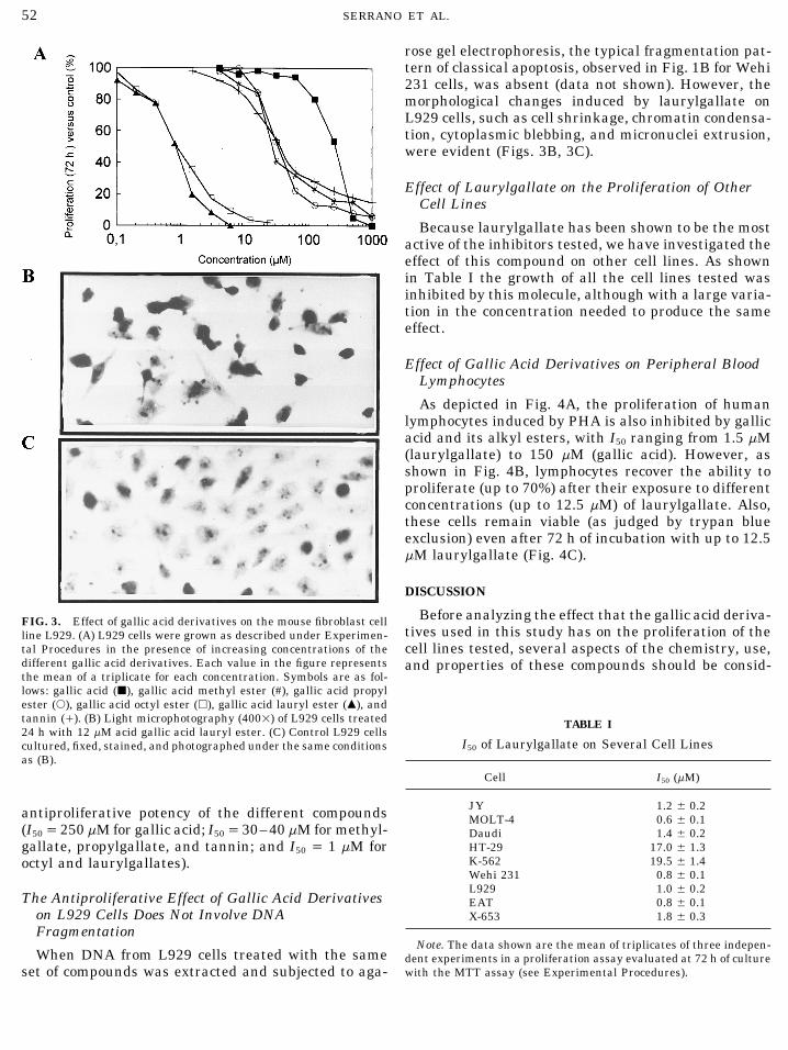
Before analyzing the effect that the gallic acid deriva-FIG. 3. Effect of gallic acid derivatives on the mouse fibroblast celltives used in this study has on the proliferation of theline L929. (A) L929 cells were grown as described under Experimen-
tal Procedures in the presence of increasing concentrations of the cell lines tested, several aspects of the chemistry, use,different gallic acid derivatives. Each value in the figure represents and properties of these compounds should be consid-the mean of a triplicate for each concentration. Symbols are as fol-lows: gallic acid (j), gallic acid methyl ester (#), gallic acid propylester (s), gallic acid octyl ester (h), gallic acid lauryl ester (m), andtannin (/). (B) Light microphotography (4001) of L929 cells treated TABLE I24 h with 12 mM acid gallic acid lauryl ester. (C) Control L929 cells
I50 of Laurylgallate on Several Cell Linescultured, fixed, stained, and photographed under the same conditionsas (B).
Cell I50 (mM)
JY 1.2 { 0.2antiproliferative potency of the different compounds MOLT-4 0.6 { 0.1(I50Å 250 mM for gallic acid; I50Å 30–40 mM for methyl- Daudi 1.4 { 0.2
HT-29 17.0 { 1.3gallate, propylgallate, and tannin; and I50 Å 1 mM forK-562 19.5 { 1.4octyl and laurylgallates).Wehi 231 0.8 { 0.1L929 1.0 { 0.2
The Antiproliferative Effect of Gallic Acid Derivatives EAT 0.8 { 0.1on L929 Cells Does Not Involve DNA X-653 1.8 { 0.3Fragmentation
Note. The data shown are the mean of triplicates of three indepen-When DNA from L929 cells treated with the same dent experiments in a proliferation assay evaluated at 72 h of culture
with the MTT assay (see Experimental Procedures).set of compounds was extracted and subjected to aga-
AID ABB 0474 / 6b49$$$$23 01-02-98 23:34:12 arca

53APOPTOSIS INDUCED BY GALLIC ACID DERIVATIVES
reactions that involve their oxidation to quinoid prod-ucts. Second, these compounds have been used for along time as antioxidant food additives, for preventingthe rancidity of fats, as code Nos. E-310 (propylgallate),E-311 (octylgallate), and E-312 (laurylgallate). Third,we have recently reported that the triphenolic com-pounds, and especially those derivatives of gallic acidtested in this study, are excellent inhibitors of partiallypurified PTKs from human spleen, exhibiting an inhib-itory potency superior to that of widely used PTK inhib-itors such as genistein or tyrphostins (9). The fact thatmany of the phenolic compounds used as antioxidantsare also potent PTK inhibitors may explain the diffi-culties found in the interpretation of the results ob-tained with these compounds when used in differentsystems. Contradictory results such as the antitumorpromotion observed when used at low concentrationsand tumor promotion when used at higher concentra-tions have been reported for other antioxidants (18).These paradoxical results may be explained by themany metabolic pathways with which these compoundsmay interact. Another well-known property is that phe-nols act as uncouplers of the mytochondrial oxidative-phosphorylation, thus influencing the energetic stateof the cell. With all these considerations in mind, thestudy of the mechanism(s) by which these compoundsproduce their effect on cells appears to be of great com-plexity. Figure 1A shows the effect of a series of gallicacid derivatives of increasing hydrophobicity (gallicacid, methyl, propyl, octyl, and lauryl esters) on theproliferation of the mouse B cell lymphoma Wehi 231.The fact that hydrophobicity is so important (laurylgallate is around 40 times more potent than gallic acid)may be due to better permeability of the cell membraneFIG. 4. Effect of gallic acid derivatives on human peripheral bloodto this type of compound or/and to better interactionlymphocytes (PBL). (A) After 72 h of culture, proliferation was evalu-of the compound with the enzyme(s) with which theyated by MTT conversion to formazan blue and the values obtained
were related to the untreated controls (100% proliferation). Each interact, since the inhibitory potency of lauryl gallatevalue in the figure represents the mean of a triplicate for each concen- in vitro is about 10 times higher than that of gallic acidtration. Symbols are as follows: gallic acid (j), gallic acid methyl for partially purified PTK (9). We have performed aester (#), gallic acid propyl ester (s), gallic acid octyl ester (h), gallic
comparative study of the permeability of gallic acidacid lauryl ester (m), and tannin (/). (B) Reversibility of the effectpropyl ester and gallic acid lauryl ester by flow cytome-of lauryl gallate on human PBL. PBL suspension (106 cells/ml) in
the medium described above was exposed to several concentrations try using DCFHDA (2*-7*-dichlorofluorescin diacetate),of gallic acid lauryl ester as follows: exposed to increasing concentra- and H2O2-sensitive dye whose fluorescence is rapidlytions of laurylgallate (24 h), washed and cultured in fresh medium quenched by laurylgallate (10 min); propylgallatefor 72 h in presence of PHA (l); exposed and stimulated with PHA
needs longer periods of incubation, indicating a differ-(40 mg/ml) simultaneously and cultured (without washing) for 72 h(l). (C) Viability of human PBL stimulated with PHA. After 72 h of ent permeability of the cell membrane toward theseexposure to several concentrations of gallic acid lauryl ester, the two compounds (to be published elsewhere). As shownviability was evaluated by trypan blue exclusion (s). The symbol (l) in Fig. 1B, the ladder-like agarose electrophoretic pat-at concentration 0 represents the viability of the untreated control.
terns of the DNA extracted from Wehi 231 cells treatedwith the different compounds is demonstrative of acti-vation of programmed cell death mechanisms by thesemolecules.ered. First, they are triphenolic compounds with anti-
oxidant and ROS scavenging activities derived from At present, the mechanisms by which these mole-cules trigger apoptosis pathways are not clearly under-the adjacent position of three hydroxyl groups in the
benzene ring, which enable these molecules to interfere stood. However, as can be deduced from Fig. 2, lauryl-gallate inhibits protein tyrosine phosphorylation inwith the homeostatic redox state of the cell through
AID ABB 0474 / 6b49$$$$24 01-02-98 23:34:12 arca

54 SERRANO ET AL.
whole cells, corroborating the results obtained with thors in which tannins (19), gallic acid (8), and otherantioxidants such as epigallo catechin gallate (20) orcrude extracts or partially purified PTKs that we havetertbutylhydroquinone (21) provide protection againstalready reported (9). It is also worth noting that thiscarcinogenesis induced by a variety of tumor promotorscompound is able to inhibit the protein tyrosine phos-and are involved in the redox regulation of gene tran-phorylation induced by nonspecific activators such asscription (22).pervanadate (Fig. 2A), or by a more specific activator
such as anti-IgM, which has been used widely as anACKNOWLEDGMENTinducer of protein tyrosine phosphorylation in Wehi
231 cells (Fig. 2B). This work was supported by Research Grant 95/773 from the Fondode Investigaciones Sanitarias de la Seguridad Social.Gallic acid derivatives also inhibit the proliferation
of other tumoral cell lines, although the fragmentationREFERENCESof DNA is not always evident. As shown in Fig. 3A the
growth of the mouse fibroblast cell line L929 is inhib- 1. Kerr, J. F., Wyllie, A. H., and Curie, A. R. (1972) Br. J. Cancer26, 239–245.ited by the same set of compounds. Again laurylgallate
2. Wyllie, A. H., Kerr, J. F. R., and Curie, A. R. (1980) Int. Rev.and octylgallate showed the highest antiproliferativeCytol. 68, 251–306.activity (around 250 times more potent than gallic
3. Ellis, R. E., Yuan, I., and Horvitz, H. R. (1991) Annu. Rev. Cell.acid). However, the agarose gel electrophoretic pattern Biol. 7, 663–698.of the DNA extracted did not show signs of DNA degra- 4. Hockenbery, D. M., Oltval, Z. N., Yin, X. M., Milliam, C. L., anddation (data not shown). Nevertheless, the morphologi- Korsmeyer, S. J. (1993) Cell 74, 241–251.cal characteristics of the cells before and after treat- 5. Sellins, K. S., and Cohen, J. J. (1987) J. Immunol. 139, 3199–
3206.ment with laurylgallate (Fig. 3B) were highly sugges-6. Ames, B. N., Shigenage, M. K., and Hagen, T. M. (1993) Proc.tive of apoptosis rather than necrosis. Similar apoptotic
Natl. Acad. Sci. USA 90, 7915–7922.behavior without concomitant DNA fragmentation has7. Buck, J., Myc, A., Garbe, A., and Cathomas, G. (1991) J. Cell.been described for the antiproliferative effect of stauro-
Biol. 115, 851–859.sporine on MOLT-4 cells (10).8. Inoue, M., Suzuki, R., Koide, T., Sakapuchi, N., Ogihara, Y., andTable I shows that laurylgallate inhibited the growth Yabu, Y. (1994) Biochem. Biophys. Res. Commun. 204, 898–904.
of other cell lines that we had available in our labora- 9. Lazaro, I., Palacios, C., Gonzalez, M., and Gonzalez-Porque, P.tory. In this respect, it is worth noting that the I50 for (1995) Anal. Biochem. 225, 180–183.most cells was between 0.5 and 2 mM. Two types of 10. Falcieri, E., Martelli, A. M., Bareggi, R., Cataldi, A., and Cocco,
L. (1993) Biochem. Biophys. Res. Commun. 193, 19–25.cells (HT29 and K562) showed higher resistance to the11. Otani, H., Erdos, M., and Leonard, W. J. (1993) J. Biol. Chem.effect of these compounds. The presence of esterases
268, 22733–22736.cleaving intracellularly laurylgallate, yielding gallic12. Uckun, F. M., Evans, W. W., Forsyth, C. J., Waddick, K. G., Ahl-acid as a product with a lower inhibitory potency to-
green, L. T., Chelstrom, L. M., Buskhardt, A., Bolen, J., and My-ward PTK, could not be excluded in these type of cells. ers, D. E. (1995) Science 267, 886–891.Undoubtedly, experiments dealing with the metabo- 13. Ni, R., Tomita, Y., Matsuda, K., Ichihara, A., Ishimura, K., Oga-lism and detoxification of laurylgallate by these type sawara, J., and Nagata, S. (1994) Exp. Cell Res. 215, 332–337.of cells could shed light on the behavior of different 14. Mosmann, T. (1983) J. Immunol. Methods. 65, 55–63.cells toward this compound. 15. Carmichael, J., DeGraff, W. G., Gazdar, A. F., Minna, J. D., and
Mitchell, J. B. (1987) Cancer Res. 47, 943–946.Also, the stimulation of PBL by PHA is inhibited by16. Hansen, M. B., Nielsen, S. E., and Berg, K. (1989) J. Immunol.gallic acid derivatives with I50 similar to those calcu-
Methods 119, 203–210.lated for other cell types (Fig. 4A). However, PBL can17. Doyle, A., Griffiths, J. B., and Newell, D. G. (1994) in Cell andwithstand higher concentrations of laurylgallate in a Tissue Culture: Laboratory Procedures. J. Wiley & Sons, Chi-
resting state, and when washed, the cells recover their chester, England [8B3.1–8B3.5]ability to proliferate upon the addition of mitogen (Fig. 18. Kahl, R. (1991) in Oxidative Stress: Oxidants and Antioxidants,
pp. 245–273, Academic Press, London.4B). Also, they remain viable (as judged by trypan blue19. Gali, H. U., Perchellet, E. M., and Perchellet, J. M. (1991) Cancerexclusion) after a period of 72 h (Fig. 4C).
Res. 51, 2820–2825.The observation that lymphocytes can withstand a20. Fujiki, H., Suganuma, M., Suguri, H., Takagi, K., Voshizowa,higher concentration of laurylgallate than the tumoral
S., Otsuyama, A., Tanooka, H., Okuda, T., Kobayashi, M., andcell lines tested may indicate a selectivity of action for Sugimure, T. (1990) Basic Life Sci. 52, 205–212.rapidly growing cells, thus opening the possibility of 21. Yoshioka, K., Deng, T., Cavigelli, M., and Karin, M. (1995) Proc.its study as a potential antitumoral agent. Supporting Natl. Acad. Sci. USA 92, 4972–4976.
22. Sen, C. K., and Packer, L. (1996) FASEB J. 10, 709–720.this hypothesis are the results reported by other au-
AID ABB 0474 / 6b49$$$$24 01-02-98 23:34:12 arca
Copyright © 2022 FDOKUMEN