Excavations at Utica by the Tunisian-British Utica Project 2012
Upload
independentCategory
view
2download
0
1 23
Environmental Science and PollutionResearch ISSN 0944-1344Volume 21Number 19 Environ Sci Pollut Res (2014)21:11433-11438DOI 10.1007/s11356-014-3099-x
Trace metal quantification in bladderbiopsies from tumoral lesions of Tunisiancancer and controls subjects
Molka Feki-Tounsi, Pablo Olmedo,Fernando Gil, Mohamed-Nabil Mhiri,Ahmed Rebai & Amel Hamza-Chaffai
1 23
Your article is protected by copyright and
all rights are held exclusively by Springer-
Verlag Berlin Heidelberg. This e-offprint is
for personal use only and shall not be self-
archived in electronic repositories. If you wish
to self-archive your article, please use the
accepted manuscript version for posting on
your own website. You may further deposit
the accepted manuscript version in any
repository, provided it is only made publicly
available 12 months after official publication
or later and provided acknowledgement is
given to the original source of publication
and a link is inserted to the published article
on Springer's website. The link must be
accompanied by the following text: "The final
publication is available at link.springer.com”.

RESEARCH ARTICLE
Trace metal quantification in bladder biopsies from tumorallesions of Tunisian cancer and controls subjects
Molka Feki-Tounsi & Pablo Olmedo & Fernando Gil &Mohamed-Nabil Mhiri & Ahmed Rebai &Amel Hamza-Chaffai
Received: 28 August 2013 /Accepted: 26 May 2014 /Published online: 7 June 2014# Springer-Verlag Berlin Heidelberg 2014
Abstract The incidence of bladder tumors has been dramat-ically increasing since the 1970s, possibly as a consequence ofongoing environmental pollution. Previous studies have pro-vided some evidence of an association between cancer andexposure to carcinogenic metals. In order to examine theassociation between levels of toxic metals in patients withbladder tumors and controls, the amounts of arsenic, cadmi-um, chromium, and nickel were measured in tumoral lesionsand adjacent normal part of the bladder mucosa excised forcarcinoma and compared with those in the bladder mucosa ofvolunteer subjects operated for non-neoplastic diseases. Thequantification of metals in tissue was assessed by atomicabsorption spectroscopy. In tumoral tissues of the excisedbladder mucosa, content of Cr and Ni was significantly lowcompared to that of adjacent normal tissues and control tissueswhile that of As and Cd in normal tissues adjacent to the tumorwere significantly elevated compared to controls. Though thesample size was small, the present study shows that concen-trations of metals such as Cd, Cr, As, and Ni in bladder tissue
may be used as a biomarker of exposure. On the basis of theresults obtained in this study, high amounts of As and Cd inadjacent normal parts of the bladders with carcinomas com-pared to controls would strongly suggest possible, individualor synergistic, effects of these pollutants on enzymatic sys-tems, priming an oncogenic pathway.
Keywords Bladder tumor .Metal quantification . Cadmium .
Arsenic . Chromium . Nickel
Introduction
Metals carcinogenicity has been the subject of several studiesusing animal models or immortalized cells, which have shownthat metals alter the normal cellular function by differentmechanisms leading to the development of cancer (Senset al. 2004; Oller et al. 2008; Koedrith et al. 2013). Addition-ally, human biomonitoring of dose nowadays has tremendousutility providing an efficient means of measuring human ex-posure to metallic elements (Angerer et al. 2007). Severalbiomonitoring studies suggested a significant association be-tween cancer and exposure to metals (Navarro Silvera andRohan 2007). Bladder, in particular, an organ in which excre-tion of the great majority of released toxins from the bodyhappens, is especially exposed to the adverse effects of carci-nogenic metals. Thus, measurement of these elements at thebladder mucosa may provide relevant information on thedose-response relationships at the target organ level.
Studies of associations between toxic metals and bladdercancer in humans were based on monitoring of metals inblood and/or urine of exposed subjects, which can only pro-vide an estimate of the overall absorbed dose (Wolf et al.2009; Feki-Tounsi et al. 2012, 2013). Nevertheless, it wouldbe interesting to study this association by collecting detailedinformation about the effective dose at the target organ level.
Responsible editor: Philippe Garrigues
M. Feki-Tounsi (*) :A. Hamza-ChaffaiUnit of Marine and Environmental Toxicology, IPEIS, SfaxUniversity, PB 805, 3018 Sfax, Tunisiae-mail: [email protected]
A. Hamza-Chaffaie-mail: [email protected]
M. Feki-Tounsi :A. RebaiDepartment of Bioinformatics and Human Genetics, Center ofBiotechnology of Sfax, Sfax, Tunisia
P. Olmedo : F. GilDepartment of Legal Medicine and Toxicology, Faculty ofMedicine,University of Granada, Granada, Spain
M.<N. MhiriDepartment of Urology, CHU Habib Bourguiba, Sfax, Tunisia
Environ Sci Pollut Res (2014) 21:11433–11438DOI 10.1007/s11356-014-3099-x
Author's personal copy

In this context, the aim of the present study was to determineconcentration of four carcinogenic metals known to be moreor less associated with the risk of cancer in the bladder tumorallesions, the normal adjacent mucosa of bladder cancer patientsundergoing surgical intervention. Metal bladder concentra-tions were also determined in a group of patients with anegative diagnosis of bladder cancer. The investigated ele-ments have been classified as class 1 carcinogens (carcino-genic to humans) by the International Agency for Research onCancer (IARC), namely, chromium [Cr, as Cr(VI)] (1990),nickel (Ni, only in non-metallic form) (1990) cadmium (Cd)(1993), and arsenic (As) (1980, 1987). To our knowledge, thisis the first investigation of metals in human bladder mucosa.Therefore, the provided values may be useful for comparisonsin future studies.
Materials and methods
Subjects
Bladder biopsies were collected in 49 bladder cancer patientsand 36 controls undergoing bladder resection at the Depart-ment of Urology of Sfax Hospital between June 2007 andMay 2010. About 96 % bladder cancer patients were transi-tional cell carcinoma and the others were epidermoid carcino-ma. The main patients’ characteristics are reported in Table 1.Among bladder cancer patients, 24 (53 %) had a tumor stage 1and 19 (42 %) had a stage 2. The control subjects were notwith cancerous diseases but were operated for prostate hyper-plasia intervention. None of the enrolled patients receivedprevious chemotherapy or radiotherapy treatments. A struc-tured questionnaire was administered to the subjects in orderto collect socio-demographic information, past occupational/environmental exposure, and lifestyle data. Two trained inter-viewers evaluated questionnaires blindly and attributed pastoccupational exposure to metallic elements according to thereported job titles.
According to the smoking habits, subjects were classifiedinto the following subgroups: currents smokers, ex-smokers,and non-smokers. All the enrolled subjects gave their writteninformed consent to the study that was approved by the localethics committee.
Study design, sample collection, and storage
Two tissue fragments (about 5 mm per side, each) wereexcised from each patient during surgery: at least one fromthe cancerous mass and another from the seemingly normaltissue, far at least 3 cm from cancerous tissue, in the samespecimen. All fragments were excised far from surgical clipsor staple lines to avoid any metal contamination. Fragmentswhose mass is much less than 200 mg were removed from the
metal quantification, and only 40 cancer biopsies and 36biopsies of normal tissues surrounding the tumors were usedfor the metal analysis. Fragments were put into sterile vials(Eppendorf Int., Hamburg, Germany) and immediately storedat −20 °C. The biological sampling was performed as laiddown in the Declaration of Helsinki.
Sample digestion and metallic element analysis
In this study, a modified version of the US EPA method 3052protocol (US EPA 1996) was employed for the digestion ofbladder tissue samples. Little pieces of the collected fragmentsof cancerous and non-tumoral bladder tissue (about 200 mgwet tissue) were digested in a solution 1:1 of hyperpure nitricacid 70 % and bi-distilled water for 30 min at 90 °C. Resultantsample solutions were diluted and analyzed for the quantifica-tion of As, Cd, Cr, andNi. Levels of Cd, Cr, andNi compoundswere determined by a Perkin Elmer Analyst 800 AtomicAbsorption Spectrometer (Perkin Elmer, Norwalk, USA)equipped with a Zeeman background correction and an AS-800 autosampler by graphite furnace and graphite tubes withintegrated L’vov platform (Perkin Elmer). Appropriate matrixmodifiers were used for the selected heavy metal studied andprepared in 0.2 % (v/v) nitric acid and 0.1 % Triton X-100.
A direct flow-injection atomic absorption spectrometrictechnique (FI-HGAAS) was used to measure sample levelsof total arsenic. Details of arsenic determination have beenpresented elsewhere (Feki-Tounsi et al. 2012).
The limit of detection (LOD) was 0.03 μg/L for both Cdand As and 0.19 and 0.24 μg/L for Cr and Ni, respectively.Calibration graphs were linear until 7, 30, 20, 15, and
Table 1 Main characteristics of the study population. The possibleoccupational exposure to metals was extrapolated by past occupationalhistory, and it cannot be considered as a quantitative variable
Variables Patients (n=49) Controls (n=37)
Age (years) 62.8±13.5* 72.3±7.2
Smoking habits
Current smokers 31 (63.2 %) 23 (62.1 %)
Ex-smokers 12 (24.4 %) 13 (35.1 %)
Non-smokers 6 (12.2 %) 0 (0 %)
Pack year (PY) 29.34±25.3 27.6±22.15
Tumor grade
High grade 21 (42.8 %)
Low grade 28 (57.1 %)
Occupational exposure to metals
Yes 13 (26 %)** 7 (18 %)
No 37 (74 %) 31 (82 %)
*p<0.05 (student T test)
**p<0.05 (Pearson chi-square test)
11434 Environ Sci Pollut Res (2014) 21:11433–11438
Author's personal copy

200 μg/L (for more details, see Gil et al. 2006; Olmedo et al.2010). Further, a quality check sample was always run witheach set of samples for metal analysis to maintain accuracy.
The validation of analytical procedures developed for thedetermination of metal compounds has been reported else-where (Gil et al. 2006; Olmedo et al. 2010). This validationprotocol included the limit of detection (LOD) and quantifi-cation (LOQ), linear range, precision (minimal, intermediate,and reproducibility), accuracy, recovery, characteristic mass,and uncertainty.
The analytical method was controlled by using externalcertified reference materials (CRM). Reference samples (threelevels, refs. 201505, 201605, and 201705) were supplied bySeronorm (Billingstad, Norway).
Statistical analysis
In order to obtain a normal distribution of data, we chose toperform log-transformation, the independent and the pairedsamples T tests were used for comparisons between twoindependent and two dependent data sets, respectively. TheSpearman’s correlation coefficient was calculated in assessingthe relationship between pairs of variables. Finally, statisticalanalysis was performed using the SPSS 17.0 software (SPSSinch, Chicago, IL, USA), and a significant p value of 0.05 waschosen. When the concentration was below the LOD, weleveled to the half of the LOD values to reduce mathematicalartifacts in statistical analysis due to the variability of tissueweight.
Results
The subject characteristics are presented in Table 1. Bladdercancer cases and controls were comparable in age. Concerningsmoking habits, there were as many current smokers in bothgroups, and the difference in smoking rate (pack-year index)was not significant. With regard to occupational exposure,bladder cancer cases were more exposed (26 %) than controls(18 %).
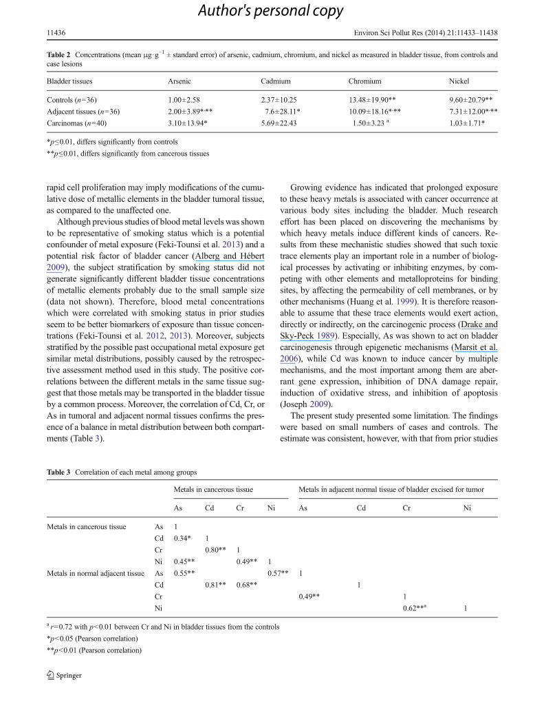
Mean concentrations of arsenic, cadmium, chromium, andnickel measured in bladder tissue, from controls and caselesions, are presented in Table 2. We observed significantlyhigher levels of Cd and As and lower levels of Cr and Ni,respectively (p<0.01), in normal tissues adjacent to the tumorsfrom cancerous subjects vs. controls. There were also signif-icantly higher As and lower Cr and Ni levels in cancerous vs.control tissues and vs. adjacent tissues (p<0.01), respectively,but no significant difference between Cd levels in the tumorallesions and the normal adjacent tissues. Stratification of sub-jects for smoking status or occupational exposure did not
generate significant difference in metal levels (data notshown).
With regard to the correlation between metals levels, intumoral, adjacent, and control tissues, Cr was always corre-lated with Ni (r=0.49; r=0.62, and r=0.72, respectively,p<0.01). As was also correlated with Cr in normal tissue ofbladder cancer patients (r=0.49; p<0.01). Moreover, in tu-moral tissues, Cd was correlated with As and Cr (r=0.34,p<0.05 and r=0.80, p<0.01, respectively), and Ni was alsocorrelated with As (r=0.45, p<0.01). Cd and As levels in atumoral lesion were correlated with Cd and As levels inadjacent tissues (r=0.55 and r=0.81, respectively, p<0.01).
Discussion
Bladder cancer incidence and prevalence have been increasedthe last decades (Cancer Registry in Southern Tunisia 2002).Smoking was not only the first established risk factor for thisdisease (Crivelli et al. 2013), but also recent studies identifiedan association between exposure to metals such as Cd and Asand bladder cancer occurrence (Huff et al. 2007; Letašiováet al. 2012). Blood levels of both metals have been shown tobe good biomarkers of exposure (Kellen et al. 2007; Feki-Tounsi et al. 2012, 2013). However, few are the dataconcerning metal quantification in bladder tissue (Amaralet al. 2009) to our knowledge; the present study is the firstto describe metal content in human bladder tissue. The levelsof As, Cd, Cr, and Ni were quantified in tumoral lesions andadjacent normal part of the bladder mucosa and comparedwith those in the bladder mucosa of volunteer subjects oper-ated for non-neoplastic diseases. The high levels of Cd and Asin normal tissues of bladder cancer subjects vs. controls wereconcordant with prior results describing higher Cd levels inthe blood of bladder cancer patients compared to that ofcontrols (Feki-Tounsi et al. 2012, 2013; Kellen et al. 2007).Moreover, this finding argues for the validity of Cd and As inthe bladder normal tissue as a biomarker of exposure. Thelower levels of Cr and Ni compared to controls were also inagreement with prior not published findings describing sig-nificantly lower blood levels of both metals in bladder cancerpatients compared to that in the general population (Khlifiet al. 2013). The results show that As accumulate in thetumoral tissues while Cr, Ni, and, to a lesser extent, Cd arediluted in the tumor due to its rapid proliferation, and thisatypical behavior of As in the tumor compared to that of theremaining elements may be attributable to its dose-dependentanti-proliferative property described by previous experimentalstudies (Liu et al. 2006; Jutooru et al. 2010). Therefore,unaffected tissue seemed to be more representative of thecumulative bladder tissue dose of Cd, Cr, and Ni, as comparedto the cancerous one. Structure modifications occurring incancerous tissue such as neo-vascularization, necrosis, and
Environ Sci Pollut Res (2014) 21:11433–11438 11435
Author's personal copy
rapid cell proliferation may imply modifications of the cumu-lative dose of metallic elements in the bladder tumoral tissue,as compared to the unaffected one.
Although previous studies of blood metal levels was shownto be representative of smoking status which is a potentialconfounder of metal exposure (Feki-Tounsi et al. 2013) and apotential risk factor of bladder cancer (Alberg and Hébert2009), the subject stratification by smoking status did notgenerate significantly different bladder tissue concentrationsof metallic elements probably due to the small sample size(data not shown). Therefore, blood metal concentrationswhich were correlated with smoking status in prior studiesseem to be better biomarkers of exposure than tissue concen-trations (Feki-Tounsi et al. 2012, 2013). Moreover, subjectsstratified by the possible past occupational metal exposure getsimilar metal distributions, possibly caused by the retrospec-tive assessment method used in this study. The positive cor-relations between the different metals in the same tissue sug-gest that those metals may be transported in the bladder tissueby a common process. Moreover, the correlation of Cd, Cr, orAs in tumoral and adjacent normal tissues confirms the pres-ence of a balance in metal distribution between both compart-ments (Table 3).
Growing evidence has indicated that prolonged exposureto these heavy metals is associated with cancer occurrence atvarious body sites including the bladder. Much researcheffort has been placed on discovering the mechanisms bywhich heavy metals induce different kinds of cancers. Re-sults from these mechanistic studies showed that such toxictrace elements play an important role in a number of biolog-ical processes by activating or inhibiting enzymes, by com-peting with other elements and metalloproteins for bindingsites, by affecting the permeability of cell membranes, or byother mechanisms (Huang et al. 1999). It is therefore reason-able to assume that these trace elements would exert action,directly or indirectly, on the carcinogenic process (Drake andSky-Peck 1989). Especially, As was shown to act on bladdercarcinogenesis through epigenetic mechanisms (Marsit et al.2006), while Cd was known to induce cancer by multiplemechanisms, and the most important among them are aber-rant gene expression, inhibition of DNA damage repair,induction of oxidative stress, and inhibition of apoptosis(Joseph 2009).
The present study presented some limitation. The findingswere based on small numbers of cases and controls. Theestimate was consistent, however, with that from prior studies
Table 2 Concentrations (mean μg·g−1 ± standard error) of arsenic, cadmium, chromium, and nickel as measured in bladder tissue, from controls andcase lesions
Bladder tissues Arsenic Cadmium Chromium Nickel
Controls (n=36) 1.00±2.58 2.37±10.25 13.48±19.90** 9.60±20.79**
Adjacent tissues (n=36) 2.00±3.89*,** 7.6±28.11* 10.09±18.16*,** 7.31±12.00*,**
Carcinomas (n=40) 3.10±13.94* 5.69±22.43 1.50±3.23 a 1.03±1.71*
*p≤0.01, differs significantly from controls
**p≤0.01, differs significantly from cancerous tissues
Table 3 Correlation of each metal among groups
Metals in cancerous tissue Metals in adjacent normal tissue of bladder excised for tumor
As Cd Cr Ni As Cd Cr Ni
Metals in cancerous tissue As 1
Cd 0.34* 1
Cr 0.80** 1
Ni 0.45** 0.49** 1
Metals in normal adjacent tissue As 0.55** 0.57** 1
Cd 0.81** 0.68** 1
Cr 0.49** 1
Ni 0.62**a 1
a r=0.72 with p<0.01 between Cr and Ni in bladder tissues from the controls
*p<0.05 (Pearson correlation)
**p<0.01 (Pearson correlation)
11436 Environ Sci Pollut Res (2014) 21:11433–11438
Author's personal copy

which imply cadmium and arsenic as causal factors in bladdercarcinogenesis.
Conclusion
In summary, the present study is the first to analyze carcino-genic metals in human tumoral and normal bladder tissuesamples by the atomic absorption spectrophotometric method.In adjacent tissues to tumors, dominant mean concentrationswere revealed byCd andAs, respectively, compared to controltissues, while levels of As and Cd were significantly elevatedcompared to controls. Average concentrations of Cr and Niwere noted to be significantly lower in the malignant tissuescompared with that in the benign tissues. On the other hand,As mean concentration was significantly higher in the carci-nomas, while Cd level was shown to be lower but not signif-icantly lower than that in the benign tissues. Significantlystrong correlations (r>0.50) in malignant tissues were ob-served between Cd and Cr, whereas Ni and Cr revealed strongand significant relationships in benign tissues at p<0.01. Thestudy revealed a considerably different pattern of distributionand mutual correlations of trace metals in the bladder tissuesof benign and cancerous patients.
On the basis of the results obtained in this study, thedifferent distribution of Cd and As found in adjacent normaltissue of excised tumoral bladder would strongly indicate thepossible pathogenetic role of these metals in bladder carcino-genesis. Simultaneous presence of both pollutants may triggersynergistic effects, which would potentiate the resultant car-cinogenicity. Though the sample size was small, the presentstudy shows that concentrations of metals such as cadmium,chromium, arsenic, and nickel in bladder tissue may be usedas a biomarker of exposure.
Acknowledgements This work was supported by the Unit of Marineand Environmental Toxicology, University of Sfax, Ministry of HighEducation and Scientific research in Tunisia. Thanks are addressed toMrs. Raida Tounsi for language assistance in the revision of this paper.
Conflict of interest Funding was provided by the Ministry of HighEducation and Scientific research in Tunisia. The funders had no role instudy design, data collection and analysis, decision to publish, or prepa-ration of the manuscript. The authors have declared that no competinginterests exist.
References
Alberg AJ, Hébert JR (2009) Cigarette smoking and bladder cancer: anew twist in an old saga? J Natl Cancer Inst 101(1525):1526,Corrected and republished in J. Natl. Cancer Inst. 102(2), 138
Amaral AF, Cymbron T, Gärtner F, Lima M, Rodrigues AS (2009) Tracemetals and over-expression of metallothioneins in bladder tumorallesions: a case-control study. BMC Vet Res 18(5):24
Angerer J, Ewers U, Wilhelm M (2007) Human biomonitoring: state ofthe art. Int J Hyg Environ Health. 210(3–4):201–228
Cancer Registry in Southern Tunisia (2002) Cancer incidence for theyears 1997–1999. Available on http://www.insp.nat.tn/fr/unite_the/regis/inci97.99b.pdf
Crivelli JJ, Xylinas E, Kluth LA, Rieken M, Rink M, Shariat SF (2013)Effect of smoking on outcomes of urothelial carcinoma: a systematicreview of the literature. Eur Urol. doi:10.1016/j.eururo.2013.06.010
Drake EN, Sky-peck HH (1989) Discriminant analysis of trace elementdistribution in normal and malignant human tissues. Cancer Res 49:4210–4215
Feki-Tounsi M, Olmedo P, Gil F, Khlifi R, Mhiri MN, Rebai A, Hamza-Chaffai A (2012) Low-level arsenic exposure is associated withbladder cancer risk and cigarette smoking: a case-control studyamong men in Tunisia. Environ Sci Pollut Res Int. doi:10.1007/s11356-012-1335-9
Feki-Tounsi M, Olmedo P, Gil F, Khlifi R, Mhiri MN, Rebai A, Hamza-Chaffai A (2013) Cadmium in blood of Tunisian men and risk ofbladder cancer: interactions with arsenic exposure and smoking.Environ Sci Pollut Res Int. doi:10.1007/s11356-013-1716-8
Gil F, Capitán-Vallvey LF, De Santiago E, Ballesta J, Pla A, HernándezAF, Gutiérrez-Bedmar M, Fernández-Crehuet J, Gómez J, López-Guarnido O, Rodrigo L, Villanueva E (2006) Heavy metal concen-trations in the general population of Andalusia, South of Spain: acomparison with the population within the area of influence ofAznalcóllar mine spill (SW Spain). Sci Total Environ 372(1):49–57
Huang YL, Sheu JY, Lin TH (1999) Association between oxidative stressand changes of trace elements in patients with bladder cancer. ClinBiochem 32(2):131–136
Huff J, Lunn RM, Waalkes MP, Tomatis L, Infante PF (2007) Cadmium-induced cancers in animals and in humans. Int J Occup EnvironHealth 13(2):202–212
IARC (1980) Some metals and metallic compounds. IARC Monog EvalCarcinog Risk Hum, 23. International Agency for Research onCancer, Lyon, France
IARC (1987) Overall evaluation of carcinogenicity: an updating of IARCmonographs volumes 1 to 42. IARC Monog Eval Carcinog RiskHum, Suppl 7. International Agency for Research on Cancer, Lyon,France
IARC (1990) Chromium, nickel and welding. IARC Monog EvalCarcinog Risk Hum, 49. International Agency for Research onCancer, Lyon, France
IARC (1993) Beryllium, cadmium, mercury and exposures in the glassmanufacturing industry. IARC Monog Eval Carcinog Risk Hum,58. International Agency for Research on Cancer, Lyon, France
Joseph P (2009) Mechanisms of cadmium carcinogenesis. Toxicol ApplPharmacol 238(3):272–279. doi:10.1016/j.taap.2009.01.011
Jutooru I, Chadalapaka G, Sreevalsan S, Lei P, Barhoumi R, Burghardt R,Safe S (2010) Arsenic trioxide downregulates specificity protein(Sp) transcription factors and inhibits bladder cancer cell and tumorgrowth. Exp Cell Res 316(13):2174–2188. doi:10.1016/j.yexcr.2010.04.027
Kellen E, Zeegers MP, Hond ED, Buntinx F (2007) Blood cadmium maybe associated with bladder carcinogenesis: the Belgian case-controlstudy on bladder cancer. Cancer Detect Prev 31(1):77–82
Khlifi R, Olmedo P, Gil F, Feki-Tounsi M, Chakroun A, Rebai A, Hamza-Chaffai A (2013) Blood nickel and chromium levels in associationwith smoking and occupational exposure among head and neckcancer patients in Tunisia. Environ Sci Pollut Res Int. doi:10.1007/s11356-013-1466-7
Koedrith P, Kim H, Weon JI, Seo YR (2013) Toxicogenomic approachesfor understanding molecular mechanisms of heavy metal mutage-nicity and carcinogenicity. Int J Hyg Environ Health 216(5):587–598. doi:10.1016/j.ijheh.2013.02.010
Letašiová S, Medve'ová A, Šovčíková A, Dušinská M, Volkovová K,Mosoiu C, Bartonová A (2012) Trace elements and cancer risk: a
Environ Sci Pollut Res (2014) 21:11433–11438 11437
Author's personal copy
review of the epidemiologic evidence. Environ Health 11(Suppl 1):S11. doi:10.1186/1476-069X-11-S1-S11
Liu B, Pan S, Dong X, Qiao H, Jiang H, Krissansen GW, Sun X (2006)Opposing effects of arsenic trioxide on hepatocellular carcinomas inmice. Cancer Sci 97(7):675–681
Marsit CJ, Karagas MR, Schned A, Kelsey KT (2006) Carcinogenexposure and epigenetic silencing in bladder cancer. Ann N YAcad Sci 1076:810–821
Navarro Silvera SA, Rohan TE (2007) Bladder cancer, a reviewof the environmental risk factors. Cancer Causes Control18(1):7–27
Oller AR, Kirkpatrick DT, Radovsky A, Bates HK (2008) Inhalationcarcinogenicity study with nickel metal powder in Wistar rats.Toxicol Appl Pharmacol 233(2):262–275. doi:10.1016/j.taap.2008.08.017
Olmedo P, Pla A, Hernández AF, López-Guarnido O, Rodrigo L, Gil F(2010) Validation of a method to quantify chromium, cadmium,manganese, nickel and lead in human whole blood, urine, salivaand hair samples by electrothermal atomic absorption spectrometry
Sens DA, Park S, Gurel V, Sens MA, Garrett SH, Somji S (2004)Inorganic cadmium- and arsenite-induced malignant transformationof human bladder urothelial cells. Toxicol Sci 79:56–63
U.S. Environmental Protection Agency (1996) Microwave assisted aciddigestion of siliceous and organically based matrices, Method 3052,Office of Solid Waste and Emergency Response, U.S. GovernmentPrinting Office, Washington, DC
Wolf C, Strenziokb R, Kyriakopoulosa A (2009) Elevated MT-boundcadmium concentrations in urine from bladder carcinoma patients,investigated by size exclusion chromatography-inductively coupledplasma mass spectrometry. Anal Chim Acta 631(2):218–222
11438 Environ Sci Pollut Res (2014) 21:11433–11438
Author's personal copy
Copyright © 2022 FDOKUMEN