Pseudomonas aeruginosa tolerance to tobramycin, hydrogen peroxide and polymorphonuclear leukocytes...
11
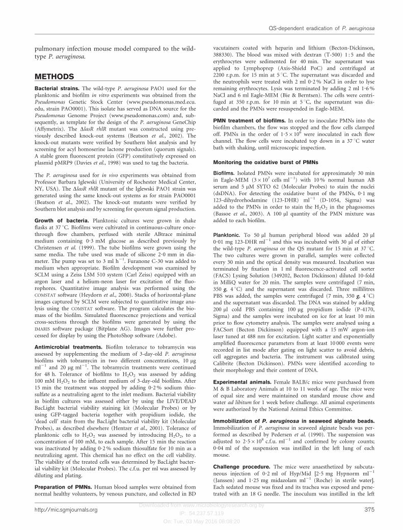
Downloaded from www.microbiologyresearch.org by IP: 54.237.57.119 On: Tue, 03 May 2016 08:08:20 Pseudomonas aeruginosa tolerance to tobramycin, hydrogen peroxide and polymorphonuclear leukocytes is quorum-sensing dependent Thomas Bjarnsholt, 1 Peter Østrup Jensen, 2 Mette Burmølle, 3 Morten Hentzer, 4 Janus A. J. Haagensen, 1 Hans Petter Hougen, 5 Henrik Calum, 2 Kit G. Madsen, 1 Claus Moser, 2 Søren Molin, 1 Niels Høiby 2 and Michael Givskov 1 Correspondence Michael Givskov [email protected] 1 Centre for Biomedical Microbiology, BioCentrum, Technical University of Denmark, DK-2800 Lyngby, Denmark 2 Department of Clinical Microbiology, Rigshospitalet, DK-2100 Copenhagen Ø, Denmark 3 Department of Microbiology, University of Copenhagen, DK-1307 Copenhagen K, Denmark 4 Carlsberg Research Center, DK-2500 Valby, Denmark 5 Institute of Forensic Medicine, University of Copenhagen, DK-2100 Copenhagen Ø, Denmark Received 24 June 2004 Revised 21 September 2004 Accepted 14 October 2004 The opportunistic human pathogen Pseudomonas aeruginosa is the predominant micro-organism of chronic lung infections in cystic fibrosis (CF) patients. P. aeruginosa colonizes the CF lungs by forming biofilm structures in the alveoli. In the biofilm mode of growth the bacteria are highly tolerant to otherwise lethal doses of antibiotics and are protected from bactericidal activity of polymorphonuclear leukocytes (PMNs). P. aeruginosa controls the expression of many of its virulence factors by means of a cell–cell communication system termed quorum sensing (QS). In the present report it is demonstrated that biofilm bacteria in which QS is blocked either by mutation or by administration of QS inhibitory drugs are sensitive to treatment with tobramycin and H 2 O 2 , and are readily phagocytosed by PMNs, in contrast to bacteria with functional QS systems. In contrast to the wild-type, QS-deficient biofilms led to an immediate respiratory-burst activation of the PMNs in vitro. In vivo QS-deficient mutants provoked a higher degree of inflammation. It is suggested that quorum signals and QS-inhibitory drugs play direct and opposite roles in this process. Consequently, the faster and highly efficient clearance of QS-deficient bacteria in vivo is probably a two-sided phenomenon: down regulation of virulence and activation of the innate immune system. These data also suggest that a combination of the action of PMNs and QS inhibitors along with conventional antibiotics would eliminate the biofilm-forming bacteria before a chronic infection is established. INTRODUCTION The progressive deterioration of the lungs in patients suffering from cystic fibrosis (CF) is mainly caused by an inflammatory response to chronic endobronchial biofilm- forming bacterial infection (Koch & Hoiby, 1993; Konstan & Berger, 1997). The progressive development of the lung disease is the main cause of morbidity and mortality in CF patients (Davis et al., 1996), and up to 80 % of young adults suffering from CF are chronically infected with P. aeruginosa (Koch & Hoiby, 2000; Gilligan, 1991; Bauernfeind et al., 1987). The bacteria form biofilms in the host, which makes the bacteria tolerant to antibiotic treatment and the action of the host defence system. Contributing to this is their ability by mutation to develop conventional resistance to antibiotics such as b-lactam antibiotics (Ciofu, 2003; Bagge et al., 2004a, b). Although several mechanisms have been postulated to explain the reduced susceptibility of biofilms to antimicro- bials, it is becoming evident that biofilm tolerance to antimicrobial treatments is multifactorial with one factor being the low physiological activity of the bacterial cells deep below the surface of the biofilm where anaerobic conditions exists. Different barriers, such as ionic trapping of antibiotics, do not seem to play a critical role (Stewart & Costerton, 2001; Gilbert et al., 2002). However, free Abbreviations: CF, cystic fibrosis; 123-DHR, 123-dihydrorhodamine; FACS, fluorescence-activated cell sorter; GFP, green fluorescent protein; PMN, polymorphonuclear leukocyte; QS, quorum sensing; QSI, quorum-sensing inhibitory. 0002-7463 G 2005 SGM Printed in Great Britain 373 Microbiology (2005), 151, 373–383 DOI 10.1099/mic.0.27463-0
-
Upload
independent -
Category
Documents
-
view
4 -
download
0
Transcript of Pseudomonas aeruginosa tolerance to tobramycin, hydrogen peroxide and polymorphonuclear leukocytes...

Downloaded from www.microbiologyresearch.org by
IP: 54.237.57.119
On: Tue, 03 May 2016 08:08:20
Pseudomonas aeruginosa tolerance to tobramycin,hydrogen peroxide and polymorphonuclearleukocytes is quorum-sensing dependent
Thomas Bjarnsholt,1 Peter Østrup Jensen,2 Mette Burmølle,3
Morten Hentzer,4 Janus A. J. Haagensen,1 Hans Petter Hougen,5
Henrik Calum,2 Kit G. Madsen,1 Claus Moser,2 Søren Molin,1 Niels Høiby2
and Michael Givskov1
Correspondence
Michael Givskov
1Centre for Biomedical Microbiology, BioCentrum, Technical University of Denmark,DK-2800 Lyngby, Denmark
2Department of Clinical Microbiology, Rigshospitalet, DK-2100 Copenhagen Ø, Denmark
3Department of Microbiology, University of Copenhagen, DK-1307 Copenhagen K, Denmark
4Carlsberg Research Center, DK-2500 Valby, Denmark
5Institute of Forensic Medicine, University of Copenhagen, DK-2100 Copenhagen Ø, Denmark
Received 24 June 2004
Revised 21 September 2004
Accepted 14 October 2004
The opportunistic human pathogen Pseudomonas aeruginosa is the predominant
micro-organism of chronic lung infections in cystic fibrosis (CF) patients. P. aeruginosa
colonizes the CF lungs by forming biofilm structures in the alveoli. In the biofilm mode of growth
the bacteria are highly tolerant to otherwise lethal doses of antibiotics and are protected from
bactericidal activity of polymorphonuclear leukocytes (PMNs). P. aeruginosa controls the
expression of many of its virulence factors by means of a cell–cell communication system termed
quorum sensing (QS). In the present report it is demonstrated that biofilm bacteria in which QS
is blocked either by mutation or by administration of QS inhibitory drugs are sensitive to treatment
with tobramycin and H2O2, and are readily phagocytosed by PMNs, in contrast to bacteria with
functional QS systems. In contrast to the wild-type, QS-deficient biofilms led to an immediate
respiratory-burst activation of the PMNs in vitro. In vivo QS-deficient mutants provoked a higher
degree of inflammation. It is suggested that quorum signals and QS-inhibitory drugs play direct
and opposite roles in this process. Consequently, the faster and highly efficient clearance of
QS-deficient bacteria in vivo is probably a two-sided phenomenon: down regulation of virulence
and activation of the innate immune system. These data also suggest that a combination of the
action of PMNs and QS inhibitors along with conventional antibiotics would eliminate the
biofilm-forming bacteria before a chronic infection is established.
INTRODUCTION
The progressive deterioration of the lungs in patientssuffering from cystic fibrosis (CF) is mainly caused by aninflammatory response to chronic endobronchial biofilm-forming bacterial infection (Koch & Hoiby, 1993; Konstan& Berger, 1997). The progressive development of thelung disease is the main cause of morbidity and mortalityin CF patients (Davis et al., 1996), and up to 80% ofyoung adults suffering from CF are chronically infectedwith P. aeruginosa (Koch & Hoiby, 2000; Gilligan, 1991;
Bauernfeind et al., 1987). The bacteria form biofilms inthe host, which makes the bacteria tolerant to antibiotictreatment and the action of the host defence system.Contributing to this is their ability by mutation to developconventional resistance to antibiotics such as b-lactamantibiotics (Ciofu, 2003; Bagge et al., 2004a, b).
Although several mechanisms have been postulated toexplain the reduced susceptibility of biofilms to antimicro-bials, it is becoming evident that biofilm tolerance toantimicrobial treatments is multifactorial with one factorbeing the low physiological activity of the bacterial cellsdeep below the surface of the biofilm where anaerobicconditions exists. Different barriers, such as ionic trappingof antibiotics, do not seem to play a critical role (Stewart& Costerton, 2001; Gilbert et al., 2002). However, free
Abbreviations: CF, cystic fibrosis; 123-DHR, 123-dihydrorhodamine;FACS, fluorescence-activated cell sorter; GFP, green fluorescentprotein; PMN, polymorphonuclear leukocyte; QS, quorum sensing;QSI, quorum-sensing inhibitory.
0002-7463 G 2005 SGM Printed in Great Britain 373
Microbiology (2005), 151, 373–383 DOI 10.1099/mic.0.27463-0

Downloaded from www.microbiologyresearch.org by
IP: 54.237.57.119
On: Tue, 03 May 2016 08:08:20
chromosomal b-lactamase in and around the biofilm maycontribute significantly (Giwercman et al., 1992; Ciofu2003).
As an active means to evade the host defences, P. aeruginosacontrols the production and secretion of virulence factorsby cell–cell communication signals in a process termedquorum sensing (QS; Fuqua et al., 1994). The concentrationof signal molecules reflects the number of bacterial cellsand at a certain threshold value the population becomes‘quorate’, i.e. a sufficient number of cells have amassed tomake behavioural decisions.
In P. aeruginosa the QS communication apparatus is com-posed of the Las and the Rhl systems. Both systems consistof two essential pairs of genes, lasR and lasI, and rhlR andrhlI, respectively. The I genes encode two synthetases thatsynthesize either 3-oxo-dodecanoyl-homoserine lactone(3-oxo-C12-HSL) for the las system or butyryl-homoserinelactone (C4-HSL) for the rhl system. The R genes encoderegulatory proteins which, in concert with the signal mole-cules, activate gene expression of numerous target genes(Hentzer et al., 2003; Schuster et al., 2003; Wagner et al.,2003). The two QS systems of P. aeruginosa are hierarchi-cally arranged, with the Las system being on top of thesignalling cascade (Latifi et al., 1996; Pesci et al., 1997).Intertwined in this QS hierarchy is the quinolone signal(PQS) system, which provides a link between the Las andRhl QS systems (Pesci et al., 1999) by cooperativelyregulating the expression of rhlI and lasB (McKnight et al.,2000). Recently, however, Medina et al. (2003) showed thatthe rhl system is also capable of operating on its own.
The QS system has on several occasions been shown toaffect in vivo infections. In a murine model of chronic lunginfection, a lasI rhlI mutant of P. aeruginosa was clearedfaster than the wild-type (Wu et al., 2001). Rumbaugh et al.(1999) used a burned mouse model to show that QS-deficient mutants were less virulent than the wild-type. Inaccordance with this, Hentzer et al. (2003) showed that micewith pulmonary P. aeruginosa infections treated with thespecific QSI drug furanone C-30 exhibited clearing that was1000-fold more efficient than mice treated with a placebo.Davies et al. (1998) andHentzer et al. (2003) demonstrated arole for QS in the development of structural differentiatedbiofilms that showed tolerance to SDS and tobramycintreatment.
QS signal molecules have been reported to interfere withthe host immune system. The first evidence for 3-oxo-C12-HSL acting as an immune modulator was publishedby DiMango et al. (1995), in which the production of IL-8by respiratory epithelial cells was stimulated by 3-oxo-C12-HSL. This was supported by in vitro experiments onhuman cell lines by Smith et al. (2001). Smith et al. (2002)demonstrated that 3-oxo-C12-HSL induces production ofcyclooxygenase-2 (cox-2) and prostaglandin E2 in humanlung fibroblasts in vitro. Telford et al. (1998) showed that3-oxo-C12-HSL, in a murine model, down regulated IL-12.
In addition, Tateda et al. (2003) reported that 3-oxo-C12-HSL, but not C4-HSL, in concentrations greater than 12 mMaccelerated apoptosis in macrophages and neutrophils. Itwas confirmed by Chhabra et al. (2003) that it is theL-homoserine lactone ring acylated with a 3-oxo- or 3-hydroxy-substituted 12–14-carbon side chain which isoptimum for immune modulation.
Once established, the chronic P. aeruginosa lung infectiontolerates the highest deliverable doses of antibiotics (Anwaret al., 1992) and cannot be eradicated (Koch &Hoiby, 1993).The inflammatory response to the chronic infection ismainly characterized by the constant influx of polymorpho-nuclear leukocytes (PMNs), which surround the bacteria(Baltimore et al., 1989). Moser et al. (2000) showed thatthe immune response to the chronic P. aeruginosa lunginfection in CF patients is predominantly the Th2 type(dominated by a PMN and antibody response). This corre-lates with the finding that inappropriate stimulation of theabundant PMNs is the major reason for the deteriorationof the lung tissue during the inflammation of the chronicP. aeruginosa lung infection in CF patients (Koch & Hoiby,1993; Konstan et al., 1995; Goldstein & Doring, 1986).
PMNs react to intruding foreign organisms either byphagocytosis or secretion; in both cases the PMNs launcha cocktail of antimicrobial agents, in particular free oxygenradicals. H2O2 has a bactericidal effect, but the biofilmmatrix of P. aeruginosa protects the bacteria since alginate isan oxygen radical scavenger. This has been reported to bedue to the presence of catalase activity (Elkins et al., 1999).Palma et al. (2004) demonstrated by means of transcrip-tomic analysis that the early response of P. aeruginosa toH2O2 consists of an up regulation of protective mechani-sms and a down regulation of primary metabolism. Theyreported more than 500 genes to be up or down regulatedmore than fivefold. Mathee et al. (1999) demonstrated thatthe conversion of P. aeruginosa to a mucoid phenotype isstrongly promoted by the presence of H2O2.
The PMNs are the first line of defence for the human bodyagainst intruders and are an important part of the immunesystem in normal and healthy human beings. The PMNshave been termed Janus cells due to their beneficial anddestructive properties (Nussler et al., 1999; Jensen, 2003),which are particularly evident in CF. It has been shown byin vitro experiments that the wild-type biofilm is imperme-able to PMNs. They settle on top without disrupting thebiofilm (Jesaitis et al., 2003).
In the present communication we have investigated the roleof a functional QS system in the development of biofilmtolerance to tobramycin treatment and H2O2 exposure.Next, we investigated the role of a functional QS systemin the extent of PMN phagocytosis and biofilm elimina-tion. Finally, we have investigated the role of a functionalQS system in development and magnitude of the oxida-tive burst. Accordingly we demonstrate that a lasR rhlRquorum mutant is cleared 100-fold more efficiently from a
374 Microbiology 151
T. Bjarnsholt and others
Downloaded from www.microbiologyresearch.org by
IP: 54.237.57.119
On: Tue, 03 May 2016 08:08:20
pulmonary infection mouse model compared to the wild-type P. aeruginosa.
METHODS
Bacterial strains. The wild-type P. aeruginosa PAO1 used for theplanktonic and biofilm in vitro experiments was obtained from thePseudomonas Genetic Stock Center (www.pseudomonas.med.ecu.edu, strain PAO0001). This isolate has served as DNA source for thePseudomonas Genome Project (www.pseudomonas.com) and, sub-sequently, as template for the design of the P. aeruginosa GeneChip(Affymetrix). The DlasR rhlR mutant was constructed using pre-viously described knock-out systems (Beatson et al., 2002). Theknock-out mutants were verified by Southern blot analysis and byscreening for acyl homoserine lactone production (quorum signals).A stable green fluorescent protein (GFP) constitutively expressed onplasmid pMRP9 (Davies et al., 1998) was used to tag the bacteria.
The P. aeruginosa used for in vivo experiments was obtained fromProfessor Barbara Iglewski (University of Rochester Medical Center,NY, USA). The DlasR rhlR mutant of the Iglewski PAO1 strain wasgenerated using the same knock-out systems as for strain PAO0001(Beatson et al., 2002). The knock-out mutants were verified bySouthern blot analysis and by screening for quorum signal production.
Growth of bacteria. Planktonic cultures were grown in shakeflasks at 37 uC. Biofilms were cultivated in continuous-culture once-through flow chambers, perfused with sterile ABtrace minimalmedium containing 0?3 mM glucose as described previously byChristensen et al. (1999). The tube biofilms were grown using thesame media. The tube used was made of silicone 2?0 mm in dia-meter. The pump was set to 3 ml h21. Furanone C-30 was added tomedium when appropriate. Biofilm development was examined bySCLM using a Zeiss LSM 510 system (Carl Zeiss) equipped with anargon laser and a helium-neon laser for excitation of the fluo-rophores. Quantitative image analysis was performed using theCOMSTAT software (Heydorn et al., 2000). Stacks of horizontal-planeimages captured by SCLM were subjected to quantitative image ana-lysis using the COMSTAT software. The program calculates the bio-mass of the biofilm. Simulated fluorescence projections and verticalcross-sections through the biofilms were generated by using theIMARIS software package (Bitplane AG). Images were further pro-cessed for display by using the PhotoShop software (Adobe).
Antimicrobial treatments. Biofilm tolerance to tobramycin wasassessed by supplementing the medium of 3-day-old P. aeruginosabiofilms with tobramycin in two different concentrations, 10 mgml21 and 20 mg ml21. The tobramycin treatments were continuedfor 48 h. Tolerance of biofilms to H2O2 was assessed by adding100 mM H2O2 to the influent medium of 3-day-old biofilms. After15 min the treatment was stopped by adding 0?2% sodium thio-sulfate as a neutralizing agent to the inlet medium. Bacterial viabilityin biofilm cultures was assessed either by using the LIVE/DEADBacLight bacterial viability staining kit (Molecular Probes) or byusing GFP-tagged bacteria together with propidium iodide, the‘dead cell’ stain from the BacLight bacterial viability kit (MolecularProbes), as described elsewhere (Hentzer et al., 2001). Tolerance ofplanktonic cells to H2O2 was assessed by introducing H2O2, to aconcentration of 100 mM, to each sample. After 15 min the reactionwas inactivated by adding 0?2% sodium thiosulfate for 10 min as aneutralizing agent. This chemical has no effect on the cell viability.The viability of the treated cells was determined by BacLight bacter-ial viability kit (Molecular Probes). The c.f.u. per ml was assessed bydiluting and plating.
Preparation of PMNs. Human blood samples were obtained fromnormal healthy volunteers, by venous puncture, and collected in BD
vacutainers coated with heparin and lithium (Becton-Dickinson,388330). The blood was mixed with dextran (T-500) 1 : 5 and theerythrocytes were sedimented for 40 min. The supernatant wasapplied to Lymphoprep (Axis-Shield PoC) and centrifuged at2200 r.p.m. for 15 min at 5 uC. The supernatant was discarded andthe neutrophils were treated with 2 ml 0?2% NaCl in order to lyseremaining erythrocytes. Lysis was terminated by adding 2 ml 1?6%NaCl and 6 ml Eagle-MEM (Bie & Berntsen). The cells were centri-fuged at 350 r.p.m. for 10 min at 5 uC, the supernatant was dis-carded and the PMNs were resuspended in Eagle-MEM.
PMN treatment of biofilms. In order to inoculate PMNs into thebiofilm chambers, the flow was stopped and the flow cells clampedoff. PMNs in the order of 1?56106 were inoculated in each flowchannel. The flow cells were incubated top down in a 37 uC waterbath with shaking, until microscopic inspection.
Monitoring the oxidative burst of PMNs
Biofilms. Isolated PMNs were incubated for approximately 30 minin Eagle-MEM (36107 cells ml21) with 10% normal human ABserum and 5 mM SYTO 62 (Molecular Probes) to stain the nuclei(dsDNA). For detecting the oxidative burst of the PMNs, 0?1 mg123-dihydrorhodamine (123-DHR) ml21 (D-1054, Sigma) wasadded to the PMNs in order to stain the H2O2 in the phagosomes(Bassoe et al., 2003). A 100 ml quantity of the PMN mixture wasadded to each biofilm.
Planktonic. To 50 ml human peripheral blood was added 20 ml0?01 mg 123-DHR ml21 and this was incubated with 30 ml of eitherthe wild-type P. aeruginosa or the QS mutant for 15 min at 37 uC.The two cultures were grown in parallel, samples were collectedevery 30 min and the optical density was measured. Incubation wasterminated by fixation in 1 ml fluorescence-activated cell sorter(FACS) Lysing Solution (349202, Becton Dickinson) diluted 10-foldin MilliQ water for 20 min. The samples were centrifuged (7 min,350 g, 4 uC) and the supernatant was discarded. Three millilitresPBS was added, the samples were centrifuged (7 min, 350 g, 4 uC)and the supernatant was discarded. The DNA was stained by adding200 ml cold PBS containing 100 mg propidium iodide (P-4170,Sigma) and the samples were incubated on ice for at least 10 minprior to flow cytometry analysis. The samples were analysed using aFACSort (Becton Dickinson) equipped with a 15 mW argon-ionlaser tuned at 488 nm for excitation. Light scatter and exponentiallyamplified fluorescence parameters from at least 10 000 events wererecorded in list mode after gating on light scatter to avoid debris,cell aggregates and bacteria. The instrument was calibrated usingCalibrite (Becton Dickinson). PMNs were identified according totheir morphology and their content of DNA.
Experimental animals. Female BALB/c mice were purchased fromM & B Laboratory Animals at 10 to 11 weeks of age. The mice wereof equal size and were maintained on standard mouse chow andwater ad libitum for 1 week before challenge. All animal experimentswere authorized by the National Animal Ethics Committee.
Immobilization of P. aeruginosa in seaweed alginate beads.Immobilization of P. aeruginosa in seaweed alginate beads was per-formed as described by Pedersen et al. (1990). The suspension wasadjusted to 2?56108 c.f.u. ml21 and confirmed by colony counts;0?04 ml of the suspension was instilled in the left lung of eachmouse.
Challenge procedure. The mice were anaesthetized by subcuta-neous injection of 0?2 ml of Hyp/Mid [2?5 mg Hypnorm ml21
(Janssen) and 1?25 mg midazolam ml21 (Roche) in sterile water].Each sedated mouse was fixed and its trachea was exposed and pene-trated with an 18 G needle. The inoculum was instilled in the left
http://mic.sgmjournals.org 375
QS-dependent eradication of P. aeruginosa

Downloaded from www.microbiologyresearch.org by
IP: 54.237.57.119
On: Tue, 03 May 2016 08:08:20
lung approximately 11 mm from the tracheal penetration site with abead-tipped needle (Moser et al., 1997). The incision was suturedwith silk and healed without any complications. Pentobarbital(DAK), 2?0 ml per kg body weight, was used to sacrifice the animals.The mice were sacrificed at day 5 after infection.
Bacteriology. Lungs from the mice were prepared for bacteriolo-gical examination as described by Moser et al. (1997). Isolated lungswere homogenized on ice. A serial dilution of the lung homogenatewas performed and plated on blue agar plates (States SerumInstitute), selective for Gram-negative bacilli, for colony counting(Hoiby, 1974).
Histopathology. For histopathological examination the isolatedlungs were immediately fixed in PBS with 4% paraformaldehydeand kept at 5 uC before further preparation.
Slides with two tissue sections mounted on each were used forevaluation by microscopy. Based on an overview at low magnification(662?5) and detailed studies of five different areas of each tissuesection under high magnification (6500), the degree of inflamma-tion was scored as 0 (no inflammation), 1 (mild inflammation), 2(moderate to severe inflammation) or 3 (severe inflammation withnecrosis or with severe inflammation throughout the lung). Thehistopathological evaluation was done blindly.
RESULTS AND DISCUSSION
Increased sensitivity towards tobramycin is QSdependent
The effect of the QS inhibitors furanone C-30 and C-56on biofilm sloughing and sensitivity to tobramycin treat-ment (Hentzer et al., 2003) led us to hypothesize that afunctional QS system in general might play a role in thedevelopment of the characteristic biofilm tolerance to avariety of antimicrobial treatments. To investigate thishypothesis two sets of biofilms were inoculated, one withthe wild-type P. aeruginosa and one with the DlasR rhlRQS-receptor mutant. The latter strain does not produce
detectable amounts of the signal molecules and is considerednon-responsive to externally added quorum signals.
The biofilms were allowed to grow and develop for 3 days.At day 3, the biofilms were each treated with either 10 mgtobramycin ml21 or 20 mg tobramycin ml21. Propidiumiodide was added, together with the tobramycin, to theinfluent medium for continuously monitoring viability ofthe biofilm cells. A clear difference in the number of red(dead) cells was recorded within 48 h of treatment (Fig. 1).The QS-deficient biofilms were almost completely killedby both concentrations of tobramycin (Fig. 1e, f), whereasonly cells at the surface layer of the wild-type biofilm wereaffected by the treatments (Fig. 1b, c). The experiment wasrepeated three times showing identical results.
To verify the bacterial viability stain by means of enumera-tion, we decided to grow biofilms in tubes, from whichisolation of bacteria is more reproducible compared to theflow cells. Three-day-old tube biofilms of both the wild-type P. aeruginosa and the DlasR rhlR mutant were treatedwith tobramycin for 48 h. The bacteria were harvested andthe cells were washed twice before plating. The result of thelive-dead stain for tobramycin treatment was verified bythis method (Fig. 2). No difference was observed betweenthe untreated wild-type and QS mutant, whereas 10 mgtobramycin ml21 led to a 15-fold reduction in c.f.u. forthe QS mutant compared to the wild-type, and 20 mgtobramycin ml21 led to 1000-fold reduced c.f.u. for theQS mutant compared to the wild-type. The experiment wasrepeated twice with identical results.
Increased sensitivity towards H2O2 is QSdependent
When exposed to bacteria, activated PMNs secrete toxicoxygen radicals and other reactive oxygen products. Thetolerance of planktonic P. aeruginosa towards reactive
Fig. 1. Tobramycin treatment of P. aerugi-
nosa biofilms. Wild-type P. aeruginosa andthe DlasR rhlR mutant, both expressing GFPas a tag, were grown as biofilms in flowchambers for 3 days. On day 3, 10 mgtobramycin ml”1 or 20 mg tobramycin ml”1
was added to the biofilm flow medium.Propidium iodide was added to the mediaafter day 3 to continuously monitor the killingof the tobramycin-treated biofilms and thecontrol. The present pictures show biofilmsafter 48 h of tobramycin treatment. (a)Untreated wild-type; (b) 10 mg tobramycinml”1, wild-type; (c) 20 mg tobramycin ml”1,wild-type; (d) untreated DlasR rhlR; (e)10 mg tobramycin ml”1, DlasR rhlR; (f)20 mg tobramycin ml”1, DlasR rhlR.
(a)
(d)
(b)
(e)
(c)
(f)
376 Microbiology 151
T. Bjarnsholt and others

Downloaded from www.microbiologyresearch.org by
IP: 54.237.57.119
On: Tue, 03 May 2016 08:08:20
oxygen was previously suggested by Hassett et al. (1999) tobe under QS control using the lasI rhlI mutant, JP2. Toinvestigate the response of our QS mutant and to comparethe tolerance of wild-type P. aeruginosa and the DlasR rhlRmutant to H2O2, the two strains were grown in shake flasks
in parallel and sampled. No difference in the c.f.u. ml21 wasobserved between untreated cultures of the wild-type andthe QS mutant, but a 1000-fold difference in c.f.u. ml21 wasobserved between the wild-type and the QS mutant whenexposed to 100 mM H2O2 for 15 min (data not shown).
Since a functional QS system seems to be involved in theprotection of planktonic P. aeruginosa to oxidative stress,we investigated whether this was also true for biofilms.Two sets of biofilms were allowed to grow and develop for3 days. At day 3, 100 mM H2O2 (final concentration) wasadded to the influent medium. After 15 min the H2O2 wasinactivated and the effect of the H2O2 treatment on viabilitywas visualized by means of propidium iodide staining. Asshown in Fig. 3(a, c) the QS-mutant biofilm appears to beslightly more densely populated than the wild-type biofilmbefore the treatment (although this does not affect theoutcome of the experiment). Fig. 3(b) shows the wild-typeafter the treatment and hardly any dead cells are detected.In contrast, very few QS mutant cells are alive in Fig. 3(d);the cells are either red or yellow, which arises from impos-ing red on green. The experiment was repeated twiceshowing identical killing of the QS mutant. It should benoted that cells can still appear green fluorescent eventhough they are dead since we used a stable version of GFPto tag the bacteria. In contrast, the cells that appear red aredead according to our control experiments and the productinformation from Molecular Probes. Green and red cellswere sorted by FACS and the red fraction did not produceany c.f.u., whereas the green fraction produced a number ofc.f.u. corresponding to the FACS count (data not shown).
Fig. 2. Viability of tobramycin-treated P. aeruginosa biofilms.Wild-type P. aeruginosa and the DlasR rhlR mutant, bothexpressing GFP as a tag, were grown as biofilms in tubes for3 days. On day 3, 10 mg tobramycin ml”1 or 20 mg tobramycinml”1 was added to the biofilms. The biofilms were harvested48 h later. The harvested cells were washed twice with sterilesaline and serially diluted before plating on LB agar. The c.f.u.obtained from the untreated wild-type (3?06109 c.f.u. ml”1)and DlasR rhlR mutant (7?76109 c.f.u. ml”1) were used to nor-malize the respective c.f.u. from the treated biofilms. No differ-ence was observed in c.f.u. between the untreated biofilms.Black columns represent the wild-type and white columnsrepresent the DlasR rhlR mutant. U indicates untreated.
(a)
(d)
(b)
(c) Fig. 3. Viability of H2O2-treated P. aerugi-
nosa biofilms. Wild-type P. aeruginosa andthe DlasR rhlR mutant, both expressing GFPas a tag, were grown in flow chambers for3 days. On day 3, H2O2 was added for15 min to the biofilm flow medium.Propidium iodide was used to stain for thepresence of dead cells. (a) Cross-sectionshowing untreated wild-type; (b) cross-section showing wild-type after H2O2 treat-ment; (c) cross-section showing untreatedDlasR rhlR mutant; (d) cross-section show-ing DlasR rhlR mutant after H2O2 treatment.
http://mic.sgmjournals.org 377
QS-dependent eradication of P. aeruginosa

Downloaded from www.microbiologyresearch.org by
IP: 54.237.57.119
On: Tue, 03 May 2016 08:08:20
Increased sensitivity of QS mutants to PMNactivity
P. aeruginosa is thought to evade the host immune systemby means of QS-regulated gene expression and establishitself by means of the biofilm mode of growth, which inturn offers protection from the action of the immunesystem. We speculated that a wild-type biofilm would beless vulnerable to the action of the PMNs compared to aQS-deficient biofilm. To test this hypothesis, we comparedthe effect of PMNs on biofilms formed by the wild-typeand the DlasR rhlR mutant. The biofilms were allowed togrow and develop for 3 days. As judged from microscopicinspection, both types of biofilms developed similarly;however, the QS-mutant biofilm created more biomassthan the wild-type, 277 000 mm3 mm22 compared to190 000 mm3 mm22 according to calculations by COMSTAT
(Heydorn et al., 2000; P<0?03; Fig. 4a, d). On day 3 freshlyisolated PMNs were inoculated into the biofilm flowchambers. The mixed PMN-biofilms were monitoredevery 30 min. After 2?5 h both the penetration of PMNsand the remaining biomass of the two biofilms weresignificantly different according to calculations by COMSTAT
(Heydorn et al., 2000; P¡0?04). The medians of thecalculated biomasses were 170 000 mm3 mm22 for the QS-mutant biofilm and 213 000 mm3 mm22 for the wild-typebiofilm. This is especially interesting since we detected morebiomass for the QS mutant prior to treatment.
The wild-type biofilms seemed to withstand the activity ofthe PMNs (the PMNs settled and remained paralysed ontop of the biofilm, see Fig. 4(b, c). In contrast, in the caseof the DlasR rhlR mutant (Fig. 4e, f) the PMNs penetratedthe biofilms down to the substratum, resulting in a signi-ficantly reduced biomass of the remaining bacteria. As seenfrom Fig. 4(g, h) bacteria were both attached to and phago-cytosed by the PMNs. This grazing phenomenon was notobserved when the PMNs were exposed to a wild-typebiofilm.
During the course of the experiments, it was discoveredthat a DlasR rhlR biofilm appeared more loose and wobblythan the wild-type biofilms. We speculate that this couldbe due to a lack of exopolymeric substances, e.g. exoploly-saccarides, as described by Shih &Huang (2002). If true, thissuggests that a DlasR rhlR biofilm exhibits less physicalhindrance to PMN action than a wild-type biofilm. This,taken together with the sensitivity to H2O2 treatment, mightat least in part explain why PMNs more efficiently graze onthe DlasR rhlR biofilm.
PMN activation is affected by a QS-controlledmechanism
Wu et al. (2001) found that pulmonary infections caused bya lasI, rhlI mutant induced a faster and stronger immuneresponse against the bacterial infection in the early phaseas judged from the severity of lung pathology, higher lungIFN-c production, stronger oxidative burst of blood PMNs
and faster antibody response compared to the wild-typecounterpart. The authors speculated that the wild-typeproduced extracellular products which would interfere withactivation and oxidative burst of the PMNs. We thereforeinvestigated whether the difference in biofilm tolerance toPMN activity was influenced by differences in activation ofthe PMNs.
Wild-type P. aeruginosa and the DlasR rhlR mutant bio-films were allowed to grow and develop for 3 days. ThePMNs were injected into the biofilm flow chambers andthe development of oxidative burst was followed over aperiod of 2 h. The results are shown in Fig. 5. A markeddifference in green fluorescence signal intensity was observedfor the PMNs incubated on the wild-type biofilms com-pared to the DlasR rhlR biofilms. In a similar experimentperformed on a biofilm consisting of a 1 : 1 mixture ofwild-type P. aeruginosa and the DlasR rhlR mutant, only aslight activation of the PMNs was observed (data notshown).
From these experiments we conclude that a wild-typebiofilm, in contrast to a QS-deficient biofilm, fails to fullyactivate the PMNs to produce H2O2. The experiments wererepeated multiple times with similar outcomes. In addition,the thickness of the biofilm is an important factor; weobserved that a (presumably pre-quorate) thin wild-typebiofilm (less than approximately 40 mm thick) was capableof activating the PMNs to about the same extent as the QSmutant (data not shown). This suggests that componentsof the QS system block PMN activation.
A flow-cytometer-based analysis was used to quantify theoxidative burst of the PMNs. In this case the PMNs wereactivated by planktonic P. aeruginosa cells. Cultures of wild-type P. aeruginosa or DlasR rhlR mutants were followed inparallel, and samples were extracted at different OD600
densities. Peripheral blood was obtained from eight differ-ent individuals. Bacteria, blood and 123-DHR were mixedfor 15 min at 37 uC. Fig. 6 shows that the fraction ofactivated PMNs mixed with the wild-type is less than thefraction of activated PMNs mixed with the QS-mutantbacteria. The activation, and especially the difference inactivation between the wild-type bacteria and the QSmutant, increased with increasing bacterial density. Thisfurther supports the view that components of the QS systemblock PMN activation.
Activation of oxidative burst is controlled by QSsignal molecules
The above results suggest that secreted virulence factors orthe quorum signal molecules produced by the wild-typebiofilms (or both) block activation of the PMNs. It hasrecently been shown that treatment with 12–50 mM 3-oxo-C12-HSL causes PMN viability loss in a concentration-dependent manner (Tateda et al., 2003).
Indeed the 3-oxo-C12-HSL signal has previously been
378 Microbiology 151
T. Bjarnsholt and others

Downloaded from www.microbiologyresearch.org by
IP: 54.237.57.119
On: Tue, 03 May 2016 08:08:20
demonstrated to act as an immune modulator (DiMangoet al., 1995; Smith et al., 2001, 2002; Telford et al., 1998;Chhabra et al., 2003; Tateda et al., 2003). To investigatewhether the lack of PMN activation was caused by thepresence of the quorum signal molecules, we grew biofilmsof the DlasR rhlR mutant for 3 days. Three hours prior tochallenge with PMNs, both signal molecules (1 mM 3-oxo-C12-HSL and 2 mM C4-HSL) were added to the influent
media of the flow chambers. As seen in Fig. 5(d) the PMNsremained inactivated after incubation with the quorumsignal-treated DlasR rhlR biofilm. These results were con-firmed by several experiments. Since the DlasR rhlR mutantis unable to produce QS-regulated virulence factors as itis unable to respond to externally added signal molecules,this experiment suggests that QS signals directly affect themagnitude of the oxidative PMN burst.
(a)
(d)
(b)
(e)
(c)
(f)
(g) (h)
Fig. 4. QS-dependent tolerance of P. aeruginosa biofilms towards PMNs. Three-day-old biofilms of wild-type P. aeruginosa
(a) and a DlasR rhlR mutant (d), both expressing GFP as a tag, were exposed to PMNs for 2?5 h. The PMNs appear redfluorescent by SYTO-62 staining. (b) 3-D projection and (c) cross-section showing the wild-type biofilm with PMNs on top.(e) 3-D projection and (f) cross-section showing the DlasR rhlR mutant fully penetrated by PMNs and the disappearance ofmuch of the biomass. As seen from the enlargement of PMNs exposed to a QS-deficient biofilm frame (g) and isolated PMNs(h), the green fluorescent bacteria are attached to and phagocytosed by the PMNs.
http://mic.sgmjournals.org 379
QS-dependent eradication of P. aeruginosa

Downloaded from www.microbiologyresearch.org by
IP: 54.237.57.119
On: Tue, 03 May 2016 08:08:20
Activation of PAO1-exposed PMNs by thefuranone C-30
Biofilms of wild-type P. aeruginosa or the DlasR rhlRmutant were allowed to grow and develop for 3 days in theabsence or presence of 10 mM furanone C-30. At day 3,freshly isolated PMNs were inoculated into the flowchambers. The results presented in Fig. 5(b) show that thePMNs respond with oxidative burst when exposed to aC-30-treated wild-type biofilm and were confirmed byseveral repetitions. No difference in oxidative burst wasobserved between the DlasR rhlR mutant grown in thepresence or absence of C-30; in both cases the PMNsbecame and remained activated (data not shown).
A DlasR rhlR mutant is cleared rapidly in vivo
BALB/c mice were used to establish a pulmonary modelof chronic lung infection. The immune response of theBALB/c mice is Th2 dominated (Moser et al., 1999),resembling the immune response of the CF patients towardsthe chronic P. aeruginosa infection (Moser et al., 2000). Two
groups of mice were challenged with alginate beads con-taining either wild-type P. aeruginosa or the DlasR rhlRmutant (Pedersen et al., 1990). Each of the two groupsconsisted of 72 mice. After the challenge, the mortality ratewas noted for 5 days. Deaths within 24 h post-challengewere rejected since they might be caused by post-surgicaltrauma. In the wild-type-infected group 73% (35/48) of themice died, whereas only 46% (27/59) died in the groupinfected with the DlasR rhlR mutant (P¡0?005).
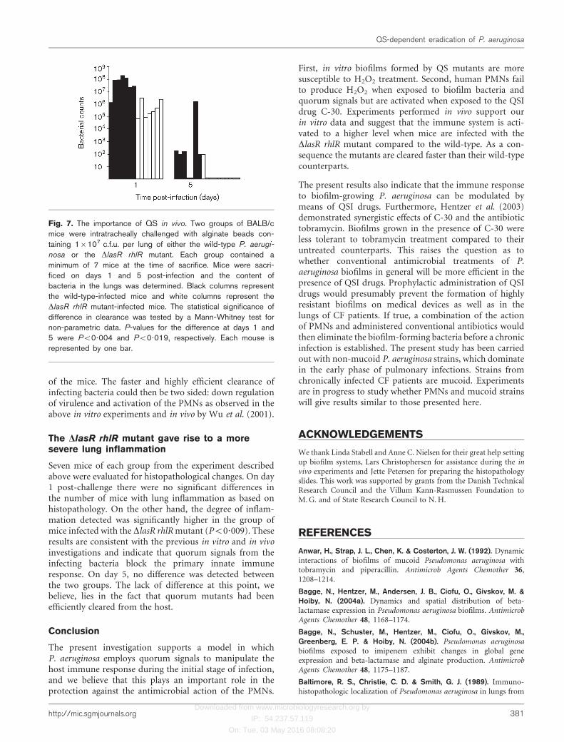
Clearance of bacteria from the lungs was assessed on day 1and day 5 post-challenge (Fig. 7). On day 1 post-challengethere was a 100-fold difference between the two groups,with the DlasR rhlR group being cleared fastest (P<0?004).On day 5, the lungs in nine out of ten mice in the group ofmice infected with DlasR rhlR were sterile, whereas for themice infected with the wild-type, five out of seven containedP. aeruginosa (P<0?02). This is in accordance with the databy Hentzer et al. (2003) using C-30 to block QS in the lungs
Fig. 6. Bacterial-cell-density-dependent activation of PMNs bywild-type and DlasR rhlR P. aeruginosa as measured by flowcytometry. The bacteria were grown in shake flasks andsamples were retrieved every 30 min starting at OD600 0?1.Blood from eight different normal persons was used (n=8).The PMNs were incubated with either wild-type P. aeruginosa
or the DlasR rhlR mutant at 37 6C for 15 min and the reactionwas stopped by placing the sample on ice. Next, the samplewas run through the flow cytometer and the fraction of PMNsemitting green florescence was counted. The fraction of acti-vated PMNs was calculated by the count of PMNs emittinggreen signal divided by the total counts of PMNs. The circlesshow the mean of each time point. The y axis shows theincreased percentage of PMNs activated by DlasR rhlR, calcu-lated by subtracting the fraction of PMNs activated by exposureto the wild-type from the fraction of PMNs activated whenexposed to the QS mutant. A statistically significant fraction ofPMNs was activated when exposed to the DlasR rhlR mutantcompared to the wild-type, tested by a paired t-test: OD600
0?11, NS; OD600 0?17, NS; OD600 0?26, NS; OD600 0?44,P¡0?03; OD600 0?48, P¡0?05; OD600 0?73, NS; OD600
0?90, NS; OD600 1?10, P¡0?04; OD600 1?30, P¡0?04;OD600 1?50, P¡0?008. NS, Not statistically significant.
(a)
(d)
(b)
(c)
Fig. 5. PMN activation measured by oxidative burst. Three-day-old biofilms of wild-type P. aeruginosa and the DlasR rhlR
mutant were exposed to PMNs for 2 h. The oxidative burst wasvisualized by the green fluorescence emitted when 123-DHRis oxidized to 123-rhodamine due to production of H2O2. (a)Wild-type; (c) DlasR rhlR mutant. (b) Furanone C-30-mediatedactivation of PMNs present on a wild-type biofilm. The biofilmwas grown for 3 days in the presence of 10 mM C-30. ThePMNs fluoresce green (indicative of oxidative burst) comparedwith the PMNs in (a). (d) Quorum-signal-mediated inactivationof PMNs present on a DlasR rhlR biofilm. 3-Oxo-C12-HSL(1 mM) and C4-HSL (2 mM) were added to the 3-day-old DlasR
rhlR mutant biofilm 3 h prior to inoculation with the PMNs.
380 Microbiology 151
T. Bjarnsholt and others
Downloaded from www.microbiologyresearch.org by
IP: 54.237.57.119
On: Tue, 03 May 2016 08:08:20
of the mice. The faster and highly efficient clearance ofinfecting bacteria could then be two sided: down regulationof virulence and activation of the PMNs as observed in theabove in vitro experiments and in vivo by Wu et al. (2001).
The DlasR rhlR mutant gave rise to a moresevere lung inflammation
Seven mice of each group from the experiment describedabove were evaluated for histopathological changes. On day1 post-challenge there were no significant differences inthe number of mice with lung inflammation as based onhistopathology. On the other hand, the degree of inflam-mation detected was significantly higher in the group ofmice infected with the DlasR rhlRmutant (P<0?009). Theseresults are consistent with the previous in vitro and in vivoinvestigations and indicate that quorum signals from theinfecting bacteria block the primary innate immuneresponse. On day 5, no difference was detected betweenthe two groups. The lack of difference at this point, webelieve, lies in the fact that quorum mutants had beenefficiently cleared from the host.
Conclusion
The present investigation supports a model in whichP. aeruginosa employs quorum signals to manipulate thehost immune response during the initial stage of infection,and we believe that this plays an important role in theprotection against the antimicrobial action of the PMNs.
First, in vitro biofilms formed by QS mutants are moresusceptible to H2O2 treatment. Second, human PMNs failto produce H2O2 when exposed to biofilm bacteria andquorum signals but are activated when exposed to the QSIdrug C-30. Experiments performed in vivo support ourin vitro data and suggest that the immune system is acti-vated to a higher level when mice are infected with theDlasR rhlR mutant compared to the wild-type. As a con-sequence the mutants are cleared faster than their wild-typecounterparts.
The present results also indicate that the immune responseto biofilm-growing P. aeruginosa can be modulated bymeans of QSI drugs. Furthermore, Hentzer et al. (2003)demonstrated synergistic effects of C-30 and the antibiotictobramycin. Biofilms grown in the presence of C-30 wereless tolerant to tobramycin treatment compared to theiruntreated counterparts. This raises the question as towhether conventional antimicrobial treatments of P.aeruginosa biofilms in general will be more efficient in thepresence of QSI drugs. Prophylactic administration of QSIdrugs would presumably prevent the formation of highlyresistant biofilms on medical devices as well as in thelungs of CF patients. If true, a combination of the actionof PMNs and administered conventional antibiotics wouldthen eliminate the biofilm-forming bacteria before a chronicinfection is established. The present study has been carriedout with non-mucoid P. aeruginosa strains, which dominatein the early phase of pulmonary infections. Strains fromchronically infected CF patients are mucoid. Experimentsare in progress to study whether PMNs and mucoid strainswill give results similar to those presented here.
ACKNOWLEDGEMENTS
We thank Linda Stabell and Anne C. Nielsen for their great help settingup biofilm systems, Lars Christophersen for assistance during the invivo experiments and Jette Petersen for preparing the histopathologyslides. This work was supported by grants from the Danish TechnicalResearch Council and the Villum Kann-Rasmussen Foundation toM.G. and of State Research Council to N.H.
REFERENCES
Anwar, H., Strap, J. L., Chen, K. & Costerton, J. W. (1992). Dynamicinteractions of biofilms of mucoid Pseudomonas aeruginosa withtobramycin and piperacillin. Antimicrob Agents Chemother 36,1208–1214.
Bagge, N., Hentzer, M., Andersen, J. B., Ciofu, O., Givskov, M. &Hoiby, N. (2004a). Dynamics and spatial distribution of beta-lactamase expression in Pseudomonas aeruginosa biofilms. AntimicrobAgents Chemother 48, 1168–1174.
Bagge, N., Schuster, M., Hentzer, M., Ciofu, O., Givskov, M.,Greenberg, E. P. & Hoiby, N. (2004b). Pseudomonas aeruginosabiofilms exposed to imipenem exhibit changes in global geneexpression and beta-lactamase and alginate production. AntimicrobAgents Chemother 48, 1175–1187.
Baltimore, R. S., Christie, C. D. & Smith, G. J. (1989). Immuno-histopathologic localization of Pseudomonas aeruginosa in lungs from
Fig. 7. The importance of QS in vivo. Two groups of BALB/cmice were intratracheally challenged with alginate beads con-taining 16107 c.f.u. per lung of either the wild-type P. aerugi-
nosa or the DlasR rhlR mutant. Each group contained aminimum of 7 mice at the time of sacrifice. Mice were sacri-ficed on days 1 and 5 post-infection and the content ofbacteria in the lungs was determined. Black columns representthe wild-type-infected mice and white columns represent theDlasR rhlR mutant-infected mice. The statistical significance ofdifference in clearance was tested by a Mann-Whitney test fornon-parametric data. P-values for the difference at days 1 and5 were P<0?004 and P<0?019, respectively. Each mouse isrepresented by one bar.
http://mic.sgmjournals.org 381
QS-dependent eradication of P. aeruginosa

Downloaded from www.microbiologyresearch.org by
IP: 54.237.57.119
On: Tue, 03 May 2016 08:08:20
patients with cystic fibrosis. Implications for the pathogenesis of
progressive lung deterioration. Am Rev Respir Dis 140, 1650–1661.
Bassoe, C. F., Li, N., Ragheb, K., Lawler, G., Sturgis, J. & Robinson,J. P. (2003). Investigations of phagosomes, mitochondria, and acidic
granules in human neutrophils using fluorescent probes. Cytometry51B, 21–29.
Bauernfeind, A., Bertele, R. M., Harms, K., Horl, G., Jungwirth, R.,Petermuller, C., Przyklenk, B. & Weisslein-Pfister, C. (1987).Qualitative and quantitative microbiological analysis of sputa of102 patients with cystic fibrosis. Infection 15, 270–277.
Beatson, S. A., Whitchurch, C. B., Semmler, A. B. & Mattick, J. S.(2002). Quorum sensing is not required for twitching motility inPseudomonas aeruginosa. J Bacteriol 184, 3598–3604.
Chhabra, S. R., Harty, C., Hooi, D. S., Daykin, M., Williams, P.,Telford, G., Pritchard, D. I. & Bycroft, B. W. (2003). Synthetic
analogues of the bacterial signal (quorum sensing) molecule N-(3-oxododecanoyl)-L-homoserine lactone as immune modulators.
J Med Chem 46, 97–104.
Christensen, B. B., Sternberg, C., Andersen, J. B., Palmer, R. J., Jr,Nielsen, A. T., Givskov, M. & Molin, S. (1999). Molecular tools for
study of biofilm physiology. Methods Enzymol 310, 20–42.
Ciofu, O. (2003). Pseudomonas aeruginosa chromosomal beta-
lactamase in patients with cystic fibrosis and chronic lung infection.Mechanism of antibiotic resistance and target of the humoral
immune response. APMIS Suppl 116, 1–47.
Davies, D. G., Parsek, M. R., Pearson, J. P., Iglewski, B. H.,Costerton, J. W. & Greenberg, E. P. (1998). The involvement of
cell-to-cell signals in the development of a bacterial biofilm. Science280, 295–298.
Davis, P. B., Drumm, M. & Konstan, M. W. (1996). Cystic fibrosis. AmJ Respir Crit Care Med 154, 1229–1256.
DiMango, E., Zar, H. J., Bryan, R. & Prince, A. (1995). Diverse
Pseudomonas aeruginosa gene products stimulate respiratory epithe-lial cells to produce interleukin-8. J Clin Invest 96, 2204–2210.
Doring, G., Buhl, V., Hoiby, N., Schiotz, P. O. & Botzenhart, K.(1984). Detection of proteases of Pseudomonas aeruginosa in immune
complexes isolated from sputum of cystic fibrosis patients. ActaPathol Microbiol Immunol Scand [C] 92, 307–12.
Elkins, J. G., Hassett, D. J., Stewart, P. S., Schweizer, H. P. &McDermott, T. R. (1999). Protective role of catalase in Pseudomonasaeruginosa biofilm resistance to hydrogen peroxide. Appl Environ
Microbiol 65, 4594–4600.
Fuqua, W. C., Winans, S. C. & Greenberg, E. P. (1994). Quorum
sensing in bacteria: the LuxR-LuxI family of cell density-responsivetranscriptional regulators. J Bacteriol 176, 269–275.
Gilbert, P., Allison, D. G. & McBain, A. J. (2002). Biofilms in vitro
and in vivo: do singular mechanisms imply cross-resistance? SympSer Soc Appl Microbiol 31, 98S–110S.
Gilligan, P. H. (1991). Microbiology of airway disease in patientswith cystic fibrosis. Clin Microbiol Rev 4, 35–51.
Giwercman, B., Meyer, C., Lambert, P. A., Reinert, C. & Hoiby, N.(1992). High-level beta-lactamase activity in sputum samplesfrom cystic fibrosis patients during antipseudomonal treatment.
Antimicrob Agents Chemother 36, 71–76.
Goldstein, W. & Doring, G. (1986). Lysosomal enzymes from
polymorphonuclear leukocytes and proteinase inhibitors in patientswith cystic fibrosis. Am Rev Respir Dis 134, 49–56.
Hassett, D. J., Ma, J. F., Elkins, J. G. & 10 other authors(1999). Quorum sensing in Pseudomonas aeruginosa controlsexpression of catalase and superoxide dismutase genes and mediates
biofilm susceptibility to hydrogen peroxide. Mol Microbiol 34,1082–1093.
Hentzer, M., Teitzel, G. M., Balzer, G. J., Heydorn, A., Molin, S.,Givskov, M. & Parsek, M. R. (2001). Alginate overproduction affectsPseudomonas aeruginosa biofilm structure and function. J Bacteriol
183, 5395–5401.
Hentzer, M., Wu, H., Andersen, J. B. & 15 other authors (2003).Attenuation of Pseudomonas aeruginosa virulence by quorum sensing
inhibitors. EMBO J 22, 3803–3815.
Heydorn, A., Nielsen, A. T., Hentzer, M., Sternberg, C., Givskov, M.,Ersboll, B. K. & Molin, S. (2000). Quantification of biofilm struc-tures by the novel computer program COMSTAT. Microbiology 146,
2395–2407.
Hoiby, N. (1974). Epidemiological investigations of the respiratorytract bacteriology in, patients with cystic fibrosis. Acta Pathol
Microbiol Scand [B] Microbiol Immunol 82, 541–550.
Jensen, P. O. (2003). Characterization and modulation of the innate
immune response during Pseudomonas aeruginosa lung infection inpatients with cystic fibrosis: an experimental and clinical study. PhD
thesis, University of Copenhagen.
Jesaitis, A. J., Franklin, M. J., Berglund, D., Sasaki, M., Lord, C. I.,Bleazard, J. B., Duffy, J. E., Beyenal, H. & Lewandowski, Z. (2003).Compromised host defense on Pseudomonas aeruginosa biofilms:characterization of neutrophil and biofilm interactions. J Immunol
171, 4329–4339.
Kharazmi, A., Hoiby, N., Doring, G. & Valerius, N. H. (1984).Pseudomonas aeruginosa exoproteases inhibit human neutrophilchemiluminescence. Infect Immun 44, 587–591.
Koch, C. & Hoiby, N. (1993). Pathogenesis of cystic fibrosis. Lancet
341, 1065–1069.
Koch, C. & Hoiby, N. (2000). Diagnosis and treatment of cystic
fibrosis. Respiration 67, 239–247.
Konstan, M. W. & Berger, M. (1997). Current understanding of theinflammatory process in cystic fibrosis: onset and etiology. Pediatr
Pulmonol 24, 137–142.
Konstan, M. W., Byard, P. J., Hoppel, C. L. & Davis, P. B. (1995).Effect of high-dose ibuprofen in patients with cystic fibrosis. N EnglJ Med 332, 848–854.
Latifi, A., Foglino, M., Tanaka, K., Williams, P. & Lazdunski, A.(1996). A hierarchical quorum-sensing cascade in Pseudomonasaeruginosa links the transcriptional activators LasR and RhIR
(VsmR) to expression of the stationary-phase sigma factor RpoS.Mol Microbiol 21, 1137–1146.
Mathee, K., Ciofu, O., Sternberg, C. & 9 other authors (1999).Mucoid conversion of Pseudomonas aeruginosa by hydrogen
peroxide: a mechanism for virulence activation in the cystic fibrosislung. Microbiology 145, 1349–1357.
McKnight, S. L., Iglewski, B. H. & Pesci, E. C. (2000). The
Pseudomonas quinolone signal regulates rhl quorum sensing inPseudomonas aeruginosa. J Bacteriol 182, 2702–2708.
Medina, G., Juarez, K., Diaz, R. & Soberon-Chavez, G. (2003).Transcriptional regulation of Pseudomonas aeruginosa rhlR, encoding
a quorum-sensing regulatory protein. Microbiology 149, 3073–3081.
Moser, C., Johansen, H. K., Song, Z., Hougen, H. P., Rygaard, J. &Hoiby, N. (1997). Chronic Pseudomonas aeruginosa lung infection is
more severe in Th2 responding BALB/c mice compared to Th1responding C3H/HeN mice. APMIS 105, 838–842.
Moser, C., Hougen, H. P., Song, Z., Rygaard, J., Kharazmi, A.& Hoiby, N. (1999). Early immune response in susceptible and
resistant mice strains with chronic Pseudomonas aeruginosa lung
infection determines the type of T-helper cell response. APMIS 107,1093–1100.
Moser, C., Kjaergaard, S., Pressler, T., Kharazmi, A., Koch, C. &Hoiby, N. (2000). The immune response to chronic Pseudomonas
382 Microbiology 151
T. Bjarnsholt and others

Downloaded from www.microbiologyresearch.org by
IP: 54.237.57.119
On: Tue, 03 May 2016 08:08:20
aeruginosa lung infection in cystic fibrosis patients is predominantlyof the Th2 type. APMIS 108, 329–335.
Nussler, A. K., Wittel, U. A., Nussler, N. C. & Beger, H. G. (1999).Leukocytes, the Janus cells in inflammatory disease. Langenbecks ArchSurg 384, 222–232.
Palma, M., DeLuca, D., Worgall, S. & Quadri, L. E. (2004).Transcriptome analysis of the response of Pseudomonas aeruginosato hydrogen peroxide. J Bacteriol 186, 248–252.
Pedersen, S. S., Shand, G. H., Hansen, B. L. & Hansen, G. N. (1990).Induction of experimental chronic Pseudomonas aeruginosa lunginfection with P. aeruginosa entrapped in alginate microspheres.APMIS 98, 203–211.
Pesci, E. C., Pearson, J. P., Seed, P. C. & Iglewski, B. H. (1997).Regulation of las and rhl quorum sensing in Pseudomonas aeruginosa.J Bacteriol 179, 3127–3132.
Pesci, E. C., Milbank, J. B., Pearson, J. P., McKnight, S., Kende,A. S., Greenberg, E. P. & Iglewski, B. H. (1999). Quinolone signalingin the cell-to-cell communication system of Pseudomonas aeruginosa.Proc Natl Acad Sci U S A 96, 11229–11234.
Rumbaugh, K. P., Griswold, J. A., Iglewski, B. H. & Hamood,A. N. (1999). Contribution of quorum sensing to the virulence ofPseudomonas aeruginosa in burn wound infections. Infect Immun 67,5854–5862.
Schuster, M., Lostroh, C. P., Ogi, T. & Greenberg, E. P. (2003).Identification, timing, and signal specificity of Pseudomonas aeruginosaquorum-controlled genes: a transcriptome analysis. J Bacteriol 185,2066–2079.
Shih, P. C. & Huang, C. T. (2002). Effects of quorum-sensing defici-ency on Pseudomonsa aeruginosa biofilm formation and antibioticresistance. J Antimicrob Chemother 49, 309–314.
Smith, R. S., Fedyk, E. R., Springer, T. A., Mukaida, N., Iglewski, B. H.
& Phipps, R. P. (2001). IL-8 production in human lung fibroblasts
and epithelial cells activated by the Pseudomonas autoinducer N-3-
oxododecanoyl homoserine lactone is transcriptionally regulated by
NF-kappa B and activator protein-2. J Immunol 167, 366–374.
Smith, R. S., Kelly, R., Iglewski, B. H. & Phipps, R. P. (2002). The
Pseudomonas autoinducer N-(3-oxododecanoyl) homoserine lactone
induces cyclooxygenase-2 and prostaglandin E2 production in
human lung fibroblasts: implications for inflammation. J Immunol
169, 2636–2642.
Stewart, P. S. & Costerton, J. W. (2001). Antibiotic resistance of
bacteria in biofilms. Lancet 358, 135–138.
Tateda, K., Ishii, Y., Horikawa, M., Matsumoto, T., Miyairi, S.,
Pechere, J. C., Standiford, T. J., Ishiguro, M. & Yamaguchi, K.
(2003). The Pseudomonas aeruginosa autoinducer N-3-oxododecanoyl
homoserine lactone accelerates apoptosis in macrophages and
neutrophils. Infect Immun 71, 5785–5793.
Telford, G., Wheeler, D., Williams, P., Tomkins, P. T., Appleby, P.,
Sewell, H., Stewart, G. S., Bycroft, B. W. & Pritchard, D. I. (1998).
The Pseudomonas aeruginosa quorum-sensing signal molecule N-(3-
oxododecanoyl)-L-homoserine lactone has immunomodulatory
activity. Infect Immun 66, 36–42.
Wagner, V. E., Bushnell, D., Passador, L., Brooks, A. I. & Iglewski,
B. H. (2003). Microarray analysis of Pseudomonas aeruginosa
quorum-sensing regulons: effects of growth phase and environment.
J Bacteriol 185, 2080–2095.
Wu, H., Song, Z., Givskov, M., Doring, G., Worlitzsch, D., Mathee, K.,
Rygaard, J. & Hoiby, N. (2001). Pseudomonas aeruginosa mutations in
lasI and rhlI quorum sensing systems result in milder chronic lung
infection. Microbiology 147, 1105–1113.
http://mic.sgmjournals.org 383
QS-dependent eradication of P. aeruginosa