ANTIGENIC ANALYSIS OF GAMONTS OF HEPATOZOON CANIS PURIFIED FROM LEUKOCYTES
Upload
independentCategory
view
2download
0
Dendritic Cells Take up and Present Antigens from Viableand Apoptotic Polymorphonuclear LeukocytesCarlos Alfaro1, Natalia Suarez1, Carmen Onate1, Jose L. Perez-Gracia2, Ivan Martinez-Forero1, Sandra
Hervas-Stubbs1, Inmaculada Rodriguez1, Guiomar Perez1, Elixabet Bolanos1, Asis Palazon1, Miguel
Fernandez de Sanmamed1,2, Aizea Morales-Kastresana1, Alvaro Gonzalez3, Ignacio Melero1,2*
1 Gene Therapy Unit, CIMA, Universidad de Navarra, Pamplona, Spain, 2 Medical Oncology Department, Clınica Universidad de Navarra, Universidad de Navarra,
Pamplona, Spain, 3 Biochemistry Department, Clınica Universidad de Navarra, Universidad de Navarra, Pamplona, Spain
Abstract
Dendritic cells (DC) are endowed with the ability to cross-present antigens from other cell types to cognate T cells. DC arepoised to meet polymorphonuclear leukocytes (PMNs) as a result of being co-attracted by interleukin-8 (IL-8), for instance asproduced by tumor cells or infected tissue. Human monocyte-derived and mouse bone marrow-derived DC can readilyinternalize viable or UV-irradiated PMNs. Such internalization was abrogated at 4uC and partly inhibited by anti-CD18 mAb.In mice, DC which had internalized PMNs containing electroporated ovalbumin (OVA) protein, were able to cross-presentthe antigen to CD8 (OT-1) and CD4 (OT-2) TCR-transgenic T cells. Moreover, in humans, tumor cell debris is internalized byPMNs and the tumor-cell material can be subsequently taken up from the immunomagnetically re-isolated PMNs by DC.Importantly, if human neutrophils had endocytosed bacteria, they were able to trigger the maturation program of the DC.Moreover, when mouse PMNs with E. coli in their interior are co-injected in the foot pad with DC, many DC loaded withfluorescent material from the PMNs reach draining lymph nodes. Using CT26 (H-2d) mouse tumor cells, it was observed thatif tumor cells are intracellularly loaded with OVA protein and UV-irradiated, they become phagocytic prey of H-2d PMNs. Ifsuch PMNs, that cannot present antigens to OT-1 T cells, are immunomagnetically re-isolated and phagocytosed by H-2b
DC, such DC productively cross-present OVA antigen determinants to OT-1 T cells. Cross-presentation to adoptivelytransferred OT-1 lymphocytes at draining lymph nodes also take place when OVA-loaded PMNs (H-2d) are coinjected in thefootpad of mice with autologous DC (H-2b). In summary, our results indicate that antigens phagocytosed by short-livedPMNs can be in turn internalized and productively cross-presented by DC.
Citation: Alfaro C, Suarez N, Onate C, Perez-Gracia JL, Martinez-Forero I, et al. (2011) Dendritic Cells Take up and Present Antigens from Viable and ApoptoticPolymorphonuclear Leukocytes. PLoS ONE 6(12): e29300. doi:10.1371/journal.pone.0029300
Editor: Antonio Bertoletti, Singapore Institute for Clinical Sciences, Singapore
Received June 27, 2011; Accepted November 23, 2011; Published December 20, 2011
Copyright: � 2011 Alfaro et al. This is an open-access article distributed under the terms of the Creative Commons Attribution License, which permitsunrestricted use, distribution, and reproduction in any medium, provided the original author and source are credited.
Funding: Financial support was from MEC/MICINN (SAF2005-03131 and SAF2008-03294), Departamento de Educacion del Gobierno de Navarra, Departamentode Salud del Gobierno de Navarra (Beca Ortiz de Landazuri), programa ‘‘Tu eliges: tu decides’’ de Caja Navarra, Redes tematicas de investigacion cooperativa RETIC(RD06/0020/0065), Fondo de investigacion sanitaria (FIS PI060932), European commission 7th framework program (ENCITE) and SUDOE-IMMUNONET, FundacionMutua Madrilena, and ‘‘UTE for project FIMA’’. CA is supported by Fundacion Cientıfica de la Asociacion Espanola Contra el Cancer (AECC). SH-S receives a contract(Ramon y Cajal) from Ministerio de Educacion y Ciencia and AP a scholarship from FIS. The funders had no role in study design, data collection and analysis,decision to publish, or preparation of the manuscript.
Competing Interests: The authors have declared that no competing interests exist.
* E-mail: [email protected]
Introduction
Neutrophils are the first defense barrier against bacteria or fungi
that have penetrated the body surfaces. Many features equip these
cells to perform such a critical function [1]. Originating in the
bone marrow, neutrophils have a short 4 h half life in circulation
and readily extravasate upon infection or inflammation in any
infected tissue [2,3]. Neutrophils sense and migrate in response to
chemotactic gradients at inflammatory sites [2]. Interleukin-8
[4,5], bacterial formyl peptides and complement factors are the
main chemotactic stimuli, while extravasation is guided by LFA-1
integrin interactions with ICAM-1 [2,6]. Once in contact with the
invading microorganisms, neutrophils degranulate microbiocide
substances, perform phagocytosis and oxidative killing of micro-
organisms [1,7], take up tissular debris, and entrap bacteria by
means of forming trapping nets with their own DNA [1,8]. Pus as
it appears in acute inflamed foci is a collection of exudates rich in
the remains of neutrophils, microorganisms and tissular debris.
Adaptive immune responses rely on antigen presentation to T-
cells as performed by professional dendritic cells (DC) [9]. At least
some of the DC subsets present in the body can take-up antigen
from third party cells and present them to lymphocytes associated
to MHC class I and II molecules [10]. In the case of MHC class I-
mediated presentation, this process is referred to as antigen cross-
presentation [11]. There is recent evidence indicating that various
human DC subsets isolated from human can mediate cross-
presentation [12]. Indeed, the CD141+ CLEC-9A+ subset seems to
be specialized in CD8 T-cell cross-priming [13,14,15].
The ability of DC to stimulate a T-cell response is not
constitutive [16]. On the contrary, DC depend on detection of
microbial molecular patterns [17], tissue damage denoting
molecules [18] and proinflamatory cytokines to experience their
maturation program. Maturation means migration towards
lymphoid organs triggered by CCR7 ligands [19], upregulation
of co-stimulatory molecules for T cells and production of T-cell
stimulating cytokines [17].
PLoS ONE | www.plosone.org 1 December 2011 | Volume 6 | Issue 12 | e29300
An interesting article indicated that DC and neutrophils may
interact in the body in sites of acute inflammation such as a
phlegmonous appendicitis [20]. Importantly the Van Kooyk group
demonstrated that a molecular interaction dependent on DC-
SIGN on the DC side and sialyl Lewis-X sugars anchored on the
CD11b integrin mediated the heterotypic adhesion of human
PMN to DC [20,21]. Other authors have shown that neutrophils
enhance the expression of co-stimulatory molecules on DC
[22,23]. There have been also reports of the ability of neutrophils
to directly present antigen to T cells [24].
From our point of view, it makes physiological and economic
sense that scanty DC would be able to interact with abundant
neutrophils that bear signs of having internalized relevant
microbiological antigens. Such stressed PMN leukocytes would
be thereby a concentrated source of microbiological antigens.
Even under sterile conditions neutrophil granule proteins have
been observed to elicit maturation of DC [23,25,26]. However, it
is clear that if DC internalized infected neutrophils this would not
only constitute a rich source of microbial antigens, but also a
concentrated source of microbial molecular patterns to elicit DC
maturation. If this hypothesis is correct, the ability of PMNs to
ferry antigenic material for DC efficient cross-presentation might
be useful in cancer immunotherapy.
We found that human and mouse DC internalize viable and
dead PMNs. Interestingly, if such PMN had been pre-exposed to
dying tumor cells, the tumor-related material ends up being taken
up by DC. We demonstrate in mice that antigens internalized by
PMNs can be cross-presented by DC. Furthermore, intracellular
antigens from dead tumor cells can be ferried by PMNs to be
readily up-taken by DC and subsequently cross-presented to CD8+
T cells. The importance of second and third hand antigen
presentation might have been overlooked, and PMN leukocytes
may play a critical role as a source of concentrated antigen and in
providing maturation stimuli to DC.
Materials and Methods
Ethics statementAnimal studies have been performed in accordance with
Spanish legislation under specific approval from the institutional
ethics board by the Ethics Committee for Animal Experimentation of the
University of Navarra (Study 03/007 approval). Human cells are
obtained from Blood donors (public blood bank of Navarra) under
written informed consent for research.
Purification of human neutrophils10 ml of peripheral blood drawn in heparin containing tubes were
mixed with 10 ml of cold PBS and 10 ml of 6% Dextran/0.9%
NaCl solution. After being inverted 18–20 times to ensure adequate
mixing, the mixture was pipetted into a 50 ml tube and the tube was
placed upright at room temperature for 1–1.5 h, or until separation
was completed. The yellowish supernatant was recovered into a
50 ml tube and spun at 1150 rpm for 12 minutes at 4uC with low
brake. The supernatant was then discarded and the pellet re-
suspended in 5 ml ACK buffer, 5 min at room temperature. After
that incubation, the cells were washed with 45 ml of cold PBS. The
supernatant was discarded and the pellet was resuspended in 2.5 ml
of PBS. The cell suspension was laid over 3 ml of Ficoll-Hypaque
(GE Healthcare Europe GMBH, Barcelona, Spain) gradients in a
15 ml tube and spun at 1500 rpm for 30 minutes at 4uC. The pellet
was resuspended in 2 ml cold PBS and stored at 4uC. To ensure the
correct purification the pellet was immunostained with CD15-PE
and a Giemsa staining of cytospins was performed. Neutrophil purity
was .95% (CD15bright neutrophils) [27].
Purification of mouse neutrophilsBALB/c mice neutrophils were obtained from bone marrow as
follows. Hind limbs were collected in ethanol and bones were
cleaned with a scalpel (peeling off as much flesh as possible). When
all bones were cleaned, their ends were cut and a 27G1/2 needle
placed in the central cavity of the long bones to flush them with
mouse complete medium [RPMI 10% FBS (Invitrogen, Paisley,
UK) + penicillin/streptomycin (Invitrogen) + b-mercaptoethanol
(Invitrogen)]. An immunomagnetic selection was made with
Ly6G AutoMACS microbeads (Miltenyi Biotec, Bergisch Glad-
bach, Germany) obtaining the positive and the negative fractions
to monitor cell population enrichment. Anti-Gr-1-biotin (Becton
Dickinson, Erembodegem, Belgium) plus streptavidin-PE (BD
Bioscience) was used for monitoring enrichment by flow
cytometry using a FACSCalibur Flow Cytometer (Becton
Dickinson). It is of note that the anti-Gr-1 mAb recognizes both
Ly-6C and Ly-6G.
Human DC generation and maturationDC were generated from filter buffy coat (FBC)-derived
monocytes donated by healthy human donors [28]. The protocol
for obtaining FBC was approved by the Ethics Committee of our
institution and donors gave informed consent. Isolated mononu-
clear cells were subjected to positive selection using anti-CD14-
conjugated paramagnetic beads and purified using the Auto-
MACS system according to manufacturers instructions (Miltenyi
Biotec). Purified monocytes were cultured for 7 days in complete
medium (RPMI 10% FBS + penicillin/streptomycin + b-
mercaptoethanol) supplemented with GM-CSF (1000 U/ml;
Leukine, Schering-Plough, Kenilworth, NJ, USA) and IL-4
(500 U/ml; R&D Systems, Minneapolis, MN, USA). Cytokines
were added every other day. DC were matured with clinical grade
TNFa (50 ng/ml; Boehringer Ingelheim, Ingelheim, Germany),
IFNa (1,000 IU/ml; Schering-Plough) and Poly I:C (20 mg/ml;
Ampligen, Bioclones, South Africa) during 48 h [29].
Murine DC generationMice DC precursors were obtained from bone marrow. Cells
were put in culture [RPMI 10% FBS (Invitrogen) + penicillin/
streptomycin (Invitrogen) + b-mercaptoethanol (Invitrogen)] at a
density of 26106 cells/ml in 10 ml per petri dish in the presence of
20 ng/ml of rmGM-CSF (R&D) [30]. After 72 h, rmGM-CSF at
10 ng/ml was added in 10 ml in RPMI complete medium per
dish. On 6th day of culture, 10 ml of medium were withdrawn per
dish. On 9th culture day, non adherent cells were collected by
thoroughly pipetting with RPMI medium. Adherent cells were
collected by adding 2 ml of accutase (PAA laboratories GmbH,
Pasching, Austria) per dish and incubated 15 minutes at 37uC.
Interaction between DC and neutrophils in vitro106 DC were stained with PKH26 and 56106 neutrophils with
PKH2 membrane marker (Sigma-Aldrich, St. Louis, MO, USA)
as reported [27]. Half of the neutrophils were killed with UV light
at 260 nm (1,232 J/cm2). After that, live or dead neutrophils were
co-cultured with DC for 4 and 24 hours at 4uC or 37uC. The
neutralizing antibodies anti-CD11b, anti-CD18 (kindly provided
by Francisco Sanchez Madrid, Hospital de la Princesa, Madrid)
and anti-DC-SIGN (kindly provided by Angel Corbı, CIB,
Madrid) were added to the culture medium at 10 mg/ml or at a
1/5 dilution of the concentrated hybridoma supernatant. After 2,
4 and 24 hours, co-culture cells were collected to analyse double
positive cells using a FACSCalibur Flow Cytometer or examined
by confocal microscopy.
DC Interplay with PMNs
PLoS ONE | www.plosone.org 2 December 2011 | Volume 6 | Issue 12 | e29300

In vitro chemotaxis assayIn vitro neutrophil and DC migration was measured in
Transwell Chambers (5 mm; Corning Costar, Corning, NY,
USA). Both PKH26-DCs (105) and PKH2-labeled neutrophils
(105) were added to the upper chamber and migration stimuli
(subconfluent monolayer of HT29 cells) were placed in the lower
chamber. DC and neutrophils were added to the upper chamber
in complete medium, complete medium plus 20 mg/ml antiIL-8
mAb (BD Pharmigen), or complete medium plus mIgG2b as
control. Transmigrated cells in the lower chamber were
quantified using fluorescence microscopy imaging of the lower
chamber. The number of PKH26-fluorescent DC or PKH2-
labeled neutrophils per microscopic field (x20) in the lower
chamber was quantitated in triplicate wells by a blinded
observer.
Flow cytometry analysesFITC and PE-labeled mAb specific for the DC maturation
markers CD80, CD83, CD86 and HLA-DR (BD Bioscience) and
isotype-matched labeled controls were used to characterize cell
surface phenotypes by flow cytometry. PE-labeled mAb specific
for CD15 and Gr-1-biotin + SAV-PE (BD Bioscience) were used
to analyse purity in human and mice leukocyte samples.
Splenocytes were obtained from OT-1 and OT-2 TCR
transgenic mice (Jackson Laboratories, Bar Harbor, ME, USA)
[31]. Lymphocyte division was analysed by serial dilution of
carboxifluorescein succinimidyl ester (CFSE) in CD4+ and CD8+
cells (BD Bioscience). Cells (105) were washed in cold PBS and
incubated 15 min at 4uC with specific APC, FITC or PE-labeled
Abs to electronically gate CD4 and CD8 cells upon FACS
analyses.
Immunofluorescence using confocal microscopyConfocal fluorescent images were obtained using a LSM 510
Zeiss confocal scan head mounted on a Zeiss Axiovert 200 M on
an inverted-based microscope using a 40x or 63x oil immersion
objective. Sequential excitation at 488 nm and 543 nm was
provided by argon and helium-neon gas lasers, respectively.
Emission filters BP500-550 and LP560 were used for collecting
green (PKH2) and red (PKH26) in channels one and two,
respectively. After sequential excitation, green and red fluorescent
images of the same cell were saved with Laser Sharp software.
Images were analyzed by Zeiss software. The term co-localization
refers to the coincidence of green and red fluorescence, as
measured by the confocal microscope.
Protein quantificationAfter culturing the neutrophils in complete medium supple-
mented with 1 mg/ml of OVA protein (Sigma-Aldrich) for 2 h,
the supernatants of serial centrifugations were collected to verify
the complete elimination of extracellular OVA. Protein concen-
tration was measured in a Nanodrop Spectrometer ND-1000 at
280 nm. Stock solution was used as control.
Chemokine productionSupernatants were collected in the indicated culture time points,
and the cytokine concentration was determined by immunoassay.
Commercially available ELISA kits were used for the detection of
human IL-12p70 and mouse IFNc (BD Biosciences).
Tumor cell linesHT29 and CT26 tumor cell lines were obtained from American
Type Culture Collection (Rockville, MD, USA).
Elicitation of DC maturation by E Coli associated toneutrophils
26105 human neutrophils were incubated with heat-inactivated
E. coli bacteria in various numbers ranging from 105–102 for
60 min. The neutrophils were then washed eight times and the
pellets and the supernatants collected. Washed PMN and the
supernatants were added to human immature DC (iDC) cultures
and 48 h later IL-12 concentrations in the supernatants were
monitored. Subsequently, such DC (105) were washed in cold PBS
and incubated 15 min at 4uC with specific FITC or PE-labeled
mAbs to measure the mean fluorescence intensity for CD80,
CD83 and CD86 surface immunostaining as assessed by flow
cytometry. Mature DC (mDC) in the presence of poly I:C, TNFaand IFNa were used as a positive control.
OVA-specific T cell response analysisSplenocytes from OT-1 and OT-2 transgenic mice were stained
with CFSE solution 2.5 mM. After 24 h co-culture DC plus live or
UV-irradiated neutrophils (cultured during 2 h with or without
OVA at 1 mg/ml), OT-1 or OT-2 splenocytes were added to the co-
culture. The capacity of DC to cross-present OVA from neutrophils
was measured as the dilution of membrane CFSE from CD4+ (OT-2
mice) or CD8+ (OT-1mice) splenocytes that were gated by
immunostaining with CD8 or CD4 specific mAb in a FACSCalibur.
OVA protein electroporation in the tumor cell line CT26For electroporation, CT26 (3.56106 cells/500 ml RPMI without
serum and antibiotics) were incubated with 500 ml OVA at 2 mg/
ml for 10 minutes at room temperature. Electroporation was
carried out using Gene PulserH II Electroporation System
(BioRad, Hercules, CA, USA) using the conditions of two pulses
at 200 V/cm and 300 mF of capacitance. Immediately after
electroporation, CT26 cells were cultured in RPMI complete
medium during at least 1 h. To ensure the internalisation of OVA
in CT26 cells, a control OVA fluorescein conjugate (Invitrogen)
was electroporated and its internalisation was assessed by flow
cytometry after extensive washing of non internalized protein.
Co-injection of DC and PMN into the mouse foot padMouse PMNs were co-cultured for 2 h with 105 heat-inactivat-
ed E. Coli bacteria. After 8 extensive washes, 56106 PMN-PKH26
and 107 mouse DC-PKH2 were injected into the foot pads of mice
in separate syringes with 30 ml PBS for each injection. 24 h later,
popliteal and inguinal lymph nodes were surgically excised and
were disaggregated to give single cell suspensions. Cells were
immunomagnetically selected to enrich CD11c+ cells. Immunose-
lected cells were processed for subsequent analysis using flow
cytometry and confocal microscopy.
StatisticsAll data are presented as the mean 6 SD. For comparisons,
unpaired Student’s t-tests or Mann–Whitney U tests were used.
Calculations were made using Prism software (Graph Pad
Software, La Jolla, CA, USA).
Results
DC and PMN are co-attracted by IL-8 derived from tumorcells
We have previously shown that human carcinoma cells produce
functional IL-8 [32]. One of the corollaries is that such IL-8 might
attract human PMNs and monocyte-derived DC [27]. Indeed, in
classical transwell chemotaxis assays towards IL-8-producing
DC Interplay with PMNs
PLoS ONE | www.plosone.org 3 December 2011 | Volume 6 | Issue 12 | e29300

monolayers of HT-29 human colon carcinoma cells, both PKH26-
labelled DC and PKH2-labelled PMNs are actively co-attracted to
the lower chamber containing a confluent monolayer of HT29 cells
(figure 1A and B). The effect was mediated by IL-8 as demonstrated
by the abrogation of the migration by adding a neutralizing anti-IL8
mAb to the lower chamber (Figure 1A and B).
DC take up live and UV-irradiated PMN leukocytes thatmight carry microbial maturation signals
Human DC and PMN were florescence-labelled and as can be
seen in figure S1, fluorescence was readily observed by flow
cytometry and confocal microscopy. In figure 2 confocal microscopy
pictures of short 4h co-cultures of PMN and DC show that
fluorescent material from human PMNs was internalized by human
DC (Figure 2). Analyses of multiple experiments indicated in
confocal microscopy planes that DC-red fluorescence was surround-
ing PMN-green fluorescence but the contrary was not observed.
PMN internalization by DC occurred both when the PMN had
been irradiated with UV-light or when live untouched PMNs were
used. UV irradiation was used to induce cell death in PMNs
(Figure S2), as they sequentially acquire staining by annexin-V and
7-AAD when undergoing apoptosis. Evidence for internalization
Figure 1. DC and PMNs are co-attracted by IL-8 secreted by HT29 colon cancer cells. (A) Chemotactic migration of DC (labeled with PKH26)and PMN (labeled with PKH2) from the upper chamber towards confluent monolayers of HT29 cells in the lower chamber. When indicated, controlIgG mAb or anti IL-8 mAb were added to the lower chamber. Microscopy photographs represent visible light and UV illumination fields with filters forred and green fluorescence showing the lower chamber when taken 24 h later. (B) Quantitative data based on experiments as in A, representingthree independent triplicate experiments counting four microscopic fields per well (mean6SD).doi:10.1371/journal.pone.0029300.g001
DC Interplay with PMNs
PLoS ONE | www.plosone.org 4 December 2011 | Volume 6 | Issue 12 | e29300
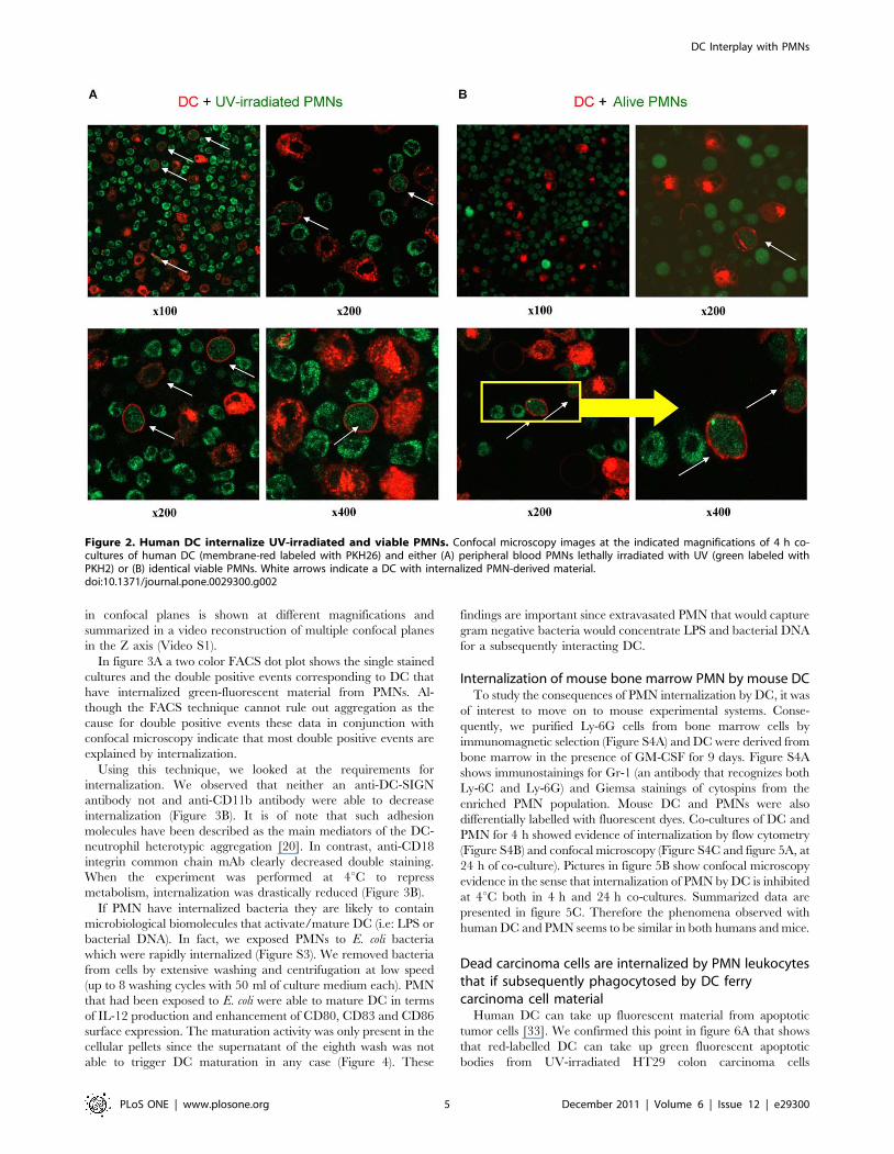
in confocal planes is shown at different magnifications and
summarized in a video reconstruction of multiple confocal planes
in the Z axis (Video S1).
In figure 3A a two color FACS dot plot shows the single stained
cultures and the double positive events corresponding to DC that
have internalized green-fluorescent material from PMNs. Al-
though the FACS technique cannot rule out aggregation as the
cause for double positive events these data in conjunction with
confocal microscopy indicate that most double positive events are
explained by internalization.
Using this technique, we looked at the requirements for
internalization. We observed that neither an anti-DC-SIGN
antibody not and anti-CD11b antibody were able to decrease
internalization (Figure 3B). It is of note that such adhesion
molecules have been described as the main mediators of the DC-
neutrophil heterotypic aggregation [20]. In contrast, anti-CD18
integrin common chain mAb clearly decreased double staining.
When the experiment was performed at 4uC to repress
metabolism, internalization was drastically reduced (Figure 3B).
If PMN have internalized bacteria they are likely to contain
microbiological biomolecules that activate/mature DC (i.e: LPS or
bacterial DNA). In fact, we exposed PMNs to E. coli bacteria
which were rapidly internalized (Figure S3). We removed bacteria
from cells by extensive washing and centrifugation at low speed
(up to 8 washing cycles with 50 ml of culture medium each). PMN
that had been exposed to E. coli were able to mature DC in terms
of IL-12 production and enhancement of CD80, CD83 and CD86
surface expression. The maturation activity was only present in the
cellular pellets since the supernatant of the eighth wash was not
able to trigger DC maturation in any case (Figure 4). These
findings are important since extravasated PMN that would capture
gram negative bacteria would concentrate LPS and bacterial DNA
for a subsequently interacting DC.
Internalization of mouse bone marrow PMN by mouse DCTo study the consequences of PMN internalization by DC, it was
of interest to move on to mouse experimental systems. Conse-
quently, we purified Ly-6G cells from bone marrow cells by
immunomagnetic selection (Figure S4A) and DC were derived from
bone marrow in the presence of GM-CSF for 9 days. Figure S4A
shows immunostainings for Gr-1 (an antibody that recognizes both
Ly-6C and Ly-6G) and Giemsa stainings of cytospins from the
enriched PMN population. Mouse DC and PMNs were also
differentially labelled with fluorescent dyes. Co-cultures of DC and
PMN for 4 h showed evidence of internalization by flow cytometry
(Figure S4B) and confocal microscopy (Figure S4C and figure 5A, at
24 h of co-culture). Pictures in figure 5B show confocal microscopy
evidence in the sense that internalization of PMN by DC is inhibited
at 4uC both in 4 h and 24 h co-cultures. Summarized data are
presented in figure 5C. Therefore the phenomena observed with
human DC and PMN seems to be similar in both humans and mice.
Dead carcinoma cells are internalized by PMN leukocytesthat if subsequently phagocytosed by DC ferrycarcinoma cell material
Human DC can take up fluorescent material from apoptotic
tumor cells [33]. We confirmed this point in figure 6A that shows
that red-labelled DC can take up green fluorescent apoptotic
bodies from UV-irradiated HT29 colon carcinoma cells
Figure 2. Human DC internalize UV-irradiated and viable PMNs. Confocal microscopy images at the indicated magnifications of 4 h co-cultures of human DC (membrane-red labeled with PKH26) and either (A) peripheral blood PMNs lethally irradiated with UV (green labeled withPKH2) or (B) identical viable PMNs. White arrows indicate a DC with internalized PMN-derived material.doi:10.1371/journal.pone.0029300.g002
DC Interplay with PMNs
PLoS ONE | www.plosone.org 5 December 2011 | Volume 6 | Issue 12 | e29300

(Figure 6A). Apoptotic tumor cells can be prey of PMN phagocytes
as well. We reasoned that if PMN leukocytes take up carcinoma
cell debris, this material will end up inside DC, in the case that
such DC eventually internalized a PMN with HT29 material
inside. To test this idea we labelled HT29 colon carcinoma cells
with PKH2 and following UV-irradiation fed them to PMN
leukocytes. After 2 h of co-culture, CD15+ cells were immuno-
magnetically selected to re-isolate the PMNs. Subsequently CD15+
PMNs were co-cultured with PKH26-labelled DC for 24 hours.
The experimental approach is summarized in figure 6B. As can be
seen in figure 6C, green fluorescent material from HT29 cells is
found inside red-labelled DC. Furthermore, quantitative data from
FACS analysis indicated that the internalization phenomenon is
quite efficient with more than half of the DC taking up green
fluorescent material derived from the carcinoma cells (Figure 6).
Such internalizing activity was abolished if the co-incubation of
PMN and DC took place at 4uC.
Accordingly it could be envisaged that PMN exposed to debris
from tumor cells could transfer this debris to third party antigen-
presenting DC. However, to explore the role in antigen cross-
presentation we needed to use mouse models.
Antigen carried by PMN can be cross-presented by DC invitro
In order to demonstrate that DC could cross-present antigen
material from internalized PMNs, we performed experiments with
bone-marrow neutrophils derived from the bone marrow of
BALB/c mice (H-2d) from which PMN were isolated as shown in
Figure 3. Human DC internalize PMNs in a temperature and CD18 dependent fashion. (A) FACS dot plots showing PKH26 staining ofHuman DC and PKH2 staining of PMNs and 4 h co-cultures of DC and PMN showing double positive events in a representative experiment of at leastfive similarly performed. (B) Co-culture experiments as in A, depicting the percentage of double positive events at 4 h of co-culture. When indicatedblocking mAb to DC-SIGN, CD11b and CD18 were added or the co-cultures were performed at 4uC.doi:10.1371/journal.pone.0029300.g003
DC Interplay with PMNs
PLoS ONE | www.plosone.org 6 December 2011 | Volume 6 | Issue 12 | e29300

Figure 4. PMNs carrying E. coli bacteria trigger the maturation of DC. PMN from human peripheral blood were exposed for 1 h to E. colibacteria for 60 min in various numbers ranging from 105–102 and then were washed eight times and the pellet and the supernatant collected.Washed PMN and the indicated washing supernatants were added to human immature DC (iDC) cultures and 48 h later IL-12 concentration in the DCculture supernatants was monitored as well as the mean fluorescence intensity for CD80, CD83 and CD86 surface immunostainings on the DC.Mature DC (mDC) in the presence of poly I:C, TNFa and IFNa were used as a positive control.doi:10.1371/journal.pone.0029300.g004
DC Interplay with PMNs
PLoS ONE | www.plosone.org 7 December 2011 | Volume 6 | Issue 12 | e29300

Figure 5. Mouse DC take up mouse PMN leukocytes. (A) Confocal microscopy images of 24 h co-cultures of viable or UV-irradiated PMNslabeled with PKH2 and bone-marrow derived DC labeled with PKH26. Arrows show co-localization of both dyes. (B) Similar experiments as in A butwith some co-cultures performed at 4uC as indicated. (C) Summary of data from five microscopic fields per condition in which internalization wasidentified by yellow staining.doi:10.1371/journal.pone.0029300.g005
DC Interplay with PMNs
PLoS ONE | www.plosone.org 8 December 2011 | Volume 6 | Issue 12 | e29300

figure S4A. Such PMN were given OVA protein for two hours in
culture followed by extensive washes (x 8 times). These PMNs
were then co-cultured overnight with DC from C57BL/6 mice (H-
2b) and exposed to OT-1 or OT-2 TCR-transgenic lymphocytes.
Proliferation of OT-1 or OT-2 T cells was monitored by CFSE
dilution 72 h later. An schematic representation of the approach
based on the fact that H-2d class I and II antigen presenting
molecules cannot present to OT-1 or OT-2 T lymphocytes is
presented in figure 7A. In figure 7B (overlay histograms) and figure
S5 (individual histograms separated for clarity), a representative
case shows the dilution of CFSE that was triggered by the cognate
peptides pulsed on DC as a positive control and by DC that had
been incubated with PMNs preloaded with OVA. T cell division
did not take place in the rest of control conditions including PMN
that had not been exposed to OVA fed to DC (Figure 7B and
figure S5). Confirmatory independent experiments offered similar
results (Figure S6A), that were consistent with analyses of IFNcconcentrations released to the tissue culture supernatants (Figure
S6B). As a whole these data indicate that OVA antigen
determinants are cross-presented by the DC which take up
antigen from PMN at least in the mouse system.
Carcinoma cells loaded with OVA transfer antigen toPMNs, then PMNs to DC and such DC cross-presentantigen to T lymphocytes
In order to study antigen transfer to DC via a PMN
intermediate, experiments were set up as summarized in
figure 8A. CT26 colon carcinoma cells of BALB/c origin (H-2d)
were electroporated in the presence of soluble OVA protein.
Figure 6. Fluorescent material from human carcinoma cell debris can be taken up by PMN and transferred to DC. (A) PKH2-labeledHT29 cells were UV-irradiated and co-cultured for 24 h with human PKH26-labelled DC and confocal images were taken indicating internalization byDC. (B) Summary of the experimental approach of co-cultures consisting of HT29 carcinoma cells previously labeled with PKH2 and UV-irradiated withPMNs which were subsequently retrieved 2 h later by CD15 immunomagnetic selection. Then PMN were co-cultured for 24 h with PKH26-labeled DC.(C) Confocal microscopy images of the indicated co-culture conditions. (D) FACS dot plots showing each cell type labeled with the correspondingfluorescent dye and co-cultures performed at 37uC and 4uC.doi:10.1371/journal.pone.0029300.g006
DC Interplay with PMNs
PLoS ONE | www.plosone.org 9 December 2011 | Volume 6 | Issue 12 | e29300

Electroporation in the presence of a soluble protein leads to
internalization of protein as checked with OVA-FITC. Figure S7
shows internalization of FITC fluorescence by CT26 contingent
on the electrical shock. Once CT26 had been electroporated with
or without OVA, they were extensively washed, UV-irradiated
and fed to PMN of BALB/c origin. Such PMN were recovered
2 h later from the cultures by Ly-6G magnetic immunoselection
(taking advantage of the magnetic beads still bound to the cells).
Immunoselected PMNs were then co-cultured overnight with DC
of C57Bl/6 (H-2b) origin. Subsequently these DC were used to
stimulate CFSE-loaded OT-1 T lymphocytes. As can be seen in a
representative experiment shown in figure 8B, dilution of OT-1
CFSE only took place when the CT26 tumor cells had been
electroporated with OVA, but not in the remaining control co-
cultures. As a positive control DC pulsed with the cognate peptide
were used and elicited strong proliferation of OT-1 lymphocytes.
This correlated with IFNc release to the co-culture supernatant
(data not shown).
These data indicate that it is possible that tumor derived
antigens can end up productively presented by DC to T cells after
being transiently ferried by PMNs that are internalized by the DC.
DC co-injected in the foot pad with PMNs that haveinternalized bacteria reach draining lymph nodes withPMN-derived material
To present antigen to T cells DC need to reach lymph nodes
through afferent lymphatic vessels [34]. This is a function tightly
controlled upon maturation of the DC by the induction of CCR7
expression [17,19]. If DC and PMN are injected in the foot pad
they should be able to meet thus mimicking inflamed tissue. In the
experimental approach shown in figure 9A, differentially fluores-
cence labelled bone marrow derived DC and PMN preloaded with
E. coli bacteria were injected with separate syringes into the foot
pad. 24 h later draining lymph nodes were surgically excised and
CD11c cells enriched by immunomagnetic selection. Double
immunofluorescence events analyzed by flow cytometry and
summarized in figure 9B indicate that about 2% of the injected
DC were recovered from the draining LN carrying fluorescent
material from the PMNs while about 5% of the injected DC were
recovered from the lymph nodes with no evidence of carrying
PMN-derived fluorescent material. These data indicate that is
possible for DC which internalize PMN material to reach T cell
compartments. It is of note that if PMN had not been exposed to
bacteria very few CD11c+ fluorescent events were recovered from
lymph nodes and virtually no double positive cells were found
(data not shown). Confocal microscopy images of cytospin
preparations made with CD11c+ magnetically-selected cells from
draining lymph nodes indicated co-localization of fluorescent dyes
in the same cells.
Next we examined if cross-presentation of antigens taken by DC
from PMNs could take place in vivo. For this purpose H-2b DC
and H-2d neutrophils were injected in different syringes in the
footpad of C57BL/6 mice. PMNs had phagocytosed OVA and
thoroughly washed for eight times as described in figure 10A. Mice
had received 24h prior to the experiment CFSE-labelled OT-1
cells to monitor their proliferation. As shown in figures 10B and C,
OT-1 lymphocytes only proliferated when the mice had been
Figure 7. Ovalbumin taken up by mouse PMNs can be transferred to DC that in turn cross-present antigen. (A) Schematicrepresentation of the experimental conditions in which OVA was added to bone-marrow-derived PMNs from BALB/c mice which were extensivelywashed 2 h later and subsequently subjected or not to UV-irradiation. Then PMNs were co-cultured overnight with DC derived from the bonemarrow of C57BL/6 mice. The next morning CFSE-loaded OT-1 or OT-2 TCR-transgenic T cells were added to the cultures. (B) FACS histogramsshowing CFSE dilution at 72 h of culture in the indicated conditions. DC pulsed with the cognate OVA peptides recognized by OT-1 and OT-2 Tlymphocytes were used as positive controls. The experiment shown is representative of at least three similarly performed.doi:10.1371/journal.pone.0029300.g007
DC Interplay with PMNs
PLoS ONE | www.plosone.org 10 December 2011 | Volume 6 | Issue 12 | e29300

injected with PMNs which had phagocytosed OVA. Since PMNs
cannot directly present antigen to OT-1 T lymphocytes it is
concluded that it is the independently injected DC present
antigen. It is of note that when injecting OVA-loaded H-2d PMN
without DC a certain level of OT-1 proliferation was observed
(25–35%) that can be attributed to endogenous antigen presenting
cells (data not shown).
Discussion
In the defence of the organism against infection it makes sense
that the early innate response would cue the adaptive immune
responses. In the bridge between innate and adaptive immune
responses dendritic cells play a pivotal role [35]. We reasoned that
pyocytes (neutrophils forming pus) would be an excellent source of
concentrated microbial antigens and microbial molecular pattern
signals, if eventually internalized by DC.
Therefore we first found that both human and mouse DC avidly
internalize PMNs in co-culture. Both apoptotic and viable PMN
are internalized although with more efficiency in the case of
apoptotic PMN. It is postulated that under steady state conditions
this phenomenon should feed DC with innocuous self antigens that
if presented by the immature DC should lead to tolerance [36].
The nature of the surface molecules playing a role in the
internalization is unclear. We found partial inhibition by blocking
the CD18 integrin chain but not upon blockade of DC-SIGN and
CD11b in many attempts. This is in disagreement with a paper from
the group of Van Kooyk whose findings indicated a role for DC-SIGN
on DC and sialyl Lewis-X decorating CD11b in the heterotypic
cellular interaction [20,21]. We observe blockade by a monoclonal
antibody binding the common leukocyte integrin b chain, while
CD11b seems not to be involved. Therefore a careful re-examination
of the surface molecules dynamically involved in the DC-neutrophil
interactions is needed and is the subject of current experiments.
Figure 8. Internal antigens can be ferried from tumor cells to DC via PMNs and then be subsequently cross-presented to Tlymphocytes. (A) Schematic representation of the experiments in which CT26 tumor cells were electroporated in the presence or not of solubleOVA protein, extensively washed 30 min later and then UV-irradiated. Such apoptotic tumor cells were co-cultured with Ly-6G-immunoselected PMNfrom the bone marrow of BALB/c mice (H-2d) during 2 h. These PMN that kept labeled with the immunomagnetic beads were re-isolated in columnsunder a magnetic field and subsequently exposed to DC from C57BL/6 mice, whose H-2Kb antigen presenting molecules can present antigen to OT-1T cells. Following overnight DC culture with the PMNs, CFSE OT-1 lymphocytes were added to the cultures and CFSE dilution in CD8 cells wasmonitored 72 h later. (B) Representative histograms of CFSE dilution in the indicated conditions including DC pulsed with the cognate peptide of OT-1 T cells as a positive control. (C) Summary (mean6SD) of three experiments similarly performed. Asterisks indicate statistical significance p,0.001 instudent’s t test.doi:10.1371/journal.pone.0029300.g008
DC Interplay with PMNs
PLoS ONE | www.plosone.org 11 December 2011 | Volume 6 | Issue 12 | e29300

Since internalization rather than aggregation is our read out,
reasons for this difference could come from various sources.
Internalization requires active metabolism since it was nearly
abolished at low temperatures.
Under acute inflammation, extravasated PMN would be full of
phagocytosed microbes and microbial material. An important
aspect is that such pyocytes, if entering into contact with and
become internalized by DC, would transmit to DC both the
presence of the biomolecules denoting a microbial pathogen and
microbial antigens. In our proof of concept experiments, E. coli
bacteria actively loaded into PMN was able to intensely mature
the subsequently encountered DC. This makes sense because
ingestion of PMN by DC or their tissular precursors will launch
the proper maturation program in DC. In mice these events also
elicit migration of DC to draining lymph nodes. It remains to be
seen which molecular patterns of bacteria are detected, but most
likely these involve LPS detection by TLR4 and bacterial DNA
detection by TLR9 in our experiments.
DC have been in the limelight of tumor immunology and
immunotherapy [37]. In solid tumors multiple myeloid cells nest in
the stroma including cells resembling PMN. Such PMN have the
opportunity to phagocytose apoptotic or necrotic tumor cells. We
thought that it was interesting to determine if PMN could take up
antigen from carcinoma cells and then transfer it to DC, in such a
way that the relevant antigens would end up cross-presented to T
cells. To start exploring these possibilities we moved to mouse
systems in which we can probe for antigen presentation with
TCR-transgenic T cells. Our data in humans strongly argued in
favour of this possibility because HT29 fluorescent carcinoma cell
debris taken up by PMNs is internalized by DC, if those PMN are
subsequently phagocytosed. However, it could be argued that the
PMN-associated fluorescent material could be degraded and is
thereby become not suitable for antigen presentation. However, in
the mouse system we found that PMN which had internalized
OVA protein from soluble sources or OVA-electroporated tumor
cells efficiently cross-presented antigens to either OT-1 or OT-2
lymphocytes. Direct presentation by PMN or tumor cells was ruled
out because of incompatible antigen presenting molecules
although in other circumstances this can take place according to
published reports [24,38]. There is a tendency in our hands to
observe more proliferation induced in this setting on OT-1 than
on OT-2 cells and further studies should clarify the relative
efficiency of antigen presentation via class I and class II when
antigen is acquired from PMNs.
The study of the intercellular crosstalk between DC and PMN
leukocytes is in its infancy. A recent report indicates that PMN
could deactivate DC upon arrival to lymph nodes in aseptic
conditions [39]. This makes sense for immature DC but it remains
to be seen if it is the case in vivo under septic conditions, that ought
to be more relevant for the defence of the body against infection.
Figure 9. Co-injection of PMNs with endocytosed bacteria and DC in subcutaneous tissue gives rise to the detection of DC carryingPMN-derived material in the draining lymph node. (A) Schematic representation of experiments in which bone marrow-derived DC and PMNsthat had been pre-exposed to E. coli and washed were injected in separate syringes into the foot pads of mice. Both leukocyte types weredifferentially labelled with fluorescence dyes (PKH26 for PMNs and PKH2 for DC). (B) Calculations of the percentages of injected DC recovered asCD11c+ immunomagnetically selected cells from popliteal lymph nodes measuring double or single positive fluorescent events as indicated by flowcytometry. (C) Representative confocal images of cytospins made with the CD11c-immunoselected cells from popliteal lymph nodes. Experiments arerepresentative from at least two similarly performed with three mice per group each. Asterisks indicate statistical significance p,0.01 in student’s ttest.doi:10.1371/journal.pone.0029300.g009
DC Interplay with PMNs
PLoS ONE | www.plosone.org 12 December 2011 | Volume 6 | Issue 12 | e29300

Our data showing the ability of DC loaded with material derived
from PMN with internalized bacteria to reach the lymph nodes
argue in favour of DC-maturing role of PMNs under acute
infection.
Our in vivo results in mice indicate that DC can cross-present
antigen transferred from a PMNs to resting CD8+ T cells. In our
experimental setting it is likely that DC in the footpad take-up
antigen and migrate to lymph nodes where they present antigen to
T cells. Interestingly there is indication that endogenous DC can
also mediate this effect (Alfaro C. et al., unpublished results).
Our current research is focused on unravelling the implications
of these PMN-DC interactions by carrying out in vivo experiments
in mouse models. In particular our data should be interpreted with
caution since we demonstrate these phenomena with DC
differentiated in culture, but not yet with the endogenous DC.
In humans these events are much more difficult to dissect in vivo.
However, there is published evidence of DC-PMN interactions in
appendicitis surgical samples [20,22] and we have found that DC
and PMN can be co-attracted to solid malignancies by means of
the IL-8 chemokine.
All in all, our experiments point to an entangled and complex
relationship between PMN and DC. PMN greatly outnumber DC
and therefore could act as concentrators of antigen and microbial
molecules for DC. If PMN leukocytes become accessible to DC
they can be internalized and then modulate DC functions while
transferring the antigens that PMN may carry. If these functions
are mediated by endogenous DC as we observe with DC
differentiated in culture, these can be important phenomena.
However, exactly how relevant are these functions for the overall
physiology of the immune system still remains to be seen.
Figure 10. Cross-presentation of antigens taken by DC from PMNs take place in vivo. (A) Schematic representation of experiments inwhich splenocytes from OT1 CD45.1 TCR-transgenic mice were subjected to CD8+ T cell isolation by immunomagnetic negative selection. OT-1 Cellswere labeled with CFSE and 26106 were injected intravenously into C57BL/6 mice. 24 h later mice received injections of PMNs and DC (H-2b) in thefootpad. Immunomagnetically isolated PMN were from BALB/c mice (H-2d) and were pre-incubated or not with OVA for two hours and extensivelywashed. PMNs and DC were injected with different syringes in the footpads of the mice which had been transferred with CFSE-labelled OT-1 cells24 hours before. 72 h later proliferation of OT1 CD45.1+ cells was assessed by flow cytometry in draining lymph node and spleen (B) Representativehistograms of CFSE dilution seen in lymph nodes and spleen. (C) Summary data of CFSE expression from lymph nodes and spleens in 3 experimentsperformed as described in A. When indicated PMN were pre-incubated or not with OVA. Asterisks indicate statistical significance p,0.001 in Mann-Whitney U test.doi:10.1371/journal.pone.0029300.g010
DC Interplay with PMNs
PLoS ONE | www.plosone.org 13 December 2011 | Volume 6 | Issue 12 | e29300

Supporting Information
Figure S1 Labeling of human PMN and DC with the indicated
fluorescent dyes visualized by FACS and by confocal microscopy.
(TIF)
Figure S2 Labeling of human PMNs undergoing or not UV-
irradiation with Annexin V and 7-AAD visualized by FACS. The
percentages of each quadrant indicate the differences between
alive PMNs and UV-irradiated PMNs.
(TIF)
Figure S3 Microscopic GRAM-stained images (x 400 magnifi-
cation) of E. coli bacteria and PMNs exposed to E. coli bacteria at
time 0 and 2 hours later. Red circles indicate small group of E. coli
bacteria.
(TIF)
Figure S4 Purification of mouse neutrophils and co-cultures
with mouse DC. (A) Immunomagnetic selection of Ly-6G cells
from single cell suspensions from mouse femur and tibia bone-
marrow before and after immunomagnetic selection (AutomacsH).
Purity was assessed by cells showing bright Gr-1 immunostaining
and upon May-Grunwald Giemsa stainings of cytospins. (B) FACS
dot plot analysis of mouse DC and PMN labeled with PKH2 and
PKH26 as a single cell type or in co-culture as indicated. 4 h co-
cultures were performed at 37uC or 4uC as indicated. (C)
Representative confocal images of co-cultures set up with the
PMN and DC at the indicated temperature conditions.
(TIF)
Figure S5 Individual histograms from the overlay shown in
figure 7B. Histograms and graphs indicate dilution of CFSE that
was triggered by the cognate peptides pulsed on DC as a positive
control and by DC that had been incubated with PMNs pre-
loaded or not with OVA.
(TIF)
Figure S6 CFSE dilution and IFNc concentrations of OT-1 and
OT-2 cell cultures (A) Shows the extent of CFSE dilution in OT-1
and OT-2 cells. In a replicate experiment performed identically to
the one shown in figure 7B. (B) IFNc concentrations released in
72 h and 96 h to the supernatant of cultures by OT-1 and OT -2
lymphocytes in conditions identical to those in figure 7 (as
indicated in the figure) in the same experiment in which CFSE
dilution was monitored in A.
(TIF)
Figure S7 Dot plots FSC/SSC and green fluorescence (FL1) of
CT26 tumor cells set in the presence of OVA-FITC and
electroporated or mock-electroporated as indicated. Cells were
analyzed by flow cytometry for FITC fluorescence 2 h following
electroporation or mock electroporation.
(TIF)
Video S1 Video reconstruction of multiple confocal planes in the
Z axis.
(MPG)
Acknowledgments
Elena Ciordia and Eneko Elizalde are acknowledged for excellent animal
facility management, as well as technical help by Arantza Azpilikueta,
Manuela Gonzalez and Ana Larraga. Scientific discussions with Alvaro
Tejeira, Ana Rouzaut, Jesus Prieto and Juan J. Lasarte are acknowledged
as well as the generous gifts of antibodies from the laboratories of Angel
Corbı and Francisco Sanchez Madrid. We also thank Dr. Paul Miller for
editing the English in the manuscript.
Author Contributions
Conceived and designed the experiments: IM JLP-G CA NS. Performed
the experiments: CA NS AP EB CO IM-F IR AM-K GP MFdS. Analyzed
the data: IM SH-S JLP-G CA IM-F IR. Contributed reagents/materials/
analysis tools: IM AG. Wrote the paper: IM CA NS.
References
1. Borregaard N (2010) Neutrophils, from marrow to microbes. Immunity 33:
657–670.
2. Ley K, Laudanna C, Cybulsky MI, Nourshargh S (2007) Getting to the site of
inflammation: the leukocyte adhesion cascade updated. Nat Rev Immunol 7:
678–689.
3. Summers C, Rankin SM, Condliffe AM, Singh N, Peters AM, et al. (2010)
Neutrophil kinetics in health and disease. Trends Immunol 31: 318–324.
4. Mukaida N (2000) Interleukin-8: an expanding universe beyond neutrophil
chemotaxis and activation. Int J Hematol 72: 391–398.
5. Waugh DJ, Wilson C (2008) The interleukin-8 pathway in cancer. Clin Cancer
Res 14: 6735–6741.
6. Vestweber D (2007) Adhesion and signaling molecules controlling the
transmigration of leukocytes through endothelium. Immunol Rev 218: 178–196.
7. El-Benna J, Dang PM, Gougerot-Pocidalo MA, Marie JC, Braut-Boucher F
(2009) p47phox, the phagocyte NADPH oxidase/NOX2 organizer: structure,
phosphorylation and implication in diseases. Exp Mol Med 41: 217–225.
8. Brinkmann V, Reichard U, Goosmann C, Fauler B, Uhlemann Y, et al. (2004)
Neutrophil extracellular traps kill bacteria. Science 303: 1532–1535.
9. Steinman RM (2007) Dendritic cells: understanding immunogenicity.
Eur J Immunol 37 Suppl 1: S53–60.
10. Villadangos JA, Schnorrer P (2007) Intrinsic and cooperative antigen-presenting
functions of dendritic-cell subsets in vivo. Nat Rev Immunol 7: 543–555.
11. Carbone FR, Heath WR (2010) Cross-priming: its beginnings. J Immunol 185:
1353–1354.
12. Klechevsky E, Flamar AL, Cao Y, Blanck JP, Liu M, et al. (2010) Cross-priming
CD8+ T cells by targeting antigens to human dendritic cells through DCIR.
Blood 116: 1685–1697.
13. Sancho D, Mourao-Sa D, Joffre OP, Schulz O, Rogers NC, et al. (2008) Tumor
therapy in mice via antigen targeting to a novel, DC-restricted C-type lectin.
J Clin Invest 118: 2098–2110.
14. Jongbloed SL, Kassianos AJ, McDonald KJ, Clark GJ, Ju X, et al. (2010)
Human CD141+ (BDCA-3)+ dendritic cells (DCs) represent a unique myeloid
DC subset that cross-presents necrotic cell antigens. J Exp Med 207: 1247–1260.
15. Bachem A, Guttler S, Hartung E, Ebstein F, Schaefer M, et al. (2010) Superior
antigen cross-presentation and XCR1 expression define human CD11c+CD141+
cells as homologues of mouse CD8+ dendritic cells. J Exp Med 207: 1273–
1281.
16. Steinman RM, Hawiger D, Nussenzweig MC (2003) Tolerogenic dendritic cells.
Annu Rev Immunol 21: 685–711.
17. Granucci F, Foti M, Ricciardi-Castagnoli P (2005) Dendritic cell biology. Adv
Immunol 88: 193–233.
18. Klune JR, Dhupar R, Cardinal J, Billiar TR, Tsung A (2008) HMGB1:
endogenous danger signaling. Mol Med 14: 476–484.
19. MartIn-Fontecha A, Sebastiani S, Hopken UE, Uguccioni M, Lipp M, et al.
(2003) Regulation of dendritic cell migration to the draining lymph node: impact
on T lymphocyte traffic and priming. J Exp Med 198: 615–621.
20. van Gisbergen KP, Sanchez-Hernandez M, Geijtenbeek TB, van Kooyk Y (2005)
Neutrophils mediate immune modulation of dendritic cells through glycosylation-
dependent interactions between Mac-1 and DC-SIGN. J Exp Med 201: 1281–1292.
21. van Gisbergen KP, Ludwig IS, Geijtenbeek TB, van Kooyk Y (2005)
Interactions of DC-SIGN with Mac-1 and CEACAM1 regulate contact between
dendritic cells and neutrophils. FEBS Lett 579: 6159–6168.
22. van Gisbergen KP, Geijtenbeek TB, van Kooyk Y (2005) Close encounters of
neutrophils and DCs. Trends Immunol 26: 626–631.
23. Yang D, de la Rosa G, Tewary P, Oppenheim JJ (2009) Alarmins link
neutrophils and dendritic cells. Trends Immunol 30: 531–537.
24. Beauvillain C, Delneste Y, Scotet M, Peres A, Gascan H, et al. (2007)
Neutrophils efficiently cross-prime naive T cells in vivo. Blood 110: 2965–2973.
25. Tewary P, Yang D, de la Rosa G, Li Y, Finn MW, et al. (2010) Granulysin
activates antigen-presenting cells through TLR4 and acts as an immune alarmin.
Blood 116: 3465–3474.
26. Soehnlein O (2009) An elegant defense: how neutrophils shape the immune
response. Trends Immunol 30: 511–512.
27. Alfaro C, Suarez N, Martinez-Forero I, Palazon A, Rouzaut A, et al. (2011)
Carcinoma-derived interleukin-8 disorients dendritic cell migration without
impairing T-cell stimulation. PLoS One 6: e17922.
28. Mazzolini G, Alfaro C, Sangro B, Feijoo E, Ruiz J, et al. (2005) Intratumoral
injection of dendritic cells engineered to secrete interleukin-12 by recombinant
adenovirus in patients with metastatic gastrointestinal carcinomas. J Clin Oncol
23: 999–1010.
DC Interplay with PMNs
PLoS ONE | www.plosone.org 14 December 2011 | Volume 6 | Issue 12 | e29300

29. Alfaro C, Suarez N, Gonzalez A, Solano S, Erro L, et al. (2009) Influence of
bevacizumab, sunitinib and sorafenib as single agents or in combination on theinhibitory effects of VEGF on human dendritic cell differentiation from
monocytes. Br J Cancer 100: 1111–1119.
30. Tirapu I, Arina A, Mazzolini G, Duarte M, Alfaro C, et al. (2004) Improvingefficacy of interleukin-12-transfected dendritic cells injected into murine colon
cancer with anti-CD137 monoclonal antibodies and alloantigens. Int J Cancer110: 51–60.
31. Murillo O, Dubrot J, Palazon A, Arina A, Azpilikueta A, et al. (2009) In vivo
depletion of DC impairs the anti-tumor effect of agonistic anti-CD137 mAb.Eur J Immunol 39: 2424–2436.
32. Feijoo E, Alfaro C, Mazzolini G, Serra P, Penuelas I, et al. (2005) Dendritic cellsdelivered inside human carcinomas are sequestered by interleukin-8. Int J Cancer
116: 275–281.33. Hoffmann TK, Meidenbauer N, Dworacki G, Kanaya H, Whiteside TL (2000)
Generation of tumor-specific T-lymphocytes by cross-priming with human
dendritic cells ingesting apoptotic tumor cells. Cancer Res 60: 3542–3549.
34. Rouzaut A, Garasa S, Teijeira A, Gonzalez I, Martinez-Forero I, et al. (2010)
Dendritic cells adhere to and transmigrate across lymphatic endothelium in
response to IFN-alpha. Eur J Immunol 40: 3054–3063.
35. Janeway CA, Jr., Medzhitov R (2002) Innate immune recognition. Annu Rev
Immunol 20: 197–216.
36. Steinman RM, Hawiger D, Liu K, Bonifaz L, Bonnyay D, et al. (2003) Dendritic
cell function in vivo during the steady state: a role in peripheral tolerance.
Ann N Y Acad Sci 987: 15–25.
37. Steinman RM, Banchereau J (2007) Taking dendritic cells into medicine. Nature
449: 419–426.
38. Culshaw S, Millington OR, Brewer JM, McInnes IB (2008) Murine neutrophils
present Class II restricted antigen. Immunol Lett 118: 49–54.
39. Yang CW, Strong BS, Miller MJ, Unanue ER (2010) Neutrophils influence the
level of antigen presentation during the immune response to protein antigens in
adjuvants. J Immunol 185: 2927–2934.
DC Interplay with PMNs
PLoS ONE | www.plosone.org 15 December 2011 | Volume 6 | Issue 12 | e29300
Copyright © 2022 FDOKUMEN