NMDARs Mediate the Role of Monoamine Oxidase A in Pathological Aggression
20
NMDARs Mediate the Role of Monoamine Oxidase A in Pathological Aggression Marco Bortolato 1,3,6,* , Sean C. Godar 1,* , Miriam Melis 4 , Alessio Soggiu 4 , Paola Roncada 8 , Angelo Casu 4 , Giovanna Flore 7 , Kevin Chen 1 , Roberto Frau 5,6 , Andrea Urbani 3 , M. Paola Castelli 4,6 , Paola Devoto 5,6 , and Jean C. Shih 1,2 1 Department of Pharmacology and Pharmaceutical Sciences, University of Southern California, Los Angeles, California 90089 2 Department of Cell and Neurobiology, University of Southern California, Los Angeles, California 90089 3 Santa Lucia Foundation-IRCCS (Istituto Di Ricovero e Cura a Carattere Scientifico), 00143 Rome, Italy 4 Department of Neuroscience “Bernard B. Brodie,” University of Cagliari, 09124 Cagliari, Italy 5 “Guy Everett” Laboratory, Department of Neuroscience “Bernard B. Brodie,” University of Cagliari, 09124 Cagliari, Italy 6 Tourette Syndrome Center, University of Cagliari, 09124 Cagliari, Italy 7 Department of Cardiovascular and Neurological Sciences, University of Cagliari, 09124 Cagliari, Italy 8 Istituto Sperimentale Italiano “Lazzaro Spallanzani,” 20134 Milan, Italy Abstract Converging evidence shows that monoamine oxidase A (MAO A), the key enzyme catalyzing serotonin (5-hydroxytryptamine; 5-HT) and norepinephrine (NE) degradation, is a primary factor in the pathophysiology of antisocial and aggressive behavior. Accordingly, male MAO A-deficient humans and mice exhibit an extreme predisposition to aggressive outbursts in response to stress. As NMDARs regulate the emotional reactivity to social and environmental stimuli, we hypothesized their involvement in the modulation of aggression mediated by MAO A. In comparison with WT male mice, MAO A KO counterparts exhibited increases in 5-HT and NE levels across all brain regions, but no difference in glutamate concentrations and NMDAR binding. Notably, the prefrontal cortex (PFC) of MAO A KO mice exhibited higher expression of NR2A and NR2B, as well as lower levels of glycosylated NR1 subunits. In line with these changes, the current amplitude and decay time of NMDARs in PFC was significantly reduced. Furthermore, the currents of these receptors were hypersensitive to the action of the antagonists of the NMDAR complex (dizocilpine), as well as NR2A (PEAQX) and NR2B (Ro 25–6981) Copyright © 2012 the authors Correspondence should be addressed to either Dr. Marco Bortolato or Jean C. Shih, Department of Pharmacology and Pharmaceutical Sciences, School of Pharmacy, University of Southern California, Room 527 (M.B.) or Room 518 (J.C.S.), 1985 Zonal Avenue, Los Angeles, CA 90089. [email protected], or [email protected]. * M.B. and S.C.G. contributed equally to the study. Author contributions: M.B., S.G., P.R., A.U., and J.C.S. designed research; M.B., S.G., M.M., A.S., P.R., A.C., G.F., R.F., A.U., M.P.C., and P.D. performed research; K.C. and J.C.S. contributed unpublished reagents/analytic tools; M.B., S.G., M.M., A.S., P.R., and A.U. analyzed data; M.B., S.G., and M.M. wrote the paper. The authors declare no competing financial interests. NIH Public Access Author Manuscript J Neurosci. Author manuscript; available in PMC 2012 December 20. Published in final edited form as: J Neurosci. 2012 June 20; 32(25): 8574–8582. doi:10.1523/JNEUROSCI.0225-12.2012. NIH-PA Author Manuscript NIH-PA Author Manuscript NIH-PA Author Manuscript
Transcript of NMDARs Mediate the Role of Monoamine Oxidase A in Pathological Aggression

NMDARs Mediate the Role of Monoamine Oxidase A inPathological Aggression
Marco Bortolato1,3,6,*, Sean C. Godar1,*, Miriam Melis4, Alessio Soggiu4, Paola Roncada8,Angelo Casu4, Giovanna Flore7, Kevin Chen1, Roberto Frau5,6, Andrea Urbani3, M. PaolaCastelli4,6, Paola Devoto5,6, and Jean C. Shih1,2
1Department of Pharmacology and Pharmaceutical Sciences, University of Southern California,Los Angeles, California 900892Department of Cell and Neurobiology, University of Southern California, Los Angeles, California900893Santa Lucia Foundation-IRCCS (Istituto Di Ricovero e Cura a Carattere Scientifico), 00143Rome, Italy4Department of Neuroscience “Bernard B. Brodie,” University of Cagliari, 09124 Cagliari, Italy5“Guy Everett” Laboratory, Department of Neuroscience “Bernard B. Brodie,” University ofCagliari, 09124 Cagliari, Italy6Tourette Syndrome Center, University of Cagliari, 09124 Cagliari, Italy7Department of Cardiovascular and Neurological Sciences, University of Cagliari, 09124 Cagliari,Italy8Istituto Sperimentale Italiano “Lazzaro Spallanzani,” 20134 Milan, Italy
AbstractConverging evidence shows that monoamine oxidase A (MAO A), the key enzyme catalyzingserotonin (5-hydroxytryptamine; 5-HT) and norepinephrine (NE) degradation, is a primary factorin the pathophysiology of antisocial and aggressive behavior. Accordingly, male MAO A-deficienthumans and mice exhibit an extreme predisposition to aggressive outbursts in response to stress.As NMDARs regulate the emotional reactivity to social and environmental stimuli, wehypothesized their involvement in the modulation of aggression mediated by MAO A. Incomparison with WT male mice, MAO A KO counterparts exhibited increases in 5-HT and NElevels across all brain regions, but no difference in glutamate concentrations and NMDARbinding. Notably, the prefrontal cortex (PFC) of MAO A KO mice exhibited higher expression ofNR2A and NR2B, as well as lower levels of glycosylated NR1 subunits. In line with thesechanges, the current amplitude and decay time of NMDARs in PFC was significantly reduced.Furthermore, the currents of these receptors were hypersensitive to the action of the antagonists ofthe NMDAR complex (dizocilpine), as well as NR2A (PEAQX) and NR2B (Ro 25–6981)
Copyright © 2012 the authors
Correspondence should be addressed to either Dr. Marco Bortolato or Jean C. Shih, Department of Pharmacology and PharmaceuticalSciences, School of Pharmacy, University of Southern California, Room 527 (M.B.) or Room 518 (J.C.S.), 1985 Zonal Avenue, LosAngeles, CA 90089. [email protected], or [email protected].*M.B. and S.C.G. contributed equally to the study.
Author contributions: M.B., S.G., P.R., A.U., and J.C.S. designed research; M.B., S.G., M.M., A.S., P.R., A.C., G.F., R.F., A.U.,M.P.C., and P.D. performed research; K.C. and J.C.S. contributed unpublished reagents/analytic tools; M.B., S.G., M.M., A.S., P.R.,and A.U. analyzed data; M.B., S.G., and M.M. wrote the paper.
The authors declare no competing financial interests.
NIH Public AccessAuthor ManuscriptJ Neurosci. Author manuscript; available in PMC 2012 December 20.
Published in final edited form as:J Neurosci. 2012 June 20; 32(25): 8574–8582. doi:10.1523/JNEUROSCI.0225-12.2012.
NIH
-PA Author Manuscript
NIH
-PA Author Manuscript
NIH
-PA Author Manuscript

subunits. Notably, systemic administration of these agents selectively countered the enhancedaggression in MAO A KO mice, at doses that did not inherently affect motor activity. Our findingssuggest that the role of MAO A in pathological aggression may be mediated by changes inNMDAR subunit composition in the PFC, and point to a critical function of this receptor in themolecular bases of antisocial personality.
IntroductionAntisocial and aggressive behaviors have a profound socioeconomic impact (Reiss, 1993),yet current strategies to reduce these staggering phenomena are extremely unsatisfactory. Inparticular, the clinical management of these entities remains problematic, in view of theirheterogeneous symptomatic manifestations and the substantial limitations of availabletherapies (Cherek et al., 2006; Harris and Lurigio, 2007). Current theoretical perspectivesposit the existence of multiple subtypes of pathological aggression, featuring distinctpsychological profiles and neurobiological underpinnings (Blair, 2004); in particular,reactive aggression, the antisocial trait characterized by impulsive and hostile responses tostressful contingencies, has been shown to be regulated by monoamine oxidase A (MAO A),the major enzyme catalyzing the oxidative deamination of brain serotonin (5-hydroxytryptamine; 5-HT) and norepinephrine (NE) (Shih et al., 1999; Bortolato et al.,2008).
The role of MAO A in reactive aggression was first highlighted by the characterization ofBrunner syndrome, a rare X-linked condition featuring a nonsense mutation of MAOA gene,marked increases in urinary 5-HT levels, and extreme predisposition to violent outbursts inresponse to unexpected stressors (Brunner et al., 1993). Subsequent investigationsdocumented that male carriers of low-activity MAOA allelic variants had significantlyhigher susceptibility to develop impulsive aggression as a long-term sequela of early-lifemaltreatment and abuse (Caspi et al., 2002; Kim-Cohen et al., 2006; Widom andBrzustowicz, 2006; Weder et al., 2009). Finally, recent findings indicate that, in males, theseverity of antisocial traits is inversely correlated with MAO A catalytic activity in the brain(Alia-Klein et al., 2008).
Cogent neuroimaging evidence indicates that the role of MAO A in the pathophysiology ofreactive aggression involves the fore-brain circuits regulating social and environmentalassessment (Buckholtz and Meyer-Lindenberg, 2008); the neurochemical bases of thisphenomenon, however, remain elusive. An optimal tool to obviate this limitation andidentify novel therapeutic targets for aggression is afforded by MAO A KO mice. Thesemutants display a spectrum of behavioral abnormalities strikingly isotypic with thoseobserved in Brunner syndrome, including highly aggressive, maladaptive, and perseverativeresponses to perceived stress (Cases et al., 1995; Kim et al., 1997; Bortolato and Shih, 2011;Godar et al., 2011). Notably, these abnormal behaviors are mediated by forebrain regions(Chen al, 2007).
A growing body of literature documents that the NMDARs serve a central function in theregulation of information processing (Daw et al., 1993), as well as aggressive and defensiveresponses (Gould and Cameron, 1997; Carobrez et al., 2001). NMDARs are ionotropicchannels formed by the combination of different subunits, including NR1 and the fourmembers of the NR2 family (termed A through D) (Cull-Candy et al., 2001). NR1 is theobligatory subunit and has eight known splice variants, resulting from the combination ofthree alternatively splicing cassettes (N1, C1, and C2) in its coding region (Durand et al.,1993). Notably, the function and subunit composition of NMDARs are highly influenced bymonoamine neurotransmitters (Boyer et al., 1998; Masuko et al., 2004; Yuen et al., 2005).
Bortolato et al. Page 2
J Neurosci. Author manuscript; available in PMC 2012 December 20.
NIH
-PA Author Manuscript
NIH
-PA Author Manuscript
NIH
-PA Author Manuscript

This background prompted us to hypothesize that the role of MAO A in reactive aggressionmay be mediated by alterations of NMDARs. Thus, in the present study we investigated theexpression and function of NMDARs in the forebrain of MAO A KO mice, compared withtheir WT littermates.
Materials and MethodsAnimals
We used 3- to 4-month-old, experimentally naive 129S6 adult male mice, weighing 25–30 g.MAO AA863T KO were generated as previously described (Scott et al., 2008). Animals werehoused in group cages with ad libitum access to food and water. The room was maintainedat 22°C, on a 12 h light/dark cycle. In all experiments, male MAO A KO hemizygous micewere compared with their WT littermates. As Maoa gene is located on the X chromosome(Lan et al., 1989), no heterozygous or homozygous male mice are available. Animals fromat least three different litters were used, to minimize potential litter effects. Experimentalprocedures were in compliance with the National Institutes of Health guidelines andapproved by the Animal Use Committees of the University of Southern California andUniversity of Cagliari.
DrugsThe NMDAR antagonist dizocilpine (MK-801; Sigma-Aldrich), the NR2A subunitantagonist PEAQX (Tocris Bioscience), and the NR2B subunit antagonist Ro 25–6981(Tocris Bioscience) were dissolved in saline 0.9% and administered intraperitoneally in aninjection volume of 10 ml/kg.
Measurement of brain-regional 5-HT, NE, and glutamate levels by HPLCNeurotransmitter content was measured in the prefrontal cortex (PFC), striatum,hippocampus, and midbrain of WT and MAO A KO mice. Following decapitation, brainregions were rapidly dissected, frozen in liquid nitrogen, and stored at −80°C. For 5-HT andNE analyses, regions were homogenized in a solution containing 0.1 M trichloroacetic acid,10 mM sodium acetate, and 0.1 mM EDTA; 1 µM isoproterenol was used as an internalstandard. The homogenates were sonicated and centrifuged, and the supernatants were usedfor HPLC analysis. 5-HT and NE were used as standards (Sigma-Aldrich). The proteinconcentrations were determined using the pellet using the bicinchoninic acid kit (Pierce),following the manufacturer’s protocol. The mobile phase was the same as thehomogenization buffer (excluding the isoproterenol) with 7% methanol for detection of 5-HT and 5-HIAA. NE was quantified separately using trichloroacetic acid mobile phasesolution without methanol. The mobile phases were filtered and deaerated, and the pumpspeed (LC-6A liquid chromatograph, Shimadzu) was kept at 1.5 ml/min. The reverse-phasecolumn used was a Rexchrom S50100-ODS C18 column with a length of 25 cm and aninternal diameter of 4.6 mm (Regis Technologies). The compounds were measured at +0.7V using an electrochemical detector (L-ECD-6A, Shimadzu).
For glutamate measurement, experimental procedures were based on previously describedmethods (Clarke et al., 2007). Briefly, tissues were homogenized (1:50, w/v) by sonicationin buffer solution (0.1 M citric acid, 0.1 M sodium dihydrogen phosphate monohydrate, 5.6 mM
octane sulfonic acid, 10 µM EDTA, 10% methanol (v/v), pH 2.8 with 5 M NaOH), centrifugedat 14,000 rpm for 15 min at 4°C. The resulting supernatant was filtered on micro-spincentrifuge tubes (0.22 µm nylon filter) and stored at −80°C until derivatized with 2,3-naphthalene dicarboxaldehyde (NDA, Fluka) and analyzed by HPLC. The supernatant wasdiluted 1:100 in water. Diluted supernatant (40 µl) was mixed with 40 µl of borate buffer(500 mM, pH 8.7), 240 µl of KCN (10 mM) and 40 µl of NDA (5 mM in CH3OH); the solution
Bortolato et al. Page 3
J Neurosci. Author manuscript; available in PMC 2012 December 20.
NIH
-PA Author Manuscript
NIH
-PA Author Manuscript
NIH
-PA Author Manuscript
was vortex-mixed and left 4 min at room temperature for derivatization, then 15 µl wereinjected into the HPLC. The chromatographic system was a Waters 515 pump, an Agilent1200 series autosampler, and a Picometrics ZetaLIF (Laser Induced Fluorescence) detector,equipped with a Helium/Cadmium laser (CVI Melles-Griot) working at λex = 442 nm andλem = 490 nm. Separation was performed on Symmetry C18 columns (150 × 3.0 mm, 3.5µm, Waters). Mobile phase was 50 mM sodium acetate (pH 5 with glacial acetic acid) andmethanol (48:52). Peaks were quantified following standard curve calibration by customsoftware (Millennium, Waters).
Quantitative autoradiography of [3H]dizocilpine binding sitesAnimals were anesthetized with halothane and killed by decapitation; brains were rapidlyremoved, snap-frozen, and stored at −80°C until sectioning. Coronal sections (12–16 µmthick) were prepared using a cryostat at −20°C, thaw-mounted onto Superfrost Plus slides(BDH) and stored with desiccant at −20°C until use. The following regions were analyzed:cingulate cortex areas 3 and 1 (AP +2.2), caudate-putamen, nucleus accumbens core andshell (AP +1.10), CA1, CA2, CA3 hippocampal fields, dentate gyrus and basolateralamygdala (AP −1.58; AP −1.82). Stereotaxic coordinates for each region were based on theatlas by Paxinos and Franklin (2001). Adjacent sections to those used for autoradiographywere collected and stained with Neutral Red to facilitate the identification of the selectedbrain areas. [3H]Dizocilpine binding autoradiography was performed as previouslydescribed (Newell et al., 2007). Briefly, tissue slides were incubated at room temperature for2.5 h in 30 mM HEPES buffer,pH7.5, containing 100 µM glycine, 100 µM glutamate, 1 mM
EDTA and 10 nM [3H]dizocilpine (specific activity: 27.5 Ci/mmol, PerkinElmer).Nonspecific binding was determined in adjacent brain sections in the presence of 20 µM
unlabeled (+)-dizocilpine. Following the incubation, tissue slides were rinsed twice at 4°Cfor 20 min each in ice-cold HEPES buffer (30 mM, pH 7.5) containing 1 mM EDTA, dippedin ice-cold deionized water and then air-dried. Dried tissue sections and slide-mounted 3H-microscale standards (RPA 501 and 505, GE Healthcare Life Sciences) were placed in aBAS cassette (Fujifilm) with a BAS-5000 imaging plate. The resulting images wereanalyzed with an AIDA imaging system (Raytest), and optical densities were transformedinto levels of bound radioactivity (fmol/mg protein) with gray values generated bycoexposed 3H-standards.
Immunoblotting of NMDAR subunitsAdult male MAOA KO and WT mice (3 months old) were killed by cervical dislocation.Brains were rapidly removed and regions of interest (PFC, hippocampus, and striatum) wereharvested on ice. Tissues were weighed and homogenized with a Teflon-glass homogenizerin ice-cold 0.32 M sucrose (1:20 w/v), containing 1:100 Protease Inhibitor Cocktail (Sigma-Aldrich). The homogenates were centrifuged (1000 × g, 10 min, 4°C) to remove nuclei andlarge debris (P1). The supernatants were centrifuged (20,000 × g, 20 min, 4°C) to obtaincrude synaptosomal fractions (P2) that were stored at −80°C until used. Each P2 wassuspended (1:10, original w/v) in a solution of 7 M urea, 2 M thiourea, 4% CHAPS, 2%Ampholine, pH 3.5–10, and 65 mM DTT. Suspensions were homogenized under mechanicalstirring for 2 h and then centrifuged at 14,000 × g for 30 min to remove cellular debris andinsoluble materials. Protein concentrations were determine dusing the 2D-Quant Kit(NovaBlot, GE Healthcare Life Sciences). Ten micrograms of proteins were separated bySDS-PAGE using 4–12% gradient gels, and then transferred with a semidry apparatus (GEHealthcare Life Sciences) onto a polyvinylidene fluoride (PVDF) membrane in blottingbuffer (192 mM glycine, 25 mM Tris, 20% methanol, and 0.1% SDS) at 0.8 mA/cm2 for 60min. Membranes were rinsed in PBST buffer (1× PBS and 0.1% Tween 20, pH 7.4). Afterrinsing, blots were blocked for 1 h with 5% nonfat dry milk (NFM) in PBST and incubatedovernight at 4°C with one of the following primary antibodies: anti-NMDA NR1 (Cell
Bortolato et al. Page 4
J Neurosci. Author manuscript; available in PMC 2012 December 20.
NIH
-PA Author Manuscript
NIH
-PA Author Manuscript
NIH
-PA Author Manuscript

Signaling Technology), anti-NMDA NR1 subunit splice cassette N1 (AbD Serotec), anti-NMDA-NR1 subunit splice cassette C2 (Millipore), anti-NMDA NR2C (Sigma-Aldrich),anti-NMDA NR2D (Millipore) and anti-β-actin (Cell Signaling Technology), as per themanufacturer’s instructions. Membranes were rinsed in PBST and incubated for 1 h withanti-Mouse and anti-Rabbit IgG secondary antibodies (Sigma-Aldrich) conjugated tohorseradish peroxidase (HRP) diluted at 1:15,000 in PBST-5% NFM. For glycosylationanalyses, proteins from 1-DE were transferred to PVDF membranes and incubated with 1µg/ml Concanavalin A-Biotin (Sigma) in PBS containing 0.05% Tween 20 and 3% BSA for1 h at RT. After incubation, membranes were rinsed with PBS containing 0.05% Tween 20and Concanavalin A reactivity was revealed using streptavidin-HRP (Bio-Rad).
Immunoreactive bands were visualized by enhanced chemiluminescence detection(Advance, GE Healthcare Life Sciences) and acquired with LAS 1000 apparatus (Fujifilm).For semiquantitative analysis of each protein, band density in every sample was calculatedusing Total lab TL120 (Nonlinear Dynamics). Data were normalized for proteinconcentration using β-actin as loading control. Results are representative of three replicateson four animals per brain area examined.
Whole-cell patch-clamp recordingsThe preparation of PFC slices was performed as previously described (Gonzalez-Islas andHablitz, 2003). Briefly, male WT and MAO A KO mice were anesthetized with halothaneand killed. A block of tissue containing PFC was rapidly dissected and sliced in the coronalplane (300 µm) with a vibratome (Leica VT1000S) in ice-cold low-Ca2+ solution containingthe following (in mM): 126 NaCl, 1.6 KCl, 1.2 NaH2PO4, 1.2 MgCl2, 0.625 CaCl2, 18NaHCO3, and 11 glucose. Slices were transferred to a holding chamber with artificial CSF(ACSF, 37°C) saturated with 95% O2 and 5% CO2 containing the following (in mM): 126NaCl, 1.6 KCl, 1.2 NaH2PO4, 1.2 MgCl2, 2.4 CaCl2, 18 NaHCO3, and 11 glucose. Slices(four per animal) were allowed to recover for at least 1 h before being placed in therecording chamber and super-fused with the ACSF (37°C) saturated with 95% O2 and 5%CO2. Cells were visualized using an upright microscope with infrared illumination(Axioskop FS 2 plus, Zeiss), and whole-cell voltage-clamp recordings (one per slice) weremade by using an Axopatch 200B amplifier (Molecular Devices). Voltage-clampexperiments isolating NMDA-mediated EPSCs were performed with electrodes filled with asolution containing the following (in mM): 20 [117Cs]methanesulfonic acid, HEPES, 0.4EGTA, 2.8 NaCl, 5 TEA-Cl, 2.5 Mg2ATP, and 0.25 Mg2GTP, pH 7.2–7.4 (275–285mOsm). Picrotoxin (100 µM) and CNQX (10 µM) were added to the ACSF to block GABAA-and AMPA-mediated PSCs and to isolate NMDA-mediated EPSCs. Experiments began afterseries resistance reached stability (typically 15–40MΩ). Series and input resistance weremonitored continuously on-line with a 5 mV depolarizing step (25 ms). Data were filtered at2 kHz, digitized at 10 kHz, and collected on-line with acquisition software (pClamp 8.2,Molecular Devices). Layer II/III pyramidal neurons were identified by their pyramidalshape, presence of a prominent apical dendrite, and the distance from the pial surface. Abipolar stainless steel stimulating electrode (FHC) was placed 100 µm rostral to therecording electrode and was used to stimulate at a frequency of 0.1 Hz (pulse duration 50µs). The amplitudes of NMDA-EPSCs were calculated by taking a 30 ms window after thepeak of the EPSC and comparing this with the 5 ms window immediately before thestimulation artifact.
Locomotor activityLocomotor behavior was tested in a familiar arena, consisting of a Plexiglas square grayarena (40 × 40 cm) surrounded by 4 black walls (40 cm high). On the floor, 2 zones ofequivalent areas were defined: a central square quadrant of 28.28 cm per side, and a
Bortolato et al. Page 5
J Neurosci. Author manuscript; available in PMC 2012 December 20.
NIH
-PA Author Manuscript
NIH
-PA Author Manuscript
NIH
-PA Author Manuscript
concentric peripheral frame including the area within 11.72 cm from the walls. Mice werefirst habituated to the apparatus for 3 consecutive days, in 5 min testing sessions. On thefourth day, mice were treated, and placed in the center of the arena after 30 min. Locomotorbehavior was monitored for 5 min. Light and background noise in the room were kept at 10lux and 70 dB respectively. Locomotor tracking was performed with Ethovision software(Noldus).
Resident-intruder testTesting was performed as previously indicated (Bortolato et al., 2011). Male mice wereisolated for 14 d in their home cages before testing. The choice of this duration was based onprevious studies in our laboratory, which have assessed that this regimen is sufficient toevoke robust reactive aggressive responses in 129S6 MAO A KO mice, but not WTlittermates (Bortolato et al., 2011). Following isolation, resident mice were habituated to theexperimental room for 30 min. Light and sound were maintained at 10 lux and 70 dB,respectively. Mice were exposed to age- and weight-matched WT males (previouslyacclimated to the same room) from different litters, for 5 min. Behavior was video-monitored from an adjacent room, recorded and scored by trained observers unaware of thegenotype. Measures included: (1) attack latency, (2) attack duration (total), (3) number ofaggressive bouts, and (4) total locomotor activity, measured as number of crossings on agrid superimposed onto the image of each cage in the video monitor.
Statistical analysesNormality and homoscedasticity of data distribution were verified using the Kolmogorov–Smirnov and Bartlett’s test. Parametric analyses were performed with one-way or two-wayANOVA, as appropriate, followed by Tukey’s test with Spjøtvoll-Stoline correction for posthoc comparisons. Nonparametric comparisons were performed by Kruskal–Wallis test, withNemenyi’s test for post hoc comparisons. Significance threshold was set at p = 0.05.
ResultsBrain-regional 5-HT, NE, and glutamate levels
As expected, 5-HT and NE content was increased throughout all brain regions of MAO AKO mice (Fig. 1A,B), including the PFC (5-HT: F(1,9) = 25.91; p < 0.001; NE: F(1,9) =23.19; p < 0.001), striatum (5-HT: F(1,8) = 7.36; p < 0.05; NE: F(1,8) = 13.95; p < 0.01),hippocampus (5-HT: F(1,9) = 7.00; p < 0.05; NE: F(1,9) = 15.35; p < 0.01) and midbrain (5-HT: F(1,9) = 12.32; p < 0.01; NE: F(1,8) = 63.57; p < 0.001). Conversely, the analysis ofglutamate content across the same brain regions of WT and MAO A KO mice revealed nosignificant differences between genotypes (Fig. 1C).
[3H]Dizocilpine binding sitesAutoradiographic analyses revealed no differences in [3H]dizocilpine binding between WTand MAO A KO mice across any tested forebrain region, including PFC (Cg1 and Cg3),dorsal striatum (CPu), nucleus accumbens (Shell and Core), hippocampus (CA1, CA2, CA3,DG) and basolateral amygdala (Fig. 1D).
Immunoblotting of NMDA subunitsWe then examined the expression of NMDAR subunits NR1, NR2A, and NR2B in brain-regional synaptosomes of MAOA KO and WT mice. In the PFC (Fig. 2), no differenceswere found in NR1 total expression (NR1-Pan) (Fig. 2A) or in the levels of its splicevariants n1 (Fig. 2B) and c2 (Fig. 2C). Nevertheless, immunoblotting with the mannose-associating lectin concanavalin A revealed a significantly lower glycosylation level in the
Bortolato et al. Page 6
J Neurosci. Author manuscript; available in PMC 2012 December 20.
NIH
-PA Author Manuscript
NIH
-PA Author Manuscript
NIH
-PA Author Manuscript

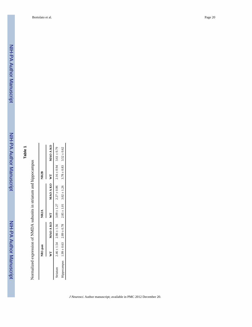
NR1 subunits of MAO A mutants (F(1,6) = 6.68; p < 0.05) (Fig. 2D). In contrast, MAO AKO mice displayed significantly higher expressions of NR2A (Fig. 2E) (F(1,6) = 6.56; p <0.05) and NR2B (Fig. 2F) (F(1,6) = 9.50; p < 0.05), but not NR2C (Fig. 2G) or NR2D (Fig.2H). No significant differences in either the glycosylation of NR2 subunits or NR2A:NR2Bratio (WT: 1.12 ± 0.08; MAO A KO: 1.50 ± 0.13, p < 0.10) were found. The expression ofNMDAR subunits in the striatum or hippocampus was comparable between the twogenotypes (Table 1).
Ex vivo electrophysiologyAs the biophysical and pharmacological properties of NMDAR are known to reflectdifferences in its subunit composition (Monyer et al., 1994; Mori and Mishina, 1995; Cull-Candy et al., 2001), the finding of higher expression of NR2A and NR2B sub-units in thePFC of MAO A KO mice prompted us to study the characteristics of NMDAR-mediatedcurrents in this region. In comparison with their WT controls, MAO A KO mice showed ahighly significant reduction in the decay time of EPSCs (Fig. 3A) (F(1,33) = 15.39; p <0.001). While the minimum stimulus intensities were equivalent between the two genotypes(Fig. 3B) (F(1,34) = 0.19; NS), EPSC amplitudes were significantly lower in MAO AKOmice (Fig. 3C) in correspondence of the range of stimulus intensities between 0.08 nA and0.8 nA [p < 0.05 at 0.08 nA; p < 0.01 at 0.09 and 0.1 nA; p < 0.05 at 0.2– 0.8 nA].Following the measurement of the physiological differences of NMDARs between the twogenotypes, we studied the responses of these targets to their antagonists. Administration ofdizocilpine produced a significant reduction in the percentage of NMDA EPSCs in MAO AKO mice at all tested doses (Fig. 3D) [p < 0.05 at 30 and 100 nM; p < 0.01 at 300 nM]. TheNR2A-specific antagonist PEAQX decreased NMDA EPSCs in MAO A KO mice; thiseffect was particularly evident at the lowest dose of PEAQX (Fig. 3E) [p < 0.05 at 5 nM; p <0.10 at 500 nM]. Finally, the NR2B subunit-specific antagonist Ro 25–6981 elicited asignificant reduction in NMDA EPSCs at all doses tested (Fig. 3F) (p < 0.01 at 5 nM; p <0.001 at 50 nM; p < 0.05 at 500 nM).
Locomotor activityTo explore the behavioral impact of altered NMDAR function, we evaluated the locomotorresponses of selective antagonists on NMDARs containing NR2A and NR2B subunits inMAO A KO mice. We first examined the behavior of WT and MAO A KO mice to theNMDAR antagonist dizocilpine (0.1 mg/kg, i.p.). As expected, saline-treated MAO A KOmice exhibited a reduction in locomotor activity, albeit not significant. Dizocil-pine eliciteda significant increment of locomotor activity in WT mice (Fig. 4B) (genotype × time: F(1,36)= 6.49; p < 0.05; p < 0.05 for WT-DIZ vs WT-SAL), but not in MAO A KO mutants. Thehigher dose of dizocilpine (0.3 mg/kg, i.p.) induced severe ataxia and stereotyped behaviorin MAO A KO mice, but not in WT littermates (data not shown). Administration of theNR2A antagonist PEAQX (2 mg/kg, i.p.) or theNR2B subunit antagonist Ro 25– 6981 (5mg/kg, i.p.) failed to significantly alter locomotor activity (Fig. 4B,C) (NR2A:F(1,29) = 0.52;NS and NR2B: F(1,31) = 0.08; NS).
Resident-intruder aggression assayAfter the assessment of the locomotor responses of WT and MAO A KO mice to NMDARantagonists, we proceeded to measure the impact of these drugs on the resident-intruderaggression paradigm.
Dizocilpine (0.1 mg/kg, i.p.) elicited marked anti-aggressive effects on MAO A KO mice, asrevealed by the significant increases in their latency to fight, which was normalized to thelevels observed in WT animals (Fig. 5A) (H(3,40) = 19.69; p < 0.001; p < 0.001 for KO-SALvs WT-SAL and KO-DIZ vs KO-SAL); this agent also reduced the number of fighting
Bortolato et al. Page 7
J Neurosci. Author manuscript; available in PMC 2012 December 20.
NIH
-PA Author Manuscript
NIH
-PA Author Manuscript
NIH
-PA Author Manuscript

episodes in MAO A KO mice (Fig. 5B) (genotype × treatment: F(1,37) = 14.44; p < 0.001; p< 0.001 for KO-SAL vs WT-SAL and KO-DIZ vs KO-SAL) and overall fighting duration(Fig. 5C) (genotype × treatment: F(1,37) = 16.88; p < 0.001; p < 0.001 for KO-SAL vs WT-SAL and KO-DIZ vs KO-SAL). In partial agreement with our previous results in the openfield, the drug did not induce any significant change in locomotor activity in either genotype(data not shown).
Both PEAQX (2 mg/kg, i.p.) and Ro 25–6981 (5 mg/kg, i.p.) also elicited a profoundreduction of aggressive behaviors, with significant increases in the latencies to the firstfighting approach (Fig. 5D,G) (NR2A: H(3,38) = 18.46; p < 0.001 and NR2B: H(3,40) =21.54; p < 0.001), number of fighting episodes (Fig.5 E,H) (NR2A: F(1,35) = 6.08; p < 0.05and NR2B: F(1,37) = 11.65; p < 0.01), and overall duration of fighting (Fig. 5F,I) (NR2A:F(1,35) = 9.56; p < 0.01 and NR2B: F(1,37) = 10.57; p < 0.01). Post hoc analyses confirmedthat saline-treated MAO A KO mice displayed a significantly shorter latency to fight(NR2A: p < 0.001 and NR2B: p < 0.001), higher number of fighting bouts (NR2A: p <0.001 and NR2B: p < 0.001) and longer fighting duration (NR2A: p < 0.001 and NR2B: p <0.001) than their WT counter-parts. Moreover, PEAQX and Ro 25–6981 administrationsignificantly increased the latency to fight (NR2A: p < 0.01 and NR2B: p < 0.001) anddecreased fighting frequency (NR2A: p < 0.001 and NR2B: p < 0.001) and duration (NR2A:p < 0.001 and NR2B: p < 0.001) in MAO A KO mice.
DiscussionWe showed that the PFC of MAO A-deficient mice exhibited significant variations ofsynaptic expression of NMDAR subunits, including increases in NR2A and NR2B andmarked deficits in N-glycosylated NR1. These imbalances led to overt abnormalities ofNMDAR properties, including marked reductions of decay time and excitability, as well ashypersensitivity to dizocilpine and selective NR2A and NR2B blockers. Notably, systemicadministration of these agents to MAO A KO mice reduced aggression to a degreecomparable to those observed in WT littermates, without affecting locomotor activity.
These findings collectively suggest that MAO A modulates aggressive behaviors bycontrolling the structure and function of synaptic NMDARs in the PFC. Cogent evidencedocuments that PFC is a key player in the modulation of aggression and allows for theemotional appraisal of socio-affective and environmental stimuli and the initiation ofappropriate behavioral responses (Davidson, 2002; Phillips et al., 2003; Ochsner and Gross,2005). Pyramidal neurons in the PFC integrate multiple synaptic signals from different brainareas, and project to main components of the limbic-subcortical circuit that regulate negativeaffect and reactive aggression, such as the amygdaloid nuclei, medial hypothalamus anddorsal periaqueductal gray (Panksepp, 1998; Gregg and Siegel, 2001). The NMDAR worksas a “coincidence detector” that enables the spatial and temporal summation of converginginputs (Seeburg et al., 1995). This mechanism plays a pivotal role in the dynamiccoordination of information processing, insofar as it allows for the amplification ofcontextually pertinent signals and the suppression of irrelevant stimuli (Phillips et al., 2010).Furthermore, NMDARs are instrumental for the activation of PFC pyramidal neurons (Shiand Zhang, 2003; Jackson et al., 2004), as well as their interactions with interneurons(Homayoun and Moghaddam, 2007). Thus, the observed reductions in decay time andexcitability in prefrontal NMDARs are likely to decrease the temporal integration ofnonsynchronous synaptic inputs, and impair the ability of pyramidal neurons to regulateemotional processing of social and environmental stimuli (Rolls et al., 2008). Theinvolvement of prefrontal NMDARs in the link between MAO A and the vulnerability toreactive aggression complements previous reports documenting PFC alterations in malecarriers of low-activity MAOA allelic variants (Meyer-Lindenberg et al., 2006; Lee and
Bortolato et al. Page 8
J Neurosci. Author manuscript; available in PMC 2012 December 20.
NIH
-PA Author Manuscript
NIH
-PA Author Manuscript
NIH
-PA Author Manuscript

Ham, 2008). These perturbations are conducive to a negative bias in the emotional appraisalof ambiguous socio-affective and contextual cues (Passamonti et al., 2006; Eisenberger etal., 2007; Brummett et al., 2008; Lee and Ham, 2008; McDermott et al., 2009), therebyenhancing the predisposition to reactive aggression in response to adverse experiences,particularly during early developmental stages (Buckholtz and Meyer-Lindenberg, 2008).Notably, MAO A KO mice exhibit alterations in risk assessment and paradoxical defensivereactivity to neutral and predator cues (Bortolato and Shih, 2011; Godar et al., 2011).
The increments in synaptic NR2A andNR2B in the PFC of MAO A-deficient mice were notparalleled by changes in the total levels of NR1 (or its splice cassettes), NR2C or NR2D, butrather by a marked reduction of NR1 N-glycosylation, as revealed by immunoblotting withthe mannose-associating lectin concanavalin A. N-Glycosylation processes occur at themembrane of the endoplasmic reticulum and require the activation of a poorly defined set ofglycosyltransferase complexes (Ivatt, 1981; Kornfeld and Kornfeld, 1985; Burda and Aebi,1999). The observed alterations may result from functional impairments of this enzymaticmachinery; accordingly, previous studies showed that, in cortical neurons, the inhibition ofthe first enzymatic step of N-glycosylation enhances the degradation of nonglycosylatedNR1, but not NR2A subunits (Gascón et al., 2007). The extensive N-glycosylation of NR1(based on the attachment of high-mannose oligosaccharide side-chains onto 12 Asparaginesites) is essential for its oligomerization with NR2 subunits (Chazot et al., 1995; Standleyand Baudry, 2000); thus, the increased expression in NR2A and NR2B may be paralleled bya reduced number of functional NR1 subunits. These variations leave the total NMDARnumber unaffected (as confirmed by our dizocilpine binding analyses), but lead topronounced alterations of the stoichiometric relations among subunits within each channel.
Changes in NR2A and NR2B subunit composition provide differential contributions to thetemporal dynamics and conductance of the NMDA channel, as well as its modulatory roleon synaptic plasticity mechanisms (Liu et al., 2004; Massey et al., 2004; Foster et al., 2010).Therefore, our observations may reflect the combined increments of both NR2A and NR2Bsubunits. Indeed, increases in NR2A subunits are linked to accelerated NMDA currentkinetics (Carmignoto and Vicini, 1992; Flint et al., 1997; Vicini et al., 1998), whereasincrements in NR2B subunits may affect the amplitude of NMDAR currents (Vicini et al.,1998). Since NR2A and NR2B regulate dendritic length and size (Ewald et al., 2008;Sepulveda et al., 2010), their increased expression may also partially account for thechanges in dendritic arborization in PFC pyramidal neurons of MAO A-deficient mice(Bortolato et al., 2011).
The neurochemical underpinnings of the reduction in N-glycosylated NR1 and increase inNR2A and NR2B subunits in the PFC remain unknown. MAO A is a critical regulator of thehomeostatic balance of 5-HT and NE in the brain, and its deficiency results in markedperturbations in the signaling of these monoamines (Shih et al., 1999; Bortolato and Shih,2011). Although the observed changes in NR1 glycosylation and NMDAR subunitexpression may be underpinned by increases in 5-HT and/or NE levels, this possibility istempered by the observation that the changes in NMDARs were only found in the PFC, incontrast with the ubiquitous enhancements in monoamine concentrations across all brainareas. The regional selectivity of the structural variations of NMDARs may instead reflectthe activation of 5-HT or NE receptors which are particularly abundant in prefrontalpyramidal neurons, such as 5-HT1A or 5-HT2A. Accordingly, activation of 5-HT1A receptorshas been found to modulate the expression of NMDAR subunits in these cells (Yuen et al.,2005). Further studies will be needed to evaluate the neurodevelopmental role of NE andother neurochemical targets in the structural, biophysical and pharmacological properties ofNMDARs in MAO A KO mice.
Bortolato et al. Page 9
J Neurosci. Author manuscript; available in PMC 2012 December 20.
NIH
-PA Author Manuscript
NIH
-PA Author Manuscript
NIH
-PA Author Manuscript

Alterations in NR2A and NR2B subunit expression patterns have been linked toneurodevelopmental changes (Monyer et al., 1994; Sheng et al., 1994; Zhong et al., 1995;Portera-Cailliau et al.,1996; van Zundert et al., 2004), which may play relevant roles in themodulation of corticogenesis (Barth and Malenka, 2001; Philpot et al., 2001; Fagiolini et al.,2003; Yoshimura et al., 2003). In rodents, the cortical distribution of NR2B is very abundantthroughout prenatal and early postnatal stages, but tends to decrease after the first postnatalweek; conversely, NR2A expression is barely detectable at birth, but increases with age(Monyer et al., 1994; Mori and Mishina, 1995). The behavioral deficits of MAOA KO miceare likely supported by early neurodevelopmental processes; for example, normalization of5-HT brain levels during the first weeks of postnatal life rescues several phenotypicalterations in MAO A KO mice (Cases et al., 1995). Building on these premises, it may behypothesized that early alterations in monoaminergic neurotransmission in male MAO A-deficient subjects may be conducive to persistent alterations in NMDA subunit expression,which may in turn disrupt PFC connectivity and increase aggression vulnerability.
The alterations in NR2A and NR2B expression also affected the pharmacological responsesof prefrontal NMDARs. In line with previous evidence (Mori and Mishina, 1995), theincreased NR2A and NR2B distribution induced hypersensitivity to NMDAR antagonists.Administration of these agents significantly reduced resident-intruder aggression in MAO AKO mice, as signified by the marked decrease in overall duration and number of fightingepisodes, as well as by the significant increase in latency to the first attack. These changeswere not accompanied by variations in locomotor activity in MAO A KO mice, suggesting aspecific anti-aggressive effect of NMDAR antagonists. Nevertheless, given that ourbehavioral analyses were only limited to the measurement of impulsive aggressive responsesin MAO AKO mice, we cannot exclude that the observed actions of NMDAR antagonistsmay reflect other underlying emotional alterations.
The available preclinical evidence indicates that NMDAR antagonists yield highly variableeffects in experimental models of aggression; in fact, whereas these agents increase fightingresponses in rodents with low proclivity to aggression, they yield the opposite effects inaggressive counterparts (Miczek and Fish,2006). These apparent discrepancies may reflectthe inability of most rodent paradigms to distinguish between reactive and instrumentalaggression (Nelson and Trainor, 2007); in this perspective, it is worth noting that the strikingisomorphism of the endophenotypes of MAO A-deficient mice with the behavioralrepertoire of impulsive antisocial manifestations in humans suggest a high predictivevalidity of these models for the development of anti-aggressive agents. The hightranslational relevance of our results points to low-dose NMDAR antagonists (or selectiveNR2A and NR2B blockers) as valuable therapeutic options for reactive aggression.Accordingly, recent studies have highlighted the potential of low-dose or low-potencyNMDA antagonists as efficacious anti-aggressive treatments in several neuropsychiatricdisorders (Bachenberg, 1998; Wilcock et al., 2008; Ballard et al., 2009).
In summary, the present findings highlight NMDARs as promising targets in the treatmentof antisocial aggressive traits in individuals with low MAO A activity. Future clinicalstudies are warranted to validate this intriguing possibility and fully establish the anti-aggressive properties of NMDAR blockers.
AcknowledgmentsThe present study was supported by National Institutes of Health Grants R01MH39085 (to J.C.S.) andR21HD070611 (to M.B.), as well as the Boyd and Elsie Welin Professorship (to J.C.S.) and the University ofSouthern California Zumberge Research Individual Grant (to M.B.). We are grateful to Anna L. Scott, ValentinaBini, Simone Tambaro, Felix Li, and Andrea Gochi for their valuable support in the execution of the experiments.
Bortolato et al. Page 10
J Neurosci. Author manuscript; available in PMC 2012 December 20.
NIH
-PA Author Manuscript
NIH
-PA Author Manuscript
NIH
-PA Author Manuscript
ReferencesAlia-Klein N, Goldstein RZ, Kriplani A, Logan J, Tomasi D, Williams B, Telang F, Shumay E, Biegon
A, Craig IW, Henn F, Wang GJ, Volkow ND, Fowler JS. Brain monoamine oxidase A activitypredicts trait aggression. J Neurosci. 2008; 28:5099–5104. [PubMed: 18463263]
Bachenberg KL. Oral ketamine for the management of combative autistic adult. Anesthesiology. 1998;89:549–550. [PubMed: 9710429]
Ballard CG, Gauthier S, Cummings JL, Brodaty H, Grossberg GT, Robert P, Lyketsos CG.Management of agitation and aggression associated with Alzheimer disease. Nat Rev Neurol. 2009;5:245–255. [PubMed: 19488082]
Barth AL, Malenka RC. NMDAR EPSC kinetics do not regulate the critical period for LTP atthalamocortical synapses. Nat Neurosci. 2001; 4:235–236. [PubMed: 11224537]
Blair, RJR. The neurobiology of aggression. In: Charney, DS.; Nestler, EJ., editors. Neurobiology ofmental illness. Ed 2. New York: Oxford UP; 2004. p. 1076-1085.
Bortolato M, Shih JC. Behavioral outcomes of monoamine oxidase deficiency: preclinical and clinicalevidence. Int Rev Neurobiol. 2011; 100:13–42. [PubMed: 21971001]
Bortolato M, Chen K, Shih JC. Monoamine oxidase inactivation: from pathophysiology totherapeutics. Adv Drug Deliv Rev. 2008; 60:1527–1533. [PubMed: 18652859]
Bortolato M, Chen K, Godar SC, Chen G, Wu W, Rebrin I, Farrell MR, Scott AL, Wellman CL, ShihJC. Social deficits and perseverative behaviors, but not overt aggression, in MAO-A hypomorphicmice. Neuropsychopharmacology. 2011; 36:2674–2688. [PubMed: 21832987]
Boyer PA, Skolnick P, Fossom LH. Chronic administration of imipramine and citalopram alters theexpression of NMDAR subunit mRNAs in mouse brain. A quantitative in situ hybridization study. JMol Neurosci. 1998; 10:219–233. [PubMed: 9770644]
Brummett BH, Boyle SH, Siegler IC, Kuhn CM, Surwit RS, Garrett ME, Collins A, Ashley-Koch A,Williams RB. HPA axis function in male caregivers: effect of the monoamine oxidase-A genepromoter (MAOA-uVNTR). Biol Psychol. 2008; 79:250–255. [PubMed: 18639608]
Brunner HG, Nelen M, Breakefield XO, Ropers HH, van Oost BA. Abnormal behavior associated witha point mutation in the structural gene for monoamine oxidase A. Science. 1993; 262:578–580.[PubMed: 8211186]
Buckholtz JW, Meyer-Lindenberg A. MAOA and the neurogenetic architecture of human aggression.Trends Neurosci. 2008; 31:120–129. [PubMed: 18258310]
Burda P, Aebi M. The dolichol pathway of N-linked glycosylation. Biochim Biophys Acta. 1999;1426:239–257. [PubMed: 9878760]
Carmignoto G, Vicini S. Activity-dependent decrease in NMDAR responses during development ofthe visual cortex. Science. 1992; 258:1007–1011. [PubMed: 1279803]
Carobrez AP, Teixeira KV, Graeff FG. Modulation of defensive behavior by periaqueductal grayNMDA/glycine-B receptor. Neurosci Biobehav Rev. 2001; 25:697–709. [PubMed: 11801295]
Cases O, Seif I, Grimsby J, Gaspar P, Chen K, Pournin S, Müller U, Aguet M, Babinet C, Shih JC, etal. Aggressive behavior and altered amounts of brain serotonin and norepinephrine in mice lackingMAOA. Science. 1995; 268:1763–1766. [PubMed: 7792602]
Caspi A, McClay J, Moffitt TE, Mill J, Martin J, Craig IW, Taylor A, Poulton R. Role of genotype inthe cycle of violence in maltreated children. Science. 2002; 297:851–854. [PubMed: 12161658]
Chazot PL, Cik M, Stephenson FA. An investigation into the role of N-glycosylation in the functionalexpression of a recombinant heteromeric NMDAR. Mol Membr Biol. 1995; 12:331–337.[PubMed: 8747278]
Cherek, DR.; Tcheremissine, OV.; Lane, SD. Psychopharmacology of human aggression: laboratoryand clinical studies. In: Nelson, RJ., editor. Biology of aggression. New York: Oxford UP; 2006.p. 424-446.
Clarke G, O’Mahony S, Malone G, Dinan TG. An isocratic high performance liquid chromatographymethod for the determination of GABA and glutamate in discrete regions of the rodent brain. JNeurosci Methods. 2007; 160:223–230. [PubMed: 17064781]
Cull-Candy S, Brickley S, Farrant M. NMDAR subunits: diversity, development and disease. CurrOpin Neurobiol. 2001; 11:327–335. [PubMed: 11399431]
Bortolato et al. Page 11
J Neurosci. Author manuscript; available in PMC 2012 December 20.
NIH
-PA Author Manuscript
NIH
-PA Author Manuscript
NIH
-PA Author Manuscript
Davidson RJ. Anxiety and affective style: role of prefrontal cortex and amygdala. Biol Psychiatry.2002; 51:68–80. [PubMed: 11801232]
Daw NW, Stein PS, Fox K. The role of NMDARs in information processing. Annu Rev Neurosci.1993; 16:207–222. [PubMed: 8460891]
Durand GM, Bennett MV, Zukin RS. Splice variants of the N-methyl-d-aspartate receptor NR1identify domains involved in regulation by polyamines and protein kinase C. Proc Natl Acad Sci US A. 1993; 90:6731–6735. [PubMed: 8341692]
Eisenberger NI, Way BM, Taylor SE, Welch WT, Lieberman MD. Understanding genetic risk foraggression: clues from the brain’s response to social exclusion. Biol Psychiatry. 2007; 61:1100–1108. [PubMed: 17137563]
Ewald RC, Van Keuren-Jensen KR, Aizenman CD, Cline HT. Roles of NR2A and NR2B in thedevelopment of dendritic arbor morphology in vivo. J Neurosci. 2008; 28:850–861. [PubMed:18216193]
Fagiolini M, Katagiri H, Miyamoto H, Mori H, Grant SG, Mishina M, Hensch TK. Separable featuresof visual cortical plasticity revealed by N-methyl-d-aspartate receptor 2A signaling. Proc NatlAcad Sci U S A. 2003; 100:2854–2859. [PubMed: 12591944]
Flint AC, Maisch US, Weishaupt JH, Kriegstein AR, Monyer H. NR2A subunit expression shortensNMDAR synaptic currents in developing neocortex. J Neurosci. 1997; 17:2469–2476. [PubMed:9065507]
Foster KA, McLaughlin N, Edbauer D, Phillips M, Bolton A, Constantine-Paton M, Sheng M. Distinctroles of NR2A and NR2B cytoplasmic tails in long-term potentiation. J Neurosci. 2010; 30:2676–2685. [PubMed: 20164351]
Gascón S, García-Gallo M, Renart J, Díaz-Guerra M. Endoplasmic reticulum-associated degradationof the NR1 but not the NR2 subunits of the N-methyl-d-aspartate receptor induced by inhibition ofthe N-glycosylation in cortical neurons. J Neurosci Res. 2007; 85:1713–1723. [PubMed:17455306]
Godar SC, Bortolato M, Frau R, Dousti M, Chen K, Shih JC. Maladaptive defensive behaviours inmonoamine oxidase A-deficient mice. Int J Neuropsychopharmacol. 2011; 14:1195–1207.[PubMed: 21156093]
Gonzalez-Islas C, Hablitz JJ. Dopamine enhances EPSCs in layer II–III pyramidal neurons in ratprefrontal cortex. J Neurosci. 2003; 23:867–875. [PubMed: 12574415]
Gould E, Cameron HA. Early NMDAR blockade impairs defensive behavior and increases cellproliferation in the dentate gyrus of developing rats. Behav Neurosci. 1997; 111:49–56. [PubMed:9109623]
Gregg TR, Siegel A. Brain structures and neurotransmitters regulating aggression in cats: implicationsfor human aggression. Prog Neuropsychopharmacol Biol Psychiatry. 2001; 25:91–140. [PubMed:11263761]
Harris A, Lurigio AJ. Mental illness and violence: a brief review of research and assessment strategies.Aggr Viol Behav. 2007; 12:542–551.
Homayoun H, Moghaddam B. NMDAR hypofunction produces opposite effects on prefrontal cortexinterneurons and pyramidal neurons. J Neurosci. 2007; 27:11496–11500. [PubMed: 17959792]
Ivatt RJ. Regulation of glycoprotein biosynthesis by formation of specific glycosyltransferasecomplexes. Proc Natl Acad Sci U S A. 1981; 78:4021–4025. [PubMed: 6457298]
Jackson ME, Homayoun H, Moghaddam B. NMDAR hypofunction produces concomitant firing ratepotentiation and burst activity reduction in the prefrontal cortex. Proc Natl Acad Sci U S A. 2004;101:8467–8472. [PubMed: 15159546]
Kim JJ, Shih JC, Chen K, Chen L, Bao S, Maren S, Anagnostaras SG, Fanselow MS, De Maeyer E,Seif I, Thompson RF. Selective enhancement of emotional, but not motor, learning in monoamineoxidase A-deficient mice. Proc Natl Acad Sci U S A. 1997; 94:5929–5933. [PubMed: 9159177]
Kim-Cohen J, Caspi A, Taylor A, Williams B, Newcombe R, Craig IW, Moffitt TE. MAOA,maltreatment, and gene-environment interaction predicting children’s mental health: new evidenceand a meta-analysis. Mol Psychiatry. 2006; 11:903–913. [PubMed: 16801953]
Kornfeld R, Kornfeld S. Assembly of asparagine-linked oligosaccharides. Annu Rev Biochem. 1985;54:631–664. [PubMed: 3896128]
Bortolato et al. Page 12
J Neurosci. Author manuscript; available in PMC 2012 December 20.
NIH
-PA Author Manuscript
NIH
-PA Author Manuscript
NIH
-PA Author Manuscript

Lan NC, Chen CH, Shih JC. Expression of functional human monoamine oxidase A and B cDNAs inmammalian cells. J Neurochem. 1989; 52:1652–1654. [PubMed: 2496202]
Lee BT, Ham BJ. Monoamine oxidase A-uVNTR genotype affects limbic brain activity in response toaffective facial stimuli. Neuroreport. 2008; 19:515–519. [PubMed: 18388730]
Liu XB, Murray KD, Jones EG. Switching of NMDAR 2A and 2B subunits at thalamic and corticalsynapses during early postnatal development. J Neurosci. 2004; 24:8885–8895. [PubMed:15470155]
Massey PV, Johnson BE, Moult PR, Auberson YP, Brown MW, Molnar E, Collingridge GL, BashirZI. Differential roles of NR2A and NR2B-containing NMDARs in cortical long-term potentiationand long-term depression. J Neurosci. 2004; 24:7821–7828. [PubMed: 15356193]
Masuko T, Suzuki I, Kizawa Y, Kusama-Eguchi K, Watanabe K, Kashiwagi K, Igarashi K, Kusama T.Monoamines directly inhibit N-methyl-D-aspartate receptors expressed in Xenopus oocytes in avoltage-dependent manner. Neurosci Lett. 2004; 371:30–33. [PubMed: 15500961]
McDermott R, Tingley D, Cowden J, Frazzetto G, Johnson DD. Monoamine oxidase A gene (MAOA)predicts behavioral aggression following provocation. Proc Natl Acad Sci U S A. 2009; 106:2118–2123. [PubMed: 19168625]
Meyer-Lindenberg A, Buckholtz JW, Kolachana B, R Hariri A, Pezawas L, Blasi G, Wabnitz A,Honea R, Verchinski B, Callicott JH, Egan M, Mattay V, Weinberger DR. Neural mechanisms ofgenetic risk for impulsivity and violence in humans. Proc Natl Acad Sci U S A. 2006; 103:6269–6274. [PubMed: 16569698]
Miczek, KA.; Fish, EW. Monoamines, GABA, Glutamate, and Aggression. In: Nelson, RJ., editor.Biology of aggression. New York: Oxford UP; 2006. p. 114-149.
Monyer H, Burnashev N, Laurie DJ, Sakmann B, Seeburg PH. Developmental and regional expressionin the rat brain and functional properties of four NMDARs. Neuron. 1994; 12:529–540. [PubMed:7512349]
Mori H, Mishina M. Structure and function of the NMDAR channel. Neuropharmacology. 1995;34:1219–1237. [PubMed: 8570021]
Nelson RJ, Trainor BC. Neural mechanisms of aggression. Nat Rev Neurosci. 2007; 8:536–546.[PubMed: 17585306]
Newell KA, Zavitsanou K, Huang XF. Short and long term changes in NMDAR binding in mousebrain following chronic phencyclidine treatment. J Neural Transm. 2007; 114:995–1001.[PubMed: 17401537]
Ochsner KN, Gross JJ. The cognitive control of emotion. Trends Cogn Sci. 2005; 9:242–249.[PubMed: 15866151]
Panksepp, J. Affective neuroscience—the foundations of human and animal emotions. New York:Oxford UP; 1998.
Passamonti L, Fera F, Magariello A, Cerasa A, Gioia MC, Muglia M, Nicoletti G, Gallo O, ProvincialiL, Quattrone A. Monoamine oxidase-a genetic variations influence brain activity associated withinhibitory control: new insight into the neural correlates of impulsivity. Biol Psychiatry. 2006;59:334–340. [PubMed: 16202396]
Paxinos, G.; Fanklin, KBJ. The mouse brain in stereotaxic coordinates. Ed 2. San Diego: Academic;2001.
Phillips ML, Drevets WC, Rauch SL, Lane R. Neurobiology of emotion perception I: the neural basisof normal emotion perception. Biol Psychiatry. 2003; 54:504–514. [PubMed: 12946879]
Phillips, WA.; von der Malsburg, C.; Singer, W. Dynamic coordination in brain and mind. In: von derMalsburg, C.; Phillips, WA.; Singer, W., editors. Dynamic coordination in the brain: from neuronsto mind. Cambridge, MA: MIT; 2010. p. 1-25.
Philpot BD, Sekhar AK, Shouval HZ, Bear MF. Visual experience and deprivation bidirectionallymodify the composition and function of NMDARs in visual cortex. Neuron. 2001; 29:157–169.[PubMed: 11182088]
Portera-Cailliau C, Price DL, Martin LJ. N-methyl-d-aspartate receptor proteins NR2A and NR2B aredifferentially distributed in the developing rat central nervous system as revealed by subunit-specific antibodies. J Neurochem. 1996; 66:692–700. [PubMed: 8592141]
Bortolato et al. Page 13
J Neurosci. Author manuscript; available in PMC 2012 December 20.
NIH
-PA Author Manuscript
NIH
-PA Author Manuscript
NIH
-PA Author Manuscript

Reiss D. The long reach of violence and aggression. Psychiatry. 1993; 56:163–165. [PubMed:8351293]
Rolls ET, Loh M, Deco G, Winterer G. Computational models of schizophrenia and dopaminemodulation in the prefrontal cortex. Nat Rev Neurosci. 2008; 9:696–709. [PubMed: 18714326]
Scott AL, Bortolato M, Chen K, Shih JC. Novel monoamine oxidase A knock out mice with human-like spontaneous mutation. Neuroreport. 2008; 19:739–743. [PubMed: 18418249]
Seeburg PH, Burnashev N, Köhr G, Kuner T, Sprengel R, Monyer H. The NMDAR channel:molecular design of a coincidence detector. Recent Prog Horm Res. 1995; 50:19–34. [PubMed:7740157]
Sepulveda FJ, Bustos FJ, Inostroza E, Zúñiga FA, Neve RL, Montecino M, van Zundert B. Differentialroles of NMDAR Subtypes NR2A and NR2B in dendritic branch development and requirement ofRasGRF1. J Neurophysiol. 2010; 103:1758–1770. [PubMed: 20107120]
Sheng M, Cummings J, Roldan LA, Jan YN, Jan LY. Changing subunit composition of heteromericNMDARs during development of rat cortex. Nature. 1994; 368:144–147. [PubMed: 8139656]
Shi WX, Zhang XX. Dendritic glutamate-induced bursting in the prefrontal cortex: furthercharacterization and effects of phencyclidine. J Pharmacol Exp Ther. 2003; 305:680–687.[PubMed: 12606677]
Shih JC, Chen K, Ridd MJ. Monoamine oxidase: from genes to behavior. Annu Rev Neurosci. 1999;22:197–217. [PubMed: 10202537]
Standley S, Baudry M. The role of glycosylation in ionotropic glutamate receptor ligand binding,function, and trafficking. Cell Mol Life Sci. 2000; 57:1508–1516. [PubMed: 11092445]
van Zundert B, Yoshii A, Constantine-Paton M. Receptor compartmentalization and trafficking atglutamate synapses: a developmental proposal. Trends Neurosci. 2004; 27:428–437. [PubMed:15219743]
Vicini S, Wang JF, Li JH, Zhu WJ, Wang YH, Luo JH, Wolfe BB, Grayson DR. Functional andpharmacological differences between recombinant N-methyl-d-aspartate receptors. JNeurophysiol. 1998; 79:555–566. [PubMed: 9463421]
Weder N, Yang BZ, Douglas-Palumberi H, Massey J, Krystal JH, Gelernter J, Kaufman J. MAOAgenotype, maltreatment, and aggressive behavior: the changing impact of genotype at varyinglevels of trauma. Biol Psychiatry. 2009; 65:417–424. [PubMed: 18996506]
Widom CS, Brzustowicz LM. MAOA and the “cycle of violence:” childhood abuse and neglect,MAOA genotype, and risk for violent and antisocial behavior. Biol Psychiatry. 2006; 60:684–689.[PubMed: 16814261]
Wilcock GK, Ballard CG, Cooper JA, Loft H. Memantine for agitation/aggression and psychosis inmoderately severe to severe Alzheimer’s disease: a pooled analysis of 3 studies. J Clin Psychiatry.2008; 69:341–348. [PubMed: 18294023]
Yoshimura Y, Ohmura T, Komatsu Y. Two forms of synaptic plasticity with distinct dependence onage, experience, and NMDAR subtype in rat visual cortex. J Neurosci. 2003; 23:6557–6566.[PubMed: 12878697]
Yuen EY, Jiang Q, Chen P, Gu Z, Feng J, Yan Z. Serotonin 5-HT1A receptors regulate NMDARchannels through a microtubule-dependent mechanism. J Neurosci. 2005; 25:5488–5501.[PubMed: 15944377]
Zhong J, Carrozza DP, Williams K, Pritchett DB, Molinoff PB. Expression of mRNAs encodingsubunits of the NMDAR in developing rat brain. J Neurochem. 1995; 64:531–539. [PubMed:7830045]
Bortolato et al. Page 14
J Neurosci. Author manuscript; available in PMC 2012 December 20.
NIH
-PA Author Manuscript
NIH
-PA Author Manuscript
NIH
-PA Author Manuscript

Figure 1.MAOA-deficient mice showed significantly higher 5-HT and NE levels in all brain regions,but similar glutamate levels and NMDAR binding in comparison with WT mice. A,HPLCanalysis revealed higher 5-HT concentrations in the PFC, striatum (STR), hippocampus(HIP), and midbrain (MID) of MAO A KO mice.B,NE levels were also significantly greaterthan those in WT mice across all brain regions tested. C, No alterations in glutamate levelswere found in any brain region tested.D, Autoradiography showed no differences inNMDAR binding in the cortical cingulate layers 1 and 3, striatal caudate-putamen, nucleusaccumbens shell and core, hippocampal cornu ammonis areas 1, 2, and 3, dentate gyrus, andbasal lateral amygdala. Values are represented as mean ± SEM. *p < 0.05, **p < 0.01, and***p < 0.001 compared with WT mice. Cg1, Cingulate layer 1; Cg3, cingulate layer 3; CPu,caudate-putamen; Core, nucleus accumbens core; Shell, nucleus accumbens shell; CA1,cornu ammonis area 1; CA2; CA3, cornu ammonis area 3; DG, dentate gyrus; BLA,basolateral amygdala.
Bortolato et al. Page 15
J Neurosci. Author manuscript; available in PMC 2012 December 20.
NIH
-PA Author Manuscript
NIH
-PA Author Manuscript
NIH
-PA Author Manuscript

Figure 2.MAO A KO mice exhibited significant alterations in NMDA receptor subunit expression inthe prefrontal cortex. A–D, Although no alterations in NR1 or its variants were detected,MAO A-deficient animals showed significantly lower levels of glycosylated NR1. E–H,Conversely, higher levels of NR2A and NR2B, but not NR2C or NR2D, subunit expressionin the prefrontal cortex were found in MAO A mutants compared with their WTcounterparts. Values are represented as mean ± SEM. *p < 0.05 compared with WT mice.Con A, concanavalin A. For further details, see text.
Bortolato et al. Page 16
J Neurosci. Author manuscript; available in PMC 2012 December 20.
NIH
-PA Author Manuscript
NIH
-PA Author Manuscript
NIH
-PA Author Manuscript
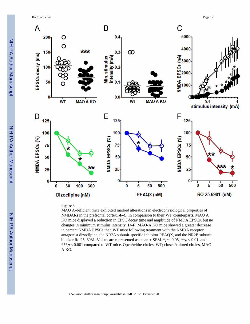
Figure 3.MAO A-deficient mice exhibited marked alterations in electrophysiological properties ofNMDARs in the prefrontal cortex. A–C, In comparison to their WT counterparts, MAO AKO mice displayed a reduction in EPSC decay time and amplitude of NMDA EPSCs, but nochanges in minimum stimulus intensity. D–F, MAO-A KO mice showed a greater decreasein percent NMDA EPSCs than WT mice following treatment with the NMDA receptorantagonist dizocilpine, the NR2A subunit-specific inhibitor PEAQX, and the NR2B subunitblocker Ro 25–6981. Values are represented as mean ± SEM. *p < 0.05, **p < 0.01, and***p < 0.001 compared to WT mice. Open/white circles, WT; closed/colored circles, MAOA KO.
Bortolato et al. Page 17
J Neurosci. Author manuscript; available in PMC 2012 December 20.
NIH
-PA Author Manuscript
NIH
-PA Author Manuscript
NIH
-PA Author Manuscript

Figure 4.Dizocilpine altered locomotor activity in WT, but not in MAO A mutant mice. A,Dizocilpine elicited a significant increase in total distance traveled by WT mice. B, C, BothNR2Aand NR2B subunit antagonism showed comparable effects in both genotypes in allparameters tested. Values are represented as mean ± SEM. *p < 0.05 compared with WTmice treated with saline.
Bortolato et al. Page 18
J Neurosci. Author manuscript; available in PMC 2012 December 20.
NIH
-PA Author Manuscript
NIH
-PA Author Manuscript
NIH
-PA Author Manuscript

Figure 5.Dizocilpine, PEAQX, and Ro 25–6981 significantly decreased aggression in MAO A-deficient mice. A–C, NMDAR antagonism elicited a significant increase in latency to fight,as well as reduced the fighting frequency and duration in MAO A KO mice. D–F, Similarly,MAO A mutants displayed an increase in latency to fight and engaged in lower fightingbouts and duration following NR2A subunit antagonism. G–I, NR2B inhibition significantlyelevated the latency, and decreased the bouts and overall duration of fighting behavior inMAOA KO mice. Values represented as mean ± SEM. ***p < 0.001 compared with saline-treated WT mice; ##p < 0.01 and ###p < 0.001 compared with MAO A KO mice treated withsaline.
Bortolato et al. Page 19
J Neurosci. Author manuscript; available in PMC 2012 December 20.
NIH
-PA Author Manuscript
NIH
-PA Author Manuscript
NIH
-PA Author Manuscript
NIH
-PA Author Manuscript
NIH
-PA Author Manuscript
NIH
-PA Author Manuscript
Bortolato et al. Page 20
Tabl
e 1
Nor
mal
ized
exp
ress
ion
of N
MD
A s
ubun
its in
str
iatu
m a
nd h
ippo
cam
pus
NR
1-pa
nN
R2A
NR
2B
WT
MA
O A
KO
WT
MA
O A
KO
WT
MA
O A
KO
Stri
atum
2.46
± 1
.54
2.86
± 1
.56
3.09
± 1
.27
2.27
± 0
.86
2.16
± 0
.94
3.61
± 0
.79
Hip
poca
mpu
s1.
99 ±
0.6
32.
89 ±
0.7
82.
85 ±
1.0
13.
02 ±
1.2
63.
78 ±
0.8
33.
52 ±
0.6
2
J Neurosci. Author manuscript; available in PMC 2012 December 20.