Primary Scarring Alopecia: Clinical-Pathological Review of 72 ...
12
E-Mail [email protected] Original Article Skin Appendage Disord 2017;3:132–143 DOI: 10.1159/000467395 Primary Scarring Alopecia: Clinical-Pathological Review of 72 Cases and Review of the Literature Salvador Villablanca Cristián Fischer S. Cecilia García-García J. Manuel Mascaró-Galy Juan Ferrando Department of Dermatology, Hospital Clinic, University of Barcelona, Barcelona, Spain while neutrophilic PSAs had a more acute onset with an evo- lution of acute outbreaks. PSAs in a late stage with an ab- sent/mild infiltrate had a subclinical onset and a slowly progressive or stable evolution. Conclusions: The PSAs are severe trichological conditions. Their high clinical and histo- pathological variability make them a diagnostic and thera- peutic challenge. Message of the Paper: Knowing the clini- cal and histopathological aspects of PSAs should be of cru- cial importance to the dermatologist. © 2017 S. Karger AG, Basel Introduction Hair is a human attribute that involves aspects of self- image, identity, ethnicity, and health, among others. Therefore, it is not surprising that disorders that cause alopecia can alter self-perception and psychosocial inter- actions [1]. Alopecias can be classified as scarring (cicatricial) and nonscarring. Scarring alopecias are divided into 2 groups: primary (PSA) and secondary. PSAs are uncommon, but not rare, heterogeneous group of disorders characterized clinically by the lack of follicular ostia and histopatho- logically by the replacement of hair follicle structures by Keywords Cicatricial alopecia · Scarring alopecia · Lymphocytic scarring alopecia · Neutrophilic scarring alopecia · Pseudopelade Abstract Purpose of the Study: To analyze the epidemiologic, demo- graphic, clinical, and histological characteristics of primary scarring alopecia (PSA) cases diagnosed over a 7-year period at the Department of Dermatology, Hospital Clinic, Barcelo- na, Spain. Procedures: Seventy-two patients diagnosed with PSA between 2006 and 2012 were included. Age, sex, ethnic group, clinical pattern, predominant histological infiltrate, final clinical diagnosis, time of onset, treatments used, and clinical evolution were evaluated and correlated. Results: The ethnic groups were distributed as follows: 93% Europe- an-Caucasian, 5% Mestizo-American, 1% oriental, and 1% Afro-American. Most cases were females (71%), and mean age was 51 ± 6 years. The follicular pattern was the most common, and the predominant inflammatory infiltrate was lymphocytic. Lichen planopilaris and frontal fibrosing alope- cia were the main diagnoses. When correlating clinical as- pects and histopathology, lymphocytic PSAs had a subacute onset and resulted in a nonchanging, more stable form, Received: January 13, 2017 Accepted: February 28, 2017 Published online: April 8, 2017 Assoc. Prof. Juan Ferrando, MD, PhD Hospital Clinic, Department of Dermatology University of Barcelona, Villarroel 170 ES–08036 Barcelona (Spain) E-Mail juanferrandobarbera @ gmail.com © 2017 S. Karger AG, Basel www.karger.com/sad
-
Upload
khangminh22 -
Category
Documents
-
view
5 -
download
0
Transcript of Primary Scarring Alopecia: Clinical-Pathological Review of 72 ...
E-Mail [email protected]
Original Article
Skin Appendage Disord 2017;3:132–143 DOI: 10.1159/000467395
Primary Scarring Alopecia:Clinical-Pathological Review of 72 Cases and Review of the Literature
Salvador Villablanca Cristián Fischer S. Cecilia García-García
J. Manuel Mascaró-Galy Juan Ferrando
Department of Dermatology, Hospital Clinic, University of Barcelona, Barcelona , Spain
while neutrophilic PSAs had a more acute onset with an evo-lution of acute outbreaks. PSAs in a late stage with an ab-sent/mild infiltrate had a subclinical onset and a slowlyprogressive or stable evolution. Conclusions: The PSAs are severe trichological conditions. Their high clinical and histo-pathological variability make them a diagnostic and thera-peutic challenge. Message of the Paper: Knowing the clini-cal and histopathological aspects of PSAs should be of cru-cial importance to the dermatologist.
© 2017 S. Karger AG, Basel
Introduction
Hair is a human attribute that involves aspects of self-image, identity, ethnicity, and health, among others. Therefore, it is not surprising that disorders that cause alopecia can alter self-perception and psychosocial inter-actions [1] .
Alopecias can be classified as scarring (cicatricial) and nonscarring. Scarring alopecias are divided into 2 groups: primary (PSA) and secondary. PSAs are uncommon, but not rare, heterogeneous group of disorders characterized clinically by the lack of follicular ostia and histopatho-logically by the replacement of hair follicle structures by
Keywords
Cicatricial alopecia · Scarring alopecia · Lymphocytic scarring alopecia · Neutrophilic scarring alopecia · Pseudopelade
Abstract
Purpose of the Study: To analyze the epidemiologic, demo-graphic, clinical, and histological characteristics of primary scarring alopecia (PSA) cases diagnosed over a 7-year period at the Department of Dermatology, Hospital Clinic, Barcelo-na, Spain. Procedures: Seventy-two patients diagnosed with PSA between 2006 and 2012 were included. Age, sex, ethnic group, clinical pattern, predominant histological infiltrate, final clinical diagnosis, time of onset, treatments used, and clinical evolution were evaluated and correlated. Results: The ethnic groups were distributed as follows: 93% Europe-an-Caucasian, 5% Mestizo-American, 1% oriental, and 1% Afro-American. Most cases were females (71%), and mean age was 51 ± 6 years. The follicular pattern was the most common, and the predominant inflammatory infiltrate was lymphocytic. Lichen planopilaris and frontal fibrosing alope-cia were the main diagnoses. When correlating clinical as-pects and histopathology, lymphocytic PSAs had a subacute onset and resulted in a nonchanging, more stable form,
Received: January 13, 2017 Accepted: February 28, 2017 Published online: April 8, 2017
Assoc. Prof. Juan Ferrando, MD, PhD Hospital Clinic, Department of Dermatology University of Barcelona, Villarroel 170 ES–08036 Barcelona (Spain) E-Mail juanferrandobarbera @ gmail.com
© 2017 S. Karger AG, Basel
www.karger.com/sad
Primary Scarring Alopecia Skin Appendage Disord 2017;3:132–143 DOI: 10.1159/000467395
133
fibrous tissue, which makes this condition irreversible. From the pathophysiological point of view, the scarring area constitutes the end of reparative fibrosis, with per-manent destruction of the preexisting tissue [2–4] .
In PSAs, the hair follicle is the main target of the in-flammatory process, which can be microscopically ob-served as a preferential destruction of the follicular epi-thelium and/or adventitial dermis, with a relative preser-vation of the interfollicular reticular dermis [5, 6] . This group of alopecias includes the following clinical entities: chronic cutaneous lupus erythematosus (LE), lichen pla-nopilaris (LPP), classic pseudopelade of Brocq (PB), fol-liculitis decalvans (FD), and dissecting folliculitis among others ( Table 1 ). The term secondary scarring alopecia refers to when the destruction of the hair follicle is not the primary pathological event, and the condition is rather caused by exogenous factors such as trauma (e.g., burns, radiation, traction) or endogenous infiltrative and in-flammatory processes (e.g., sarcoidosis, pemphigus vul-garis, and scleroderma) [2, 4, 7] .
There are currently various PSA classifications, but the most accepted is the one proposed by the North Ameri-can Hair Research Society (NAHRS) [8] , whose last revi-sion was in 2003. This classification divides PSAs into 2 large groups based on the dominant type of inflamma-tory cell infiltrate. This concept had already been suggest-ed by other authors earlier, but it was improved by this work group, who also added the lymphocytic and neutro-phil-associated subgroups, as well as mixed (such as acne keloidalis) and nonspecific idiopathic scarring alopecias
with inconclusive clinical and histological findings, most of which probably correspond to the final stage of other groups.
In the authors’ experience, PSAs can represent a true clinical challenge when clinical-pathological correlation is difficult to assess, which more commonly occurs dur-ing late and advanced stages. This makes histopathologi-cal diagnosis more difficult because the main classifica-tion criterion (the type of inflammatory infiltrate) might be impossible to assess. In these cases, additional studies such as direct immunofluorescence (DIF) can be useful. Reference sources are limited with respect to their true epidemiology, and randomized, double-blind clinical tri-als regarding treatment are currently lacking; conse-quently, therapy is mostly based on case series and the clinicians’ personal experience.
Materials and Methods
In order to obtain new clinical and pathological knowledge about PSAs, we decided to study the epidemiologic, demographic, clinical, and histological characteristics (classified in agreement with the NAHRS) of PSAs cases diagnosed over a 7-year period, at the Department of Dermatology of Hospital Clinic, Barcelona, Spain. We also evaluated the therapies used in each case, their clin-ical evolution, and the correlation of these results with the pre-dominant type of inflammatory infiltrate on histopathology.
The first step included reviewing all the diagnoses of scalp bi-opsies, and searching the histopathological database of our pa-tients, using the names of the PSA clinical entities according to the NAHRS classification, (such as LPP dissecting folliculitis, frontal fibrosing alopecia [FFA], etc.) between 2006 and 2012. We includ-ed only cases of PSA subtypes as a diagnosis, and for which there was access to the clinical history and histological findings. Col-lected data included age at diagnosis, sex, ethnic group, clinical pattern, predominant histological infiltrate, DIF result (if avail-able), final clinical diagnosis, time of onset before diagnosis, treat-ments used, and clinical evolution. The final clinical diagnosis was based on clinical-pathological correlation. Exclusion criteria were diagnoses of secondary scarring alopecia (e.g., tumors, infections, traumas, sarcoidosis, etc.), scarring alopecia outside of the scalp, and lack of access to the clinical history.
Results
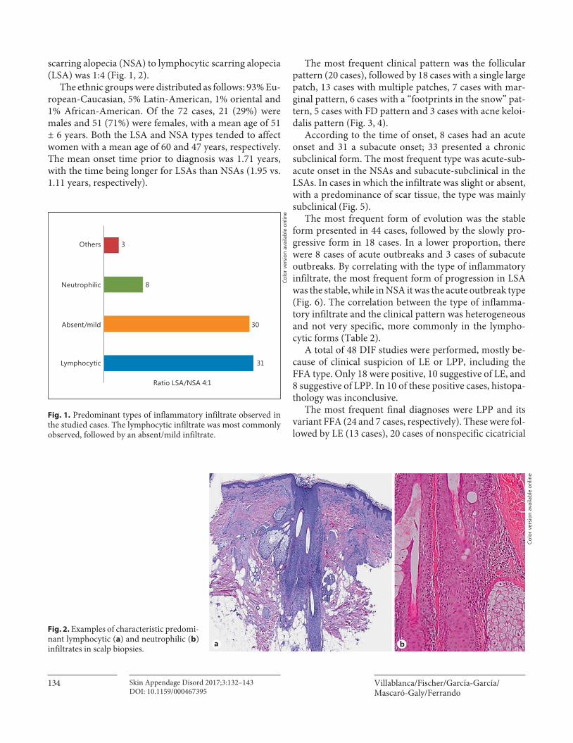
A total of 1,085 cases were found during the search for general scalp biopsies and 72 biopsies with the diagnosis of PSA. The most frequent type of predominant inflam-matory infiltrate was lymphocytic (31 cases), followed by absent or nonspecific inflammatory infiltrate (30 cases), neutrophil-associated (8 cases), and other/mixed type (2 granulomatous, 1 plasmatic). The ratio of neutrophilic
Table 1. Classification of primary scarring (cicatricial) alopecias according to the NAHRS (North American Hair Research Society) [8]
LymphocyticChronic cutaneous lupus erythematosusLichen planopilaris and its variantsClassic pseudopelade of BrocqCentral centrifugal cicatricial alopeciaAlopecia mucinosaKeratosis follicularis spinulosa decalvans
NeutrophilicFolliculitis decalvansDissecting cellulitis/folliculitis
MixedFolliculitis (acne) keloidalisFolliculitis (acne) necroticaErosive pustular dermatosis
Nonspecific
Villablanca/Fischer/García-García/Mascaró-Galy/Ferrando
Skin Appendage Disord 2017;3:132–143 DOI: 10.1159/000467395
134
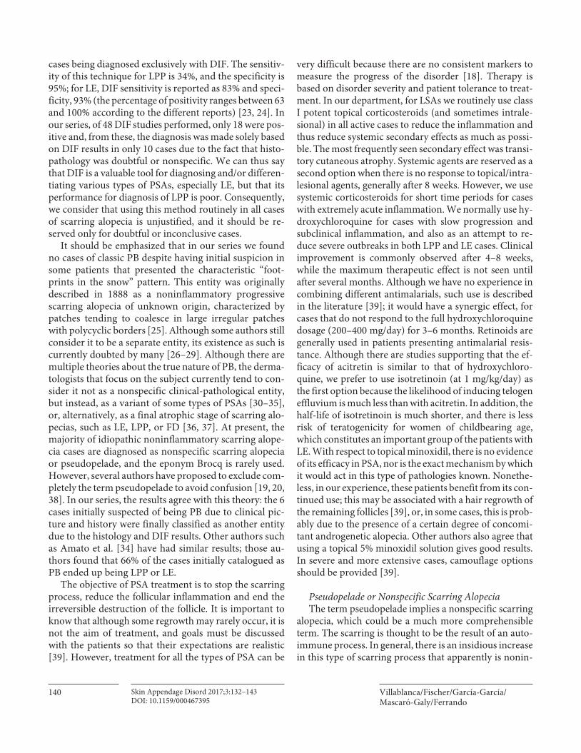
scarring alopecia (NSA) to lymphocytic scarring alopecia (LSA) was 1: 4 ( Fig. 1 , 2 ).
The ethnic groups were distributed as follows: 93% Eu-ropean-Caucasian, 5% Latin-American, 1% oriental and 1% African-American. Of the 72 cases, 21 (29%) were males and 51 (71%) were females, with a mean age of 51 ± 6 years. Both the LSA and NSA types tended to affect women with a mean age of 60 and 47 years, respectively. The mean onset time prior to diagnosis was 1.71 years, with the time being longer for LSAs than NSAs (1.95 vs. 1.11 years, respectively).
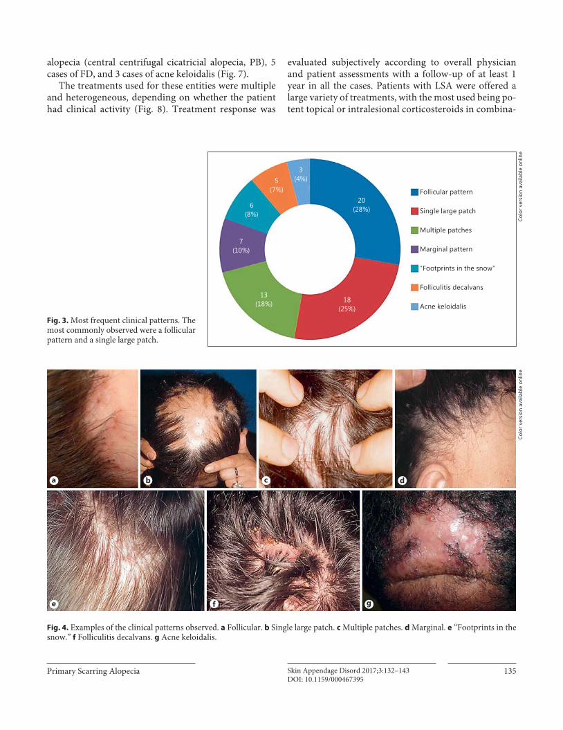
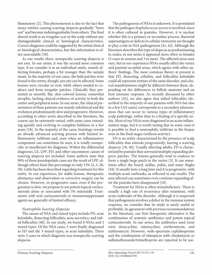
The most frequent clinical pattern was the follicular pattern (20 cases), followed by 18 cases with a single large patch, 13 cases with multiple patches, 7 cases with mar-ginal pattern, 6 cases with a “footprints in the snow” pat-tern, 5 cases with FD pattern and 3 cases with acne keloi-dalis pattern ( Fig. 3 , 4 ).
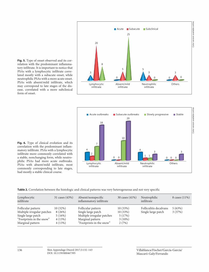
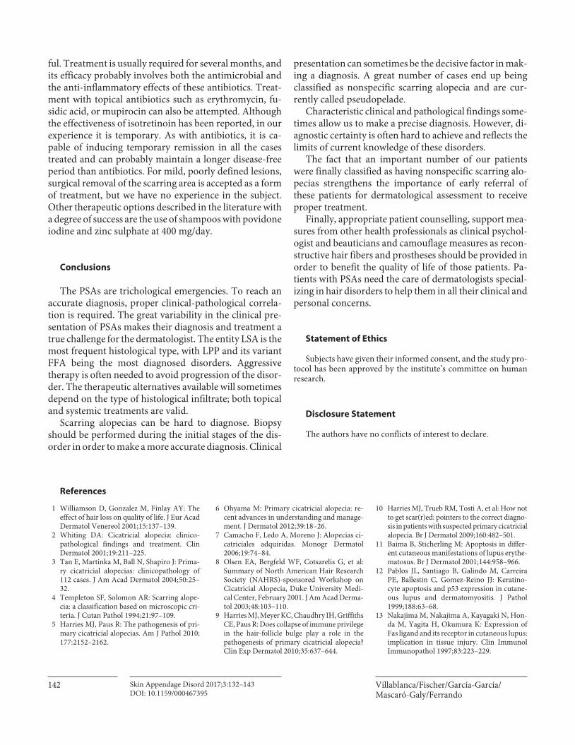
According to the time of onset, 8 cases had an acute onset and 31 a subacute onset; 33 presented a chronic subclinical form. The most frequent type was acute-sub-acute onset in the NSAs and subacute-subclinical in the LSAs. In cases in which the infiltrate was slight or absent, with a predominance of scar tissue, the type was mainly subclinical ( Fig. 5 ).
The most frequent form of evolution was the stable form presented in 44 cases, followed by the slowly pro-gressive form in 18 cases. In a lower proportion, there were 8 cases of acute outbreaks and 3 cases of subacute outbreaks. By correlating with the type of inflammatory infiltrate, the most frequent form of progression in LSA was the stable, while in NSA it was the acute outbreak type ( Fig. 6 ). The correlation between the type of inflamma-tory infiltrate and the clinical pattern was heterogeneous and not very specific, more commonly in the lympho-cytic forms ( Table 2 ).
A total of 48 DIF studies were performed, mostly be-cause of clinical suspicion of LE or LPP, including the FFA type. Only 18 were positive, 10 suggestive of LE, and 8 suggestive of LPP. In 10 of these positive cases, histopa-thology was inconclusive.
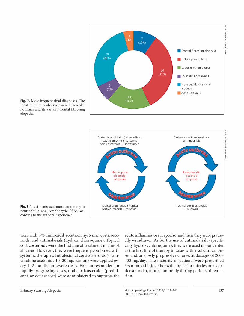
The most frequent final diagnoses were LPP and its variant FFA (24 and 7 cases, respectively). These were fol-lowed by LE (13 cases), 20 cases of nonspecific cicatricial
Others 3
8
30
31
Neutrophilic
Absent/mild
Lymphocytic
Ratio LSA/NSA 4:1
Fig. 1. Predominant types of inflammatory infiltrate observed in the studied cases. The lymphocytic infiltrate was most commonly observed, followed by an absent/mild infiltrate.
a b
Fig. 2. Examples of characteristic predomi-nant lymphocytic ( a ) and neutrophilic ( b ) infiltrates in scalp biopsies.
Colo
r ver
sion
ava
ilabl
e on
line
Colo
r ver
sion
ava
ilabl
e on
line
Primary Scarring Alopecia Skin Appendage Disord 2017;3:132–143 DOI: 10.1159/000467395
135
alopecia (central centrifugal cicatricial alopecia, PB), 5 cases of FD, and 3 cases of acne keloidalis ( Fig. 7 ).
The treatments used for these entities were multiple and heterogeneous, depending on whether the patient had clinical activity ( Fig. 8 ). Treatment response was
evaluated subjectively according to overall physician and patient assessments with a follow-up of at least 1 year in all the cases. Patients with LSA were offered a large variety of treatments, with the most used being po-tent topical or intralesional corticosteroids in combina-
3(4%)5
(7%)
6(8%)
7(10%)
13(18%) 18
(25%)
20(28%)
Follicular pattern
Folliculitis decalvans
Acne keloidalis
Single large patch
Multiple patches
Marginal pattern
“Footprints in the snow”
Fig. 3. Most frequent clinical patterns. The most commonly observed were a follicular pattern and a single large patch.
a b c d
e f g
Fig. 4. Examples of the clinical patterns observed. a Follicular. b Single large patch. c Multiple patches. d Marginal. e “Footprints in the snow.” f Folliculitis decalvans. g Acne keloidalis.
Colo
r ver
sion
ava
ilabl
e on
line
Colo
r ver
sion
ava
ilabl
e on
line
Villablanca/Fischer/García-García/Mascaró-Galy/Ferrando
Skin Appendage Disord 2017;3:132–143 DOI: 10.1159/000467395
136
Acute Subacute Subclinical
20
25
8
35 5
0
3
0 0
3
0
Lymphocyticinfiltrate
Absent/mildinfiltrate
Neutrophilicinfiltrate
Others
Fig. 5. Type of onset observed and its cor-relation with the predominant inflamma-tory infiltrate. It is important to notice that PSAs with a lymphocytic infiltrate corre-lated mostly with a subacute onset, while neutrophilic PSAs with a more acute onset. PSAs with absent/mild infiltrate, which may correspond to late stages of the dis-ease, correlated with a more subclinical form of onset.
Colo
r ver
sion
ava
ilabl
e on
line
Acute outbreaks Subacute outbreaks Slowly progressive Stable
18
7
4
0
4
0
311
0
3
00
Lymphocyticinfiltrate
Absent/mildinfiltrate
Neutrophilicinfiltrate
Others
20
10
2
Fig. 6. Type of clinical evolution and its correlation with the predominant inflam-matory infiltrate. PSAs with a lymphocytic infiltrate more commonly correlated with a stable, nonchanging form, while neutro-philic PSAs had more acute outbreaks. PSAs with absent/mild infiltrate, most commonly corresponding to late stages, had mostly a stable clinical course.
Colo
r ver
sion
ava
ilabl
e on
line
Table 2. Correlation between the histologic and clinical patterns was very heterogeneous and not very specific
Lymphocytic infiltrate
31 cases (43%) Absent/nonspecific inflammatory infiltrate
30 cases (41%) Neutrophilic infiltrate
8 cases (11%)
Follicular pattern 10 (32%) Follicular pattern 10 (33%) Folliculitis decalvans 5 (63%)Multiple irregular patches 8 (26%) Single large patch 10 (33%) Single large patch 3 (37%)Single large patch 5 (16%) Multiple irregular patches 5 (17%)“Footprints in the snow” 4 (13%) Marginal pattern 3 (10%)Marginal pattern 4 (13%) “Footprints in the snow” 2 (7%)
Primary Scarring Alopecia Skin Appendage Disord 2017;3:132–143 DOI: 10.1159/000467395
137
tion with 5% minoxidil solution, systemic corticoste-roids, and antimalarials (hydroxychloroquine). Topical corticosteroids were the first line of treatment in almost all cases. However, they were frequently combined with systemic therapies. Intralesional corticosteroids (triam-cinolone acetonide 10–30 mg/session) were applied ev-ery 1–2 months in severe cases. For nonresponders or rapidly progressing cases, oral corticosteroids (predni-sone or deflazacort) were administered to suppress the
acute inflammatory response, and then they were gradu-ally withdrawn. As for the use of antimalarials (specifi-cally hydroxychloroquine), they were used in our center as the first line of therapy in cases with a subclinical on-set and/or slowly progressive course, at dosages of 200–400 mg/day. The majority of patients were prescribed 5% minoxidil (together with topical or intralesional cor-ticosteroids), more commonly during periods of remis-sion.
3(4%) 7
(10%)
24(33%)
5(7%)
13(18%)
20(28%)
Frontal fibrosing alopecia
Acne keloidalis
Lichen planopilaris
Lupus erythematosus
Folliculitis decalvans
Nonspecific cicatricialalopecia
Fig. 7. Most frequent final diagnoses. The most commonly observed were lichen pla-nopilaris and its variant, frontal fibrosing alopecia.
Topical antibiotics ± topicalcorticosteroids + minoxidil
Systemic antibiotic (tetracyclines,azythromycin) ± systemic
corticosteroids ± isotretinoin
Neutrophiliccicatricialalopecia
Topical corticosteroids+ minoxidil
Systemic corticosteroids ±antimalarials
Lymphocyticcicatricialalopecia
Fig. 8. Treatments used more commonly in neutrophilic and lymphocytic PSAs, ac-cording to the authors’ experience.
Colo
r ver
sion
ava
ilabl
e on
line
Colo
r ver
sion
ava
ilabl
e on
line

Villablanca/Fischer/García-García/Mascaró-Galy/Ferrando
Skin Appendage Disord 2017;3:132–143 DOI: 10.1159/000467395
138
For NSA cases, most common treatments included an-tibiotics (systemic and topical), oral isotretinoin, and cor-ticosteroids (systemic and topical) in combination with 5% minoxidil. As a first-line treatment, a wide spectrum of systemic antibiotics with both anti-inflammatory and antimicrobial properties, such as doxycycline, minocy-cline, erythromycin and azithromycin, at variable dosag-es were used. The mean time of use of systemic antibiotics was 6 months (range, 2–12 months); however, since there was a high recurrence rate in the majority of cases when treatment was stopped, new therapeutic cycles were re-quired. As another treatment option, isotretinoin was used in 3 cases at dosages commonly used in acne, with significant response, but with recurrences after drug sus-pension.
Discussion
The epidemiology of PSAs in the general population is unknown. There are 2 retrospective studies that have been carried out in clinical centers specializing in hair re-search, in which the estimated prevalence was 3.2% in one center and 7.3% in the other [2, 3] . In our study, of the total scalp biopsies, PSAs corresponded to 6.6% of cases, so our results fell within this range. If we had considered all clinical consultations for disorders that affect the scalp, this number would probably be lower. A considerable number of PSA cases are not biopsied because they are diagnosed based on the clinical picture alone. The greater prevalence of these entities in females agrees with our re-sults. A possible reason for this is that women are more likely to consult for hair diseases due to the aesthetic im-plications they involve. The ratio of LSA to NSA of 4: 1 is what we constantly found in the series reviewed [3] .
There are few data in the literature about the origin of PSAs. In the majority of PSA cases, histopathology reveals inflammation affecting the upper section of the hair fol-licle. This would explain why the process is irreversible, as this is the spot where stem cells are located, at the level of the bulge (where the hair erector muscle is inserted), below the infundibulum. The location of this inflamma-tory response is sometimes the result of antigenic stimu-lation of the Langerhans cells that are mostly found at the infundibulum and in lower numbers in the isthmus. Ex-amples of these possible antigenic stimuli would be ultra-violet light in the case of LE, certain drugs in the case of LPP and Staphylococcus aureus in the case of FD. New knowledge about PSA origin proposes that a loss in the immune protection of the hair bulb stem cells might be
involved [5, 9] , as well as a dysfunction in stem cell self-perpetuation, increased autoimmunity by proinflamma-tory cytokines, and genetic/environmental predisposi-tion [10–12] . Recent data also suggest an association be-tween lipid metabolism alteration and PSA development, in which dysfunction of the sebaceous glands could play an important role in its pathogenesis. Independent of the initial event, obliteration or permanent functional altera-tion of the crucial elements for follicular reconstitution causes permanent baldness [13–17] .
In our study, we found 72 biopsied cases of PSA in a 7-year period, which is lower than the number found in similar studies in even shorter periods. An explanation for this could lie partially in the fact that many diagnoses are made clinically in agreement with patient history and clinical findings; this is the case of cutaneous LE, in which biopsy is often omitted for diagnosis in the context of a patient diagnosed with known systemic and/or cutaneous lupus and compatible clinical findings. The same is true with FFA, which can be diagnosed without need for a bi-opsy because of its typical clinical picture. Another expla-nation is that, since our Department is a hair research center of reference, patients commonly arrive from other services, with previously performed biopsies, and they are consequently not included in the database.
With respect to final clinical diagnoses, the most fre-quent was LPP and its frontal fibrosing variant (43%), fol-lowed by nonspecific scarring alopecia (28%), LE (18%), FD (7%), and acne keloidalis (4%). It should be empha-sized that it is impossible to perform a precise diagnosis in almost a third of the patients, which represents a true di-agnostic and therapeutic challenge for both dermatolo-gists and patients [2, 18] . These numbers are similar to those in recent studies, which highlight the importance to perform biopsies at the initial stages to achieve a more ac-curate management and prognosis. In the study by Whit-ing [2] , nonspecific alopecias were the most frequent forms (40.6%), followed by LPP, (12.6%) and FD (11.2%). In the series described by Tan et al. [3] , discoid LE was the most common diagnosis, with 33.9%, followed by pseu-dopelade (nonspecific alopecia), with 24.1%, and, in the third place, LPP (22.3%). These discrepancies in the vari-ous series are probably due to differences in patient demo-graphics and the pathological clinical criteria for diagnosis of pseudopelade/nonspecific alopecia. It is important to mention that diagnosis of PSAs is not a merely academic exercise, and it should be done in a prompt and accurate manner, given that early treatment of the inflammatory component can prevent the course of the disease, and avoid progression to fibrosis (which is irreversible).
Primary Scarring Alopecia Skin Appendage Disord 2017;3:132–143 DOI: 10.1159/000467395
139
Taking a correct scalp biopsy is crucial for diagnosing PSAs. Consequently, it should be performed at the initial presentation of the disorder, or shortly afterwards, taking a sample from the active (inflamed) borders of the af-fected area; this increases the possibility of finding more inflammatory infiltrate than fibrosis, therefore establish-ing a more accurate diagnosis and initiating proper and early treatment to achieve a better response. However, histopathology might not be diagnostic if the biopsy is taken from the incorrect site. This is especially relevant in PSAs since the disorder can be focal and the activity might be hard to see with the naked eye [10, 19–21] . For these reasons, we would like to emphasize the importance of dermoscopy in studying scalp pathology, in addition to allowing the physician to examine the morphology of PSAs macro- and microscopically. It also makes possible to identify subtle clinical changes, confirm the naked-eye diagnoses, monitor the treatment and guide the scalp bi-opsy for optimum results. This last usefulness was con-firmed by Miteva and Tosti [22] in a study with 80 pa-tients with PSA in which the biopsies were selected based on the dermoscopy findings, reaching a definitive diag-nosis in 95% of the cases; it is a quick, accurate method, even for identifying individually affected follicles in focal PSA or in early stages.
In normal clinical practice, we observe 2 distinct clini-cal patterns of presentation for scarring alopecia. The first, called “footprints in the snow,” is characterized by multiple irregular patches; the second, called “great patch,” corresponds to a large central patch of scarring alopecia surrounded by various smaller similar patches in the form of cicatricial satellitosis scarring. If we analyze other forms of clinical presentations of scarring alopecia, we can see that there are characteristic patterns, which we could call specific patterns: marginal (associated with FFA and traction alopecia), follicular (associated with LPP, FD, alopecia parvimaculata, acne necrotica), tufted pattern (folliculitis in tufts, acne keloidalis nuchae type), abscessing inflammatory pattern (erosive pustular scalp dermatosis, perifolliculitis capitis abscedens et suffodi-ens), and diffuse pattern (acute LPP, “red scalp” syn-drome) [18] .
In our series, the most frequent clinical patterns in LSAs were follicular and multiple patches, which coin-cides with the pattern described as characteristic of LPP. This was followed by the great central patch pattern, which fundamentally corresponded to LE, normally ini-tiating as a single patch that gradually expanded. In the case of nonspecific alopecias, the 2 main patterns were the follicular and large single patch; this could be explained
by the likelihood of final or inactive stages of some types of LSAs. It should be emphasized that clinical cases with the “footprints in the snow” pattern that are described as characteristic of the classic PB were finally classified as LPP or LE using other complementary study techniques (e.g., DIF); this would support the belief that this entity is probably not an entity in itself, but rather a final process of another. In the NSAs, the most frequent clinical pat-tern was FD, characterized by pustules in the follicular areas and sometimes associated with follicles in tufts; however, there were also 3 cases that presented as a large central patch. From these results, we can conclude that the clinical pattern can allow us to infer the type of in-flammatory infiltrate of PSAs, but that it does not con-tribute to making an etiological diagnosis.
Predominantly Lymphocytic Scarring Alopecias In our study, LPP and its frontal fibrosing variant were
the most frequent clinical diagnoses in the LSAs group, followed by cutaneous LE. This contrasts with reports from other case series in which LE is the most predomi-nant. This difference could be influenced by discrepan-cies in clinical and pathological diagnostic criteria, espe-cially in the case of the classic PB. In addition, we believe that the frequency of LE alopecia is underestimated, probably because a considerable number of cases of scar-ring alopecia due to LE in our center are diagnosed with-out histological confirmation as they usually occur in the context of patients with known history of LE. In our se-ries, both LPP and LE affected more commonly adult middle-aged females, with similar onset ages for both en-tities. The exception was the frontal fibrosing variant, which affected mainly older women. In many of the cases, skin biopsy was performed to differentiate between the 2 entities (LE vs. LPP), adding DIF as a complementary di-agnostic technique. When to use DIF to differentiate be-tween these 2 entities is not well established. Some au-thors propose that it should always be performed to diag-nose these entities accurately, while others support considering this technique only when the routine histo-pathological study is inconclusive. The most characteris-tic DIF pattern for LPP consists of deposits of globular IgM bodies (colloid bodies) next to the follicular epithe-lium or in the dermal-epidermal junction, and dense fi-brin deposits in the dermal-epidermal junction. The DIF for LE commonly shows granular or band-like deposits of immunoglobulins (IgG and IgM) and C3 in the dermal-epidermal junction, with IgA presence being rare. In the majority of the series, routine histology is usually suffi-cient for diagnosis, with only 6% of LPP and 7% of LE
Villablanca/Fischer/García-García/Mascaró-Galy/Ferrando
Skin Appendage Disord 2017;3:132–143 DOI: 10.1159/000467395
140
cases being diagnosed exclusively with DIF. The sensitiv-ity of this technique for LPP is 34%, and the specificity is 95%; for LE, DIF sensitivity is reported as 83% and speci-ficity, 93% (the percentage of positivity ranges between 63 and 100% according to the different reports) [23, 24] . In our series, of 48 DIF studies performed, only 18 were pos-itive and, from these, the diagnosis was made solely based on DIF results in only 10 cases due to the fact that histo-pathology was doubtful or nonspecific. We can thus say that DIF is a valuable tool for diagnosing and/or differen-tiating various types of PSAs, especially LE, but that its performance for diagnosis of LPP is poor. Consequently, we consider that using this method routinely in all cases of scarring alopecia is unjustified, and it should be re-served only for doubtful or inconclusive cases.
It should be emphasized that in our series we found no cases of classic PB despite having initial suspicion in some patients that presented the characteristic “foot-prints in the snow” pattern. This entity was originally described in 1888 as a noninflammatory progressive scarring alopecia of unknown origin, characterized by patches tending to coalesce in large irregular patches with polycyclic borders [25] . Although some authors still consider it to be a separate entity, its existence as such is currently doubted by many [26–29] . Although there are multiple theories about the true nature of PB, the derma-tologists that focus on the subject currently tend to con-sider it not as a nonspecific clinical-pathological entity, but instead, as a variant of some types of PSAs [30–35] , or, alternatively, as a final atrophic stage of scarring alo-pecias, such as LE, LPP, or FD [36, 37] . At present, the majority of idiopathic noninflammatory scarring alope-cia cases are diagnosed as nonspecific scarring alopecia or pseudopelade, and the eponym Brocq is rarely used. However, several authors have proposed to exclude com-pletely the term pseudopelade to avoid confusion [19, 20, 38] . In our series, the results agree with this theory: the 6 cases initially suspected of being PB due to clinical pic-ture and history were finally classified as another entity due to the histology and DIF results. Other authors such as Amato et al. [34] have had similar results; those au-thors found that 66% of the cases initially catalogued as PB ended up being LPP or LE.
The objective of PSA treatment is to stop the scarring process, reduce the follicular inflammation and end the irreversible destruction of the follicle. It is important to know that although some regrowth may rarely occur, it is not the aim of treatment, and goals must be discussed with the patients so that their expectations are realistic [39] . However, treatment for all the types of PSA can be
very difficult because there are no consistent markers to measure the progress of the disorder [18] . Therapy is based on disorder severity and patient tolerance to treat-ment. In our department, for LSAs we routinely use class I potent topical corticosteroids (and sometimes intrale-sional) in all active cases to reduce the inflammation and thus reduce systemic secondary effects as much as possi-ble. The most frequently seen secondary effect was transi-tory cutaneous atrophy. Systemic agents are reserved as a second option when there is no response to topical/intra-lesional agents, generally after 8 weeks. However, we use systemic corticosteroids for short time periods for cases with extremely acute inflammation. We normally use hy-droxychloroquine for cases with slow progression and subclinical inflammation, and also as an attempt to re-duce severe outbreaks in both LPP and LE cases. Clinical improvement is commonly observed after 4–8 weeks, while the maximum therapeutic effect is not seen until after several months. Although we have no experience in combining different antimalarials, such use is described in the literature [39] ; it would have a synergic effect, for cases that do not respond to the full hydroxychloroquine dosage (200–400 mg/day) for 3–6 months. Retinoids are generally used in patients presenting antimalarial resis-tance. Although there are studies supporting that the ef-ficacy of acitretin is similar to that of hydroxychloro-quine, we prefer to use isotretinoin (at 1 mg/kg/day) as the first option because the likelihood of inducing telogen effluvium is much less than with acitretin. In addition, the half-life of isotretinoin is much shorter, and there is less risk of teratogenicity for women of childbearing age, which constitutes an important group of the patients with LE. With respect to topical minoxidil, there is no evidence of its efficacy in PSA, nor is the exact mechanism by which it would act in this type of pathologies known. Nonethe-less, in our experience, these patients benefit from its con-tinued use; this may be associated with a hair regrowth of the remaining follicles [39] , or, in some cases, this is prob-ably due to the presence of a certain degree of concomi-tant androgenetic alopecia. Other authors also agree that using a topical 5% minoxidil solution gives good results. In severe and more extensive cases, camouflage options should be provided [39] .
Pseudopelade or Nonspecific Scarring Alopecia The term pseudopelade implies a nonspecific scarring
alopecia, which could be a much more comprehensible term. The scarring is thought to be the result of an auto-immune process. In general, there is an insidious increase in this type of scarring process that apparently is nonin-
Primary Scarring Alopecia Skin Appendage Disord 2017;3:132–143 DOI: 10.1159/000467395
141
flammatory [2] . This phenomenon is due to the fact that many entities causing scarring alopecia gradually “burn out” and become indistinguishable from others. The final shared result is an irregular scar in the scalp without any distinguishable clinical or histological characteristics. Correct diagnosis could be suggested by the initial clinical or histological characteristics, but this information is of-ten unavailable [18] .
As our results show, nonspecific scarring alopecia is not rare. In our series, it was the second most common type, if we consider it as an individual entity, mainly af-fecting females, perhaps a bit younger than the sample mean. In the majority of our cases, the bald patches were found in the vertex, though, any site can be affected. Some lesions were circular or oval, while others tended to co-alesce and form irregular patches. Clinically they pre-sented as smooth, flat, skin-colored lesions, somewhat atrophic, lacking clinical evidence of activity in both the center and peripheral areas. In our series, the clinical pre-sentation of these patients was mainly subclinical and the evolution predominantly slow and progressive. However, according to other series described in the literature, the course can be extremely varied, with some cases extend-ing quickly and evolving to complete baldness in a few years [18] . In the majority of the cases, histology reveals an already advanced scarring process with limited in-flammatory infiltrate and, even when an inflammatory component can sometimes be seen, it is totally nonspe-cific or insufficient for diagnosis. Within the differential diagnoses, LE, LPP, FD, and other uncommon causes of scarring alopecia are included. Some authors state that 90% of these pseudopelade cases are the result of LPP, al-though others limit this percentage to only 15% [2, 3, 20, 30] . Little has been described regarding treatment for this entity. In our experience, for stable lesions, therapeutic abstinence and observation or corrective surgery can be chosen. However, in progressive cases, even if the pro-gression is slow, we propose to use potent topical cortico-steroids alone or associated with 5% minoxidil. Treat-ments with oral corticosteroids or immunosuppressant agents are generally of limited efficacy.
Neutrophilic Scarring Alopecias The causes of NSA and mixed types include FD, acne
keloidalis, dissecting folliculitis, acne necrotica, and tuft-ed folliculitis [40] . In our study, we found 8 NSAs and 3 mixed types. Of the NSA cases, 5 were finally diagnosed as FD and the 3 mixed types, as acne keloidalis. There were 3 cases in which diagnosis was nonspecific scarring alopecia.
The pathogenesis of NSAs is unknown. It is postulated that the pathogen Staphylococcus aureus is involved, since it is often cultured in pustules. However, it is unclear whether this is a primary or secondary process. Bacterial superantigens or defects in cellular immunity are thought to play a role in NSA pathogenesis [41, 42] . Although the literature describes this type of alopecia as predominating in males, in our series it appeared more often in females (5 cases in women and 3 in men). The affected areas may vary, but in our experience NSAs usually affect the vertex and parietal-occipital areas, which agrees with other au-thors’ findings. The most common theory at present is that FD, dissecting cellulitis, and folliculitis keloidalis could all represent entities of the same disorder, and clin-ical manifestations might be different between them, de-pending on the differences in follicle anatomy and on host immune response. As recently discussed by other authors [43] , we also agree that tufted folliculitis (de-scribed in the majority of our patients with NSA but also in a few LSA cases) corresponds to a secondary phenom-enon that can occur in various types of inflammatory scalp pathology, rather than to a finding of a specific en-tity. Most of our NSAs were diagnosed in an acute inflam-mation stage, but it is worth noting that in some cases it is possible to find a neutrophilic infiltrate in the biopsy even in the final stages (without activity).
FD is an entity characterized by the presence of scalp folliculitis that extends progressively, leaving a scarring alopecia [18, 40] . Usually affecting adults, FD is charac-terized by pustules that surround multiple expanding alo-pecic patches. The lesions generally tend to coalesce to form a single large patch in the vertex [3] . It can some-times affect the beard, axillae, pubis, and inner thighs [44] . It usually lasts a long time and it is progressive, with multiple acute outbreaks, as reflected in our results. The area affected can sometimes even continue expanding af-ter the pustules have disappeared [18] .
Treatment for NSAs is often nonsatisfactory. There is usually a high rate of recurrence after treatment, with acute outbreaks of the disorder. Although it is suspected that pathogenesis involves a defect in the immune system response, we consider that its study is rarely useful or profitable. In agreement with previous recommendations in the literature, our first therapeutic alternative is the combination of systemic antibiotics and potent topical corticosteroids. In our series, the antibiotics used most were doxycycline, minocycline, erythromycin, and azithromycin. However, wide-spectrum cephalosporins and the combination of rifampicin with clindamycin or sulfamethoxazole/trimethoprim are reported to be use-
Villablanca/Fischer/García-García/Mascaró-Galy/Ferrando
Skin Appendage Disord 2017;3:132–143 DOI: 10.1159/000467395
142
ful. Treatment is usually required for several months, and its efficacy probably involves both the antimicrobial and the anti-inflammatory effects of these antibiotics. Treat-ment with topical antibiotics such as erythromycin, fu-sidic acid, or mupirocin can also be attempted. Although the effectiveness of isotretinoin has been reported, in our experience it is temporary. As with antibiotics, it is ca-pable of inducing temporary remission in all the cases treated and can probably maintain a longer disease-free period than antibiotics. For mild, poorly defined lesions, surgical removal of the scarring area is accepted as a form of treatment, but we have no experience in the subject. Other therapeutic options described in the literature with a degree of success are the use of shampoos with povidone iodine and zinc sulphate at 400 mg/day.
Conclusions
The PSAs are trichological emergencies. To reach an accurate diagnosis, proper clinical-pathological correla-tion is required. The great variability in the clinical pre-sentation of PSAs makes their diagnosis and treatment a true challenge for the dermatologist. The entity LSA is the most frequent histological type, with LPP and its variant FFA being the most diagnosed disorders. Aggressive therapy is often needed to avoid progression of the disor-der. The therapeutic alternatives available will sometimes depend on the type of histological infiltrate; both topical and systemic treatments are valid.
Scarring alopecias can be hard to diagnose. Biopsy should be performed during the initial stages of the dis-order in order to make a more accurate diagnosis. Clinical
presentation can sometimes be the decisive factor in mak-ing a diagnosis. A great number of cases end up being classified as nonspecific scarring alopecia and are cur-rently called pseudopelade.
Characteristic clinical and pathological findings some-times allow us to make a precise diagnosis. However, di-agnostic certainty is often hard to achieve and reflects the limits of current knowledge of these disorders.
The fact that an important number of our patients were finally classified as having nonspecific scarring alo-pecias strengthens the importance of early referral of these patients for dermatological assessment to receive proper treatment.
Finally, appropriate patient counselling, support mea-sures from other health professionals as clinical psychol-ogist and beauticians and camouflage measures as recon-structive hair fibers and prostheses should be provided in order to benefit the quality of life of those patients. Pa-tients with PSAs need the care of dermatologists special-izing in hair disorders to help them in all their clinical and personal concerns.
Statement of Ethics
Subjects have given their informed consent, and the study pro-tocol has been approved by the institute’s committee on human research.
Disclosure Statement
The authors have no conflicts of interest to declare.
References
1 Williamson D, Gonzalez M, Finlay AY: The effect of hair loss on quality of life. J Eur Acad Dermatol Venereol 2001; 15: 137–139.
2 Whiting DA: Cicatricial alopecia: clinico-pathological findings and treatment. Clin Dermatol 2001; 19: 211–225.
3 Tan E, Martinka M, Ball N, Shapiro J: Prima-ry cicatricial alopecias: clinicopathology of 112 cases. J Am Acad Dermatol 2004; 50: 25–32.
4 Templeton SF, Solomon AR: Scarring alope-cia: a classification based on microscopic cri-teria. J Cutan Pathol 1994; 21: 97–109.
5 Harries MJ, Paus R: The pathogenesis of pri-mary cicatricial alopecias. Am J Pathol 2010; 177: 2152–2162.
6 Ohyama M: Primary cicatricial alopecia: re-cent advances in understanding and manage-ment. J Dermatol 2012; 39: 18–26.
7 Camacho F, Ledo A, Moreno J: Alopecias ci-catriciales adquiridas. Monogr Dermatol 2006; 19: 74–84.
8 Olsen EA, Bergfeld WF, Cotsarelis G, et al: Summary of North American Hair Research Society (NAHRS)-sponsored Workshop on Cicatricial Alopecia, Duke University Medi-cal Center, February 2001. J Am Acad Derma-tol 2003; 48: 103–110.
9 Harries MJ, Meyer KC, Chaudhry IH, Griffiths CE, Paus R: Does collapse of immune privilege in the hair-follicle bulge play a role in the pathogenesis of primary cicatricial alopecia? Clin Exp Dermatol 2010; 35: 637–644.
10 Harries MJ, Trueb RM, Tosti A, et al: How not to get scar(r)ed: pointers to the correct diagno-sis in patients with suspected primary cicatricial alopecia. Br J Dermatol 2009; 160: 482–501.
11 Baima B, Sticherling M: Apoptosis in differ-ent cutaneous manifestations of lupus erythe-matosus. Br J Dermatol 2001; 144: 958–966.
12 Pablos JL, Santiago B, Galindo M, Carreira PE, Ballestin C, Gomez-Reino JJ: Keratino-cyte apoptosis and p53 expression in cutane-ous lupus and dermatomyositis. J Pathol 1999; 188: 63–68.
13 Nakajima M, Nakajima A, Kayagaki N, Hon-da M, Yagita H, Okumura K: Expression of Fas ligand and its receptor in cutaneous lupus: implication in tissue injury. Clin Immunol Immunopathol 1997; 83: 223–229.
Primary Scarring Alopecia Skin Appendage Disord 2017;3:132–143 DOI: 10.1159/000467395
143
14 Zheng Y, Eilertsen KJ, Ge L, et al: Scd1 is ex-pressed in sebaceous glands and is disrupted in the asebia mouse. Nat Genet 1999; 23: 268–270.
15 Sundberg JP, Boggess D, Sundberg BA, et al: Asebia-2J (Scd1(ab2J)): a new allele and a model for scarring alopecia. Am J Pathol 2000; 156: 2067–2075.
16 Stenn KS: Insights from the asebia mouse: a molecular sebaceous gland defect leading to cicatricial alopecia. J Cutan Pathol 2001; 28: 445–447.
17 Karnik P, Tekeste Z, McCormick TS, et al: Hair follicle stem cell-specific PPARgamma deletion causes scarring alopecia. J Invest Dermatol 2009; 129: 1243–1257.
18 Villablanca S, Ferrando J: Management of ac-quired primary cicatricial alopecia; in Bou-hanna P, Bouhanna E (eds): The Alopecias: Diagnosis and Treatments. Boca Raton, CRC Press, 2016.
19 Sperling LC: Scarring alopecia and the derma-topathologist. J Cutan Pathol 2001; 28: 333–342.
20 Sperling LC, Cowper SE: The histopathology of primary cicatricial alopecia. Semin Cutan Med Surg 2006; 25: 41–50.
21 Mirmirani P, Willey A, Headington JT, Stenn K, McCalmont TH, Price VH: Primary cica-tricial alopecia: histopathologic findings do not distinguish clinical variants. J Am Acad Dermatol 2005; 52: 637–643.
22 Miteva M, Tosti A: Dermoscopy guided scalp biopsy in cicatricial alopecia. J Eur Acad Der-matol Venereol 2013; 27: 1299–1303.
23 Trachsler S, Trueb RM: Value of direct im-munofluorescence for differential diagnosis of cicatricial alopecia. Dermatology 2005; 211: 98–102.
24 Stavrianeas NG, Kanelleas AI, Toumbis-Io-annou E: Evaluation of histopathological ex-amination and direct immunofluorescence in the diagnosis of cicatricial alopecia. Derma-tology 2006; 212: 400.
25 Brocq L: Les folliculites et perifolliculites de-calvantes. Bull Mem Soc Med Hop Paris 1888; 399–408.
26 Ronchese F: Pseudopelade. Arch Dermatol 1960; 82: 336–343.
27 Laymon CW: The cicatricial alopecias; an his-torical and clinical review and an histologic investigation. J Invest Dermatol 1947; 8: 99–122.
28 Braun-Falco O, Imai S, Schmoeckel C, Steger O, Bergner T: Pseudopelade of Brocq. Derma-tologica 1986; 172: 18–23.
29 Dawber R: What is pseudopelade? Clin Exp Dermatol 1992; 17: 305–306.
30 Sullivan JR, Kossard S: Acquired scalp alope-cia. I. A review. Australas J Dermatol 1998; 39: 207–219; quiz 220–201.
31 Nayar M, Schomberg K, Dawber RP, Millard PR: A clinicopathological study of scarring alopecia. Br J Dermatol 1993; 128: 533–536.
32 Anderton RL, Cullen SI: Pseudopelade of Brocq secondary to lichen planus. Cutis 1976; 17: 916–918.
33 Silvers DN, Katz BE, Young AW: Pseudope-lade of Brocq is lichen planopilaris: report of four cases that support this nosology. Cutis 1993; 51: 99–105.
34 Amato L, Mei S, Massi D, Gallerani I, Fabbri P: Cicatricial alopecia; a dermatopathologic and immunopathologic study of 33 patients (pseudopelade of Brocq is not a specific clini-co-pathologic entity). Int J Dermatol 2002; 41: 8–15.
35 Amato L, Massi D, Berti S, Moretti S, Fabbri P: A multiparametric approach is essential to define different clinicopathological entities within pseudopelade of Brocq. Br J Dermatol 2002; 146: 532–533.
36 Degos R, Rabut R, Duperrat B, Leclercq R: L’état pseudopeladique: reflexions a propos de cent cas d’alopécies cicatricielles en aires, d’apparence primitive du type pseudo-pelade. Ann Dermatol 1954; 81: 5–26.
37 Degos R, Rabut R, Lefort P: Etat pseudo-pe-ladique. (Statistique complémentaire de 109 nouveaux cas.) Considérations sur le lichen plan du cuir chevelu. Arch Belg Dermatol Sy-phil 1957; 13: 285–299.
38 Sperling LC, Solomon AR, Whiting DA: A new look at scarring alopecia. Arch Dermatol 2000; 136: 235–242.
39 Bolduc C, Sperling LC, Shapiro J: Primary cic-atricial alopecia: lymphocytic primary cicatri-cial alopecias, including chronic cutaneous lupus erythematosus, lichen planopilaris, frontal fibrosing alopecia, and Graham-Little syndrome. J Am Acad Dermatol 2016; 75: 1081–1099.
40 Ross EK, Tan E, Shapiro J: Update on primary cicatricial alopecias. J Am Acad Dermatol 2005; 53: 1–37; quiz 38–40.
41 Sinclair R: Acquired cicatricial alopecia; in Burns T, Breathnach SM, Cox N, Griffiths CE (eds): Rook’s Textbook of Dermatology, ed 8. Oxford, Wiley Blackwell, 2010.
42 Paus R, Olsen E, Messenger A: Hair growth disorders; in Wolff K, Goldsmith LA, Katz SI, Gilchrest BA, Paller AS, Leffell DJ (eds): Fitz-patrick’s Dermatology in General Medicine, ed 7. New York, McGraw-Hill, 2008.
43 Bolduc C, Sperling LC, Shapiro J: Primary cic-atricial alopecia: other lymphocytic primary cicatricial alopecias and neutrophilic and mixed primary cicatricial alopecias. J Am Acad Dermatol 2016; 75: 1101–1117.
44 Karakuzu A, Erdem T, Aktas A, Atasoy M, Gulec AI: A case of folliculitis decalvans in-volving the beard, face and nape. J Dermatol 2001; 28: 329–331.


![[Peritoneal pseudomyxoma: An overview emphasizing pathological assessment and therapeutic strategies].](https://static.fdokumen.com/doc/165x107/6331d121b6829c19b80bab58/peritoneal-pseudomyxoma-an-overview-emphasizing-pathological-assessment-and-therapeutic.jpg)