CLINICO-PATHOLOGICAL STUDY OF PRIMARY ...
52
CLINICO-PATHOLOGICAL STUDY OF PRIMARY OBSTRUCTIVE MEGAURETER Dissertation Submitted to THE TAMIL NADU Dr. M.G.R. MEDICAL UNIVERSITY In partial fulfillment of the regulations for the award of the degree of M.Ch. BRANCH – V PEDIATRIC SURGERY GOVT. CHENNAI MEDICAL COLLEGE & RI THE TAMIL NADU Dr. M.G.R. MEDICAL UNIVERSITY CHENNAI
-
Upload
khangminh22 -
Category
Documents
-
view
0 -
download
0
Transcript of CLINICO-PATHOLOGICAL STUDY OF PRIMARY ...
CLINICO-PATHOLOGICAL STUDY OF PRIMARY
OBSTRUCTIVE MEGAURETER
Dissertation Submitted toTHE TAMIL NADU Dr. M.G.R. MEDICAL UNIVERSITY
In partial fulfillment of the regulationsfor the award of the degree of
M.Ch. BRANCH – VPEDIATRIC SURGERY
GOVT. CHENNAI MEDICAL COLLEGE & RI
THE TAMIL NADU Dr. M.G.R. MEDICAL UNIVERSITY
CHENNAI

CERTIFICATE This is to certify that the dissertation entitled “Clinico-Pathological Study of Primary Obstructive Megaureter” is a bonafide work done by Dr. C. Saravanan under my guidance and supervision during the period between 2006 – 2009 towards the partial fulfillment of requirement for the award of M.Ch Branch V (Pediatric Surgery) degree examination held in August 2009 by the Tamilnadu Dr. M.G.R. Medical University, Chennai.
Prof. Philip Chandran MS., M.Ch.,
Prof of Pediatric Surgery & H.O.D., Institute of Child Health & Hospital for Children, Egmore, Chennai Medical College & Research Institute, Chennai.
Prof. Saradha Suresh MD., Phd., Director Institute of Child Health & Hospital for Children, Egmore, Chennai Medical College & Research Institute, Chennai.

DECLARATION
I solemnly declare that the dissertation entitled
“Clinico-Pathological Study of Primary Obstructive Megaureter” is the original
work done by me at Institute of Child Health & Hospital for Children, Egmore, during
the M. Ch., course (2006 to 2009), under the guidance and supervision of Prof. Philip
Chandran MS., M.Ch., Prof of Pediatric Surgery & H.O.D., The dissertation is
submitted to THE TAMILNADU Dr. M.G.R. MEDICAL UNIVERSITY towards the
partial fulfillment of requirement for the award of M. Ch., (BRANCH – V ) IN
PAEDIATRIC SURGERY.
Place : Chennai Dr. C. SARAVANAN
Date : 1st June, 2009

ACKNOWLEDGEMENT
It gives me immense pleasure to express my deep sense of gratitude to Prof. Philip Chandran
M.S., M.Ch., for his able guidance during the course of study and in preparation of this dissertation.
I thank Prof. Saravana Bhavan , Prof. S.V. Senthil Nathan and Prof. P. Mohan for
helping me to complete this dissertation.
I sincerely thank Prof. Prabakaran M.D., Prof of Radiology, Dr.Selvambigai M.D and
Dr.Jaiganesh M.D, Assistant Prof. in Department of Pathology, Institute of Child Health, Chennai
for helping me to complete this study.
I express my thanks to Assistant Prof. Dr. R. Senthil Nathan, Dr. C. Shankara
Bharathi, Dr.Krishna Mohan, Dr. Hariharan, Dr. Raghunandan, Dr.Mohan
Kumar, Dr. Muthu Kumaran , Dr.Vembar, Dr. Kasi , Dr.Giridharan, and Dr.Vel
Murugan.
I thank Dr. Saradha Suresh M.D., Phd, Director, Institute of Child Health & Hospital for
Children, Egmore, for permitting me to use all resources for my dissertation work.
I thank my family members for their support towards completing my study successfully.
Last but not the least, I heartily thank the patients and their parents for their kind support and
cooperation for successful completion of this study.

Table of ContentsIntroduction..................................................................................5
Aim of the Study............................................................................7
Literature Review...........................................................................8Anatomy – Embryology of Ureter ..................................................................... 8
Physiology of Ureter ...................................................................................... 11
Primary Obstructive Megaureter ..................................................................... 13
Definition ...................................................................................................... 13
Classification ................................................................................................. 13
Demographics ............................................................................................... 14
Pathophysiology ........................................................................................... 14
Clinical Characteristics ................................................................................... 16
Investigations ............................................................................................... 16
Treatment .................................................................................................... 21
Results and Complications ............................................................................. 30
Patients and Methods...................................................................32Selection Criteria ........................................................................................... 32
Inclusion Criteria ........................................................................................... 32
Exclusion Criteria ........................................................................................... 32
Proforma.....................................................................................35
Master Chart...............................................................................38
Observations................................................................................39
Discussions..................................................................................58
Conclusion...................................................................................63
Bibliography.................................................................................65

Introduction
Megaureter (MGU) is a nonspecific term implying a spectrum of anomalies associated with pathologically excessive ureteral diameter. It implies no particular unifying pathophysiologic principles but merely groups together a spectrum of anomalies associated with increased ureteral diameter. Because surgical solutions to the anomaly are reliable, the challenge arises in differentiating nonobstructive from obstructive variants and thus better defining the indications for surgery. This is analogous to the highly debated management of hydronephrosis caused by UPJ obstruction (Koff and Campbell, 1992), with which MGU may share similar pathophysiologic principles. It is apparent by now that all dilatations of the urinary tract do not necessarily translate into physiologically significant obstructive processes with renal functional implications despite the anatomic distortion of the collecting system that they represent.Our knowledge of the megaureter has changed greatly over the past 15 years, primarily due to fetal sonography, which has allowed us to follow the natural history of the megaureter and gain a better insight into treatment. Many cases of antenatally diagnosed MGU will resolve spontaneously.

Aim of the Study
To determine the demographic profile of primary obstructive megaureter
To analyze the clinical profile and various investigation modalities used in diagnosing primary obstructive megaureter
To analyze the methodologies used in the treatment of primary obstructive megaureter and the response to treatment during the available follow up period

Literature Review
Anatomy – Embryology of UreterThe ureters are bilateral tubular structures responsible for transporting urine from the renal pelvis to the bladder. They are generally 22 to 30 cm in length.During the 4th week of gestation, the development of the ureter begins as an outpouching of the mesonephric duct, termed the ureteric bud. The development of this structure is heavily influenced by molecular factors released from the mesodermal tissue mass, which is destined to become the renal parenchyma (i.e. the metanephric blastema). At 28 days, the ureter consists of an epithelial tube surrounded by loose mesenchymal cells. Transient luminal obstruction ensues, with subsequent recanalization during the 7th week of gestation.1,2 It appears that this recanalization process begins in the midureter and extends in a bidirectional manner both cranially and caudally. It has been suggested that angiotensin exerts its affect through the angiotensin 2 receptor (AT2) and may play a role in this recanalization process.3 Chwalle’s membrane, a two-layered cell structure, transiently divides the early ureteric bud from the urogenital sinus.4 Subsequent dissolution of this membrane results in unimpeded flow of urine from the kidney into the bladder.

Figure 1 Embryology of Ureter and Trigone
The first signs of ureteral muscularization are seen at 12 weeks of gestation. Smooth muscle differentiation is first noted at the ureterovesical junction (UVJ) and then ascends cranially toward the upper collecting system.5 With time, these fibers, which are initially randomly arranged in the wall of the ureter, become more numerous and assume a more specific orientation. 6 The epithelium attains a transitional configuration by 14 weeks. Once fully developed, the muscular wall of the ureter is characterized as having an inner longitudinal, middle circular, and outer longitudinal layer. As the ureter penetrates the bladder, its muscular layers disperse. The outer layer melds into the detrusor in the upper part of the hiatus to form Waldeyer’s sheath, which attaches the ureter to the bladder and is in continuity with the deep trigone.7 As the ureter makes its transition to an intramural location, the muscle fibers take on a primarily longitudinal orientation.8 The longitudinal muscle fibers spread out to form the borders of the superficial trigone.Proper development and spatial orientation of the ureter as it combines with the bladder are dependent on a number of molecular factors. The renin– angiotensin system appears to play a major role in ureteral development. AT2 is expressed in high concentration by the mesenchymal cells that are positioned adjacent to the ureteric bud at early developmental stages. The fact that expression of this receptor markedly decreases after birth supports the contention that AT2 might play an important role in ureteral development.9 Mice in whom the AT2 gene has been knocked-out demonstrate a high incidence of congenital anomalies of the urinary tract, including VUR, UVJ obstruction, and megaureter.10 These structural anomalies may result from delayed apoptosis of the mesenchymal cells surrounding the ureteric bud. The clinical relevance of these experimental findings has been underscored by the identification of a select group of patients with mutations at this locus who possess a syndrome that includes uteropelvic junction (UPJ) obstruction and megaureter.11 Proper spatial orientation of the ureter is probably dependent on genes responsible for cell specification and body segmentation. The PAX family of genes has been extensively studied in this regard.12 These genes appear to play a role in the interplay between the ureter and the developing kidney.13 PAX mutations have been implicated in syndromes with VUR and renal anomalies, and are inherited in an autosomal

dominant manner.14 However, to date, neither mutations in the PAX genes nor in other genes involving body segmentation have been demonstrated to any significant degree in patients with familial VUR.15
Ureteral Histology
Alterations in ureteral histology are likely to have a significant impact on ureteral function. Lee and colleagues evaluated the histology of obstructed and refluxing megaureters and identified marked differences between the two groups. They found that refluxing megaureters had a twofold increase in the tissue matrix ratio of collagen to smooth muscle when compared with patients with primary obstructed megaureter and controls. This was not only due to an increase in collagen but was also a result of decreased amounts of smooth muscle in the refluxing ureters.16 The same group of investigators subsequently demonstrated that there was a greater contribution of type III collagen in the refluxing megaureter and felt that this may play a role in the pathophysiology of refluxing megaureters.17 Because type III collagen is a less distensible fiber, it may cause an intrinsically stiffer ureter and play a role in the lower surgical success in the reimplantation of refluxing megaureters. 18, 19
The histology of the refluxing ureter associated with prune belly syndrome likewise exhibits significant histologic alteration. Ureters frequently are found to have a scarcity of muscle bundles among dense connective tissue.20
Physiology of UreterThe function of the ureter is to effectively transport urine from the renal pelvis to the bladder in a unidirectional fashion. Coordinated antegrade movement of urine is initiated by pacemaker sites located in the renal pelvis and proximal ureter.21-23 This electrical stimulus propagates caudally and, in so doing, gives rise to peristaltic activity of the ureteral musculature. Under normal conditions peristaltic contractions occur between 2 and 6 times per minute and advance down the ureters at a rate of between 2 and 6 ml/s.24,25 The following section will describe how events at the cellular level are translated into achieving this goal.
Function of the ureterovesical junction
The function of the UVJ is to facilitate the unidirectional, antegrade flow of urine from the ureter into the bladder. A number of passive and active factors play a role in maintaining this mechanism. Facilitation of antegrade flow
After the urine bolus has progressed to the level of the UVJ, it is propelled into the bladder across the intramural ureteral segment. This process is facilitated by retraction of the ureter in a superolateral direction, in keeping with its primarily longitudinally oriented muscle. As a result the resistance at the UVJ is lowered, facilitating antegrade fluid transport.26
Prevention of retrograde flow
Sampson was the first to propose a passive valvular mechanism created by the obliquity of the ureter as it courses through the bladder wall, a mechanism which he named ‘ureteral lock’.27 Paquin demonstrated that in normal subjects (i.e. children without VUR), a 5:1 ratio of tunnel length to ureteral diameter existed.28 In creating reflux by resecting the roof of the submucosal ureter in a pig model, Ransley and Risdon gave further support to the concept that ureteral tunnel length is of importance in maintaining a competent UVJ.29 In order for this passive mechanism to work successfully, the ureter must have an adequate muscle backing against which it can be compressed during bladder filling. In addition, the distal end of the ureter must be adequately anchored to the base of the bladder, so that

with filling it does not migrate laterally and experience a shortening of tunnel length. The importance of adequate muscle backing is supported by the increased incidence of VUR in patients with periureteral diverticuli.The traditional concept of the ureterovesical antireflux mechanism was that it consisted solely of this passive flap-valve mechanism. However, a number of investigators have noted sufficient anatomic and physiologic evidence to point to an active sphincteric mechanism.30 The active valve function of the human ureter is thought to consist of a ‘vesicoureteral sphincter’,which contracts in response to bladder contraction and relaxes after contraction of the external urethral sphincter.31 Although an anatomically defined circumferential sphincter has not been identified, it has been postulated that a ‘physiologic sphincter’exists.32,33 During micturition, the longitudinal muscles of the UVJ are postulated to provide this ‘active’ component by closing the meatus. Alteration in ureteral jet pattern seen in patients with VUR has been cited by some investigators as evidence for this sphincteric mechanism.34 Tanagho et al demonstrated that prolonged electrical stimulation or administration of intravenous epinephrine can cause resistance to flow, as contraction of the longitudinal muscle (which makes up part of the trigone) draws the orifice inferomedially.8 The importance of trigonal attachment in maintaining an active valvular mechanism may vary from species to species. Hannan and Stephens, in excising the muscular component of the canine trigone, failed to demonstrate a significant increase in VUR in their model and concluded that the trigone did not play a major role in the function of the UVJ in this species.35
Primary Obstructive MegaureterStructurally, normal ureteral diameter is rarely greater than 5 mm (Cussen, 1971)36, and ureters wider than 7 to 8 mm can all be considered MGUs (Hellstrom et al, 1985). This measurement is somewhat arbitrary and may be subject to variation depending on the imaging modality and concomitant insults such as infection or inflammation (Hellstrom et al, 1987).
Definition
Primary Obstructive Megaureter (POM) implies an obstruction to the lower end of a single orthotopic ureter with dilatation of the ureter above the obstruction. It excludes urethral obstruction and vesicoureteric reflux. 37
Classification
The international classification presented by Smith (1977) and Stephens (1977) as a result of the joint meeting of the pediatric urology societies held in 1976 is the most comprehensive classification available and sets the basis for today's designations and categorization for management, after King (1980). 38

Demographics
Primary MGU is a relatively common finding in neonates and infants referred for urologic evaluation. Prenatal ultrasound series suggest UVJ obstruction in up to 23% of patients with urinary tract dilatation (Brown et al, 1987). Primary MGU is two to four times more common in boys than girls, has a slight predilection (1.6 to 4.5 times) for the left side, and is bilateral in approximately 25% of patients 39
(James et al, 1998 ; Shokeir and Nijman, 2000 ; Mouriquand et al, 2001). In up to 10% to 15% of children the contralateral kidney may be absent or dysplastic 39 (Shokeir and Nijman, 2000), and concomitant obstruction of the ipsilateral UPJ area has been described on rare occasion (McGrath et al, 1987). There is no clear evidence for a hereditary predisposition, although some families with more than one affected member have been described 39 (Shokeir and Nijman, 2000).
Pathophysiology
The distal end of the ureter, as it becomes intramural and subsequently submucosal, rearranges the muscular layers in its wall. All layers become longitudinally oriented (Tanagho and Pugh, 1963; Hanna et al, 1976a), and the ureteral adventitia fuses to the bladder trigone by attachment to Waldeyer's sheath. Sympathetic and parasympathetic innervation to the distal ureter and UVJ area is believed to modulate primarily ureteral peristalsis; however, its exact role in regulating urine transport remains unclear. This anatomy is variable, depending on the type of MGU.Primary Obstructive Megaureter
Even though the precise etiology of primary obstructive MGU remains unclear, it is generally agreed that the most common finding is an aperistaltic juxtavesical (adynamic) ureteral segment that prevents urine from flowing at an acceptable rate. The predilection for distal ureteral involvement over other segments is unclear, but it may be related to arrested development of the musculature in this segment, which is the last portion of the ureter to develop (Tanagho, 1973). Abnormal morphogenic events during other processes, such as formation of the intravesical antireflux mechanism, are other possibilities.Several studies have examined the histologic and ultrastructural abnormalities that presumably alter

distal ureteral function, including: disorientation of the muscle fibers (Tanagho, 1973 ; MacKinnon, 1977)
localized muscular hypoplasia or hypertrophy within the UVJ (Mackinnon et al, 1970; Tanagho et al, 1970)
abnormal smooth muscle cells with more organelles and fewer myofilaments (Gosling and Dixon, 1978)
mural fibrosis (McLaughlin et al, 1973)
an excessive pattern of collagen deposition by light and electron microscopic studies (Gregoir and Debled, 1969 ; Notley, 1972 ; Hanna et al, 1976b ; Pagano and Passerini, 1977 ; Medel and Quesada, 1985; Lee et al, 1992)
Quantitative immunohistochemical analysis of collagen subtypes has demonstrated increases in collagen types I and III, with a predominance of type I collagen in primary obstructive MGU (Lee et al, 1998). The abnormal extracellular matrix deposition theoretically alters cell-to-cell junctions and disrupts myoelectrical propagation and peristalsis, a disturbance that is manifested by irregular wave patterns within these segments on ureteric profilometry— so-called ureteroarrhythmias (Shafik, 1996; Shafik, 1998). These disturbances can be further explained by abnormalities detected in acetylcholinesterase activity at the juxtavesical ureteral segment (immediately proximal to the constricted area) (Hofmann et al, 1986) and during the tissue's response to noradrenaline (Hertle and Nawrath, 1985), as well as in its autonomic innervation (Dixon et al, 1998).Regardless of its cause, altered peristalsis prevents the free outflow of urine, and functional obstruction results. As successive boluses of urine are unable to fully traverse the aberrant distal segment, urine accumulates and ureteral dilation proximal to the abnormal segment develops. The proximal dilated segment also has an abnormal pattern of connective tissue elements (Notley, 1972; Hanna et al, 1977; Gosling and Dixon, 1978; Tokunaka and Koyanagi, 1982; Lee et al, 1992) and acquires an arrhythmic pattern with virtually no functional wave propagation. The resulting degree of ureteral dilatation is dependent on the volume of urine that accumulates proximally. Disruption in ureteral dynamics has obvious implications for the renal parenchyma if the pelvicalyceal system is unable to dampen the proximal pressure that can develop. Theoretically, it is possible that a compliant dilated ureter provides a larger reservoir and a lower-pressure buffering system than a similar, but higher obstructive process does (i.e., UPJ obstruction) (Shokeir and Nijman, 2000).Other rare causes of primary obstructive MGU that should be considered include congenital ureteral strictures (Allen, 1970) and ureteral valves (Albertson and Talner, 1972 ; Jones et al, 1988).
Clinical Characteristics
The widespread use of obstetrical ultrasound examination with concomitant fetal screening have changed the age of presentation of congenital uropathies, including megaureter. Currently, about half of the cases are asymptomatic and discovered on prenatal ultrasound. Clinically, patients have UTIs, abdominal pain, fever, pyuria, or hematuria. Microscopic hematuria is frequent and may occur in the absence of infection. This is presumably caused by the disruption of mucosal vessels of the ureter secondary to ureteric distension. Hematuria may also be a sign of calculus formation secondary to urinary stasis. The diagnosis may be made later in life in some asymptomatic patients, either during imaging evaluation for an unrelated complaint or rarely at the time of surgery for another pathologic process. Patients with primary MGU are rarely initially seen with or progress to renal insufficiency.

Investigations
The early diagnosis of congenital obstructive megaureter is an important opportunity to prevent renal damage from obstruction and infection. Allow normal renal growth, and prevent symptoms caused by stasis and obstruction. During the past 20 years it has been recognized that urinary tract dilatation is not the same as urinary tract obstruction, which is true for cases involving the PUJ and VUJ. Currently, there is no ‘gold standard’ for assessing renal obstruction with which to compare an individual case. The diagnosis in most cases is only possible by repeated investigations and comparing changes of the variables during a longer follow-up. Investigations including a renogram, DUS and the Whitaker test are helpful.
Ultrasonography
Ultrasonography is the primary imaging modality in the assessment of antenatal hydronephrosis and the initial imaging study obtained for symptomatic genitourinary abnormalities. Sonography plays a key role in the assessment of the megaureter. Evaluating the size, shape, tortuosity, and bulbar appearance of the ureter in conjunction with similar findings of the renal pelvis can often give the impression of an obstructive process. The course of the ureter may be traced from the renal pelvis to its distal insertion. Careful pelvic sonography assists in the differential diagnosis of the megaureter. The ureter may enter an obstructed ureterocele or end in an ectopic location within the bladder neck or posterior urethra. An obstructed distal segment may be recognized when a peristaltic wave of urine abruptly stops a few centimeters from the bladder and rebounds into the dilated proximal ureter. The echogenicity of the kidney is a helpful parameter used to distinguish between an obstructive and a non-obstructive process, with increased echogenicity supporting obstruction.40 A renal resistance index (RI) >0.70 has been suggested to correlate with obstruction. However, the utility of RI in the pediatric population may be limited in the absence of normal standards based on age.41,42 In addition, sonography lacks the ability to quantify a dynamic process, and the true significance of any abnormal megaureter must be determined by a functional imaging study.
VCUG
Once ureteric dilation is detected by US, VCUG is used to exclude reflux, and asses the quality of the bladder and urethra, in which neurogenic dysfunction, outlet obstruction or PUV are common causes of secondary megaureter.39 Reflux and obstruction may coexist in a small proposition of patients; in such instances, the reflex is often the most obvious finding and concomitant obstruction may not be considered unless the possibility of a dual lesion is considered. If a delayed lilm is obtained after the VCUG, poor drainage of one or both upper tracts may suggest associated VUJ obstruction. This can be confirmed or excluded by further evaluation.
IVU
IVU usually shows the pathognomonic configuration of a dilated distal ureteric spindle, a less dilated proximal ureter and a dilated or relatively normal-appearing renal pelvis. Caliectasis is either absent or mild. IVU is currently rarely used as an initial step in the diagnosis of PM because the functional data must be inferred. The substandard quality of this study also presents a problem with neonates because

of renal immaturity and bowel gas. For these reasons, IVU should nat be used before the age of 3-4 weeks and images taken 1 and 2 h after injection with contrast medium shuld be obtained. The anatomical detail provided by IVU is useful when the level of obstruction cannot be defined. A radioisotope renogram is preferred because it offers objective reproducible values for function and obstruction.
Renography
A renogram provides anatomical information together with objective reproducible variables of both function and obstruction. The most commonly used radionuclides are 99mTc-DTPA and –MAG3. For assessing obstruction, the renogram is used in several ways, the most common of which is the diuretic renogram (DR), detecting the pattern of the frusemide washout curve and the half-time drainage (T1/2). However, several studies reported that many kidneys show stasis or a prolonged T1/2, even though the follow-up showed adequate growth and function. The findings emphasis that the diuretic curve and T1/2 can be misleading, especially in the neonatal kidney. The extensive ureteric dilatation extending to the VUJ makes the timing of the administration of the diuretic and determination of the area of interest largely empirical. Furthermore, immature glomeruli of the new-born kidney are less sensitive to diuretics because of the GFR is low. Therefore, wherever possible, it is preferable to defer the study for ≈ 3 months to allow glomerular maturation. In addition, false-positive results may be obtained if the child id dehydrated and if the single kidney GFR is <15mL/min.In an attempt to circumvent the shortcomings of the diuretic curve and T1/2, several modifications and techniques of renal scintigraphy were assessed. Deconvolution analysis of the renogram with the calculation of the parenchymal transit time was described by Whitefield et al 43, and reported to be a more sensitive indicator of obstruction than the standard DR. However, other authors have shown little advantage over the standard DR and the complexity of the deconvolution analysis, particularly in children, has restricted its use.English et al. 44 assessed a modified method of Drin which the diuretic was administered 15 min before the injection of radiopharmaceutical. This method is known as frusemide – 15 (F-15), to distinguish it from the standard DR in which frusemide is given 20 min after the injection of the tracer and identified as F+20. This modification id justified because the maximum effect of frusemide occurs 15-18 min after intravenous injection, and therefore results in maximum urinary flow rates at the start of the renogram. This helps to decrease the incidence of false-positive results in children with megaureter with grossly dilated or poorly functioning system.The extraction factor (EF) was described by Heyman and Duckett 45 as an estimate of single-kidney function in children during routine radionuclide renography with 99mTc-DTPA, and has been used by some investigators as means of assessing clinically significant obstruction in children with megaureter [3,17]. The EF is defined as the percentage uptake of radionuclide by the kidneys 2-3 min after injection with isotope. The normal mean EF in the new-born is 1.5% by each kidney, with an increase to 2.5% occurring by the end of the first year of life. The correlation coefficient of the EF with GFR is 0.92. if renal function is normal by EF and/or equal to the contralateral undilated renal unit, the dilatation seen does not represent a functionally significant obstructive uropathy. Nevertheless, the EF has the disadvantage of a variability of ≈ 10% from the GFR, and values for the percentage function in children age 1-2 years have been defined incompletely. Measuring individual renal function to detect a decrease in function has been used by some to show the presence of obstruction. The critical value of individual renal function below which obstruction is considered has varied among different studies. Values of <30% 46, <35% 47, or <40% 48 have been suggested (normal 50 ± 5%). The failure of an expected improvement in renal function on further sequential radionuclide studies could be an indicator of the presence of an obstruction.

Doppler Ultrasonography
DUS with the measurement of renal resistive index (RI) was recently introduced as a noninvasive method of diagnosing obstructive uropathy 49. Although an RI of 0.70 is accepted by most investigators as an upper level of normal in the adult population, more work is needed to establish an accepted standard of normal and abnormal for pediatric renal RI. It has been shown that the RI is age-dependent and is commonly >0.70 in healthy children in the first year of life 50. A normal RI in this setting should be of value, arguing against obstruction in the presence of a dilated collecting system. An RI threshold of 0.70 gave a sensitivity of 82%, a specificity of 63% and an overall accuracy of 76% in children with obstructive uropathy. 51
Several methods have been used to increase the diagnostic accuracy of RI. The infusion of normal saline and administration of frusemide significantly decreased the RI of unobstructed kidneys and increased the RI of obstructed units [29]. This divergent response could help to identify obstruction. The calculation of the difference between the RI of obstructed and unobstructed kidneys (Δ RI) strengthens the diagnosis. Values of Δ RI of >0.06-0.10 are diagnostic of obstruction. However, comparison with the contralateral kidney is not applicable in patients with solitary kidneys or bilateral obstruction. Recent report showed a good correlation between the RI and DR 52, 53. Therefore, the RI could be used for monitoring the dilated collecting systems under observations, obviating the need for frequent radioisotope scanning.
Magnetic resonance urography
Magnetic resonance urography (MRU) may play a future role in the evaluation of the megaureter. MRU with gadolinium has less nephrotoxic effect than contrast agents used for intravenous urography and conventional computer tomography (CT) imaging. This imaging modality may become the anatomic study of choice, particularly when renal compromise is present.
The Whitaker Test
The Whitaker test is currently rarely used in the diagnosis of children with megaureter because it is invasive, does not measure function, submits the collecting system to unphysiological rates of flow and has a variable correlation with functional studies such as the DR. Moreover, intrinsic urine output of the kidney contributes an unknown volume to the total amount of fluid being perfused, and the potential for false-positive results to be considered, particularly in neonates with a high urine output. In addition, when an obstruction is present in a hugely dilated system, the intra-pelvic pressure will not increase until after the collecting system is filled completely. Thus, there is the potential for recording a falsely low pressure if there is a leakage of fluid outside the collecting system or if the study is terminated before the pelvi-calyceal system is filled completely. These problems may be overcome by using fluoroscopy during the study.
Treatment
Theory of Spontaneous Resolution over Time
It is referred to as the maturational delay hypothesis of primary obstructive megaureter. Nicotina and

associates (1997) have proposed a correlation between segmental hypoplasia of the inner longitudinal muscle layer and abnormal levels of transforming growth factor-β (TGF-β) in the narrowed segments of primary MGUs. Autocrine overexpression of TGF-β may be the cause of delay in maturation of smooth muscle cells (Massague et al, 1986; Ewton et al, 1988; Florini and Ewton, 1988; Nilsen-Hamilton, 1990), whereas depletion of TGF-β may eventually lead to maturation into the normal “full-term” architecture of the distal ureter (Nicotina et al, 1997)TGF-β delays muscle cell differentiation and is detectable in the 11–21-week-old fetus but diminishes thereafter. TGF-β has been found in the distal ureters of children >2 years old with obstructive uropathy, but not in children >2 years old who had repair of a nonobstructed megaureter.54 The spontaneous improvement in the obstructed megaureter may correlate with TGF-β depletion within the first 2 years of life.54 The rate of spontaneous resolution is predicted by the degree of renal dilatation rather than the degree of distal ureteral dilatation. It is prudent to continue to follow up with these children with periodic ultrasound even after the resolution of hydro-nephrosis, especially those who initially present with severe hydro-nephrosis and those with residual ureteral dilatation.There is a general agreement among urologists that when significant obstruction is present there is a need for early surgical correction to preserve renal functions. However, presently there is no ideal method for assessing urinary tract obstruction, particularly in neonates and infants. Therefore, the therapeutic recommendations for neonates with obstructed PM remain controversial. Several published series supported early surgical repair 55, 56, 57. The goals of early surgery are to minimize renal damage from obstruction, to maximize the growth potential of the affected kidney and to prevent problems from PM, especially infection and stone formation. Peters et al. 55 reported on 42 infants with obstructed PM operated upon before 8 months of age. The function of the obstructed kidney improved in all patients. Similarly, Mollar et al 56 reported excellent results in 36 of 39 neonates, with obstructive PM treated by early surgery. Nevertheless, in this approach, the authors were criticized for having largely relied on drainage curves of the renal scan or on IVU to conclude that the systems were obstructed. Moreover, it was not known whether the improvement in renal function deducted by follow up renography was a result of early surgery or of the natural increase in renal function in early post natal life. Furthermore, in the neonatal period the complication rate is higher for reimplantation of a dilated ureter, especially into a small bladder. In the series of Peters et al. the rate of reoperation for reflux or obstruction was 12% (five of 42).During the past 10 years there has been an increasing trend towards conservative management 58, 59, 60-62. In this approach three main variables need to be monitored, that is symptoms, imaging, and antibiotic prophylaxis. Symptoms are relatively straightforward to follow and surgery is indicated in the following circumstances: persistent pain
breakthrough pyelonephritis
calculi
scarred kidney
poor renal function <= 35 to 40% of total function by renography
decreasing function on serial studies
bilateral megaureters or one involving a solitary renal unit which threatens total renal function
failure to improve after a reasonable period of observation
The frequency of imaging is greater at an early age, alternating between renal scintigraphy, RI, and IVU. As the urinary tract dilatation stabilizes, imaging interval increases. Renal function is best

assessed by renal scintigraphy and surgeries indicated if there is a decrease or lack of the expected increase in the percentage of renal function of the developing neonatal kidney with time. Antibiotic prophylaxis must be begun routinely soon after delivery in infants with prenatally deducted hydroureteronephrosis. Prophylaxis is continued after confirming the diagnosis of an obstructed PM. When surgical repair is undertaken prophylaxis is continued until the obstruction is relieved and no refluxes deducted. Oral penicillin-G (20, 000 IU/Kg), amoxicillin (15 mg/Kg) or trimethoprim (2 mg/Kg) once a day or commonly used.Keating et al. 58 followed 44 hydroureteronephrotic renal units in neonates who were diagnosed as having obstructed PM on the basis of renal scans alone soon after birth. None had deterioration of renal function over time. These authors concluded that obstructed PM diagnosed at birth because of antenatal hydroureteronephrosis might be a distinct entity which carries little risk of subsequent deterioration in renal function. Baskin et al. 59 continued the initial study of Ketaing et al. 58 and reported on the long term follow up of the patients from the initial study who were managed with no surgery. There was no deterioration or, any episodes of urinary calculi or pyelonephritis during the follow up, which extended beyond 12 years. Therefore, the authors proposed a more appropriate name for this entity, 'primary dilated megaureter', as obstruction implies need for operative intervention.Despite these gratifying results some authors reported a failure rate for conservative treatment of 84% 57. As there is no 'gold standard' for assessing renal obstruction to differentiate between obstructed and unobstructed PM. Controversy over how best to manage asymptomatic patients with obstructed PM will continue. Nevertheless, the uncertainty in objective assessments of renal obstruction should not be justification for inaction.
Operative approach
Temporary diversion
A percutaneous nephrostomy is helpful when rapid drainage is required. But small nephrostomy tubes are difficult to maintain longer than a few weeks. When prolonged drainage is required, a distal ureterostomy is appropriate. A pyelostomy can be undertaken, but this results in unnecessary proximal diversion when the pathology exists at the bladder level.A cutaneous ureterostomy is practical for the severely obstructed neonatal megaureter. The procedure carries minimal morbidity, allows for rapid continuous decompression, and can often be performed in the outpatient setting. The ureter is approached through a 2 cm incision placed within an ipsilateral low inguinal skin crease. The muscles are separated and the space of Retzius entered. Identification of the ureter is facilitated when the bladder is partially full and it is usually easily visualized on account of its size. Caution should be taken before opening the ureter, because bowel can be mistaken for a distended ureter, particularly in the infant. When there is doubt, a 21-gauge needle should be used to aspirate the contents and confirm urine. If the ureter is not easily identified, the obliterated umbilical artery provides a suitable landmark for initiating the search. The ureter will often be found just beneath the transected obliterated umbilical artery. Once mobilized, a cutaneous loop ureterostomy provides adequate drainage. The ureter is secured to the anterior rectus fascia with 4–0 or 5–0 absorbable suture and to the skin with the same. The size of the ureter prevents postoperative stenosis.Definitive reconstruction
The approach to the ureter for definitive urinary reconstruction can be either intravesical, extravesical, or combined. The dilated ureter is redundant and tortuous. Straightening and tapering the ureter without devascularization is required. The functional ability of the ureter to transmit urine into the bladder via peristalsis is inversely related to the size of the megaureter.The ureter is tapered to achieve a ureteral diameter that will allow for a 4–5:1 ratio of tunnel length to

ureteral diameter necessary for an antirefluxing repair. Enough ureter is mobilized in order to taper the intravesical ureter and a short portion of extravesical ureter. There should be a gradual transition from the tapered to dilated ureter to prevent a sharp gradient, which can act as a pseudo-obstruction. Multiple tailoring techniques exist for ureteral tapering and include ureteral imbrication and formal ureteral excision.63–66
Principles of Surgical Treatment
Resection of distal ureter only is rarely sufficient to permit reimplantation, and some means of reducing the caliber of the distal ureter is necessary. Two methods can be used to remodel megaureters. Formal ureteric excisional tapering is preferred for severely dilated ureters or those that are markedly thickened. Plication of the distal ureter is used when there is less dilatation, but may produce a bulky and stiff ureteric segment. Nonetheless, the frequency of postoperative reflux is no different between these methods 55. Reduction of the caliber of the distal and midureter previously tended to be more radical; the recent trend has been to limit this reduction to that section of ureter necessary to achieve an adequate length of intravesical tunnel 55. Remodel megaureters have generally been reimplanted using a variety of well described surgical techniques, including the standard cross-trigonal Leadbetter-type techniques or extra vesical submucosal tunnel repairs. Success in reimplanting remodeled megaureters, regardless of technique, is not as high as with undilated ureters; the incidence of post operative reflux is up to 10%.Imbrication
Ureteral imbrication is appropriate for marginally dilated ureters. Two common imbricating techniques are the Starr and Kalicinski plications.70,71 In the Starr plication, a Lembert technique with 6–0 absorbable suture is used to ‘fold in’ the redundant ureter over a 10 or 12 Fr catheter70. Kalicinski established a plication technique that excludes the redundant ureter from the urinary system71. A 10 or 12 Fr catheter is used for a template and an avascular segment of excess ureter is isolated. A running 5–0 or 6–0 absorbable suture follows the course of the ureter, separating it from the redundant segment. The redundant segment is then folded over the ureter and secured with a second absorbable suture layer. Regardless of the imbrication technique, the plicated ureter is brought into the bladder and secured to the floor either in a cross trigonal or a Politano–Leadbetter fashion.Ureteral stents may be placed for 3–7 days, but are often unnecessary. The advantage of ureteral placation relates to preservation of the blood supply, decreased incidence of devascularization, minimal risk of urinary leak, and infrequent obstruction. When massive ureteral dilation is present, imbrication will add unwanted bulk, making the ureter difficult to reimplant particularly if a bilateral procedure is performed.Excisional Tapering
Formal excisional tapering of the ureter is required for extremely bulky ureters or a bilateral process. Hendren pioneered ureteral remodeling and the technique has stood the test of time. 65 The ureter is loosely tapered over a 10 or 12 Fr catheter, depending on the child’s age and size. Straight Allis clamps are placed longitudinally along the ureter in a region of least vascularity. Angled Allis clamps are then placed over the ureter, securing the internal 10–12 Fr catheter. Care is taken to prevent the temptation of excluding too much redundant ureter, which results in excessive tapering around the catheter. The ureter is closed in two layers: first a running, locking 5–0 or 6–0 absorbable suture directly opposes the mucosa and the muscularis of the ureter– the locking technique limits ureteral reefing and decreases extravasation; a second running adventitial layer of absorbable suture decreases leaking. Both running layers stop a few centimeters from the distal end of the ureter. The very distal portion of the ureter is closed in two layers with interrupted sutures. This allows for excision of the distal ureter, if required, without interruption of the running suture line. A ureteral stent can be placed: to decrease urinary extravasation

to provide a scaffold for the ureter to conform to during the postoperative period
to prevent kinking
to bypass the tapered region, which initially may act as an obstruction as a result of postoperative edema.
The ureter may be stented for 7–14 days. The necessity for a Penrose drain is variable, depending on the technique used for ureteral tailoring and the presence of a ureteral stent. A Penrose drain may be helpful when a ureteral stent has not been placed and a substantial portion of tapered distal ureter remains extravesical. In most situations, only the portion of the distal ureter which is going to be placed within the bladder and 1 or 2 cm beyond needs to be tapered. There should be a gentle transition from the ureteral hiatus at the bladder to the non-tapered ureter. Proximal ureteral tapering is rarely required and limited to children with persisting proximal hydroureteronephrosis, with an obstructed pattern in the face of a patent distal ureter.66 In complex reconstructive cases, consideration should be given to a transureteroureterostomy and a unilateral tapered reimplant, particularly when there is small bladder.

Figure 2 Excisional Tapering
Figure 3 Starr Technique of ureteral imbrication
Figure 4 Kalicinski technique of ureteral imbrication
Results and Complications
Successful operative outcome of megaureter reconstruction has been reported to be 93–95% for

imbrication, 61,64,68–72 and 74–90% for excisional tapering.67,68,70,73–76 However, the complication rate and morbidity is greater than when performing a non-tapered ureteroneocystostomy. The two most common complications are obstruction and persisting reflux.Postoperative complications are not simply due to technical error; inherent ureteral characteristics and bladder dysfunction affect a successful outcome. Increased collagen deposition and altered smooth muscle may be the etiology for a higher rate of persisting vesicoureteral reflux following repair.77, 78
Postoperative edema can lead to obstruction that requires temporary percutaneous nephrostomy diversion. Physician patience is prudent to allow for the operative edema to subside, which might take 2–3 months. Persistent obstruction is probably the result of ureteral ischemia and will ultimately require revision.Persisting vesicoureteral reflux has been reported to occur in approximately 5% of tapered reimplants. 72, 79, 80 Transient mild reflux noted at 6 months can resolve with further time. Reflux persisting greater than 3 years is unlikely to improve:67 when it is significant and symptomatic, secondary corrective intervention is required. A formal reoperative procedure is undertaken, with attention paid to preserving the ureteral blood supply and maximizing the ratio of ureteral tunnel length to diameter of at least 5:1. In-situ ureteral tailoring is an appealing alternative to a formal excisional repair. It achieves a gradual decrease in intraluminal diameter without compromising vascularity and avoids the difficult ureteral dissection. This should be considered in cases when the ureteral tunnel length had been maximized at the first encounter.79 Contralateral reflux may occur more following repair of the refluxing megaureter compared with the obstructed megaureter. It has been suggested that the functional anatomy of the trigone is impaired in reflux but not in obstruction.81

Patients and Methods
It is a combined prospective and retrospective study which included patients with primary obstructive megaureter, who attended the pediatric surgery and pediatric urology OPD at the Institute of Child Health and Hospital for Children, Madras Medical College, Chennai. The study was done during the five years period, from Jan 1, 2004 to Dec 31, 2008.
Selection Criteria
Inclusion Criteria
All patients with primary obstructive megaureter were proven radiologically and sonographically.
Exclusion Criteria
All cases of megaureter with reflux
All cases with bladder dysfunction, urethral obstruction, ureterocele, and ectopic ureter
The patients were subjected to detailed clinical examination and relevant investigations were performed, namely, ultrasound examination, MCU, IVU, +/- DTPA.The treatment modalities were studied and patients were followed up to assess the effectiveness after 6 months of surgery with relevant investigations and extended to the available period. The results were tabulated and analyzed.

Figure 5 Primary Obstructive Megaureter (Intra-op photograph)
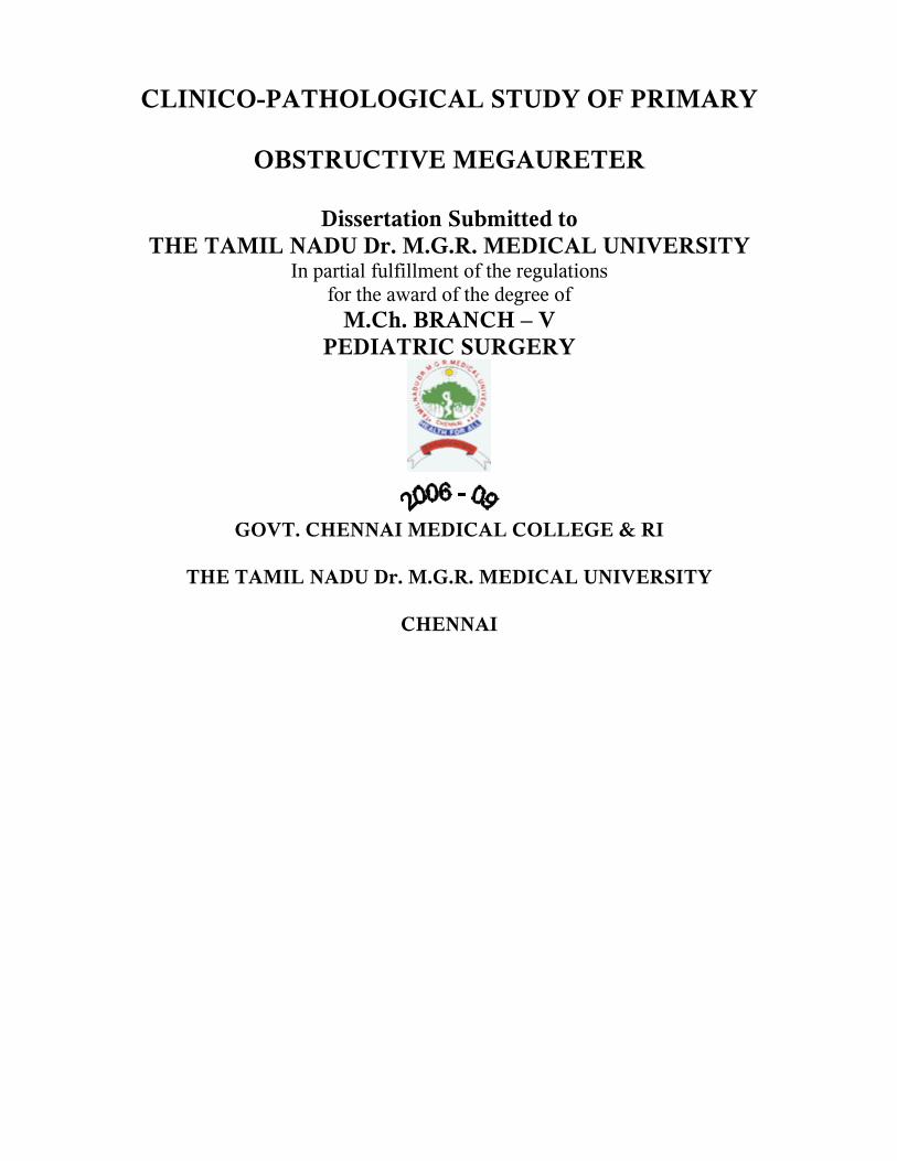
Figure 6 H & E Staining of Distal Ureteral Segment in Primary Obstructive Megaureter
Figure 7 H & E Staining of Distal Ureteral Segment in Primary Obstructive Megaureter
Megaureter Distal narrow ureteral segment
Longitudinal Muscle HypoplasiaCircular Muscle Hyperplasia
Collagen Infiltration

Proforma
Personal Details -
Name Age SexIP No.AddressDateAnte-natal Scan –
□ Done Report - □ Not Done -
Clinical Symptoms
□ Asymptomatic □ Febrile UTI □ Haematuria □ Enuresis □ Abdominal Pain □ Encopresis □ Others Laterality
□ U/L (Rt/Lt)□ B/L-
Investigations
Renal Parameters
BU/SC/SE - Contra-lateral Renal Status (in U/L cases and other problems) -
U/S KUBU Report - Retro-vesical ureteric size -
MCU –
IVU –
DTPA Scan – □ Done - □ Not Done - Report
Treatment
□ Observation -

□ Surgery - Surgical Details
Age at surgeryNo of SurgeriesTypeApproachUreteric Remodeling □ Done - □ Not Done - HPE Report
□ Circular Muscle Hyperplasia- □ Longitudinal Muscle Hypoplasia - □ Collagen Infiltration - □ Inflammation - □ Mucosal Appearance -
Post-op Evaluation
Clinical Symptoms
□ Asymptomatic □ Febrile UTI □ Haematuria □ Enuresis □ Abdominal Pain □ Encopresis □ Others Renal Parameters
BU/SC/SE - U/S KUBU Report -
Retro-vesical ureteric size - MCU –
IVU –
DTPA Scan –
□ Done - □ Not Done - Report
Treatment
□ Observation – □ Medical – □ Surgical (Repeat)
Master Chart
Master chart_POM.xls

Observations
This study of primary obstructive megaureter presented to Pediatric Surgery and Pediatric Urology Departments, ICH&HC, MMC, Chennai was undertaken between Jan 1, 2004 and Dec 31, 2008 (5 years). The following facts were obtained:During this study period, 38 patients who met the selection criteria attended the pediatric surgery and pediatric urology OPD. Distribution of Patients – State wise
State No. of Cases
Tamil Nadu 35
Andhra Pradesh 3
Total 38

Distribution of Patients – District wiseDistrict No. of Cases
Chennai 14
Kanchipuram 5
Tiruvannamalai 6
Vellore 1
Vilupuram 2
Erode 1
Krishnagiri 5
Perambalur 1
Chitoor 2
Krishna 1
Total 38
Age Distribution in Numbers
Range -> Neonate to 12 yearsMaximum Incidence -> Infants
Age Distribution in Percentage
Age No. of Cases
Neonatal 2
Infants 14
>1 yr to <3 yrs 8
>3 yrs. to <6 yrs. 6
>6 yrs. to <9 yrs. 6
>9 yrs. to <12 yrs. 2
Total 38

Age Distribution – Infants (in Nos.)
Maximum Incidence in infants -> 1 to 3 months
Age No. of Cases
Neonatal 5.26%
Infants 36.85%
>1 yr to <3 yrs 21.05%
>3 yrs. to <6 yrs. 15.79%
>6 yrs. to <9 yrs. 15.79%
>9 yrs. to <12 yrs. 5.26%
Total 100%
Age No. of Cases
>1 mth. to <3 mths. 6
>3 mths. to <6 mths. 3
>6 mths. to <9 mths. 5
>9 mths. to <12 mths. 0
Total 14
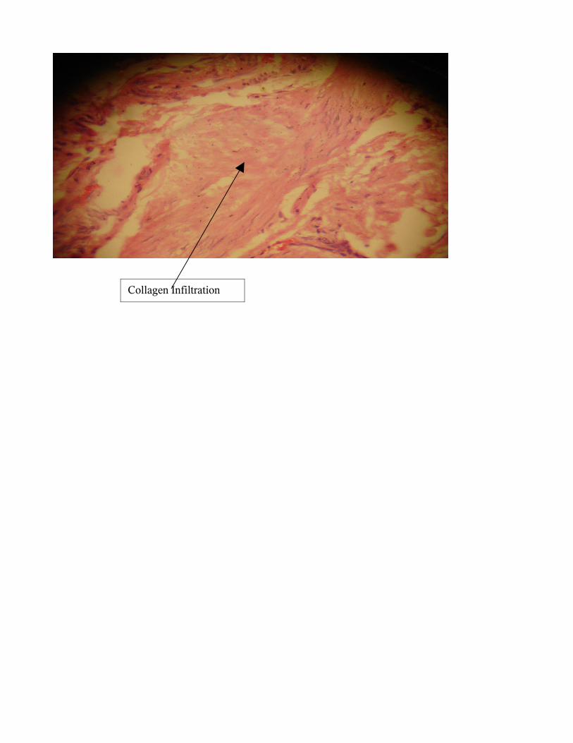
Sex Distribution (in Nos.)
Ratio – Male:Female = 5.3:1
Laterality Distribution (in Nos.)
Ratio U/L:B/L = 4.4:1Rt:Lt = 1.2:1
Sex No. of Cases
Male 32
Female 6
Total 38
Laterality No. of Cases
Right 17
Left 14
Bilateral 7
Total 38

Symptoms Distribution (in Nos.)
Most common presentation: Febrile UTISecond most presentation: Asymptomatic patients due to post-natal evaluation of ante-natally diagnosed cases
Distribution of Contra-lateral Renal Status and Other Problems
Percentage of contra-lateral renal problem in unilateral cases: 7.14%Distribution of Ante-natal Scan Status
Symptoms No. of Cases
Encopresis 1
Haematuria 1
Abdominal mass – Lt side 1
Enuresis 3
Dysuria 2
Asymptomatic 10
Febrile UTI 24
Abdominal Pain 9
Contra-lateral Renal Status
(in Unilateral Cases)
No. of Cases
Normal 26
MCDK 1
PUJ Problem – Left side 1
Distal Penile Hypospadias 1
Contra lateral Dysplastic Ureter 1
Lower Ureteric Calculus 1
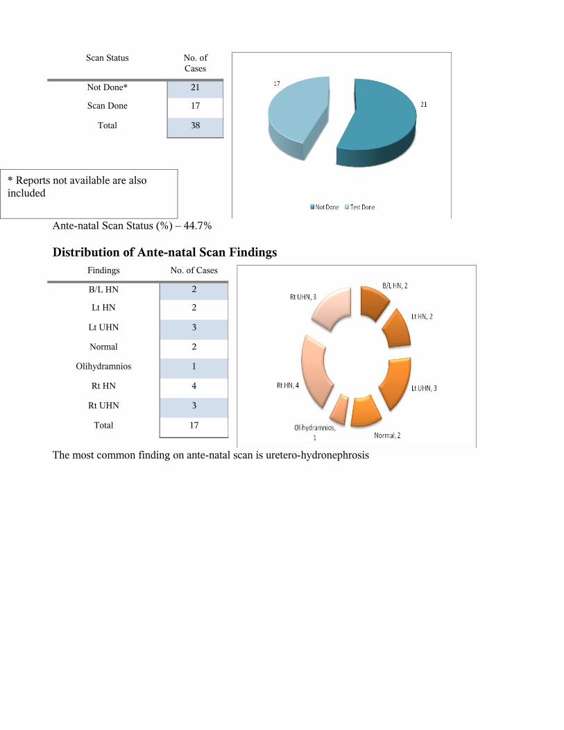
Ante-natal Scan Status (%) – 44.7%
Distribution of Ante-natal Scan Findings
The most common finding on ante-natal scan is uretero-hydronephrosis
Scan Status No. of Cases
Not Done* 21
Scan Done 17
Total 38
Findings No. of Cases
B/L HN 2
Lt HN 2
Lt UHN 3
Normal 2
Olihydramnios 1
Rt HN 4
Rt UHN 3
Total 17
* Reports not available are also included

Distribution of Ureter SizeUreter Size (in Cms) Numbers
>0.5 to <1 10
>1 to <1.5 21
>1.5 to <2 7
>2 to <2.5 5
Total 43
Maximum incidence of ureteric size – Between 1 and 1.5 cms
Distribution of DTPA Scan StatusDTPA Scan Numbers
Done 25
Not done 13
Total 38

Comparison of Age at Presentation Vs Age at SurgeryAge at Presentation Vs
Age at SurgeryNumbers
Same 24
Different 13
Total 37 *
13 patients underwent surgery after an initial period of observation from the time of presentation. These patients belong to the period of infancy.

Observation Period in Infants
Two patients underwent surgical procedure prior to definitive ureteric reimplantation.
Distribution of HPE Report HPE
ReportNo. of Cases
Available 20
Not Available
18
Total 38
Case No. Age at Presentation (in Mths.)
Age at First Surgery
(in Mths.)
Age at Second Surgery
(in Mths.)
Series 1 Series 2 Series 3
1 9 12 -
2 6 12 -
3 .022 (26 days) 1 24
4 2 9 -
5 9 12 -
6 8 8 9
7 2 6 -
8 3 6 -
9 5 6 -
10 2 24 -
11 9 12 -
12 3 9 -
13 2 36 -

HPE Report Positive CasesParameters No. of
Cases
Circular Muscle Hyperplasia 18
Longitudinal Muscle Hypoplasia 18
Collagen Infiltration 15
Inflammation 11
Mucosal Appearance (Ulcerated) 8
The coincidence of Circular Muscle Hyperplasia and Longitudinal Muscle Hypoplasia was noted in 18 patients.
Type of SurgeryType No. of
Cases
Cohen's Uretero-neocystostomy 31
Politano Leadbetter 3
Nephro-ureterectomy 1
Loop ureterostomy * 1
PCN * 1
Total 37
* Underwent Cohen's Uretero-neocystostomy second time.
The most common surgical procedure performed was Cohen's Uretero-neocystostomy. One patient underwent Nephro-ureterectomy at the first instance, due to poorly functioning pyo-nephrosis. Two patients underwent surgical procedure prior to definitive ureteric reimplantation.One patient presented at 26 days of life was evaluated. The patient underwent PCN in view of gross UHN. The other infant presented at 8 months of life, underwent loop ureterostomy in view of turbid system.Surgical Approach

Surgical Approach No. of Cases
Intra-vesical 27
Combined intra and extra vesical
10
Total 37
The most common surgical approach was intra-vesical approachUreteric-remodelling
Ureteric-remodelling No. of Cases
Excisional Tapering 7
Not done 30
Total 37
Out of 37 patients, only seven patients underwent ureteric remodeling in the form of excisional tapering (18.9%)

Post – OP Evaluation
Post operative evaluation, which includes clinical and investigational assessments was performed at six months after surgery. Only one patient in this study, who has not undergone surgery is asymptomatic and under observation. Clinical Symptoms
Clinical Symptoms No. of Cases
Asymptomatic* 20
Febrile UTI 13
Not turned up for Follow-up 5
Total* 38
*Includes one patient who is under observation (no surgery performed)
Number of patients who failed to follow up: 519 patients who followed up at six months after surgery were asymptomaticFebrile UTI was noticed in 13 patients during post-op follow up

Investigations
Pre-Op Vs Post-Op Ureter Size Comparison (U/S)Number Pre-Op 1 Pre-Op 2 Post-Op 1 Post-Op 2
1 1.9 - 1.4 -2 0.8 - 0.7 -
3 1.0 - 0.6 -
4 1.2 - 0.7 -
5 2.0 - 0.8 -
6 0.8 - 0.6 -
7 1.1 - 0.7 -
8 1.2 - 0.8 -
9 1.9 - 0.4 -
10 1.5 1.2 0.9 0.8
11 1.6 - 0.9 -
12 1.5 1.8 1.0 1.2
13 1.2 - 0.8 -
14 2.3 - 1.2 -
15 1.3 1.8 1.0 1.2
16 1.4 - 1.0 -
17 1.6 - 1.2 -
18 0.7 0.8 0.5 0.6
19 1.3 - 1.2 -
20 0.9 2.1 0.8 1.2
21 1.4 - 1.2 -
22 1.5 - 1.0 -
23 1.2 - 1.4 -
24 1.0 1.2 1.2 1.4
25 1.5 - 1.2 -
26 2.4 - 2.0 -
27 1.4 - 1.2 -
28 1.8 2.2 1.3 3.0
29 0.8 - 0.7 -
30 0.8 - 0.6 -
31 1.3 - 1.1 -
32 1.1 - 0.8 -

MCUMCU No. of
Cases
Normal 28
VUR 4
Not turned up for Follow-up 5
Total 37
Four patients developed VUR during follow up in 32 patientsOne patient with grade 3 VUR underwent redo ureteric re-implantation. Other three patients which includes one grade 2 VUR and two grade 1 VUR were observed with chemo-prophylaxis. IVU
* 1 Grade 3 1 Grade 2 2 Grade 1

IVU No. of Cases
Normal 26
Persistent Obstruction 6
Total 32
Out of 32 patients who underwent IVU evaluation in the post-op period, 26 patients had uretero-hydronephrosis with improvement in drainage of contrast in two hours. Persistent obstruction at vesico-ureteric juntion was noticed in 6 patients.

Repeat Surgery during follow-upSurgery No. of
Cases
Not Done 27
Cohen's Uretero-neocystostomy 4
Nephro-ureterectomy 1
Total 32
Four patients during the follow-up underwent re-do Cohen's Uretero-neocystostomy for persistent obstruction and VUR – Grade 3 in one case. One patient underwent nepho-ureterectomy during follow-up due to poorly functioning pyo-nephrosis. Two other patients with persistent obstruction during post-op evaluation were under chemo-prophylaxis and awaiting for surgery.

Discussions
During this study period of five years, from Jan 1, 2004 to Dec 31, 2008, a total of 38 patients were presented to the out patient departments of pediatric surgery and pediatric urology ICH & HC, MMC, Chennai, with signs and symptoms of primary obstructive megaureter.38 patients who fulfilled the criteria were taken into the study. These patients were compared with the study groups held at Princess Margaret Hospital, Perth, Australia, University of Rome, Italy, and Erasmus University, Rotterdam, The Netherlands as follows:
No of Cases - Study Group
College Year Case
Princess Margaret Hospital, Perth, Australia 1978 - 1988 23
University of Rome, Italy 1981 - 1991 38
ICH & HC, Chennai, India 2004 - 2008 38
23
38 38
0
5
10
15
2025
30
35
40
No
. o
f C
ases
1978 - 1988 1981 - 1991 2004 - 2008
Princess MargaretHospital, Perth,
Australia
University of Rome,Italy
ICH & HC, Chennai,India
Year of Study & College
The most common age group at presentation were infants. Most of them were asymptomatic and were diagnosed by post-natal evaluation of ante-natally diagnosed cases. The proportion of infants in the these study groups were compared as below:
Proportion of Infants in Total Cases
College Infants Total
Princess Margaret Hospital, Perth, Australia 11 23
University of Rome, Italy 26 38
ICH & HC, Chennai, India 14 38

11
2326
38
14
38
010203040506070
No
. o
f C
ases
PrincessMargaret
Hospital, Perth,Australia
University ofRome, Italy
ICH & HC,Chennai, India
College
Total
Infants
The sex distribution in our study was male:female = 5.3:1 and the other ratios are as follows:Male - Female Ratio
College Ratio
Princess Margaret Hospital, Perth, Australia 2.8:1
University of Rome, Italy 3.3:1
Erasmus University, Rotterdam, The Netherlands 4.0:1
ICH & HC, Chennai, India 5.3:1
2.8
1
3.3
1
4
1
5.3
1
012345
67
Male
- F
em
ale
R
ati
o
PrincessMargaretHospital,
Perth,Australia
University ofRome, Italy
ErasmusUniversity,Rotterdam,
TheNetherlands
ICH & HC,Chennai,
India
College
Female
Male
The laterality distribution between unilateral and bilateral were 4.4:1. In unilateral cases, the right:left side involvement were 1.2:1 and compared with other studies as below:
Laterality
College Right Left Bilateral
Princess Margaret Hospital, Perth, Australia 3 15 5
University of Rome, Italy 8 6 12
ICH & HC, Chennai, India 17 14 7

3
15
5
8
6
12
17
14
7
0
2
4
6
8
10
12
14
16
18
No
. o
f C
ases
PrincessMargaret
Hospital, Perth,Australia
University ofRome, Italy
ICH & HC,Chennai, India
College
Right
Left
Bilateral
The most common symptom at presentation was febrile UTI. In our study which accounted for 24 out of 38 patients, followed by 10 asymptomatic patients. This is incomparable with the study held at Princess Margaret Hospital, Perth, Australia, in which febrile UTI was noted in 14 patients out of 23.The contra-lateral renal problems were noted as MCDK in one patient and PUJ problem in another patient. The presence of dysplastic or absent contra-lateral kidney in unilateral cases of primary obstructive megaureter, was 3.26%, which is low in comparison with 10 – 15% as noted in the study held at Erasmus University, Rotterdam, The Netherlands.Ante-natal scan was done in 17 out of 38 patients, which helped in diagnosing the condition in 14 patients, which is less as compared to 21 cases of ante-natally diagnosed patients out of 26 infants in the study held at University of Rome, Italy.The number of retro-vesical ureteric size found in the range between more than 1 and 1.5 were 21 in our study which lie in the unresolved group as specified in the study held at department of urology, Children’s hospital, Harvard Medical School, Boston, Massachusetts. In our study, DTPA scan evaluation was done in 25 patients only due to the non-availability of this facility in our institute. It depends on the affordability of the patient. In our study, the age at presentation to the hospital and the age at surgery were compared. 24 patients were evaluated and underwent definitive ureteric reimplantation procedure in the same admission. 13 infants out of 38 patients were observed initially followed by definitive surgery.Two patients underwent surgical procedure prior to definitive ureteric reimplantation.One patient presented at 26 days of life was evaluated. The patient underwent PCN in view of gross UHN. The other infant presented at 8 months of life, underwent loop ureterostomy in view of turbid system. There was a significant improvement in the renal funtion by DTPA scan from 20% at the time of loop ureterostomy-left side (8 months of life) to 40% at the time of ureteric reimplantation (24 months of life).During the definitive surgical procedure, Ureteric remodeling was performed in only 7 out of 37 patients. It varied with the surgeon’s preference and the type of ureteric remodeling.In our study, reimplantation procedure failed in 10 patients out of 32 cases (5 patients failed to follow-up after surgery) after a 6 months period of follow-up. In this complication, 6 patients had persistent obstruction and four patients developed reflux. This is in comparison with the reimplantation failure noted in 6 patients (3 cases with persistent obstruction and 3 cases with reflux) out of 17 patients after a

one-year period of follow up in the study held at Princess Margaret Hospital, Perth, Australia (1978 – 1988)In this study of cases with primary obstructive megaureter, 37 out of 38 cases underwent surgery. Only one asymptomatic patient is presently under observation as shown in the Master Chart.

Conclusion
The patients with primary obstructive megaureter are noted predominantly in infants (36.85%) with maximal incidence between 1 to 3 months (6 out of 14 infants)
In this study, the sex distribution is found as male : female – 5.3 : 1, with definitive male preponderance
The laterality distribution is noted as U/L : B/L - 4.4 : 1, with slight predominance of right side over left side as 1.2 : 1
In this study, the most common symptom at presentation is febrile UTI (63.15%), followed by asymptomatic patients (26.32%)
The contra-lateral renal dysplasia in U/L cases is found to be 3.26%
The majority of cases with retro-vesical ureteric size lie between 1 to 1.5 cm (48.8%), which are not considered to belong to the spontaneously resolving group.
In this study, all patients primary obstructive megaureter are noted to have normal renal bio-chemical parameters, irrespective of bilateral presentation.
MCU and IVU are the most common investigational modality performed.
In this study, patients presented during infancy were initially observed prior to surgical intervention. But those patients presented beyond infancy were evaluated and intervened surgically during the same admission.
Cohen’s Uretero-neocystostomy with intra-vesical approach is the surgical procedure of choice, in majority of the case, in this study.
The histo-pathological evaluation revealed co-existence of circular muscle hyperplasia, longitudinal muscle hypoplasia, and collagen infiltration in majority of cases followed by features of inflammation and mucosal ulceration.
The re-implantation failure was noted in 28.12% ( includes persistent obstruction and minimal grades of VUR ).

Bibliography
1. Ruano-Gil D, Coca-Payeras A, Tejedo-Mateu A. Obstruction and normal recanalization of the ureter in the human embryo. Its relation to congenital ureteral obstruction. Eur Urol 1975; 1:287–93.
2. Alcaraz A, Vinaixa F, Tejedo-Mateu A et al. Obstruction and recanalization of the ureter during embryonic development. J Urol 1991; 145:410–16.
3. Yerkes E, Nishimura H, Miyazaki Y et al. Role of angiotensin in the congenital anomalies of the kidney and urinary tract in the moose and the human. Kidney Int Suppl 1998; 67:575–7.
4. Chwalle R. The process of formation of cystic dilatations of the vesical end of the ureter and of diverticula at the ureteral ostium. Urol Cutan Rev 1927; 31:499–504.
5. Baker LA Gomez RA. Embryonic development of the ureter and bladder: acquisition of smooth muscle. J Urol 1998; 160:545–50.
6. Escala JM, Keating MA, Boyd G et al. Development of elastic fibres in the upper urinary tract. J Urol 1989; 141:969–73.
7. Hutch J. The mesodermal component: its embryology, anatomy, physiology and role in prevention of vesicoureteral reflux. J Urol 1972; 108:406–10.
8. Tanagho EA, Meyers FH, Smith DR. The trigone: anatomical and physiological considerations. I. In relation to the ureterovesical junction. J Urol 1968; 100(5):623–32.
9. Kakuchi J, Ichiki T, Kiyama S et al. Developmental expression of renal angiotensin II receptor genes in the mouse. Kidney Int 1995; 47:140–7.
10. Nishimura H, Yerkes E, Hohenfellner K et al. Role of the angiotensin type 2 receptor gene in congenital anomalies of the kidney and urinary tract, CAKUT, of mice and men. Mol Cell 1999; 3:1–10.
11. Hohenfellner K, Hunley E, Schloemer C et al. Angiotensin type 2 receptor is important in the normal development of the ureter. Pediatr Nephrol 1999; 13:187–91.
12. Gruss P, Walther C. Pax in development. Cell 1992; 69:719–22.
13. Torres M, Gomez-Pardo E, Dressler GR, Gruss P. Pax-2 controls multiple steps of urogenital development. Development 1995; 121:4057–65.
14. Sanyanusin P, Schimmenti L, McNoe LA et al. Mutation of the PAX2 gene in a family with optic nerve colobomas, renal anomalies and vesicoureteral reflux. Nat Genet 1995; 9:358–64.
15. Choi K, McNoe LA, French MC, Guilford PJ, Eccles MR. Absence of PAX2 gene mutations in patients with primary familial vesicoureteral reflux. J Med Genet 1998; 35:338–9
16. Lee BR, Partin AW, Epstein JI et al. A quantitative histological analysis of the dilated ureter of childhood. J Urol 1992; 48(5):1482–6.
17. Lee BR, Silver RI, Partin AW et al. A quantitative histologic analysis of collagen subtypes: the primary obstructed and refluxing megaureter of childhood. Urology 1998; 51(5):820–3.
18. Rabinowitz R, Barkin M, Schillinger JF et al. The influence of etiology on the surgical management and prognosis of the massively dilated ureter in children. J Urol 1978; 119(6):808–13.
19. Kaefer M, Rink R. Ureteral reimplantation. In: Caldamone A, ed. Atlas of the Urologic Clinics of North America. Amsterdam: Elsevier Publishers, 2004.
20. Shimada K, Hosokawa S, Tohda A, Matsumoto F, Johnin K. Histology of the fetal prune belly syndrome with reference to the efficacy of prenatal decompression.Int J Urol 2000; 7(5):161–6.
21. Lee BR, Partin AW, Epstein JI et al. A quantitative histological analysis of the dilated ureter of childhood. J Urol 1992; 148(5):1482–6.
22. Lee BR, Silver RI, Partin AW et al. A quantitative histologic analysis of collagen subtypes: the primary obstructed and refluxing megaureter of childhood. Urology 1998; 51(5):820–3.

23. Rabinowitz R, Barkin M, Schillinger JF et al. The influence of etiology on the surgical management and prognosis of the massively dilated ureter in children. J Urol 1978; 119(6):808–13.
24. Kaefer M, Rink R. Ureteral reimplantation. In: Caldamone A, ed. Atlas of the Urologic Clinics of North America. Amsterdam: Elsevier Publishers, 2004.
25. Shimada K, Hosokawa S, Tohda A, Matsumoto F, Johnin K. Histology of the fetal prune belly syndrome with reference to the efficacy of prenatal decompression. Int J Urol 2000; 7(5):161–6.
26. Blok C, van Venrooij GE, Coolsaet BL. Dynamics of the ureterovesical junction; a qualitative analysis of the ureterovesical pressure profile in the pig. J Urol 1985; 134(4):818–24.
27. Sampson J. Ascending renal infections; with special reference to the reflux of urine from the bladder into the ureters as an etiological factor in its causation and maintenance. Johns Hopkins Hosp Bull 1903; 14:334.
28. Paquin A. Ureterovesical anastomosis: the description and evaluation of a technique. J Urol 1959; 82:573–83.
29. Ransley PG, Risdon RA. Renal papillary morphology and intrarenal reflux in the young pig. Urol Res 1975; 3:105–9.
30. Noordzij JW, Dabhoiwala NF. A view on the anatomy of the ureterovesical junction. Scand J Urol Nephrol 1993; 27(3):371–80.
31. Shafik A. Ureterovesical junction inhibitory reflex and vesicoureteral junction excitatory reflex: description of two reflexes and their role in the ureteric antireflux mechanism. Urol Res 1996; 24(6):339–43.
32. Leung VY, Metreweli C, Yeung CK. The ureteric jet doppler waveform as an indicator of vesicoureteric sphincter function in adults and children. An observational study. Ultrasound Med Biol 2002; 28(7):865–72.
33. Shafik A. Sigmoido-rectal junction reflex: role in the defecation mechanism. Clin Anat 1996; 9(6):391–4.
34. Leung VY, Metreweli C, Yeung CK. Immature ureteric jet doppler patterns and urinary tract infection and vesicoureteric reflux in children. Ultrasound Med Biol 2002; 28(7):873–8.
35. Hannan QH, Stephens D. The influence of trigonectomy on vesicoureteral reflux in dogs. Invest Urol 1973; 10(6):469–72.
36. Cussen LJ. The morphology of congenital dilatation of the ureter: Intrinsic ureteral lesions. Aust NZ J Surg 1971; 41:185.
37. Sripathi V, King PA, Thompson MR, Bogle MS. Primary Obstructive Megaureter. JPS 1991; 26(7):826-29
38. King LR. Megaloureter: definition, diagnosis and management (guest editorial). J Urol 1980; 123:222–3.
39. Shokeir AA, Nijman RJM. Primary megaureter: current trends in diagnosis and treatment. BJU Int 2000; 86:861–8.
40. Estroff JA, Mandell J, Benacerraf BR. Increased renal parenchymal echogenicity in the fetus: importance and clinical outcome. radiology 1991; 181:135–9.
41. Shokeir AA, Provoost AP, el-Azab M, Dawaba M, Nijman RJ. Renal Doppler ultrasound in children with normal upper urinary tracts: effect of fasting, hydration with normal saline, and furosemide administration. Urology 1996; 47:740–4.
42. Shokeir AA, Provoost AP, el-Azab M, Dawaba M, Nijman RJ. Renal Doppler ultrasound in children with obstructive uropathy: effect of intravenous normal saline fluid load and furosemide. J Urol 1996; 156: 1455–8.
43. Whitefield HN, Btitton LE, Hendry WF et al. The distinction between uropathy and nephropathy by radioisotope transit times. Br J Urol 1978; 50:433-8
44. English PJ, Testa HJ, Lawson RS et al. Modified methods of dieresis renography for the assessment of equivocal pelviureteric obstruction. Br J Urol 1987; 59: 10-4
45. Heyman S, Duckett JW. The extraction factor: an estimate of single kidney function in children during routine radio-nuclide renography with 99m-technetium diethylenetriaminepentaacetic acid. J Urol 1988; 140: 780-3.
46. Kass EJ, Fint-Bennett D. Contemporary techniques for the radioisotopic evaluation of the dilated urinary tract. Urol Clin N Am 1990; 17: 273-89.
47. Cartwright PC, Duckett JW. Keating MA et al. Manageing apparent ureteropelvic junction obstruction in the newborn. J Urol 1992; 148: 1224-8.
48. Ransley P, Dhillon H, Gordon i et al. The postnatal management of hydronephrosis diagnosed by prenatal ultrasound. J Urol 1990; 144: 584-7
49. Platt JF, Rubin JM, Ellis HM, DiPietro MA. Duplex Doppler US of the kidney: differentiation of obstructive from non-obstructive dilation. Radiology 1989; 17: 515-7.
50. Shokeir AA, Provoost AP, El-Azab M et al. Renal Doppler ultrasound in children with normal upper urinary tracts:effect of fasting, hydration with normal saline and furosemide administration. Urology 1996; 47: 740-4
51. Shokeir AA, Provoost AP, El-Azab M et al. Renal Doppler ultrasound in children with obstructive uropathy: effect of intravenous normal saline fluid load and furoscmide. J Urol 1996; 156: 1455-8.
52. Gilbert R, Garra B, Giddson MD. Renal duplex Doppler ultrasound: and adjunct in the evaluation of hydronephrosis in the child. J Urol 19993; 150: 1192-4.
53. Shokeir AA, Nijman RJM, El-Azab M, Provoost AP. Partial ureteric obstruction: a study of Doppler ultrasonography and diuretic renography in different grades and durations of obstruction. Br J Urol 1996; 78: 829-35.
54. Nicotina PA, Romeo C, Arena F, Romeo G. Segmental up-regulation of transforming growth factor-β in the pathogenesis of primary megaureter. An immunocytochemical study. Br J Urol 1997; 80:946–9.
55. Peters CA, Mandell J, Lebowitz RL. Congenital obstructed megaureters in early infancy: diagnosis and treatment. J Urol 1989; 142: 641-5.
56. Mollard P, Foray P, Godoy JL, Valigant C. Management of primary obstructive megaureter without reflux in neonates. Eur Urol 1993; 24: 505-10.
57. Sheu JC, Chang PY, wang NL et al. Is surgery necessary for primary non-refluxing megaureter? Pediatr Surg Int 1998; 13: 501-13.
58. Keating MA, Escala J, Snyder HM et al. Changing concepts in management of primary obstructive megaureter. J Urol 1989; 142: 636-40.
59. Baskin LS, Zderic SA, Snyder HM, Duckett JW. Primary dilated megaureter: long-term follow-up. Urol 1994; 152: 618-21.
60. Cozzi F, Madonna L, Maggi E et al. Management of primary megaureter in infancy. J Pediatr Surg 1993; 28: 1031-3.
61. Arena F, Baldari S, Proietto F et al. Conservative treatment in primary neonatal megaureter. J Prediatr Surg 1998; 8:347-51.
62. Vereecken RL, Proesmans W. A review of ninety-two obstructive megaureter in children. Eur Urol 1999; 36: 342-7.
63. Williams DI. The chronically dilated ureter: Hunterian Lecture. Ann R Coll Surg 1954; 14:107. 64. Burbige KA, Lebowitz RL, Colodny AH, Bauer SB, Retik AB. The megacystis–megaureter syndrome. J Urol 1984; 131:1133–6.
64. Mandell J, Lebowitz RL, Peters CA et al. Prenatal diagnosis of the megacystis–megaureter association. J Urol 1992; 148:1487–9.
65. Lettgen B, Kropfl D, Bonzel KE et al. Primary obstructed megaureter in neonates. Treatment by temporary reterocutaneostomy. Br J Urol 1993; 72:826–9.
66. Arena F, Baldari S, Proietto F et al. Conservative treatment in primary neonatal megaureter. Eur J Pediatr Surg 1998; 8:347–51.
67. Sheu JC, Chang PY, Wang NL, Tsai TC, Huang FY. Is surgery necessary for primary non-refluxing megaureter? Pediatr Surg Int 1998; 13:501–3.
68. Mollard P, Foray P, de Godoy JL. Management of primary obstructive megaureter without reflux in neonates. Eur Urol 1993; 24:505–10.
69. Stehr M, Metzger R, Schuster T, Porn U, Dietz HG. Management of the primary obstructed megaureter (POM) and indication for operative treatment. Eur J Pediatr Surg 2002; 12(1):32–7.
70. Peters CA, Mandell J, Lebowitz RL et al. Congenital obstructed megaureters in early infancy: diagnosis and

treatment. J Urol 1989; 142:641–5.
71. Atala A, Keating MA. Vesicoureteral reflux and megaureter. In: Walsh PC, Retik AB, Vaughan ED, Wein AJ, eds. Campbell’s Urology, 7th edn, vol 3. Philadelphia: WB Saunders, 1998.
72. Starr A. Ureteral plication. A new concept in ureteral tapering for megaureter. Invest Urol 1979; 17:153–8.
73. Kalicinski ZH, Kansy J, Kotarbinska B, Joszt W. Surgery of megaureters – modification of Hendren’s operation. J Pediatr Surg 1977; 12:183–8.
74. Hendren WH. Operative repair of megaureter in children. J Urol 1969; 101:491–507.
75. Hanna MK. New surgical method for one-stage total remodeling of massively dilated and tortuous ureter: tapering in situ technique. Urology 1979; 14:453–64.
76. Perdzynski W, Kalicinski ZH. Long-term results after megaureter folding in children. J Pediatr Surg 1996; 31:1211–17.
77. Ehrlich RM. The ureteral folding technique for megaureter surgery. J Urol 1985; 134:668–70.
78. Parrot TS, Woodard JR, Wolpert JJ. Ureteral tailoring: a comparison of wedge resection with infolding. J Urol 1990; 144:328–9.
79. Fretz PC, Austin JC, Cooper CS, Hawtrey CE. Longterm outcome analysis of Starr plication for primary obstructive megaureters. J Urol 2004; 172(2): 703–5.
80. Daher P, Diab N, Ghorayeb Z, Korkmaz G. The Kalicinski ureteral folding technique for megaureter in children. Experience in 23 cases. Eur J Pediatr Surg 1999; 9(3):163–6.