Spectrum of Leprosy Patients with Clinico-Histopathological Correlation: A Hospital Based Study
6
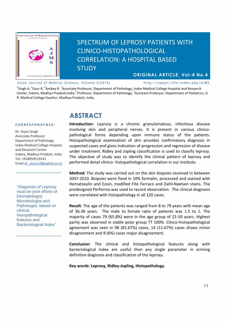
Introduction: Leprosy is a chronic granulomatous, infectious disease involving skin and peripheral nerves. It is present in various clinico- pathological forms depending upon immune status of the patients. Histopathological examination of skin provides confirmatory diagnosis in suspected cases and gives indication of progression and regression of disease under treatment. Ridley and Jopling classification is used to classify leprosy. The objective of study was to identify the clinical pattern of leprosy and performed detail clinico- histopathological correlation in our institute. Method: The study was carried out on the skin biopsies received in between 2007-2010. Biopsies were fixed in 10% formalin, processed and stained with Hematoxylin and Eosin, modified Fite Ferraco and Ziehl-Neelsen stains. The predesigned Performa was used to record observation. The clinical diagnosis were correlated with histopathology in all 120 cases. Result: The age of the patients was ranged from 8 to 79 years with mean age of 36.38 years. The male to female ratio of patients was 1.5 to 1. The majority of cases 79 (65.8%) were in the age group of 21-50 years. Highest parity was observed in stable polar group TT 100%. Clinco-histopathological agreement was seen in 98 (81.67%) cases, 14 (11.67%) cases shows minor disagreement and 8 (6%) cases major disagreement. Conclusion: The clinical and histopathological features along with bacteriological index are useful than any single parameter in arriving definitive diagnosis and classification of the leprosy. Key words: Leprosy, Ridley-Jopling, Histopathology. ABSTRACT CORRESPONDENCE : Dr. Arjun Singh Associate Professor Department of Pathology Index Medical College Hospital and Research Center Indore, Madhya Pradesh, India Tel: +918959519141 Email [email protected] ORIGINAL ARTICLE, Vol-4 No.4 Asian Journal of Medical Science, Volume-4(2013) http://nepjol.info/index.php/AJMS SPECTRUM OF LEPROSY PATIENTS WITH CLINICO-HISTOPATHOLOGICAL CORRELATION: A HOSPITAL BASED STUDY 1 Singh A, 2 Gaur R, 3 Ambey R. 1 Associate Professor, Department of Pathology, Index Medical College Hospital and Research Center, Indore, Madhya Pradesh,India. 2 Professor, Department of Pathology, 3 Assistant Professor, Department of Pediatrics, G. R. Medical College Gwalior, Madhya Pradesh, India. “Diagnosis of Leprosy must be joint efforts of Dermatologist, Microbiologist and Pathologist, based on clinical, histopathological features and Bacteriological Index” 11
-
Upload
independent -
Category
Documents
-
view
0 -
download
0
Transcript of Spectrum of Leprosy Patients with Clinico-Histopathological Correlation: A Hospital Based Study
Introduction: Leprosy is a chronic granulomatous, infectious disease
involving skin and peripheral nerves. It is present in various clinico-
pathological forms depending upon immune status of the patients.
Histopathological examination of skin provides confirmatory diagnosis in
suspected cases and gives indication of progression and regression of disease
under treatment. Ridley and Jopling classification is used to classify leprosy.
The objective of study was to identify the clinical pattern of leprosy and
performed detail clinico- histopathological correlation in our institute.
Method: The study was carried out on the skin biopsies received in between
2007-2010. Biopsies were fixed in 10% formalin, processed and stained with
Hematoxylin and Eosin, modified Fite Ferraco and Ziehl-Neelsen stains. The
predesigned Performa was used to record observation. The clinical diagnosis
were correlated with histopathology in all 120 cases.
Result: The age of the patients was ranged from 8 to 79 years with mean age
of 36.38 years. The male to female ratio of patients was 1.5 to 1. The
majority of cases 79 (65.8%) were in the age group of 21-50 years. Highest
parity was observed in stable polar group TT 100%. Clinco-histopathological
agreement was seen in 98 (81.67%) cases, 14 (11.67%) cases shows minor
disagreement and 8 (6%) cases major disagreement.
Conclusion: The clinical and histopathological features along with
bacteriological index are useful than any single parameter in arriving
definitive diagnosis and classification of the leprosy.
Key words: Leprosy, Ridley-Jopling, Histopathology.
ABSTRACT
C O R R E S P O N D E N C E :
Dr. Arjun Singh
Associate Professor
Department of Pathology
Index Medical College Hospital
and Research Center
Indore, Madhya Pradesh, India
Tel: +918959519141
Email [email protected]
ORIGINAL ARTICLE, Vol-4 No.4
Asian Journal of Medical Science, Volume-4(2013) http://nepjol . info/index.php/AJMS
SPECTRUM OF LEPROSY PATIENTS WITH
CLINICO-HISTOPATHOLOGICAL
CORRELATION: A HOSPITAL BASED
STUDY
1Singh A,
2Gaur R,
3Ambey R.
1Associate Professor, Department of Pathology, Index Medical College Hospital and Research
Center, Indore, Madhya Pradesh,India.2
Professor, Department of Pathology, 3Assistant Professor, Department of Pediatrics, G.
R. Medical College Gwalior, Madhya Pradesh, India.
“Diagnosis of Leprosy must be joint efforts of Dermatologist, Microbiologist and Pathologist, based on clinical, histopathological features and Bacteriological Index”
11

Page 12 Asian Journal of Medical Sciences 4(2013) 11-16
Leprosy also known as Hansen’s disease, is a
chronic granulomatous, infectious disease involving
skin, peripheral nerves.1 The three cardinal sign of
the disease are skin lesions, skin anesthesia and
enlarged peripheral nerves.2 In India despite
declaring leprosy elimination at national level in
January 2006,3 it is still a disease of public health
importance and endemic in many of states. The
Leprosy is a major public health problem of the
developing countries with an estimated total global
new cases detected in 2009 were 2, 27, 849 and
India account 1, 33, 717 (58.7%) cases.4
Leprosy present in various clinico-pathological
forms depending upon immune status of the
patients.5 The study of pathological changes in
leprosy help in understanding of disease,
complications and its exact typing.6 Diagnosis of
leprosy must be joint efforts of dermatologist,
microbiologist and pathologist. Leprosy can be
diagnosed by various methods including detail
clinical examination of the skin lesions and
peripheral nerves,7,8
demonstration of the Acid Fast
Bacilli (AFB) in slit skin smears by Ziehl-Neelsen
staining,9
Histopathological section,6,10
demonstration of bacilli by modified Fite-Ferraco
procedure11
, and Fine Needle Aspiration Cytology
(FNAC) of skin and nerves.12
Histopathological examination of skin provides
confirmatory information in suspected case and
gives indication of progression and regression of
disease under treatment.13
Ridley and Jopling have
suggested immunological basis of leprosy and
classified in to five types; Tuberculoid (TT),
Borderline Tuberculoid (BT), Midborderline (BB),
Borderline Lepromatous (BL), and Lepromatous
(LL).14
Later they develop clinical and bacteriological
findings in each group with respective
immunological and histopathological findings.7
The objectives of present study were to identify the
clinical pattern of leprosy and perform detail
clinico- histopathological correlation in our
institute.
INTRODUCTION MATERIALS AND METHODS The present study was carried out on the elliptical
skin biopsies received from Department of
Dermatology in the Histopathology section of
Department of Pathology, Sri Venkateshwara
Medical College Hospital and Research Centre,
Pondicherry, from June 2007-May 2010. All the
cases were selected regardless of their age, sex,
socioeconomical status, occupation and
community. Biopsies were fixed in 10% formalin
and processed. Hematoxylin and Eosin15
stained
slides of skin biopsy of all leprosy patient were
studied in detail. The sections were stained for
modified Fite-Ferraco11
stain and Ziehl-Neelsen9
staining wherever is required for the
demonstration of mycobacterium bacilli. The
predesigned proforma was used to record
observations. The biopsies were studied for the
epidermal atrophy, epitheloid granuloma,
lymphocytic and histiocytic infiltration of nerves
bundles and Grenz zone.
The clinical diagnosis of leprosy cases as provided
by Dermatology Department in to TT, BT, BB, BL,
and LL based on Ridley and Jopling classification
were correlated with histopathology in the
respective biopsies. Biopsies which did not include
full depth of dermis together with a portion of
subcutaneous fat were reported as inadequate and
requested to repeat biopsy in those cases.
RESULTS The Histopathology section of Pathology
department received 6435 specimen from June
2007 to May 2010. The skin biopsies were 760
which comprise 11.8% of the total
histopathological specimen. The leprosy biopsies
were 120 which account 15.8% of the skin biopsy
and 1.8% of total histopathology specimen.
The age of the patients ranges from 8 to 79 years
with mean age of 36.38 years. The male to female
ratio was 1.5 to 1. (Table No.1) The majority of
cases 79 (65.8%) were in the age group of 21-50
years. Clinical features were available in all cases
Asian Journal of Medical Sciences 4(2013) 11-16 Page 13
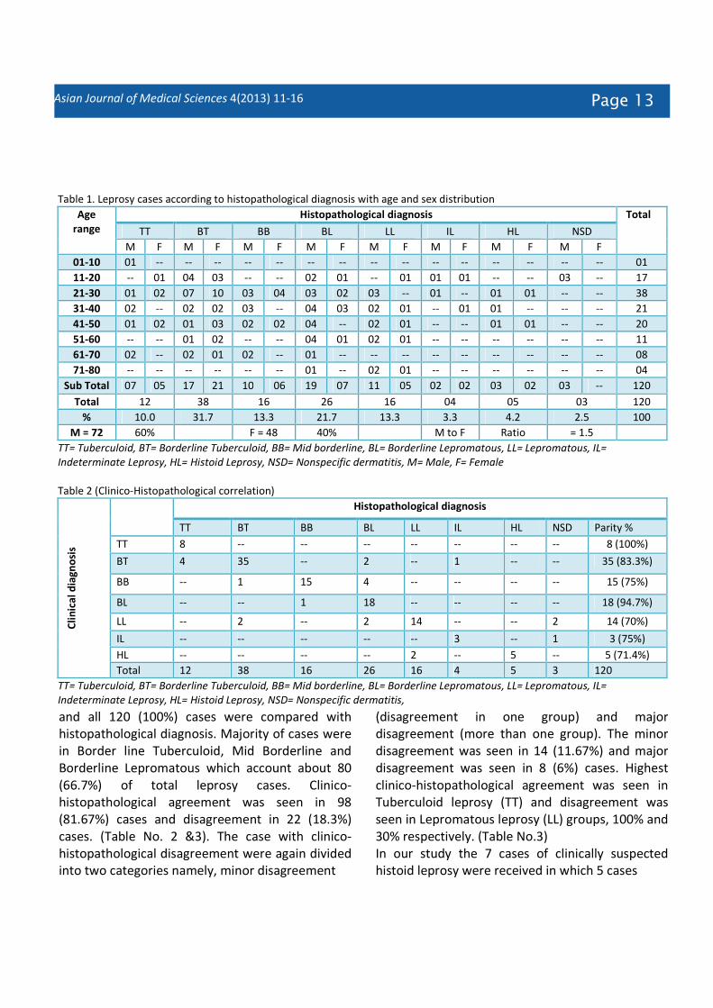
Table 1. Leprosy cases according to histopathological diagnosis with age and sex distribution
Age
range
Histopathological diagnosis Total
TT BT BB BL LL IL HL NSD
M F M F M F M F M F M F M F M F
01-10 01 -- -- -- -- -- -- -- -- -- -- -- -- -- -- -- 01
11-20 -- 01 04 03 -- -- 02 01 -- 01 01 01 -- -- 03 -- 17
21-30 01 02 07 10 03 04 03 02 03 -- 01 -- 01 01 -- -- 38
31-40 02 -- 02 02 03 -- 04 03 02 01 -- 01 01 -- -- -- 21
41-50 01 02 01 03 02 02 04 -- 02 01 -- -- 01 01 -- -- 20
51-60 -- -- 01 02 -- -- 04 01 02 01 -- -- -- -- -- -- 11
61-70 02 -- 02 01 02 -- 01 -- -- -- -- -- -- -- -- -- 08
71-80 -- -- -- -- -- -- 01 -- 02 01 -- -- -- -- -- -- 04
Sub Total 07 05 17 21 10 06 19 07 11 05 02 02 03 02 03 -- 120
Total 12 38 16 26 16 04 05 03 120
% 10.0 31.7 13.3 21.7 13.3 3.3 4.2 2.5 100
M = 72 60% F = 48 40% M to F Ratio = 1.5
TT= Tuberculoid, BT= Borderline Tuberculoid, BB= Mid borderline, BL= Borderline Lepromatous, LL= Lepromatous, IL=
Indeterminate Leprosy, HL= Histoid Leprosy, NSD= Nonspecific dermatitis, M= Male, F= Female
Table 2 (Clinico-Histopathological correlation)
Cli
nic
al
dia
gn
osi
s
Histopathological diagnosis
TT BT BB BL LL IL HL NSD Parity %
TT 8 -- -- -- -- -- -- -- 8 (100%)
BT 4 35 -- 2 -- 1 -- -- 35 (83.3%)
BB -- 1 15 4 -- -- -- -- 15 (75%)
BL -- -- 1 18 -- -- -- -- 18 (94.7%)
LL -- 2 -- 2 14 -- -- 2 14 (70%)
IL -- -- -- -- -- 3 -- 1 3 (75%)
HL -- -- -- -- 2 -- 5 -- 5 (71.4%)
Total 12 38 16 26 16 4 5 3 120
TT= Tuberculoid, BT= Borderline Tuberculoid, BB= Mid borderline, BL= Borderline Lepromatous, LL= Lepromatous, IL=
Indeterminate Leprosy, HL= Histoid Leprosy, NSD= Nonspecific dermatitis,
(disagreement in one group) and major
disagreement (more than one group). The minor
disagreement was seen in 14 (11.67%) and major
disagreement was seen in 8 (6%) cases. Highest
clinico-histopathological agreement was seen in
Tuberculoid leprosy (TT) and disagreement was
seen in Lepromatous leprosy (LL) groups, 100% and
30% respectively. (Table No.3)
In our study the 7 cases of clinically suspected
histoid leprosy were received in which 5 cases
and all 120 (100%) cases were compared with
histopathological diagnosis. Majority of cases were
in Border line Tuberculoid, Mid Borderline and
Borderline Lepromatous which account about 80
(66.7%) of total leprosy cases. Clinico-
histopathological agreement was seen in 98
(81.67%) cases and disagreement in 22 (18.3%)
cases. (Table No. 2 &3). The case with clinico-
histopathological disagreement were again divided
into two categories namely, minor disagreement

Page 14 Asian Journal of Medical Sciences 4(2013) 11-16
Table 3 (Disagreement in clinical and histopathological diagnosis)
Clinical diagnosis Cases Complete parity No. (%) Minor Disagreement
No. (%)
Major Disagreement No. (%)
TT 8 8 (100%) ---- ----
BT 42 35 (83.3%) 4 (9.5%) 3 (7.12%)
BB 20 15 (75%) 5 (25%) -----
BL 19 18 (94.7%) 1 (5.3%) ----
LL 20 14 (70%) 2 (11.2%) 4 (22.4%)
IL 4 3 (75%) ---- 1 (25%)
HL 7 5 (71.4%) 2 (28.6%) ----
Total 120 98 (81.67%) 14 (11.67%) 8 (6%)
TT= Tuberculoid, BT= Borderline Tuberculoid, BB= Mid borderline, BL= Borderline Lepromatous, LL= Lepromatous, IL=
Indeterminate Leprosy, HL= Histoid Leprosy, NSD= Nonspecific dermatitis,
were proven as Histoid leprosy while 2 cases as
Lepromatous leprosy. The Histoid leprosy cases
were 5 (4.2%) of total leprosy cases. The classical
microscopic and clinical photographs of the
tuberculoid leprosy and Lepromatous Leprosy are
provided, for the publication. (Fig. 1, 2, 3 and 4) Figure No. 1 Tuberculoid Leprosy showing neural tissue with
dense lymphocytic inflammation and dense collagen bundles
(Hematoxyline &Eosin stain at 10X x 10X)
Figure No.2 Lepromatous Leprosy showing foamy
macrophage and lymphocytic infiltration around neural
bundles (Hematoxyline & Eosin stain at 40X x 10X)
The age of the patients in present study varies
from 8 year to 79 year with mean age of 36.38
year. The maximum number of cases 79 (65.8%)
were observed in active age group of 21-50 years.
The Jindal N16
et al also observed maximum 47.8%
of cases in 20-40 year. Moorthy BN17
et al observed
Male to Female ratio of 1.8 to 1 which is close to
our study and Mittal RR16
et al observed Male to
Female ratio 3.25 to 1 which was very high. Clinical
spectrum of the leprosy cases in the present
studies revealed that most of the case were in
borderline categories BT,BB and BL which account
DISCUSSION The leprosy was classified on the basis of immunity
of the individual by Ridley and Jopling in to five
groups and it is very well correlate with the clinical,
histopathological and bacteriological findings.

Asian Journal of Medical Sciences 4(2013) 11-16 Page 15
Figure No. 3 Lepromatous Leprosy showing lepra bacilli in
clusters “globi appearance” (Fite Ferraco stain at 40X x 10X)
Figure No. 4 Lepromatous Leprosy showing facial nodules of
variable size - Clinical appearance
observed in stable polar group TT and LL 100% and
70% respectively which is similar to observation
made by the Kar PK21
et al TT (87.5%) and LL (70%).
The maximum parity observed in polar groups while
maximum disparity observed in Borderline cases
because polar cases showed a fixed histopathology
while borderline have different histopathology in
different sites and lesions.21
There is no independent gold standard for leprosy
diagnosis. The histopathology in leprosy cases
varied with the difference in sample size, choosing
the biopsy site, age of the lesion, immunological
and treatment status of the patient at the time of
biopsy.22, 23
In our study the clinico-histopathological parity of IL
was 75%, similar observation of similar observation
of 81.2% was reported by the KAR PK et al in their
study. The early leprosy lesion difficult to diagnose
even by experienced dermatologist. Histopathology
play very good role in the diagnosis of early leprosy
cases.17
In the present study the histoid leprosy was present
in 4.2% of cases. The incidence of the histoid
leprosy in India is 1.2-3.6%.24, 25
The Kaur I26
et al
also reported incidence of histoid leprosy 1.8%. In
our study it was slightly higher.
Lastly we conclude that the spectrum of leprosy is
very much overlapping hence histopathological
examination should be done for confirmation of
diagnosis and typing of disease in all cases before
starting treatment.
about 80 (66.7%) of total leprosy cases. The similar
observation also reported by Shenoi SD18
et al,
Nadkarni NS19
et al and Moorthy BN17
et al. In our
study complete parity was observed in 98 (81.67%)
cases while Moorthy BN17
et al, Jarath VP20
et al
and Kar PK21
et al observed complete parity in
62.6%, 68.5% and 70% respectively. The reason of
high parity in our study may be that the
dermatologist provided more than one clinical
categories in some cases. Highest parity was
CONCLUSION The spectrum of leprosy manifestation is very wide
and there is considerable overlap between
different types of leprosy so both clinical and
histopathological features along with
bacteriological index are more useful than any
single parameter in arriving definitive diagnosis and
classification of the disease.

Page 16 Asian Journal of Medical Sciences 4(2013) 11-16
REFERENCES 1. Tan SY, Graham C. Armauer Hansen (1841-1912): discoverer of
the cause of leprosy. Singapore Med J 2008; 49: 520-521.
2. Hastings RC, Gillis TP, Krahenbuhl JL, Franzblau SG. Leprosy.
Clin Microbiol Rev 1988; 1: 330-348.
3. Announcement: India achieves national elimination of leprosy.
Indian J Lepr 2006; 78: 101.
4. World Health Organization report 2010. Global leprosy
situation, 2010 Wkly epidemiol Rec 2010; 85: 337-
348.Available on http://www.who.int/wer (Last accessed on
15 September 2010).
5. Abulafia J, Vignale RA. Leprosy: Pathogenesis updated. Int J
Dermatol 1999; 38: 321-334.
6. Lucas SB, Ridley DS. The use of histopathology in leprosy
diagnosis and research. Lepr Rev 1989; 60: 257-262.
7. Ridley DS, Jopling WH. Classification of leprosy according to
immunity: a five group system. Int J Lepr Other Mycobact Dis
1966; 34: 255-273.
8. Lakshmi TS, Rao PN, Krishna AV, Kumar EC. Histopathology of
skin and nerve and clinical classification in leprosy patients.
Indian J Lepr 1996; 68: 258-260.
9. Ponnighaus JM, Lienhardt C, Lucas S, Fine PE, Sterne JA.
Comparison of bacillary indexes in slit-skin smears, skin and
nerve biopsy; a study from Malawi. Int J Lepr Other Mycobact
Dis 1997; 65: 211-216.
10. Chacko CJ. Role of histopathology in the early diagnosis of
leprosy. Indian J Lepr 1993; 65: 23-27.
11. Job CK, Chacko CJ. A modification of Fite’s stain for
demonstration of M leprae in tissue sections. Indian J Lepr
1986; 58: 17-18.
12. Nigam PK, Kumar P, Pathak N, Mittal S. Fine needle aspiration
cytology in reactional and non-reactional leprosy. Indian J
Dermatol Venereol Leprol 2007; 73: 247-249.
13. Ridley DS, Charles SK. The pathology of leprosy, In: Leprosy,
Hastings (edi). Churchil Livingoton. 1985. Pp. 111.
14. Ridley DS, Jopling WH. A classification of leprosy for research
purposes. Lepr Rev 1962; 33: 119-128.
15. Bancroft JD, Gamble M. Theory and Practice of Histological
techniques. 5th
edn. Edinburgh: Churchill Livingstone. 2001. p.
127-128.
16. Jindal N, Shanker V, Tegta GR, Gupta M, Verma GK. Clinico-
epidemiological trends of leprosy in Himachal Pradesh: a five
year study. Indian J Lepr 2009; 81: 173-179.
17. Moorthy BN, Kumar P, Chatura KR, Chandrasekhar HR,
Basavaraja PK. Histopathological correlation of skin biopsies in
leprosy. Indian J Dermatol Venereol Leprol 2001; 67: 299-301.
18. Shenoi SD, Siddappa K. Correlation of clinical and
histopathological features in untreated macular lesions of
leprosy- a study of 100 cases. Indian J Lepr 1988; 60: 202-206.
19. Nadkarni NS, Rege VL. Significance of histopathological
classification in leprosy. Indian J Lepr 1999; 71: 325-332.
20. Jerath VP, Desai SR. Diversities in clinical and histopathological
classification of leprosy. Lepr India 1982; 54: 130.
21. Kar PK, Arora PN, Ramasastry CV, Sayal SK, Dhaka RS. A
clinicopathological study of macular lesion in leprosy. Indian J
Lepr 1994; 66: 435-441.
22. Bhatia AS, Katoch K, Narayanan RB, Ramu G, Mukherjee A,
Lavania RK.Clinical and histopathological correlation in the
classification of leprosy. Int J Lepr Other Mycobact Dis 1993;
61: 433-438.
23. Chacko CJ. Role of histopathology in the early diagnosis of
leprosy. Indian J Lepr 1993; 65: 23-27.
24. Sehgal VN, Srivastava G. Histoid leprosy: a prospective
diagnostic study in 38 patients. Dermatologica 1988; 177: 212-
217.
25. Kalla G, Purohit S, Vyas MC.Histoid, a clinical variant of
multibacillary leprosy: report from so-called nonendemic areas.
Int J Lepr Other Mycobact Dis 2000; 68: 267-271.
26. Kaur I, Dogra S, De D, Saikia UN. Histoid leprosy: a
retrospective study of 40 cases from India. Br J Dermatol 2009;
160: 305-310.