Normal and pathological functions of the T cell signaling ...
229
HAL Id: tel-03590838 https://tel.archives-ouvertes.fr/tel-03590838 Submitted on 28 Feb 2022 HAL is a multi-disciplinary open access archive for the deposit and dissemination of sci- entific research documents, whether they are pub- lished or not. The documents may come from teaching and research institutions in France or abroad, or from public or private research centers. L’archive ouverte pluridisciplinaire HAL, est destinée au dépôt et à la diffusion de documents scientifiques de niveau recherche, publiés ou non, émanant des établissements d’enseignement et de recherche français ou étrangers, des laboratoires publics ou privés. Normal and pathological functions of the T cell signaling protein, THEMIS, in CD4+ T cells Cui Yang To cite this version: Cui Yang. Normal and pathological functions of the T cell signaling protein, THEMIS, in CD4+ T cells. Immunology. Université Paul Sabatier - Toulouse III, 2021. English. NNT: 2021TOU30118. tel-03590838
-
Upload
khangminh22 -
Category
Documents
-
view
4 -
download
0
Transcript of Normal and pathological functions of the T cell signaling ...
HAL Id: tel-03590838https://tel.archives-ouvertes.fr/tel-03590838
Submitted on 28 Feb 2022
HAL is a multi-disciplinary open accessarchive for the deposit and dissemination of sci-entific research documents, whether they are pub-lished or not. The documents may come fromteaching and research institutions in France orabroad, or from public or private research centers.
L’archive ouverte pluridisciplinaire HAL, estdestinée au dépôt et à la diffusion de documentsscientifiques de niveau recherche, publiés ou non,émanant des établissements d’enseignement et derecherche français ou étrangers, des laboratoirespublics ou privés.
Normal and pathological functions of the T cell signalingprotein, THEMIS, in CD4+ T cells
Cui Yang
To cite this version:Cui Yang. Normal and pathological functions of the T cell signaling protein, THEMIS, in CD4+ Tcells. Immunology. Université Paul Sabatier - Toulouse III, 2021. English. �NNT : 2021TOU30118�.�tel-03590838�
THÈSE
En vue de l’obtention du
DOCTORAT DE L’UNIVERSITÉ DE TOULOUSE
Délivré par l'Université Toulouse 3 - Paul Sabatier
Présentée et soutenue par
Cui YANG
Le 5 Octobre 2021
Fonctions normales et pathologiques de la protéine de signalisation des
lymphocytes T, THEMIS, dans les lymphocytes T CD4+
Ecole doctorale : BSB - Biologie, Santé, Biotechnologies
Spécialité : IMMUNOLOGIE
Unité de recherche :
INFINITY - Institut Toulousain des Maladies Infectieuses et Inflammatoires
Thèse dirigée par
Renaud LESOURNE et Hélène DANIELS
Jury
Prof. Rose ZAMOYSKA, Rapporteure Dr. Lennart MARS, Rapporteur
Dr. Cécile DELARASSE, Rapporteure Prof. Roland LIBLAU, Président
Dr.Romain RONCAGALLI, Examinateur Dr. Abdelhadi SAOUDI, Examinateur
Dr. Renaud LESOURNE, Directeur de thèse Dr. Hélène DANIELS-TREFFANDIER, Co-directrice de thèse

In order to obtain the degree of
DOCTOR OF PHILOSOPHY OF THE UNIVERSITY OF TOULOUSE
Awarded by Toulouse III - Paul Sabatier University
Defended in public by
Cui YANG
On October 5th, 2021
Normal and pathological functions of the T cell signaling protein, THEMIS, in
CD4+ T cells
Doctoral school: BSB - Biology, Health, Biotechnologies
Specialty : IMMUNOLOGY
Research institute :
INFINITY – Toulouse Institute for infectious and inflammatory diseases
Thesis supervised by
Renaud LESOURNE and Hélène DANIELS
Jury
Prof. Rose ZAMOYSKA, Reviewer Dr. Lennart MARS, Reviewer
Dr. Cécile DELARASSE, Reviewer Prof. Roland LIBLAU, President
Dr.Romain RONCAGALLI, Examiner Dr. Abdelhadi SAOUDI, Examiner
Dr. Renaud LESOURNE, Thesis supervisor Dr. Hélène DANIELS-TREFFANDIER, Thesis co-supervisor

Contents
ACKNOWLEDGMENTS ............................................................................................................................................... 3
ABBREVIATIONS .......................................................................................................................................................... 7
LIST OF FIGURES ....................................................................................................................................................... 12
SUMMARY .................................................................................................................................................................. 14
SUMMARY IN ENGLISH ............................................................................................................................................................. 15
SUMMARY IN FRENCH ............................................................................................................................................................... 17
INTRODUCTION ........................................................................................................................................................ 19
CHAPTER Ⅰ: T CELL DEVELOPMENT IN THE THYMUS AND TCR SIGNALING .............................................................. 21
1. Stages of T cell development ................................................................................................................................ 21
1.1. Rearrangement of T cell receptor (TCR) ......................................................................................................................... 23
1.2. Positive and negative selections ......................................................................................................................................... 24
1.3. CD4 versus CD8 lineage commitment ............................................................................................................................. 26
2. TCR signaling: signal transduction pathways ............................................................................................... 27
2.1. TCR/CD3 complex ................................................................................................................................................................ 29
2.2. Formation of proximal TCR signaling.............................................................................................................................. 29
2.3. Major distal signaling pathways mediated by TCR activation ................................................................................... 30
3. Function and molecular roles of THEMIS in T cells development ......................................................... 33
3.1. Discovery and classification of THEMIS ........................................................................................................................ 33
3.2. Critical role of THEMIS in T cells development ........................................................................................................... 34
3.3. THEMIS is involved in TCR signaling cascade ............................................................................................................ 36
3.4. Two models of THEMIS function in T cells development .......................................................................................... 37
3.4.1. Model I: THEMIS suppresses TCR signaling in thymocytes to prevent crossing the threshold for
negative selection ................................................................................................................................................... 37
3.4.2. Model II: THEMIS facilitates T cell development by enhancing TCR signaling above the threshold
required for positive selection. ............................................................................................................................... 40
CHAPTER Ⅱ INFLUENCE OF TCR SIGNALING ON T CELL RESPONSES IN PERIPHERAL LYMPHOID ORGANS.......... 44
1. Role of the different T cell subsets during immune responses ................................................................. 46
1.1. CD4+ T cells subsets ............................................................................................................................................................. 46
1.1.1. T helper 1 (Th1) cells ............................................................................................................................... 46
1.1.2. T helper 2 (Th2) cells ............................................................................................................................... 51
1.1.3. T helper 17 (Th17) cells ........................................................................................................................... 53
1.1.4. T follicular helper (Tfh) cells ................................................................................................................... 54
1.1.5. Regulatory T cells (Tregs) ........................................................................................................................ 55
1.2. Plasticity of T helper cells .................................................................................................................................................... 58
1.2.1. Need of specific cytokines ....................................................................................................................... 60
1.2.2. Need of the required Transcription factors ............................................................................................... 61

2
1.3. CD8+ T cells and other T cell subsets .............................................................................................................................. 62
1.3.1. CD8+ T cells ............................................................................................................................................ 62
1.3.2. T cells and NKT cells .......................................................................................................................... 63
2. Influence of TCR signal transduction on CD4 T cell response ................................................................ 64
2.1. Proximal TCR signaling ....................................................................................................................................................... 66
2.2. Distal signaling pathways .................................................................................................................................................... 68
2.2.1. MAPK/ERK pathway .............................................................................................................................. 68
2.2.2. PKC-θ/NF-kB pathway ............................................................................................................................ 70
2.2.3. Ca+2-calcineurin-NFAT pathway............................................................................................................. 70
2.2.4. AKT/mTOR pathway ............................................................................................................................... 71
3. Tyrosine phosphatases as regulators of TCR signaling .............................................................................. 72
3.1. Effect of the Tyrosine phosphatase SHP-1 on T cell responses ................................................................................. 74
3.2. Effect of the Tyrosine phosphatase SHP-2 on T cell responses ................................................................................. 74
4. Impact of Co-receptors on TCR signaling ...................................................................................................... 76
4.1. Immunoglobulin superfamily (IgSF) ................................................................................................................................ 76
4.1.1. The Stimulatory co-receptor CD28 .......................................................................................................... 78
4.1.2. Stimulatory co-receptor ICOS .................................................................................................................. 78
4.1.3. Inhibitory co-receptor CTLA-4, PD-1 and BTLA .................................................................................... 79
4.2. Engagement of CD5 .............................................................................................................................................................. 82
CHAPTER Ⅲ ROLE OF THEMIS IN PERIPHERAL T CELLS ............................................................................................... 84
1. THEMIS is important for the suppressive function of regulatory T cells ............................................ 84
2. THEMIS regulates the metabolism reprograming of CD4 T cells .......................................................... 85
3. THEMIS is required for the maintenance of peripheral CD8 T cells .................................................... 86
4. THEMIS is involved in CAR-T cell-induced tumor regression by recruiting SHP-1 to the CAR
synapse................................................................................................................................................................................... 88
5. Association between THEMIS genetic variants and autoimmune disorders ...................................... 89
OBJECTIVES ................................................................................................................................................................ 91
RESULTS ...................................................................................................................................................................... 93
DISCUSSION ............................................................................................................................................................. 137
PORTFOLIO ............................................................................................................................................................... 165
REFERENCES ............................................................................................................................................................. 167
ANNEXES ................................................................................................................................................................... 211

3
Acknowledgments

4
First of all, I would like to thank all the members of my thesis jury. Thank the reviewers, Pr. Rose
Zamoyska, Dr. Cécile Delarasse and Dr. Lennart Mars for taking the time to read and evalluate this
manuscript. Thanks to Dr. Romain Roncagalli and Dr. Abdelhadi Saoudi for being examiners of my
thesis. Thank Pr. Roland Liblau as well for accepting my invitation as the chair of the thesis defense.
This manuscript is the accumulation of four years of doctoral studies. I would like to thank each one
who contributed to this project and everyone who helped me.
I would like to express my deepest appreciation to my first supervisor, Dr. Renaud Lesourne for
offering an interview four years ago and welcoming that Chinese girl who barely speaks English. Thank
you for opening the door to immunology for me and bring the cute but hard-core molecule, THEMIS,
to my thesis life. It was difficult at the beginning as I had no immunology background, and there were
language problems and cultural differences. Thank you for your supervision, all the training, advice and
patience. You helped me gradually developed scientific thinking: focus, exploration, innovation and
insistence for science. Thank you for always taking time to discuss the project when I was stuck. Thank
you also for all the help in university registration, applying for funding for the fourth year of thesis and
preparing our THEMIS article and my thesis presentation.
I would like to sincerely thank my co-supervisor, Dr. Hélène Daniel, for your contribution to the
biochemistry part of this project. We won’t have that nice western blot figure without you. Thank you
for your clear and patient guidance on experiments. I am appreciated to work with you and I have learned
a lot from you. You always seem to be full of passion and vitality, but in fact you not only work in our
Lab but also teach at the university, which inspires me to arrange my time well, face all the difficulties,
and grow into a positive and strong woman. In addition, thank you for all the language corrections to
the files I used in my work and life here. I will not forget that you took me to police station for filing
when I lost a “expensive” package and you almost helped me solve the visa extension problem every
year.
Then I would like to thank all of my team members. Thanks to Dr. Gaëtan Blaize, our first THEMIS
prince. Thank you for all the fundamental work you’ve done for this project. You are the giant builder
of our THEMIS story, it’s my pleasure to stand on your shoulder and continue this project. Thank you
also for training me how to design and manipulate animal experiments, how to do experiments with
murine cells and how to analysis FACs data. You are the best never ever! I would like to thank our
engineer, Nelly Rouquié, for your contribution to many in vivo experiments, and for helping me a lot
in different manipulations. Thank you also for always helping me make appointments with doctors, my

5
insurance company and university. Thanks to Dr. Jeremy Argenty. You are the strongest knowledge
and technical support of our team, thank you for all comments and corrections to the article and my
thesis, as well as good suggestions for some experiments. Also thank you for often play jokes with a
serious face, which makes you look more hilarious. You’ve already started a new life in Belgium. No
chance to “eat” my mice anymore. I hope you can find some other “delicious mice” there, or how could
you survive. Next one I would like to thank is our “THEMIS princess”, Suzanne Mélique. Thank you
for your help in EAE experiments, and for expanding our THEMIS story and making it more attractive.
Thank you for daily translating French for me, yes, almost every day. Also thank you for chatting with
me on a wild range of topics, social issues, cultural differences, different but similar childhoods, and
funny but whimsical things. This is almost the best way to release pressure in the Lab. Then, Thank you
Aurelie for your hard work in protein biochemistry, and for helping me analyse those western blot bands
when I was lost in the massive amount of raw data. Thank you Claire for your nice arrangement of T-
meet in the past 2 years, and for answering phone for me when I must speak French. Thank you Mathieu
for often bring us tasty desserts. They are the best energy! Thanks to Dr. Yolla and Dr. Laurane, who
defended their thesis last year, for your help in the preparation of my thesis. Thanks to our another
engineer, Lidia, for joining our team and sorting perfect cells for me while you were working at the
cytometry platform. Thank Guilhen and Kilian, who just excellently finished their master project in
our team and will continue their PhD life here. It’s my pleasure to work with you in the same team.
Thanks to Vincent Pous, a previous internship in our team. I am inspired by your self-discipline and
clear life plan. Then, sincerely thanks to Dr. Loïc Dupré, Dr. Pierre Lutz and Dr. Isabelle Lamsoul, the
other three excellent researchers in our team. Thank you for all your good suggestions on the project,
the figures in our scientific article and the presentation of my thesis defense.
I would like to thank our collaborators in INFINITy. Thank Dr. Abdelhadi Saoudi, Dr. Nicolas Fazilleau
and Dr. Anne Dejean for your support and advice for this project, as well as your feedback and
corrections to the article. Thanks to Cyrielle Bories for your work in the part of in vivo experiments.
Thanks to Rémi Marrocco and Emeline Joulia for your work and help in EAE experiments, and for
correcting the article. I am very grateful to Emeline. She encouraged me a lot when I was stuck in EAE
experiments for around 1 year, by doing the experiment together with me, confirming my manipulation
and analyzing all the possible reasons. I also would like to thank Dr. Mehdi Benmer for data analysis
and article correction, and for always willing to chat with me when I need it. You always gave a big
smile first, which already makes me feel happy. Thank you also for your superhero bird “ piu piu”. She
really cheered me up sometimes and made me feel energetic.
A big thank you to the ladies of cytometry platform in INFINITy. Fatima, Anne-Laure and Valerie, you

6
were so helpful and sweet. Thank you for willing to spend time to sort my cells, and always save me
when the cytometers were out of service. I sincerely appreciate your help.
Thanks to my thesis committee members: Dr. Geanncarlo Lugo, Dr. Celine Colacios and Dr. Sophie
laffont-pradines for your comments and guidance. Special thanks to my godmother Dr. Nabila Jabrane-
Ferrat, for always encouraging and supporting me with words and hugs.
I would like to acknowledge the financial support for this thesis project. Thanks to China Scholarship
Council for providing me with a three-year doctoral grant, and thank ARC for supporting me in the
fourth year.
To my Chinese friends in Toulouse. Dr. Chen qian and Tang min, your appearance here makes my life
more beautiful and interesting. Especially in some Chinese traditional festivals, staying with you two
can completely kill loneliness. Zheng linjie, congratulations on successfully passing your PhD thesis
defence and becoming Dr. Zheng last month.
From the bottom of my heart I would like to thank my best friend in China, Tang qian, for always
supporting and encouraging me. So glad to hear that you are married. Look forward to your wedding! I
haven’t been a bridesmaid for many years! I also want to thank Luo jiaxin, Wang zhizhong, Xiao zhiqi,
Li ting, Qiao wen, Tang yuqi and Tang Juan. You are all amazing friends. It’s so chill to stay with you.
Finally, thanks to the most important people in my life, my parents. Without your support and
understanding, I could not be here today. Thank you for giving me enough space and time to think about
what I want and what kind of person I want to be. Well, I’m still exploring, I tell you when I find it.
Mom and dad, I love you.
Thank you all.

7
Abbreviations
8
- A -
ADAP Adhesion and degranulation adaptor protein
APC Antigen presenting cells
AhR Aryl hydrocarbon receptor
AP-1 Activator protein 1
- B -
BN rats Brown Norway rats
Bcl6 Bcl6 Transcription Repressor
BD Behcet's disease
- C -
CABIT Cysteine containing all beta in themis
c-Cbl casistas B-lineage lymphoma
CAR Chimeric antigen receptor
CCR C-C chemokine receptor
CIA Collagen-induced arthritis
cTECs Cortical thymic epithelial cells
CNS Central nervous system
CWD Chronic wasting disease
- D -
DP Double positive
DN Double negative
DAG Diacylglycerol
DSS Dextran Sulfate Sodium
- E -
ENU N-ethyl-N-nitrosourea
ERK Ras-dependent extracellular signal-regulated kinase
EGF Epidermal growth factor
ECM Experimental cerebral malaria
EAE Experimental autoimmune encephalomyelitis
9
- F -
FEZF2 FEZ family zinc finger protein 2
- G -
GAREM Grb2-associated regulator of ERK/MAPK1
Grb2 Growth factor receptor-bound protein 2
Grap GRB2 related adaptor protein
Gads GRB2-related adapter protein 2
GWASs Genome-wide association studies
GvHD Graft versus host disease
GM-CSF Granulocyte-macrophage colony-stimulating factor
- H -
HSC Hematopoietic stem cell
HIV Human Immunodeficiency Virus
HVEM Herpesvirus entry mediator
- I-
IL Interleukin
IP3 Inositol 1,4,5-trisphosphate
IKK IκB kinase
ITIM Immunoreceptor tyrosine-based inhibitory motif
ITAM Immunoreceptor tyrosines-based activation motifs
ITSM immunoreceptor tyrosine-based switch motif
ITK IL-2 inducible T cell kinase
iTreg induced T regulatory cells
IC Immune checkpoints
IBD Inflammatory bowel disease
ICOS Inducible T-cell COS stimulator
- J -
JNK c-Jun N-terminal kinases
- L -
10
Lck Lymphocyte-specific protein tyrosine kinase
LAT Linker for activation of T cell
LIP T cell proliferation under lymphogenic conditions
LA1R1 Leukocyte-associated Ig-like receptor 1
LM – OVA Listeria monocytogenes expressing OVA
- M -
MAPK Mitogen-activated protein kinases
MS Multiple sclerosis
MOG35-55 Myelin oligodendrocyte glycoprotein epitope 35-55
mTECs Medullary thymic epithelial cells
mTOR mechanistic target of rapamycin
- N -
NFAT Nuclear factor of activated T cells
NF-κB Nuclear factor-kappa B
NK Natural killer cells
NKT Natural killer T cells
NLS Nuclear localization signal
NF-M Neurofilament medium polypeptide
- P -
PH domain Pleckstrin homology domain
PIP2 Phosphatidylinositol 4,5-bisphosphate
PI3K Phosphatidylinositol 3-kinase
PKCθ Protein kinase C
PLC-γ1 Phospholipase C gamma
PTP Protein tyrosine phosphatase
PRR Proline-rich region
PDK1 Phosphoinositide-dependent kinase 1
pTrge Peripheral T regulatory cells
PBMCs Eripheral blood mononuclear cells
11
- R -
Rac1 Ras-related C3 botulinum toxin substrate 1
RAG1/2 Recombination activating protein
RhoA Ras homolog gene family, member A
ROS Reactive oxygen species
RUNX3 Runt-related transcription factor 3
RA Rheumatoid arthritis
- S -
SP Single positive
SH2 Src homology domain 2
SH3 Src homology domain 3
SLP-76 SH2 domain containing Leucocytes Phosphoprotein of 76Kda
SOCS Suppressor of cytokine signaling protein
SOCS3 Cytokine-inducible SH2 protein-3
Sos1 Guanine nucleotide exchange factor
- T -
THEMIS THymocyte Expressed Molecule Involved in Selection
Tespa1 Thymocytes-expressed positive selection-associated 1
tTreg Thymic T regulatory cells
Tconv Conventional T cells
TGF-β Transforming growth factor-β
Th-POK T-helper-inducing POZ/Kruppel-like factor
TILs Tumor-infiltrating lymphocytes
- U -
USP9X Ubiquitin Specific Peptidase 9 X-Linked
- V -
VAV1 Vav Guanine Nucleotide Exchange Factor 1
- Z -
Zap-70 Zeta-chain-associated protein kinase 70

12
List of Figures
13
Figure 1. Overview of thymocytes development ..................................................................................... 20
Figure 2. Formation of TCRαβ. ................................................................................................................ 22
Figure 3. Cell lineage decisions and TCR affinity ................................................................................... 25
Figure 4. Overview of the major TCR signaling pathways ..................................................................... 28
Figure 5. Domain architectures of THEMIS ............................................................................................ 35
Figure 6. Controversial models: how THEMIS promotes the positive selection of thymocytes through
the regulation of TCR signaling ................................................................................................................. 39
Figure 7. Schematic of 11 proteins that preferentially interact with THEMIS in pervanadate-stimulated
thymocytes .................................................................................................................................................. 41
Figure 8. Schematic T cell activation ....................................................................................................... 45
Figure 9. The differentiation of naïve CD4+ T cell into distinct effector subpopulations .................... 47
Figure 10. Networks of cytokines and transcription factors in Th1 cells ............................................... 49
Figure 11. Plasticity of major T helper cells............................................................................................. 59
Figure 12. TCR signaling regulates CD4+ T cell differentiation ............................................................ 65
Figure 13. Several tyrosine phosphatases are involved in T lymphocytes responses ............................ 73
Figure 14. Co-stimulatory and co inhibitory signaling pathways downstream of CD28 family receptors
in T lymphocytes ......................................................................................................................................... 77
Figure 15 . Hypothetical model of THEMIS function in CD4+ T cells following TCR stimulation in
vitro ............................................................................................................................................................ 142
Figure 16. SHP-2 phosphatase activity and IFN production in polarized Th1 cells in vitro ............. 144
Figure 17. Enhanced Th1 cells production following immune checkpoints blockade ........................ 147
Figure 18. Hypothetical model of THEMIS function in CD4+ T cells upon antigen stimulation in vivo
.................................................................................................................................................................... 151
Figure 19. Effect of THEMIS on CD4+ T cell responses in vitro and in vivo ..................................... 163

14
Summary
15
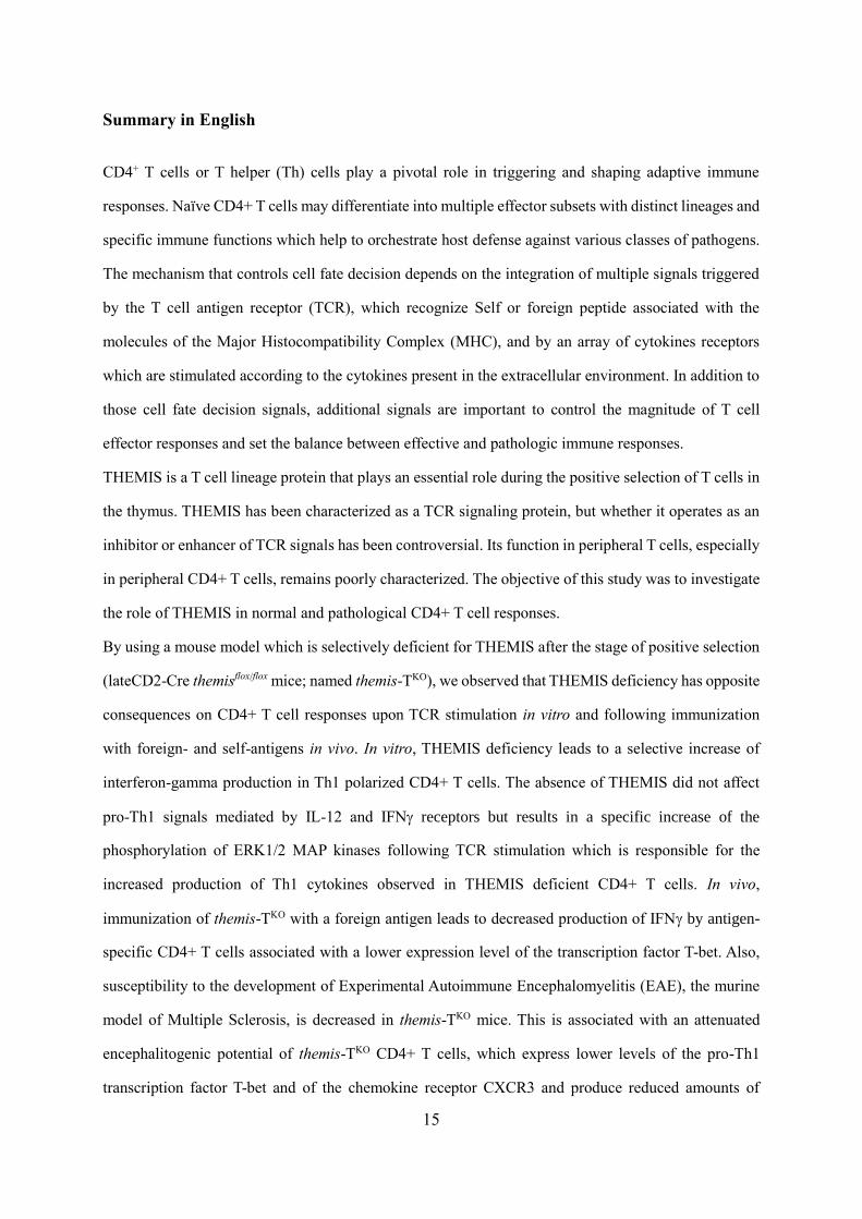
Summary in English
CD4+ T cells or T helper (Th) cells play a pivotal role in triggering and shaping adaptive immune
responses. Naïve CD4+ T cells may differentiate into multiple effector subsets with distinct lineages and
specific immune functions which help to orchestrate host defense against various classes of pathogens.
The mechanism that controls cell fate decision depends on the integration of multiple signals triggered
by the T cell antigen receptor (TCR), which recognize Self or foreign peptide associated with the
molecules of the Major Histocompatibility Complex (MHC), and by an array of cytokines receptors
which are stimulated according to the cytokines present in the extracellular environment. In addition to
those cell fate decision signals, additional signals are important to control the magnitude of T cell
effector responses and set the balance between effective and pathologic immune responses.
THEMIS is a T cell lineage protein that plays an essential role during the positive selection of T cells in
the thymus. THEMIS has been characterized as a TCR signaling protein, but whether it operates as an
inhibitor or enhancer of TCR signals has been controversial. Its function in peripheral T cells, especially
in peripheral CD4+ T cells, remains poorly characterized. The objective of this study was to investigate
the role of THEMIS in normal and pathological CD4+ T cell responses.
By using a mouse model which is selectively deficient for THEMIS after the stage of positive selection
(lateCD2-Cre themisflox/flox mice; named themis-TKO), we observed that THEMIS deficiency has opposite
consequences on CD4+ T cell responses upon TCR stimulation in vitro and following immunization
with foreign- and self-antigens in vivo. In vitro, THEMIS deficiency leads to a selective increase of
interferon-gamma production in Th1 polarized CD4+ T cells. The absence of THEMIS did not affect
pro-Th1 signals mediated by IL-12 and IFN receptors but results in a specific increase of the
phosphorylation of ERK1/2 MAP kinases following TCR stimulation which is responsible for the
increased production of Th1 cytokines observed in THEMIS deficient CD4+ T cells. In vivo,
immunization of themis-TKO with a foreign antigen leads to decreased production of IFN by antigen-
specific CD4+ T cells associated with a lower expression level of the transcription factor T-bet. Also,
susceptibility to the development of Experimental Autoimmune Encephalomyelitis (EAE), the murine
model of Multiple Sclerosis, is decreased in themis-TKO mice. This is associated with an attenuated
encephalitogenic potential of themis-TKO CD4+ T cells, which express lower levels of the pro-Th1
transcription factor T-bet and of the chemokine receptor CXCR3 and produce reduced amounts of
16
interferon-gamma. Altogether, these results suggest that THEMIS exerts inhibitory effects on TCR-
mediated signaling and CD4+ T cell responses in vitro, which are overcome in vivo by TCR-independent
signals that promote normal and neuro-inflammatory Th1 responses.
In conclusion, this thesis work presents insights that may explain the previous discrepancy associated
with the function of THEMIS on TCR signaling and identifies a deleterious effect of THEMIS on the
regulation of Central Nervous System autoimmunity.
Keywords: THEMIS, T cell antigen receptor, CD4+ T cells, TCR signaling, Experimental autoimmune
encephalomyelitis
17
Summary in French
Les lymphocytes T (LT) CD4+ (aussi appelés LT helper ou LTh) jouent un rôle central dans le
déclenchement et le contrôle des réponses immunitaires adaptatives. Les LT CD4+ dits naïfs peuvent se
différencier en différentes sous-populations de LT effecteurs ayant des fonctions immunitaires
spécifiques, qui contribuent à orchestrer la défense de l'hôte contre diverses classes d'agents pathogènes.
Ce processus de différentiation dépend de l'intégration de plusieurs signaux déclenchés par le récepteur
d’antigène des LT (TCR), qui reconnait des peptides du Soi ou étrangers associés aux molécules du
complexe majeur d'histocompatibilité (CMH), et par un ensemble de récepteurs aux cytokines plus ou
moins stimulés selon les cytokines présentes dans le milieu extracellulaire. En plus de signaux contrôlant
la différentiation des LT effecteurs, des signaux supplémentaires sont importants pour contrôler
l'amplitude des réponses effectrices des LT et établir un équilibre optimal entre une bonne efficacité des
réponses immunitaires et la prévention de réponses potentiellement pathologiques.
THEMIS est une protéine de la lignée des LT qui joue un rôle essentiel lors de leur sélection positive
dans le thymus. THEMIS a été identifiée comme une protéine de signalisation du TCR mais sa fonction
dans ce contexte est encore controversée. Son rôle dans les LT périphériques, en particulier dans les LT
CD4+, reste mal caractérisé. L'objectif de cette étude a été d'étudier le rôle de THEMIS dans les réponses
normales et pathologiques des lymphocytes T CD4+.
En utilisant un modèle murin sélectivement déficient pour THEMIS après l'étape de sélection positive
dans le thymus (souris lateCD2-Cre themisflox/flox; nommées themis-TKO), nous avons observé que le
déficit en THEMIS a des conséquences opposées sur les réponses des LT CD4+ lors de la stimulation
du TCR in vitro et après immunisation avec des antigènes étrangers et/ou du Soi in vivo. In vitro, le
déficit en THEMIS entraîne une augmentation sélective de la production d’IFN dans les LTh1 due à
une augmentation spécifique de la phosphorylation des MAP kinases ERK1/2 suite à la stimulation du
TCR. En revanche, l'absence de THEMIS n'a pas d’effet sur les signaux pro-Th1 déclenchés par les
récepteurs à l’IL-12 et à l’IFN. In vivo, la déficience en THEMIS entraîne une diminution de la
production d’IFN par les LT CD4+ spécifiques d'antigènes exogènes associée à des niveaux
d'expression plus faible du facteur de transcription pro-Th1 T-bet. En outre, la susceptibilité au
développement de l'encéphalomyélite auto-immune expérimentale (EAE), le modèle murin classique de
sclérose en plaques, est diminuée chez les souris themis-TKO. Les LT CD4+ déficients en THEMIS sont

18
caractérisés par un potentiel encéphalitogénique atténué, de faibles niveaux d’expression du facteur de
transcription T-bet et du récepteur CXCR3 ainsi qu’une moindre capacité à produire de l’IFN. Dans
l'ensemble, ces résultats suggèrent que THEMIS exerce des effets inhibiteurs sur la signalisation du
TCR et les réponses des lymphocytes T CD4+ in vitro ; effets contrebalancés in vivo par des signaux
indépendants du TCR qui amplifient les réponses Th1 normales et neuro-inflammatoires.
En conclusion, ce travail de thèse apporte un nouvel éclairage sur les fonctions de THEMIS sur la
signalisation du TCR et identifie des effets potentiellement pathologiques de THEMIS dans les maladies
auto-immunes affectant le système nerveux central.
Mots-clés : THEMIS, Récepteur d’antigène des lymphocytes T, Lymphocytes T CD4+, Signalisation
du TCR, Encéphalomyélite Auto-immune Expérimentale.

19
Introduction
20
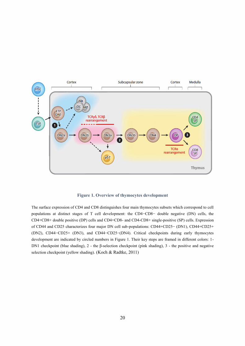
Figure 1. Overview of thymocytes development
The surface expression of CD4 and CD8 distinguishes four main thymocytes subsets which correspond to cell
populations at distinct stages of T cell development: the CD4−CD8− double negative (DN) cells, the
CD4+CD8+ double positive (DP) cells and CD4+CD8- and CD4-CD8+ single-positive (SP) cells. Expression
of CD44 and CD25 characterizes four major DN cell sub-populations: CD44+CD25− (DN1), CD44+CD25+
(DN2), CD44−CD25+ (DN3), and CD44−CD25−(DN4). Critical checkpoints during early thymocytes
development are indicated by circled numbers in Figure 1. Their key steps are framed in different colors: 1-
DN1 checkpoint (blue shading), 2 - the β-selection checkpoint (pink shading), 3 - the positive and negative
selection checkpoint (yellow shading). (Koch & Radtke, 2011)
21
Chapter Ⅰ: T cell development in the thymus and TCR signaling
T cells play an essential role in the immune system, especially in the adaptive immune responses. Their
precursors originate from hematopoietic stem cells (HSCs) in the bone marrow and develop in the
thymus, maturing into distinct types of T cells. Some of these T cells stay in the thymus, while the
majority migrate to peripheral lymphoid organs, becoming mature immunocompetent T cells which
contribute to the immune response or recirculate back into the thymus (Hale & Fink, 2009; Takahama,
2006). T cell development in the thymus is tightly associated with signaling events triggered by the T
cell antigen receptor (TCR). In this chapter, we will describe the different stages of T cell development,
the main signaling events associated to TCR signaling and the role of the TCR signaling protein
THEMIS in T cell development.
1. Stages of T cell development
As shown in Figure 1, T-cell progenitors originated from HSCs can migrate through the blood
circulation into the thymus via the cortical-medullary junctions, acquiring the expression of CD117 and
becoming early thymic progenitors (ETPs). Those ETPs are able to develop into natural killer (NK)
cells, macrophages, dendritic cells (DC) and T cells in the cortex. The development of T cells in the
thymus is characterized by 3 major stages depending on the expression of CD4 and CD8 co-receptors:
double negative (DN, CD4-CD8-), double positive (DP, CD4+CD8+) and single positive (SP,
CD4+CD8- or CD4-CD8+) cells. DN cells are subdivided into four distinct populations according to
the expression level of CD44 and CD25 co-receptors on their surface: DN1 cells (CD44+ CD25-), DN2
cells (CD44+ CD25+), DN3 cells (CD44- CD25+) and DN4 cells (CD44- CD25-) (Godfrey et al., 1993).
At the early stage of T cell development, Notch1 receptor signaling allows ETP/DN1 cells to evolve
into DN2a, preventing them from differentiating into other cells, which is also called DN1 checkpoint.
The decreased expression of CD117 on the surface of DN2a cells is associated to the DN2b stage, in
which the expression of RAG1 recombinase and pre-TCRα gradually increase along with the cell
differentiation into the DN3a cell stage in the subscapular thymic zone (SCZ).
22
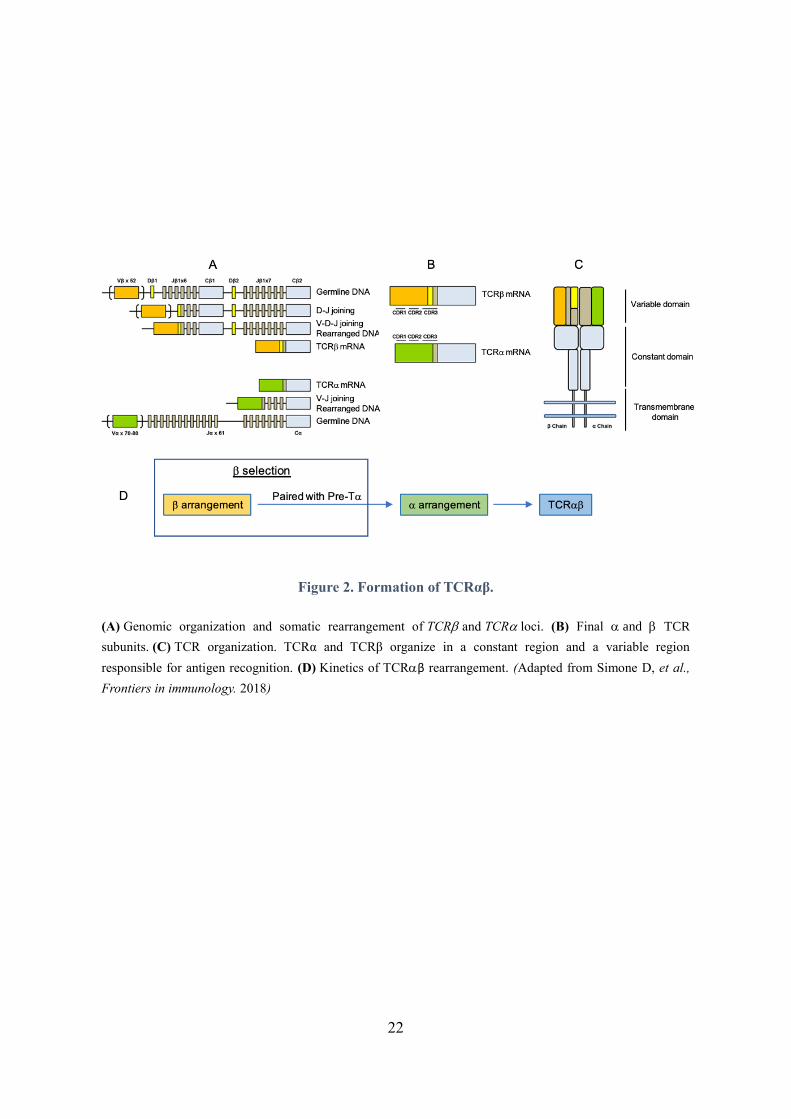
Figure 2. Formation of TCRαβ.
(A) Genomic organization and somatic rearrangement of TCR and TCR loci. (B) Final and TCR
subunits. (C) TCR organization. TCRα and TCRβ organize in a constant region and a variable region
responsible for antigen recognition. (D) Kinetics of TCRβ rearrangement. (Adapted from Simone D, et al.,
Frontiers in immunology. 2018)
23
1.1. Rearrangement of T cell receptor (TCR)
T cell responses to antigens are mediated by the T cell antigen receptor (TCR), a surface protein that
can specifically recognize antigenic peptides presented by the major histocompatibility complex (MHC)
at the surface of antigen-presenting cells. The TCR is a heterodimeric molecule composed of either α
and β or γ and δ chains. Each chain comprises a constant and a variable domain, the latter being
genetically encoded by a series of fragments on the same genetic locus. The cutting and joining of these
gene fragments by RAG recombinases produce a rich variety of unique TCRs, which recognize different
peptides and their multiplicity is referred to as the T cell repertoire. The heterodimeric association of the
four transmembrane chains α, β, γ and δ enables the formation of TCRαβ and TCRγδ.
TCRαβ-expressing T cells represent the vast majority of T cells. They are restricted to the recognition
of MHCII molecules for CD4+ T cells and of MHCI molecules for CD8+ T cells, and migrate toward
the secondary lymphoid tissues after their development. By contrast, TCRγδ-expressing T cells is a
small subset of T cells whose ability to recognize antigens is not restricted by MHC molecules and
which are mostly detected in intraepithelial tissues. The molecular events leading to the linage
commitment to the two types of T cells have not been fully resolved yet. High expression of the IL-7
receptor (IL-7R) on DN2 cells leads to the development of γδ thymocytes, while low expression results
in further development toward the αβ cell lineage (Kang et al., 2001). At the stage of DN3, Notch
signaling is required for the recombination of the β chain that is able to pair with the α chain later. By
contrast, the rearrangement of γδ chains seems Notch-independent and can be promoted by the ID3
transcription factor (Lauritsen et al., 2009; Wolfer et al., 2002). So far, antigenic peptides recognized by
γδ T cells are mostly unknown and their response to pathogens is not fully understood. The rest of this
manuscript will focus on TCRαβ-expressing T cells.
As mentioned earlier, the variability in TCR specificity is generated through the rearrangement of gene
fragments at the α and β loci, which is initiated from the DN2a stage using RAG 1/2 recombinases.
Indeed, the diversity of α and β chains mainly depends on the semi-stochastic rearrangement of the V,
D and J genes for the β chain and of the V and J genes for the chain α (Figure 2). Regarding TCRαβ
rearrangement, the β chain is first rearranged by semi-random gene recombination controlled by RAG1
24
and RAG2 nucleases which cleave DNA at the level of recombination signal sequence (RSS) on both
sides of the Dβ and Jβ genes (Schatz et al., 1992). The DβJβ fragment then rearranges with the Vβ
fragment at the DN3a stage. The enzyme Terminal deoxynucleotidyl Transferase (TdT) randomly adds
nucleotides in the junction between the segments, adding “junctional diversity” to the TCR repertoire
(Cabaniols et al., 2001). Meanwhile, T cells begin to express the invariant α-chain, named pre-Tα. The
successful pairing of the rearranged β chain with the pre-Tα results in the expression of a pre-TCR at
the end of the DN3a phase. If this pre-TCR is functional, it triggers intracellular signals which lead to a
decrease in the expression of RAG recombinases and lead to the allelic exclusion of TCRβ to inhibit the
formation of other β chains(von Boehmer, 2005). To eliminate the production of non-functional TCR,
only cells that have successfully rearranged the β chain and express a functional pre-TCR are allowed
to evolve towards the cell stage DN3b. The “failed” cells die through apoptosis. This process is called
β selection. The engagement of pre-TCR with MHC molecules associated with Self-peptides (Self-
pMHC) – on the surface of thymic epithelial cells – allows thymocytes to survive, proliferate and
differentiate into DN4 cells. Notch signaling is essential for this process (Ciofani et al., 2004). The DN4
thymocytes then leave the SCZ and acquire the expression of co-receptors CD4 and CD8 in the thymic
cortex where they differentiate into DP cells (Porritt et al., 2003). At this stage, the activity of RAG1
and RAG2 nucleases increases again to allow rearrangement of the Vα and Jα segments to form the
TCRα chain, also known as α rearrangement. The DP cells continue to develop with a low expression
of TCRαβ (TCRαβlow).
1.2. Positive and negative selections
The high variability of TCRs allows T cells to trigger immune responses which are adapted and specific
to a broad spectrum of antigens. However, not all the TCRs which are generated in a semi-random
manner are able to effectively recognize Self-pMHC. On the other hand, the generated TCRs can
recognize Self-pMHC complexes, sometimes with an “excessive” affinity, which can lead to the
generation of self-reactive T cells that may induce the development of autoimmune diseases. Therefore,
in order to eliminate thymocytes which express ineffective or potentially self-reactive TCRs, positive
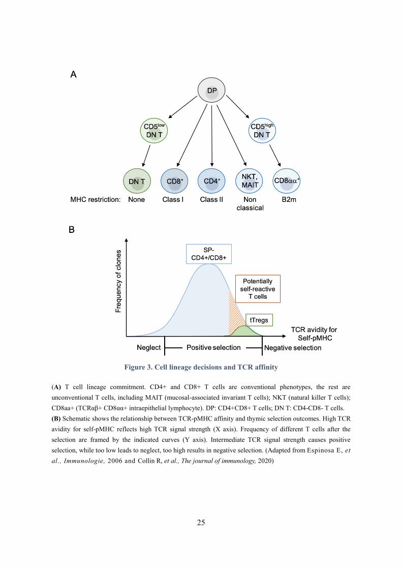
and negative selection processes are set in the thymic cortex and medulla. Positive selection (Figure 3)
takes place in the thymic cortex and involves cortical thymic epithelial cells (cTECs) which express
25
Figure 3. Cell lineage decisions and TCR affinity
(A) T cell lineage commitment. CD4+ and CD8+ T cells are conventional phenotypes, the rest are
unconventional T cells, including MAIT (mucosal-associated invariant T cells); NKT (natural killer T cells);
CD8aa+ (TCRαβ+ CD8αα+ intraepithelial lymphocyte). DP: CD4+CD8+ T cells; DN T: CD4-CD8- T cells.
(B) Schematic shows the relationship between TCR-pMHC affinity and thymic selection outcomes. High TCR
avidity for self-pMHC reflects high TCR signal strength (X axis). Frequency of different T cells after the
selection are framed by the indicated curves (Y axis). Intermediate TCR signal strength causes positive
selection, while too low leads to neglect, too high results in negative selection. (Adapted from Espinosa E, et
al., Immunologie, 2006 and Collin R, et al., The journal of immunology, 2020)

26
Self-pMHC that are presented to the TCRs of DP thymocytes. Successful positive selection results in
the generation of SP-CD4+ cells (recognizing MHC class II) and SP-CD8+ cells (recognizing MHC I).
Lysosomal cathepsin L and thymic-specific serine protease (TSSP) specifically expressed by cTECs are
necessary to generate the spectrum of self-peptides involved in the selection of SP-CD4+ thymocytes
(Gommeaux et al., 2009; Viret et al., 2011). Regarding SP-CD8+ cells, the thymoproteasome subunit
β5t is required to generate TCRs recognizing efficiently MHC class I (Murata et al., 2007). As shown
in Figure 3, the positive selection is associated with TCRs which have a sufficient affinity for self-
pMHC complexes (Merkenschlager et al., 1997; Starr et al., 2003). Thymocytes whose TCRs have
relatively high affinity for Self-pMHC undergo negative selection, which restricts the susceptibility to
develop autoimmune responses.
Negative selection occurs after positive selection. It requires the migration of the cells from the thymic
cortex to the medulla (Nitta et al., 2009). The expression of two transcription factors, AIRE
(Autoimmune Regulator) and FEZF-2 (Family Zinc Finger 2), expressed by medullar thymic epithelial
cells (mTEC) is necessary for the recognition of tissue restricted antigens by thymocytes, hence for
negative selection. AIRE or FEZF-2 deficiency in mice leads to the development of severe
autoimmunity (Anderson et al., 2002; Takaba et al., 2015). The process of negative selection involves
co-stimulatory signals on thymocytes. Indeed, perinatal blockade of CD80/CD86 molecules which are
the ligands of the co-stimulatory molecule CD28 expressed by thymocytes leads to a decrease in
negative selection (Gao et al., 2002). In addition to positive selection and negative selection, thymocytes
whose TCRs cannot respond to self-peptide-MHC complexes are neglected and die by apoptosis.
1.3. CD4 versus CD8 lineage commitment
At the end of positive selection, SP-CD4+ and SP-CD8+ thymocytes express high level of TCRαβ
(TCRαβhi) and develop into mature SP T cells. It has been reported that the TCR signaling initiated by
the recognition of TCR and Self-pMHC plays a major role in T cell commitment. Both strength and
duration of the signal are critical for the lineage choice (Gascoigne et al., 2016; Yasutomo et al., 2000).
The generation of SP-CD4+ T cells is associated with sustained and/or strong TCR signaling (although
lower than the negative selection threshold), whereas transient and/or weak TCR signaling (although
27
higher than the neglect threshold) preferentially leads to the development of SP-CD8+ T cells. In
addition, ThPOK and Runx3 are two key transcription factors implicated in the full commitment of SP
T cells. ThPOK promotes CD4+ lineage commitment and blocks the expression of genes associated to
the CD8+ lineage, whereas Runx3 contributes to the generation SP-CD8+ cells (He et al., 2008; Liu et
al., 2005; Wang et al., 2008); they repress the expression of each other (Egawa & Littman, 2008). Not
all Self-pMHC specific T cells are eliminated in the thymic medulla by negative selection; some of them
differentiate into thymic regulatory T cells (tTregs). These cells represent a population enriched in
autoreactive thymocytes, which specifically express the transcription factor FoxP3 and are involved in
the control of autoimmune responses (Brunkow et al., 2001; Romagnoli et al., 2002). The generation of
tTregs requires a strong TCR signaling and is regulated by the stimulation of thymocytes with specific
cytokines, including IL-2 and TGFβ (Liu et al., 2008; Moran et al., 2011; Soper et al., 2007). In addition,
some DP thymocytes can differentiate into invariant Natural Killer T cells (iNKTs). These cells do not
express the co-receptor CD8, some express CD4. They have an invariant TCR with a single type of
chain α (Vα14 Jα18) which can be associated with three types of β chain (Vβ8.2, Vβ7, Vβ2) and interacts
with a CD1d “MHC I-like” lipid molecule (Arase et al., 1992; Egawa et al., 2005). With the
development of further research, some new thymic-derived T cells were found in the thymus and
peripheral tissues. TCRαβ+CD8αα+ intraepithelial lymphocytes (IELs) and CD4−CD8−TCRαβ+
thymocytes are two newly characterized unconventional T cells which develop from DN T cells with
high or low expression of CD5, transiting through DP stage (Collin et al., 2020; Ruscher et al., 2017).
The generation of CD4−CD8−TCRαβ+ T cells is MHC-independent(McDonald et al., 2018).
TCRαβ+CD8αα+ IELs can be selected by both classic and non-classic MHC I, as they recognize β2
macroglobulin (β2m), an essential component of these molecules (McDonald et al., 2018; Ruscher et
al., 2017).
2. TCR signaling: signal transduction pathways
As mentioned above, the positive selection of thymocytes is dependent on the affinity between the TCR
and Self-pMHC which determines the strength and the duration of TCR signaling. The second part of
chapter I will describe the main steps of TCR signaling and their implication in thymocytes development.
28
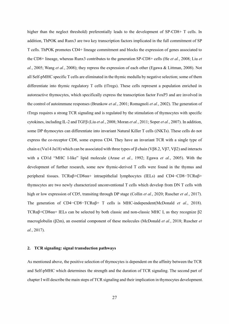
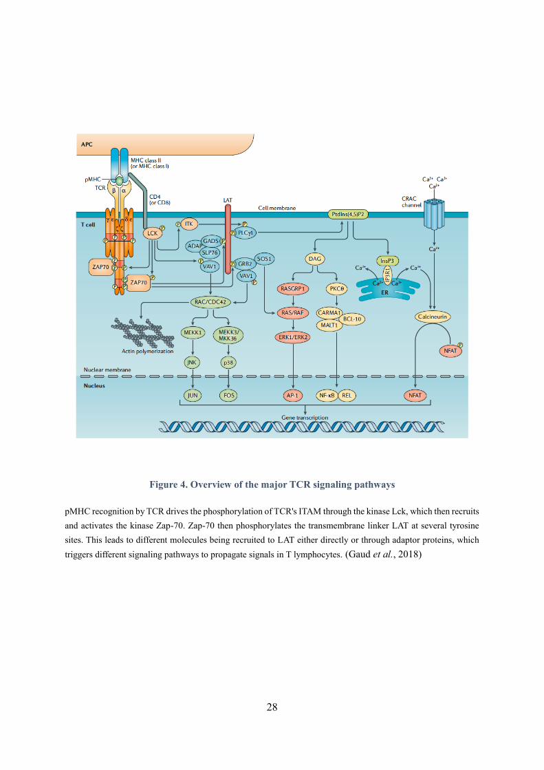
Figure 4. Overview of the major TCR signaling pathways
pMHC recognition by TCR drives the phosphorylation of TCR's ITAM through the kinase Lck, which then recruits
and activates the kinase Zap-70. Zap-70 then phosphorylates the transmembrane linker LAT at several tyrosine
sites. This leads to different molecules being recruited to LAT either directly or through adaptor proteins, which
triggers different signaling pathways to propagate signals in T lymphocytes. (Gaud et al., 2018)
29
2.1. TCR/CD3 complex
TCR signaling is initiated upon the stimulation of TCR/CD3 complex by peptides presented by MHC
molecules (pMHC) and depends on a series of tyrosine kinases (Figure 4), adaptor proteins and
additional enzymes which operate together to trigger more distal signaling events. In mammals,
TCR/CD3 complex, also known as the TCR complex, is composed of TCR α and β chains, CD3
heterodimers δε and γε and a TCRζζ homodimer. To express a functional TCR, the TCRαβ associates
with the CD3 δε and γε heterodimers, forming a TCRαβ / CD3δε / CD3γε complex, then combines with
TCRζζ before being expressed on the surface of T cells (San José et al., 1998). The intracellular tail of
each , and CD3 chains contains one immunoreceptor tyrosine-based activation motif (ITAM), which
consists of two tyrosine residues separated by 6-8 amino acids (YxxLx6-8-YxxL), whereas each TCR ζ
chain has three ITAMs (Samelson et al., 1985).
2.2. Formation of proximal TCR signaling
TCR engagement by specific pMHC leads to the phosphorylation of ITAMs by the Src family kinases
Lck or Fyn, two important proteins initiating the intracellular signaling events (Samelson et al., 1990;
Straus & Weiss, 1992). In vitro TCR stimulation of Lck-deficient T cells with an anti-CD3 antibody
results in a remarkable reduction in the phosphorylation of many signaling events, including PLCγ1,
PKC, ERK kinases and mobilization of calcium flux (Lovatt et al., 2006). The catalytic activity of Lck
is regulated by a phosphorylation balance between the tyrosine Y394, an activating autophosphorylation
site, and the tyrosine Y505, an inhibiting phosphorylation site which can be phosphorylated by the
tyrosine kinase Csk, leading to the formation of a closed conformation of Lck, and dephosphorylated by
phosphatase CD45 (Zikherman et al., 2010). Regarding Fyn, upon the stimulation of anti-CD3 antibody
in vitro, Fyn-deficient T cells with an anti-CD3 antibody leads to a reduction in the phosphorylation of
ERK, an essential tyrosine kinase of distal TCR signaling (Sugie et al., 2004).
After ITAMs phosphorylation by Lck or Fyn, these motifs serve as docking sites for other proteins
containing SH2 domains, such as the Syk family enzymes Zap70 and Syk. Recruitment of Zap70 to
phosphorylated ITAMs modifies its conformation and enables its phosphorylation on its tyrosine Y493
by Src kinases which enhances its catalytic activity and enables consequently the phosphorylation of the
30
transmembrane linker for activation of T cells LAT (Chan et al., 1995). In addition, ZAP-70 contains
three other phosphorylation sites Y292, Y315 and Y319 that recruit SH2-containing proteins which exert
negative and positive regulatory effects on TCR signaling. For example, the recruitment of ubiquitin
ligase c-Cbl to p-Y292 can inhibit TCR signaling by degrading signaling proteins (Wang et al., 2001),
whereas the recruitment of VAV1 to p-Y315 and Lck to p-Y319 enhances TCR signal (Pelosi et al.,
1999). LAT has four tyrosine sites, Y132, Y171, Y191 and Y226, all of which can be phosphorylated by
Zap-70, thereby recruiting a variety of molecules to LAT, such as Phospholipase C gamma 1 (PLCγ1),
Growth factor receptor-bound protein 2 (Grb2) and Grb2-related adapter protein 2 (Gads), all of which
are important for relaying early TCR-mediated signaling events to downstream pathways (Kuhné et al.,
2003). This suggests a central role for LAT in the signaling pathways triggered upon TCR engagement.
PLCγ1 plays a key role in the mobilization of calcium flux in T cells. The recruitment of PLCγ1 to LAT
leads to the hydrolysis of phosphatidylinositol (4,5)-biphosphate PI (4,5) P2 to inositol 1, 4, 5-
triphosphate (IP3) and diacylglycerol (DAG), thereby activating the downstream signaling components.
Gads constitutively interacts with the adapter protein Slp76, allowing its recruitment to LAT and forming
a LAT-Gads-Slp76 complex (Liu et al., 2001). This complex creates a platform which further recruits
multiple signaling molecules to LAT, such as VAV1, Nck, c-Cbl, Interleukin 2 tyrosine kinase (Itk) and
ADAP, transmitting TCR signaling (Wu & Koretzky, 2004).
2.3. Major distal signaling pathways mediated by TCR activation
Calcium flux:
The binding of IP3 to its receptor (IP3R) located on the membrane of the endoplasmic reticulum (ER)
leads to the mobilization of calcium flux (Nagaleekar et al., 2008). Indeed, following IP3 binding, ER
calcium is released into the cytoplasm and enables the activation of the Ca2+ Release-activated Ca2+
(CRAC) Channel allowing further entry of Ca2+ into the cell. The mobilization of calcium flux leads to
the activation of calmodulin kinases and calcineurin. Calcineurin then dephosphorylates multiple
phosphoserine residues on the transcription factor NFAT – a key regulator of gene expression in T cell
development and function – leading to its activation and nuclear translocation (Macian, 2005). During
this process, the LAT-Gads-Slp76 complex stabilizes the interaction of PLCγ1 on LAT (Zhang et al.,
2000). The proteins Itk, RIAM (Rap1 GTP interacting adaptor molecule) and Tespa1 (Thymocyte-
expressed positive selection-associated 1) all positively regulate the activation of PLCγ1 and calcium
31
pathway (Patsoukis et al., 2009; Schaeffer et al., 2000). DAG is another product of PIP2 hydrolysis,
which functions as a lipid second messenger. It activates Ras guanyl nucleotide-releasing protein
(RasGRP) and protein kinase C θ (PKC - θ), two initiators of the Ras/MAPK/ERK cascade and of the
NF-B pathway, respectively (Ebinu et al., 1998; Lin et al., 2000).
MAP Kinases pathway:
The kinases ERK (extracellular signal-regulated protein kinase) belong to the class of mitogen-activated
protein kinases (MAPK). They are associated with cytokines production, cell proliferation and T cell
development through activation of several transcription factors such as AP1, which is highly expressed
during cell division and can form a complex with NFAT to regulate the expression of various cytokines
including IL-2, IL-4 and IFN (Angel & Karin, 1991; Macián et al., 2001). The activation of an optimum
ERK cascade is dependent mostly on the protein RasGRP (RAS guanyl nucleotide-releasing protein)
and to a lesser extent on the Grb2-SOS complex which is recruited to LAT after the engagement of TCR
by pMHC (Roose et al., 2007). p38 kinases and c-Jun N terminal kinase (JNK) are two other MAPK.
They can both be activated by the LAT-recruited protein VAV1 through regulating Ras-related GTP-
binding proteins, such as Rac, Rho and Cdc42 (Kaminuma et al., 2001; Salojin et al., 1999).
Phosphorylation of p38 and JNK leads to the activation of many transcription factors such as c-Jun, p53
and Fos, regulating the cell growth, differentiation, survival and apoptosis. Besides, VAV1-mediated
activation of Ras-related GTP-binding proteins is also associated with the assembly of the actin
cytoskeleton (Tapon & Hall, 1997).
PKC/NFκB pathway:
Upon its activation by DAG, PKCθ is recruited near the plasma membrane where it phosphorylates the
adapter protein CARD11 (CARMA1), which then forms a complex with the proteins Bcl10 and MALT1.
This complex recruits TRAF2 and TRAF6, which then activates IκB kinases (IKK), leading to the
degradation of IκBand releasing the transcription factor NFκB (Rosebeck et al., 2011; Schulze-
Luehrmann & Ghosh, 2006). In addition, the engagement of TNFR superfamily receptors, such as 4-
1BB and OX40, stabilizes the NFκB-inducing kinase (NIK), which can phosphorylate the inhibitor of
NF-κB kinase α (IKKα) and NFκB2 precursor protein p100, thereby mediating activation of the
32
p52/RelB NFκB complex (Sun, 2012). As a central mediator of cytokines expression, NFκB can enter
the nucleus and induce the expression of genes harboring specific NFκB DNA-binding sites.
PI3K/AKT/mTOR pathway:
The PI3K/AKT/mTOR pathway is important for the regulation of many T cells responses. It's activated
by the recruitment to the plasma membrane of phosphoinositide 3-kinases (PI3K), a family of enzymes
expressed by T cells, which allows the phosphorylation of membrane Pi (4,5) P2 to Pi (3,4,5) P3. The
natural inhibitor of this process is PTEN (Maehama & Dixon, 1998). mTOR is a member of PI3K family,
which functions as the core component of mTORC1 and mTORC2 complexes, phosphorylating serines,
threonines and tyrosines of many signaling proteins (Hay & Sonenberg, 2004). ). PIP3 can activate and
bind to proteins that contain pleckstrin homology domains (PHD), such as phosphoinositide-dependent
kinase 1 (PDK1). Binding of PDK1 to PIP3, leading to the phosphorylation of AKT at the threonine 308
residue (T308). This triggers the phosphorylation of AKT at serine 473 (S473) by mTORC2 complex
(Currie et al., 1999; Franke et al., 1995), which upregulates AKT serine/threonine kinase activity,
enabling Akt to phosphorylate many downstream substrates in order to regulate cellular processes, such
as survival and metabolism (Alessi et al., 1996; Sarbassov et al., 2005). Akt can further serves as an
activator of many downstream molecules, regulating different cell processes. For example, it upregulates
cell survival by phosphorylating FOXO1 – a positive regulator of apoptosis – which inhibit its
translocation to the nucleus (Zhang et al., 2011). It also promotes the expression of the cAMP response
element (CREB), an important transcription factor related to proliferation (Kops et al., 1999; Peltier et
al., 2007).
In addition to the major signaling proteins described above, TCR signaling is regulated by many other
factors, such as phosphatases, co-receptors and microRNA. Previous studies have shown that the
participation of tyrosine phosphatases SHP-1 can dephosphorylate several proximal TCR signaling
components, including Zap-70 and Lck, leading to the down-regulation of TCR signal (Kosugi et al.,
2001). The engagement of stimulatory co-receptor CD28 and inhibitory co-receptor CTLA-4 enhances
and limits the strength of TCR signaling, respectively (Holt et al., 2017). The miR-181a dynamically
regulates TCR signal strength, as its expression in T cells leads to reduced activity of multiple
33
phosphatases (Grewers & Krueger, 2020). Amongst these many proteins involved in TCR signaling,
THEMIS (Thymocytes-Expressed Molecule Involved in Selection) was identified in 2009 as necessary
for the development of T cells in the thymus. Being the main subject of my research work, the third part
of this chapter will focus on how THEMIS regulates thymocytes development through TCR signaling.
3. Function and molecular roles of THEMIS in T cells development
3.1. Discovery and classification of THEMIS
THEMIS, also known as THEMIS1, is a protein initially found in murine thymus in 2009
(GenBank:E430004N04Rik). It was named “Thymocyte-Expressed Molecule Involved in Selection”
following independent studies of five research teams headed by Paul Love (Lesourne et al., 2009),
Richard Cornall and Ron Schwartz (Johnson et al., 2009), Nicholas Gascoigne (Fu et al., 2009), Hiroshi
Kawamoto and Hisahiro Yoshida (Kakugawa et al., 2009) and Harumi Suzukia (Patrick et al., 2009).
Orthologous themis genes are found in mammals (Mus musculus, Homo sapiens), birds (Gallus gallus)
and bony fish (Danio rerio) (Fu et al., 2009). THEMIS family gathers structurally related proteins,
containing a specific and conserved globular domain named CABIT, either singular or in tandem copies
(Johnson et al., 2009). In mammals, THEMIS family counts five members. The first two, GAREM1 and
GAREM2, ubiquitous and brain specific respectively, bear a single CABIT-module and have been
characterized as adaptor proteins involved in EGFR signaling (Taniguchi et al., 2013; Tashiro et al.,
2009). The other three homologs are THEMIS1, expressed in T cells, NKT cells and mast cells,
THEMIS2 which is expressed in B cells, dendritic cells and macrophages and THEMIS3, which is
expressed in the intestine (Fu et al., 2009; Johnson et al., 2009; Lesourne et al., 2009).
34
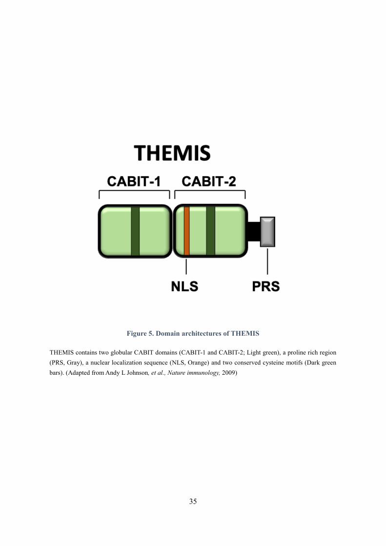
As shown in Figure 5, THEMIS1, THEMIS2 and THEMIS3 contain two tandem copies of CABIT
domain, a proline-rich region (PRS) and a bipartite nuclear localization sequence (NLS), which are all
required for the function of THEMIS in thymocytes (Johnson et al., 2009; Kakugawa et al., 2009; Okada
et al., 2014). THEMIS lacks a consensus catalytic domain but there is a conserved cysteine residue in
each CABIT domain, which suggests that THEMIS may possess some catalytic activity (Johnson et al.,
2009). In thymocytes, THEMIS is mainly localized in the cytoplasm but is still detected in the nucleus,
which suggests its function in both cellular compartments (Lesourne et al., 2009).
3.2. Critical role of THEMIS in T cells development
Murine THEMIS is highly expressed in thymus and less in lymph nodes and spleen, but it is not detected
in any other organs or tissues. In thymocytes, THEMIS has the highest expression level in
CD4+CD8+DP thymocytes, then is downregulated as thymocytes transition to the CD4+ or CD8+SP
stage (Fu et al., 2009; Lesourne et al., 2009). Studies on themis knockout mouse models (themis-/-)
expressing different types of TCR transgene (MHC class II-restricted AND TCR transgene, MHC class
I-restricted H-Y TCR transgene, MHC class I-restricted OT-I) showed that THEMIS is essential for
efficient positive and negative selections during T cell development (Fu et al., 2009; Lesourne et al.,
2009). In the absence of THEMIS, thymocytes development is blocked at the DP stage, which leads to
a drastic reduction in CD4+ and CD8+ SP thymocytes and in peripheral T cells (Fu et al., 2009; Johnson
et al., 2009; Lesourne et al., 2009). Since thymocytes selection is dependent on TCR signals (Hogquist
& Jameson, 2014), those results suggest that THEMIS is involved in TCR signaling. Moreover, in
themis-/- mice, CD4+ SP thymocytes development is more severely impacted than CD8+ SP thymocytes
development (Fu et al., 2009; Johnson et al., 2009; Kakugawa et al., 2009; Lesourne et al., 2009; Patrick
et al., 2009). Data from the literature show that the development of CD4+ SP depends on persistent TCR
signal transduction while the development of CD8+ SP depends on transient TCR signals combined
with IL-7 mediated survival signals (Singer et al., 2008).
35
Figure 5. Domain architectures of THEMIS
THEMIS contains two globular CABIT domains (CABIT-1 and CABIT-2; Light green), a proline rich region
(PRS, Gray), a nuclear localization sequence (NLS, Orange) and two conserved cysteine motifs (Dark green
bars). (Adapted from Andy L Johnson, et al., Nature immunology, 2009)
36
Analysis of T cell development in MHC class I-deficient 2m-/- mice shows that part of the MHC class
II restricted thymocytes are ‘redirected’ from the CD4 lineage to the CD8 lineage in the absence of
THEMIS (Lesourne et al., 2009), suggesting that THEMIS could be important to promote or sustain
TCR signals following TCR engagement. However, initial studies on THEMIS failed to detect
significant defect in TCR signaling in themis-deficient thymocytes following TCR stimulation in vitro
(Johnson et al., 2009; Patrick et al., 2009). Impaired TCR signaling was however suspected given the
reduced expression of the signaling sensor CD5 at the surface of CD4+CD8int thymocytes (Fu et al.,
2009), raising questions about the molecular mechanisms by which THEMIS operates during
thymocytes development.
3.3. THEMIS is involved in TCR signaling cascade
THEMIS binds to the ubiquitous cytosolic adaptor Grb2 by its PRS (Johnson et al., 2009; Lesourne et
al., 2009; Lesourne et al., 2012; Paster et al., 2013; Patrick et al., 2009). After TCR stimulation,
THEMIS is rapidly phosphorylated by Lck and ZAP-70 (Brockmeyer et al., 2011; Fu et al., 2009), and
recruited to LAT by Grb2 (Brockmeyer et al., 2011; Lesourne et al., 2012; Paster et al., 2013). Grb2
expression is necessary to maintain the stability of THEMIS (Garreau et al., 2017). Previous results
obtained in our team indicate that THEMIS expression is reduced in Grb2+/- thymocytes compared to
wild type cells (Garreau et al., 2017). This study also shows that the N-terminal CABIT domain of
THEMIS directly binds to the ubiquitin-specific protease USP9X, which de-ubiquitinates THEMIS
ubiquitin K48 chains after TCR engagement (Garreau et al., 2017). Grb2 allows the recruitment of
THEMIS/USP9X complexes to LAT, sustaining THEMIS expression in the process of positive selection,
which indicates that THEMIS-Grb2 interaction is required for the function of THEMIS in vivo (Garreau
et al., 2017; Okada et al., 2014; Paster et al., 2013; Zvezdova et al., 2014). THEMIS-Grb2 complex also
bridges THEMIS to the phosphatase SHP-1, an inhibitory tyrosine phosphatase that dephosphorylates
and inactivates several key molecules in the TCR signaling pathway including the tyrosine kinases Lck
and ZAP-70 and the guanine nucleotide exchange factor VAV1 (Lorenz, 2009; Pao et al., 2007; Stebbins
et al., 2003). THEMIS and SHP-1 separately interact with N-terminal and C-terminal SH3 domains of
Grb2 (Lesourne et al., 2012; Okada et al., 2014; Paster et al., 2013; Paster et al., 2015), which suggests
a tripartite complex involving THEMIS, Grb2 and SHP-1 (Paster et al., 2015).
37
However, a recent publication shows that THEMIS directly interacts with the phosphatase SHP-1
through binding to SHP-1 PTP domain by its CABIT modules. This interaction is enhanced by Grb2 but
is not Grb2 dependent (Choi, Warzecha, et al., 2017). THEMIS can be co-immunoprecipitated with
SHP-1 in Grb2-/- thymocytes and this co-immunoprecipitation also occurs in the absence of the
THEMIS PRS region (Choi, Warzecha, et al., 2017). In themis-/- thymocytes, tyrosine phosphorylation
of SHP-1 is markedly diminished with or without TCR stimulation (Fu et al., 2013; Zvezdova et al.,
2016), which suggests an important role for the THEMIS-SHP-1 interaction in thymocytes development.
Other studies show that THEMIS also co-immunoprecipitants with the phosphatase SHP-2 in Jurkat T
cells and in transfected HEK293 cells, suggesting an interaction between THEMIS and this regulator –
positive and negative – of TCR signaling (L. Chen & D. B. Flies, 2013; Fu et al., 2013; Lorenz, 2009;
Pao et al., 2007; Paster et al., 2015; Stanford et al., 2012).
3.4. Two models of THEMIS function in T cells development
3.4.1. Model I: THEMIS suppresses TCR signaling in thymocytes to prevent crossing the threshold
for negative selection
While initially studying THEMIS function in thymocytes development, Paul Love and collaborators
found that the expression of CD5, IL-7 R and CD69 proteins, which are correlated with the intensity of
TCR signals at the transition from DP to SP, was reduced in the CD4+CD8int themis-/- mice compared
to themis+/+ mice (Lesourne et al., 2009). In parallel, Nicholas Gascoigne’s research group found that
calcium flux and ERK activation were mildly decreased in themis-/- thymocytes after TCR stimulation
(Fu et al., 2009). They later reported that there is a reduction of ERK phosphorylation, IL-2 production
and NFAT/AP1 activation in response to TCR stimulation in themis-knockdown Jurkat T cells
(Brockmeyer et al., 2011; Gascoigne & Palmer, 2011). Altogether, these initial studies lead to identify
THEMIS as a positive regulator of TCR signaling (Brockmeyer et al., 2011; Gascoigne & Palmer, 2011;
Lesourne et al., 2009).
38
However, a new study by Nicholas Gascoigne’s research team brought this group to new and nearly
opposite conclusions about the function of THEMIS on TCR signaling. In a themis-/- OT-I TCR
transgenic mouse model, the authors engaged OT-I DP thymocytes with pMHC ligands of various
affinity and found that THEMIS negatively regulate TCR signals when relatively low affinity ligands
were used (Fu et al., 2013). Low affinity tetramer/peptide ligands induced stronger phosphorylation of
Lck, PLC-g1, LAT and ERK as well as calcium flux in themis-/- thymocytes or in themis-knockdown
Jurkat T cells than in wild-type controls (Fu et al., 2013; Paster et al., 2015). Moreover, in their studies,
the absence of THEMIS increased activated caspase-3+ cells, a readout for apoptosis (Salvesen, 2002),
and markedly decreased SHP-1 phosphorylation (Fu et al., 2013; Paster et al., 2015). When they crossed
themis-/- mice with mice knocked out for pro-apoptotic factor Bim (Bim-/-), the proportions of CD4-SP
and CD8-SP thymocytes were restored in the double knockout mice compared to Bim+/+themis-/- mice
(Fu et al., 2013). However, the proportions of SP cells in Bim-/-themis-/- mice remained decreased in
comparison to that in control Bim-/-themis+/+ mice, suggesting that the block in T cell development
persists even in the absence of Bim. In addition, authors did not show whether BIM deficiency could
restore normal numbers of peripheral CD4+ and CD8+ T cells. SHP-1 is a negative regulator of several
signaling proteins, such as Lck, Zap70 and VAV1 (Lorenz, 2009; Stebbins et al., 2003; Stefanová et al.,
2003). They hence proposed a model in which THEMIS attenuates TCR signaling by promoting SHP-
1 recruitment to LAT or enhancing SHP-1 activity in response to low affinity peptide ligands stimulation
(Fu et al., 2013; Paster et al., 2015), enhancing positive selection by reducing TCR signals below the
threshold of negative selection (Fu et al., 2013; Gascoigne & Acuto, 2015). Although the effect of
THEMIS on SHP-1 activity and recruitment to LAT was not directly address in this study, this
hypothesis is referred to as model I hereafter (Figure 6).
39
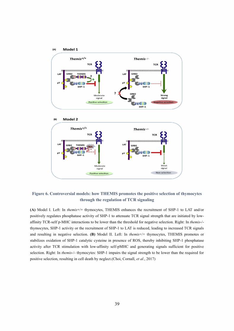
Figure 6. Controversial models: how THEMIS promotes the positive selection of thymocytes
through the regulation of TCR signaling
(A) Model I. Left: In themis+/+ thymocytes, THEMIS enhances the recruitment of SHP-1 to LAT and/or
positively regulates phosphatase activity of SHP-1 to attenuate TCR signal strength that are initiated by low-
affinity TCR-self p-MHC interactions to be lower than the threshold for negative selection. Right: In themis-/-
thymocytes, SHP-1 activity or the recruitment of SHP-1 to LAT is reduced, leading to increased TCR signals
and resulting in negative selection. (B) Model II. Left: In themis+/+ thymocytes, THEMIS promotes or
stabilizes oxidation of SHP-1 catalytic cysteine in presence of ROS, thereby inhibiting SHP-1 phosphatase
activity after TCR stimulation with low-affinity self-pMHC and generating signals sufficient for positive
selection. Right: In themis-/- thymocytes: SHP-1 impairs the signal strength to be lower than the required for
positive selection, resulting in cell death by neglect.(Choi, Cornall, et al., 2017)
40
3.4.2. Model II: THEMIS facilitates T cell development by enhancing TCR signaling above the
threshold required for positive selection.
Model II is consistent with the initial conclusion that THEMIS positively regulates TCR signaling in
thymocytes (Brockmeyer et al., 2011; Fu et al., 2009; Gascoigne & Palmer, 2011; Lesourne et al., 2009).
It is based on further results obtained by Renaud Lesourne and Paul Love’s research groups. In 2017,
they first crossed themis-/- mice expressing the AND TCR with Nur77–green fluorescent protein (GFP)
transgenic mouse model in which GFP abundance correlates with the intensity of TCR signals
transmitted during positive or negative selection (Zvezdova et al., 2016). They found that GFP
expression was reduced in activated themis-/- DP thymocytes, suggesting a positive function for
THEMIS on TCR signaling. They also showed in this study that BIM deficiency does not restore normal
numbers of peripheral T cells in themis-/- mice, suggesting that the block of T cell development in
themis-/- mice is not the consequence of apoptosis due to stronger TCR signals (Zvezdova et al., 2016).
Analysis of THEMIS interactome in thymocytes by mass spectrometry confirmed that THEMIS
interacts with SHP-1, SHP-2, Grb2 and LAT but also indicates that THEMIS strongly interacts with the
Rac-GTP exchange factor VAV1 following thymocytes stimulation with pervanadate (Figure 7).
Analysis of TCR signaling upon weak or strong stimulations with anti-CD3 antibodies showed that the
phosphorylation of ERK is increased in the absence of THEMIS upon weak anti-CD3 stimulation,
confirming the results from Nicholas’s Gascoigne group in a different experimental setting. However,
the absence of THEMIS led to a strong decrease of VAV1 phosphorylation and of the production of Rac-
1 GTP independently of the strength of the TCR stimuli (Zvezdova et al., 2016). Reduced
phosphorylation of VAV1 was also observed ex-vivo in THEMIS deficient DP thymocytes expressing
the AND TCR. Altogether, those results suggested that THEMIS could exert both positive and negative
effects on TCR signals according to the type of signaling pathways. However, the positive effects of
THEMIS seem to overcome its negative effects in T cells undergoing development.
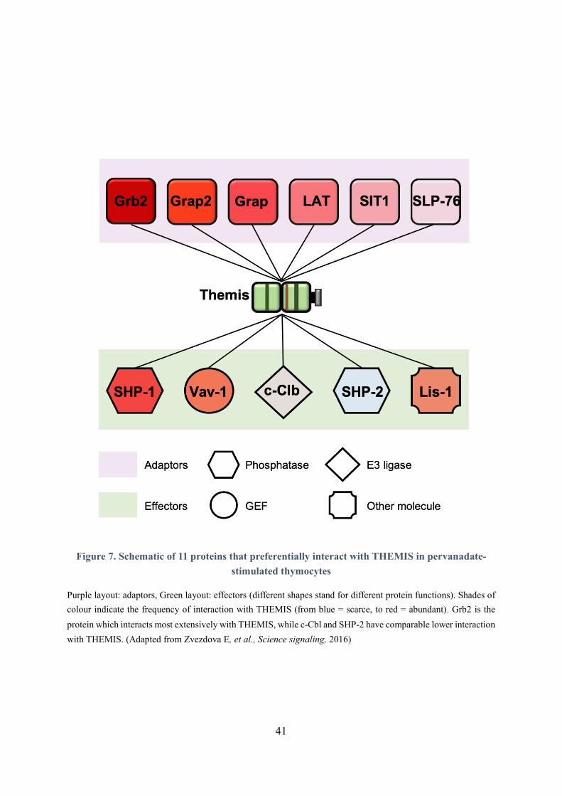
41
Figure 7. Schematic of 11 proteins that preferentially interact with THEMIS in pervanadate-
stimulated thymocytes
Purple layout: adaptors, Green layout: effectors (different shapes stand for different protein functions). Shades of
colour indicate the frequency of interaction with THEMIS (from blue = scarce, to red = abundant). Grb2 is the
protein which interacts most extensively with THEMIS, while c-Cbl and SHP-2 have comparable lower interaction
with THEMIS. (Adapted from Zvezdova E, et al., Science signaling, 2016)
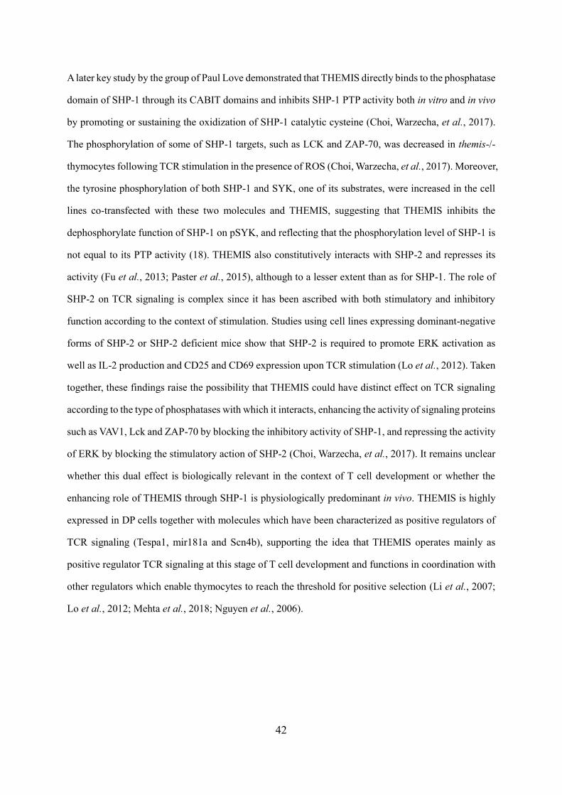
42
A later key study by the group of Paul Love demonstrated that THEMIS directly binds to the phosphatase
domain of SHP-1 through its CABIT domains and inhibits SHP-1 PTP activity both in vitro and in vivo
by promoting or sustaining the oxidization of SHP-1 catalytic cysteine (Choi, Warzecha, et al., 2017).
The phosphorylation of some of SHP-1 targets, such as LCK and ZAP-70, was decreased in themis-/-
thymocytes following TCR stimulation in the presence of ROS (Choi, Warzecha, et al., 2017). Moreover,
the tyrosine phosphorylation of both SHP-1 and SYK, one of its substrates, were increased in the cell
lines co-transfected with these two molecules and THEMIS, suggesting that THEMIS inhibits the
dephosphorylate function of SHP-1 on pSYK, and reflecting that the phosphorylation level of SHP-1 is
not equal to its PTP activity (18). THEMIS also constitutively interacts with SHP-2 and represses its
activity (Fu et al., 2013; Paster et al., 2015), although to a lesser extent than as for SHP-1. The role of
SHP-2 on TCR signaling is complex since it has been ascribed with both stimulatory and inhibitory
function according to the context of stimulation. Studies using cell lines expressing dominant-negative
forms of SHP-2 or SHP-2 deficient mice show that SHP-2 is required to promote ERK activation as
well as IL-2 production and CD25 and CD69 expression upon TCR stimulation (Lo et al., 2012). Taken
together, these findings raise the possibility that THEMIS could have distinct effect on TCR signaling
according to the type of phosphatases with which it interacts, enhancing the activity of signaling proteins
such as VAV1, Lck and ZAP-70 by blocking the inhibitory activity of SHP-1, and repressing the activity
of ERK by blocking the stimulatory action of SHP-2 (Choi, Warzecha, et al., 2017). It remains unclear
whether this dual effect is biologically relevant in the context of T cell development or whether the
enhancing role of THEMIS through SHP-1 is physiologically predominant in vivo. THEMIS is highly
expressed in DP cells together with molecules which have been characterized as positive regulators of
TCR signaling (Tespa1, mir181a and Scn4b), supporting the idea that THEMIS operates mainly as
positive regulator TCR signaling at this stage of T cell development and functions in coordination with
other regulators which enable thymocytes to reach the threshold for positive selection (Li et al., 2007;
Lo et al., 2012; Mehta et al., 2018; Nguyen et al., 2006).
43
In addition, it has been shown that THEMIS2 positively regulates BCR signaling in response to low-
avidity antigens during B-cell selection, which thereby enhances the positive selection of B1 cells and
germinal center B cells (Cheng et al., 2017). THEMIS and THEMIS2 are functionally exchangeable,
since they have highly conserved molecular features (Lesourne et al., 2012). Indeed, it has been shown
that overexpression of THEMIS2 restored the thymocytes development in themis-/- mice and that
THEMIS2 could be phosphorylated and recruited to LAT following TCR engagement (Lesourne et al.,
2012). Altogether, a new model about how THEMIS regulates T cell development was proposed,
referred here as Model II (Figure 6). In Model II, THEMIS enhances TCR signaling by blocking SHP-
1 PTP activity, thereby promoting the signal strength beyond the threshold for positive selection (Choi,
Warzecha, et al., 2017).
To conclude on THEMIS function in T cell development, it is clear that THEMIS is essential for T cell
development and the fact that THEMIS operates by blocking the catalytic function of SHP phosphatases
seems also well established. Although the phenotype of themis-/- mice clearly indicates positive function
of THEMIS on TCR signaling, the relevance of its inhibitory effect on TCR-mediated activation events
in thymocytes activation remains to be defined.
44
Chapter Ⅱ Influence of TCR signaling on T cell responses in peripheral lymphoid
organs
After leaving the thymus, T cells circulate through the lymphatic system in a resting state until they
encounter antigens for which they are specific in secondary lymphoid organs. In order to be fully
activated, T cells require three type of signals: TCR signals triggered by pMHC, co-stimulatory signals
triggered by the interaction of co-receptors with their ligands and cytokine-driven signals (Figure 8).
The TCR of CD4+ and CD8+ T cells recognize their specific antigen by binding to the peptide-MHC
(pMHC) complex displayed by APC, triggering the initial activation of T cells (signal 1). T cells full
activation also requires the engagement of their stimulatory co-receptors, such as CD28 which binds to
the costimulatory molecules B7-1 (CD80) and B7-2 (CD86) expressed on activated APCs (signal 2).
The intracellular signaling pathways induced by the engagement of stimulatory co-receptors function
together with signals triggered by antigen recognition to initiate T cells proliferation. These signals
induce characteristic changes in the expression of various surface molecules in T cells, such as increased
expression of CD69 (directs T cells to secondary lymphoid organs), CD25 (high affinity IL-2 receptor),
CD44 (retains effector T cells at the sites of infection and inflammation), CTLA-4 (a negative regulator
of T cell response). Activated T cells also increase expression of molecules that induce their migration
to the inflammation sites, such as LFA-1 and VLA-4, and reduce the expression of molecules that retain
them into lymphoid organs, such as CD62L and CCR7. In addition, the stimulation of T cells by
cytokines, such as IL-2, stimulates the growth, survival and proliferation of activated T cells (signal 3).
After several cycles of proliferation, under the control of different cytokines, activated T cells
differentiate into different subpopulations, generating effector and memory immune response.
Four major subsets of CD4 effector T cells, also named helper T cells (Th), can be distinguished: Th1,
Th2, Th17 and Tfh. They take part in host defense against different types of infections and diseases.
Regulatory T cells (Tregs) constitute a distinct CD4+ T cell population which can inhibit responses to
self- and foreign- antigens mediated by effector T cells.
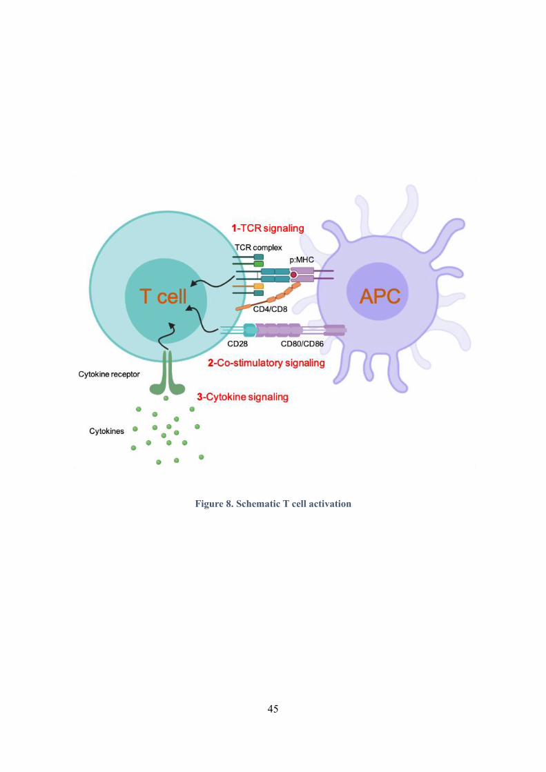
45
Figure 8. Schematic T cell activation
46
In order to eliminate pathogens, activated CD4+ and CD8+ T cells migrate to the site of inflammation
caused by infection or autoimmune disorders, express surface molecules and secrete cytokines that
activate other immune cells, such as B lymphocytes, macrophages and dendritic cells. Meanwhile, the
release of cytokines, chemokines and other inflammatory molecules by activated T cells and by other
immune cells induce further activation and proliferation of the T cells.
TCR signaling mediated by TCR-pMHC specific recognition as well as signals triggered by co-receptors
and cytokine receptors influence the outcomes of T cell responses. The following part of this manuscript
will focus on describing the mechanism by which TCR signals influence the activation and the
differentiation of T cells into effector cells in the context of normal and autoimmune responses.
1. Role of the different T cell subsets during immune responses
1.1. CD4+ T cells subsets
According to the strength of TCR stimulation and the presence of specific cytokines secreted by the
APC in the extracellular milieu, the activated CD4+ T cells may differentiate into distinct effector
subtypes which are characterized by their ability to secrete a specific set of cytokines (Figure 9).
1.1.1. T helper 1 (Th1) cells
Th1 cells predominantly produce IFN in response to intracellular pathogens such as viruses, some
bacteria, some parasites and protein antigens administrated with strong adjuvants (Cher & Mosmann,
1987). T-bet, a member of the T-box family, is essential for the polarization and maintenance of Th1
cells. T-bet-/- CD4+ T cells stimulated with anti-CD3/anti-CD28 antibodies and naïve CD4+ T cells
cultured under Th1-polarizing conditions produce low amount of IFN, suggesting a defective Th1 cell
differentiation in vitro (Szabo et al., 2002). Specific defect in Th1 differentiation was also shown in vivo,
following immunization of T-bet-/- mice with 2,4,6-trinitrophenol–keyhole limpet hemocyanin TNP-
KLH emulsified in complete CFA (to induce a mixed Th1/Th2 response). Moreover, T-bet-/- mice
exhibited increased Th2 cells in response to Leishmania major injection, showing higher production of
IL-4 and IL-5 (Szabo et al., 2002).

47
Figure 9. The differentiation of naïve CD4+ T cell into distinct effector subpopulations
Antigen-presenting cells present antigens through peptide-MHC complexes to naive CD4+ T cells expressing the
TCR and trigger TCR signals leading to the differentiation of naive CD4+ T cells into distinct effector subsets,
according to the cytokine micro-environment. The differentiation into each subtype is controlled by specific
transcription factors. The participation of additional signals, such as co-inhibitory receptors, also has the ability to
influence the outcome of differentiation. Each T helper subset possesses specific biological functions to achieve
adaptive immune response. The functionally close subpopulations are marked with similar colors.
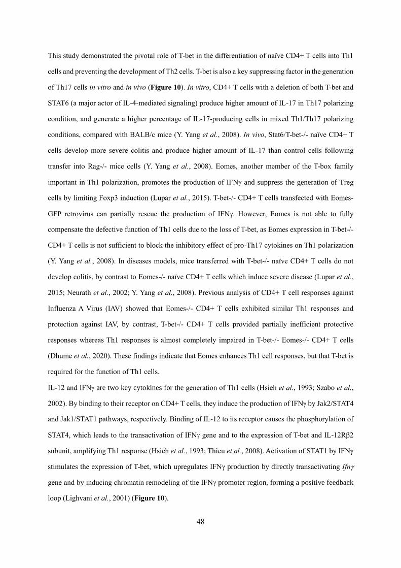
48
This study demonstrated the pivotal role of T-bet in the differentiation of naïve CD4+ T cells into Th1
cells and preventing the development of Th2 cells. T-bet is also a key suppressing factor in the generation
of Th17 cells in vitro and in vivo (Figure 10). In vitro, CD4+ T cells with a deletion of both T-bet and
STAT6 (a major actor of IL-4-mediated signaling) produce higher amount of IL-17 in Th17 polarizing
condition, and generate a higher percentage of IL-17-producing cells in mixed Th1/Th17 polarizing
conditions, compared with BALB/c mice (Y. Yang et al., 2008). In vivo, Stat6/T-bet-/- naïve CD4+ T
cells develop more severe colitis and produce higher amount of IL-17 than control cells following
transfer into Rag-/- mice cells (Y. Yang et al., 2008). Eomes, another member of the T-box family
important in Th1 polarization, promotes the production of IFN and suppress the generation of Treg
cells by limiting Foxp3 induction (Lupar et al., 2015). T-bet-/- CD4+ T cells transfected with Eomes-
GFP retrovirus can partially rescue the production of IFN. However, Eomes is not able to fully
compensate the defective function of Th1 cells due to the loss of T-bet, as Eomes expression in T-bet-/-
CD4+ T cells is not sufficient to block the inhibitory effect of pro-Th17 cytokines on Th1 polarization
(Y. Yang et al., 2008). In diseases models, mice transferred with T-bet-/- naïve CD4+ T cells do not
develop colitis, by contrast to Eomes-/- naïve CD4+ T cells which induce severe disease (Lupar et al.,
2015; Neurath et al., 2002; Y. Yang et al., 2008). Previous analysis of CD4+ T cell responses against
Influenza A Virus (IAV) showed that Eomes-/- CD4+ T cells exhibited similar Th1 responses and
protection against IAV, by contrast, T-bet-/- CD4+ T cells provided partially inefficient protective
responses whereas Th1 responses is almost completely impaired in T-bet-/- Eomes-/- CD4+ T cells
(Dhume et al., 2020). These findings indicate that Eomes enhances Th1 cell responses, but that T-bet is
required for the function of Th1 cells.
IL-12 and IFN are two key cytokines for the generation of Th1 cells (Hsieh et al., 1993; Szabo et al.,
2002). By binding to their receptor on CD4+ T cells, they induce the production of IFN by Jak2/STAT4
and Jak1/STAT1 pathways, respectively. Binding of IL-12 to its receptor causes the phosphorylation of
STAT4, which leads to the transactivation of IFN gene and to the expression of T-bet and IL-12Rβ2
subunit, amplifying Th1 response (Hsieh et al., 1993; Thieu et al., 2008). Activation of STAT1 by IFN
stimulates the expression of T-bet, which upregulates IFN production by directly transactivating Ifn
gene and by inducing chromatin remodeling of the IFN promoter region, forming a positive feedback
loop (Lighvani et al., 2001) (Figure 10).
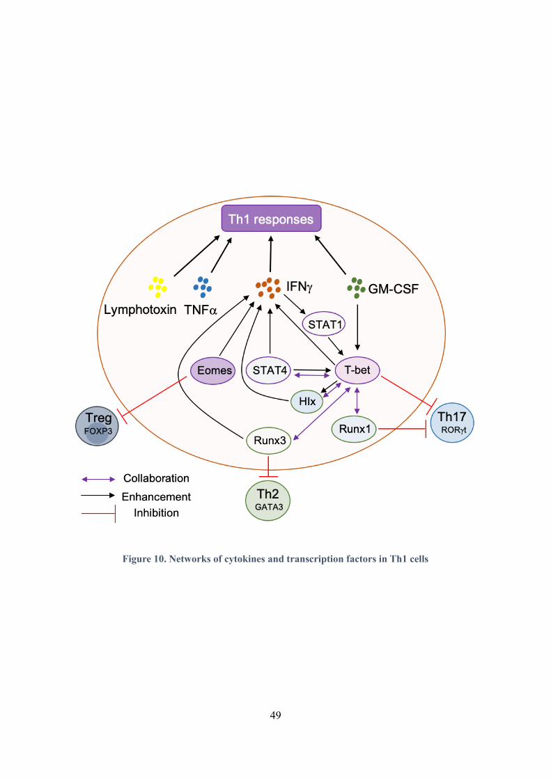
49
Figure 10. Networks of cytokines and transcription factors in Th1 cells
50
For optimal Th1 responses, collaboration between cytokines and transcription factors is required. On
one hand, they promote the differentiation of Th1 cells, on the other hand, they negatively regulate the
development of other T helper cells through repressing the expression of their key transcription factors.
Similarly, IL-27, member of IL-12 family, contributes to promote Th1 differentiation by activating the
phosphorylation of STAT1 (Amsen, Spilianakis, et al., 2009). In addition, Runx1, Runx3 and H1x are
three other transcription factors involved in the process of Th1 differentiation. Runx1, on one hand
promotes the expression of T-bet, on the other hand coordinate with T-bet, interfering with the activation
of RORt to inhibit the development of Th17 cells (Lazarevic et al., 2011). Runx3 cooperates with T-
bet, promoting IFN production by binding to promoter regions controlling the expression of IFNGR1
and IFNGR2 and by silencing the gene encoding for IL-4, which inhibits Th1 development. Meanwhile,
it inhibits Th2 differentiation by interacting with GATA3 (Djuretic et al., 2007; Kohu et al., 2009). H1x
is induced after T-bet activation and also cooperates with T-bet to enhance IFN production by Th1 cells
(244). In addition to the above-mentioned cytokines and transcription factors, Th1 cells also produce
GM-CSF, TNFα and lymphotoxin. GM-CSF enforces the maintenance of Th1 cells by up-regulating T-
bet and IFN in vitro and in vivo (Rasouli et al., 2020). TNFα and lymphotoxin contribute to the
recruitment of leukocytes and to the amplification of inflammation (Fiorentino et al., 1989; Mosmann
et al., 1986).
Th1 and Th2 cells have been the first two CD4+ T cell subsets to be discovered. They were particularly
well studied in seminal studies in the context of autoimmune disease such as Multiple Sclerosis (MS),
Collagen-Induced Arthritis (CIA), Inflammatory Bowel Diseases (IBD) and Crohn’s disease. Th1 cells
were earlier described as the major pathogenic subset in the frame of autoimmune diseases while Th2
cells were shown to exert inhibitory effects on those pathologies (Liblau et al., 1995; Nicholson &
Kuchroo, 1996). Indeed, increased numbers of Th1 cells, especially IFN-producing cells are present in
the central nervous system (CNS) inflammatory lesions at the peak of Experimental autoimmune
encephalomyelitis (EAE) , the mouse model of MS. On the contrary, the proportion of this phenotypic
cell decreases during remission, accompanied by an increase in the production of Th2 cytokines (Imam
et al., 2007; Nicholson & Kuchroo, 1996; Renno et al., 1995). In addition, the adoptive transfer of Th1
cells that express TCR recognizing MOG35-55 into Rag-/- mice develop severe EAE (Domingues et al.,
51
2010). Adoptive transfer of C57BL/6 mice with T-bet-/- CD4+ T cells pre-activated with MOG35-55 in
vivo fails to induce EAE and shows low infiltration of the CNS by CD4+ T cells (O'Connor et al., 2013).
Adoptive transfer of T-bet-/- CD4+ T cells also leads to reduced Th1-mediated colitis and Crohn’s
disease in mouse models (Neurath et al., 2002). STAT4- or IL-12p40- (a subunit of IL-12) deficient
mice are resistant to the development of EAE (Chitnis, Najafian, Benou, et al., 2001; Gran et al., 2002).
Mice deficient for Foxo3, a transcription factor which enhances Th1 generation by activating Eomes,
develop reduced EAE and have decreased numbers of GM-CSF- and IFN- producing CD4+ T cells
(Stienne et al., 2016). Besides, the treatment of mice with IL-12 during the development of CIA
aggravates disease severity (Germann et al., 1996). Taken together, these data suggest a pathogenic role
of Th1 cells in autoimmune diseases. However, the Th1-driven nature of these autoimmune diseases
was later challenged by studies on mice lacking IFN or other molecules important for Th1
differentiation. Mice deficient for IL-12p35 (the other subunit of IL-12), IL-12Rβ2, IFN or IFNR are
not protected from EAE but instead develop more severe disease, pointing-out a complex function of
Th1 cells in autoimmune disorders (Ferber et al., 1996; Gran et al., 2002; Willenborg et al., 1996; Zhang
et al., 2003). GM-CSF and TNFα are not Th1 specific cytokines, as they are produced by most T helper
subsets. However, GM-CSF maintains the phenotype of Th1 cells by up-regulating T-bet and IFN in
the CNS of EAE-developing mice, promoting neuro-inflammation (Rasouli et al., 2020), hence
representing a putative therapeutic target in MS (McQualter et al., 2001). TNFα can promote Th1 and
Th17 responses in peripheral lymphoid organs. Mice treated with anti-TNFα mAbs have reduced
incidence and delayed onset of EAE, but show no difference in disease severity compared to untreated
mice (Batoulis et al., 2014).
1.1.2. T helper 2 (Th2) cells
CD4+ T cells differentiates into Th2 subtype when facing extracellular pathogens, helminths and
allergens in the presence of IL-4. Together with TCR signals, binding of IL-4 on its receptor activates
the transcription factor STAT6 which then induce GATA-3 expression (Amsen, Antov, et al., 2009). As
an important transcription factor in Th2 differentiation, GATA-3 enhances the production of IL-4, IL-5
and IL-13 by directly binding to the enhancer regions of IL-4 gene and by upregulating the transcription
of IL-5 and IL-13 (Agarwal et al., 2000; Kishikawa et al., 2001; Siegel et al., 1995), generating a
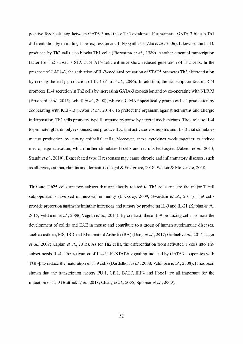
52
positive feedback loop between GATA-3 and these Th2 cytokines. Furthermore, GATA-3 blocks Th1
differentiation by inhibiting T-bet expression and IFN synthesis (Zhu et al., 2006). Likewise, the IL-10
produced by Th2 cells also blocks Th1 cells (Fiorentino et al., 1989). Another essential transcription
factor for Th2 subset is STAT5. STAT5-deficient mice show reduced generation of Th2 cells. In the
presence of GATA-3, the activation of IL-2-mediated activation of STAT5 promotes Th2 differentiation
by driving the early production of IL-4 (Zhu et al., 2006). In addition, the transcription factor IRF4
promotes IL-4 secretion in Th2 cells by increasing GATA-3 expression and by co-operating with NLRP3
(Bruchard et al., 2015; Lohoff et al., 2002), whereas C-MAF specifically promotes IL-4 production by
cooperating with KLF-13 (Kwon et al., 2014). To protect the organism against helminths and allergic
inflammation, Th2 cells promotes type II immune response by several mechanicians. They release IL-4
to promote IgE antibody responses, and produce IL-5 that activates eosinophils and IL-13 that stimulates
mucus production by airway epithelial cells. Moreover, these cytokines work together to induce
macrophage activation, which further stimulates B cells and recruits leukocytes (Jabeen et al., 2013;
Staudt et al., 2010). Exacerbated type II responses may cause chronic and inflammatory diseases, such
as allergies, asthma, rhinitis and dermatitis (Lloyd & Snelgrove, 2018; Walker & McKenzie, 2018).
Th9 and Th25 cells are two subsets that are closely related to Th2 cells and are the major T cell
subpopulations involved in mucosal immunity (Locksley, 2009; Swaidani et al., 2011). Th9 cells
provide protection against helminthic infections and tumors by producing IL-9 and IL-21 (Kaplan et al.,
2015; Veldhoen et al., 2008; Végran et al., 2014). By contrast, these IL-9 producing cells promote the
development of colitis and EAE in mouse and contribute to a group of human autoimmune diseases,
such as asthma, MS, IBD and Rheumatoid Arthritis (RA) (Deng et al., 2017; Gerlach et al., 2014; Jäger
et al., 2009; Kaplan et al., 2015). As for Th2 cells, the differentiation from activated T cells into Th9
subset needs IL-4. The activation of IL-4/Jak1/STAT-6 signaling induced by GATA3 cooperates with
TGF-β to induce the maturation of Th9 cells (Dardalhon et al., 2008; Veldhoen et al., 2008). It has been
shown that the transcription factors PU.1, Gfi.1, BATF, IRF4 and Foxo1 are all important for the
induction of IL-9 (Buttrick et al., 2018; Chang et al., 2005; Spooner et al., 2009).
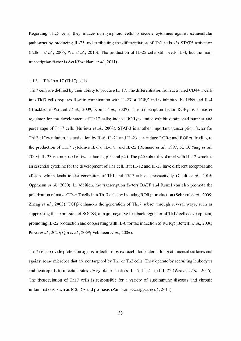
53
Regarding Th25 cells, they induce non-lymphoid cells to secrete cytokines against extracellular
pathogens by producing IL-25 and facilitating the differentiation of Th2 cells via STAT5 activation
(Fallon et al., 2006; Wu et al., 2015). The production of IL-25 cells still needs IL-4, but the main
transcription factor is Act1(Swaidani et al., 2011).
1.1.3. T helper 17 (Th17) cells
Th17 cells are defined by their ability to produce IL-17. The differentiation from activated CD4+ T cells
into Th17 cells requires IL-6 in combination with IL-23 or TGF𝛽 and is inhibited by IFNγ and IL-4
(Brucklacher-Waldert et al., 2009; Korn et al., 2009). The transcription factor RORt is a master
regulator for the development of Th17 cells; indeed RORγt-/- mice exhibit diminished number and
percentage of Th17 cells (Nurieva et al., 2008). STAT-3 is another important transcription factor for
Th17 differentiation, its activation by IL-6, IL-21 and IL-23 can induce RORα and RORt, leading to
the production of Th17 cytokines IL-17, IL-17F and IL-22 (Romano et al., 1997; X. O. Yang et al.,
2008). IL-23 is composed of two subunits, p19 and p40. The p40 subunit is shared with IL-12 which is
an essential cytokine for the development of Th1 cell. But IL-12 and IL-23 have different receptors and
effects, which leads to the generation of Th1 and Th17 subsets, respectively (Cauli et al., 2015;
Oppmann et al., 2000). In addition, the transcription factors BATF and Runx1 can also promote the
polarization of naïve CD4+ T cells into Th17 cells by inducing RORt production (Schraml et al., 2009;
Zhang et al., 2008). TGFβ enhances the generation of Th17 subset through several ways, such as
suppressing the expression of SOCS3, a major negative feedback regulator of Th17 cells development,
promoting IL-22 production and cooperating with IL-6 for the induction of RORt (Bettelli et al., 2006;
Perez et al., 2020; Qin et al., 2009; Veldhoen et al., 2006).
Th17 cells provide protection against infections by extracellular bacteria, fungi at mucosal surfaces and
against some microbes that are not targeted by Th1 or Th2 cells. They operate by recruiting leukocytes
and neutrophils to infection sites via cytokines such as IL-17, IL-21 and IL-22 (Weaver et al., 2006).
The dysregulation of Th17 cells is responsible for a variety of autoimmune diseases and chronic
inflammations, such as MS, RA and psoriasis (Zambrano-Zaragoza et al., 2014).

54
IL-17 and other Th17 cytokines such as IL-21 and IL-22 strongly contribute to the pathogenicity of this
lineage (Korn et al., 2007; Zheng et al., 2007). IL-23 plays a critical role in generating pathogenic Th17
cells during autoimmune inflammation (Langrish et al., 2005). Mice deficient for IL-23p19, a subunit
of IL-23, develop less severe autoimmune diseases, including EAE, CIA and IBD (Cua et al., 2003;
Murphy et al., 2003). Adoptive transfer of MOG-specific Th17 cells differentiated in vitro by IL-6 and
TGFβ1 fails to induce EAE while polarization of MOG-specific Th17 cells in the presence of IL-23
induces severe EAE in recipient mice(El-Behi et al., 2011; Ghoreschi et al., 2010; Langrish et al., 2005).
While IL-6 and TGF-β1 induce non-pathogenic Th17 cells producing IL-10 and expressing high amount
of PROCR, a negative regulator of pro-inflammatory genes (Ghoreschi et al., 2010; Kishi et al., 2016;
Lee et al., 2012), further investigations showed that IL-23 and RORt, alone or in cooperation,
contribute to induce GM-CSF expression in Th17 cells, a pro-inflammatory cytokine which is required
for encephalitogenic potential of Th17 cells (Codarri et al., 2011; El-Behi et al., 2011).
Th22 cells are phenotypically and functionally related to Th17 cells (Eyerich et al., 2009). Th22 subset
contributes to mucosal immunity by preventing translocation of microbes across epithelia to limit
infection (Kim et al., 2012). Like Th17 cells, Th22 cells produce IL-22, but this production needs an
environment poor in IFN and IL-17 (Trifari et al., 2009). Both subsets express chemokine receptors
CCR4 and CCR6 that are associated with cutaneous T cell homing, but CCR10 is specifically expressed
on Th22 cells (Trifari et al., 2009). In addition, Th22 cells produce IL-13, TNFα, IL-10, and the
chemokines CCL15 and CCL17. The production of IL-22 needs the expression of aryl hydrocarbon
receptor (AhR), a ligand-activated transcription factor that is activated by small molecules (Monteleone
et al., 2011). STAT-3 is another important transcription factor for the generation of Th22 cells. It can be
activated by the cytokines IL-6 and TNFα (Pickert et al., 2009). In vitro, Th22 cells can be induced in
the presence of IL-6, IL-23, IL-1β, FICZ and TGF-βR inhibitor (Plank et al., 2017).
1.1.4. T follicular helper (Tfh) cells
Tfh cells are CD4+ T cells that specifically express the chemokine receptor CXCR5 and markedly
express ICOS, PD-1 and CD40L, which regulate B lymphocyte activity (Crotty, 2019; Nurieva et al.,
2008). Tfh cells migrate into the follicle in lymph nodes and allow B cells to initiate extra follicular and
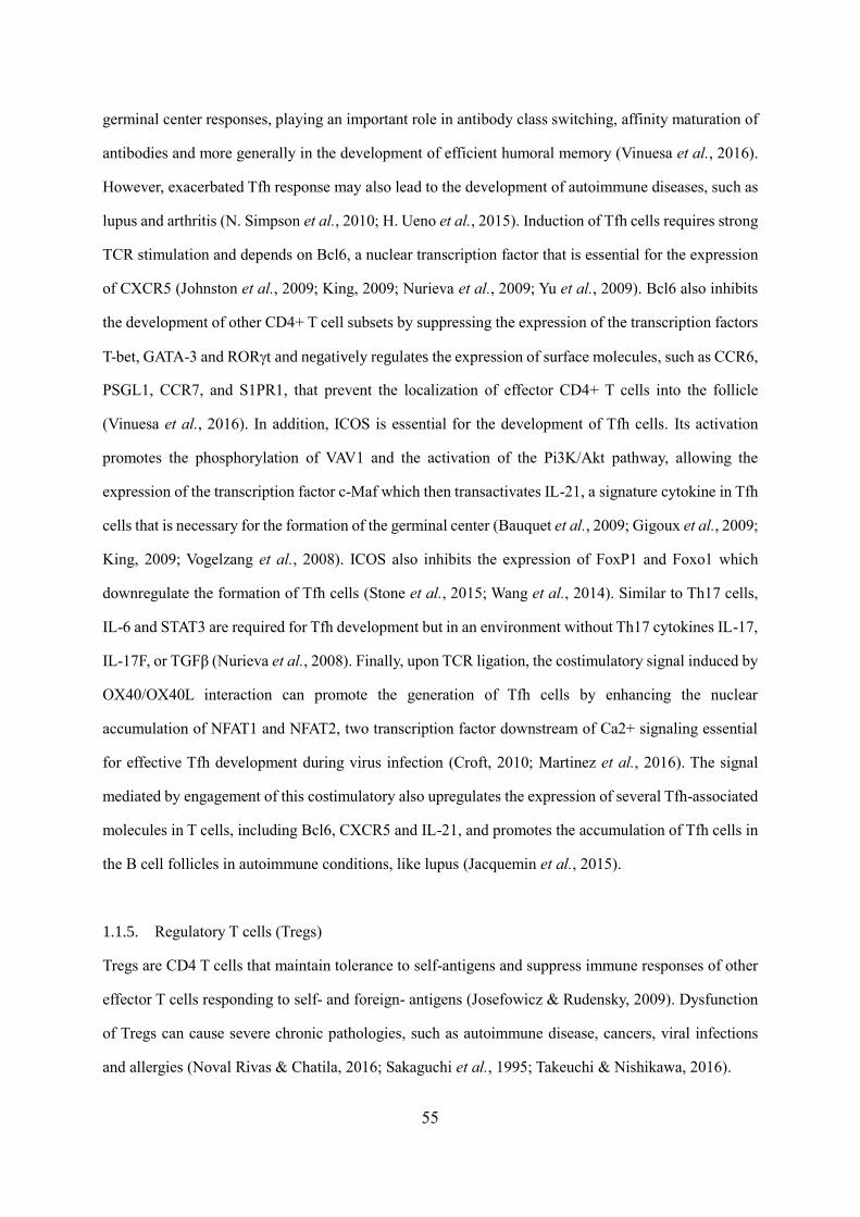
55
germinal center responses, playing an important role in antibody class switching, affinity maturation of
antibodies and more generally in the development of efficient humoral memory (Vinuesa et al., 2016).
However, exacerbated Tfh response may also lead to the development of autoimmune diseases, such as
lupus and arthritis (N. Simpson et al., 2010; H. Ueno et al., 2015). Induction of Tfh cells requires strong
TCR stimulation and depends on Bcl6, a nuclear transcription factor that is essential for the expression
of CXCR5 (Johnston et al., 2009; King, 2009; Nurieva et al., 2009; Yu et al., 2009). Bcl6 also inhibits
the development of other CD4+ T cell subsets by suppressing the expression of the transcription factors
T-bet, GATA-3 and RORt and negatively regulates the expression of surface molecules, such as CCR6,
PSGL1, CCR7, and S1PR1, that prevent the localization of effector CD4+ T cells into the follicle
(Vinuesa et al., 2016). In addition, ICOS is essential for the development of Tfh cells. Its activation
promotes the phosphorylation of VAV1 and the activation of the Pi3K/Akt pathway, allowing the
expression of the transcription factor c-Maf which then transactivates IL-21, a signature cytokine in Tfh
cells that is necessary for the formation of the germinal center (Bauquet et al., 2009; Gigoux et al., 2009;
King, 2009; Vogelzang et al., 2008). ICOS also inhibits the expression of FoxP1 and Foxo1 which
downregulate the formation of Tfh cells (Stone et al., 2015; Wang et al., 2014). Similar to Th17 cells,
IL-6 and STAT3 are required for Tfh development but in an environment without Th17 cytokines IL-17,
IL-17F, or TGFβ (Nurieva et al., 2008). Finally, upon TCR ligation, the costimulatory signal induced by
OX40/OX40L interaction can promote the generation of Tfh cells by enhancing the nuclear
accumulation of NFAT1 and NFAT2, two transcription factor downstream of Ca2+ signaling essential
for effective Tfh development during virus infection (Croft, 2010; Martinez et al., 2016). The signal
mediated by engagement of this costimulatory also upregulates the expression of several Tfh-associated
molecules in T cells, including Bcl6, CXCR5 and IL-21, and promotes the accumulation of Tfh cells in
the B cell follicles in autoimmune conditions, like lupus (Jacquemin et al., 2015).
1.1.5. Regulatory T cells (Tregs)
Tregs are CD4 T cells that maintain tolerance to self-antigens and suppress immune responses of other
effector T cells responding to self- and foreign- antigens (Josefowicz & Rudensky, 2009). Dysfunction
of Tregs can cause severe chronic pathologies, such as autoimmune disease, cancers, viral infections
and allergies (Noval Rivas & Chatila, 2016; Sakaguchi et al., 1995; Takeuchi & Nishikawa, 2016).
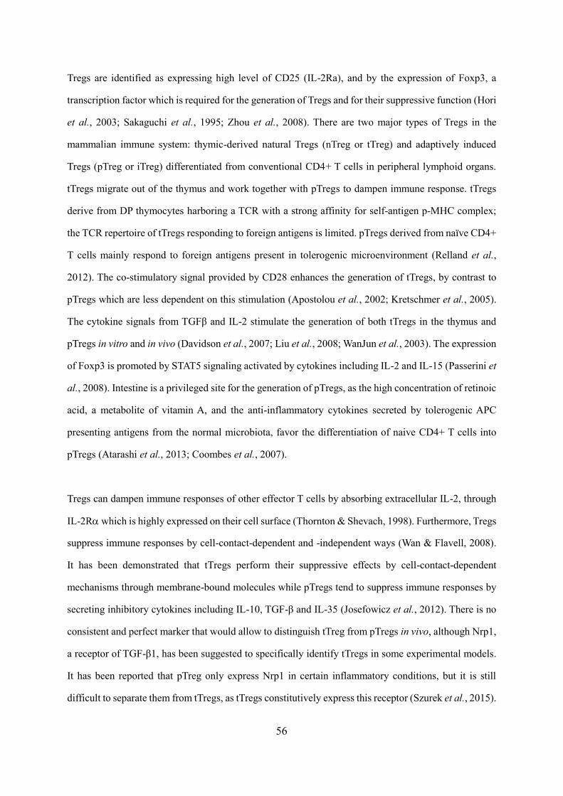
56
Tregs are identified as expressing high level of CD25 (IL-2Ra), and by the expression of Foxp3, a
transcription factor which is required for the generation of Tregs and for their suppressive function (Hori
et al., 2003; Sakaguchi et al., 1995; Zhou et al., 2008). There are two major types of Tregs in the
mammalian immune system: thymic-derived natural Tregs (nTreg or tTreg) and adaptively induced
Tregs (pTreg or iTreg) differentiated from conventional CD4+ T cells in peripheral lymphoid organs.
tTregs migrate out of the thymus and work together with pTregs to dampen immune response. tTregs
derive from DP thymocytes harboring a TCR with a strong affinity for self-antigen p-MHC complex;
the TCR repertoire of tTregs responding to foreign antigens is limited. pTregs derived from naïve CD4+
T cells mainly respond to foreign antigens present in tolerogenic microenvironment (Relland et al.,
2012). The co-stimulatory signal provided by CD28 enhances the generation of tTregs, by contrast to
pTregs which are less dependent on this stimulation (Apostolou et al., 2002; Kretschmer et al., 2005).
The cytokine signals from TGFβ and IL-2 stimulate the generation of both tTregs in the thymus and
pTregs in vitro and in vivo (Davidson et al., 2007; Liu et al., 2008; WanJun et al., 2003). The expression
of Foxp3 is promoted by STAT5 signaling activated by cytokines including IL-2 and IL-15 (Passerini et
al., 2008). Intestine is a privileged site for the generation of pTregs, as the high concentration of retinoic
acid, a metabolite of vitamin A, and the anti-inflammatory cytokines secreted by tolerogenic APC
presenting antigens from the normal microbiota, favor the differentiation of naive CD4+ T cells into
pTregs (Atarashi et al., 2013; Coombes et al., 2007).
Tregs can dampen immune responses of other effector T cells by absorbing extracellular IL-2, through
IL-2R which is highly expressed on their cell surface (Thornton & Shevach, 1998). Furthermore, Tregs
suppress immune responses by cell-contact-dependent and -independent ways (Wan & Flavell, 2008).
It has been demonstrated that tTregs perform their suppressive effects by cell-contact-dependent
mechanisms through membrane-bound molecules while pTregs tend to suppress immune responses by
secreting inhibitory cytokines including IL-10, TGF-β and IL-35 (Josefowicz et al., 2012). There is no
consistent and perfect marker that would allow to distinguish tTreg from pTregs in vivo, although Nrp1,
a receptor of TGF-β1, has been suggested to specifically identify tTregs in some experimental models.
It has been reported that pTreg only express Nrp1 in certain inflammatory conditions, but it is still
difficult to separate them from tTregs, as tTregs constitutively express this receptor (Szurek et al., 2015).
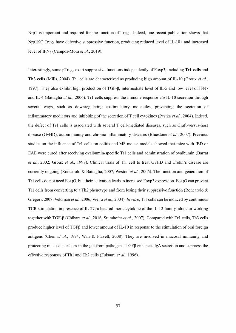
57
Nrp1 is important and required for the function of Tregs. Indeed, one recent publication shows that
Nrp1KO Tregs have defective suppressive function, producing reduced level of IL-10+ and increased
level of IFN (Campos-Mora et al., 2019).
Interestingly, some pTregs exert suppressive functions independently of Foxp3, including Tr1 cells and
Th3 cells (Mills, 2004). Tr1 cells are characterized as producing high amount of IL-10 (Groux et al.,
1997). They also exhibit high production of TGF-β, intermediate level of IL-5 and low level of IFN
and IL-4 (Battaglia et al., 2006). Tr1 cells suppress the immune response via IL-10 secretion through
several ways, such as downregulating costimulatory molecules, preventing the secretion of
inflammatory mediators and inhibiting of the secretion of T cell cytokines (Pestka et al., 2004). Indeed,
the defect of Tr1 cells is associated with several T cell-mediated diseases, such as Graft-versus-host
disease (GvHD), autoimmunity and chronic inflammatory diseases (Bluestone et al., 2007). Previous
studies on the influence of Tr1 cells on colitis and MS mouse models showed that mice with IBD or
EAE were cured after receiving ovalbumin-specific Tr1 cells and administration of ovalbumin (Barrat
et al., 2002; Groux et al., 1997). Clinical trials of Tr1 cell to treat GvHD and Crohn’s disease are
currently ongoing (Roncarolo & Battaglia, 2007; Weston et al., 2006). The function and generation of
Tr1 cells do not need Foxp3, but their activation leads to increased Foxp3 expression. Foxp3 can prevent
Tr1 cells from converting to a Th2 phenotype and from losing their suppressive function (Roncarolo &
Gregori, 2008; Veldman et al., 2006; Vieira et al., 2004). In vitro, Tr1 cells can be induced by continuous
TCR stimulation in presence of IL-27, a heterodimeric cytokine of the IL-12 family, alone or working
together with TGF-β (Chihara et al., 2016; Stumhofer et al., 2007). Compared with Tr1 cells, Th3 cells
produce higher level of TGFβ and lower amount of IL-10 in response to the stimulation of oral foreign
antigens (Chen et al., 1994; Wan & Flavell, 2008). They are involved in mucosal immunity and
protecting mucosal surfaces in the gut from pathogens. TGFβ enhances IgA secretion and suppress the
effective responses of Th1 and Th2 cells (Fukaura et al., 1996).
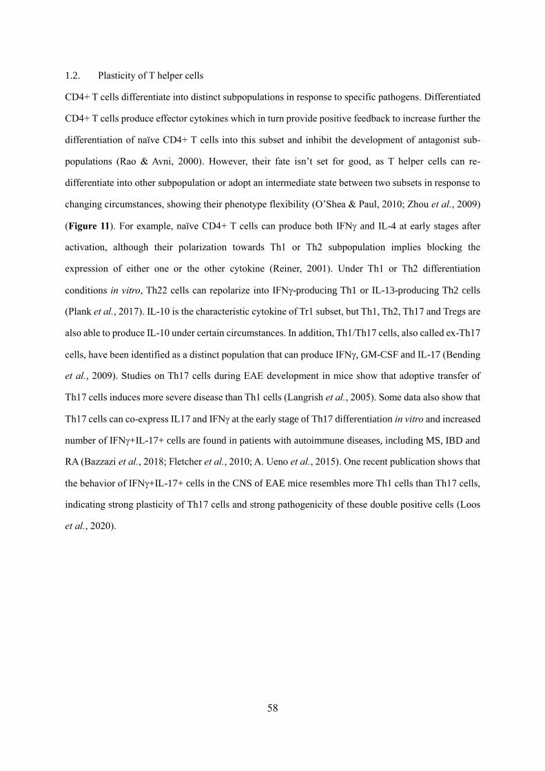
58
1.2. Plasticity of T helper cells
CD4+ T cells differentiate into distinct subpopulations in response to specific pathogens. Differentiated
CD4+ T cells produce effector cytokines which in turn provide positive feedback to increase further the
differentiation of naïve CD4+ T cells into this subset and inhibit the development of antagonist sub-
populations (Rao & Avni, 2000). However, their fate isn’t set for good, as T helper cells can re-
differentiate into other subpopulation or adopt an intermediate state between two subsets in response to
changing circumstances, showing their phenotype flexibility (O’Shea & Paul, 2010; Zhou et al., 2009)
(Figure 11). For example, naïve CD4+ T cells can produce both IFN and IL-4 at early stages after
activation, although their polarization towards Th1 or Th2 subpopulation implies blocking the
expression of either one or the other cytokine (Reiner, 2001). Under Th1 or Th2 differentiation
conditions in vitro, Th22 cells can repolarize into IFN-producing Th1 or IL-13-producing Th2 cells
(Plank et al., 2017). IL-10 is the characteristic cytokine of Tr1 subset, but Th1, Th2, Th17 and Tregs are
also able to produce IL-10 under certain circumstances. In addition, Th1/Th17 cells, also called ex-Th17
cells, have been identified as a distinct population that can produce IFN, GM-CSF and IL-17 (Bending
et al., 2009). Studies on Th17 cells during EAE development in mice show that adoptive transfer of
Th17 cells induces more severe disease than Th1 cells (Langrish et al., 2005). Some data also show that
Th17 cells can co-express IL17 and IFN at the early stage of Th17 differentiation in vitro and increased
number of IFN+IL-17+ cells are found in patients with autoimmune diseases, including MS, IBD and
RA (Bazzazi et al., 2018; Fletcher et al., 2010; A. Ueno et al., 2015). One recent publication shows that
the behavior of IFN+IL-17+ cells in the CNS of EAE mice resembles more Th1 cells than Th17 cells,
indicating strong plasticity of Th17 cells and strong pathogenicity of these double positive cells (Loos
et al., 2020).
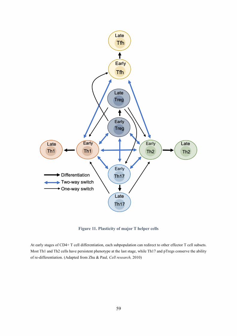
59
Figure 11. Plasticity of major T helper cells
At early stages of CD4+ T cell differentiation, each subpopulation can redirect to other effector T cell subsets.
Most Th1 and Th2 cells have persistent phenotype at the last stage, while Th17 and pTregs conserve the ability
of re-differentiation. (Adapted from Zhu & Paul, Cell research, 2010)

60
1.2.1. Need of specific cytokines
The plasticity of CD4+ T cells depends on different factors. The presence of specific cytokines in the
environment is an essential factor. During the differentiation of CD4+ T cells into Th1 cells in vitro,
enhanced IL-15 signaling from trans-presented IL15/IL-15R increase and maintain the expression of
Th1-associated genes including IFN and T-bet by promoting the expression of STAT5. However, when
expose to IL-15 alone, those Th1 cells show increased expression of Bcl6, resulting in a re-
differentiation from Th1 to Tfh cells (Cooley et al., 2015). Similarly, IL-18, a member of the IL-1 family,
was originally discovered as an enhancer of IFN production by anti-CD3 stimulation in Th1 cells
(Nakanishi et al., 2001; Okamura et al., 1995). Later studies showed that IL-18 cooperates with IL-12
to promote Th1 responses in vitro and in vivo by enhancing IFN production in Th1 cells and non-
polarized T cells. However, without IL-12, IL-18 drives the cells to produce IL-4 and IL-13, inducing
Th2 polarization (Hoshino et al., 1999; Nakanishi et al., 2001; Yoshimoto et al., 2000).
In addition, at early stage of CD4+T cell differentiation in vitro, Th1 cells can lose IFN expression and
convert into Th2 cells by adding IL-4 to the culture medium. Likewise, Th2 cells can be easily redirected
to Th1 cells by adding IL-12 (Perez et al., 1995). Th17 cells can be re-differentiated into Th1 cells in
vitro by adding IL-12 to the culture medium, in the absence of IL-6 (Zhou et al., 2009). Interestingly,
both Th1 and Th17 cells can be repolarized to Th2 cells in vivo. A study shows that injection of the
gastrointestinal helminth Nippostrongylus brasiliensis into mice that were transferred with Th1 or Th17
cells caused these cells to repolarize into Th2 cells, producing IL-4 and losing the ability to express
IFN or IL-17 (Panzer et al., 2012). In addition, during the early development of Th17 cells, CD4+ T
cells express both Foxp3 and RORt, exhibiting an intermediate state between pTregs and Th17 cells
(Huehn et al., 2009). Adding IL-6, IL-21, IL-23 and low level of TGFβ in the culture medium drives
these intermediate cells to become Th17 cells, while the presence of high level of TGFβ and IL-2
supports the development of pTregs (Huehn et al., 2009; Yun Kyung Lee et al., 2009; Weaver et al.,
2006). In the presence of IL-21, TGFβ-induced Tregs are able to mature into Tfh cells by upregulating
CXCR5 expression in aim to relocate into follicles (Nurieva et al., 2008). Finally, in the presence of IL-
4, Th2 cells also can become Tfh cells by increasing CXCR5 expression, the re-differentiated Tfh cells
still produce IL-4 but do not produce other Th2 cytokines (Dardalhon et al., 2008; Veldhoen et al., 2008).

61
Since IL-4 is required for the development of Th2, Th9 and Tfh subpopulations, Th2 cells can become
Th9 cells when IL-4 and TGFβ are present in the surrounding environment (Cantor & Shinohara, 2009).
1.2.2. Need of the required Transcription factors
Transcription factors play an essential role for the redifferentiation of T helper cells. They are not just
necessary for the polarization of the relevant CD4+ T cell subsets but also for suppressing the
development of other populations. Overexpression of T-bet in Th2 cells results in a Th1 phenotype while
overexpression of GATA3 in Th1 cells lead these cells to re-differentiate into Th2 cells (Cantor &
Shinohara, 2009). In parallel, the absence of the key transcription factors in a defined lineage unlocks
the expression of other transcription factors or polarizing mediators, leading to the differentiation of
alternative CD4+ T cell subpopulations. For example, under strong Th1 polarizing conditions, the
absence of T-bet results in upregulation of GATA3, IL-4 and IL-17 (E. L. Pearce et al., 2003; Yagi et al.,
2010). The increased production of IL-17 can be further enhanced by knocking out another T-box family
member, Eomes (Dhume et al., 2020). Besides, some transcription factors are co-expressed in
intermediate CD4+ T cells. For example, both T-bet and RORt are expressed in IFN+IL-17+-
producing cells, allowing them to maintain either a Th1/Th17 phenotype or to further develop into either
Th1 or Th17 cells (Wilson et al., 2009). In differentiated Th17 cells that are induced by myelin basic
protein (MBP-Ac-11) in vitro, IFN+IL-17+ cells have higher expression of T-bet than IFN+ cells
(Yang et al., 2009). Further studies demonstrated that T-bet is required for the switch from IL-17 to IFN.
During the development of EAE, compared with WT mice, T-bet-/- mice display a reduced production
of IFN but increased expression of IL-17 and no IFN+IL-17+ CD4 T cells (O'Connor et al., 2013).
The metabolic activity also plays a considerable role in CD4+ T cell plasticity. For example, Acetyl-
CoA Carboxylase 1 (ACC1) enhances the generation of Th17 cells but decreases the development of
Tregs in mouse models and human cells. Blocking ACC1 with Soraphen A inhibits de novo fatty acid
synthesis and glycolytic-lipogenic metabolic pathway, leading to increased generation of FoxP3-
producing cells and to decreased development of IL17-producing cells in vitro and in vivo (Berod et al.,
2014). Besides, other factors including cellular variations, genetic, epigenetic and TCR signal strength
are involved in the regulation of the CD4+ T cell plasticity.

62
1.3. CD8+ T cells and other T cell subsets
1.3.1. CD8+ T cells
CD8+ T cells, also known as cytotoxic T lymphocytes (CTLs) when they are activated, constitute a T
cell population that possess protective function towards intracellular pathogens, including intracellular
bacteria, viruses and which can also contribute to the elimination of tumor cells. CTLs kill antigen-
bearing cells by three major mechanisms. The first is the secretion of cytokines and chemokines with
antimicrobial function, such as IFN, TNFα, MIP-1α/βand RANTES (Guidotti et al., 1994; Yang et al.,
1997). The second way is to release lytic granules containing perforin and granzymes which can induce
caspase-dependent apoptosis of the target cell (Obar & Lefrançois, 2010). The third mechanism is via
Fas/FasL interaction. Binding of Fas, expressed on the surface of the target cell, with FasL, expressed
by activated CD8+ T cells, also causes the activation of caspases cascade and results in the apoptosis of
the target cells (Bakshi et al., 2014). In addition, evidence from studies in RA, MS and EAE have shown
that CD8+ T cells play a complex role in autoimmune diseases (Blanco et al., 2005; Friese & Fugger,
2009; Junker et al., 2007). Effective CD8+ T cell responses have been associated with the development
of acute MS plaques and oligoclonal expansion of CD8+ T cells in CNS of MS patients (Blanco et al.,
2005; Junker et al., 2007). By contrast, in autoimmune diseases such as RA, IBD and MS and in murine
model of MS, some CD8+ T cells develop regulatory function by targeting autoreactive CD4+ T cells
through the recognition of the MHC class Ib molecule Qa-1 in mouse and the HLA-E in human
(Baughman et al., 2011; Brimnes et al., 2005; Carvalheiro et al., 2013; Jiang et al., 1992; Lu & Cantor,
2008). During EAE development, the activation of those CD8+ regulatory T cells suppress the disease
by inhibiting CD4+ T cells responses through releasing IFNβ and perforin (Sinha et al., 2014).
As for CD4+ T cells, the activation of CD8+ T cells is mediated by TCR-pMHC I engagement and needs
additional signals from co-receptors, such as CD28, and immunomodulation by cytokines such as IL-2,
IL-7, IL-15 and IL-18 (Au-Yeung et al., 2017; Carrette & Surh, 2012; Obar & Lefrançois, 2010; Walsh
et al., 2014). Effector CD4+ T cells promote the activation of CD8+ T cells by secreting cytokines, such
as IL-2, IL-12 and IL-21, and enhancing stimulatory signal from APCs to CD8+ T cells through
CD40/CD40L interaction (Bennett et al., 1998; Castellino & Germain, 2006). T-bet and especially
Eomes are key transcription factors for the development and function of CTLs.
63
Eomes is responsible for T-bet-independent IFN production in CD8+ T cells (Erika L Pearce et al.,
2003). The absence of Eomes in CD8+ T cells leads to reduced production of perforin, IFN and
granzyme B in vitro and in vivo (Erika L Pearce et al., 2003). Mice that a have T-bet/Eomes double-
deficiency result in the development of chronic wasting disease (CWD) in response to LCMV infection
(Intlekofer et al., 2008).
1.3.2. T cells and NKT cells
In addition to CD4+ and CD8+ T cells, some populations of T cells have distinct features and functions
in host defense, including T cells and NKT cells. The TCRs expressed by these T cells have limited
diversity, but are able to recognize various antigens without being presented by MHC I or MHC II.
According to reports, T cells predominantly recognize lipid antigens, and some can recognize MHC
class Ib molecules, while it’s still rarely known about the antigenic molecules that activate this
phenotypical cells (Bonneville et al., 2010). Likewise, many of NKT cells recognize lipid antigens
presented by CD1d, a non-polymorphic major histocompatibility complex class I-like antigen presenting
molecule (Godfrey & Kronenberg, 2004). Both cell types are abundant in epithelial tissues, being
involved in and promoting innate and adaptive immunity by secreting cytokines and chemokines.
T cells have been associated with many autoimmune diseases. On one hand, they play a protective
role at the inflammation site by secreting IL-22, a cytokine that is responsible for cell-mediated
production of antimicrobial peptides and tissue repair (Holtmeier & Kabelitz, 2005; Paul & Lal, 2015).
On the other hand, they promote Th17 response by releasing IL-17. High numbers of IL-17 producing
T cells are detected during the development of IBD, RA, MS and Psoriasis (Cruz et al., 2018; Paul
& Lal, 2015; Shiromizu & Jancic, 2018).
NKT cells, also known as invariant NKT cells (iNKT), share some features with NK cells, such as
Granzyme production. They also secrete large quantities of cytokines, such as IFN and IL-2 (Vivier &
Anfossi, 2004). The interaction of iNKT and B cells can promote the prolonged antibody response and
long-term memory of B cells (Bai et al., 2013). It has been reported that the absence of iNKT cells can
induce the development of autoimmune diseases, such as diabetes or atherosclerosis (Wu & Kaer, 2009).
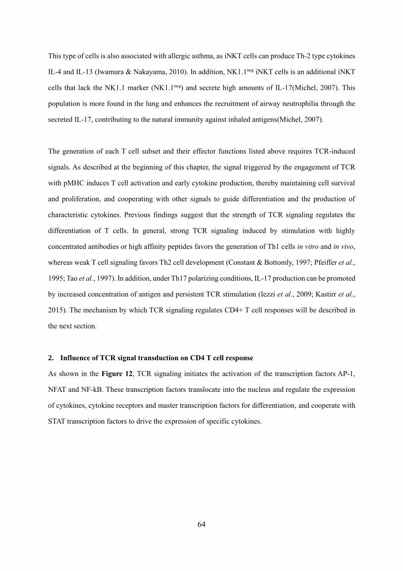
64
This type of cells is also associated with allergic asthma, as iNKT cells can produce Th-2 type cytokines
IL-4 and IL-13 (Iwamura & Nakayama, 2010). In addition, NK1.1neg iNKT cells is an additional iNKT
cells that lack the NK1.1 marker (NK1.1neg) and secrete high amounts of IL-17(Michel, 2007). This
population is more found in the lung and enhances the recruitment of airway neutrophilia through the
secreted IL-17, contributing to the natural immunity against inhaled antigens(Michel, 2007).
The generation of each T cell subset and their effector functions listed above requires TCR-induced
signals. As described at the beginning of this chapter, the signal triggered by the engagement of TCR
with pMHC induces T cell activation and early cytokine production, thereby maintaining cell survival
and proliferation, and cooperating with other signals to guide differentiation and the production of
characteristic cytokines. Previous findings suggest that the strength of TCR signaling regulates the
differentiation of T cells. In general, strong TCR signaling induced by stimulation with highly
concentrated antibodies or high affinity peptides favors the generation of Th1 cells in vitro and in vivo,
whereas weak T cell signaling favors Th2 cell development (Constant & Bottomly, 1997; Pfeiffer et al.,
1995; Tao et al., 1997). In addition, under Th17 polarizing conditions, IL-17 production can be promoted
by increased concentration of antigen and persistent TCR stimulation (Iezzi et al., 2009; Kastirr et al.,
2015). The mechanism by which TCR signaling regulates CD4+ T cell responses will be described in
the next section.
2. Influence of TCR signal transduction on CD4 T cell response
As shown in the Figure 12, TCR signaling initiates the activation of the transcription factors AP-1,
NFAT and NF-kB. These transcription factors translocate into the nucleus and regulate the expression
of cytokines, cytokine receptors and master transcription factors for differentiation, and cooperate with
STAT transcription factors to drive the expression of specific cytokines.
65
Figure 12. TCR signaling regulates CD4+ T cell differentiation
By cooperating with cytokines-mediated signals, TCR signaling induces the production of early cytokine and
the expression of cytokine receptor and master transcription factors, thereby enhancing the expression of
secondary transcription factors and cooperating with them to upregulate the expression of cytokine and cytokine
receptor genes. Additional signals are able to affect the outcome of differentiation by modulating and
cooperating with TCR signals. (Adapted from Zhu J, et al., Annual review of immunology, 2010).
66
Meanwhile, master transcriptional regulators enhance the expression of secondary transcription factors,
such as the enhancement of STAT-4 and Runx3 by T-bet in Th1 cells and RORt - upregulated STAT-3
in Th17 cells (Zhu et al., 2010). In addition, they directly upregulate cytokines and cytokine receptors,
which in turn enhance initial cytokine/Jak/STAT pathways. In addition, other signals, such as co-
inhibitory receptors and growth factors, are able to affect the outcomes of CD4+ T cell responses by
modulating TCR-induced signals. Thus, signaling and transcriptional events associated to the TCR, the
cytokine receptors and the transcription factors form an interconnected network which control the
lineage fate of effector CD4+ T cells. All these pathways cooperate with each other to direct the
differentiation of T cells as well as their functional characteristics and their response to diseases.
Notably, each transcription factor in the AP-1 family, NFAT and NF-kB is activated by specific TCR
signaling distal pathways, corresponding to their distinct effect on the different biological responses of
CD4+ T cells. Proximal TCR signaling events occur early after TCR ligation and are important to
translate early signals into cues which may modulate more ubiquitous downstream pathways. Defects
in the proximal signaling components (such as CD3 ITAMs and LAT) often result in impaired T cell
responses, leading to poor protection against pathogens. Whereas, excessive activation of proximal
signaling or each downstream pathway may lead to autoimmunity. Their particular effects on CD4+ T
cell responses and functions will be described in the following section.
2.1. Proximal TCR signaling
As described in Chapter I (1.2), the engagement of the TCR with pMHC induces the phosphorylation
of CD3 ITAMs by Lck or Fyn. Lck is a member of Src family of tyrosine kinases, its expression is
required for cell proliferation and IL-2 production (Seddon & Zamoyska, 2002). Furthermore, the
absence of Lck results in impaired development of Th2 cells with decreased expression of GATA-3,
whereas it does not affect the differentiation of Th1 subset (Kemp et al., 2010). Fyn also belongs to Src
family, functions similarly to Lck in T cells. It contributes to the activation of the ERK pathway, while
its expression is not necessary for T cell activation, and it negatively regulates Th2 polarization via
repressing IL-4R signaling (Lovatt et al., 2006; Mamchak et al., 2008; Tamura et al., 2001). ITAMs
phosphorylation by Lck promotes the recruitment and activation of Zap70. Zap70 activity is required
67
for T cell activation, survival and suppressive activity of Treg (Au-Yeung et al., 2010; Schim van der
Loeff et al., 2014). At the molecular level, Zap70 functions by recruiting SH2-containing proteins like
Cbl, VAV1 and Lck to the tyrosine sites Y292, Y315 and Y319, respectively (Kong et al., 1996). It has
been shown that the residues Y315 and Y319 enhances TCR signaling while the recruitment of Cbl to
Y292 exerts a negative role on T cell activation and on the production of IL-2 and IFN (Magnan et al.,
2001).
Zap70 then phosphorylates LAT, an adaptor protein able to recruit many other actors of TCR signaling,
such as PLC1, SLP-76 and Grb2, forming a LAT signalosome and leading to the activation of distal
signaling pathways (Wange, 2000). LAT being the link between proximal and distal TCR signaling
pathways, the deletion or mutation of LAT in peripheral CD4+ T cells lead to severe defect of TCR
signaling and T cell response, including reduced suppressive function of Tregs and lymphoproliferation
of conventional CD4+ T cells in post-thymic LAT deficient mice (Chuck et al., 2010; Mingueneau et
al., 2009; Shen et al., 2010). Over-expression of PLC-1 has been reported to occur in a variety of
malignancies including Luminal-A and -B breast cancer patients (Lattanzio et al., 2019). Both SLP-76
and Grb2 are essential for the transduction of TCR signals. Mice with conditional deficiencies for either
SLP-76 or Grb2 are more resistant to EAE compared to wild type mice, and exhibit impaired T cells
proliferation (Radtke et al., 2016; Wu et al., 2011). However, in terms of T cell differentiation, Grb2-
deficient CD4+ T cells exhibit increased ability to differentiate into Th1 and Th17 cells (Radtke et al.,
2016).
The LAT signalosome incorporates several proteins involved in the regulation of T-cell biological
response. One of them, Itk, belonging to the Tec family kinase, possesses the highest expression in T
cells compared to the other family members (Fowell et al., 1999). Once recruited, Itk activates PLC-1,
leading to the activation of Ca+2-calcineurin/NFAT pathway. Itk -/- CD4+ T cells have defective
Ca+2/NFAT pathway, correlated with impaired Th2 differentiation in vitro and increased Th1 phenotype
cells (Fowell et al., 1999; Miller et al., 2004). Loss of Itk specifically decreases IL-17A production in
Th17 cells (Andreotti et al., 2010; Gomez-Rodriguez et al., 2009). In mouse models of asthma, IBD and
MS, loss or inhibition of Itk expression in T cells alleviates the disease severity (Lechner et al., 2020).
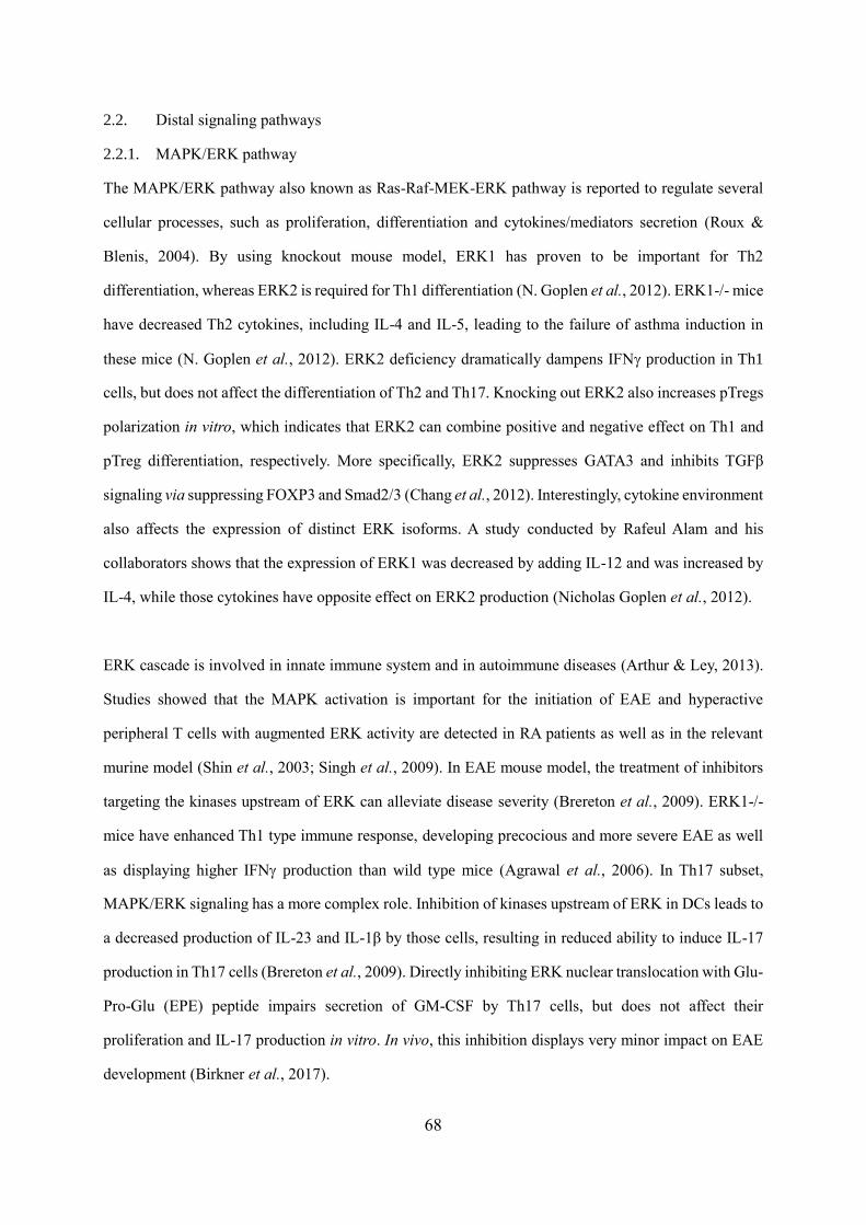
68
2.2. Distal signaling pathways
2.2.1. MAPK/ERK pathway
The MAPK/ERK pathway also known as Ras-Raf-MEK-ERK pathway is reported to regulate several
cellular processes, such as proliferation, differentiation and cytokines/mediators secretion (Roux &
Blenis, 2004). By using knockout mouse model, ERK1 has proven to be important for Th2
differentiation, whereas ERK2 is required for Th1 differentiation (N. Goplen et al., 2012). ERK1-/- mice
have decreased Th2 cytokines, including IL-4 and IL-5, leading to the failure of asthma induction in
these mice (N. Goplen et al., 2012). ERK2 deficiency dramatically dampens IFN production in Th1
cells, but does not affect the differentiation of Th2 and Th17. Knocking out ERK2 also increases pTregs
polarization in vitro, which indicates that ERK2 can combine positive and negative effect on Th1 and
pTreg differentiation, respectively. More specifically, ERK2 suppresses GATA3 and inhibits TGFβ
signaling via suppressing FOXP3 and Smad2/3 (Chang et al., 2012). Interestingly, cytokine environment
also affects the expression of distinct ERK isoforms. A study conducted by Rafeul Alam and his
collaborators shows that the expression of ERK1 was decreased by adding IL-12 and was increased by
IL-4, while those cytokines have opposite effect on ERK2 production (Nicholas Goplen et al., 2012).
ERK cascade is involved in innate immune system and in autoimmune diseases (Arthur & Ley, 2013).
Studies showed that the MAPK activation is important for the initiation of EAE and hyperactive
peripheral T cells with augmented ERK activity are detected in RA patients as well as in the relevant
murine model (Shin et al., 2003; Singh et al., 2009). In EAE mouse model, the treatment of inhibitors
targeting the kinases upstream of ERK can alleviate disease severity (Brereton et al., 2009). ERK1-/-
mice have enhanced Th1 type immune response, developing precocious and more severe EAE as well
as displaying higher IFN production than wild type mice (Agrawal et al., 2006). In Th17 subset,
MAPK/ERK signaling has a more complex role. Inhibition of kinases upstream of ERK in DCs leads to
a decreased production of IL-23 and IL-1β by those cells, resulting in reduced ability to induce IL-17
production in Th17 cells (Brereton et al., 2009). Directly inhibiting ERK nuclear translocation with Glu-
Pro-Glu (EPE) peptide impairs secretion of GM-CSF by Th17 cells, but does not affect their
proliferation and IL-17 production in vitro. In vivo, this inhibition displays very minor impact on EAE
development (Birkner et al., 2017).
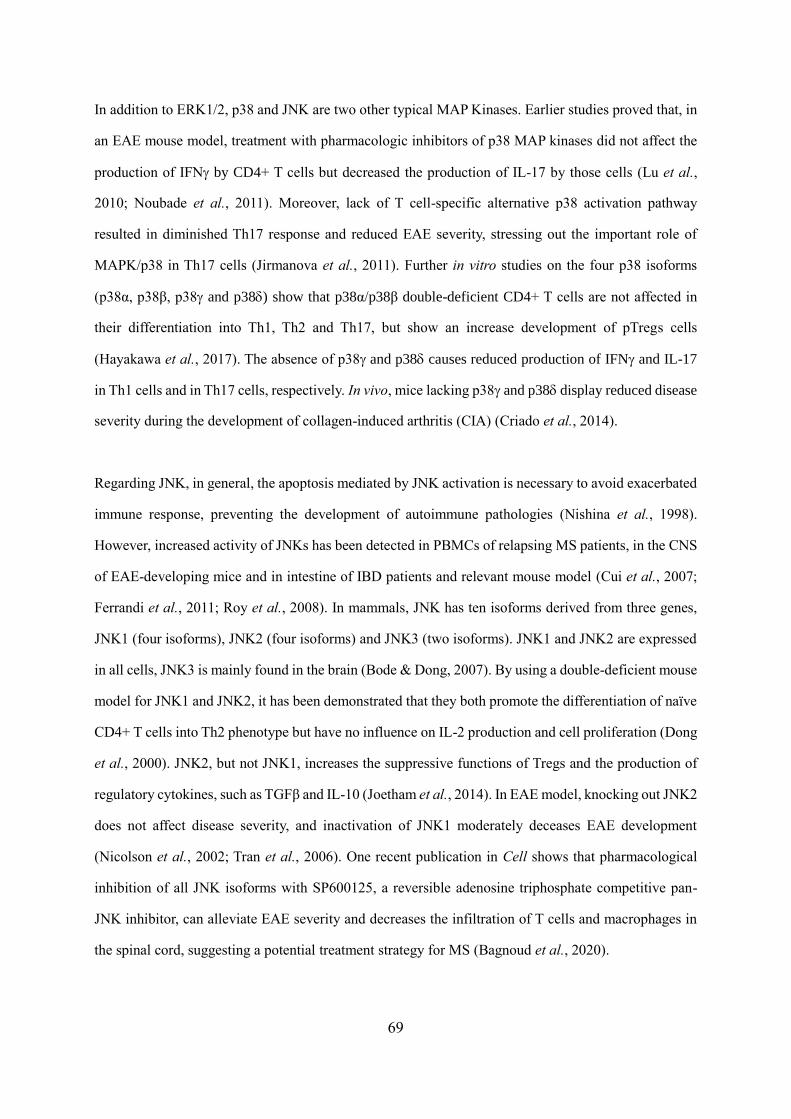
69
In addition to ERK1/2, p38 and JNK are two other typical MAP Kinases. Earlier studies proved that, in
an EAE mouse model, treatment with pharmacologic inhibitors of p38 MAP kinases did not affect the
production of IFN by CD4+ T cells but decreased the production of IL-17 by those cells (Lu et al.,
2010; Noubade et al., 2011). Moreover, lack of T cell-specific alternative p38 activation pathway
resulted in diminished Th17 response and reduced EAE severity, stressing out the important role of
MAPK/p38 in Th17 cells (Jirmanova et al., 2011). Further in vitro studies on the four p38 isoforms
(p38α, p38β, p38 and p38) show that p38α/p38β double-deficient CD4+ T cells are not affected in
their differentiation into Th1, Th2 and Th17, but show an increase development of pTregs cells
(Hayakawa et al., 2017). The absence of p38 and p38 causes reduced production of IFN and IL-17
in Th1 cells and in Th17 cells, respectively. In vivo, mice lacking p38 and p38 display reduced disease
severity during the development of collagen-induced arthritis (CIA) (Criado et al., 2014).
Regarding JNK, in general, the apoptosis mediated by JNK activation is necessary to avoid exacerbated
immune response, preventing the development of autoimmune pathologies (Nishina et al., 1998).
However, increased activity of JNKs has been detected in PBMCs of relapsing MS patients, in the CNS
of EAE-developing mice and in intestine of IBD patients and relevant mouse model (Cui et al., 2007;
Ferrandi et al., 2011; Roy et al., 2008). In mammals, JNK has ten isoforms derived from three genes,
JNK1 (four isoforms), JNK2 (four isoforms) and JNK3 (two isoforms). JNK1 and JNK2 are expressed
in all cells, JNK3 is mainly found in the brain (Bode & Dong, 2007). By using a double-deficient mouse
model for JNK1 and JNK2, it has been demonstrated that they both promote the differentiation of naïve
CD4+ T cells into Th2 phenotype but have no influence on IL-2 production and cell proliferation (Dong
et al., 2000). JNK2, but not JNK1, increases the suppressive functions of Tregs and the production of
regulatory cytokines, such as TGFβ and IL-10 (Joetham et al., 2014). In EAE model, knocking out JNK2
does not affect disease severity, and inactivation of JNK1 moderately deceases EAE development
(Nicolson et al., 2002; Tran et al., 2006). One recent publication in Cell shows that pharmacological
inhibition of all JNK isoforms with SP600125, a reversible adenosine triphosphate competitive pan-
JNK inhibitor, can alleviate EAE severity and decreases the infiltration of T cells and macrophages in
the spinal cord, suggesting a potential treatment strategy for MS (Bagnoud et al., 2020).
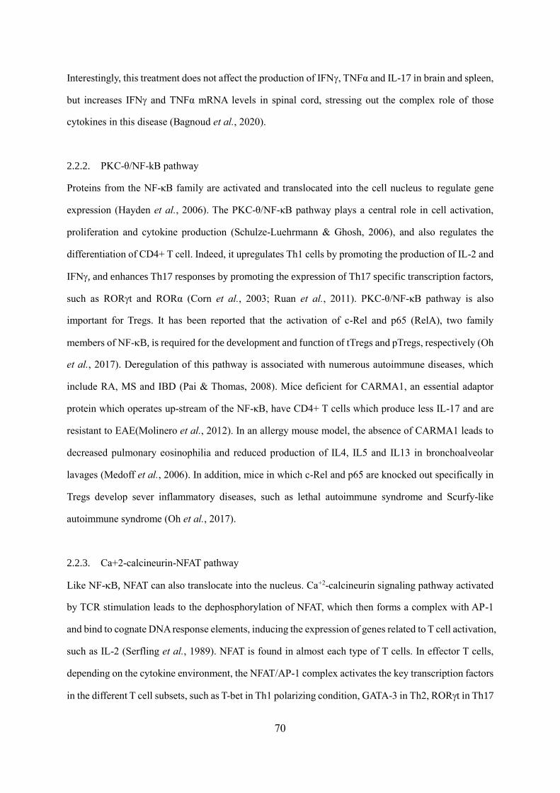
70
Interestingly, this treatment does not affect the production of IFN, TNFα and IL-17 in brain and spleen,
but increases IFN and TNFα mRNA levels in spinal cord, stressing out the complex role of those
cytokines in this disease (Bagnoud et al., 2020).
2.2.2. PKC-θ/NF-kB pathway
Proteins from the NF-κB family are activated and translocated into the cell nucleus to regulate gene
expression (Hayden et al., 2006). The PKC-θ/NF-κB pathway plays a central role in cell activation,
proliferation and cytokine production (Schulze-Luehrmann & Ghosh, 2006), and also regulates the
differentiation of CD4+ T cell. Indeed, it upregulates Th1 cells by promoting the production of IL-2 and
IFN, and enhances Th17 responses by promoting the expression of Th17 specific transcription factors,
such as RORt and RORα (Corn et al., 2003; Ruan et al., 2011). PKC-θ/NF-κB pathway is also
important for Tregs. It has been reported that the activation of c-Rel and p65 (RelA), two family
members of NF-B, is required for the development and function of tTregs and pTregs, respectively (Oh
et al., 2017). Deregulation of this pathway is associated with numerous autoimmune diseases, which
include RA, MS and IBD (Pai & Thomas, 2008). Mice deficient for CARMA1, an essential adaptor
protein which operates up-stream of the NF-κB, have CD4+ T cells which produce less IL-17 and are
resistant to EAE(Molinero et al., 2012). In an allergy mouse model, the absence of CARMA1 leads to
decreased pulmonary eosinophilia and reduced production of IL4, IL5 and IL13 in bronchoalveolar
lavages (Medoff et al., 2006). In addition, mice in which c-Rel and p65 are knocked out specifically in
Tregs develop sever inflammatory diseases, such as lethal autoimmune syndrome and Scurfy-like
autoimmune syndrome (Oh et al., 2017).
2.2.3. Ca+2-calcineurin-NFAT pathway
Like NF-B, NFAT can also translocate into the nucleus. Ca+2-calcineurin signaling pathway activated
by TCR stimulation leads to the dephosphorylation of NFAT, which then forms a complex with AP-1
and bind to cognate DNA response elements, inducing the expression of genes related to T cell activation,
such as IL-2 (Serfling et al., 1989). NFAT is found in almost each type of T cells. In effector T cells,
depending on the cytokine environment, the NFAT/AP-1 complex activates the key transcription factors
in the different T cell subsets, such as T-bet in Th1 polarizing condition, GATA-3 in Th2, RORt in Th17
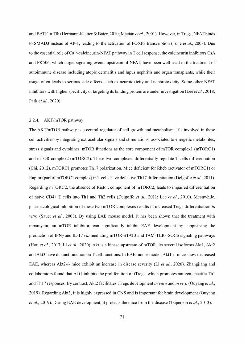
71
and BATF in Tfh (Hermann-Kleiter & Baier, 2010; Macián et al., 2001). However, in Tregs, NFAT binds
to SMAD3 instead of AP-1, leading to the activation of FOXP3 transcription (Tone et al., 2008). Due
to the essential role of Ca+2-calcineurin-NFAT pathway in T cell response, the calcineurin inhibitors CsA
and FK506, which target signaling events upstream of NFAT, have been well used in the treatment of
autoimmune disease including atopic dermatitis and lupus nephritis and organ transplants, while their
usage often leads to serious side effects, such as neurotoxicity and nephrotoxicity. Some other NFAT
inhibitors with higher specificity or targeting its binding protein are under investigation (Lee et al., 2018;
Park et al., 2020).
2.2.4. AKT/mTOR pathway
The AKT/mTOR pathway is a central regulator of cell growth and metabolism. It’s involved in these
cell activities by integrating extracellular signals and stimulations, associated to energetic metabolites,
stress signals and cytokines. mTOR functions as the core component of mTOR complex1 (mTORC1)
and mTOR complex2 (mTORC2). These two complexes differentially regulate T cells differentiation
(Chi, 2012). mTORC1 promotes Th17 polarization. Mice deficient for Rheb (activator of mTORC1) or
Raptor (part of mTORC1 complex) in T cells have defective Th17 differentiation (Delgoffe et al., 2011).
Regarding mTORC2, the absence of Rictor, component of mTORC2, leads to impaired differentiation
of naïve CD4+ T cells into Th1 and Th2 cells (Delgoffe et al., 2011; Lee et al., 2010). Meanwhile,
pharmacological inhibition of these two mTOR complexes results in increased Tregs differentiation in
vitro (Sauer et al., 2008). By using EAE mouse model, it has been shown that the treatment with
rapamycin, an mTOR inhibitor, can significantly inhibit EAE development by suppressing the
production of IFN and IL-17 via mediating mTOR-STAT3 and TAM-TLRs-SOCS signaling pathways
(Hou et al., 2017; Li et al., 2020). Akt is a kinase upstream of mTOR, its several isoforms Akt1, Akt2
and Akt3 have distinct function on T cell functions. In EAE mouse model, Akt1-/- mice show decreased
EAE, whereas Akt2-/- mice exhibit an increase in disease severity (Li et al., 2020). Zhangjiang and
collaborators found that Akt1 inhibits the proliferation of tTregs, which promotes antigen-specific Th1
and Th17 responses. By contrast, Akt2 facilitates tTregs development in vitro and in vivo (Ouyang et al.,
2019). Regarding Akt3, it is highly expressed in CNS and is important for brain development (Ouyang
et al., 2019). During EAE development, it protects the mice from the disease (Tsiperson et al., 2013).
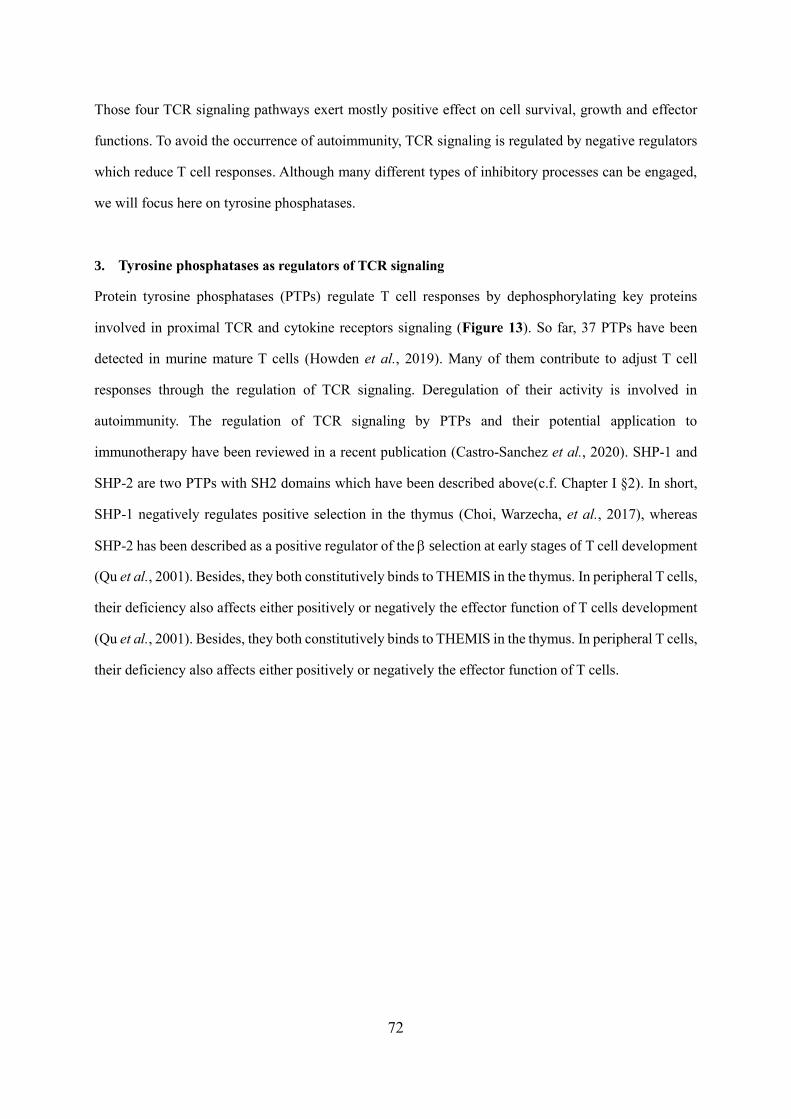
72
Those four TCR signaling pathways exert mostly positive effect on cell survival, growth and effector
functions. To avoid the occurrence of autoimmunity, TCR signaling is regulated by negative regulators
which reduce T cell responses. Although many different types of inhibitory processes can be engaged,
we will focus here on tyrosine phosphatases.
3. Tyrosine phosphatases as regulators of TCR signaling
Protein tyrosine phosphatases (PTPs) regulate T cell responses by dephosphorylating key proteins
involved in proximal TCR and cytokine receptors signaling (Figure 13). So far, 37 PTPs have been
detected in murine mature T cells (Howden et al., 2019). Many of them contribute to adjust T cell
responses through the regulation of TCR signaling. Deregulation of their activity is involved in
autoimmunity. The regulation of TCR signaling by PTPs and their potential application to
immunotherapy have been reviewed in a recent publication (Castro-Sanchez et al., 2020). SHP-1 and
SHP-2 are two PTPs with SH2 domains which have been described above(c.f. Chapter I §2). In short,
SHP-1 negatively regulates positive selection in the thymus (Choi, Warzecha, et al., 2017), whereas
SHP-2 has been described as a positive regulator of the selection at early stages of T cell development
(Qu et al., 2001). Besides, they both constitutively binds to THEMIS in the thymus. In peripheral T cells,
their deficiency also affects either positively or negatively the effector function of T cells development
(Qu et al., 2001). Besides, they both constitutively binds to THEMIS in the thymus. In peripheral T cells,
their deficiency also affects either positively or negatively the effector function of T cells.
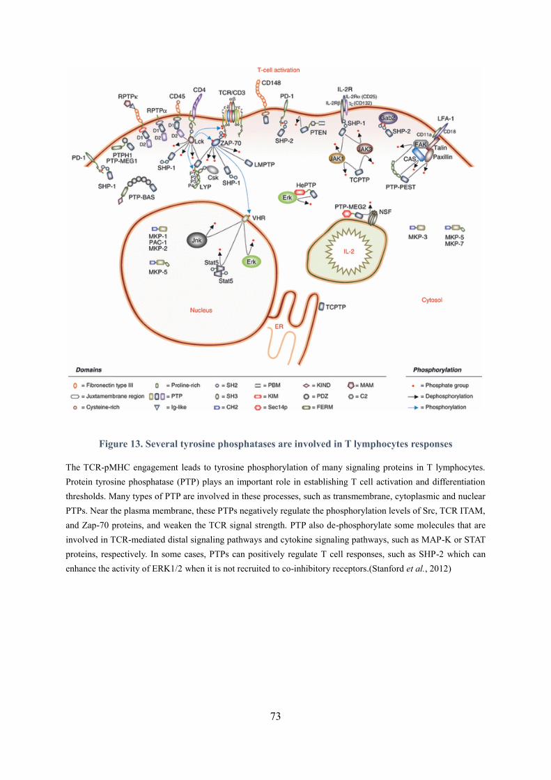
73
Figure 13. Several tyrosine phosphatases are involved in T lymphocytes responses
The TCR-pMHC engagement leads to tyrosine phosphorylation of many signaling proteins in T lymphocytes.
Protein tyrosine phosphatase (PTP) plays an important role in establishing T cell activation and differentiation
thresholds. Many types of PTP are involved in these processes, such as transmembrane, cytoplasmic and nuclear
PTPs. Near the plasma membrane, these PTPs negatively regulate the phosphorylation levels of Src, TCR ITAM,
and Zap-70 proteins, and weaken the TCR signal strength. PTP also de-phosphorylate some molecules that are
involved in TCR-mediated distal signaling pathways and cytokine signaling pathways, such as MAP-K or STAT
proteins, respectively. In some cases, PTPs can positively regulate T cell responses, such as SHP-2 which can
enhance the activity of ERK1/2 when it is not recruited to co-inhibitory receptors.(Stanford et al., 2012)
74
3.1. Effect of the Tyrosine phosphatase SHP-1 on T cell responses
SHP-1 is characterized as a broad negative regulator of TCR signaling. Mice deficient for SHP-1 exhibit
increased frequencies of memory T cells characterized by highly expression of CD44 (Johnson et al.,
2013). Although conventional CD4+ T cells lacking SHP-1 respond normally to TCR stimulation in
vitro, they are more resistant to Treg suppressive responses in vitro and in vivo (Johnson et al., 2013;
Mercadante & Lorenz, 2017). SHP-1 also regulates the suppressive function of Tregs. Tregs with absent
or repressed expression of SHP-1 have a stronger ability to inhibit the response of conventional T cells
in vitro and in vivo, and to repress inflammation in vivo (Iype et al., 2010). Furthermore, SHP-1 represses
signaling events associated to cytokine receptors. The absence of SHP-1 in CD4+ T cells leads to
increased Th2 polarization in vitro by promoting the phosphorylation of STAT6 (Johnson et al., 2013).
In an OVA-induced allergic airway inflammation model, naturel SHP-1 mutant mice derived from viable
motheaten (meV) developed more severe allergic inflammation (Kamata et al., 2003). In addition, loss
of SHP-1 in CD4+ T cells causes increased phosphorylation of STAT3 and cell polarization towards
Th17 phenotype (Mauldin et al., 2012). Early studies on motheaten (me) and viable motheaten (meV)
found that undetectable and reduced SHP-1 expression can induce severe autoimmunity in the mice 2
to 3 weeks following their birth (Shultz et al., 1993; Tsui et al., 1993). During the development of EAE,
SHP-1-deficient viable motheaten mice (mev+/-) showed increased disease severity compared to wild
type mice (Deng et al., 2002). In the PBMCs of MS patients, higher level of pSTAT6 and low expression
of SHP-1 were detected. Both SHP-1 siRNA and transduction of PBMCs with a lentiviral vector
expressing SHP-1 decreased pSTAT6 (Christophi et al., 2008).
3.2. Effect of the Tyrosine phosphatase SHP-2 on T cell responses
Unlike SHP-1, SHP-2 exerts both positive and negative roles on T cell responses according to its
substrates and to the context of its recruitment to the membrane. Upon TCR stimulation alone, SHP-2
enhances effector T cell responses by positively regulating the activity of ERK, a positive actor of IFN
production. Publications report that SHP-2-/- T cells exhibit decreased proliferation and ERK activation
upon anti-CD3 stimulation in vitro (Nguyen et al., 2006). By dephosphorylating CBP, a Csk binding
protein, SHP-2 inhibits the recruitment of Csk, a negative regulator of ERK activity, to ERK (Zhang et
al., 2004). SHP-2 also promotes ERK signaling upstream of the MAPK cascade by dephosphorylating
75
Ras-GAP, an inhibitor of Ras (Agazie & Hayman, 2003). SHP-2 has been reported to increase the
severity of systemic lupus erythematosus (SLE) (Wang et al., 2016). Treatment of lupus-prone MRL/lpr
mice with SHP-2 inhibitor can ameliorate disease severity, leading to decreased cell proliferation,
ERK/MAPK signaling and the production of IFN and IL-17A/F (Wang et al., 2016), suggesting that
SHP-2 also enhances ERK signaling in T cells during lupus erythematosus in vivo. One study on EAE
found that treatment of C57BL/6 mice with SHP-2 inhibitor, NSC-87877, can almost completely protect
the mice from EAE development (Luo et al., 2014). This study demonstrates that NSC-87877 prevents
EAE development by blocking the infiltration of T cells into the CNS. However, in the same study, mice
with SHP-2 conditional knockout in peripheral T cells develop EAE comparable to that in wild-type
C57BL/6 mice. The authors propose that NSC-87877 may also inhibit SHP-2 activity in other cells than
T cells (Luo et al., 2014). It may also be due to the off-target effects of NSC-87877.
It has been shown that SHP-2 negatively regulates cytokine induced Jak/STAT pathway in T cells. One
previous study showed that over-activation of SHP-2 in Th1 cells decreases the production of Th1
cytokines by preventing the recruitment of STAT1 to IFNR through directly binding to STAT1 and
sequestering it in cytoplasm (Wu et al., 2012). Upon transfer with these cells, C57BL/6 mice present a
significantly reduced 2, 4, 6-trinitrobenzene sulfonic acid-induced colitis. Furthermore, the decreased
Th1 cytokine production and disease severity can be restored and even worsen by using SHP-2 inhibitor
NSC-87877 or transferring conditional SHP-2 knockout T cells (Wu et al., 2012). In the mouse model
of Dextran sulfate sodium (DSS) -induced acute colitis, mice with T cell- specific conditional knockout
of SHP-2 also exhibit more severe disease. Compared to the control mice, increased number of Th1 and
Th17 cells are detected in their spleen and lamina propria, and higher amount of Th1 and Th17 cytokines
are found in the mucosa (Liu et al., 2017). Surprisingly, SHP-2 deficiency increases inflammation which
can prevent the development of colitis associated cancers, such as carcinoma (Liu et al., 2017). This
protective phenomenon, however, is dependent on the location and stage of the tumor. In a mouse model
of melanoma, the absence of SHP-2 in CD4+ T cells leads to increased melanoma metastasis in mice
and inflammatory cytokines, especially IL-6 (Zhang et al., 2013), suggesting that, in melanoma model,
SHP-2 prevents the tumor progression and metastasis by repressing IL-6/STAT3 signaling in CD4+ T
cells.

76
Taken together, SHP-2 can enhance T cell responses by positively regulating TCR signal-induced ERK
activity, while SHP-1 operates more broadly as a negative regulator of TCR signaling. In the context
of cytokine receptors signaling, both SHP-1 and SHP-2 dephosphorylate Jak/STAT and impair T cell
differentiation and cytokine production. In the context of inhibitory co-receptors, they both
dephosphorylate proteins in different signaling pathways, negatively regulating T cell responses. This
will be described in detail in the following section (c.f. Chapter II §4).
4. Impact of Co-receptors on TCR signaling
In addition to specific antigen-induced signals, signals mediated by the engagement of co-stimulatory
and co-inhibitory receptors on T cell surface are required for optimal T cell responses. Co-stimulatory
signals contribute to full activation of T cells, while co-inhibitory signals function to repress T cell
signaling, limiting T cell activation and preventing autoimmunity.
4.1. Immunoglobulin superfamily (IgSF)
IgSF is a large protein superfamily of cell surface and soluble proteins that share structural homology
with immunoglobulins and are involved in cell recognition, binding and adhesion. CD4 and CD8 are
two IgSF members that are required for T cell activation. They cooperate with TCR during the
recognition of pMHC, causing the initiation of TCR signaling. Another IgSF subgroup that tightly
regulate T cell activation is CD28 family, which is constituted of a large number of costimulatory
receptors including CD28 and ICOS, and inhibitory receptors, such as CTLA-4, PD-1 and BTLA
(Lieping Chen & Dallas B Flies, 2013) (Figure 14).
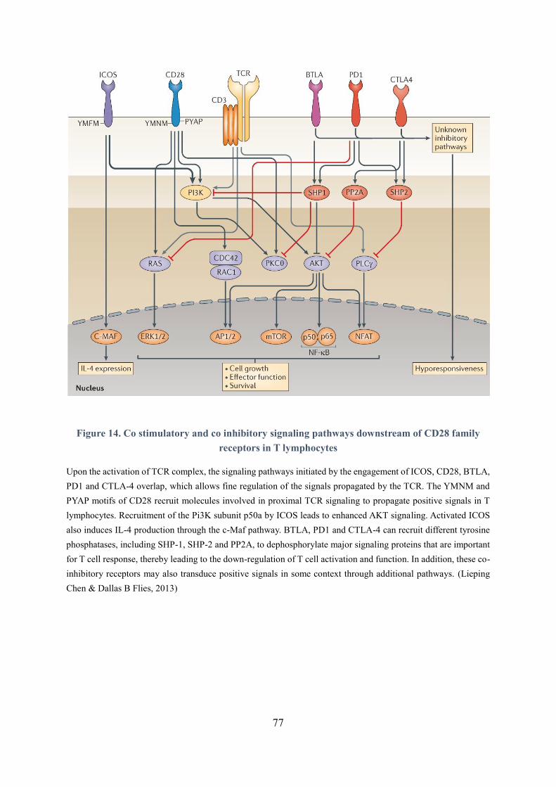
77
Figure 14. Co stimulatory and co inhibitory signaling pathways downstream of CD28 family
receptors in T lymphocytes
Upon the activation of TCR complex, the signaling pathways initiated by the engagement of ICOS, CD28, BTLA,
PD1 and CTLA-4 overlap, which allows fine regulation of the signals propagated by the TCR. The YMNM and
PYAP motifs of CD28 recruit molecules involved in proximal TCR signaling to propagate positive signals in T
lymphocytes. Recruitment of the Pi3K subunit p50a by ICOS leads to enhanced AKT signaling. Activated ICOS
also induces IL-4 production through the c-Maf pathway. BTLA, PD1 and CTLA-4 can recruit different tyrosine
phosphatases, including SHP-1, SHP-2 and PP2A, to dephosphorylate major signaling proteins that are important
for T cell response, thereby leading to the down-regulation of T cell activation and function. In addition, these co-
inhibitory receptors may also transduce positive signals in some context through additional pathways. (Lieping
Chen & Dallas B Flies, 2013)
78
4.1.1. The Stimulatory co-receptor CD28
Among those co-receptors, only CD28 is expressed on naïve murine T cells, the others being induced
only after T cell activation. CD28 has an intercellular domain containing a proximal YMNM motif and
a distal PYAP motif, which enables CD28 to recruit several kinases and adaptors with SH2 and/or SH3
domains (Boomer & Green, 2010). The recruitment of SH2-containing proteins including PI3K, Grb2
and Gads to its YMNM motif as well as Src family kinases, such as Lck and Itk, to PYAP have been
described as the major mechanisms to regulate TCR signaling (King et al., 1997; Prasad et al., 1994;
Raab et al., 1995). Upon TCR activation, their recruitment following the engagement of CD28 with its
ligands B7-1 and B7-2 positively regulate the production of IL-2, pro-survival factor Bcl-XL, IFN and
IL-4 by enhancing distal TCR signaling pathways (Bricker et al., 2014; Kane et al., 2001; Kubo et al.,
1999; Okkenhaug et al., 2001; Shahinian et al., 1993). T cells of CD28-/- mice show normal activation
but cannot induce IL-2 in response to lectins. CD28-/- mice produce lower concentration of
Immunoglobulins (Ig), and their CD8 T cells show delayed-type hypersensitivity respond to viruses in
vivo (Shahinian et al., 1993). CD28-/- mice are resistant to EAE development, which corresponds to
reduced MOG-specific IFN-producing cells and significantly less production of chemokine MIP-1α
(macrophage inflammatory protein-1α) by T cells (Chitnis, Najafian, Abdallah, et al., 2001; Karpus et
al., 1995; Perrin et al., 1999). In CD4+ T cells, continuous CD28 signaling is required for the
polarization and expansion of T cells in response to viral or bacterial infection (Linterman et al., 2014).
This signaling is also important for the generation of Treg in vivo. CD28-/- and B7-1/B7-2-/- mice
present a dramatically decreased Treg count and develop spontaneous diabetes (Salomon et al., 2000).
However, in humans, increased CD4+CD28- T cells, also called cytotoxic CD4+ T cells, are associated
with aging and chronic diseases, such as RA (Martens et al., 1997; Weyand et al., 1998). Compared to
normal CD4+ T cells, they express NK-cell receptors, produce more IFN and TNFα and downregulate
Tregs activity (Appay et al., 2002; Fasth et al., 2010; Thewissen et al., 2007; Téo et al., 2013).
4.1.2. Stimulatory co-receptor ICOS
The signaling of ICOS, another co-stimulatory receptor that belongs to the CD28 family, is triggered by
binding to ICOS-L. Besides its positive regulation of T cell activation, ICOS signaling has been reported
to promote the development of Th2 cells (Dong et al., 2001; Rudd & Schneider, 2003).
79
Indeed, ICOS -/- CD4+ T cells produce less IL-4 but more IFN in vitro (Dong et al., 2001). ICOS has
also been reported to promote Th17 and Tfh responses (Bauquet et al., 2009; Paulos et al., 2010). One
possible mechanism could be that it recruits SH2-domain containing protein PI3K, which upregulates
downstream pathways, leading to the increased production of IL-4 and IL-21 (Gigoux et al., 2009; T. R.
Simpson et al., 2010). Meanwhile, ICOS has been shown to promote the expression of IL-21 expression-
related transcription factor c-Maf and the Tfh transcription factor Klf2, which therefore enhances the
development of Th17 and Tfh (Bauquet et al., 2009; Weber et al., 2015). In vivo, ICOS has a complex
role in different diseases model. In CIA, ICOS-deficient mice are completely resistant to disease
induction (Dong & Nurieva, 2003). By contrast, these same mice exhibit extremely high susceptibility
to EAE, presumably due to increased Th1 and Th17 cytokines (Dong & Nurieva, 2003; Galicia et al.,
2009). Moreover, in EAE model, selective blockade of ICOS during the priming stage (before day 10
after immunization) exacerbates disease development, whereas blocking ICOS later on (day 10 to 20)
alleviates the disease (Sporici et al., 2001).
4.1.3. Inhibitory co-receptor CTLA-4, PD-1 and BTLA
CTLA-4, PD-1 and BTLA act as “immune checkpoints” to prevent the over-activation of T cells in
different immunological context. Among them, CLTA-4 was the first co-inhibitory receptor to be
identified (Brunet et al., 1987; Ishida et al., 1992; Watanabe et al., 2003). CTLA-4 is homologous to
CD28 and they share the same ligands, B7-1(CD80) and B7-2(CD86), however their affinity for CTLA-
4 is higher than for CD28 (Greene et al., 1996). It has been shown that CTLA-4 negatively regulates
IL-2 production as well as the duration of interaction between APC and T cells, thereby enhancing the
inhibition of T cell activation and proliferation (Krummel & Allison, 1996; Schneider, Smith, et al.,
2008). CTLA-4 intracellular domain is similar to CD28, enabling CTLA-4 to recruit SH2- and SH3-
domain containing proteins, such as the PI3K and tyrosine phosphatase SHP-2 and PP2A (Chuang et
al., 2000). The recruitment of PI3K by CTLA-4 leads to the activation of Rap1, a positive key regulator
of T cell and APC interactions as well as of the polarization and motility of T cells (Wei et al., 2007). In
allergy, the recruitment of PI3K and AKT to CTLA-4 promotes the expression of anti-apoptotic factors
Bcl2 and Bcl-XL to prevent cell death (Schneider, Valk, et al., 2008). However, the recruitment of SHP-
2 and PP2A to CTLA-4 dephosphorylates CD3, ZAP-70 and AKT (Parry et al., 2005; Rudd et al., 2009),
80
indicating that CTLA-4 has both positive and negative effects on T cell responses, depending on the
molecules it recruits. In addition, CTLA-4 exhibits a dual influence on MAPK pathways since it inhibits
the ERK activity but promotes JNK activation, driving the polarization towards Th2 lineage (Schneider
et al., 2002). Besides, although the expression of CTLA-4 is undetectable on naïve T cells, it is highly
expressed on tTregs and activated pTregs (Perkins et al., 1996; Walunas et al., 1994). The conditional
CTLA-4 knockout in Tregs leads to spontaneous development of systemic lymphoproliferation and
autoimmune diseases, which demonstrates its crucial role in the suppressive function of Tregs (Wing et
al., 2008).
Compared to CTLA-4, PD-1 and BTLA both possess a cytoplasmic tail containing an immunoreceptor
tyrosine-based inhibitory motif (ITIM) and an immunoreceptor tyrosine-based switch motif (ITSM),
which binds and activates the tyrosine phosphatases SHP-1 and SHP-2, inhibiting tyrosine kinases-
dependent signaling pathways (Chemnitz et al., 2006; Chemnitz et al., 2004). In a similar way to CD28,
ICOS and CTLA-4, the ligands of PD-1, PD-L1 and PL-L2, belong to B7 family while the ligand of
BTLA, herpesvirus entry mediator (HVEM), is part of the TNF-R family (Freeman et al., 2000; Sedy et
al., 2005).
Following phosphorylation, PD-1 recruits SHP-2 rather than SHP-1 (Xu et al., 2020). PD-1-associated
SHP-2 dephosphorylates TCR proximal signaling molecules CD28, CD226 and CD3 (Hui et al., 2017).
PD-1 deficiency in mice results in the development of lupus-like autoimmune diseases (Nishimura et
al., 1999). In vitro, the engagement of PD-1 with PD-L1 leads to a decrease in the proliferation of anti-
CD3-stimulated T cells, which is related to the reduction in IL-2 and IFN production (Carter et al.,
2002; Freeman et al., 2000). PD-1/PD-L1 interaction leads to different outcomes on the responses of
different CD4 subpopulations. A previous study on asthma showed that blocking PD-1/PD-L1
interaction in mice leads to enhanced airway hyper reactivity (McAlees et al., 2015), which is associated
with increased development of Th17 cells but no obvious change in Th2 response. In vitro analysis of
this study showed that PD-1 ligation leads to reduced differentiation of naïve CD4+ T cells into Th1 and
Th17 cells but to a slight increase of Th2 polarization.
81
PD-1-/- CD4+ T cells produce higher amount of IFN and IL-17 but decrease amount of IL-13 in
response to ConA or anti-CD3 antibody stimulation ex vivo, indicating that PD-1/PD-L1 interaction
inhibits Th17 response but slightly enhances Th2 response, and that the increased Th17 cell development
can lead to worse asthma. In addition, a study showed that the transfer of naïve CD4+ T cells to PD-
L1(-/-) PD-L2(-/-) RAG(-/-) mice resulted in a significant reduction in the number of pTregs. This study
demonstrated that PD-1 signaling promotes pTregs development by inhibiting the phosphorylation of
AKT, mTOR and ERK (Francisco et al., 2009).
Regarding BTLA, the HVEM/BTLA interaction allows the recruitment of both SHP-1 and SHP-2
phosphatases to BTLA (Celis-Gutierrez et al., 2019; Gavrieli et al., 2003). However, some studies have
found that BTLA recruits preferentially SHP-1 over SHP-2 in activated CD4 T cells (Chemnitz et al.,
2006; Mintz et al., 2019). BTLA-associated SHP-1 recruitment potently dephosphorylates CD28 and
CD3 (Sedy et al., 2005). The binding of BTLA on T cells by HVEM leads to decreased proliferation
and IL-2 production in vitro (Sedy et al., 2005). In vivo, reduced level of BTLA was detected in PBMC
and CD4+ T cells from patients with Behcet's disease (BD), a chronic inflammatory disease associated
with hyperactive Th1 and th17 responses (Ye et al., 2016). In addition, transfer of CD4+CD45RBhi
BTLA-/- T cells to RAG2-/- mice can induce more severe IBD in the recipients than transfer with BTLA-
expressing T cells (Steinberg et al., 2008). However, in the same study, BTLA-/- mice did not develop
spontaneous autoimmune diseases such as PD-1-/- mice. This observation could be explained by the
dual regulatory influence of BTLA in T cells. Indeed, compared with PD-1, in addition to ITIM and
ITSM motifs, BTLA possesses two additional tyrosine residues (Y226 and Y243) N-terminal to the
ITIM, which may enable direct binding of Grb2 protein and indirect recruitment of PI3K through Grb2,
resulting in the stimulation of the PI3K pathway and thereby promoting T cell proliferation and survival
(Gavrieli & Murphy, 2006). Yet, SHP-1 and SHP-2 recruitment is not the only mechanisms used by PD-
1 and BTLA to exert their inhibitory function. It has been shown that, in activated T cells in absence of
both SHP-1 and SHP-2, proliferation and cytokines production can still be suppressed by PD-1 and
BTLA (Xu et al., 2020). In addition, PD-1 negatively regulates the activation of ERK, independently of
SHP-1 and SHP-2, to block the G1-S transition of the cell cycle and proliferation of T cells (Patsoukis
et al., 2012).

82
Interestingly, suppressing T cells responses by activating immune checkpoints is an effective immune
evasion strategy employed by tumor cells. Therefore, co-inhibitory signals involved in the development
of various cancers have been extensively studied. Thanks to Nobel prize winners Dr. James P Allison
and Dr. Tasuku Honja’s contribution on the discovery of cancer therapy by inhibition of immune
checkpoints, many therapeutic antibodies based on CTLA-4 and PD-1 blockade are currently approved
by the U.S. Food and Drug Administration for cancer treatment (FDA), or are evaluated in clinical trials
(Ansell et al., 2015; Topalian et al., 2012). For example, ipilimumab (anti-CTLA4) has been used for
the treatment of metastatic melanoma, nivolumab (anti–PD-1) is used for treating many types of tumor
including melanoma and non-small cell lung cancer, and a combination therapy with ipilimumab plus
nivolumab has been approved for the treatment of Renal cell carcinoma(Iwai et al., 2017; Wei et al.,
2018). In addition to CTLA-4, PD-1 and BTLA, a lot of other inhibitory co-receptors in IgSF, such as
LAG-3, VISTA and TIGIT have become new targets for research related to immune tolerance,
autoimmune diseases and antitumoral immunotherapy (Attanasio & Wherry, 2016; Śledzińska et al.,
2015).
4.2. Engagement of CD5
During my PhD, I have contributed to a study on the signaling function of CD5 (Blaize et al., 2020), a
type 1 transmembrane cell surface glycoprotein that belongs to Scavenger Receptor Cysteine-Rich super
family (SRCR-SF). Only two members of this family are expressed by T cells, CD5 being one of them.
As for other co-receptors, several ligands of CD5 have been reported, but its biological activity doesn’t
require their interaction (Davies et al., 1992). After TCR stimulation, the intracytoplasmic domain of
CD5 is phosphorylated by Lck (Davies et al., 1992; Dennehy et al., 2001). Previous studies showed that
increased expression of CD5 on T cells are correlated with the affinity of the TCR for self-pMHC in T
cells, suggesting that CD5 could be important for the maintenance of tolerance (Azzam et al., 1998;
Mandl et al., 2013). Based on TCR engagement, the loss of CD5 in T cells leads to increased
mobilization of calcium flux and phosphorylation of several signaling proteins, such as TCR, Zap-70
and VAV1, and the engagement of CD5 by antibodies decreases the phosphorylation of those proteins,
which indicates a negative role for CD5 on TCR signaling (Bhandoola et al., 2002; Perez-Villar et al.,
1999; Tarakhovsky et al., 1995). However, opposite role of CD5 were shown in other studies.
83
The co-engagement of CD3 and CD5 by antibodies leads to an increase phosphorylation of PI3K, ERK
and AKT in both human T lymphocytes and murine CD4+CD8+ thymocytes in vitro (Gringhuis et al.,
1998; Zhou et al., 2000). In vivo, CD5-/- mice show decreased susceptibility to the development of EAE
and IBD (Axtell et al., 2004; Dasu et al., 2008). Later findings show that the decreased T cell responses
caused by the absence of CD5 is associated with increased number and activity of Tregs. Compared to
CD5+/+ mice, CD5-/- mice show alleviated IBD severity and increased Tregs count in their colon (Dasu
et al., 2008). The differentiation of CD5-deficient CD4+ T cells/thymocytes into Tregs can be inhibited
by very low amount of IL-4, IL-6 and IFN in response to self and tolerizing antigens (Henderson et al.,
2015). In addition, CD5-/- Balb/c mice show an increase in the number of Tregs in the thymus and in
peripheral lymphoid organs, confirming the role of CD5 in the generation of Tregs (Ordoñez-Rueda et
al., 2009). This study also found that CD5 increases the phosphorylation of AKT in naïve CD4+ T cells
but decreases the level of p-ERK in Tregs, suggesting a complex regulation mechanism of CD5 on TCR
signaling. Together with other studies, a recent work conducted in our team to which I have contributed
has identified several proteins interacting with CD5, such as PI3K, c-Cbl, CIN85, UBASH3A and SHIP,
including thus both positive and negative regulators of TCR signaling (Blaize et al., 2020; Dennehy et
al., 1998; Dennehy et al., 1997; Gringhuis et al., 1998). Our study found that, upon TCR activation,
tyrosine 429 of CD5 is selectively phosphorylated and recruit a signalosome composed of c-Cbl, CIN85
and CRKL. This signalosome, especially c-Cbl, helps to connect CD5 with the positive (PI3K) and
negative (UBASH3A, SHIP1) regulators of TCR signaling, coordinately repressing the activity of ERK
and promoting AKT activity to inhibit FOXP3 expression (Blaize et al., 2020).
84
Chapter Ⅲ Role of THEMIS in peripheral T cells
THEMIS is expressed in peripheral T-cells although less than in thymocytes (Fu et al., 2009; Lesourne
et al., 2009). It’s well known that THEMIS is crucial for thymocyte development (Fu et al., 2009;
Johnson et al., 2009; Kakugawa et al., 2009; Lesourne et al., 2009; Patrick et al., 2009) and the
mechanisms of THEMIS function in the thymus have been thoroughly studied (c.f. Chapter I §3).
However, the role of THEMIS in peripheral T cells still remains elusive, as themis-/- mice present a
severe T cell lymphopenia. In thymocytes, THEMIS is involved in TCR signaling since it is indirectly
recruited to LAT by interacting with Grb2 after TCR stimulation (Brockmeyer et al., 2011; Lesourne et
al., 2012; Paster et al., 2013). In peripheral CD4+ T cells, THEMIS and Grb2 show similar kinetics of
interaction with LAT, with maximum binding observed at 2min and maintained for up to 5min after anti-
CD3 and anti-CD4 stimulation (Roncagalli et al., 2014), suggesting that THEMIS is involved in TCR
signaling in peripheral T cells and may play a crucial role within these cells.
1. THEMIS is important for the suppressive function of regulatory T cells
An early study on THEMIS in Brown Norway (BN) rats showed that themis-deficient BN rats (BNthemis-
/-) present a defect in the suppressive activity of CD4+ CD25bright T cells, resulting in spontaneous
development of IBD (Chabod et al., 2012). These BNthemis-/- rats also exhibit a sharp and specific
decrease in the number of effector CD4+ T cells and tTregs. In addition, they display an increase of
activated CD4+ T cells producing elevated levels of Th2/Th17 cytokines, but which show reduced IFN
production as compared to that in BN control rats (Chabod et al., 2012). This study initially suggested
that THEMIS might positively regulate the suppressive function of Tregs, preventing the pathogenesis
of IBD. Indeed, further research on Tregs subset through proteomic analysis revealed the importance of
THEMIS in controlling the inhibitory function of the cells (Duguet et al., 2017). The authors found that
the expression of THEMIS in Tregs was strongly down-regulated compared to conventional T cells. In
addition, Tregs purified from themistg mice – over-expressing themis – showed better suppressive
activity on the proliferation of conventional T cells in vitro compared to Tregs of themiswt mice. Finally,
co-injection of CD4+CD25-CD45RBhigh colitogenic T cells with Tregs purified from themistg mice
exhibited better protection against the development of IBD than coinjection with Tregs from themiswt
mice (Duguet et al., 2017).
85
However, the positive regulation of THEMIS on the inhibitory function of Tregs does not only depend
on THEMIS. In themis-deficient LEWIS (LEW) rats, the same research team found that these rats
exhibited the same lymphopenia as BNm rats, but had no impairment in Tregs suppressive function and
no spontaneous development of IBD or other diseases (Pedros et al., 2015). Compared with LEW
background, BN rats contain a 117-kb interval region harboring a nonsynonymous polymorphism
affecting the expression and functions of VAV1 (Colacios et al., 2011). The authors hence generated
BNm rats deficient for THEMIS and in which the 117-kb interval from LEW rats was introduced. They
found that the introduction of the 117-kb interval region from LEW rats restored Tregs function in
BNthemis-/- rats and protected them from spontaneous IBD development, highlighting the importance of
THEMIS/VAV1 interaction on Treg suppressive function and susceptibility to IBD development
(Colacios et al., 2011).
2. THEMIS regulates the metabolism reprograming of CD4 T cells
Little is known about the function of THEMIS in peripheral T cells because of lymphopenia in themis-
/- mice. An initial study conducted by Paul Love and collaborators found that, compared with themis+/+
mice, themis-/- peripheral CD4+ T cells had a comparable proliferation profile in response to anti-CD3
stimulation (Lesourne et al., 2009). However, several publications from Nicholas R. J. Gascoigne’s team
show that themis-/- peripheral CD4+ T cells are poorly responsive to anti-CD3 stimulation and show
reduced proliferative responses and reduced cytokine production under stimulation with anti-CD3/anti-
CD28 antibodies in vitro (Fu et al., 2009; Prasad et al., 2020). In vivo, themis-/- CD4+ T cells have
reduced ability to develop IBD, showing weaker inflammatory potential compared with themis+/+
peripheral CD4 T cells (Prasad et al., 2020). In addition, their studies showed that themis-/- CD4+ T
cells have reduced glucose uptake, mitochondrial dysfunction and mTOR signaling responses, showing
defective metabolomic reprogramming (Prasad et al., 2020). Under the same conditions of TCR
stimulation, the absence of THEMIS in CD4+ T cells leads to reduced expression of insulin receptor
(IR), a protein that initializes insulin signaling pathway and, like THEMIS, binds to Grb2 and shares
common downstream signaling pathways with TCR, such as ERK and mTOR activation (Brockmeyer
et al., 2011; Saltiel & Kahn, 2001). themis-/- CD4+ T cells also exhibit defective nutrient uptake,
mitochondrial function and mTOR upregulation under TCR stimulation, which indicates a connection
86
to the reduced TCR response (Prasad et al., 2020). Indeed, the authors found that themis-/- CD4+ T cells
have decreased nuclear translocation of NFAT, a transcription factor which is regulated by TCR
signaling and leads to transcription programs for T cell activation and effector functions, such as IL-2
(Macián et al., 2001). As Chip-seq and RNA-seq demonstrated, that the nuclear translocation of NFAT
controls IR gene expression, which suggests that the defective TCR signal in themis-/- CD4+ T cells
causes reduced NFAT nuclear translocation which in turn results in decreased IR expression, defective
metabolic reprogramming and mTOR upregulation, culminating in impaired effector functions,
including proliferation, cytokines production and inflammatory potential to cause colitis (Prasad et al.,
2020). Altogether, these data lead the authors to conclude that THEMIS regulates metabolic signaling
and effector functions in CD4+ T cells by controlling NFAT nuclear translocation (Prasad et al., 2020).
However, the defective metabolism and effector function of themis-/- peripheral CD4 T cells may not
be caused by the intrinsic deficiency of THEMIS in peripheral CD4+ T cells and may result from the
abnormal maturation of CD4-SP caused by the impaired thymic development. This indirect defect due
to lymphopenia could also result in CD4+ T cell functional anomalies. At this stage, in order to better
understand the role of THEMIS in peripheral CD4+ T cells, an appropriate conditionally knocked model
seems necessary.
3. THEMIS is required for the maintenance of peripheral CD8 T cells
One recent study published by Nicholas R. J. Gascoigne and collaborators shows THEMIS is required
for the maintenance of peripheral CD8+ T cells and enhances homeostatic signals resulting from the
integration of signals induce by weak TCR stimulation and by cytokines receptors such as IL-7R
(Brzostek et al., 2020). To investigate the role of THEMIS in peripheral T cells, the authors generated
themis conditional knockout (cKO) mice by crossing themisflox/flox mice with dlLck-Cre+ in the OT-I TCR
transgenic background. In this way, their OT-I themis-cKO mice express H-2Kb-restricted TCR that can
recognize a well-defined series of agonist peptides with different affinities (Hogquist et al., 1994). In
this study, OT-I themis-cKO mice exhibit normal thymocytes development and normal CD4+ T cells
number but present a 50% reduction in peripheral CD8+ T cells count (Brzostek et al., 2020).
87
OT-I themis-cKO mice and acute THEMIS deletion in peripheral T cells using tamoxifen-inducible
ERT2-Cre both lead to reduced CD5 expression on CD8 SP thymocytes and on peripheral CD8+ T cells
separately, leading to an increase in CD8 surface expression, which suggests a role of THEMIS in the
regulation of signaling from Self-pMHC (Brzostek et al., 2020). Results of lymphopenia induced
proliferation (LIP) show that the transfer of OT-I themis-cKO CD8 T cells into RAG1-/- mice induces
an important loss in proliferation capacity, expression of CD44 and CD122 as well as IFN production,
compared to OT-I themis-wt CD8 T cells, indicating that THEMIS is required for the activation and
effector functions of CD8 T cells in vivo (Brzostek et al., 2020). However, CD8+ T cells from OT-I
themis-cKO and from its wild type control show a similar activation, proliferation, TCR signaling
pathway response and cytokine production in response to different doses of OVA-based peptide ligands
in vitro. Moreover, no difference is detected between these two types of CD8+ T cells concerning IL-7
signaling and response, a necessary component besides self-pMHC for CD8+ T cell maintenance and
for LIP, suggesting that THEMIS deficiency does not alter agonistic pMHC ligand discrimination and
IL-7 signaling responses in CD8+ T cells (Brzostek et al., 2020). Other in vitro results of this study show
that themis-cKO OT-I CD8+ T cells exhibit reduced proliferation, Akt signaling and c-Myc upregulation
in response to stimulation with low-affinity pMHC in presence of IL-2 alone, IL-7 and IL-18 or IL-7
and IL-12. To assess the role of THEMIS in signal integration between TCR and cytokine receptors in
CD8+ T cells, the authors investigated whether the absence of THEMIS affects CD8+ T cells response
to Listeria monocytogenes expressing OVA (LM-OVA). The result showed that themis-cKO CD8+ T
cells have reduced response to Listeria monocytogenes infection in vivo, exhibiting a decrease in the
proliferation, cytokines production and IL-2/IL-12 signaling responses, which indicates that THEMIS
promotes anti-bacterial CD8+ T cell responses in vivo by modulating signal integration from low-
affinity TCR and cytokine receptor (Brzostek et al., 2020). Furthermore, the conditional knockout of
SHP-1 can rescue the reduced cell numbers of CD8 T cells in OT-I themis-cKO mice and the impaired
activation and effector functions of these CD8 T cells in response to low-affinity TCR and cytokine
signal integration, which suggests the negative role of SHP-1 in CD8 homeostasis and indicates that
THEMIS function in peripheral CD8 T cells is mediated through SHP-1 phosphatase (Brzostek et al.,
2020). This study also checked the effect of conditional THEMIS deficiency in peripheral CD4+ T cells.
Although peripheral CD4+ T cells from themis-cKO have reduced CD5 expression, they proliferate
88
normally in response to TCR ligation alone or in combination with IL-7 or IL-15 stimulation, suggesting
a specific effect of THEMIS on signal integration in CD8+ T cells (Brzostek et al., 2020). However, this
study did not analyze the effect of THEMIS on the polarization of CD4+ T cells in vitro and in vivo.
Taken together, these data suggest that THEMIS enhances the maintenance and effector function of
peripheral CD8+ T cells in response to low-affinity pMHC and cytokine-mediated signals by inhibiting
the phosphatase SHP-1. However, the role of THEMIS in peripheral CD4+ T cells is still poorly explored
particularly because the conditional knockout model based on the transgenic Cre expression driven by
the distal Lck promoter doesn’t allow complete gene deletion in peripheral CD4+ T cells (D'Souza et
al., 2008)
4. THEMIS is involved in CAR-T cell-induced tumor regression by recruiting SHP-1 to the CAR
synapse
Chimeric Antigen Receptors (CARs) are synthetic molecules composed of a single chain variable
fragment (scFv), co-stimulatory moieties (either CD28 or 4-1BB) and a CD3ζ signaling domain. CARs
expression in CD4+ and CD8+ T cells triggers their lytic machinery and co-stimulation after antigen
engagement (Dotti et al., 2014; Zhao et al., 2015). CAR-T cell co-stimulation mediated by CD28 and
4-1BB plays an essential role in CAR-T cell-induced tumor regression and has been associated with
distinct antitumor kinetics, as CD28-mediated CAR-T cell co-stimulation leads to faster antitumoral
activity showing pronounced glycolytic metabolism and higher susceptibility to exhaustion while the 4-
1BB-mediated co-stimulation results in predominantly oxidative metabolism and lower susceptibility to
exhaustion (Kawalekar et al., 2016; Long et al., 2015; Zhao et al., 2015). One recent publication in
Cancer Cell shows that the antitumoral kinetics of CAR-T cells is correlated with the recruitment of
Lck and THEMIS-SHP-1 complex to the CAR-T cell / APC synapse (Sun et al., 2020). In this study,
the recruitment of Lck to the synapse of CD28-encoding CAR-T cells causes antigen-independent CAR-
CD3ζ phosphorylation and increased antigen-dependent T cell activation. By contrast, the synapse
formed by 4–1BB-engaged CAR-T cells recruits the THEMIS-SHP1 complex leading to reduced CAR-
CD3ζ phosphorylation, which can be overcome by overexpression of Lck (Sun et al., 2020).
89
Co-immunoprecipitation experiments show that FLAG-tagged human THEMIS can pull down 4–1BB-
encoding CAR in T cells and Jurkat cells. While this interaction is abolished when the gene encoding
for 4-1BB is mutated, which corresponds to increased CAR-CD3ζ phosphorylation, Ca2+ influx, CD69
expression and IFN production, indicating that THEMIS is able to directly interact with 4-1BB-
encoded CAR synapse (Sun et al., 2020). However, since THEMIS does not have direct phosphatase
activity, the dephosphorylation function of THEMIS-SHP-1 complex must come from SHP-1. Indeed,
this same study shows that modified SHP-1, able to be directly recruited to the CAR synapse without
interacting with THEMIS, can attenuate the activation of CD28 encoding CAR-T cells in vitro and in
vivo (Sun et al., 2020). Taken together, these results demonstrate that the indirect recruitment of SHP-1
to 4-1BB through THEMIS tunes Lck-mediated CAR-induced T cells priming and that the balance
between the recruitment of THEMIS-SHP-1 complex and Lck to CAR synapse regulates the antitumoral
kinetics of CAR-induced T cells (Sun et al., 2020).
5. Association between THEMIS genetic variants and autoimmune disorders
Genome-wide association studies (GWAS) have identified themis as a potential susceptibility gene for
the development of autoimmune diseases such as MS (Davies et al., 2016; Sawcer et al., 2011) and
Celiac disease(Bondar et al., 2014) and inflammatory diseases including IBD (Jostins et al., 2012; Liu
et al., 2015) and psoriasis (Kim et al., 2015). Further investigation is needed to clarify the role of
THEMIS in these pathologies. In the disease model of experimental cerebral malaria (ECM), ENU-
induced themis mutant mice (themisI23N) have reduced disease severity after infection by Plasmodium
berghei ANKA compared to wild type mice (Torre et al., 2015). This is associated with reduced cerebral
cellular infiltration, retention of blood-brain barrier integrity and decreased production of pro-
inflammatory cytokines including IFN and TNFα by themisI23N CD4+ and CD8+ T cells, suggesting
that THEMIS may enhance the pro-inflammatory function of peripheral T cells in response to parasitic
infection in vivo. To confirm the effect of themis mutation on T cell responses in vivo, both control and
themisI23N mice were infected with Mycobacterium tuberculosis, leading to a more severe infection in
themisI23N mice. This result indicates that fully functional THEMIS is necessary for effector T cell
response against mycobacterial infections (Torre et al., 2015).
90
Overall, this study shows that THEMIS is required for the neuroinflammation of cerebral malaria and
protection against M. tuberculosis, which may correspond to its enhancement on the pro-inflammatory
responses of peripheral T cells. However, further study is necessary, as themisI23N mice display peripheral
lymphopenia (Torre et al., 2015).
In summary, human THEMIS is involved in the antitumoral kinetics of CAR T cells by recruiting SHP-
1 to the synapse. In mouse, the studies in themis-/- peripheral T cells demonstrate that THEMIS is
important for the suppressive function of Tregs and for the metabolism of peripheral CD4+ T cells. It is
also required for pathogenesis of Experimental Cerebral Malaria and protection against Pulmonary
Tuberculosis. In addition, THEMIS sustains the maintenance of peripheral CD8+ T cells by regulating
T cell receptor and cytokine signal integration. However, the various mouse models used for all these
studies exhibit major drawbacks. Firstly, the impact of lymphopenia of themis-/- mice cannot be ignored
and secondly, it must be kept in mind that themis deletion in peripheral CD4 T cells is incomplete in the
conditional knockout dlLck-Cre themis mouse model.

91
Objectives
92
THEMIS is expressed in peripheral T cells, but its role in these cells, especially on CD4+ T cell
responses, is still poorly understood. In humans, themis has been identified as a susceptibility gene in
early GWAS on patients suffering from multiple sclerosis, a demyelinating disease which is tightly
related to the inflammation process mediated by T cells. The functions of THEMIS in the frame of this
disease needs to be explored using the relevant mouse model of EAE.
Therefore, this project aims to study the function of protein THEMIS in normal and pathological
peripheral CD4+ T cell responses.
Studies on CD4+ T cells were based so far on mouse models with germline THEMIS deficiency, which
may lead to indirect effects on those cells due to the block on T cell development and conditional models
that do not always induce efficient deletion of candidate gene in this population. In order to avoid defects
related to lymphopenia in peripheral T cells, we generated a conditional themis knockout mouse line
CD2-Cre-themisflox/flox (named as themis-TKO), in which themis is invalidated by the Cre recombinase
controlled by human CD2 promoter which is active after positive selection in the thymus. To focus on
the effect of THEMIS on CD4+ T cells, we also generated CD4-CreERT2-themisflox/flox, in which themis is
conditionally deleted in peripheral CD4+ T cells by CD4-CreERT2 following tamoxifen treatment.
The result section investigates the function of THEMIS on peripheral CD4+ T cell responses from the
following three main aspects:
1. Phenotype of thymocytes and peripheral T cells in themis-TKO mice.
2. Effect of THEMIS deficiency on the activation and differentiation of CD4+ T cells upon TCR
triggering in vitro and foreign antigen recognition in vivo.
3. Effect of THEMIS deficiency on CD4+ T cells in the context of EAE development.

93
Results
1
THEMIS enhances the magnitude of normal and neuroinflammatory Th1 1
responses by promoting TCR-independent signals 2
(Under review) 3
4
Cui Yang1, Gaëtan Blaize1*, Rémi Marrocco1*, Nelly Rouquié1*, Cyrielle Bories1, Mylène 5
Gador1, Suzanne Mélique1, Emeline Joulia1, Mehdi Benamar1, Anne S. Dejean1, Hélène 6
Daniels-Treffandier1,3, Paul E. Love3, Nicolas Fazilleau1, Abdelhadi Saoudi1, Renaud 7
Lesourne1 8
9
10 1 Infinity, Université Toulouse, CNRS5051, INSERM1291, UPS, Toulouse, France. 11 2IHAP, Université de Toulouse, ENVT, INRA, UMR 1225, 31076 Toulouse, France. 12 3Section on Hematopoiesis and Lymphocyte Biology, Eunice Kennedy Shriver National 13
Institute of Child Health and Human Development, National Institutes of Health, Bethesda, 14
MD 20892, USA. 15
* Equally contributed to this work 16
17
Correspondence should be addressed to R.L. 18
Email: [email protected] 19
20
21
22
2
Abstract 23
24
Signals that determine the differentiation of naïve CD4+ helper T cells (Th) into specific 25
effector subsets are primarily triggered by cytokines but additional signals are required to 26
adjust the magnitude of Th responses and set the balance between effective immunity and 27
immunological tolerance. By inducing a post-thymic deletion of the T-cell lineage signaling 28
protein THEMIS, we show that THEMIS promotes the development of optimal Th1 responses 29
to foreign antigens but triggers signals that in return favour encephalitogenic responses to self 30
neuroantigens. THEMIS exerts positive effects on T-BET expression and on IFNg production 31
and enhances the ability of encephalitogenic CD4+ T cells to migrate into the central nervous 32
system. Although the phenotype observed in vivo is indicative of positive function of THEMIS 33
on TCR-mediated responses, we show that THEMIS exerts opposite effects in vitro by 34
inhibiting TCR-mediated signals leading to Th1 responses, indicating that THEMIS exerts 35
TCR independent functions which increase the magnitude of normal and pathogenic Th1-36
mediated responses. 37
38
39
40
41
3
Introduction 42
43
One essential feature of the immune system is its ability to engage distinct defensive strategies 44
adapted to the variety of invading pathogens. This is partially dependent on CD4+ helper T 45
cells (Th) which may differentiate from a so-called naïve state into distinct effector subsets 46
(Th1, Th2, Th17, T-follicular helper cells [Tfh], induced-regulatory T cells [iTreg], Type-1 47
regulatory T cells [Tr1]) with specialized immune functions. The lineage choice decision of Th 48
cells results from the complex integration of signaling events triggered by the T cell antigen 49
receptor (TCR), which recognize self- or foreign-peptide ligands bound to major 50
histocompatibility complex (pMHC) at the surface of antigen presenting cells, and by a 51
combination of cytokine receptors which are stimulated specifically according to the cytokine 52
milieu at the site of CD4+ T cell activation (1). In addition to those cell-fate decision signals, 53
additional signaling events triggered by the TCR and by co-stimulatory and co-inhibitory 54
receptors (2) contribute to regulate the magnitude of specific effector responses, promoting 55
thereby effective immunity and preventing the development of potentially pathogenic 56
inflammatory responses. 57
58
THEMIS is a T cell lineage-specific protein that has important role during T cell development 59
(3-5). The phenotype of THEMIS deficient mice shows that THEMIS is essential for the 60
positive selection of CD4+CD8+ (double-positive [DP]) thymocytes (3-5) and for their 61
commitment to the CD4+ lineage (5). THEMIS contains a proline-rich C-terminal sequence 62
(PRS), which binds to the adaptor protein GRB2 (5, 6) that is required for the recruitment of 63
THEMIS to the transmembrane adaptor LAT following TCR engagement (5, 6), and two 64
globular cysteine-containing, all-b in THEMIS (CABIT) domains (4), which bind to the 65
phosphatase domains of the tyrosine phosphatases SHP-1 and SHP-2 (7). 66
67
Although THEMIS has been characterized as an important regulator of T cell development 68
more than a decade ago, the mechanism by which it operates in this context has remained the 69
subject to nearly opposite interpretations (8). A first model posits that THEMIS dampens TCR 70
signaling initiated by low-affinity self-peptide-bound MHC (pMHC) interactions, preventing 71
4
the transmission of a strong signaling response to these stimuli that would trigger negative 72
selection (9). THEMIS is speculated here to function by promoting SHP-1 and SHP-2 73
phosphatase domain activation and or by facilitating their recruitment to LAT where they are 74
brought into contact with their primary targets (9, 10). This model was mostly deduced from 75
in vitro studies in which preselected “naïve” DP thymocytes were stimulated with low-affinity 76
pMHCs tetramers (9). More recently, a second model has been proposed in which THEMIS 77
operates as an enhancer of TCR signaling, which enables thymocytes to reach the threshold for 78
positive selection, avoiding death by neglect (7, 11). THEMIS operates here by blocking the 79
catalytic function of the tyrosine phosphatase SHP-1, which has inhibitory effect on TCR 80
signaling (7). This action is mediated by THEMIS’s CABIT modules, which bind to the 81
phosphatase domain of SHP-1 and promote or stabilize the oxidation of SHP-1’s catalytic 82
cysteine residue, inhibiting the tyrosine-phosphatase activity of SHP-1 (7). In this second 83
model, the enhancing effects of THEMIS on TCR signaling were mainly deduced from in vivo 84
observations using TCR-signal sensing mouse models (11) and complementation studies with 85
SHP-1 deficient mice (7). Although the second model is more likely to explain the block of T 86
cell development observed in THEMIS deficient mice and although the repressing effect of 87
THEMIS on SHP-1 and SHP-2 phosphatase activity is based on solid molecular evidences (7), 88
the reasons for the apparently contradictory findings remain unexplained. 89
90
The role of THEMIS during thymic selection has been well characterized, yet less is known 91
about its function in peripheral T cells. Conditional disruption of the gene encoding THEMIS 92
at late stage of T cell development using CRE recombinase transgenes driven by the distal lck 93
promoter identified a positive role for THEMIS in controlling peripheral CD8+ T cell 94
maintenance (12). The impaired homeostasis of peripheral CD8+ T in THEMIS-deficient mice 95
was strongly suggestive of a positive function for THEMIS on tonic TCR signaling and could 96
be rescue by deleting the gene encoding for SHP-1 (12). Interestingly, this effect was restricted 97
to CD8+ T cells, since no effect of THEMIS on CD4+ T cell function could be detected in that 98
study (12). 99
100
5
In the present study, we identified a positive function for THEMIS in CD4+ T cell responses 101
to self and foreign antigens. Post-thymic disruption of the Themis gene, using Cre recombinase 102
transgenes driven by the hCD2 promoter, shows that THEMIS promotes Th1 responses in vivo 103
and enhances encephalitogenic responses of CD4+ T cells to a neuroantigen in a mouse model 104
of central nervous system (CNS) auto-immunity. Although THEMIS exerts a positive function 105
in Th1 responses following antigen immunization, we show that THEMIS deficiency leads to 106
opposite outcomes following TCR stimulation, suggesting that the inhibitory effects of 107
THEMIS on TCR signaling are overcomed during immune responses by additional THEMIS-108
mediated signals which promote rather than repress T-cell effector function. 109
110
6
Materials and Methods 111
112
Mice 113
For the generation of Themis-TKOmice, ES cells from the EUCOMM consortium (clone 114
HEPD0712-6-H05) were used to generate Themisflox/flox mice in which the 4th exon of themis 115
was surrounded by two loxp sites. These mice were bred with hCD2-cre transgenic mice 116
(https://www.jax.org/strain/027406) in which the human cd2 promoter directs the expression 117
of the CRE recombinase at late stages of T cell development in mature CD4+ and CD8+ single-118
positive thymocytes. Themisflox/flox mice were also bred with CD4-CreERT2 transgenic mice 119
which contain tamoxifen inducible CRE under the control of the cd4 promoter 120
(https://www.jax.org/strain/022356). Themis-TKO were crossed with transgenic mice 121
expressing the 2D2 TCR (13). All the experiments were conducted with sex and age-matched 122
mice between 8 and 12 weeks old housed under specific pathogen-free conditions at the 123
INSERM animal facility (US-006; accreditation number A-31 55508 delivered by the French 124
Ministry of Agriculture to perform experiments on live mice). All experimental protocols were 125
approved by a Ministry-approved ethics committee (CEEA-122) and follow the French and 126
European regulations on care and protection of the Laboratory Animals (EC Directive 2010/63). 127
128
Antibodies 129
For CD4+ T cell purifications: monoclonal antibodies anti-B220 (RA3-6-B2), anti-CD11b 130
(M1/70), anti-MHC class II (m5-114), anti-FcγRs (2.4G2), NK1.1 (83PK136), anti-CD4 131
(GK1.5), anti-CD8 (SN-H59) and anti-CD25 (PC61) were generated in our Lab. For 132
stimulation and cell culture: anti-CD3ε (145-2C11) and anti-CD28 (37.51) antibodies were 133
from Biolegend. Recombinant murine IL-2, IL-12p70, IL-6, TGF-b and IL-27 are from 134
PeproTech. Neutralizing anti-IL-4 (BVD4-1D11) and anti-IFNg (XMG1.2) antibodies are 135
from BD Bioscience. The ERK inhibitor U0126 is from Cell Signaling. For ELISA analysis: 136
capture anti-IFNg (AN-18), anti-IL-2 (JES6-1A12), anti-GM-CSF (MP1-22E9) and anti-IL-17 137
(TC11-18H10) antibodies and detection biotin anti-IFNg (XMG 1.2), anti-IL-2 (JES6-SH4), 138
anti-GM-CSF (MP1-31G6) and anti-IL-17 (TC11-8H4) antibodies are from BD Bioscience. 139
Capture anti-IL-13 (eBio13A) and detection biotin anti-IL-13 (eBio1316H) antibodies were 140
7
from eBioscience. For Immunoprecipitation and Western blot: anti-THEMIS (06-1328) and 141
anti-Rac (23A8) antibodies are from EMD Millipore. Anti-pERK (Thr202/Tyr204), anti-142
pSHP-1 (Tyr564), anti-pSHP-2 (Tyr580) and anti-pP38 MAPK (Thr180/Tyr182) antibodies 143
were purchased from Cell Signaling. HRP-conjugated goat anti-rabbit and goat anti-mouse 144
antibodies were from Santa Cruz Biotechnology. For cell sorting and flow cytometry analysis: 145
anti-CD4 (RM 4-5), anti-GM-CSF (MP1-22E9), anti-IL-10 (JES5-16E3 and anti-BTLA (6A6) 146
antibodies were from Biolegend; anti-CD8a (53-6.7), anti-TCRb (H57-597), anti-CD25 147
(PC61.5), anti-CD44 (IM7), anti-CD69 (H1.2F3), anti-EOMES (Dan11mag), anti-T-bet 148
(4B10), anti-IL-13 (eBio13A), anti-RORgt (B2D), anti-FOXP3 (FJK-16s), anti-CXCR3 149
(CXCR3-173), anti-PC-1 (J43), anti-IFNgR (2E2), anti-pSTAT4 (4LURPIE) and anti-150
Granzyme B (NGZB) antibodies were from eBioscience. Anti-CD62L (MEL-14), anti-151
IFNg (XMG1.2), anti-TNFa (MP6-XT22), anti-IL-17 (TC11-18H10), anti-Ki-67 (B56) 152
antibodies were from BD Bioscience. Anti-CD45.1 (A20), anti-TCR Vα3.2 (REA359) and 153
anti-TCR Vβ11(REA657) were from Miltenyi Biotec. 154
155
Flow cytometry and cell sorting 156
For flow cytometry analysis, single-cell suspensions from thymus, spleen or lymph nodes were 157
incubated with diluted eBioscience™ Fixable Viability Dye eFluor™ 780 (ThermoFisher) in 158
phosphate-buffered saline (PBS), 1% Fetal Calf Serum (FCS) and 2 mM 159
ethylenediaminetetraacetic acid disodium salt solution (EDTA) prior staining with 160
fluorochrome-conjugated antibodies. Intracellular staining was performed using the FOXP3 161
staining buffer kit (e-Biosciences) according to the manufacturer’s instructions before data 162
acquisition on a BD LSRII flow cytometer and analysis with the FlowJo software. Purification 163
of naïve CD62LhiCD25- CD4+ and CD8+ T cells was performed by cell sorting using a 164
FACSAria-SORP (BD Biosciences). 165
166
Cell cultures 167
For proliferation assays, naïve CD62LhiCD25- CD4+ T cells were labeled with 2 μM Cell Trace 168
Violet (CTV; Life Technologies) for 15 min at 37 °C and cultured in 96-well plates coated with 169
variable doses of anti-CD3 antibodies at a density of 106 per ml. The incorporation of CTV 170
8
was measured by flow cytometry after 3 days of culture. For polarization assays, naïve CD4+ 171
T cells were stimulated for three (Th1, iTreg), four (Tr1) and five days (Th2, Th17) with the 172
indicated doses of anti-CD3 antibodies and with 1µg/ml of soluble anti-CD28 antibodies in 173
presence of different cytokines and neutralizing antibodies cocktails, such as: Th1 : IL-12 174
(10ng/ml) and anti-IL-4 (10µg/mL); Th2: IL-4 (20ng/ml) and anti-IFNg (6µg/ml); Th17: IL-6 175
(20ng/ml), TGF-beta (2ng/ml), anti-IFNg (6ug/ml) and anti-IL-4 (10µg/ml); Tr1: IL-27 176
(50ng/mL); iTreg: TGFb (0.25ng/ml) and IL-2 (1ng/mL). For the analysis of cytokine 177
production by flow cytometry, cells were re-stimulated for 4 hours with 500 ng/ml Phorbol 12-178
myristate 13-acetate (PMA), 1µg/mL of ionomycin and 1µg/ml of Golgi-plug (BD Bioscience). 179
To analyze STAT4 phosphorylation, naïve CD4+ T cells were stimulated for two days with 180
anti-CD3 (2 µg/ml) and anti-CD28 (1 µg/ml) antibodies. Cells were rested in RPMI at 37°C 181
overnight and stimulated with the indicated doses of IL-12 at 37°C for 40 min. 182
For in vitro stimulation of 2D2+ CD4+ T cells, naïve CD62LhiCD25- CD4+T cells from 183
Themis-TWT and Themis-TKO mice expressing the 2D2 TCR were labeled with CTV and 184
cultured in Th1 or Th17 polarizing conditions with MOG35-55 or NF-M15-35 at 10 or 50 µg/mL 185
in presence of 1x10⁵ cells/well of irradiated splenocytes from C57BL/6J mice. 186
187
ELISA 188
Enzyme immunoassays were used to measure cytokines in culture supernatants. Briefly, 96 189
well plates were coated for 2h at 37°C with anti-IFN-γ, anti-IL13, anti-IL-17 or anti-GM-CSF 190
in carbonate buffer 0.05 M pH 9.6. Culture supernatants or standards were incubated 2h at 191
37°C. The plates were then incubated 1h30min with a secondary biotinylated antibody specific 192
for each cytokine, followed by 20 min of incubation with streptavidin-phosphatase alkaline at 193
37°C. Finally, plates were revealed by phosphatase alkaline substrate and absorbance was 194
measured at 450/540 nm. 195
196
Immunization with 1W1K peptide 197
Mice were immunized subcutaneously on each side at the base of the tail with 4 μg of peptide 198
1W1K (EAWGALANKAVDKA from Genecust) in complete Freund’s adjuvant (CFA, 199
Sigma). CD4+ T cell populations from the draining lymph nodes were analyzed 9 days after 200
9
immunization. For staining of antigen-specific cells, 1W1K-IAb tetramers were obtained from 201
the NIH Tetramer core facility. Overall, cells at 108 per ml were first stained at room 202
temperature for two hours with an optimal concentration of tetramer. Then, cell staining for 203
additional markers was performed as described previously. To measure cytokines production, 204
6×10⁶ cells from the draining lymph nodes were stimulated with the indicated dose of 1W1K 205
peptide overnight and incubated with Golgi-plug for 1h30min. 206
207
Experimental autoimmune encephalomyelitis 208
For active EAE, Themis-TWT and Themis-TKO mice were immunized with 50 μg of MOG35-55 209
peptide (Covalab) emulsified with CFA containing 1 mg/ml of Mycobacterium tuberculosis 210
(Difco). 200 ng of pertussis toxin (List Biological Lab) was given through intravenous (i.v.) 211
injection at day 0 and day 2 after immunization. CD4-CreERT2-Themisflox/flox mice were treated 212
by oral gavage with 5mg of tamoxifen for 3 days and rested for two weeks prior immunization 213
with 200 μg of MOG35-55 peptide emulsified with CFA containing 2 mg/ml of Mycobacterium 214
tuberculosis. 200 ng of pertussis toxin was given i.v. at day 0 and day 2 after immunization. 215
Analysis of CD4+ and CD8+T cells in the lymph nodes, the brain and the spinal cord was 216
performed at day 28 after immunization. Clinical scores were evaluated on a five-stage scale 217
from 0 to 5. For active EAE following adoptive transfer, naïve CD62LhiCD25- CD45.1+CD4+ 218
T cells were purified from lymph nodes and splenocytes from Themis-TWT and Themis-TKO 219
mice expressing the 2D2 TCR. 2x10⁵ cells in PBS were injected i.v. into C57BL/6J mice 220
(CD45.2+) one day before immunization with 100 µg of MOG35-55 peptide emulsified with 221
CFA containing 1 mg/ml of Mycobacterium tuberculosis. Analysis of CD45.1+ CD4+ T cells 222
in the brain and the spinal cord was performed at day 14 after immunization. For in vivo 223
analysis of proliferation, 2D2+CD4+ T cells were stained with CTV and injected i.v. into 224
C57BL/6J mice one day before immunization with MOG35-55 peptide. The dilution of CTV was 225
evaluated on day 3 after immunization. The phenotype of the CD45.1+CD4+ T cells from the 226
draining lymph nodes was analyzed by flow cytometry on day 6 after immunization. For the 227
analysis of cytokines production, 1x10⁶ cells isolated from the draining lymph nodes were 228
cultured in the presence of MOG35-55 or NF-M15-35 peptides (10 and 50 µg/ml) for 2 days and 229
then incubated with 1µg/mL of Golgi-plug for 4 hours. 230
10
Western blot analysis 231
To analyze TCR signaling, CD4+ T cells were resuspended at a density of 107 cells per 50 µl 232
and incubated at 37°C for 10 min before being stimulated. Antibody complexes were prepared 233
at a 2× concentration before being used to stimulate cells at 37°C for 10 min with biotin-234
conjugated anti-CD3 and anti-CD4 antibodies mixed with equal concentrations of streptavidin. 235
Cells were stimulated with 50 µl of antibody complexes (60 µg/ml) for the times indicated in 236
the figure legends. Cell lysates were prepared by resuspending cells in 1ml of ice-cold lysis 237
buffer (10 mM tris-HCl pH 7.4, 150 mM NaCl, 1% Triton, 2 mM Na3VO4, 5 mM NaF, 1 mM 238
EDTA, and protease inhibitor tablets (Roche)) for 20min at 4°C. Lysates were cleared by 239
centrifugation at 18,000g for 15 min at 4°C. For Western blot analysis, proteins were resolved 240
by SDS-PAGE gel and transferred to PVDF membranes according standard protocols. 241
Membranes were blocked with 5% milk in tris-buffered saline containing Tween at 0.05% for 242
1 hour at room temperature before being incubated with primary antibodies at 4°C overnight. 243
After washing, membranes were incubated with secondary antibodies for 1 hour at room 244
temperature. Subsequently, membranes were incubated with enhanced chemiluminescence 245
solution (Sigma) for 5 minutes in the dark, and luminescence was captured with a Bio-Rad 246
XRS+ imager. Band intensities were quantified with Bio-Rad ImageLab software. Values from 247
quantitated bands were normalized for loading differences with Rac1 as a control. The intensity 248
values at each time point were normalized to the highest intensity value obtained for wild-type 249
controls in each individual experiment. 250
Statistical analysis 251
Data are presented as mean ± SEM. Statistical analysis were performed using unpaired t test. 252
Mann-Whitney test was used for nonparametric tests. For clinical score and incidence in EAE, 253
two-ways ANOVA method was used for calculating the statistical difference. P values < 0.05 254
were considered statistically significant (*p<0,05 **p<0,01 ***p<0,001, ****p<0,001). 255
256
257
258

11
Results 259
260
THEMIS negatively regulates IFNg production in polarized Th1 cells by repressing 261
ERK1/2-mediated signaling 262
To evaluate the post-thymic functions of THEMIS in CD4+ T cells, we conditionally disrupted 263
Themis gene using two different Cre recombinase transgenes driven by the human Cd2 and the 264
distal lck promoters. Contrary to other Cd2- and lck-driven transgenes, these constructs are not 265
active in most DP thymocytes and CD4 single positive (CD4SP) thymocytes, but are 266
upregulated mainly after thymic development in peripheral T cells (14, 15). THEMIS 267
expression is comparable in thymocytes from Themisflox/flox (thereafter referred as to Themis-268
TWT) and Cd2-Cre Themisflox/flox (thereafter called Themis-TKO) mice but becomes undetectable 269
in peripheral Themis-TKO CD4+ and CD8+ T cells (Fig. S1A). Since a significant amount of 270
THEMIS was detected in peripheral CD4+ T cells from dLck-Cre Themisflox/flox (Fig. S1B), we 271
decided to mainly focus our analysis on the Cd2-driven knockout model. Proportions and 272
numbers of DP and SP thymocytes and of peripheral CD4+ T cells observed in Themis-TKO 273
mice were comparable to those observed in Themis-TWT mice (Fig. S1C). The proportions and 274
numbers of lymph nodes and splenic CD8+ T cells were slightly reduced in Themis-TKO mice 275
(Fig. S1D), confirming previously published findings obtained in the dLck-Cre Themisflox/flox 276
model indicating a role for THEMIS in peripheral CD8+ T cell maintenance (12). The absence 277
of THEMIS in peripheral T cells did not affect the proportions and numbers of FOXP3+ 278
regulatory T cells (Fig. S1E) and did not significantly modify the percentages of 279
CD62LloCD44hi memory-like CD4+ T cells as compared to that in the Themis-TWT group (Fig. 280
S1F). 281
282
Since THEMIS has been characterized as a regulator of TCR signaling, we first examined the 283
effect of THEMIS deficiency on CD4+ T cell responses induced in vitro following TCR 284
triggering with anti-CD3 and anti-CD28 antibodies. The viability and the ability of CD4+ T 285
cells to proliferate in response to TCR stimulation were similar in Themis-TKO and Themis-TWT 286
mice (Fig. 1A). TCR stimulation also resulted in similar expression of the activation markers 287
CD25 and CD69 on CD4+ T cells in the two groups of mice but induced a higher expression 288
12
of CD44 at the surface of Themis-TKO mice (Fig. 1B), suggesting that THEMIS may have an 289
inhibitory function on a restricted set of TCR-mediated responses in CD4+ T cells. THEMIS 290
deficiency had no impact on the production of IL-2, IFNg and GM-CSF in a similar stimulating 291
context (Fig. 1C and S2A), indicating that THEMIS does not regulate the secretion of cytokines 292
associated to primary CD4+ T cell activation. The absence of THEMIS had minimal impact 293
on in vitro CD8+ T cell responses following TCR stimulation (Fig. S2B, S2C and S2D), 294
confirming previous findings obtained in the dLck-Cre Themisflox/flox mouse model (12). 295
296
We next investigated whether THEMIS deficiency would impact the production of cytokines 297
in CD4+ T cells stimulated in Th1, Th2, Th17, iTreg and Tr1 polarizing conditions. We 298
observed that THEMIS deficient CD4+ T cells produced significantly higher amount of IFNg 299
in Th1 polarizing conditions as compared to that in Themis-TWT CD4+ T cells (Fig. 2A). This 300
difference was mostly detected when high doses of anti-CD3 antibodies were used, suggesting 301
that THEMIS could be part of a negative feedback loop that may prevent excessive and 302
potentially detrimental effects of IFNg. This effect was specifically observed in Th1 polarized 303
cells, since the production of IFNg was not affected by THEMIS deficiency in Th2 and Th17 304
polarized cells (Fig. S3A). The production of IL-13, IL-17 and IL-10 by CD4+ T cells polarized 305
respectively under Th2, Th17 and Tr1 conditions and the expression of FOXP3 in iTreg-306
polarized CD4+ T cells were also comparable in Themis-TKO and Themis-TWT mice (Fig. 2A, 307
S3B, S3C), indicating that THEMIS selectively represses the production of cytokines in CD4+ 308
T cells polarized in the Th1 subset. Accordingly, the absence of THEMIS also led to a moderate 309
increase of TNFa production in Th1 polarized cells (Fig. S3D), although no significant effects 310
were observed on IL-2 secretion in similar experimental settings (Fig. S3E). The transcription 311
factors T-BET and EOMES were expressed at similar level in Themis-TKO and Themis-TWT 312
CD4+ T cells polarized in Th1 conditions (Fig. 2B), suggesting that THEMIS represses Th1 313
responses rather than the differentiation program leading to the acquisition of the Th1 lineage. 314
315
We next investigated the mechanism by which THEMIS negatively regulates the secretion of 316
cytokines in Th1 cells. The expression level of the IL-12 receptor (IL-12R) and the 317
phosphorylation of STAT4 induced by IL-12 stimulation (16) were comparable in Themis-TKO 318
13
and Themis-TWT CD4+ T cells (Fig. S4A and S4B), indicating that the increased secretion of 319
Th1 cytokines in Themis-TKO CD4+ T cells was not the consequences of exacerbated IL-12R 320
signaling. Since the binding of IFNg to its receptor (IFNgR) triggers a positive feedback loop 321
in Th1 cells (17), we analyzed whether the inhibitory effects of THEMIS on IFNg secretion 322
were dependent on IFNgR-mediated signaling. The expression levels of IFNgR were similar in 323
Themis-TKO and Themis-TWT CD4+ T cells (Fig. S4C). The production of IFNg remained 324
increased in Themis-TKO CD4+ T as compared to that in control cells, when the IFNgR was 325
blocked using antagonist antibodies (Fig. S4D), suggesting that THEMIS operates 326
independently of IFNgR-mediated signaling. 327
328
THEMIS deficiency leads to a selective increase of the MAP kinases ERK1/2 phosphorylation 329
following TCR stimulation in thymocytes (9, 11). Since ERK2 enhances selectively the 330
production of Th1 cytokines without affecting T-BET expression (18), we speculated that 331
THEMIS might negatively regulate Th1 responses by repressing ERK2 activity. Confirming 332
previous observations in thymocytes (9, 11), the phosphorylation of ERK1/2 was increased in 333
Themis-TKO CD4+ T cells as compared to that in control cells (Fig. 3A and 3B). In comparison, 334
the presence of THEMIS did not affect the phosphorylation of P38, indicative of specific effect 335
of THEMIS on TCR signaling pathways (Fig. 3A and 3B). The phosphorylation of SHP-1 and 336
SHP-2 were decreased in Themis-TKO CD4+ T cells as compared to that in Themis-TWT CD4+ 337
T cells (Fig. 3A), suggesting that THEMIS negatively regulates ERK by regulating those 338
phosphatases. To address whether exacerbated ERK activity could be responsible for the 339
increased production of Th1 cytokines in Themis-TKO CD4+ T cells, we next compared the 340
responsiveness of Themis-TKO and Themis-TWT CD4+ T cells to ERK inhibitor treatments 341
following TCR stimulation of CD4+ T cells in Th1 polarized conditions. The production of 342
IFNg by CD4+ T cells was progressively decreased as the doses of the ERK inhibitor U0126 343
increased, confirming the enhancing effect of ERK1/2-mediated signaling on IFNg production 344
in Th1 cells (Fig. 3C). By contrast the production of TNFa was not affected by those treatments, 345
indicating that ERK1/2-dependent signals regulate a restricted set of Th1 cytokines (Fig. S4E). 346
Higher doses of inhibitor were required to inhibit by 50% the production of IFNg in Themis-347
14
TKO Th1 cells as compared to that in control cells (Fig. 3C), confirming that the increased 348
activity of ERK1/2 enhances the production of IFNg in Themis-TKO CD4+ T cells. 349
350
THEMIS promotes the expression of T-BET and enhances the secretion of IFNg by 351
antigen specific CD4+ T cells 352
To examine the in vivo relevance of those observations, we next analyzed CD4+ T cell 353
responses following immunization of Themis-TWT and Themis-TKO mice with a peptide variant 354
(EAWGALANKAVDKA, called 1W1K peptide hereafter) of the I-E alpha chain 355
immunodominant peptide 52-68 in the presence of the Th1/Th17 polarizing Complete Freund’s 356
Adjuvant (CFA). To follow antigen-specific CD4+ T cells, we stained cells from the draining 357
lymph nodes with 1W1K-pMHCII tetramer and first analyzed the expression of CD44, T-BET 358
and RORgt. The percentages of CD4+ T cells expressing high level of CD44 and the 359
percentages of CD44hiCD4+ T cells positive for tetramer staining were similar in Themis-TWT 360
and Themis-TKO mice, indicating that THEMIS does not significantly impact the activation and 361
the clonal expansion of antigen-specific CD4+ T cells in this experimental setting (Fig. 4A). 362
The percentages of tetramer+CD4+ T cells expressing T-BET were decreased in Themis-TKO 363
mice as compared to those in Themis-TWT mice (Fig. 4B). In comparison, the expression of 364
RORgt in tetramer+CD4+ T cells was similar between Themis-TWT and Themis-TKO mice (Fig. 365
4C). The proportions of antigen-specific CD4+ T cells producing IFNg were decreased in 366
Themis-TKO mice in comparison to that in Themis-TWT mice, whereas the absence of THEMIS 367
had no significant impact on the percentages of cells producing IL-17 and TNFa in the same 368
experimental conditions (Fig. 4D). Thus, contrasting with results indicating inhibitory effects 369
of THEMIS on CD4+ responses in Th1 polarized cells, immunization with a foreign antigen 370
reveals positive effect of THEMIS on the development of type-1 responses. 371
372
THEMIS enhances the susceptibility to the development of Central nervous system 373
autoimmunity 374
Initial studies on THEMIS suggest that it attenuates TCR signal strength in thymocytes in 375
response to low- but not high-affinity TCR engagement (9), suggesting that THEMIS might 376
have a different influence on CD4+ T cells responses when cells are stimulated by self rather 377
15
than foreign antigens. To address this possibility, we examined the effect of THEMIS 378
expression on the development of Experimental Auto-immune Encephalomyelitis (EAE), a 379
well-characterized mouse model of multiple sclerosis. The disease was induced by immunizing 380
Themis-TKO and Themis-TWT mice with a peptide from the myelin oligodendrocyte glycoprotein 381
(MOG35-55) emulsified in CFA. The incidence and the clinical scores of EAE were decreased 382
in the group of Themis-TKO mice in comparison to that in the wild-type group (Fig. 5A). 383
Although the Cd2-driven Cre transgene begins to be expressed at relatively late stage of T cell 384
development (14), we could not definitively exclude that the effect of THEMIS on EAE 385
development could be the consequence of an altered selection of the TCR repertoire in the 386
thymus. To evaluate this possibility, we conditionally disrupted Themis gene in peripheral 387
CD4+ T cells by administrating tamoxifen to Themisflox/flox mice expressing the CD4-CreERT2-388
driven transgene (21) and analyzed the development of EAE in this new model. The incidence 389
and clinical scores of EAE were decreased in THEMIS deficient mice similarly to what was 390
observed in the Cd2-Cre driven transgenic model (Fig. 5B). Numbers of CD4+ and CD8+ T 391
cells that have migrated into the brain were decreased in THEMIS deficient mice in comparison 392
to that in littermate controls (Fig. 5C and 5D). This was associated with increased numbers of 393
CD4+ and CD8+ T cells in the lymph nodes, whereas the numbers of cells recovered in the 394
spinal cord were comparable between the two groups of mice (Fig. 5C and 5D). 395
396
THEMIS enhances the expression of T-BET and the production of IFNg and GM-CSF in 397
encephalitogenic CD4+ T cells 398
To investigate further the mechanism by which THEMIS modulates the development of EAE 399
and to avoid potential bias due to the perturbation of the TCR repertoire or to tamoxifen 400
treatment, we adoptively transferred Themis-TKO and Themis-TWT CD62LhighCD25- CD4+ T 401
cells expressing the MOG35-55 specific TCR 2D2 and the CD45.1 allotypic marker into 402
C57BL/6 mice and analyzed the development of EAE in recipient mice following 403
immunization with MOG35-55 in the presence of CFA. The incidence and the clinical scores of 404
EAE were decreased in recipient mice that have received Themis-TKO 2D2+CD4+ T cells in 405
comparison to that in the control group (Fig. 6A). The cumulative scores and the maximum 406
scores were also lower in recipient mice injected with Themis-TKO 2D2+CD4+ T cells (Fig. 407
16
6A), suggesting that THEMIS enhances the encephalitogenic potential of CD4+ T cells in this 408
model. The numbers of CD45.1+CD4+ T cells infiltrating the brain and the spinal cord were 409
decreased in recipient mice injected with Themis-TKO CD4+ T cells fourteen days after 410
immunization (Fig. 6B). However, similar percentages of 2D2+ T cells out of total CD4+ T 411
cells were recovered in those organs, suggesting that THEMIS promotes the migration of 412
encephalitogenic 2D2+CD4+ T cells and may also facilitate the migration of endogenous 413
CD4+ T cells through bystander effects (Fig. 6B). 414
415
To analyze whether THEMIS regulates the activation and the differentiation of 2D2+ T cells, 416
we performed a phenotypic analysis of lymph nodes CD45.1+CD4+ T cells prior the onset of 417
the disease. The percentages and numbers of Themis-TKO CD45.1+CD4+ T cells recovered in 418
the draining lymph nodes were decreased in comparison to Themis-TWT control cells, 419
suggesting that THEMIS might be important for the maintenance or the clonal expansion of 420
2D2+ T cells (Fig. 6C). To examine whether THEMIS regulates the ability of CD4+ T cells to 421
proliferate in response to MOG35-55 peptide, we injected Themis-TKO and Themis-TWT 422
2D2+CD45.1+ CD4+ T cells stained with Cell-Trace-Violet (CTV) into C57BL/6 mice and 423
analyzed CTV staining in CD45.1+CD4+ T cells three days after immunization. The absence 424
of THEMIS had no detectable impact on the percentages of CTVlow and CTVvery-low 425
CD45.1+CD4+ T cells, indicating that THEMIS is not required for the proliferation of antigen 426
specific T cells (Fig. 6D). Confirming this observation, the percentages of 2D2+ T cells 427
expressing the cell division marker Ki67 were not affected by THEMIS deficiency (Fig. 6E). 428
The expression of the TCR signaling sensor CD5 (22) was decreased in Themis-TKO 429
CD45.1+CD4+ T cells as compared to that in wild-type controls, indicating positive function 430
of THEMIS on TCR signaling during CD4+ T cell activation (Fig. 6F). Although the 431
percentages of CD45.1+CD4+ T cells expressing the IL-2 receptor alpha chain, CD25, were 432
lower in the absence of THEMIS, the activation markers CD44 and PD-1 were expressed at 433
similar levels on Themis-TKO and Themis-TWT CD45.1+CD4+ T cells, indicating that THEMIS 434
deficiency does not lead to an overall defect of CD4+ T cell priming in this model (Fig. 6F). 435
We next analyzed the expression of T-BET and RORgt in CD45.1+CD4+ T cells to determine 436
the influence of THEMIS on the generation of Th1 and Th17 effector subsets. The percentages 437
17
of CD45.1+CD4+ T cells expressing T-BET were decreased in the absence of THEMIS in 438
comparison to those in control cells, whereas the expression of RORgt in CD45.1+CD4+ T 439
cells was not affected by THEMIS expression (Fig. 6G). The chemokine receptor CXCR3, 440
whose expression is positively regulated by T-BET (23), was expressed at lower level on 441
Themis-TKO CD45.1+CD4+ T cells as compared to that in control cells (Fig. 6H). 442
443
We next examined the production of inflammatory cytokines by THEMIS deficient peripheral 444
2D2+CD4+ T cells in this model. To evaluate whether the affinity of TCR engagement could 445
influence the effect mediated by THEMIS on CD4+ T cell responses, 2D2+ T cells were 446
harvested from the lymph nodes six days after immunization and re-stimulated in vitro with 447
MOG35-55 or with a peptide corresponding to the Neurofilament medium polypeptide epitope 448
(NF-M15-35), which exhibits a higher affinity than the MOG35-55 peptide for the 2D2 TCR (24). 449
Contrasting again with previous results obtained following TCR stimulation, the percentages 450
of 2D2+CD44hi cells producing IFNg were decreased in the absence of THEMIS in responses 451
to MOG35-55 and NF-M15-35 stimulation (Fig. 7A). The production of GM-CSF was also 452
decreased in Themis-TKO 2D2+CD44hi T cells but only when cells were stimulated with low 453
doses of MOG35-55 (Fig. 7B). In comparison, the absence of THEMIS had no detectable effect 454
on the production of IL-17 by 2D2+CD44hi cells (Fig. 7C). 455
456
One discrepancy between the stimulation of naïve CD4+ T cells in vitro and the re-stimulation 457
of effector CD4+ T cells ex-vivo relies on the nature of the TCR stimulations, which are based 458
on anti-CD3 antibodies in vitro and on peptide presented by antigen presenting cells ex-vivo. 459
To evaluate whether this could explain the difference between our in vitro and in vivo data, we 460
purified naïve CD4+ T cells expressing the 2D2 TCR from Themis-TKO and Themis-TWT mice 461
and stimulated them with irradiated splenic cells in presence of MOG35-55 or NF-M15-35 peptides 462
in Th1 polarized conditions. The ability of 2D2+ T cells to proliferate in response to MOG35-463
55 or NF-M15-35 stimulation was not affected by THEMIS deficiency (Fig. S5A). Confirming 464
previous data obtained with antibody-based stimulations, the percentages of 2D2+ T cells 465
producing IFNg and TNFa were enhanced in the absence of THEMIS as compared to those in 466
control cells (Fig. 7D and 7E). The inhibitory effect of THEMIS on IFNg production was 467
18
mostly observed at high doses of peptides and seemed more pronounced with NF-M15-35 than 468
with MOG35-55 stimulations, supporting the idea that THEMIS might be part of a negative 469
feedback loop engaged upon strong TCR stimulation. The expression of T-BET was 470
comparable in Themis-TKO and Themis-TWT CD4+ T cells (Fig. S5B). Finally, the absence of 471
THEMIS did not significantly affect the percentages of 2D2+ cells producing IL-17 when cells 472
were polarized in Th17 conditions (Fig. 7F). Altogether those results indicate that THEMIS 473
exerts dual inhibitory and enhancing effects on CD4+ T cell responses and that enhancing 474
effects predominates in the physiological context of immune responses against self- and foreign 475
antigens. 476
477
19
Discussion 478
479
Nearly opposite functions have been assigned to THEMIS to explain its role on T cell 480
development (8). Whereas in vitro studies on DP thymocytes suggest that THEMIS could 481
prevent “misplaced” negative selection events by negatively regulating TCR signals elicited 482
by weak TCR stimulation (9), in vivo studies are indicative of an enhancing role on TCR 483
signaling which enables thymocytes to reach the threshold for positive selection and prevent 484
death by neglect (7, 11). One potential interpretation to explain this discrepancy is that 485
THEMIS may promote different outcomes according to the context of stimulation, operating 486
as a negative regulator of T cell responses when TCR signals emanate exclusively from the 487
TCR, and as an enhancer of those responses when TCR signals integrate multiple regulatory 488
inputs triggered by additional environmental stimuli. Biochemically speaking, THEMIS would 489
operate similarly in both contexts i.e by repressing the phosphatase activity of SHP-1 and SHP-490
2, but the consequence of this effect might be different according to how those phosphatases 491
are respectively engaged by the TCR and by additional receptors such as co-inhibitory and 492
cytokine receptors. 493
494
Accordingly, we show here that THEMIS deficiency has nearly opposite consequences on 495
CD4+ T cell responses according to the context of stimulation. THEMIS negatively regulates 496
the production of IFNg and TNFa in CD4+ T cells polarized under Th1 conditions. The 497
inhibitory effect of THEMIS is restricted to Th1-mediated responses since the absence of 498
THEMIS did not affect naïve CD4+ T cell priming and differentiation into Th2 and Th17 499
subsets. T-BET and EOMES were induced normally in THEMIS deficient CD4+ T cells 500
stimulated in those polarizing conditions, suggesting that in those stimulatory conditions, 501
THEMIS regulates Th1 responsiveness to TCR stimulation rather than the ability of naïve 502
CD4+ T cells to differentiate into this subset. Paralleling this relatively narrow modulatory 503

20
effect of THEMIS on Th1 responses, we showed that THEMIS inhibits preferentially the 504
phosphorylation of ERK1/2 but has no detectable effect on the phosphorylation of p38 upon 505
TCR stimulation or on the phosphorylation STAT4 upon IL-12R stimulation. The effects of 506
erk2 conditional disruption in CD4+ T are relatively symmetrical to those observed in THEMIS 507
deficient CD4+ T cells, leading to a specific decrease of IFNg production in polarized Th1 cells 508
associated with the absence of detectable effect on T-BET and on Th2 or Th17-mediated 509
responses (18), suggesting that the selective effect of THEMIS on Th1 cytokine responses may 510
be the consequence of a targeted effect of THEMIS on ERK2 activity. Supporting this 511
possibility, ERK1/2 inhibitors repressed less efficiently IFNg production in THEMIS-deficient 512
CD4+ T cells as compared to that in control CD4+ T cells. Of note, the inhibition of ERK1/2 513
activity had no detectable effect on the production TNFa in Th1 polarized cells, suggesting 514
that the regulatory effect of THEMIS on this cytokine might be dependent on alternative 515
signaling pathways. 516
517
Previous analysis of TCR signaling in Themis-/- thymocytes showed that THEMIS exerts 518
uncoupled and dual outcomes on TCR-mediated signaling events by repressing on one hand 519
the phosphorylation of ERK1/2 induced by weak TCR stimulation (9, 11) and by enhancing 520
on the other hand the activity of Vav1 independently of the strength of the TCR stimulation 521
(11). The molecular explanation of this combined effect is not yet resolved but may rely on the 522
type of SHP phosphatases that interact with THEMIS. THEMIS binds and directly inhibits 523
SHP-1 and SHP-2, with the inhibitory effect on SHP-2 being less pronounced as that on SHP-524
1 (7). SHP-1 appears to have an exclusively inhibitory role in T-cell signaling by 525
dephosphorylating key positive regulators of TCR signaling among which Vav1 (25-27). By 526
contrast, SHP-2 has been ascribed both inhibitory and activating roles depending on the 527
substrate and the context of its recruitment to the membrane. Whereas SHP-2 is described as a 528
negative regulator of TCR- and CD28-mediated signaling events following its recruitment to 529

21
the inhibitory receptor PD-1 (28-30), SHP-2 is associated to positive function on TCR signaling 530
when TCR signals do not integrate additional stimulatory inputs (19, 31). Inactivation of SHP-531
2 catalytic function in Jurkat cells (31) and conditional disruption of Ptpn11, the gene encoding 532
SHP-2, in thymocytes (19) result in impaired TCR-induced ERK activation, suggesting that 533
the repressing effect of THEMIS on ERK1/2 activity may be the consequence of the inhibitory 534
effect of THEMIS on SHP-2 catalytic function and that, by contrast, the activating effect of 535
THEMIS on Vav1 may results from the inhibition of SHP-1. 536
537
THEMIS-mediated inhibition of SHP-1’s phosphatase activity is associated with an enhanced 538
phosphorylation of SHP-1 in THEMIS-deficient thymocytes, presumably due to reduced auto- 539
or trans-dephosphorylation by SHP-1 (7, 9). We observed that THEMIS positively regulates 540
the phosphorylation of SHP-2 in CD4+ T cells, supporting the possibility that THEMIS 541
represses the phosphatase activity of SHP-2 in those cells and inhibit thereby Th1 cytokine 542
responses. Although, we also detected a decreased phosphorylation of SHP-1 in THEMIS 543
deficient CD4+ T cells, this was not associated with a significant decrease of VAV1 544
phosphorylation (data not shown) and, more generally, with any detectable decrease of CD4+ 545
T cell responses. This suggests that the functional outcomes resulting from THEMIS-mediated 546
SHP-2 inhibition could be predominant over those resulting from SHP-1 inhibition in these 547
settings. Although several reports identified inhibitory function for SHP-1 on CD4+ T cell 548
responses in vivo (32-35), the conditional deletion of ptpn6, the gene encoding for SHP-1, has 549
minimal effect on CD4+ T cell responses in vitro when TCR signals emanate exclusively from 550
TCR cross-linking (33, 35, 36), suggesting that SHP-1 might operate more effectively when 551
recruited to inhibitory receptors (30, 37, 38), which might enrich SHP-1 in proximity of TCR 552
signaling complexes, or in the context of cytokine receptor signaling (33, 39, 40), rather than 553
though negative feedback loops triggered by the TCR itself. This might explain why THEMIS-554
22
mediated positive effects on TCR signaling are not revealed in in vitro experiments where only 555
the TCR is stimulated. 556
557
The effect of THEMIS on CD4+ T cell responses was nearly the opposite in vivo when mice 558
were immunized with self or foreign antigens. THEMIS exacerbates auto-immunity following 559
the immunization with a neuroantigen and enhanced the encephalitogenic potential of 2D2+ 560
CD4+ T cells. This was associated with a positive effect of THEMIS on the secretion of IFNg 561
and to a lesser extent on the secretion of GM-CSF by 2D2+ CD4+ T cells. Similar to our in 562
vitro findings, THEMIS expression did not significantly affect molecular and cellular events 563
associated to naïve CD4+ T cell priming and to the development of efficient Th17 responses. 564
The presence of THEMIS was associated with an enhanced expression of T-BET in MOG- and 565
1W1K-specific CD4+ T cells, suggesting that THEMIS might be important in vivo for the 566
differentiation of naïve CD4+ T cells into type 1 effector cells by contrast to its selective effect 567
observed in vitro on Th1 cell responsiveness. T-BET is required to promote full pathogenic 568
responses during the development of EAE (41, 42) and may operate, at least partly, by inducing 569
the secretion of IFNg which promotes pathogenesis at the early phase of the disease (43, 44). 570
The reduced susceptibility of THEMIS deficient mice to develop EAE might thus result from 571
the inefficient up-regulation of T-BET which may consequently affect the production level of 572
IFNg by encephalitogenic CD4+ T cells. The absence of THEMIS is also associated with 573
decreased numbers of encephalitogenic CD4+ T cells in the brain, suggesting that THEMIS 574
might favor the migration of auto-reactive CD4+ T cells in the CNS. T-BET is required for 575
optimal migration of CD4+ T cells to inflammatory sites by controlling the attachment of CD4+ 576
T cells to P-selectin and by enhancing the expression of chemokine receptors such as CXCR3 577
(23). Accordingly, the surface expression of CXCR3 was reduced in THEMIS deficient CD4+ 578
T cells, suggesting that THEMIS that may enhances CD4+ T cell migration into the CNS 579
through a T-BET-mediated process. 580
23
581
As THEMIS is expressed in naïve CD4+ T cells prior their differentiation into T-helper subsets, 582
it is intriguing that it promotes more specifically the development of Th1 responses over other 583
type of effector cells. The differentiation of naïve CD4+ T cells into distinct Th subsets is 584
dependent on cytokine co-stimulation but also on the magnitude of TCR signaling events which 585
is contingent on antigen loads, on the affinity of the TCR for pMHC complex and on additional 586
input signals which emanate from co-stimulatory receptors. Several evidences suggest that 587
stronger TCR signals are required for the differentiation of naïve CD4+ T cells into the Th1 588
lineage than for the Th2 lineage (45, 46), suggesting that THEMIS might operate in CD4+ T 589
cells similarly as in thymocytes, by enabling reaching a TCR signaling threshold required for 590
the Th1 lineage commitment. Accordingly, the expression level of the TCR signaling sensor 591
CD5 was reduced in antigen specific CD4+ T cells following immunization with MOG35-55. 592
This suggests that THEMIS could enhance TCR signals independently of the TCR itself by 593
regulating stimulatory and inhibitory signals triggered by additional receptors express at the 594
surface of T cells. Another possibility could be that THEMIS operates downstream of 595
receptors involved more selectively in the regulation of Th1 responses. Immune checkpoints 596
such as PD-1 and BTLA are characterized as effective repressors of Th1 responses (37, 47, 48) 597
and operate by recruiting directly SHP-1 and SHP-2 through inhibitory motifs located in their 598
cytoplasmic tail (28, 30, 37, 49, 50). THEMIS may therefore enhance Th1 responses by 599
blocking SHP-1 and SHP-2-mediated inhibitory effects associated to those receptors on Th1 600
responses. This possibility may explain why in vitro studies, which exclusively focused on 601
isolated TCR stimulation may have failed to identify THEMIS as a positive regulator of T cell 602
responses. 603
604
24
Acknowledgements and Fundings 605
606
We acknowledge the technical assistance provided by the personnel of INSERM US006 607
Anexplo/creffre animal facility. Flow cytometry experiments have been done at the INFINITy 608
center core facility connected to ‘Toulouse Réseau Imagerie’ network. We thank Fatima-609
Ezzahra L’Faqihi, Valérie Duplan and Anne-Laure Iscache for technical assistance and for cell 610
sorting. Funding: this work was supported by INSERM; the Foundation ARSEP; the 611
Association pour la Recherche sur le Cancer (ARC); the Agence Nationale de la Recherche 612
(ANR-20-CE15-0002); the Intramural Research Program of the Eunice Kennedy Shriver, 613
National Institute of Child Health and Human Development; a Marie Curie International 614
Reintegration Grant (R.L.); the China Scholarship Council (PhD fellowship for C.Y.); the 615
French Ministry of Higher Education and Research (PhD fellowship for G.B., R.M. and S.M.). 616
The authors declare no competing interest. 617
25
References 618 619 1. Zhu, J., H. Yamane, and W. E. Paul. 2010. Differentiation of effector CD4 T cell 620
populations (*). Annual review of immunology 28: 445-489. 621 2. Chen, L., and D. B. Flies. 2013. Molecular mechanisms of T cell co-stimulation and 622
co-inhibition. Nature reviews. Immunology 13: 227-242. 623 3. Fu, G., S. Vallee, V. Rybakin, M. V. McGuire, J. Ampudia, C. Brockmeyer, M. 624
Salek, P. R. Fallen, J. A. Hoerter, A. Munshi, Y. H. Huang, J. Hu, H. S. Fox, K. 625 Sauer, O. Acuto, and N. R. Gascoigne. 2009. Themis controls thymocyte selection 626 through regulation of T cell antigen receptor-mediated signaling. Nature immunology 627 10: 848-856. 628
4. Johnson, A. L., L. Aravind, N. Shulzhenko, A. Morgun, S. Y. Choi, T. L. Crockford, 629 T. Lambe, H. Domaschenz, E. M. Kucharska, L. Zheng, C. G. Vinuesa, M. J. 630 Lenardo, C. C. Goodnow, R. J. Cornall, and R. H. Schwartz. 2009. Themis is a 631 member of a new metazoan gene family and is required for the completion of 632 thymocyte positive selection. Nature immunology 10: 831-839. 633
5. Lesourne, R., S. Uehara, J. Lee, K. D. Song, L. Li, J. Pinkhasov, Y. Zhang, N. P. 634 Weng, K. F. Wildt, L. Wang, R. Bosselut, and P. E. Love. 2009. Themis, a T cell-635 specific protein important for late thymocyte development. Nature immunology 10: 636 840-847. 637
6. Paster, W., C. Brockmeyer, G. Fu, P. C. Simister, B. de Wet, A. Martinez-Riano, J. A. 638 Hoerter, S. M. Feller, C. Wulfing, N. R. Gascoigne, and O. Acuto. 2013. GRB2-639 mediated recruitment of THEMIS to LAT is essential for thymocyte development. J 640 Immunol 190: 3749-3756. 641
7. Choi, S., C. Warzecha, E. Zvezdova, J. Lee, J. Argenty, R. Lesourne, L. Aravind, and 642 P. E. Love. 2017. THEMIS enhances TCR signaling and enables positive selection by 643 selective inhibition of the phosphatase SHP-1. Nature immunology. 644
8. Choi, S., R. Cornall, R. Lesourne, and P. E. Love. 2017. THEMIS: Two Models, 645 Different Thresholds. Trends in immunology 38: 622-632. 646
9. Fu, G., J. Casas, S. Rigaud, V. Rybakin, F. Lambolez, J. Brzostek, J. A. Hoerter, W. 647 Paster, O. Acuto, H. Cheroutre, K. Sauer, and N. R. Gascoigne. 2013. Themis sets the 648 signal threshold for positive and negative selection in T-cell development. Nature 649 504: 441-445. 650
10. Mehta, M., J. Brzostek, E. W. Chen, D. W. H. Tung, S. T. Chen, S. Sankaran, J. Yap, 651 V. Rybakin, and N. R. J. Gascoigne. 2018. Themis-associated phosphatase activity 652 controls signaling in T cell development. Proceedings of the National Academy of 653 Sciences of the United States of America 115: E11331-E11340. 654
26
11. Zvezdova, E., J. Mikolajczak, A. Garreau, M. Marcellin, L. Rigal, J. Lee, S. Choi, G. 655 Blaize, J. Argenty, J. Familiades, L. Li, A. Gonzalez de Peredo, O. Burlet-Schiltz, P. 656 E. Love, and R. Lesourne. 2016. Themis1 enhances T cell receptor signaling during 657 thymocyte development by promoting Vav1 activity and Grb2 stability. Science 658 signaling 9: ra51. 659
12. Brzostek, J., N. Gautam, X. Zhao, E. W. Chen, M. Mehta, D. W. H. Tung, Y. L. 660 Chua, J. W. Yap, S. H. Cho, S. Sankaran, V. Rybakin, G. Fu, and N. R. J. Gascoigne. 661 2020. T cell receptor and cytokine signal integration in CD8(+) T cells is mediated by 662 the protein Themis. Nature immunology 21: 186-+. 663
13. Bettelli, E., M. Pagany, H. L. Weiner, C. Linington, R. A. Sobel, and A. K. Kuchroo. 664 2003. Myelin oligodendrocyte glycoprotein-specific T cell receptor transgenic mice 665 develop spontaneous autoimmune optic neuritis. Journal of Experimental Medicine 666 197: 1073-1081. 667
14. Vacchio, M. S., L. Wang, N. Bouladoux, A. C. Carpenter, Y. Xiong, L. C. Williams, 668 E. Wohlfert, K. D. Song, Y. Belkaid, P. E. Love, and R. Bosselut. 2014. A ThPOK-669 LRF transcriptional node maintains the integrity and effector potential of post-thymic 670 CD4+ T cells. Nature immunology 15: 947-956. 671
15. Zhang, D. J., Q. Wang, J. Wei, G. Baimukanova, F. Buchholz, A. F. Stewart, X. Mao, 672 and N. Killeen. 2005. Selective expression of the Cre recombinase in late-stage 673 thymocytes using the distal promoter of the Lck gene. J Immunol 174: 6725-6731. 674
16. Thierfelder, W. E., J. M. vanDeursen, K. Yamamoto, R. A. Tripp, S. R. Sarawar, R. 675 T. Carson, M. Y. Sangster, D. A. A. Vignali, P. C. Doherty, G. C. Grosveld, and J. N. 676 Ihle. 1996. Requirement for Stat4 in interleukin-12-mediated responses of natural 677 killer and T cells. Nature 382: 171-174. 678
17. Lighvani, A. A., D. M. Frucht, D. Jankovic, H. Yamane, J. Aliberti, B. D. Hissong, B. 679 V. Nguyen, M. Gadina, A. Sher, W. E. Paul, and J. J. O'Shea. 2001. T-bet is rapidly 680 induced by interferon-gamma in lymphoid and myeloid cells. Proceedings of the 681 National Academy of Sciences of the United States of America 98: 15137-15142. 682
18. Chang, C. F., W. N. D'Souza, I. L. Ch'en, G. Pages, J. Pouyssegur, and S. M. Hedrick. 683 2012. Polar opposites: Erk direction of CD4 T cell subsets. J Immunol 189: 721-731. 684
19. Nguyen, T. V., Y. Ke, E. E. Zhang, and G. S. Feng. 2006. Conditional deletion of 685 Shp2 tyrosine phosphatase in thymocytes suppresses both pre-TCR and TCR signals. 686 J Immunol 177: 5990-5996. 687
20. Frearson, J. A., and D. R. Alexander. 1998. The phosphotyrosine phosphatase SHP-2 688 participates in a multimeric signaling complex and regulates T cell receptor (TCR) 689 coupling to the Ras/Mitogen-activated protein kinase (MAPK) pathway in Jurkat T 690 cells. Journal of Experimental Medicine 187: 1417-1426. 691
27
21. Aghajani, K., S. Keerthivasan, Y. Yu, and F. Gounari. 2012. Generation of 692 CD4CreERT(2) Transgenic Mice to Study Development of Peripheral CD4-T-Cells. 693 Genesis 50: 908-913. 694
22. Azzam, H. S., A. Grinberg, K. Lui, H. Shen, E. W. Shores, and P. E. Love. 1998. 695 CD5 expression is developmentally regulated by T cell receptor (TCR) signals and 696 TCR avidity. The Journal of experimental medicine 188: 2301-2311. 697
23. Lord, G. M., R. M. Rao, H. Choe, B. M. Sullivan, A. H. Lichtman, F. W. Luscinskas, 698 and L. H. Glimcher. 2005. T-bet is required for optimal proinflammatory CD4(+) T-699 cell trafficking. Blood 106: 3432-3439. 700
24. Krishnamoorthy, G., A. Saxena, L. T. Mars, H. S. Domingues, R. Mentele, A. Ben-701 Nun, H. Lassmann, K. Dornmair, F. C. Kurschus, R. S. Liblau, and H. Wekerle. 2009. 702 Myelin-specific T cells also recognize neuronal autoantigen in a transgenic mouse 703 model of multiple sclerosis. Nature medicine 15: 626-U142. 704
25. Rhee, I., and A. Veillette. 2012. Protein tyrosine phosphatases in lymphocyte 705 activation and autoimmunity. Nature immunology 13: 439-447. 706
26. Stebbins, C. C., C. Watzl, D. D. Billadeau, P. J. Leibson, D. N. Burshtyn, and E. O. 707 Long. 2003. Vav1 dephosphorylation by the tyrosine phosphatase SHP-1 as a 708 mechanism for inhibition of cellular cytotoxicity. Molecular and cellular biology 23: 709 6291-6299. 710
27. Mesecke, S., D. Urlaub, H. Busch, R. Eils, and C. Watzl. 2011. Integration of 711 Activating and Inhibitory Receptor Signaling by Regulated Phosphorylation of Vav1 712 in Immune Cells. Science signaling 4. 713
28. Okazaki, T., A. Maeda, H. Nishimura, T. Kurosaki, and T. Honjo. 2001. PD-1 714 immunoreceptor inhibits B cell receptor-mediated signaling by recruiting src 715 homology 2-domain-containing tyrosine phosphatase 2 to phosphotyrosine. 716 Proceedings of the National Academy of Sciences of the United States of America 98: 717 13866-13871. 718
29. Hui, E. F., J. Cheung, J. Zhu, X. L. Su, M. J. Taylor, H. A. Wallweber, D. K. Sasmal, 719 J. Huang, J. M. Kim, I. Mellman, and R. D. Vale. 2017. T cell costimulatory receptor 720 CD28 is a primary target for PD-1-mediated inhibition. Science 355: 1428-+. 721
30. Celis-Gutierrez, J., P. Blattmann, Y. Zhai, N. Jarmuzynski, K. Ruminski, C. Gregoire, 722 Y. Ounoughene, F. Fiore, R. Aebersold, R. Roncagalli, M. Gstaiger, and B. Malissen. 723 2019. Quantitative Interactomics in Primary T Cells Provides a Rationale for 724 Concomitant PD-1 and BTLA Coinhibitor Blockade in Cancer Immunotherapy. Cell 725 reports 27: 3315-3330 e3317. 726
31. Frearson, J. A., and D. R. Alexander. 1998. The phosphotyrosine phosphatase SHP-2 727 participates in a multimeric signaling complex and regulates T cell receptor (TCR) 728
28
coupling to the Ras/mitogen-activated protein kinase (MAPK) pathway in Jurkat T 729 cells. The Journal of experimental medicine 187: 1417-1426. 730
32. Mauldin, I. S., K. S. Tung, and U. M. Lorenz. 2012. The tyrosine phosphatase SHP-1 731 dampens murine Th17 development. Blood 119: 4419-4429. 732
33. Kamata, T., M. Yamashita, M. Kimura, K. Murata, M. Inami, C. Shimizu, K. Sugaya, 733 C. R. Wang, M. Taniguchi, and T. Nakayama. 2003. src homology 2 domain-734 containing tyrosine phosphatase SHP-1 controls the development of allergic airway 735 inflammation. Journal of Clinical Investigation 111: 109-119. 736
34. Deng, C. S., A. Minguela, R. Z. Hussain, A. E. Lovett-Racke, C. Radu, E. S. Ward, 737 and M. K. Racke. 2002. Expression of the tyrosine phosphatase Src homology 2 738 domain-containing protein tyrosine phosphatase 1 determines T cell activation 739 threshold and severity of experimental autoimmune encephalomyelitis. Journal of 740 Immunology 168: 4511-4518. 741
35. Johnson, D. J., L. I. Pao, S. Dhanji, K. Murakami, P. S. Ohashi, and B. G. Neel. 2013. 742 Shp1 regulates T cell homeostasis by limiting IL-4 signals. Journal of Experimental 743 Medicine 210: 1419-1431. 744
36. Mercadante, E. R., and U. M. Lorenz. 2017. T Cells Deficient in the Tyrosine 745 Phosphatase SHP-1 Resist Suppression by Regulatory T Cells. Journal of 746 Immunology 199: 129-137. 747
37. Watanabe, N., M. Gavrieli, J. R. Sedy, J. Yang, F. Fallarino, S. K. Loftin, M. A. 748 Hurchla, N. Zimmerman, J. Sim, X. Zang, T. L. Murphy, J. H. Russell, J. P. Allison, 749 and K. M. Murphy. 2003. BTLA is a lymphocyte inhibitory receptor with similarities 750 to CTLA-4 and PD-1. Nature immunology 4: 670-679. 751
38. Sathish, J. G., J. Walters, J. C. Luo, K. G. Johnson, F. G. LeRoy, P. Brennan, K. P. 752 Kim, S. P. Gygi, B. G. Neel, and R. J. Matthews. 2004. CD22 is a functional ligand 753 for SH2 domain-containing protein-tyrosine phosphatase-1 in primary T cells. 754 Journal of Biological Chemistry 279: 47783-47791. 755
39. Johnson, D., L. Pao, S. Dhanji, K. Murakami, B. Neel, and P. Ohashi. 2013. Shp1 756 regulates T cell homeostasis by antagonizing IL-4 signalling. Journal of Immunology 757 190. 758
40. Johnson, D. J., L. I. Pao, S. Dhanji, K. Murakami, P. S. Ohashi, and B. G. Neel. 2013. 759 Shp1 regulates T cell homeostasis by limiting IL-4 signals. The Journal of 760 experimental medicine 210: 1419-1431. 761
41. Bettelli, E., B. Sullivan, S. J. Szabo, R. A. Sobel, H. Glimcher, and V. K. Kuchroo. 762 2004. Loss of T-bet, but not STAT1, prevents the development of experimental 763 autoimmune encephalomyelitis. Journal of Experimental Medicine 200: 79-87. 764
29
42. Lovett-Racke, A. E., A. E. Rocchini, J. Choy, S. C. Northrop, R. Z. Hussain, R. B. 765 Ratts, D. Sikder, and M. K. Racke. 2004. Silencing T-bet defines a critical role in the 766 differentiation of autoreactive T lymphocytes. Immunity 21: 719-731. 767
43. Naves, R., S. P. Singh, K. S. Cashman, A. L. Rowse, R. C. Axtell, L. Steinman, J. D. 768 Mountz, C. Steele, P. De Sarno, and C. Raman. 2013. The Interdependent, 769 Overlapping, and Differential Roles of Type I and II IFNs in the Pathogenesis of 770 Experimental Autoimmune Encephalomyelitis. Journal of Immunology 191: 2967-771 2977. 772
44. Furlan, R., E. Brambilla, F. Ruffini, P. L. Poliani, A. Bergami, P. C. Marconi, D. M. 773 Franciotta, G. Penna, G. Comi, L. Adorini, and G. Martino. 2001. Intrathecal delivery 774 of IFN-gamma protects C57BL/6 mice from chronic-progressive experimental 775 autoimmune encephalomyelitis by increasing apoptosis of central nervous system-776 infiltrating lymphocytes. Journal of Immunology 167: 1821-1829. 777
45. Yamane, H., J. Zhu, and W. E. Paul. 2005. Independent roles for IL-2 and GATA-3 in 778 stimulating naive CD4+ T cells to generate a Th2-inducing cytokine environment. 779 The Journal of experimental medicine 202: 793-804. 780
46. van Panhuys, N., F. Klauschen, and R. N. Germain. 2014. T-cell-receptor-dependent 781 signal intensity dominantly controls CD4(+) T cell polarization In Vivo. Immunity 41: 782 63-74. 783
47. Li, J., H. B. Jie, Y. Lei, N. Gildener-Leapman, S. Trivedi, T. Green, L. P. Kane, and 784 R. L. Ferris. 2015. PD-1/SHP-2 inhibits Tc1/Th1 phenotypic responses and the 785 activation of T cells in the tumor microenvironment. Cancer research 75: 508-518. 786
48. Wang, J., T. Yoshida, F. Nakaki, H. Hiai, T. Okazaki, and T. Honjo. 2005. 787 Establishment of NOD-Pdcd1(-/-) mice as an efficient animal model of type I 788 diabetes. Proceedings of the National Academy of Sciences of the United States of 789 America 102: 11823-11828. 790
49. Chemnitz, J. M., R. V. Parry, K. E. Nichols, C. H. June, and J. L. Riley. 2004. SHP-1 791 and SHP-2 associate with immunoreceptor tyrosine-based switch motif of 792 programmed death 1 upon primary human T cell stimulation, but only receptor 793 ligation prevents T cell activation. J Immunol 173: 945-954. 794
50. Yokosuka, T., M. Takamatsu, W. Kobayashi-Imanishi, A. Hashimoto-Tane, M. 795 Azuma, and T. Saito. 2012. Programmed cell death 1 forms negative costimulatory 796 microclusters that directly inhibit T cell receptor signaling by recruiting phosphatase 797 SHP2. The Journal of experimental medicine 209: 1201-1217. 798
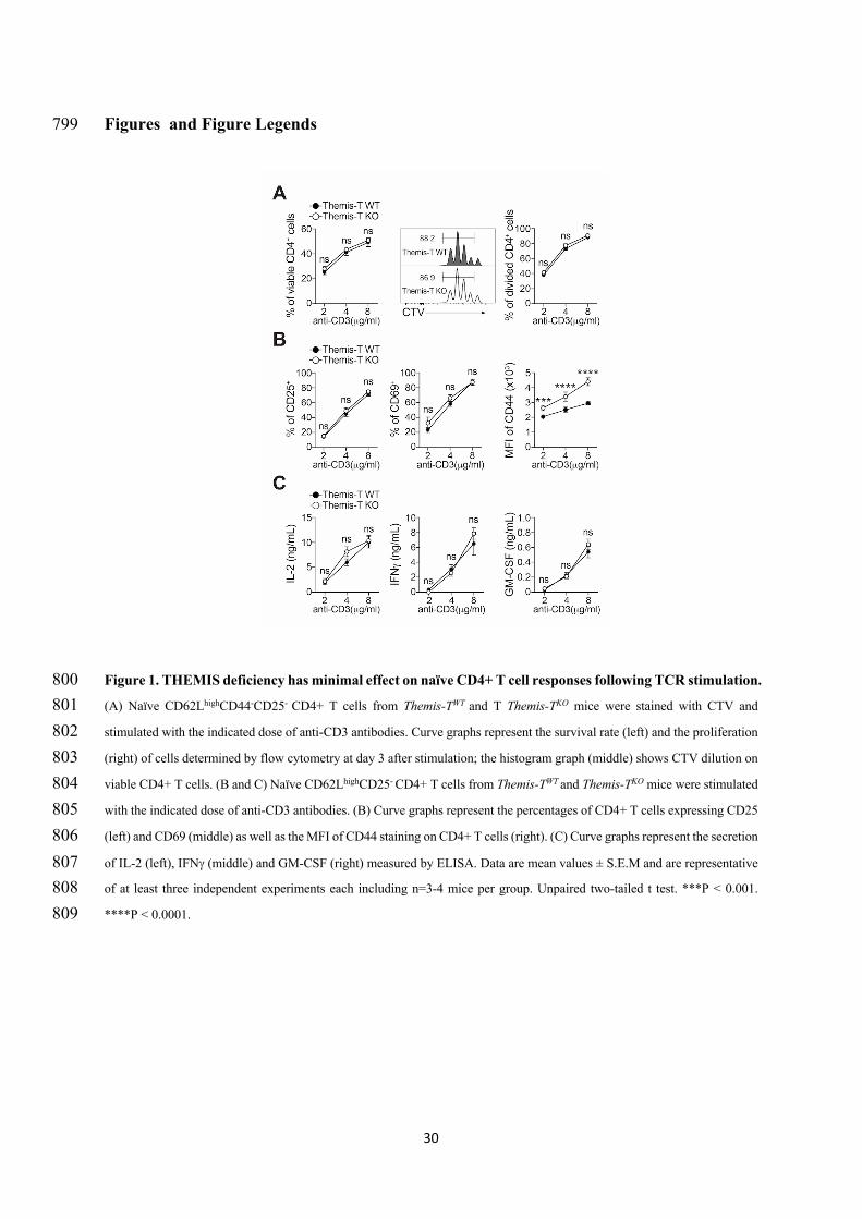
30
Figures and Figure Legends 799
Figure 1. THEMIS deficiency has minimal effect on naïve CD4+ T cell responses following TCR stimulation. 800 (A) Naïve CD62LhighCD44-CD25- CD4+ T cells from Themis-TWT and T Themis-TKO mice were stained with CTV and 801 stimulated with the indicated dose of anti-CD3 antibodies. Curve graphs represent the survival rate (left) and the proliferation 802 (right) of cells determined by flow cytometry at day 3 after stimulation; the histogram graph (middle) shows CTV dilution on 803 viable CD4+ T cells. (B and C) Naïve CD62LhighCD25- CD4+ T cells from Themis-TWT and Themis-TKO mice were stimulated 804 with the indicated dose of anti-CD3 antibodies. (B) Curve graphs represent the percentages of CD4+ T cells expressing CD25 805 (left) and CD69 (middle) as well as the MFI of CD44 staining on CD4+ T cells (right). (C) Curve graphs represent the secretion 806 of IL-2 (left), IFNg (middle) and GM-CSF (right) measured by ELISA. Data are mean values ± S.E.M and are representative 807 of at least three independent experiments each including n=3-4 mice per group. Unpaired two-tailed t test. ***P < 0.001. 808 ****P < 0.0001. 809
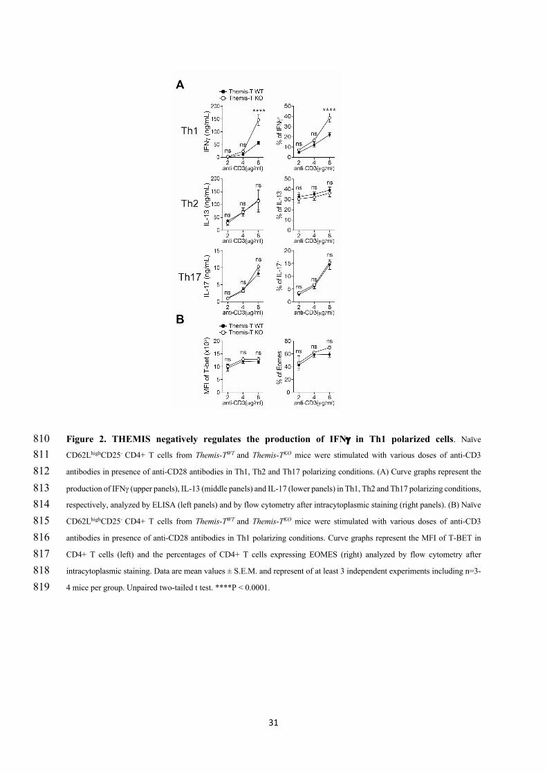
31
Figure 2. THEMIS negatively regulates the production of IFNg in Th1 polarized cells. Naïve 810 CD62LhighCD25- CD4+ T cells from Themis-TWT and Themis-TKO mice were stimulated with various doses of anti-CD3 811 antibodies in presence of anti-CD28 antibodies in Th1, Th2 and Th17 polarizing conditions. (A) Curve graphs represent the 812 production of IFNg (upper panels), IL-13 (middle panels) and IL-17 (lower panels) in Th1, Th2 and Th17 polarizing conditions, 813 respectively, analyzed by ELISA (left panels) and by flow cytometry after intracytoplasmic staining (right panels). (B) Naïve 814 CD62LhighCD25- CD4+ T cells from Themis-TWT and Themis-TKO mice were stimulated with various doses of anti-CD3 815 antibodies in presence of anti-CD28 antibodies in Th1 polarizing conditions. Curve graphs represent the MFI of T-BET in 816 CD4+ T cells (left) and the percentages of CD4+ T cells expressing EOMES (right) analyzed by flow cytometry after 817 intracytoplasmic staining. Data are mean values ± S.E.M. and represent of at least 3 independent experiments including n=3-818 4 mice per group. Unpaired two-tailed t test. ****P < 0.0001. 819
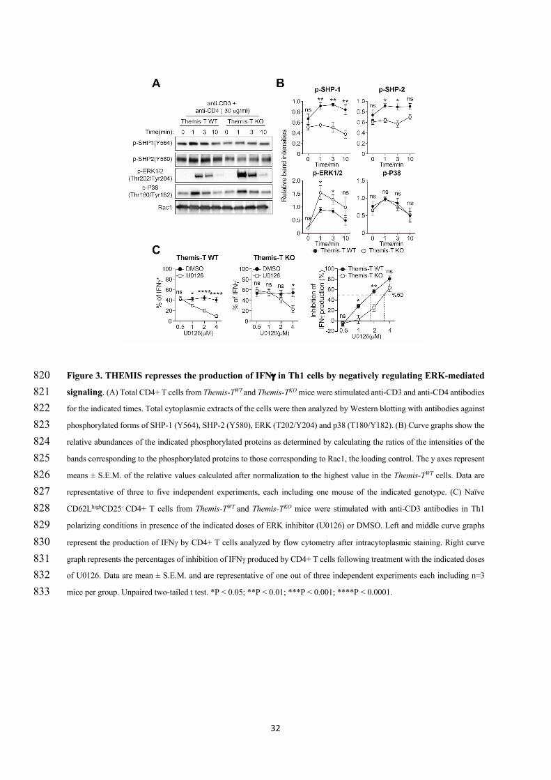
32
Figure 3. THEMIS represses the production of IFNg in Th1 cells by negatively regulating ERK-mediated 820 signaling. (A) Total CD4+ T cells from Themis-TWT and Themis-TKO mice were stimulated anti-CD3 and anti-CD4 antibodies 821 for the indicated times. Total cytoplasmic extracts of the cells were then analyzed by Western blotting with antibodies against 822 phosphorylated forms of SHP-1 (Y564), SHP-2 (Y580), ERK (T202/Y204) and p38 (T180/Y182). (B) Curve graphs show the 823 relative abundances of the indicated phosphorylated proteins as determined by calculating the ratios of the intensities of the 824 bands corresponding to the phosphorylated proteins to those corresponding to Rac1, the loading control. The y axes represent 825 means ± S.E.M. of the relative values calculated after normalization to the highest value in the Themis-TWT cells. Data are 826 representative of three to five independent experiments, each including one mouse of the indicated genotype. (C) Naïve 827 CD62LhighCD25- CD4+ T cells from Themis-TWT and Themis-TKO mice were stimulated with anti-CD3 antibodies in Th1 828 polarizing conditions in presence of the indicated doses of ERK inhibitor (U0126) or DMSO. Left and middle curve graphs 829 represent the production of IFNg by CD4+ T cells analyzed by flow cytometry after intracytoplasmic staining. Right curve 830 graph represents the percentages of inhibition of IFNg produced by CD4+ T cells following treatment with the indicated doses 831 of U0126. Data are mean ± S.E.M. and are representative of one out of three independent experiments each including n=3 832 mice per group. Unpaired two-tailed t test. *P < 0.05; **P < 0.01; ***P < 0.001; ****P < 0.0001. 833
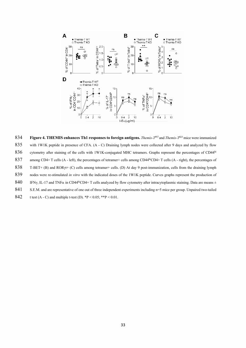
33
Figure 4. THEMIS enhances Th1 responses to foreign antigens. Themis-TWT and Themis-TKO mice were immunized 834 with 1W1K peptide in presence of CFA. (A - C) Draining lymph nodes were collected after 9 days and analyzed by flow 835 cytometry after staining of the cells with 1W1K-conjugated MHC tetramers. Graphs represent the percentages of CD44hi 836 among CD4+ T cells (A - left), the percentages of tetramer+ cells among CD44hiCD4+ T cells (A - right), the percentages of 837 T-BET+ (B) and RORgt+ (C) cells among tetramer+ cells. (D) At day 9 post-immunization, cells from the draining lymph 838 nodes were re-stimulated in vitro with the indicated doses of the 1W1K peptide. Curves graphs represent the production of 839 IFNγ, IL-17 and TNFa in CD44hiCD4+ T cells analyzed by flow cytometry after intracytoplasmic staining. Data are means ± 840 S.E.M. and are representative of one out of three independent experiments including n=5 mice per group. Unpaired two-tailed 841 t test (A - C) and multiple t-test (D). *P < 0.05; **P < 0.01. 842
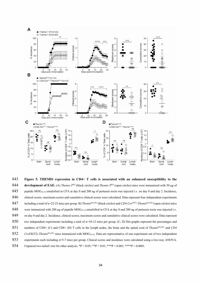
34
Figure 5. THEMIS expression in CD4+ T cells is associated with an enhanced susceptibility to the 843 development of EAE. (A) Themis-TWT (black circles) and Themis-TKO (open circles) mice were immunized with 50 μg of 844 peptide MOG35-55 emulsified in CFA at day 0 and 200 ng of pertussis toxin was injected i.v. on day 0 and day 2. Incidence, 845 clinical scores, maximum scores and cumulative clinical scores were calculated. Data represent four independent experiments 846 including a total of n=22-23 mice per group. B) Themisflox/flox (black circles) and CD4-CreERT2-Themisflox/flox (open circles) mice 847 were immunized with 200 μg of peptide MOG35-55 emulsified in CFA at day 0 and 200 ng of pertussis toxin was injected i.v. 848 on day 0 and day 2. Incidence, clinical scores, maximum scores and cumulative clinical scores were calculated. Data represent 849 two independent experiments including a total of n=10-12 mice per group. (C, D) Dot graphs represent the percentages and 850 numbers of CD4+ (C) and CD8+ (D) T cells in the lymph nodes, the brain and the spinal cord of Themisflox/flox and CD4 851 CreER(T2)-Themisflox/flox mice immunized with MOG35-55. Data are representative of one experiment out of two independent 852 experiments each including n=5-7 mice per group. Clinical scores and incidence were calculated using a two-way ANOVA. 853 Unpaired two-tailed t test for other analysis. *P < 0.05; **P < 0.01; ***P < 0.001; ****P < 0.0001. 854
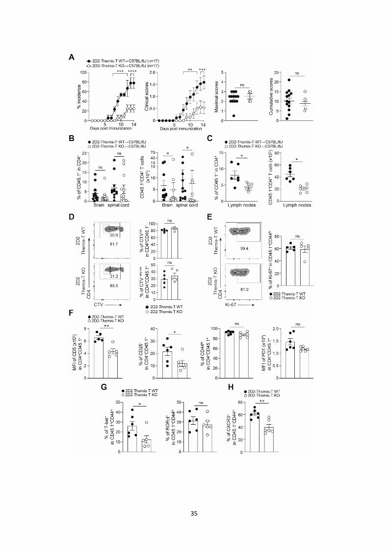
35
36
Figure 6. THEMIS enhances the encephalitogenic potential of CD4+ T cells expressing the MOG35-55 855 specific 2D2 TCR. C57BL/6j mice (CD45.2+) were injected i.v. with 2 x 10⁵ naïve CD45.1+CD4+ T cells from 2D2-856 Themis-TWT and 2D2-Themis-TKO mice. Mice were then immunized with 100 μg of peptide MOG35-55 emulsified in CFA at 857 day 0 and injected i.v. with 200 ng of pertussis toxin at day 0 and 2. (A) Incidence, clinical scores, maximum scores and 858 cumulative clinical scores were calculated. Data represent three independent experiments including a total of n=17 mice per 859 group. (B and C) Dot graph represent the percentages and numbers of CD45.1+ cells among CD4+ T cells in the brain and the 860 spinal cord (B) and in the lymph nodes (C) at day 14 and 6 after immunization, respectively. Data in (B) represent two 861 independent experiments including a total of n=12 mice per group. Data in C is representative of one experiment out of four 862 independent experiments including n=6 mice per group (D) C57BL/6j mice (CD45.2+) were injected i.v. with 2 x 10⁵ CTV 863 stained naïve CD45.1+CD4+ T cells from 2D2-Themis-TWT and 2D2-Themis-TKO mice. Mice were then immunized with 100 864 μg of peptide MOG35-55 emulsified in CFA at day 0 and injected i.v. with 200 ng of pertussis toxin at day 0 and 2. Proliferation 865 was analyzed at day three after immunization. Contour plots represent CD4 vs CTV staining on CD45.1+ cells. Histogram 866 bars represent the percentages of divided and highly divided cells based on the gating strategy indicated in the contour plots. 867 (E) Contour plots represent CD4 vs Ki-67 staining on CD45.1+ cells. Histogram bars represent the percentage of Ki-67+ cells 868 in CD45.1+CD44hi cells. (F) Histogram bars represent the percentages of CD44hi and CD25+ cells among CD45.1+CD4+ T 869 cells as well as the MFI of PD-1 on CD45.1+CD4+ T cells. (G) Histogram bars represent the percentages of CD45.1+CD4+ 870 T cells expressing T-BET and RORgt cells. (H) Histogram bars represent the percentages of CD45.1+CD4+ T cells expressing 871 CXCR3. For D, E, F, G and H, data are representative of one experiment out of three to five independent experiments each 872 including n=5-7 mice per group. Clinical scores and incidence were calculated using a two-way ANOVA. Unpaired two-tailed 873 t test for the other analysis. *P < 0.05; **P < 0.01; ***P < 0.001; ****P < 0.0001. 874 875
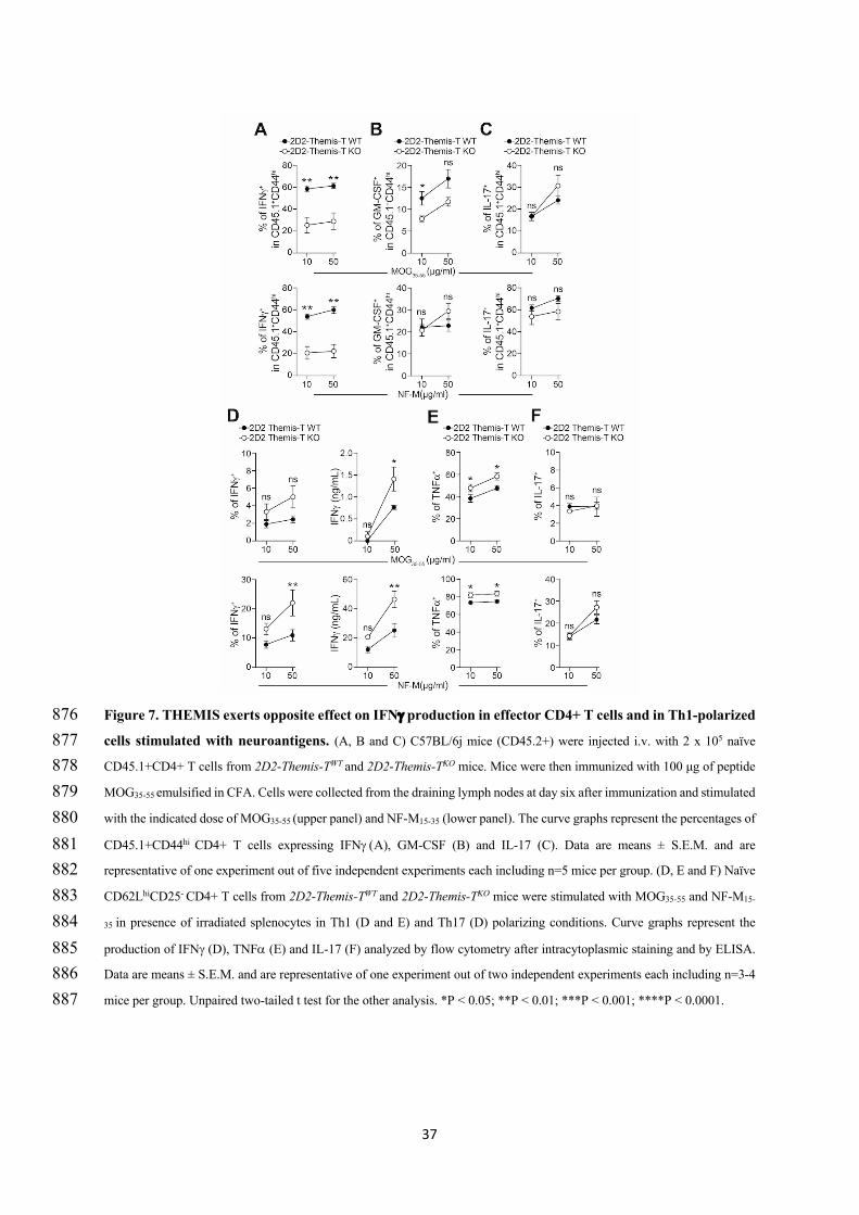
37
Figure 7. THEMIS exerts opposite effect on IFNg production in effector CD4+ T cells and in Th1-polarized 876 cells stimulated with neuroantigens. (A, B and C) C57BL/6j mice (CD45.2+) were injected i.v. with 2 x 10⁵ naïve 877 CD45.1+CD4+ T cells from 2D2-Themis-TWT and 2D2-Themis-TKO mice. Mice were then immunized with 100 μg of peptide 878 MOG35-55 emulsified in CFA. Cells were collected from the draining lymph nodes at day six after immunization and stimulated 879 with the indicated dose of MOG35-55 (upper panel) and NF-M15-35 (lower panel). The curve graphs represent the percentages of 880 CD45.1+CD44hi CD4+ T cells expressing IFNg (A), GM-CSF (B) and IL-17 (C). Data are means ± S.E.M. and are 881 representative of one experiment out of five independent experiments each including n=5 mice per group. (D, E and F) Naïve 882 CD62LhiCD25- CD4+ T cells from 2D2-Themis-TWT and 2D2-Themis-TKO mice were stimulated with MOG35-55 and NF-M15-883 35 in presence of irradiated splenocytes in Th1 (D and E) and Th17 (D) polarizing conditions. Curve graphs represent the 884 production of IFNg (D), TNFa (E) and IL-17 (F) analyzed by flow cytometry after intracytoplasmic staining and by ELISA. 885 Data are means ± S.E.M. and are representative of one experiment out of two independent experiments each including n=3-4 886 mice per group. Unpaired two-tailed t test for the other analysis. *P < 0.05; **P < 0.01; ***P < 0.001; ****P < 0.0001. 887
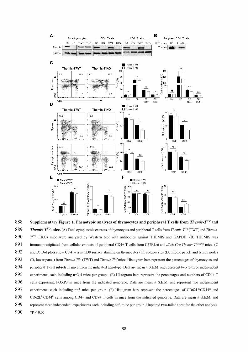
38
Supplementary Figure 1. Phenotypic analyses of thymocytes and peripheral T cells from Themis-TWT and 888 Themis-TKO mice. (A) Total cytoplasmic extracts of thymocytes and peripheral T cells from Themis-TWT (TWT) and Themis-889 TKO (TKO) mice were analyzed by Western blot with antibodies against THEMIS and GAPDH. (B) THEMIS was 890 immunoprecipitated from cellular extracts of peripheral CD4+ T cells from C57BL/6 and dLck-Cre Themis-Tflox/flox mice. (C 891 and D) Dot plots show CD4 versus CD8 surface staining on thymocytes (C), splenocytes (D, middle panel) and lymph nodes 892 (D, lower panel) from Themis-TWT (TWT) and Themis-TKO mice. Histogram bars represent the percentages of thymocytes and 893 peripheral T cell subsets in mice from the indicated genotype. Data are mean ± S.E.M. and represent two to three independent 894 experiments each including n=3-4 mice per group. (E) Histogram bars represent the percentages and numbers of CD4+ T 895 cells expressing FOXP3 in mice from the indicated genotype. Data are mean ± S.E.M. and represent two independent 896 experiments each including n=3 mice per group. (F) Histogram bars represent the percentages of CD62LhiCD44lo and 897 CD62LloCD44hi cells among CD4+ and CD8+ T cells in mice from the indicated genotype. Data are mean ± S.E.M. and 898 represent three independent experiments each including n=3 mice per group. Unpaired two-tailed t test for the other analysis. 899 *P < 0.05. 900
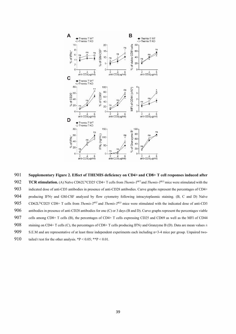
39
Supplementary Figure 2. Effect of THEMIS deficiency on CD4+ and CD8+ T cell responses induced after 901 TCR stimulation. (A) Naïve CD62LhiCD25- CD4+ T cells from Themis-TWT and Themis-TKO mice were stimulated with the 902 indicated dose of anti-CD3 antibodies in presence of anti-CD28 antibodies. Curve graphs represent the percentages of CD4+ 903 producing IFNg and GM-CSF analyzed by flow cytometry following intracytoplasmic staining. (B, C and D) Naïve 904 CD62LhiCD25- CD8+ T cells from Themis-TWT and Themis-TKO mice were stimulated with the indicated dose of anti-CD3 905 antibodies in presence of anti-CD28 antibodies for one (C) or 3 days (B and D). Curve graphs represent the percentages viable 906 cells among CD8+ T cells (B), the percentages of CD4+ T cells expressing CD25 and CD69 as well as the MFI of CD44 907 staining on CD4+ T cells (C), the percentages of CD8+ T cells producing IFNg and Granzyme B (D). Data are mean values ± 908 S.E.M and are representative of at least three independent experiments each including n=3-4 mice per group. Unpaired two-909 tailed t test for the other analysis. *P < 0.05; **P < 0.01. 910
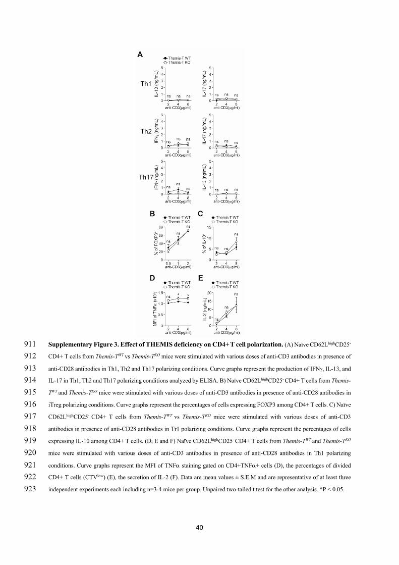
40
Supplementary Figure 3. Effect of THEMIS deficiency on CD4+ T cell polarization. (A) Naïve CD62LhighCD25- 911 CD4+ T cells from Themis-TWT vs Themis-TKO mice were stimulated with various doses of anti-CD3 antibodies in presence of 912 anti-CD28 antibodies in Th1, Th2 and Th17 polarizing conditions. Curve graphs represent the production of IFNg, IL-13, and 913 IL-17 in Th1, Th2 and Th17 polarizing conditions analyzed by ELISA. B) Naïve CD62LhighCD25- CD4+ T cells from Themis-914 TWT and Themis-TKO mice were stimulated with various doses of anti-CD3 antibodies in presence of anti-CD28 antibodies in 915 iTreg polarizing conditions. Curve graphs represent the percentages of cells expressing FOXP3 among CD4+ T cells. C) Naïve 916 CD62LhighCD25- CD4+ T cells from Themis-TWT vs Themis-TKO mice were stimulated with various doses of anti-CD3 917 antibodies in presence of anti-CD28 antibodies in Tr1 polarizing conditions. Curve graphs represent the percentages of cells 918 expressing IL-10 among CD4+ T cells. (D, E and F) Naïve CD62LhighCD25- CD4+ T cells from Themis-TWT and Themis-TKO 919 mice were stimulated with various doses of anti-CD3 antibodies in presence of anti-CD28 antibodies in Th1 polarizing 920 conditions. Curve graphs represent the MFI of TNFa staining gated on CD4+TNFa+ cells (D), the percentages of divided 921 CD4+ T cells (CTVlow) (E), the secretion of IL-2 (F). Data are mean values ± S.E.M and are representative of at least three 922 independent experiments each including n=3-4 mice per group. Unpaired two-tailed t test for the other analysis. *P < 0.05. 923
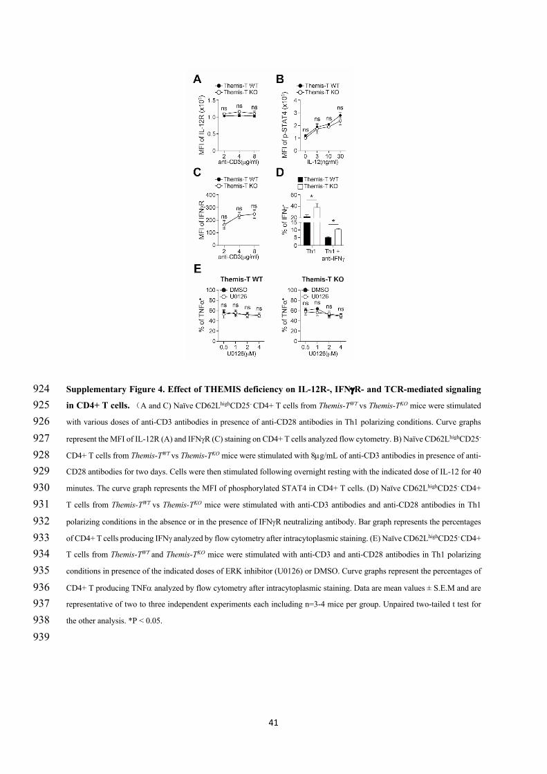
41
Supplementary Figure 4. Effect of THEMIS deficiency on IL-12R-, IFNgR- and TCR-mediated signaling 924 in CD4+ T cells. (A and C) Naïve CD62LhighCD25- CD4+ T cells from Themis-TWT vs Themis-TKO mice were stimulated 925 with various doses of anti-CD3 antibodies in presence of anti-CD28 antibodies in Th1 polarizing conditions. Curve graphs 926 represent the MFI of IL-12R (A) and IFNgR (C) staining on CD4+ T cells analyzed flow cytometry. B) Naïve CD62LhighCD25- 927 CD4+ T cells from Themis-TWT vs Themis-TKO mice were stimulated with 8µg/mL of anti-CD3 antibodies in presence of anti-928 CD28 antibodies for two days. Cells were then stimulated following overnight resting with the indicated dose of IL-12 for 40 929 minutes. The curve graph represents the MFI of phosphorylated STAT4 in CD4+ T cells. (D) Naïve CD62LhighCD25- CD4+ 930 T cells from Themis-TWT vs Themis-TKO mice were stimulated with anti-CD3 antibodies and anti-CD28 antibodies in Th1 931 polarizing conditions in the absence or in the presence of IFNgR neutralizing antibody. Bar graph represents the percentages 932 of CD4+ T cells producing IFNg analyzed by flow cytometry after intracytoplasmic staining. (E) Naïve CD62LhighCD25- CD4+ 933 T cells from Themis-TWT and Themis-TKO mice were stimulated with anti-CD3 and anti-CD28 antibodies in Th1 polarizing 934 conditions in presence of the indicated doses of ERK inhibitor (U0126) or DMSO. Curve graphs represent the percentages of 935 CD4+ T producing TNFa analyzed by flow cytometry after intracytoplasmic staining. Data are mean values ± S.E.M and are 936 representative of two to three independent experiments each including n=3-4 mice per group. Unpaired two-tailed t test for 937 the other analysis. *P < 0.05. 938 939
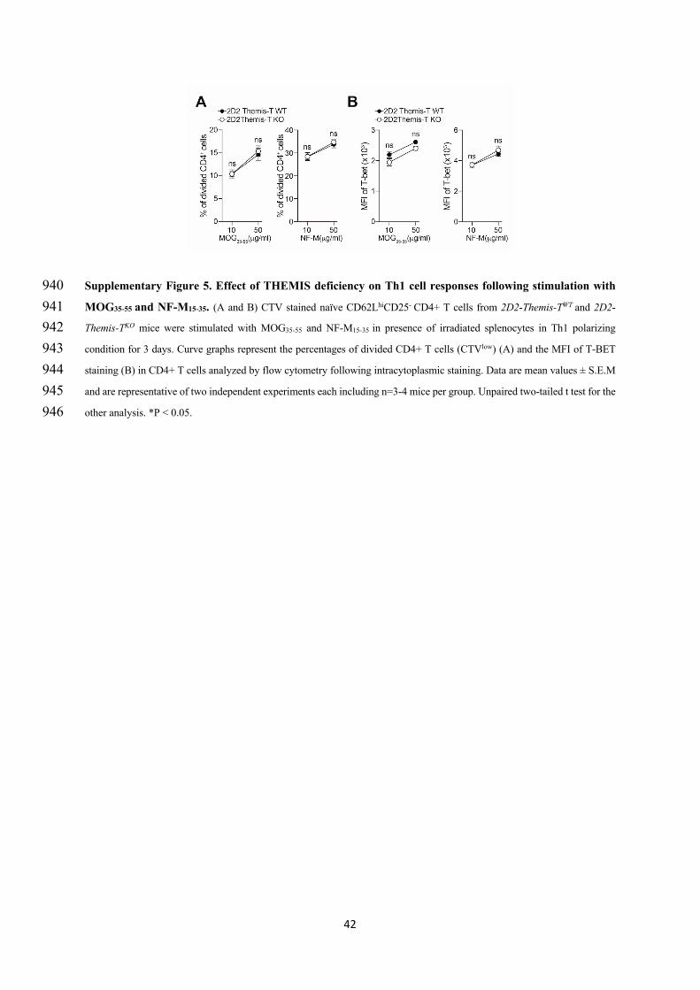
42
Supplementary Figure 5. Effect of THEMIS deficiency on Th1 cell responses following stimulation with 940 MOG35-55 and NF-M15-35. (A and B) CTV stained naïve CD62LhiCD25- CD4+ T cells from 2D2-Themis-TWT and 2D2-941 Themis-TKO mice were stimulated with MOG35-55 and NF-M15-35 in presence of irradiated splenocytes in Th1 polarizing 942 condition for 3 days. Curve graphs represent the percentages of divided CD4+ T cells (CTVlow) (A) and the MFI of T-BET 943 staining (B) in CD4+ T cells analyzed by flow cytometry following intracytoplasmic staining. Data are mean values ± S.E.M 944 and are representative of two independent experiments each including n=3-4 mice per group. Unpaired two-tailed t test for the 945 other analysis. *P < 0.05. 946

137
Discussion
138
Peripheral CD4+ T cells plays a centre role in the adaptive immune response. The activation of CD4+
T cells leads to their proliferation and to their differentiation into distinct Th subpopulations which
migrate toward the inflammation sites. Each Th subset secrete a specific panel of cytokines and
chemokines which contribute to activate other immune cells and promote their recruitment to infection
sites. The exacerbated activation and differentiation of CD4+ T cells into specific subsets may lead to
the development of organ-specific autoimmune diseases such as RA, MS or may enhance the
susceptibility to develop inflammatory diseases such as IBD. The magnitude of CD4+ T cell responses
is modulated by proteins which regulate signals triggered by the TCR, cytokines receptors and additional
receptors. Identifying new signalling proteins involved in the regulation of CD4+ T cell responses may
help to better understand the mechanism by which autoimmune and inflammatory diseases occur.
THEMIS is as a T cell-lineage protein, which promotes the positive selection of thymocytes by
regulating TCR signalling (Choi, Warzecha, et al., 2017; Fu et al., 2009; Lesourne et al., 2009). The
germline deletion of themis (themis-/-) results in a strong reduction of CD4+ and CD8+ T cells in the
thymus and in peripheral lymphoid organs. themis-/- mice do not constitute a good model for exploring
the function of THEMIS in peripheral T cells since the lymphopenia status of those mice may alter the
profile of T cells responses independently of THEMIS expression. To avoid this, we created a model, in
which the gene encoding for THEMIS is invalidated at late stages of T cell development in the thymus.
By using this mouse model, my PhD work revealed that:
- THEMIS deficiency leads to opposite outcomes on peripheral CD4+ T cell responses following
TCR triggering in vitro and antigen recognition in vivo.
- THEMIS enhances the susceptibility to EAE development by enhancing encephalitogenic Th1
responses in the early stage of the disease.
139
1. Opposite outcomes of THEMIS deficiency on peripheral CD4+ T cell responses in vitro and in
vivo
Our results show that THEMIS deficiency leads to exacerbated production of Th1 cytokines when naïve
CD4+ T cells were polarized into Th1 cells. THEMIS inhibits IFN production in those conditions by
repressing ERK-dependent Th1 responses but has no effect on T-bet, the key transcription factor for
differentiation into Th1 cells. Similar effects were observed when cells were stimulated with anti-CD3
antibodies and with the encephalitogenic peptides MOG35-55 and NF-M15-35, suggesting that this outcome
is not due to the methodology of stimulation. The repressive effect of THEMIS on Th1 responses
observed in vitro appeared stronger when high concentration of anti-CD3 antibodies and NF-M15-35
peptide were used, suggesting that THEMIS may operate in a negative feedback loop triggered by strong
TCR stimulation. By contrast to these in vitro data, THEMIS deficiency leads to decreased T-bet
expression and reduced INFproduction in effector CD4+ T cells re-stimulated with MOG35-55 and NF-
M15-35 after immunization with MOG35-55. Thus, the absence of THEMIS leads to nearly opposite
outcomes upon the polarization of naïve CD4+ T cells into Th1 cells and the re-stimulation of effector
CD4+ T cells with the same set of antigenic peptides. The stimulation environment in vitro presumably
does not reflect the environment in vivo, where CD4+ T cells receive multiple stimuli that may affect
the ability of cells to respond to antigen challenge. Analysis of Th1 responses in vitro were performed
partly by using irradiated splenic cells as APC. Those CD4+ T cells may not express similar co-
stimulatory and co-inhibitory molecules as the CD4+ T cells which have migrated into lymph nodes
after immunization. The irradiation of cells may affect the expression of co-receptor ligands on APCs
and the production of cytokines which may promote additional signals controlling Th1 responses.
Although we could not formally explain the reasons of those opposite effects, we speculate that the
phosphatases SHP-1 and SHP-2, which are the main target of THEMIS, may not be engaged similarly
by the TCR and additional cell surface receptors in naïve CD4+ T cells polarized into Th1 cells and in
effector T cells. Since SHP-1 and SHP-2 can have opposite outcomes on CD4+ T cell responses, we
believe that THEMIS may exert distinct effect according to which phosphatases is functionally
preponderant in a given stimulatory context.
140
A. What is the mechanism by which THEMIS regulates Th1 responsiveness in vitro?
THEMIS seems to repress Th1 responses independently of SHP-1
SHP-1 is mainly characterized as an inhibitory regulator of TCR signaling, which can repress the activity
of several key TCR signaling molecules, such as the TCR ζ chain, the adaptor LAT and the Src-family
kinases and the Rac-GTP exchange factor VAV1 (Carter et al., 1999; Fawcett & Lorenz, 2005; Kosugi
et al., 2001; Stebbins et al., 2003; Su et al., 2001). Whereas SHP-2 has been ascribed with both
inhibitory and enhancing roles on TCR signaling, depending on its substrate and on the context of its
recruitment to the membrane (Arnaud et al., 2004; Dong et al., 2015; Frearson & Alexander, 1998;
Nguyen et al., 2006; Niogret et al., 2019; Salmond et al., 2005; Yokosuka et al., 2012). In thymocytes,
THEMIS constitutively binds with SHP-1 and to a lesser extent with SHP-2 (Choi, Warzecha, et al.,
2017). The role of THEMIS on SHP-1-mediated activity has been the subject of controversy and has led
to nearly two opposite models to explain the mechanism by which THEMIS regulates T cell
development. In the first model, THEMIS promotes positive selection by recruiting SHP-1 to TCR
signaling complexes and/or by promoting its phosphatase activity, reducing thereby the strength of
TCR signals below the threshold of negative selection (Fu et al., 2013). In the second model, THEMIS
enhances positive selection by blocking SHP-1 activity, promoting or stabilizing the oxidation of its
catalytic cysteine, and enhancing thereby TCR signal strength beyond the TCR signalling threshold
required for positive selection (Choi, Warzecha, et al., 2017). Although THEMIS inhibits SHP-2 to a
lesser extent than SHP-1 (Choi, Warzecha, et al., 2017), the consequence of this inhibition on T cell
development and T cell responses has not been clearly evaluated.
One can wonder therefore which of these two models would explain the best our results. Early studies
using the motheaten mouse model identified SHP-1 as a negative regulator of the generation of Th1
subpopulation (Kamata et al., 2003; Yu et al., 2005). SHP-1 deficiency leads to increased proliferation
of polarized Th1 and increased production of Th1 cytokines. This is partially similar to our in vitro
results, suggesting that THEMIS may repress IFN production in Th1 cells as described in Model 1, by
recruiting SHP-1 into TCR signalling complexes or by enhancing its PTP activity. However, our results
show that, THEMIS deficiency does not affect the proliferation and survival of Th1 cells, suggesting
that a different mechanism might be engaged. SHP-1 deficiency also leads to exacerbated Th2 responses
141
(Johnson et al., 2013), which is not the case of THEMIS deficiency. In addition, naïve CD4+ T cells
with either natural mutation on ptpn6, the gene encoding for SHP-1 or deficient for SHP-1 exhibit
normal signalling responses following TCR stimulation, contrasting with our data showing that
THEMIS deficiency leads to increased ERK1/2 activity upon TCR stimulation. Furthermore, our results
show that THEMIS deficiency leads to increased production of IF in Th1 particularly upon strong
TCR stimulations (high doses of anti-CD3 or high affinity peptides). This is different from what has
been described in studies supporting model 1, which detected an increase in TCR signalling events when
using weak TCR stimulation (Fu et al., 2013). Altogether, these considerations suggest that THEMIS
does not inhibit Th1 responses in vitro by promoting or by repressing SHP-1.
Studies based on SHP-1 deficient mice support model 2 more than model 1
More generally, model 1 which is mostly based on in vitro studies has been challenged by several in
vivo observations (Choi, Cornall, et al., 2017). A prediction from Model 1 is that SHP-1 deficiency
should lead to similar effect on T cell development than THEMIS deficiency. This is not the case since
positive selection is mildly exacerbated in mice expressing inactive form of SHP-1 (Johnson et al., 2013;
Kamata et al., 2003; Mercadante & Lorenz, 2017). The absence of similarity was explained by authors
of Model 1, by the potential redundancy between SHP-1 and SHP-2 in thymocytes (Fu et al., 2013;
Mehta et al., 2018). However, this explanation was challenged recently by the phenotype of mice with
double deficiencies in THEMIS and SHP-1. According to model 1, SHP-1 deficiency in a THEMIS
deficient background should either aggravate the effect of THEMIS on positive selection or have no
additional consequences as the ones observed in the context of THEMIS deficiency alone. However
studies from Paul Love’s team showed that SHP-1 deficiency rescued the impaired positive selection of
thymocytes due to THEMIS deficiency, arguing against model 1 and in favour of model 2 (Choi,
Warzecha, et al., 2017). Analysis of TCR signal strength using the TCR signalling sensor transgenic
mouse model, Nur-77 promoter-driven GFP, reveals that TCR signalling is reduced in the absence of
THEMIS in vivo rather than increased as predicted by model 1 (Zvezdova et al., 2016). Altogether those
suggest are in favour model 2 rather than of model 1.
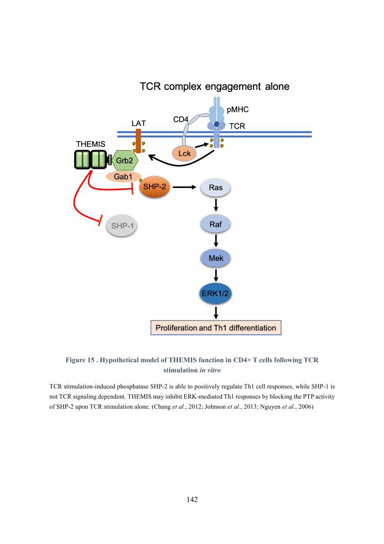
142
Figure 15 . Hypothetical model of THEMIS function in CD4+ T cells following TCR
stimulation in vitro
TCR stimulation-induced phosphatase SHP-2 is able to positively regulate Th1 cell responses, while SHP-1 is
not TCR signaling dependent. THEMIS may inhibit ERK-mediated Th1 responses by blocking the PTP activity
of SHP-2 upon TCR stimulation alone. (Chang et al., 2012; Johnson et al., 2013; Nguyen et al., 2006)
143
THEMIS would repress Th1 responses in vitro by blocking SHP-2 activity
The inactivation of SHP-2 activity in Jurkat cells and SHP-2 deficiency in thymocytes lead to reduced
ERK activation following TCR engagement in vitro (Frearson & Alexander, 1998; Nguyen et al., 2006).
SHP-2 participates in a multimeric signaling complex that regulates TCR coupling to the Ras/MAPK
pathway in Jurkat T cells. It suggests that THEMIS may inhibits ERK1/2-mediated IFN production in
Th1 cells by repressing the PTP activity of SHP-2 (Figure 15).
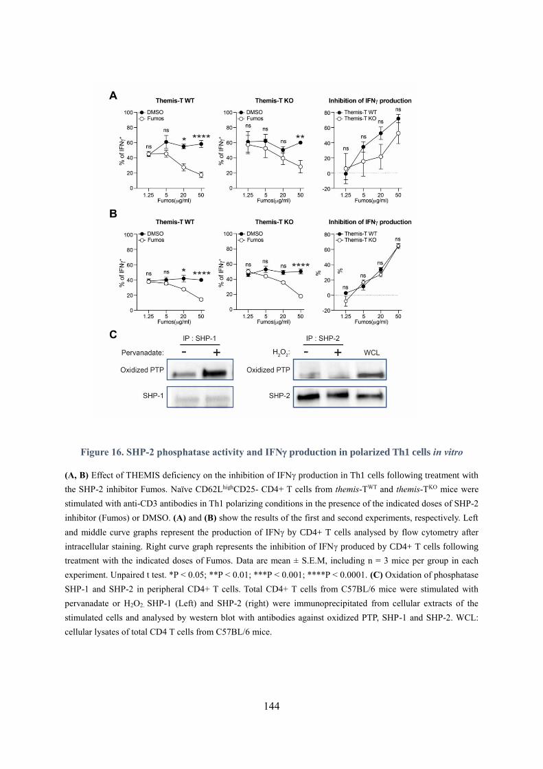
144
Figure 16. SHP-2 phosphatase activity and IFN production in polarized Th1 cells in vitro
(A, B) Effect of THEMIS deficiency on the inhibition of IFN production in Th1 cells following treatment with
the SHP-2 inhibitor Fumos. Naïve CD62LhighCD25- CD4+ T cells from themis-TWT and themis-TKO mice were
stimulated with anti-CD3 antibodies in Th1 polarizing conditions in the presence of the indicated doses of SHP-2
inhibitor (Fumos) or DMSO. (A) and (B) show the results of the first and second experiments, respectively. Left
and middle curve graphs represent the production of IFN by CD4+ T cells analysed by flow cytometry after
intracellular staining. Right curve graph represents the inhibition of IFN produced by CD4+ T cells following
treatment with the indicated doses of Fumos. Data are mean ± S.E.M, including n = 3 mice per group in each
experiment. Unpaired t test. *P < 0.05; **P < 0.01; ***P < 0.001; ****P < 0.0001. (C) Oxidation of phosphatase
SHP-1 and SHP-2 in peripheral CD4+ T cells. Total CD4+ T cells from C57BL/6 mice were stimulated with
pervanadate or H2O2. SHP-1 (Left) and SHP-2 (right) were immunoprecipitated from cellular extracts of the
stimulated cells and analysed by western blot with antibodies against oxidized PTP, SHP-1 and SHP-2. WCL:
cellular lysates of total CD4 T cells from C57BL/6 mice.
145
In support of this interpretation, we found that SHP-2 phosphorylation is reduced in THEMIS deficient
CD4+ T cells. The reduced phosphorylation of SHP-2 might be the consequences of an hyperactivated
state of this phosphatase which could presumably dephosphorylate itself through auto- or trans-
phosphatase activity. We tried to test this hypothesis further by treating naïve CD4+ T cells with
Fumosorine (Fumos), a specific SHP-2 inhibitor, under Th1-polarizing conditions, in two distinct
experiments (Figure 16 – A, B). Result from one experiment suggests that higher doses of inhibitor
were required to inhibit the production of IFN in THEMIS deficient Th1 cells to the same extent as
compared to that in control cells (Figure 16 – A). However, this was not confirmed in the second
experiment (Figure 16 – B). We are planning to perform additional experiments to resolve this question.
The stimulation of immune cell surface receptors has been shown to generate intracellular reactive
oxygen species (ROS), which may modulate signal transduction by inactivating the PTP domain of
phosphatases (Pao et al., 2007). In thymocytes, THEMIS inhibits SHP-1 activity by promoting or
stabilizing the oxidation of SHP-1 catalytic cysteine in the presence of ROS (Choi, Warzecha, et al.,
2017). In Jurkat T cells, SHP-2 has been shown to regulate T cell adhesion in a redox-dependent manner
by downregulating the phosphorylation of ADAP and its recruitment to SLP-76 (Choi, Warzecha, et al.,
2017). These findings suggest that THEMIS may inhibit the PTP activity of SHP-2 by regulating its
susceptibility to ROS oxidation. Therefore, we analysed the effect of THEMIS deficiency on the
oxidation level of SHP-2 in CD4+ T cells treated with pervanadate using specific antibodies which
recognize the oxidated form of the catalytic cysteine contained in the catalytic site of several tyrosine
phosphatases. Unfortunately, we were not able to detect an oxidation of SHP-2, by contrast to SHP-1,
after pervanadate treatment. It remains possible that this antibody does not efficiently recognize
oxidized-SHP-2 (Figure 16 – C) by contrast to other oxidized forms of tyrosine phosphatases.
B. What is the mechanism by which THEMIS regulates Th1 responses in vivo?
Although our in vitro data indicate that THEMIS inhibits the secretion of Th1 cytokines, results obtained
in vivo suggest that THEMIS promotes the differentiation of naïve CD4+ T cells into Th1 cells. One
potential explanation to this apparent discrepancy could be that THEMIS regulates additional signals
that are triggered specifically in vivo and which may not be recapitulated in vitro in a reductionist model.
CD4+ T cell responses induced in vitro are triggered either exclusively by anti-CD3 stimulation or by
146
irradiated splenic cells which may not express the same set of co-stimulatory and co-inhibitory
molecules expressed by APC in the draining lymph nodes following immunization. The phosphatases
SHP-1 and SHP-2 are recruited to several co-inhibitory and cytokines receptors which may be engaged
by their ligands in vivo but not in vitro (Rhee & Veillette, 2012). Several of those receptors were shown
to be important in the regulation of Th1 responses, suggesting that THEMIS may enhance type 1
responses in vivo by operating downstream of those receptors rather than downstream of the TCR.
A major tumour escape mechanism relies on the stimulation of inhibitory receptors expressed on T cells,
so called immune checkpoints (IC), leading to the inhibition of signalling events triggered by the TCR
and by addition co-stimulatory receptors (Rudd, 2019), and ultimately to the development of a state of
exhaustion in which T cell required higher TCR stimulation to respond to antigen challenge. In the
context of cancer treatment, blocking the activation of immune checkpoints with antagonist antibodies
results partly in an increase in the production of IFN by Th1 and other T cells, which is associated with
a better anti-tumour response (Chen et al., 2015; Curran et al., 2010; Laheurte et al., 2019; Wei et al.,
2017).
Blocking the stimulation of CTLA4 and PD-1 enhances the co-stimulatory effects of ICOS on Th1 anti-
tumor response (Figure 17). CD4+ICOShiIFN-γ+ cells show strong anti-tumor activity in the context
bladder cancer and melanoma in humans, and are shown to produce high level of Th1-cytokines IFNγ
and TNF (Fu et al., 2011; Liakou et al., 2008). Two studies showed that this population could be
effectively expanded by blocking CTLA-4 alone or in combination with PD-1, resulting in decreased
tumour expansion (Fan et al., 2014; Wei et al., 2019). A study of James P. Allison’s team based on t-
SNE analysis found that blocking CTLA-4 led to an expansion of tumor infiltrating Th1-like cells which
highly express T-bet (Wei et al., 2017). According to previous studies, CTLA-blockade enhances T-bet
expression induced by ICOS-mediated PI3K/Akt signaling (Chen et al., 2014). THEMIS may thus
supress the CTLA-4/SHP-2-mediated inhibition of PI3K pathway, thereby enhancing ICOS-mediated
signaling associated with T-bet expression in CD4+ T cells.
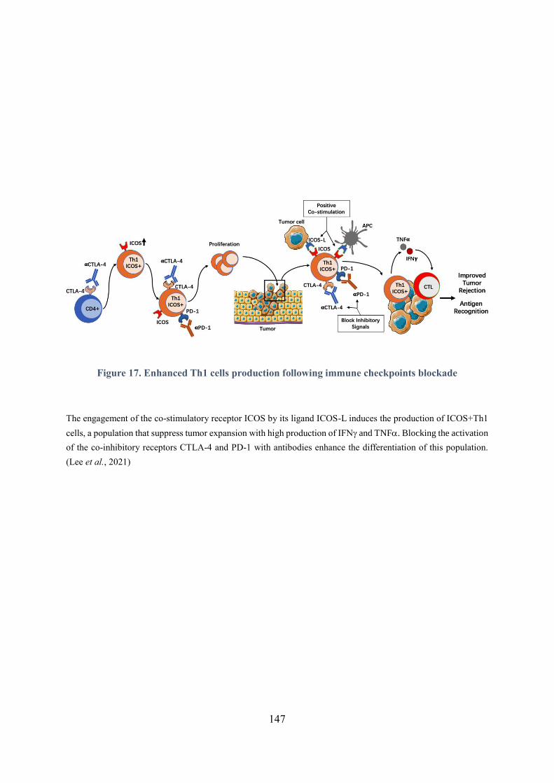
147
Figure 17. Enhanced Th1 cells production following immune checkpoints blockade
The engagement of the co-stimulatory receptor ICOS by its ligand ICOS-L induces the production of ICOS+Th1
cells, a population that suppress tumor expansion with high production of IFN and TNF. Blocking the activation
of the co-inhibitory receptors CTLA-4 and PD-1 with antibodies enhance the differentiation of this population.
(Lee et al., 2021)
148
The immune checkpoint PD-1 has also been shown to exert selective effect on Th1 responses in the
context of cancer. Robert L. Ferris’s team found that the expression of SHP-2 and PD-1 are significantly
higher in tumor-infiltrating lymphocytes (TILs) than in peripheral blood lymphocytes [10]. Their results
showed that stimulation of TILs by PD-L1 suppresses the STAT1/T-bet signalling axis in a SHP-2
dependent fashion, resulting in decreased production of Th1 cytokines by those cells. These reduced
Th1 responses were restored by anti-PD-1 blocking antibodies.
Another interesting finding of this study is that the stimulation of PD-1 represses the production of Th1
cytokines but not Th2 cytokines by those TILs upon TCR stimulation, indicating a preferential inhibitory
effect of PD-1 on the Th1 subset. These suggest that THEMIS may enhance T-bet-mediated Th1
responses by supressing the PD1/SHP-2/pSTAT1.
Additional inhibitory receptors that recruit SHP-1 and SHP-2 were shown to be important in the
regulation of Th1 responses in vivo. The co-inhibitory receptor BTLA which operates mainly by
recruiting SHP-1 (Celis-Gutierrez et al., 2019), has been described as preferentially expressed on Th1
cells (Loyet et al., 2005). Mice deficient for BTLA exhibit exacerbated EAE development due to
enhanced auto-immune CD4+ T cell activity (Watanabe et al., 2003). The Leukocyte-associated Ig-like
receptor 1 (LAIR1), another inhibitory co-receptor which functions by recruiting both SHP-1 and SHP-
2, is associated with reduced Th1 responses [22].
Based on those evidences, we speculate that THEMIS may enhance Th1 responses in vivo by blocking
the inhibitory activity of one or several of those receptors. THEMIS would operate by blocking the PTP
activity of SHP-1 and SHP-2 which are required for the signaling function of those receptors. Imaging
studies suggest that THEMIS is recruited to the Human T-cell leukaemia virus type 1 (HTLV-1) bZIP
factor (HBZ) and modulates in this context the interaction of PD-1 with SHP-2, resulting in enhanced T
cell proliferation (Kinosada et al., 2017). Molecularly speaking, we believe that THEMIS operates
similarly in vitro and in vivo by blocking the PTP activity of SHP-1 and SHP-2.
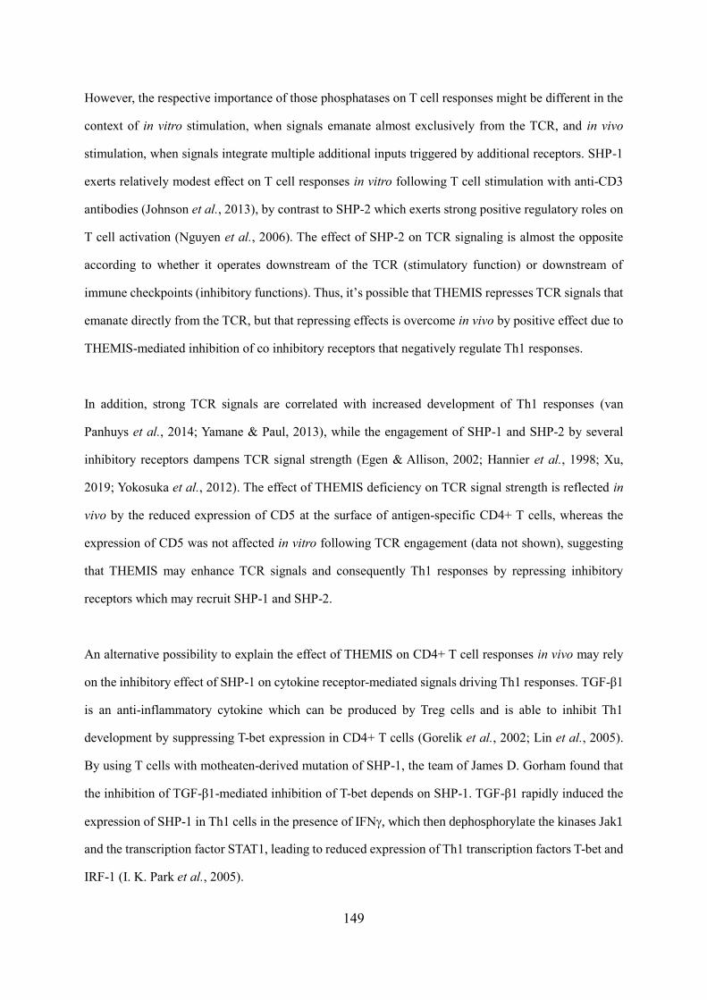
149
However, the respective importance of those phosphatases on T cell responses might be different in the
context of in vitro stimulation, when signals emanate almost exclusively from the TCR, and in vivo
stimulation, when signals integrate multiple additional inputs triggered by additional receptors. SHP-1
exerts relatively modest effect on T cell responses in vitro following T cell stimulation with anti-CD3
antibodies (Johnson et al., 2013), by contrast to SHP-2 which exerts strong positive regulatory roles on
T cell activation (Nguyen et al., 2006). The effect of SHP-2 on TCR signaling is almost the opposite
according to whether it operates downstream of the TCR (stimulatory function) or downstream of
immune checkpoints (inhibitory functions). Thus, it’s possible that THEMIS represses TCR signals that
emanate directly from the TCR, but that repressing effects is overcome in vivo by positive effect due to
THEMIS-mediated inhibition of co inhibitory receptors that negatively regulate Th1 responses.
In addition, strong TCR signals are correlated with increased development of Th1 responses (van
Panhuys et al., 2014; Yamane & Paul, 2013), while the engagement of SHP-1 and SHP-2 by several
inhibitory receptors dampens TCR signal strength (Egen & Allison, 2002; Hannier et al., 1998; Xu,
2019; Yokosuka et al., 2012). The effect of THEMIS deficiency on TCR signal strength is reflected in
vivo by the reduced expression of CD5 at the surface of antigen-specific CD4+ T cells, whereas the
expression of CD5 was not affected in vitro following TCR engagement (data not shown), suggesting
that THEMIS may enhance TCR signals and consequently Th1 responses by repressing inhibitory
receptors which may recruit SHP-1 and SHP-2.
An alternative possibility to explain the effect of THEMIS on CD4+ T cell responses in vivo may rely
on the inhibitory effect of SHP-1 on cytokine receptor-mediated signals driving Th1 responses. TGF-β1
is an anti-inflammatory cytokine which can be produced by Treg cells and is able to inhibit Th1
development by suppressing T-bet expression in CD4+ T cells (Gorelik et al., 2002; Lin et al., 2005).
By using T cells with motheaten-derived mutation of SHP-1, the team of James D. Gorham found that
the inhibition of TGF-β1-mediated inhibition of T-bet depends on SHP-1. TGF-β1 rapidly induced the
expression of SHP-1 in Th1 cells in the presence of IFN, which then dephosphorylate the kinases Jak1
and the transcription factor STAT1, leading to reduced expression of Th1 transcription factors T-bet and
IRF-1 (I. K. Park et al., 2005).
150
It has been reported that SHP-2 also impairs IFN induced JAK1/STAT1 signaling by binding with un-
phosphorylated form of STAT1, inhibiting the recruitment of STAT1 to IFNR (Wu et al., 2012).
Moreover, PD-1 ligation can enhance this inhibition of IFN -induced pathway by SHP-2 (Li et al.,
2015). Therefore, one possibility would be that THEMIS enhances T-bet-mediated Th1 development by
inhibiting the recruitment of SHP-1 and SHP-2 to IFN -induced JAK1/STAT1/T-bet signaling.
Taken together, based on model 2, suggesting that THEMIS regulates T activation by blocking the
phosphatase activity of SHP-1 and SHP-2, we speculate that THEMIS may enhance T-bet-mediated Th1
responses in vivo by regulating TCR signal strength and other signalling pathways independent of the
TCR (Figure 18).
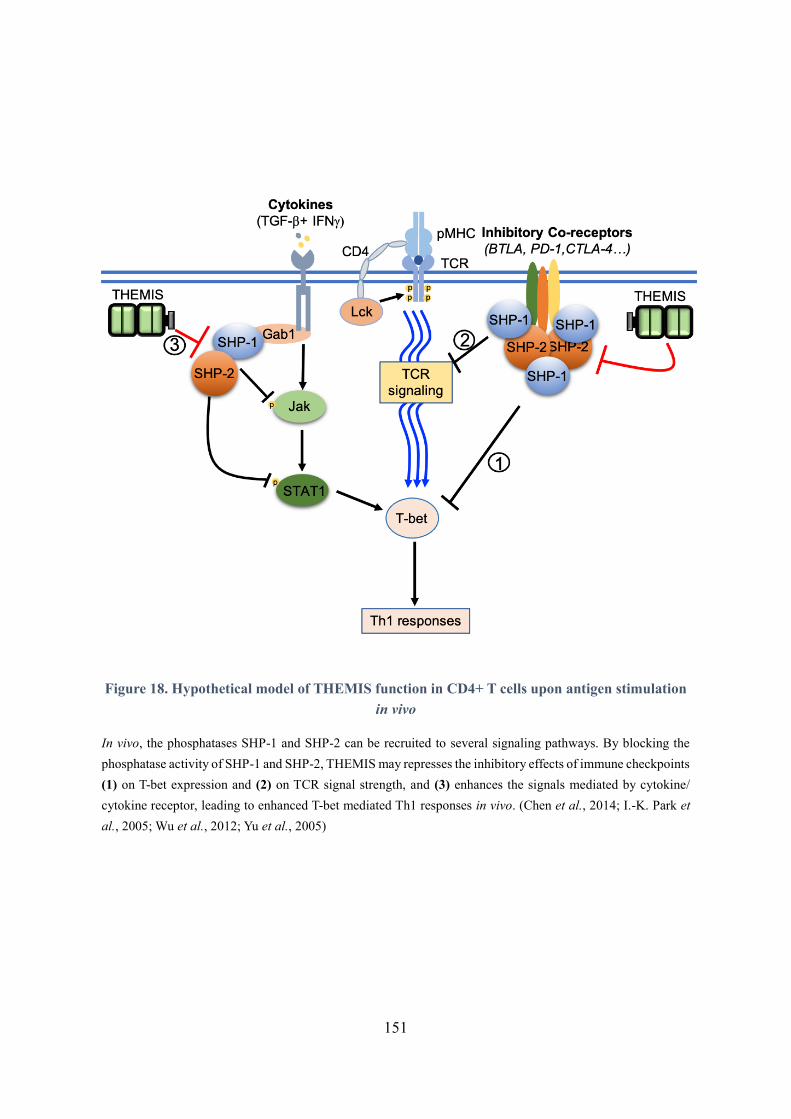
151
Figure 18. Hypothetical model of THEMIS function in CD4+ T cells upon antigen stimulation
in vivo
In vivo, the phosphatases SHP-1 and SHP-2 can be recruited to several signaling pathways. By blocking the
phosphatase activity of SHP-1 and SHP-2, THEMIS may represses the inhibitory effects of immune checkpoints
(1) on T-bet expression and (2) on TCR signal strength, and (3) enhances the signals mediated by cytokine/
cytokine receptor, leading to enhanced T-bet mediated Th1 responses in vivo. (Chen et al., 2014; I.-K. Park et
al., 2005; Wu et al., 2012; Yu et al., 2005)
152
2. Role of THEMIS in the pathophysiology of EAE
Our study demonstrated that THEMIS enhances the susceptibility to the development of EAE. The
absence of THEMIS is correlated with the decreased expression of T-bet in MOG35-55 specific CD4+ T
cells and the reduced production of IFN following re-stimulation with encephalitogenic peptides ex
vivo. This is associated with the decreased infiltration of effector CD4+ T cells in the CNS. The
expression of T-bet is also decreased in CD4+ T cells following immunization with a foreign antigen
1W1K, and is associated with a reduction in the production of IFN. Given the central role of T-bet in
the development of EAE, this suggests that THEMIS promotes EAE by positively regulating T-bet
expression.
A. Effect of THEMIS on T-bet and IFN in the pathogenicity of CD4+ T cell in EAE
The absence of T-bet in CD4+ T cells leads to a selective defect in the differentiation of naïve T cells
into Th1 cells in vitro and in vivo (Szabo et al., 2002). Moreover, T-bet is expressed in CD8+ T cells, B
cells and NK cells in addition to CD4+ T cells but its effect on IFN production in these cells is not as
pronounced as in CD4+ T cells (Lugo-Villarino, 2003; Sullivan et al., 2003; Szabo et al., 2002),
suggesting a pivotal role of T-bet in Th1 cells. Regarding the effect of T-bet in EAE development, initial
studies showed that the silencing or the invalidation of T-bet in mice confers a strong resistance to the
development of MOG35-55 peptide-induced EAE (Bettelli et al., 2004; Lovett-Racke et al., 2004). T-bet-
/- splenocytes had remarkably reduced secretion of IFN but increased production of IL-10 in response
to either MOG35-55 or PLP180-199 ex vivo (Bettelli et al., 2004; Nath, 2006). Even though this was
challenged by a later study, in which the absence of T-bet affected more mildly the development of EAE
(O'Connor et al., 2013). However, in this study, compared with the wild type control, the peripheral and
CNS-infiltrated T-bet-/- CD4+ T cells produced very low amounts of IFN but significantly higher
amounts of IL-17, which is related to the inhibition of Th17 responses by T-bet (Lazarevic et al., 2011;
O'Connor et al., 2013).
In addition, the same study also showed that adoptive transfer of C57BL/6L mice with MOG35-55 pre-
activated T-bet-/- Th1 cells failed to induce EAE, while the control mice developed sever disease,
indicating that T-bet is required for the encephalitogenic potential of Th1 cells (O'Connor et al., 2013).
153
The reason for the difference in the impact of T-bet on the development of EAE between different reports
is still unclear. It may be related to the different backgrounds of mice. One of the earlier studies used
antisense RNAs to silence TBX21 gene in vivo, while this technology might have bystander effects
(Lovett-Racke et al., 2004). T-bet KO mice used in the other two studies, were generated in 129/Sv ES
cells and backcrossed to C57BL/6 for only 5 generations (Bettelli et al., 2004; Szabo et al., 2002). By
comparison, the T-bet-/- mice used in the later studies, showing that T-bet has mild effect on EAE
induction, have been further backcrossed to C57BL/6, reflecting a different genetic background (Grifka-
Walk et al., 2013; O'Connor et al., 2013).
The mechanism by which T-bet controls EAE is not clear but it may be dependent at least partly on its
ability to positively regulate the production of IFN in CD4+ T cells. The effect of IFN in the context
of CNS autoimmunity is complex since it produces both stimulatory and inhibitory outcomes on the
disease. In initial studies, IFN was identified as an aggravating factor in EAE and MS, as the increased
production of IFN was associated with increased demyelination in the CNS (O'Connor et al., 2008;
Olsson, 1992; Panitch et al., 1987; Pettinelli & McFarlin, 1981). However, this theory was challenged
by later studies, in which mice lacking IFN or IFNR or mice treated with IFN neutralizing mAbs
developed severer EAE, suggesting a beneficial role of IFN in this autoimmune disease(Sosa et al.,
2015). Gradually, further explorations have shown that IFN may have distinct effects on EAE according
to the stage of EAE at which it operates. It is proposed that IFN enhances the severity of the disease
during the initiation phase, while it has beneficial role on the pathology during the chronic phase and in
the context of therapy (Furlan et al., 2001; Naves et al., 2013). Accordingly, the production of IFN in
CD4+ T cells was decreased at the initiation stage of EAE (6 days after the immunization) in the absence
of THEMIS, suggesting that the reduced incidence and disease severity of EAE could be partly due to
the reduced production of this inflammatory cytokines.
The role of IFN produced by Th1 cells in the CNS is also complex. On one hand, IFN aggravate the
disease by enhancing Th1 responses and the infiltration of immune cells into the spinal cord (Sanvito et
al., 2010; Stromnes et al., 2008).
154
On the other hand, IFN attenuates the disease by inhibiting Th9 and Th17-mediated pathogenic
responses and positively regulating the number and function of Treg cells (Murugaiyan et al., 2012;
Wang et al., 2006; Wildbaum et al., 2010).
The absence of THEMIS led to reduced number of antigen-specific CD4+ T cells in the CNS, both in
the 2D2 transfer model and in active EAE with CD4-CreERT2-themisfox/flox mice, suggesting that the
reduced production of IFN in THEMIS deficient CD4+ T cells in dLN may lead to a reduced infiltration
of the CNS by immune cells. The percentages of 2D2+CD4+ T cells infiltrating the CNS were not
affected by THEMIS expression, suggesting also that THEMIS may promote the recruitment of
endogenous CD4+ T cells or other leukocyte subsets through bystander effects using similar effector
mechanism. Regarding the cytokines production in CNS, our study showed that the absence of THEMIS
did not affect the percentages of cells producing IFN, GM-CSF and IL-17 in antigen-specific CD4+ T
cells (data not shown), but their cell numbers exhibit decreased due to the general reduction in the
number of antigen-specific themis-deficient CD4+ T cells. One possible reason is that THEMIS would
operate early by controlling the polarization of naïve CD4+ T cells into Th1 cells and by modulating the
ability of those cells to migrate into the CNS. The percentage of IFN-producing CD4+ T cells remains
possibly unchanged because only cells that successfully develop into Th1 without THEMIS manage to
migrate to CNS.
B. Effect of THEMIS on GM-CSF in the pathogenicity of CD4+ T cell in EAE
Although the effect was not as pronounced as that on IFN production, we also show that the expression
of THEMIS is positively associated with the production of GM-CSF following MOG35-55 restimulation
of 2D2+CD4+T cells. It has been reported that GM-CSF promotes neuroinflammation in EAE by
inducing the activation and proliferation of microglia and by enhancing the production of additional
proinflammatory cytokines that favor demyelination (Parajuli et al., 2012; Ponomarev et al., 2007).
Previous studies showed that Th1 cells primed with MOG35-55, which produce IFN and GM-CSF,
transfer a sever EAE in recipient mice accompanied by enrichment of proinflammatory cytokines and
infiltrated monocytes, in a GM-CSF-dependent fashion (El-Behi et al., 2011; Grifka-Walk et al., 2015).
155
In addition, GM-CSF is produced by Th17 and ThGM cells (a newly identified pro-neuroinflammatory
population) (Y. K. Lee et al., 2009; Rasouli et al., 2020). Although we did not detect any differences in
Th17 cells development based on our analysis on RORt expression and IL-17 secretion, we could not
exclude that THEMIS might also be important for the development of ThGM cells in the context of
EAE. Interestingly the encephalitogenic potential of ThGM cells is dependent on T-bet, suggesting that
THEMIS might be important to enhance the pathogenic potential of those cells.
One study suggests that NFAT positively regulates the expression of GM-CSF by binding to the
enhancer upstream of the transcription start site of the gene encoding for GM-CSF and enhancing the
formation of DNase I hypersensitive site within this enhancer to induce transactivation (Bert et al., 2007).
Recent investigation on THEMIS by the team of Nicholas Gascoigne show that CD4+ T cells bearing a
germline mutation in themis gene exhibited a reduction of NFAT translocation upon TCR stimulation
(Prasad et al., 2020), suggesting that THEMIS may enhance the production of GM-CSF by peripheral
CD4+ T cells in vivo by upregulating NFAT translocation.
C. Effect of THEMIS on the infiltration of the CNS by CD4+ T cells
Analysis of T cell infiltration in mice after the appearance of symptoms suggests that the expression of
THEMIS is positively associated with the infiltration of CD4+ T cells in the CNS. The reduced
expression of T-bet in THEMIS-deficient 2D2+ T cells transferred in recipient mice was associated with
a decreased expression of the chemokine receptor CXCR3. It has been shown that T-bet is required for
Th1 cell migration to inflammatory sites by controlling the binding of CD4+ T cells to E-selectin and
P-selectin on inflamed endothelium and by enhancing the expression of CXCR3 on the T cells (Lord et
al., 2005; Underhill et al., 2005) .
It has long been recognized that the infiltrated T cells in MS lesions exhibit high expression of the
chemokine receptor CXCR3 (Balashov et al., 1999; Simpson et al., 2000; Sørensen et al., 1999). In
EAE mouse models, the CNS infiltrates are characterized by large percentages of CXCR3+IFN+-
producing T cells, which are correlated with the upregulation of CXCL10 ( a ligand of CXCR3) on
adjacent astrocytes (O'Connor et al., 2012; Sporici & Issekutz, 2010). Also, an early study showed that

156
CXCR3 blockade resulted in decreased migration of peripheral T cells into the CNS (Sporici & Issekutz,
2010), suggesting a positive association between CXCR3 expression on T cells and their ability to
migrate into the CNS. Although CXCR3 is highly expressed on Th1 cells (Loetscher et al., 1998;
Sallusto et al., 1998), active EAE in CXCR3-/- mice and adoptive transfer of EAE with polarized-
CXCR3-/- Th1 cells showed that CXCR3 expression does not affect the disease severity and the number
of CNS-infiltrated T cells (Lalor & Segal, 2013). One possible reason of this observation may be the
development of compensatory effect mediated by other chemokine receptors. Accordingly, a previous
publication showed that, compared with targeting CXCR3 alone, blocking CXCR3 and CXCR4 at the
same time has a therapeutic effect on adoptively transferred EAE (Kohler et al., 2008).
D. Does THEMIS regulate CD4+ T cell responses in vivo through T-bet-independent mechanism?
Results of both active EAE and transferring EAE in our study indicate that the expression of THEMIS
is positively associated with the infiltration of CD4+ T cells to CNS. Based on the discussion above, we
speculate that THEMIS positively regulates CD4+ T cell infiltration in the CNS through a T-bet
dependent process. However, in the transfer model of EAE, we also observed that THEMIS deficiency
results in significantly lower percentages and numbers of 2D2+ CD4+ T cells in the dLNs and the SPL
of recipient mice at the stage of T cell priming after EAE induction, suggesting that the effect of
THEMIS on EAE could result from insufficient expansion or survival of encephalitogenic T cells after
transfer into recipient mice.
The analysis of CD4+ T cells following staining with MHC tetramers coupled to MOG35-55 peptide
suggest that THEMIS does not affect the proportions of CD4+ T cells that recognize encephalitogenic
peptide following immunization (data not shown), ruling out the possibility that the reduced severity of
EAE in THEMIS deficient mice could be exclusively the consequence of a lack of antigen recognition.
Moreover, by tracing the CTV-stained naïve CD4+ T cells in response to MOG35-55 stimulation in vitro
and in vivo and analyzing the expression of the proliferative marker Ki-67+CD4+ T cells, we could not
detect an effect of THEMIS on the proliferation of CD4+ T cells, suggesting that the reduced numbers
of THEMIS-deficient CD4+ T cells does not result from a defective priming of CD4+ T cells.
157
We could not exclude however that the reduced EAE, observed following transfer of THEMIS deficient
2D2+ T cells in recipient mice, could be the consequence of the reduced ability of those cells to survive
efficiently in their host or to migrate into LN prior immunization.
Recent studies suggest that THEMIS is important for the homeostatic maintenance of CD8+ T cells by
enhancing signaling events resulting from the integration of TCR and cytokine receptor stimulation
(Brzostek et al., 2020). Although authors in this study could not detect significant effect of THEMIS on
CD4+ T cells maintenance using their mouse model (Lck-Cre themisflox/flox) and although numbers of
CD4+ T cells were also normal in the absence of THEMIS in our model (CD2-Cre themisflox/flox), we
cannot exclude that THEMIS-deficient CD4+ may present a disadvantage for homeostatic maintenance
over wild-type cells in a competitive environment where endogenous CD4+ T cells reside. An additional
study from Nicholas Gascoigne’s team showed that naïve CD4+ T cells from germline deficient themis-
/- mice result in poor expansion of effector cells following injection into RAG-/- mice suggesting that
THEMIS might be important to promote the survival or the proliferation of those cells in this
experimental setting (Prasad et al., 2020). This study suggests that THEMIS enhances the expansion of
effector cells by promoting metabolic reprogramming through a NFAT dependent process (Prasad et al.,
2020). However, those results are to be interpreted with caution, since they were obtained using a
germline THEMIS-deficient line which exhibit a strong block in T cell development causing
lymphopenia in peripheral organs, which could result in aberrant changes due to functional
compensation in the thymus and in indirect effects on T cells caused by the lymphopenic environment.
Likewise, the defective metabolism caused an overall defect in CD4+ T cell responses, leading to
impaired expansion and function of both Th1 and Th17 cells in RAG-/- mice (Prasad et al., 2020).
However, our study shows that THEMIS deficiency in peripheral CD4 T cells does not affect their
proliferation or differentiation into Th17 cells in vitro and in vivo, suggesting that THEMIS is not
essential for the metabolic events associated to those T cell responses.
158
In addition, according to the reduced numbers of THEMIS-deficient antigen-specific CD4+ T cells and
the decreased expression of CD5 on those cells in our study, a previous study on cell survival in CD5-/-
mice may need to be mentioned. This study demonstrated that CD5 enhances EAE severity by positively
regulating the survival of activated T cells in response to MOG35-55 stimulation (Axtell et al., 2004). Due
to the rapid activation and proliferation of T cells (increased at day 4 but sharply decreased at day 10),
CD5-/- mice exhibited enhanced activation-induced cell death (ACID) and low numbers of effector T
cells in the spleen and dLN, leading to markedly delayed onset and reduced severity of EAE. This
suggests that THEMIS may enhance the survival of activated antigen-specific CD4+ T cells by
upregulating CD5 and preventing the elimination of effector T cells in vivo. While, in fact, our analysis
did not show a rapid activation and proliferation of antigen-specific CD4+ T cells in the absence of
THEMIS in vivo, indicating that THEMIS does not regulate the stability of activated CD4+ T cells
mediated by CD5.
One can wonder therefore whether the reduced numbers of THEMIS-deficient CD4+ T cells observed
in the draining lymph nodes could results from an impaired ability of those cells to circulate from the
blood to lymphoid organs. The chemokine receptor CCR7 has been identified as a critical receptor of T
cell entry into peripheral lymph organs. Some previous adoptive transfer experiments showed that
CCR7-/- T cells had reduced migration from peripheral tissues to dLN compared to the control (Bromley
et al., 2005; Debes et al., 2005). In addition, the interaction of CD44 with the macrophage mannose
receptor (MRC) is also important for the migration of T cells into dLN (Salmi et al., 2013). Our study
shows that THEMIS deficiency has no obvious effect on the expression of CCR7 (data not shown) and
CD44 on the transferred CD4+ T cells. In addition, we analysed the cell number of injected CD4+ T
cells in the dLN before proliferation (day 2 after the EAE induction), and found no statistical difference
between the number of themis-TWT and themis-TKO antigen-specific CD4+ T cells (data not shown).
These results suggest that THEMIS may be not required for the entry of T cells to secondary lymphoid
organs.
159
3. Outlook for THEMIS function in other peripheral T-cell subsets in regard of our results
In peripheral CD8+ T cells: recent studies from Nicholas Gascoigne’s team suggest that THEMIS
promotes the maintenance and function of CD8+ T cells by enhancing signaling events resulting from
the co-stimulation of the TCR and cytokine receptors involved in CD8+ T cell homeostasis such as the
IL-7R (Brzostek et al., 2020). Interestingly, the absence of THEMIS had no detectable effect on CD8+
T cell responses when signals only emanate from TCR stimulation, recapitulating results obtained in
CD4+ T cells in unpolarized conditions and supporting the hypothesis that THEMIS might be important
to modulate TCR signals through other receptors than the TCR itself. The reduced maintenance of CD8+
T cells was rescued by SHP-1 deficiency, suggesting that decreased CD8+ T cell-maintenance signals
might result from exacerbated SHP-1 activity. Thus, those results suggest that THEMIS operates by
blocking, rather than enhancing, SHP-1 phosphatase activity. Besides, a very recent publication from
this team showed that themis germline deficiency reduces the diversity of TCRα repertoire and in
counterpart results in an increase in the proportions of CD8+ T cells expressing Vα3.2+ TCR, which
represent a virtual memory subpopulation that exhibit higher activation and stronger response to self-
peptide and cytokines, than Vα3- CD8+ T cells (Prasad et al., 2021). Of note, authors did not detect
alteration of the TCR repertoire in CD4+ T cells, consistent with the idea that the reduced EAE in
THEMIS-deficient mice does not result from a major change of TCRs diversity in this cell compartment.
In Treg cells: Compared to conventional CD4+ and CD8+ T cells, the expression of THEMIS in Treg
cells is much lower (Duguet et al., 2017). An initial study, performed on the rat Brown-Norway (BN)
line by the team of Abdelhadi Saoudi in our institute, identified a spontaneous invalidating mutation on
themis gene which led to impaired inhibitory function of Treg cells and to the spontaneous development
of IBD in the mice (Chabod et al., 2012; Duguet et al., 2017). Later studies by the same group showed
that the impact of THEMIS deficiency on Treg suppressive functions depends on a 117-kb interval
coding for a R63W polymorphism which leads to reduced expression of the rac-GTP exchange factor
VAV1 (Pedros et al., 2015). Interestingly, the analysis of THEMIS interactome identified VAV1 as one
of THEMIS preferential binding partner in thymocytes.
160
THEMIS deficiency also results in the decreased phosphorylation of VAV1 following TCR stimulation
and reduced production of Rac1-GTP in thymocytes (Zvezdova et al., 2016). Of note, VAV1 is
dephosphorylated by SHP-1 in T cells (Stebbins et al., 2003) and NK cells (Mesecke et al., 2011),
suggesting that THEMIS may enhance VAV1 activity by repressing the inhibitory effect of SHP-1 on
VAV1. Thus, the decreased suppressive function of Treg cells was only observed when THEMIS
deficiency was combined with a genetic alteration resulting in lower VAV1 expression. Recent studies
from the same group show that combined mutations lead to a profound defect in the activation of RelA,
a NF-B transcription factors which cooperate with FOXP3 to promote Treg responses (Benamar et al.,
Science signaling, 2021, in press). The activity of RelA and the suppressive function of Treg cells could
be restored by treatment with SHP-1 inhibitors, suggesting once again that THEMIS enhances Treg
responses by blocking SHP-1 activity.
the further analysis showed that, by binding to the VAV1 that have full adaptor functions, THEMIS
enhances the suppressive function of Treg cells both in vitro and in vivo, whereas THEMIS deficiency
alone has no impairment of on the suppressive function of Treg cells. VAV1 is a positive regulator of
TCR signaling, in thymocytes, THEMIS enhances the activity of the VAV1(Zvezdova et al., 2016). As
THEMIS inhibits SHP-1 PTP activity in thymocytes, SHP-1 was identified as an endogenous inhibitor
of the suppressive function of Treg cells and VAV1 is a predominant target of SHP-1, it’s speculated
that THEMIS may enhances VAV1 activity by blocking SHP-1(Duguet et al., 2017; Iype et al., 2010;
Stebbins et al., 2003). While further research is needed for the verification of this hypothesis.
4. THEMIS and human diseases
Variants inside or in proximity of THEMIS have been associated to several autoimmune disorders and
inflammatory diseases. So far, studies identify themis gene as a potential susceptibility locus in IBD and
atopic dermatitis (Kim et al., 2015; Momozawa et al., 2018). Higher expression of THEMIS was
identified in the intestinal mucosa of patients suffering from Celiac disease (Bondar et al., 2014). The
gene encoding for THEMIS was also characterized as a potential susceptibility locus in patients
suffering from multiple sclerosis (Beecham et al., 2013; Sawcer et al., 2011).
161
More recently the group of Joanne Jones identified 16 novel isoforms of themis mRNA in human T cells,
and show that the expression of the exon 1-containing mRNA isoforms is associated with a genotype
that is protective against Multiple Sclerosis (Davies et al., 2016). Contrasting with results obtained in
mouse models, these data suggest that the expression of full-length themis might be beneficial in the
context of this auto-immune disease. However, authors could not confirm a significant increase of exon
1-containing THEMIS expression at the protein level and were unable to identified specific changes of
CD4+ T cell responses associated to the expression of this isoform, indicating that further investigations
will be required to characterize THEMIS as a protective factor in this disease.
In addition, THEMIS with a full function is required for pathogenesis of Cerebral Malaria and protection
against Pulmonary Tuberculosis, a typical intracellular infection mainly inhibited by Th1
proinflammatory cytokines (Berghout et al., 2013; Torre et al., 2015). ENU-induced themisI23N mutation
could protect more than 80% of mice from the experimental cerebral malaria (ECM) induced by
Plasmodium berghei ANKA, corresponding to a significantly reduced cerebral cellular infiltration and
pro-inflammatory cytokines production (Torre et al., 2015). This presents a similar effect of THEMIS
deficiency on Th1 responses during EAE development in our study, further enhancing the connection
between THEMIS expression and the development of autoimmune diseases. However, those results are
to be taken with caution since the ENU-induced themisI23N mutation caused a general decreased in
peripheral T cells, presumably resulting from a block in T cell development similar to what is seen in
germline knockout mice, which might be responsible of the observed effect.
Several autoimmune diseases have been associated with a Th1 dominant state. T-bet, as the key
transcription factor of Th1 cells, is considered as a strong biomarker of Th1-mediated autoimmune
disease in humans and mouse models, including Type I diabetes (Sasaki et al., 2004), collagen II-
induced arthritis (Xue et al., 2014), Th1 cell-transfer mediated Colitis (Neurath et al., 2002) and EAE
(O'Connor et al., 2013). As well, T-bet is also considered as a predictor for the treatment of the related
diseases. Some studies showed that, compared with patients in remission or healthy subjects, T-bet is
one of the transcription factors upregulated in PBMCs of MS patients at the relapsing stage (Frisullo et
al., 2006; Iorio et al., 2009).
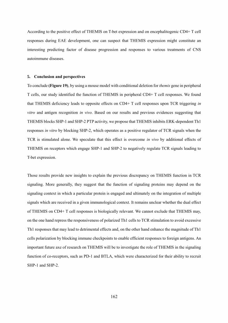
162
According to the positive effect of THEMIS on T-bet expression and on encephalitogenic CD4+ T cell
responses during EAE development, one can suspect that THEMIS expression might constitute an
interesting predicting factor of disease progression and responses to various treatments of CNS
autoimmune diseases.
5. Conclusion and perspectives
To conclude (Figure 19), by using a mouse model with conditional deletion for themis gene in peripheral
T cells, our study identified the function of THEMIS in peripheral CD4+ T cell responses. We found
that THEMIS deficiency leads to opposite effects on CD4+ T cell responses upon TCR triggering in
vitro and antigen recognition in vivo. Based on our results and previous evidences suggesting that
THEMIS blocks SHP-1 and SHP-2 PTP activity, we propose that THEMIS inhibits ERK-dependent Th1
responses in vitro by blocking SHP-2, which operates as a positive regulator of TCR signals when the
TCR is stimulated alone. We speculate that this effect is overcome in vivo by additional effects of
THEMIS on receptors which engage SHP-1 and SHP-2 to negatively regulate TCR signals leading to
T-bet expression.
Those results provide new insights to explain the previous discrepancy on THEMIS function in TCR
signaling. More generally, they suggest that the function of signaling proteins may depend on the
signaling context in which a particular protein is engaged and ultimately on the integration of multiple
signals which are received in a given immunological context. It remains unclear whether the dual effect
of THEMIS on CD4+ T cell responses is biologically relevant. We cannot exclude that THEMIS may,
on the one hand repress the responsiveness of polarized Th1 cells to TCR stimulation to avoid excessive
Th1 responses that may lead to detrimental effects and, on the other hand enhance the magnitude of Th1
cells polarization by blocking immune checkpoints to enable efficient responses to foreign antigens. An
important future axe of research on THEMIS will be to investigate the role of THEMIS in the signaling
function of co-receptors, such as PD-1 and BTLA, which were characterized for their ability to recruit
SHP-1 and SHP-2.
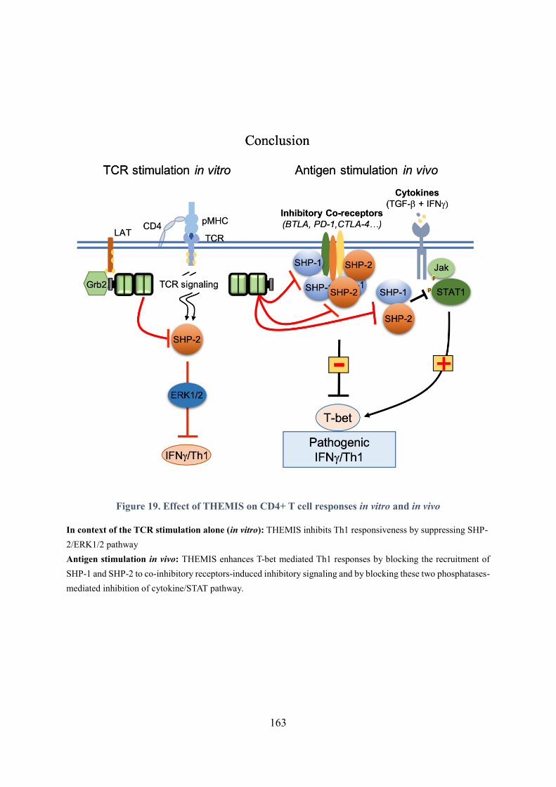
163
Figure 19. Effect of THEMIS on CD4+ T cell responses in vitro and in vivo
In context of the TCR stimulation alone (in vitro): THEMIS inhibits Th1 responsiveness by suppressing SHP-
2/ERK1/2 pathway
Antigen stimulation in vivo: THEMIS enhances T-bet mediated Th1 responses by blocking the recruitment of
SHP-1 and SHP-2 to co-inhibitory receptors-induced inhibitory signaling and by blocking these two phosphatases-
mediated inhibition of cytokine/STAT pathway.
164
Our in vivo analysis highlights a specific effect of THEMIS on the production of T-bet and IFN by
CD4+ T cells. We speculated from this observation that THEMIS modulates Th1 responses. However,
both Th1-like Th17 cells and type I Tfh cells co-express T-bet and IFN (Cunill et al., 2017; Kamali et
al., 2019; Ma & Phan, 2017; Unger, 2018), suggesting that THEMIS may exert a specific effect on T-
bet and IFN but not a broad effect on the development of Th1 cells. Therefore, a more extensive RNA-
Seq characterization of 2D2 T cells in vivo would be interesting to investigate further the effect of
THEMIS on the signature of those effector cells in the context of EAE.
Given the important role of Th1 cells in the context of cancer and given the key role of immune
checkpoints blocking therapies, an interesting perspective would be to explore the role of THEMIS in a
cancer model which relies on Th1 cells, such as bladder cancer and melanoma and to address whether
THEMIS expression affects the susceptibility to immune checkpoint blockade therapy.

165
Portfolio
166
Scientific publications during this thesis
THEMIS enhances the magnitude of normal and neuroinflammatory Th1 responses by promoting TCR-
independent signals
Cui Yang, Gaêtan Blaize*, Rémi Marrocco*, Nelly Rouquié*, Cyrielle Bories, Mylène Gador, Suzanne
Mélique, Emeline Joulia, Mehdi Benamar, Anne S. Dejean, Hélène Daniels-Treffandier, Paul E. Love,
Nicolas Fazilleau, Abdelhadi Saoudi, Renaud Lesourne.
Under review
CD5 signalosome coordinates antagonist TCR signals to control the generation of Treg cells induced
by foreign antigens
Blaize G., Daniels-Treffandier H., Aloulou M., Rouquié N., Yang C., Marcellin M., Gador M., Benamar
M., Ducatez M., Song K., Burlet-Schiltz O., Saoudi A., E. Love P, Fazilleau N., Gonzalez de Peredo A.,
& Lesourne R.
PNAS, 2020 June 9
(Annexes)
Awards and honors
2021 – EFIS-EJI Abstract Grant at the 15th World Immune Regulation Meeting
Workshop presentation
2020 – 2021 Fondation ARC pour la recherche sur le cancer for the 4th year of PhD student
2017 – 2020 China scholarship council for PhD student
Experimental skills acquired
- Flow cytometry analysis (LSR-II and LSR-FORTESSA): Extracellular and intracellular staining
for the analysis of T cell phenotype, proliferation, and differentiation.
- Animal experiment: Induction of EAE in mice, transfer of mice with T cells, and isolation of
leukocytes from the central nervous system of mice.
- Cell culture of mouse primary lymphocytes (LT) and stimulation of LT with antibodies and with
peptides in vitro.
- Protein biochemistry: Analysis of phosphorylated proteins, immunoprecipitation and Western blot.
Courses and Training attended
- Fluorescence microscopy - Level 1
- Transmission electron microscopy for in situ localizations. Obtaining and interpreting images
- French speaking and writing - Level A2
- Speak English Professionally: In Person, Online & On the Phone

167
References
168
References
Agarwal, S., Avni, O., & Rao, A. (2000). Cell-type-restricted binding of the transcription factor
NFAT to a distal IL-4 enhancer in vivo. Immunity, 12(6), 643-652.
Agazie, Y. M., & Hayman, M. J. (2003). Molecular mechanism for a role of SHP2 in epidermal
growth factor receptor signaling. Mol Cell Biol, 23(21), 7875-7886.
https://doi.org/10.1128/MCB.23.21.7875-7886.2003
Agrawal, A., Dillon, S., Denning, T. L., & Pulendran, B. (2006). ERK1-/- mice exhibit Th1 cell
polarization and increased susceptibility to experimental autoimmune
encephalomyelitis. J Immunol, 176(10), 5788-5796.
https://doi.org/10.4049/jimmunol.176.10.5788
Alessi, D. R., Andjelkovic, M., Caudwell, B., Cron, P., Morrice, N., Cohen, P., & Hemmings, B.
A. (1996). Mechanism of activation of protein kinase B by insulin and IGF-1. EMBO J,
15(23), 6541-6551.
Amsen, D., Antov, A., & Flavell, R. A. (2009). The different faces of Notch in T-helper-cell
differentiation. Nature Reviews Immunology, 9(2), 116-124.
Amsen, D., Spilianakis, C. G., & Flavell, R. A. (2009). How are TH1 and TH2 effector cells
made? Current opinion in immunology, 21(2), 153-160.
Anderson, M. S., Venanzi, E. S., Klein, L., Chen, Z., Berzins, S. P., Turley, S. J., . . . Mathis, D.
(2002). Projection of an immunological self shadow within the thymus by the aire
protein. Science, 298(5597), 1395-1401. https://doi.org/10.1126/science.1075958
Andreotti, A. H., Schwartzberg, P. L., Joseph, R. E., & Berg, L. J. (2010). T-cell signaling
regulated by the Tec family kinase, Itk. Cold Spring Harb Perspect Biol, 2(7), a002287.
https://doi.org/10.1101/cshperspect.a002287
Angel, P., & Karin, M. (1991). The role of Jun, Fos and the AP-1 complex in cell-proliferation
and transformation. Biochimica et Biophysica Acta (BBA)-Reviews on Cancer, 1072(2-
3), 129-157.
Ansell, S. M., Lesokhin, A. M., Borrello, I., Halwani, A., Scott, E. C., Gutierrez, M., . . . Freeman,
G. J. (2015). PD-1 blockade with nivolumab in relapsed or refractory Hodgkin's
lymphoma. New England Journal of Medicine, 372(4), 311-319.
Apostolou, I., Sarukhan, A., Klein, L., & von Boehmer, H. (2002). Origin of regulatory T cells
with known specificity for antigen. Nat Immunol, 3(8), 756-763.
https://doi.org/10.1038/ni816
Appay, V., Zaunders, J. J., Papagno, L., Sutton, J., Jaramillo, A., Waters, A., . . . Kelleher, A.
D. (2002). Characterization of CD4(+) CTLs ex vivo. J Immunol, 168(11), 5954-5958.
https://doi.org/10.4049/jimmunol.168.11.5954
Arase, H., Arase, N., Ogasawara, K., Good, R. A., & Onoé, K. (1992). An NK1.1+ CD4+8-
single-positive thymocyte subpopulation that expresses a highly skewed T-cell antigen
receptor V beta family. Proc Natl Acad Sci U S A, 89(14), 6506-6510.
https://doi.org/10.1073/pnas.89.14.6506
Arnaud, M., Mzali, R., Gesbert, F., Crouin, C., Guenzi, C., Vermot-Desroches, C., . . . Bertoglio,
J. (2004). Interaction of the tyrosine phosphatase SHP-2 with Gab2 regulates Rho-
dependent activation of the c-fos serum response element by interleukin-2.
Biochemical Journal, 382(2), 545-556.
169
Arthur, J. S. C., & Ley, S. C. (2013). Mitogen-activated protein kinases in innate immunity.
Nature Reviews Immunology, 13(9), 679-692.
Atarashi, K., Tanoue, T., Oshima, K., Suda, W., Nagano, Y., Nishikawa, H., . . . Honda, K.
(2013). Treg induction by a rationally selected mixture of Clostridia strains from the
human microbiota. Nature, 500(7461), 232-236. https://doi.org/10.1038/nature12331
Attanasio, J., & Wherry, E. J. (2016). Costimulatory and Coinhibitory Receptor Pathways in
Infectious Disease. Immunity, 44(5), 1052-1068.
https://doi.org/10.1016/j.immuni.2016.04.022
Au-Yeung, B. B., Levin, S. E., Zhang, C., Hsu, L. Y., Cheng, D. A., Killeen, N., . . . Weiss, A.
(2010). A genetically selective inhibitor demonstrates a function for the kinase Zap70
in regulatory T cells independent of its catalytic activity. Nat Immunol, 11(12), 1085-
1092. https://doi.org/10.1038/ni.1955
Au-Yeung, B. B., Smith, G. A., Mueller, J. L., Heyn, C. S., Jaszczak, R. G., Weiss, A., &
Zikherman, J. (2017). IL-2 modulates the TCR signaling threshold for CD8 but not CD4
T cell proliferation on a single-cell level. The Journal of Immunology, 198(6), 2445-2456.
Axtell, R. C., Webb, M. S., Barnum, S. R., & Raman, C. (2004). Cutting edge: critical role for
CD5 in experimental autoimmune encephalomyelitis: inhibition of engagement
reverses disease in mice. J Immunol, 173(5), 2928-2932.
https://doi.org/10.4049/jimmunol.173.5.2928
Azzam, H. S., Grinberg, A., Lui, K., Shen, H., Shores, E. W., & Love, P. E. (1998). CD5
expression is developmentally regulated by T cell receptor (TCR) signals and TCR
avidity. The Journal of experimental medicine, 188(12), 2301-2311.
Bagnoud, M., Briner, M., Remlinger, J., Meli, I., Schuetz, S., Pistor, M., . . . Hoepner, R. (2020).
c-Jun N-Terminal Kinase as a Therapeutic Target in Experimental Autoimmune
Encephalomyelitis. Cells, 9(10). https://doi.org/10.3390/cells9102154
Bai, L., Deng, S., Reboulet, R., Mathew, R., Teyton, L., Savage, P. B., & Bendelac, A. (2013).
Natural killer T (NKT)–B-cell interactions promote prolonged antibody responses and
long-term memory to pneumococcal capsular polysaccharides. Proceedings of the
National Academy of Sciences, 110(40), 16097-16102.
Bakshi, R., Cox, M., & Zajac, A. (2014). Cytotoxic T lymphocytes. Encyclopedia of Medical
Immunology: Springer: New York, 332-332.
Balashov, K. E., Rottman, J. B., Weiner, H. L., & Hancock, W. W. (1999). CCR5(+) and
CXCR3(+) T cells are increased in multiple sclerosis and their ligands MIP-1alpha and
IP-10 are expressed in demyelinating brain lesions. Proc Natl Acad Sci U S A, 96(12),
6873-6878. https://doi.org/10.1073/pnas.96.12.6873
Barrat, F. J., Cua, D. J., Boonstra, A., Richards, D. F., Crain, C., Savelkoul, H. F., . . . O'Garra,
A. (2002). In vitro generation of interleukin 10–producing regulatory CD4+ T cells is
induced by immunosuppressive drugs and inhibited by T helper type 1 (Th1)–and Th2-
inducing cytokines. The Journal of experimental medicine, 195(5), 603-616.
Batoulis, H., Recks, M., Holland, F., Thomalla, F., Williams, R., & Kuerten, S. (2014). Blockade
of tumour necrosis factor‐α in experimental autoimmune encephalomyelitis reveals
differential effects on the antigen‐specific immune response and central nervous
system histopathology. Clinical & Experimental Immunology, 175(1), 41-48.
Battaglia, M., Gregori, S., Bacchetta, R., & Roncarolo, M. G. (2006). Tr1 cells: from discovery
170
to their clinical application. Semin Immunol, 18(2), 120-127.
https://doi.org/10.1016/j.smim.2006.01.007
Baughman, E. J., Mendoza, J. P., Ortega, S. B., Ayers, C. L., Greenberg, B. M., Frohman, E.
M., & Karandikar, N. J. (2011). Neuroantigen-specific CD8+ regulatory T-cell function
is deficient during acute exacerbation of multiple sclerosis. J Autoimmun, 36(2), 115-
124. https://doi.org/10.1016/j.jaut.2010.12.003
Bauquet, A. T., Jin, H., Paterson, A. M., Mitsdoerffer, M., Ho, I.-C., Sharpe, A. H., & Kuchroo,
V. K. (2009). The costimulatory molecule ICOS regulates the expression of c-Maf and
IL-21 in the development of follicular T helper cells and T H-17 cells. Nature
immunology, 10(2), 167.
Bazzazi, H., Aghaei, M., Memarian, A., Asgarian-Omran, H., Behnampour, N., & Yazdani, Y.
(2018). Th1-Th17 Ratio as a New Insight in Rheumatoid Arthritis Disease. Iran J Allergy
Asthma Immunol, 17(1), 68-77.
Beecham, A. H., Patsopoulos, N. A., Xifara, D. K., Davis, M. F., Kemppinen, A., Cotsapas,
C., . . . (IIBDGC), I. I. G. C. (2013). Analysis of immune-related loci identifies 48 new
susceptibility variants for multiple sclerosis. Nat Genet, 45(11), 1353-1360.
https://doi.org/10.1038/ng.2770
Bending, D., De la Peña, H., Veldhoen, M., Phillips, J. M., Uyttenhove, C., Stockinger, B., &
Cooke, A. (2009). Highly purified Th17 cells from BDC2.5NOD mice convert into Th1-
like cells in NOD/SCID recipient mice. J Clin Invest, 119(3), 565-572.
https://doi.org/10.1172/JCI37865
Bennett, S. R., Carbone, F. R., Karamalis, F., Flavell, R. A., Miller, J. F., & Heath, W. R. (1998).
Help for cytotoxic-T-cell responses is mediated by CD40 signalling. Nature, 393(6684),
478-480. https://doi.org/10.1038/30996
Berghout, J., Langlais, D., Radovanovic, I., Tam, M., MacMicking, J. D., Stevenson, M. M., &
Gros, P. (2013). Irf8-regulated genomic responses drive pathological inflammation
during cerebral malaria. PLoS Pathog, 9(7), e1003491.
https://doi.org/10.1371/journal.ppat.1003491
Berod, L., Friedrich, C., Nandan, A., Freitag, J., Hagemann, S., Harmrolfs, K., . . . Bähre, H.
(2014). De novo fatty acid synthesis controls the fate between regulatory T and T helper
17 cells. Nature medicine, 20(11), 1327-1333.
Bert, A. G., Johnson, B. V., Baxter, E. W., & Cockerill, P. N. (2007). A modular enhancer is
differentially regulated by GATA and NFAT elements that direct different tissue-specific
patterns of nucleosome positioning and inducible chromatin remodeling. Molecular and
cellular biology, 27(8), 2870-2885.
Bettelli, E., Carrier, Y., Gao, W., Korn, T., Strom, T. B., Oukka, M., . . . Kuchroo, V. K. (2006).
Reciprocal developmental pathways for the generation of pathogenic effector TH 17
and regulatory T cells. Nature, 441(7090), 235-238.
Bettelli, E., Sullivan, B., Szabo, S. J., Sobel, R. A., Glimcher, L. H., & Kuchroo, V. K. (2004).
Loss of T-bet, but not STAT1, prevents the development of experimental autoimmune
encephalomyelitis. The Journal of experimental medicine, 200(1), 79-87.
Bhandoola, A., Bosselut, R., Yu, Q., Cowan, M. L., Feigenbaum, L., Love, P. E., & Singer, A.
(2002). CD5‐mediated inhibition of TCR signaling during intrathymic selection and
development does not require the CD5 extracellular domain. European journal of
171
immunology, 32(6), 1811-1817.
Birkner, K., Wasser, B., Loos, J., Plotnikov, A., Seger, R., Zipp, F., . . . Bittner, S. (2017). The
Role of ERK Signaling in Experimental Autoimmune Encephalomyelitis. Int J Mol Sci,
18(9). https://doi.org/10.3390/ijms18091990
Blaize, G., Daniels-Treffandier, H., Aloulou, M., Rouquié, N., Yang, C., Marcellin, M., . . .
Lesourne, R. (2020). CD5 signalosome coordinates antagonist TCR signals to control
the generation of Treg cells induced by foreign antigens. Proc Natl Acad Sci U S A,
117(23), 12969-12979. https://doi.org/10.1073/pnas.1917182117
Blanco, P., Viallard, J.-F., Pellegrin, J.-L., & Moreau, J.-F. (2005). Cytotoxic T lymphocytes and
autoimmunity. Current opinion in rheumatology, 17(6), 731-734.
Bluestone, J. A., Thomson, A. W., Shevach, E. M., & Weiner, H. L. (2007). What does the future
hold for cell-based tolerogenic therapy? Nature Reviews Immunology, 7(8), 650-654.
Bode, A. M., & Dong, Z. (2007). The functional contrariety of JNK. Mol Carcinog, 46(8), 591-
598. https://doi.org/10.1002/mc.20348
Bondar, C., Plaza-Izurieta, L., Fernandez-Jimenez, N., Irastorza, I., Withoff, S., Wijmenga,
C., . . . Bilbao, J. R. (2014). THEMIS and PTPRK in celiac intestinal mucosa:
coexpression in disease and after in vitro gliadin challenge. European Journal of
Human Genetics, 22(3), 358-362.
Bonneville, M., O'Brien, R. L., & Born, W. K. (2010). Gammadelta T cell effector functions: a
blend of innate programming and acquired plasticity. Nat Rev Immunol, 10(7), 467-478.
https://doi.org/10.1038/nri2781
Boomer, J. S., & Green, J. M. (2010). An enigmatic tail of CD28 signaling. Cold Spring Harb
Perspect Biol, 2(8), a002436. https://doi.org/10.1101/cshperspect.a002436
Brereton, C. F., Sutton, C. E., Lalor, S. J., Lavelle, E. C., & Mills, K. H. (2009). Inhibition of ERK
MAPK suppresses IL-23- and IL-1-driven IL-17 production and attenuates autoimmune
disease. J Immunol, 183(3), 1715-1723. https://doi.org/10.4049/jimmunol.0803851
Bricker, T. L., Green, J. M., Boomer, J. S., Deppong, C. M., & Shah, D. D. (2014). Cutting Edge:
A Double-Mutant Knockin of. Journal of Immunology, 192, 3465-3469.
Brimnes, J., Allez, M., Dotan, I., Shao, L., Nakazawa, A., & Mayer, L. (2005). Defects in CD8+
regulatory T cells in the lamina propria of patients with inflammatory bowel disease. J
Immunol, 174(9), 5814-5822. https://doi.org/10.4049/jimmunol.174.9.5814
Brockmeyer, C., Paster, W., Pepper, D., Tan, C. P., Trudgian, D. C., McGowan, S., . . . Salek,
M. (2011). T cell receptor (TCR)-induced tyrosine phosphorylation dynamics identifies
THEMIS as a new TCR signalosome component. Journal of Biological Chemistry,
286(9), 7535-7547.
Bromley, S. K., Thomas, S. Y., & Luster, A. D. (2005). Chemokine receptor CCR7 guides T cell
exit from peripheral tissues and entry into afferent lymphatics. Nat Immunol, 6(9), 895-
901. https://doi.org/10.1038/ni1240
Bruchard, M., Rebé, C., Derangère, V., Togbé, D., Ryffel, B., Boidot, R., . . . Berger, H. (2015).
The receptor NLRP3 is a transcriptional regulator of TH 2 differentiation. Nature
immunology, 16(8), 859-870.
Brucklacher-Waldert, V., Steinbach, K., Lioznov, M., Kolster, M., Hölscher, C., & Tolosa, E.
(2009). Phenotypical characterization of human Th17 cells unambiguously identified by
surface IL-17A expression. The Journal of Immunology, 183(9), 5494-5501.
172
Brunet, J.-F., Denizot, F., Luciani, M.-F., Roux-Dosseto, M., Suzan, M., Mattei, M.-G., &
Golstein, P. (1987). A new member of the immunoglobulin superfamily—CTLA-4.
Nature, 328(6127), 267-270.
Brunkow, M. E., Jeffery, E. W., Hjerrild, K. A., Paeper, B., Clark, L. B., Yasayko, S. A., . . .
Ramsdell, F. (2001). Disruption of a new forkhead/winged-helix protein, scurfin, results
in the fatal lymphoproliferative disorder of the scurfy mouse. Nat Genet, 27(1), 68-73.
https://doi.org/10.1038/83784
Brzostek, J., Gautam, N., Zhao, X., Chen, E. W., Mehta, M., Tung, D. W. H., . . . Gascoigne,
N. R. J. (2020). T cell receptor and cytokine signal integration in CD8. Nat Immunol,
21(2), 186-198. https://doi.org/10.1038/s41590-019-0570-3
Buttrick, T. S., Wang, W., Yung, C., Trieu, K. G., Patel, K., Khoury, S. J., . . . Elyaman, W. (2018).
Foxo1 promotes Th9 cell differentiation and airway allergy. Scientific reports, 8(1), 1-
10.
Cabaniols, J.-P., Fazilleau, N., Casrouge, A., Kourilsky, P., & Kanellopoulos, J. M. (2001). Most
α/β T cell receptor diversity is due to terminal deoxynucleotidyl transferase. The Journal
of experimental medicine, 194(9), 1385-1390.
Campos-Mora, M., Contreras-Kallens, P., Gálvez-Jirón, F., Rojas, M., Rojas, C., Refisch, A., . . .
Pino-Lagos, K. (2019). CD4+ Foxp3+ T regulatory cells promote transplantation
tolerance by modulating effector CD4+ T cells in a neuropilin-1-dependent manner.
Frontiers in immunology, 10, 882.
Cantor, H., & Shinohara, M. L. (2009). Regulation of T-helper-cell lineage development by
osteopontin: the inside story. Nature Reviews Immunology, 9(2), 137-141.
Carrette, F., & Surh, C. D. (2012). IL-7 signaling and CD127 receptor regulation in the control
of T cell homeostasis. Seminars in immunology,
Carter, J. D., Neel, B. G., & Lorenz, U. (1999). The tyrosine phosphatase SHP-1 influences
thymocyte selection by setting TCR signaling thresholds. International Immunology,
11(12), 1999-2014.
Carter, L., Fouser, L. A., Jussif, J., Fitz, L., Deng, B., Wood, C. R., . . . Carreno, B. M. (2002).
PD-1:PD-L inhibitory pathway affects both CD4(+) and CD8(+) T cells and is overcome
by IL-2. Eur J Immunol, 32(3), 634-643. https://doi.org/10.1002/1521-
4141(200203)32:3<634::AID-IMMU634>3.0.CO;2-9
Carvalheiro, H., da Silva, J. A., & Souto-Carneiro, M. M. (2013). Potential roles for CD8(+) T
cells in rheumatoid arthritis. Autoimmun Rev, 12(3), 401-409.
https://doi.org/10.1016/j.autrev.2012.07.011
Castellino, F., & Germain, R. N. (2006). Cooperation between CD4+ and CD8+ T cells: when,
where, and how. Annu. Rev. Immunol., 24, 519-540.
Castro-Sanchez, P., Teagle, A. R., Prade, S., & Zamoyska, R. (2020). Modulation of TCR
Signaling by Tyrosine Phosphatases: From Autoimmunity to Immunotherapy. Front Cell
Dev Biol, 8, 608747. https://doi.org/10.3389/fcell.2020.608747
Cauli, A., Piga, M., Floris, A., & Mathieu, A. (2015). Current perspective on the role of the
interleukin-23/interleukin-17 axis in inflammation and disease (chronic arthritis and
psoriasis). ImmunoTargets and therapy, 4, 185.
Celis-Gutierrez, J., Blattmann, P., Zhai, Y., Jarmuzynski, N., Ruminski, K., Grégoire, C., . . .
Malissen, B. (2019). Quantitative Interactomics in Primary T Cells Provides a Rationale
173
for Concomitant PD-1 and BTLA Coinhibitor Blockade in Cancer Immunotherapy. Cell
Rep, 27(11), 3315-3330.e3317. https://doi.org/10.1016/j.celrep.2019.05.041
Chabod, M., Pedros, C., Lamouroux, L., Colacios, C., Bernard, I., Lagrange, D., . . . Dejean,
A. S. (2012). A spontaneous mutation of the rat Themis gene leads to impaired function
of regulatory T cells linked to inflammatory bowel disease. PLoS Genet, 8(1), e1002461.
https://doi.org/10.1371/journal.pgen.1002461
Chan, A. C., Dalton, M., Johnson, R., Kong, G. H., Wang, T., Thoma, R., & Kurosaki, T. (1995).
Activation of ZAP-70 kinase activity by phosphorylation of tyrosine 493 is required for
lymphocyte antigen receptor function. EMBO J, 14(11), 2499-2508.
Chang, C.-F., D’Souza, W. N., Ch’en, I. L., Pages, G., Pouyssegur, J., & Hedrick, S. M. (2012).
Polar opposites: Erk direction of CD4 T cell subsets. The Journal of Immunology,
189(2), 721-731.
Chang, H.-C., Zhang, S., Thieu, V. T., Slee, R. B., Bruns, H. A., Laribee, R. N., . . . Kaplan, M.
H. (2005). PU. 1 expression delineates heterogeneity in primary Th2 cells. Immunity,
22(6), 693-703.
Chemnitz, J. M., Lanfranco, A. R., Braunstein, I., & Riley, J. L. (2006). B and T lymphocyte
attenuator-mediated signal transduction provides a potent inhibitory signal to primary
human CD4 T cells that can be initiated by multiple phosphotyrosine motifs. The
Journal of Immunology, 176(11), 6603-6614.
Chemnitz, J. M., Parry, R. V., Nichols, K. E., June, C. H., & Riley, J. L. (2004). SHP-1 and SHP-
2 associate with immunoreceptor tyrosine-based switch motif of programmed death 1
upon primary human T cell stimulation, but only receptor ligation prevents T cell
activation. J Immunol, 173(2), 945-954. https://doi.org/10.4049/jimmunol.173.2.945
Chen, C., Cao, M., Zhu, S., Wang, C., Liang, F., Yan, L., & Luo, D. (2015). Discovery of a Novel
Inhibitor of the Protein Tyrosine Phosphatase Shp2. Sci Rep, 5, 17626.
https://doi.org/10.1038/srep17626
Chen, H., Fu, T., Suh, W. K., Tsavachidou, D., Wen, S., Gao, J., . . . Sharma, P. (2014). CD4 T
cells require ICOS-mediated PI3K signaling to increase T-Bet expression in the setting
of anti-CTLA-4 therapy. Cancer Immunol Res, 2(2), 167-176.
https://doi.org/10.1158/2326-6066.CIR-13-0155
Chen, L., & Flies, D. B. (2013). Molecular mechanisms of T cell co-stimulation and co-inhibition.
Nature Reviews Immunology, 13(4), 227-242.
Chen, L., & Flies, D. B. (2013). Molecular mechanisms of T cell co-stimulation and co-inhibition.
Nat Rev Immunol, 13(4), 227-242. https://doi.org/10.1038/nri3405
Chen, Y., Kuchroo, V. K., Inobe, J., Hafler, D. A., & Weiner, H. L. (1994). Regulatory T cell
clones induced by oral tolerance: suppression of autoimmune encephalomyelitis.
Science, 265(5176), 1237-1240. https://doi.org/10.1126/science.7520605
Cheng, D., Deobagkar-Lele, M., Zvezdova, E., Choi, S., Uehara, S., Baup, D., . . . Ferry, H.
(2017). Themis2 lowers the threshold for B cell activation during positive selection.
Nature immunology, 18(2), 205-213.
Cher, D. J., & Mosmann, T. R. (1987). Two types of murine helper T cell clone. II. Delayed-type
hypersensitivity is mediated by TH1 clones. J Immunol, 138(11), 3688-3694.
Chi, H. (2012). Regulation and function of mTOR signalling in T cell fate decisions. Nat Rev
Immunol, 12(5), 325-338. https://doi.org/10.1038/nri3198
174
Chihara, N., Madi, A., Karwacz, K., Awasthi, A., & Kuchroo, V. K. (2016). Differentiation and
Characterization of Tr1 Cells. Curr Protoc Immunol, 113, 3.27.21-23.27.10.
https://doi.org/10.1002/0471142735.im0327s113
Chitnis, T., Najafian, N., Abdallah, K. A., Dong, V., Yagita, H., Sayegh, M. H., & Khoury, S. J.
(2001). CD28-independent induction of experimental autoimmune encephalomyelitis.
J Clin Invest, 107(5), 575-583. https://doi.org/10.1172/JCI11220
Chitnis, T., Najafian, N., Benou, C., Salama, A. D., Grusby, M. J., Sayegh, M. H., & Khoury, S.
J. (2001). Effect of targeted disruption of STAT4 and STAT6 on the induction of
experimental autoimmune encephalomyelitis. J Clin Invest, 108(5), 739-747.
https://doi.org/10.1172/JCI12563
Choi, S., Cornall, R., Lesourne, R., & Love, P. E. (2017). THEMIS: two models, different
thresholds. Trends in immunology, 38(9), 622-632.
Choi, S., Warzecha, C., Zvezdova, E., Lee, J., Argenty, J., Lesourne, R., . . . Love, P. E. (2017).
THEMIS enhances TCR signaling and enables positive selection by selective inhibition
of the phosphatase SHP-1. Nature immunology, 18(4), 433-441.
Christophi, G. P., Hudson, C. A., Gruber, R. C., Christophi, C. P., Mihai, C., Mejico, L. J., . . .
Massa, P. T. (2008). SHP-1 deficiency and increased inflammatory gene expression in
PBMCs of multiple sclerosis patients. Lab Invest, 88(3), 243-255.
https://doi.org/10.1038/labinvest.3700720
Chuang, E., Fisher, T. S., Morgan, R. W., Robbins, M. D., Duerr, J. M., Vander Heiden, M.
G., . . . Thompson, C. B. (2000). The CD28 and CTLA-4 receptors associate with the
serine/threonine phosphatase PP2A. Immunity, 13(3), 313-322.
https://doi.org/10.1016/s1074-7613(00)00031-5
Chuck, M. I., Zhu, M., Shen, S., & Zhang, W. (2010). The role of the LAT–PLC-γ1 interaction
in T regulatory cell function. The Journal of Immunology, 184(5), 2476-2486.
Ciofani, M., Schmitt, T. M., Ciofani, A., Michie, A. M., Cuburu, N., Aublin, A., . . . Zúñiga-Pflücker,
J. C. (2004). Obligatory role for cooperative signaling by pre-TCR and Notch during
thymocyte differentiation. J Immunol, 172(9), 5230-5239.
https://doi.org/10.4049/jimmunol.172.9.5230
Codarri, L., Gyülvészi, G., Tosevski, V., Hesske, L., Fontana, A., Magnenat, L., . . . Becher, B.
(2011). RORγt drives production of the cytokine GM-CSF in helper T cells, which is
essential for the effector phase of autoimmune neuroinflammation. Nat Immunol, 12(6),
560-567. https://doi.org/10.1038/ni.2027
Colacios, C., Casemayou, A., Dejean, A. S., Gaits-Iacovoni, F., Pedros, C., Bernard, I., . . .
Saoudi, A. (2011). The p.Arg63Trp polymorphism controls Vav1 functions and Foxp3
regulatory T cell development. J Exp Med, 208(11), 2183-2191.
https://doi.org/10.1084/jem.20102191
Collin, R., Lombard-Vadnais, F., Hillhouse, E. E., Lebel, M.-È., Chabot-Roy, G., Melichar, H. J.,
& Lesage, S. (2020). MHC-independent thymic selection of CD4 and CD8 coreceptor
negative αβ T Cells. The Journal of Immunology, 205(1), 133-142.
Constant, S. L., & Bottomly, K. (1997). Induction of Th1 and Th2 CD4plus T cell responses:
The alternative approaches. Annual review of immunology, 15, 297.
Cooley, I. D., Read, K. A., & Oestreich, K. J. (2015). Trans-presentation of IL-15 modulates
STAT5 activation and Bcl-6 expression in TH1 cells. Sci Rep, 5, 15722.
175
https://doi.org/10.1038/srep15722
Coombes, J. L., Siddiqui, K. R., Arancibia-Cárcamo, C. V., Hall, J., Sun, C.-M., Belkaid, Y., &
Powrie, F. (2007). A functionally specialized population of mucosal CD103+ DCs
induces Foxp3+ regulatory T cells via a TGF-β–and retinoic acid–dependent
mechanism. Journal of Experimental Medicine, 204(8), 1757-1764.
Corn, R. A., Aronica, M. A., Zhang, F., Tong, Y., Stanley, S. A., Kim, S. R. A., . . . Mora, A. (2003).
T cell-intrinsic requirement for NF-κB induction in postdifferentiation IFN-γ production
and clonal expansion in a Th1 response. The Journal of Immunology, 171(4), 1816-
1824.
Criado, G., Risco, A., Alsina-Beauchamp, D., Pérez-Lorenzo, M. J., Escós, A., & Cuenda, A.
(2014). Alternative p38 MAPKs are essential for collagen-induced arthritis. Arthritis
Rheumatol, 66(5), 1208-1217. https://doi.org/10.1002/art.38327
Croft, M. (2010). Control of immunity by the TNFR-related molecule OX40 (CD134). Annu Rev
Immunol, 28, 57-78. https://doi.org/10.1146/annurev-immunol-030409-101243
Crotty, S. (2019). T follicular helper cell biology: a decade of discovery and diseases. Immunity,
50(5), 1132-1148.
Cruz, M. S., Diamond, A., Russell, A., & Jameson, J. M. (2018). Human αβ and γδ T cells in
skin immunity and disease. Frontiers in immunology, 9, 1304.
Cua, D. J., Sherlock, J., Chen, Y., Murphy, C. A., Joyce, B., Seymour, B., . . . Sedgwick, J. D.
(2003). Interleukin-23 rather than interleukin-12 is the critical cytokine for autoimmune
inflammation of the brain. Nature, 421(6924), 744-748.
https://doi.org/10.1038/nature01355
Cui, J., Zhang, M., Zhang, Y. Q., & Xu, Z. H. (2007). JNK pathway: diseases and therapeutic
potential. Acta Pharmacol Sin, 28(5), 601-608. https://doi.org/10.1111/j.1745-
7254.2007.00579.x
Cunill, V., Clemente, A., Lanio, N., Barceló, C., Andreu, V., Pons, J., & Ferrer, J. M. (2017).
Follicular T Cells from smB. Front Immunol, 8, 174.
https://doi.org/10.3389/fimmu.2017.00174
Curran, M. A., Montalvo, W., Yagita, H., & Allison, J. P. (2010). PD-1 and CTLA-4 combination
blockade expands infiltrating T cells and reduces regulatory T and myeloid cells within
B16 melanoma tumors. Proceedings of the National Academy of Sciences, 107(9),
4275-4280.
Currie, R. A., Walker, K. S., Gray, A., Deak, M., Casamayor, A., Downes, C. P., . . . Lucocq, J.
(1999). Role of phosphatidylinositol 3, 4, 5-trisphosphate in regulating the activity and
localization of 3-phosphoinositide-dependent protein kinase-1. Biochemical Journal,
337(3), 575-583.
D'Souza, W. N., Chang, C.-F., Fischer, A. M., Li, M., & Hedrick, S. M. (2008). The Erk2 MAPK
regulates CD8 T cell proliferation and survival. The Journal of Immunology, 181(11),
7617-7629.
Dardalhon, V., Awasthi, A., Kwon, H., Galileos, G., Gao, W., Sobel, R. A., . . . Ho, I.-C. (2008).
IL-4 inhibits TGF-β-induced Foxp3+ T cells and, together with TGF-β, generates IL-9+
IL-10+ Foxp3− effector T cells. Nature immunology, 9(12), 1347-1355.
Dasu, T., Qualls, J. E., Tuna, H., Raman, C., Cohen, D. A., & Bondada, S. (2008). CD5 plays
an inhibitory role in the suppressive function of murine CD4+ CD25+ Treg cells.
176
Immunology letters, 119(1-2), 103-113.
Davidson, T. S., DiPaolo, R. J., Andersson, J., & Shevach, E. M. (2007). Cutting edge: IL-2 is
essential for TGF-β-mediated induction of Foxp3+ T regulatory cells. The Journal of
Immunology, 178(7), 4022-4026.
Davies, A. A., Ley, S. C., & Crumpton, M. J. (1992). CD5 is phosphorylated on tyrosine after
stimulation of the T-cell antigen receptor complex. Proc Natl Acad Sci U S A, 89(14),
6368-6372. https://doi.org/10.1073/pnas.89.14.6368
Davies, J. L., Thompson, S., Kaur-Sandhu, H., Sawcer, S., Coles, A., Ban, M., & Jones, J.
(2016). Increased THEMIS first exon usage in CD4+ T-cells is associated with a
genotype that is protective against multiple sclerosis. PloS one, 11(7), e0158327.
De Simone, M., Rossetti, G., & Pagani, M. (2018). Single Cell T Cell Receptor Sequencing:
Techniques and Future Challenges. Front Immunol, 9, 1638.
https://doi.org/10.3389/fimmu.2018.01638
Debes, G. F., Arnold, C. N., Young, A. J., Krautwald, S., Lipp, M., Hay, J. B., & Butcher, E. C.
(2005). Chemokine receptor CCR7 required for T lymphocyte exit from peripheral
tissues. Nature immunology, 6(9), 889-894.
Delgoffe, G. M., Pollizzi, K. N., Waickman, A. T., Heikamp, E., Meyers, D. J., Horton, M. R., . . .
Powell, J. D. (2011). The kinase mTOR regulates the differentiation of helper T cells
through the selective activation of signaling by mTORC1 and mTORC2. Nat Immunol,
12(4), 295-303. https://doi.org/10.1038/ni.2005
Deng, C., Minguela, A., Hussain, R. Z., Lovett-Racke, A. E., Radu, C., Ward, E. S., & Racke,
M. K. (2002). Expression of the tyrosine phosphatase SRC homology 2 domain-
containing protein tyrosine phosphatase 1 determines T cell activation threshold and
severity of experimental autoimmune encephalomyelitis. J Immunol, 168(9), 4511-4518.
https://doi.org/10.4049/jimmunol.168.9.4511
Deng, Y., Wang, Z., Chang, C., Lu, L., Lau, C. S., & Lu, Q. (2017). Th9 cells and IL-9 in
autoimmune disorders: Pathogenesis and therapeutic potentials. Human immunology,
78(2), 120-128.
Dennehy, K. M., Broszeit, R., Ferris, W. F., & Beyers, A. D. (1998). Thymocyte activation
induces the association of the proto-oncoprotein c-cbl and ras GTPase-activating
protein with CD5. Eur J Immunol, 28(5), 1617-1625.
https://doi.org/10.1002/(SICI)1521-4141(199805)28:05<1617::AID-
IMMU1617>3.0.CO;2-7
Dennehy, K. M., Broszeit, R., Garnett, D., Durrheim, G. A., Spruyt, L. L., & Beyers, A. D. (1997).
Thymocyte activation induces the association of phosphatidylinositol 3-kinase and
pp120 with CD5. Eur J Immunol, 27(3), 679-686.
https://doi.org/10.1002/eji.1830270316
Dennehy, K. M., Ferris, W. F., Veenstra, H., Zuckerman, L. A., Killeen, N., & Beyers, A. D.
(2001). Determination of the tyrosine phosphorylation sites in the T cell transmembrane
glycoprotein CD5. International immunology, 13(2), 149-156.
Dhume, K., Finn, C., Singh, A., Tejero, J., Devarajan, P., Swain, S. L., & McKinstry, K. K. (2020).
The T-box transcription factors T-bet and Eomes repress protective Th17 CD4 T cell
responses against Influenza A Virus. In: Am Assoc Immnol.
Djuretic, I. M., Levanon, D., Negreanu, V., Groner, Y., Rao, A., & Ansel, K. M. (2007).
177
Transcription factors T-bet and Runx3 cooperate to activate Ifng and silence Il4 in T
helper type 1 cells. Nature immunology, 8(2), 145-153.
Domingues, H. S., Mues, M., Lassmann, H., Wekerle, H., & Krishnamoorthy, G. (2010).
Functional and pathogenic differences of Th1 and Th17 cells in experimental
autoimmune encephalomyelitis. PloS one, 5(11), e15531.
Dong, B., Gao, Y., Zheng, X., Gao, G., Gu, H., Chen, X., & Zhang, J. (2015). T cell activation
is reduced by the catalytically inactive form of protein tyrosine phosphatase SHP-2.
International journal of clinical and experimental medicine, 8(4), 6568.
Dong, C., Juedes, A. E., Temann, U.-A., Shresta, S., Allison, J. P., Ruddle, N. H., & Flavell, R.
A. (2001). ICOS co-stimulatory receptor is essential for T-cell activation and function.
Nature, 409(6816), 97-101.
Dong, C., & Nurieva, R. I. (2003). Regulation of immune and autoimmune responses by ICOS.
Journal of autoimmunity, 21(3), 255-260.
Dong, C., Yang, D. D., Tournier, C., Whitmarsh, A. J., Xu, J., Davis, R. J., & Flavell, R. A. (2000).
JNK is required for effector T-cell function but not for T-cell activation. Nature,
405(6782), 91-94.
Dotti, G., Gottschalk, S., Savoldo, B., & Brenner, M. K. (2014). Design and development of
therapies using chimeric antigen receptor-expressing T cells. Immunol Rev, 257(1),
107-126. https://doi.org/10.1111/imr.12131
Duguet, F., Locard-Paulet, M., Marcellin, M., Chaoui, K., Bernard, I., Andreoletti, O., . . . Saoudi,
A. (2017). Proteomic Analysis of Regulatory T Cells Reveals the Importance of
Themis1 in the Control of Their Suppressive Function. Mol Cell Proteomics, 16(8),
1416-1432. https://doi.org/10.1074/mcp.M116.062745
Ebinu, J. O., Bottorff, D. A., Chan, E. Y., Stang, S. L., Dunn, R. J., & Stone, J. C. (1998).
RasGRP, a Ras guanyl nucleotide- releasing protein with calcium- and diacylglycerol-
binding motifs. Science, 280(5366), 1082-1086.
https://doi.org/10.1126/science.280.5366.1082
Egawa, T., Eberl, G., Taniuchi, I., Benlagha, K., Geissmann, F., Hennighausen, L., . . . Littman,
D. R. (2005). Genetic evidence supporting selection of the Vα14i NKT cell lineage from
double-positive thymocyte precursors. Immunity, 22(6), 705-716.
Egawa, T., & Littman, D. R. (2008). ThPOK acts late in specification of the helper T cell lineage
and suppresses Runx-mediated commitment to the cytotoxic T cell lineage. Nat
Immunol, 9(10), 1131-1139. https://doi.org/10.1038/ni.1652
Egen, J. G., & Allison, J. P. (2002). Cytotoxic T lymphocyte antigen-4 accumulation in the
immunological synapse is regulated by TCR signal strength. Immunity, 16(1), 23-35.
https://doi.org/10.1016/s1074-7613(01)00259-x
El-Behi, M., Ciric, B., Dai, H., Yan, Y., Cullimore, M., Safavi, F., . . . Rostami, A. (2011). The
encephalitogenicity of TH 17 cells is dependent on IL-1-and IL-23-induced production
of the cytokine GM-CSF. Nature immunology, 12(6), 568-575.
Espinosa Eric, C. P. (2006). Immunologie. In.
Eyerich, S., Eyerich, K., Pennino, D., Carbone, T., Nasorri, F., Pallotta, S., . . . Behrendt, H.
(2009). Th22 cells represent a distinct human T cell subset involved in epidermal
immunity and remodeling. The Journal of clinical investigation, 119(12), 3573-3585.
Fallon, P. G., Ballantyne, S. J., Mangan, N. E., Barlow, J. L., Dasvarma, A., Hewett, D. R., . . .
178
McKenzie, A. N. (2006). Identification of an interleukin (IL)-25–dependent cell
population that provides IL-4, IL-5, and IL-13 at the onset of helminth expulsion. The
Journal of experimental medicine, 203(4), 1105-1116.
Fan, X., Quezada, S. A., Sepulveda, M. A., Sharma, P., & Allison, J. P. (2014). Engagement of
the ICOS pathway markedly enhances efficacy of CTLA-4 blockade in cancer
immunotherapy. J Exp Med, 211(4), 715-725. https://doi.org/10.1084/jem.20130590
Fasth, A. E., Björkström, N. K., Anthoni, M., Malmberg, K. J., & Malmström, V. (2010).
Activating NK-cell receptors co-stimulate CD4(+)CD28(-) T cells in patients with
rheumatoid arthritis. Eur J Immunol, 40(2), 378-387.
https://doi.org/10.1002/eji.200939399
Fawcett, V. C., & Lorenz, U. (2005). Localization of Src homology 2 domain-containing
phosphatase 1 (SHP-1) to lipid rafts in T lymphocytes: functional implications and a
role for the SHP-1 carboxyl terminus. The Journal of Immunology, 174(5), 2849-2859.
Ferber, I. A., Brocke, S., Taylor-Edwards, C., Ridgway, W., Dinisco, C., Steinman, L., . . .
Fathman, C. G. (1996). Mice with a disrupted IFN-gamma gene are susceptible to the
induction of experimental autoimmune encephalomyelitis (EAE). The journal of
immunology, 156(1), 5-7.
Ferrandi, C., Richard, F., Tavano, P., Hauben, E., Barbié, V., Gotteland, J. P., . . . Zaratin, P. F.
(2011). Characterization of immune cell subsets during the active phase of multiple
sclerosis reveals disease and c-Jun N-terminal kinase pathway biomarkers. Mult Scler,
17(1), 43-56. https://doi.org/10.1177/1352458510381258
Fiorentino, D. F., Bond, M. W., & Mosmann, T. (1989). Two types of mouse T helper cell. IV.
Th2 clones secrete a factor that inhibits cytokine production by Th1 clones. Journal of
Experimental Medicine, 170(6), 2081-2095.
Fletcher, J. M., Lalor, S., Sweeney, C., Tubridy, N., & Mills, K. (2010). T cells in multiple
sclerosis and experimental autoimmune encephalomyelitis. Clinical & Experimental
Immunology, 162(1), 1-11.
Fowell, D. J., Shinkai, K., Liao, X. C., Beebe, A. M., Coffman, R. L., Littman, D. R., & Locksley,
R. M. (1999). Impaired NFATc translocation and failure of Th2 development in Itk-
deficient CD4+ T cells. Immunity, 11(4), 399-409. https://doi.org/10.1016/s1074-
7613(00)80115-6
Francisco, L. M., Salinas, V. H., Brown, K. E., Vanguri, V. K., Freeman, G. J., Kuchroo, V. K.,
& Sharpe, A. H. (2009). PD-L1 regulates the development, maintenance, and function
of induced regulatory T cells. J Exp Med, 206(13), 3015-3029.
https://doi.org/10.1084/jem.20090847
Franke, T. F., Yang, S. I., Chan, T. O., Datta, K., Kazlauskas, A., Morrison, D. K., . . . Tsichlis,
P. N. (1995). The protein kinase encoded by the Akt proto-oncogene is a target of the
PDGF-activated phosphatidylinositol 3-kinase. Cell, 81(5), 727-736.
https://doi.org/10.1016/0092-8674(95)90534-0
Frearson, J. A., & Alexander, D. R. (1998). The phosphotyrosine phosphatase SHP-2
participates in a multimeric signaling complex and regulates T cell receptor (TCR)
coupling to the Ras/mitogen-activated protein kinase (MAPK) pathway in Jurkat T cells.
The Journal of experimental medicine, 187(9), 1417-1426.
Freeman, G. J., Long, A. J., Iwai, Y., Bourque, K., Chernova, T., Nishimura, H., . . . Honjo, T.
179
(2000). Engagement of the PD-1 immunoinhibitory receptor by a novel B7 family
member leads to negative regulation of lymphocyte activation. J Exp Med, 192(7),
1027-1034. https://doi.org/10.1084/jem.192.7.1027
Friese, M. A., & Fugger, L. (2009). Pathogenic CD8+ T cells in multiple sclerosis. Annals of
Neurology: Official Journal of the American Neurological Association and the Child
Neurology Society, 66(2), 132-141.
Frisullo, G., Angelucci, F., Caggiula, M., Nociti, V., Iorio, R., Patanella, A. K., . . . Batocchi, A.
P. (2006). pSTAT1, pSTAT3, and T-bet expression in peripheral blood mononuclear
cells from relapsing-remitting multiple sclerosis patients correlates with disease activity.
J Neurosci Res, 84(5), 1027-1036. https://doi.org/10.1002/jnr.20995
Fu, G., Casas, J., Rigaud, S., Rybakin, V., Lambolez, F., Brzostek, J., . . . Cheroutre, H. (2013).
Themis sets the signal threshold for positive and negative selection in T-cell
development. Nature, 504(7480), 441-445.
Fu, G., Vallée, S., Rybakin, V., McGuire, M. V., Ampudia, J., Brockmeyer, C., . . . Munshi, A.
(2009). Themis controls thymocyte selection through regulation of T cell antigen
receptor–mediated signaling. Nature immunology, 10(8), 848.
Fu, T., He, Q., & Sharma, P. (2011). The ICOS/ICOSL pathway is required for optimal antitumor
responses mediated by anti-CTLA-4 therapy. Cancer Res, 71(16), 5445-5454.
https://doi.org/10.1158/0008-5472.CAN-11-1138
Fukaura, H., Kent, S. C., Pietrusewicz, M. J., Khoury, S. J., Weiner, H. L., & Hafler, D. A. (1996).
Induction of circulating myelin basic protein and proteolipid protein-specific
transforming growth factor-beta1-secreting Th3 T cells by oral administration of myelin
in multiple sclerosis patients. The Journal of clinical investigation, 98(1), 70-77.
Furlan, R., Brambilla, E., Ruffini, F., Poliani, P. L., Bergami, A., Marconi, P. C., . . . Martino, G.
(2001). Intrathecal delivery of IFN-gamma protects C57BL/6 mice from chronic-
progressive experimental autoimmune encephalomyelitis by increasing apoptosis of
central nervous system-infiltrating lymphocytes. J Immunol, 167(3), 1821-1829.
https://doi.org/10.4049/jimmunol.167.3.1821
Galicia, G., Kasran, A., Uyttenhove, C., De Swert, K., Van Snick, J., & Ceuppens, J. L. (2009).
ICOS deficiency results in exacerbated IL-17 mediated experimental autoimmune
encephalomyelitis. J Clin Immunol, 29(4), 426-433. https://doi.org/10.1007/s10875-
009-9287-7
Gao, J. X., Zhang, H., Bai, X. F., Wen, J., Zheng, X., Liu, J., . . . Liu, Y. (2002). Perinatal
blockade of b7-1 and b7-2 inhibits clonal deletion of highly pathogenic autoreactive T
cells. J Exp Med, 195(8), 959-971. https://doi.org/10.1084/jem.20011948
Garreau, A., Blaize, G., Argenty, J., Rouquié, N., Tourdès, A., Wood, S. A., . . . Lesourne, R.
(2017). Grb2-mediated recruitment of USP9X to LAT enhances themis stability
following thymic selection. The Journal of Immunology, 199(8), 2758-2766.
Gascoigne, N. R., & Acuto, O. (2015). THEMIS: a critical TCR signal regulator for ligand
discrimination. Curr Opin Immunol, 33, 86-92. https://doi.org/10.1016/j.coi.2015.01.020
Gascoigne, N. R., & Palmer, E. (2011). Signaling in thymic selection. Current opinion in
immunology, 23(2), 207-212.
Gascoigne, N. R., Rybakin, V., Acuto, O., & Brzostek, J. (2016). TCR signal strength and T cell
development. Annual review of cell and developmental biology, 32, 327-348.
180
Gaud, G., Lesourne, R., & Love, P. E. (2018). Regulatory mechanisms in T cell receptor
signalling. Nat Rev Immunol, 18(8), 485-497. https://doi.org/10.1038/s41577-018-
0020-8
Gavrieli, M., & Murphy, K. M. (2006). Association of Grb-2 and PI3K p85 with phosphotyrosile
peptides derived from BTLA. Biochem Biophys Res Commun, 345(4), 1440-1445.
https://doi.org/10.1016/j.bbrc.2006.05.036
Gavrieli, M., Watanabe, N., Loftin, S. K., Murphy, T. L., & Murphy, K. M. (2003).
Characterization of phosphotyrosine binding motifs in the cytoplasmic domain of B and
T lymphocyte attenuator required for association with protein tyrosine phosphatases
SHP-1 and SHP-2. Biochemical and biophysical research communications, 312(4),
1236-1243.
Gerlach, K., Hwang, Y., Nikolaev, A., Atreya, R., Dornhoff, H., Steiner, S., . . . Waisman, A.
(2014). TH 9 cells that express the transcription factor PU. 1 drive T cell–mediated
colitis via IL-9 receptor signaling in intestinal epithelial cells. Nature immunology, 15(7),
676-686.
Germann, T., Hess, H., Szeliga, J., & Rüde, E. (1996). Characterization of the adjuvant effect
of IL-12 and efficacy of IL-12 inhibitors in type II collagen-induced arthritis. Ann N Y
Acad Sci, 795, 227-240. https://doi.org/10.1111/j.1749-6632.1996.tb52672.x
Ghoreschi, K., Laurence, A., Yang, X.-P., Tato, C. M., McGeachy, M. J., Konkel, J. E., . . .
Bouladoux, N. (2010). Generation of pathogenic TH 17 cells in the absence of TGF-β
signalling. Nature, 467(7318), 967-971.
Gigoux, M., Shang, J., Pak, Y., Xu, M., Choe, J., Mak, T. W., & Suh, W. K. (2009). Inducible
costimulator promotes helper T-cell differentiation through phosphoinositide 3-kinase.
Proc Natl Acad Sci U S A, 106(48), 20371-20376.
https://doi.org/10.1073/pnas.0911573106
Godfrey, D. I., Kennedy, J., Suda, T., & Zlotnik, A. (1993). A developmental pathway involving
four phenotypically and functionally distinct subsets of CD3-CD4-CD8- triple-negative
adult mouse thymocytes defined by CD44 and CD25 expression. J Immunol, 150(10),
4244-4252.
Godfrey, D. I., & Kronenberg, M. (2004). Going both ways: immune regulation via CD1d-
dependent NKT cells. J Clin Invest, 114(10), 1379-1388.
https://doi.org/10.1172/JCI23594
Gomez-Rodriguez, J., Sahu, N., Handon, R., Davidson, T. S., Anderson, S. M., Kirby, M. R., . . .
Schwartzberg, P. L. (2009). Differential expression of interleukin-17A and-17F is
coupled to T cell receptor signaling via inducible T cell kinase. Immunity, 31(4), 587-
597.
Gommeaux, J., Grégoire, C., Nguessan, P., Richelme, M., Malissen, M., Guerder, S., . . .
Carrier, A. (2009). Thymus-specific serine protease regulates positive selection of a
subset of CD4+ thymocytes. Eur J Immunol, 39(4), 956-964.
https://doi.org/10.1002/eji.200839175
Goplen, N., Karim, Z., Guo, L., Zhuang, Y., Huang, H., Gorska, M. M., . . . Alam, R. (2012).
ERK1 is important for Th2 differentiation and development of experimental asthma.
The FASEB Journal, 26(5), 1934-1945.
Goplen, N., Karim, Z., Guo, L., Zhuang, Y., Huang, H., Gorska, M. M., . . . Alam, R. (2012).
181
ERK1 is important for Th2 differentiation and development of experimental asthma.
FASEB J, 26(5), 1934-1945. https://doi.org/10.1096/fj.11-196477
Gorelik, L., Constant, S., & Flavell, R. A. (2002). Mechanism of transforming growth factor β–
induced inhibition of T helper type 1 differentiation. Journal of Experimental Medicine,
195(11), 1499-1505.
Gran, B., Zhang, G. X., Yu, S., Li, J., Chen, X. H., Ventura, E. S., . . . Rostami, A. (2002). IL-
12p35-deficient mice are susceptible to experimental autoimmune encephalomyelitis:
evidence for redundancy in the IL-12 system in the induction of central nervous system
autoimmune demyelination. J Immunol, 169(12), 7104-7110.
https://doi.org/10.4049/jimmunol.169.12.7104
Greene, J. L., Leytze, G. M., Emswiler, J., Peach, R., Bajorath, J., Cosand, W., & Linsley, P. S.
(1996). Covalent dimerization of CD28/CTLA-4 and oligomerization of CD80/CD86
regulate T cell costimulatory interactions. J Biol Chem, 271(43), 26762-26771.
https://doi.org/10.1074/jbc.271.43.26762
Grewers, Z., & Krueger, A. (2020). MicroRNA miR-181-A Rheostat for TCR Signaling in Thymic
Selection and Peripheral T-Cell Function. Int J Mol Sci, 21(17).
https://doi.org/10.3390/ijms21176200
Grifka-Walk, H. M., Giles, D. A., & Segal, B. M. (2015). IL-12-polarized Th1 cells produce GM-
CSF and induce EAE independent of IL-23. Eur J Immunol, 45(10), 2780-2786.
https://doi.org/10.1002/eji.201545800
Grifka-Walk, H. M., Lalor, S. J., & Segal, B. M. (2013). Highly polarized Th17 cells induce EAE
via a T-bet independent mechanism. Eur J Immunol, 43(11), 2824-2831.
https://doi.org/10.1002/eji.201343723
Gringhuis, S. I., de Leij, L. F., Coffer, P. J., & Vellenga, E. (1998). Signaling through CD5
activates a pathway involving phosphatidylinositol 3-kinase, Vav, and Rac1 in human
mature T lymphocytes. Mol Cell Biol, 18(3), 1725-1735.
https://doi.org/10.1128/MCB.18.3.1725
Groux, H., O'Garra, A., Bigler, M., Rouleau, M., Antonenko, S., de Vries, J. E., & Roncarolo, M.
G. (1997). A CD4+ T-cell subset inhibits antigen-specific T-cell responses and prevents
colitis. Nature, 389(6652), 737-742. https://doi.org/10.1038/39614
Guidotti, L. G., Ando, K., Hobbs, M. V., Ishikawa, T., Runkel, L., Schreiber, R. D., & Chisari, F.
V. (1994). Cytotoxic T lymphocytes inhibit hepatitis B virus gene expression by a
noncytolytic mechanism in transgenic mice. Proc Natl Acad Sci U S A, 91(9), 3764-
3768. https://doi.org/10.1073/pnas.91.9.3764
Hale, J. S., & Fink, P. J. (2009). Back to the thymus: peripheral T cells come home. Immunology
and cell biology, 87(1), 58-64.
Hannier, S., Tournier, M., Bismuth, G., & Triebel, F. (1998). CD3/TCR complex-associated
lymphocyte activation gene-3 molecules inhibit CD3/TCR signaling. J Immunol, 161(8),
4058-4065.
Hay, N., & Sonenberg, N. (2004). Upstream and downstream of mTOR. Genes & development,
18(16), 1926-1945.
Hayakawa, M., Hayakawa, H., Petrova, T., Ritprajak, P., Sutavani, R. V., Jiménez-Andrade, G.
Y., . . . Venigalla, R. K. (2017). Loss of functionally redundant p38 isoforms in T cells
enhances regulatory T cell induction. Journal of Biological Chemistry, 292(5), 1762-
182
1772.
Hayden, M. S., West, A. P., & Ghosh, S. (2006). NF-kappaB and the immune response.
Oncogene, 25(51), 6758-6780. https://doi.org/10.1038/sj.onc.1209943
He, X., Park, K., Wang, H., He, X., Zhang, Y., Hua, X., . . . Kappes, D. J. (2008). CD4-CD8
lineage commitment is regulated by a silencer element at the ThPOK transcription-
factor locus. Immunity, 28(3), 346-358.
Henderson, J. G., Opejin, A., Jones, A., Gross, C., & Hawiger, D. (2015). CD5 instructs
extrathymic regulatory T cell development in response to self and tolerizing antigens.
Immunity, 42(3), 471-483. https://doi.org/10.1016/j.immuni.2015.02.010
Hermann-Kleiter, N., & Baier, G. (2010). NFAT pulls the strings during CD4+ T helper cell
effector functions. Blood, 115(15), 2989-2997. https://doi.org/10.1182/blood-2009-10-
233585
Hogquist, K. A., & Jameson, S. C. (2014). The self-obsession of T cells: how TCR signaling
thresholds affect fate'decisions' and effector function. Nature immunology, 15(9), 815.
Hogquist, K. A., Jameson, S. C., Heath, W. R., Howard, J. L., Bevan, M. J., & Carbone, F. R.
(1994). T cell receptor antagonist peptides induce positive selection. Cell, 76(1), 17-27.
https://doi.org/10.1016/0092-8674(94)90169-4
Holt, M. P., Punkosdy, G. A., Glass, D. D., & Shevach, E. M. (2017). TCR signaling and
CD28/CTLA-4 signaling cooperatively modulate T regulatory cell homeostasis. The
Journal of Immunology, 198(4), 1503-1511.
Holtmeier, W., & Kabelitz, D. (2005). γδ T cells link innate and adaptive immune responses.
Mechanisms of epithelial defense, 86, 151-183.
Hori, S., Nomura, T., & Sakaguchi, S. (2003). Control of regulatory T cell development by the
transcription factor Foxp3. Science, 299(5609), 1057-1061.
https://doi.org/10.1126/science.1079490
Hoshino, T., Wiltrout, R. H., & Young, H. A. (1999). IL-18 is a potent coinducer of IL-13 in NK
and T cells: a new potential role for IL-18 in modulating the immune response. The
Journal of Immunology, 162(9), 5070-5077.
Hou, H., Miao, J., Cao, R., Han, M., Sun, Y., Liu, X., & Guo, L. (2017). Rapamycin Ameliorates
Experimental Autoimmune Encephalomyelitis by Suppressing the mTOR-STAT3
Pathway. Neurochem Res, 42(10), 2831-2840. https://doi.org/10.1007/s11064-017-
2296-7
Howden, A. J. M., Hukelmann, J. L., Brenes, A., Spinelli, L., Sinclair, L. V., Lamond, A. I., &
Cantrell, D. A. (2019). Quantitative analysis of T cell proteomes and environmental
sensors during T cell differentiation. Nat Immunol, 20(11), 1542-1554.
https://doi.org/10.1038/s41590-019-0495-x
Hsieh, C. S., Macatonia, S. E., Tripp, C. S., Wolf, S. F., O'Garra, A., & Murphy, K. M. (1993).
Development of TH1 CD4+ T cells through IL-12 produced by Listeria-induced
macrophages. Science, 260(5107), 547-549. https://doi.org/10.1126/science.8097338
Huehn, J., Polansky, J. K., & Hamann, A. (2009). Epigenetic control of FOXP3 expression: the
key to a stable regulatory T-cell lineage? Nature Reviews Immunology, 9(2), 83-89.
Hui, E., Cheung, J., Zhu, J., Su, X., Taylor, M. J., Wallweber, H. A., . . . Vale, R. D. (2017). T
cell costimulatory receptor CD28 is a primary target for PD-1-mediated inhibition.
Science, 355(6332), 1428-1433. https://doi.org/10.1126/science.aaf1292
183
Iezzi, G., Sonderegger, I., Ampenberger, F., Schmitz, N., Marsland, B. J., & Kopf, M. (2009).
CD40–CD40L cross-talk integrates strong antigenic signals and microbial stimuli to
induce development of IL-17-producing CD4+ T cells. Proceedings of the National
Academy of Sciences, 106(3), 876-881.
Imam, S. A., Guyton, M. K., Haque, A., Vandenbark, A., Tyor, W. R., Ray, S. K., & Banik, N. L.
(2007). Increased calpain correlates with Th1 cytokine profile in PBMCs from MS
patients. J Neuroimmunol, 190(1-2), 139-145.
https://doi.org/10.1016/j.jneuroim.2007.07.016
Intlekofer, A. M., Banerjee, A., Takemoto, N., Gordon, S. M., DeJong, C. S., Shin, H., . . . Reiner,
S. L. (2008). Anomalous type 17 response to viral infection by CD8+ T cells lacking T-
bet and eomesodermin. Science, 321(5887), 408-411.
Iorio, R., Frisullo, G., Nociti, V., Patanella, K. A., Bianco, A., Marti, A., . . . Batocchi, A. P. (2009).
T-bet, pSTAT1 and pSTAT3 expression in peripheral blood mononuclear cells during
pregnancy correlates with post-partum activation of multiple sclerosis. Clin Immunol,
131(1), 70-83. https://doi.org/10.1016/j.clim.2008.10.013
Ishida, Y., Agata, Y., Shibahara, K., & Honjo, T. (1992). Induced expression of PD‐1, a novel
member of the immunoglobulin gene superfamily, upon programmed cell death. The
EMBO journal, 11(11), 3887-3895.
Iwai, Y., Hamanishi, J., Chamoto, K., & Honjo, T. (2017). Cancer immunotherapies targeting
the PD-1 signaling pathway. J Biomed Sci, 24(1), 26. https://doi.org/10.1186/s12929-
017-0329-9
Iwamura, C., & Nakayama, T. (2010). Role of NKT cells in allergic asthma. Current opinion in
immunology, 22(6), 807-813.
Iype, T., Sankarshanan, M., Mauldin, I. S., Mullins, D. W., & Lorenz, U. (2010). The protein
tyrosine phosphatase SHP-1 modulates the suppressive activity of regulatory T cells.
J Immunol, 185(10), 6115-6127. https://doi.org/10.4049/jimmunol.1000622
Jabeen, R., Goswami, R., Awe, O., Kulkarni, A., Nguyen, E. T., Attenasio, A., . . . Tepper, R. S.
(2013). Th9 cell development requires a BATF-regulated transcriptional network. The
Journal of clinical investigation, 123(11), 4641-4653.
Jacquemin, C., Schmitt, N., Contin-Bordes, C., Liu, Y., Narayanan, P., Seneschal, J., . . .
Dumortier, H. (2015). OX40 ligand contributes to human lupus pathogenesis by
promoting T follicular helper response. Immunity, 42(6), 1159-1170.
Jiang, H., Zhang, S. I., & Pernis, B. (1992). Role of CD8+ T cells in murine experimental allergic
encephalomyelitis. Science, 256(5060), 1213-1215.
https://doi.org/10.1126/science.256.5060.1213
Jirmanova, L., Giardino Torchia, M. L., Sarma, N. D., Mittelstadt, P. R., & Ashwell, J. D. (2011).
Lack of the T cell–specific alternative p38 activation pathway reduces autoimmunity
and inflammation. Blood, The Journal of the American Society of Hematology, 118(12),
3280-3289.
Joetham, A., Schedel, M., Takeda, K., Jia, Y., Ashino, S., Dakhama, A., . . . Gelfand, E. W.
(2014). JNK2 regulates the functional plasticity of naturally occurring T regulatory cells
and the enhancement of lung allergic responses. The Journal of Immunology, 193(5),
2238-2247.
Johnson, A. L., Aravind, L., Shulzhenko, N., Morgun, A., Choi, S.-Y., Crockford, T. L., . . . Zheng,
184
L. (2009). Themis is a member of a new metazoan gene family and is required for the
completion of thymocyte positive selection. Nature immunology, 10(8), 831-839.
Johnson, D. J., Pao, L. I., Dhanji, S., Murakami, K., Ohashi, P. S., & Neel, B. G. (2013). Shp1
regulates T cell homeostasis by limiting IL-4 signals. J Exp Med, 210(7), 1419-1431.
https://doi.org/10.1084/jem.20122239
Johnston, R. J., Poholek, A. C., DiToro, D., Yusuf, I., Eto, D., Barnett, B., . . . Crotty, S. (2009).
Bcl6 and Blimp-1 are reciprocal and antagonistic regulators of T follicular helper cell
differentiation. Science, 325(5943), 1006-1010.
https://doi.org/10.1126/science.1175870
Josefowicz, S. Z., Lu, L. F., & Rudensky, A. Y. (2012). Regulatory T cells: mechanisms of
differentiation and function. Annu Rev Immunol, 30, 531-564.
https://doi.org/10.1146/annurev.immunol.25.022106.141623
Josefowicz, S. Z., & Rudensky, A. (2009). Control of regulatory T cell lineage commitment and
maintenance. Immunity, 30(5), 616-625. https://doi.org/10.1016/j.immuni.2009.04.009
Jostins, L., Ripke, S., Weersma, R. K., Duerr, R. H., McGovern, D. P., Hui, K. Y., . . . Anderson,
C. A. (2012). Host–microbe interactions have shaped the genetic architecture of
inflammatory bowel disease. Nature, 491(7422), 119-124.
Junker, A., Ivanidze, J., Malotka, J., Eiglmeier, I., Lassmann, H., Wekerle, H., . . . Dornmair, K.
(2007). Multiple sclerosis: T-cell receptor expression in distinct brain regions. Brain,
130(11), 2789-2799.
Jäger, A., Dardalhon, V., Sobel, R. A., Bettelli, E., & Kuchroo, V. K. (2009). Th1, Th17, and Th9
effector cells induce experimental autoimmune encephalomyelitis with different
pathological phenotypes. The Journal of Immunology, 183(11), 7169-7177.
Kakugawa, K., Yasuda, T., Miura, I., Kobayashi, A., Fukiage, H., Satoh, R., . . . Kawamoto, H.
(2009). A novel gene essential for the development of single positive thymocytes.
Molecular and cellular biology, 29(18), 5128-5135.
Kamali, A. N., Noorbakhsh, S. M., Hamedifar, H., Jadidi-Niaragh, F., Yazdani, R., Bautista, J.
M., & Azizi, G. (2019). A role for Th1-like Th17 cells in the pathogenesis of inflammatory
and autoimmune disorders. Mol Immunol, 105, 107-115.
https://doi.org/10.1016/j.molimm.2018.11.015
Kamata, T., Yamashita, M., Kimura, M., Murata, K., Inami, M., Shimizu, C., . . . Nakayama, T.
(2003). src homology 2 domain-containing tyrosine phosphatase SHP-1 controls the
development of allergic airway inflammation. J Clin Invest, 111(1), 109-119.
https://doi.org/10.1172/JCI15719
Kaminuma, O., Deckert, M., Elly, C., Liu, Y.-C., & Altman, A. (2001). Vav-Rac1-mediated
activation of the c-Jun N-terminal kinase/c-Jun/AP-1 pathway plays a major role in
stimulation of the distal NFAT site in the interleukin-2 gene promoter. Molecular and
cellular biology, 21(9), 3126-3136.
Kane, L. P., Andres, P. G., Howland, K. C., Abbas, A. K., & Weiss, A. (2001). Akt provides the
CD28 costimulatory signal for up-regulation of IL-2 and IFN-γ but not TH 2 cytokines.
Nature immunology, 2(1), 37-44.
Kang, J., Volkmann, A., & Raulet, D. H. (2001). Evidence that γδ versus αβ T cell fate
determination is initiated independently of T cell receptor signaling. Journal of
Experimental Medicine, 193(6), 689-698.

185
Kaplan, M. H., Hufford, M. M., & Olson, M. R. (2015). The development and in vivo function of
T helper 9 cells. Nature Reviews Immunology, 15(5), 295-307.
Karpus, W. J., Lukacs, N. W., McRae, B. L., Strieter, R. M., Kunkel, S. L., & Miller, S. D. (1995).
An important role for the chemokine macrophage inflammatory protein-1 alpha in the
pathogenesis of the T cell-mediated autoimmune disease, experimental autoimmune
encephalomyelitis. The Journal of Immunology, 155(10), 5003-5010.
Kastirr, I., Crosti, M., Maglie, S., Paroni, M., Steckel, B., Moro, M., . . . Geginat, J. (2015).
Signal strength and metabolic requirements control cytokine-induced Th17
differentiation of uncommitted human T cells. The Journal of Immunology, 195(8),
3617-3627.
Kawalekar, O. U., O' Connor, R. S., Fraietta, J. A., Guo, L., McGettigan, S. E., Posey, A. D., . . .
June, C. H. (2016). Distinct Signaling of Coreceptors Regulates Specific Metabolism
Pathways and Impacts Memory Development in CAR T Cells. Immunity, 44(3), 712.
https://doi.org/10.1016/j.immuni.2016.02.023
Kemp, K. L., Levin, S. D., Bryce, P. J., & Stein, P. L. (2010). Lck mediates Th2 differentiation
through effects on T-bet and GATA-3. J Immunol, 184(8), 4178-4184.
https://doi.org/10.4049/jimmunol.0901282
Kim, C. J., Nazli, A., Rojas, O. L., Chege, D., Alidina, Z., Huibner, S., . . . Kaul, R. (2012). A
role for mucosal IL-22 production and Th22 cells in HIV-associated mucosal
immunopathogenesis. Mucosal Immunol, 5(6), 670-680.
https://doi.org/10.1038/mi.2012.72
Kim, K. W., Myers, R. A., Lee, J. H., Igartua, C., Lee, K. E., Kim, Y. H., . . . Hirota, T. (2015).
Genome-wide association study of recalcitrant atopic dermatitis in Korean children.
Journal of Allergy and Clinical Immunology, 136(3), 678-684. e674.
King, C. (2009). New insights into the differentiation and function of T follicular helper cells.
Nat Rev Immunol, 9(11), 757-766. https://doi.org/10.1038/nri2644
King, P. D., Sadra, A., Teng, J. M., Xiao-Rong, L., Han, A., Selvakumar, A., . . . Dupont, B.
(1997). Analysis of CD28 cytoplasmic tail tyrosine residues as regulators and
substrates for the protein tyrosine kinases, EMT and LCK. J Immunol, 158(2), 580-590.
Kinosada, H., Yasunaga, J. I., Shimura, K., Miyazato, P., Onishi, C., Iyoda, T., . . . Matsuoka,
M. (2017). HTLV-1 bZIP Factor Enhances T-Cell Proliferation by Impeding the
Suppressive Signaling of Co-inhibitory Receptors. PLoS Pathog, 13(1), e1006120.
https://doi.org/10.1371/journal.ppat.1006120
Kishi, Y., Kondo, T., Xiao, S., Yosef, N., Gaublomme, J., Wu, C., . . . Joller, N. (2016). Protein
C receptor (PROCR) is a negative regulator of Th17 pathogenicity. Journal of
Experimental Medicine, 213(11), 2489-2501.
Kishikawa, H., Sun, J., Choi, A., Miaw, S.-C., & Ho, I.-C. (2001). The cell type-specific
expression of the murine IL-13 gene is regulated by GATA-3. The Journal of
Immunology, 167(8), 4414-4420.
Koch, U., & Radtke, F. (2011). Mechanisms of T cell development and transformation. Annual
review of cell and developmental biology, 27, 539-562.
Kohler, R. E., Comerford, I., Townley, S., Haylock-Jacobs, S., Clark-Lewis, I., & McColl, S. R.
(2008). Antagonism of the chemokine receptors CXCR3 and CXCR4 reduces the
pathology of experimental autoimmune encephalomyelitis. Brain Pathol, 18(4), 504-
186
516. https://doi.org/10.1111/j.1750-3639.2008.00154.x
Kohu, K., Ohmori, H., Wong, W. F., Onda, D., Wakoh, T., Kon, S., . . . Satake, M. (2009). The
Runx3 transcription factor augments Th1 and down-modulates Th2 phenotypes by
interacting with and attenuating GATA3. The Journal of Immunology, 183(12), 7817-
7824.
Kong, G., Dalton, M., Bubeck Wardenburg, J., Straus, D., Kurosaki, T., & Chan, A. C. (1996).
Distinct tyrosine phosphorylation sites in ZAP-70 mediate activation and negative
regulation of antigen receptor function. Mol Cell Biol, 16(9), 5026-5035.
https://doi.org/10.1128/MCB.16.9.5026
Kops, G. J., de Ruiter, N. D., De Vries-Smits, A. M., Powell, D. R., Bos, J. L., & Burgering, B.
M. (1999). Direct control of the Forkhead transcription factor AFX by protein kinase B.
Nature, 398(6728), 630-634. https://doi.org/10.1038/19328
Korn, T., Bettelli, E., Gao, W., Awasthi, A., Jäger, A., Strom, T. B., . . . Kuchroo, V. K. (2007).
IL-21 initiates an alternative pathway to induce proinflammatory TH 17 cells. Nature,
448(7152), 484-487.
Korn, T., Bettelli, E., Oukka, M., & Kuchroo, V. K. (2009). IL-17 and Th17 Cells. Annual review
of immunology, 27, 485-517.
Kosugi, A., Sakakura, J., Yasuda, K., Ogata, M., & Hamaoka, T. (2001). Involvement of SHP-
1 tyrosine phosphatase in TCR-mediated signaling pathways in lipid rafts. Immunity,
14(6), 669-680. https://doi.org/10.1016/s1074-7613(01)00146-7
Kretschmer, K., Apostolou, I., Hawiger, D., Khazaie, K., Nussenzweig, M. C., & von Boehmer,
H. (2005). Inducing and expanding regulatory T cell populations by foreign antigen. Nat
Immunol, 6(12), 1219-1227. https://doi.org/10.1038/ni1265
Krummel, M. F., & Allison, J. P. (1996). CTLA-4 engagement inhibits IL-2 accumulation and
cell cycle progression upon activation of resting T cells. J Exp Med, 183(6), 2533-2540.
https://doi.org/10.1084/jem.183.6.2533
Kubo, M., Yamashita, M., Abe, R., Tada, T., Okumura, K., Ransom, J. T., & Nakayama, T.
(1999). CD28 costimulation accelerates IL-4 receptor sensitivity and IL-4-mediated Th2
differentiation. J Immunol, 163(5), 2432-2442.
Kuhné, M. R., Lin, J., Yablonski, D., Mollenauer, M. N., Ehrlich, L. I., Huppa, J., . . . Weiss, A.
(2003). Linker for activation of T cells, zeta-associated protein-70, and Src homology 2
domain-containing leukocyte protein-76 are required for TCR-induced microtubule-
organizing center polarization. J Immunol, 171(2), 860-866.
https://doi.org/10.4049/jimmunol.171.2.860
Kwon, S. J., Crespo-Barreto, J., Zhang, W., Wang, T., Kim, D. S., Krensky, A., & Clayberger,
C. (2014). KLF13 cooperates with c-Maf to regulate IL-4 expression in CD4+ T cells.
The Journal of Immunology, 192(12), 5703-5709.
Laheurte, C., Dosset, M., Vernerey, D., Boullerot, L., Gaugler, B., Gravelin, E., . . . Adotévi, O.
(2019). Distinct prognostic value of circulating anti-telomerase CD4. Br J Cancer,
121(5), 405-416. https://doi.org/10.1038/s41416-019-0531-5
Lalor, S. J., & Segal, B. M. (2013). Th1-mediated experimental autoimmune encephalomyelitis
is CXCR3 independent. Eur J Immunol, 43(11), 2866-2874.
https://doi.org/10.1002/eji.201343499
Langrish, C. L., Chen, Y., Blumenschein, W. M., Mattson, J., Basham, B., Sedgwick, J. D., . . .
187
Cua, D. J. (2005). IL-23 drives a pathogenic T cell population that induces autoimmune
inflammation. Journal of Experimental Medicine, 201(2), 233-240.
Lattanzio, R., Iezzi, M., Sala, G., Tinari, N., Falasca, M., Alberti, S., . . . Piantelli, M. (2019).
PLC-gamma-1 phosphorylation status is prognostic of metastatic risk in patients with
early-stage Luminal-A and -B breast cancer subtypes. BMC Cancer, 19(1), 747.
https://doi.org/10.1186/s12885-019-5949-x
Lauritsen, J. P. H., Wong, G. W., Lee, S.-Y., Lefebvre, J. M., Ciofani, M., Rhodes, M., . . . Wiest,
D. L. (2009). Marked induction of the helix-loop-helix protein Id3 promotes the γδ T cell
fate and renders their functional maturation Notch independent. Immunity, 31(4), 565-
575.
Lazarevic, V., Chen, X., Shim, J.-H., Hwang, E.-S., Jang, E., Bolm, A. N., . . . Glimcher, L. H.
(2011). T-bet represses TH 17 differentiation by preventing Runx1-mediated activation
of the gene encoding RORγt. Nature immunology, 12(1), 96-104.
Lechner, K. S., Neurath, M. F., & Weigmann, B. (2020). Role of the IL-2 inducible tyrosine
kinase ITK and its inhibitors in disease pathogenesis. J Mol Med (Berl), 98(10), 1385-
1395. https://doi.org/10.1007/s00109-020-01958-z
Lee, J., Lozano-Ruiz, B., Yang, F. M., Fan, D. D., Shen, L., & González-Navajas, J. M. (2021).
The Multifaceted Role of Th1, Th9, and Th17 Cells in Immune Checkpoint Inhibition
Therapy. Front Immunol, 12, 625667. https://doi.org/10.3389/fimmu.2021.625667
Lee, J. U., Kim, L. K., & Choi, J. M. (2018). Revisiting the Concept of Targeting NFAT to Control
T Cell Immunity and Autoimmune Diseases. Front Immunol, 9, 2747.
https://doi.org/10.3389/fimmu.2018.02747
Lee, K., Gudapati, P., Dragovic, S., Spencer, C., Joyce, S., Killeen, N., . . . Boothby, M. (2010).
Mammalian target of rapamycin protein complex 2 regulates differentiation of Th1 and
Th2 cell subsets via distinct signaling pathways. Immunity, 32(6), 743-753.
https://doi.org/10.1016/j.immuni.2010.06.002
Lee, Y., Awasthi, A., Yosef, N., Quintana, F. J., Xiao, S., Peters, A., . . . Hafler, D. A. (2012).
Induction and molecular signature of pathogenic TH 17 cells. Nature immunology,
13(10), 991-999.
Lee, Y. K., Mukasa, R., Hatton, R. D., & Weaver, C. T. (2009). Developmental plasticity of Th17
and Treg cells. Current opinion in immunology, 21(3), 274-280.
Lee, Y. K., Turner, H., Maynard, C. L., Oliver, J. R., Chen, D., Elson, C. O., & Weaver, C. T.
(2009). Late developmental plasticity in the T helper 17 lineage. Immunity, 30(1), 92-
107. https://doi.org/10.1016/j.immuni.2008.11.005
Lesourne, R., Uehara, S., Lee, J., Song, K.-D., Li, L., Pinkhasov, J., . . . Wang, L. (2009).
Themis, a T cell–specific protein important for late thymocyte development. Nature
immunology, 10(8), 840-847.
Lesourne, R., Zvezdova, E., Song, K.-D., El-Khoury, D., Uehara, S., Barr, V. A., . . . Love, P. E.
(2012). Interchangeability of Themis1 and Themis2 in thymocyte development reveals
two related proteins with conserved molecular function. The Journal of Immunology,
189(3), 1154-1161.
Li, J., Jie, H.-B., Lei, Y., Gildener-Leapman, N., Trivedi, S., Green, T., . . . Ferris, R. L. (2015).
PD-1/SHP-2 inhibits Tc1/Th1 phenotypic responses and the activation of T cells in the
tumor microenvironment. Cancer research, 75(3), 508-518.

188
Li, Q.-J., Chau, J., Ebert, P. J., Sylvester, G., Min, H., Liu, G., . . . Skare, P. (2007). miR-181a
is an intrinsic modulator of T cell sensitivity and selection. Cell, 129(1), 147-161.
Li, X. L., Zhang, B., Liu, W., Sun, M. J., Zhang, Y. L., Liu, H., & Wang, M. X. (2020). Rapamycin
Alleviates the Symptoms of Multiple Sclerosis in Experimental Autoimmune
Encephalomyelitis (EAE) Through Mediating the TAM-TLRs-SOCS Pathway. Front
Neurol, 11, 590884. https://doi.org/10.3389/fneur.2020.590884
Liakou, C. I., Kamat, A., Tang, D. N., Chen, H., Sun, J., Troncoso, P., . . . Sharma, P. (2008).
CTLA-4 blockade increases IFNgamma-producing CD4+ICOShi cells to shift the ratio
of effector to regulatory T cells in cancer patients. Proc Natl Acad Sci U S A, 105(39),
14987-14992. https://doi.org/10.1073/pnas.0806075105
Liblau, R. S., Singer, S. M., & McDevitt, H. O. (1995). Th1 and Th2 CD4+ T cells in the
pathogenesis of organ-specific autoimmune diseases. Immunology today, 16(1), 34-
38.
Lighvani, A. A., Frucht, D. M., Jankovic, D., Yamane, H., Aliberti, J., Hissong, B. D., . . . Paul,
W. E. (2001). T-bet is rapidly induced by interferon-γ in lymphoid and myeloid cells.
Proceedings of the National Academy of Sciences, 98(26), 15137-15142.
Lin, J. T., Martin, S. L., Xia, L., & Gorham, J. D. (2005). TGF-β1 uses distinct mechanisms to
inhibit IFN-γ expression in CD4+ T cells at priming and at recall: differential involvement
of Stat4 and T-bet. The Journal of Immunology, 174(10), 5950-5958.
Lin, X., O'Mahony, A., Mu, Y., Geleziunas, R., & Greene, W. C. (2000). Protein kinase C-theta
participates in NF-kappaB activation induced by CD3-CD28 costimulation through
selective activation of IkappaB kinase beta. Mol Cell Biol, 20(8), 2933-2940.
https://doi.org/10.1128/MCB.20.8.2933-2940.2000
Linterman, M. A., Denton, A. E., Divekar, D. P., Zvetkova, I., Kane, L., Ferreira, C., . . . Espéli,
M. (2014). CD28 expression is required after T cell priming for helper T cell responses
and protective immunity to infection. Elife, 3, e03180.
Liu, J. Z., Van Sommeren, S., Huang, H., Ng, S. C., Alberts, R., Takahashi, A., . . . Shah, T.
(2015). Association analyses identify 38 susceptibility loci for inflammatory bowel
disease and highlight shared genetic risk across populations. Nature genetics, 47(9),
979-986.
Liu, S. K., Berry, D. M., & McGlade, C. J. (2001). The role of Gads in hematopoietic cell
signalling. Oncogene, 20(44), 6284-6290. https://doi.org/10.1038/sj.onc.1204771
Liu, W., Guo, W., Shen, L., Chen, Z., Luo, Q., Luo, X., . . . Xu, Q. (2017). T lymphocyte SHP2-
deficiency triggers anti-tumor immunity to inhibit colitis-associated cancer in mice.
Oncotarget, 8(5), 7586.
Liu, X., Taylor, B. J., Sun, G., & Bosselut, R. (2005). Analyzing expression of perforin, Runx3,
and Thpok genes during positive selection reveals activation of CD8-differentiation
programs by MHC II-signaled thymocytes. J Immunol, 175(7), 4465-4474.
https://doi.org/10.4049/jimmunol.175.7.4465
Liu, Y., Zhang, P., Li, J., Kulkarni, A. B., Perruche, S., & Chen, W. (2008). A critical function for
TGF-β signaling in the development of natural CD4+ CD25+ Foxp3+ regulatory T cells.
Nature immunology, 9(6), 632-640.
Lloyd, C. M., & Snelgrove, R. J. (2018). Type 2 immunity: Expanding our view. Science
immunology, 3(25).
189
Lo, W.-L., Donermeyer, D. L., & Allen, P. M. (2012). A voltage-gated sodium channel is
essential for the positive selection of CD4+ T cells. Nature immunology, 13(9), 880-887.
Locksley, R. M. (2009). Nine lives: plasticity among T helper cell subsets. J Exp Med, 206(8),
1643-1646. https://doi.org/10.1084/jem.20091442
Loetscher, M., Loetscher, P., Brass, N., Meese, E., & Moser, B. (1998). Lymphocyte-specific
chemokine receptor CXCR3: regulation, chemokine binding and gene localization. Eur
J Immunol, 28(11), 3696-3705. https://doi.org/10.1002/(SICI)1521-
4141(199811)28:11<3696::AID-IMMU3696>3.0.CO;2-W
Lohoff, M., Mittrücker, H.-W., Prechtl, S., Bischof, S., Sommer, F., Kock, S., . . . Mak, T. W.
(2002). Dysregulated T helper cell differentiation in the absence of interferon regulatory
factor 4. Proceedings of the National Academy of Sciences, 99(18), 11808-11812.
Long, A. H., Haso, W. M., Shern, J. F., Wanhainen, K. M., Murgai, M., Ingaramo, M., . . . Mackall,
C. L. (2015). 4-1BB costimulation ameliorates T cell exhaustion induced by tonic
signaling of chimeric antigen receptors. Nat Med, 21(6), 581-590.
https://doi.org/10.1038/nm.3838
Loos, J., Schmaul, S., Noll, T. M., Paterka, M., Schillner, M., Löffel, J. T., . . . Bittner, S. (2020).
Functional characteristics of Th1, Th17, and ex-Th17 cells in EAE revealed by intravital
two-photon microscopy. Journal of neuroinflammation, 17(1), 1-12.
Lord, G. M., Rao, R. M., Choe, H., Sullivan, B. M., Lichtman, A. H., Luscinskas, F. W., &
Glimcher, L. H. (2005). T-bet is required for optimal proinflammatory CD4+ T-cell
trafficking. Blood, 106(10), 3432-3439. https://doi.org/10.1182/blood-2005-04-1393
Lorenz, U. (2009). SHP‐1 and SHP‐2 in T cells: two phosphatases functioning at many levels.
Immunological reviews, 228(1), 342-359.
Lovatt, M., Filby, A., Parravicini, V., Werlen, G., Palmer, E., & Zamoyska, R. (2006). Lck
regulates the threshold of activation in primary T cells, while both Lck and Fyn
contribute to the magnitude of the extracellular signal-related kinase response. Mol Cell
Biol, 26(22), 8655-8665. https://doi.org/10.1128/MCB.00168-06
Lovett-Racke, A. E., Rocchini, A. E., Choy, J., Northrop, S. C., Hussain, R. Z., Ratts, R. B., . . .
Racke, M. K. (2004). Silencing T-bet defines a critical role in the differentiation of
autoreactive T lymphocytes. Immunity, 21(5), 719-731.
https://doi.org/10.1016/j.immuni.2004.09.010
Loyet, K. M., Ouyang, W., Eaton, D. L., & Stults, J. T. (2005). Proteomic profiling of surface
proteins on Th1 and Th2 cells. J Proteome Res, 4(2), 400-409.
https://doi.org/10.1021/pr049810q
Lu, L., & Cantor, H. (2008). Generation and regulation of CD8(+) regulatory T cells. Cell Mol
Immunol, 5(6), 401-406. https://doi.org/10.1038/cmi.2008.50
Lu, L., Wang, J., Zhang, F., Chai, Y., Brand, D., Wang, X., . . . Zheng, S. G. (2010). Role of
SMAD and non-SMAD signals in the development of Th17 and regulatory T cells. J
Immunol, 184(8), 4295-4306. https://doi.org/10.4049/jimmunol.0903418
Lugo-Villarino, G., Maldonado-López, R., Possemato, R., Peñaranda, C., & Glimcher, L. H.
(2003). T-bet is required for optimal production of IFN-γ and antigen-specific T cell
activation by dendritic cells. In. Proceedings of the National Academy of Sciences.
Luo, Q., Sun, Y., Gong, F. Y., Liu, W., Zheng, W., Shen, Y., . . . Xu, Q. (2014). Blocking initial
infiltration of pioneer CD8(+) T-cells into the CNS via inhibition of SHP-2 ameliorates
190
experimental autoimmune encephalomyelitis in mice. Br J Pharmacol, 171(7), 1706-
1721. https://doi.org/10.1111/bph.12565
Lupar, E., Brack, M., Garnier, L., Laffont, S., Rauch, K. S., Schachtrup, K., . . . Izcue, A. (2015).
Eomesodermin Expression in CD4+ T Cells Restricts Peripheral Foxp3 Induction. J
Immunol, 195(10), 4742-4752. https://doi.org/10.4049/jimmunol.1501159
Ma, C. S., & Phan, T. G. (2017). Here, there and everywhere: T follicular helper cells on the
move. Immunology, 152(3), 382-387. https://doi.org/10.1111/imm.12793
Macian, F. (2005). NFAT proteins: key regulators of T-cell development and function. Nat Rev
Immunol, 5(6), 472-484. https://doi.org/10.1038/nri1632
Macián, F., López-Rodríguez, C., & Rao, A. (2001). Partners in transcription: NFAT and AP-1.
Oncogene, 20(19), 2476-2489. https://doi.org/10.1038/sj.onc.1204386
Maehama, T., & Dixon, J. E. (1998). The tumor suppressor, PTEN/MMAC1, dephosphorylates
the lipid second messenger, phosphatidylinositol 3, 4, 5-trisphosphate. Journal of
Biological Chemistry, 273(22), 13375-13378.
Magnan, A., Di Bartolo, V., Mura, A. M., Boyer, C., Richelme, M., Lin, Y. L., . . . Malissen, M.
(2001). T cell development and T cell responses in mice with mutations affecting
tyrosines 292 or 315 of the ZAP-70 protein tyrosine kinase. J Exp Med, 194(4), 491-
505. https://doi.org/10.1084/jem.194.4.491
Mamchak, A. A., Sullivan, B. M., Hou, B., Lee, L. M., Gilden, J. K., Krummel, M. F., . . .
DeFranco, A. L. (2008). Normal development and activation but altered cytokine
production of Fyn-deficient CD4+ T cells. J Immunol, 181(8), 5374-5385.
https://doi.org/10.4049/jimmunol.181.8.5374
Mandl, J. N., Monteiro, J. P., Vrisekoop, N., & Germain, R. N. (2013). T cell-positive selection
uses self-ligand binding strength to optimize repertoire recognition of foreign antigens.
Immunity, 38(2), 263-274.
Martens, P. B., Goronzy, J. J., Schaid, D., & Weyand, C. M. (1997). Expansion of unusual
CD4+ T cells in severe rheumatoid arthritis. Arthritis Rheum, 40(6), 1106-1114.
https://doi.org/10.1002/art.1780400615
Martinez, G. J., Hu, J. K., Pereira, R. M., Crampton, J. S., Togher, S., Bild, N., . . . Rao, A.
(2016). Cutting Edge: NFAT Transcription Factors Promote the Generation of Follicular
Helper T Cells in Response to Acute Viral Infection. J Immunol, 196(5), 2015-2019.
https://doi.org/10.4049/jimmunol.1501841
Mauldin, I. S., Tung, K. S., & Lorenz, U. M. (2012). The tyrosine phosphatase SHP-1 dampens
murine Th17 development. Blood, The Journal of the American Society of Hematology,
119(19), 4419-4429.
McAlees, J. W., Lajoie, S., Dienger, K., Sproles, A. A., Richgels, P. K., Yang, Y., . . . Lewkowich,
I. P. (2015). Differential control of CD4(+) T-cell subsets by the PD-1/PD-L1 axis in a
mouse model of allergic asthma. Eur J Immunol, 45(4), 1019-1029.
https://doi.org/10.1002/eji.201444778
McDonald, B. D., Jabri, B., & Bendelac, A. (2018). Diverse developmental pathways of
intestinal intraepithelial lymphocytes. Nat Rev Immunol, 18(8), 514-525.
https://doi.org/10.1038/s41577-018-0013-7
McQualter, J. L., Darwiche, R., Ewing, C., Onuki, M., Kay, T. W., Hamilton, J. A., . . . Bernard,
C. C. (2001). Granulocyte macrophage colony-stimulating factor: a new putative
191
therapeutic target in multiple sclerosis. J Exp Med, 194(7), 873-882.
https://doi.org/10.1084/jem.194.7.873
Medoff, B. D., Seed, B., Jackobek, R., Zora, J., Yang, Y., Luster, A. D., & Xavier, R. (2006).
CARMA1 is critical for the development of allergic airway inflammation in a murine
model of asthma. The Journal of Immunology, 176(12), 7272-7277.
Mehta, M., Brzostek, J., Chen, E. W., Tung, D. W. H., Chen, S., Sankaran, S., . . . Gascoigne,
N. R. J. (2018). Themis-associated phosphatase activity controls signaling in T cell
development. Proc Natl Acad Sci U S A, 115(48), E11331-E11340.
https://doi.org/10.1073/pnas.1720209115
Mercadante, E. R., & Lorenz, U. M. (2017). T Cells Deficient in the Tyrosine Phosphatase SHP-
1 Resist Suppression by Regulatory T Cells. J Immunol, 199(1), 129-137.
https://doi.org/10.4049/jimmunol.1602171
Merkenschlager, M., Graf, D., Lovatt, M., Bommhardt, U., Zamoyska, R., & Fisher, A. G. (1997).
How many thymocytes audition for selection? J Exp Med, 186(7), 1149-1158.
https://doi.org/10.1084/jem.186.7.1149
Mesecke, S., Urlaub, D., Busch, H., Eils, R., & Watzl, C. (2011). Integration of activating and
inhibitory receptor signaling by regulated phosphorylation of Vav1 in immune cells. Sci
Signal, 4(175), ra36. https://doi.org/10.1126/scisignal.2001325
Michel, M. L., Keller, A. C., Paget, C., Fujio, M., Trottein, F., Savage, P. B., ... & Leite-de-Moraes,
M. C. (2007). Identification of an IL-17–producing NK1. 1neg iNKT cell population
involved in airway neutrophilia. In (Vol. 204(5), pp. 995-1001). The Journal of
experimental medicine.
Miller, A. T., Wilcox, H. M., Lai, Z., & Berg, L. J. (2004). Signaling through Itk promotes T helper
2 differentiation via negative regulation of T-bet. Immunity, 21(1), 67-80.
Mills, K. H. (2004). Regulatory T cells: friend or foe in immunity to infection? Nat Rev Immunol,
4(11), 841-855. https://doi.org/10.1038/nri1485
Mingueneau, M., Roncagalli, R., Grégoire, C., Kissenpfennig, A., Miazek, A., Archambaud,
C., . . . Malissen, B. (2009). Loss of the LAT adaptor converts antigen-responsive T
cells into pathogenic effectors that function independently of the T cell receptor.
Immunity, 31(2), 197-208. https://doi.org/10.1016/j.immuni.2009.05.013
Mintz, M. A., Felce, J. H., Chou, M. Y., Mayya, V., Xu, Y., Shui, J. W., . . . Cyster, J. G. (2019).
The HVEM-BTLA Axis Restrains T Cell Help to Germinal Center B Cells and Functions
as a Cell-Extrinsic Suppressor in Lymphomagenesis. Immunity, 51(2), 310-323.e317.
https://doi.org/10.1016/j.immuni.2019.05.022
Molinero, L. L., Cubre, A., Mora-Solano, C., Wang, Y., & Alegre, M.-L. (2012). T cell
receptor/CARMA1/NF-κB signaling controls T-helper (Th) 17 differentiation.
Proceedings of the National Academy of Sciences, 109(45), 18529-18534.
Momozawa, Y., Dmitrieva, J., Théâtre, E., Deffontaine, V., Rahmouni, S., Charloteaux, B., . . .
Consortium, I. I. G. (2018). IBD risk loci are enriched in multigenic regulatory modules
encompassing putative causative genes. Nat Commun, 9(1), 2427.
https://doi.org/10.1038/s41467-018-04365-8
Monteleone, I., Rizzo, A., Sarra, M., Sica, G., Sileri, P., Biancone, L., . . . Monteleone, G. (2011).
Aryl hydrocarbon receptor-induced signals up-regulate IL-22 production and inhibit
inflammation in the gastrointestinal tract. Gastroenterology, 141(1), 237-248, 248.e231.
192
https://doi.org/10.1053/j.gastro.2011.04.007
Moran, A. E., Holzapfel, K. L., Xing, Y., Cunningham, N. R., Maltzman, J. S., Punt, J., &
Hogquist, K. A. (2011). T cell receptor signal strength in Treg and iNKT cell
development demonstrated by a novel fluorescent reporter mouse. Journal of
Experimental Medicine, 208(6), 1279-1289.
Mosmann, T. R., Cherwinski, H., Bond, M. W., Giedlin, M. A., & Coffman, R. L. (1986). Two
types of murine helper T cell clone. I. Definition according to profiles of lymphokine
activities and secreted proteins. The Journal of immunology, 136(7), 2348-2357.
Murata, S., Sasaki, K., Kishimoto, T., Niwa, S., Hayashi, H., Takahama, Y., & Tanaka, K. (2007).
Regulation of CD8+ T cell development by thymus-specific proteasomes. Science,
316(5829), 1349-1353. https://doi.org/10.1126/science.1141915
Murphy, C. A., Langrish, C. L., Chen, Y., Blumenschein, W., McClanahan, T., Kastelein, R.
A., . . . Cua, D. J. (2003). Divergent pro-and antiinflammatory roles for IL-23 and IL-12
in joint autoimmune inflammation. The Journal of experimental medicine, 198(12),
1951-1957.
Murugaiyan, G., Beynon, V., Pires Da Cunha, A., Joller, N., & Weiner, H. L. (2012). IFN-γ limits
Th9-mediated autoimmune inflammation through dendritic cell modulation of IL-27. J
Immunol, 189(11), 5277-5283. https://doi.org/10.4049/jimmunol.1200808
Nagaleekar, V. K., Diehl, S. A., Juncadella, I., Charland, C., Muthusamy, N., Eaton, S., . . .
Rincón, M. (2008). IP3 receptor-mediated Ca2+ release in naive CD4 T cells dictates
their cytokine program. J Immunol, 181(12), 8315-8322.
https://doi.org/10.4049/jimmunol.181.12.8315
Nakanishi, K., Yoshimoto, T., Tsutsui, H., & Okamura, H. (2001). Interleukin-18 regulates both
Th1 and Th2 responses. Annual review of immunology, 19(1), 423-474.
Nath, N., Prasad, R., Giri, S., Singh, A. K., & Singh, I. (2006). T‐bet is essential for the
progression of experimental autoimmune encephalomyelitis. In. Immunology.
Naves, R., Singh, S. P., Cashman, K. S., Rowse, A. L., Axtell, R. C., Steinman, L., . . . Raman,
C. (2013). The interdependent, overlapping, and differential roles of type I and II IFNs
in the pathogenesis of experimental autoimmune encephalomyelitis. J Immunol, 191(6),
2967-2977. https://doi.org/10.4049/jimmunol.1300419
Neurath, M. F., Weigmann, B., Finotto, S., Glickman, J., Nieuwenhuis, E., Iijima, H., . . .
Blumberg, R. S. (2002). The transcription factor T-bet regulates mucosal T cell
activation in experimental colitis and Crohn's disease. J Exp Med, 195(9), 1129-1143.
https://doi.org/10.1084/jem.20011956
Nguyen, T. V., Ke, Y., Zhang, E. E., & Feng, G. S. (2006). Conditional deletion of Shp2 tyrosine
phosphatase in thymocytes suppresses both pre-TCR and TCR signals. J Immunol,
177(9), 5990-5996. https://doi.org/10.4049/jimmunol.177.9.5990
Nicholson, L. B., & Kuchroo, V. K. (1996). Manipulation of the Th1/Th2 balance in autoimmune
disease. Current opinion in immunology, 8(6), 837-842.
Nicolson, K., Freland, S., Weir, C., Delahunt, B., Flavell, R. A., & Bäckström, B. T. (2002).
Induction of experimental autoimmune encephalomyelitis in the absence of c-Jun N-
terminal kinase 2. Int Immunol, 14(8), 849-856. https://doi.org/10.1093/intimm/dxf051
Niogret, C., Miah, S. S., Rota, G., Fonta, N. P., Wang, H., Held, W., . . . Vivier, E. (2019). Shp-
2 is critical for ERK and metabolic engagement downstream of IL-15 receptor in NK

193
cells. Nature communications, 10(1), 1-14.
Nishimura, H., Nose, M., Hiai, H., Minato, N., & Honjo, T. (1999). Development of lupus-like
autoimmune diseases by disruption of the PD-1 gene encoding an ITIM motif-carrying
immunoreceptor. Immunity, 11(2), 141-151. https://doi.org/10.1016/s1074-
7613(00)80089-8
Nishina, H., Radvanyi, L., Raju, K., Sasaki, T., Kozieradzki, I., & Penninger, J. M. (1998).
Impaired TCR-mediated apoptosis and Bcl-XL expression in T cells lacking the stress
kinase activator SEK1/MKK4. The Journal of Immunology, 161(7), 3416-3420.
Nitta, T., Nitta, S., Lei, Y., Lipp, M., & Takahama, Y. (2009). CCR7-mediated migration of
developing thymocytes to the medulla is essential for negative selection to tissue-
restricted antigens. Proc Natl Acad Sci U S A, 106(40), 17129-17133.
https://doi.org/10.1073/pnas.0906956106
Noubade, R., Krementsov, D. N., Del Rio, R., Thornton, T., Nagaleekar, V., Saligrama, N., . . .
Teuscher, C. (2011). Activation of p38 MAPK in CD4 T cells controls IL-17 production
and autoimmune encephalomyelitis. Blood, 118(12), 3290-3300.
https://doi.org/10.1182/blood-2011-02-336552
Noval Rivas, M., & Chatila, T. A. (2016). Regulatory T cells in allergic diseases. J Allergy Clin
Immunol, 138(3), 639-652. https://doi.org/10.1016/j.jaci.2016.06.003
Nurieva, R. I., Chung, Y., Hwang, D., Yang, X. O., Kang, H. S., Ma, L., . . . Dong, C. (2008).
Generation of T follicular helper cells is mediated by interleukin-21 but independent of
T helper 1, 2, or 17 cell lineages. Immunity, 29(1), 138-149.
https://doi.org/10.1016/j.immuni.2008.05.009
Nurieva, R. I., Chung, Y., Martinez, G. J., Yang, X. O., Tanaka, S., Matskevitch, T. D., . . . Dong,
C. (2009). Bcl6 mediates the development of T follicular helper cells. Science,
325(5943), 1001-1005. https://doi.org/10.1126/science.1176676
O'Connor, R. A., Cambrook, H., Huettner, K., & Anderton, S. M. (2013). T‐bet is essential for
T h1‐mediated, but not T h17‐mediated, CNS autoimmune disease. European journal
of immunology, 43(11), 2818-2823.
O'Connor, R. A., Li, X., Blumerman, S., Anderton, S. M., Noelle, R. J., & Dalton, D. K. (2012).
Adjuvant immunotherapy of experimental autoimmune encephalomyelitis: immature
myeloid cells expressing CXCL10 and CXCL16 attract CXCR3+CXCR6+ and myelin-
specific T cells to the draining lymph nodes rather than the central nervous system. J
Immunol, 188(5), 2093-2101. https://doi.org/10.4049/jimmunol.1101118
O'Connor, R. A., Prendergast, C. T., Sabatos, C. A., Lau, C. W., Leech, M. D., Wraith, D. C., &
Anderton, S. M. (2008). Cutting edge: Th1 cells facilitate the entry of Th17 cells to the
central nervous system during experimental autoimmune encephalomyelitis. J
Immunol, 181(6), 3750-3754. https://doi.org/10.4049/jimmunol.181.6.3750
Obar, J. J., & Lefrançois, L. (2010). Memory CD8+ T cell differentiation. Annals of the New
York Academy of Sciences, 1183, 251.
Oh, H., Grinberg-Bleyer, Y., Liao, W., Maloney, D., Wang, P., Wu, Z., . . . Schmid, R. M. (2017).
An NF-κB transcription-factor-dependent lineage-specific transcriptional program
promotes regulatory T cell identity and function. Immunity, 47(3), 450-465. e455.
Okada, T., Nitta, T., Kaji, K., Takashima, A., Oda, H., Tamehiro, N., . . . Suzuki, H. (2014).
Differential function of Themis CABIT domains during T cell development. PloS one,
194
9(2), e89115.
Okamura, H., Tsutsui, H., Komatsu, T., Yutsudo, M., Hakura, A., Tanimoto, T., . . . Hattori, K.
(1995). Cloning of a new cytokine that induces IFN-γ production by T cells. Nature,
378(6552), 88-91.
Okkenhaug, K., Wu, L., Garza, K. M., La Rose, J., Khoo, W., Odermatt, B., . . . Rottapel, R.
(2001). A point mutation in CD28 distinguishes proliferative signals from survival
signals. Nat Immunol, 2(4), 325-332. https://doi.org/10.1038/86327
Olsson, T. (1992). Cytokines in neuroinflammatory disease: role of myelin autoreactive T cell
production of interferon-gamma. J Neuroimmunol, 40(2-3), 211-218.
https://doi.org/10.1016/0165-5728(92)90135-8
Oppmann, B., Lesley, R., Blom, B., Timans, J. C., Xu, Y., Hunte, B., . . . Singh, K. (2000). Novel
p19 protein engages IL-12p40 to form a cytokine, IL-23, with biological activities similar
as well as distinct from IL-12. Immunity, 13(5), 715-725.
Ordoñez-Rueda, D., Lozano, F., Sarukhan, A., Raman, C., Garcia-Zepeda, E. A., & Soldevila,
G. (2009). Increased numbers of thymic and peripheral CD4+ CD25+Foxp3+ cells in
the absence of CD5 signaling. Eur J Immunol, 39(8), 2233-2247.
https://doi.org/10.1002/eji.200839053
Ouyang, S., Zeng, Q., Tang, N., Guo, H., Tang, R., Yin, W., . . . Zhang, J. (2019). Akt-1 and
Akt-2 Differentially Regulate the Development of Experimental Autoimmune
Encephalomyelitis by Controlling Proliferation of Thymus-Derived Regulatory T Cells.
J Immunol, 202(5), 1441-1452. https://doi.org/10.4049/jimmunol.1701204
O’Shea, J. J., & Paul, W. E. (2010). Mechanisms underlying lineage commitment and plasticity
of helper CD4+ T cells. Science, 327(5969), 1098-1102.
Pai, S., & Thomas, R. (2008). Immune deficiency or hyperactivity-Nf-κb illuminates
autoimmunity. Journal of autoimmunity, 31(3), 245-251.
Panitch, H. S., Hirsch, R. L., Schindler, J., & Johnson, K. P. (1987). Treatment of multiple
sclerosis with gamma interferon: exacerbations associated with activation of the
immune system. Neurology, 37(7), 1097-1102. https://doi.org/10.1212/wnl.37.7.1097
Panzer, M., Sitte, S., Wirth, S., Drexler, I., Sparwasser, T., & Voehringer, D. (2012). Rapid in
vivo conversion of effector T cells into Th2 cells during helminth infection. J Immunol,
188(2), 615-623. https://doi.org/10.4049/jimmunol.1101164
Pao, L. I., Badour, K., Siminovitch, K. A., & Neel, B. G. (2007). Nonreceptor protein-tyrosine
phosphatases in immune cell signaling. Annu Rev Immunol, 25, 473-523.
https://doi.org/10.1146/annurev.immunol.23.021704.115647
Parajuli, B., Sonobe, Y., Kawanokuchi, J., Doi, Y., Noda, M., Takeuchi, H., . . . Suzumura, A.
(2012). GM-CSF increases LPS-induced production of proinflammatory mediators via
upregulation of TLR4 and CD14 in murine microglia. J Neuroinflammation, 9, 268.
https://doi.org/10.1186/1742-2094-9-268
Park, I.-K., Shultz, L. D., Letterio, J. J., & Gorham, J. D. (2005). TGF-β1 inhibits T-bet induction
by IFN-γ in murine CD4+ T cells through the protein tyrosine phosphatase Src
homology region 2 domain-containing phosphatase-1. The Journal of Immunology,
175(9), 5666-5674.
Park, I. K., Shultz, L. D., Letterio, J. J., & Gorham, J. D. (2005). TGF-beta1 inhibits T-bet
induction by IFN-gamma in murine CD4+ T cells through the protein tyrosine
195
phosphatase Src homology region 2 domain-containing phosphatase-1. J Immunol,
175(9), 5666-5674. https://doi.org/10.4049/jimmunol.175.9.5666
Park, Y. J., Yoo, S. A., Kim, M., & Kim, W. U. (2020). The Role of Calcium-Calcineurin-NFAT
Signaling Pathway in Health and Autoimmune Diseases. Front Immunol, 11, 195.
https://doi.org/10.3389/fimmu.2020.00195
Parry, R. V., Chemnitz, J. M., Frauwirth, K. A., Lanfranco, A. R., Braunstein, I., Kobayashi, S.
V., . . . Riley, J. L. (2005). CTLA-4 and PD-1 receptors inhibit T-cell activation by distinct
mechanisms. Mol Cell Biol, 25(21), 9543-9553.
https://doi.org/10.1128/MCB.25.21.9543-9553.2005
Passerini, L., Allan, S. E., Battaglia, M., Di Nunzio, S., Alstad, A. N., Levings, M. K., . . .
Bacchetta, R. (2008). STAT5-signaling cytokines regulate the expression of FOXP3 in
CD4+CD25+ regulatory T cells and CD4+CD25- effector T cells. Int Immunol, 20(3),
421-431. https://doi.org/10.1093/intimm/dxn002
Paster, W., Brockmeyer, C., Fu, G., Simister, P. C., de Wet, B., Martinez-Riaño, A., . . .
Gascoigne, N. R. (2013). GRB2-mediated recruitment of THEMIS to LAT is essential
for thymocyte development. The Journal of Immunology, 190(7), 3749-3756.
Paster, W., Bruger, A. M., Katsch, K., Grégoire, C., Roncagalli, R., Fu, G., . . . Feller, S. M.
(2015). A THEMIS: SHP 1 complex promotes T‐cell survival. The EMBO journal, 34(3),
393-409.
Patrick, M. S., Oda, H., Hayakawa, K., Sato, Y., Eshima, K., Kirikae, T., . . . Natsume, T. (2009).
Gasp, a Grb2-associating protein, is critical for positive selection of thymocytes.
Proceedings of the National Academy of Sciences, 106(38), 16345-16350.
Patsoukis, N., Lafuente, E. M., Meraner, P., sub Kim, J., Dombkowski, D., Li, L., & Boussiotis,
V. A. (2009). RIAM regulates the cytoskeletal distribution and activation of PLC-γ1 in T
cells. Science signaling, 2(99), ra79-ra79.
Patsoukis, N., Sari, D., & Boussiotis, V. A. (2012). PD-1 inhibits T cell proliferation by
upregulating p27 and p15 and suppressing Cdc25A. Cell Cycle, 11(23), 4305-4309.
https://doi.org/10.4161/cc.22135
Paul, S., & Lal, G. (2015). Role of gamma‐delta (γδ) T cells in autoimmunity. Journal of
leukocyte biology, 97(2), 259-271.
Paulos, C. M., Carpenito, C., Plesa, G., Suhoski, M. M., Varela-Rohena, A., Golovina, T. N., . . .
June, C. H. (2010). The inducible costimulator (ICOS) is critical for the development of
human TH17 cells. Science translational medicine, 2(55), 55ra78-55ra78.
Pearce, E. L., Mullen, A. C., Martins, G. A., Krawczyk, C. M., Hutchins, A. S., Zediak, V. P., . . .
Mao, C.-a. (2003). Control of effector CD8+ T cell function by the transcription factor
Eomesodermin. Science, 302(5647), 1041-1043.
Pearce, E. L., Mullen, A. C., Martins, G. A., Krawczyk, C. M., Hutchins, A. S., Zediak, V. P., . . .
Reiner, S. L. (2003). Control of effector CD8+ T cell function by the transcription factor
Eomesodermin. Science, 302(5647), 1041-1043.
https://doi.org/10.1126/science.1090148
Pedros, C., Gaud, G., Bernard, I., Kassem, S., Chabod, M., Lagrange, D., . . . Saoudi, A. (2015).
An Epistatic Interaction between Themis1 and Vav1 Modulates Regulatory T Cell
Function and Inflammatory Bowel Disease Development. J Immunol, 195(4), 1608-
1616. https://doi.org/10.4049/jimmunol.1402562
196
Pelosi, M., Di Bartolo, V., Mounier, V., Mège, D., Pascussi, J. M., Dufour, E., . . . Acuto, O.
(1999). Tyrosine 319 in the interdomain B of ZAP-70 is a binding site for the Src
homology 2 domain of Lck. J Biol Chem, 274(20), 14229-14237.
https://doi.org/10.1074/jbc.274.20.14229
Peltier, J., O'Neill, A., & Schaffer, D. V. (2007). PI3K/Akt and CREB regulate adult neural
hippocampal progenitor proliferation and differentiation. Dev Neurobiol, 67(10), 1348-
1361. https://doi.org/10.1002/dneu.20506
Perez, L. G., Kempski, J., McGee, H. M., Pelzcar, P., Agalioti, T., Giannou, A., . . . Xu, H. (2020).
TGF-β signaling in Th17 cells promotes IL-22 production and colitis-associated colon
cancer. Nature communications, 11(1), 1-14.
Perez, V. L., Lederer, J. A., Lichtman, A. H., & Abbas, A. K. (1995). Stability of Th1 and Th2
populations. Int Immunol, 7(5), 869-875. https://doi.org/10.1093/intimm/7.5.869
Perez-Villar, J. J., Whitney, G. S., Bowen, M. A., Hewgill, D. H., Aruffo, A. A., & Kanner, S. B.
(1999). CD5 negatively regulates the T-cell antigen receptor signal transduction
pathway: involvement of SH2-containing phosphotyrosine phosphatase SHP-1. Mol
Cell Biol, 19(4), 2903-2912. https://doi.org/10.1128/MCB.19.4.2903
Perkins, D., Wang, Z., Donovan, C., He, H., Mark, D., Guan, G., . . . Finn, P. W. (1996).
Regulation of CTLA-4 expression during T cell activation. J Immunol, 156(11), 4154-
4159.
Perrin, P. J., Lavi, E., Rumbley, C. A., Zekavat, S. A., & Phillips, S. M. (1999). Experimental
autoimmune meningitis: a novel neurological disease in CD28-deficient mice. Clin
Immunol, 91(1), 41-49. https://doi.org/10.1006/clim.1998.4684
Pestka, S., Krause, C. D., Sarkar, D., Walter, M. R., Shi, Y., & Fisher, P. B. (2004). Interleukin-
10 and related cytokines and receptors. Annu Rev Immunol, 22, 929-979.
https://doi.org/10.1146/annurev.immunol.22.012703.104622
Pettinelli, C. B., & McFarlin, D. E. (1981). Adoptive transfer of experimental allergic
encephalomyelitis in SJL/J mice after in vitro activation of lymph node cells by myelin
basic protein: requirement for Lyt 1+ 2- T lymphocytes. J Immunol, 127(4), 1420-1423.
Pfeiffer, C., Stein, J., Southwood, S., Ketelaar, H., Sette, A., & Bottomly, K. (1995). Altered
peptide ligands can control CD4 T lymphocyte differentiation in vivo. The Journal of
experimental medicine, 181(4), 1569-1574.
Pickert, G., Neufert, C., Leppkes, M., Zheng, Y., Wittkopf, N., Warntjen, M., . . . Becker, C.
(2009). STAT3 links IL-22 signaling in intestinal epithelial cells to mucosal wound
healing. J Exp Med, 206(7), 1465-1472. https://doi.org/10.1084/jem.20082683
Plank, M. W., Kaiko, G. E., Maltby, S., Weaver, J., Tay, H. L., Shen, W., . . . Foster, P. S. (2017).
Th22 cells form a distinct Th lineage from Th17 cells in vitro with unique transcriptional
properties and Tbet-dependent Th1 plasticity. The Journal of Immunology, 198(5),
2182-2190.
Ponomarev, E. D., Shriver, L. P., Maresz, K., Pedras-Vasconcelos, J., Verthelyi, D., & Dittel, B.
N. (2007). GM-CSF production by autoreactive T cells is required for the activation of
microglial cells and the onset of experimental autoimmune encephalomyelitis. J
Immunol, 178(1), 39-48. https://doi.org/10.4049/jimmunol.178.1.39
Porritt, H. E., Gordon, K., & Petrie, H. T. (2003). Kinetics of steady-state differentiation and
mapping of intrathymic-signaling environments by stem cell transplantation in
197
nonirradiated mice. J Exp Med, 198(6), 957-962. https://doi.org/10.1084/jem.20030837
Prasad, K. V., Cai, Y. C., Raab, M., Duckworth, B., Cantley, L., Shoelson, S. E., & Rudd, C. E.
(1994). T-cell antigen CD28 interacts with the lipid kinase phosphatidylinositol 3-kinase
by a cytoplasmic Tyr(P)-Met-Xaa-Met motif. Proc Natl Acad Sci U S A, 91(7), 2834-
2838. https://doi.org/10.1073/pnas.91.7.2834
Prasad, M., Brzostek, J., Gautam, N., Balyan, R., Rybakin, V., & Gascoigne, N. R. J. (2020).
Themis regulates metabolic signaling and effector functions in CD4. Cell Mol Immunol.
https://doi.org/10.1038/s41423-020-00578-4
Prasad, M., Wojciech, L., Brzostek, J., Hu, J., Chua, Y. L., Tung, D. W. H., . . . Gascoigne, N.
R. (2021). Expansion of an Unusual Virtual Memory CD8+ Subpopulation Bearing Vα3.
2 TCR in Themis-Deficient Mice. Frontiers in Immunology, 12, 1099.
Qin, H., Wang, L., Feng, T., Elson, C. O., Niyongere, S. A., Lee, S. J., . . . Serra, R. (2009).
TGF-β promotes Th17 cell development through inhibition of SOCS3. The Journal of
Immunology, 183(1), 97-105.
Qu, C. K., Nguyen, S., Chen, J., & Feng, G. S. (2001). Requirement of Shp-2 tyrosine
phosphatase in lymphoid and hematopoietic cell development. Blood, 97(4), 911-914.
https://doi.org/10.1182/blood.v97.4.911
Raab, M., Cai, Y.-C., Bunnell, S. C., Heyeck, S. D., Berg, L. J., & Rudd, C. E. (1995). p56Lck
and p59Fyn regulate CD28 binding to phosphatidylinositol 3-kinase, growth factor
receptor-bound protein GRB-2, and T cell-specific protein-tyrosine kinase ITK:
implications for T-cell costimulation. Proceedings of the National Academy of Sciences,
92(19), 8891-8895.
Radtke, D., Lacher, S. M., Szumilas, N., Sandrock, L., Ackermann, J., Nitschke, L., & Zinser,
E. (2016). Grb2 is important for T cell development, Th cell differentiation, and induction
of experimental autoimmune encephalomyelitis. The Journal of Immunology, 196(7),
2995-3005.
Rao, A., & Avni, O. (2000). Molecular aspects of T-cell differentiation. Br Med Bull, 56(4), 969-
984. https://doi.org/10.1258/0007142001903634
Rasouli, J., Casella, G., Yoshimura, S., Zhang, W., Xiao, D., Garifallou, J., . . . Mari, E. R.
(2020). A distinct GM-CSF+ T helper cell subset requires T-bet to adopt a TH1
phenotype and promote neuroinflammation. Science Immunology, 5(52).
Reiner, S. L. (2001). Helper T cell differentiation, inside and out. Current opinion in immunology,
13(3), 351-355.
Relland, L. M., Williams, J. B., Relland, G. N., Haribhai, D., Ziegelbauer, J., Yassai, M., . . .
Williams, C. B. (2012). The TCR repertoires of regulatory and conventional T cells
specific for the same foreign antigen are distinct. J Immunol, 189(7), 3566-3574.
https://doi.org/10.4049/jimmunol.1102646
Renno, T., Krakowski, M., Piccirillo, C., Lin, J., & Owens, T. (1995). TNF-alpha expression by
resident microglia and infiltrating leukocytes in the central nervous system of mice with
experimental allergic encephalomyelitis. Regulation by Th1 cytokines. The Journal of
Immunology, 154(2), 944-953.
Rhee, I., & Veillette, A. (2012). Protein tyrosine phosphatases in lymphocyte activation and
autoimmunity. Nat Immunol, 13(5), 439-447. https://doi.org/10.1038/ni.2246
Romagnoli, P., Hudrisier, D., & van Meerwijk, J. P. (2002). Preferential recognition of self
198
antigens despite normal thymic deletion of CD4(+)CD25(+) regulatory T cells. J
Immunol, 168(4), 1644-1648. https://doi.org/10.4049/jimmunol.168.4.1644
Romano, M., Sironi, M., Toniatti, C., Polentarutti, N., Fruscella, P., Ghezzi, P., . . . Sozzani, S.
(1997). Role of IL-6 and its soluble receptor in induction of chemokines and leukocyte
recruitment. Immunity, 6(3), 315-325.
Roncagalli, R., Hauri, S., Fiore, F., Liang, Y., Chen, Z., Sansoni, A., . . . Malissen, B. (2014).
Quantitative proteomics analysis of signalosome dynamics in primary T cells identifies
the surface receptor CD6 as a Lat adaptor-independent TCR signaling hub. Nat
Immunol, 15(4), 384-392. https://doi.org/10.1038/ni.2843
Roncarolo, M.-G., & Battaglia, M. (2007). Regulatory T-cell immunotherapy for tolerance to self
antigens and alloantigens in humans. Nature Reviews Immunology, 7(8), 585-598.
Roncarolo, M. G., & Gregori, S. (2008). Is FOXP3 a bona fide marker for human regulatory T
cells? Eur J Immunol, 38(4), 925-927. https://doi.org/10.1002/eji.200838168
Roose, J. P., Mollenauer, M., Ho, M., Kurosaki, T., & Weiss, A. (2007). Unusual interplay of two
types of Ras activators, RasGRP and SOS, establishes sensitive and robust Ras
activation in lymphocytes. Mol Cell Biol, 27(7), 2732-2745.
https://doi.org/10.1128/MCB.01882-06
Rosebeck, S., Rehman, A. O., Lucas, P. C., & McAllister-Lucas, L. M. (2011). From MALT
lymphoma to the CBM signalosome: three decades of discovery. Cell Cycle, 10(15),
2485-2496.
Roux, P. P., & Blenis, J. (2004). ERK and p38 MAPK-activated protein kinases: a family of
protein kinases with diverse biological functions. Microbiology and molecular biology
reviews, 68(2), 320-344.
Roy, P. K., Rashid, F., Bragg, J., & Ibdah, J. A. (2008). Role of the JNK signal transduction
pathway in inflammatory bowel disease. World J Gastroenterol, 14(2), 200-202.
https://doi.org/10.3748/wjg.14.200
Ruan, Q., Kameswaran, V., Zhang, Y., Zheng, S., Sun, J., Wang, J., . . . Chen, Y. H. (2011).
The Th17 immune response is controlled by the Rel–RORγ–RORγT transcriptional axis.
Journal of Experimental Medicine, 208(11), 2321-2333.
Rudd, C. E. (2019). Advances in T-cell co-receptor biology and cancer immunotherapy. Semin
Immunol, 42, 101281. https://doi.org/10.1016/j.smim.2019.101281
Rudd, C. E., & Schneider, H. (2003). Unifying concepts in CD28, ICOS and CTLA4 co-receptor
signalling. Nature reviews Immunology, 3(7), 544-556.
Rudd, C. E., Taylor, A., & Schneider, H. (2009). CD28 and CTLA‐4 coreceptor expression and
signal transduction. Immunological reviews, 229(1), 12-26.
Ruscher, R., Kummer, R. L., Lee, Y. J., Jameson, S. C., & Hogquist, K. A. (2017). CD8αα
intraepithelial lymphocytes arise from two main thymic precursors. Nature immunology,
18(7), 771.
Sakaguchi, S., Sakaguchi, N., Asano, M., Itoh, M., & Toda, M. (1995). Immunologic self-
tolerance maintained by activated T cells expressing IL-2 receptor alpha-chains (CD25).
Breakdown of a single mechanism of self-tolerance causes various autoimmune
diseases. J Immunol, 155(3), 1151-1164.
Sallusto, F., Lenig, D., Mackay, C. R., & Lanzavecchia, A. (1998). Flexible programs of
chemokine receptor expression on human polarized T helper 1 and 2 lymphocytes. J
199
Exp Med, 187(6), 875-883. https://doi.org/10.1084/jem.187.6.875
Salmi, M., Karikoski, M., Elima, K., Rantakari, P., & Jalkanen, S. (2013). CD44 binds to
macrophage mannose receptor on lymphatic endothelium and supports lymphocyte
migration via afferent lymphatics. Circ Res, 112(12), 1577-1582.
https://doi.org/10.1161/CIRCRESAHA.111.300476
Salmond, R. J., Huyer, G., Kotsoni, A., Clements, L., & Alexander, D. R. (2005). The src
homology 2 domain-containing tyrosine phosphatase 2 regulates primary T-dependent
immune responses and Th cell differentiation. The Journal of Immunology, 175(10),
6498-6508.
Salojin, K. V., Zhang, J., & Delovitch, T. L. (1999). TCR and CD28 are coupled via ZAP-70 to
the activation of the Vav/Rac-1-/PAK-1/p38 MAPK signaling pathway. J Immunol,
163(2), 844-853.
Salomon, B., Lenschow, D. J., Rhee, L., Ashourian, N., Singh, B., Sharpe, A., & Bluestone, J.
A. (2000). B7/CD28 costimulation is essential for the homeostasis of the CD4+CD25+
immunoregulatory T cells that control autoimmune diabetes. Immunity, 12(4), 431-440.
https://doi.org/10.1016/s1074-7613(00)80195-8
Saltiel, A. R., & Kahn, C. R. (2001). Insulin signalling and the regulation of glucose and lipid
metabolism. Nature, 414(6865), 799-806. https://doi.org/10.1038/414799a
Salvesen, G. S. (2002). Caspases: opening the boxes and interpreting the arrows. Cell Death
Differ, 9(1), 3-5. https://doi.org/10.1038/sj.cdd.4400963
Samelson, L. E., Harford, J. B., & Klausner, R. D. (1985). Identification of the components of
the murine T cell antigen receptor complex. Cell, 43(1), 223-231.
https://doi.org/10.1016/0092-8674(85)90027-3
Samelson, L. E., Phillips, A. F., Luong, E. T., & Klausner, R. D. (1990). Association of the fyn
protein-tyrosine kinase with the T-cell antigen receptor. Proc Natl Acad Sci U S A,
87(11), 4358-4362. https://doi.org/10.1073/pnas.87.11.4358
San José, E., Sahuquillo, A. G., Bragado, R., & Alarcón, B. (1998). Assembly of the TCR/CD3
complex: CD3 epsilon/delta and CD3 epsilon/gamma dimers associate indistinctly with
both TCR alpha and TCR beta chains. Evidence for a double TCR heterodimer model.
Eur J Immunol, 28(1), 12-21. https://doi.org/10.1002/(SICI)1521-
4141(199801)28:01<12::AID-IMMU12>3.0.CO;2-9
Sanvito, L., Constantinescu, C. S., Gran, B., & Hart, B. A. (2010). The multifaceted role of
interferon-γ in central nervous system autoimmune demyelination. The Open
Autoimmunity Journal, 2(1).
Sarbassov, D. D., Guertin, D. A., Ali, S. M., & Sabatini, D. M. (2005). Phosphorylation and
regulation of Akt/PKB by the rictor-mTOR complex. Science, 307(5712), 1098-1101.
https://doi.org/10.1126/science.1106148
Sasaki, Y., Ihara, K., Matsuura, N., Kohno, H., Nagafuchi, S., Kuromaru, R., . . . Hara, T. (2004).
Identification of a novel type 1 diabetes susceptibility gene, T-bet. Hum Genet, 115(3),
177-184. https://doi.org/10.1007/s00439-004-1146-2
Sauer, S., Bruno, L., Hertweck, A., Finlay, D., Leleu, M., Spivakov, M., . . . Merkenschlager, M.
(2008). T cell receptor signaling controls Foxp3 expression via PI3K, Akt, and mTOR.
Proc Natl Acad Sci U S A, 105(22), 7797-7802.
https://doi.org/10.1073/pnas.0800928105
200
Sawcer, S., Hellenthal, G., Pirinen, M., Spencer, C. C., Patsopoulos, N. A., Moutsianas, L., . . .
Hunt, S. E. (2011). Genetic risk and a primary role for cell-mediated immune
mechanisms in multiple sclerosis. Nature, 476(7359), 214.
Schaeffer, E. M., Broussard, C., Debnath, J., Anderson, S., McVicar, D. W., & Schwartzberg,
P. L. (2000). Tec family kinases modulate thresholds for thymocyte development and
selection. The Journal of experimental medicine, 192(7), 987-1000.
Schatz, D. G., Oettinger, M. A., & Schlissel, M. S. (1992). V (D) J recombination: molecular
biology and regulation. Annual review of immunology, 10(1), 359-383.
Schim van der Loeff, I., Hsu, L. Y., Saini, M., Weiss, A., & Seddon, B. (2014). Zap70 is essential
for long-term survival of naive CD8 T cells. J Immunol, 193(6), 2873-2880.
https://doi.org/10.4049/jimmunol.1400858
Schneider, H., Mandelbrot, D. A., Greenwald, R. J., Ng, F., Lechler, R., Sharpe, A. H., & Rudd,
C. E. (2002). Cutting edge: CTLA-4 (CD152) differentially regulates mitogen-activated
protein kinases (extracellular signal-regulated kinase and c-Jun N-terminal kinase) in
CD4+ T cells from receptor/ligand-deficient mice. J Immunol, 169(7), 3475-3479.
https://doi.org/10.4049/jimmunol.169.7.3475
Schneider, H., Smith, X., Liu, H., Bismuth, G., & Rudd, C. E. (2008). CTLA-4 disrupts ZAP70
microcluster formation with reduced T cell/APC dwell times and calcium mobilization.
Eur J Immunol, 38(1), 40-47. https://doi.org/10.1002/eji.200737423
Schneider, H., Valk, E., Leung, R., & Rudd, C. E. (2008). CTLA-4 activation of
phosphatidylinositol 3-kinase (PI 3-K) and protein kinase B (PKB/AKT) sustains T-cell
anergy without cell death. PLoS One, 3(12), e3842.
https://doi.org/10.1371/journal.pone.0003842
Schraml, B. U., Hildner, K., Ise, W., Lee, W.-L., Smith, W. A.-E., Solomon, B., . . . Cemerski, S.
(2009). The AP-1 transcription factor Batf controls TH 17 differentiation. Nature,
460(7253), 405-409.
Schulze-Luehrmann, J., & Ghosh, S. (2006). Antigen-receptor signaling to nuclear factor κB.
Immunity, 25(5), 701-715.
Seddon, B., & Zamoyska, R. (2002). TCR signals mediated by Src family kinases are essential
for the survival of naive T cells. J Immunol, 169(6), 2997-3005.
https://doi.org/10.4049/jimmunol.169.6.2997
Sedy, J. R., Gavrieli, M., Potter, K. G., Hurchla, M. A., Lindsley, R. C., Hildner, K., . . . Murphy,
T. L. (2005). B and T lymphocyte attenuator regulates T cell activation through
interaction with herpesvirus entry mediator. Nature immunology, 6(1), 90-98.
Serfling, E., Barthelmäs, R., Pfeuffer, I., Schenk, B., Zarius, S., Swoboda, R., . . . Karin, M.
(1989). Ubiquitous and lymphocyte-specific factors are involved in the induction of the
mouse interleukin 2 gene in T lymphocytes. EMBO J, 8(2), 465-473.
Shahinian, A., Pfeffer, K., Lee, K. P., Kündig, T. M., Kishihara, K., Wakeham, A., . . . Mak, T. W.
(1993). Differential T cell costimulatory requirements in CD28-deficient mice. Science,
261(5121), 609-612. https://doi.org/10.1126/science.7688139
Shen, S., Chuck, M. I., Zhu, M., Fuller, D. M., Yang, C. W., & Zhang, W. (2010). The importance
of LAT in the activation, homeostasis, and regulatory function of T cells. J Biol Chem,
285(46), 35393-35405. https://doi.org/10.1074/jbc.M110.145052
Shin, T., Ahn, M., Jung, K., Heo, S., Kim, D., Jee, Y., . . . Yeo, E.-J. (2003). Activation of mitogen-
201
activated protein kinases in experimental autoimmune encephalomyelitis. Journal of
neuroimmunology, 140(1-2), 118-125.
Shiromizu, C. M., & Jancic, C. C. (2018). γδ T lymphocytes: An effector cell in autoimmunity
and infection. Frontiers in immunology, 9, 2389.
Shultz, L. D., Schweitzer, P. A., Rajan, T., Yi, T., Ihle, J. N., Matthews, R. J., . . . Beier, D. R.
(1993). Mutations at the murine motheaten locus are within the hematopoietic cell
protein-tyrosine phosphatase (Hcph) gene. Cell, 73(7), 1445-1454.
Siegel, M. D., Zhang, D.-H., Ray, P., & Ray, A. (1995). Activation of the interleukin-5 promoter
by cAMP in murine EL-4 cells requires the GATA-3 and CLE0 elements. Journal of
Biological Chemistry, 270(41), 24548-24555.
Simpson, J. E., Newcombe, J., Cuzner, M. L., & Woodroofe, M. N. (2000). Expression of the
interferon-gamma-inducible chemokines IP-10 and Mig and their receptor, CXCR3, in
multiple sclerosis lesions. Neuropathol Appl Neurobiol, 26(2), 133-142.
https://doi.org/10.1046/j.1365-2990.2000.026002133.x
Simpson, N., Gatenby, P. A., Wilson, A., Malik, S., Fulcher, D. A., Tangye, S. G., . . . Cook, M.
C. (2010). Expansion of circulating T cells resembling follicular helper T cells is a fixed
phenotype that identifies a subset of severe systemic lupus erythematosus. Arthritis
Rheum, 62(1), 234-244. https://doi.org/10.1002/art.25032
Simpson, T. R., Quezada, S. A., & Allison, J. P. (2010). Regulation of CD4 T cell activation and
effector function by inducible costimulator (ICOS). Current opinion in immunology,
22(3), 326-332.
Singer, A., Adoro, S., & Park, J.-H. (2008). Lineage fate and intense debate: myths, models
and mechanisms of CD4-versus CD8-lineage choice. Nature Reviews Immunology,
8(10), 788-801.
Singh, K., Deshpande, P., Pryshchep, S., Colmegna, I., Liarski, V., Weyand, C. M., & Goronzy,
J. J. (2009). ERK-dependent T cell receptor threshold calibration in rheumatoid arthritis.
The Journal of Immunology, 183(12), 8258-8267.
Sinha, S., Itani, F. R., & Karandikar, N. J. (2014). Immune regulation of multiple sclerosis by
CD8+ T cells. Immunologic research, 59(1-3), 254-265.
Soper, D. M., Kasprowicz, D. J., & Ziegler, S. F. (2007). IL‐2Rβ links IL‐2R signaling with
Foxp3 expression. European journal of immunology, 37(7), 1817-1826.
Sosa, R. A., Murphey, C., Robinson, R. R., & Forsthuber, T. G. (2015). IFN-γ ameliorates
autoimmune encephalomyelitis by limiting myelin lipid peroxidation. Proc Natl Acad Sci
U S A, 112(36), E5038-5047. https://doi.org/10.1073/pnas.1505955112
Spooner, C. J., Cheng, J. X., Pujadas, E., Laslo, P., & Singh, H. (2009). A recurrent network
involving the transcription factors PU. 1 and Gfi1 orchestrates innate and adaptive
immune cell fates. Immunity, 31(4), 576-586.
Sporici, R., & Issekutz, T. B. (2010). CXCR3 blockade inhibits T‐cell migration into the CNS
during EAE and prevents development of adoptively transferred, but not actively
induced, disease. European journal of immunology, 40(10), 2751-2761.
Sporici, R. A., Beswick, R. L., von Allmen, C., Rumbley, C. A., Hayden-Ledbetter, M., Ledbetter,
J. A., & Perrin, P. J. (2001). ICOS ligand costimulation is required for T-cell
encephalitogenicity. Clin Immunol, 100(3), 277-288.
https://doi.org/10.1006/clim.2001.5074
202
Stanford, S. M., Rapini, N., & Bottini, N. (2012). Regulation of TCR signalling by tyrosine
phosphatases: from immune homeostasis to autoimmunity. Immunology, 137(1), 1-19.
Starr, T. K., Jameson, S. C., & Hogquist, K. A. (2003). Positive and negative selection of T cells.
Annual review of immunology, 21(1), 139-176.
Staudt, V., Bothur, E., Klein, M., Lingnau, K., Reuter, S., Grebe, N., . . . Taube, C. (2010).
Interferon-regulatory factor 4 is essential for the developmental program of T helper 9
cells. Immunity, 33(2), 192-202.
Stebbins, C. C., Watzl, C., Billadeau, D. D., Leibson, P. J., Burshtyn, D. N., & Long, E. O.
(2003). Vav1 dephosphorylation by the tyrosine phosphatase SHP-1 as a mechanism
for inhibition of cellular cytotoxicity. Molecular and cellular biology, 23(17), 6291-6299.
Stefanová, I., Hemmer, B., Vergelli, M., Martin, R., Biddison, W. E., & Germain, R. N. (2003).
TCR ligand discrimination is enforced by competing ERK positive and SHP-1 negative
feedback pathways. Nat Immunol, 4(3), 248-254. https://doi.org/10.1038/ni895
Steinberg, M. W., Turovskaya, O., Shaikh, R. B., Kim, G., McCole, D. F., Pfeffer, K., . . .
Kronenberg, M. (2008). A crucial role for HVEM and BTLA in preventing intestinal
inflammation. J Exp Med, 205(6), 1463-1476. https://doi.org/10.1084/jem.20071160
Stienne, C., Michieletto, M. F., Benamar, M., Carrié, N., Bernard, I., Nguyen, X.-H., . . . Hedrick,
S. M. (2016). Foxo3 transcription factor drives pathogenic T helper 1 differentiation by
inducing the expression of eomes. Immunity, 45(4), 774-787.
Stone, E. L., Pepper, M., Katayama, C. D., Kerdiles, Y. M., Lai, C. Y., Emslie, E., . . . Hedrick,
S. M. (2015). ICOS coreceptor signaling inactivates the transcription factor FOXO1 to
promote Tfh cell differentiation. Immunity, 42(2), 239-251.
https://doi.org/10.1016/j.immuni.2015.01.017
Straus, D. B., & Weiss, A. (1992). Genetic evidence for the involvement of the lck tyrosine
kinase in signal transduction through the T cell antigen receptor. Cell, 70(4), 585-593.
https://doi.org/10.1016/0092-8674(92)90428-f
Stromnes, I. M., Cerretti, L. M., Liggitt, D., Harris, R. A., & Goverman, J. M. (2008). Differential
regulation of central nervous system autoimmunity by TH 1 and TH 17 cells. Nature
medicine, 14(3), 337-342.
Stumhofer, J. S., Silver, J. S., Laurence, A., Porrett, P. M., Harris, T. H., Turka, L. A., . . . Hunter,
C. A. (2007). Interleukins 27 and 6 induce STAT3-mediated T cell production of
interleukin 10. Nat Immunol, 8(12), 1363-1371. https://doi.org/10.1038/ni1537
Su, M. W.-C., Yu, C.-L., Burakoff, S. J., & Jin, Y.-J. (2001). Targeting Src homology 2 domain-
containing tyrosine phosphatase (SHP-1) into lipid rafts inhibits CD3-induced T cell
activation. The Journal of Immunology, 166(6), 3975-3982.
Sugie, K., Jeon, M.-S., & Grey, H. M. (2004). Activation of naive CD4 T cells by anti-CD3
reveals an important role for Fyn in Lck-mediated signaling. Proceedings of the
National Academy of Sciences, 101(41), 14859-14864.
Sullivan, B. M., Juedes, A., Szabo, S. J., von Herrath, M., & Glimcher, L. H. (2003). Antigen-
driven effector CD8 T cell function regulated by T-bet. Proc Natl Acad Sci U S A,
100(26), 15818-15823. https://doi.org/10.1073/pnas.2636938100
Sun, C., Shou, P., Du, H., Hirabayashi, K., Chen, Y., Herring, L. E., . . . Dotti, G. (2020).
THEMIS-SHP1 Recruitment by 4-1BB Tunes LCK-Mediated Priming of Chimeric
Antigen Receptor-Redirected T Cells. Cancer Cell, 37(2), 216-225.e216.
203
https://doi.org/10.1016/j.ccell.2019.12.014
Sun, S. C. (2012). The noncanonical NF‐κB pathway. Immunological reviews, 246(1), 125-140.
Swaidani, S., Bulek, K., Kang, Z., Gulen, M. F., Liu, C., Yin, W., . . . Li, X. (2011). T cell-derived
Act1 is necessary for IL-25–mediated Th2 responses and allergic airway inflammation.
The Journal of Immunology, 187(6), 3155-3164.
Szabo, S. J., Sullivan, B. M., Stemmann, C., Satoskar, A. R., Sleckman, B. P., & Glimcher, L.
H. (2002). Distinct effects of T-bet in TH1 lineage commitment and IFN-γ production in
CD4 and CD8 T cells. Science, 295(5553), 338-342.
Szurek, E., Cebula, A., Wojciech, L., Pietrzak, M., Rempala, G., Kisielow, P., & Ignatowicz, L.
(2015). Differences in Expression Level of Helios and Neuropilin-1 Do Not Distinguish
Thymus-Derived from Extrathymically-Induced CD4+Foxp3+ Regulatory T Cells. PLoS
One, 10(10), e0141161. https://doi.org/10.1371/journal.pone.0141161
Sørensen, T. L., Tani, M., Jensen, J., Pierce, V., Lucchinetti, C., Folcik, V. A., . . . Ransohoff, R.
M. (1999). Expression of specific chemokines and chemokine receptors in the central
nervous system of multiple sclerosis patients. J Clin Invest, 103(6), 807-815.
https://doi.org/10.1172/JCI5150
Takaba, H., Morishita, Y., Tomofuji, Y., Danks, L., Nitta, T., Komatsu, N., . . . Takayanagi, H.
(2015). Fezf2 Orchestrates a Thymic Program of Self-Antigen Expression for Immune
Tolerance. Cell, 163(4), 975-987. https://doi.org/10.1016/j.cell.2015.10.013
Takahama, Y. (2006). Journey through the thymus: stromal guides for T-cell development and
selection. Nature Reviews Immunology, 6(2), 127-135.
Takeuchi, Y., & Nishikawa, H. (2016). Roles of regulatory T cells in cancer immunity. Int
Immunol, 28(8), 401-409. https://doi.org/10.1093/intimm/dxw025
Tamura, T., Igarashi, O., Hino, A., Yamane, H., Aizawa, S., Kato, T., & Nariuchi, H. (2001).
Impairment in the expression and activity of Fyn during differentiation of naive CD4+ T
cells into the Th2 subset. J Immunol, 167(4), 1962-1969.
https://doi.org/10.4049/jimmunol.167.4.1962
Taniguchi, T., Tanaka, S., Ishii, A., Watanabe, M., Fujitani, N., Sugeo, A., . . . Konishi, H. (2013).
A brain-specific Grb2-associated regulator of extracellular signal-regulated kinase
(Erk)/mitogen-activated protein kinase (MAPK) (GAREM) subtype, GAREM2,
contributes to neurite outgrowth of neuroblastoma cells by regulating Erk signaling. J
Biol Chem, 288(41), 29934-29942. https://doi.org/10.1074/jbc.M113.492520
Tao, X., Constant, S., Jorritsma, P., & Bottomly, K. (1997). Strength of TCR signal determines
the costimulatory requirements for Th1 and Th2 CD4+ T cell differentiation. J Immunol,
159(12), 5956-5963.
Tapon, N., & Hall, A. (1997). Rho, Rac and Cdc42 GTPases regulate the organization of the
actin cytoskeleton. Curr Opin Cell Biol, 9(1), 86-92. https://doi.org/10.1016/s0955-
0674(97)80156-1
Tarakhovsky, A., Kanner, S. B., Hombach, J., Ledbetter, J. A., Müller, W., Killeen, N., &
Rajewsky, K. (1995). A role for CD5 in TCR-mediated signal transduction and
thymocyte selection. Science, 269(5223), 535-537.
https://doi.org/10.1126/science.7542801
Tashiro, K., Tsunematsu, T., Okubo, H., Ohta, T., Sano, E., Yamauchi, E., . . . Konishi, H. (2009).
GAREM, a novel adaptor protein for growth factor receptor-bound protein 2, contributes
204
to cellular transformation through the activation of extracellular signal-regulated kinase
signaling. Journal of Biological Chemistry, 284(30), 20206-20214.
Thewissen, M., Somers, V., Hellings, N., Fraussen, J., Damoiseaux, J., & Stinissen, P. (2007).
CD4+ CD28null T cells in autoimmune disease: pathogenic features and decreased
susceptibility to immunoregulation. The Journal of Immunology, 179(10), 6514-6523.
Thieu, V. T., Yu, Q., Chang, H. C., Yeh, N., Nguyen, E. T., Sehra, S., & Kaplan, M. H. (2008).
Signal transducer and activator of transcription 4 is required for the transcription factor
T-bet to promote T helper 1 cell-fate determination. Immunity, 29(5), 679-690.
https://doi.org/10.1016/j.immuni.2008.08.017
Thornton, A. M., & Shevach, E. M. (1998). CD4+CD25+ immunoregulatory T cells suppress
polyclonal T cell activation in vitro by inhibiting interleukin 2 production. J Exp Med,
188(2), 287-296. https://doi.org/10.1084/jem.188.2.287
Tone, Y., Furuuchi, K., Kojima, Y., Tykocinski, M. L., Greene, M. I., & Tone, M. (2008). Smad3
and NFAT cooperate to induce Foxp3 expression through its enhancer. Nat Immunol,
9(2), 194-202. https://doi.org/10.1038/ni1549
Topalian, S. L., Hodi, F. S., Brahmer, J. R., Gettinger, S. N., Smith, D. C., McDermott, D. F., . . .
Atkins, M. B. (2012). Safety, activity, and immune correlates of anti–PD-1 antibody in
cancer. New England Journal of Medicine, 366(26), 2443-2454.
Torre, S., Faucher, S. P., Fodil, N., Bongfen, S. E., Berghout, J., Schwartzentruber, J. A., . . .
Vidal, S. M. (2015). THEMIS is required for pathogenesis of cerebral malaria and
protection against pulmonary tuberculosis. Infection and immunity, 83(2), 759-768.
Tran, E. H., Azuma, Y.-T., Chen, M., Weston, C., Davis, R. J., & Flavell, R. A. (2006).
Inactivation of JNK1 enhances innate IL-10 production and dampens autoimmune
inflammation in the brain. Proceedings of the National Academy of Sciences, 103(36),
13451-13456.
Trifari, S., Kaplan, C. D., Tran, E. H., Crellin, N. K., & Spits, H. (2009). Identification of a human
helper T cell population that has abundant production of interleukin 22 and is distinct
from TH-17, TH 1 and TH 2 cells. Nature immunology, 10(8), 864-871.
Tsiperson, V., Gruber, R. C., Goldberg, M. F., Jordan, A., Weinger, J. G., Macian, F., & Shafit-
Zagardo, B. (2013). Suppression of inflammatory responses during myelin
oligodendrocyte glycoprotein-induced experimental autoimmune encephalomyelitis is
regulated by AKT3 signaling. J Immunol, 190(4), 1528-1539.
https://doi.org/10.4049/jimmunol.1201387
Tsui, H. W., Siminovitch, K. A., de Souza, L., & Tsui, F. W. (1993). Motheaten and viable
motheaten mice have mutations in the haematopoietic cell phosphatase gene. Nat
Genet, 4(2), 124-129. https://doi.org/10.1038/ng0693-124
Téo, F. H., de Oliveira, R. T., Mamoni, R. L., Ferreira, M. C., Nadruz, W., Coelho, O. R., . . .
Blotta, M. H. (2013). Characterization of CD4+CD28null T cells in patients with
coronary artery disease and individuals with risk factors for atherosclerosis. Cell
Immunol, 281(1), 11-19. https://doi.org/10.1016/j.cellimm.2013.01.007
Ueno, A., Ghosh, A., Hung, D., Li, J., & Jijon, H. (2015). Th17 plasticity and its changes
associated with inflammatory bowel disease. World J Gastroenterol, 21(43), 12283-
12295. https://doi.org/10.3748/wjg.v21.i43.12283
Ueno, H., Banchereau, J., & Vinuesa, C. G. (2015). Pathophysiology of T follicular helper cells
205
in humans and mice. Nat Immunol, 16(2), 142-152. https://doi.org/10.1038/ni.3054
Underhill, G. H., Zisoulis, D. G., Kolli, K. P., Ellies, L. G., Marth, J. D., & Kansas, G. S. (2005).
A crucial role for T-bet in selectin ligand expression in T helper 1 (Th1) cells. Blood,
106(12), 3867-3873.
Unger, S., Seidl, M., van Schouwenburg, P., Rakhmanov, M., Bulashevska, A., Frede, N., ... &
Warnatz, K. (2018). The TH1 phenotype of follicular helper T cells indicates an IFN-γ–
associated immune dysregulation in patients with CD21low common variable
immunodeficiency. In. Journal of allergy and clinical immunology.
van Panhuys, N., Klauschen, F., & Germain, R. N. (2014). T-cell-receptor-dependent signal
intensity dominantly controls CD4(+) T cell polarization In Vivo. Immunity, 41(1), 63-74.
https://doi.org/10.1016/j.immuni.2014.06.003
Veldhoen, M., Hocking, R. J., Atkins, C. J., Locksley, R. M., & Stockinger, B. (2006). TGFβ in
the context of an inflammatory cytokine milieu supports de novo differentiation of IL-
17-producing T cells. Immunity, 24(2), 179-189.
Veldhoen, M., Uyttenhove, C., Van Snick, J., Helmby, H., Westendorf, A., Buer, J., . . .
Stockinger, B. (2008). Transforming growth factor-β'reprograms' the differentiation of T
helper 2 cells and promotes an interleukin 9–producing subset. Nature immunology,
9(12), 1341-1346.
Veldman, C., Pahl, A., Beissert, S., Hansen, W., Buer, J., Dieckmann, D., . . . Hertl, M. (2006).
Inhibition of the transcription factor Foxp3 converts desmoglein 3-specific type 1
regulatory T cells into Th2-like cells. J Immunol, 176(5), 3215-3222.
https://doi.org/10.4049/jimmunol.176.5.3215
Vieira, P. L., Christensen, J. R., Minaee, S., O’Neill, E. J., Barrat, F. J., Boonstra, A., . . . O’Garra,
A. (2004). IL-10-secreting regulatory T cells do not express Foxp3 but have comparable
regulatory function to naturally occurring CD4+ CD25+ regulatory T cells. The Journal
of Immunology, 172(10), 5986-5993.
Vinuesa, C. G., Linterman, M. A., Yu, D., & MacLennan, I. C. (2016). Follicular Helper T Cells.
Annu Rev Immunol, 34, 335-368. https://doi.org/10.1146/annurev-immunol-041015-
055605
Viret, C., Lamare, C., Guiraud, M., Fazilleau, N., Bour, A., Malissen, B., . . . Guerder, S. (2011).
Thymus-specific serine protease contributes to the diversification of the functional
endogenous CD4 T cell receptor repertoire. J Exp Med, 208(1), 3-11.
https://doi.org/10.1084/jem.20100027
Vivier, E., & Anfossi, N. (2004). Inhibitory NK-cell receptors on T cells: witness of the past,
actors of the future. Nature Reviews Immunology, 4(3), 190-198.
Vogelzang, A., McGuire, H. M., Yu, D., Sprent, J., Mackay, C. R., & King, C. (2008). A
fundamental role for interleukin-21 in the generation of T follicular helper cells.
Immunity, 29(1), 127-137. https://doi.org/10.1016/j.immuni.2008.06.001
von Boehmer, H. (2005). Unique features of the pre-T-cell receptor alpha-chain: not just a
surrogate. Nat Rev Immunol, 5(7), 571-577. https://doi.org/10.1038/nri1636
Végran, F., Berger, H., Boidot, R., Mignot, G., Bruchard, M., Dosset, M., . . . Ryffel, B. (2014).
The transcription factor IRF1 dictates the IL-21-dependent anticancer functions of TH
9 cells. Nature immunology, 15(8), 758-766.
Walker, J. A., & McKenzie, A. N. (2018). TH 2 cell development and function. Nature Reviews
206
Immunology, 18(2), 121-133.
Walsh, M. C., Pearce, E. L., Cejas, P. J., Lee, J., Wang, L.-S., & Choi, Y. (2014). IL-18
Synergizes with IL-7 To Drive Slow Proliferation of Naive CD8 T Cells by Costimulating
Self-Peptide–Mediated TCR Signals. The Journal of Immunology, 193(8), 3992-4001.
Walunas, T. L., Lenschow, D. J., Bakker, C. Y., Linsley, P. S., Freeman, G. J., Green, J. M., . . .
Bluestone, J. A. (1994). CTLA-4 can function as a negative regulator of T cell activation.
Immunity, 1(5), 405-413.
Wan, Y. Y., & Flavell, R. A. (2008). TGF-β and regulatory T cell in immunity and autoimmunity.
Journal of clinical immunology, 28(6), 647-659.
Wang, H., Geng, J., Wen, X., Bi, E., Kossenkov, A. V., Wolf, A. I., . . . Hu, H. (2014). The
transcription factor Foxp1 is a critical negative regulator of the differentiation of follicular
helper T cells. Nat Immunol, 15(7), 667-675. https://doi.org/10.1038/ni.2890
Wang, H.-Y., Altman, Y., Fang, D., Elly, C., Dai, Y., Shao, Y., & Liu, Y.-C. (2001). Cbl promotes
ubiquitination of the T cell receptor ζ through an adaptor function of Zap-70. Journal of
Biological Chemistry, 276(28), 26004-26011.
Wang, J., Mizui, M., Zeng, L. F., Bronson, R., Finnell, M., Terhorst, C., . . . Kontaridis, M. I.
(2016). Inhibition of SHP2 ameliorates the pathogenesis of systemic lupus
erythematosus. J Clin Invest, 126(6), 2077-2092. https://doi.org/10.1172/JCI87037
Wang, L., Wildt, K. F., Zhu, J., Zhang, X., Feigenbaum, L., Tessarollo, L., . . . Bosselut, R.
(2008). Distinct functions for the transcription factors GATA-3 and ThPOK during
intrathymic differentiation of CD4(+) T cells. Nat Immunol, 9(10), 1122-1130.
https://doi.org/10.1038/ni.1647
Wang, Z., Hong, J., Sun, W., Xu, G., Li, N., Chen, X., . . . Zhang, J. Z. (2006). Role of IFN-γ in
induction of Foxp3 and conversion of CD4+ CD25–T cells to CD4+ Tregs. The Journal
of clinical investigation, 116(9), 2434-2441.
Wange, R. L. (2000). LAT, the linker for activation of T cells: a bridge between T cell-specific
and general signaling pathways. Science's STKE, 2000(63), re1-re1.
WanJun, C., Wenwen, J., Hardegen, N., Ke-jian, L., Li, L., Marinos, N., . . . Wahl, S. (2003).
Conversion of peripheral CD CD25 naive T cells to CD4 CD25 regulatory T cells by
TGF-induction of transcription factor foxp3. J. Exp. Med, 198, 1875-1886.
Watanabe, N., Gavrieli, M., Sedy, J. R., Yang, J., Fallarino, F., Loftin, S. K., . . . Murphy, K. M.
(2003). BTLA is a lymphocyte inhibitory receptor with similarities to CTLA-4 and PD-1.
Nat Immunol, 4(7), 670-679. https://doi.org/10.1038/ni944
Weaver, C. T., Harrington, L. E., Mangan, P. R., Gavrieli, M., & Murphy, K. M. (2006). Th17: an
effector CD4 T cell lineage with regulatory T cell ties. Immunity, 24(6), 677-688.
Weber, J. P., Fuhrmann, F., Feist, R. K., Lahmann, A., Al Baz, M. S., Gentz, L.-J., . . . Riedel,
R. (2015). ICOS maintains the T follicular helper cell phenotype by down-regulating
Krüppel-like factor 2. Journal of Experimental Medicine, 212(2), 217-233.
Wei, B., da Rocha Dias, S., Wang, H., & Rudd, C. E. (2007). CTL-associated antigen-4 ligation
induces rapid T cell polarization that depends on phosphatidylinositol 3-kinase, Vav-1,
Cdc42, and myosin light chain kinase. J Immunol, 179(1), 400-408.
https://doi.org/10.4049/jimmunol.179.1.400
Wei, S. C., Anang, N. A. S., Sharma, R., Andrews, M. C., Reuben, A., Levine, J. H., . . . Allison,
J. P. (2019). Combination anti-CTLA-4 plus anti-PD-1 checkpoint blockade utilizes
207
cellular mechanisms partially distinct from monotherapies. Proc Natl Acad Sci U S A,
116(45), 22699-22709. https://doi.org/10.1073/pnas.1821218116
Wei, S. C., Duffy, C. R., & Allison, J. P. (2018). Fundamental Mechanisms of Immune
Checkpoint Blockade Therapy. Cancer Discov, 8(9), 1069-1086.
https://doi.org/10.1158/2159-8290.CD-18-0367
Wei, S. C., Levine, J. H., Cogdill, A. P., Zhao, Y., Anang, N. A. S., Andrews, M. C., . . . Allison,
J. P. (2017). Distinct Cellular Mechanisms Underlie Anti-CTLA-4 and Anti-PD-1
Checkpoint Blockade. Cell, 170(6), 1120-1133.e1117.
https://doi.org/10.1016/j.cell.2017.07.024
Weston, L., Geczy, A., & Briscoe, H. (2006). Production of IL-10 by alloreactive sibling donor
cells and its influence on the development of acute GVHD. Bone marrow
transplantation, 37(2), 207-212.
Weyand, C. M., Brandes, J. C., Schmidt, D., Fulbright, J. W., & Goronzy, J. J. (1998).
Functional properties of CD4+ CD28- T cells in the aging immune system. Mech Ageing
Dev, 102(2-3), 131-147. https://doi.org/10.1016/s0047-6374(97)00161-9
Wildbaum, G., Zohar, Y., & Karin, N. (2010). Antigen-specific CD25- Foxp3- IFN-gamma(high)
CD4+ T cells restrain the development of experimental allergic encephalomyelitis by
suppressing Th17. Am J Pathol, 176(6), 2764-2775.
https://doi.org/10.2353/ajpath.2010.090855
Willenborg, D. O., Fordham, S., Bernard, C., Cowden, W. B., & Ramshaw, I. A. (1996). IFN-
gamma plays a critical down-regulatory role in the induction and effector phase of
myelin oligodendrocyte glycoprotein-induced autoimmune encephalomyelitis. The
Journal of Immunology, 157(8), 3223-3227.
Wilson, C. B., Rowell, E., & Sekimata, M. (2009). Epigenetic control of T-helper-cell
differentiation. Nature reviews immunology, 9(2), 91-105.
Wing, K., Onishi, Y., Prieto-Martin, P., Yamaguchi, T., Miyara, M., Fehervari, Z., . . . Sakaguchi,
S. (2008). CTLA-4 control over Foxp3+ regulatory T cell function. Science, 322(5899),
271-275. https://doi.org/10.1126/science.1160062
Wolfer, A., Wilson, A., Nemir, M., MacDonald, H. R., & Radtke, F. (2002). Inactivation of Notch1
impairs VDJβ rearrangement and allows pre-TCR-independent survival of early αβ
lineage thymocytes. Immunity, 16(6), 869-879.
Wu, G. F., Corbo, E., Schmidt, M., Smith-Garvin, J. E., Riese, M. J., Jordan, M. S., . . .
Maltzman, J. S. (2011). Conditional deletion of SLP-76 in mature T cells abrogates
peripheral immune responses. Eur J Immunol, 41(7), 2064-2073.
https://doi.org/10.1002/eji.201040809
Wu, J. N., & Koretzky, G. A. (2004). The SLP-76 family of adapter proteins. Semin Immunol,
16(6), 379-393. https://doi.org/10.1016/j.smim.2004.08.018
Wu, L., & Kaer, L. V. (2009). Natural killer T cells and autoimmune disease. Current molecular
medicine, 9(1), 4-14.
Wu, L., Zepp, J. A., Qian, W., Martin, B. N., Ouyang, W., Yin, W., . . . Li, X. (2015). A novel IL-
25 signaling pathway through STAT5. The Journal of Immunology, 194(9), 4528-4534.
Wu, X., Guo, W., Wu, L., Gu, Y., Gu, L., Xu, S., . . . Xu, Q. (2012). Selective sequestration of
STAT1 in the cytoplasm via phosphorylated SHP-2 ameliorates murine experimental
colitis. J Immunol, 189(7), 3497-3507. https://doi.org/10.4049/jimmunol.1201006
208
Xu, X., Fulzele, A., Zhao, Y., Wu, Z., Hu, Y., Jiang, Y., ... & Hui, E. (2019). BTLA and PD-1
employ distinct phosphatases to differentially repress T cell signaling. In. bioRxiv.
Xu, X., Hou, B., Fulzele, A., Masubuchi, T., Zhao, Y., Wu, Z., . . . Hui, E. (2020). PD-1 and BTLA
regulate T cell signaling differentially and only partially through SHP1 and SHP2. J Cell
Biol, 219(6). https://doi.org/10.1083/jcb.201905085
Xue, Y., Yang, Y., Su, Z., Barnie, P. A., Zheng, D., Zhang, Y., . . . Xu, H. (2014). Local delivery
of T-bet shRNA reduces inflammation in collagen II‑induced arthritis via downregulation
of IFN-γ and IL-17. Molecular medicine reports, 9(3), 899-903.
Yagi, R., Junttila, I. S., Wei, G., Urban Jr, J. F., Zhao, K., Paul, W. E., & Zhu, J. (2010). The
transcription factor GATA3 actively represses RUNX3 protein-regulated production of
interferon-γ. Immunity, 32(4), 507-517.
Yamane, H., & Paul, W. E. (2013). Early signaling events that underlie fate decisions of naive
CD4(+) T cells toward distinct T-helper cell subsets. Immunol Rev, 252(1), 12-23.
https://doi.org/10.1111/imr.12032
Yang, O. O., Kalams, S. A., Trocha, A., Cao, H., Luster, A., Johnson, R. P., & Walker, B. D.
(1997). Suppression of human immunodeficiency virus type 1 replication by CD8+ cells:
evidence for HLA class I-restricted triggering of cytolytic and noncytolytic mechanisms.
J Virol, 71(4), 3120-3128. https://doi.org/10.1128/JVI.71.4.3120-3128.1997
Yang, X. O., Pappu, B. P., Nurieva, R., Akimzhanov, A., Kang, H. S., Chung, Y., . . . Schluns,
K. S. (2008). T helper 17 lineage differentiation is programmed by orphan nuclear
receptors RORα and RORγ. Immunity, 28(1), 29-39.
Yang, Y., Weiner, J., Liu, Y., Smith, A. J., Huss, D. J., Winger, R., . . . Lovett-Racke, A. E. (2009).
T-bet is essential for encephalitogenicity of both Th1 and Th17 cells. Journal of
Experimental Medicine, 206(7), 1549-1564.
Yang, Y., Xu, J., Niu, Y., Bromberg, J. S., & Ding, Y. (2008). T-bet and eomesodermin play
critical roles in directing T cell differentiation to Th1 versus Th17. The Journal of
Immunology, 181(12), 8700-8710.
Yasutomo, K., Doyle, C., Miele, L., Fuchs, C., & Germain, R. N. (2000). The duration of antigen
receptor signalling determines CD4+ versus CD8+ T-cell lineage fate. Nature,
404(6777), 506-510. https://doi.org/10.1038/35006664
Ye, Z., Deng, B., Wang, C., Zhang, D., Kijlstra, A., & Yang, P. (2016). Decreased B and T
lymphocyte attenuator in Behcet’s disease may trigger abnormal Th17 and Th1
immune responses. Scientific reports, 6(1), 1-11.
Yokosuka, T., Takamatsu, M., Kobayashi-Imanishi, W., Hashimoto-Tane, A., Azuma, M., &
Saito, T. (2012). Programmed cell death 1 forms negative costimulatory microclusters
that directly inhibit T cell receptor signaling by recruiting phosphatase SHP2. Journal
of Experimental Medicine, 209(6), 1201-1217.
Yoshimoto, T., Mizutani, H., Tsutsui, H., Noben-Trauth, N., Yamanaka, K., Tanaka, M., . . .
Nakanishi, K. (2000). IL-18 induction of IgE: dependence on CD4+ T cells, IL-4 and
STAT6. Nat Immunol, 1(2), 132-137. https://doi.org/10.1038/77811
Yu, D., Rao, S., Tsai, L. M., Lee, S. K., He, Y., Sutcliffe, E. L., . . . Simpson, N. (2009). The
transcriptional repressor Bcl-6 directs T follicular helper cell lineage commitment.
Immunity, 31(3), 457-468.
Yu, W.-M., Wang, S., Keegan, A. D., Williams, M. S., & Qu, C.-K. (2005). Abnormal Th1 cell
209
differentiation and IFN-γ production in T lymphocytes from motheaten viable mice
mutant for Src homology 2 domain-containing protein tyrosine phosphatase-1. The
Journal of Immunology, 174(2), 1013-1019.
Zambrano-Zaragoza, J. F., Romo-Martínez, E. J., Durán-Avelar, M., García-Magallanes, N., &
Vibanco-Pérez, N. (2014). Th17 cells in autoimmune and infectious diseases.
International journal of inflammation, 2014.
Zhang, F., Meng, G., & Strober, W. (2008). Interactions among the transcription factors Runx1,
RORgammat and Foxp3 regulate the differentiation of interleukin 17-producing T cells.
Nat Immunol, 9(11), 1297-1306. https://doi.org/10.1038/ni.1663
Zhang, G.-X., Gran, B., Yu, S., Li, J., Siglienti, I., Chen, X., . . . Rostami, A. (2003). Induction
of experimental autoimmune encephalomyelitis in IL-12 receptor-β2-deficient mice: IL-
12 responsiveness is not required in the pathogenesis of inflammatory demyelination
in the central nervous system. The Journal of Immunology, 170(4), 2153-2160.
Zhang, S. Q., Yang, W., Kontaridis, M. I., Bivona, T. G., Wen, G., Araki, T., . . . Neel, B. G.
(2004). Shp2 regulates SRC family kinase activity and Ras/Erk activation by controlling
Csk recruitment. Mol Cell, 13(3), 341-355. https://doi.org/10.1016/s1097-
2765(04)00050-4
Zhang, T., Guo, W., Yang, Y., Liu, W., Guo, L., Gu, Y., . . . Xu, Q. (2013). Loss of SHP-2 activity
in CD4+ T cells promotes melanoma progression and metastasis. Sci Rep, 3, 2845.
https://doi.org/10.1038/srep02845
Zhang, W., Trible, R. P., Zhu, M., Liu, S. K., McGlade, C. J., & Samelson, L. E. (2000).
Association of Grb2, Gads, and phospholipase C-γ1 with phosphorylated LAT tyrosine
residues: effect of LAT tyrosine mutations on T cell antigen receptor-mediated signaling.
Journal of Biological Chemistry, 275(30), 23355-23361.
Zhang, X., Tang, N., Hadden, T. J., & Rishi, A. K. (2011). Akt, FoxO and regulation of apoptosis.
Biochim Biophys Acta, 1813(11), 1978-1986.
https://doi.org/10.1016/j.bbamcr.2011.03.010
Zhao, Z., Condomines, M., van der Stegen, S. J. C., Perna, F., Kloss, C. C., Gunset, G., . . .
Sadelain, M. (2015). Structural Design of Engineered Costimulation Determines Tumor
Rejection Kinetics and Persistence of CAR T Cells. Cancer Cell, 28(4), 415-428.
https://doi.org/10.1016/j.ccell.2015.09.004
Zheng, Y., Danilenko, D. M., Valdez, P., Kasman, I., Eastham-Anderson, J., Wu, J., & Ouyang,
W. (2007). Interleukin-22, a TH 17 cytokine, mediates IL-23-induced dermal
inflammation and acanthosis. Nature, 445(7128), 648-651.
Zhou, L., Chong, M. M., & Littman, D. R. (2009). Plasticity of CD4+ T cell lineage differentiation.
Immunity, 30(5), 646-655. https://doi.org/10.1016/j.immuni.2009.05.001
Zhou, X., Jeker, L. T., Fife, B. T., Zhu, S., Anderson, M. S., McManus, M. T., & Bluestone, J. A.
(2008). Selective miRNA disruption in T reg cells leads to uncontrolled autoimmunity.
The Journal of experimental medicine, 205(9), 1983-1991.
Zhou, X.-Y., Yashiro-Ohtani, Y., Toyo-oka, K., Park, C.-S., Tai, X.-G., Hamaoka, T., & Fujiwara,
H. (2000). CD5 costimulation up-regulates the signaling to extracellular signal-
regulated kinase activation in CD4+ CD8+ thymocytes and supports their differentiation
to the CD4 lineage. The Journal of Immunology, 164(3), 1260-1268.
Zhu, J., & Paul, W. E. (2010). Heterogeneity and plasticity of T helper cells. Cell Res, 20(1), 4-

210
12. https://doi.org/10.1038/cr.2009.138
Zhu, J., Yamane, H., Cote-Sierra, J., Guo, L., & Paul, W. E. (2006). GATA-3 promotes Th2
responses through three different mechanisms: induction of Th2 cytokine production,
selective growth of Th2 cells and inhibition of Th1 cell-specific factors. Cell research,
16(1), 3-10.
Zhu, J., Yamane, H., & Paul, W. E. (2010). Differentiation of effector CD4 T cell populations (*).
Annu Rev Immunol, 28, 445-489. https://doi.org/10.1146/annurev-immunol-030409-
101212
Zikherman, J., Jenne, C., Watson, S., Doan, K., Raschke, W., Goodnow, C. C., & Weiss, A.
(2010). CD45-Csk phosphatase-kinase titration uncouples basal and inducible T cell
receptor signaling during thymic development. Immunity, 32(3), 342-354.
https://doi.org/10.1016/j.immuni.2010.03.006
Zvezdova, E., Lee, J., El‐Khoury, D., Barr, V., Akpan, I., Samelson, L., & Love, P. E. (2014). In
vivo functional mapping of the conserved protein domains within murine Themis1.
Immunology and cell biology, 92(8), 721-728.
Zvezdova, E., Mikolajczak, J., Garreau, A., Marcellin, M., Rigal, L., Lee, J., . . . Familiades, J.
(2016). Themis1 enhances T cell receptor signaling during thymocyte development by
promoting Vav1 activity and Grb2 stability. Science signaling, 9(428), ra51-ra51.
Ś ledzińska, A., Menger, L., Bergerhoff, K., Peggs, K. S., & Quezada, S. A. (2015). Negative
immune checkpoints on T lymphocytes and their relevance to cancer immunotherapy.
Molecular oncology, 9(10), 1936-1965.

211
Annexes
212
CD5 signalosome coordinates antagonist TCR signals to control the generation of Treg
cells induced by foreign antigens
Gaëtan Blaize, Hélène Daniels-Treffandier, Meryem Aloulou, Nelly Rouquié, Cui Yang, Marlène
Marcellin, Mylène Gador, Mehdi Benamar, Mariette Ducatez, Ki-duk Song, Odile Burlet-Schiltz,
Abdelhadi Saoudi, Paul E. Love, Nicolas Fazilleau, Anne Gonzalez de Peredo, and Renaud Lesourne
PNAS, 2020 June 9, 117(23), 12969-12979, DOI: 10.1073/pnas.1917182117
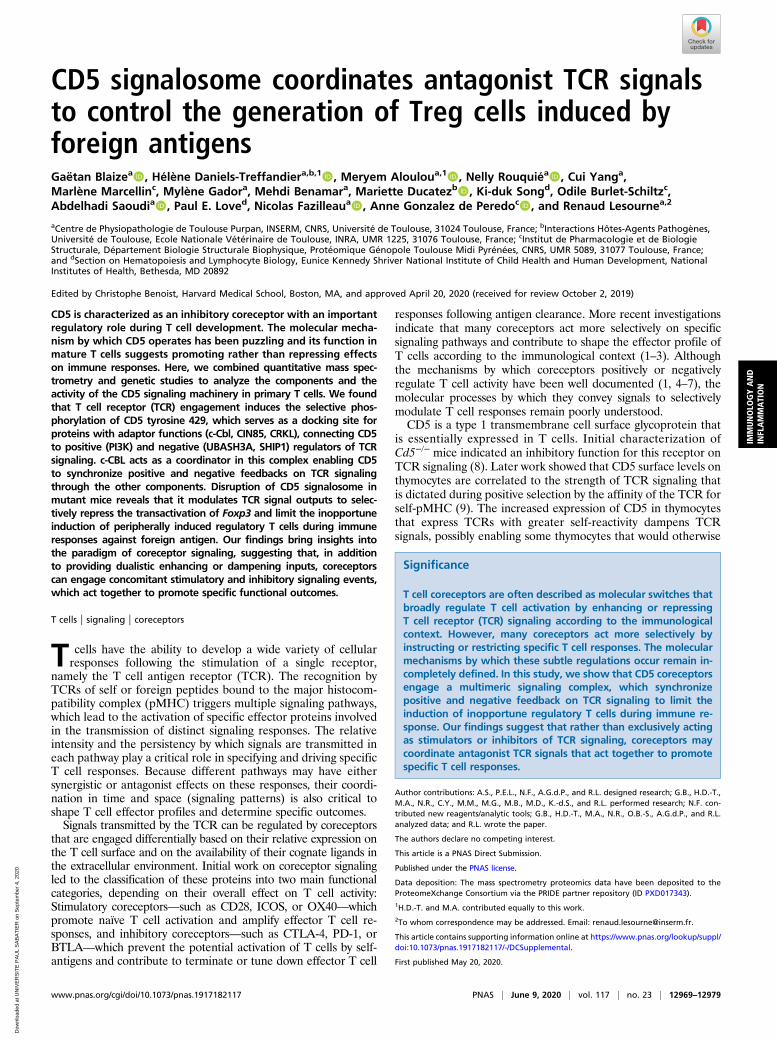
CD5 signalosome coordinates antagonist TCR signalsto control the generation of Treg cells induced byforeign antigensGaëtan Blaizea, Hélène Daniels-Treffandiera,b,1, Meryem Alouloua,1
, Nelly Rouquiéa, Cui Yanga,Marlène Marcellinc, Mylène Gadora, Mehdi Benamara, Mariette Ducatezb, Ki-duk Songd, Odile Burlet-Schiltzc,Abdelhadi Saoudia, Paul E. Loved, Nicolas Fazilleaua
, Anne Gonzalez de Peredoc, and Renaud Lesournea,2
aCentre de Physiopathologie de Toulouse Purpan, INSERM, CNRS, Université de Toulouse, 31024 Toulouse, France; bInteractions Hôtes-Agents Pathogènes,Université de Toulouse, Ecole Nationale Vétérinaire de Toulouse, INRA, UMR 1225, 31076 Toulouse, France; cInstitut de Pharmacologie et de BiologieStructurale, Département Biologie Structurale Biophysique, Protéomique Génopole Toulouse Midi Pyrénées, CNRS, UMR 5089, 31077 Toulouse, France;and dSection on Hematopoiesis and Lymphocyte Biology, Eunice Kennedy Shriver National Institute of Child Health and Human Development, NationalInstitutes of Health, Bethesda, MD 20892
Edited by Christophe Benoist, Harvard Medical School, Boston, MA, and approved April 20, 2020 (received for review October 2, 2019)
CD5 is characterized as an inhibitory coreceptor with an importantregulatory role during T cell development. The molecular mecha-nism by which CD5 operates has been puzzling and its function inmature T cells suggests promoting rather than repressing effectson immune responses. Here, we combined quantitative mass spec-trometry and genetic studies to analyze the components and theactivity of the CD5 signaling machinery in primary T cells. We foundthat T cell receptor (TCR) engagement induces the selective phos-phorylation of CD5 tyrosine 429, which serves as a docking site forproteins with adaptor functions (c-Cbl, CIN85, CRKL), connecting CD5to positive (PI3K) and negative (UBASH3A, SHIP1) regulators of TCRsignaling. c-CBL acts as a coordinator in this complex enabling CD5to synchronize positive and negative feedbacks on TCR signalingthrough the other components. Disruption of CD5 signalosome inmutant mice reveals that it modulates TCR signal outputs to selec-tively repress the transactivation of Foxp3 and limit the inopportuneinduction of peripherally induced regulatory T cells during immuneresponses against foreign antigen. Our findings bring insights intothe paradigm of coreceptor signaling, suggesting that, in additionto providing dualistic enhancing or dampening inputs, coreceptorscan engage concomitant stimulatory and inhibitory signaling events,which act together to promote specific functional outcomes.
T cells | signaling | coreceptors
T cells have the ability to develop a wide variety of cellularresponses following the stimulation of a single receptor,
namely the T cell antigen receptor (TCR). The recognition byTCRs of self or foreign peptides bound to the major histocom-patibility complex (pMHC) triggers multiple signaling pathways,which lead to the activation of specific effector proteins involvedin the transmission of distinct signaling responses. The relativeintensity and the persistency by which signals are transmitted ineach pathway play a critical role in specifying and driving specificT cell responses. Because different pathways may have eithersynergistic or antagonist effects on these responses, their coordi-nation in time and space (signaling patterns) is also critical toshape T cell effector profiles and determine specific outcomes.Signals transmitted by the TCR can be regulated by coreceptors
that are engaged differentially based on their relative expression onthe T cell surface and on the availability of their cognate ligands inthe extracellular environment. Initial work on coreceptor signalingled to the classification of these proteins into two main functionalcategories, depending on their overall effect on T cell activity:Stimulatory coreceptors—such as CD28, ICOS, or OX40—whichpromote naïve T cell activation and amplify effector T cell re-sponses, and inhibitory coreceptors—such as CTLA-4, PD-1, orBTLA—which prevent the potential activation of T cells by self-antigens and contribute to terminate or tune down effector T cell
responses following antigen clearance. More recent investigationsindicate that many coreceptors act more selectively on specificsignaling pathways and contribute to shape the effector profile ofT cells according to the immunological context (1–3). Althoughthe mechanisms by which coreceptors positively or negativelyregulate T cell activity have been well documented (1, 4–7), themolecular processes by which they convey signals to selectivelymodulate T cell responses remain poorly understood.CD5 is a type 1 transmembrane cell surface glycoprotein that
is essentially expressed in T cells. Initial characterization ofCd5−/− mice indicated an inhibitory function for this receptor onTCR signaling (8). Later work showed that CD5 surface levels onthymocytes are correlated to the strength of TCR signaling thatis dictated during positive selection by the affinity of the TCR forself-pMHC (9). The increased expression of CD5 in thymocytesthat express TCRs with greater self-reactivity dampens TCRsignals, possibly enabling some thymocytes that would otherwise
Significance
T cell coreceptors are often described as molecular switches thatbroadly regulate T cell activation by enhancing or repressingT cell receptor (TCR) signaling according to the immunologicalcontext. However, many coreceptors act more selectively byinstructing or restricting specific T cell responses. The molecularmechanisms by which these subtle regulations occur remain in-completely defined. In this study, we show that CD5 coreceptorsengage a multimeric signaling complex, which synchronizepositive and negative feedback on TCR signaling to limit theinduction of inopportune regulatory T cells during immune re-sponse. Our findings suggest that rather than exclusively actingas stimulators or inhibitors of TCR signaling, coreceptors maycoordinate antagonist TCR signals that act together to promotespecific T cell responses.
Author contributions: A.S., P.E.L., N.F., A.G.d.P., and R.L. designed research; G.B., H.D.-T.,M.A., N.R., C.Y., M.M., M.G., M.B., M.D., K.-d.S., and R.L. performed research; N.F. con-tributed new reagents/analytic tools; G.B., H.D.-T., M.A., N.R., O.B.-S., A.G.d.P., and R.L.analyzed data; and R.L. wrote the paper.
The authors declare no competing interest.
This article is a PNAS Direct Submission.
Published under the PNAS license.
Data deposition: The mass spectrometry proteomics data have been deposited to theProteomeXchange Consortium via the PRIDE partner repository (ID PXD017343).1H.D.-T. and M.A. contributed equally to this work.2To whom correspondence may be addressed. Email: [email protected].
This article contains supporting information online at https://www.pnas.org/lookup/suppl/doi:10.1073/pnas.1917182117/-/DCSupplemental.
First published May 20, 2020.
www.pnas.org/cgi/doi/10.1073/pnas.1917182117 PNAS | June 9, 2020 | vol. 117 | no. 23 | 12969–12979
IMMUNOLO
GYAND
INFLAMMATION
Dow
nloa
ded
at U
NIV
ER
SIT
E P
AU
L S
AB
AT
IER
on
Sep
tem
ber
4, 2
020
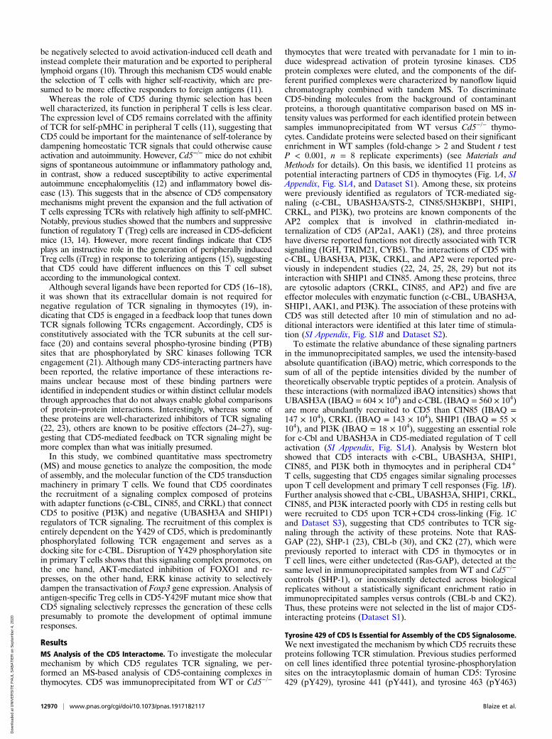
be negatively selected to avoid activation-induced cell death andinstead complete their maturation and be exported to peripherallymphoid organs (10). Through this mechanism CD5 would enablethe selection of T cells with higher self-reactivity, which are pre-sumed to be more effective responders to foreign antigens (11).Whereas the role of CD5 during thymic selection has been
well characterized, its function in peripheral T cells is less clear.The expression level of CD5 remains correlated with the affinityof TCR for self-pMHC in peripheral T cells (11), suggesting thatCD5 could be important for the maintenance of self-tolerance bydampening homeostatic TCR signals that could otherwise causeactivation and autoimmunity. However, Cd5−/− mice do not exhibitsigns of spontaneous autoimmune or inflammatory pathology and,in contrast, show a reduced susceptibility to active experimentalautoimmune encephalomyelitis (12) and inflammatory bowel dis-ease (13). This suggests that in the absence of CD5 compensatorymechanisms might prevent the expansion and the full activation ofT cells expressing TCRs with relatively high affinity to self-pMHC.Notably, previous studies showed that the numbers and suppressivefunction of regulatory T (Treg) cells are increased in CD5-deficientmice (13, 14). However, more recent findings indicate that CD5plays an instructive role in the generation of peripherally inducedTreg cells (iTreg) in response to tolerizing antigens (15), suggestingthat CD5 could have different influences on this T cell subsetaccording to the immunological context.Although several ligands have been reported for CD5 (16–18),
it was shown that its extracellular domain is not required fornegative regulation of TCR signaling in thymocytes (19), in-dicating that CD5 is engaged in a feedback loop that tunes downTCR signals following TCRs engagement. Accordingly, CD5 isconstitutively associated with the TCR subunits at the cell sur-face (20) and contains several phospho-tyrosine binding (PTB)sites that are phosphorylated by SRC kinases following TCRengagement (21). Although many CD5-interacting partners havebeen reported, the relative importance of these interactions re-mains unclear because most of these binding partners wereidentified in independent studies or within distinct cellular modelsthrough approaches that do not always enable global comparisonsof protein–protein interactions. Interestingly, whereas some ofthese proteins are well-characterized inhibitors of TCR signaling(22, 23), others are known to be positive effectors (24–27), sug-gesting that CD5-mediated feedback on TCR signaling might bemore complex than what was initially presumed.In this study, we combined quantitative mass spectrometry
(MS) and mouse genetics to analyze the composition, the modeof assembly, and the molecular function of the CD5 transductionmachinery in primary T cells. We found that CD5 coordinatesthe recruitment of a signaling complex composed of proteinswith adapter functions (c-CBL, CIN85, and CRKL) that connectCD5 to positive (PI3K) and negative (UBASH3A and SHIP1)regulators of TCR signaling. The recruitment of this complex isentirely dependent on the Y429 of CD5, which is predominantlyphosphorylated following TCR engagement and serves as adocking site for c-CBL. Disruption of Y429 phosphorylation sitein primary T cells shows that this signaling complex promotes, onthe one hand, AKT-mediated inhibition of FOXO1 and re-presses, on the other hand, ERK kinase activity to selectivelydampen the transactivation of Foxp3 gene expression. Analysis ofantigen-specific Treg cells in CD5-Y429F mutant mice show thatCD5 signaling selectively represses the generation of these cellspresumably to promote the development of optimal immuneresponses.
ResultsMS Analysis of the CD5 Interactome. To investigate the molecularmechanism by which CD5 regulates TCR signaling, we per-formed an MS-based analysis of CD5-containing complexes inthymocytes. CD5 was immunoprecipitated from WT or Cd5−/−
thymocytes that were treated with pervanadate for 1 min to in-duce widespread activation of protein tyrosine kinases. CD5protein complexes were eluted, and the components of the dif-ferent purified complexes were characterized by nanoflow liquidchromatography combined with tandem MS. To discriminateCD5-binding molecules from the background of contaminantproteins, a thorough quantitative comparison based on MS in-tensity values was performed for each identified protein betweensamples immunoprecipitated from WT versus Cd5−/− thymo-cytes. Candidate proteins were selected based on their significantenrichment in WT samples (fold-change > 2 and Student t testP < 0.001, n = 8 replicate experiments) (see Materials andMethods for details). On this basis, we identified 11 proteins aspotential interacting partners of CD5 in thymocytes (Fig. 1A, SIAppendix, Fig. S1A, and Dataset S1). Among these, six proteinswere previously identified as regulators of TCR-mediated sig-naling (c-CBL, UBASH3A/STS-2, CIN85/SH3KBP1, SHIP1,CRKL, and PI3K), two proteins are known components of theAP2 complex that is involved in clathrin-mediated in-ternalization of CD5 (AP2a1, AAK1) (28), and three proteinshave diverse reported functions not directly associated with TCRsignaling (IGH, TRIM21, CYB5). The interactions of CD5 withc-CBL, UBASH3A, PI3K, CRKL, and AP2 were reported pre-viously in independent studies (22, 24, 25, 28, 29) but not itsinteraction with SHIP1 and CIN85. Among these proteins, threeare cytosolic adaptors (CRKL, CIN85, and AP2) and five areeffector molecules with enzymatic function (c-CBL, UBASH3A,SHIP1, AAK1, and PI3K). The association of these proteins withCD5 was still detected after 10 min of stimulation and no ad-ditional interactors were identified at this later time of stimula-tion (SI Appendix, Fig. S1B and Dataset S2).To estimate the relative abundance of these signaling partners
in the immunoprecipitated samples, we used the intensity-basedabsolute quantification (iBAQ) metric, which corresponds to thesum of all of the peptide intensities divided by the number oftheoretically observable tryptic peptides of a protein. Analysis ofthese interactions (with normalized iBAQ intensities) shows thatUBASH3A (IBAQ = 604 × 104) and c-CBL (IBAQ = 560 × 104)are more abundantly recruited to CD5 than CIN85 (IBAQ =147 × 104), CRKL (IBAQ = 143 × 104), SHIP1 (IBAQ = 55 ×104), and PI3K (IBAQ = 18 × 104), suggesting an essential rolefor c-Cbl and UBASH3A in CD5-mediated regulation of T cellactivation (SI Appendix, Fig. S1A). Analysis by Western blotshowed that CD5 interacts with c-CBL, UBASH3A, SHIP1,CIN85, and PI3K both in thymocytes and in peripheral CD4+
T cells, suggesting that CD5 engages similar signaling processesupon T cell development and primary T cell responses (Fig. 1B).Further analysis showed that c-CBL, UBASH3A, SHIP1, CRKL,CIN85, and PI3K interacted poorly with CD5 in resting cells butwere recruited to CD5 upon TCR+CD4 cross-linking (Fig. 1Cand Dataset S3), suggesting that CD5 contributes to TCR sig-naling through the activity of these proteins. Note that RAS-GAP (22), SHP-1 (23), CBL-b (30), and CK2 (27), which werepreviously reported to interact with CD5 in thymocytes or inT cell lines, were either undetected (Ras-GAP), detected at thesame level in immunoprecipitated samples from WT and Cd5−/−
controls (SHP-1), or inconsistently detected across biologicalreplicates without a statistically significant enrichment ratio inimmunoprecipitated samples versus controls (CBL-b and CK2).Thus, these proteins were not selected in the list of major CD5-interacting proteins (Dataset S1).
Tyrosine 429 of CD5 Is Essential for Assembly of the CD5 Signalosome.We next investigated the mechanism by which CD5 recruits theseproteins following TCR stimulation. Previous studies performedon cell lines identified three potential tyrosine-phosphorylationsites on the intracytoplasmic domain of human CD5: Tyrosine429 (pY429), tyrosine 441 (pY441), and tyrosine 463 (pY463)
12970 | www.pnas.org/cgi/doi/10.1073/pnas.1917182117 Blaize et al.
Dow
nloa
ded
at U
NIV
ER
SIT
E P
AU
L S
AB
AT
IER
on
Sep
tem
ber
4, 2
020
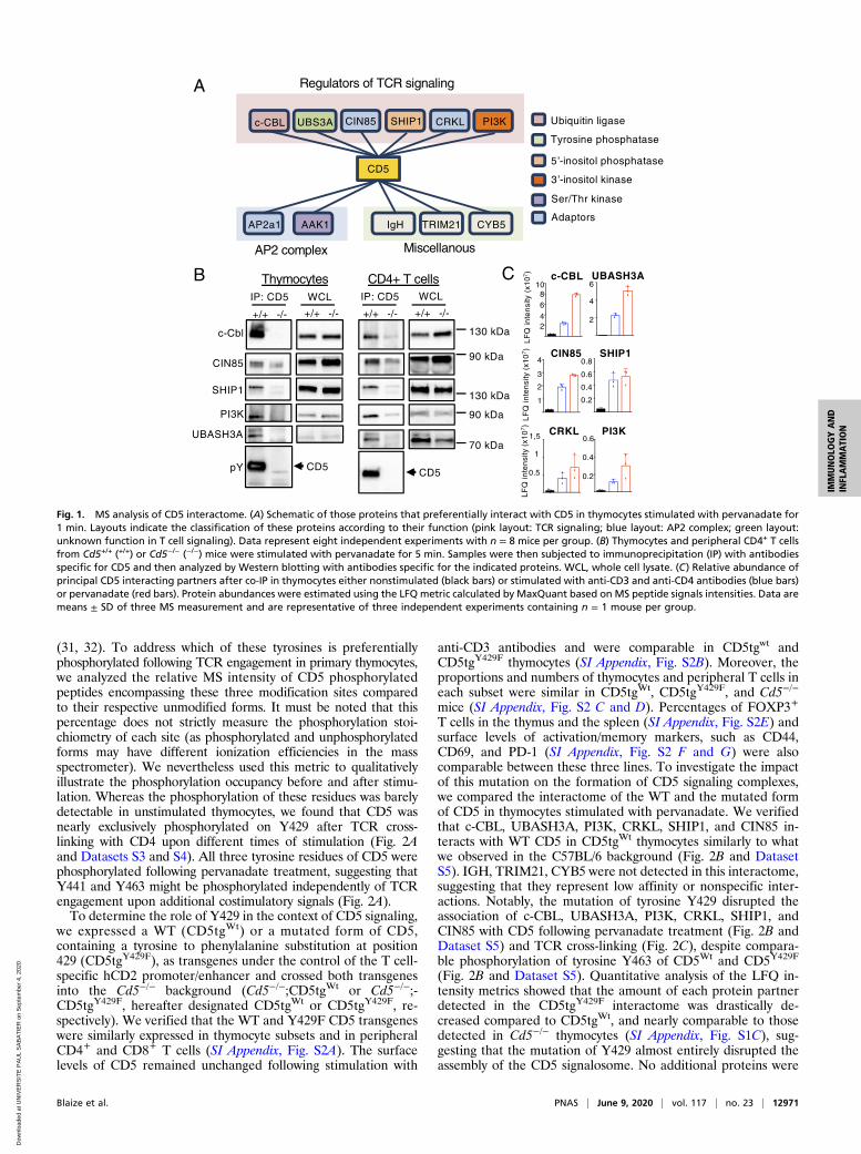
(31, 32). To address which of these tyrosines is preferentiallyphosphorylated following TCR engagement in primary thymocytes,we analyzed the relative MS intensity of CD5 phosphorylatedpeptides encompassing these three modification sites comparedto their respective unmodified forms. It must be noted that thispercentage does not strictly measure the phosphorylation stoi-chiometry of each site (as phosphorylated and unphosphorylatedforms may have different ionization efficiencies in the massspectrometer). We nevertheless used this metric to qualitativelyillustrate the phosphorylation occupancy before and after stimu-lation. Whereas the phosphorylation of these residues was barelydetectable in unstimulated thymocytes, we found that CD5 wasnearly exclusively phosphorylated on Y429 after TCR cross-linking with CD4 upon different times of stimulation (Fig. 2Aand Datasets S3 and S4). All three tyrosine residues of CD5 werephosphorylated following pervanadate treatment, suggesting thatY441 and Y463 might be phosphorylated independently of TCRengagement upon additional costimulatory signals (Fig. 2A).To determine the role of Y429 in the context of CD5 signaling,
we expressed a WT (CD5tgWt) or a mutated form of CD5,containing a tyrosine to phenylalanine substitution at position429 (CD5tgY429F), as transgenes under the control of the T cell-specific hCD2 promoter/enhancer and crossed both transgenesinto the Cd5−/− background (Cd5−/−;CD5tgWt or Cd5−/−;-CD5tgY429F, hereafter designated CD5tgWt or CD5tgY429F, re-spectively). We verified that the WT and Y429F CD5 transgeneswere similarly expressed in thymocyte subsets and in peripheralCD4+ and CD8+ T cells (SI Appendix, Fig. S2A). The surfacelevels of CD5 remained unchanged following stimulation with
anti-CD3 antibodies and were comparable in CD5tgwt andCD5tgY429F thymocytes (SI Appendix, Fig. S2B). Moreover, theproportions and numbers of thymocytes and peripheral T cells ineach subset were similar in CD5tgWt, CD5tgY429F, and Cd5−/−
mice (SI Appendix, Fig. S2 C and D). Percentages of FOXP3+
T cells in the thymus and the spleen (SI Appendix, Fig. S2E) andsurface levels of activation/memory markers, such as CD44,CD69, and PD-1 (SI Appendix, Fig. S2 F and G) were alsocomparable between these three lines. To investigate the impactof this mutation on the formation of CD5 signaling complexes,we compared the interactome of the WT and the mutated formof CD5 in thymocytes stimulated with pervanadate. We verifiedthat c-CBL, UBASH3A, PI3K, CRKL, SHIP1, and CIN85 in-teracts with WT CD5 in CD5tgWt thymocytes similarly to whatwe observed in the C57BL/6 background (Fig. 2B and DatasetS5). IGH, TRIM21, CYB5 were not detected in this interactome,suggesting that they represent low affinity or nonspecific inter-actions. Notably, the mutation of tyrosine Y429 disrupted theassociation of c-CBL, UBASH3A, PI3K, CRKL, SHIP1, andCIN85 with CD5 following pervanadate treatment (Fig. 2B andDataset S5) and TCR cross-linking (Fig. 2C), despite compara-ble phosphorylation of tyrosine Y463 of CD5Wt and CD5Y429F
(Fig. 2B and Dataset S5). Quantitative analysis of the LFQ in-tensity metrics showed that the amount of each protein partnerdetected in the CD5tgY429F interactome was drastically de-creased compared to CD5tgWt, and nearly comparable to thosedetected in Cd5−/− thymocytes (SI Appendix, Fig. S1C), sug-gesting that the mutation of Y429 almost entirely disrupted theassembly of the CD5 signalosome. No additional proteins were
Ubiquitin ligase
Tyrosine phosphatase
Adaptors
-inositol phosphatase
A
-inositol kinaseCD5
UBS3Ac-CBL CIN85 CRKLSHIP1 PI3K
Regulators of TCR signaling
AP2a1 AAK1 TRIM21 CYB5IgH
AP2 complex Miscellanous
Ser/Thr kinase
130 kDa
90 kDa
130 kDa
90 kDa
70 kDa
CD5
c-Cbl
CIN85
SHIP1
PI3K
UBASH3A
pY
+/+ -/- +/+ -/-
IP: CD5 WCL
+/+ -/- +/+ -/-
IP: CD5 WCL
CD5
B Thymocytes CD4+ T cells UBASH3A
2468
10
2
4
6
0.2
0.4
0.6
0.8
1
2
3
0.2
0.4
c-CBL
CIN85
CRKL
SHIP1
PI3K
LF
Q in
ten
sity
(x1
07)
4
0.6
0.5
1
1,5
LF
Q in
ten
sity
(x1
07)
LF
Q in
ten
sity
(x1
07)
C
Fig. 1. MS analysis of CD5 interactome. (A) Schematic of those proteins that preferentially interact with CD5 in thymocytes stimulated with pervanadate for1 min. Layouts indicate the classification of these proteins according to their function (pink layout: TCR signaling; blue layout: AP2 complex; green layout:unknown function in T cell signaling). Data represent eight independent experiments with n = 8 mice per group. (B) Thymocytes and peripheral CD4+ T cellsfrom Cd5+/+ (+/+) or Cd5−/− (−/−) mice were stimulated with pervanadate for 5 min. Samples were then subjected to immunoprecipitation (IP) with antibodiesspecific for CD5 and then analyzed by Western blotting with antibodies specific for the indicated proteins. WCL, whole cell lysate. (C) Relative abundance ofprincipal CD5 interacting partners after co-IP in thymocytes either nonstimulated (black bars) or stimulated with anti-CD3 and anti-CD4 antibodies (blue bars)or pervanadate (red bars). Protein abundances were estimated using the LFQ metric calculated by MaxQuant based on MS peptide signals intensities. Data aremeans ± SD of three MS measurement and are representative of three independent experiments containing n = 1 mouse per group.
Blaize et al. PNAS | June 9, 2020 | vol. 117 | no. 23 | 12971
IMMUNOLO
GYAND
INFLAMMATION
Dow
nloa
ded
at U
NIV
ER
SIT
E P
AU
L S
AB
AT
IER
on
Sep
tem
ber
4, 2
020
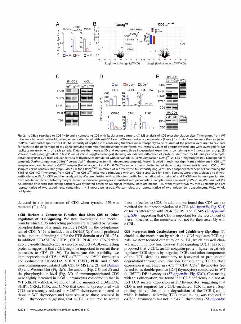
detected in the interactome of CD5 when tyrosine 429 wasmutated (Fig. 2B).
c-CBL Harbors a Connective Function that Links CD5 to OtherRegulators of TCR Signaling. We next investigated the mecha-nism by which CD5 interacting proteins are recruited by tyrosinephosphorylation of a single residue (Y429) on the cytoplasmictail of CD5. Y429 is included in a D(N/D)XpY motif predictedto be a potential binding site for the PTB domain of c-CBL (33).In addition, UBASH3A, SHIP1, CRKL, PI3K, and CIN85 werealso previously characterized as direct or indirect c-CBL–interactingproteins, suggesting that c-CBL might be important to recruit thesemolecules to CD5 (34–36). To investigate this possibility, weimmunoprecipitated CD5 in WT, c-Cbl−/−, and Cd5−/− thymocytesand evaluated if UBASH3A, SHIP1, CRKL, PI3K, and CIN85were coimmunoprecipitated with CD5 by MS (Fig. 2D and DatasetS3) and Western blot (Fig. 2E). The amount (Fig. 2 D and E) andthe phosphorylation level (Fig. 2E) of immunoprecipitated CD5were slightly increased in c-Cbl−/− thymocytes compared to that inWT cells. Nevertheless, we found that the amounts of UBASH3A,SHIP1, CRKL, PI3K, and CIN85 that coimmunoprecipitated withCD5 were strongly reduced in c-Cbl−/− thymocytes compared tothose in WT thymocytes and were similar to those observed inCd5−/− thymocytes, suggesting that c-CBL is required to recruit
these molecules to CD5. In addition, we found that CD5 was notrequired for the phosphorylation of c-CBL (SI Appendix, Fig. S3A)or for its interaction with PI3K, SHIP1, and CIN85 (SI Appendix,Fig. S3B), suggesting that CD5 is important for the recruitment ofthese molecules at the membrane but not for their assembly withc-CBL.
CD5 Integrates Both Costimulatory and Coinhibitory Signaling. Toelucidate the mechanism by which the CD5 regulates TCR sig-nals, we next focused our study on c-CBL, which has well char-acterized inhibitory functions on TCR signaling (37). It has beenproposed that c-CBL, an E3 ubiquitin-protein ligase, negativelyregulates TCR signals by targeting TCRs and other componentsof the TCR signaling machinery to lysosomal or proteasomaldegradation through ubiquitination. Consequently, TCR surfaceexpression is increased in c-Cbl−/− CD4+CD8+ thymocytes (re-ferred to as double-positive [DP] thymocytes) compared to WT(c-Cbl+/+) DP thymocytes (SI Appendix, Fig. S3C). Contrastingwith this observation, we found that CD5 deficiency did not af-fect TCR surface expression in DP thymocytes, suggesting thatCD5 is not required for c-CBL–mediated TCR turnover. Sup-porting this conclusion, the degradation of the TCR ζ-chain,which is induced following TCR cross-linking, was reduced inc-Cbl−/− thymocytes but not in Cd5−/− thymocytes (SI Appendix,
A
CIN85
SHIP1
PI3K
CD5
PY20
c-Cbl
Cbl+/+ WCLIP: CD5
Cbl-/-
CD5
Cbl+/+ WCL
IP: CD5
Cbl-/-
C
B
CD5
PI3Kc-CBL
SHIP1
CIN85
CRKL
CD5
CD5tgY429FCD5tgWt
UBASH3A
0 2 4 6 8-2-4-6-8 0 2 4 6-2-4-6-8
Log2 (fold change tgWt/KO) Log2 (fold change tgY429F/KO)
pY
463
0.51
1.52
2.53
4
5
3.5
4.5
020
40
60
UBASH3Ac-CBLCD5
Wt c-Cbl-/- CD5-/-
24
8
102030
100
200
510152025
468
10
5
10
15
LF
Q in
ten
sity
(x1
05 )
300 40
6
0
0 0 0
0
12 10
% o
f sig
na
l of p
Yfo
rms
-CD3 + -CD4
0,5
1
2
4
6
20
40
60
80
Unstim. Perva.
8
Y429 Y441 Y463
CIN85
CD5
SHIP1
c-Cbl
PI3K
tgWt tgY429 tgWt tgY429
IP: CD5 WCL
CIN85 CRKL PI3K
tgWt
tgY429F
D E
Fig. 2. c-CBL is recruited to CD5 Y429 and is connecting CD5 with its signaling partners. (A) MS analysis of CD5 phosphorylation sites. Thymocytes from WTmice were left unstimulated (Unstim.) or were stimulated with anti-CD3 + anti-CD4 antibodies or pervanadate (Perva.) for 1 min. Samples were then subjectedto IP with antibodies specific for CD5. MS intensity of peptide ions containing the three main phosphotyrosine residues of the protein were used to calculatefor each site the percentage of MS signal deriving from modified phosphotyrosine forms. MS intensity values of phosphorylated ions were averaged for MSreplicate measurements of each sample. Data are the means ± SD and represent three independent experiments containing n = 1 mouse per group. (B)Volcano plots [−log10(Student t test P value) versus log2(fold-change)] showing abundances difference of proteins identified by MS analysis of samplesobtained by IP of CD5 from cellular extracts of thymocytes stimulated with pervanadate. (Left) Comparison CD5tgWt vs. Cd5−/− thymocytes (n = 6 independentsamples); (Right) comparison CD5tgWt versus Cd5−/− thymocytes (n = 5 independent samples). Protein labeled in red show significant enrichment in CD5tgWt
samples compared to control Cd5−/− samples (fold-change > 2 and P < 0.05). The same proteins plotted in red show no significant enrichment in CD5tgY429F
samples versus controls. Bar graph (Inset ) in the CD5tgY429F volcano plot represent the MS intensity (log10) of CD5 phosphorylated peptides containing theY463 of CD5. (C) Thymocytes from CD5tgWt or CD5tgWt mice were stimulated with anti-CD3 + anti-CD4 for 1 min. Samples were then subjected to IP withantibodies specific for CD5 and then analyzed by Western blotting with antibodies specific for the indicated proteins. (D and E) CD5 was immunoprecipitatedfrom cellular extracts of total thymocytes from the indicated genotypes stimulated with pervanadate. Samples were analyzed by MS (D) or Western blot (E).Abundance of specific interacting partners was estimated based on MS signal intensity. Data are means ± SD from at least two MS measurements and arerepresentative of two experiments containing n = 1 mouse per group. Western blots are representative of two independent experiments. WCL, wholecell lysate.
12972 | www.pnas.org/cgi/doi/10.1073/pnas.1917182117 Blaize et al.
Dow
nloa
ded
at U
NIV
ER
SIT
E P
AU
L S
AB
AT
IER
on
Sep
tem
ber
4, 2
020
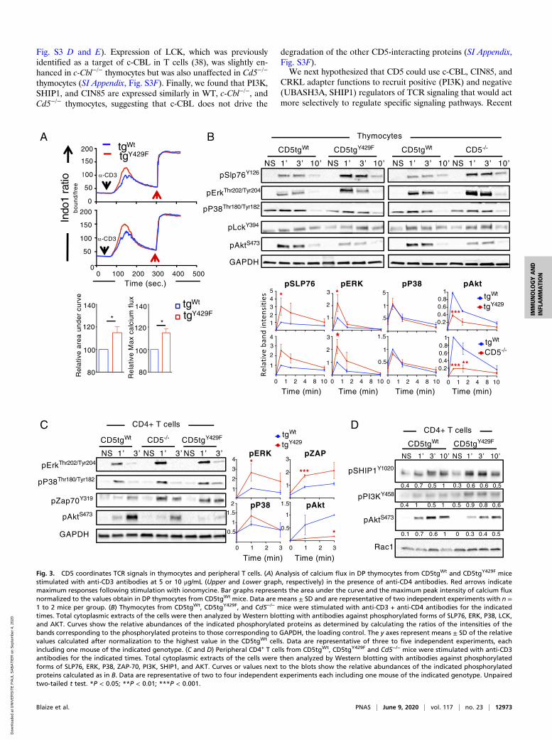
Fig. S3 D and E). Expression of LCK, which was previouslyidentified as a target of c-CBL in T cells (38), was slightly en-hanced in c-Cbl−/− thymocytes but was also unaffected in Cd5−/−
thymocytes (SI Appendix, Fig. S3F). Finally, we found that PI3K,SHIP1, and CIN85 are expressed similarly in WT, c-Cbl−/−, andCd5−/− thymocytes, suggesting that c-CBL does not drive the
degradation of the other CD5-interacting proteins (SI Appendix,Fig. S3F).We next hypothesized that CD5 could use c-CBL, CIN85, and
CRKL adapter functions to recruit positive (PI3K) and negative(UBASH3A, SHIP1) regulators of TCR signaling that would actmore selectively to regulate specific signaling pathways. Recent
A
pErkThr202/Tyr204
pSlp76Y126
CD5tgWt CD5tgY429F CD5-/-CD5tgWt
pAktS473
GAPDH
pLckY394
Thymocytes
pP38Thr180/Tyr182
0 1 2 4 8 10 0 1 2 4 8 10 0 1 2 4 8 10 0 1 2 4 8 10
1
2
3
4
12345
1
2
3
1
1.5
0.50.20.40.60.8
1
1
2
3
1
1.5
0.50.20.40.60.8
1pSLP76 pERK pP38 pAkt
tgWt
tgY429
tgWt
CD5-/-
Time (min) Time (min) Time (min) Time (min)
C
GAPDH
pZap70Y319
CD5tgWt CD5tgY429FCD5-/-
CD4+ T cells
pAktS473
pErkThr202/Tyr204
pP38Thr180/Tyr182
1
2
3
4
1
2
3
1
1.5
0.5
2
1
1.5
0.5
pERK
pP38 pAkt
pZAP
Time (min) Time (min)1 2 30 1 2 30
tgWt
tgY429
pSHIP1Y1020
pPI3KY458
Rac1
pAktS473
CD5tgWt CD5tgY429F
0.1 0.7 0.6 1 0 0.3 0.4 0.5
0.4 0.7 0.5 1 0.3 0.6 0.6 0,5
0.4 1 0.5 1 0.5 0.9 0.8 0,6
Time (sec.)100 200 300 400 5000
0
50
100
150
200
Indo
1 ra
tiob
ou
nd
/fre
e
-CD3
-CD3
tgWt
tgY429F
0
50
100
150
200
Re
lativ
e M
ax
calc
ium
flu
x
Re
lativ
e a
rea
un
de
rcu
rve
80 80
100
120
140 tgWt
tgY429F
100
120
140
B
D
* *
* *
***
*
*** **
****
*
CD4+ T cells
Fig. 3. CD5 coordinates TCR signals in thymocytes and peripheral T cells. (A) Analysis of calcium flux in DP thymocytes from CD5tgWt and CD5tgY429F micestimulated with anti-CD3 antibodies at 5 or 10 μg/mL (Upper and Lower graph, respectively) in the presence of anti-CD4 antibodies. Red arrows indicatemaximum responses following stimulation with ionomycine. Bar graphs represents the area under the curve and the maximum peak intensity of calcium fluxnormalized to the values obtain in DP thymocytes from CD5tgWt mice. Data are means ± SD and are representative of two independent experiments with n =1 to 2 mice per group. (B) Thymocytes from CD5tgWt, CD5tgY429F, and Cd5−/− mice were stimulated with anti-CD3 + anti-CD4 antibodies for the indicatedtimes. Total cytoplasmic extracts of the cells were then analyzed by Western blotting with antibodies against phosphorylated forms of SLP76, ERK, P38, LCK,and AKT. Curves show the relative abundances of the indicated phosphorylated proteins as determined by calculating the ratios of the intensities of thebands corresponding to the phosphorylated proteins to those corresponding to GAPDH, the loading control. The y axes represent means ± SD of the relativevalues calculated after normalization to the highest value in the CD5tgWt cells. Data are representative of three to five independent experiments, eachincluding one mouse of the indicated genotype. (C and D) Peripheral CD4+ T cells from CD5tgWt, CD5tgY429F and Cd5−/− mice were stimulated with anti-CD3antibodies for the indicated times. Total cytoplasmic extracts of the cells were then analyzed by Western blotting with antibodies against phosphorylatedforms of SLP76, ERK, P38, ZAP-70, PI3K, SHIP1, and AKT. Curves or values next to the blots show the relative abundances of the indicated phosphorylatedproteins calculated as in B. Data are representative of two to four independent experiments each including one mouse of the indicated genotype. Unpairedtwo-tailed t test. *P < 0.05; **P < 0.01; ***P < 0.001.
Blaize et al. PNAS | June 9, 2020 | vol. 117 | no. 23 | 12973
IMMUNOLO
GYAND
INFLAMMATION
Dow
nloa
ded
at U
NIV
ER
SIT
E P
AU
L S
AB
AT
IER
on
Sep
tem
ber
4, 2
020
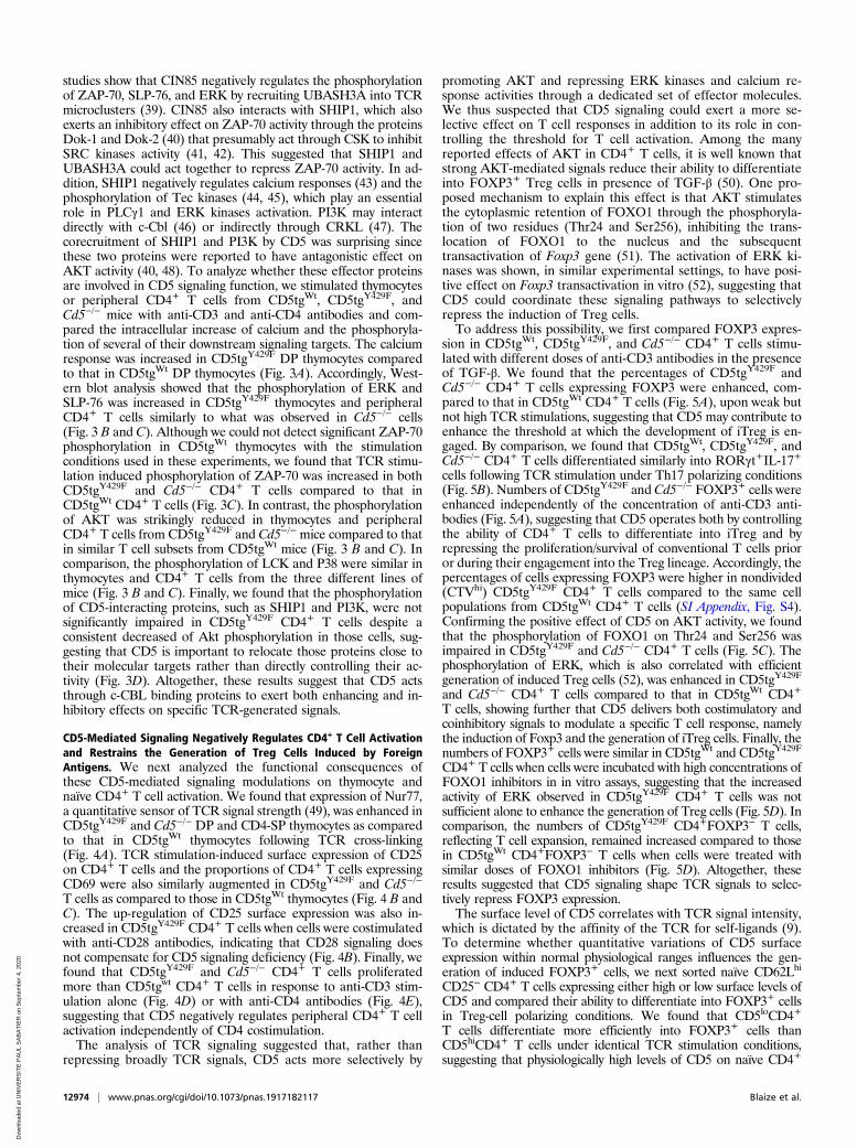
studies show that CIN85 negatively regulates the phosphorylationof ZAP-70, SLP-76, and ERK by recruiting UBASH3A into TCRmicroclusters (39). CIN85 also interacts with SHIP1, which alsoexerts an inhibitory effect on ZAP-70 activity through the proteinsDok-1 and Dok-2 (40) that presumably act through CSK to inhibitSRC kinases activity (41, 42). This suggested that SHIP1 andUBASH3A could act together to repress ZAP-70 activity. In ad-dition, SHIP1 negatively regulates calcium responses (43) and thephosphorylation of Tec kinases (44, 45), which play an essentialrole in PLCγ1 and ERK kinases activation. PI3K may interactdirectly with c-Cbl (46) or indirectly through CRKL (47). Thecorecruitment of SHIP1 and PI3K by CD5 was surprising sincethese two proteins were reported to have antagonistic effect onAKT activity (40, 48). To analyze whether these effector proteinsare involved in CD5 signaling function, we stimulated thymocytesor peripheral CD4+ T cells from CD5tgWt, CD5tgY429F, andCd5−/− mice with anti-CD3 and anti-CD4 antibodies and com-pared the intracellular increase of calcium and the phosphoryla-tion of several of their downstream signaling targets. The calciumresponse was increased in CD5tgY429F DP thymocytes comparedto that in CD5tgWt DP thymocytes (Fig. 3A). Accordingly, West-ern blot analysis showed that the phosphorylation of ERK andSLP-76 was increased in CD5tgY429F thymocytes and peripheralCD4+ T cells similarly to what was observed in Cd5−/− cells(Fig. 3 B and C). Although we could not detect significant ZAP-70phosphorylation in CD5tgWt thymocytes with the stimulationconditions used in these experiments, we found that TCR stimu-lation induced phosphorylation of ZAP-70 was increased in bothCD5tgY429F and Cd5−/− CD4+ T cells compared to that inCD5tgWt CD4+ T cells (Fig. 3C). In contrast, the phosphorylationof AKT was strikingly reduced in thymocytes and peripheralCD4+ T cells from CD5tgY429F and Cd5−/− mice compared to thatin similar T cell subsets from CD5tgWt mice (Fig. 3 B and C). Incomparison, the phosphorylation of LCK and P38 were similar inthymocytes and CD4+ T cells from the three different lines ofmice (Fig. 3 B and C). Finally, we found that the phosphorylationof CD5-interacting proteins, such as SHIP1 and PI3K, were notsignificantly impaired in CD5tgY429F CD4+ T cells despite aconsistent decreased of Akt phosphorylation in those cells, sug-gesting that CD5 is important to relocate those proteins close totheir molecular targets rather than directly controlling their ac-tivity (Fig. 3D). Altogether, these results suggest that CD5 actsthrough c-CBL binding proteins to exert both enhancing and in-hibitory effects on specific TCR-generated signals.
CD5-Mediated Signaling Negatively Regulates CD4+ T Cell Activationand Restrains the Generation of Treg Cells Induced by ForeignAntigens. We next analyzed the functional consequences ofthese CD5-mediated signaling modulations on thymocyte andnaïve CD4+ T cell activation. We found that expression of Nur77,a quantitative sensor of TCR signal strength (49), was enhanced inCD5tgY429F and Cd5−/− DP and CD4-SP thymocytes as comparedto that in CD5tgWt thymocytes following TCR cross-linking(Fig. 4A). TCR stimulation-induced surface expression of CD25on CD4+ T cells and the proportions of CD4+ T cells expressingCD69 were also similarly augmented in CD5tgY429F and Cd5−/−
T cells as compared to those in CD5tgWt thymocytes (Fig. 4 B andC). The up-regulation of CD25 surface expression was also in-creased in CD5tgY429F CD4+ T cells when cells were costimulatedwith anti-CD28 antibodies, indicating that CD28 signaling doesnot compensate for CD5 signaling deficiency (Fig. 4B). Finally, wefound that CD5tgY429F and Cd5−/− CD4+ T cells proliferatedmore than CD5tgwt CD4+ T cells in response to anti-CD3 stim-ulation alone (Fig. 4D) or with anti-CD4 antibodies (Fig. 4E),suggesting that CD5 negatively regulates peripheral CD4+ T cellactivation independently of CD4 costimulation.The analysis of TCR signaling suggested that, rather than
repressing broadly TCR signals, CD5 acts more selectively by
promoting AKT and repressing ERK kinases and calcium re-sponse activities through a dedicated set of effector molecules.We thus suspected that CD5 signaling could exert a more se-lective effect on T cell responses in addition to its role in con-trolling the threshold for T cell activation. Among the manyreported effects of AKT in CD4+ T cells, it is well known thatstrong AKT-mediated signals reduce their ability to differentiateinto FOXP3+ Treg cells in presence of TGF-β (50). One pro-posed mechanism to explain this effect is that AKT stimulatesthe cytoplasmic retention of FOXO1 through the phosphoryla-tion of two residues (Thr24 and Ser256), inhibiting the trans-location of FOXO1 to the nucleus and the subsequenttransactivation of Foxp3 gene (51). The activation of ERK ki-nases was shown, in similar experimental settings, to have posi-tive effect on Foxp3 transactivation in vitro (52), suggesting thatCD5 could coordinate these signaling pathways to selectivelyrepress the induction of Treg cells.To address this possibility, we first compared FOXP3 expres-
sion in CD5tgWt, CD5tgY429F, and Cd5−/− CD4+ T cells stimu-lated with different doses of anti-CD3 antibodies in the presenceof TGF-β. We found that the percentages of CD5tgY429F andCd5−/− CD4+ T cells expressing FOXP3 were enhanced, com-pared to that in CD5tgWt CD4+ T cells (Fig. 5A), upon weak butnot high TCR stimulations, suggesting that CD5 may contribute toenhance the threshold at which the development of iTreg is en-gaged. By comparison, we found that CD5tgWt, CD5tgY429F, andCd5−/− CD4+ T cells differentiated similarly into RORγt+IL-17+cells following TCR stimulation under Th17 polarizing conditions(Fig. 5B). Numbers of CD5tgY429F and Cd5−/− FOXP3+ cells wereenhanced independently of the concentration of anti-CD3 anti-bodies (Fig. 5A), suggesting that CD5 operates both by controllingthe ability of CD4+ T cells to differentiate into iTreg and byrepressing the proliferation/survival of conventional T cells prioror during their engagement into the Treg lineage. Accordingly, thepercentages of cells expressing FOXP3 were higher in nondivided(CTVhi) CD5tgY429F CD4+ T cells compared to the same cellpopulations from CD5tgWt CD4+ T cells (SI Appendix, Fig. S4).Confirming the positive effect of CD5 on AKT activity, we foundthat the phosphorylation of FOXO1 on Thr24 and Ser256 wasimpaired in CD5tgY429F and Cd5−/− CD4+ T cells (Fig. 5C). Thephosphorylation of ERK, which is also correlated with efficientgeneration of induced Treg cells (52), was enhanced in CD5tgY429F
and Cd5−/− CD4+ T cells compared to that in CD5tgWt CD4+
T cells, showing further that CD5 delivers both costimulatory andcoinhibitory signals to modulate a specific T cell response, namelythe induction of Foxp3 and the generation of iTreg cells. Finally, thenumbers of FOXP3+ cells were similar in CD5tgWt and CD5tgY429F
CD4+ T cells when cells were incubated with high concentrations ofFOXO1 inhibitors in in vitro assays, suggesting that the increasedactivity of ERK observed in CD5tgY429F CD4+ T cells was notsufficient alone to enhance the generation of Treg cells (Fig. 5D). Incomparison, the numbers of CD5tgY429F CD4+FOXP3− T cells,reflecting T cell expansion, remained increased compared to thosein CD5tgWt CD4+FOXP3− T cells when cells were treated withsimilar doses of FOXO1 inhibitors (Fig. 5D). Altogether, theseresults suggested that CD5 signaling shape TCR signals to selec-tively repress FOXP3 expression.The surface level of CD5 correlates with TCR signal intensity,
which is dictated by the affinity of the TCR for self-ligands (9).To determine whether quantitative variations of CD5 surfaceexpression within normal physiological ranges influences the gen-eration of induced FOXP3+ cells, we next sorted naïve CD62Lhi
CD25− CD4+ T cells expressing either high or low surface levels ofCD5 and compared their ability to differentiate into FOXP3+ cellsin Treg-cell polarizing conditions. We found that CD5loCD4+
T cells differentiate more efficiently into FOXP3+ cells thanCD5hiCD4+ T cells under identical TCR stimulation conditions,suggesting that physiologically high levels of CD5 on naïve CD4+
12974 | www.pnas.org/cgi/doi/10.1073/pnas.1917182117 Blaize et al.
Dow
nloa
ded
at U
NIV
ER
SIT
E P
AU
L S
AB
AT
IER
on
Sep
tem
ber
4, 2
020
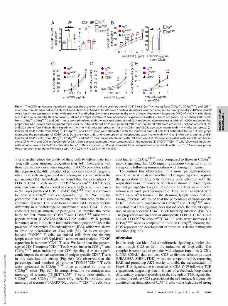
T cells might reduce the ability of these cells to differentiate intoTreg cells upon antigenic recognition (Fig. 6A). Contrasting withthose results, previous studies suggested that CD5 promotes, ratherthan represses, the differentiation of peripherally induced Treg cellswhen those cells are generated in a tolerogenic context such as thegut mucosa (15). Accordingly, we found that the percentages ofFOXP3+CD4+ T cells and of FOXP3+NeuropilinlowCD4+ T cells,which are essentially composed of iTreg cells (53), were decreasedin the Peyer patches of CD5−/− and CD5tgY429F mice as comparedto those in CD5tgWt mice (SI Appendix, Fig. S5). We thus hy-pothesized that CD5 signalosome might be influenced by the en-vironment in which T cells are localized and that CD5 may operatedifferently in a nontolerogenic environment when CD4+ T cellsencounter foreign antigens or pathogens. To examine this possi-bility, we first immunized CD5tgWt and CD5tgY429F mice with apeptide variant (EAWGALANKAVDKA, called 1W1K peptidehereafter) of the I‐E α-chain immunodominant peptide 52‐68 in thepresence of incomplete Freunds adjuvant (IFA), which was shownto favor the polarization of iTreg cells (54). To follow antigen-induced FOXP3+ T cells, we stained cells from the draininglymph nodes with 1W1K-pMHCII tetramer and analyzed FOXP3expression in tetramer+CD4+ T cells. We found that the percent-ages of CD44+tetramer+CD4+ T cells were similar in CD5tgWt andCD5tgY429F mice, indicating that CD5 signaling does not signifi-cantly impact the clonal expansion of antigen-specific CD4+ T cellsin this experimental setting (Fig. 6B). We observed that thepercentages and numbers of tetramer+FOXP3+CD4+ T cellswere increased in CD5tgY429F mice compared to those inCD5tgWt mice (Fig. 6C). In comparison, the percentages andnumbers of tetramer+T-BET+CD4+ T cells were similar inCD5tgWt and CD5tgY429F mice (Fig. 6D). Proportions andnumbers of tetramer+FOXP3+NeuropilinlowCD4+ T cells were
also higher in CD5tgY429F mice compared to those in CD5tgWt
mice, suggesting that CD5 signaling restrains the generation ofiTreg cells following immunization with foreign antigens.To confirm this observation in a more pathophysiological
model, we next analyzed whether CD5 signaling could repressthe generation of Treg cells following mice infection with therespiratory virus influenza A, which was shown to drive impor-tant antigen-specific Treg cell responses (55). Mice were infectedintranasally and pathogen-specific Treg were analyzed withNP311-325-IAb tetramer in the draining lymph nodes 5 d fol-lowing infection. We found that the percentages of virus-specificCD4+ T cells were comparable in CD5tgWt and CD5tgY429F mice,indicating that CD5 signaling does not influence the overall expan-sion of antigen-specific CD4+ T cell following infection (Fig. 6E).The proportions and numbers of virus-specific FOXP3+CD4+ T cellsand of FOXP3+NeuropilinlowCD4+ T cells were increased inCD5tgY429F mice as compared to those in CD5tgWt, showing thatCD5 represses the development of those cells during pathogenicinfection (Fig. 6F).
DiscussionIn this study, we identified a multimeric signaling complex thatacts through CD5 to limit the induction of Treg cells. Thiscomplex is composed of proteins with adaptor functions (c-CBL,CIN85, CRKL) that connect CD5 to distinct effector proteins(UBASH3A, SHIP1, PI3K), which acts cooperatively by repressingERK and promoting AKT activity to inhibit the transactivation ofFoxp3. This signalosome is recruited to CD5 Y429 following TCRengagement, suggesting that it is part of a feedback loop that isdifferentially engaged according to the strength of TCR signals thatgradually regulates CD5 expression at the cell surface. It is generallyadmitted that stimulation of CD4+ T cells with a high dose of strong
Ra
tio M
FI
CD
25
stim
/un
stim
C
A
0
20406080
100
0 0.25 0.5 1
anti-CD3 (g/ml)
*
*
B
*
0
10
20
40
0.5 1 2 4 8
30
***
******
***
anti-CD3 (g/ml)
0 0.25% o
f C
D6
9+
ce
lls
020
4060
0.5
anti-CD3 (g/ml)
1
**
***80
100
***
tgWt
tgY429F
CD5-/-
2 8
anti-CD3 (g/ml)+ anti-CD28
* *
nsns
nsns
1
0
23
4
5
Ra
tio M
FI
Nu
r77
stim
/un
stim
DP
***
CD4-SP
***
2
4
68
10
0 0.5 1 3
tgWt
tgY429F
CD5-/-**
10
20
30
40
nb
of
CT
Vlo
wce
lls(x
10
2 )
anti-CD3 (g/ml)+ anti-CD4
tgWt
tgY429F
CD5-/-
0
D EtgWt
tgY429F
CD5-/-
tgWt
tgY429F
CD5-/-
tgWt
tgY429F
0
10
20
40
30
50
% o
f C
TV
low
cells ****
Fig. 4. The CD5 signalosome negatively regulates the activation and the proliferation of CD4+ T cells. (A) Thymocytes from CD5tgWt, CD5tgY429F, and Cd5−/−
mice were stimulated or not with anti-CD3 and anti-CD28 antibodies for 4 h. Nur77 protein abundance was then analyzed by flow cytometry in DP and CD4-SPcells after intracytoplasmic staining with anti-Nur77 antibodies. Bar graphs represents the ratio of mean fluorescent intensities (MFI) of Nur77 in stimulatedcells to unstimulated cells. Data are means ± SD and are representative of two independent experiments, with n = 3 mice per group. (B) Peripheral CD4+ T cellsfrom CD5tgWt, CD5tgY429F, and Cd5−/− mice were stimulated with the indicated doses of anti-CD3 antibodies alone (curves) or with anti-CD28 antibodies (bargraphs) for 24 h. Curves and bar graphs represents the ratio of MFI of CD25 in stimulated cells to unstimulated cells. Data are mean ± SD and represent, foranti-CD3 alone, four independent experiments with n = 12 mice per group or, for anti-CD3 + anti-CD28, two experiments with n = 4 mice per group. (C)Peripheral CD4+ T cells from CD5tgWt, CD5tgY429F, and Cd5−/− mice were stimulated with the indicated doses of anti-CD3 antibodies for 24 h. Curve graphrepresents the percentages of CD69+ cells. Data are mean ± SD and represent three independent experiments with n = 4 to 8 mice per group. (D and E)Peripheral CD4+ T cells from CD5tgWt, CD5tgY429F, and Cd5−/− mice previously stained with cell trace violet (CTV) were stimulated with anti-CD3 antibodiesalone (D) or with anti-CD4 antibodies (E) for 72 h. Curve graphs represent the percentages (D) or the numbers (E) of CTVlow CD4+ T cells following stimulationwith variable doses of anti-CD3 antibodies for 72 h. Data are mean ± SD and represent three independent experiments with n = 7 to 11 mice per group.Unpaired two-tailed Mann–Whitney t test. *P < 0.05; **P < 0.01; ***P < 0.001.
Blaize et al. PNAS | June 9, 2020 | vol. 117 | no. 23 | 12975
IMMUNOLO
GYAND
INFLAMMATION
Dow
nloa
ded
at U
NIV
ER
SIT
E P
AU
L S
AB
AT
IER
on
Sep
tem
ber
4, 2
020
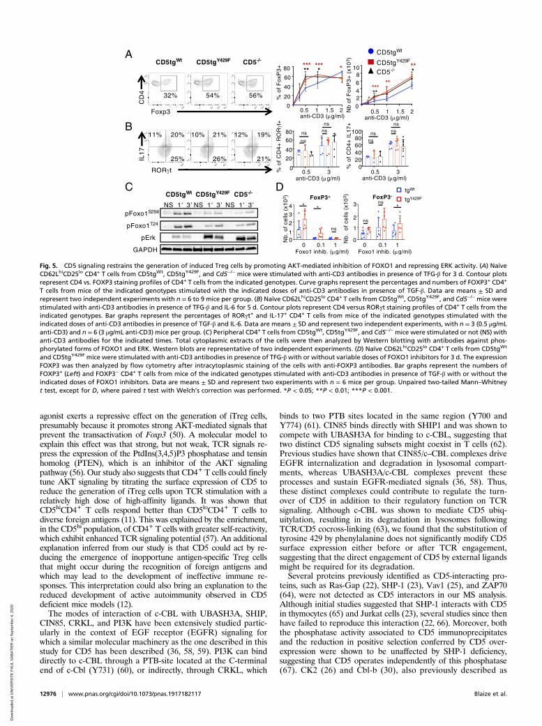
agonist exerts a repressive effect on the generation of iTreg cells,presumably because it promotes strong AKT-mediated signals thatprevent the transactivation of Foxp3 (50). A molecular model toexplain this effect was that strong, but not weak, TCR signals re-press the expression of the PtdIns(3,4,5)P3 phosphatase and tensinhomolog (PTEN), which is an inhibitor of the AKT signalingpathway (56). Our study also suggests that CD4+ T cells could finelytune AKT signaling by titrating the surface expression of CD5 toreduce the generation of iTreg cells upon TCR stimulation with arelatively high dose of high-affinity ligands. It was shown thatCD5hiCD4+ T cells respond better than CD5loCD4+ T cells todiverse foreign antigens (11). This was explained by the enrichment,in the CD5hi population, of CD4+ T cells with greater self-reactivity,which exhibit enhanced TCR signaling potential (57). An additionalexplanation inferred from our study is that CD5 could act by re-ducing the emergence of inopportune antigen-specific Treg cellsthat might occur during the recognition of foreign antigens andwhich may lead to the development of ineffective immune re-sponses. This interpretation could also bring an explanation to thereduced development of active autoimmunity observed in CD5deficient mice models (12).The modes of interaction of c-CBL with UBASH3A, SHIP,
CIN85, CRKL, and PI3K have been extensively studied partic-ularly in the context of EGF receptor (EGFR) signaling forwhich a similar molecular machinery as the one described in thisstudy for CD5 has been described (36, 58, 59). PI3K can binddirectly to c-CBL through a PTB-site located at the C-terminalend of c-Cbl (Y731) (60), or indirectly, through CRKL, which
binds to two PTB sites located in the same region (Y700 andY774) (61). CIN85 binds directly with SHIP1 and was shown tocompete with UBASH3A for binding to c-CBL, suggesting thattwo distinct CD5 signaling subsets might coexist in T cells (62).Previous studies have shown that CIN85/c–CBL complexes driveEGFR internalization and degradation in lysosomal compart-ments, whereas UBASH3A/c-CBL complexes prevent theseprocesses and sustain EGFR-mediated signals (36, 58). Thus,these distinct complexes could contribute to regulate the turn-over of CD5 in addition to their regulatory function on TCRsignaling. Although c-CBL was shown to mediate CD5 ubiq-uitylation, resulting in its degradation in lysosomes followingTCR/CD5 cocross-linking (63), we found that the substitution oftyrosine 429 by phenylalanine does not significantly modify CD5surface expression either before or after TCR engagement,suggesting that the direct engagement of CD5 by external ligandsmight be required for its degradation.Several proteins previously identified as CD5-interacting pro-
teins, such as Ras-Gap (22), SHP-1 (23), Vav1 (25), and ZAP70(64), were not detected as CD5 interactors in our MS analysis.Although initial studies suggested that SHP-1 interacts with CD5in thymocytes (65) and Jurkat cells (23), several studies since thenhave failed to reproduce this interaction (22, 66). Moreover, boththe phosphatase activity associated to CD5 immunoprecipitatesand the reduction in positive selection conferred by CD5 over-expression were shown to be unaffected by SHP-1 deficiency,suggesting that CD5 operates independently of this phosphatase(67). CK2 (26) and Cbl-b (30), also previously described as
A
C
pFoxo1T24
pFoxo1S256
pErk
GAPDH
CD5tgWt CD5tgY429F CD5-/-D
B
ROR t
11% 20%
25%
10% 21%
26%
12% 19%
21%
32% 54% 56%
CD5-/-CD5tgY429FCD5tgWt
CD
4
Foxp3
Foxo1 inhib. ( g/ml)
Nb
. o
f ce
lls(x
10
3 )
1
2
00.1 10
Nb
. o
f ce
lls(x
10
3 )
0
1
2
3
Foxo1 inhib. ( g/ml)0.1 10
FoxP3+ FoxP3-
* *
tgWt
tgY429F
ns
ns
ns3
4 *
% o
f C
D4
+ R
OR
t+
40
60
20
anti-CD3 ( g/ml)3
80
0.5
% o
f C
D4
+ I
L1
7+
0
4060
20
80
anti-CD3 ( g/ml)30.5
0
% o
f F
oxP
3+
0
20
40
60
Nb
of
Fo
xP3
+ (
x10
3 )
02
80
4
ns
ns nsns
nsns
nsns
68
10*****
**** *
CD5tgWt
CD5tgY429F
CD5-/-
*
anti-CD3 ( g/ml)10.5
anti-CD3 ( g/ml)21.5 10.5 21.5
*****
**
***
100
IL1
7
Fig. 5. CD5 signaling restrains the generation of induced Treg cells by promoting AKT-mediated inhibition of FOXO1 and repressing ERK activity. (A) NaïveCD62LhiCD25lo CD4+ T cells from CD5tgWt, CD5tgY429F, and Cd5−/− mice were stimulated with anti-CD3 antibodies in presence of TFG-β for 3 d. Contour plotsrepresent CD4 vs. FOXP3 staining profiles of CD4+ T cells from the indicated genotypes. Curve graphs represent the percentages and numbers of FOXP3+ CD4+
T cells from mice of the indicated genotypes stimulated with the indicated doses of anti-CD3 antibodies in presence of TGF-β. Data are means ± SD andrepresent two independent experiments with n = 6 to 9 mice per group. (B) Naïve CD62LhiCD25lo CD4+ T cells from CD5tgWt, CD5tgY429F, and Cd5−/− mice werestimulated with anti-CD3 antibodies in presence of TFG-β and IL-6 for 5 d. Contour plots represent CD4 versus RORγt staining profiles of CD4+ T cells from theindicated genotypes. Bar graphs represent the percentages of RORγt+ and IL-17+ CD4+ T cells from mice of the indicated genotypes stimulated with theindicated doses of anti-CD3 antibodies in presence of TGF-β and IL-6. Data are means ± SD and represent two independent experiments, with n = 3 (0.5 μg/mLanti-CD3) and n = 6 (3 μg/mL anti-CD3) mice per group. (C) Peripheral CD4+ T cells from CD5tgWt, CD5tgY429F, and Cd5−/− mice were stimulated or not (NS) withanti-CD3 antibodies for the indicated times. Total cytoplasmic extracts of the cells were then analyzed by Western blotting with antibodies against phos-phorylated forms of FOXO1 and ERK. Western blots are representative of two independent experiments. (D) Naïve CD62LhiCD25lo CD4+ T cells from CD5tgWt
and CD5tgY429F mice were stimulated with anti-CD3 antibodies in presence of TFG-βwith or without variable doses of FOXO1 inhibitors for 3 d. The expressionFOXP3 was then analyzed by flow cytometry after intracytoplasmic staining of the cells with anti-FOXP3 antibodies. Bar graphs represent the numbers ofFOXP3+ (Left) and FOXP3− CD4+ T cells from mice of the indicated genotypes stimulated with anti-CD3 antibodies in presence of TGF-β with or without theindicated doses of FOXO1 inhibitors. Data are means ± SD and represent two experiments with n = 6 mice per group. Unpaired two-tailed Mann–Whitneyt test, except for D, where paired t test with Welch’s correction was performed. *P < 0.05; **P < 0.01; ***P < 0.001.
12976 | www.pnas.org/cgi/doi/10.1073/pnas.1917182117 Blaize et al.
Dow
nloa
ded
at U
NIV
ER
SIT
E P
AU
L S
AB
AT
IER
on
Sep
tem
ber
4, 2
020
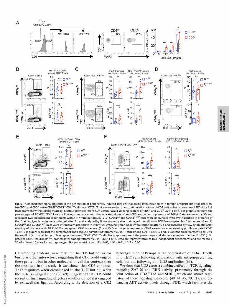
CD5-binding proteins, were recruited to CD5 but not as ro-bustly as other interactors, suggesting that CD5 could engagethese proteins but in other molecular or cellular contexts thanthe one used in this study. It was shown that CD5 enhancesTh17 responses when cross-linked to the TCR but not whenthe TCR is engaged alone (68, 69), suggesting that CD5 couldrecruit distinct signaling effectors whether or not it is engagedby extracellular ligands. Accordingly, the deletion of a CK2
binding site on CD5 impairs the polarization of CD4+ T cellsinto Th17 cells following stimulation with antigen-presentingcells but not following anti-CD3 antibodies (69).We show that CD5 exerts a combined effect on TCR signaling,
reducing ZAP-70 and ERK activity, presumably through thejoint action of UBASH3A and SHIP1, which are known regu-lators of these signaling molecules (40, 44, 45, 70, 71), and en-hancing AKT activity, likely through PI3K, which facilitates the
FoxP3+ amongNP311-325-I-Ab+ T cells
CD44+NP311-325-I-Ab+
FoxP3+ among1W1K-I-Ab+ T cells
Nrpl1loFoxP3+ among1W1K-I-Ab+ T cells
1W1K-I-Ab+ CD44+
among CD4+ T cells
1W1K
-I-A
b
CD44
0.36%
0
0.2
0.4
0.6
0.8
1.0
0
2
4
8
6
nb X
10
4%
0.35%
FoxP3
Nrp
1
%
0
10
20
30
40
50
0
5
10
20
15nb
X 1
03
0
10
15
20
%0
4
6
8
nb X
10
3
tgWt
tgY429F
FoxP3
Nrp
1
CD44 NP311 325 I A
CD44
NP
311-
325-
I-A
b
24%
14%
10%
5%
0,88%
0,85%
20%
10%
0
0.5
1.0
1.5
2.0
%
0
1
2
4
3
nb X
10
4
%
0
10
20
30
40
0
2
4
8
6
nb X
103
0
5
10
15
20
%
0
1
2
4
3
nb X
103
9%
3%
tgWt
tgY429F
tgWt
tgY429F
FoxP3
Nrp
1
NP311-325-I-Ab+ CD44+
among CD4+ T cellsNrpl1loFoxP3+ among
NP311-325-I-Ab+ T cells
tgWt
tgY429F
*
***
**
*
*
** **
*
CD4+ T cells
CD4+ T cells
B C
E F
CD
5tg
Y42
9FC
D5t
gW
t
CD
5tg
Y42
9FC
D5t
gW
t
CD
5tg
Y42
9FC
D5t
gW
t
CD
5tg
Y42
9FC
D5t
gW
t
5
2
21%
18%
CD
4
tgWt
tgY429F
0
10
20
30
40
50
0
8
16
24
ns
ns
Tbet
CD44+1W1K-I-Ab+
CD
5tg
Y42
9FC
D5t
gW
t
Tbet+ among1W1K-I-Ab+ T cells
%nb
X 1
03
D
ns
ns
ns
ns
CD44+1W1K-I-Ab+
% o
f CD
4+ F
oxP
3+
CD5
MFI: 9100 MFI: 1780
%fC
D4
FP
3
FoxP3
CD
4
9% 25%
CD5hiCD5lo
anti-CD3 (mg/ml)
0
20
40
60
0 0.25 0.5 1
CD4+CD62LhiCD25lo
CD5loCD5hi
A
CD5hi
CD5lo
** **
ns
Fig. 6. CD5-mediated signaling restrain the generation of peripherally induced Treg cells following immunization with foreign antigens and viral infection.(A) CD5lo and CD5hi naïve CD62LhiCD25lo CD4+ T cells from C57BL/6 mice were sorted prior to stimulation with anti-CD3 antibodies in presence of TFG-β for 3 d.Histograms show the sorting strategy. Contour plots represent CD4 versus FOXP3 staining profiles of CD5lo and CD5hi CD4+ T cells. Bar graphs represent thepercentages of FOXP3+ CD4+ T cells following stimulation with the indicated doses of anti-CD3 antibodies in presence of TGF-β. Data are means ± SD andrepresent two independent experiments with n = 7 mice per group. (B–D) CD5tgWt and CD5tgY429F mice were immunized with 1W1K peptide in presence ofIFA. Draining lymph nodes were collected after 7 d and analyzed by flow cytometry after staining of the cells with 1W1K-conjugated MHC tetramers. (E and F)CD5tgWt and CD5tgY429F mice were intranasally infected with PR8 virus. Draining lymph nodes were collected after 5 d and analyzed by flow cytometry afterstaining of the cells with NP311-325-conjugated MHC tetramers. (B and E) Contour plots represents CD44 versus tetramer staining profile on gated CD4+
T cells. Bar graphs represent the percentages and absolute numbers of tetramer+CD44+ T cells among CD4+ T cells. (C and F) Contour plots represents FoxP3 vs.Neuropilin1 (Nrp1) staining profile on gated tetramer+CD44+ CD4+ T cells. Bar graphs represent the percentages and absolute numbers of either FoxP3+ (solidgate) or FoxP3+ neuropilinlow (dashed gate) among tetramer+CD44+ CD4+ T cells. Data are representative of two independent experiments and are means ±SD of at least 10 mice for each genotype. Nonparametric t test *P < 0.05; **P < 0.01; ***P < 0.001.
Blaize et al. PNAS | June 9, 2020 | vol. 117 | no. 23 | 12977
IMMUNOLO
GYAND
INFLAMMATION
Dow
nloa
ded
at U
NIV
ER
SIT
E P
AU
L S
AB
AT
IER
on
Sep
tem
ber
4, 2
020

retention of FOXO1 in the cytoplasm and prevents the conse-quent transactivation of Foxp3 (51). This signaling complex doesnot detectably modulate the expression of RORγt or T-BET inactivated CD4+ T cells in vitro and in vivo, respectively, indic-ative of a selective effect of CD5 on the generation of iTreg cells.Remarkably, studies using SHIP1- and PI3K-deficient murinemodels identified repressive functions for both proteins on thegeneration of induced Treg cells (72, 73). Whether UBASH3A isalso directly involved in the control of this population has not yetbeen studied. However, a recent investigation performed in hu-man T cells has shown that it represses NF-κB signaling (74), akey pathway for the generation of induced Treg cells (75). Thus,CD5 could act as a scaffold that selectively engages specificsignaling effectors involved in the generation of peripheral Tregcells to selectively control the expansion of this populationduring immune responses.Previous studies have shown that CD5 deficiency on the
BALB/c but not on the C57BL/6 genetic background leads toincreased numbers of thymic Treg cells, suggesting that CD5 mayalso repress the generation of these cells according to the mo-lecular context in which CD5 operates in the thymus (13, 14).CD5 is highly expressed at the surface of thymic Treg cells, in-dicating that the ability of CD5 to negatively regulate FoxP3expression in the thymus could also be overcome by additionalsignals (such as high TCR affinity for self-ligand) that imposeFoxP3 expression despite the opposite force exerted by CD5. Wethus speculate that CD5-mediated inhibition may be sufficient toblock Foxp3 expression and Treg lineage commitment in thy-mocytes that express TCRs that bind with intermediate affinityto self-ligands, preventing their commitment to the Treg lineage.In addition, coreceptors, such as GITR, OX40, and TNFR2, arehighly expressed by thymic Treg progenitors and were also shownto trigger costimulatory signals that induce thymic Treg differen-tiation (76). In contrast, these coreceptors are poorly expressed bynaïve peripheral CD4 T cells, suggesting that the negative ef-fect of CD5 on FoxP3 expression may become predominant inthis subset.A recent study reported an effect of CD5 opposite to that
shown in our study on the generation of peripherally inducedTreg cells in a mouse model in which tolerance to experimentalautoimmune encephalomyelitis is induced by direct delivery ofencephalitogenic peptides to antigen-presenting dendritic cells inthe absence of adjuvant (15). Those authors found that CD5prevents the inhibition of Treg cell induction potentially medi-ated by effector cell cytokines, such as IL-6, IL-4, and IFN-γ.Although they do not provide a clear signaling mechanism bywhich CD5 operates here, they show that CD5 promotes thegeneration of Treg cells by inhibiting IL-6–mediated AKT sig-naling leading to mammalian target of rapamycin activation. Onepotential explanation for the apparent discrepancy betweenthose results and our current findings is that CD5 might exertopposite effects on PI3K/AKT signaling in tolerized and non-tolerized T cells. A recent proteomic study showed that CD5interacts with both c-CBL and CBL-b in peripheral CD4+ T cells(30), suggesting that distinct pools of CD5, connected either to a
c-CBL or CBL-b signalosome, could be preferentially assembledunder specific stimulation conditions. Whereas c-CBL has knownpositive effects on PI3K/AKT signaling (46), CBL-b blocks thispathway by inducing PI3K ubiquitylation (77) and by blockingNedd4-mediated ubiquitylation of PTEN (78). Because CBL-bexpression is up-regulated in CD4+ T cells after tolerizing signals(79) and degraded in CD4+ T cells that have received appropriatecostimulatory signals (80), we speculate that CD5 signaling poolsmay vary in size or composition, tuning up or down the PI3K/AKTsignaling axis according to the immunological context in which cellsare stimulated.Altogether, the findings of our study suggest that CD5 engages
a polymorphic signaling machinery that can transduce both stim-ulatory and inhibitory signals to selectively control specific T cellresponses depending on the immunological context. Our resultsalso provide new insights into the paradigm of coreceptor signal-ing, suggesting that, in addition to providing classic enhancing ordampening inputs, coreceptors coordinate TCR signals that mayhave antagonist effects to promote specific functional outcomes,such as the generation of iTreg cells.
Materials and MethodsFull details of materials and methods, including mice, antibodies, cell stim-ulation, immunoprecipitations, MS analysis, calcium flux, Western blot, im-munization and influenza virus infection, statistical analysis are provided inSI Appendix.
Mice. All the experiments were conducted with sex- and age-matched micebetween 6- and 12-wk-old housed under specific pathogen-free conditionsat the INSERM Zootechnie US-006 animal facility, which is accredited by theFrench Ministry of Agriculture to perform experiments on live mice (ac-creditation number A-31 55508). All experimental protocols were approvedby the local ethics committee and are in compliance with the French andEuropean Union regulations on care and protection of laboratory animals(EC Directive 2010/63).
Data Availability Statement. The data generated or analyzed during this studyare included in the article and its SI Appendix or dataset files. The MS pro-teomics data have been deposited to the ProteomeXchange Consortium viathe PRIDE partner repository with the dataset identifier PXD017343. Rawdata from Figs. 1 A and C and 2 A, B, and D and SI Appendix, Fig. S1 A–C canbe found in Datasets S1–S3 and S5.
ACKNOWLEDGMENTS. We thank L. Dupré for critical reading of the manu-script; F.-E. L’Faqihi-Olive, V. Duplan-Eche, and A.-L. Iscache for technicalassistance at the flow-cytometry facility of INSERM U1043; the personnelof the Centre Régional d’Exploration Fonctionnelle et de Ressources Expér-imentales for expert animal care; and L. Guennec for administrative assis-tance. This work was supported by INSERM and Sanofi (Avenir grant to R.L.);the Association pour la Recherche sur le Cancer; the Intramural ResearchProgram of the Eunice Kennedy Shriver, National Institute of Child Healthand Human Development; a Marie Curie International Reintegration Grant(to R.L.); the French Ministry of Higher Education and Research (PhD fellow-ship to G.B.); the Région Midi-Pyrénées, European funds (Fonds Européensde Développement Régional), Toulouse Métropole, and the French Ministryof Research with the ‘Investissement d’Avenir Infrastructures Nationales enBiologie et Santé program’ (ProFI, Proteomics French Infrastructure project,ANR-10-INBS-08) (to O.B.-S.).
1. L. Chen, D. B. Flies, Molecular mechanisms of T cell co-stimulation and co-inhibition.Nat. Rev. Immunol. 13, 227–242 (2013).
2. J. M. Coquet, L. Rausch, J. Borst, The importance of co-stimulation in the orchestrationof T helper cell differentiation. Immunol. Cell Biol. 93, 780–788 (2015).
3. A. C. Anderson, N. Joller, V. K. Kuchroo, Lag-3, Tim-3, and TIGIT: Co-inhibitory receptorswith specialized functions in immune regulation. Immunity 44, 989–1004 (2016).
4. O. Acuto, F. Michel, CD28-mediated co-stimulation: A quantitative support for TCRsignalling. Nat. Rev. Immunol. 3, 939–951 (2003).
5. R. V. Parry, J. L. Riley, S. G. Ward, Signalling to suit function: Tailoring phosphoino-sitide 3-kinase during T-cell activation. Trends Immunol. 28, 161–168 (2007).
6. C. E. Rudd, A. Taylor, H. Schneider, CD28 and CTLA-4 coreceptor expression and signaltransduction. Immunol. Rev. 229, 12–26 (2009).
7. L. S. K. Walker, PD-1 and CTLA4: Two checkpoints, one pathway? Sci. Immunol. 2,eaan3864 (2017).
8. A. Tarakhovsky et al., A role for CD5 in TCR-mediated signal transduction and thy-mocyte selection. Science 269, 535–537 (1995).
9. H. S. Azzam et al., CD5 expression is developmentally regulated by T cell receptor(TCR) signals and TCR avidity. J. Exp. Med. 188, 2301–2311 (1998).
10. H. S. Azzam et al., Fine tuning of TCR signaling by CD5. J. Immunol. 166, 5464–5472(2001).
11. J. N. Mandl, J. P. Monteiro, N. Vrisekoop, R. N. Germain, T cell-positive selection usesself-ligand binding strength to optimize repertoire recognition of foreign antigens.Immunity 38, 263–274 (2013).
12. R. C. Axtell, M. S. Webb, S. R. Barnum, C. Raman, Cutting edge: Critical role for CD5 inexperimental autoimmune encephalomyelitis: Inhibition of engagement reversesdisease in mice. J. Immunol. 173, 2928–2932 (2004).
13. T. Dasu et al., CD5 plays an inhibitory role in the suppressive function of murineCD4(+) CD25(+) T(reg) cells. Immunol. Lett. 119, 103–113 (2008).
12978 | www.pnas.org/cgi/doi/10.1073/pnas.1917182117 Blaize et al.
Dow
nloa
ded
at U
NIV
ER
SIT
E P
AU
L S
AB
AT
IER
on
Sep
tem
ber
4, 2
020
14. D. Ordoñez-Rueda et al., Increased numbers of thymic and peripheral CD4+ CD25+Foxp3+cells in the absence of CD5 signaling. Eur. J. Immunol. 39, 2233–2247 (2009).
15. J. G. Henderson, A. Opejin, A. Jones, C. Gross, D. Hawiger, CD5 instructs extrathymicregulatory T cell development in response to self and tolerizing antigens. Immunity42, 471–483 (2015).
16. H. Van de Velde, I. von Hoegen, W. Luo, J. R. Parnes, K. Thielemans, The B-cell surfaceprotein CD72/Lyb-2 is the ligand for CD5. Nature 351, 662–665 (1991).
17. J. Vera et al., The CD5 ectodomain interacts with conserved fungal cell wall compo-nents and protects from zymosan-induced septic shock-like syndrome. Proc. Natl.Acad. Sci. U.S.A. 106, 1506–1511 (2009).
18. C. Zhang et al., CD5 binds to interleukin-6 and induces a feed-forward loop with thetranscription factor STAT3 in B cells to promote cancer. Immunity 44, 913–923 (2016).
19. A. Bhandoola et al., CD5-mediated inhibition of TCR signaling during intrathymicselection and development does not require the CD5 extracellular domain. Eur.J. Immunol. 32, 1811–1817 (2002).
20. N. Osman, A. I. Lazarovits, M. J. Crumpton, Physical association of CD5 and the T cellreceptor/CD3 antigen complex on the surface of human T lymphocytes. Eur.J. Immunol. 23, 1173–1176 (1993).
21. K. E. Burgess, M. Yamamoto, K. V. Prasad, C. E. Rudd, CD5 acts as a tyrosine kinase substratewithin a receptor complex comprising T-cell receptor zeta chain/CD3 and protein-tyrosinekinases p56lck and p59fyn. Proc. Natl. Acad. Sci. U.S.A. 89, 9311–9315 (1992).
22. K. M. Dennehy, R. Broszeit, W. F. Ferris, A. D. Beyers, Thymocyte activation inducesthe association of the proto-oncoprotein c-cbl and ras GTPase-activating protein withCD5. Eur. J. Immunol. 28, 1617–1625 (1998).
23. J. J. Perez-Villar et al., CD5 negatively regulates the T-cell antigen receptor signaltransduction pathway: Involvement of SH2-containing phosphotyrosine phosphataseSHP-1. Mol. Cell. Biol. 19, 2903–2912 (1999).
24. K. M. Dennehy et al., Thymocyte activation induces the association of phosphatidy-linositol 3-kinase and pp120 with CD5. Eur. J. Immunol. 27, 679–686 (1997).
25. S. I. Gringhuis, L. F. de Leij, P. J. Coffer, E. Vellenga, Signaling through CD5 activates apathway involving phosphatidylinositol 3-kinase, Vav, and Rac1 in human mature Tlymphocytes. Mol. Cell. Biol. 18, 1725–1735 (1998).
26. C. Raman, R. P. Kimberly, Differential CD5-dependent regulation of CD5-associatedCK2 activity in mature and immature T cells: Implication on TCR/CD3-mediated acti-vation. J. Immunol. 161, 5817–5820 (1998).
27. C. Raman, A. Kuo, J. Deshane, D. W. Litchfield, R. P. Kimberly, Regulation of caseinkinase 2 by direct interaction with cell surface receptor CD5. J. Biol. Chem. 273,19183–19189 (1998).
28. X. Lu et al., AP2 adaptor complex-dependent internalization of CD5: Differentialregulation in T and B cells. J. Immunol. 168, 5612–5620 (2002).
29. G. Voisinne et al., Quantitative interactomics in primary T cells unveils TCR signaldiversification extent and dynamics. Nat. Immunol. 20, 1530–1541 (2019).
30. G. Voisinne et al., Co-recruitment analysis of the CBL and CBLB signalosomes in pri-mary T cells identifies CD5 as a key regulator of TCR-induced ubiquitylation.Mol. Syst.Biol. 12, 876 (2016).
31. K. M. Dennehy et al., Determination of the tyrosine phosphorylation sites in the T celltransmembrane glycoprotein CD5. Int. Immunol. 13, 149–156 (2001).
32. J. M. Vilà et al., Residues Y429 and Y463 of the human CD5 are targeted by proteintyrosine kinases. Eur. J. Immunol. 31, 1191–1198 (2001).
33. M. L. Lupher Jr., Z. Songyang, S. E. Shoelson, L. C. Cantley, H. Band, The Cblphosphotyrosine-binding domain selects a D(N/D)XpY motif and binds to the Tyr292negative regulatory phosphorylation site of ZAP-70. J. Biol. Chem. 272, 33140–33144 (1997).
34. J. E. van Leeuwen, P. K. Paik, L. E. Samelson, Activation of nuclear factor of activatedT cells-(NFAT) and activating protein 1 (AP-1) by oncogenic 70Z Cbl requires an intactphosphotyrosine binding domain but not Crk(L) or p85 phosphatidylinositol 3-kinaseassociation. J. Biol. Chem. 274, 5153–5162 (1999).
35. H. Take et al., Cloning and characterization of a novel adaptor protein, CIN85, thatinteracts with c-Cbl. Biochem. Biophys. Res. Commun. 268, 321–328 (2000).
36. K. Kowanetz et al., Suppressors of T-cell receptor signaling Sts-1 and Sts-2 bind to Cbl andinhibit endocytosis of receptor tyrosine kinases. J. Biol. Chem. 279, 32786–32795 (2004).
37. M. Naramura, H. K. Kole, R. J. Hu, H. Gu, Altered thymic positive selection and intracellularsignals in Cbl-deficient mice. Proc. Natl. Acad. Sci. U.S.A. 95, 15547–15552 (1998).
38. N. Rao et al., Negative regulation of Lck by Cbl ubiquitin ligase. Proc. Natl. Acad. Sci.U.S.A. 99, 3794–3799 (2002).
39. M. S. Kong et al., Inhibition of T cell activation and function by the adaptor proteinCIN85. Sci. Signal. 12, eaav4373 (2019).
40. S. Dong et al., T cell receptor for antigen induces linker for activation of T cell-dependent activation of a negative signaling complex involving Dok-2, SHIP-1, andGrb-2. J. Exp. Med. 203, 2509–2518 (2006).
41. P. Van Slyke et al., Dok-R mediates attenuation of epidermal growth factor-dependent mitogen-activated protein kinase and Akt activation through processiverecruitment of c-Src and Csk. Mol. Cell. Biol. 25, 3831–3841 (2005).
42. M. Zhao, J. A. Janas, M. Niki, P. P. Pandolfi, L. Van Aelst, Dok-1 independently attenuatesRas/mitogen-activated protein kinase and Src/c-myc pathways to inhibit platelet-derivedgrowth factor-induced mitogenesis. Mol. Cell. Biol. 26, 2479–2489 (2006).
43. Q. Liu et al., The inositol polyphosphate 5-phosphatase ship is a crucial negativeregulator of B cell antigen receptor signaling. J. Exp. Med. 188, 1333–1342 (1998).
44. M. G. Tomlinson, V. L. Heath, C. W. Turck, S. P. Watson, A. Weiss, SHIP family inositolphosphatases interact with and negatively regulate the Tec tyrosine kinase. J. Biol.Chem. 279, 55089–55096 (2004).
45. A. M. Scharenberg et al., Phosphatidylinositol-3,4,5-trisphosphate (PtdIns-3,4,5-P3)/Tec kinase-dependent calcium signaling pathway: A target for SHIP-mediated in-hibitory signals. EMBO J. 17, 1961–1972 (1998).
46. C. B. Thien et al., c-Cbl promotes T cell receptor-induced thymocyte apoptosis by activatingthe phosphatidylinositol 3-kinase/Akt pathway. J. Biol. Chem. 285, 10969–10981 (2010).
47. M. Sattler et al., Steel factor induces tyrosine phosphorylation of CRKL and binding ofCRKL to a complex containing c-kit, phosphatidylinositol 3-kinase, and p120(CBL).J. Biol. Chem. 272, 10248–10253 (1997).
48. J. A. Deane et al., T-cell function is partially maintained in the absence of class IAphosphoinositide 3-kinase signaling. Blood 109, 2894–2902 (2007).
49. A. E. Moran et al., T cell receptor signal strength in Treg and iNKT cell developmentdemonstrated by a novel fluorescent reporter mouse. J. Exp. Med. 208, 1279–1289 (2011).
50. M. O. Li, A. Y. Rudensky, T cell receptor signalling in the control of regulatory T celldifferentiation and function. Nat. Rev. Immunol. 16, 220–233 (2016).
51. S. Fabre et al., Stable activation of phosphatidylinositol 3-kinase in the T cell immu-nological synapse stimulates Akt signaling to FoxO1 nuclear exclusion and cell growthcontrol. J. Immunol. 174, 4161–4171 (2005).
52. L. Lu et al., Role of SMAD and non-SMAD signals in the development of Th17 andregulatory T cells. J. Immunol. 184, 4295–4306 (2010).
53. J. M. Weiss et al., Neuropilin 1 is expressed on thymus-derived natural regulatoryT cells, but not mucosa-generated induced Foxp3+ T reg cells. J. Exp. Med. 209,1723–1742 (2012).
54. T. Korn et al., IL-6 controls Th17 immunity in vivo by inhibiting the conversion ofconventional T cells into Foxp3+ regulatory T cells. Proc. Natl. Acad. Sci. U.S.A. 105,18460–18465 (2008).
55. R. J. Betts et al., Influenza A virus infection results in a robust, antigen-responsive, andwidely disseminated Foxp3+ regulatory T cell response. J. Virol. 86, 2817–2825 (2012).
56. J. Gomez-Rodriguez et al., Itk-mediated integration of T cell receptor and cytokinesignaling regulates the balance between Th17 and regulatory T cells. J. Exp. Med. 211,529–543 (2014).
57. S. P. Persaud, C. R. Parker, W. L. Lo, K. S. Weber, P. M. Allen, Intrinsic CD4+ T cellsensitivity and response to a pathogen are set and sustained by avidity for thymic andperipheral complexes of self peptide and MHC. Nat. Immunol. 15, 266–274 (2014).
58. P. Soubeyran, K. Kowanetz, I. Szymkiewicz, W. Y. Langdon, I. Dikic, Cbl-CIN85-en-dophilin complex mediates ligand-induced downregulation of EGF receptors. Nature416, 183–187 (2002).
59. R. Avraham, Y. Yarden, Feedback regulation of EGFR signalling: Decision making byearly and delayed loops. Nat. Rev. Mol. Cell Biol. 12, 104–117 (2011).
60. S. Hunter, E. A. Burton, S. C. Wu, S. M. Anderson, Fyn associates with Cbl and phos-phorylates tyrosine 731 in Cbl, a binding site for phosphatidylinositol 3-kinase. J. Biol.Chem. 274, 2097–2106 (1999).
61. C. E. Andoniou, C. B. Thien, W. Y. Langdon, The two major sites of cbl tyrosinephosphorylation in abl-transformed cells select the crkL SH2 domain. Oncogene 12,1981–1989 (1996).
62. G. Peruzzi et al., The adaptor molecule CIN85 regulates Syk tyrosine kinase level by acti-vating the ubiquitin-proteasome degradation pathway. J. Immunol. 179, 2089–2096 (2007).
63. D. Demydenko, c-Cbl mediated ubiquitylation and regulation of cell surface exposureof CD5. Biochem. Biophys. Res. Commun. 392, 500–504 (2010).
64. H. Gary-Gouy, V. Lang, S. Sarun, L. Boumsell, G. Bismuth, In vivo association of CD5with tyrosine-phosphorylated ZAP-70 and p21 phospho-zeta molecules in humanCD3+ thymocytes. J. Immunol. 159, 3739–3747 (1997).
65. G. Pani, K. D. Fischer, I. Mlinaric-Rascan, K. A. Siminovitch, Signaling capacity of theT cell antigen receptor is negatively regulated by the PTP1C tyrosine phosphatase.J. Exp. Med. 184, 839–852 (1996).
66. H. Gary-Gouy et al., The pseudo-immunoreceptor tyrosine-based activation motif of CD5mediates its inhibitory action on B-cell receptor signaling. J. Biol. Chem. 275, 548–556 (2000).
67. B. Dong et al., CD5-mediated inhibition of TCR signaling proceeds normally in theabsence of SHP-1. Int. J. Mol. Med. 38, 45–56 (2016).
68. J. de Wit et al., CD5 costimulation induces stable Th17 development by promoting IL-23R expression and sustained STAT3 activation. Blood 118, 6107–6114 (2011).
69. C. M. Sestero et al., CD5-dependent CK2 activation pathway regulates threshold forT cell anergy. J. Immunol. 189, 2918–2930 (2012).
70. N. Carpino et al., Regulation of ZAP-70 activation and TCR signaling by two relatedproteins, Sts-1 and Sts-2. Immunity 20, 37–46 (2004).
71. B. San Luis, B. Sondgeroth, N. Nassar, N. Carpino, Sts-2 is a phosphatase that nega-tively regulates zeta-associated protein (ZAP)-70 and T cell receptor signaling path-ways. J. Biol. Chem. 286, 15943–15954 (2011).
72. M. Kashiwada et al., Downstream of tyrosine kinases-1 and Src homology2-containing inositol 5′-phosphatase are required for regulation of CD4+CD25+ T celldevelopment. J. Immunol. 176, 3958–3965 (2006).
73. S. Sauer et al., T cell receptor signaling controls Foxp3 expression via PI3K, Akt, andmTOR. Proc. Natl. Acad. Sci. U.S.A. 105, 7797–7802 (2008).
74. Y. Ge, T. K. Paisie, J. R. B. Newman, L. M. McIntyre, P. Concannon, UBASH3A mediatesrisk for type 1 diabetes through inhibition of T-cell receptor-induced NF-κB signaling.Diabetes 66, 2033–2043 (2017).
75. H. Oh et al., An NF-kappaB transcription-factor-dependent lineage-specific tran-scriptional program promotes regulatory T cell identity and function. Immunity 47,450–465 e5 (2017).
76. S. A. Mahmud et al., Costimulation via the tumor-necrosis factor receptor superfamilycouples TCR signal strength to the thymic differentiation of regulatory T cells. Nat.Immunol. 15, 473–481 (2014).
77. D. Fang, Y. C. Liu, Proteolysis-independent regulation of PI3K by Cbl-b-mediatedubiquitination in T cells. Nat. Immunol. 2, 870–875 (2001).
78. H. Guo et al., E3 ubiquitin ligase Cbl-b regulates Pten via Nedd4 in T cells in-dependently of its ubiquitin ligase activity. Cell Rep. 1, 472–482 (2012).
79. M. S. Jeon et al., Essential role of the E3 ubiquitin ligase Cbl-b in T cell anergy in-duction. Immunity 21, 167–177 (2004).
80. B. Yang et al., Nedd4 augments the adaptive immune response by promotingubiquitin-mediated degradation of Cbl-b in activated T cells. Nat. Immunol. 9,1356–1363 (2008).
Blaize et al. PNAS | June 9, 2020 | vol. 117 | no. 23 | 12979
IMMUNOLO
GYAND
INFLAMMATION
Dow
nloa
ded
at U
NIV
ER
SIT
E P
AU
L S
AB
AT
IER
on
Sep
tem
ber
4, 2
020
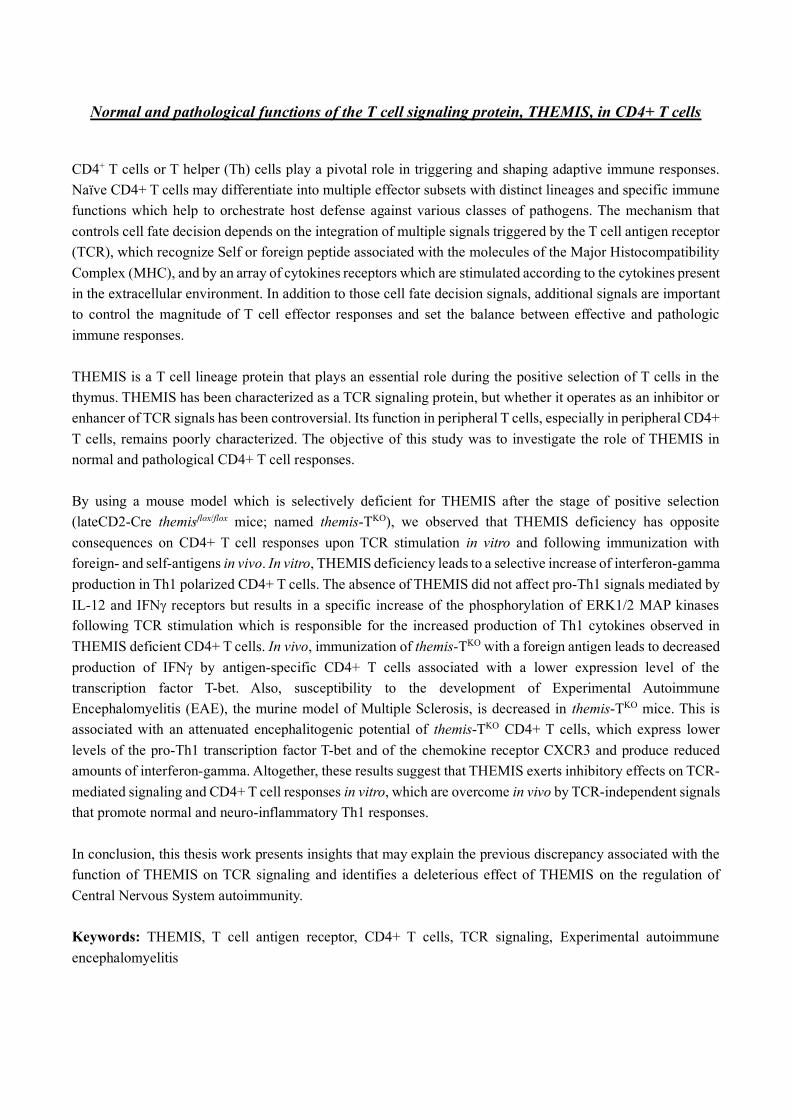
Normal and pathological functions of the T cell signaling protein, THEMIS, in CD4+ T cells
CD4+ T cells or T helper (Th) cells play a pivotal role in triggering and shaping adaptive immune responses.
Naïve CD4+ T cells may differentiate into multiple effector subsets with distinct lineages and specific immune
functions which help to orchestrate host defense against various classes of pathogens. The mechanism that
controls cell fate decision depends on the integration of multiple signals triggered by the T cell antigen receptor
(TCR), which recognize Self or foreign peptide associated with the molecules of the Major Histocompatibility
Complex (MHC), and by an array of cytokines receptors which are stimulated according to the cytokines present
in the extracellular environment. In addition to those cell fate decision signals, additional signals are important
to control the magnitude of T cell effector responses and set the balance between effective and pathologic
immune responses.
THEMIS is a T cell lineage protein that plays an essential role during the positive selection of T cells in the
thymus. THEMIS has been characterized as a TCR signaling protein, but whether it operates as an inhibitor or
enhancer of TCR signals has been controversial. Its function in peripheral T cells, especially in peripheral CD4+
T cells, remains poorly characterized. The objective of this study was to investigate the role of THEMIS in
normal and pathological CD4+ T cell responses.
By using a mouse model which is selectively deficient for THEMIS after the stage of positive selection
(lateCD2-Cre themisflox/flox mice; named themis-TKO), we observed that THEMIS deficiency has opposite
consequences on CD4+ T cell responses upon TCR stimulation in vitro and following immunization with
foreign- and self-antigens in vivo. In vitro, THEMIS deficiency leads to a selective increase of interferon-gamma
production in Th1 polarized CD4+ T cells. The absence of THEMIS did not affect pro-Th1 signals mediated by
IL-12 and IFN receptors but results in a specific increase of the phosphorylation of ERK1/2 MAP kinases
following TCR stimulation which is responsible for the increased production of Th1 cytokines observed in
THEMIS deficient CD4+ T cells. In vivo, immunization of themis-TKO with a foreign antigen leads to decreased
production of IFN by antigen-specific CD4+ T cells associated with a lower expression level of the
transcription factor T-bet. Also, susceptibility to the development of Experimental Autoimmune
Encephalomyelitis (EAE), the murine model of Multiple Sclerosis, is decreased in themis-TKO mice. This is
associated with an attenuated encephalitogenic potential of themis-TKO CD4+ T cells, which express lower
levels of the pro-Th1 transcription factor T-bet and of the chemokine receptor CXCR3 and produce reduced
amounts of interferon-gamma. Altogether, these results suggest that THEMIS exerts inhibitory effects on TCR-
mediated signaling and CD4+ T cell responses in vitro, which are overcome in vivo by TCR-independent signals
that promote normal and neuro-inflammatory Th1 responses.
In conclusion, this thesis work presents insights that may explain the previous discrepancy associated with the
function of THEMIS on TCR signaling and identifies a deleterious effect of THEMIS on the regulation of
Central Nervous System autoimmunity.
Keywords: THEMIS, T cell antigen receptor, CD4+ T cells, TCR signaling, Experimental autoimmune
encephalomyelitis
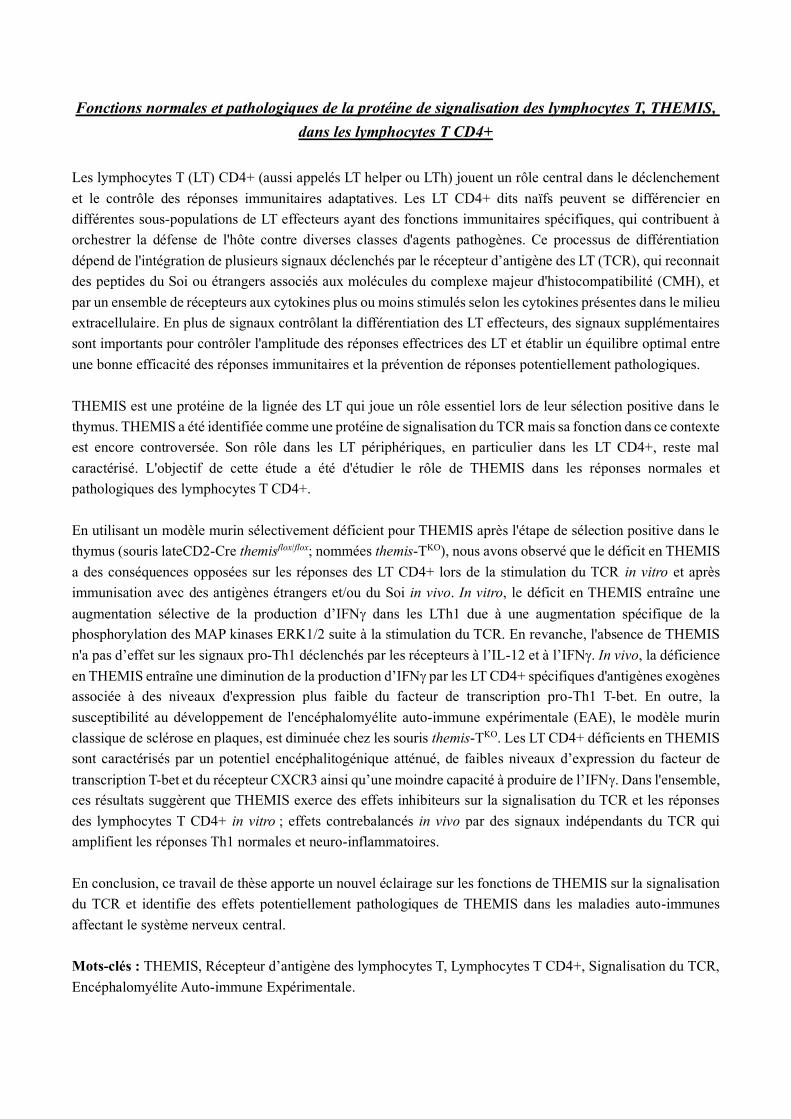
Fonctions normales et pathologiques de la protéine de signalisation des lymphocytes T, THEMIS,
dans les lymphocytes T CD4+
Les lymphocytes T (LT) CD4+ (aussi appelés LT helper ou LTh) jouent un rôle central dans le déclenchement
et le contrôle des réponses immunitaires adaptatives. Les LT CD4+ dits naïfs peuvent se différencier en
différentes sous-populations de LT effecteurs ayant des fonctions immunitaires spécifiques, qui contribuent à
orchestrer la défense de l'hôte contre diverses classes d'agents pathogènes. Ce processus de différentiation
dépend de l'intégration de plusieurs signaux déclenchés par le récepteur d’antigène des LT (TCR), qui reconnait
des peptides du Soi ou étrangers associés aux molécules du complexe majeur d'histocompatibilité (CMH), et
par un ensemble de récepteurs aux cytokines plus ou moins stimulés selon les cytokines présentes dans le milieu
extracellulaire. En plus de signaux contrôlant la différentiation des LT effecteurs, des signaux supplémentaires
sont importants pour contrôler l'amplitude des réponses effectrices des LT et établir un équilibre optimal entre
une bonne efficacité des réponses immunitaires et la prévention de réponses potentiellement pathologiques.
THEMIS est une protéine de la lignée des LT qui joue un rôle essentiel lors de leur sélection positive dans le
thymus. THEMIS a été identifiée comme une protéine de signalisation du TCR mais sa fonction dans ce contexte
est encore controversée. Son rôle dans les LT périphériques, en particulier dans les LT CD4+, reste mal
caractérisé. L'objectif de cette étude a été d'étudier le rôle de THEMIS dans les réponses normales et
pathologiques des lymphocytes T CD4+.
En utilisant un modèle murin sélectivement déficient pour THEMIS après l'étape de sélection positive dans le
thymus (souris lateCD2-Cre themisflox/flox; nommées themis-TKO), nous avons observé que le déficit en THEMIS
a des conséquences opposées sur les réponses des LT CD4+ lors de la stimulation du TCR in vitro et après
immunisation avec des antigènes étrangers et/ou du Soi in vivo. In vitro, le déficit en THEMIS entraîne une
augmentation sélective de la production d’IFN dans les LTh1 due à une augmentation spécifique de la
phosphorylation des MAP kinases ERK1/2 suite à la stimulation du TCR. En revanche, l'absence de THEMIS
n'a pas d’effet sur les signaux pro-Th1 déclenchés par les récepteurs à l’IL-12 et à l’IFN. In vivo, la déficience
en THEMIS entraîne une diminution de la production d’IFN par les LT CD4+ spécifiques d'antigènes exogènes
associée à des niveaux d'expression plus faible du facteur de transcription pro-Th1 T-bet. En outre, la
susceptibilité au développement de l'encéphalomyélite auto-immune expérimentale (EAE), le modèle murin
classique de sclérose en plaques, est diminuée chez les souris themis-TKO. Les LT CD4+ déficients en THEMIS
sont caractérisés par un potentiel encéphalitogénique atténué, de faibles niveaux d’expression du facteur de
transcription T-bet et du récepteur CXCR3 ainsi qu’une moindre capacité à produire de l’IFN. Dans l'ensemble,
ces résultats suggèrent que THEMIS exerce des effets inhibiteurs sur la signalisation du TCR et les réponses
des lymphocytes T CD4+ in vitro ; effets contrebalancés in vivo par des signaux indépendants du TCR qui
amplifient les réponses Th1 normales et neuro-inflammatoires.
En conclusion, ce travail de thèse apporte un nouvel éclairage sur les fonctions de THEMIS sur la signalisation
du TCR et identifie des effets potentiellement pathologiques de THEMIS dans les maladies auto-immunes
affectant le système nerveux central.
Mots-clés : THEMIS, Récepteur d’antigène des lymphocytes T, Lymphocytes T CD4+, Signalisation du TCR,
Encéphalomyélite Auto-immune Expérimentale.