Trends in Acute Ischemic Stroke Trials Through the 20th Century
Upload
independentCategory
view
3download
0
Document down posted on the Web before copyediting.
ARTICLE IN PRESS+ModelREPC-497; No. of Pages 7
Rev Port Cardiol. 2014;xxx(xx):xxx---xxx
Revista Portuguesa de
CardiologiaPortuguese Journal of Cardiology
www.revportcardiol.org
ORIGINAL ARTICLE
Revisiting the slow force response: The role of the PKGsignaling pathway in the normal and the ischemicheart�
Ricardo Castro-Ferreira1, João Sérgio Neves1, Ricardo Ladeiras-Lopes1,André M. Leite-Moreira, Manuel Neiva-Sousa, João Almeida-Coelho,João Ferreira-Martins, Adelino F. Leite-Moreira ∗
Department of Physiology and Cardiothoracic Surgery, Faculty of Medicine, University of Porto, Porto, Portugal
Received 4 June 2013; accepted 11 March 2014
KEYWORDSCyclicGMP-dependentprotein kinase type I;Nitric oxide;Phosphodiesterase 5inhibitors;Myocardial ischemia;Cardiovascularphysiology
AbstractIntroduction: The myocardial response to acute stretch consists of a two-phase increase in con-tractility: an acute increase by the Frank-Starling mechanism and a gradual and time-dependentincrease in force generated known as the slow force response (SFR). The SFR is actively mod-ulated by different signaling pathways, but the role of protein kinase G (PKG) signaling isunknown. In this study we aim to characterize the role of the PKG signaling pathway in theSFR under normal and ischemic conditions.Methods: Rabbit papillary muscles were stretched from 92 to 100% of maximum length (Lmax)under basal conditions, in the absence (1) or presence of: a PKG agonist (2) and a PKG inhibitor(3); under ischemic conditions in the absence (4) or presence of: a PKG agonist (5); a nitricoxide (NO) donor (6) and a phosphodiesterase 5 (PDE5) inhibitor (7).Results: Under normoxia, the SFR was significantly attenuated by inhibition of PKG andremained unaltered with PKG activation. Ischemia induced a progressive decrease inmyocardial contractility after stretch. Neither the PKG agonist nor the NO donor alteredthe myocardial response to stretch under ischemic conditions. However, the use of a PDE5inhibitor in ischemia partially reversed the progressive deterioration in contractility.Conclusions: PKG activity is essential for the SFR. During ischemia, a progressive decline in theforce is observed in response to acute myocardial stretch. This dysfunctional response can bepartially reversed by the use of PDE5 inhibitors.
loaded from http://www.elsevier.pt, day 25/09/2014. This copy is for personal use. Any transmission of this document by any media or format is strictly prohibited. This early online article has been reviewed, accepted and
Please cite this article in press as: Castro-Ferreira R, et al. Revisiting the slow force response: The role of the PKG signalingpathway in the normal and the ischemic heart. Rev Port Cardiol. 2014. http://dx.doi.org/10.1016/j.repc.2014.03.006
© 2013 Sociedade Portuguesa de Cardiologia. Published by Elsevier España, S.L.U. All rightsreserved.
� Supported by the Portuguese Foundation for Science and Technology Grants PEst-C/SAU/UI0051/2011 and EXCL/BIM-MEC/0055/2012through the Cardiovascular R&D Unit and by European Commission Grant FP7-Health-2010; MEDIA-261409.
∗ Corresponding author.E-mail address: [email protected] (A. F. Leite-Moreira).
1 These authors equally contributed to this article.
http://dx.doi.org/10.1016/j.repc.2014.03.0060870-2551/© 2013 Sociedade Portuguesa de Cardiologia. Published by Elsevier España, S.L.U. All rights reserved.
ARTICLE IN PRESS+ModelREPC-497; No. of Pages 7
2 R. Castro-Ferreira et al.
PALAVRAS-CHAVEProteína cínasedependente do GMPcíclico tipo 1;Óxido nítrico;Inibidores dafosfodiesterase 5;Isquemia miocárdica;Fisiologiacardiovascular
Resposta inotrópica tardia: o papel da via de sinalizacão da PKG no miocárdio normale na isquemia)
ResumoIntroducão: A resposta ao estiramento agudo do miocárdio consiste num aumento bifásico dacontractilidade: um aumento agudo pelo mecanismo de Frank-Starling e um aumento gra-dual denominado resposta inotrópica tardia (SFR). A SFR é modulável por diferentes vias desinalizacão, no entanto, o papel da via da Proteína Cínase G (PKG) permanece desconhecido.Assim, no presente estudo, pretendemos caracterizar o papel da via de sinalizacão da PKG naSFR em condicões normais e em condicões isquémicas.Métodos: Músculos papilares de coelho foram estirados de 92 para 100% de Lmax em condicõesbasais, na ausência (1) ou na presenca de: um agonista da PKG (2) e um inibidor da PKG (3); emcondicões isquémicas, na ausência (4) ou na presenca de: um agonista da PKG (5), um dador deóxido nítrico (NO) (6) e um inibidor da fosfodiesterase 5 (PDE5) (7).Resultados: Em condicões basais, a SFR foi significativamente atenuada pela inibicão da PKG,não tendo sido alterada pela ativacão da PKG. A isquemia induziu uma diminuicão progressiva dacontratilidade após o estiramento agudo. Esta resposta não foi alterada pela adicão de agonistada PKG nem pelo uso de um dador de NO. No entanto, o uso de um inibidor da PDE5 durantea isquemia foi capaz de reverter parcialmente a deterioracão progressiva da contractilidade.Conclusões: A atividade da PKG é essencial para a SFR. Durante a isquemia observa-se umadiminuicão progressiva da contratilidade em resposta ao estiramento agudo. Esta respostadisfuncional pode ser revertida pelo uso de inibidores da PDE5.© 2013 Sociedade Portuguesa de Cardiologia. Publicado por Elsevier España, S.L.U. Todos osdireitos reservados.
I
ThsrdrcciaaiCEI(
oamirsrPrmNhhompodtpad
M
Ma
Document downloaded from http://www.elsevier.pt, day 25/09/2014. This copy is for personal use. Any transmission of this document by any media or format is strictly prohibited. This early online article has been reviewed, accepted and posted on the Web before copyediting.
List of abbreviations
AT active tensioncGMP cyclic guanosine monophosphateEDV end-diastolic volumeFSM Frank-Starling mechanismNO nitric oxidePDE phosphodiesterasePKG protein kinase GSFR slow force responseTnI troponin I
ntroduction
he heart is able to adapt continuously to ever-changingemodynamic conditions. An acute hemodynamic overload,uch as the beginning of aerobic exercise, increases venouseturn, eliciting myocardial stretch and an increase in end-iastolic volume (EDV) and pressure. The systolic arm of theesponse to myocardial stretch was first described over aentury ago by Ernest Starling and Otto Frank,1---5 who dis-overed that increased EDV promotes an immediate increasen contractility and stroke volume, the Frank-Starling mech-nism (FSM). After the initial response, there is a progressivend time-dependent increase in contractility, first describedn 1912 by von Anrep6 and later explored by Parmley and
Please cite this article in press as: Castro-Ferreira R, et al. Revispathway in the normal and the ischemic heart. Rev Port Cardio
huck in 1973.7 In vivo, this mechanism allows the return ofDV to its initial value and is known as the von Anrep effect.ts in-vitro counterpart is known as the slow force responseSFR).
Tmpi
The SFR is a consequence of an increase in magnitudef calcium transients, due to activation of the Na+/H+
ntiporter. This mechanism favors the reverse operatingode of the Na+/Ca2+ antiporter, leading to an increase
n intracellular calcium.8---10 This phase of the myocardialesponse to stretch is known to be dependent on variousignaling pathways and acutely modulated by various neu-ohumoral mediators.8,9,11 Despite the importance of theKG signaling pathway in cardiovascular homeostasis, itsole in the SFR still mostly unexplored. Pharmacologicalodulation of this pathway, mostly by drugs that activateO pathways (nitrates), is extremely common in ischemiceart disease and myocardial infarction. Recently, thereas been considerable interest in the beneficial effectsf phosphodiesterase 5 (PDE5) inhibitors in the treat-ent of acute myocardial ischemia.12,13 Myocardial ischemiaromotes contractile dysfunction and acute hemodynamicverload, thereby leading to myocardial stretch. Therefore,iscerning the influence of the PKG pathway on the contrac-ile response to stretch under ischemic conditions and,articularly, its pharmacological modulation (by NO donorsnd PDE5 inhibitors) is central to more accurate treatmentecisions.
ethods
odulation of the PKG signaling pathway duringcute myocardial stretch
iting the slow force response: The role of the PKG signalingl. 2014. http://dx.doi.org/10.1016/j.repc.2014.03.006
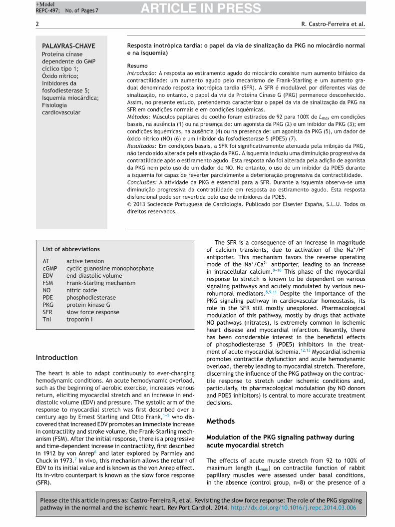
he effects of acute muscle stretch from 92 to 100% ofaximum length (Lmax) on contractile function of rabbitapillary muscles were assessed under basal conditions,n the absence (control group, n=8) or the presence of a
IN PRESS+Model
onse 3
Control
Isch
Stretch
Stretch
A
B
AT
AT
15′
15′
5′
5′
15′
15′
– 8-bromo-cGMP (PKGa)
– Rp-8Br-PET-cGMPS (PKGi)
20′
20′
– 8-bromo-cGMP (Isch-PKGa)
– SNAP (Isch-No)
– Sildenafil (Isch-PDE5i)
Figure 1 Representative bands of the contractile response toacute stretch (upper images) in the control group (A) and Ischgroup (B) and schematic illustration of experimental protocols(bottom diagrams). Gray bar: stabilization period; white bar:presence of O2 and glucose; dark bar: absence of O2 and glucose(
Cm
Is
Document downloaded from http://www.elsevier.pt, day 25/09/2014. This copy is for personal use. Any transmission of this document by any media or format is strictly prohibited. This early online article has been reviewed, accepted and posted on the Web before copyediting.
ARTICLEREPC-497; No. of Pages 7
The role of the PKG signaling pathway in the slow force resp
cell-permeable PKG activator (8-bromo-cGMP, 10−6 M; PKGagroup, n=7) and a PKG inhibitor (Rp-8-Br-PET-cGMPS, 10−6
M; PKGi group, n=7). These substances were dissolved inKrebs-Ringer solution and myocardial stretch was elicited20 min later.
Modulation of the PKG signaling pathway duringacute myocardial stretch under ischemicconditions
In a second set of protocols, the contractile response tostretch was studied under ischemic conditions. After sta-bilization, the bathing solution was replaced by anotherwithout glucose and without O2 supply. After 5 min ofischemia, muscles were stretched from 92 to 100% of Lmax
(Isch group, n=8). Furthermore, to explore the influence ofmodulation of the PKG pathway on the systolic responseto stretch during ischemia, the latter protocol was alsoperformed in the presence of a PKG activator (8-bromo-cGMP, 10−6 M); Isch-PKGa group, n=7; (H) an NO donor(S-nitroso-N-acetyl-penicillamine (SNAP), 10−5 M; Isch-NOgroup, n=9) and (I) a PDE5 inhibitor (sildenafil, 10−6 M;Isch-PDE5i group, n=7). These drugs interfere with theNO/PKG pathway at different targets and allow bettercharacterization of SFR modulation. Ischemia was main-tained for 15 min. To confirm myocardial viability afterthe ischemic period, the muscles were reperfused witha glucose-containing Krebs-Ringer solution and O2 sup-ply.
A schematic representation of the protocols is shown inFigure 1.
The doses were selected according to the literature dataand our preliminary studies. All chemicals were obtainedfrom Sigma-Aldrich (St. Louis, MO, USA).
Statistical analysis
Values are means ± standard error. Within the same group,the effects of acute muscle stretch on the various exper-imental parameters at specific time points were analyzedwith a paired Student’s t test. The time-dependent effectsof muscle stretch within the same group were analyzedby repeated measures one-way ANOVA, while repeatedmeasures two-way ANOVA was used to compare the time-dependent effects of muscle stretch between severalgroups. When significant differences were detected with anyof the ANOVA tests, the Holm-Sidak method was used to per-form multiple comparisons. A value of p<0.05 was acceptedas significant.
Results
Basal pharmacological effects
Please cite this article in press as: Castro-Ferreira R, et al. Revispathway in the normal and the ischemic heart. Rev Port Cardio
The muscles stabilized in the presence of the drug for20 min, during which no significant variations in active ten-sion were observed.
t(sc
ischemia). AT: active tension.
ontractile response to acute stretch in normalyocardium
n the control group, active tension (AT) at Lmax, followingtretch, was 23.4 ± 3.8 mN/mm2. In the following 15 min
iting the slow force response: The role of the PKG signalingl. 2014. http://dx.doi.org/10.1016/j.repc.2014.03.006
here was a progressive increase in AT to 29.1 ± 4.3 mN/mm2
p<0.001), corresponding to an SFR of 27.8 ± 3.9%. A repre-entative band of the contractile response of a muscle in theontrol group is shown in Figure 1A.

ARTICLE IN PRESS+ModelREPC-497; No. of Pages 7
4 R. Castro-Ferreira et al.
Control
30
20
10
0
–10
0 1 2 3 4 5 6 7 8 9 10 12 15 15
AT v
aria
tion
(%)
PKGa
PKGi
Con
trol
PK
Ga
PK
Gi
Min after stretch to LmaxMin after stretch to Lmax
F SFR was impaired in the PKGi group. *p<0.05 vs. control. AT: activet
P
I1tPop
As
Iidpid2ri
N
Tmtv
P
Twa−
PDE5 inhibition under ischemic conditions
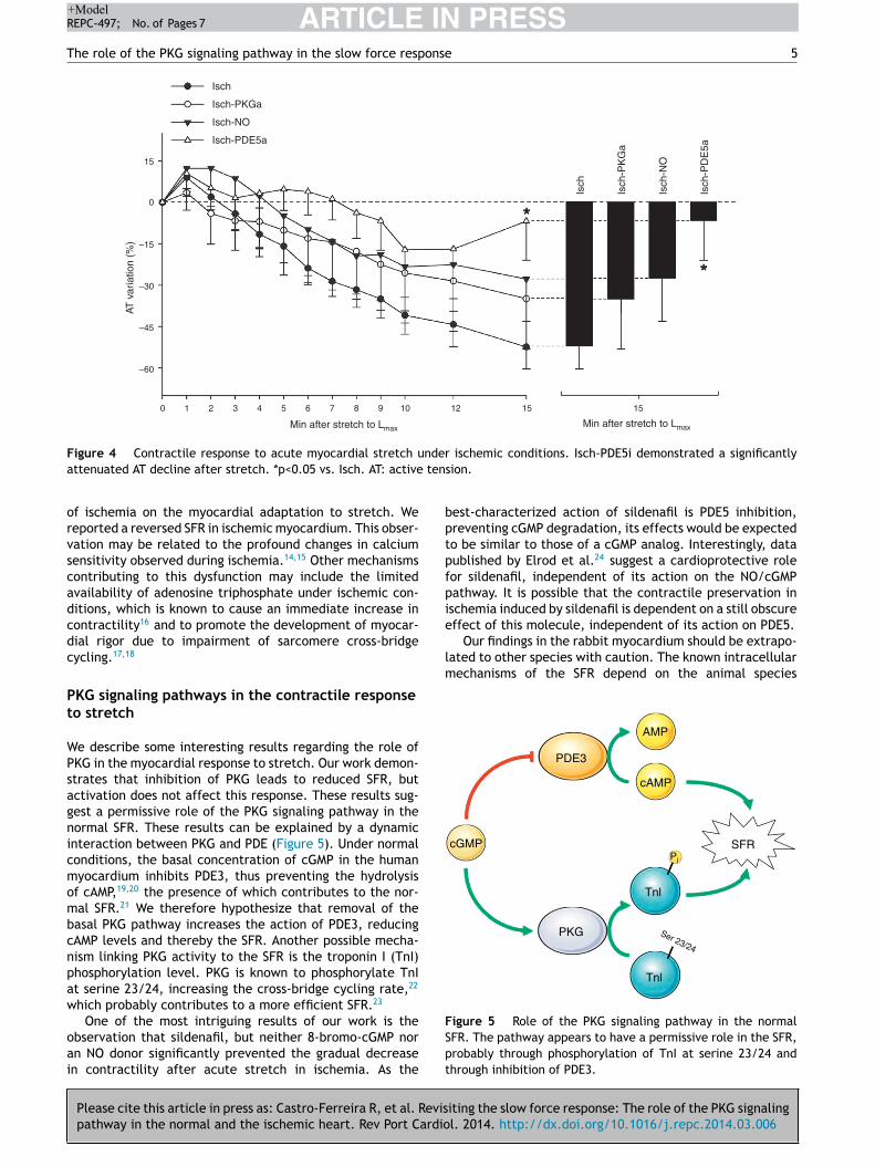
In contrast to Isch-PKGa, PDE5 inhibition significantlyprevented the decrease in contractility after stretch(−8.6 ± 17.6% vs. −52.4 ± 7.9% in Isch, p=0.018) (Figure 4).
Discussion
The effect of ischemia on myocardial responseto stretch
Acute myocardial ischemia induces immediate contractiledysfunction, resulting in sudden hemodynamic overload.Hence, it is interesting to characterize the myocardialresponse to stretch under ischemic conditions. Despite itsimportance, there is limited knowledge about the effect
Control
Ischemia
30
15
0
–15
–30
–60
AT v
aria
tion
(%)
–45
Document downloaded from http://www.elsevier.pt, day 25/09/2014. This copy is for personal use. Any transmission of this document by any media or format is strictly prohibited. This early online article has been reviewed, accepted and posted on the Web before copyediting.
igure 2 Modulation of the SFR by PKG under normoxia. The
ension.
KG-mediated effects
n the PKGa group no differences were found in SFR after5 min (20.9 ± 8.13% vs. 27.8 ± 3.9%, p=0.324) (Figure 2). Onhe other hand, myocardial stretch in the presence of theKG inhibitor was associated with a significant attenuationf SFR (SFR at 15 min: 11.5 ± 5.9% vs. 27.8 ± 3.9% in controls,=0.034) (Figure 2).
daptation of contractile function to myocardialtretch under ischemic conditions
n the first 5 min of ischemia a significant decreasen myocardial contractility of 51.0 ± 4.0% was seen (ATecreased from 20.1 ± 3.1 mN/mm2 to 10.2 ± 2.2 mN/mm2,<0.001). After stretch, muscles did not display SFR;nstead, they exhibited a significant and progressiveecrease in contractility (AT decreased from 12.4 ±.3 mN/mm2 to 5.7 ± 1.9 mN/mm2, p<0.001) (Figure 3). Aepresentative band of a muscle of the Isch group is shownn the Figure 1B.
O-mediated effects under ischemic conditions
he use of an NO donor did not significantly influence theyocardial response to acute stretch under ischemic condi-
ions (AT variation after stretch: −27.7 ± 15.2% in Isch-NOs. −52.4 ± 7.9% in Isch, p=0.229) (Figure 4).
KG-mediated effects under ischemic conditions
Please cite this article in press as: Castro-Ferreira R, et al. Revisiting the slow force response: The role of the PKG signalingpathway in the normal and the ischemic heart. Rev Port Cardiol. 2014. http://dx.doi.org/10.1016/j.repc.2014.03.006
he decrease in contractility over the 15 min after stretchas not significantly different in the presence of the PKGctivator compared to the Isch group (−34.9 ± 18.2% vs.52.4 ± 7.9% in Isch, p=0.373) (Figure 4).
Figure 3 Modulation of the SFR by ischemia. In the Isch group,muscles did not display SFR; instead, they exhibited a significantand progressive decrease in contractility during the 15 min afterstretch. *p<0.05 vs. control. AT: active tension.
ARTICLE IN PRESS+ModelREPC-497; No. of Pages 7
The role of the PKG signaling pathway in the slow force response 5
Isch
0
–15
15
–60
–45
–30
0 1 2 3 4 5 6 7 8 9 10 12 15 15
Min after stretch to LmaxMin after stretch to Lmax
AT v
aria
tion
(%)
Isch-PKGa
Isch-NO
Isch-PDE5a
Isch
Isch
-PK
Ga
Isch
-NO
Isch
-PD
E5a
nder ischemic conditions. Isch-PDE5i demonstrated a significantly tension.
best-characterized action of sildenafil is PDE5 inhibition,preventing cGMP degradation, its effects would be expectedto be similar to those of a cGMP analog. Interestingly, datapublished by Elrod et al.24 suggest a cardioprotective rolefor sildenafil, independent of its action on the NO/cGMPpathway. It is possible that the contractile preservation inischemia induced by sildenafil is dependent on a still obscureeffect of this molecule, independent of its action on PDE5.
Our findings in the rabbit myocardium should be extrapo-lated to other species with caution. The known intracellularmechanisms of the SFR depend on the animal species
cGMP
cAMP
PDE3
PKG
Tnl
Ser 23/24
Tnl
SFRPi
AMP
Document downloaded from http://www.elsevier.pt, day 25/09/2014. This copy is for personal use. Any transmission of this document by any media or format is strictly prohibited. This early online article has been reviewed, accepted and posted on the Web before copyediting.
Figure 4 Contractile response to acute myocardial stretch uattenuated AT decline after stretch. *p<0.05 vs. Isch. AT: active
of ischemia on the myocardial adaptation to stretch. Wereported a reversed SFR in ischemic myocardium. This obser-vation may be related to the profound changes in calciumsensitivity observed during ischemia.14,15 Other mechanismscontributing to this dysfunction may include the limitedavailability of adenosine triphosphate under ischemic con-ditions, which is known to cause an immediate increase incontractility16 and to promote the development of myocar-dial rigor due to impairment of sarcomere cross-bridgecycling.17,18
PKG signaling pathways in the contractile responseto stretch
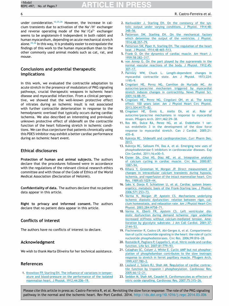
We describe some interesting results regarding the role ofPKG in the myocardial response to stretch. Our work demon-strates that inhibition of PKG leads to reduced SFR, butactivation does not affect this response. These results sug-gest a permissive role of the PKG signaling pathway in thenormal SFR. These results can be explained by a dynamicinteraction between PKG and PDE (Figure 5). Under normalconditions, the basal concentration of cGMP in the humanmyocardium inhibits PDE3, thus preventing the hydrolysisof cAMP,19,20 the presence of which contributes to the nor-mal SFR.21 We therefore hypothesize that removal of thebasal PKG pathway increases the action of PDE3, reducingcAMP levels and thereby the SFR. Another possible mecha-nism linking PKG activity to the SFR is the troponin I (TnI)phosphorylation level. PKG is known to phosphorylate TnIat serine 23/24, increasing the cross-bridge cycling rate,22
which probably contributes to a more efficient SFR.23
Please cite this article in press as: Castro-Ferreira R, et al. Revisiting the slow force response: The role of the PKG signalingpathway in the normal and the ischemic heart. Rev Port Cardiol. 2014. http://dx.doi.org/10.1016/j.repc.2014.03.006
One of the most intriguing results of our work is theobservation that sildenafil, but neither 8-bromo-cGMP noran NO donor significantly prevented the gradual decreasein contractility after acute stretch in ischemia. As the
Figure 5 Role of the PKG signaling pathway in the normalSFR. The pathway appears to have a permissive role in the SFR,probably through phosphorylation of TnI at serine 23/24 andthrough inhibition of PDE3.
IN+ModelR
6
ucashafiom
Ci
Iapdtowhiufttd
E
PdwcM
Cd
Rd
C
T
A
W
R
1
1
1
1
1
1
1
1
1
1
2
Document downloaded from http://www.elsevier.pt, day 25/09/2014. This copy is for personal use. Any transmission of this document by any media or format is strictly prohibited. This early online article has been reviewed, accepted and posted on the Web before copyediting.
ARTICLEEPC-497; No. of Pages 7
nder consideration.10,25,26 However, the increase in cal-ium transients due to activation of the Na+/H+ exchangernd reverse operating mode of the Na+/Ca2+ exchangereems to be angiotensin-II independent in both rabbit anduman myocardium, depending on acute mechanical stretchlone.25,26 In this way, it is probably easier to extrapolate thendings of this work to the human myocardium than to thether commonly used animal models such as cat, rat, andouse.
onclusions and potential therapeuticmplications
n this work, we evaluated the contractile adaptation tocute stretch in the presence of modulators of PKG signalingathways, crucial therapeutic weapons in ischemic heartisease and myocardial infarction. From a clinical perspec-ive, we showed that the well-known protective effectf nitrates during an ischemic insult is not associatedith further contractile deterioration in response to theemodynamic overload that typically occurs during cardiacschemia. We also described an interesting and previouslynknown protective effect of sildenafil on the contractileunction of the heart following stretch in ischemic condi-ions. We can thus conjecture that patients chronically usinghis PDE5 inhibitor may exhibit a better cardiac performanceuring an ischemic heart event.
thical disclosures
rotection of human and animal subjects. The authorseclare that the procedures followed were in accordanceith the regulations of the relevant clinical research ethicsommittee and with those of the Code of Ethics of the Worldedical Association (Declaration of Helsinki).
onfidentiality of data. The authors declare that no patientata appear in this article.
ight to privacy and informed consent. The authorseclare that no patient data appear in this article.
onflicts of interest
he authors have no conflicts of interest to declare.
cknowledgment
Please cite this article in press as: Castro-Ferreira R, et al. Revispathway in the normal and the ischemic heart. Rev Port Cardio
e wish to thank Marta Oliveira for her technical assistance.
eferences
1. Knowlton FP, Starling EH. The influence of variations in temper-ature and blood-pressure on the performance of the isolatedmammalian heart. J Physiol. 1912;44:206---19.
2
2
2
PRESSR. Castro-Ferreira et al.
2. Markwalder J, Starling EH. On the constancy of the sys-tolic output under varying conditions. J Physiol. 1914;48:348---56.
3. Patterson SW, Starling EH. On the mechanical factorswhich determine the output of the ventricles. J Physiol.1914;48:357---79.
4. Patterson SW, Piper H, Starling EH. The regulation of the heartbeat. J Physiol. 1914;48:465---513.
5. Frank O. On the dynamics of cardiac muscle. Am Heart J.1959;58:282---317.
6. von Anrep G. On the part played by the suprarenals in thenormal vascular reactions of the body. J Physiol. 1912;45:307---17.
7. Parmley WW, Chuck L. Length-dependent changes inmyocardial contractile state. Am J Physiol. 1973;224:1195---9.
8. Cingolani HE, Perez NG, Camilion de Hurtado MC. Anautocrine/paracrine mechanism triggered by myocardialstretch induces changes in contractility. News Physiol Sci.2001;16:88---91.
9. Cingolani HE, Perez NG, Cingolani OH, et al. The Anrepeffect: 100 years later. Am J Physiol Heart Circ Physiol.2013;304:H175---82.
0. Cingolani HE, Ennis IL, Aiello EA, et al. Role ofautocrine/paracrine mechanisms in response to myocardialstrain. Pflugers Arch. 2011;462:29---38.
1. Ros MN, Dulce RA, Perez NG, et al. Endothelin 1 ver-sus endothelin 3 in the development of the slow forceresponse to myocardial stretch. Can J Cardiol. 2005;21:435---8.
2. Kukreja RC. Sildenafil and cardioprotection. Curr Pharm Des.2013.
3. Kukreja RC, Salloum FN, Das A, et al. Emerging new uses ofphosphodiesterase-5 inhibitors in cardiovascular diseases. ExpClin Cardiol. 2011;16:e30---5.
4. Eisner DA, Choi HS, Díaz ME, et al. Integrative analysisof calcium cycling in cardiac muscle. Circ Res. 2000;87:1087---94.
5. Kihara Y, Grossman W, Morgan JP. Direct measurement ofchanges in intracellular calcium transients during hypoxia,ischemia, and reperfusion of the intact mammalian heart. CircRes. 1989;65:1029---44.
6. Saks V, Dzeja P, Schlattner U, et al. Cardiac system bioen-ergetics: metabolic basis of the Frank-Starling law. J Physiol.2006;571:253---73.
7. Varma N, Morgan JP, Apstein CS. Mechanisms underlyingischemic diastolic dysfunction: relation between rigor, cal-cium homeostasis, and relaxation rate. Am J Physiol Heart CircPhysiol. 2003;284:H758---71.
8. Varma N, Eberli FR, Apstein CS. Left ventricular dia-stolic dysfunction during demand ischemia: rigor underliesincreased stiffness without calcium-mediated tension. Ame-lioration by glycolytic substrate. J Am Coll Cardiol. 2001;37:2144---53.
9. Fischmeister R, Castro LR, Abi-Gerges A, et al. Compartmenta-tion of cyclic nucleotide signaling in the heart: the role of cyclicnucleotide phosphodiesterases. Circ Res. 2006;99:816---28.
0. Rastaldo R, Pagliaro P, Cappello S, et al. Nitric oxide and cardiacfunction. Life Sci. 2007;81:779---93.
1. Calaghan SC, Colyer J, White E. Cyclic AMP but not phosphor-ylation of phospholamban contributes to the slow inotropicresponse to stretch in ferret papillary muscle. Pflugers Arch.1999;437:780---2.
2. Layland J, Solaro RJ, Shah AM. Regulation of cardiac contrac-
iting the slow force response: The role of the PKG signalingl. 2014. http://dx.doi.org/10.1016/j.repc.2014.03.006
tile function by troponin I phosphorylation. Cardiovasc Res.2005;66:12---21.
3. Seddon M, Shah AM, Casadei B. Cardiomyocytes as effectors ofnitric oxide signalling. Cardiovasc Res. 2007;75:315---26.
IN+Model
onse
Document downloaded from http://www.elsevier.pt, day 25/09/2014. This copy is for personal use. Any transmission of this document by any media or format is strictly prohibited. This early online article has been reviewed, accepted and posted on the Web before copyediting.
ARTICLEREPC-497; No. of Pages 7
The role of the PKG signaling pathway in the slow force resp
24. Elrod JW, Greer JJ, Lefer DJ. Sildenafil-mediated acute car-
Please cite this article in press as: Castro-Ferreira R, et al. Revispathway in the normal and the ischemic heart. Rev Port Cardio
dioprotection is independent of the NO/cGMP pathway. Am JPhysiol Heart Circ Physiol. 2007;292:H342---7.
25. Luers C, Fialka F, Elgner A, et al. Stretch-dependent modulationof [Na+]i, [Ca2+]i, and pHi in rabbit myocardium --- a mechanism
2
PRESS 7
for the slow force response. Cardiovasc Res. 2005;68:
iting the slow force response: The role of the PKG signalingl. 2014. http://dx.doi.org/10.1016/j.repc.2014.03.006
454---63.6. von Lewinski D, Stumme B, Fialka F, et al. Functional relevance
of the stretch-dependent slow force response in failing humanmyocardium. Circ Res. 2004;94:1392---8.
Copyright © 2022 FDOKUMEN