RIN2 Deficiency Results in Macrocephaly, Alopecia, Cutis Laxa, and Scoliosis: MACS Syndrome
10
REPORT RIN2 Deficiency Results in Macrocephaly, Alopecia, Cutis Laxa, and Scoliosis: MACS Syndrome Lina Basel-Vanagaite, 1,2,14 Ofer Sarig, 4,14 Dov Hershkovitz, 6,7 Dana Fuchs-Telem, 2,4 Debora Rapaport, 3 Andrea Gat, 5 Gila Isman, 4 Idit Shirazi, 4 Mordechai Shohat, 1,2 Claes D. Enk, 10 Efrat Birk, 2 Ju ¨rgen Kohlhase, 11 Uta Matysiak-Scholze, 11 Idit Maya, 1 Carlos Knopf, 9 Anette Peffekoven, 12 Hans-Christian Hennies, 12 Reuven Bergman, 8 Mia Horowitz, 3 Akemi Ishida-Yamamoto, 13 and Eli Sprecher 2,4,6, * Inherited disorders of elastic tissue represent a complex and heterogeneous group of diseases, characterized often by sagging skin and occasionally by life-threatening visceral complications. In the present study, we report on an autosomal-recessive disorder that we have termed MACS syndrome ( macrocephaly, alopecia, cutis laxa, and scoliosis). The disorder was mapped to chromosome 20p11.21-p11.23, and a homozygous frameshift mutation in RIN2 was found to segregate with the disease phenotype in a large consan- guineous kindred. The mutation identified results in decreased expression of RIN2, a ubiquitously expressed protein that interacts with Rab5 and is involved in the regulation of endocytic trafficking. RIN2 deficiency was found to be associated with paucity of dermal micro- fibrils and deficiency of fibulin-5, which may underlie the abnormal skin phenotype displayed by the patients. Disorders of elastic tissue often share common phenotypic features, including redundant skin, hyperlaxity of joints, and, in severe cases, rupture of hollow body organs, with catastrophic consequences. 1,2 The prototype for this type of abnormality is cutis laxa (CL), a heterogeneous group of acquired and inherited disorders characterized by redun- dant, loose, and inelastic skin. 3 CL can be present at birth or appear at different stages of growth and development. Genetic forms of CL include autosomal-recessive CL type I (ARCL1 [MIM 219100]), autosomal-recessive CL type II (ARCL2 [MIM 219200]), X-linked CL, also known as X-linked occipital horn syndrome (OHS [MIM 304150]), and autosomal-dominant CL (ADCL MIM 123700). ARCL1 is usually severe or even lethal in childhood as a result of pulmonary emphysema and cardiovascular complications and can be caused by mutations in the FBLN4 (MIM 603634) and FBLN5 (MIM 604580) genes, encoding fibulin-4 and -5, respectively. 4–6 FBLN4 mutations are also associated with bone fractures, vascular tortuosity, aortic aneurysm, emphysema, hernias, joint laxity, and pectus excavatum. ADCL is caused by mutations in the ELN (MIM 130160) gene, 7 which encodes elastin. Fibulins are part of the microfibrillar component of the elastic fibers and form a structural backbone onto which amorphous elastin is deposited, explaining the overlapping phenotype resulting from mutations affecting both elastin and fibulin genes. 8 ARCL2 is caused by mutations in the ATP6V0A2 (MIM 611716) gene, and these mutations have been shown to result in decreased secretion of elastin. 9,10 In addition, several other autosomal-recessive syndromes, characterized by redundant skin and additional clinical features, have been reported, including Ehlers Danlos syndrome type VII (MIM 225410), caused by mutations in the PLOD1 (MIM 153454) gene, 11 and de Barsy syn- drome (MIM 219150), 12 whose etiology is still unknown. Growth retardation, alopecia, pseudoanodontia, and optic atrophy (GAPO [MIM 230740]) syndrome is characterized by redundant appearance of facial skin (but not of other areas of the body), short stature, alopecia, pseudoanodon- tia, various eye abnormalities (including optic atrophy and glaucoma), and a typical facial dysmorphism. 13–17 Here, too, the etiology of the syndrome remains elusive. In the present report, we studied a new syndrome related to the CL group of inherited disorders. The patients with this syndrome are all members of two related consanguin- eous Israeli-Arab families and were all born with normal birth weights after full-term, uneventful pregnancies. Psychomotor development was normal. On clinical exam- ination, macrocephaly (Figures 1A and 1B), downward- slanting palpebral fissures, puffy eyelids (Figures 1A and 1B), mild ichthyosis (Figure 1C), sagging cheeks, everted lower lip, retrognathia with abnormal skull morphology (Figure 1D), gingival hyperplasia (Figure 1G), abnormal position of the teeth (Figures 1G and 1H), severe hyperlax- ity, and flat feet were observed. Unfortunately, no data on the occipitofrontal circumference (OFC) at birth or on the 1 Schneider Children’s Medical Center of Israel and Raphael Recanati Genetics Institute, Rabin Medical Center, Beilinson Campus, 49100 Petah Tiqva, Israel; 2 Sackler Faculty of Medicine, 3 Department of Cell Research and Immunology, Tel Aviv University, 69978 Ramat Aviv, Israel; 4 Department of Dermatology, 5 Department of Pathology, Tel Aviv Sourasky Medical Center, 64289 Tel Aviv, Israel; 6 Center for Translational Genetics, Rappaport Institute and Faculty of Medicine, Technion – Israel Institute of Technology, 31096 Haifa, Israel; 7 Department of Pathology, 8 Department of Dermatology, 9 Metabolic Disease Unit, Meyer Children’s Hospital, Rambam Health Care Campus, 31096 Haifa, Israel; 10 Department of Dermatology, Hadassah Hebrew University Medical School, 91120 Jerusalem, Israel; 11 Center for Human Genetics, 79100 Freiburg, Germany; 12 Division of Dermatogenetics, Cologne Center for Genomics, University of Cologne, 50674 Cologne, Germany; 13 Department of Dermatology, Asahikawa Medical College, 078-8510 Asahikawa, Japan 14 These authors contributed equally to this work *Correspondence: [email protected] DOI 10.1016/j.ajhg.2009.07.001. ª2009 by The American Society of Human Genetics. All rights reserved. 254 The American Journal of Human Genetics 85, 254–263, August 14, 2009
-
Upload
chevronindonesia -
Category
Documents
-
view
4 -
download
0
Transcript of RIN2 Deficiency Results in Macrocephaly, Alopecia, Cutis Laxa, and Scoliosis: MACS Syndrome

REPORT
RIN2 Deficiency Results in Macrocephaly, Alopecia,Cutis Laxa, and Scoliosis: MACS Syndrome
Lina Basel-Vanagaite,1,2,14 Ofer Sarig,4,14 Dov Hershkovitz,6,7 Dana Fuchs-Telem,2,4 Debora Rapaport,3
Andrea Gat,5 Gila Isman,4 Idit Shirazi,4 Mordechai Shohat,1,2 Claes D. Enk,10 Efrat Birk,2
Jurgen Kohlhase,11 Uta Matysiak-Scholze,11 Idit Maya,1 Carlos Knopf,9 Anette Peffekoven,12
Hans-Christian Hennies,12 Reuven Bergman,8 Mia Horowitz,3 Akemi Ishida-Yamamoto,13
and Eli Sprecher2,4,6,*
Inherited disorders of elastic tissue represent a complex and heterogeneous group of diseases, characterized often by sagging skin and
occasionally by life-threatening visceral complications. In the present study, we report on an autosomal-recessive disorder that we
have termed MACS syndrome (macrocephaly, alopecia, cutis laxa, and scoliosis). The disorder was mapped to chromosome
20p11.21-p11.23, and a homozygous frameshift mutation in RIN2 was found to segregate with the disease phenotype in a large consan-
guineous kindred. The mutation identified results in decreased expression of RIN2, a ubiquitously expressed protein that interacts with
Rab5 and is involved in the regulation of endocytic trafficking. RIN2 deficiency was found to be associated with paucity of dermal micro-
fibrils and deficiency of fibulin-5, which may underlie the abnormal skin phenotype displayed by the patients.
Disorders of elastic tissue often share common phenotypic
features, including redundant skin, hyperlaxity of joints,
and, in severe cases, rupture of hollow body organs, with
catastrophic consequences.1,2 The prototype for this type
of abnormality is cutis laxa (CL), a heterogeneous group
of acquired and inherited disorders characterized by redun-
dant, loose, and inelastic skin.3 CL can be present at birth
or appear at different stages of growth and development.
Genetic forms of CL include autosomal-recessive CL type
I (ARCL1 [MIM 219100]), autosomal-recessive CL type II
(ARCL2 [MIM 219200]), X-linked CL, also known as
X-linked occipital horn syndrome (OHS [MIM 304150]),
and autosomal-dominant CL (ADCL MIM 123700).
ARCL1 is usually severe or even lethal in childhood as a
result of pulmonary emphysema and cardiovascular
complications and can be caused by mutations in the
FBLN4 (MIM 603634) and FBLN5 (MIM 604580) genes,
encoding fibulin-4 and -5, respectively.4–6 FBLN4 mutations
are also associated with bone fractures, vascular tortuosity,
aortic aneurysm, emphysema, hernias, joint laxity, and
pectus excavatum. ADCL is caused by mutations in the
ELN (MIM 130160) gene,7 which encodes elastin. Fibulins
are part of the microfibrillar component of the elastic fibers
and form a structural backbone onto which amorphous
elastin is deposited, explaining the overlapping phenotype
resulting from mutations affecting both elastin and fibulin
genes.8 ARCL2 is caused by mutations in the ATP6V0A2
(MIM 611716) gene, and these mutations have been
254 The American Journal of Human Genetics 85, 254–263, August
shown to result in decreased secretion of elastin.9,10 In
addition, several other autosomal-recessive syndromes,
characterized by redundant skin and additional clinical
features, have been reported, including Ehlers Danlos
syndrome type VII (MIM 225410), caused by mutations
in the PLOD1 (MIM 153454) gene,11 and de Barsy syn-
drome (MIM 219150),12 whose etiology is still unknown.
Growth retardation, alopecia, pseudoanodontia, and optic
atrophy (GAPO [MIM 230740]) syndrome is characterized
by redundant appearance of facial skin (but not of other
areas of the body), short stature, alopecia, pseudoanodon-
tia, various eye abnormalities (including optic atrophy and
glaucoma), and a typical facial dysmorphism.13–17 Here,
too, the etiology of the syndrome remains elusive.
In the present report, we studied a new syndrome related
to the CL group of inherited disorders. The patients with
this syndrome are all members of two related consanguin-
eous Israeli-Arab families and were all born with normal
birth weights after full-term, uneventful pregnancies.
Psychomotor development was normal. On clinical exam-
ination, macrocephaly (Figures 1A and 1B), downward-
slanting palpebral fissures, puffy eyelids (Figures 1A and
1B), mild ichthyosis (Figure 1C), sagging cheeks, everted
lower lip, retrognathia with abnormal skull morphology
(Figure 1D), gingival hyperplasia (Figure 1G), abnormal
position of the teeth (Figures 1G and 1H), severe hyperlax-
ity, and flat feet were observed. Unfortunately, no data on
the occipitofrontal circumference (OFC) at birth or on the
1Schneider Children’s Medical Center of Israel and Raphael Recanati Genetics Institute, Rabin Medical Center, Beilinson Campus, 49100 Petah Tiqva, Israel;2Sackler Faculty of Medicine, 3Department of Cell Research and Immunology, Tel Aviv University, 69978 Ramat Aviv, Israel; 4Department of Dermatology,5Department of Pathology, Tel Aviv Sourasky Medical Center, 64289 Tel Aviv, Israel; 6Center for Translational Genetics, Rappaport Institute and Faculty of
Medicine, Technion – Israel Institute of Technology, 31096 Haifa, Israel; 7Department of Pathology, 8Department of Dermatology, 9Metabolic Disease Unit,
Meyer Children’s Hospital, Rambam Health Care Campus, 31096 Haifa, Israel; 10Department of Dermatology, Hadassah Hebrew University Medical School,
91120 Jerusalem, Israel; 11Center for Human Genetics, 79100 Freiburg, Germany; 12Division of Dermatogenetics, Cologne Center for Genomics, University
of Cologne, 50674 Cologne, Germany; 13Department of Dermatology, Asahikawa Medical College, 078-8510 Asahikawa, Japan14These authors contributed equally to this work
*Correspondence: [email protected]
DOI 10.1016/j.ajhg.2009.07.001. ª2009 by The American Society of Human Genetics. All rights reserved.
14, 2009
timing of closure of the fontanelles were available. There
has been no progression of the macrocephaly over the
past two years in patient II-4.
The phenotype also included coarse and swollen facial
appearance affecting the eyelids, lips, and cheeks. Skin
laxitywas more apparent in the facial than in the abdominal
area (Figures 1A and 1B). Scalp hair was sparse (Figure 1E).
Short stature was caused mainly by moderate to severe scoli-
osis in all three affected individuals (Figure 1F).
The facial phenotype in the two older affected family
members (II-5 and II-6) was more prominent, including
sagging eyelids and sagging cheeks, as compared with
that of the youngest patient (II-4) (Figures 1A and 1B).
Easy bruising, atrophic scars, transparent veins, and blue
sclera were not observed. The patients denied respiratory
problems or any other systemic complaints, except for
severe back pain in the youngest affected family member,
Figure 1. Clinical Phenotype(A and B) Facial appearance of patients II-4 (A) and II-5 (B). Noteage-dependant worsening of the sagging skin phenotype.(C) Mild ichthyosis can be seen in patient II-4.(D) Skull radiographs of a normal individual (left) and of a patient(right).(E) Short and sparse hair can be seen in patient II-5.(F) Severe kyphoscoliosis can be seen in patient II-4.(G) Irregularly placed teeth and gingival hypertrophy can be seenin patient II-4.(H) Panoramic X-ray of the oral cavity in patient II-5, demon-strating unerupted wisdom and upper canine teeth.
The Ameri
who displayed severe kyphoscoliosis on examination
(Figure 1F). Neurological examination was normal. Eye
examination, including fundus, was normal in patients
II-4 and II-8. The clinical features of all affected family
members are summarized in Table 1.
Complete blood counts, renal and liver function tests,
and thyroid stimulating hormone (TSH) level were all
normal. A skeletal X-ray survey of patient II-4 revealed
severe scoliosis. On echocardiography, mild dilatation of
the aorta was detected in patient II-4. Panoramic X-ray of
the teeth in patient II-5 demonstrated unerupted wisdom
and canine teeth (Figure 1H). A bone density scan in patient
II-5 revealed generalized osteoporosis, more severe in the
spine. Serum transferrin isoform analysis was not suggestive
of a congenital glycosylation defect. In addition, isoelectric
focusing analysis revealed a normal apolipoprotein CIII
profile (Apo CIII2¼ 48%, Apo CIII1¼ 49%, Apo CIII0¼ 3%).
Light microscopic examination of skin biopsies obtained
from patients II-4 and II-5 revealed a normal epidermis. In
the papillary (upper) dermis, elastic fibers were sparse, with
an almost complete absence of oxytalin fibers (Figures 2A–
2D). With the use of Verhoeff’s Van Gieson staining, elastic
fibers appeared normally distributed in the reticular
(lower) dermis (Figure 2E). Collagen fiber structure and
distribution appeared normal throughout the skin
(Figure 2F). Minimal focal interstitial mucin deposition
was detected with Alcian blue staining (not shown).
Transmission electron microscopy of dermal elastic
fibers demonstrated diminished microfibrillar component
at the periphery of elastic fibers (Figure 2G). These findings
correlated with markedly decreased staining for fibulin-5,
a major component of dermal elastic microfibrils (Figures
2H and 2I). We did not observe any abnormalities in lyso-
some appearance with the use of electron microscopy. In
addition, the patients did not show clinical signs of lyso-
somal storage disease (neurological abnormalities, hepa-
tosplenomegaly, thrombocytopenia, progressive disease
course, dysostosis multiplex on skeletal X-rays, etc.).
Scanning electron microscopy examination of hair
obtained from affected patients showed no abnormalities
(not shown).
Taken altogether, the unique constellation of signs dis-
played by the patients reported in this study demarcates
a clinically new syndrome, which we have termed MACS
syndrome—for macrocephaly, alopecia, cutis laxa, and
scoliosis.
Genomic DNA was extracted from leukocytes from
peripheral venous blood in EDTA, with the use of standard
procedures, after informed consent was obtained from all
family members or their legal guardians, in accordance
with a protocol approved and reviewed by the National
Committee for Genetic Studies, Israel Ministry of Health.
We genotyped affected family members, using Affymetrix
Human Mapping NspI 250K arrays in accordance with the
manufacturer’s instructions, and identified a continuous
segment of homozygosity shared by the patients and
encompassing 815 consecutive SNPs over 6.8 Mb between
can Journal of Human Genetics 85, 254–263, August 14, 2009 255

Table 1. Clinical Features of the Patients
Patient II-4 Patient II-5 Patient II-6
General and familial data
Birth weight (g) 3500 NA NA
Age (yrs) 15 32 35
Height (cm) (SD) 149 (�2) 165 (�1.5) NA
Maternal height (cm) (SD) 160 (0)
Paternal height (cm) (SD) 167 (�1)
Weight (kg) (SD) 44 (�2) 47 (�2.5) NA
OFC (cm) (SD) 59.5 (þ3) 58.5 (þ2) NA
Maternal OFC (cm) (SD) 56.5 (þ1.5)
Paternal OFC (cm) (SD) 57 (þ1.5)
Facial characteristics
Downslanting palpebral fissures þ þ þ
Puffy droopy eyelids þ þ þ
Full, everted lips þ þ þ
Skin characteristics
Soft, redundant skin (especially facial) þ þ þ
Gum hypertrophy þ þ þ
Irregular dentition þ þ þ
Hair characteristics
Receding anterior hairline, sparse hair þ þ þ
Skeletal manifestations
Joint hypermobility þ þ þ
Scoliosis severe mild mild
Fractures � þ �
Other features
Umbilical hernia þ � �
Eye abnormalities � � NA
Genitourinary abnormalities Urethral stenosis, undescended testis � �
Abnormal (high-pitched) voice þ þ þ
Various Ichthyosis on lower limbs, high-archedpalate, mild aortic dilatation
Recurrent aphthous stomatitis,severe osteoporosis
Bone density scan NA
Abbreviations are as follows: NA, not available; OFC, occipitofrontal circumference. Plus signs and minus signs indicate presence and absence, respectively.
markers rs6034837 and rs2092468 on 20p11.21-p11.23
(Figure 3A). We then genotyped all family members for
eight microsatellite markers (D20S186, D20S112,
D20S475, D20S1145, D20S868, D20S101, D20S848,
D20S195) spanning the disease interval. Only three of these
markers were found to be informative or semi-informative.
Microsatellite marker typing data were consistent with a
gene locus for MACS syndrome in the 20p11.21-p11.23
chromosomal region, although the LOD score did not reach
a critical value of 3 as a result of the lack of informativity of
the markers typed in the region (not shown).
256 The American Journal of Human Genetics 85, 254–263, August 1
The candidate region was found to include 55 known or
predicted genes. Given the fact that the MACS syndrome is
transmitted in an autosomal-recessive fashion, and given
the fact that most recessive disorders are caused by loss-
of-function mutations often resulting in decreased mRNA
levels, we hypothesized that screening the expression of
the various genes located within the disease interval may
point to candidate genes of interest. We therefore estab-
lished fibroblast cell lines from punch skin biopsies
obtained from two patients and four ethnically matched
healthy controls. We then compared global gene
4, 2009
Figure 2. Pathological Features(A–C) Histopathological examination ofa skin biopsy obtained from patient II-5(A) revealed a normal epidermis, witha striking absence of elastic fibers in thepapillary dermis (C) as opposed to thereticular dermis (B).(D) A higher magnification demonstratesalmost complete absence of oxytalinfibers.(E and F) Von Giesen (E) and MassonTrichrome (F) staining demonstrates anormal structure of elastic and collagenfibers in the reticular dermis.(G) Transmission electron microscopy ofdermal elastic fibers demonstrates dimin-ished microfibrillar component at theperiphery of elastic fibers (arrows) (e, elasticfiber; c, collagen fibers).(H and I) Immunohistochemical stainingdemonstrates decreased expression of fibu-lin-5 in the patient dermis (I) as comparedwith normal skin (H).
expression, using microarrays in the six cell lines (all genes
contained within the disease interval were represented on
the array). Total RNA (200 ng) was reverse transcribed and
cRNA prepared with the use of the TotalPrep RNA Amplifi-
cation Kit (Applied Biosystems/Ambion, Austin, TX, USA),
in accordance with the manufacturer’s protocol. A total of
1.5 mg of biotinylated cRNA was hybridized to the Sentrix
Human WG-6 v2 array (encompassing 48,701 transcript
targets), washed, and scanned on a BeadArray Reader (Illu-
mina, San Diego, CA, USA). The scanning data were ex-
ported to MatLab sofware and quantile normalized, and
transcripts with a detection p value greater than 0.01
were removed from the analysis (more than 13,000 tran-
scripts had a p value < 0.01). In the global GO term anal-
ysis, we tested all ‘‘Process’’ GO terms that were present
in our gene set more than once. The gene set was com-
posed from the genes that show up- or downregulation
(91 up and 144 down) in fibroblast cultures established
from affected subjects as compared with control fibro-
blasts. For each term, we randomly selected the same
number of genes as appeared in our gene set and calculated
each gene’s frequency in this set. We repeated the process
1000 times and built a histogram of each GO term
frequency. We analyzed the results with the one-sample
Wilcoxon signed-ranks test to assess relative enrichment
in our experimental gene set, using a cutoff of p < 0.05.
Out of all genes mapping to the disease interval, one,
termedRIN2 (MIM610222),demonstrateddecreased expres-
sion in both patients as compared with the nonaffected indi-
viduals (Figure 3B). RIN2 encodes the Ras and Rab interactor
2 protein.18,19 Supporting the relevance of the expression
data to the disease phenotype, the global pathway GO
The Amer
term analysis of the data set (p value < 0.05) showed that
many of the process terms found to be significantly enriched
in the analysis were relevant to the vesicle trafficking associ-
ated with the secretory and endocytic pathways, as well as to
bone development (Figure S1, available online).
RIN2 is located in the middle of the disease interval on
chromosome 20p11.23, at 19,818,210-19,931,100 (UCSC
Genome Browser). RIN2 was sequenced with the use of
oligonucleotide primer pairs listed in Table S1, Taq poly-
merase, and Q solution (QIAGEN, Valencia, CA, USA),
with the following cycling conditions used: 94�C for
5 min, followed by 35 cycles at 95�C for 30 s, 57�C for
45 s, then 72�C for 1 min 30 s. Gel-purified (QIAquick Gel
Extraction Kit, QIAGEN) amplicons were subjected to bidi-
rectional DNA sequencing via the BigDye Terminator
System on an ABI Prism 3100 Sequencer (PE Applied Biosys-
tems, Foster City, CA, USA). Direct sequencing of PCR prod-
ucts encompassing the entire coding sequence of the gene
revealed a homozygous C deletion at cDNA position 1731
(c.1731delC [p.Ile578SerfsX4]) (Figure 3C). To screen for
the p.c1731delC mutation, we used forward primer 50-
CACCTACTTCGGGTGCTTAGTG-30 and a mutation-specific
reverse primer, 50-ATTTGGTCTTCAGGGATCAGCGACTC
GACG-30, which generates a recognition site for DNA endo-
nuclease DrdI in the absence of the mutation. A 183 bp frag-
ment was PCR amplified and subsequently digested with
DrdI. With this assay used, the mutation was found to segre-
gate with the disease phenotype throughout the family
(Figure 3D) and could be excluded from a panel of 182 ethni-
cally matched control individuals (364 chromosomes).
We assessed the consequences of the c.1731delC muta-
tion. For quantitative real-time RT-PCR, cDNA was
ican Journal of Human Genetics 85, 254–263, August 14, 2009 257
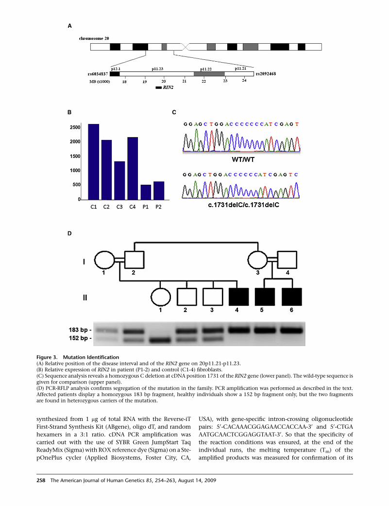
Figure 3. Mutation Identification(A) Relative position of the disease interval and of the RIN2 gene on 20p11.21-p11.23.(B) Relative expression of RIN2 in patient (P1-2) and control (C1-4) fibroblasts.(C) Sequence analysis reveals a homozygous C deletion at cDNA position 1731 of the RIN2 gene (lower panel). The wild-type sequence isgiven for comparison (upper panel).(D) PCR-RFLP analysis confirms segregation of the mutation in the family. PCR amplification was performed as described in the text.Affected patients display a homozygous 183 bp fragment, healthy individuals show a 152 bp fragment only, but the two fragmentsare found in heterozygous carriers of the mutation.
synthesized from 1 mg of total RNA with the Reverse-iT
First-Strand Synthesis Kit (ABgene), oligo dT, and random
hexamers in a 3:1 ratio. cDNA PCR amplification was
carried out with the use of SYBR Green JumpStart Taq
ReadyMix (Sigma) with ROX reference dye (Sigma) on a Ste-
pOnePlus cycler (Applied Biosystems, Foster City, CA,
258 The American Journal of Human Genetics 85, 254–263, August
USA), with gene-specific intron-crossing oligonucleotide
pairs: 50-CACAAACGGAGAACCACCAA-30 and 50-CTGA
AATGCAACTCGGAGGTAAT-30. So that the specificity of
the reaction conditions was ensured, at the end of the
individual runs, the melting temperature (Tm) of the
amplified products was measured for confirmation of its
14, 2009
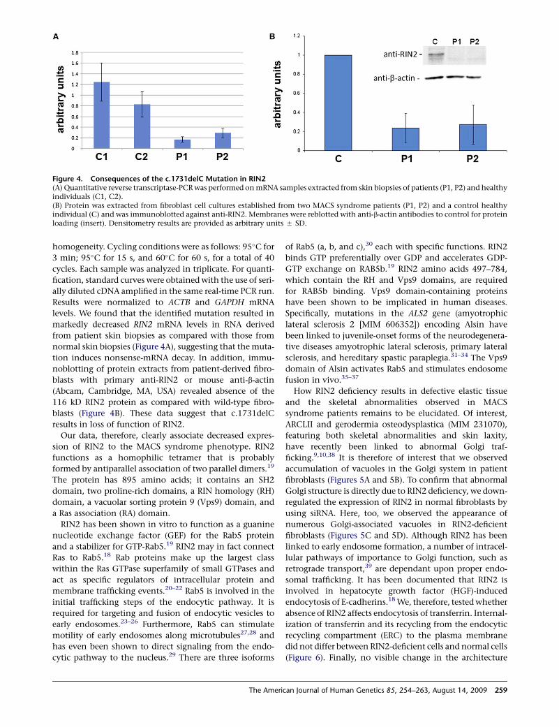
Figure 4. Consequences of the c.1731delC Mutation in RIN2(A) Quantitative reverse transcriptase-PCR was performed on mRNA samples extracted from skin biopsies of patients (P1, P2) and healthyindividuals (C1, C2).(B) Protein was extracted from fibroblast cell cultures established from two MACS syndrome patients (P1, P2) and a control healthyindividual (C) and was immunoblotted against anti-RIN2. Membranes were reblotted with anti-b-actin antibodies to control for proteinloading (insert). Densitometry results are provided as arbitrary units 5 SD.
homogeneity. Cycling conditions were as follows: 95�C for
3 min; 95�C for 15 s, and 60�C for 60 s, for a total of 40
cycles. Each sample was analyzed in triplicate. For quanti-
fication, standard curves were obtained with the use of seri-
ally diluted cDNA amplified in the same real-time PCR run.
Results were normalized to ACTB and GAPDH mRNA
levels. We found that the identified mutation resulted in
markedly decreased RIN2 mRNA levels in RNA derived
from patient skin biopsies as compared with those from
normal skin biopsies (Figure 4A), suggesting that the muta-
tion induces nonsense-mRNA decay. In addition, immu-
noblotting of protein extracts from patient-derived fibro-
blasts with primary anti-RIN2 or mouse anti-b-actin
(Abcam, Cambridge, MA, USA) revealed absence of the
116 kD RIN2 protein as compared with wild-type fibro-
blasts (Figure 4B). These data suggest that c.1731delC
results in loss of function of RIN2.
Our data, therefore, clearly associate decreased expres-
sion of RIN2 to the MACS syndrome phenotype. RIN2
functions as a homophilic tetramer that is probably
formed by antiparallel association of two parallel dimers.19
The protein has 895 amino acids; it contains an SH2
domain, two proline-rich domains, a RIN homology (RH)
domain, a vacuolar sorting protein 9 (Vps9) domain, and
a Ras association (RA) domain.
RIN2 has been shown in vitro to function as a guanine
nucleotide exchange factor (GEF) for the Rab5 protein
and a stabilizer for GTP-Rab5.19 RIN2 may in fact connect
Ras to Rab5.18 Rab proteins make up the largest class
within the Ras GTPase superfamily of small GTPases and
act as specific regulators of intracellular protein and
membrane trafficking events.20–22 Rab5 is involved in the
initial trafficking steps of the endocytic pathway. It is
required for targeting and fusion of endocytic vesicles to
early endosomes.23–26 Furthermore, Rab5 can stimulate
motility of early endosomes along microtubules27,28 and
has even been shown to direct signaling from the endo-
cytic pathway to the nucleus.29 There are three isoforms
The Amer
of Rab5 (a, b, and c),30 each with specific functions. RIN2
binds GTP preferentially over GDP and accelerates GDP-
GTP exchange on RAB5b.19 RIN2 amino acids 497–784,
which contain the RH and Vps9 domains, are required
for RAB5b binding. Vps9 domain-containing proteins
have been shown to be implicated in human diseases.
Specifically, mutations in the ALS2 gene (amyotrophic
lateral sclerosis 2 [MIM 606352]) encoding Alsin have
been linked to juvenile-onset forms of the neurodegenera-
tive diseases amyotrophic lateral sclerosis, primary lateral
sclerosis, and hereditary spastic paraplegia.31–34 The Vps9
domain of Alsin activates Rab5 and stimulates endosome
fusion in vivo.35–37
How RIN2 deficiency results in defective elastic tissue
and the skeletal abnormalities observed in MACS
syndrome patients remains to be elucidated. Of interest,
ARCLII and gerodermia osteodysplastica (MIM 231070),
featuring both skeletal abnormalities and skin laxity,
have recently been linked to abnormal Golgi traf-
ficking.9,10,38 It is therefore of interest that we observed
accumulation of vacuoles in the Golgi system in patient
fibroblasts (Figures 5A and 5B). To confirm that abnormal
Golgi structure is directly due to RIN2 deficiency, we down-
regulated the expression of RIN2 in normal fibroblasts by
using siRNA. Here, too, we observed the appearance of
numerous Golgi-associated vacuoles in RIN2-deficient
fibroblasts (Figures 5C and 5D). Although RIN2 has been
linked to early endosome formation, a number of intracel-
lular pathways of importance to Golgi function, such as
retrograde transport,39 are dependant upon proper endo-
somal trafficking. It has been documented that RIN2 is
involved in hepatocyte growth factor (HGF)-induced
endocytosis of E-cadherins.18 We, therefore, tested whether
absence of RIN2 affects endocytosis of transferrin. Internal-
ization of transferrin and its recycling from the endocytic
recycling compartment (ERC) to the plasma membrane
did not differ between RIN2-deficient cells and normal cells
(Figure 6). Finally, no visible change in the architecture
ican Journal of Human Genetics 85, 254–263, August 14, 2009 259
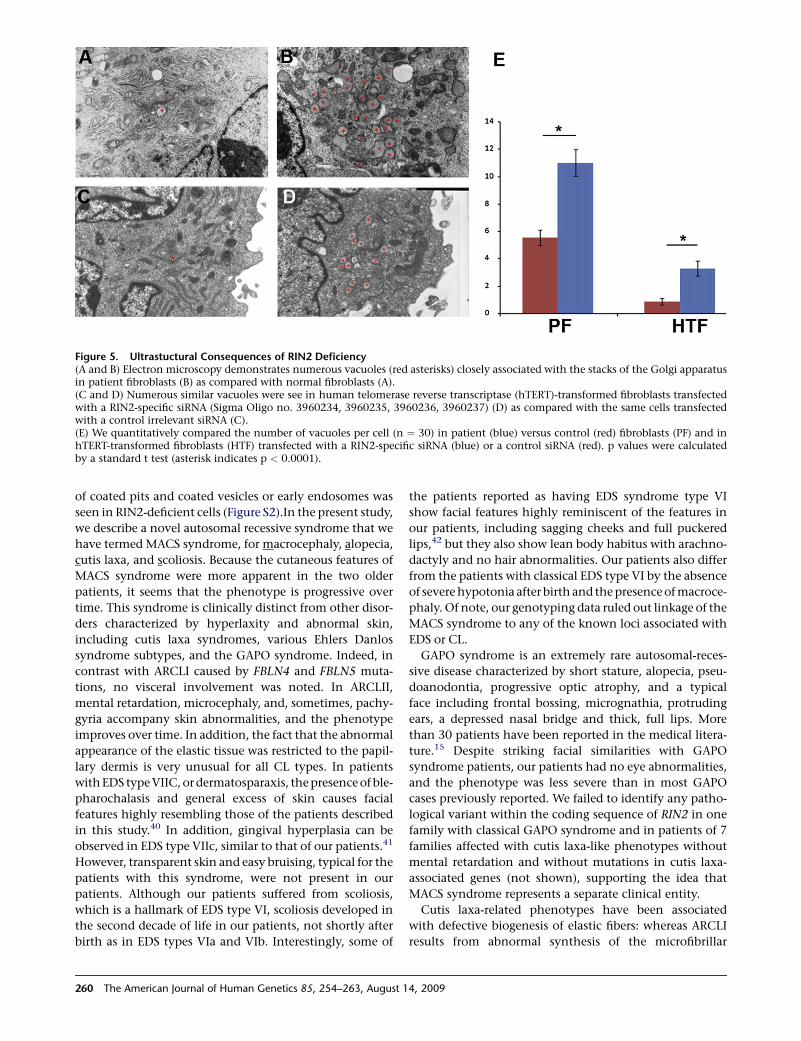
Figure 5. Ultrastuctural Consequences of RIN2 Deficiency(A and B) Electron microscopy demonstrates numerous vacuoles (red asterisks) closely associated with the stacks of the Golgi apparatusin patient fibroblasts (B) as compared with normal fibroblasts (A).(C and D) Numerous similar vacuoles were see in human telomerase reverse transcriptase (hTERT)-transformed fibroblasts transfectedwith a RIN2-specific siRNA (Sigma Oligo no. 3960234, 3960235, 3960236, 3960237) (D) as compared with the same cells transfectedwith a control irrelevant siRNA (C).(E) We quantitatively compared the number of vacuoles per cell (n ¼ 30) in patient (blue) versus control (red) fibroblasts (PF) and inhTERT-transformed fibroblasts (HTF) transfected with a RIN2-specific siRNA (blue) or a control siRNA (red). p values were calculatedby a standard t test (asterisk indicates p < 0.0001).
of coated pits and coated vesicles or early endosomes was
seen in RIN2-deficient cells (Figure S2).In the present study,
we describe a novel autosomal recessive syndrome that we
have termed MACS syndrome, for macrocephaly, alopecia,
cutis laxa, and scoliosis. Because the cutaneous features of
MACS syndrome were more apparent in the two older
patients, it seems that the phenotype is progressive over
time. This syndrome is clinically distinct from other disor-
ders characterized by hyperlaxity and abnormal skin,
including cutis laxa syndromes, various Ehlers Danlos
syndrome subtypes, and the GAPO syndrome. Indeed, in
contrast with ARCLI caused by FBLN4 and FBLN5 muta-
tions, no visceral involvement was noted. In ARCLII,
mental retardation, microcephaly, and, sometimes, pachy-
gyria accompany skin abnormalities, and the phenotype
improves over time. In addition, the fact that the abnormal
appearance of the elastic tissue was restricted to the papil-
lary dermis is very unusual for all CL types. In patients
with EDS type VIIC, or dermatosparaxis, the presence of ble-
pharochalasis and general excess of skin causes facial
features highly resembling those of the patients described
in this study.40 In addition, gingival hyperplasia can be
observed in EDS type VIIc, similar to that of our patients.41
However, transparent skin and easy bruising, typical for the
patients with this syndrome, were not present in our
patients. Although our patients suffered from scoliosis,
which is a hallmark of EDS type VI, scoliosis developed in
the second decade of life in our patients, not shortly after
birth as in EDS types VIa and VIb. Interestingly, some of
260 The American Journal of Human Genetics 85, 254–263, August
the patients reported as having EDS syndrome type VI
show facial features highly reminiscent of the features in
our patients, including sagging cheeks and full puckered
lips,42 but they also show lean body habitus with arachno-
dactyly and no hair abnormalities. Our patients also differ
from the patients with classical EDS type VI by the absence
of severe hypotonia after birth and the presence of macroce-
phaly. Of note, our genotyping data ruled out linkage of the
MACS syndrome to any of the known loci associated with
EDS or CL.
GAPO syndrome is an extremely rare autosomal-reces-
sive disease characterized by short stature, alopecia, pseu-
doanodontia, progressive optic atrophy, and a typical
face including frontal bossing, micrognathia, protruding
ears, a depressed nasal bridge and thick, full lips. More
than 30 patients have been reported in the medical litera-
ture.15 Despite striking facial similarities with GAPO
syndrome patients, our patients had no eye abnormalities,
and the phenotype was less severe than in most GAPO
cases previously reported. We failed to identify any patho-
logical variant within the coding sequence of RIN2 in one
family with classical GAPO syndrome and in patients of 7
families affected with cutis laxa-like phenotypes without
mental retardation and without mutations in cutis laxa-
associated genes (not shown), supporting the idea that
MACS syndrome represents a separate clinical entity.
Cutis laxa-related phenotypes have been associated
with defective biogenesis of elastic fibers: whereas ARCLI
results from abnormal synthesis of the microfibrillar
14, 2009
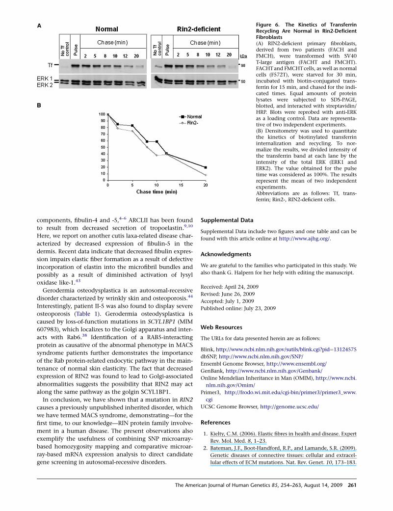
Figure 6. The Kinetics of TransferrinRecycling Are Normal in Rin2-DeficientFibroblasts(A) RIN2-deficient primary fibroblasts,derived from two patients (FACH andFMCH), were transformed with SV40T-large antigen (FACHT and FMCHT).FACHT and FMCHT cells, as well as normalcells (F572T), were starved for 30 min,incubated with biotin-conjugated trans-ferrin for 15 min, and chased for the indi-cated times. Equal amounts of proteinlysates were subjected to SDS-PAGE,blotted, and interacted with streptavidin/HRP. Blots were reprobed with anti-ERKas a loading control. Data are representa-tive of two independent experiments.(B) Densitometry was used to quantitatethe kinetics of biotinylated transferrininternalization and recycling. To nor-malize the results, we divided intensity ofthe transferrin band at each lane by theintensity of the total ERK (ERK1 andERK2). The value obtained for the pulsetime was considered as 100%. The resultsrepresent the mean of two independentexperiments.Abbreviations are as follows: Tf, trans-ferrin; Rin2-, RIN2-deficient cells.
components, fibulin-4 and -5,4–6 ARCLII has been found
to result from decreased secretion of tropoelastin.9,10
Here, we report on another cutis laxa-related disease char-
acterized by decreased expression of fibulin-5 in the
dermis. Recent data indicate that decreased fibulin expres-
sion impairs elastic fiber formation as a result of defective
incorporation of elastin into the microfibril bundles and
possibly as a result of diminished activation of lysyl
oxidase like-1.43
Gerodermia osteodysplastica is an autosomal-recessive
disorder characterized by wrinkly skin and osteoporosis.44
Interestingly, patient II-5 was also found to display severe
osteoporosis (Table 1). Gerodermia osteodysplastica is
caused by loss-of-function mutations in SCYL1BP1 (MIM
607983), which localizes to the Golgi apparatus and inter-
acts with Rab6.38 Identification of a RAB5-interacting
protein as causative of the abnormal phenotype in MACS
syndrome patients further demonstrates the importance
of the Rab protein-related endocytic pathway in the main-
tenance of normal skin elasticity. The fact that decreased
expression of RIN2 was found to lead to Golgi-associated
abnormalities suggests the possibility that RIN2 may act
along the same pathway as the golgin SCYL1BP1.
In conclusion, we have shown that a mutation in RIN2
causes a previously unpublished inherited disorder, which
we have termed MACS syndrome, demonstrating—for the
first time, to our knowledge—RIN protein family involve-
ment in a human disease. The present observations also
exemplify the usefulness of combining SNP microarray-
based homozygosity mapping and comparative microar-
ray-based mRNA expression analysis to direct candidate
gene screening in autosomal-recessive disorders.
The Amer
Supplemental Data
Supplemental Data include two figures and one table and can be
found with this article online at http://www.ajhg.org/.
Acknowledgments
We are grateful to the families who participated in this study. We
also thank G. Halpern for her help with editing the manuscript.
Received: April 24, 2009
Revised: June 26, 2009
Accepted: July 1, 2009
Published online: July 23, 2009
Web Resources
The URLs for data presented herein are as follows:
Blink, http://www.ncbi.nlm.nih.gov/sutils/blink.cgi?pid¼13124575
dbSNP, http://www.ncbi.nlm.nih.gov/SNP/
Ensembl Genome Browser, http://www.ensembl.org/
GenBank, http://www.ncbi.nlm.nih.gov/Genbank/
Online Mendelian Inheritance in Man (OMIM), http://www.ncbi.
nlm.nih.gov/Omim/
Primer3, http://frodo.wi.mit.edu/cgi-bin/primer3/primer3_www.
cgi
UCSC Genome Browser, http://genome.ucsc.edu/
References
1. Kielty, C.M. (2006). Elastic fibres in health and disease. Expert
Rev. Mol. Med. 8, 1–23.
2. Bateman, J.F., Boot-Handford, R.P., and Lamande, S.R. (2009).
Genetic diseases of connective tissues: cellular and extracel-
lular effects of ECM mutations. Nat. Rev. Genet. 10, 173–183.
ican Journal of Human Genetics 85, 254–263, August 14, 2009 261
3. Ringpfeil, F. (2005). Selected disorders of connective tissue:
pseudoxanthoma elasticum, cutis laxa, and lipoid proteinosis.
Clin. Dermatol. 23, 41–46.
4. Hucthagowder, V., Sausgruber, N., Kim, K.H., Angle, B.,
Marmorstein, L.Y., and Urban, Z. (2006). Fibulin-4: a novel
gene for an autosomal recessive cutis laxa syndrome. Am. J.
Hum. Genet. 78, 1075–1080.
5. Markova, D., Zou, Y., Ringpfeil, F., Sasaki, T., Kostka, G., Timpl,
R., Uitto, J., and Chu, M.L. (2003). Genetic heterogeneity of
cutis laxa: a heterozygous tandem duplication within the fibu-
lin-5 (FBLN5) gene. Am. J. Hum. Genet. 72, 998–1004.
6. Loeys, B., Van Maldergem, L., Mortier, G., Coucke, P., Gerniers,
S., Naeyaert, J.M., and De Paepe, A. (2002). Homozygosity for
a missense mutation in fibulin-5 (FBLN5) results in a severe
form of cutis laxa. Hum. Mol. Genet. 11, 2113–2118.
7. Zhang, M.C., He, L., Giro, M., Yong, S.L., Tiller, G.E., and
Davidson, J.M. (1999). Cutis laxa arising from frameshift
mutations in exon 30 of the elastin gene (ELN). J. Biol.
Chem. 274, 981–986.
8. Hu, Q., Loeys, B.L., Coucke, P.J., De Paepe, A., Mecham, R.P.,
Choi, J., Davis, E.C., and Urban, Z. (2006). Fibulin-5 muta-
tions: mechanisms of impaired elastic fiber formation in reces-
sive cutis laxa. Hum. Mol. Genet. 15, 3379–3386.
9. Kornak, U., Reynders, E., Dimopoulou, A., van Reeuwijk, J.,
Fischer, B., Rajab, A., Budde, B., Nurnberg, P., Foulquier, F.,
Lefeber, D., et al. (2008). Impaired glycosylation and cutis
laxa caused by mutations in the vesicular Hþ-ATPase subunit
ATP6V0A2. Nat. Genet. 40, 32–34.
10. Hucthagowder, V., Morava, E., Kornak, U., Lefeber, D.J.,
Fischer, B., Dimopoulou, A., Aldinger, A., Choi, J., Davis,
E.C., Abuelo, D.N., et al. (2009). Loss-of-function mutations
in ATP6V0A2 impair vesicular trafficking, tropoelastin secre-
tion, and cell survival. Hum. Mol. Genet. 18, 2149–2165.
11. Hyland, J., Ala-Kokko, L., Royce, P., Steinmann, B., Kivirikko,
K.I., and Myllyla, R. (1992). A homozygous stop codon in
the lysyl hydroxylase gene in two siblings with Ehlers-Danlos
syndrome type VI. Nat. Genet. 2, 228–231.
12. Kivuva, E.C., Parker, M.J., Cohen, M.C., Wagner, B.E., and
Sobey, G. (2008). De Barsy syndrome: a review of the pheno-
type. Clin. Dysmorphol. 17, 99–107.
13. Demirgunes, E.F., Ersoy-Evans, S., and Karaduman, A. (2009).
GAPO syndrome with the novel features of pulmonary hyper-
tension, ankyloglossia, and prognathism. Am. J. Med. Genet.
A. 149A, 802–805.
14. Kocabay, G., and Mert, M. (2009). GAPO syndrome associated
with dilated cardiomyopathy: an unreported association. Am.
J. Med. Genet. A. 149A, 415–416.
15. Goloni-Bertollo, E.M., Ruiz, M.T., Goloni, C.B., Muniz, M.P.,
Valerio, N.I., and Pavarino-Bertelli, E.C. (2008). GAPO
syndrome: three new Brazilian cases, additional osseous mani-
festations, and review of the literature. Am. J. Med. Genet. A.
146A, 1523–1529.
16. Manouvrier-Hanu, S., Largilliere, C., Benalioua, M., Farriaux,
J.P., and Fontaine, G. (1987). The GAPO syndrome. Am. J.
Med. Genet. 26, 683–688.
17. Tipton, R.E., and Gorlin, R.J. (1984). Growth retardation,
alopecia, pseudo-anodontia, and optic atrophy–the GAPO
syndrome: report of a patient and review of the literature.
Am. J. Med. Genet. 19, 209–216.
18. Kimura, T., Sakisaka, T., Baba, T., Yamada, T., and Takai, Y.
(2006). Involvement of the Ras-Ras-activated Rab5 guanine
nucleotide exchange factor RIN2-Rab5 pathway in the hepa-
262 The American Journal of Human Genetics 85, 254–263, August 1
tocyte growth factor-induced endocytosis of E-cadherin. J.
Biol. Chem. 281, 10598–10609.
19. Saito, K., Murai, J., Kajiho, H., Kontani, K., Kurosu, H., and
Katada, T. (2002). A novel binding protein composed of ho-
mophilic tetramer exhibits unique properties for the small
GTPase Rab5. J. Biol. Chem. 277, 3412–3418.
20. Fukuda, M. (2008). Regulation of secretory vesicle traffic by
Rab small GTPases. Cell. Mol. Life Sci. 65, 2801–2813.
21. Fukuda, M. (2006). Rab27 and its effectors in secretory
granule exocytosis: a novel docking machinery composed of
a Rab27.effector complex. Biochem. Soc. Trans. 34, 691–695.
22. Grosshans, B.L., Ortiz, D., and Novick, P. (2006). Rabs and
their effectors: achieving specificity in membrane traffic.
Proc. Natl. Acad. Sci. USA 103, 11821–11827.
23. Carney, D.S., Davies, B.A., and Horazdovsky, B.F. (2006). Vps9
domain-containing proteins: activators of Rab5 GTPases from
yeast to neurons. Trends Cell Biol. 16, 27–35.
24. Simpson, J.C., and Jones, A.T. (2005). Early endocytic Rabs:
functional prediction to functional characterization. Bio-
chem. Soc. Symp. 72, 99–108.
25. Barbieri, M.A., Roberts, R.L., Mukhopadhyay, A., and Stahl,
P.D. (1996). Rab5 regulates the dynamics of early endosome
fusion. Biocell 20, 331–338.
26. Li, G. (1996). Rab5 GTPase and endocytosis. Biocell 20, 325–
330.
27. Loubery, S., Wilhelm, C., Hurbain, I., Neveu, S., Louvard, D.,
and Coudrier, E. (2008). Different microtubule motors move
early and late endocytic compartments. Traffic 9, 492–509.
28. Nielsen, E., Severin, F., Backer, J.M., Hyman, A.A., and Zerial,
M. (1999). Rab5 regulates motility of early endosomes on
microtubules. Nat. Cell Biol. 1, 376–382.
29. Zerial, M., and McBride, H. (2001). Rab proteins as membrane
organizers. Nat. Rev. Mol. Cell Biol. 2, 107–117.
30. Bucci, C., Lutcke, A., Steele-Mortimer, O., Olkkonen, V.M.,
Dupree, P., Chiariello, M., Bruni, C.B., Simons, K., and Zerial,
M. (1995). Co-operative regulation of endocytosis by three
Rab5 isoforms. FEBS Lett. 366, 65–71.
31. Gros-Louis, F., Gaspar, C., and Rouleau, G.A. (2006). Genetics
of familial and sporadic amyotrophic lateral sclerosis. Bio-
chim. Biophys. Acta 1762, 956–972.
32. Eymard-Pierre, E., Lesca, G., Dollet, S., Santorelli, F.M.,
di Capua, M., Bertini, E., and Boespflug-Tanguy, O. (2002).
Infantile-onset ascending hereditary spastic paralysis is associ-
ated with mutations in the alsin gene. Am. J. Hum. Genet. 71,
518–527.
33. Hadano, S., Hand, C.K., Osuga, H., Yanagisawa, Y., Otomo, A.,
Devon, R.S., Miyamoto, N., Showguchi-Miyata, J., Okada, Y.,
Singaraja, R., et al. (2001). A gene encoding a putative GTPase
regulator is mutated in familial amyotrophic lateral sclerosis 2.
Nat. Genet. 29, 166–173.
34. Yang, Y., Hentati, A., Deng, H.X., Dabbagh, O., Sasaki, T.,
Hirano, M., Hung, W.Y., Ouahchi, K., Yan, J., Azim, A.C.,
et al. (2001). The gene encoding alsin, a protein with three
guanine-nucleotide exchange factor domains, is mutated in
a form of recessive amyotrophic lateral sclerosis. Nat. Genet.
29, 160–165.
35. Otomo, A., Hadano, S., Okada, T., Mizumura, H., Kunita, R.,
Nishijima, H., Showguchi-Miyata, J., Yanagisawa, Y., Kohiki,
E., Suga, E., et al. (2003). ALS2, a novel guanine nucleotide
exchange factor for the small GTPase Rab5, is implicated in
endosomal dynamics. Hum. Mol. Genet. 12, 1671–1687.
4, 2009

36. Topp, J.D., Carney, D.S., and Horazdovsky, B.F. (2005).
Biochemical characterization of Alsin, a Rab5 and Rac1 guanine
nucleotide exchange factor. Methods Enzymol. 403, 261–276.
37. Topp, J.D., Gray, N.W., Gerard, R.D., and Horazdovsky, B.F.
(2004). Alsin is a Rab5 and Rac1 guanine nucleotide exchange
factor. J. Biol. Chem. 279, 24612–24623.
38. Hennies, H.C., Kornak, U., Zhang, H., Egerer, J., Zhang, X.,
Seifert, W., Kuhnisch, J., Budde, B., Natebus, M., Brancati, F.,
et al. (2008). Gerodermia osteodysplastica is caused by muta-
tions in SCYL1BP1, a Rab-6 interacting golgin. Nat. Genet.
40, 1410–1412.
39. Johannes, L., and Popoff, V. (2008). Tracing the retrograde
route in protein trafficking. Cell 135, 1175–1187.
40. Colige, A., Nuytinck, L., Hausser, I., van Essen, A.J., Thiry, M.,
Herens, C., Ades, L.C., Malfait, F., Paepe, A.D., Franck, P., et al.
(2004). Novel types of mutation responsible for the dermato-
sparactic type of Ehlers-Danlos syndrome (Type VIIC) and
The Ameri
common polymorphisms in the ADAMTS2 gene. J. Invest.
Dermatol. 123, 656–663.
41. De Coster, P.J., Malfait, F., Martens, L.C., and De Paepe, A.
(2003). Unusual oral findings in dermatosparaxis (Ehlers-
Danlos syndrome type VIIC). J. Oral Pathol. Med. 32, 568–570.
42. Steinmann, B., Gitzelmann, R., Vogel, A., Grant, M.E.,
Harwood, R., and Sear, C.H. (1975). Ehlers-Danlos syndrome
in two siblings with deficient lysyl hydroxylase activity in
cultured skin fibroblasts but only mild hydroxylysine deficit
in skin. Helv. Paediatr. Acta 30, 255–274.
43. Choi, J., Bergdahl, A., Zheng, Q., Starcher, B., Yanagisawa, H.,
and Davis, E.C. (2009). Analysis of Dermal Elastic Fibers in the
Absence of Fibulin-5 Reveals Potential Roles for Fibulin-5 in
Elastic Fiber Assembly. Matrix Biol., in press.
44. Boente Mdel, C., Asial, R.A., andWinik, B.C. (2006). Geroderma
osteodysplastica. Report of a new family. Pediatr. Dermatol. 23,
467–472.
can Journal of Human Genetics 85, 254–263, August 14, 2009 263