Gut–Brain Axis as a Pathological and Therapeutic Target for ...
15
Citation: Toledo, A.R.L.; Monroy, G.R.; Salazar, F.E.; Lee, J.-Y.; Jain, S.; Yadav, H.; Borlongan, C.V. Gut–Brain Axis as a Pathological and Therapeutic Target for Neurodegenerative Disorders. Int. J. Mol. Sci. 2022, 23, 1184. https:// doi.org/10.3390/ijms23031184 Academic Editor: Amedeo Amedei Received: 9 December 2021 Accepted: 13 January 2022 Published: 21 January 2022 Publisher’s Note: MDPI stays neutral with regard to jurisdictional claims in published maps and institutional affil- iations. Copyright: © 2022 by the authors. Licensee MDPI, Basel, Switzerland. This article is an open access article distributed under the terms and conditions of the Creative Commons Attribution (CC BY) license (https:// creativecommons.org/licenses/by/ 4.0/). International Journal of Molecular Sciences Review Gut–Brain Axis as a Pathological and Therapeutic Target for Neurodegenerative Disorders Alma Rosa Lezama Toledo 1,† , Germán Rivera Monroy 1,† , Felipe Esparza Salazar 1,† , Jea-Young Lee 1 , Shalini Jain 2 , Hariom Yadav 2 and Cesario Venturina Borlongan 1, * 1 Center of Excellence for Aging and Brain Repair, Department of Neurosurgery and Brain Repair, Morsani College of Medicine, University of South Florida, 12901 Bruce B Downs Blvd, Tampa, FL 33612, USA; [email protected] (A.R.L.T.); [email protected] (G.R.M.); [email protected] (F.E.S.); [email protected] (J.-Y.L.) 2 Center for Microbiome Research, Department of Neurosurgery and Brain Repair, Morsani College of Medicine, University of South Florida, 12901 Bruce B Downs Blvd, Tampa, FL 33612, USA; [email protected] (S.J.); [email protected] (H.Y.) * Correspondence: [email protected] † These authors contributed equally to this manuscript. Abstract: Human lifestyle and dietary behaviors contribute to disease onset and progression. Neu- rodegenerative diseases (NDDs), considered multifactorial disorders, have been associated with changes in the gut microbiome. NDDs display pathologies that alter brain functions with a tendency to worsen over time. NDDs are a worldwide health problem; in the US alone, 12 million Americans will suffer from NDDs by 2030. While etiology may vary, the gut microbiome serves as a key element underlying NDD development and prognosis. In particular, an inflammation-associated microbiome plagues NDDs. Conversely, sequestration of this inflammatory microbiome by a correction in the dys- biotic state of the gut may render therapeutic effects on NDDs. To this end, treatment with short-chain fatty acid-producing bacteria, the main metabolites responsible for maintaining gut homeostasis, ameliorates the inflammatory microbiome. This intimate pathological link between the gut and NDDs suggests that the gut-brain axis (GBA) acts as an underexplored area for developing therapies for NDDs. Traditionally, the classification of NDDs depends on their clinical presentation, mostly manifesting as extrapyramidal and pyramidal movement disorders, with neuropathological evalua- tion at autopsy as the gold standard for diagnosis. In this review, we highlight the evolving notion that GBA stands as an equally sensitive pathological marker of NDDs, particularly in Alzheimer’s disease, Parkinson’s disease, amyotrophic lateral sclerosis and chronic stroke. Additionally, GBA represents a potent therapeutic target for treating NDDs. Keywords: neurodegeneration; microbiome; stem cells; epigenetics; neurological disorders 1. Introduction Neurodegenerative diseases (NDDs) are commonly defined as pathologies that lower normal brain function, usually accompanied by brain tissue atrophy and lower cognition capacity with a tendency to worsen with chronicity [1,2] Although NDDs manifest as chronic and aging brain pathologies, the exact timing for a brain pathology to turn into a neurodegenerative aberration remains not well understood. Acute brain insults with chronic pathological manifestations, such as stroke, present symptoms of neurodegener- ative disease [3]. NDDs stand as a major health problem that affect millions of people worldwide, including those suffering from Alzheimer’s disease (AD), Parkinson’s disease (PD) and Amyotrophic Lateral Sclerosis (ALS), the most common pathologies around the world [4,5]. Most NDDs tend to be associated with aged people’s brain pathologies, and with the constant increase in life expectancy, the incidence of these diseases is expected to increase as well [1,4]. Int. J. Mol. Sci. 2022, 23, 1184. https://doi.org/10.3390/ijms23031184 https://www.mdpi.com/journal/ijms
-
Upload
khangminh22 -
Category
Documents
-
view
1 -
download
0
Transcript of Gut–Brain Axis as a Pathological and Therapeutic Target for ...

�����������������
Citation: Toledo, A.R.L.; Monroy,
G.R.; Salazar, F.E.; Lee, J.-Y.; Jain, S.;
Yadav, H.; Borlongan, C.V. Gut–Brain
Axis as a Pathological and
Therapeutic Target for
Neurodegenerative Disorders. Int. J.
Mol. Sci. 2022, 23, 1184. https://
doi.org/10.3390/ijms23031184
Academic Editor: Amedeo Amedei
Received: 9 December 2021
Accepted: 13 January 2022
Published: 21 January 2022
Publisher’s Note: MDPI stays neutral
with regard to jurisdictional claims in
published maps and institutional affil-
iations.
Copyright: © 2022 by the authors.
Licensee MDPI, Basel, Switzerland.
This article is an open access article
distributed under the terms and
conditions of the Creative Commons
Attribution (CC BY) license (https://
creativecommons.org/licenses/by/
4.0/).
International Journal of
Molecular Sciences
Review
Gut–Brain Axis as a Pathological and Therapeutic Target forNeurodegenerative DisordersAlma Rosa Lezama Toledo 1,†, Germán Rivera Monroy 1,†, Felipe Esparza Salazar 1,†, Jea-Young Lee 1,Shalini Jain 2, Hariom Yadav 2 and Cesario Venturina Borlongan 1,*
1 Center of Excellence for Aging and Brain Repair, Department of Neurosurgery and Brain Repair,Morsani College of Medicine, University of South Florida, 12901 Bruce B Downs Blvd,Tampa, FL 33612, USA; [email protected] (A.R.L.T.); [email protected] (G.R.M.);[email protected] (F.E.S.); [email protected] (J.-Y.L.)
2 Center for Microbiome Research, Department of Neurosurgery and Brain Repair, Morsani Collegeof Medicine, University of South Florida, 12901 Bruce B Downs Blvd, Tampa, FL 33612, USA;[email protected] (S.J.); [email protected] (H.Y.)
* Correspondence: [email protected]† These authors contributed equally to this manuscript.
Abstract: Human lifestyle and dietary behaviors contribute to disease onset and progression. Neu-rodegenerative diseases (NDDs), considered multifactorial disorders, have been associated withchanges in the gut microbiome. NDDs display pathologies that alter brain functions with a tendencyto worsen over time. NDDs are a worldwide health problem; in the US alone, 12 million Americanswill suffer from NDDs by 2030. While etiology may vary, the gut microbiome serves as a key elementunderlying NDD development and prognosis. In particular, an inflammation-associated microbiomeplagues NDDs. Conversely, sequestration of this inflammatory microbiome by a correction in the dys-biotic state of the gut may render therapeutic effects on NDDs. To this end, treatment with short-chainfatty acid-producing bacteria, the main metabolites responsible for maintaining gut homeostasis,ameliorates the inflammatory microbiome. This intimate pathological link between the gut andNDDs suggests that the gut-brain axis (GBA) acts as an underexplored area for developing therapiesfor NDDs. Traditionally, the classification of NDDs depends on their clinical presentation, mostlymanifesting as extrapyramidal and pyramidal movement disorders, with neuropathological evalua-tion at autopsy as the gold standard for diagnosis. In this review, we highlight the evolving notionthat GBA stands as an equally sensitive pathological marker of NDDs, particularly in Alzheimer’sdisease, Parkinson’s disease, amyotrophic lateral sclerosis and chronic stroke. Additionally, GBArepresents a potent therapeutic target for treating NDDs.
Keywords: neurodegeneration; microbiome; stem cells; epigenetics; neurological disorders
1. Introduction
Neurodegenerative diseases (NDDs) are commonly defined as pathologies that lowernormal brain function, usually accompanied by brain tissue atrophy and lower cognitioncapacity with a tendency to worsen with chronicity [1,2] Although NDDs manifest aschronic and aging brain pathologies, the exact timing for a brain pathology to turn intoa neurodegenerative aberration remains not well understood. Acute brain insults withchronic pathological manifestations, such as stroke, present symptoms of neurodegener-ative disease [3]. NDDs stand as a major health problem that affect millions of peopleworldwide, including those suffering from Alzheimer’s disease (AD), Parkinson’s disease(PD) and Amyotrophic Lateral Sclerosis (ALS), the most common pathologies around theworld [4,5]. Most NDDs tend to be associated with aged people’s brain pathologies, andwith the constant increase in life expectancy, the incidence of these diseases is expected toincrease as well [1,4].
Int. J. Mol. Sci. 2022, 23, 1184. https://doi.org/10.3390/ijms23031184 https://www.mdpi.com/journal/ijms

Int. J. Mol. Sci. 2022, 23, 1184 2 of 15
Over the past decade, the accumulating evidence has implicated a pathological linkbetween brain and gut microbiota, which may also represent a novel therapeutic targetfor treating NDDs [6,7]. The gut-brain axis (GBA) is the term used to describe the inter-action between brain and gut microbiota, highlighting the complex interaction of braindevelopment, aging, and functioning [7,8]. Direct anatomical and physiological connec-tions of the GBA may entail the vagus nerve, hormone signaling, metabolism of specificmolecules such as tryptophan and even the immune system, altogether corresponding tokey pathways for interrogating the GBA microbiome [6,9]. GBA may play an importantrole in brain functions, such as cognition learning and memory, suggesting that targetingthe patients’ specific gut microbiota may alleviate neurological symptoms in NDDs, such asAD and PD [8,10]. Indeed, dampening the pro-inflammatory immune response product ofpathogenic bacteria secretions and natural immune response in the gut affords therapeuticeffects in NDDs [11].
Current medical treatment for NDDs is primarily symptomatic. Specific drugs thattarget the brain under a regimen of polypharmaceutical therapies can retard the progressionof NDDs, but most of these pharmacotherapeutics are palliative and do not directly alterthe disease pathology [6,12]. Novel therapies designed to promote disease-modifyingoutcomes, such as stem cell therapy, generate promising preclinical and clinical outcomesin NDDs [13,14]. Of note, several in vitro and preclinical models of NDDs reveal themechanistic action of this stem cell therapy involves downregulation of the deleteriouspro-inflammatory response closely associated with NDDs [14,15]. Recognizing the closeinteraction of inflammation in GBA and NDDs, in this article, we discussed the pathologicalhallmarks of NDDs (AD, PD, ALS, and chronic stroke), emphasizing the role of the gutmicrobiome in the disease progression as well as in developing innovative treatment viaanti-inflammatory strategies, such as stem cell therapy. Many paradigms implicate theassociation of neurodegenerative diseases with infectious diseases and the putative biologi-cal function of some of the primary proteins implicated in these disorders. The aberrantaccumulation of specific proteins, such as TDP-43 [16] and SOD1 [17] in ALS, amyloid β [18]and Tau [19] in AD, and α-synuclein [20] in PD, with similar proteinopathies seen in thechronic stage of ischemic stroke [21] can initiate a cascade of deleterious innate immunityprocesses that may contribute to the dysbiotic and dysfunctional GBA pathophysiologicalmanifestations of NDDs. Recent review papers highlight the interaction between GBA andNDDs, with emphasis on diet, exercise, prebiotics and probiotics as treatments towardsmaintaining healthy microbiota in GBA [22–27]. Here, we chose to probe the underexploreduse of stem cells for improving the microbiota in NDDs. While ethical and technical caveatsabound, such as stem cell source, cell purity and amplification, and potential tumorigenicrisks, hinder stem cell therapy, the optimization of the safety and efficacy of this approachmay open new avenues of research and clinical application of cell-based therapeutics fortreating NDDs.
2. Epigenetics and Neurodegeneration
Early human developmental stages represent a key period that may affect health inadulthood [28–30]. Indeed, a differentiation process takes place during the first 1000 days oflife that leads to specialized cells of the pluripotent mediated by an epigenetic remodelingthat is responsible for deactivating unnecessary genes for a particular tissue while helpingto express those that are essential for the same [28]. To differentiate cells, DNA methylationprocesses dependent on DNA methyltransferases (DNMT) are necessary, which catalyzethe methylation of CpG islands in the gene promoter. When the presence of methyl groupsobstructs the interaction between the transcription factors and the promoter region, thebinding with RNA polymerases that initiates early gene expression is suppressed [28–30].An additional gene expression regulation through a complex process entails methylationof regulatory regions as well as histone methylation. The availability of methyl groupdonors determines the methylation process during pregnancy and throughout life via thefolate metabolism pathway. A diet rich in folates, with folic acid supplementation during

Int. J. Mol. Sci. 2022, 23, 1184 3 of 15
pregnancy, and the availability of vitamins B6 and B12 help to promote a good availabilityof methyl groups. In addition to DNA methylation, post-translational modifications andhistone modification also stand as highly relevant epigenetic mechanisms associated withregulating a healthy and an unhealthy phenotype [28–31].
Chromatin remodeling is associated with the activation or inhibition of gene ex-pression through processes such as histone methylation, acetylation, phosphorylation,ubiquitination, sumoylation, and glycation, altogether representing the first step for geneexpression [28]. For the modulation of these processes, specific precursors are required forsurveillance of adequate quantity and quality of nutrient intake to maintain a balanced ratioof FAD/FADH2. A reduced intake of folates during early life has been linked to incorrectDNA methylation with long-term effects, such as a decrease in the insulin growth factor2 (IGF2) promoter methylation of the maternal allele transmission to offspring, overweightin men at age 20 and glucose intolerance at age 50 [28]. Additionally, low birth weight,obesity with coronary heart disease, and deterioration in neurocognitive development inadult life, with maternal smoking exacerbating this phenotype [28–31].
Neurological disorders, especially those presenting with neurodegeneration, havebeen associated with harmful environmental factors identified in childhood; in particular,an unbalanced diet that alters early gene expression leads to epigenetic changes thatmanifest in adulthood [28]. Early neurobehavioral deficits accompany remodeling of theepigenome by environmental factors such as smoking, alcohol, stress, and exposure topesticides [32]. An iron deficit in early life is related to permanent deficits in recognitionmemory and, later in life, in procedural memory [33]. On the contrary, an excess of maternaliron or during adulthood can have deficiencies in development due to epigenetic andneuroinflammatory processes [34]. Studies in animal models showed that an iron deficiencyin the neonatal age coincides with a neurodevelopmental dysfunction consequent withaltered hippocampal DNA methylation and deficient expression of genes involved in theregulation of permeability, hypoxia, and angiogenesis [35]. A decrease in fetal neurogenesismay manifest as deficiencies in metals such as copper and zinc due to an impaired DNAmethylation process, which during adulthood may contribute to the production of theβ-amyloid peptide present in plaques of AD patients [28–31]. Similarly, dysfunctionalfetal neurogenesis of immature dopaminergic neurons has been implicated in PD [20],whereas in ALS cellular modeling, induction, but not inhibition, of inflammation in fetalbrain-derived human neural stem cells enhances their proliferation and differentiationinto oligodendrocytes [36]. Interestingly in stroke, lactation protects the maternal brainagainst ischemic insult partly through angiogenic and neurogenic remodeling processes [37].Altogether, these findings suggest that epigenetics in early life, or when recapitulatedduring pregnancy, may play a significant role in adult health, specifically regulating thebrain capacity to undergo repair or neuroregeneration.
2.1. Amyotrophic Lateral Sclerosis
ALS, also known as motor neuron disease, manifests as a multifactorial neurodegener-ative disease characterized by progressive degeneration and death of motor neurons in thebrain and spinal cord, which leads to both motor and extra-motor symptoms. [38,39]. ALSdiagnosis often occurs during the third and fourth decade of life and can be classified assporadic, accounting for 90% of the cases, or hereditary, accounting for 10% of the cases [40].Clinical features typically include muscle weakness, dysarthria, dysphagia, and, in moreadvanced stages, respiratory problems due to diaphragm paralysis [39,41].
The clinical neurodegeneration observed in ALS consists of decreasing ability tocontrol and activate skeletal or smooth muscles, eventually manifesting as muscle weaknessand wasting [42]. This muscular dysfunction is due to a loss of neuromuscular connectionas well as axonal retraction, which leads to cell death of both upper and lower motorneurons [42,43]. Clinical symptoms can start with bulbar symptoms, such as dysarthriaand dysphagia or can manifest in muscles of the extremities. Independent of mechanistictriggers of symptom initiation, neurodegeneration proceeds, and motor neuron death

Int. J. Mol. Sci. 2022, 23, 1184 4 of 15
progresses to a point where patients are not self-sufficient anymore and need external helpfor basic life chores such as moving and eating [44]. Unfortunately, most of the patients diedue to breathing or eating inability produced by near-complete incapacitation of breathingor swallowing muscles [43,44]. Despite the severity of motor neuron death, patients donot display cognitive or mental dysfunctions, thereby relegating ALS as a purely motorneurodegenerative disease.
Although ALS etiology remains not well established, the gut microbiota may me-diate the disease pathology, mainly due to pro-inflammatory gut microbiomes [45,46](Figure 1). Of note, the gut pro-inflammatory state leads to neural disturbance, and withtime, to neurodegeneration [46]. Pro-inflammatory cytokine expression contributes tothe progressive damage of the central nervous system (CNS) and inhibits self-repairingprocesses [47,48]. Likewise, a deleterious feedback loop ensues with the initial inflam-matory insult, subsequently triggering pro-inflammatory immune components such asmicroglia, macrophages, neutrophils, and natural killers to encroach cerebral tissue andcreate neurological dysfunctions [49,50].
Figure 1. ALS and GBA. A dysfunctional gut accompanies the progression of ALS, with increasedbacteria, including E. coli and enterobacteriaceae, leading to upregulation of damaging reactiveoxygen species (ROS) and eventually contributing to motor neuron death, which is a hallmarkpathological manifestation of the disease.
Microglial activation acts as a major element of chronic neurodegeneration [51,52]. Pa-tients suffering from ALS have higher levels of pro-inflammatory cytokines and biomarkersin cerebrospinal fluid and the spinal cord, such as IL-8, IL-6, MCP-1, and the expression ofCD1, CD40, among others [53,54]. Pathologic bacteria expression of LPS and inflammatorycytokines, which are commonly associated with dysbiotic gut microbiomes, exacerbateschronic microglial activation [55,56]. Accordingly, an unhealthy gut state can lead to sev-eral neurologic disbalances, such as neurovascular unit (NVU) disturbance, blood–brainbarrier (BBB) leakage, neurotoxic environment, etc., which together increase the risk todevelop NNDs, such as ALS [46,53]. Upregulation of specific pathologic bacterias, suchas E. coli and other enterobacteriaceae, accompanies ALS clinical manifestations and apoorer prognosis for long-term survival [53,57]. That the GBA may be the source of ALSpathophysiology is recognized from patients, as well as animal models, in their inability toeliminate reactive oxygen species (ROS) and other neurotoxic agents, which consequentlyincreases motor neuron death [58,59]. This specific pathologic characteristic of ALS, inaddition to the pro-inflammatory state produced by gut dysbiosis, creates a health scenariowhere it is crucial to attend to both the clinical neurological manifestations as well as theimbalance that exists at the gut level.
Int. J. Mol. Sci. 2022, 23, 1184 5 of 15
2.2. Alzheimer’s Disease
Advanced age is the main risk factor for AD. The composition of the intestinal micro-biota changes as we age, and certain protective bacteria, such as Bacteroidetes, Bifidobac,and Lactobacillus, decrease [9,60,61]. The intestinal microbiota contains large amounts ofbacterial amyloid, and the most studied is Escherichia Coli. The production of amyloidproteins prompts bacterial cells to form biofilms that confer resistance against the destruc-tion of immune factors [62–65]. Exposure to bacterial amyloid proteins in the gut enhancesthe immune response to endogenous neuronal amyloid accumulation in the brain [11,66]
AD entails a complex neurodegenerative process that involves the aberrant formationof amyloid-β (Aβ) plaques as well as hyperphosphorylated Tau neurofibrillary tangles,thereafter producing neurotoxicity and neuroinflammation and resulting in cell death andlowering normal brain functions [67,68]. AD patients suffer from neuronal loss, specificallyfrom the middle and lower temporal lobes, as revealed by imaging studies such as CT scansand MRIs [68]. This cellular death coincides with clinical manifestations such as lowersemantic and episodic memory, which with time can make a person not self-sufficient forliving alone or socializing with people [68,69]. The clinical onset of AD neurodegenerationusually starts with patients having difficulty remembering places, words, or names thatthey used to know [70]. Likewise, they have problems with learning new things or conceptsand maintaining focus on a specific chore [71]. As neurodegeneration progresses, thesymptoms worsen, and patients may not recognize familiar faces, places, or objects, recentactivities, or known concepts, leading to behavioral changes [72,73].
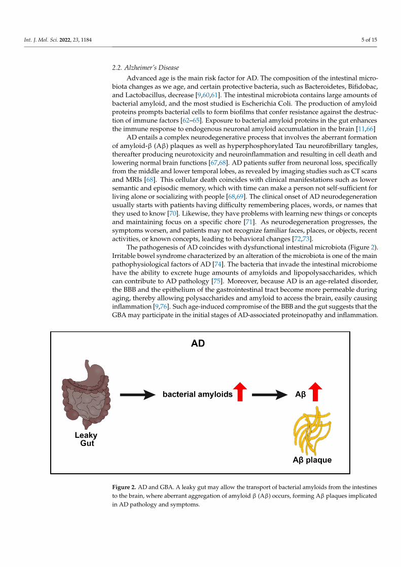
The pathogenesis of AD coincides with dysfunctional intestinal microbiota (Figure 2).Irritable bowel syndrome characterized by an alteration of the microbiota is one of the mainpathophysiological factors of AD [74]. The bacteria that invade the intestinal microbiomehave the ability to excrete huge amounts of amyloids and lipopolysaccharides, whichcan contribute to AD pathology [75]. Moreover, because AD is an age-related disorder,the BBB and the epithelium of the gastrointestinal tract become more permeable duringaging, thereby allowing polysaccharides and amyloid to access the brain, easily causinginflammation [9,76]. Such age-induced compromise of the BBB and the gut suggests that theGBA may participate in the initial stages of AD-associated proteinopathy and inflammation.
Figure 2. AD and GBA. A leaky gut may allow the transport of bacterial amyloids from the intestinesto the brain, where aberrant aggregation of amyloid β (Aβ) occurs, forming Aβ plaques implicatedin AD pathology and symptoms.

Int. J. Mol. Sci. 2022, 23, 1184 6 of 15
2.3. Parkinson’s Disease
PD corresponds to the most common movement disorder affecting up to 1% of thepopulation over 60 years of age. PD neurodegeneration etiology remains unclear, but neuro-toxicity appears to arise from combined genetics and epigenetics alterations. The hallmarkpathology of PD entails the depletion of dopaminergic cells located in the pars compacta ofthe midbrain [50,77]. Dopaminergic cell death leads to a dysfunction of dopaminergic path-ways, mainly the nigrostriatal pathway, which is responsible for movement control [78].Major clinical symptoms include resting tremor, bradykinesia and rigidity [79,80]. Asneurodegeneration progresses, patients can experience mood and behavioral changes andlimited facial movements and physical activity [81,82]. By the time clinical symptomsare clearly evident, it is estimated that around 80% of dopaminergic neurons have beenlost [81].
The association of the microbiome with PD is of particular interest since a healthyand dysbiotic microbiome can influence gut and brain homeostasis through complex two-way communication along the GBA [83,84] (Figure 3). The intestinal microbiome, largelyaffected by the diet, serves as a source of disease pathology but also represents a therapeutictarget in preventing, modifying, or stopping PD [83]. A change in the gut composition oftransgenic PD mice reveals GBA’s role in the pathogenesis of the disease since α-synucleinaggregates easily spread upward from the enteric nervous system to the brain [84]. Similarly,the components of the diet are closely related to the risk of suffering from PD since patientswith this disease show a dysregulated intestinal microbiome (dysbiosis) characterizedmainly by the loss of short-chain fatty acid bacteria and an increase in lipopolysaccharidebacteria [85,86]. Downstream neurodegeneration-inducing mechanisms of an alteredmicrobiota in PD models include aberrant activation of the NLRP3 inflammasome, impairedinsulin resistance, and dysfunctional mitochondrial [84,87,88].
Figure 3. PD and GBA. Prior to dopaminergic depletion in the brain and even before the manifestationof PD symptoms, preclinical and clinical evidence indicates a dysregulated gut characterized bydownregulated short-chain fatty acid bacteria but upregulated lipopolysaccharide bacteria, resultingin abnormal accumulation of α-synuclein in the gut that subsequently aggregates in the brain andcauses dopaminergic degeneration, a PD pathological hallmark.
The classic motor symptoms of PD reflect the death of dopamine-generating cellsin the substantia nigra, but a wide spectrum of nonmotor clinical manifestations, amongwhich there is a loss of smell, alterations in the gastrointestinal, cardiovascular and uro-genital systems also accompany the disease [89]. Interestingly, gastrointestinal dysfunc-tion is present in more than 80% of people with PD patients, suggesting that a deficient

Int. J. Mol. Sci. 2022, 23, 1184 7 of 15
GBA contributes significantly to its pathogenesis [90]. As noted above, a bidirectionalcommunication in GBA may preclude an initial excessive stimulation of the systemic in-nate immune system due to the dysregulation of the gastrointestinal system or bacterialovergrowth. Such dysfunctional GBA subsequently compromises the BBB permeability,resulting in a systemic-to-CNS influx of inflammatory microbiome, α-synuclein, altogetherinducing deleterious immune and inflammation responses and ultimately dopaminergicneurodegeneration [84,90]. Another very important point to consider in this GBA-inducedneurodegenerative pathology is that intestinal bacteria are capable of synthesizing variousneurotransmitters and neuromodulators that allow intracellular communication [90].
Bacterial colonization is closely associated with postnatal development, includingthe maturation of the immune, endocrine and neural systems. These processes are highlyrelevant for efficient CNS signaling [91,92]. Indeed, a dysfunctional GBA accompaniesdisorders characterized by stress, depression, anxiety, irritable bowel syndrome, inflam-matory bowel disease, and neurodevelopmental disorders, such as autism [90]. Of note, asignificant percentage of PD patients present with symptoms, such as abnormal salivation,dysphagia, nausea, constipation, and impaired defecation, altogether corresponding to bod-ily functions associated with the gut [93,94]. Although a decrease in brain dopamine maymediate some gastrointestinal symptoms, peripheral organs (i.e., gut) are likely involved inthe non-motor pathophysiology of PD [90]. The incorrect folding of α-synuclein is rampantin the intestinal microbiota of PD animals and accompanies the peripheral damage indopaminergic neurons [95]. In parallel, the eradication of H. pylori in PD animals improvedthe absorption of the levodopa and reduced the motor symptoms [96,97]. In PD patients,worsening of motor severity proceeds with an infection of H. pylori [97]. Taken together,these findings suggest the close interaction of GBA in PD pathology and treatment.
2.4. Stroke
Stroke stands as the fifth cause of sudden death in the US. Common risk factors forstroke include arterial hypertension, smoking, age, and obesity [98,99]. This pathologyinvolves thrombosis, embolism, or focal hypoperfusion that leads to cerebral blood flow in-terruption and consequent ischemia [100,101]. Lack of blood supply leads to cellular death,excitotoxicity, and an immediate pro-inflammatory response characterized by macrophagestype 1 and T cells infiltration, the release of pro-inflammatory chemokines, oxidative stress,and reactive oxygen species production [102,103]. If blood reperfusion is not quicklyrestored or the pro-inflammatory environment is not sequestered, it can lead to severecomplications such as BBB disruption and critical neuronal loss with considerable brainfunctional disability [104].
While traditionally considered an acute injury, stroke manifests with chronic neurode-generation. A stroke consists of two key pathological events [105]. The first one involvesthe initial injury and death of neurons due to ischemia. Cell loss in the initial injurycannot be recovered, and, depending on the anatomical location of the ischemia, specificclinical symptoms ensue [15,105]; frontal lobe ischemia can lead to motor dysfunction,while temporal lobe ischemia may induce language and speech deficit as well as memoryand cognitive impairment [106]. The second stroke event entails a neurodegenerativeevent likely mediated by microglial activation, BBB leakage, oxidative stress, chronic in-flammation, among other cell death mechanisms [15,107]. Our long-standing interest inchronic neuroinflammation reveals that this cell death process can be present weeks oreven months after the ischemic event and can enhance late neurodegeneration [15,107,108].Similar to acute stroke symptoms, chronic stroke symptoms associated with neurode-generation may vary depending on the anatomical region of the ischemia, but some ofthe most commonly reported manifestations include dizziness, amnesia, disorientation,and constant headache [108,109]. Treating acute stroke, as well as chronic stroke (albeit),neuroinflammation may need enhanced post-ischemic patient medical care.
Probing the role of GBA bidirectional communication in stroke reveals cell deathpathways [110] (Figure 4). After ischemia, damage-associated molecular patterns (DAMPs)

Int. J. Mol. Sci. 2022, 23, 1184 8 of 15
not only trigger cerebral inflammation but also induce a gut inflammatory response [111].Gut inflammation can lead to intestinal injury, increased gut permeability, and even sep-sis [112]. Furthermore, gut inflammation confers systemic inflammation that contributesto brain inflammation [110,113]. Pro-inflammatory gut microbiomes accompany a worststroke prognosis likely due to a heightened immune system that generates a detrimentalpro-inflammatory response after cerebral ischemia [110,114]. Reminiscent of establishedneurodegenerative disorders, such as ALS, AD, and PD, as discussed above, the significantcontribution of GBA to stroke secondary injury requires a closer examination of this celldeath pathway in the stroke pathology and its treatment.
Figure 4. Stroke and GBA. Following the initial primary injury of ischemic injury (acute phase), thegut mounts an inflammatory response, resulting in the production of deleterious pro-inflammatorymicrobiomes, which, when uncontrolled over time (chronic phase), leads to detrimental inflammationthat damages the neurovascular unit, thereby exacerbating stroke outcomes.
Current stroke treatments, such as tissue plasminogen activator (tPA) and mechan-ical thrombectomy, are highly timing-dependent after the stroke onset in order to beeffective [115]. The therapeutic window of 4.5 h and 24 h for tPA and mechanical thrombec-tomy [116–119] are limited to treating the acute stroke injury, i.e., restoration of blood supply.However, cognizant of the chronic stroke complications, specifically the neurodegeneration,novel treatment strategies need to be developed to improve stroke clinical outcomes.
3. GBA-Based Stem Cell Therapy for NDDs
Treatment options for neurological diseases are very limited and mostly palliativeinstead of disease-modifying therapies. Stem cell therapy represents a breakthrough inabrogating the neurodegenerative disease process owing in large part to the regenerativefeatures of the stem cells that recapitulate brain development [120]. Indeed, the applicationof stem cell therapy in neurological diseases has reached clinical trials based on solidsafety and efficacy data over the last three decades [121]. Stem cells have unique properties,among which are their capacity for self-renewal, differentiation, and growth factor secretion,which by themselves can initiate the regenerative process or stimulate the host brain tofoster brain repair [121].
Int. J. Mol. Sci. 2022, 23, 1184 9 of 15
There are several sources for stem cells, such as the fetus, embryo, and adult tissuessuch as bone marrow, adipose, placenta, and umbilical cord, among others, each withpromising applications [122–124]. Pluripotent stem cells appear to confer multi-prongedregenerative processes, including neural differentiation and by-stander growth factoreffects, which may afford disease-modifying outcomes, especially in neurodegenerativediseases and ischemic processes [15,124–126]. Moreover, the wide window (e.g., severaldays to weeks and even many months after disease diagnosis) in transplanting stem cellsin animal models and patients with NDDs circumvents the narrow treatment interventiontiming seen with tPA and mechanical thrombectomy in the case of stroke.
Animal models of NDDs have shown an improvement in neural function after thetransplantation of different cells or their derivatives by replacing lost neural cells, releasingcytokines, modulating inflammation, and mediating remyelination, among other regenera-tive mechanisms [127–130]. Although preclinical and clinical studies show the safety oftransplanting stem cells, whether directly into the brain or peripherally via intravenous orintra-arterial routes, the demonstration of efficacy remains elusive for a number of factorsbut primarily due to optimization of cell dose and timing.
Recognizing that the pathology of NDDs encompasses not just brain degenerationbut early aberrant alterations in the gut point to a novel strategy for transplanting stemcells in these neurological disorders. In particular, targeting the stem cells to the gut ratherthan the brain may be more practical and effective in the view that gut dysbiosis precedesneurodegeneration. We, and others, have shown that many intravenously administeredstem cells in PD animal models preferentially migrated into the gut than the brain [131,132].Moreover, this preferential gut migration of the stem cells reduced inflammatory microbiotaand dampened inflammation in both gut and brain [131,132]. Such GBA-targeting of stemcells has also been explored in ALS, in that reducing the microbial burden in mutantmice by transplanting gut microflora from a protective environment suppressed harmfulsystemic and neural inflammation produced by gut dysbiosis even at the ALS symptomaticperiod [133]. Our studies provide further evidence that the microbial composition of ourgut has an important role in brain health and can interact in surprising ways with well-known genetic risk factors for disorders of the nervous system. In AD, while directlytransplanting stem cells into the gut remains to be tested, a similar concept of treatinggut dysbiosis with healthy microflora specifically with B. bifidum BGN4 and B. longumBORI effectively blocked amyloidosis and apoptotic processes, enhanced synaptic plasticity,and reduced cognitive and memory impairment in AD mice [134]. Finally, in strokes, weadvance a similar GBA-focused stem cell therapy whereby we highlight that peripheralinflammatory responses accompany strokes, necessitating a paradigm shift from purelycentral towards incorporating peripheral sequestration of cell death pathways to improvestroke therapeutic outcomes [135–138]. The body of evidence from basic and clinicalinvestigations of NDDs suggests that the underlying homeostatic and pathophysiologicalfunctions of GBA represents a novel approach in advancing our knowledge of the diseasepathology and treatment, i.e., cell-based regenerative medicine needs to consider GBA-targeted treatments.
4. Conclusions
A better understanding of the GBA could provide novel perspectives of NDD patho-physiology and therapeutic approaches. Profiling of the microbiome signature of specificNDDs may reveal distinct microbiota associated with gut dysbiosis. In the same token,these microbiota may serve as therapeutic targets for treating NDDs. To this end, an inflam-matory microbiome closely approximates NDD progression, and dampening this harmfulmicrobiome retards neurodegeneration. In particular, transplanting stem cells into the gutof preclinical models of NDDs reduces the inflammatory microbiome not just in the gutbut also in the brain accompanied by improvement in neurological functions. Whereasthe present paper focuses on just four NDDs, other neurological disorders present withsimilar GBA alterations that accompany the disease progression, including Huntington’s

Int. J. Mol. Sci. 2022, 23, 1184 10 of 15
disease [139–141] and multiple sclerosis [142–144]. Accordingly, disease-specific tailoring ofstem cell transplantation targeting GBA may provide disease-modifying outcomes for theseneurological disorders. The fact that the GBA plays a significant role in disease pathologyadvances the innovative concept of GBA-based therapeutics for NDDs.
Author Contributions: A.R.L.T., G.R.M., F.E.S., J.Y.L., S.J., H.Y. and C.V.B., conceptualized, reviewedand interpreted the literature, wrote, revised and finalized the draft and the published version of themanuscript. C.V.B. supervised the writing of this manuscript. All authors have read and agreed tothe published version of the manuscript.
Funding: C.V.B. was funded by the National Institutes of Health (NIH) R01NS090962, NIH R01NS102395and NIH R21NS109575. S.J. was supported by USF Center for Microbiome Research and NIHRF1AG071762. H.Y. was supported by funding from NIH- R21AG072379; RF1AG071762; R56AG064075,and Department of Defense- W81XWH-18-PRARP-NIRA.
Institutional Review Board Statement: Not applicable.
Informed Consent Statement: Not applicable.
Data Availability Statement: This manuscript is a review paper based on literature review, thus noprimary data are available.
Conflicts of Interest: C.V.B. declares patents and patent applications related to stem cell therapy.Additionally, C.V.B. was funded and received royalties and stock options from Astellas, Asterias,Sanbio, Athersys, KMPHC, and International Stem Cell Corporation; and has also received consultantcompensation from Chiesi Farmaceutici. The other authors have no other relevant affiliationsor financial involvement with any organization or entity with a financial interest in or financialconflict with the subject matter or materials discussed in the manuscript apart from those disclosed.The authors thank the entire staff of the Borlongan Neural Transplantation Laboratory for criticaldiscussions of this manuscript.
References1. Heemels, M.T. Neurodegenerative diseases. Nature 2016, 539, 179. [CrossRef] [PubMed]2. Kovacs, G.G. Concepts and classification of neurodegenerative diseases. Handb. Clin. Neurol. 2017, 145, 301–307. [CrossRef]3. Ibarra, A.; García, E.; Flores, N.; Martiñón, S.; Reyes, R.; Campos, M.G.; Maciel, M.; Mestre, H. Immunization with neural-derived
antigens inhibits lipid peroxidation after spinal cord injury. Neurosci. Lett. 2010, 476, 62–65. [CrossRef]4. Erkkinen, M.G.; Kim, M.O.; Geschwind, M.D. Clinical neurology and epidemiology of the major neurodegenerative diseases.
Cold Spring Harb. Perspect. Biol. 2018, 104, a033118. [CrossRef] [PubMed]5. Checkoway, H.; Lundin, J.I.; Kelada, S.N. Neurodegenerative diseases. IARC Sci. Publ. 2011, 163, 407–419.6. Quigley, E.M.M. Microbiota-brain-gut axis and neurodegenerative diseases. Curr. Neurol. Neurosci. Rep. 2017, 17, 94. [CrossRef]7. Dinan, T.G.; John, F.C. Gut instincts: Microbiota as a key regulator of brain development, ageing and neurodegeneration. J.
Physiol. 2017, 595, 489–503. [CrossRef]8. Tilocca, B.; Pieroni, L.; Soggiu, A.; Britti, D.; Bonizzi, L.; Roncada, P.; Greco, V. Gut-brain axis and neurodegeneration: State-of-the-
art of meta-omics sciences for microbiota characterization. Int. J. Mol. Sci. 2020, 21, 4045. [CrossRef] [PubMed]9. Kowalski, K.; Agata, M. Brain-gut-microbiota axis in Alzheimer’s disease. J. Neurogastroenterol. Motil. 2019, 25, 48–60. [CrossRef]10. Westfall, S.; Lomis, N.; Kahouli, I.; Dia, S.Y.; Singh, S.P.; Prakash, S. Microbiome, probiotics and neurodegenerative diseases:
Deciphering the gut brain axis. Cell Mol. Life Sci. 2017, 74, 3769–3787. [CrossRef]11. Megur, A.; Baltriukien, D.; Bukelskiene, V.; Burokas, A. The microbiota-gut-brain axis and Alzheimer’s disease: Neuroinflamma-
tion is to blame? Nutrients 2020, 131, 137. [CrossRef] [PubMed]12. Ridolfi, B.; Hanin, A. Neurodegenerative disorders treatment: The microRNA role. Curr. Gene Ther. 2017, 17, 327–363. [CrossRef]13. Borlongan, C.V. Concise review: Stem cell therapy for stroke patients: Are we there yet? Stem Cells Transl. Med. 2019, 8, 983–988.
[CrossRef] [PubMed]14. Corey, S.; Bonsack, B.; Borlongan, C.V. Stem cell-based regenerative medicine for neurological disorders: A special tribute to Dr.
Teng Ma. Brain Circ. 2019, 5, 97–100. [CrossRef] [PubMed]15. Stonesifer, C.; Corey, S.; Ghanekar, S.; Diamandis, Z.; Acosta, S.A.; Borlongan, C.V. Stem cell therapy for abrogating stroke-induced
neuroinflammation and relevant secondary cell death mechanisms. Prog. Neurobiol. 2017, 158, 94–131. [CrossRef]16. Altman, T.; Ionescu, A.; Ibraheem, A.; Priesmann, D.; Gradus-Pery, T.; Farberov, L.; Alexandra, G.; Shelestovich, N.; Dafinca, R.;
Shomron, N.; et al. Axonal TDP-43 condensates drive neuromuscular junction disruption through inhibition of local synthesis ofnuclear encoded mitochondrial proteins. Nat. Commun. 2021, 12, 6914. [CrossRef]

Int. J. Mol. Sci. 2022, 23, 1184 11 of 15
17. Madaro, L.; Passafaro, M.; Sala, D.; Etxaniz, U.; Lugarini, F.; Proietti, D.; Alfonsi, M.V.; Nicoletti, C.; Gatto, S.; De Bardi, M.; et al.Denervation-activated STAT3-IL-6 signalling in fibro-adipogenic progenitors promotes myofibres atrophy and fibrosis. Nat. CellBiol. 2018, 20, 917–927. [CrossRef] [PubMed]
18. Perosa, V.; Oltmer, J.; Munting, L.P.; Freeze, W.M.; Auger, C.A.; Scherlek, A.A.; van der Kouwe, A.J.; Iglesias, J.E.; Atzeni, A.;Bacskai, B.J.; et al. Perivascular space dilation is associated with vascular amyloid-β accumulation in the overlying cortex. ActaNeuropathol. 2021, 1–18. [CrossRef] [PubMed]
19. Leuzy, A.; Smith, R.; Cullen, N.C.; Strandberg, O.; Vogel, J.W.; Binette, A.P.; Borroni, E.; Janelidze, S.; Ohlsson, T.; Jögi, J.; et al.Biomarker-Based Prediction of Longitudinal Tau Positron Emission Tomography in Alzheimer Disease. JAMA Neurol. 2021.[CrossRef]
20. Wegner, F.; Kraft, R.; Busse, K.; Schaarschmidt, G.; Härtig, W.; Schwarz, S.C.; Schwarz, J. Glutamate receptor properties of humanmesencephalic neural progenitor cells: NMDA enhances dopaminergic neurogenesis in vitro. J. Neurochem. 2009, 111, 204–216.[CrossRef] [PubMed]
21. Klostranec, J.M.; Vucevic, D.; Bhatia, K.D.; Kortman, H.G.J.; Krings, T.; Murphy, K.P.; terBrugge, K.G.; Mikulis, D.J. CurrentConcepts in Intracranial Interstitial Fluid Transport and the Glymphatic System: Part II-Imaging Techniques and ClinicalApplications. Radiology 2021, 301, 516–532. [CrossRef]
22. Elmaleh, D.R.; Downey, M.A.; Kundakovic, L.; Wilkinson, J.E.; Neeman, Z.; Segal, E. New Approaches to Profile the Microbiomefor Treatment of Neurodegenerative Disease. J. Alzheimers Dis. 2021, 82, 1373–1401. [CrossRef] [PubMed]
23. Cryan, J.F.; O’Riordan, K.J.; Sandhu, K.; Peterson, V.; Dinan, T.G. The gut microbiome in neurological disorders. Lancet Neurol.2020, 19, 179–194. [CrossRef]
24. Needham, B.D.; Kaddurah-Daouk, R.; Mazmanian, S.K. Gut microbial molecules in behavioural and neurodegenerative conditions.Nat. Rev. Neurosci. 2020, 21, 717–731. [CrossRef] [PubMed]
25. Gubert, C.; Kong, G.; Renoir, T.; Hannan, A.J. Exercise, diet and stress as modulators of gut microbiota: Implications forneurodegenerative diseases. Neurobiol. Dis. 2020, 134, 104621. [CrossRef]
26. Bhuiyan, P.; Chen, Y.; Karim, M.; Dong, H.; Qian, Y. Bidirectional communication between mast cells and the gut-brain axis inneurodegenerative diseases: Avenues for therapeutic intervention. Brain Res. Bull. 2021, 172, 61–78. [CrossRef]
27. Goyal, D.; Ali, S.A.; Singh, R.K. Emerging role of gut microbiota in modulation of neuroinflammation and neurodegenerationwith emphasis on Alzheimer’s disease. Prog. Neuropsychopharmacol. Biol. Psychiatry 2021, 106, 110112. [CrossRef] [PubMed]
28. Gabbianelli, R.; Damiani, E. Epigenetics and neurodegeneration: Role of early-life nutrition. J. Nutr. Biochem. 2018, 57, 1–13.[CrossRef]
29. Hwang, J.Y.; Aromolaran, K.A.; Zukin, R.S. The emerging field of epigenetics in neurodegeneration and neuroprotection. Nat.Rev. Neurosci. 2017, 18, 347–361. [CrossRef]
30. Miraglia, F.; Colla, E. Microbiome, Parkinson’s disease and molecular mimicry. Cells 2019, 8, 222. [CrossRef] [PubMed]31. Coppedè, F. One-carbon epigenetics and redox biology of neurodegeneration. Free Radic. Biol. Med. 2020, 170, 19–33. [CrossRef]
[PubMed]32. Lardenoije, R.; Iatrou, A.; Kenis, G.; Kompotis, K.; Steinbusch, H.W.; Mastroeni, D.; Coleman, P.; Lemere, C.A.; Hof, P.R.; van den
Hove, D.L.; et al. The epigenetics of aging and neurodegeneration. Prog. Neurobiol. 2015, 131, 21–64. [CrossRef] [PubMed]33. Hare, D.J.; Arora, M.; Jenkins, N.L.; Finkelstein, D.I.; Doble, P.A.; Bush, A.I. Is early-life iron exposure critical in neurodegeneration?
Nat. Rev. Neurol. 2015, 11, 536–544. [CrossRef] [PubMed]34. D’Mello, S.R.; Kindy, M.C. Overdosing on iron: Elevated iron and degenerative brain disorders. Exp. Biol. Med. 2020, 245,
1444–1473. [CrossRef]35. McMillen, S.; Lönnerdal, B. Postnatal iron supplementation with ferrous sulfate vs. ferrous bis-glycinate chelate: Effects on iron
metabolism, growth, and central nervous system development in sprague dawley rat pups. Nutrients 2021, 13, 1406. [CrossRef][PubMed]
36. Grasselli, C.; Ferrari, D.; Zalfa, C.; Soncini, M.; Mazzoccoli, G.; Facchini, F.A.; Marongiu, L.; Granucci, F.; Copetti, M.; Vescovi,A.L.; et al. Toll-like receptor 4 modulation influences human neural stem cell proliferation and differentiation. Cell Death Dis.2018, 9, 280. [CrossRef] [PubMed]
37. Marefati, N.; Mokhtari-Zaer, A.; Roghani, M.; Karimian, S.M.; Khamse, S.; Fatima, S.; Ebrahimnia, P.; Sadeghipour, H.R. Lactationameliorates neurobehavioral outcomes in the ischemic rat dams. J. Matern. Fetal Neonatal Med. 2020, 1–9. [CrossRef]
38. Oskarsson, B.; Gendron, T.F.; Staff, N.P. Amyotrophic lateral sclerosis: An update for 2018. Mayo Clin. Proc. 2018, 93, 1617–1628.[CrossRef]
39. Brown, R.H.; Ammar, A. Amyotrophic lateral sclerosis. N. Engl. J. Med. 2017, 377, 162–172. [CrossRef] [PubMed]40. Ralli, M.; Lambiase, A.; Artico, M.; de Vincentiis, M.; Greco, A. Amyotrophic lateral sclerosis: Autoimmune pathogenic
mechanisms, clinical features, and therapeutic perspectives. Isr. Med. Assoc. J. 2019, 21, 438–443.41. Hardiman, O.; Al-Chalabi, A.; Chio, A.; Corr, E.M.; Logroscino, G.; Robberecht, W.; Shaw, P.J.; Simmons, Z.; van den Berg, L.H.
Amyotrophic lateral sclerosis. Nat. Rev. Dis. Primers 2017, 3, 17071–17085. [CrossRef] [PubMed]42. Yamanaka, K.; Okiru, K. The multi-dimensional roles of astrocytes in ALS. Neurosci. Res. 2018, 126, 31–38. [CrossRef] [PubMed]43. Hulisz, D. Amyotrophic lateral sclerosis: Disease state overview. Am. J. Manag. Care. 2018, 25, 320–326.44. Grad, L.I.; Rouleau, G.A.; Ravits, J.; Cashman, N.R. Clinical spectrum of amyotrophic lateral sclerosis (ALS). Cold Spring Harb.
Perspect. Med. 2017, 7, 024117. [CrossRef] [PubMed]

Int. J. Mol. Sci. 2022, 23, 1184 12 of 15
45. Obrenovich, M.; Jaworski, H.; Tadimalla, T.; Mistry, A.; Sykes, L.; Perry, G.; Bonomo, R.A. The role of the microbiota-gut-brainaxis and antibiotics in ALS and neurodegenerative diseases. Microorganisms 2020, 8, 784. [CrossRef]
46. Boddy, S.L.; Giovannelli, I.; Sassani, M.; Cooper-Knock, J.; Snyder, M.P.; Segal, E.; Elinav, E.; Barker, L.A.; Shaw, P.J.; McDermott,C.J. The gut microbiome: A key player in the complexity of amyotrophic lateral sclerosis (ALS). BMC Med. 2021, 19, 13. [CrossRef]
47. Guerreiro, S.; Privat, A.L.; Bressac, L.; Toulorge, D. CD38 in Neurodegeneration and neuroinflammation. Cells 2020, 9, 471.[CrossRef]
48. Chen, W.W.; Zhang, X.; Huang, W.J. Role of neuroinflammation in neurodegenerative diseases (Review). Mol. Med. Rep. 2016, 13,3391–3396. [CrossRef] [PubMed]
49. Wong, C.B.; Kobayashi, Y.; Xiao, J.Z. Probiotics for preventing cognitive impairment in Alzheimer’s disease. Gut Microbiota-BrainAxis 2018, 85–104. [CrossRef]
50. Marogianni, C.; Sokratous, M.; Dardiotis, E.; Hadjigeorgiou, G.M.; Bogdanos, D.; Xiromerisiou, G. Neurodegeneration andinflammation: An interesting interplay in Parkinson’s disease. Int. J. Mol. Sci. 2020, 21, 8421. [CrossRef] [PubMed]
51. Subhramanyam, C.S.; Wang, C.; Hu, Q.; Dheen, S.T. Microglia-mediated neuroinflammation in neurodegenerative diseases.Semin. Cell Dev. Biol. 2019, 94, 112–120. [CrossRef] [PubMed]
52. Xu, L.; He, D.; Bai, Y. Microglia-mediated inflammation and neurodegenerative disease. Mol. Neurobiol. 2016, 53, 6709–6715.[CrossRef]
53. Spielman, L.J.; Gibson, D.L.; Klegeris, A. Unhealthy gut, unhealthy brain: The role of the intestinal microbiota in neurodegenera-tive diseases. Neurochem. Int. 2018, 120, 149–163. [CrossRef]
54. Henkel, J.S.; Engelhardt, J.I.; Siklos, L.; Simpson, E.P.; Kim, S.H.; Pan, T.; Goodman, J.C.; Siddique, T.; Beers, D.R.; Appel, S.H.Presence of dendritic cells, MCP-1, and activated microglia/macrophages in amyotrophic lateral sclerosis spinal cord tissue. Ann.Neurol. 2004, 55, 221–235. [CrossRef]
55. Sochocka, M.; Donskow-Łysoniewska, K.; Diniz, B.S.; Kurpas, D.; Brzozowska, E.; Leszek, J. The gut microbiome alterationsand inflammation-driven pathogenesis of Alzheimer’s disease- A critical review. Mol. Neurobiol. 2019, 56, 1841–1851. [CrossRef][PubMed]
56. Thaiss, C.A.; Zmora, N.; Levy, M.; Elinav, E. The microbiome and innate immunity. Nature 2016, 535, 65–74. [CrossRef]57. Mazzini, L.; Mogna, L.; De Marchi, F.; Amoruso, A.; Pane, M.; Aloisio, I.; Cionci, N.B.; Gaggìa, F.; Lucenti, A.; Bersano, E.; et al.
Potential role of gut microbiota in ALS pathogenesis and possible novel therapeutic strategies. J. Clin. Gastroenterol. 2017, 52,68–70. [CrossRef] [PubMed]
58. Ustyantseva, E.I.; Medvedev, S.P.; Zakian, S.M. Studying ALS: Current approaches, effect on potential treatment strategy. Adv.Exp. Med. Biol. 2020, 1241, 195–217. [CrossRef]
59. Bonafede, R.; Raffaella, M. ALS pathogenesis and therapeutic approaches: The role of mesenchymal stem cells and extracellularvesicles. Front. Cell Neurosci. 2017, 11, 80. [CrossRef]
60. Zhang, C.; Liu, Y.; Gilthorpe, J.; van der Maarel, J.R. MRP14 (S100A9) protein interacts with Alzheimer beta-amyloid peptide andinduces its fibrillization. PLoS ONE 2012, 7, 32953. [CrossRef] [PubMed]
61. Marizzoni, M.; Provasi, S.; Cattaneo, A.; Frisoni, G.B. Microbiota and neurodegenerative diseases. Curr. Opin. Neurol. 2017, 30,630–638. [CrossRef] [PubMed]
62. Zhao, Y.; Dua, P.; Lukiw, W.J. Microbial sources of amyloid and relevance to amyloidogenesis and Alzheimer’s disease (AD). J.Alzheimers Dis. Parkinsonism 2015, 5, 177.
63. Kesika, P.; Suganthy, N.; Sivamaruthi, B.S.; Chaiyasut, C. Role of gut-brain axis, gut microbial composition, and probioticintervention in Alzheimer’s disease. Life Sci. 2021, 264, 118627. [CrossRef]
64. Bostanciklioglu, M. The role of gut microbiota in pathogenesis of Alzheimer’s disease. J. Appl. Microbiol. 2019, 127, 954–967.[CrossRef]
65. Onyango, I.G.; Jauregui, G.V.; Carná, M.; Bennett, J.P., Jr.; Stokin, G.B. Neuroinflammation in Alzheimer’s disease. Biomedicines2021, 9, 524. [CrossRef]
66. Jiang, C.; Li, G.; Huang, P.; Liu, Z.; Zhao, B. The gut microbiota and Alzheimer’s disease. J. Alzheimers Dis. 2017, 58, 1–15.[CrossRef] [PubMed]
67. Jeong, S. Molecular and cellular basis of neurodegeneration in Alzheimer’s disease. Mol. Cells 2017, 40, 613–620. [CrossRef][PubMed]
68. Narayanan, S.E.; Sekhar, N.; Rajamma, R.G.; Marathakam, A.; Al Mamun, A.; Uddin, M.S.; Mathew, B. Exploring the role ofaggregated proteomes in the pathogenesis of Alzheimer’s disease. Curr. Protein Pept. Sci. 2020, 21, 1164–1173. [CrossRef]
69. Vasic, V.; Barth, K.; Schmidt, M.H.H. Neurodegeneration and neuro-regeneration-Alzheimer’s disease and stem cell therapy. Int.J. Mol. Sci. 2019, 20, 4272. [CrossRef] [PubMed]
70. Hane, F.T.; Robinson, M.; Lee, B.Y.; Bai, O.; Leonenko, Z.; Albert, M.S. Recent progress in Alzheimer’s disease research, part 3:Diagnosis and treatment. J. Alzheimers Dis. 2017, 57, 645–665. [CrossRef]
71. Guest, F.L.; Rahmoune, H.; Guest, P.C. Early diagnosis and targeted treatment strategy for improved therapeutic outcomes inAlzheimer’s disease. Adv. Exp. Med. Biol. 2020, 1260, 175–191. [CrossRef]
72. Atri, A. The Alzheimer’s disease clinical spectrum: Diagnosis and management. Med. Clin. N. Am. 2019, 103, 263–293. [CrossRef][PubMed]

Int. J. Mol. Sci. 2022, 23, 1184 13 of 15
73. Matej, R.; Tesar, A.; Rusina, R. Alzheimer’s disease and other neurodegenerative dementias in comorbidity: A clinical andneuropathological overview. Clin. Biochem. 2019, 73, 26–31. [CrossRef] [PubMed]
74. Ghaisas, S.; Maher, J.; Kanthasamy, A. Gut microbiome in health and disease: Linking the microbiome-gut-brain axis andenvironmental factors in the pathogenesis of systemic and neurodegenerative diseases. Pharmacol. Ther. 2016, 158, 52–62.[CrossRef] [PubMed]
75. Rutsch, A.; Kantsjö, J.B.; Ronchi, F. The gut-brain axis: How microbiota and host inflammasome influence brain physiology andpathology. Front. Immunol. 2020, 11, 604179. [CrossRef]
76. Sun, M.; Ma, K.; Wen, J.; Wang, G.; Zhang, C.; Li, Q.; Bao, X.; Wang, H. A review of the brain-gut-microbiome axis and thepotential role of microbiota in Alzheimer’s disease. J. Alzheimers Dis. 2020, 73, 849–865. [CrossRef] [PubMed]
77. Vivekanantham, S.; Shah, S.; Dewji, R.; Dewji, A.; Khatri, C.; Ologunde, R. Neuroinflammation in Parkinson’s disease: Role inneurodegeneration and tissue repair. Int. J. Neurosci. 2015, 125, 717–725. [CrossRef]
78. Michel, P.P.; Hirsch, E.C.; Hunot, S. Understanding dopaminergic cell death pathways in Parkinson disease. Neuron 2016,90, 675–691. [CrossRef]
79. Gratwicke, J.; Jahanshahi, M.; Foltynie, T. Parkinson’s disease dementia: A neural networks perspective. Brain 2015, 138,1454–1476. [CrossRef] [PubMed]
80. Travagli, R.A.; Browning, K.N.; Camilleri, M. Parkinson disease and the gut: New insights into pathogenesis and clinical relevance.Nat. Rev. Gastroenterol. Hepatol. 2020, 17, 673–685. [CrossRef]
81. Jankovic, J. Parkinson’s disease: Clinical features and diagnosis. J. Neurol. Neurosurg. Psychiatry 2008, 79, 368–376. [CrossRef][PubMed]
82. Cabreira, V.; Massano, J. Doença de Parkinson: Revisão Clínica e Atualização (Parkinson’s disease: Clinical review and update).Acta Med. Port. 2019, 32, 661–670. [CrossRef] [PubMed]
83. Jackson, A.; Forsyth, C.B.; Shaikh, M.; Voigt, R.M.; Engen, P.A.; Ramirez, V.; Keshavarzian, A. Diet in Parkinson’s disease: Criticalrole for the microbiome. Front. Neurol. 2019, 10, 1245. [CrossRef] [PubMed]
84. Lubomski, M.; Tan, A.H.; Lim, S.Y.; Holmes, A.J.; Davis, R.L.; Sue, C.M. Parkinson’s disease and the gastrointestinal microbiome.J. Neurol. 2020, 267, 2507–2523. [CrossRef] [PubMed]
85. Sun, M.F.; Shen, Y.Q. Dysbiosis of gut microbiota and microbial metabolites in Parkinson’s disease. Ageing Res. Rev. 2018, 45,53–61. [CrossRef]
86. Boulos, C.; Yaghi, N.; El, H.R.; Heraoui, G.N.; Fakhoury, S.N. Nutritional risk factors, microbiota and Parkinson’s disease: What isthe current evidence? Nutrients 2019, 11, 1896. [CrossRef]
87. Baizabal, C.J.F.; Alonso, J.M. The link between gut dysbiosis and neuroinflammation in Parkinson’s disease. Neuroscience 2020,432, 160–173. [CrossRef]
88. Lin, C.H.; Chen, C.C.; Chiang, H.L.; Liou, J.M.; Chang, C.M.; Lu, T.P.; Chuang, E.Y.; Tai, Y.C.; Cheng, C.; Lin, H.Y.; et al. Alteredgut microbiota and inflammatory cytokine responses in patients with Parkinson’s disease. J. Neuroinflammation 2019, 16, 129.[CrossRef]
89. Ivan, I.F.; Irincu, V.L.; Diaconu, S, .; Falup-Pecurariu, O.; Ciopleias, , B.; Falup-Pecurariu, C. Gastro-intestinal dysfunctions inParkinson’s disease. Exp. Ther. Med. 2021, 22, 1083. [CrossRef]
90. Mulak, A.; Bonaz, B. Brain-gut-microbiota axis in Parkinson’s disease. World J. Gastroenterol. 2015, 21, 10609–10620. [CrossRef]91. Yang, I.; Corwin, E.J.; Brennan, P.A.; Jordan, S.; Murphy, J.R.; Dunlop, A. The infant microbiome: Implications for infant health
and neurocognitive development. Nurs. Res. 2016, 65, 76–88. [CrossRef]92. Grenham, S.; Clarke, G.; Cryan, J.F.; Dinan, T.G. Brain-gut-microbe communication in health and disease. Front. Physiol. 2011,
2, 94. [CrossRef]93. Mischley, L.K. Nutrition and nonmotor symptoms of Parkinson’s disease. Int. Rev. Neurobiol. 2017, 134, 1143–1161. [CrossRef]94. Santos, G.D.; de Deus, T.; Tejera, P.C.; Exposito, R.I.; Suarez, C.E.; Carpintero, P.; Macias, A.M. Gastroparesia y otros síntomas
gastrointestinales en la enfermedad de Parkinson. Rev. Neurol. 2015, 61, 261–270. [CrossRef]95. Chandra, R.; Hiniker, A.; Kuo, Y.M.; Nussbaum, R.L.; Liddle, R.A. α-Synuclein in gut endocrine cells and its implications for
Parkinson’s disease. JCI Insight 2017, 2, 92295. [CrossRef] [PubMed]96. Liu, H.; Su, W.; Li, S.; Du, W.; Ma, X.; Jin, Y.; Li, K.; Chen, H. Eradication of helicobacter pylori infection might improve clinical
status of patients with Parkinson’s disease, especially on bradykinesia. Clin. Neurol. Neurosurg. 2017, 160, 101–104. [CrossRef]97. Lolekha, P.; Sriphanom, T.; Vilaichone, R.K. Helicobacter pylori eradication improves motor fluctuations in advanced Parkinson’s
disease patients: A prospective cohort study (HP-PD trial). PLoS ONE 2021, 16, 0251042. [CrossRef] [PubMed]98. Doria, J.W.; Peter, B.F. Incidence, implications, and management of seizures following ischemic and hemorrhagic stroke. Curr.
Neurol. Neurosci. Rep. 2019, 19, 37. [CrossRef]99. Sarikaya, H.; Ferro, J.; Arnold, M. Stroke prevention–medical and lifestyle measures. Eur. Neurol. 2015, 73, 150–157. [CrossRef]100. Qin, C.; Luo, Q.Z.; Xiao, T.M.; Zi, W.H.; Sheng, Y.; Man, C.; Dale, B.B.; Long, J.W.; Dai, S.T. Dual functions of microglia in ischemic
stroke. Neurosci. Bull. 2019, 35, 921–933. [CrossRef] [PubMed]101. Makris, K.; Alexander, H.; Maria, C.; Georgios, T. Blood biomarkers in ischemic stroke: Potential role and challenges in clinical
practice and research. Crit. Rev. Clin. Lab. Sci. 2018, 294–328. [CrossRef]102. Yang, C.; Kimberly, E.H.; Sylvain, D.; Eduardo, C.J. Neuroinflammatory mechanisms of blood-brain barrier damage in ischemic
stroke. Am. J. Physiol. Cell Physiol. 2019, 316, 135–153. [CrossRef] [PubMed]

Int. J. Mol. Sci. 2022, 23, 1184 14 of 15
103. Xu, S.; Jianan, L.; Anwen, S.; John, H.Z.; Jianmin, Z. Glial Cells: Role of the immune response in ischemic stroke. Front. Immunol.2020, 11, 294. [CrossRef]
104. Mo, Y.; Yin, Y.S.; Kang, Y.L. Autophagy and inflammation in ischemic stroke. Neural Regen. Res. 2020, 15, 1388–1396. [PubMed]105. O’Donnell, M.E.; Yuan, J.X. Pathophysiology of stroke: What do cells of the neurovascular unit have to do with it? Am. J. Physiol.
Cell Physiol. 2019, 316, C1. [CrossRef] [PubMed]106. Sanford, A.M. Mild cognitive impairment. Clin. Geriatr. Med. 2017, 33, 325–337. [CrossRef] [PubMed]107. Jayaraj, R.L.; Azimullah, S.; Beiram, R.; Jalal, F.Y.; Rosenberg, G.A. Neuroinflammation: Friend and foe for ischemic stroke. J.
Neuroinflammation 2019, 16, 142. [CrossRef] [PubMed]108. Pluta, R.; Januszewski, S.; Czuczwar, S.J. Neuroinflammation in post-ischemic neurodegeneration of the brain: Friend, foe, or
both? Int. J. Mol. Sci. 2021, 22, 4405. [CrossRef]109. García, C.A.; Durán, L.V.; Peña, M.C.; Ballesteros, I.; Pradillo, J.M.; Díaz, G.J.; Lizasoain, I.; Moro, M.A. Myeloid cells as therapeutic
targets in neuroinflammation after stroke: Specific roles of neutrophils and neutrophil-platelet interactions. J. Cereb. Blood FlowMetab. 2018, 38, 2150–2164. [CrossRef] [PubMed]
110. Arya, A.K.; Bingren, H. Brain-gut axis after stroke. Brain Circ. 2018, 4, 165–173. [CrossRef]111. Shi, K.; Tian, D.C.; Li, Z.G.; Ducruet, A.F.; Lawton, M.T.; Shi, F.D. Global brain inflammation in stroke. Lancet Neurol. 2019, 18,
1058–1066. [CrossRef]112. Spychala, M.S.; Venna, V.R.; Jandzinski, M.; Doran, S.J.; Durgan, D.J.; Ganesh, B.P.; Ajami, N.J.; Putluri, N.; Graf, J.;
Bryan, R.M.; et al. Age-related changes in the gut microbiota influence systemic inflammation and stroke outcome. Ann. Neurol.2018, 84, 23–36. [CrossRef] [PubMed]
113. Tomasello, E.; Sammy, B. Intestinal innate immune cells in gut homeostasis and immunosurveillance. Immunol. Cell Biol. 2013, 91,201–203. [CrossRef]
114. Benakis, C.; Martin, G.C.; Trezzi, J.P.; Melton, P.; Liesz, A.; Wilmes, P. The microbiome-gut-brain axis in acute and chronic braindiseases. Curr. Opin. Neurobiol. 2020, 61, 1–9. [CrossRef]
115. Catanese, L.; Tarsia, J.; Fisher, M. Acute ischemic stroke therapy overview. Circ. Res. 2017, 120, 541–558. [CrossRef] [PubMed]116. Rabinstein, A.A. Treatment of Acute Ischemic Stroke. Continuum 2017, 23, 62–81. [CrossRef] [PubMed]117. Phipps, M.S.; Cronin, C.A. Management of Acute Ischemic Stroke. BMJ 2020, 368, L6983. [CrossRef]118. Derex, L.; Cho, T.H. Mechanical thrombectomy in acute ischemic stroke. Rev. Neurol. 2017, 173, 106–113. [CrossRef]119. McCarthy, D.J.; Diaz, A.; Sheinberg, D.L.; Snelling, B.; Luther, E.M.; Chen, S.H.; Yavagal, D.R.; Peterson, E.C.; Starke, R.M.
Long-term outcomes of mechanical thrombectomy for stroke: A meta-analysis. Sci. World J. 2019, 2019, 7403104. [CrossRef][PubMed]
120. Kolagar, T.A.; Farzaneh, M.; Nikkar, N.; Khoshnam, S.E. Human pluripotent stem cells in neurodegenerative diseases: Potentials,advances and limitations. Curr. Stem Cell Res. Ther. 2020, 15, 102–110. [CrossRef] [PubMed]
121. Alessandrini, M.; Preynat-Seauve, O.; De Bruin, K.; Pepper, M.S. Stem cell therapy for neurological disorders. South Afr. Med. J.2019, 109, 70–77. [CrossRef]
122. Ford, E.; Pearlman, J.; Ruan, T.; Manion, J.; Waller, M.; Neely, G.G.; Caron, L. Human pluripotent stem cells-based therapies forneurodegenerative diseases: Current status and challenges. Cells 2020, 9, 2517. [CrossRef]
123. Zakrzewski, W.; Dobrzynski, M.; Szymonowicz, M.; Rybak, Z. Stem cells: Past, present, and future. Stem Cell Res. Ther. 2019, 10,68. [CrossRef]
124. Tuazon, J.P.; Castelli, V.; Lee, J.Y.; Desideri, G.B.; Stuppia, L.; Cimini, A.M.; Borlongan, C.V. Neural stem cells. Adv. Exp. Med. Biol.2019, 12, 79–91. [CrossRef]
125. Grochowski, C.; Radzikowska, E.; Maciejewski, R. Neural stem cell therapy-brief review. Clin. Neurol. Neurosurg. 2018, 173, 8–14.[CrossRef] [PubMed]
126. Nguyen, H.; Zarriello, S.; Coats, A.; Nelson, C.; Kingsbury, C.; Gorsky, A.; Rajani, M.; Neal, E.G.; Borlongan, C.V. Stem celltherapy for neurological disorders: A focus on aging. Neurobiol. Dis. 2019, 126, 85–104. [CrossRef] [PubMed]
127. Han, F.; Lu, P. Introduction for stem cell-based therapy for neurodegenerative diseases. Adv. Exp. Med. Biol. 2020, 1266, 1–8.[CrossRef] [PubMed]
128. Incontri, A.D.; Gonzales, M.; Ibarra, A.; Borlongan, C.V. Stand alone or join forces? Stem cell therapy for stroke. Expert Opin. Biol.Ther. 2019, 19, 25–33. [CrossRef]
129. Liska, M.G.; Crowley, M.G.; Nguyen, H.; Borlongan, C.V. Biobridge concept in stem cell therapy for ischemic stroke. J. Neurosurg.Sci. 2017, 61, 173–179. [CrossRef]
130. Sugaya, K.; Vaidya, M. Stem cell therapies for neurodegenerative diseases. Adv. Exp. Med. Biol. 2018, 1056, 61–84. [CrossRef]131. Lee, J.Y.; Xu, K.; Nguyen, H.; Guedes, V.A.; Borlongan, C.V.; Acosta, S.A. Stem Cell-induced biobridges as possible tools to aid
neuroreconstruction after CNS injury. Front. Cell Dev. Biol. 2017, 5, 51. [CrossRef] [PubMed]132. Lee, J.Y.; Lin, R.; Nguyen, H.; Grant, L.M.; Lippert, T.; Kaneko, Y.; Borlongan, C.V. Histopathological and behavioral assessments
of aging effects on stem cell transplants in an experimental traumatic brain injury. Methods Mol. Biol. 2019, 2045, 299–310.[CrossRef]
133. Burberry, A.; Wells, M.F.; Limone, F.; Couto, A.; Smith, K.S.; Keaney, J.; Gillet, G.; van Gastel, N.; Wang, J.Y.; Pietilainen, O.; et al.C9orf72 suppresses systemic and neural inflammation induced by gut bacteria. Nature 2020, 582, 89–94. [CrossRef]

Int. J. Mol. Sci. 2022, 23, 1184 15 of 15
134. Kim, H.; Kim, S.; Park, S.J.; Park, G.; Shin, H.; Park, M.S.; Kim, J. Administration of Bifidobacterium bifidum BGN4 and Bifidobacteriumlongum BORI improves cognitive and memory function in the mouse model of Alzheimer’s disease. Front. Aging Neurosci. 2021,13, 709091. [CrossRef]
135. Bonsack, B.; Jiang, R.H.; Borlongan, C.V. A gut feeling about stroke reveals gut-brain axis’ active role in homeostasis and dysbiosis.J. Cereb. Blood Flow Metab. 2020, 40, 1132–1134. [CrossRef] [PubMed]
136. Wheater, E.N.W.; Stoy, D.Q.; Cox, S.R.; Wardlaw, J.M.; Drake, A.J.; Bastin, M.E.; Boardman, J.P. DNA methylation and brainstructure and function across the life course: A systematic review. Neurosci. Biobehav. Rev. 2020, 113, 133–156. [CrossRef]
137. Jones, P.A. Functions of DNA methylation: Islands, start sites, gene bodies and beyond. Nat. Rev. Genet. 2012, 13, 484–492.[CrossRef] [PubMed]
138. Martínez, I.O.; Carrera, I.; Carril, J.C.; Fernández, N.L.; Cacabelos, N.; Cacabelos, R. DNA methylation in neurodegenerative andcerebrovascular disorders. Int. J. Mol. Sci. 2020, 21, 2220. [CrossRef] [PubMed]
139. Kong, G.; Cao, K.L.; Judd, L.M.; Li, S.; Renoir, T.; Hannan, A.J. Microbiome profiling reveals gut dysbiosis in a transgenic mousemodel of Huntington’s disease. Neurobiol. Dis. 2020, 135, 104268. [CrossRef] [PubMed]
140. Stan, T.L.; Soylu-Kucharz, R.; Burleigh, S.; Prykhodko, O.; Cao, L.; Franke, N.; Sjögren, M.; Haikal, C.; Hållenius, F.; Björkqvist, M.Increased intestinal permeability and gut dysbiosis in the R6/2 mouse model of Huntington’s disease. Sci. Rep. 2020, 10, 18270.[CrossRef]
141. Wasser, C.I.; Mercieca, E.C.; Kong, G.; Hannan, A.J.; McKeown, S.J.; Glikmann-Johnston, Y.; Stout, J.C. Gut dysbiosis inHuntington’s disease: Associations among gut microbiota, cognitive performance and clinical outcomes. Brain Commun. 2020, 2,110. [CrossRef] [PubMed]
142. Schnell, A.; Huang, L.; Singer, M.; Singaraju, A.; Barilla, R.M.; Regan, B.M.L.; Bollhagen, A.; Thakore, P.I.; Dionne, D.; Delorey,T.M.; et al. Stem-like intestinal Th17 cells give rise to pathogenic effector T cells during autoimmunity. Cell 2021, 184, 6281–6298.[CrossRef] [PubMed]
143. Ghezzi, L.; Cantoni, C.; Pinget, G.V.; Zhou, Y.; Piccio, L. Targeting the gut to treat multiple sclerosis. J. Clin. Invest. 2021, 131,143774. [CrossRef] [PubMed]
144. Farshbafnadi, M.; Agah, E.; Rezaei, N. The second brain: The connection between gut microbiota composition and multiplesclerosis. J. Neuroimmunol. 2021, 360, 577700. [CrossRef] [PubMed]