Gut-Brain Axis - CORA - University College Cork
268
UCC Library and UCC researchers have made this item openly available. Please let us know how this has helped you. Thanks! Title The microbiota-gut-brain axis Author(s) Cryan, John F.; O'Riordan, Kenneth J.; Cowan, Caitlin S. M.; Sandhu, Kiran V.; Bastiaanssen, Thomaz F. S.; Boehme, Marcus; Codagnone, Martin G.; Cussotto, Sofia; Fulling, Christine; Golubeva, Anna V.; Guzzetta, Katherine E.; Jaggar, Minal; Long-Smith, Caitriona M.; Lyte, Joshua M.; Martin, Jason A.; Molinero-Perez, Alicia; Moloney, Gerard; Morelli, Emanuela; Morillas, Enrique; O'Connor, Rory; Cruz-Pereira, Joana S.; Peterson, Veronica L.; Rea, Kieran; Ritz, Nathaniel L.; Sherwin, Eoin; Spichak, Simon; Teichman, Emily M.; van de Wouw, Marcel; Ventura-Silva, Ana Paula; Wallace-Fitzsimons, Shauna E.; Hyland, Niall; Clarke, Gerard; Dinan, Timothy G. Publication date 2019-08-28 Original citation Cryan, J. F., O'Riordan, K. J., Cowan, C. S. M., Sandhu, K. V., Bastiaanssen, T. F. S., Boehme, M., Codagnone, M. G., Cussotto, S., Fulling, C., Golubeva, A. V., Guzzetta, K. E., Jaggar, M., Long-Smith, C. M., Lyte, J. M., Martin, J. A., Molinero-Perez, A., Moloney, G., Morelli, E., Morillas, E., O'Connor, R., Cruz-Pereira, J. S., Peterson, V. L., Rea, K., Ritz, N. L., Sherwin, E., Spichak, S., Teichman, E. M., Wouw, M. v. d., Ventura-Silva, A. P., Wallace-Fitzsimons, S. E., Hyland, N., Clarke, G. and Dinan, T. G. (2019) 'The Microbiota-Gut- Brain Axis', Physiological Reviews, 99(4), pp. 1877-2013. doi: 10.1152/physrev.00018.2018 Type of publication Article (peer-reviewed) Link to publisher's version https://journals.physiology.org/doi/full/10.1152/physrev.00018.2018 http://dx.doi.org/10.1152/physrev.00018.2018 Access to the full text of the published version may require a subscription. Rights © 2019 the American Physiological Society Item downloaded from http://hdl.handle.net/10468/10506 Downloaded on 2022-09-03T08:52:05Z
-
Upload
khangminh22 -
Category
Documents
-
view
1 -
download
0
Transcript of Gut-Brain Axis - CORA - University College Cork

UCC Library and UCC researchers have made this item openly available.Please let us know how this has helped you. Thanks!
Title The microbiota-gut-brain axis
Author(s) Cryan, John F.; O'Riordan, Kenneth J.; Cowan, Caitlin S. M.; Sandhu,Kiran V.; Bastiaanssen, Thomaz F. S.; Boehme, Marcus; Codagnone,Martin G.; Cussotto, Sofia; Fulling, Christine; Golubeva, Anna V.;Guzzetta, Katherine E.; Jaggar, Minal; Long-Smith, Caitriona M.; Lyte,Joshua M.; Martin, Jason A.; Molinero-Perez, Alicia; Moloney, Gerard;Morelli, Emanuela; Morillas, Enrique; O'Connor, Rory; Cruz-Pereira,Joana S.; Peterson, Veronica L.; Rea, Kieran; Ritz, Nathaniel L.;Sherwin, Eoin; Spichak, Simon; Teichman, Emily M.; van de Wouw,Marcel; Ventura-Silva, Ana Paula; Wallace-Fitzsimons, Shauna E.;Hyland, Niall; Clarke, Gerard; Dinan, Timothy G.
Publication date 2019-08-28
Original citation Cryan, J. F., O'Riordan, K. J., Cowan, C. S. M., Sandhu, K. V.,Bastiaanssen, T. F. S., Boehme, M., Codagnone, M. G., Cussotto, S.,Fulling, C., Golubeva, A. V., Guzzetta, K. E., Jaggar, M., Long-Smith,C. M., Lyte, J. M., Martin, J. A., Molinero-Perez, A., Moloney, G.,Morelli, E., Morillas, E., O'Connor, R., Cruz-Pereira, J. S., Peterson, V.L., Rea, K., Ritz, N. L., Sherwin, E., Spichak, S., Teichman, E. M.,Wouw, M. v. d., Ventura-Silva, A. P., Wallace-Fitzsimons, S. E.,Hyland, N., Clarke, G. and Dinan, T. G. (2019) 'The Microbiota-Gut-Brain Axis', Physiological Reviews, 99(4), pp. 1877-2013. doi:10.1152/physrev.00018.2018
Type of publication Article (peer-reviewed)
Link to publisher'sversion
https://journals.physiology.org/doi/full/10.1152/physrev.00018.2018http://dx.doi.org/10.1152/physrev.00018.2018Access to the full text of the published version may require asubscription.
Rights © 2019 the American Physiological Society
Item downloadedfrom
http://hdl.handle.net/10468/10506
Downloaded on 2022-09-03T08:52:05Z


The Microbiota-Gut-Brain Axis
John F. Cryan1,2, Kenneth J. O’Riordan1, Caitlin S. M. Cowan1, Kiran V. Sandhu1, Thomaz F. S. Bastiaanssen1,2, Marcus Boehme1,2, Martin G. Codagnone1, Sofia Cussotto1,2, Christine Fulling1, Anna V. Golubeva1,2, Kather-
ine E. Guzzetta1,2, Minal Jaggar1, Caitriona M. Long-Smith1, Joshua M. Lyte1, Jason A. Martin1,3, Alicia Moline-ro-Perez1, Gerard Moloney1,2, Emanuela Morelli1, Enrique Morillas1, Rory O’Connor1, Joana Pereira1,2, Veron-ica L. Peterson1, Kieran Rea1, Nathaniel L Ritz1,2, Eoin Sherwin1, Simon Spichak1, Emily M. Teichman1, Marcel
van de Wouw1,2, Ana Paula Ventura-Silva1, Shauna E. Wallace-Fitzsimons1, Niall Hyland1,4, Gerard Clarke1,3 and Timothy G. Dinan1,3
1APC Microbiome Ireland, University College Cork, Cork, Ireland2Department of Anatomy and Neuroscience, University College Cork, Cork, Ireland
3Department of Psychiatry and Neurobehavioural Science, University College Cork, Ireland4Department of Physiology, University College Cork, Ireland
Invited Review: Physiological Reviews
Corresponding author:
Prof. John F. Cryan
Department of Anatomy and Neuroscience, University College Cork
Room 3.86, Western Gateway Building
Cork, Ireland
Phone: +353 (21) 420-5497
Fax: +353 (21) 420-5471
Email: [email protected]

I. Table of ContentsI. Table of Contents.................................................................................................................. 2
II. Abbreviations ....................................................................................................................... 6
III. Abstract ............................................................................................................................. 12
IV. Introduction ....................................................................................................................... 13
D. Microbiota: Friends with Benefits ................................................................................. 15
E. Gut-Brain Axis ............................................................................................................. 18
F. Microbiota-Gut-Brain Axis ............................................................................................ 18
G. Evolution, Microbiota and the Holobiont ....................................................................... 19
V. Studying the Microbiota-Gut-Brain Axis ................................................................................ 19
A. Germ-Free Models ...................................................................................................... 20
B. Antibiotics................................................................................................................... 21
C. Fecal Microbiota Transplant (FMT) ................................................................................ 22
D. Prebiotics and Fermented Foods ................................................................................... 24
Resistant Starch .................................................................................................... 25
Inulin 26
GOS and FOS ........................................................................................................ 26
E. Probiotics and Psychobiotics ......................................................................................... 27
F. Brain Imaging .............................................................................................................. 28
Preclinical studies ................................................................................................. 28
Human studies ..................................................................................................... 28
G. Techniques to Measure the Microbiome: Who is There and What are They Doing? .......... 30
Bioinformatics ...................................................................................................... 31
Microbiome Sequencing ...................................................................................... 31
Metrics to Describe the Microbiome ................................................................... 33
In Silico Models of the Microbiome ..................................................................... 35
VI. Microbiota-Gut-Brain Axis across the Lifespan ...................................................................... 36
A. Early Life ..................................................................................................................... 36
B. Adolescence ................................................................................................................ 38
C. Aging .......................................................................................................................... 40
VII. Pathways of Communication ............................................................................................... 43
A. Autonomic Nervous System (ANS) ................................................................................ 43
The Vagus Nerve and Beyond ............................................................................... 45
B. Enteric Nervous System (ENS) ....................................................................................... 48
C. Immune System and Neuroimmunity ............................................................................ 53
Innate Immune System ........................................................................................ 54
Adaptive Immune System..................................................................................... 56
D. Enteroendocrine Signaling ............................................................................................ 57

Enteroendocrine L cells ........................................................................................ 58
Enterochromaffin Cells ......................................................................................... 60
E. Neurotransmitters ....................................................................................................... 61
Catecholamines .................................................................................................... 62
Gamma-amino-butyric acid (GABA) ..................................................................... 63
Histamine ............................................................................................................. 64
Serotonin, Tryptophan, and Kynurenine .............................................................. 64
F. Branched Chain Amino Acids (BCAAs) ........................................................................... 67
G. Bile Moieties ............................................................................................................... 69
H. Short-Chain Fatty Acids (SCFAs) ..................................................................................... 71
SCFA Uptake and Metabolism .............................................................................. 72
Epigenetic Modulation of SCFAs ........................................................................... 72
SCFA Receptors ..................................................................................................... 73
SCFA-Induced Enteroendocrine Signaling ............................................................ 73
SCFA-induced Vagus Nerve Activation .................................................................. 75
SCFAs and Host Systemic Immunity...................................................................... 75
SCFAs: Direct Effects on the Brain? ....................................................................... 76
Gut-Derived SCFAs, Brain Physiology, and Behavior ............................................. 76
I. Spinal Mechanisms ...................................................................................................... 78
J. Hypothalamic-Pituitary-Adrenal (HPA) Axis .................................................................... 79
Connecting the Microbiota to the HPA Axis ......................................................... 79
Dialogue between the HPA Axis and Other Pathways of Microbe to Brain Communication 80
K. Peptidoglycans ............................................................................................................ 80
VIII. Microbiota and Synaptic Plasticity ........................................................................................ 82
A. Synaptic Plasticity in the CNS ........................................................................................ 82
B. Synaptic Plasticity in the ENS ........................................................................................ 86
IX. Factors Influencing the Microbiota-Gut-Brain Axis ................................................................. 89
A. Genetics and Epigenetics.............................................................................................. 89
B. Mode of Delivery at Birth ............................................................................................. 90
C. Diet ............................................................................................................................ 92
Western diet ......................................................................................................... 92
Mediterranean diet .............................................................................................. 93
Ketogenic diet....................................................................................................... 94
Carbohydrates ...................................................................................................... 95
Protein .................................................................................................................. 96
Fats .................................................................................................................... 97
D. Environment ............................................................................................................... 98
E. Exercise ...................................................................................................................... 98
F. Medications and the Microbiome ............................................................................... 101
G. Stress ....................................................................................................................... 103

H. Circadian Rhythm ...................................................................................................... 104
X. Behavior and the Microbiota-Gut-Brain Axis ....................................................................... 105
A. Food Intake ............................................................................................................... 106
B. Social Behavior .......................................................................................................... 108
C. Cognition .................................................................................................................. 109
Rodent Studies ................................................................................................... 109
Human Studies ................................................................................................... 111
D. Fear .......................................................................................................................... 112
E. Stress-Related Behaviors ............................................................................................ 114
Stress Susceptibility ............................................................................................ 114
Probiotics and stress-related changes ................................................................ 115
Prebiotics ............................................................................................................ 117
Other Microbiota Interventions ......................................................................... 117
XI. Diseases and Disease Processes ......................................................................................... 119
A. Autism Spectrum Disorder ......................................................................................... 119
B. Major Depressive Disorder (MDD) .............................................................................. 122
C. Anxiety ..................................................................................................................... 125
D. Schizophrenia ............................................................................................................ 126
E. Bipolar Disorder ........................................................................................................ 127
F. Anorexia Nervosa & Cachexia ..................................................................................... 128
G. Addiction .................................................................................................................. 128
H. Attention Deficit Hyperactivity Disorder (ADHD) .......................................................... 130
I. Post-Traumatic Stress Disorder (PTSD) ......................................................................... 130
J. Obsessive-Compulsive Disorders (OCD) ....................................................................... 130
K. Obesity ..................................................................................................................... 131
L. Irritable Bowel Syndrome (IBS) ................................................................................... 133
M.Pain Disorders 134
Inflammatory Pain .............................................................................................. 135
Visceral Pain ....................................................................................................... 136
Neuropathic and Pathological Pain .................................................................... 137
Preclinical evidence for a role of microbiota in pain response ........................... 137
N. Parkinson’s disease .................................................................................................... 139
O. Alzheimer’s disease and Dementia .............................................................................. 142
P. Stroke and Brain Injury ............................................................................................... 145
Q. Multiple Sclerosis (MS) .............................................................................................. 146
R. Obstructive sleep apnea ............................................................................................. 146
S. Epilepsy .................................................................................................................... 148
T. Amyotrophic Lateral Sclerosis (ALS) ............................................................................. 148
U. Huntington’s disease .................................................................................................. 148
V. Infections and the Brain ............................................................................................. 149

XII. Beyond the “Bacteriome”.................................................................................................. 150
The Virome ......................................................................................................... 150
The Mycobiome.................................................................................................. 153
XIII. Conclusions ...................................................................................................................... 153
Expansion of the Gut-Brain Axis into Microbiota-Gut-Brain Axis or Diet- Microbiota-Gut-Brain Axis 153
XIV. References ....................................................................................................................... 155
XV. Figure Legends ................................................................................................................. 251
XVI. Tables .............................................................................................................................. 252
Table 1: Model organisms currently utilized for the study of the microbiota-gut-brain axis. 253
Table 2: Germ-free animal studies of the microbiota-gut-brain axis, categorized by model organism. 255
Table 3: Antibiotic studies of the microbiota-gut-brain axis, categorized by model organism. 260
Table 4: Prebiotic studies of the microbiota-gut-brain axis, categorized by model organism. 263
Table 5: Probiotic studies of the microbiota-gut-brain axis, categorized by model organism. 266
Table 6: Tools used in the analysis of the gut microbiome. ......................................... 276
XVII. Tables Abbreviations ......................................................................................................... 277

II. Abbreviations5-HT – 5-hydroxytryptamine or serotonin
5-HT1A - serotonin 1A receptor
α-MSH - α-Melanocyte-stimulating hormone
A. muciniphila - Akkermansia muciniphila
Aβ - Amyloid β
ACTH Adrenocorticotropic hormone
AD – Alzheimer’s disease
ADHD - Attention deficit hyperactivity disorder
AH - After-hyperpolarization
ALS - Amyotrophic Lateral Sclerosis
AMPA - α-amino-3-hydroxy-5-methylisoxazole-4-propionate
ANS – Autonomic nervous system
APP - Amyloid precursor protein
ASD - Autism spectrum disorder
ASF - Altered schaedler flora
ASO - α- synuclein overexpressing
ATP - Adenosine triphosphate
B. acidophilus– Bifidobacterium acidophilus
B. adolescentis – Bifidobacterium adolescentis
B. animalis – Bifidobacterium animalis
B. bifidum – Bifidobacterium bifidum
B. breve – Bifidobacterium breve
B. catenulaturm– Bifidobacterium catenulaturm
B. dentium– Bifidobacterium dentium
B. fragilis – Bifidobacterium fragilis
B. infantis– Bifidobacterium infantis
B. lactis – Bifidobacterium lactis
B. longum – Bifidobacterium longum
B. subtilis – Bifidobacterium subtilis
B. fragilis - Bacteroides (B.) fragilis
B-GOS® - Bimuno-galactooligosaccharide
BBB – Blood-brain barrier
BCAA – Branched-chain amino acid
BCAAem – BCAA mixture

BCE – Before current era
BDNF – Brain-derived neurotrophic factor
BMI – Body mass index
BPA - Bisphenol A
BSH - Bile salt hydrolase
C. rodentium – Citrobacter rodentium
C. butyricum - Clostridium butyricum
C. perfringens - Clostridium perfringens
C. difficile - Clostridium difficile
C-section – cesarean section
CA - Cornu ammonis
CCK - Cholecystokinin
CDI - Clostridium difficile infection
ClpB - Caseinolytic protease B
CNS – Central nervous system
CRD – Colorectal distension
CRF - Corticotropin-releasing factor
CSF - Cerebrospinal fluid
CVD - Cardiovascular disease
DNA - Deoxyribonucleic acid
E - Embryonic day
E. coli - Escherichia coli
E. rectale - Escherichia rectale
eAAs – Essential amino acids
EC - Enterochromaffin cell
EECs - Enteroendocrine cells
ENS – Enteric nervous system
EPM - Elevated plus maze
EU - European union
FBA - Flux Balance Analysis
FFAR – Free fatty acid receptor
FGF - Fibroblast growth factor
FGID - Functional gastrointestinal disorder
fMRI – Functional magnetic resonance imaging
FMT - Fecal microbiota transplant

FODMAP - Fermentable oligosaccharides, disaccharides, monosaccharides and polyols
FOS – Fructooligosaccharide
FST - Forced-swim test
FXR - Farnesoid X receptor
GABA - γ-aminobutyric acid
GENREs - Generation and refinement of genome-scale metabolic reconstructions
GCN2 - General control nonderepressible 2
GF – Germ-free
GI – Gastrointestinal
GLP-1 - Glucagon-like peptide-1
GOS – Galactooligosaccharide
GPCR – G-protein coupled receptor
GRIN - Glutamate [NMDA] receptor subunit
GWAS - Genome wide association studies
HDAC – Histone deacetylase
HFD – High-fat diet
HIV – Human immunodeficiency virus
HPA - Hypothalamic-pituitary-adrenal
HSV-1- Herpes simplex virus 1
IBD - Inflammatory bowel disease
IBS - Irritable bowel syndrome
IDO - Indoleamine-2,3-dioxygenase
IFN – Interferon
IL – Interleukin
L. acidophilus – Lactobacillus acidophilus
L. brevis– Lactobacillus brevis
L. bulgaricus – Lactobacillus bulgaricus
L. casei – Lactobacillus casei
L. curvatus – Lactobacillus curvatus
L. delbrueckii – Lactobacillus delbrueckii
L. farciminis– Lactobacillus farciminis
L. fermentum– Lactobacillus fermentum
L. helveticus – Lactobacillus helveticus
L. johnsonii– Lactobacillus johnsonii

L. paracasei – Lactobacillus paracasei
L. plantarum – Lactobacillus plantarum
L. reuteri – Lactobacillus reuteri
L. rhamnosus – Lactobacillus rhamnosus
L. salivarius– Lactobacillus salivarius
LPS – Lipopolysaccharides
LTP – Long-term potentiation
MDD - Major depressive disorder
MDS - Multidimensional scaling
mGlu - Metabotropic glutamate
MMSE - Mini mental state exam
MRI - Magnetic resonance imaging
mRNA - Messenger ribonucleic acid
MRS - Magnetic resonance spectroscopy
MS - Multiple sclerosis
mTOR – Mammalian target of rapamycin
MWM - Morris water maze
NFKB - Nuclear factor kappa-light-chain-enhancer of activated B cells
NMDA - N-methyl-D-aspartate
NMDAR - NMDA receptor
NOD1/2 - Nucleotide-binding oligomerization domain-containing protein ½
NOR – Novel object recognition
NPY - Neuropeptide Y
NSAIDs – Non-steroidal anti-inflammatory drugs
NTS - Nucleus tractus solitarii (the nucleus of the solitary tract)
OCD - Obsessive-compulsive disorders zf
Olfr78 – Olfactory receptor 78
OFT - Open-field test
OR51E1 - Olfactory receptor 51E1 is
OSA - Obstructive sleep apnea
PCB - Polychlorinated biphenyl
PCR – Polymerase chain reaction
PCA - Principle component analysis
PCR - Polymerase chain reaction
PD - Parkinson’s disease
PFC – Prefrontal cortex

PGLN - Peptidoglycan
PGLYRP - Peptidoglycan recognition proteins
PICRUSt - Phylogenetic investigation of communities by reconstruction of unobserved states
PNS - Peripheral nervous system
PPI – Proton pump inhibitor
PRRs - Pattern recognition receptors
PS1 – Presenilin 1
PTSD - Post traumatic stress disorder
PUFA - Polyunsaturated fatty acids
PVN - Paraventricular nucleus
PYY – Peptide YY
Rag2 - Recombination activation gene 2
RNA - Ribonucleic acid
RS - Resistant starche
S. thermophilus - Streptococcus (S.) thermophilus
SCFA – Short-chain fatty acid
SIT - Social interaction test
SPF – Specific pathogen-free
T2DM - Type II diabetes mellitus
TBI - Traumatic brain injury
TCA - Tricyclic antidepressants
TDO - tryptophan-2,3-dioxygenase
TGF - Transforming growth factor
TGR5 - G-protein coupled bile acid receptor Gpbar1 receptor
TLR – Toll-like receptor
TNF-α/β - Tumor necrosis factor alpha/beta
TPH1 - Tryptophan hydroxylase
TRPV1 - Transient receptor potential vanilloid 1
UPDRS - Unified Parkinson’s disease rating scale
VNS - Vagus nerve stimulation
VPA - Valproic acid
WGS - Whole genome shotgun sequencing

III. Abstract
The importance of the gut-brain axis in maintaining homeostasis has long been appreciated. However, the past 15 years have seen the emergence of the microbiota (the trillions of microorganisms within and on our bodies) as one of the key regulators of gut-brain function and has led to the appreciation of the importance of a distinct microbiota-gut-brain axis. This axis is gaining ever more traction in fields investigating the bio-logical and physiological basis of psychiatric, neurodevelopmental, age-related and neurodegenerative dis-orders. The microbiota and the brain communicate with each other via various routes including the immune system, tryptophan metabolism, the vagus nerve and the enteric nervous system (ENS), involving microbial metabolites such as short-chain fatty acids (SCFAs), branched chain amino acids (BCAAs) and peptidoglycans. Many factors can influence microbiota composition in early life, including infection, mode of birth delivery, use of antibiotic medications, the nature of nutritional provision, environmental stressors, and host genetics. At the other extreme of life, microbial diversity diminishes alongside with aging. Stress, in particular, can sig-nificantly impact the microbiota-gut-brain axis at all stages of life. Much recent work has implicated the gut microbiota in many conditions including autism, anxiety, obesity, schizophrenia, Parkinson’s disease and Alz-heimer’s disease. Animal models have been paramount in linking the regulation of fundamental neural pro-cesses, such as neurogenesis and myelination, to microbiome activation of microglia. Moreover, translational human studies are ongoing and will greatly enhance the field. Future studies will focus on understanding the mechanisms underlying the microbiota-gut-brain axis and attempt to elucidate microbial-based intervention and therapeutic strategies for neuropsychiatric disorders.

IV. Introduction
“All disease begins in the gut.”
– Hippocrates of Kos (Hippokrátēs ho Kṓos: c. 460 – c. 370 BCE)
It was over two thousand years ago when the Greek physician Hippocrates, oft-lauded as the father of mod-ern medicine, is purported to have made this proclamation. Although the attribution to Hippocrates has been questioned, its inherent wisdom continues to influence researchers and practitioners in medicine (and beyond) regardless of its authenticity (Dinan and Cryan, 2016).
Although the links between rural Michigan and ancient Greece are not obvious, it was there in the 1800s that an unfortunate injury to a Canadian fur-trader Alexis St. Martin created a serendipitous opportunity to advance the study of the gut and digestion in line with the sentiments of Hippocrates and the other great Greek physician-philosopher, Galen of Pergamon (Mattern, 2013). St. Martin was accidentally shot at close range and, during his treatment by the US Army surgeon William Beaumont, became one of the most famous patients in gastroenterology (Beaumont, 1833). The surgery left St. Martin with a fistula in his gut, a window into the intestine, for Beaumont to study. The doctor took careful notes throughout the recovery period and discovered the manner in which many aspects of digestion occurred via experiments where he inserted food into St. Martin’s stomach, then later removing it to observe the extent of digestion. He took gastric secretion samples and sent them to chemists of the day for analysis, a very uncommon medical process for the mid-19th century. This was one of the first recorded observations of human digestion taking place in real time. Even more fascinating were Beaumont’s notes of “pain and uneasiness” at corporeal sites far from the wound, linking digestion with disease, and emotionality. Moreover, when St Martin became angry or irrita-ble, it greatly affected the rate of digestion, indicating that the subject’s emotional state affected digestion i.e. there was a brain-gut axis. Notwithstanding the discomfort of his patient, Beaumont’s work moved the field beyond the 2nd-century teachings of Galen (Mattern, 2013) to pioneer a new era of precise clinical data collection, observation, and recording of conclusions for future reference. Other great historical scientists, including Charles Darwin (Ayala, 2009) and Claude Bernard (Bernard, 1949 (Originally 1865)), continued the effort to formally establish and standardize the use of the scientific method in medicine. While Darwin was fastidiously investigating, collecting and cataloging biological specimens to build evidence for his famed the-ory of natural selection (Darwin, 1859), Bernard was practicing the scientific method at the Sorbonne and the Natural History Museum in Paris, France. Through his feeding experiments with rabbits, Bernard determined the process for the emulsification and saponification of fats by the pancreas and identified that the process of digestion took place not in the stomach but the small intestine. Further studies of glycogen stores in the liver and blood sugar levels illustrated that digestion not only breaks up complex molecules from food but also stores them for future energy requirements. Encapsulating his body of research, Bernard developed the concept of milieu intérieur, stating that “The stability of the internal environment is the condition for the free and independent life” (Bernard, 1878). This would later become the foundation for our understanding of corporeal homeostasis.

Bernard, as one of the earliest pioneers of animal experimentation, also paved the way for future scientif-ic discovery. Amongst those following in this tradition was Ivan Pavlov, whose defining studies of classical conditioning were directly inspired by William Beaumont’s observations of digestion. Under the tutelage of Carl Ludwig in Leipzig, Germany, Ivan Pavlov helped develop the Pavlov pouch (Pavlov, 1897), a piece of exteriorized dog intestine used to study the processes of digestion in dogs. He perfected the technique by maintaining the innervation to the exteriorized intestine section to allow more accurate measurement of digestive processes in real time over extended periods; it is believed that this is one of the first recorded uses of a chronic model of animal experimentation in modern science. These studies set the basis for our under-standing of the critical role that the gut-brain axis plays in homeostatic processes in health and disease. With the advent of brain imaging technology in the 1980s, the full appreciation of the bidirectionality of this axis emerged. Studies showed that distention of the gut, resulted in activation of key pathways within the brain and that such pathways are exaggerated in disorders such as irritable bowel syndrome (IBS), a functional GI disorder with dysregulated microbiota-gut-brain axis (Farmer and Aziz, 2014; Kennedy et al., 2014; Mayer, 2000). Moreover, the gut-brain axis is seen as an important node in mammalian interoception (Craig, 2009).
Finally, in the past decades, a new player has emerged as a key regulator of the gut-brain axis, the trillions of microbes within the gut, the microbiota. Five separate lines of evidence converged to establish this. Firstly, studies in germ-free (GF) animals showed that the brain is affected in the absence of microbiota (see Table 2) (Clarke et al., 2013; Diaz Heijtz et al., 2011; Gareau et al., 2011; Hegstrand and Hine, 1986; Neufeld et al., 2011; Sudo et al., 2004). Secondly, animals given specific strains of bacteria had alterations in behavior (Bercik et al., 2011a; Bravo et al., 2011; Desbonnet et al., 2015; McKernan et al., 2010; Savignac et al., 2014; Verdu et al., 2008) and human studies of such strains confirmed the potential translatability of such findings (Allen et al., 2016; Pinto-Sanchez et al., 2017b; Tillisch et al., 2013). Thirdly, population-based studies of peo-ple exposed to infection, most notably in Walkerton in Canada, demonstrated alterations in gut-brain symp-toms (Thabane et al., 2010). These findings were also echoed in animal studies where low-level infections altered behavior even in the absence of immune activation (Lyte et al., 1998). Fourthly, preclinical studies with antibiotic administration, either in early life (O’Mahony et al., 2014) or adulthood (Verdu et al., 2008), have shown long-lasting effects on brain, spinal cord, and the ENS. Finally, these data synergized with the long known clinical situation that hepatic encephalopathy could be broadly treated by targeting the micro-biota with antibiotics in humans (see (Collins, 2016)). Once it was understood that our commensal friends in the gut could effectively communicate with our brain, a rush of studies sought to understand the intricate processes involved. The concept of the microbiota-gut-brain axis thus emerged (Cryan and O’Mahony, 2011; De Palma et al., 2014; Rhee et al., 2009) based upon the rich historical legacy of the illustrious scientific fig-ures discussed above, amongst many others.
In this review, we aim to give the reader a comprehensive overview of how this field has pushed the frontiers of our understanding about the influence of the microbiota on our bodies and on our minds, and what still remains to be understood in order to fully realize the potential for microbiota-based medicine.

D. Microbiota: Friends with Benefits
We are living in a microbial world. Microbes have inhabited the earth for hundreds of millions of years longer than humans, and there has never been a time when our body has not received signals from microbes. The human microbiota is the collective term for the trillions of microorganisms that live in and on us (Ursell et al., 2012)Ribosomal, 16S/analysis</keyword><keyword>Sequence Analysis</keyword></keywords><dates><-year>2012</year><pub-dates><date>Aug</date></pub-dates></dates><isbn>1753-4887 (Electronic. Over the past two decades, microbiome research has accelerated at an incredible pace and is revealing the myriad of ways these microscopic inhabitants are impacting our daily lives. It is now apparent that the microbiota is a critical determinant of human health and disease and a key regulator of host physiology. In terms of num-bers, the sheer scale of the microbiota is so vast. Advances in sequencing technologies coupled with microbi-ome bioinformatic pipeline development are making analysis of microbiota composition cheaper and more sophisticated. Indeed, initial estimates that we had ten times more microbial cells than human cells have recently been revised downwards from a 10:1 ratio to that of 1.3:1 (Sender et al., 2016). This is still an awe-in-spiring figure to wrestle with. Even more so at the genetic level, more than 99% of the genes in our bodies are microbial, numbering over 10 million (D’Argenio and Salvatore, 2015; Dinan et al., 2015; Donia et al., 2014; Gill et al., 2006; Nicholson et al., 2005; Qin et al., 2010). As we have co-evolved with this microbiota, it plays a key role in programming all other bodily systems (D’Argenio and Salvatore, 2015; Walsh et al., 2014). While our inherited genome is essentially stable for the lifetime of the host, the microbiome is immensely diverse (Mosca et al., 2016; Pasolli et al., 2019), dynamic (Lloyd-Price et al., 2017), and responsive to external input, enhancing its potential as a target for therapeutic intervention (see Section III).
There is a distinct microbiome in almost every niche of the human body. However, the main sites of human microbial colonization are the skin, the airways, the urogenital tract, the eyes, and the gastrointestinal (GI) tract. While it is appreciated that other sites such as the oral (Kilian et al., 2016) and pulmonary microbio-ta (Lynch, 2016) are important, the majority of our microbial inhabitants reside in the gut. The intriguing complexity of this microbial community, alongside the fact that certain gut microbes tend to grow well in laboratory environments, has resulted in the gut microbiota being historically the most well studied of our microbial biogeographical niches. The gut hosts a diverse population of microorganisms including yeasts, archaea, parasites such as helminths, viruses, and protozoa, but the bacterial population is currently the most well characterized (Eckburg et al., 2005; Gaci et al., 2014; Lankelma et al., 2015; Scarpellini et al., 2015; Williamson et al., 2016).
Current ongoing large collaborative efforts including the Human Microbiome Project (Human Microbiome Jumpstart Reference Strains et al., 2010; Human Microbiome Project, 2012), MetaHIT (Li et al., 2014; Qin et al., 2010), American Gut Project (McDonald et al., 2018), British Gut Project (Jackson et al., 2018), as well as important gut microbiome cohort analyses (Falony et al., 2016; Zhernakova et al., 2016) have been instru-mental in surveying and describing the gut microbiota at a population level. Current combined Human Mi-crobiome Project and MetaHIT data estimate that there are at least 2776 prokaryotic species that have been isolated from human fecal matter. These have been classified into 11 different phyla with Proteobacteria, Firmicutes, Actinobacteria and Bacteroidetes comprising over 90% of the microbiome (Bilen et al., 2018; Hu-

gon et al., 2015; Li et al., 2014), while Fusobacteria and Verrucomicrobia phyla are present in low abundance (Eckburg et al., 2005).
We are only at the beginning of understanding what relative shifts in the microbiome correspond to func-tionally. Thus in this review, although we endeavor to report broad correlations between large obvious com-positional changes in the microbiota, in many instances it is not yet possible to define a causal role for these correlational observations. This endeavor is further complicated by the fact that the fine structure of the healthy microbiota seems to be unique to individuals; intra-individual differences across time are typically much smaller than differences between individuals (Caporaso et al., 2011; Costello et al., 2009). Incredibly, recent findings have identified an elementary layer of variability in the microbiome identified as microbial genomic structural variants (the term pertaining to the existence of a few genes which are different between otherwise identical bacterial strains) that are specifically unique to the host microbiota and demonstrate a strong association with host metabolic health (Zeevi et al., 2019). As a result, throughout this review, we will state outcomes from studies on a case-by-case basis and will discuss where possible when it is known if the changes seen are causative or correlative. What does appear to be important, however, is maintenance of homeostasis for each balanced compositional signature with disruption of this homeostasis conferring disease susceptibility (Lin and Zhang, 2017), such as that found with colorectal cancer (Wirbel et al., 2019). Despite the challenges posed by such wide inter-individual variation, some have attempted to classify human gut microbiota colonies into different enterotypes (Arumugam et al., 2011). While this classification system remains somewhat controversial (Costea et al., 2018) and over-simplistic (Knights et al., 2014) three distinct enterotypes have been proposed, each of which is characterized by relatively high levels of a single microbial genus: Bacteroides., Prevotella. or Ruminococcus. (Arumugam et al., 2011; Roager et al., 2014). These en-terotypes do seem to have some functional relevance with the Bacteroides. enterotype being associated with high fat or protein diets, and the Prevotella. enterotype with high-carbohydrate diets (Wu and Hui, 2011).
It is hoped that future studies in the field will capitalize on newer technologies, such as whole-genome shot-gun metagenomics, which provide higher resolution and sensitivity in microbiome analysis. Currently, metag-enome-wide association studies are being conducted (Wang and Jia, 2016; Wang et al., 2018b) (see Section II.G). If lessons are learned from Genome-Wide Association Studies (GWAS) in human genetics, such studies will not only allow more reliable estimates of the composition and diversity of our microbiome, but also pro-vide valuable insight into the functional potential of the microbiome as we seek to understand its influence on the host and the gut-brain axis in particular (Poretsky et al., 2014; Tessler et al., 2017). Moreover, the importance of metabolomic analysis in going beyond describing what microbes are there to what they are doing has become increasingly informative (Zierer et al., 2018). The most recent combination analysis using GWAS of the microbiome and metagenomic sequencing has discovered a causal effect of the gut microbiome on metabolic traits, suggesting increased gut butyrate production associated with improved insulin response after an oral glucose-tolerance test, but errors in production or absorption of propionate causally related to enhanced risk of type II diabetes (Sanna et al., 2019). One can only hope that studies like these propagate quickly in the field given their immense potential to inform alternative therapies for human diseases.

E. Gut-Brain Axis
As previously described, the GI tract exerts an influence on brain function, and vice versa (See also Section IV). Much of the earlier work regarding gut-brain communication concentrated on digestive function and sa-tiety (Berthoud, 2008a; Konturek et al., 2004; Tache et al., 1980), but recent research has taken an increasing focus on higher-order cognitive and psychological effects of gut-to-brain and brain-to-gut communication (Agusti et al., 2018; Carabotti et al., 2015; Rhee et al., 2009; Sarkar et al., 2016). Through this research, we now understand some of the pathophysiological consequences of an aberrant reciprocal gut-brain network, including exacerbated gut inflammation disorders (Bernstein, 2017; Breit et al., 2018; Mayer et al., 2015b), altered responses to acute and chronic stress (Dinan and Cryan, 2017; Foster et al., 2017; Gao et al., 2018; Maniscalco and Rinaman, 2018; Marin et al., 2017; Partrick et al., 2018; Provensi et al., In Press; Sun et al., 2018), as well as altered behavioral states (Arentsen et al., 2018; Dinan and Cryan, 2017; Foster et al., 2017; Heintz-Buschart et al., 2018; Jaglin et al., 2018; Luk et al., 2018; Maniscalco and Rinaman, 2018). As a result, the gut-brain axis presents an attractive target for the development of novel therapeutics for an ever-growing list of disorders related to mental health and cognitive function (Clapp et al., 2017; Dinan and Cryan, 2017; Jiang et al., 2017; Tognini, 2017) obesity (Torres-Fuentes et al., 2017), and GI disorders such as inflammatory bowel disease (IBD) (Bernstein, 2017; Bonaz and Bernstein, 2013) and IBS (Collins et al., 2012; Mayer, 2011). Improved targeting of the gut-brain axis, for example through application of psychobiotics (targeted microbi-ota interventions that support good mental health) (Allen et al., 2016; Sarkar et al., 2016; Valles-Colomer et al., 2019), is expected to pave the way for the development of novel disease therapies (Sherwin et al., 2018) (see Section VIII).
F. Microbiota-Gut-Brain Axis
Over recent decades, the fields of microbiology and neuroscience have become ever more entwined. Al-though the concept of a microbiota-gut-brain axis is relatively new, it is becoming increasingly accepted that the resident microbiota can exert considerable influence over host behavior (Cleary et al., 2017; Clemente et al., 2012; Karst, 2016; Sekirov et al., 2010; Turroni et al., 2018), which we shall illustrate in Section X. (Be-havior and the Microbiota-Gut-Brain Axis), and Section XI. (Diseases and Disease Processes). Bidirectional communication along the gut-brain axis is a fundamental aspect of the synergy between microbiota and host in accessing gut-brain signaling pathways to modulate host brain and behavior (see Section VII. (Cryan and Dinan, 2012; Dinan and Cryan, 2017; Grenham et al., 2011; Mayer et al., 2015b; McVey Neufeld et al., 2015; Rhee et al., 2009). The studies conducted to identify and examine the microbiota-gut-brain axis have used different, yet complementary microbiota interventions, including GF rodents (see Table 2) (Luczynski et al., 2016a; Luk et al., 2018), antibiotic-induced depletion (see Table 3) (Desbonnet et al., 2015; Guida et al., 2018; Staley et al., 2017), prebiotic/probiotic supplementation (see Tables 4 and 5) (Burokas et al., 2017; Fukui et al., 2018; Grimaldi et al., 2018; Kao et al., 2018; Kazemi et al., 2019; Tabouy et al., 2018), GI infection (Harris et al., 2017; Zuo et al., 2018), and fecal microbiota transplantation (FMT) (see Section V.C.) (Cryan and Dinan, 2015b; Sherwin et al., 2016a; Singh et al., 2018a; Zhou et al., 2017), all of which will be discussed in greater detail in Section IV.

G. Evolution, Microbiota and the Holobiont
It is important to contextualize the recent appreciation of the microbiome on host health in an evolutionary context. Over time the microbiota has co-evolved with host organisms, becoming mutually co-dependent for survival (Bordenstein and Theis, 2015; Gaulke et al., 2018). Given that there has never been a time when mammals existed without microbes (apart from under highly restrictive laboratory conditions), there has also never been a time when the brain has been without signals from the gut, and it is important to con-sider the relationship between the host and its microbiota from an evolutionary perspective (Stilling et al., 2014a). The concept of the holobiont has been developed to describe the ecological unit comprising both the host species and its symbiotic microbiota (Bordenstein and Theis, 2015; Shropshire and Bordenstein, 2016; Zilber-Rosenberg and Rosenberg, 2008). This, in turn, has led to the hologenome theory of evolution, which suggests that the holobiont and its associated hologenome acts as a unit of evolutionary selection (Zil-ber-Rosenberg and Rosenberg, 2008). One key principle of the hologenome theory is that genetic variation in the holobiont is facilitated by both the host genome and its associated microbial genome.
Moreover, genetic variation of the hologenome can be enhanced through transmission of different microbial symbiont populations that facilitate the optimum adaption to different environmental demands (e.g., chang-es in nutrition, stress, temperature). The hologenome theory may even account for complex biological phe-nomena such as certain behaviors. For instance, behavior that facilitates social interaction among holobionts might be considered evolutionarily adaptive/advantageous as it gives rise to greater transmission of micro-biota, thereby enhancing genetic variation (Rosenberg et al., 2010; Rosenberg et al., 2009; Zilber-Rosenberg and Rosenberg, 2008). In light of these inextricable links between the microbiota and the brain throughout evolutionary history, it is imperative for the study of our own biology (and that of the entire animal kingdom) to understand how microbial symbionts influence brain physiology and behavior.
V. Studying the Microbiota-Gut-Brain Axis
Although we do not yet fully understand the functional significance of the symbiotic relationship between host and microbe especially in the context of brain health, a number of tools and animal models have been invaluable in allowing the scientific research community to constantly narrow the gaps in our understanding of the microbiota-gut-brain axis (see Table 1).
A. Germ-Free Models
GF animals (Bisgaard et al., 2011; Williams, 2014) have been invaluable tools for understanding microbe-host relationships (see Table 2). Lacking exposure to microorganisms since birth, GF animals provide insights into how the microbiota is integral in shaping the behavior, physiology, and neurobiology of its host (Weger et al., 2019).
In 1885, Louis Pasteur hypothesized that certain microbes were essential for the survival of complex life due

to the co-existence and co-evolution of micro- and macro-organisms (Pasteur, 1885). Yet in the post-World War II era, coinciding with the discovery of antibiotics, public distrust of bacteria evolved to a point where the dream of living GF increasingly appeared in fictional futuristic fantasies (Kirk, 2012). The concept of humans living in sterile worlds was even realized in 1971, when David Vetter was isolated in GF conditions as a new-born due to a severe combined immune deficiency and thus became known as the “Bubble Boy” (Kirk, 2012).
Perhaps the first reported GF animals were guinea pigs produced in 1897 via aseptic cesarean section (C-sec-tion) and kept free of microbes for two weeks (Nuttall and Thierfelder, 1987). However, successive gener-ations of GF rodents were not produced until the mid-20th century (Gustafsson, 1946). Currently, similar methods are typically used to generate many generations of GF animals. To avoid inoculation of pups by microbiota, C-section is carried out carefully to avoid contact between pup and the microbes residing on both the dam’s vagina and skin, and pups are then hand-raised in an aseptic isolator (Al-Asmakh and Zadjali, 2015; Gordon et al., 1966a; Luczynski et al., 2016a; Moya-Perez et al., 2017a; Stilling et al., 2014a). From this point on, colonies are maintained GF with sterile food, bedding, and water. Cages and feces are regularly swabbed to confirm that no bacteria are present (Bibiloni, 2012). Subsequent GF animals can then be bred in an isola-tor and GF pups born per vaginum. Alternatively, GF animals can be produced by embryonic transfer at the 2-cell stage into a GF host mother (Bibiloni, 2012).
Animals lacking microbiota have extraordinarily different development and physiology than animals hosting commensal bacteria, (see Table 2 for a comprehensive summary of studies involving GF animals). GF animals are smaller in body weight and have impaired intestinal function (Aluwihare, 1971; Jeppsson et al., 1979; Sav-age et al., 1981), have lower concentrations of most gastrointestinal luminal amino acids than SPF mice (Ya-mamoto et al., 2018), and actually live longer (Gordon et al., 1966b; Reyniers and Sacksteder, 1958; Tazume et al., 1991; Wostman, 1968). Due to the lack of commensal microbes, GF animals have impaired immune systems, dysregulated hormone signaling, altered metabolism, and differences in neurotransmission from conventional counterparts (Kawase et al., 2017; Neufeld et al., 2011; Pan et al., 2018; Sudo et al., 2004; We-ger et al., 2019). Interestingly, phenotypes of GF animals vary across species, sex, research group, and even strain, demonstrating that both microbiota and host genetics are important influencers of phenotype. For example, some studies involving GF Swiss Webster mice show a decreased anxiety-like behavior (Arentsen et al., 2018; Clarke et al., 2013; Neufeld et al., 2011) whereas the opposite has been found in male GF BALB/c mice (Chen et al., 2017b; Nishino et al., 2013).
Although an important tool, GF mice have many limitations in terms of aberrant physiology, neurodevelop-ment and immunity, as well as limited translatability to human situations (Nguyen et al., 2015). Nonetheless, they have been an important starting point in answering the question of whether the microbiota is involved in a given process or not (Luczynski et al., 2016a). Moreover, GF studies are now being extended to other non-rodent species including porcine models to enhance the translational value of findings (Charbonneau et al., 2016).

Alternatively, colonization of mice with specific, known strains of bacteria has also shown to be a useful ap-proach to interrogate microbiota-physiology interactions (Gordon and Pesti, 1971). From these gnotobiotic animals, it is possible to decipher mechanisms of communication between specific members of the micro-biota and the host organism. Among such approaches, the altered Schaedler flora (ASF) mouse line is most utilized (Lyte et al., 2019a; Orcutt et al., 1987; Wymore Brand et al., 2015). ASF mice are colonized with just eight bacterial species allowing a more simplified study of microbiota involvement in brain function relative to conventionally colonized strains, but with more clinical relevance than GF studies. The minimalist bacterial colonization of the ASF mouse avoids the complications of the high abundance and diversity in conventional mice, while overcoming the host developmental hurdles seen in GF mice, such as an underdeveloped im-mune system (Atarashi et al., 2011; Helgeland et al., 1996), slower intestinal epithelial turnover (Savage et al., 1981), differing nutritional requirements and less body fat (Backhed et al., 2004)., The ASF model thus presents an attractive alternative option for translational microbiota-gut-brain axis research, particularly in-volving stress (Lyte et al., 2019a).
B. Antibiotics
While initially developed to fight infections, antibiotics are also a useful pharmacological tool for investigat-ing the impact of microbiota perturbations on brain and behavior (see Table 3). They offer much greater temporal flexibility and specificity compared to the GF model of microbiota ablation as they can be delivered acutely or chronically at any stage across an animal’s lifespan (e.g., during periods of potential vulnerability such as the early postnatal period (Leclercq et al., 2017; O’Mahony et al., 2014), adolescence (Desbonnet et al., 2015), or in aging). Additionally, the ability to titrate the dose of antibiotics allows for a greater level of control over the extent of microbiota depletion, from minor perturbations to the microbiota through sub-therapeutic doses of a single antibiotic, to cocktails of antibiotics designed to substantially ablate the en-tire microbiota. An important consideration in the use of antibiotics to investigate the microbiota-gut-brain axis is their absorption from the GI tract. Non-absorbable antibiotics (i.e. vancomycin, neomycin, and baci-tracin (Tochitani et al., 2016)) offer the advantage that they knockdown the microbiota in the gut while not entering systemic circulation, thereby avoiding any potential systemic and even central nervous system (CNS) effects and allowing us to directly assess the effect of a loss of a microbiota on the brain. Other antibiotics such as metronidazole and minocycline can potentially enter the central nervous system and can have direct action on brain and behavior (261) (e.g., microglial inhibition with minocycline; Riazi et al., 2015), so these re-sults must be interpreted with caution. Despite such limitations, antibiotics have been crucial in corroborat-ing the behavioral and biological observations documented in GF animals. Indeed, antibiotic administration to laboratory animals has been shown to influence behaviors such as sociability and anxiety (Degroote et al., 2016; Frohlich et al., 2016; Guida et al., 2018).
A final advantage of antibiotics is that they offer a tool to model the clinical scenario in humans. Adminis-tration schedules can be made to model the courses of antibiotics that millions of people take each year for multiple conditions, allowing us to determine the effect that such treatments may be having on the brain and behavior. The flexibility and translational relevance of antibiotics make them a hugely valuable tool in the study of the microbiota-gut-brain axis and they will form a key component of future studies in the field.

Table 3 summarizes the current state of knowledge regarding the impact of antibiotics on brain physiology and behavior.
C. Fecal Microbiota Transplant (FMT)
FMT is a procedure that involves the transfer of intestinal microbiota from one individual to another, commonly performed via oral administration of fecal material in rodents or colonoscopy in humans. When effective, this technique initially establishes a donor-like microbiome in the GI tract of the recip-ient, allowing stronger inferences to be made regarding the causal relationships between gut microbi-ota and host outcomes. The use of FMT in human medical treatment is gaining popularity, though it is not novel. Around 1,700 years ago, Ge Hong, a traditional Chinese medical doctor, documented the treatment of patients with food poisoning and severe diarrhea via oral administration of human fecal suspension (Zhang et al., 2012b). Later, in the 17th century, Italian anatomist Fabricius Aquapendente described bacteriotherapy using fecal flora in veterinary medicine (Brandt et al., 2012). 1958 marked the first documented use of FMT for therapeutic treatment of pseudomembranous colitis in humans (Eiseman et al., 1958; Han et al., 2016). Since that time, the FMT procedure has become most well-known for its remarkable success rate in the treatment of refractory C. difficile infection (CDI) (Han et al., 2016; Sekirov et al., 2010; van Beurden et al., 2017; van Nood et al., 2013). Moving from the clinic to the laboratory, FMT has opened up possibilities for more mechanistic investigations of the micro-biota’s role in various clinical conditions via “humanization” of the rodent microbiota.
Such studies have found that various behavioral phenotypes can be transferred by FMT, including anxiety-like behavior and aspects of depressive symptomatology, suggesting that gut microbiota are key components of regulating anxiety and depression (Bercik et al., 2011a; Bruce-Keller et al., 2015; Kelly et al., 2016; Zheng et al., 2016b). Furthermore, the composition of the gut microbiota has been linked to obesity and insulin resistance (Caricilli and Saad, 2014; Everard and Cani, 2013; Tai et al., 2015). GF mice were shown to have reduced body weight, and when conventionalized with nor-mal intestinal microbiota, the animals experienced a 60% increase in body fat content and insulin resistance, combined with reduced food consumption (Backhed et al., 2004). Furthermore, the hu-manization of GF mice with microbiota from obese individuals resulted in a significant increase in body weight compared to individuals colonized with microbiota from lean individuals (Turnbaugh et al., 2006)Bacterial/genetics</keyword><keyword>Mice</keyword><keyword>Mice, Inbred C57BL</keyword><keyword>Mice, Obese</keyword><keyword>Obesity/*metabolism/*microbiology</keyword><keyword>Sequence Analysis, DNA</keyword><keyword>Thinness/microbiology</key-word></keywords><dates><year>2006</year><pub-dates><date>Dec 21</date></pub-dates></dates><isbn>1476-4687 (Electronic, illustrating that characteristics of the donor are important.
Typical FMT administration in non-GF rodents generally consists of treating the recipient with a cock-tail of antibiotics, often provided via drinking water, followed by a single or repetitive oral gavage of inoculum consisting of donor fecal material over several days. Broad-spectrum antibiotics are often used to deplete existing microbiota and provide administered bacteria a less competitive environ-ment in which to proliferate. Various studies use different combinations of antibiotic cocktails that

differ in concoction, concentration, and dosage time. Commonly used cocktails usually do not exceed a combination of five antibiotics at various individual doses and may include ampicillin, ciprofloxacin, neomycin, vancomycin, metronidazole, streptomycin and penicillin (Bruce-Keller et al., 2015; Ericsson et al., 2017; He et al., 2015; Kelly et al., 2016; Suez et al., 2014; Yano et al., 2015; Zhou et al., 2017). Antibiotic treatment time generally ranges from 3 days to 35 days, with a common treatment time of 1-2 weeks (Bruce-Keller et al., 2015; Ubeda et al., 2013; Yano et al., 2015; Zhou et al., 2017).
Some studies have shown successful transfer of the microbiota, even with no pretreatment of anti-biotics, occasionally utilizing group housing of coprophagic animals, such as mice, to induce passive gut microbiota transfer, (Elinav et al., 2011; Manichanh et al., 2010; Stecher et al., 2010). However, a recent study compared three methods of FMT: pretreatment with antibiotics (ampicillin, neomycin, and vancomycin), pretreatment with bowel cleansing solution, and no pretreatment, all followed by three days of high-volume oral gavage, and found that pretreatment with antibiotics allowed for high-er FMT efficacy (Ji et al., 2017). Interestingly, FMT can be achieved between different animal strains and species, including FMT from human to rodents (Kelly et al., 2016; Staley et al., 2017). Ultimately, utilizing GF animals as recipients of FMT provides an easier environment for introduced microbiota to colonize, and eliminates the potential need for antibiotic treatment prior to FMT but comes with the caveat that the GF animals are markedly altered before FMT (de Groot et al., 2017)
FMT is increasingly being utilized in humans for the treatment of CDI in the clinic (Cammarota et al., 2017) and, in a research setting, FMT has also been tested for the treatment of IBD, IBS, and chronic constipation. In a double-blind, randomized trial treating IBS with FMT, 65% of participants receiving FMT showed a response to treatment at three months, compared to 43% receiving a pla-cebo (Johnsen et al., 2018). CDI is generally treated with antibiotics, but in the case of recurrent CDI, treatment with FMT ultimately cured 98% (Brandt et al., 2012). The potential of FMT in research and as a medicinal therapy provides promise for the treatment of GI-related diseases and conditions, including the practice of autologous FMT. Here, a patient is given an FMT of their own pre-surgery/ ‘healthy’ fecal matter during the recovery phase, effectively reconstituting their major commensal bacterial populations and reestablishing the patient’s gut microbiota diversity as well as composition (Suez et al., 2018; Taur et al., 2018). This may well result in an increase in the practice of fecal matter banking for post-treatment recolonization of a patient’s gut microbiota, a practice could become commonplace in the very near future.
D. Prebiotics and Fermented Foods
The definition of prebiotics as determined by the International Scientific Association for Probiotics and Prebi-otics is “a substrate that is selectively utilized by host microorganisms conferring a health benefit” (Gibson et al., 2017). One of the main classes of prebiotics is dietary fiber, often defined as “carbohydrates with a degree of polymerization greater than 2, which fail to be hydrolyzed or absorbed in the small intestine” (Stephen et al., 2017). These include inulin, fructooligosaccharides (FOS), galactooligosaccharides (GOS), resistant starch and other soluble dietary fibers, amongst others (though not all dietary fibers are prebiotic). Typical dietary sources of prebiotics include fruits and vegetables such as asparagus, leek, banana, chicory, and grains such as oats and wheat. As Western-style diet consumption increases, a drop in prebiotic intake that correlates

with a rise in the incidence of inflammatory diseases, obesity, metabolic syndrome and anxiety, stress and other ‘lifestyle’ disorders have been seen. Importantly, prebiotics do not always change the composition and activity of the gut microbiota in a selective and predictable manner (Bindels et al., 2015). Nonetheless, prebiotic supplementation has been demonstrated to reduce stress-responsiveness, anxiety- and depres-sive-like behavior, as well as facilitate changes in hippocampal synaptic efficacy, including increased hippo-campal brain-derived neurotrophic factor (BDNF) expression, general hypothalamic neuronal activity, and enhanced cognition and learning (see Table 4). Most studies thus far have been descriptive and are limited to demonstrating prebiotic influence on brain physiology and behavior (see Table 4). Further studies should, therefore, aim to understand the mechanisms by which prebiotics can affect brain physiology and behavior, with a specific focus on which gut microbial-derived metabolites are involved, and through which pathways these effects are mediated. Following is a more detailed description of different prebiotics currently in use.
Resistant Starch
Resistant starches (RS) are undigested carbohydrates and classified into four different types: the physically inaccessible RS1, a native granular starch consisting of ungelatinized granules called RS2, a retrograde amylose known as RS3 and the indigestible chemically modified RS4 (Haub et al., 2010). Different resistant starches have been shown to induce different changes in the gut microbiota com-position in animal models (Lyte et al., 2016; Maier et al., 2017). One study has shown that rodents on a resistant starch diet demonstrated a reduction in exploratory behaviors in an open-field test (Lyte et al., 2016). Consumption of resistant starch for ten weeks significantly increased the abundance of Ruminococcus bromii, constituting 17% of total bacteria compared with 3.8% on the non-starch diet. Furthermore, individuals on a resistant starch diet showed an increase in relative abundance of uncul-tured Oscillibacter and Eubacterium rectale (Walker et al., 2011). A randomized study of 39 individu-als found that a high resistant starch diet resulted in a significant increase in the Firmicutes:Bacteroi-detes, along with an increase in the overall relative abundance of Firmicutes, alongside an increase in enzymatic pathways and metabolites associated with lipid metabolism in the gut (Maier et al., 2017). The Firmicutes:Bacteroidetes is a correlational and observational output of microbiome analysis that is currently somewhat informative based on the direction of change from a known starting or control point, where Firmicutes and Bacteroidetes represent over 99% of the known bacteria in the gut.
Inulin
Inulins are well-established prebiotics, which are predominantly found in a variety of fruits, vegeta-bles and wheat. Numerous studies have shown that inulin can stimulate the growth of Bifidobacte-rium spp. and Faecalibacterium prausnitzii, while increasing butyrate production (de Preter et al., 2008; Kolida et al., 2007; Ramirez-Farias et al., 2009). Furthermore, administration of inulin to a dex-tran sulfate sodium-induced colitis mouse model resulted in an attenuation of the colitis symptoms in addition to an increase in Lactobacillus composition (Videla et al., 2001). Moreover, exposure to inulin/GOS prebiotic supplementation during pregnancy and lactation has been shown to bring about protection against food allergies with a decrease in histamine levels and intestinal permeability in the offspring (Bouchaud et al., 2016).

GOS and FOS
Galacto-oligosaccharides are well-established prebiotics known to be present in human milk (Barile and Rastall, 2013; Vandenplas, 2007). Infants fed formula supplemented with Bimuno-galactooligosaccharide (B-GOS®; Bimuno™, Clasado Biosciences Ltd, Buckinghamshire, UK), a proprietary product containing at least 65% GOS, had increased abundance of Bifidobacterium and Lactobacillus compared to unsupplemented in-fants, similar to levels reported in breastfed infants (Garrido et al., 2013; Vandenplas et al., 2014). Adminis-tration of B-GOS® in an elderly population reported a significant increase in Bacteroides and Bifidobacterium spp. with elevated levels of lactic acid in fecal water. Moreover, they also reported administration of B-GOS® resulted in a reduction in proinflammatory cytokines with an increase in both IL-10 and IL-8, anti-inflammato-ry cytokines (Vulevic et al., 2015). Studies have demonstrated a significant increase in pro-inflammatory cyto-kine with stress (Rea et al., 2016). However, administration of B-GOS® in mice attenuated post-inflammatory anxiety (Savignac et al., 2016). In addition, B-GOS® prevented a lipopolysaccharide (LPS)-mediated increase in cortical 1L-1β and 5-HT2A receptor levels (Savignac et al., 2016). Administration of B-GOS® to individuals induced suppression of the neuroendocrine stress response and an increase in the processing of positive ver-sus negative attentional vigilance, thus resulting in an early anxiolytic-like phenotype (Schmidt et al., 2015).
Fructo-oligosaccharide (FOS), are oligosaccharides known to be predominantly present in fruits. A dou-ble-blind intervention study in obese women with FOS showed an enhanced abundance in Bifidobacterium and Faecalibacterium prausnitzii (Dewulf et al., 2013). In a randomized, double-blind crossover study admin-istration of FOS and GOS for 14 days showed significant increases in Bifidobacterium along with a reduction in butyrate-producing bacteria with adverse glycemic metabolism (Liu et al., 2017b). Administration of FOS-+GOS and GOS has been shown to reduce stress-induced corticosterone release, combined with a significant increase in cecal acetate and propionate concentrations, with a reduction in iso-butyrate levels. Moreover, mice fed FOS+GOS spent more time in the center of an open-field test, with an increase in the percentage of entries into the open area (Burokas et al., 2017), indicating a reduced anxiety phenotype.
H. Probiotics and Psychobiotics
Probiotics refer to candidate species of live bacteria that, when ingested in adequate amounts, confer ben-eficial health effects upon the host (Butel, 2014). Through interacting with the host microbiota and intesti-nal epithelium, probiotics have been shown to exert a wide range of effects upon host health, with various strains improving metabolism, immunity, endocrine function, and slowing aging in preclinical studies (El Aidy et al., 2015; Patterson et al., 2016). Although looking forward towards utilizing candidate probiotics for host health, we must acknowledge the potential impact that the inherent host diet and microbiota complexity can have on the probiotic itself, such as that seen recently with cumulative genetic mutations occurring to E. coli Nissle during passage through the murine gut (Crook et al., 2019). Perhaps the most intriguing effect of probiotics on the host is their modulation of brain physiology and behavior. Faecalibacterium prausnitzii (ATCC 27766) may function as a promising psychobiotic where it recently demonstrated an anxiolytic and an-tidepressant-like phenotype in rats, probably via increasing cecal SCFA and plasma IL-10 levels while reducing corticosterone and IL-6 levels (Hao et al., 2019). Considerable research over the last decade has documented how probiotics can influence various central neuronal processes such as neurotransmission, neurogenesis,

expression of neuropeptides, neuroinflammation, and even behavior (Sherwin et al., 2016b). Indeed, certain bacterial strains or cocktails of multiple bacteria have demonstrated efficacy in improving behavioral symp-toms of various disorders from depression and anxiety to autism (see also Section VIII) (Allen et al., 2016; Bravo et al., 2011; Buffington et al., 2016; Hsiao et al., 2013; Kang et al., 2017; Savignac et al., 2014). These findings, (summarized in Table 5), have led to the concept of psychobiotics for the treatment of various neu-rological and psychiatric disorders through targeting of the gut microbiota (Dinan et al., 2013). Psychobiotics are now defined as microbiota-targeted interventions such as ‘beneficial bacteria (probiotics) or support for such bacteria (e.g., prebiotics) that influence bacteria–brain relationships’ (Sarkar et al., 2016). As the evi-dence to support the effects of psychobiotics on brain and behavior grows (Chong et al., 2019), the field is now turning to mechanistic studies in order to elucidate the biological underpinnings of psychobiotic effects.
I. Brain Imaging
The advent of human brain imaging techniques such as positron emission tomography in the 1980s allowed for conclusive demonstrations that alterations in the gut (e.g. by distension) lead to activation of key brain networks (Mayer et al., 2009; Van Oudenhove et al., 2007). Currently, studies that examine the interaction between gut microbes, brain and behavior in humans are limited. Magnetic resonance imaging (MRI) as a brain imaging tool became widely available in the early 2000s, with the field of neuroimaging reaching a stage where the once limited practical applications of structural and functional brain imaging have now become feasible to utilize, offering an ideal method of studying gut-brain interactions, in vivo (Mayer et al., 2015b)
Preclinical studies
A variety of different brain imaging techniques have been used to understand the microbiota-gut-brain axis. Using magnetic resonance spectroscopy (MRS), it has been shown that the bacterial strain L. rhamnosus- JB-1 was capable of increasing the neurotransmitter glutamate and its precursor glutamine in addition to N-acetyl aspartate and γ-aminobutyric acid (GABA) (Janik et al., 2016). Interestingly, the scale and timing of the response varied across the affected metabolites. In a recent study, diffusion tensor imaging was used to identify global changes in white matter structural integrity occurring in a diet-dependent manner in rats (Ong et al., 2018); although not surprising, microbiota analysis indicated changes in bacterial populations as a function of diet. By using a machine learning classifier for quantitative assessment of the strength of micro-biota-brain region associations, changes in brain structure were found to be associated with diet-dependent changes in the gut microbiome.
Human studies
By combining human brain imaging techniques with neuropsychological measures, a landmark study investi-gated how ingestion of a fermented milk drink, combining four different bacterial strains, was able to affect brain function in healthy women (Tillisch et al., 2013). Alterations were observed in resting brain activity showing that ingestion of the fermented milk product was associated with changes in midbrain connectivity centered on the periaqueductal gray, along with other brain network regions including the prefrontal cortex (PFC), precuneus, basal ganglia, and the parahippocampal gyrus, which likely explain the differences ob-

served in activity during the tasks. Efforts have also been made to evaluate interactions among gut microbio-ta composition, brain microstructure, and a cognitive test (i.e., the Trail Making Test - an easily administered test that involves motor speed, attention, and cognitive flexibility) in obese (N=20) and non-obese (N=19) individuals (Fernandez-Real et al., 2015). The gut microbiota composition, specifically the abundance of the Actinobacteria phylum, of obese and non-obese subjects was linked with the cognitive testing scores, as well as alterations in neural activity in the thalamus, hypothalamus, and amygdala, suggesting that obesity affects the microbiota composition and subsequent cognitive performance (Fernandez-Real et al., 2015).
Two separate studies have investigated the association between IBS, changes in the microbiota, and brain-re-lated alterations (Labus et al., 2017; Pinto-Sanchez et al., 2017b). An fMRI analysis showed that a B. longum strain reduced responses to negative emotional stimuli in multiple brain areas, including the amygdala and fronto-limbic regions, compared with placebo (Pinto-Sanchez et al., 2017a). Simply, the probiotic marginally reduced depression but not anxiety while increasing quality of life scores in patients with IBS, with improve-ments associated with changes in brain activation. The second study (Labus et al., 2017) investigated the re-lationship between brain region activation using fMRI, behavioral characteristics and microbiota composition in healthy women. The analyzed participants (N=39) were separated into two identifiable groups based on microbiota composition: a Prevotella-high group with 7 participants, and a Bacteroides-high group of 32 par-ticipants. Differential responses to negatively-valenced images were observed such that negative affect was associated with functional and structural differences in the right hippocampus within the Prevotella group. Although small scale, this is one of the first reports of behavioral and neurobiological differences related to microbial composition in healthy humans. It represents an exciting prospect for a better understanding of how differences in emotional, attentional, and sensory processing responses may be directed by the gut.
Brain imaging techniques have also begun to be used to explore the possible interactive role of gut microbi-ota and brain function in various neuropsychiatric disorders. For example, a recent study investigated both gut microbiota and choline concentrations in the anterior cingulate cortex in the prodromal stage of schizo-phrenia (He et al., 2018). Increased relative abundance of the orders Clostridiales, Lactobacillales and Bac-teroidales were observed in fecal samples from individuals who were designated ultra-high-risk. Moreover, changes in the composition of gut microbiota indicated the increased production of short-chain fatty acid (SCFAs) which was coupled with increases in the levels of choline in the anterior cingulate cortex (He et al., 2018).
Preclinical studies have increasingly shown compelling evidence and consensus that microorganisms inhabit-ing the gut influence brain structure and function from birth and through the first years of life (Codagnone et al., 2018). In an essential first step in translating neonatal data into clinical neonatal populations, one group (Carlson et al., 2018) tested whether microbial composition at one year of age is associated with cognitive outcomes and fMRI measures. The investigators subtyped three different groups of infants based on their bacterial composition. Cognitive function at two years of age differed significantly between clusters. A higher α diversity was associated with lower scores on the overall composite score, and the visual reception scale, as well as the expressive language scale by two years of age, suggesting a slower rate of development. Ex-

ploratory analyses of neuroimaging data suggested that the gut microbiota had minimal effects on regional brain volumes at one and two years of age (Carlson et al., 2018).
Ongoing work in the field of brain imaging includes an approach to connect gut microbial ecology (Saulnier et al., 2011) with large-scale brain networks (Irimia and Van Horn, 2013). Such approaches will aid in our ability to determine how the microbiota influences brain function and potentially identify multiple mediators of the gut-brain axis. To date, the number of studies from which to draw a consensus is few, and further exam-ination of the interaction between gut microbes, brain, and effect in humans is needed to inform preclinical reports that microbial modulation may or may not successfully influence cognitive function and subsequent behavior.
J. Techniques to Measure the Microbiome: Who is There and What are They Doing?
In microbiome research, answering these two headline questions (of who is there and what are they doing?) is central to investigating and understanding the dynamics not only within the microbiome but also between it and other systems, such as the brain. It involves the multi-collaborative efforts in both bioinformatic and sequencing approaches in tandem with biology and medicine.
Bioinformatics
The field of bioinformatics includes all work where computational algorithms are used to study biological phenomena (Luscombe et al., 2001; Ma, In Press). Generally, bioinformatics refers to biostatistics, data anal-ysis, and computational biology. Due to the massive amounts of data that need to be processed when work-ing with the microbiota, bioinformatics has played an important role in developing the field, and vice versa (Gevers et al., 2012; Hao et al., 2017). Indeed, endeavors such as the Human Microbiome Project (Group et al., 2009) and MetaHIT (Qin et al., 2010) have only been possible thanks to our rapidly improving capac-ity to handle big data(Ma, 2015). Fortunately, fields like ecology and statistics have dealt with comparable problems in the past, albeit often on a different scale (Gloor et al., 2017; Ma, 2015, In Press). Methods like multidimensional scaling and metrics such as diversity originate from these fields and are frequently used as powerful tools to analyze the microbiome, once they have been modified to suit microbiome data. Here, we will focus on the analysis of the microbiome, as well as alternate datasets like proteomics and metabolom-ics, which are often incorporated into microbiome analysis. For a review of metabolomics, metagenomics or transcriptomics, please see (Durack and Lynch, 2018), and see Table 6. (Gloor et al., 2017; Ma, 2015, In Press)
Microbiome Sequencing
Rapid improvements in sequencing technology have facilitated the development of two techniques that are integral to answering these questions: 16S sequencing and whole genome shotgun sequencing (Claesson et al., 2017). While the techniques are arguably similar, they are not mutually exclusive but provide com-plementary readouts that can inform each other (Clooney et al., 2016), see Fig. 2. A consequence of these

rapid improvements in sequencing technology has been a multitude of similar protocols that each have their own set of biases in their results. This makes pooling the data from two different studies more complicated (Clooney et al., 2016)Ribosomal, 16S/genetics</keyword><keyword>Sequence Analysis, RNA</keyword></keywords><dates><year>2016</year></dates><isbn>1932-6203 (Electronic. Recent efforts have been made to consolidate and standardize sequencing and analysis protocols, to improve overall quality and compatibil-ity of research in the microbiome field (Knight et al., 2018; Mallick et al., 2017; Pollock et al., 2018).
16S amplicon sequencing
16S or amplicon sequencing represents a comparatively cheap way to measure the relative abundance of microbes present in a sample using next-generation sequencing technology. The idea behind the technique is to use PCR to amplify a highly conserved genetic sequence that is present in all bacterial members of the mi-crobiota. These amplified sequences, or amplicons, can then be clustered based on their genetic relatedness and tallied to give an estimate of their relative abundance in a sample. While the technique can theoretically be performed using any amplicon, in practice, regions of the highly conserved 16S ribosomal RNA subunit are often used.
The relative composition of the microbiota on its own can be used to classify and differentiate samples. In addi-tion, the popular bioinformatics tool PICRUSt (Phylogenetic Investigation of Communities by Reconstruction of Unobserved States) (Langille et al., 2013), along with Tax4Fun (Asshauer et al., 2015)Factual</keyword><key-word>Databases, Nucleic Acid</keyword><keyword>Genes, Bacterial/*genetics</keyword><keyword>-Genetic Markers</keyword><keyword>Metagenome</keyword><keyword>Metagenomics/*methods</keyword><keyword>Phylogeny</keyword><keyword>RNA, Bacterial/genetics</keyword><keyword>R-NA, Ribosomal, 16S/*genetics</keyword><keyword>Sequence Analysis, RNA/*methods</keyword><key-word>*Software</keyword></keywords><dates><year>2015</year><pub-dates><date>Sep 1</date></pub-dates></dates><isbn>1367-4811 (Electronic and Piphillin (Iwai et al., 2016), enable the prediction of the functional potential of the microbiota from 16S output by cross-referencing the identified microbiota with known genetic sequences (Langille et al., 2013). Thus, there is valuable information to be gained from 16S sequencing, although there are also important limitations. Firstly, 16S sequencing cannot identify novel microbial species nor account for intraspecies variation and mutations as the technique is restricted to ge-netic reference sequences that must be defined in a database a priori. Secondly, PCR introduces a bias in 16S tables, as some amplicon sequences will inevitably be amplified more efficiently than others (Acinas et al., 2005; Schloss et al., 2011) (Fig. 2).
Whole genome shotgun sequencing (WGS)
Whole genome shotgun sequencing (WGS) is more expensive and demanding on computational resources than 16S sequencing, but also capable of providing a strain level resolution of both microbiota abundance and functional capacity. In WGS, all DNA in a sample is isolated and sequenced using next-generation se-quencing technology. After filtering out unwanted DNA (e.g., human DNA from a human stool sample), the remaining sequences can be used to either construct de novo genomes or align the sequences to a reference

database.
WGS offers several advantages over 16S (Clooney et al., 2016; Ranjan et al., 2016). Notably, there is usually no PCR amplification step in WGS, removing one source of bias. Furthermore, because the entire genome is sequenced, a much more reliable estimation can be made of the functional potential of the microbiome, and it is also possible to identify novel strains or mutations in a sample. Overall, the higher resolution of the information generated by WGS makes it possible to infer more about the microbiota in the sample.
Besides the cheaper cost, there are some situations where 16S is preferable to WGS. In samples that are heavily contaminated by off-target DNA (usually from the host, e.g., in biopsy samples), the signal of WGS can be too low for accurate measurement. Because 16S sequencing relies on the 16S subunit ribosomal RNA, which is unique to prokaryotes, most host contamination is not an issue. Furthermore, 16S sequencing can be performed when lower amounts of genetic material are available, which can be a key consideration when working with environmental samples like soil.
Metrics to Describe the Microbiome
Working with a highly complex ecosystem such as the microbiome, introduces many challenges, especially with regards to parsing a large amount of high-dimensional data into human-readable metrics. Fortunately, fields like ecology and statistics have dealt with comparable problems in the past, albeit often on a differ-ent scale. Methods like multidimensional scaling and metrics such as diversity originate from these fields and are frequently used as powerful tools to analyze the microbiome, once they have been modified to suit microbiome data (Fig. 2.). Perhaps the most important point to consider when analyzing microbiome data is that both 16S and WGS produce compositional data, data that contains information about the ratios between parts of the whole, but never contains information about absolute counting numbers (Aitchison, 1986). Many of the properties of compositional data are distinct from those of classical datasets and classical statistical tools such as Pearson’s correlation coefficient are often not appropriate (Pearson, 1897). Fortu-nately, compositional data analysis is a well-studied and documented field of statistics (Aitchison, 1986). Recently, great efforts have been made to apply compositional data analysis techniques to the microbiome field (Gloor et al., 2017)J. M.</author><author>Pawlowsky-Glahn, V.</author><author>Egozcue, J. J.</au-thor></authors></contributors><auth-address>Department of Biochemistry, University of Western Ontario, London, ON, Canada.
Departments of Computer Science, Applied Mathematics, and Statistics, Univer-sitat de Girona, Girona, Spain.
Department of Applied Mathematics, Universitat Politecnica de Catalun-ya, Barcelona, Spain.</auth-address><titles><title>Microbiome Datasets Are Compositional: And This Is Not Optional</title><secondary-title>Front Microbiol</secondary-title></titles><periodical><full-title>Front Microbiol</full-title></periodical><pages>2224</pages><volume>8</volume><edition>2017/12/01</edi-tion><keywords><keyword>Bayesian estimation</keyword><keyword>compositional data</keyword><key-word>correlation</keyword><keyword>count normalization</keyword><keyword>high-throughput sequencing</keyword><keyword>microbiota</keyword><keyword>relative abundance</keyword></key-words><dates><year>2017</year></dates><isbn>1664-302X (Print.

Comparative relative abundance
The most straightforward method of comparing two samples is to compare the presence of specific mi-crobes. It should be noted that because of the nature of the output of both 16S sequencing and WGS, it is very rare to be able to work with absolute counts of organisms. Rather, relative abundance is used to com-pare samples. This is an important point since relative abundance can change in situations where absolute levels may not, and vice versa
Diversity
Going one level further than comparative relative abundance, microbiome diversity can be used to quan-tify the degree of heterogeneity within a sample or the difference between two samples. There are many different formulas that produce diversity metrics, all of which can be categorized into three closely related classes: α, β and γ diversity (Tuomisto, 2010a, b). α-diversity describes the diverseness within a sample, while β-diversity describes the diversity, or dissimilarity, between samples (Tuomisto, 2010a). γ-diversity is rarely used and describes the total species diversity over all samples, comparable to α-diversity in a single sample. Different formulas for diversity lay different weights on factors like the richness of a sample in terms of the number of different species represented, or the evenness of the distribution of the species in a sample. It can often be valuable to calculate diversity using several different metrics, depending on the research question.
Principal component analysis (PCA)
A common way to visualize high-dimensional data, like relative abundance, is by applying a multidimensional scaling (MDS) algorithm. Principal component analysis (PCA) is both common and appropriate in microbiome research, but other algorithms are available and sometimes useful. In PCA, the distances between every combination of two points in a set are taken as an input, and a set of coordinates is generated as an output. These coordinates can be used to plot each sample as a point in 2-dimensional or 3-dimensional space, using the principal coordinates as the axis. On the plot, points that are closer together represent samples that are more similar in composition.
Functional metagenomics
The analysis of function represents a high-level readout of the microbiome that can give insight into the ef-fect or consequence that a change in the microbial community can have on its host. Functional metagenom-ics are often a product of either WGS or the application of functional prediction tools on 16S datasets. The reliability of functional analysis depends on the availability of high-quality curated data on the metagenome in question (Heintz-Buschart and Wilmes, 2018). Promisingly, the classical ecology concept of functional “guilds,” groups of taxonomically distinct but functionally related organisms, has been proposed to apply to the microbiome (Zhao et al., 2018).

Robustness
Recently, robustness has been explored in the context of the microbiome (Eng and Borenstein, 2018). Ro-bustness as a metric expresses information about the degree of change a community will undergo after a perturbation and how fast and to what degree it can recover, if at all. Robustness highlights an interesting aspect of complex ecosystems. For example, a species, or clade (a branch of a phylogenetic/ contextual tree), can have little or no effect on its environment by itself but plays an important role in the maintenance of a steady state. This phenomenon is known in ecology as a keystone species, whereby the disappearance of that species will cause the ecosystem to drastically change or even collapse (Mills et al., 1993).
In Silico Models of the Microbiome
Although human and animal models have many advantages, there are numerous situations where the spe-cific constraints of an in vivo model can over-complicate an experiment. In cases like this, in silico models of the microbiome can be utilized. Metabolic models of the microbiome, often utilizing our growing capacity to produce, process and handle big data, have yielded invaluable understanding in microbe-microbe interac-tions and microbe-environment interactions (Magnusdottir and Thiele, 2018; Sung et al., 2016). For instance, Flux Balance Analysis (FBA) models have been used to predict and understand the behavior of microbes given their environment (Orth et al., 2010; Thiele et al., 2013; Zelezniak et al., 2015). The generation and refinement of genome-scale metabolic reconstructions (GENREs) is essential for realistic modeling of these types of interactions, which will be facilitated by the recent publication of a large database containing 773 high-quality human gut GENREs (Magnusdottir et al., 2017). In silico models of the microbiome can be used not only to answer experimental questions but also to verify the reliability of the results. One of the current limitations of the field is the difficulty in finding a gold standard dataset with which to validate statistical mod-els for microbiome data. Often, there is no sufficiently large dataset of the required type available. However, synthetic datasets, generated by programs like SparseDOSSA (Human Microbiome Project, 2012), can be used to tackle this problem by generating realistic samples from a completely known and controllable source. Hopefully, with the development of evermore powerful computing technologies, and intricately designed programming suites, in silico modeling will increasingly complement the necessary use of animal models in fundamental microbiome research.
VI. Microbiota-Gut-Brain Axis across the Lifespan
As stated earlier the microbiota has been our constant companion in life throughout evolutionary history. This rich ecosystem is not static, but rather is in a constant state of flux across the lifespan. Within individu-als, small day-to-day variations in microbiota composition are generally observed (Costello et al., 2009), but these fluctuations become most obvious when we take a wider view across the lifespan (see Fig. 3). At both extremes of life, the microbiota is characteristically different from the typical adult gut microbiota in both levels of diversity and representation of specific taxa (Claesson et al., 2012; Salazar et al., 2014; Yatsunenko et al., 2012). It has been hypothesized that these periods of transformation in the microbiota may be likened to sensitive periods, during which the microbiota is not only responsive to external influences (and therefore amenable to treatment) but also highly influential with regards to the overall health of the host (Borre et al.,

2014; Gensollen et al., 2016; Gur et al., 2015). In the neuroscience literature, a sensitive period is defined as a developmental time window during which the brain is more sensitive/vulnerable to environmental in-puts (Hensch, 2005). Originally applied to the development of different sensory systems, sensitive periods are now being investigated for higher order behaviors as well, such as language development and cognition (Callaghan et al., 2013; Hensch and Bilimoria, 2012; Werker and Hensch, 2015). It is likely to be biologically relevant that sensitive periods in the microbiota align with sensitive periods in the development or decline of other bodily systems; this includes, but is not limited to, the innate immune system, the hypothalamic-pitu-itary-adrenal (HPA) axis, and brain development in general (Borre et al., 2014; Gensollen et al., 2016; Goyal et al., 2015). In this section, we discuss three broad sensitive periods – early-life, adolescence, and aging – and examine the role of the microbiome in determining host brain function during these critical windows.
A. Early Life
There is some controversy about when the human GI tract is first colonized, with recent studies reporting the existence of a placental microbiota and in utero colonization of the fetus (Aagaard et al., 2014; Collado et al., 2016; Jimenez et al., 2008), while others suggest that this evidence is limited by a lack of contamina-tion controls (Lauder et al., 2016; Lim et al., 2018; Perez-Munoz et al., 2017; Theis et al., 2019), maintaining that the placenta and womb are sterile (Kim et al., 2017; Lauder et al., 2016; Lim et al., 2018; Milani et al., 2017; Perez-Munoz et al., 2017). If in utero colonization does occur, it seems to have a limited effect on the early postnatal microbiota composition relative to the initial seeding of the microbiota during birth (either by Cesarean section or per vaginum; see Section VI. B). This is not to discount the potential impact of prena-tal transfer of even small numbers of bacteria and other microorganisms from the mother to the infant. As stated earlier, GF mice, which have never had microbial colonization, (but see: (Hornef and Penders, 2017), have been invaluable tools in parsing the role of the microbiota in gut-brain signaling across the lifespan (Luczynski et al., 2016a); see Section II.A, and Fig. 1). Indeed, GF status during pregnancy has dramatic effects on offspring development in rodents. For example, the blood-brain barrier (BBB) typically develops around the second week of embryonic life, with permeability decreasing sharply at approximately embryonic day 15 in the mouse (Braniste et al., 2014). However, GF mouse embryos exhibit increased BBB permeability and low expression of the tight junction protein occludin on embryonic days16-18, effects that were maintained postnatally and into adulthood (Braniste et al., 2014). Importantly, this study identified that BBB integrity could be restored by postnatal recolonization of the microbiota, implying a causal role for the microbiota in ensuring development of the BBB. Aside from maternal GF status, other prenatal maternal factors such as diet, obesity, immune activation, and stress, all of which are known to alter offspring mental and physical health outcomes, have also been found to influence offspring microbiota composition in rodents and/or hu-mans (Buffington et al., 2016; Galley et al., 2014; Hsiao et al., 2013; Jasarevic et al., 2017; Rincel et al., 2019; Sugino et al., 2019; Zijlmans et al., 2015).
As for all stages of life, it is difficult to give a strict definition of what constitutes a healthy microbiota during early life. However, it is known that the microbiota tends to follow one of a few trajectories of development, with early-colonizing species shaping the long-term composition (Martinez et al., 2018). Soon after birth, the microbiota is typically characterized by relatively high abundances of Enterobacteriaceae, Bifidobacteriaceae,

and Clostridiaceae, but low levels of Lachnospiraceae and Ruminococcaceae at the family level (Bokulich et al., 2016; Chu et al., 2017; Yassour et al., 2016). As the infant matures, strict anaerobes gradually take over as the dominant taxa, and there is an increase in overall diversity to adult-like levels by around 1-3 years of age, coinciding with weaning and a shift to solid food intake (Koenig et al., 2011; Palmer et al., 2007; Yatsunenko et al., 2012). This rapid development at weaning is observed across species and was recently shown to be critical for protection against later development of immunopathology in mice (Al Nabhani et al., 2019). How-ever, even after weaning, the microbiota continues to change (Al Nabhani et al., 2019). The composition and function of the microbiota in healthy children (measured at 7-12 years of age) remains significantly different from the healthy adult microbiota (Hollister et al., 2015). In particular, the gut microbiota of children is func-tionally enriched in pathways supporting ongoing development (e.g., genes involved in vitamin synthesis and de novo folate synthesis, anti-inflammatory pathways; Hollister et al., 2015).
The infant and child microbiota is susceptible to a range of environmental influences, from birth mode (vagi-nal vs C-section), and birth location (home vs hospital) , to diet, (including breastfeeding versus formula feed-ing) (Azad et al., 2013; Biasucci et al., 2010; Dominguez-Bello et al., 2010; Ho et al., 2018), maternal gesta-tional diet and weight gain (Baumann-Dudenhoeffer et al., 2018; Seppo et al., 2019), pet ownership (Azad et al., 2013; Tun et al., 2017), physical illness (Combellick et al., 2018; Kan et al., 2019), antibiotic use (Korpela et al., 2016; Yassour et al., 2016), and stress (Gur et al., 2015; Rincel et al., 2019; Zhong et al., 2019; Zijlmans et al., 2015). Although some of these factors exert a diminishing influence on the microbiota over time (e.g., the effects of birth mode on microbiota composition are no longer apparent by 6 weeks of age; Chu et al., 2017; Hill et al., 2017), these early life factors may still have long-lasting implications for the physical and mental health of the individual (Callaghan et al., 2019; Gensollen et al., 2016; Milani et al., 2017), consistent with the idea that there are sensitive periods for microbiota-gut-brain interactions. Longitudinal studies testing this hypothesis are currently limited, but emerging research in both humans and rodents suggests that early-life microbiota manipulations can alter trajectories of physical and mental health or cognitive performance.
In humans, the currently available evidence for enduring effects of early-life microbiota changes on host physiology and brain health, although moving more towards mechanism and causation, is largely correla-tional. The evidence supports a link between early-life microbiota composition, or antibiotic use, and later metabolic and immune function (specifically overweight/obesity and asthma/allergy; Abrahamsson et al., 2014; Azad et al., 2014; Penders et al., 2007; Sjogren et al., 2009; Stanislawski et al., 2018a; Sugino et al., 2019). Correlations have also been observed between childhood microbiota composition and behavioral temperament (Christian et al., 2015), functional activity/connectivity in the brain (Callaghan et al., 2019; Gao et al., 2019), and cognitive function (Carlson et al., 2018). Beyond correlation, preliminary clinical trials of probiotic interventions for at-risk children have yielded promising results with regards to reducing risk for GI problems (Indrio et al., 2014; Weizman et al., 2005), sepsis (Panigrahi et al., 2017), and even autism spectrum disorder (ASD) (Parracho et al., 2010; Partty et al., 2015) and attention deficit hyperactivity disorder (Partty et al., 2015), although a recent study of post-natal probiotics (L. rhamnosus HN001 or B. animalis HN019) found no effect of either strain on later neurocognitive outcomes (Slykerman et al., 2018). Such investigations are becoming more widespread based on strong preclinical evidence that early-life disruption of the microbiota alters a wide range of behavioral and neural outcomes both during development and later in life (Clarke et

al., 2013; Diaz Heijtz et al., 2011; Leclercq et al., 2017; Minter et al., 2017; O’Mahony et al., 2014; Olszak et al., 2012; Stilling et al., 2015). Preclinical research also provides cause to focus on early-life microbiota interventions. Several groups have now shown that early probiotic interventions mitigate the effects of anti-biotic-treated and C-section delivered infants, early-life stress, maternal high-fat diet, and maternal immune activation on infant outcomes (see Table 5) (Buffington et al., 2016; Callaghan et al., 2016; Cowan et al., 2016; Fukui et al., 2018; Gareau et al., 2007; Hsiao et al., 2013; Korpela et al., 2018).
B. Adolescence
Adolescence has been labeled a time of “storm and stress,” reflecting the unique challenges associated with this stage of life (Hall, 1904). In addition to the well-known hormonal fluctuations that occur during adoles-cence, the brain undergoes vast re-shaping, including pruning, myelination, volumetric changes in various regions and changes in functional connectivity (Blakemore, 2012; Blakemore and Choudhury, 2006; Casey et al., 2008; Spear, 2000) (Fig. 3). Rapid physical development of the body and brain coincides with dramatic shifts in social networks (notably increased independence from caregivers), diet, sleep patterns, and expo-sure to alcohol and drugs. Faced with this cocktail of stressful new experiences on a background of unstable hormones and altered brain function, it is perhaps unsurprising that adolescents are vulnerable to mental health problems (Kessler et al., 2005; Lee et al., 2014; Paus et al., 2008). Given that all of these factors have been linked to alterations in the microbiota-gut-brain axis in adults, exploration of the microbiota during the sensitive period of adolescence has the potential to provide both insights and interventions to improve ado-lescent well-being (Flannery et al., 2019; McVey Neufeld et al., 2016a; McVey Neufeld et al., 2016b).
There have been very few studies directly comparing the adolescent and adult microbiota in humans. In a small cohort (n = 22) of healthy 11-18-year-old adolescents, a correlation between age and microbiota composition was observed such that older adolescents were more similar to (but generally still separate from) the adult microbiota profile (Agans et al., 2011)Bacterial/genetics</keyword><keyword>Female</key-word><keyword>Genome, Bacterial</keyword><keyword>Humans</keyword><keyword>Intestines/*mi-crobiology</keyword><keyword>Male</keyword><keyword>*Metagenome</keyword><keyword>Middle Aged</keyword><keyword>Oligonucleotide Array Sequence Analysis</keyword><keyword>Polymerase Chain Reaction</keyword><keyword>Principal Component Analysis</keyword><keyword>Young Adult</keyword></keywords><dates><year>2011</year><pub-dates><date>Aug</date></pub-dates></dates><isbn>1574-6941 (Electronic. At the genus level, adolescents had higher relative abundances of Bi-fidobacterium and Clostridium, but lower relative abundances of Prevotella and Sutterella. These differences are similar to those reported in other studies of the child microbiota (Hopkins et al., 2001), indicating that there is a gradual transition from childhood to adulthood rather than a distinct adolescent profile, although further work is needed to confirm this result. Initial reports show that certain features of the microbiota correlate with both diet and metabolic outcomes in adolescent populations (Del Chierico et al., 2018; Jang et al., 2017; Stanislawski et al., 2018b), suggesting that more in-depth analyses of the functional relevance of the microbiota in adolescence are warranted. This pursuit would be aided by more preclinical studies in this area as there is a paucity of research addressing the question of age-specificity. In rodents, it has been shown that sex differences in the microbiota emerge only after puberty onset (Markle et al., 2013) and that

probiotic treatment during a period of early-life stress reverses stress-induced changes in the timing of pu-berty onset (Cowan and Richardson, 2018). Further, long-term antibiotic-induced depletion of the microbiota from adolescence alters adult cognition, social behavior, and anxiety (Desbonnet et al., 2015). This chronic treatment also reduced central levels of BDNF, oxytocin and vasopressin and altered tryptophan metabolism in adulthood. While this particular study did not provide clarity with regards to the idea that adolescence is a sensitive period (because the treatment continued into adulthood and no adulthood-only treatment was included). However, there are now a number of reports that early-life microbiota interventions are more efficacious when directly compared to the same interventions administered in adulthood (Buffington et al., 2016; Diaz Heijtz et al., 2011; Ellekilde et al., 2014; Olszak et al., 2012; Sudo et al., 2004). Overall, this work provides further support for the existence of early-life sensitive periods of microbiota-gut-brain interaction for at least some behavioral and neural outcomes.
C. Aging
Aging is a slow process of deterioration of various homeostatic functions accompanied by an increased prev-alence of disease (Lopez-Otin et al., 2013). The United Nations defines ‘older persons’ as those over 60 years of age, which roughly translates to 20 months in a rodent model (Prenderville et al., 2015). Distinct hallmarks of aging are apparent from the genetic level (genomic instability, epigenetic alterations, telomere attrition) to the cellular level (mitochondrial dysfunction, cellular senescence, stem cell exhaustion, oxidative stress, altered proteasomal activity and autophagy), including an imbalance of key messengers (decline in growth factors, neurotransmitter imbalance, dysregulated immunity) and altered receptor sensing (altered stress axis activity, deregulated nutrient sensing), ultimately disrupting the homeostasis of the aging organism (Lo-pez-Otin et al., 2013) (Fig. 3.). Aging is also associated with changes in gut physiology, including hypochlo-rhydria, gastric motility disorders, and degenerative changes in the ENS, yielding dramatic effects on the composition and function of the gut microbiome (Konturek et al., 2015).
While the composition of the adult human gut microbiota is generally stable if unperturbed, its stability de-teriorates in old age (Claesson et al., 2011). Diet and physical activity, two factors that generally decline in later life, can dramatically affect well-being, cognitive performance, and the microbiota at any stage of life, but their effects seem to be exaggerated in older individuals (Vauzour et al., 2017). Aside from this loss of stability, or perhaps because of it, characterization of the aging gut microbiome has proven difficult. First, the timing of the transition to an “elder-type” microbiome is not as clearly demarcated as the shift from an infant-type to an adult-type microbiome. Furthermore, while comparisons of adult and elderly individuals have identified clear differences in the microbiota composition between these two groups, the differences between studies have been sizable, perhaps due to cultural, geographical or methodological variances (Biagi et al., 2016; Biagi et al., 2017; Claesson et al., 2012; Mueller et al., 2006; Odamaki et al., 2016; Salazar et al., 2017; Shen et al., 2018). Decreasing diversity of the gut microbiota, generally associated with adverse outcomes in adults, has been linked to aging (Biagi et al., 2010) and age-related impairments like frailty in humans (Claesson et al., 2012; Jackson et al., 2016). In contrast, aged (24-month old) mice have been shown to exhibit increased diversity compared to younger adult mice (Scott et al., 2017). In terms of specific taxa, some studies have observed a decrease in beneficial Lactobacillus and Bifidobacterium in aging (Hebuterne,

2003). A reduced Firmicutes:Bacteroidetes ratio has been reported in Irish and French elderly compared to young adults (Claesson et al., 2011; Mariat et al., 2009), although this effect was not observed in a compar-ison of Italian centenarians, elderly and young adults (Biagi et al., 2010). Studies in semi-supercentenarians (people between 105-109 years of age) have found specific taxa such as Akkermansia to be more abundant, suggesting distinct gut microbiota changes at this extreme of life might be promoting healthy aging and lon-gevity (Biagi et al., 2016; van der Lugt et al., 2018), as well as restore intestinal permeability and subsequent immunomodulation in aged mice (Bodogai et al., 2018).
Despite difficulties in identifying consistent aging-related compositional changes in the microbiota, further evidence supports the hypothesis that the microbiota plays a functional role in (un)healthy aging. A recent study demonstrated that that suppression of the gut microbiome using broad-spectrum antibiotics restored arterial function in old mice (20-24 months old) to levels observed in young animals (8-10 weeks old), which was coupled with normalization of both oxidative stress and inflammation (Brunt et al., 2019). Earlier stud-ies in rodents demonstrated that GF mice live longer than conventional controls, linking gut microbiota to the decline of immune system function, or senescence (Glimstedt, 1959; Gustafsson, 1946). More recently in a human population, the ELDERMET study reported breakthrough research in which age-related shifts in gut microbiota composition were linked to various functional health measures, including frailty, cognition, depression, and inflammatory markers (Claesson et al., 2011; Claesson et al., 2012). For example, one study found that the more diverse the diet, the more diverse the microbiota, which was linked to improved health and reduced frailty (Claesson et al., 2012). Specific microbial taxa are also associated with reduced frailty in elderly populations, including Bacteroidetes (Bartosch et al., 2004; Hopkins et al., 2001; van Tongeren et al., 2005), Clostridium cluster XIVa and Faecalibacterium prausnitzii, one of many butyrate-producing bacteria with anti-inflammatory properties (Bartosch et al., 2004; van Tongeren et al., 2005; Zwielehner et al., 2009). In contrast, the bacterial family Porphyromonadaceae has been linked to declines in cognition and affective disorders (Bajaj et al., 2012; Collins et al., 2012). This nicely parallels results in a preclinical study showing that aged mice have a higher relative abundance of Porphyromonadaceae and that the levels of this family correlate with increased anxiety-like behavior (Scott et al., 2017).
Altered aged gut microbiota compositions have also been proposed to contribute to “inflammaging” (Theva-ranjan et al., 2017), the heightened proinflammatory status and decline in adaptive immunity progressively expressed in older age (Franceschi et al., 2000). Inflammaging contributes to the speed of the aging process and may progress the development of age-related diseases (Xia et al., 2016), from neurological disorders like Alzheimer’s disease (Giunta et al., 2007) to metabolic and other physical disorders like heart disease, osteoporosis, and type II diabetes (Boren and Gershwin, 2004; Franceschi et al., 2001; Lencel and Magne, 2011). Both aging and stress weaken the integrity and function of the GI barrier (Kelly et al., 2016; Theva-ranjan et al., 2017) and negatively affect BBB permeability (Esposito et al., 2002; Montagne et al., 2015) po-tentially accelerating inflammaging. By utilizing various models targeting the gut microbiome including pre-, pro- and postbiotics recent studies have shown a role for the gut microbiome in regulating neuroimmunity from middle- to old age which have implications for therapeutic interventions combatting age-related neu-rodegeneration and cognitive decline (Boehme et al., In Press; Fonken et al., 2018; Matt et al., 2018). One prebiotic study examining the effect of diets differing in sugar, fat, and fiber content on the gut microbiota

of mice humanized with microbiota from healthy or frail older people, reported that the frailty-associated gut microbiota did not reciprocally switch to a younger healthy-subject like state (Tran et al., 2019). Further, supplementation with prebiotics was associated with fewer detected effects in humans than diet adjustment in animal models (Tran et al., 2019).
Further evidence of a link between the microbiota and inflammaging comes from studies showing compo-sitional changes in the microbiota that occur with age induce subclinical intestinal inflammation in elderly individuals with a high incidence of chronic disease (Guigoz et al., 2008), while reduced levels of Akkerman-sia following FMT from old mice into young GF mice was associated with an inflammaging phenotype in the recipient young mice (Fransen et al., 2017). An important component of the immune system and potential contributor to inflammaging are microglia, which are the brain’s resident immune cells. As key players in the brain’s immune orchestra, microglia shape neuronal wiring and activity, synaptic plasticity, phagocytosis and support the survival of neurons and neuronal progenitors via the secretion of growth factors (London et al., 2013; Tay et al., 2017) (see Sections IV C., E., and V). However, during aging, microglia develop into a highly reactive and unbalanced state promoting cognitive dysfunction including altered brain plasticity and neurodegeneration (Boehme et al., 2014; Lui et al., 2016; Tay et al., 2017; von Bernhardi et al., 2015). Recent studies have shown that GF mice display deficits in microglia maturation and function, while recolonization or administration of key gut microbiota metabolites such as SCFAs restore microglial function (Erny et al., 2015). Such studies suggest that targeting microglia could present an interventional approach to ameliorate neurodegenerative disease (Boehme et al., In Press; Matt et al., 2018).
Indeed, administration of microbiota-targeted diets to prevent the age-associated decline of beneficial Bi-fidobacterium has been found to have positive effects on gut microbiota composition and associated health. A recent study found that a 14-week long dietary intervention with prebiotics increased both Bifidobacte-rium and Akkermansia in middle-aged mice (Boehme et al., In Press). Moreover, the abundance of Bifido-bacterium strongly correlated with genes involved in colonic health in the early phase of aging (van der Lugt et al., 2018). Similarly, Bifidobacterium species have been shown to be negatively correlated with pro- and anti-inflammatory cytokine levels in humans indicating that modulation of Bifidobacteria may represent a target for reducing the inflammatory response (Ouwehand et al., 2008).
Thus far, dietary restriction has been the most effective strategy to demonstrate an increase in lifespan across a whole range of investigated species, including nonhuman primates (Colman et al., 2009; Fontana et al., 2010; Fraumene et al., 2018; Lin et al., 2002; Mattison et al., 2012; Rusli et al., 2018; Zhang et al., 2013). This, once again, highlights diet as a key determinant of healthy aging, possibly via modulation of the gut micro-biota. Studies in rodents support this interpretation, finding that short-term to life-long caloric restriction leads to structural changes of the gut microbiota (Fabbiano et al., 2018; Fraumene et al., 2018; Zhang et al., 2013) with the genus Lactobacillus, amongst others, positively correlated with lifespan (Zhang et al., 2013) and accompanied with changes in microbial metabolite production such as SCFAs (Tanca et al., 2018). More-over, an intriguing FMT study in the short-living killifish showed that exchange of microbial communities from young to old increased longevity in the older group (Smith et al., 2017). This illustrates the potential for gut

microbiota modulation as a therapeutic strategy to benefit an aging host.
VII. Pathways of Communication
There are many pathways of potential communication between the gut microbiota and the brain, from intri-cately innervated and highly modifiable neuronal pathways to incredibly subtle and difficult to measure small molecule messaging systems, both locally in the gut and distally in the brain. In the following section, we will introduce many of these communication methods (Fig. 4.). However, much work is needed to fully resolve the exact mechanisms as to how bacteria in the lumen of the gastrointestinal tract can exert such marked effects on brain and behavior.
A. Autonomic Nervous System (ANS)
The autonomic nervous system (ANS) is a neural relay network, with neurons located within the central and peripheral nervous systems, controlling bodily functions without conscious effort (autonomously), such as breathing, heartbeat and digestion. The ANS is comprised of the sympathetic and parasympathetic branches. Combined with activity from the ENS and modulation by the CNS, the ANS is responsible for physiological homeostasis, as well as responding to endocrine, motor, autonomic, and behavioral areas. The individual components of the microbiota-gut-brain axis communicate with each other bi-directionally, both antagonis-tically and synergistically, within the ANS (Mulak and Bonaz, 2004). In combination with the HPA axis, the ANS comprises a vast and complex network of integrated communication between the brain and the gut, involun-tarily establishing and regulating host physiological homeostasis (Janig, 2006). The ANS, in combination with neuronal and neuroendocrine signaling, can induce CNS-modulated changes in the gut (top-down effects) (Mayer et al., 2015b). Key GI functions such as gut motility and permeability, epithelial fluid maintenance, luminal osmolarity, secretion of bile, carbohydrate levels, mechanical distortion of the mucosa, bicarbonate and mucus production as well as the mucosal immune response and intestinal-fluid handling, are all controlled by the ANS (Wehrwein et al., 2016).
Incoming visceral information from the gut via the ANS is processed by the CNS, which then directs responses essential for survival. Further processing of this information involves positive and negative feedback loops which act on peripheral organs (Bonaz and Bernstein, 2013). The ANS provides the gut with the most direct neurological response available, leading to rapid changes in gut physiology, through innervation of the target organ, in both health and disease (Bienenstock et al., 2015), such as with the pain response (Mayer and Til-lisch, 2011), and stress (Ulrich-Lai and Herman, 2009).
Direct or indirect ENS-microbiota interactions can occur as a result of ANS activity. The sympathetic and parasympathetic systems can influence ENS neurocircuitry, resulting in changes in motility which can affect the rate of delivery of pre and pro-biotics to the small intestine and colon, including resistant starches and dietary fibers, and other critical microbial nutrients (Maier et al., 2017). Local GI autonomic activation can be triggered by interoceptive afferent feedback from the gut as well as CNS cognitive and emotional efferent

modulation (Mayer and Tillisch, 2011).
Microbes can communicate with each other via metabolites, similar to those recognized by host cells and can thereby interact with gut ANS synapses (Rhee et al., 2009). Microbiota-derived neuromodulatory metabolites include tryptophan precursors and metabolites, serotonin (5-hydroxytryptamine, 5-HT), GABA and catechol-amines (Fig. 4.). Multiple research groups (Chey et al., 2001; Hsiao et al., 2013; Lyte, 2013; Sokol and Adolph, 2018; Wall et al., 2014) have demonstrated that the microbially-modulated metabolite 4-ethylphenylsulfate is sufficient to induce anxiety-like behavior in mice. Indeed, the gut microbiota has been shown to modulate locomotor activity in Drosophila, probably via bacterial-derived metabolites (Schretter et al., 2018), (Chen et al., 2019). Further, vagal sensory neuron activation via Campylobacter jejuni gut inoculation of mice resulted in direct activation of the neuronal activation marker c-Fos in the vagal sensory ganglia and the primary sen-sory relay nucleus for the vagus nerve, the nucleus tractus solitarius (NTS) in the medulla oblongata (Goehler et al., 2005). These findings indicate that gut autonomic nerves carrying sensory information that can signal directly to the brain upon local GI microbial metabolite stimulation.
Sympathetic gut innervation involves subclasses of postganglionic vasoconstrictor as well as secretion and motility suppressing neurons. Altered neurophysiology of sympathetic innervation results in altered GI tran-sit, motility and secretion, primarily via modification of cholinergic transmission and sphincter contractions on smooth muscle (Furness, 2000; Janig, 2006). The size and quality of the intestinal mucus layer is believed to be mediated by sympathetic innervation where mucosal immune system modulation (Elenkov et al., 2000) and microbial composition and behavior alterations (commensal and pathogenic) were seen (Macfarlane and Dillon, 2007; Rhee et al., 2009)
The Vagus Nerve and Beyond
The vagus nerve is the 10th cranial nerve and the fastest and most direct route that connects the gut and the brain. Its name is derived from the Latin for wandering, due to its extensive innervation, which allows col-lection of information from different visceral organs (Berthoud and Neuhuber, 2000). It is composed of 80% afferent and 20% efferent fibers (Agostoni et al., 1957; Prechtl and Powley, 1990), which tonically transmit vital information from the GI, respiratory and cardiovascular systems (bottom-up), and provide feedback to the viscera (top-down).
The gut is innervated by the hepatic and celiac branches of the vagus nerve, with decreasing density moving caudally from the proximal duodenum, ileum and the ileocecal junction as well as the remainder of the small and large intestines up to the level of the transverse colon (Wang and Powley, 2007). Vagal afferents form three different types of connections in the gut: Intraganglionic laminar endings and intramuscular arrays, which both end in muscle wall, as well as terminal axonal endings in the mucosa, and a recently described connection with a subset of enteroendocrine cells, now referred to as neuropods, which form synapses with vagal neurons (Kaelberer et al., 2018). Depending on their location and type, vagal afferents are ideally suited to detect stretch, tension or intestinal molecules such as bacterial by-products, gut hormones or neurotrans-

mitters. Due to the vast variety of receptors expressed on vagal afferents, they are thought to be polymodal, in that they respond to a variety of signals which are mechanical, chemical or hormonal in nature (Berthoud et al., 2004). Nevertheless, evidence suggests that there are distinct populations and subpopulations of vagal afferents responding to specific stimuli, including stretch (Williams et al., 2016a) or gut hormones (Egerod et al., 2018).
The vago-vagal anti-inflammatory reflex loop comprises vagal efferents originating mostly in the medullary dorsal motor nucleus of the vagus, which can modulate circulating levels of proinflammatory cytokines. Stim-ulation of the reflex can result in vagal modification of macrophage activation (Pavlov and Tracey, 2005), which is an important and well-documented mechanism involved in the pathophysiology of inflammatory bowel diseases (Mulak and Bonaz, 2004) (see Section VIII.M.). A reduction in parasympathetically controlled intestinal transit has been associated with an increase in small intestinal bacterial overgrowth and bacterial translocation (Van Felius et al., 2003). Moreover, pain responses in functional bowel disorders of the gut-brain axis such as IBS are associated with altered parasympathetic activity (Chey et al., 2001). Further, a bi-phasic vagal activation consisting of gastro-vagal inhibition and sacral activation has been correlated with the incidence of acute stress (Martinez et al., 1997) which potentially could lead to altered regional regulation of the microbiota.
Sensory vagal fiber cell bodies reside in the nodose ganglia and synapse on various nuclei of the brainstem. Vagal fibers from the GI tract mostly synapse bilaterally on the NTS; whereas afferents from the intestine synapse within the subnucleus commisuralis and medialis in the intermediate to caudal part of the NTS (Alt-schuler et al., 1993). From there Information is relayed to other brainstem nuclei and forebrain structures (Ri-naman, 2010). Also, multisynaptic pathways ascending from the NTS link information from the viscera with the entire brain. Most of the sensory vagal afferents are glutamatergic, where second-order NTS neurons express α-amino-3-hydroxy-5-methylisoxazole-4-propionate (AMPA), N-methyl-D-aspartate (NMDA) and me-tabotropic glutamate (mGlu) receptors (Ambalavanar et al., 1998). In addition, NTS neurons express a variety of neuropeptides including cholecystokinin (CCK), prolactin-releasing peptide or pro-opiomelanocortin (Ro-man et al., 2016), and their receptors. Expression patterns of these neuropeptides and the target region of the axons can be used to sub-categorize NTS neurons, allowing the identification of NTS neurons responsible for specific emotional and behavioral responses (Maniscalco and Rinaman, 2018). For example, activation of projections of CCK and immunopositive neurons to the parabrachial nucleus results in decreased food intake without influencing anxiety-related behavior (Roman et al., 2016). In contrast, projections to the lat-eral hypothalamic area induce feeding behavior upon activation and projections from the NTS to the bed nucleus of the stria terminalis, and the central amygdala are involved in anxiety as well as fear and avoidance behaviors, respectively (Bienkowski and Rinaman, 2013). Furthermore, neuronal projections to the nucleus accumbens and the basolateral amygdala are capable of modulating memory facilitation after physiological arousal (Kerfoot et al., 2008; Roozendaal et al., 1999). By either direct or multisynaptic projections, the NTS is capable of influencing major neurotransmitter systems such as noradrenaline (A2 cell group in the NTS and locus coeruleus), dorsal amygdala (nucleus accumbens, ventral tegmental area) and 5-HT (dorsal raphe). It appears that the NTS is well suited to coordinate the integration of interoceptive feedback transmitted via the vagus nerve from the gut to the brain and from the brain to the periphery, thereby acting as an excellent

hub for microbiota-gut-brain signaling.
Studies utilizing vagotomy have clearly demonstrated the importance of constant bidirectional vagal signal-ing for appropriate brain function including host behavior. In humans, there have been older reports that ablation of the vagus nerve, a facet of gastrectomy formerly used for the treatment of peptic ulcer, resulted in an increase in the incidence of psychiatric-related disorders (Browning and Houseworth, 1953; Whitlock, 1961). Moreover, neurogenic bowel dysfunction is quite common in patients with chronic traumatic com-plete spinal cord injury (Zhang et al., 2018). In rodents, vagotomy results in decreased locomotor activity during the dark phase (Itoh et al., 1981) and increased epinephrine concentrations in plasma at baseline and after immobilization stress (Khasar et al., 2003; Mravec et al., 2015). Furthermore, vagotomy reduced prolif-eration and survival of newborn cells, decreased the number of immature neurons (O’Leary et al., 2018) and activation of microglia in the dentate gyrus (DG) of the hippocampus (Ronchi et al., 2012), all symptoms that can also be found in psychiatric disease (Section VIII). In addition, subdiaphragmatic vagal deafferentation (Vazquez et al., 2016) a method that eliminates abdominal vagal afferents, but keeps most efferents intact (Norgren and Smith, 1994) highlights the involvement of vagal afferents in anxiety-like (elevated plus maze, open-field test, food neophobia) and fear-related behavior (auditory-cued fear conditioning) (Klarer et al., 2014). Further, it demonstrated an enhancement in reinforced left-right discrimination and reversal learning (Klarer et al., 2017), sensorimotor gating (pre-pulse inhibition) and attentional control of associative learning such as conditioned taste aversion (Klarer et al., 2018).
Consistent with the data collected on vagotomy, studies on vagus nerve stimulation (VNS) support the role of the vagus nerve in mood regulation. In humans, VNS is used for the treatment of refractory depression (Breit et al., 2018) in addition to chronic pain (Chakravarthy et al., 2015; Ren et al., 1989) Crohn’s disease (Bonaz et al., 2016), and certain epilepsies (Krahl, 2012; Penry and Dean, 1990). VNS in rodent models has shown to increase adult hippocampal neurogenesis (Grimonprez et al., 2015), and modulate the release of norepinephrine, 5-HT and dopamine in brain regions related to anxiety and depression (Breit et al., 2018), as well as increase hippocampal BDNF expression improving depressive-like behaviors in chronic restraint stress animals (Shin et al., 2019). Furthermore, a recent study in mice has shown that activation of GI vagal affer-ents influence reward behavior in mice (Han et al., 2018), further reinforcing the concept that vagal signaling is involved in behavior.
Evidence that gut bacteria utilize vagal afferents to alter their hosts’ emotional and behavioral responses comes from studies observing changes in c-Fos expression in the cell bodies of vagal afferents following oral administration of Campylobacter jejuni (Goehler et al., 2005). Campylobacter jejuni administration had previously been shown to induce anxiety-related behavior without initiating a systemic immune response (Lyte et al., 1998). Moreover, vagotomy inhibited the effects of the prebiotic 2’-Fucosyllactose in associative learning related paradigms and hippocampal LTP (Vazquez et al., 2016). Interestingly, animals subjected to subdiaphragmatic vagal deafferentation show altered gene expression in the nucleus accumbens (Klarer et al., 2018). Further, an increase in the concentration of indole in the gut, a microbial metabolite, has been linked to the activation of the vagus nerve (Jaglin et al., 2018), similar to findings which show that administra-

tion of L. rhamnosus increases the firing rate of the mesenteric nerve bundle, which contains vagal and spi-nal afferents (Perez-Burgos et al., 2013)Cellular</keyword><keyword>Mice</keyword><keyword>*Neural Conduction</keyword><keyword>Pressure</keyword><keyword>*Probiotics</keyword><keyword>Reac-tion Time</keyword><keyword>Time Factors</keyword><keyword>Vagotomy</keyword><keyword>Vagus Nerve/*physiology/surgery</keyword></keywords><dates><year>2013</year><pub-dates><date>Jan 15</date></pub-dates></dates><isbn>1522-1547 (Electronic. Finally, recent studies in a genetic mouse model of autism (Shank3B−/− mouse) shows that the effects of L. reuetri on social behavior are no longer present in vagotomized animals (Sgritta et al., 2019). Despite plenty of evidence implicating the vagus as a conduit for microbiome to brain signaling, it has thus far not been possible to map the neuronal networks underlying microbiota-gut-brain axis in detail, and more work will be required to disentangle such circuits.
B. Enteric Nervous System (ENS)
At the interface between the microbiota and the host lies a network of neurons known as the enteric ner-vous system (ENS), positioned to respond either directly, or indirectly, to the microbiota and its metabolites. Broadly, the ENS is structured into two ganglionated plexi, the submucosal and myenteric plexus, and is large-ly responsible for the coordination of gut functions such as motility and control of fluid movement (Furness, 2012). In the context of gut-brain signaling, the ENS communicates with the CNS via intestinofugal neurons to sympathetic ganglia with sensory information traveling via extrinsic primary afferent neurons that follow spinal and vagal afferent routes (Furness, 2012). These intrinsic and afferent neural pathways provide oppor-tunities for factors derived from the gut lumen, and therefore potentially the microbiota, to influence not only gut function but also the CNS.
ENS structure and neurochemistry resembles that of the CNS, and therefore any mechanisms implicated in CNS dysfunction may also lead to ENS dysfunction (Rao and Gershon, 2016) or vice versa. While the develop-ment of the ENS occurs primarily during embryogenesis, key events such as proliferation of progenitor cells, differentiation of mature neuronal phenotypes and formation of functional neural circuits continue during the postnatal period (Goldstein et al., 2013a), concurrent with the development of the gut microbiota (Wo-pereis et al., 2014). For example, the density of ganglion cells decreases significantly across the first 3 to 4 years of life (Burns and Thapar, 2013). This provides a critical window for the microbiota to influence the ENS during critical neurodevelopmental periods. We have recently reviewed the mechanisms by which the mi-crobiota influences ENS development and function which includes activation of pattern recognition receptors including Toll-like receptors (TLR), in particular, TLR2 and TLR4, which are involved in recognition of microbial molecules (Hyland and Cryan, 2016). Inferences on the role of the microbiota on ENS development can also be drawn from studies in GF rodents (Hyland and Cryan, 2016; Kabouridis and Pachnis, 2015) (see Table 2).
Regarding the expression of pattern recognition receptors, the ENS has the molecular machinery to respond to viral RNA and LPS (the membrane component of gram-negative bacteria) (Raio and Phelps, 2015), through expression of TLRs 3,7 and 4 respectively (Barajon et al., 2009). Though an interesting observation, whether microbial factors can directly access the ENS, at least in a healthy state, remains unclear. Nonetheless, func-

tionally, the absence of TLRs impacts functions dependent on the ENS. For example, TLR4 deficient mice dis-play a decrease in fecal pellet output and stool water content, perhaps reflective of alterations in myenteric and submucosal plexus function, respectively (Caputi et al., 2017), while TLR2 deficient mice display dys-regulated small intestinal motility (Brun et al., 2015). A consistent finding across studies investigating mem-bers of the TLR family and the ENS have identified ultrastructural and neurochemical, as well as functional alterations between mice lacking TLRs, and their wild-type counterparts (Hyland and Cryan, 2016). Perhaps providing a more definitive role for the microbiota in this regard, GF mice also display significant ENS abnor-malities in terms of ultrastructure and neurochemistry in the early postnatal period (Collins et al., 2014). These abnormalities observed in GF mice were not apparent in colonized animals, or following colonization with a defined bacterial consortia (ASF). This suggests that there is potential for particular bacterial species to play a role in determining the fate of ENS development and hints at the possible therapeutic application to offset detrimental factors which may negatively influence the trajectory of normal ENS development and function. In addition, GF animals also appear to display deficits in intrinsic sensory signaling, which one could infer might also consequently influence communication with the CNS. Moreover, this could be reversed upon conventionalization of the animals with microbiota from specific pathogen-free (SPF) donors (McVey Neufeld et al., 2015).
A recent study has provided mechanistic insight into how the microbiota might influence the ENS, implicating a role for 5-HT in this regard (De Vadder et al., 2018). Colonization of GF mice increased enteric neural 5-HT and expression of 5-HT4 receptors while colonization of GF mice lacking tryptophan hydroxylase 1 (TPH1) failed to restore enteric neuron numbers to those observed in GF and conventional Tph1 transgenic mice and decreased Nestin+ neural precursors indicating reduced levels of neurogenesis (De Vadder et al., 2018). Moreover, colonization of GF mice in the presence of a 5-HT4 receptor antagonist negatively influenced the ENS. In contrast stimulation of 5-HT4 receptors in GF animals had the opposite effect and restored normal gut physiology (De Vadder et al., 2018). This work provides a body of evidence implicating a role for micro-biota-induced effects on 5-HT and its impact on the ENS, though there are subtleties in the findings that would suggest this pathway may differentially influence neurogenesis, differentiation, cell turnover and gut function depending on the nature and timing of the interventions (Fig. 4.).
Antibiotic-induced disruption of the gut microbiota has also been applied as a method to determine the impact of this microbial community on the ENS and resulted in wide-ranging effects on ENS architecture (neuronal and glial), neurochemistry and function (Caputi et al., 2017) (see Table 3). Within enteric ganglia reside glial cells, which in antibiotic-treated animals were decreased (Caputi et al., 2017), and evidence from GF animals suggests a more specific role for the microbiota in modulating mucosal enteric glial cell migration which was not restricted to the early postnatal developmental period (Kabouridis et al., 2015). Notably, the density and proportion of Substance P-containing neurons, a neurotransmitter involved in motor and sensory neurotransmission in the gut, were increased in response to antibiotic-induced disruption of the gut micro-biota as was the density of TLR2, which when activated partially recovered deficits in gut function (Caputi et al., 2017). These changes occurred concomitantly with a rebalancing of fecal bile acid profile reflected as increased levels of fecal taurocholic acid and a reduction of cholic acid in antibiotic-treated animals (Caputi et al., 2017). However, whether such changes in bile acid profile are mechanistically linked to the structural

and functional alterations in the ENS warrants further investigation. Notwithstanding this, in a mouse model of autism, not only was abnormal gut function observed, characterized by a constipation-like phenotype, ultrastructural and neurochemical changes in the ENS but also changes in fecal bile acid profile not dissimilar to those observed in antibiotic-treated mice, with elevated fecal taurocholic acid also observed in this mouse model of autism (Golubeva et al., 2017). However, shifts in the microbiota composition associated with stress and ENS abnormalities can occur independently of each other as observed in offspring of dams exposed to prenatal stress in which the most significant stress-induced changes in the gut microbiota failed to correlate with gut physiological and ENS parameters (Golubeva et al., 2015). Therefore, one must exert a degree of caution in interpreting concomitant changes in the microbiota and ENS as being intrinsically linked.
Microbial status, however, is not the only determinant of ENS function/ activity, which also displays sensi-tivity to exposure to early-life stress (De Palma et al., 2015). In a recent study, stress-induced alterations in ENS activity, characterized by stimulated acetylcholine release, was influenced by both maternal separa-tion and the microbiota (De Palma et al., 2015), thus providing evidence that the microbiota may underpin ENS-related gut dysfunction associated with early-life stress. At a single cell level, GF status is accompanied by significant alterations in the electrophysiology and characteristic profile of after-hyperpolarization-type enteric neurons (intrinsic primary afferent neurons) reflected as decreased excitability and discharge (McVey Neufeld et al., 2013), which in the context of the microbiota-gut-brain axis may be especially important as these intrinsic afferent neurons may represent the first neural elements to sense changes in the microbiota with consequential effects on the gut and the brain. However, these single cell changes may not necessarily manifest as changes in gut function, where secretomotor responses, and those stimulated by capsaicin, and presumably driven by the submucosal plexus, were comparable in colonic segments from GF animals and colonized controls (Lomasney et al., 2014b).
In addition to displaying sensitivity to colonization with complex or simplified microbiota, the ENS also re-sponds to specific bacterial strains, or components thereof, perhaps providing some insight into the microbi-al mechanisms which may affect ENS function. While some of these studies have taken a direct approach by stimulating enteric neurons with bacterial supernatants or conditioned media (Khoshdel et al., 2013; Kunze et al., 2009), others have considered the epithelial barrier which separates the gut lumen from its underly-ing nervous system (Al-Nedawi et al., 2015; Kunze et al., 2009). The latter approach has demonstrated that not only do different bacterial strains differentially influence enteric nerve activity but that they may do so by different mechanisms (Mao et al., 2013). Bacteroides (B.) fragilis for example, likely as a consequence of its capsular exopolysaccharide, can influence ENS function while an L. rhamnosus strain (JB-1) does so via a G-protein-coupled receptor-mediated pathway (Mao et al., 2013). Of note in this latter study, the authors show that the effects on the ENS are not dependent on diffusion of molecules from the epithelium to the myenteric neurons. Using a similar methodology, the same laboratory also demonstrated that the micro-bial-derived SCFA, butyrate, and epithelial-applied 5-HT, could affect ENS activity providing some further insight into additional mechanisms by which the microbiota may interact with the ENS (Kunze et al., 2009). Functional studies support that different microbial strains can differentially affect neurally-driven secretomo-tor responses (Lomasney et al., 2014a). Further characterization of the mechanism underlying the effect of the L. rhamnosus strain (JB-1) on ENS function confirmed that an intact epithelium was required to mediate

the effect of microvesicles derived from JB-1, which themselves recapitulated the effects of the strain per se on the ENS (Al-Nedawi et al., 2015), indicative that epithelial elements may be an important intermediary in transducing microbial signals from the gut lumen to the ENS. The ENS is also central to facilitating changes in motility consequential to diet-microbe interactions (Dey et al., 2015).
Evidence now also suggests that there may be a reciprocal relationship between the ENS and the gut micro-biota. Thus far, we have considered the impact of the microbiota on the ENS. However, the ENS appears to be able to exert control of the microbiota (Rolig et al., 2017). In zebrafish lacking an ENS as a consequence of a mutation in an SRY-related HMG-box (sox10 transcription factor gene), a ‘pro-inflammatory’ microbiota profile developed, the effects of which could be ameliorated by transplantation of wild-type ENS precursors into sox10 mutants or by the introduction of ‘anti-inflammatory’ microbes (Rolig et al., 2017). These data might challenge the dogma that alterations in the microbiota precede alterations in the ENS and thus lead to the proposal that changes in neural activity may precede alterations in the gut microbiota.
Abnormalities in the ENS are associated with life-threatening GI disorders including, Hirschsprung disease and neuropathic chronic intestinal pseudo-obstruction (Gariepy, 2001). Also, the ENS has now also been implicated in disorders of the CNS, including ASD, Alzheimer’s disease and Parkinson’s disease, generally considered primary disorders of the CNS (Rao and Gershon, 2016). ASD is particularly noteworthy given the high level of comorbid GI symptoms observed (Rao and Gershon, 2016) (see Section VIII. A.). Future studies are needed to fully appreciate the relative contribution of the microbiota in shaping the pathological effects of ENS dysfunction.
C. Immune System and Neuroimmunity
The GI tract harbors the densest concentration of immune cells in the body and is in constant communica-tion with the trillions of microbes that inhabit our gut, either through direct physical contact or the release of secreted compounds (Fig. 4.). As such, one critical function of the single-cell layer of our gut is to limit the contact of intestinal microbiota with the visceral tissue, which it does by secreting a protective viscous mucus layer from goblet cells of the epithelium. This luminal-mucosal interface is where the majority of host-microbe interaction occurs, and the exchange of molecules through the mucous layer and epithelium serve to facilitate communication between the gut and the immune system through the recognition of self and non-self antigens (Fasano and Shea-Donohue, 2005), and thus to prime the immune system to identify potentially harmful pathogens.
As well as providing a physical barrier, the epithelium contains a number of different cell types including en-terocytes, secretory cells, chemosensory cells and gut-associated lymphoid tissue (Pott and Hornef, 2012). The enterocytes express innate immune receptors and can release cytokines and chemokines, while the gut-associated lymphoid tissue utilizes lymphocytes to mount a more specific immune response (immuno-globulins). Chemosensory cells play a key role in defense against helminths (Allaire et al., 2018), while secre-tory cells are involved in mucus release from goblet cells, the release of antimicrobials from Paneth cells and

the release of neuroendocrine compounds including ghrelin, somatostatin, cholecystokinin, peptide YY and 5-HT amongst others from enteroendocrine cells (Cani et al., 2013).
Peptidoglycans, polysaccharides and other antigens on bacteria that confer beneficial roles for the bacteria (protection against degradation) also allows the host immune cells to identify the numerous and diverse bacteria to the host, and to identify a change in the homeostatic balance of the gut. Epithelial pattern recog-nition receptors recognize molecular patterns unique to bacteria and other microorganisms (pathogen-asso-ciated molecular patterns) (Duerkop et al., 2009; Vaishnava et al., 2008) of which the TLR family are the most studied, and once activated can recruit inflammatory mediators, cytokine production and chemokine-medi-ated recruitment of acute inflammatory cells (Takeda and Akira, 2004).
Along with activating the innate immune system, many commensal bacteria metabolites including neuro-modulators, bacteriocins, bile acids, choline and SCFA’s are immunomodulatory (Fig. 4.). A growing body of evidence suggests that microbiota-host interactions at the level of the gut leads to the release of cytokines, chemokines, neurotransmitters, neuropeptides, endocrine messengers and microbial by-products that can infiltrate the blood and lymphatic systems, or influence neural messages carried by the vagal and spinal afferent neurons to constantly communicate with the brain and update as to health status, and possibly to regulate brain and behavior. However, it seems unlikely that any one of these classes of compounds is individ-ually responsible for mediating all microbiota-gut-brain interactions and these will be discussed individually in subsequent sections.
Innate Immune System
The immune system is subdivided into two separate subsystems –innate and adaptive/ acquired. Innate im-munity is regarded as the body’s primary line of defense against potentially infectious organisms and involves the activity of cells derived from the myeloid lineage – monocytes and macrophages, basophils, eosinophils, and neutrophils, in addition to mast cells, platelets, and natural killer cells (Stewart, 2012). The gut microbi-ota influences the relative populations, migration, and function of various subsets of immune cells including Helper T cells, Regulatory T cells, mononuclear phagocytes and innate lymphoid cells (Dorrestein et al., 2014; Rooks and Garrett, 2016), and the mechanisms as to how a microbial population of the gut can modulate both the innate and adaptive immune responses at mucosal surfaces during infection, inflammation, stress, and autoimmunity are being unraveled (Cassel et al., 2008; El Aidy et al., 2015; Kamada et al., 2013a; Mazma-nian et al., 2005; Powell et al., 2017). However, until recently, the interaction between microbes confined to the gut and immune cells of the brain had not been investigated.
As alluded to in earlier sections microbiota-microglia interactions are receiving much attention of late as key mechanisms underlying microbiota-immune-brain interactions (Abdel-Haq et al., 2019; Rea et al., 2016; Vuong et al., 2017). The CNS-restricted microglia are innate sentinel immune cells that can detect subtle changes in the surrounding molecular milieu (Bilbo and Schwarz, 2012; Delpech et al., 2015; Hickman et al., 2013; Kim and de Vellis, 2005; Nimmerjahn et al., 2005; Tremblay et al., 2011), and are responsible for

the mounting of neuroinflammatory responses (Kettenmann et al., 2011; Ransohoff and El Khoury, 2015; Yamasaki et al., 2014). These resident cells compromising 5-12% of all cells in the brain are highly ramified and extremely plastic, and once activated can release a number of cytokines and chemokines, express nu-merous antigenic markers, regulate neurotransmitters and undergo extreme morphological changes (Pocock and Kettenmann, 2007). Cytokines and chemokines are chemical messengers involved in recruiting other immune cells to the vicinity, the repair of damaged tissue and the restoration of homeostasis (Charo and Ransohoff, 2006). Cytokines are generally classed as pro-inflammatory or anti-inflammatory, which facilitate or inhibit inflammatory processes respectively, and both play an equally important role in the mounting of a suitable physiological neuroinflammatory response, and the return to homeostasis. Indeed, the balance between these anti- and pro-inflammatory cytokines and chemical messengers are key determinants in an appropriate defense of the host against infection or tissue damage.
Along with their established immune role in the CNS, the constitutively active microglia are critically involved in neuronal events at various stages in development and adulthood, including synaptic remodeling to im-prove neuronal network signaling (Schafer and Stevens, 2015). Recently, it was determined that a diverse GI microbiota is necessary for the maintenance of, and maturation of, microglia in a healthy functional state (Erny et al., 2015). In contrast, the absence of a complex host microbiota (when using GF animals or antibi-otic-treated mice) increased microglial populations, caused defects in microglia maturation, activation state and differentiation, altered microglia morphology (with longer processes and increased branching, terminal points and clubbing at synaptic boutons), and compromised immune response to bacterial or viral infection (Erny et al., 2015). These alterations in microglial phenotype were reversed with recolonization of gut mi-crobiota, following six weeks co-habitation of GF mice with control mice. These findings demonstrate that a healthy and diverse GI microbiota is essential for the continuous preservation of healthy microglia and prop-er brain function throughout our lifespans (Cryan and Dinan, 2015a; Thion et al., 2018).
Aside from releasing cytokines and chemokines to recruit local immune cells, microglia also recruit mono-cytes from the periphery to the brain to aid in defense and cell debris clearing. While the multimodal in-fluence of microbiota on physiological events at the level of the CNS has gained credence, more recently there has been a shift toward understanding the role of the microbiota in regulating neuroinflammation via intervention in the recruitment of local immune regulators (trafficking monocytes) from the periphery to the brain (Wohleb et al., 2014). Evidence suggests that the trafficking of these monocytes to the brain appeared to be governed by TNFα-mediated microglia activation (D’Mello et al., 2009) and was reversible in preclinical studies with probiotic intervention with VSL#3 (D’Mello et al., 2015). Indeed, the mediation of certain be-havioral phenotypes as a consequence of prolonged HPA axis activation involves the trafficking of monocyte from the spleen (Wohleb et al., 2014), a tissue with a high density of free fatty acid receptor type 2 (FFAR2) (Erny et al., 2015). These FFAR2 receptors are G-protein coupled receptors that have been localized to pe-ripheral lymphocytes (Nilsson et al., 2003)FFA2R, expressed on leukocytes and activated by short-chain fatty acids</title><secondary-title>Biochem Biophys Res Commun</secondary-title></titles><pages>1047-52</pages><volume>303</volume><number>4</number><edition>2003/04/10</edition><keywords><key-word>Amino Acid Sequence</keyword><keyword>Animals</keyword><keyword>Calcium/metabolism</keyword><keyword>Dose-Response Relationship, Drug</keyword><keyword>Fatty Acids, Volatile/*phar-

macology</keyword><keyword>Humans</keyword><keyword>Leukocytes/*metabolism</keyword><key-word>Ligands</keyword><keyword>Mice</keyword><keyword>Molecular Sequence Data</keyword><key-word>RNA, Messenger/biosynthesis</keyword><keyword>Rats</keyword><keyword>Receptors, Cell Surface/genetics/*metabolism</keyword><keyword>Sequence Homology, Amino Acid</keyword><key-word>Tissue Distribution</keyword></keywords><dates><year>2003</year><pub-dates><date>Apr 18</date></pub-dates></dates><isbn>0006-291X (Print. GI microbiota are responsible for the production of SC-FAs, the natural ligands for FFARs, which were shown to reverse the detrimental effects on microglia density, morphology, and maturity in the absence of a complex microbiota (Erny et al., 2015). Taken together these findings suggest that GI microbiota may govern centrally mediated events indirectly through regulation of monocyte trafficking to the brain and subsequent microglia activation, possibly via SCFA-mediated mecha-nisms.
Adaptive Immune System
Adaptive immunity induces a specifically targeted response to pathogens and is comprised of cells originat-ing in the lymphoid lineage (i.e. B and T lymphocytes). The adaptive immune system distinguishes itself by its specialization, specificity, memory, regulation, diversity and tolerance (Vanguri, 2014). Furthermore, these cells are also capable of secreting both pro- and anti-inflammatory (Chung, 2009). The adaptive immune sys-tem develops postnatally and is, therefore, critically shaped by exposure to microbes. Further, disease states such as allergy and asthma and/or the immune hypothesis of depression/schizophrenia, are linked to the disruption of microbes and immune function.
Over the last decade, efforts have been made to increase our understanding of the potential role the adap-tive immune system plays in the brain and behavior (Amor and Woodroofe, 2014; Filiano et al., 2015; Mon-teiro et al., 2016). Recent intriguing studies in recombination activation gene 2 knockout (Rag2) transgenic mice, which lack mature B and T lymphocytes , showed that adoptive transfer of lymphocytes from stressed mice reduced anxiety, increased social behavior, and increased hippocampal cell proliferation compared with those receiving no cells or cells from unstressed donors (Brachman et al., 2015), and that transferred lym-phoid cells infiltrate the choroid plexus and the meninges (Scheinert et al., 2016). Recently, intraepithelial lymphocytes (CD4CD8αα+ double-positive T cells specifically) were shown to require the presence of L. reu-teri, in combination with a tryptophan-rich diet, to reprogram intraepithelial CD4+ T cells into immunoregu-latory T cells (Cervantes-Barragan et al., 2017), evidence that gut microbiome influences/interacts with the adaptive immune system. Further, Drosophila social behavior appears to be regulated through immunoregu-lation and microbiota maintenance (Chen et al., 2019). This finding reveals that adaptive immune cells might be mediating the behavioral response to stress thus representing an interesting novel pathway, which needs to be further explored.
Interestingly, adaptive immunity and the intestinal microbiota also seem to be acting synergistically towards modulating homeostasis. IgA plasma cells from the gut are able to access the CNS in the induction of ex-perimental autoimmune encephalomyelitis multiple sclerosis mouse model, and that these cells suppress inflammation in a pathway that involves IL-10 (Rojas et al., 2019). In Rag2 transgenic mice, adaptive immunity

influences microbiota composition (Galvez et al., 2017). A link between stress, the microbiota, and the adap-tive immune system has been illustrated in a recent study examining the impact of chronic stress on long-last-ing altered levels of IL-10+ T regulatory cells (Bharwani et al., 2016). The expression of IL-10 was associated with an increased abundance of Clostridium, therefore reinforcing the hypothesis that microbes in the gut, the adaptive immune system, and the central nervous system may be working synergistically. Subsequently, it was shown that the impairment in the adaptive immune system in Rag1 transgenic mice is linked to alter-ations in cognition and anxiety-like behavioral tasks, which were ameliorated by treatment with a combina-tion of L. rhamnosus and L. helveticus. This study, therefore, establishes that lymphocyte deficiency results in impairments in behavior, which can be normalized by probiotic treatment, implicating a specific role of adaptive immunity in the microbiota-gut-brain axis (Smith et al., 2014). Adaptive immunity has also been shown to respond to specific bacterial antigens which are modulated by dietary components, suggesting that dietary modifications could be a therapeutic avenue for human disease such as IBD (Wegorzewska et al., 2019). A link between the adaptive immune system, IFNy signaling in the meninges, and social behavior was also illustrated (Filiano et al., 2016). This study showed how IFNy acts as a link between meningeal immunity and neural circuits recruited for social behavior. Given the importance of the microbiome in shaping social behavior (see Section X.B) further studies should focus on the possible role of the microbiome in shaping such immune-brain communication.
K. Enteroendocrine Signaling
Though enteroendocrine cells (EECs) represent only 1% of epithelial cells in the GI tract, they are critically important for the maintenance of gut homeostasis due to the pleiotropic effects of their secreted signaling molecules. Ten different types of EECs have been described so far, all of which are sensory cells, coordinating changes in the gut-nutrient luminal content with metabolic and behavioral responses, such as the regulation of insulin secretion or food intake (Gribble and Reimann, 2016). Two of the best understood EEC’s are en-teroendocrine L cells and enterochromaffin cells which are abundant in the distal small and large intestines, where the majority of bacterial taxa reside. They can establish direct contact with the luminal constituents via the apical surface, including bacterial metabolites. They have a long lifespan (Bohorquez et al., 2015; Tsubouchi and Leblond, 1979), potentially enabling them to integrate into the local signaling network of the enteric nervous system, glia and immune cells of the GI submucosa.
Enteroendocrine L cells
Enteroendocrine L cells secrete glucagon-like peptide-1 (GLP-1) and peptide YY (PYY) in the postprandial state. Both peptides are potent anorexigenic hormones involved in the modulation of eating. The receptors to these peptides are expressed locally in the gut enteric neurons and vagal afferents, as well as in the central nervous system (CNS), including the brainstem and hypothalamus (De Silva and Bloom, 2012; Richards et al., 2014). GLP-1 and PYY can stimulate satiation and inhibit eating, acting either directly on the hypothalamic centers of appetite control, or indirectly via the vagal-brainstem-hypothalamic pathway (Latorre et al., 2016; Steinert et al., 2017). Which of these pathways is more important for the effects of peripherally released hormones on food intake is still not fully understood. Notably, a new route of communication between L

cells and the enteric nervous system has recently been discovered (Bohorquez et al., 2014; Bohorquez et al., 2015; Kaelberer et al., 2018). A basolateral cytoplasmic process of the L cells – the neuropod – was shown to establish a functional synaptic contact with the enteric glia and vagal afferents. The existence of synaptic connections between L cells and the ENS suggests that the EECs signaling from the gut to the brain can re-spond more precisely and much faster than we initially thought and that the CNS can have a tuning effect on the L cells.
In the proximal gut, the release of GLP-1 and PYY is activated mainly by luminal nutrients, providing a post-prandial spike of peptide release. For instance, L cells can sense carbohydrates via the sodium-coupled glu-cose transporter (SLC5A1) (Gorboulev et al., 2012; Reimann et al., 2008), long-chain fatty acids via FFAR1 and FFAR4 receptors (Edfalk et al., 2008; Hirasawa et al., 2004), and monoacylglycerols via the GPR119 receptor (Reimann et al., 2008). Strikingly, in the distal gut, the activation of L cells is triggered almost exclusively by bacteria-derived metabolites. Moreover, SCFAs can stimulate the secretion of GLP-1 and PYY through the FFAR2 and, to a lesser extent, the FFAR3 receptor (Lin et al., 2012; Psichas et al., 2015; Tolhurst et al., 2012). Bacteria-derived secondary bile acids (lithocholic acid, deoxycholic acid) can activate the G-protein coupled bile acid receptor Gpbar1 receptor (TGR5) in L cells, promoting the peripheral release of GLP-1 and PYY (Katsuma et al., 2005; Thomas et al., 2009; Ullmer et al., 2013). Indole was shown to induce GLP-1 secretion (Chimerel et al., 2014). Further, bacterial LPS were recently shown to enhance GLP-1 secretion, suggesting the involvement of TLR’s in L cell activity (Larraufie et al., 2017; Nguyen et al., 2014).
Because bacterial fermentation of non-digested nutrients continues between meals, bacterial metabolites can support the activity of colonic L cells and the secretion of anorexigenic hormones for many hours after a meal (Gribble and Reimann, 2016). This basal secretion of GLP-1 and PYY is thought to play an important role in the control of food intake, body weight, and metabolism since obese individuals are generally character-ized by decreased serum levels of both GLP-1 and PYY (De Silva and Bloom, 2012). Changing the fermentation capacity of the gut microbiota through chronic dietary supplementation of prebiotics or probiotics was quite successful in decreasing food intake, body weight and improving glucose tolerance in preclinical models of obesity and diabetes (see Tables 4, 5; for further details, see review (Plovier and Cani, 2017)). Some prebi-otics, a polysaccharide inulin as well as FOS and GOS were shown to increase the production of both GLP-1 and PYY (Cani et al., 2005; Everard et al., 2011). Certain Lactobacillus strains were capable of stimulating GLP-1 production both in vitro and in vivo (Balakumar et al., 2018; Panwar et al., 2016; Simon et al., 2015). Altogether, these findings clearly demonstrate the importance of bacterial metabolites and adequate diet in the maintenance of metabolic health, and that gut microbes can regulate the secretion of GLP-1 and PYY.
Although EEC L cells are perhaps the best-studied component of the host-microbe dialogue at the entero-endocrine interface other protagonists have also emerged. Autoantibodies directed against the peptide α-Melanocyte-stimulating hormone (α-MSH), which is involved in appetite control, have been discovered in both healthy individuals and in subjects with eating disorders (Fetissov et al., 2002; Fetissov et al., 2008). Many of these antibodies represented the IgA class, which points to an intestinal origin of such antigens. This was a genuinely intriguing discovery because it raised the possibility that across evolution, in close interactions with the host, bacterial taxa could have acquired protein mimetics of mammalian appetite-reg-

ulating hormones. This endows gut bacteria with a powerful tool to control nutrient supply by manipulating host food intake. A few years later a caseinolytic protease B (ClpB) heat-shock protein was discovered in gut commensal E. coli, an antigen-mimetic of α-MSH which confirmed this hypothesis (Tennoune et al., 2014). Moreover, it was shown that ClpB plasma levels were elevated in a variety of eating disorders, and its concen-tration has been correlated with various psychopathologic traits (Breton et al., 2016a). Moreover, chronic in-tragastric delivery of ClpB-expressing E. coli in mice stimulated the production of α-MSH-reactive antibodies and decreased food intake (Breton et al., 2016b; Tennoune et al., 2014). Interestingly, peripheral α-MSH was shown to trigger the release of PYY and GLP-1 from EEC L cells in the gut via activation of the melanocortin 4 receptor (Panaro et al., 2014). This suggests PYY and GLP-1 could mediate the effects of bacterial ClpB on satiety. These ground-breaking findings posit that evolution of molecular mimicry between gut bacteria and the host (Oldstone, 2005) can affect host eating behavior. Most intriguingly, humans seem to carry autoanti-bodies to some appetite-regulating peptides including PYY and ghrelin (Fetissov et al., 2008), and the corre-sponding bacterial antigens, as well as their physiological role, have yet to be unraveled.
Enterochromaffin Cells
Enterochromaffin cells (ECs) produce the majority of 5-HT in the body from dietary tryptophan (see also Sec-tion VII E). 5-HT activates a diverse family of receptors on intrinsic and extrinsic afferent nerve fibers in the GI tract and mediates many GI functions including intestinal peristalsis, electrolyte secretion, pain perception and inflammatory responses (Mawe and Hoffman, 2013). Unlike L cells, interactions between gut microbiota and ECs are much less understood.
Taking advantage of GF mice, recent studies have shown that specific gut bacterial strains, such as the spore-forming clostridia taxa, can up-regulate the expression of colonic TPH1 (the rate-limiting enzyme in the production of 5-HT), boosting 5-HT biosynthesis in the gut, and increasing intestinal transit in the host (Reig-stad et al., 2015; Yano et al., 2015). It is unclear whether this was a direct interaction of bacteria with ECs or indirect stimulation of ECs with bacterial metabolites. However, a single intrarectal injection of deoxycholic acid (a secondary bile acid produced by clostridia) could partially replicate the effects of the bacteria (Yano et al., 2015). Interestingly, earlier findings suggest that secondary bile acids can mediate the effect of gut bac-teria on 5-HT synthesis in the host. ECs were shown to express TGR5, and administration of deoxycholic acid increased 5-HT secretion in colonic tissue of the wild-type, but not Tgr5 transgenic mice (Alemi et al., 2013). There is also evidence that bacterial LPS can increase 5-HT secretion via TLR4 activation in ECs isolated from Crohn’s disease patients, but not from healthy subjects (Kidd et al., 2009).
Variations in intestinal 5-HT production are unlikely to have a direct effect on the brain because 5-HT cannot cross the BBB (Donovan and Tecott, 2013). The availability of tryptophan for the brain, as a source for 5-HT synthesis, is also partially determined by the metabolism of tryptophan along the physiologically dominant kynurenine pathway (Kennedy et al., 2017) (see Section IV.E.). However, by modulating vagal afferent activity (Mawe and Hoffman, 2013) and inflammatory responses in the gut (Shajib et al., 2017), 5-HT released from EC cells can potentially have an impact on gut-brain signaling. Perhaps the best-known example of such an in-teraction is during chemotherapy-induced nausea and emesis, caused by a massive release of intestinal 5-HT

and consequent activation of vagal afferents in the gut (Gale, 1995). More recently, altered 5-HT signaling has been associated with IBS (Mawe et al., 2006) (see Section VIII.M.). Moreover, in a mouse model of ASD (the BTBR T+ Itpr3tf/J mouse), (see section VIII.A.) (Kong et al.; Meyza et al., 2013; Moy et al., 2007), a strong association between the reduction in the relative abundance of Blautia species from Clostridiales order and a decrease in 5-HT production in the gut was observed (Golubeva et al., 2017). Further studies utilizing specific bacterial strains with pro-5-HT activity (such as bile-metabolizing clostridia) are required to shed more light on the role of gut microbiota and intestinal 5-HT in gut-brain communication.
Adding to the complexity of this task, it is worth noting that EC-produced 5-HT is not only secreted towards the intestinal submucosa, but a substantial amount of 5-HT is continuously released in the apical direction, which can be easily detected in the gut lumen (Patel, 2011). Furthermore, gut bacteria were shown to decon-jugate host-produced catecholamines via the β-glucuronidase enzyme pathway, and thus generate free lumi-nal 5-HT (Asano et al., 2012; Hata et al., 2017). The physiological consequences of these processes, either for the host or the microbiota, are not yet known or will be the subject of future studies.
L. Neurotransmitters
Microbial endocrinology is becoming an important concept in advancing our knowledge of the microbio-ta-gut-brain axis, helping move our understanding from correlation to causation (Lyte, 2014). One of the main facets of the concept of microbial endocrinology is based on the shared neurochemical language that exists between host and microbe. It has been long understood that microbes are capable of producing neu-rochemicals that we typically associate with mammalian organisms. Landmark studies in the 1990s were the first and demonstrate that bacteria respond to host neuroendocrine signaling molecules (Lyte and Ernst, 1992, 1993), including norepinephrine and epinephrine, and that the microbiota can affect host behavior via the gut-brain axis (Lyte et al., 1998). Since that time, the microbiota has been shown to synthesize and respond to several key neurochemicals (e.g. 5-HT, GABA, amongst others) that are involved in host mood, be-havior, and cognition. Many of these host- and microbial-derived neuroactive molecules are also important signaling molecules in host-microbiota interactions at the intestinal interface.
Catecholamines
Catecholamines play diverse roles in host physiology, ranging from the stress-induced fight-or-flight response (Starkman et al., 1990) to influencing gut integrity (Meddings and Swain, 2000) and affecting host motiva-tional behavior as well as decision-making (Terbeck et al., 2016). Likewise, norepinephrine and epinephrine induce wide-ranging responses in bacteria, including the promotion of pathogenesis and growth (Lyte et al., 2016). The catecholaminergic system was one of the first demonstrated to mediate host-microbe cross-talk, and holds important clinical implications for both animals and humans (Lyte, 1993). Although it was previous-ly accepted that susceptibility to illness following acute stress was a consequence of a suppressed immune system, it was shown that the host production of norepinephrine caused the induction of bacterial virulence genes, thereby driving infection and mortality (Lyte, 2014). Norepinephrine signals these changes in bacteria via several methods, including possible receptor-based mechanisms. At increasing levels within the host,

norepinephrine exerts a chemotactic effect on bacteria, which can increase bacterial migration toward the host intestinal mucosa (Lyte et al., 2018). Second, catecholamines can behave as siderophores, which cause the release of iron from host-iron sequestering proteins, increasing the availability of iron for bacteria, there-by enhancing bacterial growth (Freestone et al., 2012). Also, norepinephrine can act as a quorum sensing molecule to increase the expression of bacterial virulence genes through interaction with bacterial quorum sensing histidine kinase (Curtis et al., 2014; Moreira et al., 2016). In GF rodents circulating norepinephrine concentration is higher compared to conventional rodents (Kingsley et al., 1991), and within ileal, cecal, and colonic lumens, dopamine, norepinephrine, and epinephrine are detectable (Asano et al., 2012).
Enteric nerves contain the requisite genetic machinery to synthesize dopamine and norepinephrine, but lack phenylethanolamine N-methyltransferase, the enzyme that converts norepinephrine into epinephrine (Costa et al., 2000). This is an important consideration because the microbiota, and in particular the bacterial enzyme β-glucuronidase, have been demonstrated to play a critical role in converting host norepinephrine and dopamine from a biologically-inactive to a biologically-active form (Asano et al., 2012). It was recently demonstrated that certain bacteria contain plasma membrane monoamine transporter and organic cation transporter proteins which enable the transfer of extracellular norepinephrine into the bacterial cell (Lyte and Brown, 2018).
A variety of bacteria, many of which are found within the human GI tract, are recognized to produce cate-cholamines that are identical in chemical structure to those produced by the host. Evolutionary adaptations, including late horizontal gene transfer (Iyer et al., 2004), have been proposed to explain bacterial metabolic pathways of neuroendocrine molecules. Briefly, members of the genus Escherichia, including human gut commensals, are known to produce catecholamines, such as norepinephrine (Shishov et al., 2009). Bacillus are likewise known to produce norepinephrine in addition to dopamine (Tsavkelova et al., 2000). Although several host cell types in the GI tract are responsive to catecholamines (Bellono et al., 2017), it is largely un-known how catecholamines of bacterial origin influence host physiology.
Gamma-amino-butyric acid (GABA)
Both host and bacteria have the capacity to convert the amino acid glutamate to GABA (Smith et al., 1992; Strandwitz et al., 2019), the major inhibitory neurotransmitter of the host nervous system. Indeed, Escherich-ia spp. (Richard and Foster, 2003) and Lactobacillus spp. (Siragusa et al., 2007) have been demonstrated to synthesize GABA. Additionally, several fermented foods used by Lactobacillus spp. also contain millimolar lev-els of GABA, such as the Chinese paocai (Li et al., 2008). Interestingly, certain Lactobacillus spp. identified in fermented foods are also known to synthesize glutamic acid (Zareian et al., 2012). Although the importance of the bacterial production of GABA in the host intestinal lumen is unclear, GF mice exhibit significantly lower luminal GABA levels compared to mice that have a microbiota (Matsumoto et al., 2012). Interestingly, it has been shown that the probiotic E. coli strain Nissle 1917 (EcN) can produce a GABA associated analgesic lipo-peptide, C12AsnGABA OH (Perez-Berezo et al., 2017). They were able to demonstrate that the addition of the C12AsnGABA OH moiety to GABA facilitated diffusion across the epithelial barrier, allowing the activation of GABA receptors on sensory neurons. It is currently unknown if this is a common feature of other microorgan-

isms in the GI tract. In a study examining Lactobacillus spp. and Bifidobacterium spp. isolated from the human GI tract, five strains were found to convert GABA from monosodium glutamate (Barrett et al., 2012). Recently, evidence from the Human Microbiome Project has suggested the fecal microbiome contains the capacity to encode glutamate decarboxylase, the enzyme that converts glutamic acid to GABA (Pokusaeva et al., 2017). The oral administration of B. dentium, a GABA-producing bacterial species isolated from human feces, was shown to inhibit visceral hypersensitivity in rats suggesting functional insight into the in vivo function of bac-terial-produced GABA (Pokusaeva et al., 2017). Likewise, bacteria express receptors capable of sensing extra-cellular, GABA (Guthrie and Nicholson-Guthrie, 1989). It is therefore likely that host production of GABA can influence the microbiota. Indeed the administration of GABA to Pseudomonas aeruginosa culture has been demonstrated to increase the organism’s virulence (Dagorn et al., 2013). Further studies are required to re-veal the functional dynamics and importance of glutamate and GABA as mediators of host-microbe crosstalk.
Histamine
Histamine is a biogenic amine that is synthesized from histidine via histidine decarboxylase. This enzymatic pathway is conserved among mammals and certain species of bacteria, and therefore represents an import-ant area of host-microbe communication (Bailey et al., 2011). In mammals, histamine plays several roles in host physiology including modulating wakefulness (Thakkar, 2011) as well as a diverse array of immune func-tions. Several host immune cell-types synthesize histamine, including mast cells and gastric enterochromaf-fin-like cells (Andersson et al., 1998), which are found in the GI mucosa, are at the interface of host-microbe interaction. Mucosal mast cell degranulation within the gut can affect host intestinal integrity (Santos et al., 2001) and has been related to visceral pain (Barbara et al., 2004). The bacterial production of histamine has long been recognized as an important food safety concern (Colombo et al., 2018). Interestingly vagal afferents can respond to histamine (Keita and Soderholm, 2010), suggesting a route by which microbial-derived hista-mine may interact with the host nervous system. Although the microbiota contains bacterial species capable of synthesizing histamine, it is not yet clear how this influences host physiology. For example, commensals Morganella morganii and E. coli, have been shown to produce biogenic amines (Pugin et al., 2017), including histamine (Behling and Taylor, 1982; Kim et al., 2000). Indeed, the human fecal microbiota is enriched with bacterial species that express the histidine decarboxylase gene, suggesting a capacity to synthesize hista-mine within the host gut (Barcik et al., 2016). Although little is known about how the microbial production of histamine in the host gut affects host physiology, in vitro evidence has shown histamine produced by L. reuteri suppressed human monocytoid production of tumor necrosis factor (TNF)-α via the TLR (Thomas et al., 2012). Perhaps most interestingly, L. reuteri was shown to modulate host gut immune function (Ferstl et al., 2014), as well as intestinal inflammation via the histamine H2 receptor (Gao et al., 2015). It is clear that histamine functions as a neuroendocrine-immune mediator of host-microbe crosstalk; however, how host histamine may impact microbial function remains to be uncovered.
Serotonin, Tryptophan, and Kynurenine
Serotonin (5-HT) is a neurotransmitter produced from tryptophan via the enzyme tryptophan hydroxylase (TPH). The capacity to synthesize 5-HT from tryptophan is widely conserved among mammals and several bacterial species (Bailey et al., 2011). In mammals, 5-HT plays diverse roles ranging from modulation of host

behavior to impacting GI motility as well as influencing bone remodeling and erythrocyte health (Spohn and Mawe, 2017). Although enterochromaffin (EC) cells of the GI tract are responsible for over 95% of the body’s 5-HT production (Foster et al., 2017), the gut microbiota are understood to impact the host GI serotonergic system (Hata et al., 2017; O’Mahony et al., 2015). Indeed, it has been long appreciated that the bacterial species C. perfringens, a member of the human and rodent microbiota, modulates gut production of 5-HT (Beaver and Wostmann, 1962). It was recently demonstrated that C. perfringens modulates host colonic 5-HT synthesis via host TPH-1 (Yano et al., 2015). Since microbial products such as SCFAs modulate host TPH-1 (Reigstad et al., 2015), there may be microbial regulation of host GI motility via the gut serotonergic system (Ge et al., 2018). Likewise, 5-HT concentrations in the cecal and colonic lumens are significantly reduced in GF mice (Hata et al., 2017). The conventionalization of GF mice with a microbiota resulted in increased con-centrations of intestinal luminal 5-HT. Moreover, in the same study, the conjugated, biologically inactive form of 5-HT was found at a higher percentage in GF mice, whereas the unconjugated, biologically-active form of 5-HT was found in greater concentration in conventionalized GF mice. Indeed, transporter-based mechanisms for the update of extracellular 5-HT were recently highlighted in a probiotic strain of Lactobacillus. As a neu-roendocrine signal of host-microbe crosstalk, 5-HT has been shown to modulate bacterial motility and induce the expression of virulence genes in bacteria via a quorum-sensing mechanism (Biaggini et al., 2015; Knecht et al., 2016). GF animals exhibit increased circulating tryptophan levels, which are corrected following colo-nization with a microbiota (Clarke et al., 2013). This was associated with elevated hippocampal 5-HT, which was not corrected by colonization. GF rats, on the other hand, have reduced circulating 5-HT levels (Wikoff et al., 2009), as well as reduced 5-HT in the hippocampus (Crumeyrolle-Arias et al., 2014). As the serotonergic system is involved in several human behavioral and physical maladies (see Section VIII), it has become ever more important to understand how the microbiota influence 5-HT along the microbiota-gut-brain axis.
Tryptophan can be diverted away from 5-HT production, into the kynurenine pathway. Indeed, it has been estimated that approximately 90% of available tryptophan is funneled towards the production of kynurenine (O’Mahony et al., 2015; Ruddick et al., 2006). This activity consists of several enzymatic steps that can differ depending on tissue type. For example, indoleamine-2,3-dioxygenase (IDO-1) is found in most tissues includ-ing the intestine, and tryptophan-2,3-dioxygenase (TDO) is found in the liver (Badawy, 2017). Kynurenine and its metabolites, including kynurenic acid and quinolinic acid, have been implicated in mental health (Cer-venka et al., 2017; Schwarcz et al., 2012). In GF mice, there is reduced metabolism of tryptophan along the kynurenine pathway, which is likely partly responsible for the increased circulating availability of tryptophan (Clarke et al., 2013; Kennedy et al., 2017). Importantly, hepatic TDO is strongly regulated by glucocorticoids (Danesch et al., 1983; Ohta et al., 2017), while IDO is immune responsive. Since the microbiota modulates host circulating glucocorticoids and their production in response to stress (Sudo et al., 2004), as well as educating the immune system, the microbiota may indirectly affect host kynurenine production via gluco-corticoid control of liver TDO (Badawy, 2017; O’Farrell and Harkin, 2017) or immune-based IDO activation (Strasser et al., 2017). Moreover, a correlation was seen between the reduction of L. reuteri in mice following chronic stress with an increase in serum kynurenine concentrations, mediated via a proposed mechanism based on bacterial-regulation of GI IDO via H2O2 production (Marin et al., 2017). This is consistent with the fact that kynurenine pathway enzymes such as IDO are sensitive to the redox environment (Gonzalez Esquiv-el et al., 2017).

In addition to the metabolic pathways leading to the production of 5-HT and kynurenine, microbial-derived indole derivatives of tryptophan are increasingly recognized as vital in the crosstalk between microbiota and host (Agus et al., 2018). For example, indole production from the bacterial metabolism of L-tryptophan can result in diverse changes in host physiology. It can be further metabolized by the host and/or microbial path-ways into biologically active metabolites (Lee et al., 2015). Indeed, indole has been demonstrated to affect host intestinal epithelial barrier integrity (Bansal et al., 2010), modulate intestinal inflammation (Lamas et al., 2016) and protect against mortality following chemically-induced colitis (Shimada et al., 2013), as well as positively affect host longevity (Sonowal et al., 2017). In human feces, indole is detectable at micromolar con-centrations (Darkoh et al., 2015), thereby warranting increased translational investigation of indole effects on host-microbe crosstalk in humans. Recently, microbial metabolites derived from tryptophan have been implicated in microglial control of astrocytes in the CNS (Dodd et al., 2017).
Indole-3-propionic acid and other biologically-active compounds are produced in the liver from microbio-ta-derived indole, supplied via hepatic portal circulation (Lee et al., 2015). Indole-3-propionic acid has also been reported to be produced by C. sporogenes (Dodd et al., 2017) and is usually undetectable in GF animals. GF mice were found to only have indole-3-propionic acid following colonization with C. sporogenes (Wikoff et al., 2009). Indole-3-propionate has been shown to affect host intestinal inflammation and to be reduced in patients with active colitis (Alexeev et al., 2018). Also, indole-3-propionic acid may affect other aspects of host physiology, including glucose metabolism (Abildgaard et al., 2018).
Isatin, another bacterial and host metabolite of indole (Gillam et al., 2000) has been demonstrated to af-fect anxiety-like behavior in rodents (Jaglin et al., 2018). Interestingly, isatin is detectable in a range of host tissues, including human cerebral spinal fluid and the hippocampus (Medvedev et al., 2007), and has been shown to be elevated in patients diagnosed with bulimia nervosa (Brewerton et al., 1995). Although isatin has been reported in micromolar concentrations in both blood and host tissues (Medvedev et al., 2007), the precise details of the pathway control points (be they microbial, host or at the host-microbe interface) leading from microbial-produced indole to the subsequent potential impact of isatin on host health, brain and behavior, such as anxiety, remain unclear. Indole metabolites have also been recognized to act on the aryl hydrocarbon receptor, a xenobiotic receptor distributed throughout the GI tract (Lee et al., 2015). Mi-crobial-endocrinology-based media has recently been developed to study the production mechanisms of microbial neuroendocrine signals within different host-simulated environments (Villageliu et al., 2018) and may help shed some light on this important issue.
In addition to bacterial-derived indole and its byproducts, the gut microbiota is also associated with the pro-duction of tryptamine from tryptophan, a monoamine similar to 5-HT, and at least 10% of the human popula-tion have the necessary gut microbiota decarboxylases for this reaction (Williams, 2014). Although there are data from neuropharmacological and electrophysiological studies to suggest that it might activate postsyn-aptic receptors for tryptamine independent of those for 5-HT, its CNS function remains unclear (Jones, 1982). Much attention has been directed towards the GI tract, in which tryptamine activates the 5-HT4 receptor expressed in the colonic epithelium to control colonic transit (Bhattarai et al., 2018; Cryan et al., 2018).

M. Branched Chain Amino Acids (BCAAs)
Branched-chain amino acids (BCAAs), such as valine, leucine, and isoleucine are considered essential amino acids because they cannot be synthesized de novo and must be obtained from the diet. They participate indirectly and directly in a variety of biochemical functions in the peripheral and central nervous systems (Brosnan and Brosnan, 2006; Fernstrom, 2005; Sperringer et al., 2017). These include protein synthesis, insu-lin secretion, energy production, brain amino acid uptake, and immunity in humans and animals. In addition, BCAAs are considered key nitrogen donors involved in inter-organ and intracellular nitrogen shuttling. The liv-er and skeletal muscle play a role in inter-organ shuttling of BCAA nitrogen, whereas in the brain intercellular shuttling is predominant (Sperringer et al., 2017). Although vital for normal physiological function, excessive amounts of BCAAs are considered toxic and can cause severe tissue damage, especially to the CNS, as evi-denced from the neuropathology associated with maple syrup urine disease, an autosomal recessive met-abolic disorder affecting BCAA levels (Menkes et al., 1954). Further, disruption of BCAA levels by branched chain ketoacid dehydrogenase kinase and SLC7A5 mutations has been shown to have extensive implications for survival and function of several neuronal circuits (Novarino et al., 2012).
BCAAs and other large neutral amino acids are transported quite readily across the BBB for transamination or delivery to neurons (Conway and Hutson, 2016; Oldendorf, 1971). However, the influx of leucine exceeds that of all others (Smith et al., 1987; Yudkoff, 1997). BCAAs also act as a regulator to promote intestinal de-velopment, nutrient transporters, and immune-related function.
Gut bacteria produce a higher proportion of BCAAs relative to the other amino acids but whether this influ-ences host BCAAs availability remain to be determined (Dai et al., 2011; Neis et al., 2015), although a num-ber of amino acids are altered in GF animals (Wikoff et al., 2009). Microbial-derived BCAAs include valerate, isobutyrate, and isovalerate. Notably, these bacterial metabolites have been shown to influence epithelial physiology and the mucosal immune system of the host (Blachier et al., 2007; Schaible and Kaufmann, 2005). It has been shown that the addition of BCAA’s to trypticase yeast extract increases the yield of BCAA’s in Clostridia (Elsden and Hilton, 1978) suggesting that amino acids can be used for SCFA and BCAA production by gut bacteria. Interestingly, supplementation with a BCAA cocktail has been shown to promote well-being and extend the lifespan of mice (D’Antona et al., 2010) similar to the benefits conferred by caloric restriction (Guarente, 2008; Valerio et al., 2011). A recent study showed that caloric restriction could establish a struc-turally balanced composition of the gut microbiota in mice indicating a potential association between caloric restriction, BCAA supplementation and the gut ecosystem (Zhang et al., 2013). Further, BCAA mixture sup-plementation has been shown to influence gut microbiota and metabolism (Yang et al., 2016). At 15 months of age, BCAA mixture -supplemented BALB/c mice compared with the control group was showed to display decreased Bacteroidetes and increased Firmicutes phyla (Yang et al., 2016). The decrease of Bacteroidetes phylum was related to reduced LPS-binding protein leading to reduced inflammation in BCAA mixture -sup-plemented mice (Raio and Phelps, 2015; Yang et al., 2016). Other findings have reported a strong relationship between changes in Firmicutes:Bacteroidetes ratio and obesity (Castaner et al., 2018). Interestingly, previous studies have found that BCAA supplementation modulates the expression of endogenous intestinal β‑Defen-

sin, which can regulate porcine LPS (Ren et al., 2016). Consistent with the role BCAAs play in inflammatory disorders, acute amino acid starvation was reported to suppress intestinal inflammation via a mechanism de-pendent on the serine/threonine-protein kinase general-control-nonderepressible 2 (GCN2), an amino acid deficiency sensing enzyme (Ravindran et al., 2016). More recently, the role of amino acids was associated with assessing seizure susceptibility. Specifically, it was demonstrated that the restriction of peripheral keto-genic amino acids is necessary for mediating microbiota- and ketogenic diet-dependent increases in seizure resistance (Olson et al., 2018).
N. Bile Moieties
Bile acids are best known for facilitating the absorption of dietary lipids and lipid-soluble vitamins from the gut lumen. Primary bile acids are synthesized in the liver from cholesterol: cholic acid and chenodeoxycho-lic acid in humans, and cholic acid and α/β-muricholic acid in mice (Russell, 2003). After conjugation with either taurine or glycine, primary bile acids are released into the intestine to assist with lipid digestion and are recycled back into the liver (Thomas et al., 2008). Bile acids have been recognized as potent and versatile signaling molecules in and around the GI tract. By activating nuclear farnesoid X receptor (FXR) and plasma membrane TGR5 receptor, bile acids were shown to regulate systemic lipid, cholesterol, and glucose metab-olism, as well as energy and immune homeostasis (Calmus and Poupon, 2014; de Aguiar Vallim et al., 2013; Thomas et al., 2008).
Recent advances in deciphering genome and metabolic capacities of the gut microbiota have shown bidirec-tional communication between the host bile system and gut bacteria, potentially leading to neural modula-tion. On the host side, bile acids help to limit the expansion of the bacterial population within the GI tract. Deficits in luminal bile acid levels have been associated with small intestine bacterial overgrowth, activation of inflammation, and subsequent damage to the epithelium (Inagaki et al., 2006; Lorenzo-Zuniga et al., 2003). Bile acids can exert antimicrobial effects directly, due to their membrane-solubilizing properties (Begley et al., 2005). Furthermore, by activating FXR signaling, bile acids can induce the expression of anti-microbial defense genes in the host (such as the inducible isoform of nitric oxide synthases - iNOS, and the antibacte-rial lectin - RegIIIγ) protecting the gut from epithelial deterioration and bacterial translocation (Inagaki et al., 2006; Joyce et al., 2014). These findings suggest that bile acids act as an important component of intestinal antimicrobial defense.
On the bacterial side, selective pressure of conjugated bile acids has favored the expansion of bile-metabo-lizing bacterial taxa in the human gut. Strikingly, all major gut-associated bacterial divisions, including Lacto-bacillus, Bifidobacterium and Bacteroidetes taxa, were shown to express bile salt hydrolase (BSH) enzymes which allow de-conjugation of bile acids from taurine and glycine (Begley et al., 2006; Jones et al., 2008). Bacteria with BSH activity have benefited from enhanced tolerance against bile and better survival in the gut environment (Begley et al., 2006; Devkota et al., 2012). The expansion of microbial BSH activity has increased the diversity of bile acids in the host (Joyce and Gahan, 2016). De-conjugated bile acids are less effectively re-absorbed from the small intestine and thus can leak into the large intestine, as recently reviewed (Hegyi et al., 2018). Numerous studies have explicitly demonstrated that bacteria-mediated transformation of host bile has a notable impact on host physiology, regulating bile acid and cholesterol metabolism, host energy

balance and weight gain (Hartmann et al., 2018; Pathak et al., 2018; Sayin et al., 2013; Wahlstrom et al., 2016).
From the perspective of microbiota-gut-brain signaling, the role of bile acids in the regulation of barrier func-tion and immune status of the GI tract is of particular interest. Activation of TGR5 has generally been associ-ated with anti-inflammatory effects and protective features towards the intestinal epithelium (Sarathy et al., 2017; Ward et al., 2017). The beneficial effects of TGR5 activation are at least in part mediated through direct action on the GI immune system (Biagioli et al., 2017; Cipriani et al., 2011; Haselow et al., 2013; Ichikawa et al., 2012). Multiple reports regarding TGR5 function (Gadaleta et al., 2011; Hartmann et al., 2018; Inagaki et al., 2006; Vavassori et al., 2009) have indicated that a very fine balance between host- and microbiota-pro-duced bile acids is required to support epithelial barrier function and control the inflammatory response in the intestine. Maintenance of epithelial integrity is critically important not only for the gut but also for brain health (see Section VIII). To this end, alterations in gut microbiota composition which result in changes to the repertoire of bile acids present can have an impact on the gut-brain axis signaling via the inflammatory route of communication.
In support of this, it was recently shown that deficient bile acid de-conjugation was associated with impaired intestinal barrier function and behavior in a mouse model of ASD (the BTBR T+ Itpr3tf/J mouse) (Golubeva et al., 2017). Moreover, such changes were coincident with a substantial reduction in the relative abundance of bile-metabolizing bacterial taxa (Bifidobacterium and Blautia). An understanding of the bile-metabolizing capacities of specific probiotic strains would be important in parsing the relationships between bile and the brain. However, even though the two of the most widely used probiotic genera – Bifidobacterium and Lac-tobacillus species –demonstrate on average very high BSH activity, there are only scarce data available on their impact on host bile metabolism. For example, the multispecies VSL#3 probiotic (carrying Lactobacillus and Bifidobacterium strains) was shown to enhance bile acid de-conjugation and upregulate hepatic bile acid synthesis via the intestinal FXR/FGF15 signaling pathway (Degirolamo et al., 2014). BSH-active L. reuteri NCIMB 30242 reduced cholesterol levels and increased plasma bile acid levels in hypercholesterolemic sub-jects (Martoni et al., 2015).
Intriguingly, some recent evidence indicated that bile acids can affect the brain function directly. First, it has been shown that bile-induced activation of FXR/FGF signaling in the ileum can suppress hypothalamic AGRP/NPY (Agouti-related peptide/Neuropeptide Y) neurons in the brain through the centrally expressed FGF re-ceptors, and markedly improve glucose tolerance in obese mice (Liu et al., 2018). It was further demonstrat-ed that the murine olfactory system is capable of discriminating between male and female feces, based on sex-dependent differences in the fecal composition of bile acids (Doyle et al., 2016); some olfactory neurons were firing exclusively in response to either primary or secondary bile acid application. This suggests that bile acids can act as natural murine pheromones. Further studies are required to examine the possible link between gut microbiota, bile signaling and neural function, potentially implicating bile acids directly in gut-brain axis signaling.

O. Short-Chain Fatty Acids (SCFAs)
Perhaps the most examined gut microbial-derived metabolites are SCFAs, of which more than 95% consists of acetate, propionate and butyrate. SCFAs have been implicated in a variety of host processes including GI function (Gill et al., 2018), blood-pressure regulation (Pluznick, 2017), circadian rhythm (Tahara et al., 2018), and (neuro)immune function (Erny et al., 2017), even though their role in brain physiology and behavior has received less attention. Nonetheless, decreased fecal SCFA levels have been reported in various disorders where brain physiology and behavior are changed, including anorexia nervosa (Morita et al., 2015) and Par-kinson’s disease (Unger et al., 2016). In animal models, decreased gastrointestinal SCFA levels have been as-sociated with Alzheimer’s disease (Zhang et al., 2017a) (see Section VIII) and in some studies chronic stress, including (Maltz et al., 2018). Conversely, in humans, increased SCFA levels have been associated with obesi-ty (Fernandes et al., 2014; Rahat-Rozenbloom et al., 2014; Schwiertz et al., 2010), chronic psychosocial stress in children (Michels et al., 2017) and ASD (Wang et al., 2012b). Although, a more recent report demonstrated decreased fecal acetate and butyrate levels in children with ASD (Liu et al., 2019). Such data implicates the potential of SCFAs to be a key player in microbiota-gut-brain axis communication, which we will explore in this section.
The primary source of SCFAs is the microbial fermentation of specific host-indigestible dietary fibers (Baxter et al., 2019; Macfarlane and Macfarlane, 2003), which is supported by the fact that GF animals and antibiotic treatment results in drastically lower SCFA levels (Backhed et al., 2007; Hoverstad and Midtvedt, 1986; Palleja et al., 2018; Zhao et al., 2016). Other sources of endogenous SCFAs include the breakdown of proteins by the microbiota (Yao et al., 2016), host metabolism of long-chain fatty acids and pyruvate into acetate (Knowles et al., 1974), as well as the consumption of alcohol (Sarkola et al., 2002). Finally, minor amounts of SCFAs can be attained by the consumption of fermented foods (Gill et al., 2018). Levels of SCFAs in human feces are ap-proximately 60 g/kg for acetate, 10–20 g/kg for propionate and 3.5–32.6 g/kg for butyrate (Macfarlane and Macfarlane, 2003; McOrist et al., 2011), which is in line with the typically cited 60:20:20 ratio (den Besten et al., 2013). Gut-derived SCFAs are absorbed by the host epithelium, after which butyrate, in particular, is used as an energy source for colonocytes (Clausen and Mortensen, 1994; Hamer et al., 2008). The remainder of butyrate, as well as the majority of propionate, is subsequently metabolized by hepatocytes, resulting in 1- to 15- µmol/L concentrations of propionate and butyrate in circulation, whereas acetate is found within a range of 100–200 µmol/L (Bloemen et al., 2009; Cummings et al., 1987; Peters et al., 1992). This is supported by reports showing that exogenously administered SCFAs are metabolized in the same SCFA-specific preferential manner (i.e., butyrate > propionate > acetate) (Boets et al., 2017). As such, only acetate has been detected in cerebral spinal fluid (CSF) at approximate concentrations of 35 µmol/L (Nagashima et al., 2010), whereas no data on human physiological concentrations of propionate and butyrate in the brain or CSF has been pub-lished to our knowledge.
SCFA Uptake and Metabolism
SCFAs can diffuse through epithelia as non-ionized SCFA (Mascolo et al., 1991), or through transporters such as the pH-dependent hydrogen-coupled monocarboxylate transporter 1 and 4 (Kekuda et al., 2013; Tamai et al., 1995), the sodium-coupled monocarboxylate transporter 1 (Miyauchi et al., 2004), and, specifically for

butyrate, the liver organic anion transporter 7 (Shin et al., 2007). In the cell, SCFAs are mainly metabolized via the Krebs cycle as an energy source. This subsequently boosts mTOR (mammalian target of rapamycin), which is known to be an ATP and a homeostatic cellular energy sensor (Dennis et al., 2001) has been shown to be implicated in brain physiology (O’Riordan et al., 2014) and behavior (Xu et al., 2018).
Epigenetic Modulation of SCFAs
All SCFAs induce histone deacetylase (HDAC) inhibitory effects, with butyrate being the most potent inhibitor of class I and IIa HDACs (Cleophas et al., 2016; Davie, 2003). In addition, acetate can be converted to ace-tyl-CoA, which leads to increased histone acetylation (Soliman et al., 2012). Finally, SCFAs have recently been implicated in histone crotonylation, even though the relevance to brain physiology and behavior needs to be further investigated (Fellows et al., 2018).
Butyrate has been used as a tool for investigating the role of HDAC inhibition in brain physiology and behavior (for a comprehensive review covering this topic, see (Stilling et al., 2016)). This is usually achieved by acute intraperitoneal or subcutaneous administration of supraphysiological doses of butyrate, resulting in an im-mediate burst of increased histone acetylation. As such, studies using this methodology might not necessar-ily translate to gut microbiota-derived butyrate. These studies revealed that such an acute dose of butyrate enhances (re-)learning and memory, decreases depressive-like and perseverative behavior, and increases sociability in a mouse model of ASD (Bredy et al., 2007; Fischer et al., 2007; Kratsman et al., 2016; Levenson et al., 2004; Schroeder et al., 2007; Tsankova et al., 2006). It is also interesting to note that, using this meth-od of administration results in a stress-like effect and increased anxiety-like behavior (Gagliano et al., 2014; Gundersen and Blendy, 2009). This might be explained by the fact that FFAR3 is expressed in the peripheral nervous system, activation of which results in increased sympathetic nervous system activity (Kimura et al., 2011).
SCFA Receptors
SCFAs can activate several G-protein coupled receptors (GPCR), of which free fatty acid receptor 2 (FFAR2, also referred to as GPR43) and 3 (FFAR3, also referred to as GPR41) are the most investigated (Brown et al., 2003; Le Poul et al., 2003; Nilsson et al., 2003). Expression of both receptors has been reported in the colon, various immune cells, and the heart. Only FFAR2 is expressed in adipocytes and skeletal muscle, whereas FFAR3 is expressed in the peripheral nervous system and the BBB (Hoyles et al., 2018; Koh et al., 2016). No expression of FFAR2 has been reported in the brain to our knowledge (Nohr et al., 2015).
A lesser-known colonic GPCR activated by butyrate is the receptor for niacin: hydroxycarboxylic acid receptor 2 (Thangaraju et al., 2009). Another receptor activated by butyrate is the olfactory receptor family 51 sub-family E member 1 (OR51E1 for humans, olfactory receptor 558 - Olfr558 for mice) (Priori et al., 2015). Im-portantly, since both receptors are activated by butyrate in higher micromolar to millimolar concentrations, it is unlikely that circulating butyrate could activate either of these receptors in non-gut or -liver tissue. The

only one activated by acetate and propionate is the GPCR olfactory receptor family 51 subfamily member 2 (OR51E2 for humans, olfactory receptor 78 – Olfr78 for mice) (Pluznick et al., 2013), which has been detected in the brain of rodents, but not humans (Yuan et al., 2001). It is clear that more work is needed to understand the relative contribution of these receptors especially at key time windows across the lifespan where their expression may vary, to microbiota-brain signaling.
SCFA-Induced Enteroendocrine Signaling
FFAR2, FFAR3, OR51E1, and Olfr78 are expressed by colonic enteroendocrine L cells (Fleischer et al., 2015; Karaki et al., 2006; Karaki et al., 2008; Priori et al., 2015; Tazoe et al., 2008), of which FFAR2 and FFAR3 acti-vation results in the secretion of GLP-1 and peptide YY (PYY) into the peripheral circulation (Cherbut et al., 1998; Dumoulin et al., 1998; Psichas et al., 2015). Also, SCFA signaling stimulates the production of PYY in en-teroendocrine cells, which is mostly mediated by HDAC inhibition (Larraufie et al., 2018). Importantly, acute delivery of propionate to the human colon using an inulin-propionate ester resulted in increased PYY and GLP-1 levels (Chambers et al., 2015), even though this was not replicated in a later study (Byrne et al., 2016). Interestingly, an infusion of acetate into the human distal, but not proximal colon, also results in increased circulating PYY, stressing the importance of spatial differences across the colon in regard to SCFA-induced enteroendocrine signaling (van der Beek et al., 2016).
Another well-studied hormone affected by SCFA-induced signaling is leptin, of which the secretion from adi-pocytes is enhanced following SCFA stimulation (Xiong et al., 2004; Zaibi et al., 2010). In addition, administra-tion of butyrate to fasting mice results in elevated levels of GLP-1, GIP, PYY, insulin and amylin. The same was reported for propionate regarding GIP, insulin and amylin, whereas no effects were observed in acetate-treat-ed mice (Lin et al., 2012). These data may implicate the diurnal variation of anorexigenic hormones (Fetissov, 2017), as SCFA levels also show a diurnal variation (Leone et al., 2015; Tahara et al., 2018), potentially in-volving SCFA-driven changes in anorexigenic hormones in daily host eating behavior. Interestingly, long-term supplementation of tributyrin, a butyrate prodrug, to high-fat diet (HFD) -induced obese mice, resulted in an amelioration of heightened leptin, resistin and insulin levels associated with obesity (Vinolo et al., 2012). In diet-induced obese, low-density-lipoprotein receptor transgenic mice, butyrate decreased insulin levels (Arnoldussen et al., 2017). As such, the acute administration of propionate and butyrate led to an increase in various anorexigenic hormones, while long-term supplementation decreased levels of such hormones in models of diet-induced obesity. Importantly, it has been reported that acetate promotes glucose-stimulated insulin and ghrelin secretion as well as obesity (Perry et al., 2016), which is in line with some reports indicat-ing that obesity is associated with increased SCFA levels (Fernandes et al., 2014; Rahat-Rozenbloom et al., 2014; Schwiertz et al., 2010). As such, more research is needed regarding the mechanisms by which SCFAs affect host metabolism and endocrine systems, to achieve a better understanding of the exact role SCFAs play herein under conditions of increased endogenous SCFAs and SCFA supplementation.
Initial reports illustrated that SCFAs can stimulate the secretion of the neurotransmitter 5-HT into the lumen of the gut, as well as into the vasculature (Fukumoto et al., 2003). Later it was shown that the central, but not the orad or caudad compartment of the colon secreted 5-HT in response to SCFAs, highlighting once

again the importance of spatial differences across the colon (Grider and Piland, 2007)Nonesterified/*phar-macology</keyword><keyword>Intestinal Mucosa/drug effects/physiology</keyword><keyword>Kinetics</keyword><keyword>Muscle, Smooth/drug effects/physiology</keyword><keyword>Peristalsis/drug ef-fects/*physiology</keyword><keyword>Physical Stimulation</keyword><keyword>Rats</keyword><key-word>Serotonin/*metabolism</keyword></keywords><dates><year>2007</year><pub-dates><date>Jan</date></pub-dates></dates><isbn>0193-1857 (Print. In addition, butyrate induces the secretion of 5-HT in vitro, which was not the case for acetate (Yano et al., 2015). Conversely, SCFAs failed to stimulate 5-HT secre-tion from primary mouse enterochromaffin cells (Martin et al., 2017). Further, SCFAs stimulate colonic TPH1 gene expression indicating increased 5-HT production (Reigstad et al., 2015; Yano et al., 2015). Overall, more research is needed to be performed on the mechanisms as to how SCFAs affect colonic, systemic and CNS se-rotonergic signaling, with an emphasis on spatial differences across the colon, including the 5-HT receptors, seeing that acetate has been shown to decrease 5-HT3B expression (Bhattarai et al., 2017).
SCFA-induced Vagus Nerve Activation
It is well established that SCFAs can activate the PNS, where FFAR3 receptor has been detected in the en-teric neural plexus, the portal nerve and autonomic and sensory ganglia (De Vadder et al., 2014; Kaji et al., 2016; Nohr et al., 2015; Won et al., 2013). Also, Olfr78 has also been detected in the PNS (Pluznick et al., 2013). Locally, in the gut, FFAR3 is expressed on nitrergic and cholinergic enteric neural plexuses (Kaji et al., 2018; Kaji et al., 2016). Interestingly, butyrate can increase the proportion of myenteric cholinergic neurons through HDAC inhibition (Soret et al., 2010), indicating that SCFAs might play a role in the homeostasis of ENS physiology. Particularly important for gut-brain axis signaling, activation of FFAR3 on the vagus nerve innervating the portal vein results in increased neuronal activity in the dorsal vagal complex, parabrachial nuclei and hypothalamus (De Vadder et al., 2014). Even though oral administration of butyrate to fasting mice results in decreased neuronal activity in the NTS and dorsal vagal complex, as well as decreased ac-tivity of orexigenic NPY-positive neurons in the hypothalamus, indicating a dynamic regulation of SCFAs on hypothalamic neuronal circuitry (Li et al., 2018c). Overall, it is clear that SCFA-induced vagus nerve signaling results in the activation of various neurons in the central nervous system. However, more research needs to be performed investigating which specific neuronal populations are activated by SCFAs-induced vagus nerve signaling throughout the brain and how this relates to behavior.
SCFAs and Host Systemic Immunity
SCFAs have been implicated in the first line of defense between the microbiota and host intestinal barrier permeability. First of all, SCFAs can enhance the mucosal barrier through the stimulation of mucus produc-tion (Barcelo et al., 2000; Finnie et al., 1995; Jung et al., 2015; Willemsen et al., 2003), which is likely mediat-ed through FFAR3 (Said et al., 2017). In addition, SCFAs have been demonstrated to alleviate impairments in epithelial barrier integrity by stimulating tight junction assembly (Peng et al., 2009; Tong et al., 2016; Wang et al., 2012a). Further, SCFAs have been shown to modulate immune cells themselves (Correa-Oliveira et al., 2016; Kim et al., 2014). These include the regulation of neutrophils chemotaxis and inflammation (Le Poul et al., 2003; Vieira et al., 2017; Vinolo et al., 2011), the suppression of cytokine production from myeloid cells (Nastasi et al., 2015; Park et al., 2007) and by regulating T regulatory, T helper 1, and T helper 17 cell differ-

entiation (Al Nabhani et al., 2019; Asarat et al., 2016; Park et al., 2015; Smith et al., 2013; Tan et al., 2016). Overall, SCFAs influence the immune system in an anti-inflammatory manner.
SCFAs: Direct Effects on the Brain?
Current research indicates that gut microbial-derived SCFAs might be able to affect brain physiology through direct interactions. Specifically, acetate has been reported to be present in the cerebrospinal fluid, and gut microbial-derived acetate has been shown to reach the brain and trigger PNS signaling (Nagashima et al., 2010; Perry et al., 2016). It is therefore interesting to note that acetate signaling in the hypothalamus pro-motes an expression profile of regulatory neuropeptides that favor appetite suppression (Frost et al., 2014).
Considering the relatively low levels of propionate and butyrate in the circulation, whether these SCFAs can directly impact the brain is still very much uncertain. However, it is important to note that a propionate con-centration as low as 1 μM can affect the BBB, indicating that physiologically relevant SCFAs could potentially impact the brain (Hoyles et al., 2018). Notably, a study tracing radio-labeled butyrate in primates found that only 20% remained after 5 minutes post-administration, with the brain only taking up less than 0.006% (Kim et al., 2013). As such, more research needs to be performed to validate whether gut microbial-derived pro-pionate and butyrate can directly affect brain physiology.
Gut-Derived SCFAs, Brain Physiology, and Behavior
As stated earlier, SCFAs have been shown to ameliorate deficits in microglia morphology and immaturity as seen in GF mice (Erny et al., 2015). In addition, FFAR2 transgenic mice have severely malformed microglia even though microglia do not express FFAR2, indicating that FFAR2 plays an indirect role in microglia function and homeostasis (Erny et al., 2015). GF mice also have increased BBB permeability, which can be rescued by SCFAs (Braniste et al., 2014). Interestingly, the latter study also reported that butyrate rescued deficits in histone acetylation in the frontal cortex of GF mice (Braniste et al., 2014). This is in line with previous reports showing that butyrate supplementation in drinking water increased brain histone acetylation (Bonthuis et al., 2011; Minamiyama et al., 2004)
Pigs supplemented with butyrate have increased hippocampal granular cell layer volume, hippocampal neu-rogenesis and altered hippocampal glucose metabolism (Val-Laillet et al., 2018). In addition, 9-month old, but not 12-month old diet-induced obese, low-density-lipoprotein receptor transgenic mice have decreased hippocampal cerebral blood flow and functional connectivity, spatial memory, and increased hippocampal microglia activation, all of which were rescued by butyrate supplementation (Arnoldussen et al., 2017). Acute and targeted administration of propionate to the human colon using an inulin-propionate ester results in a reduced anticipatory reward response to high-energy foods in the striatum (Byrne et al., 2016). Furthermore, intrarectal administration of propionate in rats undergoing chronic unpredictable mild stress ameliorates deficits in reward-seeking and anxiety-like behaviors (Li et al., 2018b). Also, pigs supplemented with butyrate have increased glucose metabolism in the nucleus accumbens (Val-Laillet et al., 2018).

Overall, data from GF mice indicates that SCFAs play a role in the maintenance of homeostasis within the CNS. In addition, particularly the hippocampus and striatum seem to be affected by gut-derived SCFAs, indi-cating that SCFAs may play a modulatory role in learning and cognition (Arnoldussen et al., 2017; Val-Laillet et al., 2018), as well as reward-associated behaviors (Byrne et al., 2016; van de Wouw et al., 2018). A recent study has shown that supplementation of a mix of the principle SCFAs (i.e., acetate, propionate, and butyr-ate) in drinking water ameliorated long-lasting psychosocial stress-induced HPA axis hyperactivity, intestinal permeability and alterations in anhedonia in mice (van de Wouw et al., 2018). Also, SCFAs decreased anxiety- and depressive-like behavior in selective behavioral tests, which was not present when mice had previously undergone psychosocial stress. This was accompanied by decreased gene expression of the mineralocorti-coid receptor in the hypothalamus, hippocampus, and the colon, as well as decreased CRF receptor 1 and 2 in the colon. Such data further implicate SCFAs in stress-related disorders and reinforce the role SCFAs play in microbiota-gut-brain axis communication (van de Wouw et al., 2018).
It is worth noting that not all of the effects of SCFAs on behavior have been positive. For instance, increased SCFA levels have been implicated in the pathophysiology of Parkinson’s disease in mice (Sampson et al., 2016). In addition, intraventricular infusions of propionate result in increased repetitive behaviors, impaired sociability, epileptic and convulsive responses and is therefore used a reliable model of autism spectrum dis-order (MacFabe et al., 2007; MacFabe et al., 2011). As such, the authors of these latter studies hypothesized that one of the underlying mechanisms in ASD pathophysiology is an overproduction of propionate by the gut microbiota, resulting in elevated propionate levels in the brain and contributing to the ASD phenotype (MacFabe et al., 2007). However, more research is needed to understand whether propionate can influence the brain directly and whether this results in the same phenotype as that found in intraventricular propio-nate infusion-induced ASD.
P. Spinal Mechanisms
From an anatomical perspective, it is usually described that there are two distinct neuroanatomical routes for neural signaling from the intestine to the brain with the non-painful (satiety, distension, motility and other homeostatic functions) mediated predominantly by vagal/ pelvic innervation as described previously, and the painful sensory stimuli conveyed by splanchnic innervation (Vermeulen et al., 2014). However, it must be stated that the sensory tone of these signaling pathways is not mutually exclusive, and that physiological events relating to the status of the gut including distension, motility, inflammation, pain, pH change, cellular damage or temperature (Brookes et al., 2016) can also be transmitted through spinal splanchnic innervation via NTS of the brainstem (Christianson et al., 2009; Ren et al., 1989) to higher centers in the brain.
Stimuli associated with pain, inflammation, discomfort, and injury are carried through spinoreticular, spinomesencephalic, spinohypothalamic and spinothalamic tracts to the brain (Millan, 2002), and the specif-ic role of the GI microbiota in visceral pain response will be discussed further later (Section VIII.L.) The spin-oreticular tract projects to the dorsal reticular nucleus in the brainstem to regulate the affective-motivational component of pain. The spinomesencephalic tract relays information about the intensity and location of

the painful stimulus to the periaqueductal gray and other brain regions. In contrast, the spinohypothalamic tract communicates with the hypothalamus, which together with other neuroanatomical loci co-ordinates emotional, autonomic and behavioral responses to the nociceptive information, mediating the sensation of pain, heat, cold and touch (Millan, 2002). The thalamus functions as a relay station where multiple somatic and visceral information converge. Once the information has been processed, further inputs are conveyed to cortical regions including the anterior cingulate cortex, the insula and somatosensory cortex to be inter-preted as a painful stimulus. Once the nociceptive information has been processed, the descending pathways (from brain to spinal cord) can exert an inhibitory or facilitatory effect on the sensation of pain. However, under normal physiological conditions, at the level of the spinal cord, the neurons involved in nociception are under ‘gated’ control. Until a certain threshold of stimulation is surpassed, these neurons are quiescent, and no nociceptive information is propagated to supraspinal sites.
Q. Hypothalamic-Pituitary-Adrenal (HPA) Axis
The HPA axis is one of the main neuroendocrine systems in the human body and one of the key non-neuronal routes of communication within the microbiota-gut-brain axis. It is best known as the principal neuroendo-crine coordinator of the response to stress (Mayer, 2000). Upon disturbance of homeostasis, corticotrophin releasing factor (CRF) is released from paraventricular nucleus (PVN) of the hypothalamus, stimulating the release of adrenocorticotrophic hormone (ACTH) from the anterior pituitary (Fig. 4). This hormone is secret-ed into systemic circulation and targets the adrenal cortex, ultimately resulting in a release of glucocorticoids (de Wied et al., 1993). Once in systemic circulation, they are distributed throughout the body. In the brain, glucocorticoids interact with high-affinity mineralocorticoid receptors and lower affinity glucocorticoid re-ceptors (Herman et al., 2016; Smith and Vale, 2006; Tsigos and Chrousos, 2002). The primary function of the HPA axis activation is to prime the body for the ‘fight or flight’ response (Mayer, 2000). One of the main outputs is negative feedback in which glucocorticoids act back on the hypothalamus and pituitary gland inhibiting adrenal secretion. At the same time, the activity of the (PVN is regulated by multiple afferent sym-pathetic, parasympathetic and limbic circuits (Smith and Vale, 2006).
Connecting the Microbiota to the HPA Axis
The seminal research linking the gut microbiota to the HPA axis has come from work with GF mice (see Ta-ble 2). In response to restraint stress, male GF mice exhibited a hyper-responsive HPA axis, characterized by elevated levels of plasma corticosterone (Clarke et al., 2013; Sudo et al., 2004) suggesting that the exposure to microbiota has a specific timeframe of functionality (Sudo et al., 2004). Alterations in hippocampal NMDA and 5-HT1A receptor mRNA expression have also been recorded in GF mice (Neufeld et al., 2011). Both of these receptors are known to influence CRF release from the hypothalamus and changes in their expression may contribute to altered HPA function in GF animals. In humans, patients with irritable bowel syndrome had exaggerated ACTH and cortisol responses to CRF infusion (Dinan et al., 2006) together with an altered mi-crobiota (Eisenstein, 2016; Grenham et al., 2011). It is clear that although the microbiome regulates the HPA axis, the opposite is also true. In animal models of stress with HPA axis alterations, there are marked changes in microbiota composition (see Section VII. H. and Table 2).

Dialogue between the HPA Axis and Other Pathways of Microbe to Brain Communication
The HPA axis interacts with other non-neuronal as well as neuronal pathways of communication between the gut and the brain. Interestingly, there is an interplay between the vagus nerve and the HPA axis. In rodents, vagal stimulation increased the expression of CRF mRNA in the hypothalamus (Hosoi et al., 2000). Further-more, plasma levels of ACTH and corticosterone were strikingly elevated after vagal stimulation.
Immunity-HPA axis interactions are implicated in a variety of stress and inflammatory disorders; thus it is not surprising that such cross-talk may also affect microbiota-gut-brain signaling. In animal models of psycholog-ical stress, gut permeability is increased, and translocation of native bacteria into the host occurs (Demaude et al., 2006). Activation of the mucosal immune response via exposure to bacteria and bacterial antigens beyond the epithelial barrier induces pro-inflammatory cytokine secretion and ultimately activates the HPA axis. In the absence of the resident microbiota, members of the TLR family have low or absent expression profiles in the gut, thus compromising appropriate neuroendocrine responses to pathogenic threats (O’Hara and Shanahan, 2006). In TLR4 transgenic mice, activation of the HPA axis in response to Gram-negative bac-teria is absent (Gosselin and Rivest, 2008). Although much has been learned from these and other important studies linking the microbiota to HPA activity, it is evident that more scrutiny needs to be placed on this in-fluential relationship.
R. Peptidoglycans
Pattern recognition receptors (PRRs), as their name suggests, detect distinct microbial patterns or struc-tures of microorganisms such as proteins, lipids, glycans and nucleic acids (amongst others) known as patho-gen-associated molecular patterns (Qureshi et al., 1999), for example, LPS (Raio and Phelps) on the surface of gram-negative bacteria and peptidoglycan (PGLN) on the surface of gram-positive bacteria (Medzhitov, 2007). Consisting of sugars and amino acids, PGLN are a critical part of most bacterial cell walls. The thickness of the bacterial cell wall is governed by the thickness of the peptidoglycan. In Gram-positive bacteria the cell wall can be between 20 and 80 nm thick whereas in Gram-negative bacteria it is much thinner, measuring 7 to 8 nm (Costerton et al., 1974). Throughout evolution, multicellular organisms were required to develop the capacity to differentiate self from non-self, given the large number of microorganisms in the environment, the host had to evolve a controlled and specific structural recognition of these bacterial cell wall components (Rogers and Perkins, 1980; Vollmer et al., 2008). Similarly, PGLN is a large polymeric molecule which structur-ally is similar between organisms, but critically the core sugar backbone is made of repeating disaccharides, which vary significantly between bacterial species (Schleifer and Kandler, 1972; Vollmer et al., 2008). This sugar backbone is also cross-linked with amino-acid side chains, which can be cross-linked in different confir-mations, adding an additional layer of complexity (Vollmer et al., 2008).
The innate immune system has evolved to detect both complete and fragmented peptidoglycan using nu-merous PRRs both intracellularly and at the cell surface (Medzhitov, 2007), (Kawai and Akira, 2009). There are four peptidoglycan recognition proteins (PGLYRP 1-4) which bind to peptidoglycan in bacterial cell walls

via recognition of the muramyl pentapeptide (or tetrapeptide) structures (Royet et al., 2011). Interestingly, PGLYRPs are also capable of binding LPS using different binding sites to those used for binding PGLN, meaning they can bind to both Gram-positive and Gram-negative bacterial cell walls (Royet et al., 2011). The exact method by which PGLYRPs induce cell death through binding to PGLN are unclear, but it is thought to be facilitated by the activation of enzymes that induce oxidative and stress response cell suicide (Kashyap et al., 2017; Kashyap et al., 2014). Interestingly, a recently discovered anti-inflammatory protein, Micro Integral Membrane Protein, found on the surface of L. Plantarum, has been shown to enhance gut barrier function and can modulate microbiota and inflammatory cytokines (Yin et al., 2018).
The gut microbiota is an important source of PGLN where it translocates from the mucosa to the systemic circulation under physiological conditions (Clarke et al., 2010). In the circulation it has been found to interact with bone-marrow-derived neutrophils, a cell not normally observed in the gut, indicating that PGLN may be present also at body sites far removed from the gut (Arentsen et al., 2017). Interestingly, these studies revealed that PGLNs and their receptors from commensal microbes resident in the gut were present in the brain of normal developing mice. The PRR nucleotide-binding oligomerization domain-containing protein-1 (Nod1) which identifies meso-2,6-Diaminopimelic acid -containing PGLN was recognized as the key regu-lator of the systemic effects of PGN. Moreover, these results suggest that PGLN can cross the BBB under normal conditions and that viral or bacterial infection is not required for this breach. PGLYRP-2 was highly expressed (along with Nod1) in neurons implying that PGLN may influence neurons directly (Arentsen et al., 2017). While PGLYRPs have predominately been implicated in innate immunity, their role in behavior has also recently been explored. In the bacterial peptidoglycan sensing molecule Pglyrp2-transgenic mouse, major sex-dependent alterations of motor and anxiety-like behaviors in aged mice were observed. Furthermore, there were subtle alterations of synaptic-related gene expression in brain regions associated with the pro-cessing of emotional stimuli (Arentsen et al., 2018). This data suggests that bacteria are sensed by the innate immune system and may affect brain function and development.
VIII. Microbiota and Synaptic Plasticity
Neuroplasticity is an important aspect of neural function in both the GI tract and the brain, and as a result, should be a crucial consideration when examining microbiota-gut to brain communication processes. How-ever, the extent that the microbiota can exact long-lasting neuroplastic changes in the host’s gut or brain is understudied, and currently unresolved. In the following section, we examine what the current state of the field is and identify important future potential avenues of study.
One important and well-characterized method of measuring any potential long-term effects of microbial influence on the innervation of the gut, and neural network of the brain, is through the measurement of synaptic plasticity. Plasticity specifically refers to a long-lasting change to a system, with a specific functional purpose. Plasticity of the synapse involves alteration of the synaptic strength, where increased or decreased neuronal activity results in long-lasting changes in synaptic weights distributed throughout a network (Bliss and Lomo, 1973). Much work has been performed studying synaptic plasticity of the CNS, but recently it has become ever more apparent that neuronal plastic changes also occur in the ENS (Demir et al., 2013; Lomax et al., 2005). In the following section, we will examine what is known and what future directions are most

compelling.
A. Synaptic Plasticity in the CNS
From initial observations by Ramón y Cajal over a century ago (Jones, 1994), to the development of Hebb’s rule in 1949 (Hebb et al., 1994), with first experimental evidence emerging from seminal work by Eric Kandel and colleagues soon after (Byrne et al., 1978; Castellucci et al., 1978), investigating synaptic plasticity has long been at the forefront of our quest to understand the mechanisms of the working brain. CNS synaptic plasticity specifically refers to the cellular molecular and physiological processes underlying cognitive and emotional behavioral phenotypes such as learning and memory (Bailey et al., 2015). In the brain, activity-de-pendent modification of the synapse is believed to be pivotally involved in the brains capacity to assimilate transient experiences to form perseverant memory engrams (Citri and Malenka, 2008). Here, an activity-de-pendent persistent strengthening of synaptic efficacy is described as long-term potentiation (LTP) (Madison et al., 1991). In the widely studied unidirectional tri-synaptic excitatory pathway of the rodent hippocampus (Knierim, 2015), any significant enhancement in synaptic efficacy lasting at least an hour after a conditioning stimulus is believed to be LTP (Nicoll, 2017). LTP is used as an indicator of healthy brain function and is im-paired in many rodent models of neurodegenerative disease (Nicoll, 2017). Further, deficits in LTP usually ac-company cognitive dysfunction (Li et al., 2015; Lin et al., 2014; Wiescholleck and Manahan-Vaughan, 2013), and it is regarded as a reliable, functional readout for neurophysiological processes.
A change in the state of plasticity can involve presynaptic, postsynaptic or extrinsic factors modifying the synapse. These changes can be short-lived, resulting in a more dynamic synaptic state, or long-term, resulting in a change in synaptic efficacy that can last for several months if not longer (Kandel et al., 2014). Synaptic plasticity in the brain is particularly vulnerable to disruption by factors that alter cognition in neurological and psychiatric disease. Examples of processes that involve alteration of synaptic plasticity include synaptic remodeling (Ziv and Brenner, 2018), synaptogenesis (Bednarek and Caroni, 2011), axonal sprouting/ pruning and dendritic remodeling (Selemon, 2013), as well as (neurogenesis) and recruitment (Ganguly and Poo, 2013) many of which have been shown to be influenced by gut microbes (984). Most importantly, any senso-ry, motor, inter or CNS neuron-neuron connection is a synaptic connection. Therefore, any mechanism that can alter the plasticity of the synapse can be considered as having a potential impact on the phenotype asso-ciated with the synapses involved; in the CNS neuronal synapse, this includes cognitive and emotional pheno-types. For example, the hippocampus is highly responsive to glucocorticoids (Lupien et al., 2005), which can be metabolized by the microbiota (Ridlon et al., 2013) into the steroid and pro-hormone 11β-hydroxy-andro-stenedione (11β-OHAD) (Bokkenheuser et al., 1984; Ridlon et al., 2013). Hormone binding to receptors in the CA1 region of the hippocampus leads to multiple cellular responses, including changes in synaptic function and, in excess, neuronal injury (Chen et al., 2006). Many models of microbiota perturbation have been used in the study of synaptic plasticity and cognitive function. However, in totality, this area has lagged behind other physiological or behavioral readouts in parsing the role of the microbiome in brain function.

Germ-free animal studies of CNS and the implications for synaptic plasticity and behavior
GF animals have been utilized in examining the involvement of the microbiota in CNS synaptic plasticity (see Table 2). However, for perhaps logistical reasons for setting an electrophysiological rig within a germ-free setting there has been limited direct plasticity measures published. Nonetheless, GF animals have altered expression of genes implicated in neurophysiology in the amygdala (Stilling et al., 2015) and hippocampus (Chen et al., 2017a) (see Table 2). This ultimately leads to an altered transcriptional profile, combined with a marked increase in immediate-early gene (Fos, Egr2, Fosb, Arc) expression in the amygdala (Hoban et al., 2018), as well as enhanced expression of gene splicing factors and alternative exon usage (Stilling et al., 2018). A recent study, utilizing the GF mouse model, linked maternal diet, gut microbiota imbalance, and ventral tegmental area plasticity with behavior modifications (Buffington et al., 2016).
BDNF is an important plasticity-related protein. It has been shown to be involved in synaptogenesis (Cunha et al., 2010) and neurogenesis (Taliaz et al., 2010), as well as restoring LTP in aged rats when overexpressed (Rex et al., 2006), but see (Edelmann et al., 2014). Moreover, BDNF is believed to play an important role in anxi-ety and depression in humans and has been reported to be expressed in lower than normal levels in the GF mouse cortex and hippocampus (Clarke et al., 2013; Diaz Heijtz et al., 2011). The gut microbiota have been shown to directly moderate brainstem and hypothalamic expression of BDNF, where conventionally raised mice differed significantly from GF mice (Schele et al., 2013). Moreover, studies from Gareau and colleagues showed BDNF expression as measured by immunohistochemistry was decreased in the CA1 hippocampal brain area in GF stressed mice compared to stressed controls (Gareau et al., 2011). GF studies also show that microbes can also regulate NMDA receptors (NMDAR) (Neufeld et al., 2011), and these proteins have an important role in brain development and neural plasticity (Bercik et al., 2010; O’Sullivan et al., 2011; Sudo et al., 2004). Further studies into microbiota driven modulation of neuronal NMDAR and BNDF expression are warranted.
Antibiotics in animal studies of the CNS and implications for synaptic plasticity and behavior
As previously stated, antibiotic-induced ablation of the gut microbiota in rodents results in long-term ef-fects on neurochemistry and behavior (Champagne-Jorgensen et al., 2019; Desbonnet et al., 2015; Guida et al., 2018; Neufeld et al., 2011) (see Table 3). In a recent long-term treatment regime, application of a broad-spectrum antibiotic for seven weeks (Mohle et al., 2016) resulted in behavioral deficits in the hippo-campus-reliant novel object recognition (NOR) task as well as decreased neurogenesis. These deficits were corrected with adoptive transfer of Ly-6chi monocytes into antibiotic-treated animals, coupled with access to a running wheel (voluntary exercise), or probiotic treatment. In another study, two weeks of antibiotic cock-tail administration resulted in marked changes in hippocampal BDNF tropomyosin receptor kinase B (TrkB) signaling, transient receptor potential vanilloid 1 (TRPV1) phosphorylation and overall heavily reduced hip-pocampal CA3-CA1 synaptic activity. This was somewhat ameliorated by treatment with the probiotic L. casei DG probably via a gut anti-inflammatory mechanism (Guida et al., 2018). Results such as these demonstrate that neurogenesis (Ogbonnaya et al., 2015; Sawada et al., 2018), apoptosis, and synaptic pruning, and hence synaptic plasticity, can be regulated by microbiota signaling.

Prebiotics, probiotics and CNS implications for synaptic plasticity and behavior
A number of studies have shown that administration of pre- or probiotics can alter components of neuroplas-ticity (see Section II and Tables 4, 5). Indeed, the prebiotic B-GOS® elevated rat brain NMDAR levels (Savignac et al., 2013) and enhanced cortical responses to PFC-NMDAR activation. The electrophysiological phenotype induced by the prebiotic was consistent with improved behavioral attentional set-shifting and increased cor-tical glutamate [NMDA] receptor subunit epsilon-2(GRIN2) A/B-containing populations of NMDAR’s (Gronier et al., 2018), rather than increased cortical glutamate levels per se (Savignac et al., 2013). Further, prebiotics and probiotics were recently shown to act therapeutically in middle-aged rats, improving spatial memory (Romo-Araiza et al., 2018).
The potential for coordinated hypothalamic synaptic plasticity modulation by a probiotic combination has also recently been shown. The combination treatment of B. longum (R0175) and L. helveticus (R0052) on neural plasticity genes, hypothalamic BDNF was increased whereas Growth Associated Protein 43 (GAP43), GFAP (glial fibrillary acid protein), Vim (vimentine), Syt4 (synaptotagmin), Snap25 (synaptic-associated pro-tein 25 kDa) and cell adhesion markers (Reln, Sema [semaphorine]) were all decreased relative to a stressed control cohort (Ait-Belgnaoui et al., 2014). Hippocampal NMDAR expression was also increased in the DG and CA1 following daily consumption of the probiotic L. fermentum strain NS9, which also enhanced radial maze memory compared to antibiotic-treated rat (Wang et al., 2015). In a seminal in vivo, hippocampal elec-trophysiology experiment neurophysiological and behavioral impairments in a diabetic rat model of diabetes mellitus were rescued with the probiotic treatment of L. acidophilus, B. lactis and L. fermentum (Davari et al., 2013).
A novel probiotic formulation of bifidobacteria (SLAB51) partially restores deficits in autophagy and the ubiq-uitin-proteasome system, two neuronal proteolytic pathways that influence neuroplasticity in a mouse mod-el of AD (3xTg-AD) (Bonfili et al., 2017). Moreover, chronic administration of L. rhamnosus (JB-1) induced region-specific alterations in GABAB1b and GABAAα2 mRNA (Bravo et al., 2011). Further, L. rhamnosus and B. longum reversed antibiotic-induced reductions in GABAA receptor α5 and δ subunits in the juvenile rat hip-pocampus (Liang et al., 2017). One further study found that L. reuteri, could reverse observed social behavior deficits and restore hypothalamus oxytocin immunoreactive neurons to control levels as well as correct defi-cits in synaptic plasticity (Zhu et al., 2017).
Interestingly, memory dysfunction observed following short-term colitis, induced via infection with Citro-bacter (C.) rodentium, was prevented following daily probiotic (mixture composed of an L. rhamnosus and L. helveticus) administration (Gareau et al., 2011). These changes were coincident with changes in BDNF and c-Fos modulation in the hippocampus. Synbiotic approaches (combining specific potential probiotics with prebiotics) have also been utilized to investigate the effect of microbiota on synaptic plasticity. In a combined pre/pro-biotic study, L. casei and inulin fed rats enhanced 5-HT1A mRNA levels in the DG and CA1 areas of the hippocampus in healthy juvenile rats, indicating an early behavioral change as a direct result of synbiotic

administration (Barrera-Bugueno et al., 2017).
Multispecies approaches have also been used to examine brain plasticity modulation via microbiota. A recent study utilized a probiotic mixture comprising eight different gram-positive bacterial strains, VSL#3, which is believed to have an anti-inflammatory effect (Mimura et al., 2004)-(Kim et al., 2003; Reiff et al., 2009) and re-duce pain in an animal model of visceral hypersensitivity (Distrutti et al., 2013)Wistar</keyword><keyword>-Real-Time Polymerase Chain Reaction</keyword><keyword>Visceral Pain/etiology/*prevention & con-trol</keyword></keywords><dates><year>2013</year></dates><isbn>1932-6203 (Electronic. VSL#3, which contains Streptococcus (S.) thermophilus DSM24731, B. breve DSM24732, B. longum DSM24736, B. infantis DSM24737, L. acidophilus DSM24735, L. plantarum DSM24730, L. paracasei DSM24733, L. delbrueckii sub-species Bulgaricus DSM24734, was shown to rescue age-related deficits in LTP (Distrutti et al., 2014). VSL#3 was also able to modulate the expression of a cohort of genes in the cortex associated with age-related de-cline. Here, they witnessed an increase in hippocampal BDNF and synapsin expression, suggesting that VSL#3 exerted a neuro-synaptotrophic effect. This is consistent with the fact that VSL#3 acted as an anti-inflamma-tory in the mouse gut, which was mirrored in the brain, as noted by a reduction in CD11b and CD68 mRNA expression (two markers of microglial activation) in the hippocampus in VSL#3-treated aged rats (Distrutti et al., 2014). While it is obvious that gut microbiota can influence central neuroplasticity, much has yet to be discovered, especially in terms of mechanistic insight and whether microbiota interventions have a clinically relevant therapeutic option for modulating neuroplasticity in humans
S. Synaptic Plasticity in the ENS
As previously mentioned (Section IV.), the ENS encompasses a large part of the ANS. Due to the organization and structure of the number, morphology, and neurochemical nature of the ENS networks within the ANS, it is believed to be more analogous to the CNS than other networks within the peripheral nervous system (Ger-shon, 2018; Timmermans et al., 1997; Wood, 2008, 2016). Much like the CNS, the ENS maintains its capacity to adapt and reorganize its synapses throughout the host’s lifetime, similar to synaptic plasticity in the CNS (Demir et al., 2013; Lomax et al., 2005). It has been shown that enteric ganglia share some morphological similarities to CNS astrocytes (Jessen and Mirsky, 1983), which lie adjacent to enteric neuronal cell bodies, which are very much like glia in the CNS (Yoo and Mazmanian, 2017).
One specific example of ENS plasticity includes increased sensitivity at sensory nerve terminals in the gut, either directly or indirectly, by both chemical and mechanical stimuli (Grundy et al., 2006). It is possible that bacterial infections can enhance pain signaling and induce a plastic state in the gut by directly activating sensory neuron afferents and modulating inflammation (Chiu et al., 2013). It is believed that plasticity of a heightened pain response, known as central sensitization, contributes to chronic pain. The processes are remarkably similar to LTP in the hippocampus where increased synaptic transmission, and hence sensory neuronal plasticity, can be promoted via the application of glutamate (Larsson and Broman, 2011), sub-stance P (Yu et al., 2016) or calcitonin gene-related peptide (CGRP), (Iyengar et al., 2017), onto ascending excitatory pathways, leading to CNS sensitization on second-order spinal neurons. However, an unintended consequence of enhanced signaling and sensitization promotes abnormal processing of pain (see Section

VIII. L.). Interestingly, L. reuteri (DSM 17938) has been shown to attenuate jejunal spinal nerve firing evoked by distension or capsaicin via TRPV1 channel recruitment (Perez-Burgos et al., 2015).
It is worth contextualizing some of the commonalities between plasticity in CNS and ENS. An important neu-rodevelopment and plasticity-related protein growth-associated protein-43 (GAP-43) is expressed in both the CNS and the ENS; it is found in high levels throughout the lifespan in the hippocampus (Simmons et al., 2008), as well as in the myenteric and submucous ganglia (Giaroni et al., 1999; Stewart et al., 1992). Further, the neurotransmitter vasoactive intestinal peptide (VIP), which is involved in pre- and postsynaptic GABAer-gic enhancement to hippocampal interneurons, resulting in an increase of excitatory synaptic transmission in hippocampal pyramidal cells (Cunha-Reis et al., 2004; Yang et al., 2009), a process involved in the expression of LTP, is also present in inhibitory muscle motoneurons (Benarroch, 2007). Another CNS active neurotrans-mitter, acetylcholine, is active at excitatory enteric motoneuron synapses (Zhu et al., 2011). Further, the neurotransmitter dopamine is also found in both the central and enteric nervous systems (Gershon, 2018; Li et al., 2006). It has been suggested that enteric crest-derived stem cells, believed to be largely responsible for synaptic plasticity in the ENS, could one day be used to seed the CNS in order to mitigate neuronal loss in neurodevelopmental disease (Schafer et al., 2009). As one would expect, much research examining ENS plasticity has been carried out on the various models of gut perturbation.
Germ-free animal studies of the ENS and implications for synaptic plasticity
GF animals have been used extensively in the search for alterations in ENS plasticity (see Table 2). These ani-mals appear to have altered mesenteric nerve firing rates (McVey Neufeld et al., 2015), as well as decreased basal neural excitability, as evidenced by an augmented post action potential, as well as altered intrinsic primary afferent neuron excitability, as measured in the form of an enhanced slow afterhyperpolarization, when compared to SPF and conventionally re-colonized GF control animals (McVey Neufeld et al., 2013). It is now understood that the microbiota plays a crucial role in moderating normal expression of the intracel-lular calcium-binding protein calbindin (McVey Neufeld et al., 2015). Furthermore, network activity of host physiology in myenteric sensory neurons differed greatly in GF mice from conventional and SPF strains in a study examining jejunum intracellular current-clamp recordings (McVey Neufeld et al., 2013), indicating a fundamental shift in synaptic efficacy in the absence of microbiota. GF animals have been and will continue to be, instrumental in uncovering the fundamentals of ENS mechanisms of plasticity.
Currently, to our knowledge, there are no known studies directly examining ENS physiology utilizing antibiotics but clearly such studies are warranted.
Prebiotic, probiotics and ENS implications for synaptic plasticity
Some of the best links with gut neuroplasticity and microbiome come from studies focused on visceral pain. In a rat model of acute colorectal distension (CRD)-evoked pain, there was an increase in the number of spikes discharged, and a decrease in the threshold for action potential generation, during a standard de-polarizing test stimulus. Prior treatment with L. rhamnosus (JB-1) (formally misidentified as a L. reuteri)

blocked the CRD-mediated gut electrophysiological changes usually associated with acute visceral pain (Ma et al., 2009), where usually the myenteric intrinsic sensory neurons become hyperexcitable. From an earlier study, (Kamiya et al., 2006) this strain was known to decrease CRD-evoked bradycardia and single unit dorsal root fiber discharge in the anesthetized rat. Direct gastric administration of L. casei Shirota strain has been shown to potentially act on the afferent vagal nerve, decreasing sympathetic nerve outflow and regulating tissue-specific efferent sympathetic nervous activity via the CNS (Tanida et al., 2016). It has become apparent that commensal bacteria can signal to local sensory neurons to alter their excitability state, which itself can last long after the bacteria have been removed (Tanida et al., 2005), somewhat reminiscent of hippocampal synaptic plasticity. Even though much advancement has been accomplished examining the effect microbio-ta-gut-brain perturbations have on neuronal plasticity, and vice versa, much more investigation is still neces-sary to elucidate this integral and intricate communication pathway.
IX. Factors Influencing the Microbiota-Gut-Brain Axis
There are many factors that have been shown to have a modulating effect on both the brain and microbiota, including socioeconomic status (Bowyer et al., 2019), host diet, congenital factors, environmental factors, exercise and level of host activity, medications and mode of delivery at birth (Fig. 5). In the following section, we will delve into the currently known consequences that each of these factors have upon the microbio-ta-gut-brain axis and consider future possible directions of investigation.
A. Genetics and Epigenetics
The relationship between host genetics and microbiota composition is an important and understudied area of research especially in the context of brain health. There is a growing number of studies examining the relationship between host genetics in humans and mice and the variation of the microbiota and its individ-ual constituent taxa (Bonder et al., 2016). For example, taxa abundance was significantly more correlated in monozygotic twins than in dizygotic twins, with Christensenellaceae being the most highly heritable family, while also being associated with low body mass index (BMI) (Goodrich et al., 2014), thus confirming the gut microbiota is an important mediator of the interaction between host-genetics and phenotype (Rothschild et al., 2018; Wang et al., 2018b). Therefore, it may be possible that gut-microbial products could affect neuronal transcription, and hence host behavior (Stilling et al., 2014b). The microbiota may act as an important modu-lator of the host genome via gene-environment interactions (Stilling et al., 2014a), and in time be recognized as a separate epigenetic entity (Hoban et al., 2018; Stilling et al., 2018).
The microbiota is predominantly in direct contact with epithelial cells, but can also indirectly interact with peripheral neurons and immune cells (Stilling et al., 2014b). In gut epithelial cells, HDAC3 was shown to be critical in maintaining host-commensal signaling (Alenghat et al., 2013). Furthermore, pharmacological inhi-bition of HDAC activity reversed early-life stress-induced visceral hypersensitivity and anxiety (Moloney et al., 2015), behaviors dependent on appropriate microbiota-gut-brain axis signaling (De Palma et al., 2014; O’Ma-hony et al., 2011). Future studies should focus on whether bacterial-derived modulators of HDAC activity can modify such physiological and behavioral outcomes.

Another component of genetic-microbiome interactions is at the level of regulatory RNAs or non-coding RNAs which represent a cornerstone of molecular regulation of transcription, gene expression and protein abundance (Ambros, 2001; O’Connor et al., 2016). Animal models of microbial manipulation, including the GF mouse (see Table 2), have become invaluable tools in the study of the microbiota-gut-brain axis, and they have been key in studying one branch of the non-coding RNA network, microRNAs (miRNA). miRNAs regulate gene expression through translational repression and inhibition, and they are viable targets for intervention in neuropsychiatric disease (Issler and Chen, 2015; O’Connor et al., 2013; Scott et al., 2015). The gut mi-crobiota has been shown to regulate the expression of microRNA (miRNA) in the amygdala and PFC of GF mice (Hoban et al., 2017). Similarly, depletion of the gut microbiota with an antibiotic cocktail also impacted miRNA expression levels in these specific brain regions. For example, within the amygdala, miR-183-5p was decreased in GF mice, but in recolonized mice, the expression of this miRNA returned to control levels. Also, the hippocampus has demonstrated susceptibility towards gut microbiota diversity and abundance modula-tion with expression of 7 miRNA’s altered in GF mice which were subsequently restored to control levels in re-colonized mice (Chen et al., 2017a).
T. Mode of Delivery at Birth
Mode of delivery has a tremendous impact on the establishment of the microbiota of infants, as the moment of birth is the first opportunity for large-scale bacterial colonization. The initial seeding of microbiota during vaginal-delivery (i.e., natural birth) occurs during parturition, as the infant passes through the birth canal, where they are exposed to the maternal vaginal microbiota. A seminal study has shown that there is vertical transmission of bacteria from the mother to the newborn, with the microbiota of vaginally-delivered babies closely resembling the vaginal microbiota of their mothers (Dominguez-Bello et al., 2010).
On the other hand, when a C-section is performed, vertical transmission of vaginal microbiota from the mother to the baby is circumvented, as the C-section delivered newborn does not pass through the birth canal. Instead, the initial colonization of bacteria in C-section delivered babies seems to be dominated by bacteria typically present on the skin and in the environment, with increased levels of Staphylococcus spp. (Dominguez-Bello et al., 2010). Moreover, in that study there was no relation between the specific skin mi-crobiota of the mother and the microbiota of their C-section born infants, demonstrating that bacterial col-onization originates from non-maternal sources. In comparison to vaginal delivery, the C-section procedure is associated with decreased colonization rates of Bifidobacterium, Bacteroides and Lactobacillus (Biasucci et al., 2010; Penders et al., 2006), and with a decrease in diversity and richness of the microbiota (Azad et al., 2013; Jakobsson et al., 2014; Stewart et al., 2018). Interestingly, a recent study has shown that in addition to these differences in bacterial representation, mode of delivery has an impact on the fecal virome of infants, with increased viral and bacteriophages diversity in babies born per vaginum (McCann et al., 2018).
Despite at least 14 different cohorts showing differences in the microbiota of infants depending on the de-livery mode, the full impact of the C-section procedure on the microbiota is still unclear; there are several

confounding factors that can influence the results. Specifically, it has recently been shown that differences in microbiota may be dependent on body site (Chu et al., 2017). Another crucial factor to be taken into con-sideration is whether the C-section is elective or performed for a medical need, and if so, whether labor was initiated before the C-section procedure. In this case, the microbiota of the C-section born babies seems to be similar to vaginally delivered newborns as these infants come into contact with the birth canal during the early stages of labor (Azad et al., 2013; Chu et al., 2017).
Longitudinal studies have shown that the differences between microbiota attributed to mode of delivery are transitory. Some studies report that birth mode effects on the microbiota can be absent as little as 6-8 weeks after birth (Chu et al., 2017; Hill et al., 2017) or as late as the first two years of life (Bokulich et al., 2016; Jakobsson et al., 2014; Palmer et al., 2007). However, despite this transient nature, early life differen-tial colonization has a long-lasting effect and several studies show a correlation between birth by C-section and increased risk of developing a variety of disorders including obesity, type 1 diabetes, as well as immune disorders such as asthma or allergies (Bisgaard et al., 2011; Blustein et al., 2013; Jakobsson et al., 2014; Mon-toya-Williams et al., 2018; Roduit et al., 2009). C-section born babies also have a higher risk of developing neonatal infections by methicillin-resistant Staphylococcus aureus, (Dominguez-Bello et al., 2010) or C. diffi-cile (Penders et al., 2006). Consequently, there appears to be a minor correlation between C-section delivery and poor school performance in adolescents (Curran et al., 2017). In animal models, delivery by C-section leads to transient alterations in the neural development including decreased dendritic arborization (Chiesa et al., 2018; Juarez et al., 2008) and increased neuronal cell death at birth (Castillo-Ruiz et al., 2018). Both neurodevelopmental alterations are associated with early-life changes in vocalization. Further, C-section can also induce long-term changes to dopamine function (Boksa and El-Khodor, 2003).
Independent of the elective or emergency nature of the C-section procedure, more studies are needed to understand the impact of delivery mode and how this impact can be minimized. It has been suggested that using vaginal seeding to transfer vaginal microbiota from the mother to the newborn may be an effective in-tervention (Dominguez-Bello et al., 2010). Although this technique seems to partially restore the microbiota in these babies, safety concerns have been raised (Cunnington et al., 2016; Stinson et al., 2018) and further studies must be done to evaluate the effects. In addition to this, supplementation with probiotics and prebi-otics can be used to decrease the impact of delivery mode on the microbiota (Chua et al., 2017; Moya-Perez et al., 2017a). Factors such as diet (breastfeeding versus formula), environment and antibiotic use also need to be taken into consideration as possible modulators of the microbiome in early-life.
U. Diet
Diet has been shown to be one of the most critical factors modulating gut microbiota composition, and hence the brain and behavior (Turnbaugh et al., 2008). Clinical and preclinical data have shown how different sources of diet significantly affects the composition of the gut microbiota (Sandhu et al., 2017), and mood in non-mood disorder diagnosed individuals (Taylor et al., 2019). Moreover, the effects of diet can be dramatic in terms of both drastic compositional changes and the immediacy of such effects. For instance, individuals

on either an animal- or plant-based diet, when switched to the other diet, experienced substantial changes in the gut microbiota composition, within 24 hours (David et al., 2014). A different dietary pattern has been shown to influence β diversity of the gut microbiota composition, but not α diversity (Basso et al., 2016), which was quite divergent in individuals on an animal-based diet (David et al., 2014). Changes in our lifestyle and food preferences have had a drastic impact on our diet (Aslam et al., 2018; Dinan et al., 2018; Miranda et al., 2016; Moya-Perez et al., 2017a; Reichelt et al., In Press). Indeed, different types of diet have been associ-ated with different impacts on the composition of the gut microbiota. In fact, dietary iron has been shown to regulate insulin resistance to affect susceptibility to infection, highlighting the importance of evolved co-op-erative mechanisms (Sanchez et al., 2018). However, much more research is needed to delineate if the effects of diets or dietary components on the microbiota are driving changes in overall brain function or happiness or if they are occurring in a microbiota-independent fashion.
Western diet
The Western diet, a diet rich in sugar, salt and/or fat, has been widely regarded as a major contributing factor in the onset of metabolic disorders and associated pathological conditions. Individuals on a Western diet share a similar gut microbiota profile to that observed in obese individuals (David et al., 2014). Intake of an HFD consisting of only animal-based products, including meat and cheese, profoundly shifted their gut microbiota community and altered β diversity, within 48 hours of consumption (Carmody et al., 2015). In an-other study, five different inbred mouse strains and four different transgenic strains (Rag-/-, MyD88-/-, NOD2, and ob/ob) crucial for host-microbiota interaction, combined with over two hundred outbred mice, were administered a Western high-fat and high-sugar diet. They reported a dramatic shift in the gut microbiota composition in animal groups due to consumption of the western diet (Carmody et al., 2015).
Further, animal models fed an HFD demonstrated an alteration in their gut microbiota profile with a reduc-tion in Bacteroidetes levels and an increase in both Proteobacteria and Firmicute levels (Hildebrandt et al., 2009). This was also reported in a similar study where the animals fed with a diet rich in animal-derived satu-rated fats demonstrated a significant increase in Proteobacteria (Bilophila wadsworthia) abundance (Devko-ta et al., 2012). It is worth noting that B. wadsworthia is a major member of sulfidogenic bacteria found in the human gut and have been shown to induce systemic inflammation (Feng et al., 2017). However, one must consider this increase could be attributed to the source of fat being animal based which is rich in saturated fats and salt. This further illustrates the critical role diet has on gut microbiota function (Wilck et al., 2017) and such strong shifts in gut microbiota profiles are subsequently poised to affect brain and behavior.
Mediterranean diet
The Mediterranean diet has garnered a lot of media attention for its numerous potential health benefits. A Mediterranean diet consists mostly of cereals (whole grains), nuts, legumes, vegetables and fruits with moderate consumption of poultry and fish (Estruch et al., 2013), and results in distinctive and identifiable gut microbiota characteristics (Mitsou et al., 2017). Human intervention studies have shown that the consump-tion of a Mediterranean diet can dramatically reduce the incidence of neurodegenerative disorders (Karstens

et al., 2019; Romagnolo and Selmin, 2017; Scarmeas et al., 2006), psychiatric conditions, cancer (Schwing-shackl et al., 2017) and cardiovascular disease (CVD (Bonaccio et al., 2013; Tsivgoulis et al., 2015; Wu and Sun, 2017)). Interestingly, the Mediterranean diet has been shown in a number of studies to correlate with a reduced risk of depression (Akbaraly et al., 2009; Estruch et al., 2013; Lassale et al., 2018; Milaneschi et al., 2011; Sanchez-Villegas et al., 2006), probably due to the rich source polyphenols present in different com-ponents. The positive impacts of a Mediterranean diet are mediated by its anti-inflammatory potential but is also associated with marked changes at the level of the gut microbiota, resulting in an increase in the abun-dance of Bacteroides and Clostridium phyla, and a reduction in Proteobacteria and Firmicutes phyla (Marlow et al., 2013) and associated metabolome (De Filippis et al., 2016). Further, consumption of a Mediterranean or Western diet resulted in distinct mammary gland microbiota and metabolite profiles, with Lactobacillus in greater abundance in Mediterranean diet-fed monkeys, indicating that diet can influence mammary gland microbiota, indicating a potential novel avenue for breast cancer prevention (Shively et al., 2018).
A randomized controlled trial of dietary intervention in major depression (SMILES) confirmed a poor diet cor-related with a depressed cohort (N=67) and furthermore showed that the Mediterranean diet intervention (ModiMedDiet) improved depression scores (Jacka et al., 2017). An earlier study in a cohort of 119 depressed patients, which again focused on providing dietary advice, provided some evidence of a beneficial effect of diet to the individual. However, participants in this study were at a mild level of depression at baseline and may have had less scope for improvement (Forsyth et al., 2015).
These studies are just the beginning of a new era of nutritional psychiatry (Jacka, 2017) However, much more work is needed to define what the contribution of the microbiome in mediating such effects is and to deter-mine how diet and its components imbue their effect on the microbiota-gut-brain axis.
Ketogenic diet
The ketogenic diet is a high-fat, low-carbohydrate diet that mimics the metabolic effects of starvation by forcing the body to use its primary fat reserves (Barañano, 2008). It was devised based on the observation that fasting had anti-seizure properties. Administration of the ketogenic diet resulted in increased levels of the ketone bodies β-hydroxybutyrate, acetoacetate, and acetone in the peripheral blood and urine. This elevation in the serum ketones has been shown to inhibit apoptotic proteins and improve mitochondrial activity and thus reducing apoptosis in neurodegenerative diseases (Cavaleri and Bashar, 2018). Moreover, a ketogenic diet mediates its neuroprotective function through the attenuation of oxidative stress and induc-tion of protein expression of antioxidants (Greco et al., 2016), as well as the modulation of neurotransmitter levels such as GABA, monoamines and glutamate (Hartman et al., 2007; Yudkoff et al., 2008). Therefore, a ketogenic diet can offer beneficial health effects towards ameliorating symptoms of neurological conditions including autism, depression, epilepsy, cancer, Alzheimer’s and Parkinson’s diseases (Bostock et al., 2017; Gasior et al., 2006; Lange et al., 2017). However, more recently the microbiome has emerged as a key player in the mechanism of action of the ketogenic diet.

One study showed the ketogenic diet increasing the relative abundance of Akkermansia, Parabacteroides, Sutterella and Erysipelotrichaceae levels in the gut microbiota in mice compared to a control group, and that the gut microbiota is required for ketogenic diet-mediated protection against acute epileptogenic seizures (Olson et al., 2018). Furthermore, colonization of GF mice with ketogenic diet-associated strains, Akkerman-sia and Parabacteroides, restored seizure protection. They also reported alterations in colonic luminal, se-rum, and hippocampal metabolomic profiles that also correlate with seizure protection (Olson et al., 2018). This evidence demonstrates that different dietary components can induce differential effects on the brain via their actions on the gut microbiota. Further studies of the ketogenic diet in animal models of autism (Newell et al., 2016) and schizophrenia (Kraeuter et al., In Press) are also encouraging that such effects extend beyond epilepsy.
Carbohydrates
Carbohydrates contribute to a major portion of the human diet. A culture-based study showed changes in staple carbohydrate acutely altered gut microbiota composition (Li et al., 2017a), in just one week. For in-stance, wheat and oat-based diets resulted in increases in B. catenulaturm, B. adolescentis with a reduction in Latobacillus, Runimococcus and Bacteroides; whereas the consumption of rice suppresses B. adolescentis and B. longum levels (Li et al., 2017a).
Carbohydrates come in two forms: digestible and indigestible. Digestible carbohydrates include starch and sugars, which are enzymatically degraded (glucose, fructose, sucrose and lactose) and readily absorbed into the bloodstream and can stimulate insulin release into the bloodstream (Singh et al., 2017). A diet rich in glucose, fructose, and sucrose can significantly increase the abundance of Bifidobacterium with a significant reduction in Bacteroides (Eid et al., 2014). Mice fed a fructose-rich diet demonstrated significant increases in Coprococcus, Ruminococcus and Clostridium, and a reduction in Clostridiaceae family (Crescenzo et al., 2017). However, various Clostridium cluster species have been shown to be associated with inflammatory bowel syndrome (Bassotti et al., 2018), metabolic disorders (Woting and Blaut, 2016) and psychiatric condi-tions (Li et al., 2017b).
Undigested carbohydrates i.e. resistant carbohydrates, which are often fermented by the microbiota residing in the distal part of the colon (Sandhu et al., 2017; So et al., 2018), are often a source of microbiota accessible carbohydrates (Joyce and Gahan, 2016), which are a rich carbon source for the microbes and are known to influence the intestinal environment (Lozupone et al., 2012; Sonnenburg and Sonnenburg, 2014). As a result, most prebiotics are undigested carbohydrates known to induce beneficial effects on the host GI system via stimulating the growth of healthy gut microbiota.
Protein
Protein is an important component in our diet as it is the prime source of amino acids, the building blocks of life. Amino acids are critical for the synthesis of neurotransmitters and brain health. There are different

sources of protein based on their origin, such as plant or animal-based protein (Singh et al., 2017). Numer-ous studies have shown a strong correlation between protein intake and overall microbial diversity (Madsen et al., 2017). Individuals on a plant-based protein diet reported low levels of Firmicutes: Bacteroidetes ratio with higher microbiota diversity compared to individuals on high-fat, high-sugar diet (De Filippo et al., 2010). Different sources of protein in a diet has been shown to influence different gut microbiota profiles (Singh et al., 2017). Rats treated with fish and chicken meat reported a significant increase in Firmicutes with a reduc-tion in Bacteroidetes abundance in the gut microbiota profile. In contrast, intake of a soy-rich diet resulted in an increase in abundance of Bacteroidetes in the microbiota of rats. Furthermore, intake of chicken protein showed an increase in abundance in Actinobacteria whereas administration of beef increased the abundance of Proteobacteria (Zhu, 2015).
An increase in a protein-based diet has been associated with increases in SCFAs and branched chain amino acids (Singh et al., 2017). For example, consumption of pea protein increases intestinal SCFA levels, which have been associated with anti-inflammatory effects and is critical for the maintenance of the mucosal bar-rier (Kim et al., 2014). On the contrary, consumption of a protein-rich diet is associated with increases in the abundance of Bacteroides because they are crucial for the initial proteolysis of protein into amino acids in the gut. Intake of an animal-based diet showed significant increases in bile-tolerant anaerobes including Alistipes, Bilophila and Bacteroides (Cotillard et al., 2013; David et al., 2014). A similar effect was observed in a culture-based study comparing Italian children fed animal protein to children from rural Africa fed an agrar-ian diet; the Italian children showed an increased abundance of Alistipes and Bacteroides in their microbiota corresponding to protein intake (De Filippo et al., 2010). Clinical data have shown an increased abundance of Alistipes in the gut microbiota of individuals with depression (Jiang et al., 2015). This further supports the role of diet in the modulation of the microbiota-gut-brain axis. Recent research has suggested long-term animal-based diets have a deleterious effect on the gut microbiota (Moreno-Perez et al., 2018). Therefore, more research is required to further dissect the underlying mechanisms of the effect of various components of a protein-based diet on the gut microbiota and its metabolites.
Fats
Diets rich in saturated and trans-fat are associated with a high incidence of CVD and an increase in total blood low-density lipoprotein cholesterol. For example, administration of an HFD to healthy subjects caused a de-crease in Bacteroidetes levels with an increase in both Firmicutes and Proteobacteria levels of gut microbiota (Hildebrandt et al., 2009; Zhang et al., 2012a). Similar changes in the phylum were observed with mice fed on a high-fat and high-sucrose diet (Parks et al., 2013).
Healthy fats, also known as polyunsaturated fatty acids (PUFAs), such as omega-6 and omega-3, have been shown to induce beneficial effects by lowering the onset of CVD and can be protective against depression, some cancers, arthritis and cognitive decline (Costantini et al., 2017), as well as to support visual, cognitive, motor, and social development in mice (Carlson et al., 2019). Studies show intake of PUFAs increase the abundance of healthy microbiota including Roseburia, Bifidobacterium and Lactobacillus spp. PUFAs have also been shown to prevent alteration of the gut microbiota (Devillard et al., 2007). For instance, early-life

exposure to omega-3 prevented the onset of metabolic disorders known to be mediated through a perturbed gut microbiota profile post-antibiotic treatment (Kaliannan et al., 2016). Similarly, in adult mice chronic ex-posure to omega-3 showed a significant increase in Bifidobacterium and Lactobacillus spp. with higher bi-fidobacteria to enterobacteria ratio in adult mice exposed to omega-3 (Robertson et al., 2017a). A recent study comparing 876 middle aged female subjects showed that the administration of omega-3 was able to modulate gut microbiota by significantly increasing α diversity (Menni et al., 2017). Docosahexaenoic acid levels from subjects on an omega-3 diet strongly correlated with 38 operational taxonomic units from the Lachnospiraceae family (Menni et al., 2017), which have been reported to be in abundance in herbivorous animals (Furet et al., 2009)Bacterial/genetics</keyword><keyword>Feces/*microbiology</keyword><key-word>Humans</keyword><keyword>Polymerase Chain Reaction/*methods</keyword><keyword>RNA, Ribosomal, 16S/genetics</keyword><keyword>Sensitivity and Specificity</keyword><keyword>Sequence Analysis, DNA</keyword></keywords><dates><year>2009</year><pub-dates><date>Jun</date></pub-dates></dates><isbn>1574-6941 (Electronic. In humans, members belonging to the Lachnospiraceae family have been shown to be protective against C. difficle (Petrof, 2013) and metabolic disorders (Cho et al., 2012). The members of Lachnospiraceae are potent producers of SCFAs (Duncan et al., 2002). Overall, more work is needed in understanding the relative contribution of the effects of fats on the microbiota to their impact on brain function.
D. Environment
The environment we live in has perhaps one of the greatest impacts in shaping cross-cultural differences in health outcomes and microbiota composition. For example, different members of the gut microbiota have been shown to be involved in metabolizing more than 40 different environmental chemicals utilizing the enzymes azoreductase, nitroreductase, β-glucuronidases, sulfatases and β-lyases (Claus et al., 2016). Con-versely, these chemicals also modulate the gut microbiota composition (Claus et al., 2016), which may thus have effects on brain and behavior through the microbiota-gut-brain axis. For instance, bisphenol A (BPA) is an endocrine-disrupting chemical widely used in the manufacture of plastic containers. Exposure to BPA through diet results in a dramatic reduction in species diversity of gut microbiota in rodents, while increasing the abundance of Proteobacteria and Helicobacteraceae, and reducing Clostridia (Lai et al., 2016).
Heavy metals such as cadmium, mercury, arsenic, and lead are potent toxicants to a living organism (Lu et al., 2014)Inbred C57BL</keyword><keyword>Microbiota/*drug effects</keyword><keyword>Molec-ular Sequence Data</keyword><keyword>RNA, Ribosomal, 16S/genetics/metabolism</keyword><key-word>Sequence Analysis, DNA</keyword><keyword>Specific Pathogen-Free Organisms</keyword></key-words><dates><year>2014</year><pub-dates><date>Mar</date></pub-dates></dates><isbn>1552-9924 (Electronic; they are highly carcinogenic, can disrupt the immune system, damage nucleic acid structures and cause oxidative stress (Bajaj et al., 2013; Jin et al., 2017; Valko et al., 2006). Exposure to heavy met-als can also alter the gut microbiota composition in both animals and humans (Claus et al., 2016; Lu et al., 2014; Zhang et al., 2015). Administration of heavy metals to animals dramatically perturbed their gut microbiota and metabolomic profile, increasing relative levels of Lactobacillaceae and Erysipelotrichaceae while reducing relative levels of Lachnospiraceae (Breton et al., 2013). Low levels of Lachnospiraceae have

been associated with human depression (Naseribafrouei et al., 2014)Hedmark University College, Hamar, Norway.</auth-address><titles><title>Correlation between the human fecal microbiota and depression</title><secondary-title>Neurogastroenterol Motil</secondary-title></titles><pages>1155-62</pages><vol-ume>26</volume><number>8</number><edition>2014/06/04</edition><keywords><keyword>Adult</keyword><keyword>Depressive Disorder/*microbiology</keyword><keyword>Feces/*microbiology</keyword><keyword>Female</keyword><keyword>Humans</keyword><keyword>Male</keyword><key-word>*Microbiota</keyword><keyword>Middle Aged</keyword><keyword>RNA, Ribosomal, 16S/genet-ics</keyword><keyword>16SrRNA gene</keyword><keyword>Illumina deep sequencing</keyword><key-word>depression</keyword><keyword>gut microbiota</keyword></keywords><dates><year>2014</year><pub-dates><date>Aug</date></pub-dates></dates><isbn>1365-2982 (Electronic, hinting at a poten-tial microbiota-gut-brain pathway for the mental health impact of exposure to various environmental pollut-ants.
Finally, extensive abuse of antibiotic usage by humans has contributed to an increase in the concentration of various antibiotics in our natural environment, including lakes, rivers, agriculture soil, waste and surface wa-ter (Dong et al., 2016; Ferro et al., 2016; Qian et al., 2016). In combination with the high levels of antibiotics in many food sources, this will likely have wide-spread consequences not only for individual host health but also from a broader planetary health perspective (Prescott and Logan, 2019).
E. Exercise
Regular exercise is synonymous with good health including brain health. Moderate amounts of exercise can make a meaningful difference to brain structures and their function; for example, in rodents, voluntary ex-ercise impacts the rate of neurogenesis (Adami and Bottai, 2016; van Praag et al., 1999). Exercise has also demonstrated a capacity to overcome age-dependent depletion of hippocampal neurogenesis (van Praag et al., 2005), offering a prospective potential for the reversal of aging within brain structures. Despite the extent of research looking at the relationship between exercise and brain structures/function, there is relatively little information currently available on the effect of exercise on the gut, its microbiota and the influence of exercise upon the gut-brain axis. Exercise along with a number of different factors, such as diet, infection, disease, antibiotics (Grenham et al., 2011; Mackie et al., 1999) also modulates the health and α-diversity of gut microbiota. Moderate levels of exercise are particularly beneficial in reducing levels of stress and building immunity along with positive changes in energy homeostasis and regulation (Bermon et al., 2015; Mika et al., 2015). It is reasonable to think of exercise as being crucial to healthy interactions within the gut microbiota (Choi et al., 2013). The extent of beneficial effects of exercise on the brain via the gut microbiota remains unclear.
Research examining the reciprocal relationship between exercise and gut microbiota composition is still in its infancy, as such little is known about how the gut microbiota may contribute to an individual’s exercise performance (Mach and Fuster-Botella, 2017). To date, the majority of studies undertaken have been pre-clinical animal studies that only indirectly address how exercise positively influences gut microbiota compo-sition. The potential role of exercise-induced microbial changes in preventing HFD induced obesity has been

investigated in mice (Evans et al., 2014). The authors reported that voluntary exercise had significant effects on the relative balance of the major bacterial phyla (Bacteroidetes and Firmicutes) that was concurrent with prevention of dietary-induced obesity and normalization in glucose tolerance and that the change in ratio between Firmicutes: Bacteroidetes phyla was proportional to the distance ran (in the HFD fed mice). In a case-control study, free access to exercise was associated with a significant increase Lactobacillus, Bifidobac-terium, and Blautia coccoides – Eubacterium rectale species, in principle, improving the α-diversity of the gut microbiota (Queipo-Ortuno et al., 2013), with potential subsequent brain and behavior effects. Exercise was reported to cause massive shifts in mice gut microbiota at nearly the same magnitude as an HFD, with exer-cise reducing the phyla Bacteroidetes, and increased Firmicutes, Proteobacteria and Actinobacteria. Exercise increased cognitive abilities but was not able to prevent a significant increase in anxiety associated with the HFD. Similarly, the effects of controlled exercise training on gut microbial composition in obese, non-obese, and hypertensive male rats has been studied (Petriz et al., 2014).
Recent human clinical studies have attempted to elucidate how exercise modifies gut microbiota composi-tion using different approaches to stratify the level of physical activity or fitness. In the American Gut Project 1493 participants fecal samples were categorized based on exercise frequency (Never, Rarely, Occasionally (1-2 times per week), Regularly (3-5 times per week) and Daily), and analyses reported that groups who exer-cised more frequently had a greater α diversity, with an elevation in certain members of the Firmicutes phy-lum (McFadzean, 2014). A finding along similar lines was reported in a recent study of professional athletes from the Irish international rugby union squad suggested that professional athletes in comparison to the sedentary individuals (Clarke et al., 2014) had a significantly higher diversity of gut microbiota, that positively correlated with protein and creatine kinase consumption, than both control groups matched for physical size, age, and gender. Notably, both the athletes and low BMI group had significantly higher proportions of the genus Akkermansia than the high BMI group, a factor generally associated with a healthier metabolic profile (Le Chatelier et al., 2013). Furthermore, indices of microbiota diversity of the athletes positively correlated with protein intake and levels of plasma creatine kinase with support for the protein and microbiota diversity relationship provided by a positive correlation between urea levels - a by-product of diets that are rich in protein and microbiota diversity - suggesting that both diet and exercise are drivers of biodiversity in the gut.
An increase of microbial diversity was once again reported in individuals who performed a 4-day cross-coun-try ski-march in Arctic conditions compared to controls, by (Karl et al., 2017), amongst other changes. To determine whether increasing physical activity and/or increased protein intake modulates gut microbial composition and function, a recent study (Cronin et al., 2018) challenged healthy sedentary adults with an 8-week combined exercise regime, with and without concurrent daily whey protein consumption. A com-bined aerobic and resistance training exercise regime led to modest alterations in the composition and ac-tivity of the gut microbiota of the sedentary individuals. The link of such changes to brain function requires further analysis.
Perhaps the most compelling argument for the benefits of exercise can be taken from studies that show the negative consequences during withdrawal of regular exercise, where symptoms of increased negative mood and fatigue were seen in healthy individuals in as little as 14 days, compared to controls (Kop et al., 2008). It may be that changes in mood and fatigue are associated more with the ability of the gut microbi-

ota to control host tryptophan metabolism and levels of plasma kynurenine, which are strongly correlated with depression (Claes et al., 2011). A recent preclinical study investigated a potential peripheral mechanism through which exercise may bring about its beneficial effects via the kynurenine pathway of tryptophan me-tabolism and exercise-related PGC-1a1 expression (Agudelo et al., 2014; Harkin, 2014); increased expression of muscle-specific PGC-1a1 in transgenic mice was associated with greater resilience to chronic stress and stress-induced CNS inflammation (Agudelo et al., 2014).
Taken together, although exercise has many potential beneficial effects upon the gut microbiota there is a need for longitudinal studies to resolve the many gaps in current knowledge and to fully understand the mechanisms that regulate changes in the composition and functions of microbiota especially in the context of brain health.
V. Medications and the Microbiome
Among the different therapeutic classes, antibiotics represent the most direct and effective way of targeting the gut microbiota (Antunes et al., 2011; Jakobsson et al., 2010) (see Table 3). However, a growing body of evidence suggests that non-antibiotic drugs can also affect the composition of the gut microbiota, with po-tential implications for behavior, as well as the involvement of the microbiota in drug pharmacokinetics in general (Clarke et al., 2019). In a recent large-scale observational study, the use of medical interventions was associated with a significant variation in the microbiota. Of the 69 microbiota covariates from the Belgian Flemish Gut Flora Project, 13 were drugs belonging to the following classes: antibiotics, osmotic laxatives, IBD medications, female hormones, benzodiazepines, antidepressants, and antihistamines (Falony et al., 2016). In another population-based study, deep-sequencing of the gut microbiotas revealed a relationship between the microbiota and 44 categories of drugs (Zhernakova et al., 2016). As expected, antibiotics were significantly associated with altered microbiota composition; more surprising was the number of other drug categories, including proton pump inhibitors (PPIs), metformin, statins, and laxatives that were also shown to have robust effects on the gut microbiota.
Polypharmacy, the concurrent use of multiple medications by a patient, has also been associated with gut microbiota changes. One study demonstrated that there was a significant negative correlation between the number of different drugs consumed and microbial diversity, although it is unknown if the lower diversity resulted in reduced cognitive function (Ticinesi et al., 2017). Specifically, the drug classes that had the stron-gest association with single taxa abundance were PPIs, antidepressants and antipsychotics. Gut microbiota samples exposed to different drugs have also been analyzed through metatranscriptomic approaches. The impact of short-term exposure of human feces to various non-antibiotic drugs including cardiac glycosides, a gastric-acid suppressant (nizatidine), an anthelmintic (levamisole), an analgesic (phenacetin), and sulfasala-zine, significantly changed the expression of microbial genes linked to drug import and metabolism (Maurice et al., 2013). Several studies have been performed in vitro in order to determine the antimicrobial activity of non-antibiotic drugs (Gunics et al., 2002; Gunics et al., 2001; Kruszewska et al., 2000, 2002), all of which have been shown to possess antimicrobial activity, with a strong potential for subsequent functional neural effects.

Given the rise of interest in the microbiota-gut-brain axis, it is unsurprising that in recent years much effort has been placed on understanding the role of psychotropic medications. The antidepressant SSRIs (selec-tive-serotonin reuptake inhibitors), including sertraline, paroxetine and fluoxetine (Jin et al., 2018), have an-timicrobial activity against gram-positive bacteria such as Staphylococcus and Enterococcus (Ayaz et al., 2015; Coban et al., 2009). In addition, the antimicrobial activity of some antidepressants has been confirmed by the synergistic effect of some SSRIs in combination with antibiotics, as well as their effects against some anti-biotic-resistant bacteria (Bohnert et al., 2011; Munoz-Bellido et al., 1996, 2000). In one recent study chronic fluoxetine administration induced a depletion of cecal levels of Prevotella and Succinivibrio (Cussotto et al., In Press), while another witnessed a reduction in Lactobacillus johnsonii and Bacteroidales S24-7 which belong to a phyla that has been associated with the regulation of body mass (Lyte et al., 2019b). Another class of antidepressants, tricyclic antidepressants (TCAs), have been shown to prevent the growth of gut pathogens such as E. coli, Yersinia enterocolitica (Csiszar and Molnar, 1992; Molnar, 1988) and the parasite Giardia lam-blia (Weinbach et al., 1992).
Antipsychotics belonging to the non-antibiotic phenothiazines class and their derivatives have been shown to protect mice from E.coli infection (Komatsu et al., 1997). This action has been confirmed in a clinical study where promethazine was shown to exert a synergistic effect when combined with gentamycin in children with frequently recurring pyelonephritis (Molnar et al., 1990). In a large cohort study in elderly hospitalized patients, antipsychotics were one of the three drug classes that exhibited the strongest association with single taxa abundance, together with PPIs and antidepressants (Ticinesi et al., 2017). Another recent study of patients with bipolar disease found that atypical antipsychotics induced a decrease in microbial diversi-ty, with the effect being present in females but not in males (Flowers et al., 2017). At the microbiota genus level, individuals in this bipolar cohort treated with atypical antipsychotics exhibited a significant increase in Lachnospiraceae abundance and a significant decrease in Akkermansia. Finally, in the large-scale study of > 1000 drugs mentioned above nearly all subclasses of the chemically diverse antipsychotics exhibited an-ticommensal activity (Maier et al., 2018), raising the possibility that direct bacterial effects might be part of the mechanism of action of these drugs. This is in line with animal studies on antipsychotics whereby chronic administration of olanzapine and risperidone affects the gut microbiota composition (Davey et al., 2013; Davey et al., 2012; Kao et al., 2018; Morgan et al., 2014). Overall, one cannot neglect the pressing need to examine any potential effect all orally consumed medications may have on our microbiota, and subsequent efficacy of the active compounds thereafter.
W. Stress
Stress is a state in which the normal homeostasis of an organism is disturbed due to an actual or perceived threat (de Kloet et al., 1994; McEwen et al., 2016). Acute stress activates the HPA axis (see Section IV. J), resulting in an immediate release of cortisol (or corticosterone in rodents; (Dickerson and Kemeny, 2004; Smith and Vale, 2006)). This is an evolutionarily conserved response that prepares the individual to defend against or escape from threat. After the threat subsides, normal homeostasis should return. However, when that fails to occur chronic activation of the stress response results in dysregulation of the HPA axis (Dickerson and Kemeny, 2004) and an increased risk of subsequent maladies. In humans, stress-related disorders such

as anxiety and depression cost the European Union upward of €160 million in 2010 (Olesen et al., 2012). As such, chronic stress is rapidly becoming a global societal challenge.
More than four decades ago, a link between stress and the abundance of lactobacilli in mice was discovered for the first time (Tannock and Savage, 1974). This finding was subsequently replicated in the rhesus monkey (Bailey and Coe, 1999), or rat models of early life maternal separation stress (Gareau et al., 2007; O’Mahony et al., 2011; Rincel et al., 2019). Many preclinical studies have since demonstrated that stress impacts gut microbial composition in a number of different hosts, including rodents (Bharwani et al., 2016; Golubeva et al., 2015; Partrick et al., 2018), pigs (Mudd et al., 2016), horses (Mach and Fuster-Botella, 2017) and non-hu-man primates (Bailey and Coe, 1999; Bailey et al., 2004) (see Table 1). In one instance, chronic psychosocial stress induced an increase of Helicobacter pylori in the gastric mucosa, concurrent with an increase in serum corticosterone levels in mice (Guo et al., 2009). These effects appear to be replicated across different stress models. Psychological stressors ranging from water avoidance to maternal separation, heat and acoustic stress and overcrowding have all been shown to change the composition of the gut microbiota (Bailey et al., 2011; Bharwani et al., 2016; De Palma et al., 2015; Hsiao et al., 2013; O’Mahony et al., 2009; Sun et al., 2013). In addition, maternal stress during pregnancy display a distinct fecal microbiota profile (Hantsoo et al., 2019; Hechler et al., 2019), that has generational consequences. The maternal microbiota influences offspring mi-crobiota and correlates with hyper-reactivity of the HPA axis, together with other perinatal factors, as a key determinant of offspring outcomes (see Section III. A). Furthermore, the transfer of the maternal vaginal mi-crobiota from stressed dams to non-stressed pups is sufficient to alter their response to stress in adulthood (Jasarevic et al., 2018). These findings also translated to humans in a population-based study whereby infants born to mothers with high cumulative stress during pregnancy exhibited an aberrant microbial composition (Zijlmans et al., 2015). It has become quite clear that many factors one comes in contact with daily can have an impact on our microbiota-gut-brain axis, some as subtle as food cravings to as long-lasting and impacting as congenital heredity and mode of delivery. In the following section (see Section VII), we will delve further into behaviors modified directly and indirectly by the microbiota, after which we will examine the role of the microbiota-gut-brain axis in disease.
X. Circadian Rhythm
The circadian rhythm describes the 24-hour cycle that regulates bodily functions, from rest/wake timing to cellular level metabolic processes in the majority of organisms, including humans, mice, and bacteria (Voi-gt et al., 2016a). Modern day lifestyles contain many unavoidable and harmful disruptions to the circadian rhythm including jet lag and shift work. Both have been linked to metabolic (Arble et al., 2010; Maury et al., 2010) and psychiatric (Pantazopoulos et al., 2018; Wulff et al., 2010) illness. Recently, the interplay between circadian rhythm and the microbiome is being investigated in the context of obesity, CVD, diabetes, psychi-atric disorders, and neurodegenerative disorders (Dyar et al., 2018; Liang and FitzGerald, 2017; Voigt et al., 2016a) which may have key implications for our understanding of the microbiota-gut-brain axis.
The mammalian circadian clock follows a feedback loop of transcription and translation. The positive tran-scription factors CLOCK and BMAL1 regulate expression of the inhibitory transcription factors Period (PER1/2)

and cryptochrome (CRY1/2), which in turn repress CLOCK and BMAL1, restarting the cycle (Takahashi, 2017). Interestingly, the average human circadian rhythm is 24.2 hours which is amenable to the endogenous bio-logical clock zeitgebers, light and food (Voigt et al., 2016a). Recent evidence has identified the microbiota as a potential circadian clock modulator, effecting change to the peripheral and central clocks (Liang and FitzGer-ald, 2017; Thaiss et al., 2014). Furthermore, it appears that circadian disruption can alter the intestinal micro-biota (Voigt et al., 2014). When there is a change in the light, food (diet), or the microbiome, the peripheral clocks are affected. Further, disruption or dysregulation of the peripheral or central clocks can lead to serious negative consequences, including microbiome dysregulation, as one recent study has demonstrated utilizing transgenic mice containing deletions of circadian clock genes (Voigt et al., 2014). The researchers witnessed significant changes to the microbiome, and a dampening or abolishment of microbiota compositional oscilla-tions (Liang et al., 2015b; Thaiss et al., 2014; Voigt et al., 2016b). In one study in particular, the dysregulation of the microbiota was rescued by specifically timed feeding (Thaiss et al., 2014). From a human perspective, it has been reported that patients with IBD have an overall reduction in circadian clock gene transcripts (Hill-Burns et al., 2017a). Since both circadian disruption and microbiome dysregulation express complex bidirectional regulation leading to immune activation and inflammation, it may be that inflammation acts as an intermediary between the circadian rhythm and the microbiome (Hill-Burns et al., 2017a; Voigt et al., 2016a). The mammalian circadian clock may be modulated by the microbiome by tuning the amplitude of the circadian gene NFIL3 with the microbial metabolite LPS, potentially resulting in microbially controlled energy storage and body-fat accumulation (Wang et al., 2017)
Upon examination of circadian clock mRNA in GF mice, it was reported that the microbiota was required for correct integration of liver clock-oscillations, which in turn regulate metabolic gene expression for optimal liver function (Montagner et al., 2016). Moreover, conventional mice treated with antibiotics demonstrated systemic disruption of microbiota diurnal rhythmicity (Thaiss et al., 2016). Indeed, host homeostatic colo-nization disruption leads to a loss in microbiota compositional and biogeographical rhythmicity, which in turn disrupts hosts rhythmicity (Thaiss et al., 2016). Further, a human study examining jet lag and mouse models of shift work found an arrhythmic microbiome that had a significantly altered microbiota composi-tion that promoted metabolic imbalance, which was transferrable to GF mice via FMT (Thaiss et al., 2014). A contemporaneous study reported that mice undergoing a 12-hour phase shift, mimicking human shift work when fed a high-fat and high-sugar diet, experienced a significant decrease in microbial diversity and marked changes to their microbiota (Voigt et al., 2014). The exacerbation of shift work and jet lag’s adverse metabolic effects by a high-fat diet, or a high-fat and high-sugar diet, is especially poignant considering these lifestyles are often accompanied by poor eating habits, and the microbiota modulation presents as a potential thera-peutic avenue for these conditions. More work is needed to understand the relationship between circadian rhythms, microbiota and brain health but it is clear that inter-relationship will be physiological and perhaps clinically relevant.
X. Behavior and the Microbiota-Gut-Brain Axis
The microbiota-gut-brain axis is poised to affect and be reciprocally affected by many factors, including social and cognitive behavior, fear, stress and food intake (Fig. 6.) We are slowly beginning to understand the rela-

tive contribution this axis has to such complex physiology and behavior. In this section, we will examine these behaviors and propose possible therapeutic avenues.
A. Food Intake
What we eat and when we eat is likely affected by the composition and function of our microbiota, where an array of orexigenic (i.e., ghrelin, NPY) and anorexigenic hormones (i.e., GLP-1, PYY, CCK, and CRF; see Section VII D and F) play a crucial role (Fetissov, 2017; van de Wouw et al., 2017). This is supported by the fact that many conditions wherein food intake behavior is dysregulated, like anorexia nervosa and obesity (see Sec-tion XI J), are associated with an altered gut microbiota (Kleiman et al., 2015; Le Chatelier et al., 2013; Mack et al., 2016; Morita et al., 2015; Turnbaugh et al., 2008; Turnbaugh et al., 2006; Turnbaugh et al., 2009). In addition, amelioration of obesity by bariatric or Roux-en-Y gastric bypass surgery results in a change in gut microbial composition (Gralka et al., 2015; Tremaroli et al., 2015; Zhang et al., 2009), even though this is not the case for weight gain in anorexia nervosa (Mack et al., 2016). Many conditions in which food intake behavior is dysfunctional share co-morbidities with other psychiatric disorders (Klump et al., 2009; Lopresti and Drummond, 2013). As such, it is important to note that levels of depression, anxiety, and eating disorder psychopathology correlate with various measures of gut microbiota composition (Kleiman et al., 2015).
One of the most provocative propositions has been that gut microbes are under a selective and evolutionary pressure to manipulate the eating behavior of the host to enhance their own fitness, which can be done by inducing dysphoria until we consume the nutrient these microbes thrive on, or by generating cravings for food that specifically promote their own growth and survival (Alcock et al., 2014). Gut microbial diversity plays a key role in this theory, as decreased diversity inherently has an increased prevalence of specific bac-terial species, which might allow them to affect the host more efficiently. This is because these microbes have relatively less competition and have to spend fewer resources on survival while having more resources available for the manipulation of host eating behavior (Alcock et al., 2014). Such theories are difficult to prove experimentally. However, an intriguing study in Drosophila points to a key role for the microbiota in food choice behavior, as when GF flies were given Acetobacter pomorum and Lactobacillus spp., it changed their overall food preference (Leitao-Goncalves et al., 2017).
Various mechanisms through which the microbiota can affect food intake behavior have been suggested. Gut microbes can produce protein sequences of ≥5 amino acids that share an arrangement identical to var-ious appetite-regulating peptides in the host (Ericson et al., 2015; Fetissov et al., 2008), which could trigger the production of immunoglobulins. These immunoglobulins can inhibit the degradation of such hormones, which has been reported for the orexigenic hormone ghrelin (Takagi et al., 2013). As mentioned earlier (Sec-tion VII. D.), E. coli has been reported to produce caseinolytic protease B (ClpB), a small protein sequence and antigen-mimetic of α-MSH (Breton et al., 2016a; Tennoune et al., 2014; Tennoune et al., 2015). Increased ClpB levels have been reported in anorexia nervosa, bulimia nervosa, and binge-eating disorder and were correlated with various psychopathologic traits (Breton et al., 2016a). Administration of ClpB-producing E. coli decreases short-term body weight and food intake compared to ClpB-deficient E. coli (Tennoune et al., 2014). As such, gut microbial-derived peptide sequences provide a pathway in which the gut microbiota can

influence host-eating behaviors.
Another likely mechanism by which the microbiota can influence food intake behavior is by affecting the ability to sense and taste nutrients. Interestingly, obesity is associated with a decreased responsiveness to sweet and fatty tastes, which results in needing a higher intensity of such stimuli to attain the same level of taste perception (Berthoud and Zheng, 2012). Anorexia nervosa is also associated with an impaired taste perception (Dazzi et al., 2013; Szalay et al., 2010), and weight gain has been shown to ameliorate these im-pairments (Aschenbrenner et al., 2008; Nozoe et al., 1996). Signals conveying taste are mediated through taste receptors on the tongue, which can be either transmitted directly through the solitary tract to the thal-amus (Breslin and Huang, 2006; Rolls, 2015), or by the local secretion of anorexic hormones like GLP-1, PYY and CCK (Feng et al., 2008; Martin et al., 2009; Shen et al., 2005). The role of these anorexigenic hormones is supported by the fact that PYY transgenic mice have a decreased behavioral response to fat- and bitter-tast-ing compounds, of which their response to fat-taste is improved after reconstitution of salivary PYY (La Sala et al., 2013). A continuous supply of oral taste receptor cells plays a crucial role in this process, and the dis-ruption hereof can be detrimental for taste signaling (Feng et al., 2014). Particularly the immune system is implicated in taste receptor cell renewal, as its activation results in the decrease of cell renewal and lifespan (Cohn et al., 2010; Kim et al., 2012; Wang et al., 2007). Moreover, systemically administered LPS results in decreased taste cell lifespan (Cohn et al., 2010), as well as decreased taste preference (Aubert and Dantzer, 2005; Cross-Mellor et al., 2005; Larson, 2006), indicating systemic immunity additionally plays a role in taste perception. Overall, data indicate that the GI microbiota can affect taste perception, although more studies on microbiota-dependent mechanisms need to be performed to further validate this theory.
B. Social Behavior
Sociability may be classified as any form of interaction between more than one animal. It is a fundamental behavior for all species as it facilitates many beneficial outcomes such as learning, cooperation, protection, and mating. Interestingly, social behavior across the animal kingdom appears to be strongly influenced by the microbiota. Studies in GF mice showed they spend less time interacting with a novel conspecific compared with a conventionally colonized mouse (Buffington et al., 2016; Desbonnet et al., 2014; Sgritta et al., 2019; Stilling et al., 2018), but see (Arentsen et al., 2017). Moreover, while a conventionally colonized mouse will prefer to spend more time interacting with a novel conspecific over a familiar one, GF mice are unable to distinguish between either animal, which represents a cognitive deficit in the identification of social novelty (Desbonnet et al., 2014; Stilling et al., 2018). These findings of social deficits in the absence of a microbiota have been corroborated with studies of antibiotic administration in rodents. Several studies have document-ed that antibiotic administration, resulting in a marked reduction in gut microbial diversity, is associated with deficits in social behavior (Degroote et al., 2016; Desbonnet et al., 2015). The precise mechanisms that un-derlie microbiota-mediated regulation of social behavior are unknown and most likely involves a multitude of biological pathways acting cooperatively.

However, several studies have taken great strides in elucidating how gut bacteria may influence social behav-iors. For instance, knockdown of Pglyrp2 in mice resulted in a greater level of social interaction with conspe-cifics, which suggests that bacterial components such as peptidoglycan are capable of crossing from the gut into the brain (see Section IV. K.) and influencing social behavior circuitry via this signaling cascade (Arentsen et al., 2017). The amygdala appears to be a brain region that is central to the influence of the microbiota on social behavior. GF mice display heightened expression of transcription factors related to neuronal activation (e.g., fos) in the amygdala relative to conventional controls (Stilling et al., 2015). Moreover, morphologi-cal analysis of the murine GF amygdala reveals extensive neuronal hypertrophy and dendritic arborization leading to an overall increase in the volume of the various amygdalar sub-nuclei (Luczynski et al., 2016a). In response to social interaction, there are profound alterations in the transcriptome and spliceosome in the amygdala of GF mice, which may contribute to the social deficits observed (Stilling et al., 2018). These genetic and functional changes in the amygdala of GF mice demonstrate that this brain region is not only a crucial node in the neuronal circuitry underlying social behavior, but it is also the focal point through which the mi-crobiota modulates this behavior.
As the microbiota appears to be an influential factor in shaping social behavior, it stands to reason that mod-ulation of the microbiota through diet or probiotic administration can also affect sociability. Indeed, adminis-tration of L. reuteri, to mice resulted in an increase in the circulation levels and expression of oxytocin, which was associated with an increase in social behavior (Buffington et al., 2016; Poutahidis et al., 2013). In another experimental rodent model, bile duct ligation-induced social withdrawal behavior in mice was ameliorated following treatment with the probiotic mixture, VSL#3, in addition to lowering circulating levels of proinflam-matory cytokines (i.e., TNF-α) (D’Mello et al., 2015). Consequently, modulation of immune signaling to the brain may also be an additional means through which the microbiota may influence social behavior. Dietary mediated alterations in the microbiome may also have an important bearing upon neurocircuitry of social behaviors, especially during development. Experimental dietary deficiency in polyunsaturated omega-3 fatty acids resulted in social behavior impairments in adult, but not adolescent, rats (Robertson et al., 2017a). Moreover, supplementation of mice with omega-3 fatty acids prevented social behavior impairments follow-ing induction of allergy (de Theije et al., 2014a). While a polyunsaturated fatty acid-rich diet may be exerting its prosocial effects via modulation of fatty acid membrane levels in the brain, it has also been shown to positively affect the composition of the microbiota (Pusceddu et al., 2015; Robertson et al., 2017b) and thus a role for the microbiota is likely (Robertson et al., 2018). Future studies should focus on translating these an-imal studies to the human disorders of social behavior such as ASD, schizophrenia and social anxiety disorder.
C. Cognition
There is increasing evidence supporting the idea that changes in the composition of the gut microbiota can influence cognitive function at multiple levels.
Rodent Studies
The complete absence of microorganisms can induce numerous disruptions of host cognition. It has been shown that GF mice exhibit an impaired ability to remember a familiar object when presented with a novel

object (Gareau et al., 2011; Luk et al., 2018), as well as impaired working memory in remembering a familiar environment in the spontaneous alternation task (Gareau et al., 2011). In addition, these animals exhibit al-tered BDNF expression in the hippocampus (Bercik et al., 2011a; Diaz Heijtz et al., 2011; Gareau et al., 2011), which as stated earlier has an important role in synaptic plasticity and cognition (Baj et al., 2013; Lu et al., 2014; Neufeld et al., 2011), suggesting a crucial involvement of microbes in regulating hippocampal-depen-dent memory function.
Antibiotics are known to disrupt the intestinal microbial community, which can result in detrimental effects on brain function and behavior (see Table 3). It has been shown that antibiotic administration from weaning age and onwards can induce gut microbiota changes, along with subsequent object recognition memory impairments and altered BDNF expression in the hippocampus when measured during adulthood (Desbon-net et al., 2015). More recent studies have found similar impairments in object recognition memory after the administration of antibiotics in adulthood. An 11-day exposure to an antibiotic cocktail disrupted object recognition memory in adult male mice (Frohlich et al., 2016). This memory impairment was associated with changes in the expression of signaling molecules relevant to cognition (i.e., BDNF, GRIN2B, 5-HT transporter and neuropeptide Y) within the hippocampus among other memory-related brain regions. Similarly, chronic long-term antibiotic treatment was found to induce similar memory deficits in adult female mice (Mohle et al., 2016), along with decreased hippocampal neurogenesis.
Such findings highlight the importance of the gut microbiota in recognition memory performance and hippo-campal function. Nonetheless, antibiotic-induced microbiota depletion has generated mixed results in other types of hippocampal-dependent memories. While chronic antibiotic treatment impaired spatial memory in the Morris Water Maze in adult rats (Hoban et al., 2016a; Wang et al., 2015) acute treatments did not affect this type of memory when administered in early life in rats (O’Mahony et al., 2014), nor when administered during adulthood in mice and tested in the Barnes maze (Frohlich et al., 2016). This suggests differential ef-fects based on rodent background, behavioral task, and especially in the type and/or duration of antibiotic treatment. Hence, further research is needed to clarify the effects of gut microbiota depletion on spatial memory.
A large number of studies have focused on exploring the beneficial effects of probiotic and prebiotic treat-ments in modulating health and preventing or restoring cognitive deficits associated with changes in gut microbiota (see Table’s 4 and 5). In this regard, Lactobacillus strains have been widely used for this purpose. Indeed, different Lactobacillus strains were capable of restoring deficits in object recognition memory in-duced by chronic restraint stress (Liang et al., 2015a), as well as preventing these deficits in GF mice (Gareau et al., 2011), in a mouse model of colitis (Emge et al., 2016), and in immunodeficient mice (Smith et al., 2014). These strains also proved effective in restoring spatial memory impairments induced by diet in immunode-ficient mice (Ohland et al., 2013), age (Jeong et al., 2015), hyperammonemia (Luo et al., 2014), and by gut microbiota depletion after chronic antibiotic treatment (Wang et al., 2015). It has been found that probiotic treatment with selective Bifidobacterium strains in healthy mice can selectively improve object recognition memory, decrease the number of errors in a spatial memory test, and induce better long-term learning in

fear conditioning (Savignac et al., 2014) indicating the beneficial effects of a rich microbiota in cognitive behavior. Furthermore, a recent study found a pro-cognitive effect of the prebiotic B-GOS® in healthy rats (Gronier et al., 2018). B-GOS® fed rats showed an improved performance in the attentional set-shifting task, thereby indicating greater cognitive flexibility, along with an increase in cortical NMDAR function within the frontal cortex.
Other probiotic strains were able to induce similar beneficial effects. For example, probiotic supplemen-tation with a mixture of Lactobacillus and Bifidobacterium strains considerably improved spatial memory deficits in diabetic animals (Davari et al., 2013). VSL#3, a commercially available probiotic mixture consisting of Streptococcus, Bifidobacterium and Lactobacillus strains restored object recognition memory impairment induced by chronic antibiotic treatment (Mohle et al., 2016), and cafeteria-diet supplementation (Beilharz et al., 2018). More recently, a mixture of ‘infant type’ Bifidobacterium strains had the same beneficial effects improving the cognitive deficits in GF mice (Luk et al., 2018), suggesting the importance of early-life microbi-ota in adult cognitive behavior. Together, these data demonstrate the substantial impact of enriching the gut microbiota in reverting cognitive deficits associated with diverse factors.
Human Studies
Although there is abundant evidence from animal research supporting the role of the gut microbiota in modulating cognitive function, only a few studies have examined the influence of gut microbes on human cognition. Most of these studies are relatively small, but some are promising nonetheless. One of these stud-ies showed that the gut microbiota composition of obese and non-obese subjects was linked with scores in speed, attention, and cognitive flexibility in a Trail Making Test coupled with alterations in neural activity in the thalamus, hypothalamus, and amygdala, suggesting that obesity affects the microbiota composition and subsequent cognitive performance (Fernandez-Real et al., 2015). In a more recent study, the microbiota com-position in one-year-old babies was associated with cognitive development tested with the Mullen Scales of Early Learning showing differences in brain volume (Carlson et al., 2018). They identified three groups de-pending on the microbial composition (i.e., high levels of Faecalibacterium, Bacteroides, and Ruminococca-ceae), and found better performance in the group with higher levels of Bacteroides. This group was also less likely to be born via C-section, which fits with previous studies linking mode of delivery with child cognitive development (Polidano et al., 2017), highlighting the importance of gut microbiota colonization in cognitive development and function.
Probiotics have also been employed in humans demonstrating beneficial effects on cognitive performance in both healthy and diseased individuals. Treatment with Lactobacillus strains in healthy elderly subjects induced an improvement in cognitive test performance compared to a placebo group (Chung et al., 2014). In healthy women, consumption of a fermented milk product supplemented with a probiotic modulated the activity in brain regions involved in cognitive performance during an emotional attention test (Tillisch et al., 2013). Improvements in memory tasks and subjective improvements in mood were observed in a study on healthy individuals receiving a prebiotic of oligofructose-enriched inulin (Smith et al., 2015). B. longum 1714 which had shown pro-cognitive effects in mice (Savignac et al., 2014) was shown to attenuate stress-in-
duced cortisol increases reducing perceived and subjective anxiety, and moderately enhancing hippocam-pus-dependent visuospatial memory performance (Allen et al., 2016). Moreover, L. plantarum P8 was also found to alleviate stress and anxiety while reducing levels of pro-inflammatory cytokines in stressed adults, accompanied by enhanced social-emotional cognition and verbal learning and memory (Lew et al., 2018). Recently, a probiotic mixture containing B. longum and different Lactobacillus strains positively affected cog-nitive function and metabolic status in Alzheimer’s disease patients (Akbari et al., 2016). Further, cognitive improvements in impulsive choice and decision-making were observed in patients diagnosed with Fibromyal-gia after receiving a multispecies probiotic intervention (Roman et al., 2018), a group of patients that present with an altered microbiome as measured by disrupted microbiota metabolites (Malatji et al., 2019). These results taken together indicate the potential efficacy of probiotics for improving cognitive function in both healthy and AD clinical populations. However, much more work is needed to understand why specific strains/interventions have the potential to modulate cognition and the constraints that exist on this. Moreover, it is clear that the combination of brain imaging techniques with neuropsychological and cognitive measures will greatly enhance our understanding of the microbiota-gut-brain axis in regulating cognition in healthy and vulnerable populations.
D. Fear
One aspect of cognition that warrants deeper examination is that of fear regulation. Exaggerated fear is a core symptom of clinical anxiety and fear responding is closely linked to stress (Maren and Holmes, 2016; McEwen et al., 2016; Raio and Phelps, 2015), which (as described in Sections VI. G. and VII. H.) is tightly intertwined with gut-brain axis function. The expression and inhibition of learned fear is largely regulated by the amygdala, hippocampus, and PFC in adult rodents and humans (Knapska et al., 2012; LeDoux, 2003; Maren and Quirk, 2004; Tovote et al., 2015) and these brain regions are all modulated by changes in the mi-crobiota (Bercik et al., 2011a; Clarke et al., 2013; Cowan et al., 2018; Gacias et al., 2016; Hoban et al., 2017; Hoban et al., 2016b; Luczynski et al., 2016b; Neufeld et al., 2011; Ogbonnaya et al., 2015; Stilling et al., 2015). Despite this, studies of the microbiota in fear regulation remain relatively scarce.
In humans, there are now a few fMRI studies linking the gut microbiota to functional brain activity during observation of threat stimuli (negatively valanced emotional images). The initial study in this area demon-strated, in a sample of healthy women, that intake of a fermented milk product containing multiple probiotic strains could reduce the neural response to faces showing negative affect (fear or anger) (Tillisch et al., 2013). Specifically, brain reactivity was reduced across a distributed network of brain regions including the PFC and parahippocampal gyrus. Another study from the same group later showed that natural differences in the mi-crobiota composition of healthy women were associated with altered reactivity of the right hippocampus to negative emotional images (Tillisch et al., 2017). Finally, in a recent study of adversity-exposed, and control children, variation in Bacteroides and Lachnospiraceae were associated with brain reactivity to fear faces, particularly in the PFC (Callaghan et al., 2019).
Using animal models, emerging research is beginning to delineate the links between the microbiota and

learned fear responses, or more specifically fear memory and extinction. With GF mice, short-term fear recall was shown to be impaired in the absence of the microbiota (Hoban et al., 2018). Adult convention-ally-colonized, GF, and ex-GF mice underwent classical fear conditioning, whereby a previously innocuous conditioned stimulus was paired with an innately aversive unconditioned stimulus. Six hours later, GF animals exhibited low levels of conditioned fear responding relative to conventionally-colonized animals, but this deficit was rescued in the ex-GF mice that were re-colonized at weaning. These behavioral changes were accompanied by altered amygdala gene expression, specifically indicating elevated baseline amygdala activ-ity and reduced responsiveness to the fear stimuli in the GF animals. Similar impairments of fear recall and additional enhancements of fear extinction are observed following acute antibiotic exposure in humans and rodents (Bach et al., 2018; Davis et al., 2006; Rodrigues et al., 2014). Although the broad-spectrum antibiotics used in these cases (doxycycline and D-cycloserine) were chosen for their direct neuromodulatory proper-ties, the similarities to GF function suggest that it is worthwhile considering the possibility of an alternate microbiota-mediated mechanism in these cases as well.
Providing further support for microbial modulation of learned fear, fear responding is also altered by probi-otic treatments, at least in rodents. These effects are strain-dependent, with different outcomes depending on the chosen probiotic. For example, B. longum 1714 enhanced fear learning and short-term memory in adult mice, without affecting extinction, while B. breve 1205 had no effect on either measure (Savignac et al., 2015). Another probiotic strain, L. rhamnosus (JB-1), slowed extinction learning in adult mice (Bravo et al., 2011), whereas heat-killed Mycobacterium vaccae has the opposite effect, accelerating extinction learning in adult rats (Fox et al., 2017). Finally, administration of a probiotic formulation containing L. rhamnosus R0011 and L. helveticus R0052 has been shown to prevent the effects of early-life stress on fear behavior and its sup-porting neural network during development (Callaghan et al., 2016; Cowan et al., 2016). Specifically, young rats exposed to early-life stress exhibit a phenotype characterized by persistent fear memory and high rates of fear relapse following extinction (Callaghan and Richardson, 2013; Cowan et al., 2013), a profile that is passed down the male line for at least two generations (i.e., the grand-offspring of the stressed males exhibit the same high-fear phenotype despite never directly experiencing stress) (Callaghan et al., 2016). Probiotic treatment rescued the normal trajectory of fear memory development in stressed rats, restoring an age-ap-propriate normal phenotype of rapid forgetting and low rates of fear relapse not only in probiotic-exposed neonatal rats but also their offspring (Callaghan and Richardson, 2013; Cowan et al., 2013).
E. Stress-Related Behaviors
As described previously (see Section VI. G.), an ever-growing body of preclinical studies using a variety of animal models has shown that stress can alter the gut microbiota (see Tables 2-5). This relationship, like the microbiota-gut-brain axis itself, is bidirectional – the microbiota can modulate stress-induced alterations in anxiety, memory, cognition and neuroinflammation (for reviews, see Cussotto et al., 2018; Farzi et al., 2018; Foster et al., 2017; Rea et al., 2016; Sherwin et al., 2016a). Many studies were conducted using GF mice (see also Section II. A. and Table 2) (Clarke et al., 2013; Crumeyrolle-Arias et al., 2014; Diaz Heijtz et al., 2011; Luczynski et al., 2016a; Neufeld et al., 2011; Sudo et al., 2004). Their collective findings were critical in shed-ding light on the existence of a link between the microbiota and stress. Moreover, this work paved the way

for further research on the stress-microbiota link to explore potential therapeutic benefits for microbiota manipulations in the context of stress.
Stress Susceptibility
Given the link between the microbiota and the stress response, a relationship between resilience and micro-biota composition has also been proposed (Kentner et al., 2018). Current studies are exploring whether basal microbiota composition itself can predict stress susceptibility. For example, a single exposure to social stress in Syrian hamsters was sufficient to induce changes in the microbiota, where multiple encounters exacerbat-ed the effect (Partrick et al., 2018). Microbiota was differentially affected depending on the outcome of the chronic social defeat encounter. Further, using a chronic social defeat stress protocol, mice with higher social avoidance scores had greater correlational changes in cecal microbiota taxa than controls (Szyszkowicz et al., 2017). When analyzing the fecal microbiome from rats more vulnerable to repeated social defeat stress, compared to those classified as more resilient, shotgun metagenome sequencing identified an increase in the expression of immune-modulating microbiota, including Clostridia, in the vulnerable rats (Pearson-Leary et al., 2019). The depressive-like behavioral phenotype was transferrable via FMT from stress vulnerable to naïve rats, where ventral hippocampal microglial density and IL-1β expression were enhanced, when com-pared to naïve rats receiving FMT from stress resilient donors (Pearson-Leary et al., 2019). In a different study, defeated mice displayed increased depressive-like behaviors that correlated with a reduction in the abundance of specific bacterial genera (McGaughey et al., 2019). Future studies should focus on whether the microbiota provides a mechanism for stress susceptibility or resilience in both rodents and humans.
Probiotics and stress-related changes
There is growing evidence that specific manipulations of the microbiota might modulate the negative ef-fects of stress, including stress-related behavior and HPA axis activation (Sarkar et al., 2016). Much of this work has focused on administration of probiotics, and particularly Bifidobacterium and Lactobacillus species, with promising effects observed on stress and related anxiety and depression in both preclinical and human studies (see also Sections VIII:E & VIII:F and Table 5) (Butel, 2014; Pirbaglou et al., 2016; Wallace and Milev, 2017).
In a randomized placebo-controlled fMRI study, a multispecies probiotic increased a buffer against stress-re-lated negative effects on working memory, specifically in PFC recruitment, identifying the use of probiotics as a support for cognition under stressful situations in healthy individuals (Papalini et al., 2019). In a preclinical study, oral administration of L. rhamnosus (JB-1) reduced stress-induced corticosterone responses as well as anxiety- and depression-related behavior in mice, effects that were prevented by vagotomy (Bravo et al., 2011). Further, L. farciminis prevented hyper-activation of the HPA axis in response to acute stress, proba-bly due to the prevention of excessive gut permeability (Ait-Belgnaoui et al., 2012). Recently, a two-strain probiotic, L. helveticus and B. longum, significantly improved measures of HPA axis responsiveness to CRD, an acute stressor, rather than with either strain alone (Ait-Belgnaoui et al., 2018). Conversely, infection with pathogenic C. rodentium, enhanced vulnerability to stress-induced memory impairments in mice, which was

ameliorated by pretreatment with a combination of probiotics (L. rhamnosus and L. helveticus) (Gareau et al., 2011).
Similar attenuation of stress responses has also been observed in chronic stress paradigms. For example, administration of L. helveticus NS8 to Sprague Dawley rats improved behavior following chronic restraint stress (Liang et al., 2015a). A different strain of L. helveticus, MCC1848, was also shown to ameliorate social defeat stress-induced anxiety- or depressive-like behaviors in mice (Maehata et al., 2019). L. rhamnosus ab-rogated anxiety-like behaviors due to chronic social defeat stress. However, the gut microbiota alterations that manifested due to the stressor remained unchanged (Bharwani et al., 2017). Another recent study has shown that supplementation with L. plantarum successfully abrogated heightened stress responses due to chronic unpredictable stress and sleep deprivation stress and resulting in increases in host gut Lactobacillus species (Dhaliwal et al., 2018). Likewise, administration of C. butyricum prevented depressive-like behaviors associated with chronic unpredictable stress (Sun et al., 2018).
The studies discussed so far were conducted in adult animals following acute or chronic stress, but probiotic treatments also ameliorate the effects of stress during vulnerable periods, such as in early-life stress. Several different probiotic strains have been tested in the maternal separation model with rats. A combination of L. rhamnosus and L. helveticus normalized fear behavior in stressed, maternally-separated pups and their later offspring (Callaghan et al., 2016; Cowan et al., 2016). The probiotic, B. infantis, normalized behavioral deficits in adult rats exposed to maternal separation (Desbonnet et al., 2010). Interestingly, the same strain did not affect depressive-like behavior in animals reared in a stress-free early environment (Desbonnet et al., 2008). Intervention with B. animalis and Propionibacterium jensenii restored some gut microbial perturbations in adult maternally-separated animals exposed to a “second hit” of stress in adulthood (Barouei et al., 2012). Finally, the administration of B. bifidum G9-1 concomitantly to the maternal separation stress prevented ju-venile hypersensitivity to acute stress (Fukui et al., 2018).
The evidence for translating psychobiotic therapies into the clinic is becoming hard to ignore; studies are beginning to address the question of translation to humans. A combination probiotic L. helveticus R0052 and B. longum R0175 given to both rats and humans had an anxiolytic-like effect in rats and reduced urinary cor-tisol levels 24 hours following administration in humans, suggesting a normalization of the HPA axis response to stressors (Messaoudi et al., 2011a; Messaoudi et al., 2011b). Additionally, a study using the probiotic L. plantarum 299v resulted in decreased cortisol levels in a group of healthy adult students undergoing sup-plementation of probiotic during a period of exam stress (Andersson et al., 2016), and L. plantarum DR7 was shown to alleviate stress and anxiety in a randomized, double-blind, placebo-controlled study with stressed adults (Chong et al., 2019). A B. longum species, already proven to reduce anxiety and stress responses during acute stress in mice (Savignac et al., 2014), similarly reduced stress and anxiety measures in a popu-lation of healthy adults as well as improving cognition (Allen et al., 2016). Physical symptoms of exam stress, such as the onset of stress-induced GI symptoms and head-colds, proved amenable to probiotic treatment with L. casei (Kato-Kataoka et al., 2016) and B. bifidum (Langkamp-Henken et al., 2015). Studies in related IBS cohorts have also shown an improvement in stress-related GI symptoms due to probiotic treatments (Diop et

al., 2008). While not all potential probiotic interventions showing promise in preclinical studies have translat-ed successfully to human studies (Kelly et al., 2017; Romijn and Rucklidge, 2015), there is enough evidence to warrant further investigation of potential probiotics as a therapeutic strategy to alleviate the detrimental effects of stress.
Prebiotics
Although it is accepted that certain prebiotics can alter the gut microbiota (Bindels et al., 2015; Dewulf et al., 2013), their effect on stress is less well understood. Administration of sialyl lactoses (human milk oligo-saccharides) to mice exposed to social disruption prevented stress-induced colonic microbial disruption and anxiety-like behavior (Tarr et al., 2015). The prebiotics FOS and GOS have been shown to have anxiolytic effects in naïve animals, protecting mice from the impact of chronic stress on the microbiota (Burokas et al., 2017). In humans, one important small-scale placebo-controlled study in healthy individuals demonstrated that intervention with B-GOS® decreased waking salivary cortisol levels as well as increased positive process-ing of emotional information (Schmidt et al., 2015). In light of these results, it could be hypothesized that prebiotic intake could also modulate the HPA axis and have beneficial effects on stress-related disorders.
Other Microbiota Interventions
In addition to prebiotics and probiotics, other strategies to modify or deplete the microbiota have also been shown to alter stress responses. Although the mechanism is not clear, microbiota depletion using antibiot-ics has been shown to impact stress-related behaviors. Administration of non-absorbable antibiotics during pregnancy alters the maternal microbiota and can influence the behavior of the offspring, including increased anxiety-like behavior (Tochitani et al., 2016). However, prenatal and early postnatal administration of peni-cillin decreases anxiety-like behavior and sociability in a sex-specific way (Leclercq et al., 2017). Moving to a later time window of administration, exposure to antibiotics from early adolescence decreases anxiety and impairs cognition in mice (Desbonnet et al., 2015). However, antibiotics are not the only interventional strat-egy that can modulate stress related behavior. An FMT from stress-prone BALB/c to GF Swiss Webster mice increased anxiety-related behavior, whereas performing the transfer from the more stress-resilient Swiss Webster mice to GF BALB/c reduced the anxiety-like phenotype (Bercik et al., 2011a). Recently, FMT from mice exposed to chronic immobilization stress to conventional C57’s induced anxiety-like behavior and sup-pressed hippocampal BDNF expression (Jang et al., 2018). From a more translational approach, FMT from depressed patients to microbiota-depleted rats was sufficient to increase anhedonia and anxiety-like behav-iors in the rodent recipients (Kelly et al., 2016). Although these studies did not include any measurement of the neuroendocrine response to stress, they demonstrate that microbiota composition may play a causative role in stress-related behavioral changes. A clearer understanding is needed to elucidate the role neurohor-mones play in these behavioral outcomes. Intriguingly, excessive stress combined with the use of stomach acid suppressants have also been shown to additively and independently affect the composition of stomach microbiota (MacLaren et al., 2019). Indeed, multiple neurocognitive processes in the hippocampus were co-incident with such changes in the microbiota composition (MacLaren et al., 2019).

XI. Diseases and Disease Processes
As the field of microbiota-gut-brain research has progressed and matured, the microbiota has been implicat-ed in a growing list of psychological and neurological diseases and disease processes (Fig. 7). Of note, many disorders of brain and behavior are related to exogenous stressor exposure, dysregulation of the HPA axis stress response (see Section IV. J.), and individual coping mechanisms, or resilience to stress, all of which are components of stress responding, that are now recognized as being modulated by the microbiota as de-scribed previously (see Sections VI. G., VII. H. and X.D). As we will outline below, the state of the evidence varies between disorders, with some in a preliminary stage of research where studies have been limited to correlational observations of altered microbiota composition in clinical populations, while there is stronger support for a causal role of the microbiota in other disease processes.
A. Autism Spectrum Disorder
Autism spectrum disorder is a heterogeneous group of neurodevelopmental disorders characterized by pro-found deficits in sociability, stereotyped or repetitive behavior in addition to anxiety and cognitive distur-bances (Masi et al., 2017). With a worldwide prevalence of 1 in 68 children, it is more common in males, who are four times more likely to develop the condition than females (Mandic-Maravic et al., 2015). The behavioral disturbances are accompanied by profound alterations in key central physiological processes such as neuroinflammation, neurogenesis, neurotransmission and in the production of pro-social hormones, oxy-tocin and vasopressin (Sherwin et al., 2016b). Although ASD may be classically thought of as primarily neu-rological in its pathology, there is growing evidence to demonstrate a role for the GI system and its resident microbiota in aspects of ASD symptomatology also. Approximately 70% of children with ASD report comorbid GI disturbances such as bloating, constipation and diarrhea, indicating that gut physiology is indeed altered (Stephen et al., 2017). Interestingly, in a small open-label study in which children with ASD were treated with the broad-spectrum antibiotic, vancomycin, there was a marked improvement in behavioral symptoms (Kang et al., 2017; Sandler et al., 2000). Although antibiotics are not a viable long-term intervention strategy for the management of ASD, this study provided the field with a critical insight that the gut microbiota may con-tribute to the behavioral disturbances in this neurodevelopmental disorder. Several additional studies have observed significant alterations to the composition of the microbiota and in the production of microbial me-tabolites in children with ASD (Finegold, 2011; Finegold et al., 2010; Kang et al., 2018; Parracho et al., 2005; Wang et al., 2014). Of note, there is a reduced abundance of the beneficial bacteria genus, Bifidobacterium, along with an increased abundance of potentially pathogenic Desulfovibrio and Clostridia genera in the gut microbiota of children with ASD, a result that was consistent across all studies. A more recent study identified that the gene cpb2, which encodes for the clostridial toxin b, is significantly more expressed in C. perfringens isolated from the fecal microbiota of children with ASD relative to controls (Gora et al., 2018). The β2 toxin has been shown previously to cause GI-related illnesses (i.e., diarrhea) and the increased presence of this gene in clostridial species in the ASD microbiota may thus help to explain the GI-related comorbidities observed in this neurodevelopmental condition. While several studies to date have characterized the alterations of the microbiota in individuals with autism, there is limited evidence to demonstrate whether targeting the micro-biota through probiotic or dietary interventions can improve symptoms of ASD in humans. However, a small

open-label clinical intervention demonstrated that FMT of a standardized microbiota cocktail to children with ASD was efficacious in improving the GI and behavioral symptoms (Kang et al., 2017). Although the sample size was small and the experimental design lacked a randomized, double-blind structure, the study provided promising preliminary evidence to demonstrate that the microbiota may indeed be a viable target as a treat-ment strategy for ASD. A more recent randomized, double-blind, placebo-controlled study demonstrated that a combination of a casein/gluten free diet along with the prebiotic, B-GOS®, led to an improvement in the behavioral symptoms of autistic children (Grimaldi et al., 2018). These behavioral changes were ac-companied by an increase in the relative abundance of the beneficial strain, B. longum, in the microbiota of autistic children. Further clinical trials are required with larger sample sizes and more rigorous study design.
Preclinical studies have been invaluable in providing the field with an insight into how the microbiota-gut-brain axis may be involved in ASD. Knock out of the shank 3 (autism candidate) gene results in profound autistic-like behavior in mice (Sgritta et al., 2019; Tabouy et al., 2018) In addition to this, shank 3 transgenic mice also dis-play alterations to the composition of their GI microbiota, with notable reductions in Lactobacillus, Prevotella and Veillonella. Interestingly, treatment with L. reuteri improved social deficits in male, but not female, shank 3 transgenic mice. This corresponded with an increased expression of oxytocin mRNA expression within the hypothalamus of male shank 3 transgenic mice. However, the probiotic strain reduced expression of the neu-ropeptide in the hypothalamus of female mice, which may explain the absence of any improvement in social behavior in these animals (Tabouy et al., 2018). Moreover, such results suggest that, other than modulation of oxytocin expression, there may sexual dimorphic factors underlying L. reuteri’s mechanism of action.
The BTBR mouse, which displays an inherent autistic-like phenotype across multiple behavioral domains also exhibits altered GI physiology similar to what is reported in the clinical setting (Golubeva et al., 2017). For instance, BTBR mice display prolonged intestinal motility indicative of constipation. Moreover, it has been re-ported that there is a breakdown in the permeability of the small and large intestine of these animals (Coretti et al., 2017). This permeability deficit phenomenon may contribute towards bacterial or related component, translocated to systemic circulation, whereby they can elicit an inflammatory response (Golubeva et al., 2017). Profound alterations in the microbiota composition characterized by deficits in the relative abundance of Bifidobacterium and Blautia genera are also observed compared to C57BL/6 mice. The absence of these two bacterial genera was linked to additional deficits in bile acid signaling in these mice, which may contrib-ute towards its observed GI physiology.
While genetic models such as the Shank 3 and BTBR are important tools in elucidating any genetic compo-nent to the alterations in the microbiota-gut-brain axis in ASD, it is also vital to model how environmental factors may contribute towards the neurodevelopmental disorder and their impact upon the GI microbiota. For instance, in utero exposure to the viral component and TLR3 agonist, Poly I:C, facilitated the development of autistic-like behavior in mice, while also increasing intestinal permeability and altering the composition of the gut microbiota (Hsiao et al., 2013). Moreover, exposure to the teratogen valproic acid (VPA) In utero has been shown to result in intestinal inflammation (Rosenfeld, 2015), and dysregulation to the GI microbiota observed as changes in the Firmicutes: Bacteroidetes ratio (de Theije et al., 2014c) and an increase in the

abundance of the species Desulfovibrio (Finegold, 2011)DNA</keyword></keywords><dates><year>2011</year><pub-dates><date>Aug</date></pub-dates></dates><isbn>1532-2777 (Electronic. Further, VPA expo-sure leads to a deficit in social interaction in mice while also affecting serotonergic turnover in the amygdala (de Theije et al., 2014b; de Theije et al., 2014c). Given the influence that the microbiota has upon central processes such as serotonergic neurotransmission (O’Mahony et al., 2015), alterations to the gut microbiota through environmental insults such as in utero VPA or Poly I:C exposure may contribute towards the ob-served behavioral deficits in these models. Further, considering the neuroactive properties of microbial me-tabolites, such as SCFAs (Stilling et al., 2016), their role in modulating autism-related behavior should not be overlooked. Indeed, several preclinical studies have documented how neurotoxic doses of the SCFA propionic acid induces autism-like behavior in rodents (MacFabe et al., 2011; Shultz et al., 2015). Whether elevated levels of SCFAs, such as propionate, contribute towards what is observed clinically in autistic individuals is unknown but warrants further attention. Further, while studying the loss-of-function mutation in the histone demethylase KDM5 Drosophila found in ASD, social behavior appears to be modulated via immunoregulation and microbiota maintenance (Chen et al., 2019).
Preclinical models of ASD have also provided considerable information into the potential efficacy of candi-date probiotic strains in improving autism-related behavior and their underlying mechanism of action (Sgritta et al., 2019). Mice from mothers fed a HFD during pregnancy exhibit deficits in social behavior and social cog-nition (Buffington et al., 2016). Analysis of the microbiota of these mice revealed the absence of several key lactobacillus species. Interestingly, supplementation of one of these strains, L. reuteri, in mice from mothers fed a HFD resulted in a restoration of their behavioral deficits that was also associated with an increase in expression of the prosocial hormone, oxytocin, in the PVN of the hypothalamus (Buffington et al., 2016). The finding that L. reuteri increases oxytocin production has been shown to be dependent upon the integrity of the vagus nerve in addition to IL-10 signaling (Poutahidis et al., 2013; Varian et al., 2017).
Other candidate probiotic strains have been shown to improve other facets of autism-related behavior in animals. For instance, in the maternal immune activation model of ASD B. fragilis administration early in life resulted in an improvement in stereotyped and anxiety-like behavior, but not in sociability (Hsiao et al., 2013). Given that each bacterial species possesses its own unique biochemical properties, these contrasting effects of potential probiotics on autism-related behaviors are most likely due to such strains modulating different aspects of the microbiota-gut-brain axis (e.g. immune system, vagus nerve, microbial metabolites, peptides, etc.).
Y. Major Depressive Disorder (MDD)
Depression is the leading cause of disability worldwide, affecting 4.4% of the world’s population (World Health Organization Human Reproduction Programme, 2015). It is now known that Major Depressive Dis-order (MDD) correlates with increases in pro-inflammatory cytokines (Ticinesi et al., 2017), which in turn activate the HPA axis possibly accentuating its hyper-activation associated with depression (Chopra et al., 2011). Effective therapies for depression abrogate the heightened inflammatory response and decrease HPA

axis activation (Chopra et al., 2011; Pariante, 2017).
There is burgeoning evidence of a role for the microbiota-gut-brain axis in MDD (Foster and McVey Neufeld, 2013; Horne and Foster, 2018). However, there is a need for better-designed studies with clear psychiatric and psychological phenotyping when it comes to depression and anxiety and the microbiota-gut-brain axis in patient populations. Currently, most of the studies have been conducted in preclinical models (Dinan and Cryan, 2017). As stated earlier alterations in the microbiota composition and inflammation has been seen in the maternal separation model (O’Mahony et al., 2009) and the Flinders sensitive rat model of depression (Tillmann et al., 2018). Evidence from GF mice has provided further insight into the role of the microbio-ta-gut-brain axis in depression, showing that the absence of a microbiota reduces depressive-like behavior in the forced swim test (FST) (Zheng et al., 2016a). Certain probiotic and prebiotic interventions have also been shown to reduce depressive-like behaviors in rat and mouse models, in addition to ameliorating inflamma-tory responses (Bravo et al., 2011; Burokas et al., 2017; Desbonnet et al., 2010; Tillmann et al., 2018) (see Table’s 4 and 5).
There are now a number of human studies that have found differences in fecal microbiota in MDD patients when compared to healthy controls (Aizawa et al., 2016; Jiang et al., 2015; Kelly et al., 2016; Naseribafrouei et al., 2014; Zheng et al., 2016a). However, there is limited consensus between them in relation to the type of changes seen. In one study, Bifidobacterium and Lactobacillus were reduced in 43 depressed individuals (Aizawa et al., 2016). Another study witnessed increased fecal bacterial α-diversity, present in a cohort of 46 concurrently depressed patients, which was not seen in patients who had responded to treatment (Jiang et al., 2015). One observation noted that Bacteroidetes, Proteobacteria, and Actinobacteria were increased, and Firmicutes were decreased, and negatively correlated with severity of the depressive symptoms (Jiang et al., 2015). While inter-individual variability was apparent, there were significant differences found in a variety of genera when compared to controls. The order Bacteroidales were significantly increased, and the family Lachnospiraceae were significantly decreased in another cohort of 37 depressed patients versus healthy con-trols (Naseribafrouei et al., 2014)Hedmark University College, Hamar, Norway.</auth-address><titles><ti-tle>Correlation between the human fecal microbiota and depression</title><secondary-title>Neurogas-troenterol Motil</secondary-title></titles><pages>1155-62</pages><volume>26</volume><number>8</number><edition>2014/06/04</edition><keywords><keyword>Adult</keyword><keyword>Depressive Disorder/*microbiology</keyword><keyword>Feces/*microbiology</keyword><keyword>Female</key-word><keyword>Humans</keyword><keyword>Male</keyword><keyword>*Microbiota</keyword><key-word>Middle Aged</keyword><keyword>RNA, Ribosomal, 16S/genetics</keyword><keyword>16SrRNA gene</keyword><keyword>Illumina deep sequencing</keyword><keyword>depression</keyword><key-word>gut microbiota</keyword></keywords><dates><year>2014</year><pub-dates><date>Aug</date></pub-dates></dates><isbn>1365-2982 (Electronic, at the moment, all correlative. Elevated cortisol output in addition to decreased fecal microbial richness was reported in a study of 34 MDD patients (Kelly et al., 2016), and significantly different gut microbiota were noted in an MDD cohort when compared to matched controls (Zheng et al., 2016a). Furthermore, a human FMT to rats resulted in a transfer of the depressive and anxious phenotypes (Kelly et al., 2016; Zheng et al., 2016a), which was not observed with FMT from healthy human controls. Currently, it is difficult to reconcile the variations seen across the studies and it may be reflective

of the small sample size as well as medication effects. Largescale studies in drug naïve depressives is now warranted.
In an effort to address this a recent study examined the microbiota composition of > 1,000 individuals en-rolled in Belgium’s Flemish Gut Flora Project and clustered individuals into four enterotypes based on their microbiome composition (1534). Intriguingly, those who had depression, or lower quality of life score were over represented in an enterotype characterized by lower relative abundance of the genus Faecalibacterium, in addition to an overall reduced microbial load. On deeper analysis, the researchers found that there was an association between bacteria that produce the SCFA butyrate (Faecalibacterium and Coprococcus) and higher quality of life indicators. On the other hand, the levels of Coprococcus and Dialister were significantly lower in people with depression. Many of the findings were replicated the Dutch LifeLinesDEEP cohort as well as in a small cohort of clinically refractory depressed patients. Additionally, identified potential microbial-derived metabolites that have neuroactive potential from the literature, identified relevant pathways in the shotgun metagenome data, and clustered them into a modular framework. In total, they curated and annotated 56 different gut–brain modules, each corresponding to a single neuroactive compound production or degrada-tion process. These included modules involved in the potential ability of the gut microbiome to synthesize the dopamine metabolite 3,4-dihydroxyphenylacetic acid (DOPAC). Two other modules relevant to glutamate and GABA pathways also tended to be altered in depression. Interestingly, bacteria that have the capacity to generate or breakdown GABA have been implicated in changes in brain function and behaviour (108, 1206, 1429). Although still correlative, they are among the most convincing to date to show a relationship between microbiota composition and mental health. Moreover, they agree with recent crowdsourced funding ap-proaches such as the American Gut Project, which have shown a link between microbiota composition with depression (1015).
A probiotic intervention study using an L. casei strain described improvements to mood ratings in a healthy elderly cohort following treatment, with most benefit for those who had lower mood at baseline (Benton et al., 2007). Furthermore, a triple-strain probiotic (L. acidophilus, L. casei, and B. bifidum) resulted in improve-ments in depression scores, in addition to beneficial metabolic effects in an MDD cohort (Akkasheh et al., 2016). Importantly, L. rhamnosus HN001 supplementation during pregnancy resulted in significant lowering of anxiety and postnatal depression (Slykerman et al., 2017). One recent study examined the effects of the prebiotic GOS combined with a probiotic containing L. helveticus and B. longum on mild to moderate MDD in a placebo-controlled parallel study (Kazemi et al., 2019). Beneficial effects of the probiotic were observed, including decreases in depression scores as well as improvements in tryptophan signaling. Another recent open-label study in patients with treatment-resistant depression has shown promise for the probiotic C. bu-tyricum as an adjunct therapy, used in combination with anti-depressant drugs (Miyaoka et al., 2018). Cog-nitive performance was further enhanced upon treatment of MDD patients with the probiotic L. Plantarum 299v (Rudzki et al., 2019).
Several studies in the literature failed to show any benefit to depression scores upon administration of pro-biotics in patients presenting with MDD (Chung et al., 2014; Romijn et al., 2017). However, promising results

in healthy controls and preclinical studies warrant further investigation in clinical populations (Messaoudi et al., 2011a; Messaoudi et al., 2011b). Overall, systematic reviews of probiotics as a potential adjunct therapy in MDD are encouraging, describing that probiotics effectively improve mood in humans (Huang et al., 2016; Pirbaglou et al., 2016; Wallace and Milev, 2017) although another recent meta-analysis calls into question the significance of these findings, highlighting the confounding comparability of studies due to strain differ-ences and severity of disease (Ng et al., 2018). It is of note that poor diet is now accepted as a risk factor for depression, as well as a therapeutic target (Jacka, 2017; Jacka et al., 2017). Improvements in diet will likely lead to an increase in consumption of prebiotic foods. There are limited studies on the effects of prebiotics in depression. Nonetheless, there is some evidence for a reduction of stress in human studies, which is intrinsi-cally linked with depression (Schmidt et al., 2015; Silk et al., 2009). One study recently described no benefit from an eight-week GOS interventional therapy for mild to moderate MDD (Kazemi et al., 2019). Polyphenols, such as resveratrol, have been described to reduce depressive-like behaviors, in addition to ameliorating increases in corticosterone and pro-inflammatory cytokines (Yang et al., 2017), preclinically. Curcumin has described benefits in MDD patients (Ng et al., 2018), but further clinical studies on potential benefits of poly-phenols in MDD are needed.
Z. Anxiety
Anxiety and depression frequently go hand-in-hand, and most studies of the microbiota-gut-brain axis in anxiety include measures of depression and vice versa. Much of the evidence for a link between anxiety and the microbiota-gut-brain axis comes from preclinical studies (Foster and McVey Neufeld, 2013), whereby an anxious-like phenotype may be inferred from the animal’s behavior in certain environments, such as the open-field test, elevated plus maze, light/dark box, or in reaction to certain stressors (see Tables 2-5). GF mice display diminished anxiety-like behavior in comparison with conventionally reared animals (Clarke et al., 2013; Diaz Heijtz et al., 2011; Neufeld et al., 2011) alongside exaggerated corticosterone responses to stress indicating altered HPA-axis function, although GF rats exhibit exaggerated anxiety responses (Crumey-rolle-Arias et al., 2014). It is important to note that changes in anxiety due to probiotic treatment are species and strain-dependent (Bravo et al., 2011; Savignac et al., 2014).
Improvements in specific anxiety measures have been shown in a small number of probiotic intervention studies of healthy control participants (Messaoudi et al., 2011b) and in chronic fatigue syndrome (Rao et al., 2009). Furthermore, a multi-strain probiotic (S. thermophilus (2 different strains), L. bulgaricus, L. lactis subsp. Lactis, L. acidophilus, L. plantarum, B. lactis, L. reuteri) was recently demonstrated to have anxiolytic effects in a small study of healthy controls (Colica et al., 2017). Interestingly, B-GOS® significantly improved anxiety levels in individuals with IBS (Silk et al., 2009). Intriguing recent work has identified Lactobacillus plantarum DR7 as a possible psychobiotic, where it alleviated anxiety and stress associated symptoms in a randomized, double-blind, placebo-controlled study (Chong et al., 2019). However, there is a clear need for further translational studies to specifically examine the effects of psychobiotics on anxiety.

AA. Schizophrenia
Schizophrenia is a complex and heterogeneous debilitating neural disorder, which has proven to be one of the most difficult psychiatric conditions to treat. It is characterized by both positive and negative neurobe-havioral dysfunctions including, but not limited to, psychosis, cognitive dysfunction, delusion, apathy, and social withdrawal (Owen et al., 2016). There have been ideas circulated purporting a role for the GI tract im-mune response in the pathogenesis of schizophrenia (Patterson, 2009; Severance et al., 2015; van Kesteren et al., 2017), where risk factors include Toxoplasma gondii infection, food intolerances, GI inflammation and cell barrier defects. There is evidence for both genetic and environmental etiological factors in the presen-tation of schizophrenia symptoms (Demjaha et al., 2012; European Network of National Networks studying Gene-Environment Interactions in et al., 2014). As a result of the immune system role associated with this psychotic disease and the key role the microbiota plays in establishing and maintaining immune function, much recent effort has examined possible influences of the microbiota in schizophrenia (Belkaid and Hand, 2014; Hooper et al., 2012; Schwarz et al., 2018). One recent study performed a metagenomic analysis of the oropharyngeal microbiota in schizophrenic individuals and healthy controls, identifying large differences at the phylum and genus levels (Castro-Nallar et al., 2015). Proteobacteria, Firmicutes, Bacteroidetes, and Actinobacteria presented with the most divergence between the two cohorts, with the fungi Ascomycota present in greater abundance in schizophrenia patients. Further, α-diversity was decreased in medicated and unmedicated patients, and FMT into germ-free mice had reduced hippocampal glutamate, and increased lev-els of glutamine and GABA, suggesting that the schizophrenic microbiome itself can affect neurochemistry, which may be relevant to the human condition (Zheng et al., 2019). Intriguingly, antibiotic use reduced mi-croglia-mediated synapse engulfment in an in vitro assay, where reduced synapse density is a hallmark seen in postmortem cortical tissue from schizophrenia patients (Sellgren et al., 2019). Upon examining electronic health records of a cohort of young adults, minocycline usage correlated with a modest decrease in incident schizophrenia (Sellgren et al., 2019), indicating that much more work is needed to examine the involvement of the microbiome in schizophrenia.
No study has yet proven successful in altering any positive or negative behavioral symptoms while examining the potential for probiotic intervention in schizophrenia, in line with the severe nature and complexity of the disorder. However, some reports have indicated that at least some probiotic-induced alleviation of bowel problems commonly associated with schizophrenia are possible. In one human study, severe bowel difficulty was reduced over the course of the trial in a small group of outpatients (Dickerson et al., 2014), even though there were no assessed psychiatric symptoms altered. Further, another human study found an association between levels of Candida albicans and gut discomfort (Severance et al., 2017), where a trend for improved positive psychiatric symptoms occurred in males treated with a probiotic formulation (containing L. rhamno-sus GG and B. animalis subsp. lactis Bb12), and who were seronegative for Candida albicans. Combined, ac-cruing information is helping uncover a whole new array of potential treatment options for severe psychiatric illness, including consideration of bowel comfort in therapeutic analyses. More work is needed to increase the understanding of microbiota-gut-brain axis contributions in schizophrenia, increasing sample size and longitudinal analysis.

AB. Bipolar Disorder
Another serious neuropsychiatric illness, bipolar disorder is also hypothesized to have some origin associated with microbiota influence (Bengesser et al., 2019). Implications for the microbiome have emerged whereby patients presenting with bipolar mania were nearly twice as likely as other patients to have been recently treated with systemic antibiotics (Yolken et al., 2016). Fecal microbiome analysis has identified decreased abundance of Firmicutes, specifically Faecalibacterium, in patients presenting with bipolar disorder (Evans et al., 2017), which also correlated with self-reporting symptom severity, indicating a potential therapeutic avenue for these people. In a first of its kind, a recent probiotic clinical intervention indicated that probiotic therapy could reduce the rate of rehospitalization of patients who were recently discharged following hospi-talization for mania (Dickerson et al., 2018).
AC. Anorexia Nervosa & Cachexia
Anorexia nervosa has one of the highest mortality rates amongst psychiatric illnesses with the greatest im-pact on quality of life (Arcelus et al., 2011; Huas et al., 2011). It is characterized by a perverted self body-im-age resulting in self-imposed food intake restriction leading to subsequent severe weight loss, associated with hyperactivity and hypothermia (Hebebrand et al., 2003). Currently, there is no definitive understanding of its etiology, nor are there any effective medication. Further, there is no clear gut microbiota role or involve-ment ascribable in this disorder. Nonetheless, some recent evidence has seen potential gut microbiota in-volvement and identifies it as a possible intervention target (for reviews see (Kleiman et al., 2015; Ruusunen et al., 2019).
Cachexia is a metabolic condition characterized by the loss of skeletal muscle mass, both dependent and in-dependent of fat mass loss and is usually associated with severe illness including cancer and AIDS (Fearon et al., 2013). The pathophysiology of cancer cachexia includes reduced food intake and abnormal metabolism, leading to a negative energy/protein balance (Fearon et al., 2011), where the primary drivers are mediated via inflammatory pathways and the CNS (Fearon et al., 2013). However, the role of the microbiota in shaping such responses needs further attention.
AD. Addiction
Recent research has investigated the microbiota-gut-brain axis in the context of addiction, which is defined as a chronic relapsing disorder where an individual continues to engage in a maladaptive behavior, despite negative consequences (Koob, 2015). Most studies to date have focused on the impact of drug use on the gut microbiota. In addition to drugs of abuse having a direct effect on gut microbiota (Peterson et al., 2017), addiction includes a myriad of comorbidities that have previously been linked to the microbiota-gut-brain axis, including stress and anxiety (Bailey et al., 2011; Bravo et al., 2011; Burokas et al., 2017; Xiao et al., 2018), depression (de Timary et al., 2015; Temko et al., 2017), and chronic inflammation (Allais et al., 2016; Lee et al., 2018b; Littman and Pamer, 2011; Lowe et al., 2017; Panduro et al., 2017).

The impact of substance abuse on the microbiota has been most extensively studied in alcohol abuse. Re-search has linked alterations in gut microbiota during alcohol consumption to increased intestinal permeabil-ity and hepatic inflammation (Bjorkhaug et al., 2019; Chandler J.A. et al., 2018; Lowe et al., 2017),(Bull-Otter-son et al., 2013; Llopis et al., 2016; Mutlu et al., 2009; Mutlu et al., 2012; Yan et al., 2011), but a reduced gut microbiome can also protect from alcohol-induced neuroinflammation resulting in the alteration of intestinal and brain inflammasome expression (Lowe et al., 2018b). Preclinical supplementation with L. rhamnosus GG has been shown to lessen the severity of alcoholic hepatitis (Bull-Otterson et al., 2013). Furthermore, altered microbiota composition with alcohol administration occurs not only with oral, but also vapor administration (Peterson et al., 2017), indicating that other non-orally administered drugs of abuse have the potential to impact the microbiota. Phenotyping of compulsive alcohol-seeking behavior shows intriguing correlations between specific gut bacteria and striatal dopamine expression in rats (Jadhav et al., 2018). Clinical research has linked negative affect during alcohol withdrawal to increased intestinal permeability, inflammation, and microbiota composition (Leclercq et al., 2014).
Available data on the effects of other substances of abuse on the gut microbiota indicate similar effects to alcohol. Clinical studies investigating tobacco smokers have associated smoking and cessation to changes in the gut microbiome and inflammation (Allais et al., 2016; Benjamin et al., 2012; Biedermann et al., 2014; Biedermann et al., 2013). Chronic exposure to tobacco smoke in mice alters the microbiome and mucin gene expression (Allais et al., 2016). Murine-models of opioid use have suggested a role for gut microbi-ota in the progression of morphine-dependence and disruption of GI function (Kang et al., 2017; Lee et al., 2018b; Plein and Rittner, 2018; Wang et al., 2018a). Also, studies have linked the microbiota to altered intestinal permeability, inflammation, and metabolic processes in opioid-treated mice (Kang et al., 2018; Lee et al., 2018b; Wang et al., 2018a). In a place preference task rodents that received methamphetamine where shown to have reduced abundance in a genus of propionate-producing bacteria as well as decreases in propionate, compared to saline controls (Ning et al., 2017)China.</auth-address><titles><title>Gut Micro-biota Analysis in Rats with Methamphetamine-Induced Conditioned Place Preference</title><secondary-ti-tle>Front Microbiol</secondary-title></titles><periodical><full-title>Front Microbiol</full-title></periodi-cal><pages>1620</pages><volume>8</volume><edition>2017/09/12</edition><keywords><keyword>16S rRNA gene sequencing</keyword><keyword>gut microbiota</keyword><keyword>methamphetamine</keyword><keyword>propionates</keyword><keyword>short chain fatty acids</keyword></keywords><-dates><year>2017</year></dates><isbn>1664-302X (Print. Prolonged administration of a non-absorbable antibiotic in rodents enhanced cocaine place preference and locomotor sensitivity at low doses (5mg/kg) of cocaine, but this effect was not seen at higher doses (10mg/kg) (Kiraly et al., 2016). Interestingly, antibiotic intervention in alcohol and opioid studies reduced drug-associated increases in intestinal permeability, in-flammation, and enteric neuron signaling (Kang et al., 2017; Lowe et al., 2017). Chronic treatment with the major psychoactive constituent of cannabis, Δ9-tetrahydrocannabinol (THC), changed gut microbiota compo-sition in mice (Cluny et al., 2015). These changes contributed to a reduction in energy intake and prevention of HFD-induced increases in body weight and adiposity. This preliminary research suggests that there is an intriguing connection between gut microbiota and addiction. However, much work is still needed to defini-tively illustrate that gut microbiota composition can influence addictive behavior. Future research will need to investigate microbiota composition prior to drug administration in order to eliminate the confound of drug-induced microbiota changes and define “at risk” addictive phenotypes. The identified “at risk” pheno-

types should be studied longitudinally to investigate how the microbiota changes before drug administration, during, and following abstinence. Furthermore, this mechanism should be investigated with a focus on the dopaminergic system, which is intricately involved in reward learning, locomotor sensitivity, and reinforce-ment of addictive behavior (Rudzki et al., 2019)
AE. Attention Deficit Hyperactivity Disorder (ADHD)
Attention deficit hyperactivity disorder (ADHD) is a neurodevelopmental disorder that presents as inattention (i.e., difficulties in organization and focus), hyperactivity and impulsivity, or a combination of both (American Psychiatric Association, 2013). As yet there are limited studies on the role of the microbiome in ADHD but there is a long history of investigations into the effects of diet on ADHD symptoms, with a Western-style diet being considered a predisposing factor for the disorder (Howard et al., 2011) and elimination diets, particu-larly exclusion of artificial food color, resulting in positive outcomes (Millichap and Yee, 2012; Pelsser et al., 2017; Pelsser et al., 2011). As discussed in Section VIII.IX, diet has a profound influence on the gut microbiota (Gutierrez-Martos et al., 2018; Ma et al., 2014; Rincel et al., 2018), suggesting that the microbiota may play a role in ADHD symptomology (Sandgren and Brummer, 2018). An analysis of a small clinical cohort of adults and adolescents presenting with ADHD supported this hypothesis, finding an increase in the genus Bifido-bacterium compared to healthy controls, potentially via differential regulation of gut-based dopamine pre-cursors (Aarts et al., 2017). Promisingly, a perinatal probiotic intervention reduced risk for ADHD diagnosis later in childhood (Partty et al., 2015), reflecting the potential for microbiota-based interventions in ADHD.
AF. Post-Traumatic Stress Disorder (PTSD)
Despite strong preclinical and recent clinical evidence, for microbiota modulation of acute and chronic stress (see Section’s VI. G. and VII. H.), direct investigations of the microbiota in clinical PTSD have been lacking; nonetheless, conceptually PTSD has been receiving increasing attention to be a target for microbiome-based strategies (Leclercq et al., 2016). Several authors have hypothesized that there is a link between PTSD and the microbiota, yet to our knowledge, there has been only one published study (Hemmings et al., 2017). Further investigation into the human gut microbiome profile in situations of chronic stress is warranted.
AG. Obsessive-Compulsive Disorders (OCD)
Much speculation currently surrounds the possible involvement of the gut microbiota in obsessive-com-pulsive disorders (OCD; (Rees, 2014; Turna et al., 2016); however, little current research exists. Stress and exposure to antibiotics both of which affect microbiota composition are proposed mechanisms coinciding with the onset of symptoms in patients presenting with OCD (Karakas Ugurlu et al., 2013), both of which have been shown to negatively impact gut microbiota (Foster et al., 2017; Guida et al., 2018). Further, rodent behavior in the marble-burying test, which highlights perseverative, compulsive and repetitive actions, can be influenced by the implementation of pre- and pro-biotics suggesting a potential therapeutic benefit in fu-ture OCD treatment (Kantak et al., 2014; Nishino et al., 2013; Savignac et al., 2014). Clearly more longitudinal human studies are needed to understand to what extent the microbiome is contributing to the symptoms of

OCD (Rees, 2014; Turna et al., 2016).
AH. Obesity
Obesity is a major public health issue, increasing risk for a range of serious health problems and ultimately increasing morbidity of the individual at a substantial economic cost to society (Tremmel et al., 2017). Obe-sity is known to express comorbidly with other centrally regulated disorders such as depression (Carey et al., 2014), bipolar disorder (Goldstein et al., 2013b), anxiety disorders (van Dammen et al., 2018) and altered social behavior (Leroux et al., 2013). Alarmingly, the prevalence of obesity has increased recently in many countries worldwide, particularly countries where a Western-style diet is dominant ((NCD-RisC), 2016). As described earlier (see Section’s VIII. C and X. A.), the microbiota is highly influenced by diet (Marungruang et al., 2018)and plays a key role in the central regulation of food intake. Thus, it is not surprising that the microbiota is also implicated as a key factor in obesity (For review see (Bruce-Keller et al., 2015; Castaner et al., 2018; Cox et al., 2015; Torres-Fuentes et al., 2017)). To date, most of the focus on microbiota-obesity interactions have focused on adiposity, glycemic response, and peripheral regulation of metabolism. How-ever, there is a growing appreciation that the microbiota-gut-brain axis is involved (Sugino et al., 2019; Tor-res-Fuentes et al., 2017). For example, over the course of a single day, the gut microbiota of humanized mice can experience a shift in both composition and function which may explain the influence of the western-ized diet in obesity (Turnbaugh et al., 2009)Inbred C57BL</keyword><keyword>Molecular Sequence Data</keyword><keyword>RNA, Ribosomal, 16S/genetics</keyword></keywords><dates><year>2009</year><-pub-dates><date>Nov 11</date></pub-dates></dates><isbn>1946-6242 (Electronic. Through both human and mouse models of obesity, an alteration in microbiota composition is observed, namely an increase in the relative abundance of Firmicutes and a decrease in Bacteroidetes compared to lean controls. Interest-ingly this observation is reversed when individuals are put on a lower calorie diet, which has been shown to affect metabolic potential and increase the capacity to harvest energy from food (Turnbaugh et al., 2006; Turnbaugh et al., 2009).
The presence of food in the gut results in a cascade of signaling events that is essential in regulating energy homeostasis. Vagal afferent neurons facilitate communication between the gut and the brain via NTS (Ber-thoud, 2008b). In obese rat models, a reduction in c-fos activation in the NTS is seen following food intake, suggesting a decrease in vagal signaling from the gut to the NTS (Covasa et al., 2000). Sensitivity of the va-gal nerve to peripheral neuropeptides involved in food intake is also seen in diet-induced obesity (Daly et al., 2011) with vagal neurons becoming resistant to leptin, reducing its sensitivity to CCK and subsequently affecting the regulation of meal size (de Lartigue et al., 2012). Given the role of the vagus in signaling from microbiota to brain it is highly likely to also play a role in microbial regulation of food intake.
Bariatric surgery is an alternative intervention often used to treat morbid obesity and has been shown to alter the gut microbiota (Aron-Wisnewsky and Clement, 2014; Kong et al., 2013). This surgery has been shown to increase the hormonal and inflammatory status, reduce adiposity and improve insulin sensitivity, as well as increase the bile acid pool and microbiota composition diversity (Aron-Wisnewsky and Clement,

2014). The overall abundance of Gammaproteobacteria and Verrucomicrobia (Akkermansia) was increased, and Firmicutes decreased, in both human (Aron-Wisnewsky and Clement, 2014) and rodent (Zhang et al., 2009) bariatric studies. In one intriguing study, FMT from mice that underwent gastric bypass surgery into GF animals resulted in decreased fat mass and an overall reduction in body weight in the GF mice (Liou et al., 2013). From this study one can hypothesize that bariatric surgery can lead to alterations in gut micro-biota, resulting in variations in SCFA composition, which in turn can affect host metabolism, gut hormone secretion, and insulin sensitivity. Thus, targeting these effects could play a central role in treating obesity, metabolic syndrome, and diabetes. For review see (Aron-Wisnewsky and Clement, 2014; Berthoud, 2008b). Compounding the potential success of bariatric surgery for treating obesity, post-operative treatment with a course of probiotics has been shown to increase bacterial diversity and enhance further weight loss (Wood-ard et al., 2009).
Orexigenic peptides such as ghrelin are known to be altered in obesity, notably circulating ghrelin is de-creased in obese patients indicating changes to ghrelin synthesis in the gut and possible upregulation of the ghrelin receptor (Tschop et al., 2001). Oxyntomodulin, an anorexigenic gut-derived hormone is being consid-ered a viable target for obesity treatment (Wynne et al., 2006). For example, oxyntomodulin has been shown to reduce food intake resulting in weight loss, and reduced sensitivity and expression of anti-obesity gut-de-rived peptides, and is observed within the hypothalamus when conventionally raised mice are compared to leaner GF mice (Schele et al., 2013). Given the role of gut peptides in signaling from microbiome to brain (see Section IV) they are poised to play a key role in microbial regulation of food intake too, but more research is needed to delineate the exact nature of this crosstalk
A common side-effect of many medications includes a change in patient bodyweight and as described ear-lier in Section IX. F. many of these medications also affect microbiota composition. For example, atypical antipsychotics are known for weight gain and metabolic dysfunction as prominent side-effects (Birkenaes et al., 2008; Citrome et al., 2011; Davey et al., 2013; Davey et al., 2012; Morgan et al., 2014; Oriot et al., 2008; Perez-Iglesias et al., 2009). Moreover, a recent prebiotic approach was also shown to reverse olanzapine-in-duced weight gain (Kao et al., 2018). Risperidone, a second-generation antipsychotic often prescribed for the treatment of schizophrenia and bipolar disorder in children and adolescents (Bishop and Pavuluri, 2008), has been shown to upregulate pathways involved in weight gain, and alter gut microbial composition after chron-ic treatment (Bahra et al., 2015). Furthermore, this phenotype was transmissible via FMT as well as through treatment with phages that was isolated from risperidone-treated microbiota (Bahra et al., 2015). Future focus will concentrate on developing probiotic and prebiotic interventions which act on the gut microbiota composition to restore normal gut-brain signaling to act as novel treatments for obesity (Torres-Fuentes et al., 2017).
AI. Irritable Bowel Syndrome (IBS)
IBS is one of the most common types of functional gastrointestinal disorder (FGID) and can significantly im-pact quality of life for these patients. Symptoms include alterations in bowel habits, abdominal pain, bloat-

ing, distention and excessive flatulence (Enck et al., 2016; Saha, 2014). In addition, IBS frequently presents co-morbidly with behavioral, psychosocial, psychological and environmental factors (Jeffery et al., 2012a; Rodino-Janeiro et al., 2018; Stanisic and Quigley, 2014) with microbiota-gut-brain interactions being a prime target in patients with IBS (Collins, 2014; Kennedy et al., 2014; Mayer et al., 2015a). Moreover, with the ad-vent of Rome IV criteria, IBS has been formally recognized as a disorder of the gut-brain axis (Collins, 2014; Quigley, 2018), with a possible role for diet-microbiota interactions in the genesis of core symptoms (Rajil-ic-Stojanovic et al., 2015).
One particular pathway of importance in IBS is that of 5-HT signaling. As described earlier (Section VII.D), it has been demonstrated that a functional GI-tract involves 5-HT signaling between ECs (Mawe et al., 2006; Mawe and Hoffman, 2013) acting as sensory transducers and the majority of 5-HT is synthesized, stored, and released by these cells, which interact with intrinsic and extrinsic sensory nerve afferents in the muco-sal layer of the gut (Demjaha et al., 2012; Severance et al., 2015). Any change to this signaling can result in altered gut function, especially when considering EC-directed gut 5-HT signaling controls many GI functions including secretion, vasodilation, peristalsis as well as sensory perception such as pain and nausea (Belkaid and Hand, 2014; Castro-Nallar et al., 2015; European Network of National Networks studying Gene-Environ-ment Interactions in et al., 2014; Hooper et al., 2012; Schwarz et al., 2018). Moreover, serotonergic function and tryptophan metabolism is known to be altered in IBS patients (Anderson and Maes, 2015; Clarke et al., 2009; Clarke et al., 2012b; Dacquino et al., 2015; Dickerson et al., 2014; Fitzgerald et al., 2008; Lichtenstein et al., 2009; Severance et al., 2017).
Microbial- and diet-based treatments for IBS have given cause for hope (Dimidi et al., 2017; Simren and Tack, 2018; Whelan et al., 2018). A probiotic-based clinical treatment has been shown to be effective in ameliorating stress-related GI complaints when using a combination of L. acidophilus Rosell-52 and B. long-um Rosell-175 (Diop et al., 2008). Another study reported marginally increased positive outlook scores in patients with IBS B. longum, indicative of reduced depression (Pinto-Sanchez et al., 2017b). However, a study using a L. rhamnosus strain failed to find benefits on depression scores in another cohort of IBS patients (Dapoigny et al., 2012). It has been proposed that probiotic amelioration of IBS symptomology may be acting indirectly through an anti-inflammatory mechanism (Ticinesi et al., 2017; World Health Organization Human Reproduction Programme, 2015). Such anti-inflammatory mechanisms may also be partially responsible for the positive effects of dietary restriction, such as that seen in the low FODMAP (fermentable oligosaccha-rides, disaccharides, monosaccharides and polyols) diet in IBS (Chopra et al., 2011; Dinan and Cryan, 2017; Foster and McVey Neufeld, 2013). Here, it is believed that high doses of certain FODMAPs can result in in-creased luminal water in the small intestine (Marciani et al., 2010; Murray et al., 2014), or increased colonic gas production resulting from microbiota fermentation (Murray et al., 2014; Ong et al., 2010), which can lead to pain, bloating and diarrhoea. An alternate method for IBS treatment involves FMT from non-IBS individ-uals to patients with IBS (Halkjaer et al., 2018; Holvoet et al., 2017). FMT has become a popular treatment method for IBS (Mizuno et al., 2017; Pinn et al., 2015; Zheng et al., 2016a), as well as of chronic constipation (Bravo et al., 2011; Ding et al., 2018; Tian et al., 2017) and refractory IBS (Desbonnet et al., 2010; Zoller et al., 2015), although the relative success is questionable given current data (Halkjaer et al., 2018).

AJ. Pain Disorders
A common theme throughout this review is that the microbiota can communicate through neural, endocrine, immune and neuropeptide/neurotransmitter systems, along the gut-brain axis. This also likely holds true for pain response. Facilitation or inhibition of pain processing is mediated at a central level, with ascending afferents from the viscera (including the gut) mediated through the spinal cord as a consequence of primary afferent nociceptor activation, and microbial inflammatory mediators, proteins, and peptides all contributing to the manifestation of pain.
Crudely, pain types can be categorized as nociceptive pain, inflammatory pain (which is associated with tissue damage and the infiltration of immune cells) or pathological pain, which is a disease state caused by damage to the nervous system or by its abnormal function (examples including neuropathic pain, fibromyalgia, mi-graine and headache) (Woolf et al., 1998). Within these categories, the pain can be further sub-characterized as somatic (skin and deep tissue) or visceral (relating to internal organs). While the anatomical pathways and signaling mechanisms involved in somatic/musculoskeletal pain (skin and deep tissue) and responses to acute nociceptive insults and acute inflammatory pain are relatively well defined, the mechanisms underly-ing visceral, neuropathic and chronic pain and their treatment are proving a difficult target for therapeutic intervention.
Much of the evidence for gastrointestinal microbiota in pain response has focused on nociceptive disorders of the gut (Holzer et al., 2017; Pusceddu and Gareau, 2018), in particular, inflammatory pain disorders includ-ing C. difficile infection and IBD, and FGIDs including IBS (see Section IX. L). Chronic visceral pain is one of the predominant symptoms of FGID and IBD. It is apparent that both involve centrally and peripherally regulated mechanisms and are therefore often referred to as disorders of the gut-brain axis. However, it is as yet un-clear how, or to what extent microbiota that are confined to the gastrointestinal tract can influence visceral pain behavior associated with FGIDs (Moloney et al., 2016; Rea et al., 2017) or IBD (Holzer et al., 2017).
A number of diagnostic studies have determined an altered GI microbiota profile in patients with chronic or recurrent visceral pain, including inflammatory pain in IBD (Conte et al., 2006; Frank et al., 2007; Manichanh et al., 2006) and IBS (Carroll et al., 2012; Jeffery et al., 2012b; Kassinen et al., 2007; Matto et al., 2005; Noor et al., 2010; Shankar et al., 2015; Simren et al., 2013). A common finding across these studies is a decreased rel-ative abundance of the genera Bifidobacterium and Lactobacillus, and increased Firmicutes:Bacteriodetes ra-tios at the phylum level (Clarke et al., 2012a). The correlational nature of these cross-sectional studies means it is unclear whether these microbiota changes are causative of the nociceptive/inflammatory response, or whether the altered microbiota profile occurs in response to tissue injury or inflammation in the host.
Inflammatory Pain
While antibiotics including metronidazole, vancomycin or fidaxomicin remain first line treatment for C. dif-ficile infection, FMT is medically accepted as a highly successful procedure for the eradication of infection

and associated symptoms including abdominal pain (Culligan and Sleator, 2016; Smits et al., 2016). A recent review (Basso et al., 2018) and metadata analysis (Paramsothy et al., 2017) captures the efficacy of micro-bial-based treatments including probiotic treatments, and FMT in the treatment of IBD. FMT was reported as having >50% clinical remission in some studies while having < 50% clinical remission in others. While pro-biotics were efficacious in some but not all C. difficile infection studies where pain and intestinal discomfort were key comorbidities.
IBD is a chronic, relapsing and remitting painful inflammatory disorder of the gastrointestinal tract compris-ing ulcerative colitis and Crohn’s disease. A recent review has captured the efficacy of FMT in the treatment of IBD (Levy and Allegretti, 2019) with studies reporting 20-33% clinical remission in pain in ulcerative colitis, reduction in pain index in 50-75% Crohn’s disease individuals, and mixed effects 0-80% in pouchitis. To date, there are no probiotics demonstrating efficacy in the treatment of Crohn’s disease (Knox et al., 2019) while for ulcerative colitis there mixed reports of efficacy of probiotics, with VSL#3 possibly having effect in preven-tion of relapse in quiescent ulcerative colitis (Derwa et al., 2017). Similarly, in pouchitis, some probiotic inter-ventions were reported as effective or ineffective (Knox et al., 2019) in prevention or alleviation of symptoms.
Arthritic pain is also associated with inflammation. Despite evidence for a role for microbiota, in particular Prevotella copri in untreated new-onset rheumatoid arthritis patients (Human Microbiome Project, 2012; Qin et al., 2010; Scher et al., 2013) and evidence from animal studies (Maeda et al., 2016; Scher et al., 2016) human studies are only now being undertaken to investigate the effects of FMT in arthritis (Kragsnaes et al., 2018) and the limited randomized control trials and relatively low participant numbers in probiotic studies have not yielded any clear consensus to the efficacy of probiotics in arthritis (Lowe et al., 2018a).
Visceral Pain
Despite the preclinical evidence for a role for gut microbiota in visceral pain, clinical studies remain incon-clusive with a large ‘non-responder’ population observed in many probiotic and FMT trials. A good example is provided by the range of therapeutic responses to the somewhat controversial technique of FMT from healthy donors to patients with FGIDs. In one study involving patients with IBS (Borody et al., 1989), FMT was effective in 36% of the patients, mildly improved discomfort in a further 16% and was non-effective in the remaining 47%. In a recent double-blind, randomized, placebo-controlled parallel group study design (n=90), FMT was shown to alleviate IBS symptoms three months after transplantation although this was not apparent 12 months after transplantation. Further clinical trials and case studies have utilized FMT for the alleviation of chronic constipation (Andrews and Borody, 1993; Cao et al., 2017; Ding et al., 2018; Tian et al., 2017), refractory IBS (Holvoet et al., 2017; Pinn et al., 2015), and the pain-component of IBD (Angelberger et al., 2013; Bennet and Brinkman, 1989; Kellermayer et al., 2015; Moayyedi et al., 2015; Rossen et al., 2015) with varied success. The efficacy of FMT and probiotic supplementation in the treatment of visceral pain is synthesised in a recent review (O’Mahony et al., 2014).

Neuropathic and Pathological Pain
There is a sparsity of microbiota-related research on many pain types that lack a clear causal localized insult including diabetic neuropathy, stroke-related pain, cancer pain, migraine and headache, or MS-related pain. One case study reported an improvement in diabetic neuropathy following FMT (Cai et al., 2018). Another study reported an improvement in post-chemotherapy abdominal pain following probiotic treatment in col-orectal cancer (Osterlund et al., 2007), an event that is caused by chemotherapy-induced gastrointestinal toxicity of the gut microbiome-host immune system (Secombe et al., 2019). Clinical studies have reported a moderate reduction in number, duration and/or intensity of migraine events with probiotic administration (de Roos et al., 2017; Sensenig et al., 2001), while individuals suffering from fibromyalgia, a chronic wide-spread muscle and joint pain disorder, had no alleviation of pain following probiotic administration (Roman et al., 2016).
Preclinical evidence for a role of microbiota in pain response
GF mice exhibit a blunted response to inflammatory pain (Amaral et al., 2008). When reared in an SPF envi-ronment, SKG mice - an animal model of spontaneous Th17-cell dependent arthritis -remained healthy until exposed to zymosan, a fungal β-glycan (Yoshitomi et al., 2005). Similarly, antibiotic-induced depletion of the microbiota decreased visceral pain responses elicited by intraperitoneal acetic acid injection or intracolon-ic capsaicin infusion in mice (Aguilera et al., 2015). On the other hand, GF animals exhibit an exaggerated response to visceral pain in the CRD model (Luczynski et al., 2017), while antibiotic administration in early postnatal life or in adulthood has been shown to increase susceptibility to CRD in adult rats (Hoban et al., 2016a; O’Mahony et al., 2014). In a seminal FMT study where fecal matter from IBS patients characterized by hypersensitivity to CRD was transplanted to GF rats, an exaggerated response to CRD was observed in the recipient animals (Crouzet et al., 2013), further fueling the gathering interest in the role of the gut microbiota in visceral pain. Together, these findings suggest that depletion of the gut microbiota may be beneficial in reducing sensitivity to inflammatory, but not mechanical, nociceptive information.
There is evidence for positive modulation of pain responses to both inflammatory and mechanical stimuli through probiotic administration (although evidence for prebiotic effects is limited) (Kannampalli et al., 2014; Larauche et al., 2012). B. infantis 35624 (McKernan et al., 2010), L. paracasei NCC2461 (Verdu et al., 2006) and L. reuteri (Kamiya et al., 2006) have all been shown to be effective in blunting nociceptive responses to CRD in naïve rodents. Probiotics have also been successfully used to prevent stress-induced exaggeration of the pain response to CRD in the maternal separation model of early-life stress (specifically using L. paracasei NCC2461 in mice and VSL#3 in rats) (Distrutti et al., 2013; Eutamene et al., 2007) and in adults exposed to restraint stress (using B. lactis CNCM I-2494) (Agostini et al., 2012). Similarly, different probiotic species have been shown to ameliorate visceral hypersensitivity to CRD in a rat model of colitis, perhaps through protec-tive effects on intestinal barrier function (restoration of barrier integrity and tight junction protein levels) and/or reductions in circulating inflammatory cytokines (Johnson et al., 2011; Laval et al., 2015; Martin et al., 2016). Finally, prophylactic administration of the probiotic mix VSL#3 suppressed visceral hypersensitivity induced by inflammation via intracolonic instillation of 4% acetic acid (Dai et al., 2012).

While such studies implicate the microbiota in alleviating certain symptoms associated with GI discomfort or pain sensitivity, further research is needed to establish whether FMT results in long-lasting changes in micro-biota composition or immune, endocrine, inflammatory or neurotransmitter systems. Further double-blind, randomized control trials would also clarify the effectiveness of FMT in the treatment of FGIDs and poten-tially provide insight into the reasons for individual differences in treatment response. At the level of the gut, the sensitization of primary afferent nociceptors is thought to lead to visceral hypersensitivity. A number of different receptor types are involved in the process of peripheral sensitization including the transient recep-tor potential channel (TRPV) family, proteinase-activated receptors, CCK receptors, 5-HT receptors, cannabi-noid receptors, as well as an array of ion channels including ATP-gated ion channels, voltage-gated sodium and calcium channels, acid-sensing ion channels and pH-sensitive receptors (Akbar et al., 2009; Vermeulen et al., 2014). A range of physiologically active agents including substance P, glutamate, aspartate, vasoactive intestinal peptide, CCK, somatostatin, calcitonin gene-related peptide and galanin are released from nerve terminals of the visceral primary afferents of the gut to propagate the nociceptive signal to second-order neurons at the dorsal horn of the spinal cord.
There are many potential mechanisms by which the GI microbiota could activate these receptor systems or agents either directly or indirectly. These include local immune-mediated events at the mucosal-epithelial interface during infection, inflammation and autoimmunity (Cassel et al., 2008; El Aidy et al., 2014; Kamada et al., 2013b; Mazmanian et al., 2005; Round and Mazmanian, 2009), or through other chemical messen-gers including formyl peptides (Husebye, 1997), proteases (Cenac, 2013; Vergnolle, 2009) and PUFA release (Cenac et al., 2015), SCFA production (Cummings and Macfarlane, 1997), neurotransmitter and neuropep-tide release (Lyte, 2014) or hormone secretion (Cani et al., 2013; Cani and Knauf, 2016). Microbially-derived biomolecules involved in vasoconstriction could potentially contribute to migraine development. Evidence suggests that the gut microbiota can also stimulate the release of the body’s endogenous pain-suppressing compounds including opioids from innate neutrophils and monocytes (Boue et al., 2014), endocannabinoids from colonic tissue (Muccioli et al., 2010), as well as other pain modulators including monoamines (Oleskin and Shenderov, 2016).
AK. Parkinson’s disease
Parkinson’s disease (PD) is a progressive neurodegenerative disorder characterized by tremor, rigidity of movement and distinctive gait. Motor impairment and characteristic brain pathology do not present as symptoms until quite an advanced stage of the disease. By this time much of the dopaminergic neurons in the substantia nigra, cells responsible for motor control, have been lost or have degenerated axons (Cheng et al., 2010). Recent evidence from a number of labs (Bedarf et al., 2017; Hasegawa et al., 2015; Hill-Burns et al., 2017b; Keshavarzian et al., 2015; Sampson et al., 2016; Scheperjans et al., 2015; Unger et al., 2016) propose a relationship between the complexity and diversity of the microorganisms that inhabit our gut and PD. Indeed, even an appendectomy was recently seen as a potential prophylactic for PD initiation (Killinger et al., 2018). These studies suggest that, along with an altered GI microbiota profile, a shift to a pro-inflam-matory state occurs, with possible detrimental effects in the gut and brain (Perez-Pardo et al., 2018). This supports the frequently observed occurrence of functional GI symptoms such as constipation which often

occur prodromal, years before motor symptoms emerge (Felice et al., 2016; Poewe, 2008). Fifteen years ago, it was first postulated that the etiology of PD might begin in the gut (Braak et al., 2006). A widely used staging tool for PD, the Unified Parkinson’s disease Rating Scale (UPDRS), uses semi-quantitative descriptors of motor impairments, but additionally contains measures of non-motor symptoms, including gut effects, reflecting the common occurrence of gut dysbiosis in the disease. Preclinical studies have provided further evidence that microbiota may be causally related to PD symptomatology and pathophysiology, whereby FMT from parkinsonian patients into GF mice overexpressing the PD-linked protein α-synuclein resulted in two hallmark symptoms of PD: motor deficits and neuroinflammation (Sampson et al., 2016), suggesting that distinct mi-crobes are associated with PD. It was notable that symptoms improved when the mice were treated with antibiotics. Altogether, a role for gut microbiota in PD in the initiation and progression of PD seems more and more compelling (Dinan and Cryan, 2017; Perez-Pardo et al., 2017).
An aggregation of the protein α-synuclein in the brain is a hallmark of PD pathology. This has been seen in the mucosal and submucosal nerve fibers and ganglia of PD patients (Forsyth et al., 2011; Hilton et al., 2014). There is also preclinical evidence suggesting that a-synuclein in the gut can transport to the brain via the vagus nerve (Holmqvist et al., 2014), known to be the main conduit for signals from the gut to the brain (see Section IV. A.). a-synuclein may exert its effects via microbial influence, or via prion-like translocation to the brain, and may act as a store for pathogenic forms of α-synuclein, increasing the risk of PD development (Kill-inger et al., 2018). Further, a-synuclein aggregation appears to be modulated by the gut microbial metabolite LPS, a well-characterized interaction associated with this alternative pathway of PD progression (Bhattacha-ryya et al., 2019). Epidemiological studies have shown that truncal vagotomy in Danish and Swedish patients is protective against PD (Liu et al., 2017a; Svensson et al., 2015). Somewhat ironically, the utilization of vag-otomy was stopped when it was discovered that peptic ulcers were caused by Helicobacter pylori and could be treated with antibiotics. Further, there is also evidence linking H. pylori infection to the development of PD symptoms through degenerating dopaminergic neurons in the brain (Dobbs et al., 2016). Intriguingly, eradication of H. pylori has been shown to enhance the onset time of levodopa (first line pharmacological treatment for PD symptoms) while also improving, rigidity, walking ability and tremor (Camci and Oguz, 2016; Hashim et al., 2014).
The original work describing alterations in gut microbiota profile compared PD patients to healthy controls (Scheperjans et al., 2015) and paved the way for further studies on gut microbiota profile in PD. The PD cohort were sampled at a median of 5 years from the appearance of motor symptoms. The authors found that Provotellaceae was significantly reduced in the PD patient cohort, which was unrelated to constipation measures. Furthermore, there was an increased abundance of Enterobacteriaceae, which was positively as-sociated with increased levels of postural instability and gait disturbance. A valuable study of gut microbiota from treatment-naïve PD patients (Keshavarzian et al., 2015), also described differences in gut microbiota composition between PD patients and healthy controls. Bacteroidetes, Proteobacteria, and Verrucomicrobia were higher in PD fecal samples at the phylum level, and putative pro-inflammatory bacteria were present at higher abundance at the genus level in the PD samples. They described an association between duration of PD and microbiota profile, although no correlation was found with disease severity, making it difficult to determine if the noted changes were causative or correlative. Another study focused on 19 specific groups/

genera/species and determined that the number of Lactobacillus was higher in the fecal samples of PD pa-tients, while C. coccoides and B. fragilis groups were lower than that of healthy controls (Hasegawa et al., 2015). This group also attempted to model the disease duration using the microbiota data and observed that increased L. gasseri and decreased C. coccoides correlated with disease duration, albeit not with constipation levels, which historically correlate with disease duration in PD. An altered microbiota profile in PD was con-firmed in another study, where a defined number of bacteria were quantified using PCR (Unger et al., 2016).
Utilizing current metagenomic shotgun analysis techniques, one group discovered that Verrucomicrobia-ceae and Firmicutes were increased and Prevotellaceae and Erysipelotrichaceae decreased in a PD patient cohort, who were diagnosed within the previous year and were L-DOPA-therapy naïve (Bedarf et al., 2017), although other concomitant medications were in use. Importantly, key microbial species differences were identified between the PD and control groups, and the authors were able to model PD status using six of the taxa. Further, the effect of 39 potential confounders, including PD medications, were examined in an exciting recent study using a larger cohort of participants (197 PD patients and 130 healthy controls) (Hill-Burns et al., 2017a). This robust study determined that there is a large effect of PD medication on the microbiota profile. The study also confirmed that PD status alone was responsible for significant alterations in gut microbiota. Findings of gut microbe compositional differences in PD have more recently been confirmed in a Chinese population, with the genera Clostridium IV, Aquabacterium, Holdemania, Sphingomonas, Clostridium XVIII, Butyricicoccus and Anaerotruncus found to be increased in PD patients compared to matched controls, and a negative correlation of Escherichia/Shigella with disease duration (Qian et al., 2016). Finally, a study which also took into account a cohort of a presumptive prodromal PD state, namely Idiopathic Rapid Eye Movement Sleep Behavior Disorder, which has a high conversion rate to PD, showed changes in gut microbe composition, which were very similar to the PD cohort, and significantly different from healthy controls (Heintz-Buschart et al., 2018). This offers a tantalizing prospect of being able to utilize gut-microbial profiling as a biomarker for PD in the future. Furthermore, specific operational taxonomic units were associated with both motor and non-motor symptoms, as well as depressive co-morbidities (Heintz-Buschart et al., 2018).
Of note, samples for these human studies were entirely cross-sectional, patients were not followed longitu-dinally and most of the studies used small cohorts. This strongly identifies a need for larger, prospective and longitudinal studies to confirm these findings and determine whether gut microbiota profile continues to alter throughout disease progression. Interestingly, epidemiological studies of individuals who underwent a full truncal vagotomy for treatment of peptic ulcer disease, which would prevent certain microbiota signals reaching the brain have a diminished risk of Parkinson’s disease as they age (Liu et al., 2017a; Svensson et al., 2015). Probiotic interventions to benefit the symptoms of PD are also of interest, with one study published on the benefits of a fermented milk drink containing multiple strains of probiotics in addition to prebiotics in treating constipation in the disease (Barichella et al., 2016). A preclinical study on a nutritional supplement containing prebiotic fibers (FOS and GOS), in addition to other nutrients, has also shown benefit on motor, cognitive and gut symptoms in a mouse model of PD (Perez-Pardo et al., 2017). Much research is needed to help understand how changes in the microbiota can moderate both the non-motor and motor symptoms of Parkinson’s disease as well its co-morbidities (Dinan and Cryan, 2017; Lionnet et al., 2018).

AL. Alzheimer’s disease and Dementia
Alzheimer’s disease is the most common neurodegenerative disorder and the leading cause of dementia. The characteristic brain pathology, including Aβ plaques and hyperphosphorylated tau protein, appears in stages starting in the trans-entorhinal cortex, followed by progression to the hippocampus, and later widespread progression to cortical regions (Braak et al., 1993). The possibility of a microbial origin for the disorder has been discussed for years (Balin et al., 1998; Balin et al., 2008; Friedland, 2015; Itzhaki et al., 2016; Miklossy, 1993, 2016; Zhao et al., 2015) and new evidence continues to emerge in support of this concept (Alonso et al., 2017; MahmoudianDehkordi et al., 2018; Pisa et al., 2018; Pisa et al., 2016). There is now a strong case to be made for a link between pathogenic microbes and the development and progression of AD (succinctly reviewed in (Hill et al., 2014).
Experimental evidence (Soscia et al., 2010) has supported other research identifying the amyloid protein acting as a possible antimicrobial peptide in the brain (Kumar et al., 2016; Stilling and Cryan, 2016). Howev-er, from a Koch’s postulate perspective, it is ethically difficult to prove that there is an infective etiology to the neuroinflammation, and neurodegeneration observed. The relationship between gut proteins and brain health is receiving much-needed attention. Intriguingly, amyloid-like proteins can be produced by bacteria and have been shown to increase α-synuclein pathology in aged rats and worms (Friedland, 2015). However, much more work is needed to validate such strategies in humans.
While the accumulation of Aβ peptide, and abnormal forms of tau protein, present as traditional indicators of AD (Braak and Braak, 1991) they do not necessarily infer causality. As an infectious agent, viruses are present in the brains of most elderly people and tend to co-localize with areas of AD pathology (Jamieson et al., 1991), and Herpes Simplex Virus 1 (HSV-1) has been associated with AD (Eimer et al., 2018; Itzhaki et al., 1997). Furthermore, examination of the humoral response to the HSV-1 virus revealed that anti-HSV-1 Immunoglobulin M (IgM) antibodies increased the risk of developing AD, whereas IgG antibodies did not, suggesting that persistence of the virus may not play a role in AD but that reactivation of the virus may be contributing to its development (Lovheim et al., 2015). Also, elements of AD are transmissible as shown by inoculation of AD homogenates from humans to the brains of primates and mice (Baker et al., 1994). Further evidence for the role of viruses in AD etiology comes from work showing Aβ deposition and tau aberrations following infection with HSV-1 (Wozniak et al., 2007; Wozniak et al., 2009).
The impact of the microbiota on AD is not restricted to viruses, as bacteria have also been associated with AD pathogenesis. GF APP-PS1 mice (Radde et al., 2006) have a reduced Aβ pathology compared to conventional animals from the same background, indicating that the microbiota may play a role in Aβ biology, and subse-quently AD pathogenesis (Harach et al., 2017). Moreover, Aβ has demonstrated anti-microbial properties in murine models of AD (Kumar et al., 2016; Soscia et al., 2010). Many questions remain unanswered regarding the role of viruses and bacteria in the pathogenesis of AD. It appears clear that microorganisms are involved at key nodes of the pathogenic cycle of AD and further research must focus on whether Aβ accumulation is a malfunctioning immune response or a driver of disease (Stilling and Cryan, 2016).

Concerning microbiota composition in individuals with AD, there are two studies describing alterations in the gut microbiota profile of AD patients compared to matched controls (Vogt et al., 2017; Zhuang et al., 2018). The first analyzed samples from 25 AD patients, the majority of whom had very-mild to mild dementia, along with 25 matched controls, and described a reduction in richness and diversity of gut microbiota in the AD samples (Vogt et al., 2017). A variety of taxa were affected, with decreased Firmicutes, increased Bacteroi-detes, and decreased Bifidobacterium (Vogt et al., 2017). The alterations in microbiota correlated strongly with a pathological load of Aβ and phosphorylated tau species in a subgroup of patients who underwent lumbar puncture for AD markers. The second study also found microbiota composition changes at a variety of taxonomic levels in AD, albeit with some differences from the initial study, including Bacteroides, Actino-bacteria, Ruminococcus, Lachnospiraceae, and Selenomonadales (Zhuang et al., 2018). A difference between the Firmicutes: Bacteroidetes ratio was also noted in this latest study, which has previously been described in obesity and is intriguing in light of the well-described link between AD and type II diabetes mellitus (Ar-nold et al., 2018). Furthermore, cognitively impaired but otherwise healthy older adults have alterations in Bacteroidetes, Firmicutes, Proteobacteria, and Verrucomicrobia compared to age-matched cognitively intact individuals (Manderino et al., 2017). Cognitively impaired elderly participants with no definitive AD diagnosis at the time of the sampling, but with a high Aβ load, had a lower abundance of B. fragilis and E. rectale but higher abundance of Escherichia/Shigella compared with both healthy controls and individuals with cogni-tive impairment but no Aβ pathology (Cattaneo et al., 2017). All microbiota descriptions were observational and not intended to define correlation or causation at this stage.
There are a number of mouse models of AD, which are useful tools for the study of potential microbial-based AD therapeutics. Importantly, GF mice with AD mutations (APP/PS1 model) display markedly diminished Aβ pathology in comparison to conventionally housed AD animals (Harach et al., 2017). This study also described differences between gut microbiota in the aged APP/PS1 mouse model versus wild-type animals. A recent study described how early antibiotic treatment in APP/PS1 mice resulted in reduced Aβ deposition later in life (Minter et al., 2017). Intervention with a B. breve strain in an Aβ intracerebroventricular model prevent-ed Aβ-induced cognitive deficits, partially restored memory function, and improved inflammatory status (Kobayashi et al., 2017). A study utilizing the triple transgenic mouse model (3xTg) of AD determined that a probiotic cocktail (a mixture of lactic acid bacteria and bifidobacteria, SLAB51) reduced oxidative stress in the rodent (Bonfili et al., 2018). Another multi-strain probiotic, containing L. acidophilus, L. fermentum, B. lactis, and B. longum abrogated memory and learning deficits in an intra-hippocampal Aβ model of AD (Athari Nik Azm et al., 2018). Lastly, L. plantarum protected against cognitive decline (Nimgampalle and Kuna, 2017). Interestingly, this strain had previously been determined in the 1940s as being able to produce acetylcholine (Stephenson and Rowatt, 1947), the neurotransmitter which is reduced to very low levels in the disease, and upregulation of which comprises the bulk of current pharmacological therapy available for AD.
The translational value of microbiota-gut-brain axis therapeutics for AD patients remains an open question. One randomized, double-blind, placebo-controlled trial of a multi-strain probiotic (a milk drink containing L. acidophilus, L. casei, B. bifidum, and L. fermentum species) described improvements in the Mini Mental State Exam (Akbari et al., 2016). However, another study showed no improvement in cognition following 12 weeks

of consumption of a multi-strain probiotic, containing Lactobacillus and Bifidobacterium strains, in severe AD patients (Agahi et al., 2018). Much work remains to be done to determine whether targeting the microbio-ta-gut-brain axis can result in clinically significant improvements that slow or halt AD progression. The cur-rent literature provides some preliminary evidence that psychobiotic therapy may one day be considered a possible adjunct therapy in the treatment of AD symptoms, or even in the prevention of disease progression at the prodromal stage. It is possible that an altered gut microbiota profile, or indeed any other biomarker, could more reliably predict AD in the prodromal stages. However, we are still far from this and much work needs to be done to describe better the effects of microbiota-gut-brain axis signaling in AD.
AM. Stroke and Brain Injury
Inadequate blood flow to the brain, resulting in cell death, is a hallmark signature of stroke (Peisker et al., 2017), and a common response to brain injury (Sater et al., 2018). Peripheral and systemic factors, including an enhanced inflammatory response, can worsen outcomes after stroke (Samary et al., 2017; Stoll et al., 1998). Investigating the potential involvement of microbiota-related inflammation in stroke outcomes or re-covery is thus a burgeoning area of brain injury research. In a Chinese patient group presenting with stroke or a transient ischemic attack, fecal abundance of three major commensal microbes (Bacteroides, Prevotella, & Faecalibacterium), were severely depleted, with a concomitant enhancement in abundance of opportunistic pathogens such as Enterobacter, Megasphaera, and Desulfovibrio (Yin et al., 2015). Moreover, patients pre-senting with severe stroke had a greater abundance of Proteobacteria over those with milder stroke.
Gut permeability, GI motility, and microbiota composition are all drastically altered in mouse models of stroke (Houlden et al., 2016; Singh et al., 2016; Stanley et al., 2016). Further, FMT into GF mice from a cere-bral ischemia model transmitted functional deficits of pro-inflammatory T-lymphocyte (Th1 and Th17 Thelper phenotype) trafficking and the ischemia-induced cerebral lesion volume (Singh et al., 2016). Administration of broad-spectrum antibiotics pre-ischemic injury is associated with significantly worse outcomes (Winek et al., 2016). Moreover, antibiotic-induced microbiota dysregulation resulted in a reduction in the trafficking of the pro-inflammatory IL-17+ gd T cells, which was associated with a reduction in IL-17 associated chemokine expression in brain parenchyma, along with reduced neutrophil accumulation and a reduction in infarct vol-ume of the ischaemic site (Benakis et al., 2016). Thus, the gut microbiota appears to influence the magnitude of post-stroke neuroinflammation by modulating intestinal T cell trafficking to the meninges. On the other hand, probiotic supplementation seems to have the potential to benefit brain injury patients; C. butyricum had neuroprotective effects in a mouse model of traumatic brain injury (Li et al., 2018a; Sun et al., 2016b). The probiotic treatment was shown to act by protecting intestinal barrier integrity, increasing the expression of cerebral GLP-1R and the secretion of intestinal GLP-1, a 5-HT and appetite-modulating hormone. Although this area needs more investigation, there is great interest in the potential for pre- or probiotic -enriched treatments to mitigate some of the morbidities associated with stroke and cerebral ischemia (Brenner et al., 2017).

AN. Multiple Sclerosis (MS)
Multiple sclerosis (MS) is a CNS related autoimmune disorder defined by accruing motor deficits, blurred vi-sion, and changes in sensibility that appear spontaneously with little to no prodromal signals (Dendrou et al., 2015). Based on the immune etiology of MS, much interest is now focused on possible therapeutic interven-tion via the gut microbiota (Berer et al., 2011; Tremlett and Waubant, 2018). Remarkably, when fecal matter from MS patients was transplanted into mice, the animals began to exhibit one of the hallmark symptoms of MS: autoimmune encephalomyelitis (Berer et al., 2017; Cekanaviciute et al., 2017). GF mice also exhibited resistance to developing experimental autoimmune encephalomyelitis in a mouse model of MS, which was promoted by gram-positive segmented filamentous bacteria in the gut (Berer et al., 2017; Lee et al., 2011). It was noted that the symptom expression correlated with an increase in relative abundance of Akkermansia. This fits with a recent study showing that A. muciniphila and Acinetobacter calcoaceticus are present in rela-tively higher abundance in fecal samples from MS patients, (Cekanaviciute et al., 2017). The MS patients also exhibited reduced levels of Parabacteroides distasonis, a species associated with anti-inflammatory activity. It appears that the gut microbiota of MS patients has a notable role in the disease expression, which may be amenable to dietary intervention. Future studies with targeted microbiota interventions are needed to vali-date such a hypothesis. Interestingly, in further support of such a concept is the fact that the manifestation of experimental autoimmune encephalomyelitis has also been linked to diet-induced changes in the microbiota (Libbey et al., 2018).
AO. Obstructive sleep apnea
Obstructive sleep apnea (OSA) is defined as breathing interruptions during sleep due to the collapse of the upper respiratory tract, resulting in sleep disturbances (Randerath et al., 2018). Apneic events cause tran-sient hypoxia, decreased oxygen levels in blood, and hypercapnia, increased carbon dioxide in blood. OSA is highly prevalent in the obese and aged populations and represents a risk factor for many cardiometabolic disorders, such as diabetes mellitus type 2 and hypertension (Franklin and Lindberg, 2015). Nevertheless, the mechanisms connecting OSA and its comorbidities remain elusive. Recent studies have examined if changes in gut microbiota are connected to OSA and associated metabolic disturbances, which have been already linked to modifications in gut microbiota composition (Benedict et al., 2016; Tripathi et al., 2018). Two meth-ods utilized in the examination of this potential relationship are sleep fragmentation, and induction of hypox-ia and hypercapnia, both of which are defining characteristics of OSA.
Further evidence comes from a recent rodent study, mice that underwent four weeks of sleep fragmenta-tion displayed changes in gut microbiota composition, including an increase in the Firmicutes: Bacteroidetes ratio (Poroyko et al., 2016), similar to that seen in aging and obesity (Castaner et al., 2018; Xia et al., 2016). Mice that underwent sleep fragmentation also exhibited increased Lachnospiraceae and decreased Lacto-bacillaceae abundancies, as well as colonic barrier disruption, prompting a rise in peripheral inflammation markers (Poroyko et al., 2016). In a recent study, antibiotic treatment depressed the ventilatory response to hypercapnic stress in awake, responsive animals (O’Connor et al., 2019). Further, FMT also disrupted the gut microbiota composition that was associated with depressed ventilatory responsiveness to hypercapnia. In this instance, both antibiotic treatment and FMT resulted in significant disruptions to brainstem monoamine

neurochemistry, which correlated with the abundance of several bacteria of six different phyla (O’Connor et al., 2019). Moreover, in a recent guinea pig study, chronic intermittent hypoxia (CIH)-induced hypertension which models human sleep apnea was shown to alter gut microbiota richness and composition, brainstem neurochemistry, and autonomic control of heart rate, suggesting modulation of breathing and autonomic homeostasis via the microbiota-gut-brainstem axis (Lucking et al., 2018).
From a translational perspective a human clinical study, two nights of partial sleep deprivation raised the Firmicutes: Bacteroidetes ratio in a cohort of healthy young adults (Benedict et al., 2016), a translationally similar result to that seen in rodents. Both geriatric and pediatric patients suffering from OSA present with elevated blood levels of an immune marker of intestinal barrier disruption, LPS-binding protein (Kheiran-dish-Gozal et al., 2014; Kong et al., 2018). These indirect impacts on the gut microbiota could potentially trigger downstream pathways, leading to metabolic dysfunction. While other recent work was unable to reproduce such results with their sleep restriction model (Zhang et al., 2017b), OSA-derived hypoxia and hypercapnia resulted in gut microbiota functional and compositional alterations in humans (Moreno-Perez et al., 2018) affecting intestinal molecules such as bile acids, phytoestrogens and fatty acids, the synthesis of which is dependent on gut microbiota (Tripathi et al., 2018). Although the connection between OSA and gut microbiota appears strong using the sleep fragmentation model, it appears that hypoxic state may be integral to the effects of OSA on the gut microbiota. Further research is needed to determine the role of the gut microbiota in OSA.
AP. Epilepsy
Even though there are many studies in GF mice examining neural changes in the key brain regions implicated in epileptogenesis (Cowan et al., 2018; Diaz Heijtz et al., 2011; Hoban et al., 2018; Luczynski et al., 2016a; Luczynski et al., 2016b; Olson et al., 2018; Spinelli and Blackford, 2018), there is a paucity of knowledge on the specific involvement of the microbiota in epilepsy. As referred to earlier, there is a strong link between a ketogenic diet and its effects on microbiota composition (Newell et al., 2016; Xie et al., 2017), a common di-etary treatment prescribed to patients with epilepsy (Masino and Rho, 2019). GF mouse research has shown the beneficial effects, as well as dependency on the microbiota, a ketogenic diet can have, where a reduced fiber content diet similar to the FODMAP diet can improve symptoms but also be detrimental to the microbi-ota (Olson et al., 2018), indicating a potential role for the microbiota as a mediator of epileptogenesis.
AQ. Amyotrophic Lateral Sclerosis (ALS)
ALS is a fatal neurodegenerative motor neuron disease characterized by CNS and systemic inflammation resulting in rapid and progressive loss of peripheral motor functions; most patients die within five years of diagnosis (Zhang et al., 2017c). A lower abundance of butyrate-producing bacteria, associated with gut permeability, has been detected in a murine model of the disorder (Zhang et al., 2017c). However, human studies have thus far failed to find a link between the gut microbiota and disease progression in ALS (Brenner et al., 2018).

AR. Huntington’s disease
Huntington’s disease is a congenital progressive brain disorder of genetic origins, caused by a dominant mu-tation in the Huntingtin gene (McColgan and Tabrizi, 2018). Symptoms include progressive motor, cognitive and psychiatric decline, caused by neuronal dysfunction and cell death. Sadly, there are currently no treat-ment options for Huntington’s disease, other than symptom management and pain alleviation. Interestingly, intrinsic and extrinsic environmental factors have been shown to modify progression of the disease (van Dellen and Hannan, 2004). Altered gut microbiota-derived metabolites have been observed in subjects ex-hibiting early symptoms of Huntington’s disease, indicating the possibility for microbiota-based interventions (Kong et al., In Press). Further, recent work highlighted that the lack of a microbiome in a mouse model of HD (BACHD-GF) resulted in changes in corpus callosum myelin thickness and plasticity (Radulescu et al., 2019). Further research is needed to uncover any specific role that the microbiome may play in this disease.
AS. Infections and the Brain
Infections of the CNS have been the one area in medicine that the disciplines of microbiology and neuropsy-chiatry have converged whether it was in the case of neurosyphilis, a major problem in the 19th and early 20th century (Hackett, 1963; Tampa et al., 2014), or the realization of the cognitive effects of the HIV virus (Watkins and Treisman, 2015; Woods et al., 2009). Infections can be caused by bacterial, viral, fungal or para-sitic pathogens and their effects can be devastating (Engelhardt and Sorokin, 2009). Thus, understanding the mechanisms of how brain infections occur will have an impact on how the microbiome could influence the brain. Moreover, the presence of a microbiome within the brain itself is very controversial and much more work is needed in this regard (Servick, 2018).
Pathogens can enter the brain via three main routes. Firstly, pathogens enter the brain via the blood or CSF; under physiological conditions, this passage is prevented by the BBB or the blood-CSF barrier. However, dis-eases such as endocarditis (Ruttmann et al., 2006) and HIV (Atluri et al., 2015) can result in bacteria or viruses entering the brain. Also, long-term use of steroids and organ transplant can also compromise the BBB and blood-CSF barrier resulting in bacterial translocation (Coureuil et al., 2017). Secondly, direct entry during an ear infection, sinusitis, mastoiditis, and osteomyelitis amongst others, can take place. Lastly, pathogens can enter the brain following skull fracture or traumatic brain injury (TBI). Not every bacterium has the potential to enter the brain and those that do have adapted specific molecular methods to allow translocation. Indeed, TBI itself has been shown to result in an alteration in fecal microbiota (Treangen et al., 2018). While these barriers represent an important obstacle against infection, they also prevent the access of important drugs to the brain. Current and future research must define the exact mechanisms that pathogens use and harness these tactics for improved drug delivery to the CNS.
Bacterial infections of the brain are typically confined to the meninges, three important membranes that enclose the brain and spinal cord (Honda and Warren, 2009). One of the most common bacterial infections

of the brain is bacterial meningitis, usually caused by extracellular pathogens such as S. pneumonia, (pneu-mococcal meningitis) Neisseria meningitidis (meningococcal meningitis) and Haemophilus influenzae, the most common causes of bacterial meningitis in adults and children worldwide (Heckenberg et al., 2014). Bacteria responsible for meningitis are frequently considered commensals at mucosal surfaces and only pose an infectious threat when they enter the brain via the bloodstream or CSF (Coureuil et al., 2017). Not all bac-teria responsible for meningitis have the same tendency to infect the meninges. For example, meningitis is common during meningococcal disease whereas S. pneumoniae while capable of causing meningitis is rarely seen in pneumococcal disease (Coureuil et al., 2017). Furthermore, it is clear that bacteria may enter the brain at different sites using different methods, implying that different bacteria employ different techniques to breach the barriers to the brain (Huang and Jong, 2001).
A clearer picture of how the brain is affected by infection is essential to understand the mechanisms by which neurological disease occurs and how the microbiome-gut-brain axis may regulate it. The brain has a specific mechanism in place that have been exploited by bacteria, viruses and parasites, understanding the methods these pathogens use will present a clearer understanding about how outside threats can penetrate the brain and cause devastating disease.
XII. Beyond the “Bacteriome”
Bacteria have long been the primary subjects of microbiota research, but the remaining members within the gut microbiota have begun to garner more interest as their presence not only impacts the consortia of bac-teria but also can also directly impact the host. The highest fraction of the gut microbiota are viruses, which outnumber their mammalian and microbial hosts by a large margin; thus, they cannot be ignored. Fungi are found in lower numbers but may impart a large effect on the function of the microbiota and host, particular-ly in immunocompromised individuals. The virome (collection of viruses) and the mycobiome (collection of fungi) within the microbiota, each serves complex roles, which we will now explore.
The Virome
The gut virome is comprised of viruses capable of infecting host mammalian cells, as well as eukaryotic, bac-terial, fungal, and archaeal cells. Viruses that infect bacteria (ie. bacteriophage or simply phage) predominate in both number and diversity in the virome. Characterizing the virome has proven challenging as the phage community contains divergent genomes, and retain no conserved gene region utilizable for identification, similar to the bacterial 16S rRNA genes. Some phage in the gut are lytic (virulent) where they hijack host cell transcription/ translation machinery to generate many more phage components before lysing the host cell membrane and releasing phage particles into the local environment. However, more commonly phages in the gut are lysogenic, where they incorporate their DNA into the plasmid or genome of the host cell and replicate along with the host over time. In addition to replicating with their host through successive divisions, lysogen-ic phage can also drive diversification and evolution by imparting new genes to their bacterial host that can increase substrate utilization range, induce virulence, protect from phage superinfection, provide resistance against antimicrobials, and many other positive and negative growth factors (Manrique et al., 2017). The

most prolific phages in the gut are temperate phages and can be both lytic and lysogenic, whereas virulent phages are obligately lytic (Foca et al., 2015; Kim and Bae, 2018). Temperate phages can transition between lytic and lysogenic states depending on environmental factors. For example, lysogenic phages can become lytic when environmental stressors cause DNA injury (Monk and Kinross, 1975) or loss of host fitness (Rok-ney et al., 2008), such as with antibiotics (Cowlishaw and Ginoza, 1970), oxidation (Figueroa-Bossi and Bossi, 1999)Animal/*microbiology</keyword><keyword>Salmonella Phages/drug effects/genetics/growth & development/*physiology</keyword><keyword>Salmonella typhimurium/drug effects/genetics/pathoge-nicity/*virology</keyword><keyword>Superoxide Dismutase/genetics</keyword><keyword>Virulence/genetics</keyword><keyword>Virus Activation/drug effects</keyword></keywords><dates><year>1999</year><pub-dates><date>Jul</date></pub-dates></dates><isbn>0950-382X (Print, bacterial conjugation (Wollman et al., 1956), and heat (Rutberg, 1973). The lytic and lysogenic lifestyles of viruses make it difficult to unravel many of the potential interkingdom interactions within the gut microbiota, but the intrinsic pre-dation of bacteria by bacteriophages drives adaptation and diversification for bacterial resistance and phage infection (De Sordi et al., 2019). Since the phage population is dependent on available hosts, extrinsic factors that can alter bacterial community dynamics can also shape the phage population. A bloom of one strain of bacteria will increase phage diversity targeting predation on that bacterial group, thus increasing bacterial diversity through kill-the-winner dynamics (also described as the Red Queen effect and in the Lotka-Volterra model dynamics (Rodriguez-Valera et al., 2009; Thingstad and Lignell, 1997)). This is exemplified in IBD where reduced bacterial diversity leads to increased phage expansion and diversification (Lepage et al., 2008; Nor-man et al., 2015). Interestingly, human phage community profiles show low temporal intrapersonal diversity, but high interpersonal diversity (Reyes et al., 2010), further complicating the picture.
Phages in the gut environment are capable of playing a large role in shaping the microbiota as the gut eco-system is suggested to be one of the densest phages habitats in the world, where up to 1015 phage particles have been estimated in the GI tract (Dalmasso et al., 2014; Lepage et al., 2013; Mills et al., 2013). Identifica-tion of phage capable of utilizing quorum sensing molecules of their bacterial host has shown that phage are capable of making lysis-lysogeny decisions dependent on communication systems of bacteria that may infer bacterial growth trajectory (Silpe and Bassler, 2019). The most commonly studied and identified phages in the gut are of the order Caudovirales within the families Siphoviridae, Podoviridae, and Myoviridae (Lepage et al., 2008). More recent studies have identified crAss-like Bacteriophages as the most abundant viruses in the gut, where indirect evidence suggests they primarily infect bacteria of the genus Bacteroides (Dutilh et al., 2014; Shkoporov and Hill, 2019; Yutin et al., 2018). Phage are similarly ubiquitous in feces, reported at a concentration of approximately 108 per gram and equaling the number of bacteria, further indicating their importance for consideration during FMT (Kim et al., 2011)Kyung Hee University, 1 Hoegi-dong, Dong-daemun-gu, Seoul 130-701, Republic of Korea.</auth-address><titles><title>Diversity and abundance of sin-gle-stranded DNA viruses in human feces</title><secondary-title>Appl Environ Microbiol</secondary-title></titles><periodical><full-title>Appl Environ Microbiol</full-title></periodical><pages>8062-70</pages><vol-ume>77</volume><number>22</number><edition>2011/09/29</edition><keywords><keyword>*Bio-diversity</keyword><keyword>Centrifugation, Density Gradient</keyword><keyword>Cluster Analysis</keyword><keyword>DNA Viruses/*genetics/*isolation & purification</keyword><keyword>DNA, Sin-gle-Stranded/*genetics/isolation & purification</keyword><keyword>DNA, Viral/*genetics/isolation & purification</keyword><keyword>Feces/*virology</keyword><keyword>Filtration</keyword><key-

word>Humans</keyword><keyword>Nucleic Acid Amplification Techniques</keyword><keyword>Phy-logeny</keyword><keyword>Sequence Analysis, DNA</keyword><keyword>Sequence Homology, Nucleic Acid</keyword><keyword>*Viral Load</keyword></keywords><dates><year>2011</year><pub-dates><-date>Nov</date></pub-dates></dates><isbn>1098-5336 (Electronic. Interestingly, FMTs that have had the bacterial fraction filtered out have been shown to result in similar treatment efficacy and resolution of C. difficile infection (Ott et al., 2017; Zuo et al., 2018). In individuals with C. difficile infection that were success-fully treated with FMT, there was stable engraftment of bacteriophages from donors to recipients for up to 12 months following treatment (Draper et al., 2018). This long-term colonization is evidence that phages play an active role in the microbiota since inactive phages would be quickly washed out.
Elucidation of safe and effective phage therapy has been a desired paradigm for decades, and in the age of increasing antibiotic resistance, is of growing importance. The utmost care must be taken regarding the effects that phage therapy can have longitudinally on the mammalian host directly, or indirectly via the gut microbiota. One recent study showed that phage cocktails generated from a number of bacterial isolates resulted in increased intestinal permeability in rodents (Tetz and Tetz, 2016; Tetz et al., 2017). Further, it has been demonstrated in rodent models that phages are capable of crossing the GI barrier following gavage (Duerr et al., 2004; Hamzeh-Mivehroud et al., 2008). Additionally, phages have been shown to cross through epithelial cell layers via transcytosis, allowing access to various regions in the body that are usually consid-ered sterile (e.g. blood, lymph, organs; (Nguyen et al., 2017). Phages were not believed to directly invoke an immune response in humans (Sarkar et al., 2016), but they can be removed through innate immunity mech-anisms (Merril, 2014), and play an important role in antibacterial innate immunity. A recent study showed that bacteriophage proliferation is linked to intestinal inflammation and colitis in a preclinical IBD model, suggesting that phage can modulate varying effects on the host including TLR9 mediated IFN-γ immune response specific to Bacteroides bacteriophages and phage DNA (Gogokhia et al., 2019). The ‘bacteriophage adherence to mucus’ model proposes that phage provide antibacterial immunity to the host through mucin glycoproteins binding with proteins exposed on phage capsids. Therefore, concentrated phage in the mucus layer may provide a defense system against bacteria for the epithelial cells below (Barr et al., 2013). Since phages are capable of modulating proliferation of their bacterial hosts, sculpt microbiota composition, and modulate inflammation in the mammalian host, their effects on other aspects of human health and cognition will continue to be a burgeoning new area of research for years to come.
The Mycobiome
Fungi resident in the gut mycobiome are less prevalent than both their bacterial and viral counterparts, and like viruses, their role in the gut microbiota is not as well understood as bacteria. Fungi in the GI tract have been reported to comprise 0.001-0.1% of the total population (approximately a billion organisms; (Huseyin et al., 2017), and about 106 / gram in stool (Bojanova and Bordenstein, 2016)). In a number of studies many members of the mycobiome have been reported, yet few would be considered common, leading some re-searchers to question the role, and true colonization of the gut by fungi, since the presence of most mem-bers in stool can be explained by oral colonization and transient passage through the gut in a healthy indi-vidual (Auchtung et al., 2018; Hallen-Adams and Suhr, 2017). This perspective argues that gut colonization by fungi could be indicative of a diseased and immunocompromised individual, which could certainly help

explain why some of the most common members identified (Candida, Malassezia, Geotrichum, Cladospori-um) are also opportunistic pathogens. Other fungi unable to colonize such as Saccharomyces, Aspergillus, and Penicillium, are common either in diets or the environment and are expected to impact the ecology of the myco- and microbiota (Hallen-Adams and Suhr, 2017). In IBD, fungal communities have been described as having increased diversity and a varied composition compared to the mycobiomes of healthy individuals (Ott et al., 2008). In one study in both humans and rodents, a change in fungal composition was correlated with increased visceral hypersensitivity, supporting the notion that IBS related visceral hypersensitivity may be treated with antifungal medication (Botschuijver et al., 2017). Given the potential for interaction of no-ciceptive sensory pathways by the mycobiome, fungi may well play a larger role in the gut-brain axis than previously thought. Similar to the other less frequently studied microbes in the gut, a gold standard approach for culture-independent metagenomic analysis is lacking. Undoubtedly, more work is needed to better un-derstand the presence and purpose of fungi in the gut.
XIII. Conclusions
Expansion of the Gut-Brain Axis into Microbiota-Gut-Brain Axis or Diet- Microbio-ta-Gut-Brain Axis
Understanding how gut microbes influence gut-brain axis communication has been the subject of significant research over the past decade. There is a growing effort to dissect out the mechanisms of this communica-tion at all nodes of the axis. It is now widely believed that the gut microbiota is critically important for the appropriate development and maintenance of brain function. Moreover, as outlined above there is accumu-lating evidence from both animal and clinical studies implicating the microbiota in a variety of psychiatric, neurological and neurodegenerative diseases. However, it is still very much early days in this field and caution is needed in over-interpreting such studies. Whether changes in the microbiota are central to the pathophys-iology of at least some psychiatric and neurological disorders is currently unproven, though the subject of considerable speculation. To date, IBS is the only clinical condition where targeting the microbiota has been shown to result in clinical improvement in placebo-controlled trials. There also remain many unanswered questions regarding psychobiotics, with much work required to test optimal dosing, strain, and timing in therapeutic applications. It will be important for the field to move away from just correlative analysis towards prospective longitudinal studies, causative and mechanistic analyses, and larger scale trials of potential ther-apeutic approaches. Without doubt, studies across a wide spectrum of disorders will be available shortly, which is an exciting prospect for the promise of therapeutic applications for the microbiota is great.
One of the big conundrums in microbiota-based medicine is how to define a healthy microbiota. Inter-indi-vidual differences in microbiota composition can be great, which makes a “one size fits all” approach to tar-geting the microbiota challenging. However, it also offers opportunities as the microbiota may be the conduit for effective personalized medicine approaches in the future (but see (Zmora et al., 2018)).
Given the role of diet in modulating the microbiota, we may really be focusing on a diet-microbiota-gut-brain axis in mediating health and disease across the lifespan. Thus, in addition to the quote at the beginning of

this review, Hippocrates was also reported to have said: “let food be thy medicine.” Perhaps a modified ver-
sion now warrants consideration “let food for your microbes be thy brain medicine.”

XIV. References
(NCD-RisC), N.R.F.C. (2016). Trends in adult body-mass index in 200 countries from 1975 to 2014: a pooled analysis of 1698 population-based measurement studies with 19·2 million participants. The Lancet 387, 1377-1396.
Aagaard, K., Ma, J., Antony, K.M., Ganu, R., Petrosino, J., and Versalovic, J. (2014). The placenta harbors a unique microbiome. Sci Transl Med 6, 237ra265.
Aarts, E., Ederveen, T.H.A., Naaijen, J., Zwiers, M.P., Boekhorst, J., Timmerman, H.M., Smeekens, S.P., Netea, M.G., Buitelaar, J.K., Franke, B., et al. (2017). Gut microbiome in ADHD and its relation to neural reward anticipation. PLoS One 12, e0183509.
Abdel-Haq, R., Schlachetzki, J.C.M., Glass, C.K., and Mazmanian, S.K. (2019). Microbiome-microglia con-nections via the gut-brain axis. J Exp Med 216, 41-59.
Abildgaard, A., Elfving, B., Hokland, M., Lund, S., and Wegener, G. (2017a). Probiotic treatment protects against the pro-depressant-like effect of high-fat diet in Flinders Sensitive Line rats. Brain Behav Immun 65, 33-42.
Abildgaard, A., Elfving, B., Hokland, M., Wegener, G., and Lund, S. (2017b). Probiotic treatment reduces depressive-like behaviour in rats independently of diet. Psychoneuroendocrinology 79, 40-48.
Abildgaard, A., Elfving, B., Hokland, M., Wegener, G., and Lund, S. (2018). The microbial metabolite indole-3-propionic acid improves glucose metabolism in rats, but does not affect behaviour. Arch Physiol Biochem 124, 306-312.
Abrahamsson, T.R., Jakobsson, H.E., Andersson, A.F., Bjorksten, B., Engstrand, L., and Jenmalm, M.C. (2014). Low gut microbiota diversity in early infancy precedes asthma at school age. Clin Exp Allergy 44, 842-850.
Acinas, S.G., Sarma-Rupavtarm, R., Klepac-Ceraj, V., and Polz, M.F. (2005). PCR-induced sequence arti-facts and bias: insights from comparison of two 16S rRNA clone libraries constructed from the same sample. Appl Environ Microbiol 71, 8966-8969.
Adami, R., and Bottai, D. (2016). Movement impairment: Focus on the brain. J Neurosci Res 94, 310-317.
Agahi, A., Hamidi, G.A., Daneshvar, R., Hamdieh, M., Soheili, M., Alinaghipour, A., Esmaeili Taba, S.M., and Salami, M. (2018). Does Severity of Alzheimer’s Disease Contribute to Its Responsiveness to Modify-ing Gut Microbiota? A Double Blind Clinical Trial. Front Neurol 9, 662.
Agans, R., Rigsbee, L., Kenche, H., Michail, S., Khamis, H.J., and Paliy, O. (2011). Distal gut microbiota of adolescent children is different from that of adults. FEMS Microbiol Ecol 77, 404-412.
Agostini, S., Goubern, M., Tondereau, V., Salvador-Cartier, C., Bezirard, V., Leveque, M., Keranen, H., Theodorou, V., Bourdu-Naturel, S., Goupil-Feuillerat, N., et al. (2012). A marketed fermented dairy product containing Bifidobacterium lactis CNCM I-2494 suppresses gut hypersensitivity and colonic barrier disrup-tion induced by acute stress in rats. Neurogastroenterol Motil 24, 376-e172.
Agostoni, E., Chinnock, J.E., De Daly, M.B., and Murray, J.G. (1957). Functional and histological studies of the vagus nerve and its branches to the heart, lungs and abdominal viscera in the cat. J Physiol 135, 182-205.
Agudelo, L.Z., Femenia, T., Orhan, F., Porsmyr-Palmertz, M., Goiny, M., Martinez-Redondo, V., Correia, J.C., Izadi, M., Bhat, M., Schuppe-Koistinen, I., et al. (2014). Skeletal muscle PGC-1alpha1 modulates ky-nurenine metabolism and mediates resilience to stress-induced depression. Cell 159, 33-45.

Aguilera, M., Cerda-Cuellar, M., and Martinez, V. (2015). Antibiotic-induced dysbiosis alters host-bacterial interactions and leads to colonic sensory and motor changes in mice. Gut Microbes 6, 10-23.
Agus, A., Planchais, J., and Sokol, H. (2018). Gut Microbiota Regulation of Tryptophan Metabolism in Health and Disease. Cell Host Microbe 23, 716-724.
Agusti, A., Garcia-Pardo, M.P., Lopez-Almela, I., Campillo, I., Maes, M., Romani-Perez, M., and Sanz, Y. (2018). Interplay Between the Gut-Brain Axis, Obesity and Cognitive Function. Front Neurosci 12, 155.
Ait-Belgnaoui, A., Colom, A., Braniste, V., Ramalho, L., Marrot, A., Cartier, C., Houdeau, E., Theodorou, V., and Tompkins, T. (2014). Probiotic gut effect prevents the chronic psychological stress-induced brain activity abnormality in mice. Neurogastroenterol Motil 26, 510-520.
Ait-Belgnaoui, A., Durand, H., Cartier, C., Chaumaz, G., Eutamene, H., Ferrier, L., Houdeau, E., Fioramon-ti, J., Bueno, L., and Theodorou, V. (2012). Prevention of gut leakiness by a probiotic treatment leads to attenuated HPA response to an acute psychological stress in rats. Psychoneuroendocrinology 37, 1885-1895.
Ait-Belgnaoui, A., Eutamene, H., Houdeau, E., Bueno, L., Fioramonti, J., and Theodorou, V. (2009). Lacto-bacillus farciminis treatment attenuates stress-induced overexpression of Fos protein in spinal and supraspi-nal sites after colorectal distension in rats. Neurogastroenterol Motil 21, 567-573, e518-569.
Ait-Belgnaoui, A., Payard, I., Rolland, C., Harkat, C., Braniste, V., Theodorou, V., and Tompkins, T.A. (2018). Bifidobacterium longum and Lactobacillus helveticus Synergistically Suppress Stress-related Viscer-al Hypersensitivity Through Hypothalamic-Pituitary-Adrenal Axis Modulation. Journal of Neurogastroenter-ology and Motility 24, 138-146.
Aitchison, J. (1986). The statistical analysis of compositional data (London: Chapman and Hall).
Aizawa, E., Tsuji, H., Asahara, T., Takahashi, T., Teraishi, T., Yoshida, S., Ota, M., Koga, N., Hattori, K., and Kunugi, H. (2016). Possible association of Bifidobacterium and Lactobacillus in the gut microbiota of patients with major depressive disorder. J Affect Disord 202, 254-257.
Akbar, A., Walters, J.R., and Ghosh, S. (2009). Review article: visceral hypersensitivity in irritable bowel syndrome: molecular mechanisms and therapeutic agents. Aliment Pharmacol Ther 30, 423-435.
Akbaraly, T.N., Singh-Manoux, A., Marmot, M.G., and Brunner, E.J. (2009). Education attenuates the asso-ciation between dietary patterns and cognition. Dement Geriatr Cogn Disord 27, 147-154.
Akbari, E., Asemi, Z., Daneshvar Kakhaki, R., Bahmani, F., Kouchaki, E., Tamtaji, O.R., Hamidi, G.A., and Salami, M. (2016). Effect of Probiotic Supplementation on Cognitive Function and Metabolic Status in Alz-heimer’s Disease: A Randomized, Double-Blind and Controlled Trial. Front Aging Neurosci 8, 256.
Akkasheh, G., Kashani-Poor, Z., Tajabadi-Ebrahimi, M., Jafari, P., Akbari, H., Taghizadeh, M., Memarza-deh, M.R., Asemi, Z., and Esmaillzadeh, A. (2016). Clinical and metabolic response to probiotic administra-tion in patients with major depressive disorder: A randomized, double-blind, placebo-controlled trial. Nutri-tion 32, 315-320.
Al-Asmakh, M., and Zadjali, F. (2015). Use of Germ-Free Animal Models in Microbiota-Related Research. J Microbiol Biotechnol 25, 1583-1588.
Al-Nedawi, K., Mian, M.F., Hossain, N., Karimi, K., Mao, Y.K., Forsythe, P., Min, K.K., Stanisz, A.M., Kunze, W.A., and Bienenstock, J. (2015). Gut commensal microvesicles reproduce parent bacterial signals to host immune and enteric nervous systems. FASEB J 29, 684-695.
Al Nabhani, Z., Dulauroy, S., Marques, R., Cousu, C., Al Bounny, S., Dejardin, F., Sparwasser, T., Berard, M., Cerf-Bensussan, N., and Eberl, G. (2019). A Weaning Reaction to Microbiota Is Required for Resistance to Immunopathologies in the Adult. Immunity.
Alcock, J., Maley, C.C., and Aktipis, C.A. (2014). Is eating behavior manipulated by the gastrointestinal microbiota? Evolutionary pressures and potential mechanisms. Bioessays 36, 940-949.

Alemi, F., Poole, D.P., Chiu, J., Schoonjans, K., Cattaruzza, F., Grider, J.R., Bunnett, N.W., and Corvera, C.U. (2013). The receptor TGR5 mediates the prokinetic actions of intestinal bile acids and is required for normal defecation in mice. Gastroenterology 144, 145-154.
Alenghat, T., Osborne, L.C., Saenz, S.A., Kobuley, D., Ziegler, C.G., Mullican, S.E., Choi, I., Grunberg, S., Sinha, R., Wynosky-Dolfi, M., et al. (2013). Histone deacetylase 3 coordinates commensal-bacteria-depen-dent intestinal homeostasis. Nature 504, 153-157.
Alexeev, E.E., Lanis, J.M., Kao, D.J., Campbell, E.L., Kelly, C.J., Battista, K.D., Gerich, M.E., Jenkins, B.R., Walk, S.T., Kominsky, D.J., et al. (2018). Microbiota-Derived Indole Metabolites Promote Human and Murine Intestinal Homeostasis through Regulation of Interleukin-10 Receptor. American Journal of Patholo-gy 188, 1183-1194.
Allaire, J.M., Crowley, S.M., Law, H.T., Chang, S.Y., Ko, H.J., and Vallance, B.A. (2018). The Intestinal Epithelium: Central Coordinator of Mucosal Immunity. Trends Immunol 39, 677-696.
Allais, L., Kerckhof, F.M., Verschuere, S., Bracke, K.R., De Smet, R., Laukens, D., Van den Abbeele, P., De Vos, M., Boon, N., Brusselle, G.G., et al. (2016). Chronic cigarette smoke exposure induces microbial and inflammatory shifts and mucin changes in the murine gut. Environ Microbiol 18, 1352-1363.
Allen, A.P., Hutch, W., Borre, Y.E., Kennedy, P.J., Temko, A., Boylan, G., Murphy, E., Cryan, J.F., Dinan, T.G., and Clarke, G. (2016). Bifidobacterium longum 1714 as a translational psychobiotic: modulation of stress, electrophysiology and neurocognition in healthy volunteers. Transl Psychiatry 6, e939.
Alonso, R., Pisa, D., Aguado, B., and Carrasco, L. (2017). Identification of Fungal Species in Brain Tissue from Alzheimer’s Disease by Next-Generation Sequencing. J Alzheimers Dis 58, 55-67.
Altschuler, S.M., Escardo, J., Lynn, R.B., and Miselis, R.R. (1993). The central organization of the vagus nerve innervating the colon of the rat. Gastroenterology 104, 502-509.
Aluwihare, A.P. (1971). An ultrastructural study of the effect of neomycin on the colon in the human subject and in the conventional and the germ-free mouse. Gut 12, 341-349.
Amaral, F.A., Sachs, D., Costa, V.V., Fagundes, C.T., Cisalpino, D., Cunha, T.M., Ferreira, S.H., Cunha, F.Q., Silva, T.A., Nicoli, J.R., et al. (2008). Commensal microbiota is fundamental for the development of inflammatory pain. Proc Natl Acad Sci U S A 105, 2193-2197.
Ambalavanar, R., Ludlow, C.L., Wenthold, R.J., Tanaka, Y., Damirjian, M., and Petralia, R.S. (1998). Gluta-mate receptor subunits in the nucleus of the tractus solitarius and other regions of the medulla oblongata in the cat. J Comp Neurol 402, 75-92.
Ambros, V. (2001). microRNAs: tiny regulators with great potential. Cell 107, 823-826.
American Psychiatric Association (2013). Diagnostic and statistical manual of mental disorders (5th ed.). American Psychiatric Association.
Amor, S., and Woodroofe, M.N. (2014). Innate and adaptive immune responses in neurodegeneration and repair. Immunology 141, 287-291.
Anastasovska, J., Arora, T., Sanchez Canon, G.J., Parkinson, J.R., Touhy, K., Gibson, G.R., Nadkarni, N.A., So, P.W., Goldstone, A.P., Thomas, E.L., et al. (2012). Fermentable carbohydrate alters hypothalamic neuro-nal activity and protects against the obesogenic environment. Obesity (Silver Spring) 20, 1016-1023.
Anderson, G., and Maes, M. (2015). Bipolar disorder: role of immune-inflammatory cytokines, oxidative and nitrosative stress and tryptophan catabolites. Curr Psychiatry Rep 17, 8.
Andersson, H., Tullberg, C., Ahrne, S., Hamberg, K., Lazou Ahren, I., Molin, G., Sonesson, M., and Hakans-son, A. (2016). Oral Administration of Lactobacillus plantarum 299v Reduces Cortisol Levels in Human Saliva during Examination Induced Stress: A Randomized, Double-Blind Controlled Trial. Int J Microbiol 2016, 8469018.

Andersson, K., Chen, D., Mattsson, H., Sundler, F., and Hakanson, R. (1998). Physiological significance of ECL-cell histamine. Yale J Biol Med 71, 183-193.
Andrews, P.J., and Borody, T.J. (1993). “Putting back the bugs”: bacterial treatment relieves chronic consti-pation and symptoms of irritable bowel syndrome. Med J Aust 159, 633-634.
Angelberger, S., Reinisch, W., Makristathis, A., Lichtenberger, C., Dejaco, C., Papay, P., Novacek, G., Trauner, M., Loy, A., and Berry, D. (2013). Temporal bacterial community dynamics vary among ulcerative colitis patients after fecal microbiota transplantation. Am J Gastroenterol 108, 1620-1630.
Antunes, L.C., Han, J., Ferreira, R.B., Lolic, P., Borchers, C.H., and Finlay, B.B. (2011). Effect of antibiotic treatment on the intestinal metabolome. Antimicrob Agents Chemother 55, 1494-1503.
Arble, D.M., Ramsey, K.M., Bass, J., and Turek, F.W. (2010). Circadian disruption and metabolic disease: findings from animal models. Best Pract Res Clin Endocrinol Metab 24, 785-800.
Arcelus, J., Mitchell, A.J., Wales, J., and Nielsen, S. (2011). Mortality rates in patients with anorexia nervosa and other eating disorders. A meta-analysis of 36 studies. Arch Gen Psychiatry 68, 724-731.
Arentsen, T., Khalid, R., Qian, Y., and Diaz Heijtz, R. (2018). Sex-dependent alterations in motor and anx-iety-like behavior of aged bacterial peptidoglycan sensing molecule 2 knockout mice. Brain Behav Immun 67, 345-354.
Arentsen, T., Qian, Y., Gkotzis, S., Femenia, T., Wang, T., Udekwu, K., Forssberg, H., and Diaz Heijtz, R. (2017). The bacterial peptidoglycan-sensing molecule Pglyrp2 modulates brain development and behavior. Mol Psychiatry 22, 257-266.
Arentsen, T., Raith, H., Qian, Y., Forssberg, H., and Diaz Heijtz, R. (2015). Host microbiota modulates de-velopment of social preference in mice. Microb Ecol Health Dis 26, 29719.
Arnold, S.E., Arvanitakis, Z., Macauley-Rambach, S.L., Koenig, A.M., Wang, H.Y., Ahima, R.S., Craft, S., Gandy, S., Buettner, C., Stoeckel, L.E., et al. (2018). Brain insulin resistance in type 2 diabetes and Alzhei-mer disease: concepts and conundrums. Nat Rev Neurol 14, 168-181.
Arnoldussen, I.A.C., Wiesmann, M., Pelgrim, C.E., Wielemaker, E.M., van Duyvenvoorde, W., Amaral-San-tos, P.L., Verschuren, L., Keijser, B.J.F., Heerschap, A., Kleemann, R., et al. (2017). Butyrate restores HFD-induced adaptations in brain function and metabolism in mid-adult obese mice. Int J Obes (Lond) 41, 935-944.
Aron-Wisnewsky, J., and Clement, K. (2014). The effects of gastrointestinal surgery on gut microbiota: po-tential contribution to improved insulin sensitivity. Curr Atheroscler Rep 16, 454.
Arora, T., Loo, R.L., Anastasovska, J., Gibson, G.R., Tuohy, K.M., Sharma, R.K., Swann, J.R., Deaville, E.R., Sleeth, M.L., Thomas, E.L., et al. (2012). Differential effects of two fermentable carbohydrates on central appetite regulation and body composition. PLoS One 7, e43263.
Arumugam, M., Raes, J., Pelletier, E., Le Paslier, D., Yamada, T., Mende, D.R., Fernandes, G.R., Tap, J., Bruls, T., Batto, J.M., et al. (2011). Enterotypes of the human gut microbiome. Nature 473, 174-180.
Asano, Y., Hiramoto, T., Nishino, R., Aiba, Y., Kimura, T., Yoshihara, K., Koga, Y., and Sudo, N. (2012). Critical role of gut microbiota in the production of biologically active, free catecholamines in the gut lumen of mice. Am J Physiol Gastrointest Liver Physiol 303, G1288-1295.
Asarat, M., Apostolopoulos, V., Vasiljevic, T., and Donkor, O. (2016). Short-Chain Fatty Acids Regulate Cytokines and Th17/Treg Cells in Human Peripheral Blood Mononuclear Cells in vitro. Immunol Invest 45, 205-222.
Aschenbrenner, K., Scholze, N., Joraschky, P., and Hummel, T. (2008). Gustatory and olfactory sensitivity in patients with anorexia and bulimia in the course of treatment. J Psychiatr Res 43, 129-137.
Aslam, H., Green, J., Jacka, F.N., Collier, F., Berk, M., Pasco, J., and Dawson, S.L. (2018). Fermented

foods, the gut and mental health: a mechanistic overview with implications for depression and anxiety. Nutr Neurosci, 1-13.
Asshauer, K.P., Wemheuer, B., Daniel, R., and Meinicke, P. (2015). Tax4Fun: predicting functional profiles from metagenomic 16S rRNA data. Bioinformatics 31, 2882-2884.
Atarashi, K., Tanoue, T., Shima, T., Imaoka, A., Kuwahara, T., Momose, Y., Cheng, G., Yamasaki, S., Saito, T., Ohba, Y., et al. (2011). Induction of colonic regulatory T cells by indigenous Clostridium species. Sci-ence 331, 337-341.
Athari Nik Azm, S., Djazayeri, A., Safa, M., Azami, K., Ahmadvand, B., Sabbaghziarani, F., Sharifzadeh, M., and Vafa, M. (2018). Lactobacilli and bifidobacteria ameliorate memory and learning deficits and oxida-tive stress in beta-amyloid (1-42) injected rats. Appl Physiol Nutr Metab 43, 718-726.
Atluri, V.S., Hidalgo, M., Samikkannu, T., Kurapati, K.R., Jayant, R.D., Sagar, V., and Nair, M.P. (2015). Effect of human immunodeficiency virus on blood-brain barrier integrity and function: an update. Front Cell Neurosci 9, 212.
Aubert, A., and Dantzer, R. (2005). The taste of sickness: lipopolysaccharide-induced finickiness in rats. Physiol Behav 84, 437-444.
Auchtung, T.A., Fofanova, T.Y., Stewart, C.J., Nash, A.K., Wong, M.C., Gesell, J.R., Auchtung, J.M., Ajami, N.J., and Petrosino, J.F. (2018). Investigating Colonization of the Healthy Adult Gastrointestinal Tract by Fungi. mSphere 3.
Ayala, F.J. (2009). Darwin and the scientific method. Proc Natl Acad Sci U S A 106 Suppl 1, 10033-10039.
Ayaz, M., Subhan, F., Ahmed, J., Khan, A.U., Ullah, F., Ullah, I., Ali, G., Syed, N.I., and Hussain, S. (2015). Sertraline enhances the activity of antimicrobial agents against pathogens of clinical relevance. J Biol Res (Thessalon) 22, 4.
Azad, M.B., Bridgman, S.L., Becker, A.B., and Kozyrskyj, A.L. (2014). Infant antibiotic exposure and the development of childhood overweight and central adiposity. Int J Obes (Lond) 38, 1290-1298.
Azad, M.B., Konya, T., Maughan, H., Guttman, D.S., Field, C.J., Chari, R.S., Sears, M.R., Becker, A.B., Scott, J.A., Kozyrskyj, A.L., et al. (2013). Gut microbiota of healthy Canadian infants: profiles by mode of delivery and infant diet at 4 months. CMAJ 185, 385-394.
Azpiroz, F., Dubray, C., Bernalier-Donadille, A., Cardot, J.M., Accarino, A., Serra, J., Wagner, A., Re-spondek, F., and Dapoigny, M. (2017). Effects of scFOS on the composition of fecal microbiota and anxiety in patients with irritable bowel syndrome: a randomized, double blind, placebo controlled study. Neurogas-troenterol Motil 29.
Bach, D.R., Tzovara, A., and Vunder, J. (2018). Blocking human fear memory with the matrix metallopro-teinase inhibitor doxycycline. Mol Psychiatry 23, 1584-1589.
Backhed, F., Ding, H., Wang, T., Hooper, L.V., Koh, G.Y., Nagy, A., Semenkovich, C.F., and Gordon, J.I. (2004). The gut microbiota as an environmental factor that regulates fat storage. Proc Natl Acad Sci U S A 101, 15718-15723.
Backhed, F., Manchester, J.K., Semenkovich, C.F., and Gordon, J.I. (2007). Mechanisms underlying the resistance to diet-induced obesity in germ-free mice. Proc Natl Acad Sci U S A 104, 979-984.
Badawy, A.A. (2017). Tryptophan availability for kynurenine pathway metabolism across the life span: Con-trol mechanisms and focus on aging, exercise, diet and nutritional supplements. Neuropharmacology 112, 248-263.
Bagga, D., Aigner, C.S., Reichert, J.L., Cecchetto, C., Fischmeister, F.P.S., Holzer, P., Moissl-Eichinger, C., and Schopf, V. (2018a). Influence of 4-week multi-strain probiotic administration on resting-state functional connectivity in healthy volunteers. Eur J Nutr.

Bagga, D., Reichert, J.L., Koschutnig, K., Aigner, C.S., Holzer, P., Koskinen, K., Moissl-Eichinger, C., and Schopf, V. (2018b). Probiotics drive gut microbiome triggering emotional brain signatures. Gut Microbes, 1-11.
Bahra, S.M., Weidemann, B.J., Castro, A.N., Walsh, J.W., deLeon, O., Burnett, C.M., Pearson, N.A., Murry, D.J., Grobe, J.L., and Kirby, J.R. (2015). Risperidone-induced weight gain is mediated through shifts in the gut microbiome and suppression of energy expenditure. EBioMedicine 2, 1725-1734.
Bailey, C.H., Kandel, E.R., and Harris, K.M. (2015). Structural Components of Synaptic Plasticity and Memory Consolidation. Cold Spring Harb Perspect Biol 7, a021758.
Bailey, M.T., and Coe, C.L. (1999). Maternal separation disrupts the integrity of the intestinal microflora in infant rhesus monkeys. Dev Psychobiol 35, 146-155.
Bailey, M.T., Dowd, S.E., Galley, J.D., Hufnagle, A.R., Allen, R.G., and Lyte, M. (2011). Exposure to a so-cial stressor alters the structure of the intestinal microbiota: implications for stressor-induced immunomodu-lation. Brain Behav Immun 25, 397-407.
Bailey, M.T., Lubach, G.R., and Coe, C.L. (2004). Prenatal stress alters bacterial colonization of the gut in infant monkeys. J Pediatr Gastroenterol Nutr 38, 414-421.
Baj, G., Carlino, D., Gardossi, L., and Tongiorgi, E. (2013). Toward a unified biological hypothesis for the BDNF Val66Met-associated memory deficits in humans: a model of impaired dendritic mRNA trafficking. Front Neurosci 7, 188.
Bajaj, J.S., Heuman, D.M., Sanyal, A.J., Hylemon, P.B., Sterling, R.K., Stravitz, R.T., Fuchs, M., Ridlon, J.M., Daita, K., Monteith, P., et al. (2013). Modulation of the metabiome by rifaximin in patients with cir-rhosis and minimal hepatic encephalopathy. PLoS One 8, e60042.
Bajaj, J.S., Ridlon, J.M., Hylemon, P.B., Thacker, L.R., Heuman, D.M., Smith, S., Sikaroodi, M., and Gille-vet, P.M. (2012). Linkage of gut microbiome with cognition in hepatic encephalopathy. Am J Physiol Gas-trointest Liver Physiol 302, G168-175.
Baker, H.F., Ridley, R.M., Duchen, L.W., Crow, T.J., and Bruton, C.J. (1994). Induction of beta (A4)-am-yloid in primates by injection of Alzheimer’s disease brain homogenate. Comparison with transmission of spongiform encephalopathy. Mol Neurobiol 8, 25-39.
Balakumar, M., Prabhu, D., Sathishkumar, C., Prabu, P., Rokana, N., Kumar, R., Raghavan, S., Soundarajan, A., Grover, S., Batish, V.K., et al. (2018). Improvement in glucose tolerance and insulin sensitivity by probi-otic strains of Indian gut origin in high-fat diet-fed C57BL/6J mice. Eur J Nutr 57, 279-295.
Balin, B.J., Gerard, H.C., Arking, E.J., Appelt, D.M., Branigan, P.J., Abrams, J.T., Whittum-Hudson, J.A., and Hudson, A.P. (1998). Identification and localization of Chlamydia pneumoniae in the Alzheimer’s brain. Med Microbiol Immunol 187, 23-42.
Balin, B.J., Little, C.S., Hammond, C.J., Appelt, D.M., Whittum-Hudson, J.A., Gerard, H.C., and Hudson, A.P. (2008). Chlamydophila pneumoniae and the etiology of late-onset Alzheimer’s disease. J Alzheimers Dis 13, 371-380.
Bansal, T., Alaniz, R.C., Wood, T.K., and Jayaraman, A. (2010). The bacterial signal indole increases epithe-lial-cell tight-junction resistance and attenuates indicators of inflammation. Proc Natl Acad Sci U S A 107, 228-233.
Barajon, I., Serrao, G., Arnaboldi, F., Opizzi, E., Ripamonti, G., Balsari, A., and Rumio, C. (2009). Toll-like receptors 3, 4, and 7 are expressed in the enteric nervous system and dorsal root ganglia. J Histochem Cyto-chem 57, 1013-1023.
Barañano (2008). The Ketogenic Diet: Uses in Epilepsy and Other Neurologic Illnesses. Curr Treat Options Neurol.

Barbara, G., Stanghellini, V., De Giorgio, R., Cremon, C., Cottrell, G.S., Santini, D., Pasquinelli, G., Mor-selli-Labate, A.M., Grady, E.F., Bunnett, N.W., et al. (2004). Activated mast cells in proximity to colonic nerves correlate with abdominal pain in irritable bowel syndrome. Gastroenterology 126, 693-702.
Barcelo, A., Claustre, J., Moro, F., Chayvialle, J.A., Cuber, J.C., and Plaisancie, P. (2000). Mucin secretion is modulated by luminal factors in the isolated vascularly perfused rat colon. Gut 46, 218-224.
Barcik, W., Pugin, B., Westermann, P., Perez, N.R., Ferstl, R., Wawrzyniak, M., Smolinska, S., Jutel, M., Hessel, E.M., Michalovich, D., et al. (2016). Histamine-secreting microbes are increased in the gut of adult asthma patients. J Allergy Clin Immunol 138, 1491-1494 e1497.
Barichella, M., Pacchetti, C., Bolliri, C., Cassani, E., Iorio, L., Pusani, C., Pinelli, G., Privitera, G., Cesari, I., Faierman, S.A., et al. (2016). Probiotics and prebiotic fiber for constipation associated with Parkinson disease: An RCT. Neurology 87, 1274-1280.
Barile, D., and Rastall, R.A. (2013). Human milk and related oligosaccharides as prebiotics. Curr Opin Bio-technol 24, 214-219.
Barouei, J., Moussavi, M., and Hodgson, D.M. (2012). Effect of maternal probiotic intervention on HPA axis, immunity and gut microbiota in a rat model of irritable bowel syndrome. PLoS One 7, e46051.
Barr, J.J., Auro, R., Furlan, M., Whiteson, K.L., Erb, M.L., Pogliano, J., Stotland, A., Wolkowicz, R., Cut-ting, A.S., Doran, K.S., et al. (2013). Bacteriophage adhering to mucus provide a non-host-derived immuni-ty. P Natl Acad Sci USA 110, 10771-10776.
Barrera-Bugueno, C., Realini, O., Escobar-Luna, J., Sotomayor-Zarate, R., Gotteland, M., Julio-Pieper, M., and Bravo, J.A. (2017). Anxiogenic effects of a Lactobacillus, inulin and the synbiotic on healthy juvenile rats. Neuroscience 359, 18-29.
Barrett, E., Ross, R.P., O’Toole, P.W., Fitzgerald, G.F., and Stanton, C. (2012). gamma-Aminobutyric acid production by culturable bacteria from the human intestine. J Appl Microbiol 113, 411-417.
Bartosch, S., Fite, A., Macfarlane, G.T., and McMurdo, M.E. (2004). Characterization of bacterial commu-nities in feces from healthy elderly volunteers and hospitalized elderly patients by using real-time PCR and effects of antibiotic treatment on the fecal microbiota. Appl Environ Microbiol 70, 3575-3581.
Basso, N., Soricelli, E., Castagneto-Gissey, L., Casella, G., Albanese, D., Fava, F., Donati, C., Tuohy, K., Angelini, G., La Neve, F., et al. (2016). Insulin Resistance, Microbiota, and Fat Distribution Changes by a New Model of Vertical Sleeve Gastrectomy in Obese Rats. Diabetes 65, 2990-3001.
Basso, P.J., Camara, N.O.S., and Sales-Campos, H. (2018). Microbial-Based Therapies in the Treatment of Inflammatory Bowel Disease - An Overview of Human Studies. Front Pharmacol 9, 1571.
Bassotti, G., Macchioni, L., Corazzi, L., Marconi, P., and Fettucciari, K. (2018). Clostridium difficile-related postinfectious IBS: a case of enteroglial microbiological stalking and/or the solution of a conundrum? Cell Mol Life Sci 75, 1145-1149.
Baumann-Dudenhoeffer, A.M., D’Souza, A.W., Tarr, P.I., Warner, B.B., and Dantas, G. (2018). Infant diet and maternal gestational weight gain predict early metabolic maturation of gut microbiomes. Nat Med.
Baxter, N.T., Schmidt, A.W., Venkataraman, A., Kim, K.S., Waldron, C., and Schmidt, T.M. (2019). Dynam-ics of Human Gut Microbiota and Short-Chain Fatty Acids in Response to Dietary Interventions with Three Fermentable Fibers. MBio 10.
Beaumont, W. (1833). Experiments and Observations on the Gastric Juice and the Physiology of Digestion. Edinburgh: Maclachlan and Stewart.
Beaver, M.H., and Wostmann, B.S. (1962). Histamine and 5-hydroxytryptamine in the intestinal tract of germ-free animals, animals harbouring one microbial species and conventional animals. Br J Pharmacol Chemother 19, 385-393.

Bedarf, J.R., Hildebrand, F., Coelho, L.P., Sunagawa, S., Bahram, M., Goeser, F., Bork, P., and Wullner, U. (2017). Functional implications of microbial and viral gut metagenome changes in early stage L-DOPA-na-ive Parkinson’s disease patients. Genome Med 9, 39.
Bednarek, E., and Caroni, P. (2011). beta-Adducin is required for stable assembly of new synapses and im-proved memory upon environmental enrichment. Neuron 69, 1132-1146.
Begley, M., Gahan, C.G., and Hill, C. (2005). The interaction between bacteria and bile. FEMS Microbiol Rev 29, 625-651.
Begley, M., Hill, C., and Gahan, C.G. (2006). Bile salt hydrolase activity in probiotics. Appl Environ Micro-biol 72, 1729-1738.
Behling, A.R., and Taylor, S.L. (1982). Bacterial Histamine Production as a Function of Temperature and Time of Incubation. Journal of Food Science 47, 1311-+.
Beilharz, J.E., Kaakoush, N.O., Maniam, J., and Morris, M.J. (2018). Cafeteria diet and probiotic therapy: cross talk among memory, neuroplasticity, serotonin receptors and gut microbiota in the rat. Mol Psychiatry 23, 351-361.
Belkaid, Y., and Hand, T.W. (2014). Role of the microbiota in immunity and inflammation. Cell 157, 121-141.
Bellono, N.W., Bayrer, J.R., Leitch, D.B., Castro, J., Zhang, C., O’Donnell, T.A., Brierley, S.M., Ingraham, H.A., and Julius, D. (2017). Enterochromaffin Cells Are Gut Chemosensors that Couple to Sensory Neural Pathways. Cell 170, 185-198 e116.
Benakis, C., Brea, D., Caballero, S., Faraco, G., Moore, J., Murphy, M., Sita, G., Racchumi, G., Ling, L., Pamer, E.G., et al. (2016). Commensal microbiota affects ischemic stroke outcome by regulating intestinal gammadelta T cells. Nat Med 22, 516-523.
Benarroch, E.E. (2007). Enteric nervous system: functional organization and neurologic implications. Neu-rology 69, 1953-1957.
Benedict, C., Vogel, H., Jonas, W., Woting, A., Blaut, M., Schurmann, A., and Cedernaes, J. (2016). Gut microbiota and glucometabolic alterations in response to recurrent partial sleep deprivation in normal-weight young individuals. Mol Metab 5, 1175-1186.
Bengesser, S.A., Morkl, S., Painold, A., Dalkner, N., Birner, A., Fellendorf, F.T., Platzer, M., Queissner, R., Hamm, C., Maget, A., et al. (2019). Epigenetics of the molecular clock and bacterial diversity in bipolar disorder. Psychoneuroendocrinology 101, 160-166.
Benjamin, J.L., Hedin, C.R., Koutsoumpas, A., Ng, S.C., McCarthy, N.E., Prescott, N.J., Pessoa-Lopes, P., Mathew, C.G., Sanderson, J., Hart, A.L., et al. (2012). Smokers with active Crohn’s disease have a clinically relevant dysbiosis of the gastrointestinal microbiota. Inflamm Bowel Dis 18, 1092-1100.
Bennet, J.D., and Brinkman, M. (1989). Treatment of ulcerative colitis by implantation of normal colonic flora. Lancet 1, 164.
Benton, D., Williams, C., and Brown, A. (2007). Impact of consuming a milk drink containing a probiotic on mood and cognition. Eur J Clin Nutr 61, 355-361.
Bercik, P., Denou, E., Collins, J., Jackson, W., Lu, J., Jury, J., Deng, Y., Blennerhassett, P., Macri, J., McCoy, K.D., et al. (2011a). The intestinal microbiota affect central levels of brain-derived neurotropic factor and behavior in mice. Gastroenterology 141, 599-609, 609 e591-593.
Bercik, P., Park, A.J., Sinclair, D., Khoshdel, A., Lu, J., Huang, X., Deng, Y., Blennerhassett, P.A., Fahne-stock, M., Moine, D., et al. (2011b). The anxiolytic effect of Bifidobacterium longum NCC3001 involves vagal pathways for gut-brain communication. Neurogastroenterol Motil 23, 1132-1139.
Bercik, P., Verdu, E.F., Foster, J.A., Macri, J., Potter, M., Huang, X., Malinowski, P., Jackson, W., Blenner-

hassett, P., Neufeld, K.A., et al. (2010). Chronic gastrointestinal inflammation induces anxiety-like behavior and alters central nervous system biochemistry in mice. Gastroenterology 139, 2102-2112 e2101.
Berer, K., Gerdes, L.A., Cekanaviciute, E., Jia, X., Xiao, L., Xia, Z., Liu, C., Klotz, L., Stauffer, U., Baran-zini, S.E., et al. (2017). Gut microbiota from multiple sclerosis patients enables spontaneous autoimmune encephalomyelitis in mice. Proc Natl Acad Sci U S A 114, 10719-10724.
Berer, K., Mues, M., Koutrolos, M., Rasbi, Z.A., Boziki, M., Johner, C., Wekerle, H., and Krishnamoorthy, G. (2011). Commensal microbiota and myelin autoantigen cooperate to trigger autoimmune demyelination. Nature 479, 538-541.
Bermon, S., Petriz, B., Kajeniene, A., Prestes, J., Castell, L., and Franco, O.L. (2015). The microbiota: an exercise immunology perspective. Exerc Immunol Rev 21, 70-79.
Bernard, C. (1878). Lectures on phenomena of life common to animals and plants. JB Balliere and Son; Paris.
Bernard, C. (1949 (Originally 1865)). An Introduction to the Study of Experimental Medicine. Paris: Henry Schuman, Inc.
Bernstein, C.N. (2017). The Brain-Gut Axis and Stress in Inflammatory Bowel Disease. Gastroenterol Clin North Am 46, 839-846.
Berthoud, H.R. (2008a). Vagal and hormonal gut-brain communication: from satiation to satisfaction. Neu-rogastroenterol Motil 20 Suppl 1, 64-72.
Berthoud, H.R. (2008b). The vagus nerve, food intake and obesity. Regul Pept 149, 15-25.
Berthoud, H.R., Blackshaw, L.A., Brookes, S.J., and Grundy, D. (2004). Neuroanatomy of extrinsic afferents supplying the gastrointestinal tract. Neurogastroenterol Motil 16 Suppl 1, 28-33.
Berthoud, H.R., and Neuhuber, W.L. (2000). Functional and chemical anatomy of the afferent vagal system. Auton Neurosci 85, 1-17.
Berthoud, H.R., and Zheng, H. (2012). Modulation of taste responsiveness and food preference by obesity and weight loss. Physiol Behav 107, 527-532.
Bharwani, A., Mian, M.F., Foster, J.A., Surette, M.G., Bienenstock, J., and Forsythe, P. (2016). Structural & functional consequences of chronic psychosocial stress on the microbiome & host. Psychoneuroendocrinolo-gy 63, 217-227.
Bharwani, A., Mian, M.F., Surette, M.G., Bienenstock, J., and Forsythe, P. (2017). Oral treatment with Lactobacillus rhamnosus attenuates behavioural deficits and immune changes in chronic social stress. BMC Med 15, 7.
Bhattacharyya, D., Mohite, G.M., Krishnamoorthy, J., Gayen, N., Mehra, S., Navalkar, A., Kotler, S.A., Ratha, B.N., Ghosh, A., Kumar, R., et al. (2019). Lipopolysaccharide from Gut Microbiota Modulates al-pha-Synuclein Aggregation and Alters Its Biological Function. ACS Chem Neurosci.
Bhattarai, Y., Schmidt, B.A., Linden, D.R., Larson, E.D., Grover, M., Beyder, A., Farrugia, G., and Kashyap, P.C. (2017). Human-derived gut microbiota modulates colonic secretion in mice by regulating 5-HT3 recep-tor expression via acetate production. Am J Physiol Gastrointest Liver Physiol 313, G80-G87.
Bhattarai, Y., Williams, B.B., Battaglioli, E.J., Whitaker, W.R., Till, L., Grover, M., Linden, D.R., Akiba, Y., Kandimalla, K.K., Zachos, N.C., et al. (2018). Gut Microbiota-Produced Tryptamine Activates an Epithelial G-Protein-Coupled Receptor to Increase Colonic Secretion. Cell Host Microbe 23, 775-785 e775.
Biaggini, K., Barbey, C., Borrel, V., Feuilloley, M., Dechelotte, P., and Connil, N. (2015). The pathogenic potential of Pseudomonas fluorescens MFN1032 on enterocytes can be modulated by serotonin, substance P and epinephrine. Arch Microbiol 197, 983-990.

Biagi, E., Franceschi, C., Rampelli, S., Severgnini, M., Ostan, R., Turroni, S., Consolandi, C., Quercia, S., Scurti, M., Monti, D., et al. (2016). Gut Microbiota and Extreme Longevity. Curr Biol 26, 1480-1485.
Biagi, E., Nylund, L., Candela, M., Ostan, R., Bucci, L., Pini, E., Nikkila, J., Monti, D., Satokari, R., Fran-ceschi, C., et al. (2010). Through ageing, and beyond: gut microbiota and inflammatory status in seniors and centenarians. PLoS One 5, e10667.
Biagi, E., Rampelli, S., Turroni, S., Quercia, S., Candela, M., and Brigidi, P. (2017). The gut microbiota of centenarians: Signatures of longevity in the gut microbiota profile. Mech Ageing Dev 165, 180-184.
Biagioli, M., Carino, A., Cipriani, S., Francisci, D., Marchiano, S., Scarpelli, P., Sorcini, D., Zampella, A., and Fiorucci, S. (2017). The Bile Acid Receptor GPBAR1 Regulates the M1/M2 Phenotype of Intestinal Macrophages and Activation of GPBAR1 Rescues Mice from Murine Colitis. J Immunol 199, 718-733.
Biasucci, G., Rubini, M., Riboni, S., Morelli, L., Bessi, E., and Retetangos, C. (2010). Mode of delivery affects the bacterial community in the newborn gut. Early Hum Dev 86 Suppl 1, 13-15.
Bibiloni, R. (2012). Rodent models to study the relationships between mammals and their bacterial inhabi-tants. Gut Microbes 3, 536-543.
Biedermann, L., Brulisauer, K., Zeitz, J., Frei, P., Scharl, M., Vavricka, S.R., Fried, M., Loessner, M.J., Rogler, G., and Schuppler, M. (2014). Smoking cessation alters intestinal microbiota: insights from quantita-tive investigations on human fecal samples using FISH. Inflamm Bowel Dis 20, 1496-1501.
Biedermann, L., Zeitz, J., Mwinyi, J., Sutter-Minder, E., Rehman, A., Ott, S.J., Steurer-Stey, C., Frei, A., Frei, P., Scharl, M., et al. (2013). Smoking cessation induces profound changes in the composition of the intestinal microbiota in humans. PLoS One 8, e59260.
Bienenstock, J., Kunze, W., and Forsythe, P. (2015). Microbiota and the gut-brain axis. Nutr Rev 73 Suppl 1, 28-31.
Bienkowski, M.S., and Rinaman, L. (2013). Common and distinct neural inputs to the medial central nucle-us of the amygdala and anterior ventrolateral bed nucleus of stria terminalis in rats. Brain Struct Funct 218, 187-208.
Bilbo, S.D., and Schwarz, J.M. (2012). The immune system and developmental programming of brain and behavior. Front Neuroendocrinol 33, 267-286.
Bilen, M., Dufour, J.C., Lagier, J.C., Cadoret, F., Daoud, Z., Dubourg, G., and Raoult, D. (2018). The contri-bution of culturomics to the repertoire of isolated human bacterial and archaeal species. Microbiome 6, 94.
Bindels, L.B., Delzenne, N.M., Cani, P.D., and Walter, J. (2015). Towards a more comprehensive concept for prebiotics. Nat Rev Gastroenterol Hepatol 12, 303-310.
Birkenaes, A.B., Birkeland, K.I., Engh, J.A., Faerden, A., Jonsdottir, H., Ringen, P.A., Friis, S., Opjords-moen, S., and Andreassen, O.A. (2008). Dyslipidemia independent of body mass in antipsychotic-treated patients under real-life conditions. J Clin Psychopharmacol 28, 132-137.
Bisgaard, H., Li, N., Bonnelykke, K., Chawes, B.L., Skov, T., Paludan-Muller, G., Stokholm, J., Smith, B., and Krogfelt, K.A. (2011). Reduced diversity of the intestinal microbiota during infancy is associated with increased risk of allergic disease at school age. J Allergy Clin Immunol 128, 646-652 e641-645.
Bishop, J.R., and Pavuluri, M.N. (2008). Review of risperidone for the treatment of pediatric and adolescent bipolar disorder and schizophrenia. Neuropsychiatr Dis Treat 4, 55-68.
Bjorkhaug, S.T., Aanes, H., Neupane, S.P., Bramness, J.G., Malvik, S., Henriksen, C., Skar, V., Medhus, A.W., and Valeur, J. (2019). Characterization of gut microbiota composition and functions in patients with chronic alcohol overconsumption. Gut Microbes, 1-13.
Blachier, F., Mariotti, F., Huneau, J.F., and Tome, D. (2007). Effects of amino acid-derived luminal metabo-lites on the colonic epithelium and physiopathological consequences. Amino Acids 33, 547-562.

Blakemore, S.J. (2012). Imaging brain development: the adolescent brain. Neuroimage 61, 397-406.
Blakemore, S.J., and Choudhury, S. (2006). Development of the adolescent brain: implications for executive function and social cognition. J Child Psychol Psychiatry 47, 296-312.
Bliss, T.V., and Lomo, T. (1973). Long-lasting potentiation of synaptic transmission in the dentate area of the anaesthetized rabbit following stimulation of the perforant path. J Physiol 232, 331-356.
Bloemen, J.G., Venema, K., van de Poll, M.C., Olde Damink, S.W., Buurman, W.A., and Dejong, C.H. (2009). Short chain fatty acids exchange across the gut and liver in humans measured at surgery. Clin Nutr 28, 657-661.
Blustein, J., Attina, T., Liu, M., Ryan, A.M., Cox, L.M., Blaser, M.J., and Trasande, L. (2013). Association of caesarean delivery with child adiposity from age 6 weeks to 15 years. Int J Obes (Lond) 37, 900-906.
Bodogai, M., O’Connell, J., Kim, K., Kim, Y., Moritoh, K., Chen, C., Gusev, F., Vaughan, K., Shulzhenko, N., Mattison, J.A., et al. (2018). Commensal bacteria contribute to insulin resistance in aging by activating innate B1a cells. Sci Transl Med 10.
Boehme, M., Guenther, M., Stahr, A., Liebmann, M., Jaenisch, N., Witte, O.W., and Frahm, C. (2014). Im-pact of indomethacin on neuroinflammation and hippocampal neurogenesis in aged mice. Neurosci Lett 572, 7-12.
Boehme, M., van de Wouw, M., Bastiaanssen, T.F.S., Olavarria-Ramirez, L., Lyons, K., Fouhy, F., Golubeva, A.V., Moloney, G., Sandhu, K.V., Scott, K.A., et al. (In Press). Mid-Life Microbiota Crises: Middle age is Associated with Pervasive Neuroimmune Alterations that are Reversed by Targeting the Gut Microbiome in Male Mice. . Molecular Psychiatry
Boets, E., Gomand, S.V., Deroover, L., Preston, T., Vermeulen, K., De Preter, V., Hamer, H.M., Van den Mooter, G., De Vuyst, L., Courtin, C.M., et al. (2017). Systemic availability and metabolism of colonic-de-rived short-chain fatty acids in healthy subjects: a stable isotope study. J Physiol 595, 541-555.
Bohnert, J.A., Szymaniak-Vits, M., Schuster, S., and Kern, W.V. (2011). Efflux inhibition by selective sero-tonin reuptake inhibitors in Escherichia coli. J Antimicrob Chemother 66, 2057-2060.
Bohorquez, D.V., Samsa, L.A., Roholt, A., Medicetty, S., Chandra, R., and Liddle, R.A. (2014). An entero-endocrine cell-enteric glia connection revealed by 3D electron microscopy. PLoS One 9, e89881.
Bohorquez, D.V., Shahid, R.A., Erdmann, A., Kreger, A.M., Wang, Y., Calakos, N., Wang, F., and Liddle, R.A. (2015). Neuroepithelial circuit formed by innervation of sensory enteroendocrine cells. J Clin Invest 125, 782-786.
Bojanova, D.P., and Bordenstein, S.R. (2016). Fecal Transplants: What Is Being Transferred? PLoS Biol 14, e1002503.
Bokkenheuser, V.D., Morris, G.N., Ritchie, A.E., Holdeman, L.V., and Winter, J. (1984). Biosynthesis of androgen from cortisol by a species of Clostridium recovered from human fecal flora. J Infect Dis 149, 489-494.
Boksa, P., and El-Khodor, B.F. (2003). Birth insult interacts with stress at adulthood to alter dopaminergic function in animal models: possible implications for schizophrenia and other disorders. Neurosci Biobehav Rev 27, 91-101.
Bokulich, N.A., Chung, J., Battaglia, T., Henderson, N., Jay, M., Li, H., A, D.L., Wu, F., Perez-Perez, G.I., Chen, Y., et al. (2016). Antibiotics, birth mode, and diet shape microbiome maturation during early life. Sci Transl Med 8, 343ra382.
Bonaccio, M., Di Castelnuovo, A., Bonanni, A., Costanzo, S., De Lucia, F., Pounis, G., Zito, F., Donati, M.B., de Gaetano, G., Iacoviello, L., et al. (2013). Adherence to a Mediterranean diet is associated with a better health-related quality of life: a possible role of high dietary antioxidant content. BMJ Open 3.

Bonaz, B., Sinniger, V., Hoffmann, D., Clarencon, D., Mathieu, N., Dantzer, C., Vercueil, L., Picq, C., Troc-me, C., Faure, P., et al. (2016). Chronic vagus nerve stimulation in Crohn’s disease: a 6-month follow-up pilot study. Neurogastroenterol Motil 28, 948-953.
Bonaz, B.L., and Bernstein, C.N. (2013). Brain-gut interactions in inflammatory bowel disease. Gastroenter-ology 144, 36-49.
Bonder, M.J., Kurilshikov, A., Tigchelaar, E.F., Mujagic, Z., Imhann, F., Vila, A.V., Deelen, P., Vatanen, T., Schirmer, M., Smeekens, S.P., et al. (2016). The effect of host genetics on the gut microbiome. Nat Genet 48, 1407-1412.
Bonfili, L., Cecarini, V., Berardi, S., Scarpona, S., Suchodolski, J.S., Nasuti, C., Fiorini, D., Boarelli, M.C., Rossi, G., and Eleuteri, A.M. (2017). Microbiota modulation counteracts Alzheimer’s disease progression influencing neuronal proteolysis and gut hormones plasma levels. Sci Rep 7, 2426.
Bonfili, L., Cecarini, V., Cuccioloni, M., Angeletti, M., Berardi, S., Scarpona, S., Rossi, G., and Eleuteri, A.M. (2018). SLAB51 Probiotic Formulation Activates SIRT1 Pathway Promoting Antioxidant and Neuro-protective Effects in an AD Mouse Model. Mol Neurobiol 55, 7987-8000.
Bonthuis, P.J., Patteson, J.K., and Rissman, E.F. (2011). Acquisition of sexual receptivity: roles of chromatin acetylation, estrogen receptor-alpha, and ovarian hormones. Endocrinology 152, 3172-3181.
Bordenstein, S.R., and Theis, K.R. (2015). Host Biology in Light of the Microbiome: Ten Principles of Ho-lobionts and Hologenomes. PLoS Biol 13, e1002226.
Boren, E., and Gershwin, M.E. (2004). Inflamm-aging: autoimmunity, and the immune-risk phenotype. Au-toimmun Rev 3, 401-406.
Borody, T.J., George, L., Andrews, P., Brandl, S., Noonan, S., Cole, P., Hyland, L., Morgan, A., Maysey, J., and Moore-Jones, D. (1989). Bowel-flora alteration: a potential cure for inflammatory bowel disease and irritable bowel syndrome? Med J Aust 150, 604.
Borre, Y.E., O’Keeffe, G.W., Clarke, G., Stanton, C., Dinan, T.G., and Cryan, J.F. (2014). Microbiota and neurodevelopmental windows: implications for brain disorders. Trends Mol Med 20, 509-518.
Borrelli, L., Aceto, S., Agnisola, C., De Paolo, S., Dipineto, L., Stilling, R.M., Dinan, T.G., Cryan, J.F., Menna, L.F., and Fioretti, A. (2016). Probiotic modulation of the microbiota-gut-brain axis and behaviour in zebrafish. Sci Rep 6, 30046.
Bostock, E.C., Kirkby, K.C., and Taylor, B.V. (2017). The Current Status of the Ketogenic Diet in Psychia-try. Front Psychiatry 8, 43.
Botschuijver, S., Roeselers, G., Levin, E., Jonkers, D.M., Welting, O., Heinsbroek, S.E.M., de Weerd, H.H., Boekhout, T., Fornai, M., Masclee, A.A., et al. (2017). Intestinal Fungal Dysbiosis Is Associated With Vis-ceral Hypersensitivity in Patients With Irritable Bowel Syndrome and Rats. Gastroenterology 153, 1026-1039.
Bouchaud, G., Castan, L., Chesne, J., Braza, F., Aubert, P., Neunlist, M., Magnan, A., and Bodinier, M. (2016). Maternal exposure to GOS/inulin mixture prevents food allergies and promotes tolerance in off-spring in mice. Allergy 71, 68-76.
Boue, J., Basso, L., Cenac, N., Blanpied, C., Rolli-Derkinderen, M., Neunlist, M., Vergnolle, N., and Diet-rich, G. (2014). Endogenous regulation of visceral pain via production of opioids by colitogenic CD4(+) T cells in mice. Gastroenterology 146, 166-175.
Bowyer, R.C.E., Jackson, M.A., Le Roy, C.I., Ni Lochlainn, M., Spector, T.D., Dowd, J.B., and Steves, C.J. (2019). Socioeconomic Status and the Gut Microbiome: A TwinsUK Cohort Study. Microorganisms 7.
Braak, H., and Braak, E. (1991). Neuropathological stageing of Alzheimer-related changes. Acta Neuro-pathol 82, 239-259.

Braak, H., Braak, E., and Bohl, J. (1993). Staging of Alzheimer-related cortical destruction. Eur Neurol 33, 403-408.
Braak, H., de Vos, R.A., Bohl, J., and Del Tredici, K. (2006). Gastric alpha-synuclein immunoreactive inclu-sions in Meissner’s and Auerbach’s plexuses in cases staged for Parkinson’s disease-related brain pathology. Neurosci Lett 396, 67-72.
Brachman, R.A., Lehmann, M.L., Maric, D., and Herkenham, M. (2015). Lymphocytes from chronically stressed mice confer antidepressant-like effects to naive mice. J Neurosci 35, 1530-1538.
Brandt, L.J., Aroniadis, O.C., Mellow, M., Kanatzar, A., Kelly, C., Park, T., Stollman, N., Rohlke, F., and Surawicz, C. (2012). Long-term follow-up of colonoscopic fecal microbiota transplant for recurrent Clos-tridium difficile infection. Am J Gastroenterol 107, 1079-1087.
Braniste, V., Al-Asmakh, M., Kowal, C., Anuar, F., Abbaspour, A., Toth, M., Korecka, A., Bakocevic, N., Ng, L.G., Kundu, P., et al. (2014). The gut microbiota influences blood-brain barrier permeability in mice. Sci Transl Med 6, 263ra158.
Bravo, J.A., Forsythe, P., Chew, M.V., Escaravage, E., Savignac, H.M., Dinan, T.G., Bienenstock, J., and Cryan, J.F. (2011). Ingestion of Lactobacillus strain regulates emotional behavior and central GABA recep-tor expression in a mouse via the vagus nerve. Proc Natl Acad Sci U S A 108, 16050-16055.
Bredy, T.W., Wu, H., Crego, C., Zellhoefer, J., Sun, Y.E., and Barad, M. (2007). Histone modifications around individual BDNF gene promoters in prefrontal cortex are associated with extinction of conditioned fear. Learn Mem 14, 268-276.
Breit, S., Kupferberg, A., Rogler, G., and Hasler, G. (2018). Vagus Nerve as Modulator of the Brain-Gut Axis in Psychiatric and Inflammatory Disorders. Front Psychiatry 9, 44.
Brenner, D., Hiergeist, A., Adis, C., Mayer, B., Gessner, A., Ludolph, A.C., and Weishaupt, J.H. (2018). The fecal microbiome of ALS patients. Neurobiol Aging 61, 132-137.
Brenner, L.A., Stearns-Yoder, K.A., Hoffberg, A.S., Penzenik, M.E., Starosta, A.J., Hernandez, T.D., Hadidi, D.A., and Lowry, C.A. (2017). Growing literature but limited evidence: A systematic review regarding pre-biotic and probiotic interventions for those with traumatic brain injury and/or posttraumatic stress disorder. Brain Behav Immun 65, 57-67.
Breslin, P.A., and Huang, L. (2006). Human taste: peripheral anatomy, taste transduction, and coding. Adv Otorhinolaryngol 63, 152-190.
Breton, J., Legrand, R., Akkermann, K., Jarv, A., Harro, J., Dechelotte, P., and Fetissov, S.O. (2016a). Ele-vated plasma concentrations of bacterial ClpB protein in patients with eating disorders. Int J Eat Disord 49, 805-808.
Breton, J., Massart, S., Vandamme, P., De Brandt, E., Pot, B., and Foligne, B. (2013). Ecotoxicology inside the gut: impact of heavy metals on the mouse microbiome. BMC Pharmacol Toxicol 14, 62.
Breton, J., Tennoune, N., Lucas, N., Francois, M., Legrand, R., Jacquemot, J., Goichon, A., Guerin, C., Peltier, J., Pestel-Caron, M., et al. (2016b). Gut Commensal E. coli Proteins Activate Host Satiety Pathways following Nutrient-Induced Bacterial Growth. Cell Metab 23, 324-334.
Brewerton, T.D., Zealberg, J.J., Lydiard, R.B., Glover, V., Sandler, M., and Ballenger, J.C. (1995). Csf Isatin Is Elevated in Bulimia-Nervosa. Biological Psychiatry 37, 481-483.
Brookes, S., Chen, N., Humenick, A., Spencer, N.J., and Costa, M. (2016). Extrinsic Sensory Innervation of the Gut: Structure and Function. Adv Exp Med Biol 891, 63-69.
Brosnan, J.T., and Brosnan, M.E. (2006). Branched-chain amino acids: enzyme and substrate regulation. J Nutr 136, 207S-211S.
Brown, A.J., Goldsworthy, S.M., Barnes, A.A., Eilert, M.M., Tcheang, L., Daniels, D., Muir, A.I., Wig-

glesworth, M.J., Kinghorn, I., Fraser, N.J., et al. (2003). The Orphan G protein-coupled receptors GPR41 and GPR43 are activated by propionate and other short chain carboxylic acids. J Biol Chem 278, 11312-11319.
Browning, J.S., and Houseworth, J.H. (1953). Development of new symptoms following medical and surgi-cal treatment for duodenal ulcer. Psychosom Med 15, 328-336.
Bruce-Keller, A.J., Salbaum, J.M., Luo, M., Blanchard, E.t., Taylor, C.M., Welsh, D.A., and Berthoud, H.R. (2015). Obese-type gut microbiota induce neurobehavioral changes in the absence of obesity. Biol Psychia-try 77, 607-615.
Brun, P., Gobbo, S., Caputi, V., Spagnol, L., Schirato, G., Pasqualin, M., Levorato, E., Palu, G., Giron, M.C., and Castagliuolo, I. (2015). Toll like receptor-2 regulates production of glial-derived neurotrophic factors in murine intestinal smooth muscle cells. Mol Cell Neurosci 68, 24-35.
Brunt, V.E., Gioscia-Ryan, R.A., Richey, J.J., Zigler, M.C., Cuevas, L.M., Gonzalez, A., Vazquez-Baeza, Y., Battson, M.L., Smithson, A.T., Gilley, A.D., et al. (2019). Suppression of the gut microbiome ameliorates age-related arterial dysfunction and oxidative stress in mice. J Physiol.
Buffington, S.A., Di Prisco, G.V., Auchtung, T.A., Ajami, N.J., Petrosino, J.F., and Costa-Mattioli, M. (2016). Microbial Reconstitution Reverses Maternal Diet-Induced Social and Synaptic Deficits in Offspring. Cell 165, 1762-1775.
Bull-Otterson, L., Feng, W., Kirpich, I., Wang, Y., Qin, X., Liu, Y., Gobejishvili, L., Joshi-Barve, S., Ayvaz, T., Petrosino, J., et al. (2013). Metagenomic analyses of alcohol induced pathogenic alterations in the intesti-nal microbiome and the effect of Lactobacillus rhamnosus GG treatment. PLoS One 8, e53028.
Burns, A.J., and Thapar, N. (2013). Developmental and Postnatal Changes in the Enteric Nervous System. Journal of Pediatric Gastroenterology and Nutrition 57, S4-S8.
Burokas, A., Arboleya, S., Moloney, R.D., Peterson, V.L., Murphy, K., Clarke, G., Stanton, C., Dinan, T.G., and Cryan, J.F. (2017). Targeting the Microbiota-Gut-Brain Axis: Prebiotics Have Anxiolytic and Antide-pressant-like Effects and Reverse the Impact of Chronic Stress in Mice. Biol Psychiatry 82, 472-487.
Butel, M.J. (2014). Probiotics, gut microbiota and health. Med Mal Infect 44, 1-8.
Byrne, C.S., Chambers, E.S., Alhabeeb, H., Chhina, N., Morrison, D.J., Preston, T., Tedford, C., Fitzpatrick, J., Irani, C., Busza, A., et al. (2016). Increased colonic propionate reduces anticipatory reward responses in the human striatum to high-energy foods. Am J Clin Nutr 104, 5-14.
Byrne, J.H., Castellucci, V.F., Carew, T.J., and Kandel, E.R. (1978). Stimulus-response relations and stability of mechanoreceptor and motor neurons mediating defensive gill-withdrawal reflex in Aplysia. J Neurophysi-ol 41, 402-417.
Cai, T.T., Ye, X.L., Yong, H.J., Song, B., Zheng, X.L., Cui, B.T., Zhang, F.M., Lu, Y.B., Miao, H., and Ding, D.F. (2018). Fecal microbiota transplantation relieve painful diabetic neuropathy: A case report. Medicine (Baltimore) 97, e13543.
Callaghan, B.L., Cowan, C.S.M., and Richardson, R. (2016). Treating generational stress: Effect of paternal stress on offspring memory and extinction development is rescued by probiotic treatment. Psychological Science 27, 1171-1180.
Callaghan, B.L., Fields, A., Gee, D.G., Gabard-Durnam, L., Caldera, C., Humphreys, K.L., Goff, B., Flan-nery, J., Telzer, E.H., Shapiro, M., et al. (2019). Mind and gut: Associations between mood and gastrointesti-nal distress in children exposed to adversity. Dev Psychopathol, 1-20.
Callaghan, B.L., Graham, B.M., Li, S., and Richardson, R. (2013). From resilience to vulnerability: mecha-nistic insights into the effects of stress on transitions in critical period plasticity. Front Psychiatry 4, 90.
Callaghan, B.L., and Richardson, R. (2013). Early experiences and the development of emotional learning

systems in rats. Biol Mood Anxiety Disord 3, 8.
Calmus, Y., and Poupon, R. (2014). Shaping macrophages function and innate immunity by bile acids: mechanisms and implication in cholestatic liver diseases. Clin Res Hepatol Gastroenterol 38, 550-556.
Camci, G., and Oguz, S. (2016). Association between Parkinson’s Disease and Helicobacter Pylori. J Clin Neurol 12, 147-150.
Cammarota, G., Ianiro, G., Tilg, H., Rajilic-Stojanovic, M., Kump, P., Satokari, R., Sokol, H., Arkkila, P., Pintus, C., Hart, A., et al. (2017). European consensus conference on faecal microbiota transplantation in clinical practice. Gut 66, 569-580.
Campos, A.C., Rocha, N.P., Nicoli, J.R., Vieira, L.Q., Teixeira, M.M., and Teixeira, A.L. (2016). Absence of gut microbiota influences lipopolysaccharide-induced behavioral changes in mice. Behav Brain Res 312, 186-194.
Cani, P.D., Daubioul, C.A., Reusens, B., Remacle, C., Catillon, G., and Delzenne, N.M. (2005). Involvement of endogenous glucagon-like peptide-1(7-36) amide on glycaemia-lowering effect of oligofructose in strep-tozotocin-treated rats. J Endocrinol 185, 457-465.
Cani, P.D., Everard, A., and Duparc, T. (2013). Gut microbiota, enteroendocrine functions and metabolism. Curr Opin Pharmacol 13, 935-940.
Cani, P.D., and Knauf, C. (2016). How gut microbes talk to organs: The role of endocrine and nervous routes. Mol Metab 5, 743-752.
Cao, H., Liu, X., An, Y., Zhou, G., Liu, Y., Xu, M., Dong, W., Wang, S., Yan, F., Jiang, K., et al. (2017). Dysbiosis contributes to chronic constipation development via regulation of serotonin transporter in the intestine. Sci Rep 7, 10322.
Caporaso, J.G., Lauber, C.L., Costello, E.K., Berg-Lyons, D., Gonzalez, A., Stombaugh, J., Knights, D., Ga-jer, P., Ravel, J., Fierer, N., et al. (2011). Moving pictures of the human microbiome. Genome Biol 12, R50.
Caputi, V., Marsilio, I., Filpa, V., Cerantola, S., Orso, G., Bistoletti, M., Paccagnella, N., De Martin, S., Montopoli, M., Dall’Acqua, S., et al. (2017). Antibiotic-induced dysbiosis of the microbiota impairs gut neuromuscular function in juvenile mice. Br J Pharmacol 174, 3623-3639.
Carabotti, M., Scirocco, A., Maselli, M.A., and Severi, C. (2015). The gut-brain axis: interactions between enteric microbiota, central and enteric nervous systems. Annals of Gastroenterology 28, 203-209.
Carey, M., Small, H., Yoong, S.L., Boyes, A., Bisquera, A., and Sanson-Fisher, R. (2014). Prevalence of comorbid depression and obesity in general practice: a cross-sectional survey. Br J Gen Pract 64, e122-127.
Caricilli, A.M., and Saad, M.J. (2014). Gut microbiota composition and its effects on obesity and insulin resistance. Curr Opin Clin Nutr Metab Care 17, 312-318.
Carlson, A.L., Xia, K., Azcarate-Peril, M.A., Goldman, B.D., Ahn, M., Styner, M.A., Thompson, A.L., Geng, X., Gilmore, J.H., and Knickmeyer, R.C. (2018). Infant Gut Microbiome Associated With Cognitive Development. Biol Psychiatry 83, 148-159.
Carlson, S.J., O’Loughlin, A.A., Anez-Bustillos, L., Baker, M.A., Andrews, N.A., Gunner, G., Dao, D.T., Pan, A., Nandivada, P., Chang, M., et al. (2019). A Diet With Docosahexaenoic and Arachidonic Acids as the Sole Source of Polyunsaturated Fatty Acids Is Sufficient to Support Visual, Cognitive, Motor, and Social Development in Mice. Front Neurosci 13, 72.
Carmody, R.N., Gerber, G.K., Luevano, J.M., Jr., Gatti, D.M., Somes, L., Svenson, K.L., and Turnbaugh, P.J. (2015). Diet dominates host genotype in shaping the murine gut microbiota. Cell Host Microbe 17, 72-84.
Carroll, I.M., Ringel-Kulka, T., Siddle, J.P., and Ringel, Y. (2012). Alterations in composition and diversity of the intestinal microbiota in patients with diarrhea-predominant irritable bowel syndrome. Neurogastroen-

terol Motil 24, 521-530, e248.
Casey, B.J., Jones, R.M., and Hare, T.A. (2008). The adolescent brain. Ann N Y Acad Sci 1124, 111-126.
Cassel, S.L., Sutterwala, F.S., and Flavell, R.A. (2008). The tiny conductor: immune regulation via commen-sal organisms. Cell Host Microbe 3, 340-341.
Castaner, O., Goday, A., Park, Y.M., Lee, S.H., Magkos, F., Shiow, S.T.E., and Schroder, H. (2018). The Gut Microbiome Profile in Obesity: A Systematic Review. Int J Endocrinol 2018, 4095789.
Castellucci, V.F., Carew, T.J., and Kandel, E.R. (1978). Cellular analysis of long-term habituation of the gill-withdrawal reflex of Aplysia californica. Science 202, 1306-1308.
Castillo-Ruiz, A., Mosley, M., Jacobs, A.J., Hoffiz, Y.C., and Forger, N.G. (2018). Birth delivery mode alters perinatal cell death in the mouse brain. Proc Natl Acad Sci U S A.
Castro-Nallar, E., Bendall, M.L., Perez-Losada, M., Sabuncyan, S., Severance, E.G., Dickerson, F.B., Schro-eder, J.R., Yolken, R.H., and Crandall, K.A. (2015). Composition, taxonomy and functional diversity of the oropharynx microbiome in individuals with schizophrenia and controls. PeerJ 3, e1140.
Cattaneo, A., Cattane, N., Galluzzi, S., Provasi, S., Lopizzo, N., Festari, C., Ferrari, C., Guerra, U.P., Pagh-era, B., Muscio, C., et al. (2017). Association of brain amyloidosis with pro-inflammatory gut bacterial taxa and peripheral inflammation markers in cognitively impaired elderly. Neurobiol Aging 49, 60-68.
Cavaleri, F., and Bashar, E. (2018). Potential Synergies of beta-Hydroxybutyrate and Butyrate on the Modu-lation of Metabolism, Inflammation, Cognition, and General Health. J Nutr Metab 2018, 7195760.
Ceccarelli, G., Brenchley, J.M., Cavallari, E.N., Scheri, G.C., Fratino, M., Pinacchio, C., Schietroma, I., Fard, S.N., Scagnolari, C., Mezzaroma, I., et al. (2017a). Impact of High-Dose Multi-Strain Probiotic Sup-plementation on Neurocognitive Performance and Central Nervous System Immune Activation of HIV-1 Infected Individuals. Nutrients 9.
Ceccarelli, G., Fratino, M., Selvaggi, C., Giustini, N., Serafino, S., Schietroma, I., Corano Scheri, G., Pavone, P., Passavanti, G., Alunni Fegatelli, D., et al. (2017b). A pilot study on the effects of probiotic supplementation on neuropsychological performance and microRNA-29a-c levels in antiretroviral-treated HIV-1-infected patients. Brain Behav 7, e00756.
Cekanaviciute, E., Yoo, B.B., Runia, T.F., Debelius, J.W., Singh, S., Nelson, C.A., Kanner, R., Bencosme, Y., Lee, Y.K., Hauser, S.L., et al. (2017). Gut bacteria from multiple sclerosis patients modulate human T cells and exacerbate symptoms in mouse models. Proc Natl Acad Sci U S A 114, 10713-10718.
Cenac, N. (2013). Protease-activated receptors as therapeutic targets in visceral pain. Curr Neuropharmacol 11, 598-605.
Cenac, N., Bautzova, T., Le Faouder, P., Veldhuis, N.A., Poole, D.P., Rolland, C., Bertrand, J., Liedtke, W., Dubourdeau, M., Bertrand-Michel, J., et al. (2015). Quantification and Potential Functions of Endogenous Agonists of Transient Receptor Potential Channels in Patients With Irritable Bowel Syndrome. Gastroenter-ology 149, 433-444 e437.
Cervantes-Barragan, L., Chai, J.N., Tianero, M.D., Di Luccia, B., Ahern, P.P., Merriman, J., Cortez, V.S., Caparon, M.G., Donia, M.S., Gilfillan, S., et al. (2017). Lactobacillus reuteri induces gut intraepithelial CD4(+)CD8alphaalpha(+) T cells. Science 357, 806-810.
Cervenka, I., Agudelo, L.Z., and Ruas, J.L. (2017). Kynurenines: Tryptophan’s metabolites in exercise, in-flammation, and mental health. Science 357.
Chakravarthy, K., Chaudhry, H., Williams, K., and Christo, P.J. (2015). Review of the Uses of Vagal Nerve Stimulation in Chronic Pain Management. Curr Pain Headache Rep 19, 54.
Chambers, E.S., Viardot, A., Psichas, A., Morrison, D.J., Murphy, K.G., Zac-Varghese, S.E., MacDougall, K., Preston, T., Tedford, C., Finlayson, G.S., et al. (2015). Effects of targeted delivery of propionate to the

human colon on appetite regulation, body weight maintenance and adiposity in overweight adults. Gut 64, 1744-1754.
Champagne-Jorgensen, K., Kunze, W.A., Forsythe, P., Bienenstock, J., and McVey Neufeld, K.A. (2019). Antibiotics and the nervous system: More than just the microbes? Brain Behav Immun 77, 7-15.
Chandler J.A., Innocent L.V., Huang I.L., Yang J.L., Eisen M.B., and W.B., L. (2018). Chronic ethanol in-gestion impairs Drosophila melanogaster health in a microbiome-dependent manner. biorxiv.
Charbonneau, M.R., O’Donnell, D., Blanton, L.V., Totten, S.M., Davis, J.C., Barratt, M.J., Cheng, J., Gu-ruge, J., Talcott, M., Bain, J.R., et al. (2016). Sialylated Milk Oligosaccharides Promote Microbiota-Depen-dent Growth in Models of Infant Undernutrition. Cell 164, 859-871.
Charo, I.F., and Ransohoff, R.M. (2006). The many roles of chemokines and chemokine receptors in inflam-mation. N Engl J Med 354, 610-621.
Chen, J.J., Zeng, B.H., Li, W.W., Zhou, C.J., Fan, S.H., Cheng, K., Zeng, L., Zheng, P., Fang, L., Wei, H., et al. (2017a). Effects of gut microbiota on the microRNA and mRNA expression in the hippocampus of mice. Behav Brain Res 322, 34-41.
Chen, K., Luan, X., Liu, Q., Wang, J., Chang, X., Snijders, A.M., Mao, J.H., Secombe, J., Dan, Z., Chen, J.H., et al. (2019). Drosophila Histone Demethylase KDM5 Regulates Social Behavior through Immune Control and Gut Microbiota Maintenance. Cell Host Microbe.
Chen, L., Xu, Y., Chen, X., Fang, C., Zhao, L., and Chen, F. (2017b). The Maturing Development of Gut Microbiota in Commercial Piglets during the Weaning Transition. Front Microbiol 8, 1688.
Chen, Y., Fenoglio, K.A., Dube, C.M., Grigoriadis, D.E., and Baram, T.Z. (2006). Cellular and molecular mechanisms of hippocampal activation by acute stress are age-dependent. Mol Psychiatry 11, 992-1002.
Chen, Y., Zheng, Z., Zhu, X., Shi, Y., Tian, D., Zhao, F., Liu, N., Huppi, P.S., Troy, F.A., 2nd, and Wang, B. (2015). Lactoferrin Promotes Early Neurodevelopment and Cognition in Postnatal Piglets by Upregulating the BDNF Signaling Pathway and Polysialylation. Mol Neurobiol 52, 256-269.
Cheng, Y., Meng, Q., Wang, C., Li, H., Huang, Z., Chen, S., Xiao, F., and Guo, F. (2010). Leucine depriva-tion decreases fat mass by stimulation of lipolysis in white adipose tissue and upregulation of uncoupling protein 1 (UCP1) in brown adipose tissue. Diabetes 59, 17-25.
Cherbut, C., Ferrier, L., Roze, C., Anini, Y., Blottiere, H., Lecannu, G., and Galmiche, J.P. (1998). Short-chain fatty acids modify colonic motility through nerves and polypeptide YY release in the rat. Am J Physiol 275, G1415-1422.
Chey, W.Y., Jin, H.O., Lee, M.H., Sun, S.W., and Lee, K.Y. (2001). Colonic motility abnormality in patients with irritable bowel syndrome exhibiting abdominal pain and diarrhea. Am J Gastroenterol 96, 1499-1506.
Chiesa, M., Guimond, D., Tyzio, R., Pons-Bennaceur, A., Lozovaya, N., Burnashev, N., Ferrari, D.C., and Ben-Ari, Y. (2018). Term or Preterm Cesarean Section Delivery Does Not Lead to Long-term Detrimental Consequences in Mice. Cereb Cortex.
Chimerel, C., Emery, E., Summers, D.K., Keyser, U., Gribble, F.M., and Reimann, F. (2014). Bacterial me-tabolite indole modulates incretin secretion from intestinal enteroendocrine L cells. Cell Rep 9, 1202-1208.
Chiu, I.M., Heesters, B.A., Ghasemlou, N., Von Hehn, C.A., Zhao, F., Tran, J., Wainger, B., Strominger, A., Muralidharan, S., Horswill, A.R., et al. (2013). Bacteria activate sensory neurons that modulate pain and inflammation. Nature 501, 52-57.
Cho, I., Yamanishi, S., Cox, L., Methe, B.A., Zavadil, J., Li, K., Gao, Z., Mahana, D., Raju, K., Teitler, I., et al. (2012). Antibiotics in early life alter the murine colonic microbiome and adiposity. Nature 488, 621-626.
Choi, J.J., Eum, S.Y., Rampersaud, E., Daunert, S., Abreu, M.T., and Toborek, M. (2013). Exercise attenu-ates PCB-induced changes in the mouse gut microbiome. Environ Health Perspect 121, 725-730.

Chong, H.X., Yusoff, N.A.A., Hor, Y.Y., Lew, L.C., Jaafar, M.H., Choi, S.B., Yusoff, M.S.B., Wahid, N., Abdullah, M., Zakaria, N., et al. (2019). Lactobacillus plantarum DR7 alleviates stress and anxiety in adults: a randomised, double-blind, placebo-controlled study. Benef Microbes, 1-20.
Chopra, K., Kumar, B., and Kuhad, A. (2011). Pathobiological targets of depression. Expert Opin Ther Tar-gets 15, 379-400.
Christian, L.M., Galley, J.D., Hade, E.M., Schoppe-Sullivan, S., Kamp Dush, C., and Bailey, M.T. (2015). Gut microbiome composition is associated with temperament during early childhood. Brain Behav Immun 45, 118-127.
Christianson, J.A., Bielefeldt, K., Altier, C., Cenac, N., Davis, B.M., Gebhart, G.F., High, K.W., Kollarik, M., Randich, A., Undem, B., et al. (2009). Development, plasticity and modulation of visceral afferents. Brain Res Rev 60, 171-186.
Chu, D.M., Ma, J., Prince, A.L., Antony, K.M., Seferovic, M.D., and Aagaard, K.M. (2017). Maturation of the infant microbiome community structure and function across multiple body sites and in relation to mode of delivery. Nat Med 23, 314-326.
Chua, M.C., Ben-Amor, K., Lay, C., Neo, A.G.E., Chiang, W.C., Rao, R., Chew, C., Chaithongwongwattha-na, S., Khemapech, N., Knol, J., et al. (2017). Effect of Synbiotic on the Gut Microbiota of Cesarean Deliv-ered Infants: A Randomized, Double-blind, Multicenter Study. J Pediatr Gastroenterol Nutr 65, 102-106.
Chunchai, T., Thunapong, W., Yasom, S., Wanchai, K., Eaimworawuthikul, S., Metzler, G., Lungkaphin, A., Pongchaidecha, A., Sirilun, S., Chaiyasut, C., et al. (2018). Decreased microglial activation through gut-brain axis by prebiotics, probiotics, or synbiotics effectively restored cognitive function in obese-insulin resistant rats. J Neuroinflammation 15, 11.
Chung, K.F. (2009). Cytokines. In Asthma and COPD, pp. 327-341.
Chung, Y.C., Jin, H.M., Cui, Y., Kim, D.S., Jung, J.M., Park, J.I., Jung, E.S., Choi, E.K., and Chae, S.W. (2014). Fermented milk of Lactobacillus helveticus IDCC3801 improves cognitive functioning during cog-nitive fatigue tests in healthy older adults. Journal of Functional Foods 10, 465-474.
Cipriani, S., Mencarelli, A., Chini, M.G., Distrutti, E., Renga, B., Bifulco, G., Baldelli, F., Donini, A., and Fiorucci, S. (2011). The bile acid receptor GPBAR-1 (TGR5) modulates integrity of intestinal barrier and immune response to experimental colitis. PLoS One 6, e25637.
Citri, A., and Malenka, R.C. (2008). Synaptic plasticity: multiple forms, functions, and mechanisms. Neuro-psychopharmacology 33, 18-41.
Citrome, L., Holt, R.I., Walker, D.J., and Hoffmann, V.P. (2011). Weight gain and changes in metabolic vari-ables following olanzapine treatment in schizophrenia and bipolar disorder. Clin Drug Investig 31, 455-482.
Claes, S., Myint, A.M., Domschke, K., Del-Favero, J., Entrich, K., Engelborghs, S., De Deyn, P., Mueller, N., Baune, B., and Rothermundt, M. (2011). The kynurenine pathway in major depression: haplotype analy-sis of three related functional candidate genes. Psychiatry Res 188, 355-360.
Claesson, M.J., Clooney, A.G., and O’Toole, P.W. (2017). A clinician’s guide to microbiome analysis. Nat Rev Gastroenterol Hepatol 14, 585-595.
Claesson, M.J., Cusack, S., O’Sullivan, O., Greene-Diniz, R., de Weerd, H., Flannery, E., Marchesi, J.R., Falush, D., Dinan, T., Fitzgerald, G., et al. (2011). Composition, variability, and temporal stability of the intestinal microbiota of the elderly. Proc Natl Acad Sci U S A 108 Suppl 1, 4586-4591.
Claesson, M.J., Jeffery, I.B., Conde, S., Power, S.E., O’Connor, E.M., Cusack, S., Harris, H.M., Coakley, M., Lakshminarayanan, B., O’Sullivan, O., et al. (2012). Gut microbiota composition correlates with diet and health in the elderly. Nature 488, 178-184.
Clapp, M., Aurora, N., Herrera, L., Bhatia, M., Wilen, E., and Wakefield, S. (2017). Gut microbiota’s effect

on mental health: The gut-brain axis. Clin Pract 7, 987.
Clarke, G., Cryan, J.F., Dinan, T.G., and Quigley, E.M. (2012a). Review article: probiotics for the treatment of irritable bowel syndrome--focus on lactic acid bacteria. Aliment Pharmacol Ther 35, 403-413.
Clarke, G., Fitzgerald, P., Cryan, J.F., Cassidy, E.M., Quigley, E.M., and Dinan, T.G. (2009). Tryptophan degradation in irritable bowel syndrome: evidence of indoleamine 2,3-dioxygenase activation in a male co-hort. BMC gastroenterology 9, 6.
Clarke, G., Grenham, S., Scully, P., Fitzgerald, P., Moloney, R.D., Shanahan, F., Dinan, T.G., and Cryan, J.F. (2013). The microbiome-gut-brain axis during early life regulates the hippocampal serotonergic system in a sex-dependent manner. Mol Psychiatry 18, 666-673.
Clarke, G., McKernan, D.P., Gaszner, G., Quigley, E.M., Cryan, J.F., and Dinan, T.G. (2012b). A Distinct Profile of Tryptophan Metabolism along the Kynurenine Pathway Downstream of Toll-Like Receptor Acti-vation in Irritable Bowel Syndrome. Frontiers in pharmacology 3, 90.
Clarke, G., Sandhu, K.V., Griffin, B.T., Dinan, T.G., Cryan, J.F., and Hyland, N.P. (2019). Gut Reactions: Breaking Down Xenobiotic-Microbiome Interactions. Pharmacol Rev 71, 198-224.
Clarke, S.F., Murphy, E.F., O’Sullivan, O., Lucey, A.J., Humphreys, M., Hogan, A., Hayes, P., O’Reilly, M., Jeffery, I.B., Wood-Martin, R., et al. (2014). Exercise and associated dietary extremes impact on gut micro-bial diversity. Gut 63, 1913-1920.
Clarke, T.B., Davis, K.M., Lysenko, E.S., Zhou, A.Y., Yu, Y., and Weiser, J.N. (2010). Recognition of pepti-doglycan from the microbiota by Nod1 enhances systemic innate immunity. Nat Med 16, 228-231.
Claus, S.P., Guillou, H., and Ellero-Simatos, S. (2016). The gut microbiota: a major player in the toxicity of environmental pollutants? NPJ Biofilms Microbiomes 2, 16003.
Clausen, M.R., and Mortensen, P.B. (1994). Kinetic studies on the metabolism of short-chain fatty acids and glucose by isolated rat colonocytes. Gastroenterology 106, 423-432.
Cleary, J.L., Condren, A.R., Zink, K.E., and Sanchez, L.M. (2017). Calling all hosts: Bacterial communica-tion in situ. Chem 2, 334-358.
Clemente, J.C., Ursell, L.K., Parfrey, L.W., and Knight, R. (2012). The impact of the gut microbiota on hu-man health: an integrative view. Cell 148, 1258-1270.
Cleophas, M.C., Crisan, T.O., Lemmers, H., Toenhake-Dijkstra, H., Fossati, G., Jansen, T.L., Dinarello, C.A., Netea, M.G., and Joosten, L.A. (2016). Suppression of monosodium urate crystal-induced cytokine production by butyrate is mediated by the inhibition of class I histone deacetylases. Ann Rheum Dis 75, 593-600.
Clooney, A.G., Fouhy, F., Sleator, R.D., A, O.D., Stanton, C., Cotter, P.D., and Claesson, M.J. (2016). Com-paring Apples and Oranges?: Next Generation Sequencing and Its Impact on Microbiome Analysis. PLoS One 11, e0148028.
Cluny, N.L., Keenan, C.M., Reimer, R.A., Le Foll, B., and Sharkey, K.A. (2015). Prevention of Diet-In-duced Obesity Effects on Body Weight and Gut Microbiota in Mice Treated Chronically with Delta9-Tetra-hydrocannabinol. PLoS One 10, e0144270.
Coban, A.Y., Tanriverdi Cayci, Y., Keles Uludag, S., and Durupinar, B. (2009). [Investigation of antibacterial activity of sertralin]. Mikrobiyol Bul 43, 651-656.
Codagnone, M.G., Spichak, S., O’Mahony, S.M., O’Leary, O.F., Clarke, G., Stanton, C., Dinan, T.G., and Cryan, J.F. (2018). Programming Bugs: Microbiota and the Developmental Origins of Brain Health and Disease. Biol Psychiatry.
Cohn, Z.J., Kim, A., Huang, L., Brand, J., and Wang, H. (2010). Lipopolysaccharide-induced inflammation attenuates taste progenitor cell proliferation and shortens the life span of taste bud cells. BMC Neurosci 11,

72.
Colica, C., Avolio, E., Bollero, P., Costa de Miranda, R., Ferraro, S., Sinibaldi Salimei, P., De Lorenzo, A., and Di Renzo, L. (2017). Evidences of a New Psychobiotic Formulation on Body Composition and Anxiety. Mediators Inflamm 2017, 5650627.
Collado, M.C., Rautava, S., Aakko, J., Isolauri, E., and Salminen, S. (2016). Human gut colonisation may be initiated in utero by distinct microbial communities in the placenta and amniotic fluid. Sci Rep 6, 23129.
Collins, J., Borojevic, R., Verdu, E.F., Huizinga, J.D., and Ratcliffe, E.M. (2014). Intestinal microbiota influ-ence the early postnatal development of the enteric nervous system. Neurogastroenterol Motil 26, 98-107.
Collins, S.M. (2014). A role for the gut microbiota in IBS. Nat Rev Gastroenterol Hepatol 11, 497-505.
Collins, S.M. (2016). The Intestinal Microbiota in the Irritable Bowel Syndrome. Int Rev Neurobiol 131, 247-261.
Collins, S.M., Surette, M., and Bercik, P. (2012). The interplay between the intestinal microbiota and the brain. Nat Rev Microbiol 10, 735-742.
Colman, R.J., Anderson, R.M., Johnson, S.C., Kastman, E.K., Kosmatka, K.J., Beasley, T.M., Allison, D.B., Cruzen, C., Simmons, H.A., Kemnitz, J.W., et al. (2009). Caloric restriction delays disease onset and mortal-ity in rhesus monkeys. Science 325, 201-204.
Colombo, F.M., Cattaneo, P., Confalonieri, E., and Bernardi, C. (2018). Histamine food poisonings: A sys-tematic review and meta-analysis. Crit Rev Food Sci Nutr 58, 1131-1151.
Combellick, J.L., Shin, H., Shin, D., Cai, Y., Hagan, H., Lacher, C., Lin, D.L., McCauley, K., Lynch, S.V., and Dominguez-Bello, M.G. (2018). Differences in the fecal microbiota of neonates born at home or in the hospital. Sci Rep 8, 15660.
Conte, M.P., Schippa, S., Zamboni, I., Penta, M., Chiarini, F., Seganti, L., Osborn, J., Falconieri, P., Borrelli, O., and Cucchiara, S. (2006). Gut-associated bacterial microbiota in paediatric patients with inflammatory bowel disease. Gut 55, 1760-1767.
Conway, M.E., and Hutson, S.M. (2016). BCAA Metabolism and NH3 Homeostasis. Adv Neurobiol 13, 99-132.
Coretti, L., Cristiano, C., Florio, E., Scala, G., Lama, A., Keller, S., Cuomo, M., Russo, R., Pero, R., Paci-ello, O., et al. (2017). Sex-related alterations of gut microbiota composition in the BTBR mouse model of autism spectrum disorder. Sci Rep 7, 45356.
Correa-Oliveira, R., Fachi, J.L., Vieira, A., Sato, F.T., and Vinolo, M.A. (2016). Regulation of immune cell function by short-chain fatty acids. Clin Transl Immunology 5, e73.
Costa, M., Brookes, S.J., and Hennig, G.W. (2000). Anatomy and physiology of the enteric nervous system. Gut 47 Suppl 4, iv15-19; discussion iv26.
Costantini, L., Molinari, R., Farinon, B., and Merendino, N. (2017). Impact of Omega-3 Fatty Acids on the Gut Microbiota. Int J Mol Sci 18.
Costea, P.I., Hildebrand, F., Arumugam, M., Backhed, F., Blaser, M.J., Bushman, F.D., de Vos, W.M., Eh-rlich, S.D., Fraser, C.M., Hattori, M., et al. (2018). Enterotypes in the landscape of gut microbial community composition. Nat Microbiol 3, 8-16.
Costello, E.K., Lauber, C.L., Hamady, M., Fierer, N., Gordon, J.I., and Knight, R. (2009). Bacterial commu-nity variation in human body habitats across space and time. Science 326, 1694-1697.
Costerton, J.W., Ingram, J.M., and Cheng, K.J. (1974). Structure and function of the cell envelope of gram-negative bacteria. Bacteriol Rev 38, 87-110.
Cotillard, A., Kennedy, S.P., Kong, L.C., Prifti, E., Pons, N., Le Chatelier, E., Almeida, M., Quinquis, B.,

Levenez, F., Galleron, N., et al. (2013). Dietary intervention impact on gut microbial gene richness. Nature 500, 585-588.
Coureuil, M., Lecuyer, H., Bourdoulous, S., and Nassif, X. (2017). A journey into the brain: insight into how bacterial pathogens cross blood-brain barriers. Nat Rev Microbiol 15, 149-159.
Covasa, M., Grahn, J., and Ritter, R.C. (2000). High fat maintenance diet attenuates hindbrain neuronal response to CCK. Regul Pept 86, 83-88.
Cowan, C.S., Callaghan, B.L., and Richardson, R. (2013). Acute early-life stress results in premature emer-gence of adult-like fear retention and extinction relapse in infant rats. Behav Neurosci 127, 703-711.
Cowan, C.S., Callaghan, B.L., and Richardson, R. (2016). The effects of a probiotic formulation (Lactoba-cillus rhamnosus and L. helveticus) on developmental trajectories of emotional learning in stressed infant rats. Transl Psychiatry 6, e823.
Cowan, C.S.M., Hoban, A.E., Ventura-Silva, A.P., Dinan, T.G., Clarke, G., and Cryan, J.F. (2018). Gutsy Moves: The Amygdala as a Critical Node in Microbiota to Brain Signaling. Bioessays 40, 1700172.
Cowan, C.S.M., and Richardson, R. (2018). Early-life stress leads to sex-dependent changes in pubertal tim-ing in rats that are reversed by a probiotic formulation. Dev Psychobiol 1.
Cowan, C.S.M., Stylianakis, A.A., and Richardson, R. (2019). Early-life stress, microbiota, and brain devel-opment: Probiotics reverse the effects of maternal separation on neural circuits underpinning fear expression and extinction in infant rats. Developmental Cognitive Neuroscience.
Cowlishaw, J., and Ginoza, W. (1970). Induction of lambda prophage by nalidixic acid. Virology 41, 244-255.
Cox, A.J., West, N.P., and Cripps, A.W. (2015). Obesity, inflammation, and the gut microbiota. Lancet Dia-betes Endocrinol 3, 207-215.
Craig, A.D. (2009). How do you feel--now? The anterior insula and human awareness. Nat Rev Neurosci 10, 59-70.
Crescenzo, R., Mazzoli, A., Di Luccia, B., Bianco, F., Cancelliere, R., Cigliano, L., Liverini, G., Baccigalu-pi, L., and Iossa, S. (2017). Dietary fructose causes defective insulin signalling and ceramide accumulation in the liver that can be reversed by gut microbiota modulation. Food Nutr Res 61, 1331657.
Cronin, O., Barton, W., Skuse, P., Penney, N.C., Garcia-Perez, I., Murphy, E.F., Woods, T., Nugent, H., Fan-ning, A., Melgar, S., et al. (2018). A Prospective Metagenomic and Metabolomic Analysis of the Impact of Exercise and/or Whey Protein Supplementation on the Gut Microbiome of Sedentary Adults. mSystems 3.
Crook, N., Ferreiro, A., Gasparrini, A.J., Pesesky, M.W., Gibson, M.K., Wang, B., Sun, X., Condiotte, Z., Dobrowolski, S., Peterson, D., et al. (2019). Adaptive Strategies of the Candidate Probiotic E. coli Nissle in the Mammalian Gut. Cell Host Microbe 25, 1-14.
Cross-Mellor, S.K., Kavaliers, M., and Ossenkopp, K.P. (2005). The effects of lipopolysaccharide and lithi-um chloride on the ingestion of a bitter-sweet taste: comparing intake and palatability. Brain Behav Immun 19, 564-573.
Crouzet, L., Gaultier, E., Del’Homme, C., Cartier, C., Delmas, E., Dapoigny, M., Fioramonti, J., and Ber-nalier-Donadille, A. (2013). The hypersensitivity to colonic distension of IBS patients can be transferred to rats through their fecal microbiota. Neurogastroenterol Motil 25, e272-282.
Crumeyrolle-Arias, M., Jaglin, M., Bruneau, A., Vancassel, S., Cardona, A., Dauge, V., Naudon, L., and Rabot, S. (2014). Absence of the gut microbiota enhances anxiety-like behavior and neuroendocrine re-sponse to acute stress in rats. Psychoneuroendocrinology 42, 207-217.
Cryan, J.F., Clarke, G., Dinan, T.G., and Schellekens, H. (2018). A Microbial Drugstore for Motility. Cell Host Microbe 23, 691-692.

Cryan, J.F., and Dinan, T.G. (2012). Mind-altering microorganisms: the impact of the gut microbiota on brain and behaviour. Nat Rev Neurosci 13, 701-712.
Cryan, J.F., and Dinan, T.G. (2015a). Gut microbiota: Microbiota and neuroimmune signalling-Metchnikoff to microglia. Nat Rev Gastroenterol Hepatol 12, 494-496.
Cryan, J.F., and Dinan, T.G. (2015b). More than a gut feeling: the microbiota regulates neurodevelopment and behavior. Neuropsychopharmacology 40, 241-242.
Cryan, J.F., and O’Mahony, S.M. (2011). The microbiome-gut-brain axis: from bowel to behavior. Neuro-gastroenterol Motil 23, 187-192.
Csiszar, K., and Molnar, J. (1992). Mechanism of action of tricyclic drugs on Escherichia coli and Yersinia enterocolitica plasmid maintenance and replication. Anticancer Res 12, 2267-2272.
Culligan, E.P., and Sleator, R.D. (2016). Advances in the Microbiome: Applications to Clostridium difficile Infection. J Clin Med 5.
Cummings, J.H., and Macfarlane, G.T. (1997). Role of intestinal bacteria in nutrient metabolism. JPEN J Parenter Enteral Nutr 21, 357-365.
Cummings, J.H., Pomare, E.W., Branch, W.J., Naylor, C.P., and Macfarlane, G.T. (1987). Short chain fatty acids in human large intestine, portal, hepatic and venous blood. Gut 28, 1221-1227.
Cunha-Reis, D., Sebastiao, A.M., Wirkner, K., Illes, P., and Ribeiro, J.A. (2004). VIP enhances both pre- and postsynaptic GABAergic transmission to hippocampal interneurones leading to increased excitatory synaptic transmission to CA1 pyramidal cells. Br J Pharmacol 143, 733-744.
Cunha, C., Brambilla, R., and Thomas, K.L. (2010). A simple role for BDNF in learning and memory? Front Mol Neurosci 3, 1.
Cunnington, A.J., Sim, K., Deierl, A., Kroll, J.S., Brannigan, E., and Darby, J. (2016). “Vaginal seeding” of infants born by caesarean section. BMJ 352, i227.
Curran, E.A., Kenny, L.C., Dalman, C., Kearney, P.M., Cryan, J.F., Dinan, T.G., and Khashan, A.S. (2017). Birth by caesarean section and school performance in Swedish adolescents- a population-based study. BMC Pregnancy Childbirth 17, 121.
Curtis, M.M., Russell, R., Moreira, C.G., Adebesin, A.M., Wang, C., Williams, N.S., Taussig, R., Stew-art, D., Zimmern, P., Lu, B., et al. (2014). QseC inhibitors as an antivirulence approach for Gram-negative pathogens. MBio 5, e02165.
Cussotto, S., Sandhu, K.V., Dinan, T.G., and Cryan, J.F. (2018). The Neuroendocrinology of the Microbio-ta-Gut-Brain Axis: A Behavioural Perspective. Front Neuroendocrinol 51, 80-101.
Cussotto, S., Strain, C.R., Fouhy, F., Strain, R.G., Peterson, V.L., Clarke, G., Stanton, C., Dinan, T.G., and Cryan, J.F. (In Press). Differential effects of psychotropic drugs on microbiome composition and gastrointes-tinal function. Psychopharmacology (Berl).
D’Antona, G., Ragni, M., Cardile, A., Tedesco, L., Dossena, M., Bruttini, F., Caliaro, F., Corsetti, G., Botti-nelli, R., Carruba, M.O., et al. (2010). Branched-chain amino acid supplementation promotes survival and supports cardiac and skeletal muscle mitochondrial biogenesis in middle-aged mice. Cell Metab 12, 362-372.
D’Argenio, V., and Salvatore, F. (2015). The role of the gut microbiome in the healthy adult status. Clin Chim Acta 451, 97-102.
D’Mello, C., Le, T., and Swain, M.G. (2009). Cerebral microglia recruit monocytes into the brain in re-sponse to tumor necrosis factoralpha signaling during peripheral organ inflammation. J Neurosci 29, 2089-2102.

D’Mello, C., Ronaghan, N., Zaheer, R., Dicay, M., Le, T., MacNaughton, W.K., Surrette, M.G., and Swain, M.G. (2015). Probiotics Improve Inflammation-Associated Sickness Behavior by Altering Communication between the Peripheral Immune System and the Brain. J Neurosci 35, 10821-10830.
Dacquino, C., De Rossi, P., and Spalletta, G. (2015). Schizophrenia and bipolar disorder: The road from sim-ilarities and clinical heterogeneity to neurobiological types. Clin Chim Acta 449, 49-59.
Dagorn, A., Hillion, M., Chapalain, A., Lesouhaitier, O., Duclairoir Poc, C., Vieillard, J., Chevalier, S., Taupin, L., Le Derf, F., and Feuilloley, M.G. (2013). Gamma-aminobutyric acid acts as a specific virulence regulator in Pseudomonas aeruginosa. Microbiology 159, 339-351.
Dai, C., Guandalini, S., Zhao, D.H., and Jiang, M. (2012). Antinociceptive effect of VSL#3 on visceral hy-persensitivity in a rat model of irritable bowel syndrome: a possible action through nitric oxide pathway and enhance barrier function. Mol Cell Biochem 362, 43-53.
Dai, Z.L., Wu, G.Y., and Zhu, W.Y. (2011). Amino acid metabolism in intestinal bacteria: links between gut ecology and host health. Frontiers in Bioscience-Landmark 16, 1768-1786.
Dalmasso, M., Hill, C., and Ross, R.P. (2014). Exploiting gut bacteriophages for human health. Trends Mi-crobiol 22, 399-405.
Daly, D.M., Park, S.J., Valinsky, W.C., and Beyak, M.J. (2011). Impaired intestinal afferent nerve satiety signalling and vagal afferent excitability in diet induced obesity in the mouse. J Physiol 589, 2857-2870.
Damodaram, K.J., Ayyasamy, A., and Kempraj, V. (2016). Commensal Bacteria Aid Mate-selection in the Fruit Fly, Bactrocera dorsalis. Microb Ecol 72, 725-729.
Danesch, U., Hashimoto, S., Renkawitz, R., and Schutz, G. (1983). Transcriptional regulation of the trypto-phan oxygenase gene in rat liver by glucocorticoids. J Biol Chem 258, 4750-4753.
Dapoigny, M., Piche, T., Ducrotte, P., Lunaud, B., Cardot, J.M., and Bernalier-Donadille, A. (2012). Efficacy and safety profile of LCR35 complete freeze-dried culture in irritable bowel syndrome: a randomized, dou-ble-blind study. World J Gastroenterol 18, 2067-2075.
Darkoh, C., Chappell, C., Gonzales, C., and Okhuysen, P. (2015). A rapid and specific method for the detec-tion of indole in complex biological samples. Appl Environ Microbiol 81, 8093-8097.
Darwin, C. (1859). On the origin of species by means of natural selection (London,: J. Murray).
Davari, S., Talaei, S.A., Alaei, H., and Salami, M. (2013). Probiotics treatment improves diabetes-induced impairment of synaptic activity and cognitive function: behavioral and electrophysiological proofs for mi-crobiome-gut-brain axis. Neuroscience 240, 287-296.
Davey, K.J., Cotter, P.D., O’Sullivan, O., Crispie, F., Dinan, T.G., Cryan, J.F., and O’Mahony, S.M. (2013). Antipsychotics and the gut microbiome: olanzapine-induced metabolic dysfunction is attenuated by antibiot-ic administration in the rat. Transl Psychiatry 3, e309.
Davey, K.J., O’Mahony, S.M., Schellekens, H., O’Sullivan, O., Bienenstock, J., Cotter, P.D., Dinan, T.G., and Cryan, J.F. (2012). Gender-dependent consequences of chronic olanzapine in the rat: effects on body weight, inflammatory, metabolic and microbiota parameters. Psychopharmacology (Berl) 221, 155-169.
David, L.A., Maurice, C.F., Carmody, R.N., Gootenberg, D.B., Button, J.E., Wolfe, B.E., Ling, A.V., Devlin, A.S., Varma, Y., Fischbach, M.A., et al. (2014). Diet rapidly and reproducibly alters the human gut microbi-ome. Nature 505, 559-563.
Davie, J.R. (2003). Inhibition of histone deacetylase activity by butyrate. J Nutr 133, 2485S-2493S.
Davis, D.J., Bryda, E.C., Gillespie, C.H., and Ericsson, A.C. (2016). Microbial modulation of behavior and stress responses in zebrafish larvae. Behav Brain Res 311, 219-227.
Davis, M., Ressler, K., Rothbaum, B.O., and Richardson, R. (2006). Effects of D-cycloserine on extinction:

translation from preclinical to clinical work. Biol Psychiatry 60, 369-375.
Dazzi, F., Nitto, S.D., Zambetti, G., Loriedo, C., and Ciofalo, A. (2013). Alterations of the olfactory-gustato-ry functions in patients with eating disorders. Eur Eat Disord Rev 21, 382-385.
de Aguiar Vallim, T.Q., Tarling, E.J., and Edwards, P.A. (2013). Pleiotropic roles of bile acids in metabolism. Cell Metab 17, 657-669.
De Filippis, F., Pellegrini, N., Vannini, L., Jeffery, I.B., La Storia, A., Laghi, L., Serrazanetti, D.I., Di Cagno, R., Ferrocino, I., Lazzi, C., et al. (2016). High-level adherence to a Mediterranean diet beneficially impacts the gut microbiota and associated metabolome. Gut 65, 1812-1821.
De Filippo, C., Cavalieri, D., Di Paola, M., Ramazzotti, M., Poullet, J.B., Massart, S., Collini, S., Pieracci-ni, G., and Lionetti, P. (2010). Impact of diet in shaping gut microbiota revealed by a comparative study in children from Europe and rural Africa. Proc Natl Acad Sci U S A 107, 14691-14696.
de Groot, P.F., Frissen, M.N., de Clercq, N.C., and Nieuwdorp, M. (2017). Fecal microbiota transplantation in metabolic syndrome: History, present and future. Gut Microbes 8, 253-267.
de Kloet, E.R., Rots, N.Y., van den Berg, D.T., and Oitzl, M.S. (1994). Brain mineralocorticoid receptor function. Ann N Y Acad Sci 746, 8-20; discussion 20-21, 64-27.
de Lartigue, G., Barbier de la Serre, C., Espero, E., Lee, J., and Raybould, H.E. (2012). Leptin resistance in vagal afferent neurons inhibits cholecystokinin signaling and satiation in diet induced obese rats. PLoS One 7, e32967.
De Lorenzo, A., Costacurta, M., Merra, G., Gualtieri, P., Cioccoloni, G., Marchetti, M., Varvaras, D., Doci-mo, R., and Di Renzo, L. (2017). Can psychobiotics intake modulate psychological profile and body compo-sition of women affected by normal weight obese syndrome and obesity? A double blind randomized clinical trial. J Transl Med 15, 135.
De Palma, G., Blennerhassett, P., Lu, J., Deng, Y., Park, A.J., Green, W., Denou, E., Silva, M.A., Santacruz, A., Sanz, Y., et al. (2015). Microbiota and host determinants of behavioural phenotype in maternally separat-ed mice. Nat Commun 6, 7735.
De Palma, G., Collins, S.M., and Bercik, P. (2014). The microbiota-gut-brain axis in functional gastrointesti-nal disorders. Gut Microbes 5, 419-429.
de Preter, V., Vanhoutte, T., Huys, G., Swings, J., Rutgeerts, P., and Verbeke, K. (2008). Baseline microbiota activity and initial bifidobacteria counts influence responses to prebiotic dosing in healthy subjects. Aliment Pharmacol Ther 27, 504-513.
de Roos, N.M., van Hemert, S., Rovers, J.M.P., Smits, M.G., and Witteman, B.J.M. (2017). The effects of a multispecies probiotic on migraine and markers of intestinal permeability-results of a randomized place-bo-controlled study. Eur J Clin Nutr 71, 1455-1462.
De Silva, A., and Bloom, S.R. (2012). Gut Hormones and Appetite Control: A Focus on PYY and GLP-1 as Therapeutic Targets in Obesity. Gut Liver 6, 10-20.
De Sordi, L., Lourenco, M., and Debarbieux, L. (2019). “I will survive”: A tale of bacteriophage-bacteria coevolution in the gut. Gut Microbes 10, 92-99.
de Theije, C.G., Bavelaar, B.M., Lopes da Silva, S., Korte, S.M., Olivier, B., Garssen, J., and Kraneveld, A.D. (2014a). Food allergy and food-based therapies in neurodevelopmental disorders. Pediatr Allergy Im-munol 25, 218-226.
de Theije, C.G., Koelink, P.J., Korte-Bouws, G.A., Lopes da Silva, S., Korte, S.M., Olivier, B., Garssen, J., and Kraneveld, A.D. (2014b). Intestinal inflammation in a murine model of autism spectrum disorders. Brain Behav Immun 37, 240-247.
de Theije, C.G., Wopereis, H., Ramadan, M., van Eijndthoven, T., Lambert, J., Knol, J., Garssen, J.,

Kraneveld, A.D., and Oozeer, R. (2014c). Altered gut microbiota and activity in a murine model of autism spectrum disorders. Brain Behav Immun 37, 197-206.
de Timary, P., Leclercq, S., Starkel, P., and Delzenne, N. (2015). A dysbiotic subpopulation of alcohol-de-pendent subjects. Gut Microbes 6, 388-391.
De Vadder, F., Grasset, E., Manneras Holm, L., Karsenty, G., Macpherson, A.J., Olofsson, L.E., and Back-hed, F. (2018). Gut microbiota regulates maturation of the adult enteric nervous system via enteric serotonin networks. Proc Natl Acad Sci U S A 115, 6458-6463.
De Vadder, F., Kovatcheva-Datchary, P., Goncalves, D., Vinera, J., Zitoun, C., Duchampt, A., Backhed, F., and Mithieux, G. (2014). Microbiota-generated metabolites promote metabolic benefits via gut-brain neural circuits. Cell 156, 84-96.
de Wied, D., Diamant, M., and Fodor, M. (1993). Central nervous system effects of the neurohypophyseal hormones and related peptides. Front Neuroendocrinol 14, 251-302.
Degirolamo, C., Rainaldi, S., Bovenga, F., Murzilli, S., and Moschetta, A. (2014). Microbiota modification with probiotics induces hepatic bile acid synthesis via downregulation of the Fxr-Fgf15 axis in mice. Cell Rep 7, 12-18.
Degroote, S., Hunting, D.J., Baccarelli, A.A., and Takser, L. (2016). Maternal gut and fetal brain connection: Increased anxiety and reduced social interactions in Wistar rat offspring following peri-conceptional antibi-otic exposure. Prog Neuropsychopharmacol Biol Psychiatry 71, 76-82.
Del Chierico, F., Abbatini, F., Russo, A., Quagliariello, A., Reddel, S., Capoccia, D., Caccamo, R., Ginanni Corradini, S., Nobili, V., De Peppo, F., et al. (2018). Gut Microbiota Markers in Obese Adolescent and Adult Patients: Age-Dependent Differential Patterns. Front Microbiol 9, 1210.
Delpech, J.C., Madore, C., Nadjar, A., Joffre, C., Wohleb, E.S., and Laye, S. (2015). Microglia in neuronal plasticity: Influence of stress. Neuropharmacology 96, 19-28.
Demaude, J., Salvador-Cartier, C., Fioramonti, J., Ferrier, L., and Bueno, L. (2006). Phenotypic changes in colonocytes following acute stress or activation of mast cells in mice: implications for delayed epithelial barrier dysfunction. Gut 55, 655-661.
Demir, I.E., Schafer, K.H., Tieftrunk, E., Friess, H., and Ceyhan, G.O. (2013). Neural plasticity in the gas-trointestinal tract: chronic inflammation, neurotrophic signals, and hypersensitivity. Acta Neuropathol 125, 491-509.
Demjaha, A., MacCabe, J.H., and Murray, R.M. (2012). How genes and environmental factors determine the different neurodevelopmental trajectories of schizophrenia and bipolar disorder. Schizophr Bull 38, 209-214.
den Besten, G., van Eunen, K., Groen, A.K., Venema, K., Reijngoud, D.J., and Bakker, B.M. (2013). The role of short-chain fatty acids in the interplay between diet, gut microbiota, and host energy metabolism. J Lipid Res 54, 2325-2340.
Dendrou, C.A., Fugger, L., and Friese, M.A. (2015). Immunopathology of multiple sclerosis. Nat Rev Im-munol 15, 545-558.
Dennis, P.B., Jaeschke, A., Saitoh, M., Fowler, B., Kozma, S.C., and Thomas, G. (2001). Mammalian TOR: a homeostatic ATP sensor. Science 294, 1102-1105.
Derwa, Y., Gracie, D.J., Hamlin, P.J., and Ford, A.C. (2017). Systematic review with meta-analysis: the effi-cacy of probiotics in inflammatory bowel disease. Aliment Pharmacol Ther 46, 389-400.
Desbonnet, L., Clarke, G., Shanahan, F., Dinan, T.G., and Cryan, J.F. (2014). Microbiota is essential for social development in the mouse. Mol Psychiatry 19, 146-148.
Desbonnet, L., Clarke, G., Traplin, A., O’Sullivan, O., Crispie, F., Moloney, R.D., Cotter, P.D., Dinan, T.G., and Cryan, J.F. (2015). Gut microbiota depletion from early adolescence in mice: Implications for brain and

behaviour. Brain Behav Immun 48, 165-173.
Desbonnet, L., Garrett, L., Clarke, G., Bienenstock, J., and Dinan, T.G. (2008). The probiotic Bifidobacteria infantis: An assessment of potential antidepressant properties in the rat. J Psychiatr Res 43, 164-174.
Desbonnet, L., Garrett, L., Clarke, G., Kiely, B., Cryan, J.F., and Dinan, T.G. (2010). Effects of the probiotic Bifidobacterium infantis in the maternal separation model of depression. Neuroscience 170, 1179-1188.
Devillard, E., McIntosh, F.M., Duncan, S.H., and Wallace, R.J. (2007). Metabolism of linoleic acid by hu-man gut bacteria: different routes for biosynthesis of conjugated linoleic acid. J Bacteriol 189, 2566-2570.
Devkota, S., Wang, Y., Musch, M.W., Leone, V., Fehlner-Peach, H., Nadimpalli, A., Antonopoulos, D.A., Jabri, B., and Chang, E.B. (2012). Dietary-fat-induced taurocholic acid promotes pathobiont expansion and colitis in Il10-/- mice. Nature 487, 104-108.
Dewulf, E.M., Cani, P.D., Claus, S.P., Fuentes, S., Puylaert, P.G., Neyrinck, A.M., Bindels, L.B., de Vos, W.M., Gibson, G.R., Thissen, J.P., et al. (2013). Insight into the prebiotic concept: lessons from an explor-atory, double blind intervention study with inulin-type fructans in obese women. Gut 62, 1112-1121.
Dey, N., Wagner, V.E., Blanton, L.V., Cheng, J., Fontana, L., Haque, R., Ahmed, T., and Gordon, J.I. (2015). Regulators of gut motility revealed by a gnotobiotic model of diet-microbiome interactions related to travel. Cell 163, 95-107.
Dhaliwal, J., Singh, D.P., Singh, S., Pinnaka, A.K., Boparai, R.K., Bishnoi, M., Kondepudi, K.K., and Chopra, K. (2018). Lactobacillus plantarum MTCC 9510 supplementation protects from chronic unpredict-able and sleep deprivation-induced behaviour, biochemical and selected gut microbial aberrations in mice. J Appl Microbiol 125, 257-269.
Diaz Heijtz, R., Wang, S., Anuar, F., Qian, Y., Bjorkholm, B., Samuelsson, A., Hibberd, M.L., Forssberg, H., and Pettersson, S. (2011). Normal gut microbiota modulates brain development and behavior. Proc Natl Acad Sci U S A 108, 3047-3052.
Dickerson, F., Adamos, M., Katsafanas, E., Khushalani, S., Origoni, A., Savage, C., Schweinfurth, L., Stall-ings, C., Sweeney, K., Goga, J., et al. (2018). Adjunctive probiotic microorganisms to prevent rehospitaliza-tion in patients with acute mania: A randomized controlled trial. Bipolar Disord 20, 614-621.
Dickerson, F.B., Stallings, C., Origoni, A., Katsafanas, E., Savage, C.L., Schweinfurth, L.A., Goga, J., Khu-shalani, S., and Yolken, R.H. (2014). Effect of probiotic supplementation on schizophrenia symptoms and association with gastrointestinal functioning: a randomized, placebo-controlled trial. Prim Care Companion CNS Disord 16.
Dickerson, S.S., and Kemeny, M.E. (2004). Acute stressors and cortisol responses: a theoretical integration and synthesis of laboratory research. Psychol Bull 130, 355-391.
Dimidi, E., Rossi, M., and Whelan, K. (2017). Irritable bowel syndrome and diet: where are we in 2018? Curr Opin Clin Nutr Metab Care 20, 456-463.
Dinan, T.G., and Cryan, J.F. (2016). Mood by microbe: towards clinical translation. Genome Med 8, 36.
Dinan, T.G., and Cryan, J.F. (2017). Gut-brain axis in 2016: Brain-gut-microbiota axis - mood, metabolism and behaviour. Nat Rev Gastroenterol Hepatol 14, 69-70.
Dinan, T.G., Quigley, E.M., Ahmed, S.M., Scully, P., O’Brien, S., O’Mahony, L., O’Mahony, S., Shanahan, F., and Keeling, P.W. (2006). Hypothalamic-pituitary-gut axis dysregulation in irritable bowel syndrome: plasma cytokines as a potential biomarker? Gastroenterology 130, 304-311.
Dinan, T.G., Stanton, C., and Cryan, J.F. (2013). Psychobiotics: a novel class of psychotropic. Biol Psychia-try 74, 720-726.
Dinan, T.G., Stanton, C., Long-Smith, C., Kennedy, P., Cryan, J.F., Cowan, C.S.M., Cenit, M.C., van der Kamp, J.W., and Sanz, Y. (2018). Feeding melancholic microbes: MyNewGut recommendations on diet and

mood. Clin Nutr.
Dinan, T.G., Stilling, R.M., Stanton, C., and Cryan, J.F. (2015). Collective unconscious: how gut microbes shape human behavior. J Psychiatr Res 63, 1-9.
Ding, C., Fan, W., Gu, L., Tian, H., Ge, X., Gong, J., Nie, Y., and Li, N. (2018). Outcomes and prognostic factors of fecal microbiota transplantation in patients with slow transit constipation: results from a prospec-tive study with long-term follow-up. Gastroenterol Rep (Oxf) 6, 101-107.
Diop, L., Guillou, S., and Durand, H. (2008). Probiotic food supplement reduces stress-induced gastrointes-tinal symptoms in volunteers: a double-blind, placebo-controlled, randomized trial. Nutr Res 28, 1-5.
Distrutti, E., Cipriani, S., Mencarelli, A., Renga, B., and Fiorucci, S. (2013). Probiotics VSL#3 protect against development of visceral pain in murine model of irritable bowel syndrome. PLoS One 8, e63893.
Distrutti, E., O’Reilly, J.A., McDonald, C., Cipriani, S., Renga, B., Lynch, M.A., and Fiorucci, S. (2014). Modulation of intestinal microbiota by the probiotic VSL#3 resets brain gene expression and ameliorates the age-related deficit in LTP. PLoS One 9, e106503.
Dobbs, S.M., Dobbs, R.J., Weller, C., Charlett, A., Augustin, A., Taylor, D., Ibrahim, M.A., and Bjarnason, I. (2016). Peripheral aetiopathogenic drivers and mediators of Parkinson’s disease and co-morbidities: role of gastrointestinal microbiota. J Neurovirol 22, 22-32.
Dodd, D., Spitzer, M.H., Van Treuren, W., Merrill, B.D., Hryckowian, A.J., Higginbottom, S.K., Le, A., Cowan, T.M., Nolan, G.P., Fischbach, M.A., et al. (2017). A gut bacterial pathway metabolizes aromatic amino acids into nine circulating metabolites. Nature 551, 648-652.
Dominguez-Bello, M.G., Costello, E.K., Contreras, M., Magris, M., Hidalgo, G., Fierer, N., and Knight, R. (2010). Delivery mode shapes the acquisition and structure of the initial microbiota across multiple body habitats in newborns. Proc Natl Acad Sci U S A 107, 11971-11975.
Dong, H., Yuan, X., Wang, W., and Qiang, Z. (2016). Occurrence and removal of antibiotics in ecological and conventional wastewater treatment processes: A field study. J Environ Manage 178, 11-19.
Donia, M.S., Cimermancic, P., Schulze, C.J., Wieland Brown, L.C., Martin, J., Mitreva, M., Clardy, J., Lin-ington, R.G., and Fischbach, M.A. (2014). A systematic analysis of biosynthetic gene clusters in the human microbiome reveals a common family of antibiotics. Cell 158, 1402-1414.
Donovan, M.H., and Tecott, L.H. (2013). Serotonin and the regulation of mammalian energy balance. Front Neurosci 7, 36.
Dorrestein, P.C., Mazmanian, S.K., and Knight, R. (2014). Finding the missing links among metabolites, microbes, and the host. Immunity 40, 824-832.
Doyle, W.I., Dinser, J.A., Cansler, H.L., Zhang, X., Dinh, D.D., Browder, N.S., Riddington, I.M., and Meeks, J.P. (2016). Faecal bile acids are natural ligands of the mouse accessory olfactory system. Nat Com-mun 7, 11936.
Draper, L.A., Ryan, F.J., Smith, M.K., Jalanka, J., Mattila, E., Arkkila, P.A., Ross, R.P., Satokari, R., and Hill, C. (2018). Long-term colonisation with donor bacteriophages following successful faecal microbial transplantation. Microbiome 6, 220.
Duerkop, B.A., Vaishnava, S., and Hooper, L.V. (2009). Immune responses to the microbiota at the intestinal mucosal surface. Immunity 31, 368-376.
Duerr, D.M., White, S.J., and Schluesener, H.J. (2004). Identification of peptide sequences that induce the transport of phage across the gastrointestinal mucosal barrier. J Virol Methods 116, 177-180.
Dumoulin, V., Moro, F., Barcelo, A., Dakka, T., and Cuber, J.C. (1998). Peptide YY, glucagon-like peptide-1, and neurotensin responses to luminal factors in the isolated vascularly perfused rat ileum. Endocrinology 139, 3780-3786.

Duncan, S.H., Barcenilla, A., Stewart, C.S., Pryde, S.E., and Flint, H.J. (2002). Acetate utilization and buty-ryl coenzyme A (CoA):acetate-CoA transferase in butyrate-producing bacteria from the human large intes-tine. Appl Environ Microbiol 68, 5186-5190.
Durack, J., and Lynch, S.V. (2018). The gut microbiome: Relationships with disease and opportunities for therapy. J Exp Med.
Dutilh, B.E., Cassman, N., McNair, K., Sanchez, S.E., Silva, G.G., Boling, L., Barr, J.J., Speth, D.R., Segu-ritan, V., Aziz, R.K., et al. (2014). A highly abundant bacteriophage discovered in the unknown sequences of human faecal metagenomes. Nat Commun 5, 4498.
Dyar, K.A., Lutter, D., Artati, A., Ceglia, N.J., Liu, Y., Armenta, D., Jastroch, M., Schneider, S., de Mateo, S., Cervantes, M., et al. (2018). Atlas of Circadian Metabolism Reveals System-wide Coordination and Communication between Clocks. Cell 174, 1571-1585.e1511.
Eckburg, P.B., Bik, E.M., Bernstein, C.N., Purdom, E., Dethlefsen, L., Sargent, M., Gill, S.R., Nelson, K.E., and Relman, D.A. (2005). Diversity of the human intestinal microbial flora. Science 308, 1635-1638.
Edelmann, E., Lessmann, V., and Brigadski, T. (2014). Pre- and postsynaptic twists in BDNF secretion and action in synaptic plasticity. Neuropharmacology 76 Pt C, 610-627.
Edfalk, S., Steneberg, P., and Edlund, H. (2008). Gpr40 is expressed in enteroendocrine cells and mediates free fatty acid stimulation of incretin secretion. Diabetes 57, 2280-2287.
Egerod, K.L., Petersen, N., Timshel, P.N., Rekling, J.C., Wang, Y., Liu, Q., Schwartz, T.W., and Gautron, L. (2018). Profiling of G protein-coupled receptors in vagal afferents reveals novel gut-to-brain sensing mech-anisms. Mol Metab 12, 62-75.
Eid, N., Enani, S., Walton, G., Corona, G., Costabile, A., Gibson, G., Rowland, I., and Spencer, J.P. (2014). The impact of date palm fruits and their component polyphenols, on gut microbial ecology, bacterial metab-olites and colon cancer cell proliferation. J Nutr Sci 3, e46.
Eimer, W.A., Kumar, V., Kumar, D., Shanmugam, N., N., K., Washicosky, K.J., Rodriguez, A.S., György, B., Breakefield, X.O., Tanzi, R.E., et al. (2018). Alzheimer’s disease-associated β-amyloid is rapidly seeded by herpesviridae to protect against brain infection. Neuron.
Eiseman, B., Silen, W., Bascom, G.S., and Kauvar, A.J. (1958). Fecal enema as an adjunct in the treatment of pseudomembranous enterocolitis. Surgery 44, 854-859.
Eisenstein, M. (2016). Microbiome: Bacterial broadband. Nature 533, S104-106.
El-Ansary, A., Bacha, A.B., Bjorklund, G., Al-Orf, N., Bhat, R.S., Moubayed, N., and Abed, K. (2018). Probiotic treatment reduces the autistic-like excitation/inhibition imbalance in juvenile hamsters induced by orally administered propionic acid and clindamycin. Metab Brain Dis 33, 1155-1164.
El Aidy, S., Dinan, T.G., and Cryan, J.F. (2014). Immune modulation of the brain-gut-microbe axis. Front Microbiol 5, 146.
El Aidy, S., Dinan, T.G., and Cryan, J.F. (2015). Gut Microbiota: The Conductor in the Orchestra of Im-mune-Neuroendocrine Communication. Clin Ther 37, 954-967.
Elenkov, I.J., Wilder, R.L., Chrousos, G.P., and Vizi, E.S. (2000). The sympathetic nerve - An integrative interface between two supersystems: The brain and the immune system. Pharmacological Reviews 52, 595-638.
Elinav, E., Strowig, T., Kau, A.L., Henao-Mejia, J., Thaiss, C.A., Booth, C.J., Peaper, D.R., Bertin, J., Eisen-barth, S.C., Gordon, J.I., et al. (2011). NLRP6 inflammasome regulates colonic microbial ecology and risk for colitis. Cell 145, 745-757.
Ellekilde, M., Selfjord, E., Larsen, C.S., Jakesevic, M., Rune, I., Tranberg, B., Vogensen, F.K., Nielsen, D.S., Bahl, M.I., Licht, T.R., et al. (2014). Transfer of gut microbiota from lean and obese mice to antibiotic-treat-

ed mice. Sci Rep 4, 5922.
Elsden, S.R., and Hilton, M.G. (1978). Volatile acid production from threonine, valine, leucine and isoleu-cine by clostridia. Arch Microbiol 117, 165-172.
Emge, J.R., Huynh, K., Miller, E.N., Kaur, M., Reardon, C., Barrett, K.E., and Gareau, M.G. (2016). Modu-lation of the microbiota-gut-brain axis by probiotics in a murine model of inflammatory bowel disease. Am J Physiol Gastrointest Liver Physiol 310, G989-998.
Enck, P., Aziz, Q., Barbara, G., Farmer, A.D., Fukudo, S., Mayer, E.A., Niesler, B., Quigley, E.M., Rajil-ic-Stojanovic, M., Schemann, M., et al. (2016). Irritable bowel syndrome. Nat Rev Dis Primers 2, 16014.
Eng, A., and Borenstein, E. (2018). Taxa-function robustness in microbial communities. Microbiome 6, 45.
Engelhardt, B., and Sorokin, L. (2009). The blood-brain and the blood-cerebrospinal fluid barriers: function and dysfunction. Semin Immunopathol 31, 497-511.
Ericson, M.D., Schnell, S.M., Freeman, K.T., and Haskell-Luevano, C. (2015). A fragment of the Escherich-ia coli ClpB heat-shock protein is a micromolar melanocortin 1 receptor agonist. Bioorg Med Chem Lett 25, 5306-5308.
Ericsson, A.C., Personett, A.R., Turner, G., Dorfmeyer, R.A., and Franklin, C.L. (2017). Variable Coloni-zation after Reciprocal Fecal Microbiota Transfer between Mice with Low and High Richness Microbiota. Front Microbiol 8, 196.
Erny, D., Hrabe de Angelis, A.L., Jaitin, D., Wieghofer, P., Staszewski, O., David, E., Keren-Shaul, H., Mahlakoiv, T., Jakobshagen, K., Buch, T., et al. (2015). Host microbiota constantly control maturation and function of microglia in the CNS. Nat Neurosci 18, 965-977.
Erny, D., Hrabe de Angelis, A.L., and Prinz, M. (2017). Communicating systems in the body: how microbio-ta and microglia cooperate. Immunology 150, 7-15.
Esposito, P., Chandler, N., Kandere, K., Basu, S., Jacobson, S., Connolly, R., Tutor, D., and Theoharides, T.C. (2002). Corticotropin-releasing hormone and brain mast cells regulate blood-brain-barrier permeability induced by acute stress. J Pharmacol Exp Ther 303, 1061-1066.
Estruch, R., Ros, E., Salas-Salvado, J., Covas, M.I., Corella, D., Aros, F., Gomez-Gracia, E., Ruiz-Gutierrez, V., Fiol, M., Lapetra, J., et al. (2013). Primary prevention of cardiovascular disease with a Mediterranean diet. N Engl J Med 368, 1279-1290.
European Network of National Networks studying Gene-Environment Interactions in, S., van Os, J., Rutten, B.P., Myin-Germeys, I., Delespaul, P., Viechtbauer, W., van Zelst, C., Bruggeman, R., Reininghaus, U., Mor-gan, C., et al. (2014). Identifying gene-environment interactions in schizophrenia: contemporary challenges for integrated, large-scale investigations. Schizophr Bull 40, 729-736.
Eutamene, H., Lamine, F., Chabo, C., Theodorou, V., Rochat, F., Bergonzelli, G.E., Corthesy-Theulaz, I., Fioramonti, J., and Bueno, L. (2007). Synergy between Lactobacillus paracasei and its bacterial products to counteract stress-induced gut permeability and sensitivity increase in rats. J Nutr 137, 1901-1907.
Evans, C.C., LePard, K.J., Kwak, J.W., Stancukas, M.C., Laskowski, S., Dougherty, J., Moulton, L., Glawe, A., Wang, Y., Leone, V., et al. (2014). Exercise prevents weight gain and alters the gut microbiota in a mouse model of high fat diet-induced obesity. PLoS One 9, e92193.
Evans, S.J., Bassis, C.M., Hein, R., Assari, S., Flowers, S.A., Kelly, M.B., Young, V.B., Ellingrod, V.E., and McInnis, M.G. (2017). The gut microbiome composition associates with bipolar disorder and illness severi-ty. J Psychiatr Res 87, 23-29.
Everard, A., and Cani, P.D. (2013). Diabetes, obesity and gut microbiota. Best Pract Res Clin Gastroenterol 27, 73-83.
Everard, A., Lazarevic, V., Derrien, M., Girard, M., Muccioli, G.G., Neyrinck, A.M., Possemiers, S., Van

Holle, A., Francois, P., de Vos, W.M., et al. (2011). Responses of gut microbiota and glucose and lipid me-tabolism to prebiotics in genetic obese and diet-induced leptin-resistant mice. Diabetes 60, 2775-2786.
Fabbiano, S., Suarez-Zamorano, N., Chevalier, C., Lazarevic, V., Kieser, S., Rigo, D., Leo, S., Veyrat-Du-rebex, C., Gaia, N., Maresca, M., et al. (2018). Functional Gut Microbiota Remodeling Contributes to the Caloric Restriction-Induced Metabolic Improvements. Cell Metab.
Falcao de Arruda, I.S., and de Aguilar-Nascimento, J.E. (2004). Benefits of early enteral nutrition with gluta-mine and probiotics in brain injury patients. Clin Sci (Lond) 106, 287-292.
Falony, G., Joossens, M., Vieira-Silva, S., Wang, J., Darzi, Y., Faust, K., Kurilshikov, A., Bonder, M.J., Valles-Colomer, M., Vandeputte, D., et al. (2016). Population-level analysis of gut microbiome variation. Science 352, 560-564.
Farhangi, M.A., Javid, A.Z., Sarmadi, B., Karimi, P., and Dehghan, P. (2018). A randomized controlled trial on the efficacy of resistant dextrin, as functional food, in women with type 2 diabetes: Targeting the hypo-thalamic-pituitary-adrenal axis and immune system. Clin Nutr 37, 1216-1223.
Farmer, A.D., and Aziz, Q. (2014). Mechanisms and management of functional abdominal pain. J R Soc Med 107, 347-354.
Farzi, A., Frohlich, E.E., and Holzer, P. (2018). Gut Microbiota and the Neuroendocrine System. Neurother-apeutics 15, 5-22.
Fasano, A., and Shea-Donohue, T. (2005). Mechanisms of disease: the role of intestinal barrier function in the pathogenesis of gastrointestinal autoimmune diseases. Nat Clin Pract Gastroenterol Hepatol 2, 416-422.
Fearon, K., Arends, J., and Baracos, V. (2013). Understanding the mechanisms and treatment options in can-cer cachexia. Nat Rev Clin Oncol 10, 90-99.
Fearon, K., Strasser, F., Anker, S.D., Bosaeus, I., Bruera, E., Fainsinger, R.L., Jatoi, A., Loprinzi, C., Mac-Donald, N., Mantovani, G., et al. (2011). Definition and classification of cancer cachexia: an international consensus. Lancet Oncol 12, 489-495.
Felice, V.D., Quigley, E.M., Sullivan, A.M., O’Keeffe, G.W., and O’Mahony, S.M. (2016). Microbio-ta-gut-brain signalling in Parkinson’s disease: Implications for non-motor symptoms. Parkinsonism Relat Disord 27, 1-8.
Fellows, R., Denizot, J., Stellato, C., Cuomo, A., Jain, P., Stoyanova, E., Balazsi, S., Hajnady, Z., Liebert, A., Kazakevych, J., et al. (2018). Microbiota derived short chain fatty acids promote histone crotonylation in the colon through histone deacetylases. Nat Commun 9, 105.
Feng, P., Huang, L., and Wang, H. (2014). Taste bud homeostasis in health, disease, and aging. Chem Senses 39, 3-16.
Feng, X.H., Liu, X.M., Zhou, L.H., Wang, J., and Liu, G.D. (2008). Expression of glucagon-like peptide-1 in the taste buds of rat circumvallate papillae. Acta Histochem 110, 151-154.
Feng, Z., Long, W., Hao, B., Ding, D., Ma, X., Zhao, L., and Pang, X. (2017). A human stool-derived Bilo-phila wadsworthia strain caused systemic inflammation in specific-pathogen-free mice. Gut Pathog 9, 59.
Fernandes, J., Su, W., Rahat-Rozenbloom, S., Wolever, T.M., and Comelli, E.M. (2014). Adiposity, gut mi-crobiota and faecal short chain fatty acids are linked in adult humans. Nutr Diabetes 4, e121.
Fernandez-Real, J.M., Serino, M., Blasco, G., Puig, J., Daunis-i-Estadella, J., Ricart, W., Burcelin, R., Fer-nandez-Aranda, F., and Portero-Otin, M. (2015). Gut Microbiota Interacts With Brain Microstructure and Function. J Clin Endocrinol Metab 100, 4505-4513.
Fernstrom, J.D. (2005). Branched-chain amino acids and brain function. J Nutr 135, 1539S-1546S.
Ferro, G., Guarino, F., Castiglione, S., and Rizzo, L. (2016). Antibiotic resistance spread potential in urban

wastewater effluents disinfected by UV/H2O2 process. Sci Total Environ 560-561, 29-35.
Ferstl, R., Frei, R., Schiavi, E., Konieczna, P., Barcik, W., Ziegler, M., Lauener, R.P., Chassard, C., Lacroix, C., Akdis, C.A., et al. (2014). Histamine receptor 2 is a key influence in immune responses to intestinal his-tamine-secreting microbes. J Allergy Clin Immunol 134, 744-746 e743.
Fetissov, S.O. (2017). Role of the gut microbiota in host appetite control: bacterial growth to animal feeding behaviour. Nat Rev Endocrinol 13, 11-25.
Fetissov, S.O., Hallman, J., Oreland, L., Af Klinteberg, B., Grenback, E., Hulting, A.L., and Hokfelt, T. (2002). Autoantibodies against alpha -MSH, ACTH, and LHRH in anorexia and bulimia nervosa patients. Proc Natl Acad Sci U S A 99, 17155-17160.
Fetissov, S.O., Hamze Sinno, M., Coeffier, M., Bole-Feysot, C., Ducrotte, P., Hokfelt, T., and Dechelotte, P. (2008). Autoantibodies against appetite-regulating peptide hormones and neuropeptides: putative modulation by gut microflora. Nutrition 24, 348-359.
Figueroa-Bossi, N., and Bossi, L. (1999). Inducible prophages contribute to Salmonella virulence in mice. Mol Microbiol 33, 167-176.
Filiano, A.J., Gadani, S.P., and Kipnis, J. (2015). Interactions of innate and adaptive immunity in brain de-velopment and function. Brain Res 1617, 18-27.
Filiano, A.J., Xu, Y., Tustison, N.J., Marsh, R.L., Baker, W., Smirnov, I., Overall, C.C., Gadani, S.P., Turner, S.D., Weng, Z., et al. (2016). Unexpected role of interferon-gamma in regulating neuronal connectivity and social behaviour. Nature 535, 425-429.
Finegold, S.M. (2011). Desulfovibrio species are potentially important in regressive autism. Med Hypothe-ses 77, 270-274.
Finegold, S.M., Dowd, S.E., Gontcharova, V., Liu, C., Henley, K.E., Wolcott, R.D., Youn, E., Summanen, P.H., Granpeesheh, D., Dixon, D., et al. (2010). Pyrosequencing study of fecal microflora of autistic and control children. Anaerobe 16, 444-453.
Finnie, I.A., Dwarakanath, A.D., Taylor, B.A., and Rhodes, J.M. (1995). Colonic mucin synthesis is in-creased by sodium butyrate. Gut 36, 93-99.
Fischer, A., Sananbenesi, F., Wang, X., Dobbin, M., and Tsai, L.H. (2007). Recovery of learning and memo-ry is associated with chromatin remodelling. Nature 447, 178-182.
Fitzgerald, P., Cassidy Eugene, M., Clarke, G., Scully, P., Barry, S., Quigley Eamonn, M.M., Shanahan, F., Cryan, J., and Dinan Timothy, G. (2008). Tryptophan catabolism in females with irritable bowel syndrome: relationship to interferon-gamma, severity of symptoms and psychiatric co-morbidity. Neurogastroenterolo-gy and motility : the official journal of the European Gastrointestinal Motility Society 20, 1291-1297.
Flannery, J., Callaghan, B., Sharpton, T., Fisher, P., and Pfeifer, J. (2019). Is adolescence the missing devel-opmental link in Microbiome-Gut-Brain axis communication? Dev Psychobiol.
Fleischer, J., Bumbalo, R., Bautze, V., Strotmann, J., and Breer, H. (2015). Expression of odorant receptor Olfr78 in enteroendocrine cells of the colon. Cell Tissue Res 361, 697-710.
Fleming, S.A., Chichlowski, M., Berg, B.M., Donovan, S.M., and Dilger, R.N. (2018). Dietary Sialyllactose Does Not Influence Measures of Recognition Memory or Diurnal Activity in the Young Pig. Nutrients 10.
Fleming, S.A., Monaikul, S., Patsavas, A.J., Waworuntu, R.V., Berg, B.M., and Dilger, R.N. (2017). Dietary polydextrose and galactooligosaccharide increase exploratory behavior, improve recognition memory, and alter neurochemistry in the young pig. Nutr Neurosci, 1-14.
Flowers, S.A., Evans, S.J., Ward, K.M., McInnis, M.G., and Ellingrod, V.L. (2017). Interaction Between Atypical Antipsychotics and the Gut Microbiome in a Bipolar Disease Cohort. Pharmacotherapy 37, 261-267.

Foca, A., Liberto, M.C., Quirino, A., Marascio, N., Zicca, E., and Pavia, G. (2015). Gut inflammation and immunity: what is the role of the human gut virome? Mediators Inflamm 2015, 326032.
Fonken, L.K., Frank, M.G., D’Angelo, H.M., Heinze, J.D., Watkins, L.R., Lowry, C.A., and Maier, S.F. (2018). Mycobacterium vaccae immunization protects aged rats from surgery-elicited neuroinflammation and cognitive dysfunction. Neurobiol Aging 71, 105-114.
Fontana, L., Partridge, L., and Longo, V.D. (2010). Extending healthy life span--from yeast to humans. Sci-ence 328, 321-326.
Forsyth, A., Deane, F.P., and Williams, P. (2015). A lifestyle intervention for primary care patients with de-pression and anxiety: A randomised controlled trial. Psychiatry Res 230, 537-544.
Forsyth, C.B., Shannon, K.M., Kordower, J.H., Voigt, R.M., Shaikh, M., Jaglin, J.A., Estes, J.D., Dodiya, H.B., and Keshavarzian, A. (2011). Increased intestinal permeability correlates with sigmoid mucosa al-pha-synuclein staining and endotoxin exposure markers in early Parkinson’s disease. PLoS One 6, e28032.
Foster, J.A., and McVey Neufeld, K.A. (2013). Gut-brain axis: how the microbiome influences anxiety and depression. Trends Neurosci 36, 305-312.
Foster, J.A., Rinaman, L., and Cryan, J.F. (2017). Stress & the gut-brain axis: Regulation by the microbiome. Neurobiol Stress 7, 124-136.
Fox, J.H., Hassell, J.E., Jr., Siebler, P.H., Arnold, M.R., Lamb, A.K., Smith, D.G., Day, H.E.W., Smith, T.M., Simmerman, E.M., Outzen, A.A., et al. (2017). Preimmunization with a heat-killed preparation of Myco-bacterium vaccae enhances fear extinction in the fear-potentiated startle paradigm. Brain Behav Immun 66, 70-84.
Franceschi, C., Bonafe, M., Valensin, S., Olivieri, F., De Luca, M., Ottaviani, E., and De Benedictis, G. (2000). Inflamm-aging. An evolutionary perspective on immunosenescence. Ann N Y Acad Sci 908, 244-254.
Franceschi, C., Valensin, S., Lescai, F., Olivieri, F., Licastro, F., Grimaldi, L.M., Monti, D., De Benedictis, G., and Bonafe, M. (2001). Neuroinflammation and the genetics of Alzheimer’s disease: the search for a pro-inflammatory phenotype. Aging (Milano) 13, 163-170.
Frank, M.G., Baratta, M.V., Sprunger, D.B., Watkins, L.R., and Maier, S.F. (2007). Microglia serve as a neuroimmune substrate for stress-induced potentiation of CNS pro-inflammatory cytokine responses. Brain Behav Immun 21, 47-59.
Franklin, K.A., and Lindberg, E. (2015). Obstructive sleep apnea is a common disorder in the population - a review on the epidemiology of sleep apnea. J Thorac Dis 7, 1311-1322.
Fransen, F., van Beek, A.A., Borghuis, T., Aidy, S.E., Hugenholtz, F., van der Gaast-de Jongh, C., Savelkoul, H.F.J., De Jonge, M.I., Boekschoten, M.V., Smidt, H., et al. (2017). Aged Gut Microbiota Contributes to Systemical Inflammaging after Transfer to Germ-Free Mice. Front Immunol 8, 1385.
Fraumene, C., Manghina, V., Cadoni, E., Marongiu, F., Abbondio, M., Serra, M., Palomba, A., Tanca, A., Laconi, E., and Uzzau, S. (2018). Caloric restriction promotes rapid expansion and long-lasting increase of Lactobacillus in the rat fecal microbiota. Gut Microbes 9, 104-114.
Freestone, P.P., Hirst, R.A., Sandrini, S.M., Sharaff, F., Fry, H., Hyman, S., and O’Callaghan, C. (2012). Pseudomonas aeruginosa-catecholamine inotrope interactions: a contributory factor in the development of ventilator-associated pneumonia? Chest 142, 1200-1210.
Friedland, R.P. (2015). Mechanisms of molecular mimicry involving the microbiota in neurodegeneration. J Alzheimers Dis 45, 349-362.
Frohlich, E.E., Farzi, A., Mayerhofer, R., Reichmann, F., Jacan, A., Wagner, B., Zinser, E., Bordag, N., Magnes, C., Frohlich, E., et al. (2016). Cognitive impairment by antibiotic-induced gut dysbiosis: Analysis

of gut microbiota-brain communication. Brain Behav Immun 56, 140-155.
Frost, G., Sleeth, M.L., Sahuri-Arisoylu, M., Lizarbe, B., Cerdan, S., Brody, L., Anastasovska, J., Ghour-ab, S., Hankir, M., Zhang, S., et al. (2014). The short-chain fatty acid acetate reduces appetite via a central homeostatic mechanism. Nat Commun 5, 3611.
Fukui, H., Oshima, T., Tanaka, Y., Oikawa, Y., Makizaki, Y., Ohno, H., Tomita, T., Watari, J., and Miwa, H. (2018). Effect of probiotic Bifidobacterium bifidum G9-1 on the relationship between gut microbiota profile and stress sensitivity in maternally separated rats. Sci Rep 8, 12384.
Fukumoto, S., Tatewaki, M., Yamada, T., Fujimiya, M., Mantyh, C., Voss, M., Eubanks, S., Harris, M., Pap-pas, T.N., and Takahashi, T. (2003). Short-chain fatty acids stimulate colonic transit via intraluminal 5-HT release in rats. Am J Physiol Regul Integr Comp Physiol 284, R1269-1276.
Furet, J.P., Firmesse, O., Gourmelon, M., Bridonneau, C., Tap, J., Mondot, S., Dore, J., and Corthier, G. (2009). Comparative assessment of human and farm animal faecal microbiota using real-time quantitative PCR. FEMS Microbiol Ecol 68, 351-362.
Furness, J.B. (2000). Types of neurons in the enteric nervous system. J Auton Nerv Syst 81, 87-96.
Furness, J.B. (2012). The enteric nervous system and neurogastroenterology. Nat Rev Gastroenterol Hepatol 9, 286-294.
Gaci, N., Borrel, G., Tottey, W., O’Toole, P.W., and Brugere, J.F. (2014). Archaea and the human gut: new beginning of an old story. World J Gastroenterol 20, 16062-16078.
Gacias, M., Gaspari, S., Santos, P.M., Tamburini, S., Andrade, M., Zhang, F., Shen, N., Tolstikov, V., Kiebish, M.A., Dupree, J.L., et al. (2016). Microbiota-driven transcriptional changes in prefrontal cortex override genetic differences in social behavior. Elife 5.
Gadaleta, R.M., van Erpecum, K.J., Oldenburg, B., Willemsen, E.C., Renooij, W., Murzilli, S., Klomp, L.W., Siersema, P.D., Schipper, M.E., Danese, S., et al. (2011). Farnesoid X receptor activation inhibits inflamma-tion and preserves the intestinal barrier in inflammatory bowel disease. Gut 60, 463-472.
Gagliano, H., Delgado-Morales, R., Sanz-Garcia, A., and Armario, A. (2014). High doses of the histone deacetylase inhibitor sodium butyrate trigger a stress-like response. Neuropharmacology 79, 75-82.
Gale, J.D. (1995). Serotonergic mediation of vomiting. J Pediatr Gastroenterol Nutr 21 Suppl 1, S22-28.
Galley, J.D., Bailey, M., Kamp Dush, C., Schoppe-Sullivan, S., and Christian, L.M. (2014). Maternal obesity is associated with alterations in the gut microbiome in toddlers. PLoS One 9, e113026.
Galvez, E.J.C., Iljazovic, A., Gronow, A., Flavell, R., and Strowig, T. (2017). Shaping of Intestinal Microbi-ota in Nlrp6- and Rag2-Deficient Mice Depends on Community Structure. Cell Rep 21, 3914-3926.
Ganguly, K., and Poo, M.M. (2013). Activity-dependent neural plasticity from bench to bedside. Neuron 80, 729-741.
Gao, C., Major, A., Rendon, D., Lugo, M., Jackson, V., Shi, Z., Mori-Akiyama, Y., and Versalovic, J. (2015). Histamine H2 Receptor-Mediated Suppression of Intestinal Inflammation by Probiotic Lactobacillus reuteri. MBio 6, e01358-01315.
Gao, W., Salzwedel, A.P., Carlson, A.L., Xia, K., Azcarate-Peril, M.A., Styner, M.A., Thompson, A.L., Geng, X., Goldman, B.D., Gilmore, J.H., et al. (2019). Gut microbiome and brain functional connectivity in infants-a preliminary study focusing on the amygdala. Psychopharmacology (Berl).
Gao, X., Cao, Q., Cheng, Y., Zhao, D., Wang, Z., Yang, H., Wu, Q., You, L., Wang, Y., Lin, Y., et al. (2018). Chronic stress promotes colitis by disturbing the gut microbiota and triggering immune system response. Proc Natl Acad Sci U S A 115, E2960-E2969.
Gareau, M.G., Jury, J., MacQueen, G., Sherman, P.M., and Perdue, M.H. (2007). Probiotic treatment of rat

pups normalises corticosterone release and ameliorates colonic dysfunction induced by maternal separation. Gut 56, 1522-1528.
Gareau, M.G., Wine, E., Rodrigues, D.M., Cho, J.H., Whary, M.T., Philpott, D.J., Macqueen, G., and Sher-man, P.M. (2011). Bacterial infection causes stress-induced memory dysfunction in mice. Gut 60, 307-317.
Gariepy, C.E. (2001). Intestinal motility disorders and development of the enteric nervous system. Pediatr Res 49, 605-613.
Garrido, D., Dallas, D.C., and Mills, D.A. (2013). Consumption of human milk glycoconjugates by in-fant-associated bifidobacteria: mechanisms and implications. Microbiology 159, 649-664.
Gasior, M., Rogawski, M.A., and Hartman, A.L. (2006). Neuroprotective and disease-modifying effects of the ketogenic diet. Behav Pharmacol 17, 431-439.
Gaulke, C.A., Arnold, H.K., Humphreys, I.R., Kembel, S.W., O’Dwyer, J.P., and Sharpton, T.J. (2018). Eco-phylogenetics Clarifies the Evolutionary Association between Mammals and Their Gut Microbiota. MBio 9.
Ge, X., Pan, J., Liu, Y., Wang, H., Zhou, W., and Wang, X. (2018). Intestinal Crosstalk between Microbiota and Serotonin and its Impact on Gut Motility. Curr Pharm Biotechnol 19, 190-195.
Gensollen, T., Iyer, S.S., Kasper, D.L., and Blumberg, R.S. (2016). How colonization by microbiota in early life shapes the immune system. Science 352, 539-544.
Gershon, M.D. (2018). Development of the Enteric Nervous System: A Genetic Guide to the Perplexed. Gastroenterology 154, 478-480.
Gevers, D., Pop, M., Schloss, P.D., and Huttenhower, C. (2012). Bioinformatics for the Human Microbiome Project. PLoS Comput Biol 8, e1002779.
Giaroni, C., De Ponti, F., Cosentino, M., Lecchini, S., and Frigo, G. (1999). Plasticity in the enteric nervous system. Gastroenterology 117, 1438-1458.
Gibson, G.R., Hutkins, R., Sanders, M.E., Prescott, S.L., Reimer, R.A., Salminen, S.J., Scott, K., Stanton, C., Swanson, K.S., Cani, P.D., et al. (2017). Expert consensus document: The International Scientific Asso-ciation for Probiotics and Prebiotics (ISAPP) consensus statement on the definition and scope of prebiotics. Nat Rev Gastroenterol Hepatol 14, 491-502.
Gilbert, J.A. (2015). Social behavior shapes the chimpanzee pan-microbiome. eLife.
Gill, P.A., van Zelm, M.C., Muir, J.G., and Gibson, P.R. (2018). Review article: short chain fatty acids as potential therapeutic agents in human gastrointestinal and inflammatory disorders. Aliment Pharmacol Ther 48, 15-34.
Gill, S.R., Pop, M., Deboy, R.T., Eckburg, P.B., Turnbaugh, P.J., Samuel, B.S., Gordon, J.I., Relman, D.A., Fraser-Liggett, C.M., and Nelson, K.E. (2006). Metagenomic analysis of the human distal gut microbiome. Science 312, 1355-1359.
Gillam, E.M., Notley, L.M., Cai, H., De Voss, J.J., and Guengerich, F.P. (2000). Oxidation of indole by cyto-chrome P450 enzymes. Biochemistry 39, 13817-13824.
Girard, S.A., Bah, T.M., Kaloustian, S., Lada-Moldovan, L., Rondeau, I., Tompkins, T.A., Godbout, R., and Rousseau, G. (2009). Lactobacillus helveticus and Bifidobacterium longum taken in combination reduce the apoptosis propensity in the limbic system after myocardial infarction in a rat model. Br J Nutr 102, 1420-1425.
Giunta, M., Rigamonti, A.E., Scarpini, E., Galimberti, D., Bonomo, S.M., Venturelli, E., Muller, E.E., and Cella, S.G. (2007). The leukocyte expression of CD36 is low in patients with Alzheimer’s disease and mild cognitive impairment. Neurobiol Aging 28, 515-518.
Glimstedt, G. (1959). The germfree animal as a research tool. Ann N Y Acad Sci 78, 281-284.

Gloor, G.B., Macklaim, J.M., Pawlowsky-Glahn, V., and Egozcue, J.J. (2017). Microbiome Datasets Are Compositional: And This Is Not Optional. Front Microbiol 8, 2224.
Goehler, L.E., Gaykema, R.P., Opitz, N., Reddaway, R., Badr, N., and Lyte, M. (2005). Activation in vagal afferents and central autonomic pathways: early responses to intestinal infection with Campylobacter jejuni. Brain Behav Immun 19, 334-344.
Gogokhia, L., Buhrke, K., Bell, R., Hoffman, B., Brown, D.G., Hanke-Gogokhia, C., Ajami, N.J., Wong, M.C., Ghazaryan, A., Valentine, J.F., et al. (2019). Expansion of Bacteriophages Is Linked to Aggravated Intestinal Inflammation and Colitis. Cell Host Microbe 25, 285-299 e288.
Goldstein, A.M., Hofstra, R.M., and Burns, A.J. (2013a). Building a brain in the gut: development of the enteric nervous system. Clin Genet 83, 307-316.
Goldstein, B.I., Liu, S.-M., Schaffer, A., Sala, R., and Blanco, C. (2013b). Obesity and the three-year longi-tudinal course of bipolar disorder. Bipolar Disord 15, 284-293.
Golubeva, A.V., Crampton, S., Desbonnet, L., Edge, D., O’Sullivan, O., Lomasney, K.W., Zhdanov, A.V., Crispie, F., Moloney, R.D., Borre, Y.E., et al. (2015). Prenatal stress-induced alterations in major physiologi-cal systems correlate with gut microbiota composition in adulthood. Psychoneuroendocrinology 60, 58-74.
Golubeva, A.V., Joyce, S.A., Moloney, G., Burokas, A., Sherwin, E., Arboleya, S., Flynn, I., Khochanskiy, D., Moya-Perez, A., Peterson, V., et al. (2017). Microbiota-related Changes in Bile Acid & Tryptophan Me-tabolism are Associated with Gastrointestinal Dysfunction in a Mouse Model of Autism. EBioMedicine 24, 166-178.
Gonzalez Esquivel, D., Ramirez-Ortega, D., Pineda, B., Castro, N., Rios, C., and Perez de la Cruz, V. (2017). Kynurenine pathway metabolites and enzymes involved in redox reactions. Neuropharmacology 112, 331-345.
Goodrich, J.K., Waters, J.L., Poole, A.C., Sutter, J.L., Koren, O., Blekhman, R., Beaumont, M., Van Treuren, W., Knight, R., Bell, J.T., et al. (2014). Human genetics shape the gut microbiome. Cell 159, 789-799.
Gora, B., Gofron, Z., Grosiak, M., Aptekorz, M., Kazek, B., Kocelak, P., Radosz-Komoniewska, H., Chudek, J., and Martirosian, G. (2018). Toxin profile of fecal Clostridium perfringens strains isolated from children with autism spectrum disorders. Anaerobe 51, 73-77.
Gorboulev, V., Schurmann, A., Vallon, V., Kipp, H., Jaschke, A., Klessen, D., Friedrich, A., Scherneck, S., Rieg, T., Cunard, R., et al. (2012). Na(+)-D-glucose cotransporter SGLT1 is pivotal for intestinal glucose absorption and glucose-dependent incretin secretion. Diabetes 61, 187-196.
Gordon, H.A., Bruckner-Kardoss, E., Staley, T.E., Wagner, M., and Wostmanmn, B.S. (1966a). Characteris-tics of the Germfree Rat. Acta anat 64, 367-389.
Gordon, H.A., Bruckner-Kardoss, E., and Wostmann, B.S. (1966b). Aging in germ-free mice: life tables and lesions observed at natural death. J Gerontol 21, 380-387.
Gordon, H.A., and Pesti, L. (1971). The Gnotobiotic Animal as a Tool in the Study of Host Microbial Rela-tionships. Bacteriological Reviews 35, 390-429.
Gosselin, D., and Rivest, S. (2008). MyD88 signaling in brain endothelial cells is essential for the neuronal activity and glucocorticoid release during systemic inflammation. Mol Psychiatry 13, 480-497.
Goyal, M.S., Venkatesh, S., Milbrandt, J., Gordon, J.I., and Raichle, M.E. (2015). Feeding the brain and nurturing the mind: Linking nutrition and the gut microbiota to brain development. Proc Natl Acad Sci U S A 112, 14105-14112.
Gralka, E., Luchinat, C., Tenori, L., Ernst, B., Thurnheer, M., and Schultes, B. (2015). Metabolomic finger-print of severe obesity is dynamically affected by bariatric surgery in a procedure-dependent manner. Am J Clin Nutr 102, 1313-1322.

Greco, T., Glenn, T.C., Hovda, D.A., and Prins, M.L. (2016). Ketogenic diet decreases oxidative stress and improves mitochondrial respiratory complex activity. J Cereb Blood Flow Metab 36, 1603-1613.
Grenham, S., Clarke, G., Cryan, J.F., and Dinan, T.G. (2011). Brain-gut-microbe communication in health and disease. Front Physiol 2, 94.
Gribble, F.M., and Reimann, F. (2016). Enteroendocrine Cells: Chemosensors in the Intestinal Epithelium. Annu Rev Physiol 78, 277-299.
Grider, J.R., and Piland, B.E. (2007). The peristaltic reflex induced by short-chain fatty acids is mediated by sequential release of 5-HT and neuronal CGRP but not BDNF. Am J Physiol Gastrointest Liver Physiol 292, G429-437.
Grimaldi, R., Gibson, G.R., Vulevic, J., Giallourou, N., Castro-Mejia, J.L., Hansen, L.H., Leigh Gibson, E., Nielsen, D.S., and Costabile, A. (2018). A prebiotic intervention study in children with autism spectrum disorders (ASDs). Microbiome 6, 133.
Grimonprez, A., Raedt, R., Baeken, C., Boon, P., and Vonck, K. (2015). The antidepressant mechanism of action of vagus nerve stimulation: Evidence from preclinical studies. Neurosci Biobehav Rev 56, 26-34.
Gronier, B., Savignac, H.M., Di Miceli, M., Idriss, S.M., Tzortzis, G., Anthony, D., and Burnet, P.W.J. (2018). Increased cortical neuronal responses to NMDA and improved attentional set-shifting performance in rats following prebiotic (B-GOS((R))) ingestion. Eur Neuropsychopharmacol 28, 211-224.
Group, N.H.W., Peterson, J., Garges, S., Giovanni, M., McInnes, P., Wang, L., Schloss, J.A., Bonazzi, V., McEwen, J.E., Wetterstrand, K.A., et al. (2009). The NIH Human Microbiome Project. Genome Res 19, 2317-2323.
Gruenwald, J., Graubaum, H.J., and Harde, A. (2002). Effect of a probiotic multivitamin compound on stress and exhaustion. Adv Ther 19, 141-150.
Grundy, D., Al-Chaer, E.D., Aziz, Q., Collins, S.M., Ke, M., Tache, Y., and Wood, J.D. (2006). Fundamen-tals of neurogastroenterology: basic science. Gastroenterology 130, 1391-1411.
Guarente, L. (2008). Mitochondria--a nexus for aging, calorie restriction, and sirtuins? Cell 132, 171-176.
Guida, F., Turco, F., Iannotta, M., De Gregorio, D., Palumbo, I., Sarnelli, G., Furiano, A., Napolitano, F., Boccella, S., Luongo, L., et al. (2018). Antibiotic-induced microbiota perturbation causes gut endocannabi-noidome changes, hippocampal neuroglial reorganization and depression in mice. Brain Behav Immun 67, 230-245.
Guigoz, Y., Dore, J., and Schiffrin, E.J. (2008). The inflammatory status of old age can be nurtured from the intestinal environment. Curr Opin Clin Nutr Metab Care 11, 13-20.
Gundersen, B.B., and Blendy, J.A. (2009). Effects of the histone deacetylase inhibitor sodium butyrate in models of depression and anxiety. Neuropharmacology 57, 67-74.
Gunics, G., Farkas, S., Motohashi, N., Shah, A., Harsukh, G., Kawase, M., and Molnar, J. (2002). Interaction between 3,5-diacetyl-1,4-dihydropyridines and ampicillin, and erythromycin on different E. coli strains. Int J Antimicrob Agents 20, 227-229.
Gunics, G., Motohashi, N., Molnar, J., Farkas, S., Kawase, M., Saito, S., and Shah, A. (2001). Enhanced antibacterial effect of erythromycin in the presence of 3,5-dibenzoyl-1,4-dihydropyridines. Anticancer Res 21, 269-273.
Guo, G., Jia, K.R., Shi, Y., Liu, X.F., Liu, K.Y., Qi, W., Guo, Y., Zhang, W.J., Wang, T., Xiao, B., et al. (2009). Psychological stress enhances the colonization of the stomach by Helicobacter pylori in the BALB/c mouse. Stress 12, 478-485.
Gur, T.L., Worly, B.L., and Bailey, M.T. (2015). Stress and the commensal microbiota: importance in partu-rition and infant neurodevelopment. Front Psychiatry 6, 5.

Gustafsson, B. (1946). Germ-free rearing of rats. Acta Anat (Basel) 2, 376-391.
Guthrie, G.D., and Nicholson-Guthrie, C.S. (1989). gamma-Aminobutyric acid uptake by a bacterial system with neurotransmitter binding characteristics. Proc Natl Acad Sci U S A 86, 7378-7381.
Gutierrez-Martos, M., Girard, B., Mendonca-Netto, S., Perroy, J., Valjent, E., Maldonado, R., and Martin, M. (2018). Cafeteria diet induces neuroplastic modifications in the nucleus accumbens mediated by microg-lia activation. Addict Biol 23, 735-749.
Hackett, C.J. (1963). On the Origin of the Human Treponematoses (Pinta, Yaws, Endemic Syphilis and Ve-nereal Syphilis). Bull World Health Organ 29, 7-41.
Halkjaer, S.I., Christensen, A.H., Lo, B.Z.S., Browne, P.D., Gunther, S., Hansen, L.H., and Petersen, A.M. (2018). Faecal microbiota transplantation alters gut microbiota in patients with irritable bowel syndrome: results from a randomised, double-blind placebo-controlled study. Gut.
Hall, G.S. (1904). Adolescence its psychology and its relations to physiology, anthropology, sociology, sex, crime, religion and education (New York ; London: D. Appleton,), pp. 2 v. ill. 24 cm.
Hallen-Adams, H.E., and Suhr, M.J. (2017). Fungi in the healthy human gastrointestinal tract. Virulence 8, 352-358.
Hamer, H.M., Jonkers, D., Venema, K., Vanhoutvin, S., Troost, F.J., and Brummer, R.J. (2008). Review arti-cle: the role of butyrate on colonic function. Aliment Pharmacol Ther 27, 104-119.
Hamzeh-Mivehroud, M., Mahmoudpour, A., Rezazadeh, H., and Dastmalchi, S. (2008). Non-specific trans-location of peptide-displaying bacteriophage particles across the gastrointestinal barrier. Eur J Pharm Bio-pharm 70, 577-581.
Han, S., Shannahan, S., and Pellish, R. (2016). Fecal Microbiota Transplant: Treatment Options for Clostrid-ium difficile Infection in the Intensive Care Unit. J Intensive Care Med 31, 577-586.
Han, W., Tellez, L.A., Perkins, M.H., Perez, I.O., Qu, T., Ferreira, J., Ferreira, T.L., Quinn, D., Liu, Z.W., Gao, X.B., et al. (2018). A Neural Circuit for Gut-Induced Reward. Cell 175, 887-888.
Hanifi, A., Culpepper, T., Mai, V., Anand, A., Ford, A.L., Ukhanova, M., Christman, M., Tompkins, T.A., and Dahl, W.J. (2015). Evaluation of Bacillus subtilis R0179 on gastrointestinal viability and general well-ness: a randomised, double-blind, placebo-controlled trial in healthy adults. Benef Microbes 6, 19-27.
Hantsoo, L., Jasarevic, E., Criniti, S., McGeehan, B., Tanes, C., Sammel, M.D., Elovitz, M.A., Compher, C., Wu, G., and Epperson, C.N. (2019). Childhood adversity impact on gut microbiota and inflammatory re-sponse to stress during pregnancy. Brain Behav Immun 75, 240-250.
Hao, Y., Pei, Z., and Brown, S.M. (2017). Bioinformatics in Microbiome Analysis. In Methods in Microbiol-ogy (Elsevier), pp. 1-18.
Hao, Z., Wang, W., Guo, R., and Liu, H. (2019). Faecalibacterium prausnitzii (ATCC 27766) has preventive and therapeutic effects on chronic unpredictable mild stress-induced depression-like and anxiety-like behav-ior in rats. Psychoneuroendocrinology 104, 132-142.
Harach, T., Marungruang, N., Duthilleul, N., Cheatham, V., Mc Coy, K.D., Frisoni, G., Neher, J.J., Fak, F., Jucker, M., Lasser, T., et al. (2017). Reduction of Abeta amyloid pathology in APPPS1 transgenic mice in the absence of gut microbiota. Sci Rep 7, 41802.
Harkin, A. (2014). Muscling in on depression. N Engl J Med 371, 2333-2334.
Harris, V.C., Haak, B.W., Boele van Hensbroek, M., and Wiersinga, W.J. (2017). The Intestinal Microbi-ome in Infectious Diseases: The Clinical Relevance of a Rapidly Emerging Field. Open Forum Infect Dis 4, ofx144.
Hartman, A.L., Gasior, M., Vining, E.P., and Rogawski, M.A. (2007). The neuropharmacology of the keto-

genic diet. Pediatr Neurol 36, 281-292.
Hartmann, P., Hochrath, K., Horvath, A., Chen, P., Seebauer, C.T., Llorente, C., Wang, L., Alnouti, Y., Fouts, D.E., Starkel, P., et al. (2018). Modulation of the intestinal bile acid/farnesoid X receptor/fibroblast growth factor 15 axis improves alcoholic liver disease in mice. Hepatology 67, 2150-2166.
Hasegawa, S., Goto, S., Tsuji, H., Okuno, T., Asahara, T., Nomoto, K., Shibata, A., Fujisawa, Y., Minato, T., Okamoto, A., et al. (2015). Intestinal Dysbiosis and Lowered Serum Lipopolysaccharide-Binding Protein in Parkinson’s Disease. PLoS One 10, e0142164.
Haselow, K., Bode, J.G., Wammers, M., Ehlting, C., Keitel, V., Kleinebrecht, L., Schupp, A.K., Haussinger, D., and Graf, D. (2013). Bile acids PKA-dependently induce a switch of the IL-10/IL-12 ratio and reduce proinflammatory capability of human macrophages. J Leukoc Biol 94, 1253-1264.
Hashim, H., Azmin, S., Razlan, H., Yahya, N.W., Tan, H.J., Manaf, M.R., and Ibrahim, N.M. (2014). Erad-ication of Helicobacter pylori infection improves levodopa action, clinical symptoms and quality of life in patients with Parkinson’s disease. PLoS One 9, e112330.
Hata, T., Asano, Y., Yoshihara, K., Kimura-Todani, T., Miyata, N., Zhang, X.T., Takakura, S., Aiba, Y., Koga, Y., and Sudo, N. (2017). Regulation of gut luminal serotonin by commensal microbiota in mice. PLoS One 12, e0180745.
Haub, M.D., Hubach, K.L., Al-Tamimi, E.K., Ornelas, S., and Seib, P.A. (2010). Different types of resistant starch elicit different glucose reponses in humans. J Nutr Metab 2010.
He, B., Nohara, K., Ajami, N.J., Michalek, R.D., Tian, X., Wong, M., Losee-Olson, S.H., Petrosino, J.F., Yoo, S.H., Shimomura, K., et al. (2015). Transmissible microbial and metabolomic remodeling by soluble dietary fiber improves metabolic homeostasis. Sci Rep 5, 10604.
He, Y., Kosciolek, T., Tang, J., Zhou, Y., Li, Z., Ma, X., Zhu, Q., Yuan, N., Yuan, L., Li, C., et al. (2018). Gut microbiome and magnetic resonance spectroscopy study of subjects at ultra-high risk for psychosis may support the membrane hypothesis. Eur Psychiatry 53, 37-45.
Hebb, D.O., Martinez, J.L., and Glickman, S.E. (1994). The Organization of Behavior - a Neuropsychologi-cal Theory - Hebb,Do. Contemporary Psychology 39, 1018-1020.
Hebebrand, J., Exner, C., Hebebrand, K., Holtkamp, C., Casper, R.C., Remschmidt, H., Herpertz-Dahlmann, B., and Klingenspor, M. (2003). Hyperactivity in patients with anorexia nervosa and in semistarved rats: evidence for a pivotal role of hypoleptinemia. Physiol Behav 79, 25-37.
Hebuterne, X. (2003). Gut changes attributed to ageing: effects on intestinal microflora. Curr Opin Clin Nutr Metab Care 6, 49-54.
Hechler, C., Borewicz, K., Beijers, R., Saccenti, E., Riksen-Walraven, M., Smidt, H., and de Weerth, C. (2019). Association between Psychosocial Stress and Fecal Microbiota in Pregnant Women. Sci Rep 9, 4463.
Heckenberg, S.G., Brouwer, M.C., and van de Beek, D. (2014). Bacterial meningitis. Handb Clin Neurol 121, 1361-1375.
Hegstrand, L.R., and Hine, R.J. (1986). Variations of brain histamine levels in germ-free and nephrecto-mized rats. Neurochem Res 11, 185-191.
Hegyi, P., Maleth, J., Walters, J.R., Hofmann, A.F., and Keely, S.J. (2018). Guts and Gall: Bile Acids in Reg-ulation of Intestinal Epithelial Function in Health and Disease. Physiol Rev 98, 1983-2023.
Heintz-Buschart, A., Pandey, U., Wicke, T., Sixel-Doring, F., Janzen, A., Sittig-Wiegand, E., Trenkwalder, C., Oertel, W.H., Mollenhauer, B., and Wilmes, P. (2018). The nasal and gut microbiome in Parkinson’s dis-ease and idiopathic rapid eye movement sleep behavior disorder. Mov Disord 33, 88-98.
Heintz-Buschart, A., and Wilmes, P. (2018). Human Gut Microbiome: Function Matters. Trends Microbiol 26, 563-574.

Helgeland, L., Vaage, J.T., Rolstad, B., Midtvedt, T., and Brandtzaeg, P. (1996). Microbial colonization influences composition and T-cell receptor V beta repertoire of intraepithelial lymphocytes in rat intestine. Immunology 89, 494-501.
Hemmings, S.M.J., Malan-Muller, S., van den Heuvel, L.L., Demmitt, B.A., Stanislawski, M.A., Smith, D.G., Bohr, A.D., Stamper, C.E., Hyde, E.R., Morton, J.T., et al. (2017). The Microbiome in Posttraumatic Stress Disorder and Trauma-Exposed Controls: An Exploratory Study. Psychosom Med 79, 936-946.
Hensch, T.K. (2005). Critical period plasticity in local cortical circuits. Nat Rev Neurosci 6, 877-888.
Hensch, T.K., and Bilimoria, P.M. (2012). Re-opening Windows: Manipulating Critical Periods for Brain Development. Cerebrum 2012, 11.
Herman, J.P., McKlveen, J.M., Ghosal, S., Kopp, B., Wulsin, A., Makinson, R., Scheimann, J., and Myers, B. (2016). Regulation of the Hypothalamic-Pituitary-Adrenocortical Stress Response. Compr Physiol 6, 603-621.
Hickman, S.E., Kingery, N.D., Ohsumi, T.K., Borowsky, M.L., Wang, L.C., Means, T.K., and El Khoury, J. (2013). The microglial sensome revealed by direct RNA sequencing. Nat Neurosci 16, 1896-1905.
Hildebrandt, M.A., Hoffmann, C., Sherrill-Mix, S.A., Keilbaugh, S.A., Hamady, M., Chen, Y.Y., Knight, R., Ahima, R.S., Bushman, F., and Wu, G.D. (2009). High-fat diet determines the composition of the murine gut microbiome independently of obesity. Gastroenterology 137, 1716-1724 e1711-1712.
Hill-Burns, E.M., Debelius, J.W., Morton, J.T., Wissemann, W.T., Lewis, M.R., Wallen, Z.D., Peddada, S.D., Factor, S.A., Molho, E., Zabetian, C.P., et al. (2017a). Parkinson’s disease and Parkinson’s disease medica-tions have distinct signatures of the gut microbiome. Mov Disord 32, 739-749.
Hill-Burns, E.M., Debelius, J.W., Morton, J.T., Wissemann, W.T., Lewis, M.R., Wallen, Z.D., Peddada, S.D., Factor, S.A., Molho, E., Zabetian, C.P., et al. (2017b). Parkinson’s disease and Parkinson’s disease medica-tions have distinct signatures of the gut microbiome. Mov Disord 32, 739-749.
Hill, C.J., Lynch, D.B., Murphy, K., Ulaszewska, M., Jeffery, I.B., O’Shea, C.A., Watkins, C., Dempsey, E., Mattivi, F., Tuohy, K., et al. (2017). Evolution of gut microbiota composition from birth to 24 weeks in the INFANTMET Cohort. Microbiome 5, 4.
Hill, J.M., Clement, C., Pogue, A.I., Bhattacharjee, S., Zhao, Y., and Lukiw, W.J. (2014). Pathogenic mi-crobes, the microbiome, and Alzheimer’s disease (AD). Front Aging Neurosci 6, 127.
Hilton, D., Stephens, M., Kirk, L., Edwards, P., Potter, R., Zajicek, J., Broughton, E., Hagan, H., and Carroll, C. (2014). Accumulation of alpha-synuclein in the bowel of patients in the pre-clinical phase of Parkinson’s disease. Acta Neuropathol 127, 235-241.
Hirasawa, A., Tsumaya, K., Awaji, T., Katsuma, S., Adachi, T., Yamada, M., Sugimoto, Y., Miyazaki, S., and Tsujimoto, G. (2004). Free fatty acids regulate gut incretin glucagon-like peptide-1 secretion through GPR120. Nature Medicine 11, 90.
Ho, N.T., Li, F., Lee-Sarwar, K.A., Tun, H.M., Brown, B., Pannaraj, P.S., Bender, J.M., Azad, M.B., Thomp-son, A.L., Weiss, S.T., et al. (2018). Effects of exclusive breastfeeding on infant gut microbiota: A me-ta-analysis across studies and populations. bioRxiv.
Hoban, A.E., Moloney, R.D., Golubeva, A.V., McVey Neufeld, K.A., O’Sullivan, O., Patterson, E., Stanton, C., Dinan, T.G., Clarke, G., and Cryan, J.F. (2016a). Behavioural and neurochemical consequences of chron-ic gut microbiota depletion during adulthood in the rat. Neuroscience 339, 463-477.
Hoban, A.E., Stilling, R.M., G, M.M., Moloney, R.D., Shanahan, F., Dinan, T.G., Cryan, J.F., and Clarke, G. (2017). Microbial regulation of microRNA expression in the amygdala and prefrontal cortex. Microbiome 5, 102.
Hoban, A.E., Stilling, R.M., Moloney, G., Shanahan, F., Dinan, T.G., Clarke, G., and Cryan, J.F. (2018). The

microbiome regulates amygdala-dependent fear recall. Mol Psychiatry 23, 1134-1144.
Hoban, A.E., Stilling, R.M., Ryan, F.J., Shanahan, F., Dinan, T.G., Claesson, M.J., Clarke, G., and Cryan, J.F. (2016b). Regulation of prefrontal cortex myelination by the microbiota. Transl Psychiatry 6, e774.
Hollister, E.B., Riehle, K., Luna, R.A., Weidler, E.M., Rubio-Gonzales, M., Mistretta, T.A., Raza, S., Dod-dapaneni, H.V., Metcalf, G.A., Muzny, D.M., et al. (2015). Structure and function of the healthy pre-adoles-cent pediatric gut microbiome. Microbiome 3, 36.
Holmqvist, S., Chutna, O., Bousset, L., Aldrin-Kirk, P., Li, W., Bjorklund, T., Wang, Z.Y., Roybon, L., Mel-ki, R., and Li, J.Y. (2014). Direct evidence of Parkinson pathology spread from the gastrointestinal tract to the brain in rats. Acta Neuropathol 128, 805-820.
Holvoet, T., Joossens, M., Wang, J., Boelens, J., Verhasselt, B., Laukens, D., van Vlierberghe, H., Hindry-ckx, P., De Vos, M., De Looze, D., et al. (2017). Assessment of faecal microbial transfer in irritable bowel syndrome with severe bloating. Gut 66, 980-982.
Holzer, P., Farzi, A., Hassan, A.M., Zenz, G., Jacan, A., and Reichmann, F. (2017). Visceral Inflammation and Immune Activation Stress the Brain. Front Immunol 8, 1613.
Honda, H., and Warren, D.K. (2009). Central nervous system infections: meningitis and brain abscess. Infect Dis Clin North Am 23, 609-623.
Hooper, L.V., Littman, D.R., and Macpherson, A.J. (2012). Interactions between the microbiota and the im-mune system. Science 336, 1268-1273.
Hopkins, M.J., Sharp, R., and Macfarlane, G.T. (2001). Age and disease related changes in intestinal bacte-rial populations assessed by cell culture, 16S rRNA abundance, and community cellular fatty acid profiles. Gut 48, 198-205.
Horne, R., and Foster, J.A. (2018). Metabolic and Microbiota Measures as Peripheral Biomarkers in Major Depressive Disorder. Front Psychiatry 9.
Hornef, M., and Penders, J. (2017). Does a prenatal bacterial microbiota exist? Mucosal Immunol 10, 598-601.
Hosoi, T., Okuma, Y., and Nomura, Y. (2000). Electrical stimulation of afferent vagus nerve induces IL-1be-ta expression in the brain and activates HPA axis. Am J Physiol Regul Integr Comp Physiol 279, R141-147.
Houlden, A., Goldrick, M., Brough, D., Vizi, E.S., Lenart, N., Martinecz, B., Roberts, I.S., and Denes, A. (2016). Brain injury induces specific changes in the caecal microbiota of mice via altered autonomic activity and mucoprotein production. Brain Behav Immun 57, 10-20.
Hoverstad, T., and Midtvedt, T. (1986). Short-chain fatty acids in germfree mice and rats. J Nutr 116, 1772-1776.
Howard, A.L., Robinson, M., Smith, G.J., Ambrosini, G.L., Piek, J.P., and Oddy, W.H. (2011). ADHD is associated with a “Western” dietary pattern in adolescents. J Atten Disord 15, 403-411.
Hoyles, L., Snelling, T., Umlai, U.K., Nicholson, J.K., Carding, S.R., Glen, R.C., and McArthur, S. (2018). Microbiome-host systems interactions: protective effects of propionate upon the blood-brain barrier. Micro-biome 6, 55.
Hsiao, E.Y., McBride, S.W., Hsien, S., Sharon, G., Hyde, E.R., McCue, T., Codelli, J.A., Chow, J., Reisman, S.E., Petrosino, J.F., et al. (2013). Microbiota modulate behavioral and physiological abnormalities associat-ed with neurodevelopmental disorders. Cell 155, 1451-1463.
Huang, R., Wang, K., and Hu, J. (2016). Effect of Probiotics on Depression: A Systematic Review and Me-ta-Analysis of Randomized Controlled Trials. Nutrients 8.
Huang, S.H., and Jong, A.Y. (2001). Cellular mechanisms of microbial proteins contributing to invasion of

the blood-brain barrier. Cell Microbiol 3, 277-287.
Huas, C., Caille, A., Godart, N., Foulon, C., Pham-Scottez, A., Divac, S., Dechartres, A., Lavoisy, G., Guelfi, J.D., Rouillon, F., et al. (2011). Factors predictive of ten-year mortality in severe anorexia nervosa patients. Acta Psychiatr Scand 123, 62-70.
Hugon, P., Dufour, J.C., Colson, P., Fournier, P.E., Sallah, K., and Raoult, D. (2015). A comprehensive reper-toire of prokaryotic species identified in human beings. Lancet Infect Dis 15, 1211-1219.
Human Microbiome Jumpstart Reference Strains, C., Nelson, K.E., Weinstock, G.M., Highlander, S.K., Worley, K.C., Creasy, H.H., Wortman, J.R., Rusch, D.B., Mitreva, M., Sodergren, E., et al. (2010). A catalog of reference genomes from the human microbiome. Science 328, 994-999.
Human Microbiome Project, C. (2012). Structure, function and diversity of the healthy human microbiome. Nature 486, 207-214.
Huo, R., Zeng, B., Zeng, L., Cheng, K., Li, B., Luo, Y., Wang, H., Zhou, C., Fang, L., Li, W., et al. (2017). Microbiota Modulate Anxiety-Like Behavior and Endocrine Abnormalities in Hypothalamic-Pituitary-Adre-nal Axis. Front Cell Infect Microbiol 7, 489.
Husebye, E. (1997). Communication between CNS and ENS: do regulatory peptides play a role in control of sleep modulation of gastrointestinal motility? Neurogastroenterol Motil 9, 1-3.
Huseyin, C.E., O’Toole, P.W., Cotter, P.D., and Scanlan, P.D. (2017). Forgotten fungi-the gut mycobiome in human health and disease. FEMS Microbiol Rev 41, 479-511.
Hyland, N.P., and Cryan, J.F. (2016). Microbe-host interactions: Influence of the gut microbiota on the enter-ic nervous system. Dev Biol 417, 182-187.
Ichikawa, R., Takayama, T., Yoneno, K., Kamada, N., Kitazume, M.T., Higuchi, H., Matsuoka, K., Wata-nabe, M., Itoh, H., Kanai, T., et al. (2012). Bile acids induce monocyte differentiation toward interleukin-12 hypo-producing dendritic cells via a TGR5-dependent pathway. Immunology 136, 153-162.
Inagaki, T., Moschetta, A., Lee, Y.K., Peng, L., Zhao, G., Downes, M., Yu, R.T., Shelton, J.M., Richardson, J.A., Repa, J.J., et al. (2006). Regulation of antibacterial defense in the small intestine by the nuclear bile acid receptor. Proc Natl Acad Sci U S A 103, 3920-3925.
Indrio, F., Di Mauro, A., Riezzo, G., Civardi, E., Intini, C., Corvaglia, L., Ballardini, E., Bisceglia, M., Cinquetti, M., Brazzoduro, E., et al. (2014). Prophylactic use of a probiotic in the prevention of colic, regur-gitation, and functional constipation: a randomized clinical trial. JAMA Pediatr 168, 228-233.
Irimia, A., and Van Horn, J.D. (2013). The structural, connectomic and network covariance of the human brain. Neuroimage 66, 489-499.
Issler, O., and Chen, A. (2015). Determining the role of microRNAs in psychiatric disorders. Nat Rev Neu-rosci 16, 201-212.
Itoh, S., Katsuura, G., and Hirota, R. (1981). Diminished circadian rhythm of locomotor activity after vagot-omy in rats. Jpn J Physiol 31, 957-961.
Itzhaki, R.F., Lathe, R., Balin, B.J., Ball, M.J., Bearer, E.L., Braak, H., Bullido, M.J., Carter, C., Clerici, M., Cosby, S.L., et al. (2016). Microbes and Alzheimer’s Disease. J Alzheimers Dis 51, 979-984.
Itzhaki, R.F., Lin, W.R., Shang, D., Wilcock, G.K., Faragher, B., and Jamieson, G.A. (1997). Herpes simplex virus type 1 in brain and risk of Alzheimer’s disease. Lancet 349, 241-244.
Iwai, S., Weinmaier, T., Schmidt, B.L., Albertson, D.G., Poloso, N.J., Dabbagh, K., and DeSantis, T.Z. (2016). Piphillin: Improved Prediction of Metagenomic Content by Direct Inference from Human Microbi-omes. PLoS One 11, e0166104.
Iyengar, S., Ossipov, M.H., and Johnson, K.W. (2017). The role of calcitonin gene-related peptide in periph-

eral and central pain mechanisms including migraine. Pain 158, 543-559.
Iyer, L.M., Aravind, L., Coon, S.L., Klein, D.C., and Koonin, E.V. (2004). Evolution of cell-cell signaling in animals: did late horizontal gene transfer from bacteria have a role? Trends in Genetics 20, 292-299.
Jacka, F.N. (2017). Nutritional Psychiatry: Where to Next? EBioMedicine 17, 24-29.
Jacka, F.N., O’Neil, A., Opie, R., Itsiopoulos, C., Cotton, S., Mohebbi, M., Castle, D., Dash, S., Mihalopou-los, C., Chatterton, M.L., et al. (2017). A randomised controlled trial of dietary improvement for adults with major depression (the ‘SMILES’ trial). BMC Med 15, 23.
Jackson, M.A., Bonder, M.J., Kuncheva, Z., Zierer, J., Fu, J., Kurilshikov, A., Wijmenga, C., Zhernakova, A., Bell, J.T., Spector, T.D., et al. (2018). Detection of stable community structures within gut microbiota co-occurrence networks from different human populations. PeerJ 6, e4303.
Jackson, M.A., Jeffery, I.B., Beaumont, M., Bell, J.T., Clark, A.G., Ley, R.E., O’Toole, P.W., Spector, T.D., and Steves, C.J. (2016). Signatures of early frailty in the gut microbiota. Genome Med 8, 8.
Jadhav, K.S., Peterson, V.L., Halfon, O., Ahern, G., Fouhy, F., Stanton, C., Dinan, T.G., Cryan, J.F., and Boutrel, B. (2018). Gut microbiome correlates with altered striatal dopamine receptor expression in a model of compulsive alcohol seeking. Neuropharmacology 141, 249-259.
Jaglin, M., Rhimi, M., Philippe, C., Pons, N., Bruneau, A., Goustard, B., Dauge, V., Maguin, E., Naudon, L., and Rabot, S. (2018). Indole, a Signaling Molecule Produced by the Gut Microbiota, Negatively Impacts Emotional Behaviors in Rats. Front Neurosci 12, 216.
Jakobsson, H.E., Abrahamsson, T.R., Jenmalm, M.C., Harris, K., Quince, C., Jernberg, C., Bjorksten, B., Engstrand, L., and Andersson, A.F. (2014). Decreased gut microbiota diversity, delayed Bacteroidetes colo-nisation and reduced Th1 responses in infants delivered by caesarean section. Gut 63, 559-566.
Jakobsson, H.E., Jernberg, C., Andersson, A.F., Sjolund-Karlsson, M., Jansson, J.K., and Engstrand, L. (2010). Short-term antibiotic treatment has differing long-term impacts on the human throat and gut microbi-ome. PLoS One 5, e9836.
Jamieson, G.A., Maitland, N.J., Wilcock, G.K., Craske, J., and Itzhaki, R.F. (1991). Latent herpes simplex virus type 1 in normal and Alzheimer’s disease brains. J Med Virol 33, 224-227.
Jang, H.B., Choi, M.-K., Kang, J.H., Park, S.I., and Lee, H.-J. (2017). Association of dietary patterns with the fecal microbiota in Korean adolescents. BMC Nutrition 3, 20.
Jang, H.M., Lee, K.E., Lee, H.J., and Kim, D.H. (2018). Immobilization stress-induced Escherichia coli causes anxiety by inducing NF-kappaB activation through gut microbiota disturbance. Sci Rep 8, 13897.
Janig, W. (2006). The Integrative Action of the Autonomic Nervous System Neurobiology of Homeostasis Introduction. Integrative Action of the Autonomic Nervous System: Neurobiology of Homeostasis, 1-9.
Janik, R., Thomason, L.A.M., Stanisz, A.M., Forsythe, P., Bienenstock, J., and Stanisz, G.J. (2016). Mag-netic resonance spectroscopy reveals oral Lactobacillus promotion of increases in brain GABA, N-acetyl aspartate and glutamate. Neuroimage 125, 988-995.
Jasarevic, E., Howard, C.D., Misic, A.M., Beiting, D.P., and Bale, T.L. (2017). Stress during pregnancy alters temporal and spatial dynamics of the maternal and offspring microbiome in a sex-specific manner. Sci Rep 7, 44182.
Jasarevic, E., Howard, C.D., Morrison, K., Misic, A., Weinkopff, T., Scott, P., Hunter, C., Beiting, D., and Bale, T.L. (2018). The maternal vaginal microbiome partially mediates the effects of prenatal stress on off-spring gut and hypothalamus. Nat Neurosci.
Jeffery, I.B., O’Toole, P.W., Ohman, L., Claesson, M.J., Deane, J., Quigley, E.M., and Simren, M. (2012a). An irritable bowel syndrome subtype defined by species-specific alterations in faecal microbiota. Gut 61, 997-1006.

Jeffery, I.B., Quigley, E.M., Ohman, L., Simren, M., and O’Toole, P.W. (2012b). The microbiota link to irri-table bowel syndrome: an emerging story. Gut Microbes 3, 572-576.
Jeong, J.J., Kim, K.A., Ahn, Y.T., Sim, J.H., Woo, J.Y., Huh, C.S., and Kim, D.H. (2015). Probiotic Mixture KF Attenuates Age-Dependent Memory Deficit and Lipidemia in Fischer 344 Rats. J Microbiol Biotechnol 25, 1532-1536.
Jeppsson, B.W., Brenner, W., Hummel, R.P., James, J.H., and Fischer, J.E. (1979). Increased blood-brain transport of neutral amino acids after portacaval anastomosis in germfree rats. Surg Forum 30, 396-398.
Jessen, K.R., and Mirsky, R. (1983). Astrocyte-like glia in the peripheral nervous system: an immunohisto-chemical study of enteric glia. J Neurosci 3, 2206-2218.
Ji, S.K., Yan, H., Jiang, T., Guo, C.Y., Liu, J.J., Dong, S.Z., Yang, K.L., Wang, Y.J., Cao, Z.J., and Li, S.L. (2017). Preparing the Gut with Antibiotics Enhances Gut Microbiota Reprogramming Efficiency by Promot-ing Xenomicrobiota Colonization. Front Microbiol 8, 1208.
Jia, S., Lu, Z., Gao, Z., An, J., Wu, X., Li, X., Dai, X., Zheng, Q., and Sun, Y. (2016). Chitosan oligosaccha-rides alleviate cognitive deficits in an amyloid-beta1-42-induced rat model of Alzheimer’s disease. Int J Biol Macromol 83, 416-425.
Jiang, C., Li, G., Huang, P., Liu, Z., and Zhao, B. (2017). The Gut Microbiota and Alzheimer’s Disease. J Alzheimers Dis 58, 1-15.
Jiang, H., Ling, Z., Zhang, Y., Mao, H., Ma, Z., Yin, Y., Wang, W., Tang, W., Tan, Z., Shi, J., et al. (2015). Altered fecal microbiota composition in patients with major depressive disorder. Brain Behav Immun 48, 186-194.
Jimenez, E., Marin, M.L., Martin, R., Odriozola, J.M., Olivares, M., Xaus, J., Fernandez, L., and Rodriguez, J.M. (2008). Is meconium from healthy newborns actually sterile? Res Microbiol 159, 187-193.
Jin, M., Lu, J., Chen, Z., Nguyen, S.H., Mao, L., Li, J., Yuan, Z., and Guo, J. (2018). Antidepressant fluoxe-tine induces multiple antibiotics resistance in Escherichia coli via ROS-mediated mutagenesis. Environ Int 120, 421-430.
Jin, Y., Wu, S., Zeng, Z., and Fu, Z. (2017). Effects of environmental pollutants on gut microbiota. Environ Pollut 222, 1-9.
Johnsen, P.H., Hilpusch, F., Cavanagh, J.P., Leikanger, I.S., Kolstad, C., Valle, P.C., and Goll, R. (2018). Faecal microbiota transplantation versus placebo for moderate-to-severe irritable bowel syndrome: a dou-ble-blind, randomised, placebo-controlled, parallel-group, single-centre trial. Lancet Gastroenterol Hepatol 3, 17-24.
Johnson, A.C., Greenwood-Van Meerveld, B., and McRorie, J. (2011). Effects of Bifidobacterium infantis 35624 on post-inflammatory visceral hypersensitivity in the rat. Dig Dis Sci 56, 3179-3186.
Jones, B.V., Begley, M., Hill, C., Gahan, C.G., and Marchesi, J.R. (2008). Functional and comparative metagenomic analysis of bile salt hydrolase activity in the human gut microbiome. Proc Natl Acad Sci U S A 105, 13580-13585.
Jones, E.G. (1994). Santiago Ramon y Cajal and the Croonian Lecture, March 1894. Trends Neurosci 17, 190-192.
Jones, R.S. (1982). Tryptamine: a neuromodulator or neurotransmitter in mammalian brain? Prog Neurobiol 19, 117-139.
Joyce, S.A., and Gahan, C.G.M. (2016). Bile Acid Modifications at the Microbe-Host Interface: Potential for Nutraceutical and Pharmaceutical Interventions in Host Health. Annual Review of Food Science and Tech-nology, Vol 7 7, 313-333.
Joyce, S.A., MacSharry, J., Casey, P.G., Kinsella, M., Murphy, E.F., Shanahan, F., Hill, C., and Gahan, C.G.

(2014). Regulation of host weight gain and lipid metabolism by bacterial bile acid modification in the gut. Proc Natl Acad Sci U S A 111, 7421-7426.
Juarez, I., Gratton, A., and Flores, G. (2008). Ontogeny of altered dendritic morphology in the rat prefrontal cortex, hippocampus, and nucleus accumbens following Cesarean delivery and birth anoxia. J Comp Neurol 507, 1734-1747.
Jung, T.H., Park, J.H., Jeon, W.M., and Han, K.S. (2015). Butyrate modulates bacterial adherence on LS174T human colorectal cells by stimulating mucin secretion and MAPK signaling pathway. Nutr Res Pract 9, 343-349.
Kabouridis, P.S., Lasrado, R., McCallum, S., Chng, S.H., Snippert, H.J., Clevers, H., Pettersson, S., and Pachnis, V. (2015). Microbiota controls the homeostasis of glial cells in the gut lamina propria. Neuron 85, 289-295.
Kabouridis, P.S., and Pachnis, V. (2015). Emerging roles of gut microbiota and the immune system in the development of the enteric nervous system. J Clin Invest 125, 956-964.
Kaelberer, M.M., Buchanan, K.L., Klein, M.E., Barth, B.B., Montoya, M.M., Shen, X., and Bohorquez, D.V. (2018). A gut-brain neural circuit for nutrient sensory transduction. Science 361.
Kaji, I., Akiba, Y., Furuyama, T., Adelson, D.W., Iwamoto, K., Watanabe, M., Kuwahara, A., and Kaunitz, J.D. (2018). Free fatty acid receptor 3 activation suppresses neurogenic motility in rat proximal colon. Neu-rogastroenterol Motil 30.
Kaji, I., Akiba, Y., Konno, K., Watanabe, M., Kimura, S., Iwanaga, T., Kuri, A., Iwamoto, K., Kuwahara, A., and Kaunitz, J.D. (2016). Neural FFA3 activation inversely regulates anion secretion evoked by nicotinic ACh receptor activation in rat proximal colon. J Physiol 594, 3339-3352.
Kaliannan, K., Wang, B., Li, X.Y., Bhan, A.K., and Kang, J.X. (2016). Omega-3 fatty acids prevent ear-ly-life antibiotic exposure-induced gut microbiota dysbiosis and later-life obesity. Int J Obes (Lond) 40, 1039-1042.
Kamada, N., Chen, G.Y., Inohara, N., and Nunez, G. (2013a). Control of pathogens and pathobionts by the gut microbiota. Nat Immunol 14, 685-690.
Kamada, N., Seo, S.U., Chen, G.Y., and Nunez, G. (2013b). Role of the gut microbiota in immunity and inflammatory disease. Nat Rev Immunol 13, 321-335.
Kamiya, T., Wang, L., Forsythe, P., Goettsche, G., Mao, Y., Wang, Y., Tougas, G., and Bienenstock, J. (2006). Inhibitory effects of Lactobacillus reuteri on visceral pain induced by colorectal distension in Sprague-Dawley rats. Gut 55, 191-196.
Kan, J.M., Cowan, C.S.M., Ooi, C.Y., and Kasparian, N.A. (2019). What can the gut microbiome teach us about the connections between child physical and mental health? A systematic review. Dev Psychobiol.
Kandel, E.R., Dudai, Y., and Mayford, M.R. (2014). The molecular and systems biology of memory. Cell 157, 163-186.
Kang, D.W., Adams, J.B., Gregory, A.C., Borody, T., Chittick, L., Fasano, A., Khoruts, A., Geis, E., Maldo-nado, J., McDonough-Means, S., et al. (2017). Microbiota Transfer Therapy alters gut ecosystem and im-proves gastrointestinal and autism symptoms: an open-label study. Microbiome 5, 10.
Kang, D.W., Ilhan, Z.E., Isern, N.G., Hoyt, D.W., Howsmon, D.P., Shaffer, M., Lozupone, C.A., Hahn, J., Adams, J.B., and Krajmalnik-Brown, R. (2018). Differences in fecal microbial metabolites and microbiota of children with autism spectrum disorders. Anaerobe 49, 121-131.
Kannampalli, P., Pochiraju, S., Chichlowski, M., Berg, B.M., Rudolph, C., Bruckert, M., Miranda, A., and Sengupta, J.N. (2014). Probiotic Lactobacillus rhamnosus GG (LGG) and prebiotic prevent neonatal inflam-mation-induced visceral hypersensitivity in adult rats. Neurogastroenterol Motil 26, 1694-1704.

Kantak, P.A., Bobrow, D.N., and Nyby, J.G. (2014). Obsessive-compulsive-like behaviors in house mice are attenuated by a probiotic (Lactobacillus rhamnosus GG). Behav Pharmacol 25, 71-79.
Kao, A.C., Spitzer, S., Anthony, D.C., Lennox, B., and Burnet, P.W.J. (2018). Prebiotic attenuation of olan-zapine-induced weight gain in rats: analysis of central and peripheral biomarkers and gut microbiota. Transl Psychiatry 8, 66.
Karakas Ugurlu, G., Ugurlu, M., and Caykoylu, A. (2013). The emergence of obsessive compulsive and compulsive buying symptomatology after acute stress and short-term use of ribavirin: case reports. Ther Adv Psychopharmacol 3, 246-250.
Karaki, S., Mitsui, R., Hayashi, H., Kato, I., Sugiya, H., Iwanaga, T., Furness, J.B., and Kuwahara, A. (2006). Short-chain fatty acid receptor, GPR43, is expressed by enteroendocrine cells and mucosal mast cells in rat intestine. Cell Tissue Res 324, 353-360.
Karaki, S., Tazoe, H., Hayashi, H., Kashiwabara, H., Tooyama, K., Suzuki, Y., and Kuwahara, A. (2008). Expression of the short-chain fatty acid receptor, GPR43, in the human colon. J Mol Histol 39, 135-142.
Karl, J.P., Margolis, L.M., Madslien, E.H., Murphy, N.E., Castellani, J.W., Gundersen, Y., Hoke, A.V., Le-vangie, M.W., Kumar, R., Chakraborty, N., et al. (2017). Changes in intestinal microbiota composition and metabolism coincide with increased intestinal permeability in young adults under prolonged physiological stress. Am J Physiol Gastrointest Liver Physiol 312, G559-G571.
Karst, S.M. (2016). The influence of commensal bacteria on infection with enteric viruses. Nat Rev Microbi-ol 14, 197-204.
Karstens, A.J., Tussing-Humphreys, L., Zhan, L., Rajendran, N., Cohen, J., Dion, C., Zhou, X.J., and Lamar, M. (2019). Associations of the Mediterranean diet with cognitive and neuroimaging phenotypes of dementia in healthy older adults. Am J Clin Nutr 109, 361-368.
Kashyap, D.R., Kuzma, M., Kowalczyk, D.A., Gupta, D., and Dziarski, R. (2017). Bactericidal peptidogly-can recognition protein induces oxidative stress in Escherichia coli through a block in respiratory chain and increase in central carbon catabolism. Mol Microbiol 105, 755-776.
Kashyap, D.R., Rompca, A., Gaballa, A., Helmann, J.D., Chan, J., Chang, C.J., Hozo, I., Gupta, D., and Dziarski, R. (2014). Peptidoglycan recognition proteins kill bacteria by inducing oxidative, thiol, and metal stress. PLoS Pathog 10, e1004280.
Kassinen, A., Krogius-Kurikka, L., Makivuokko, H., Rinttila, T., Paulin, L., Corander, J., Malinen, E., Apa-jalahti, J., and Palva, A. (2007). The fecal microbiota of irritable bowel syndrome patients differs significant-ly from that of healthy subjects. Gastroenterology 133, 24-33.
Kato-Kataoka, A., Nishida, K., Takada, M., Suda, K., Kawai, M., Shimizu, K., Kushiro, A., Hoshi, R., Wata-nabe, O., Igarashi, T., et al. (2016). Fermented milk containing Lactobacillus casei strain Shirota prevents the onset of physical symptoms in medical students under academic examination stress. Benef Microbes 7, 153-156.
Katsuma, S., Hirasawa, A., and Tsujimoto, G. (2005). Bile acids promote glucagon-like peptide-1 secretion through TGR5 in a murine enteroendocrine cell line STC-1. Biochem Biophys Res Commun 329, 386-390.
Kawai, T., and Akira, S. (2009). The roles of TLRs, RLRs and NLRs in pathogen recognition. Int Immunol 21, 317-337.
Kawase, T., Nagasawa, M., Ikeda, H., Yasuo, S., Koga, Y., and Furuse, M. (2017). Gut microbiota of mice putatively modifies amino acid metabolism in the host brain. Br J Nutr 117, 775-783.
Kay, A.D., Bruning, A.J., van Alst, A., Abrahamson, T.T., Hughes, W.O., and Kaspari, M. (2014). A carbohy-drate-rich diet increases social immunity in ants. Proc Biol Sci 281, 20132374.
Kazemi, A., Noorbala, A.A., Azam, K., Eskandari, M.H., and Djafarian, K. (2019). Effect of probiotic and

prebiotic vs placebo on psychological outcomes in patients with major depressive disorder: A randomized clinical trial. Clin Nutr 38, 522-528.
Keita, A.V., and Soderholm, J.D. (2010). The intestinal barrier and its regulation by neuroimmune factors. Neurogastroenterol Motil 22, 718-733.
Kekuda, R., Manoharan, P., Baseler, W., and Sundaram, U. (2013). Monocarboxylate 4 mediated butyrate transport in a rat intestinal epithelial cell line. Dig Dis Sci 58, 660-667.
Kellermayer, R., Nagy-Szakal, D., Harris, R.A., Luna, R.A., Pitashny, M., Schady, D., Mir, S.A., Lopez, M.E., Gilger, M.A., Belmont, J., et al. (2015). Serial fecal microbiota transplantation alters mucosal gene expression in pediatric ulcerative colitis. Am J Gastroenterol 110, 604-606.
Kelly, J.R., Allen, A.P., Temko, A., Hutch, W., Kennedy, P.J., Farid, N., Murphy, E., Boylan, G., Bienen-stock, J., Cryan, J.F., et al. (2017). Lost in translation? The potential psychobiotic Lactobacillus rhamnosus (JB-1) fails to modulate stress or cognitive performance in healthy male subjects. Brain Behav Immun 61, 50-59.
Kelly, J.R., Borre, Y., C, O.B., Patterson, E., El Aidy, S., Deane, J., Kennedy, P.J., Beers, S., Scott, K., Moloney, G., et al. (2016). Transferring the blues: Depression-associated gut microbiota induces neurobe-havioural changes in the rat. J Psychiatr Res 82, 109-118.
Kennedy, P.J., Cryan, J.F., Dinan, T.G., and Clarke, G. (2014). Irritable bowel syndrome: a microbi-ome-gut-brain axis disorder? World J Gastroenterol 20, 14105-14125.
Kennedy, P.J., Cryan, J.F., Dinan, T.G., and Clarke, G. (2017). Kynurenine pathway metabolism and the microbiota-gut-brain axis. Neuropharmacology 112, 399-412.
Kentner, A.C., Cryan, J.F., and Brummelte, S. (2018). Resilience priming: Translational models for under-standing resiliency and adaptation to early life adversity. Dev Psychobiol.
Kerfoot, E.C., Chattillion, E.A., and Williams, C.L. (2008). Functional interactions between the nucleus tractus solitarius (NTS) and nucleus accumbens shell in modulating memory for arousing experiences. Neu-robiol Learn Mem 89, 47-60.
Keshavarzian, A., Green, S.J., Engen, P.A., Voigt, R.M., Naqib, A., Forsyth, C.B., Mutlu, E., and Shannon, K.M. (2015). Colonic bacterial composition in Parkinson’s disease. Mov Disord 30, 1351-1360.
Kessler, R.C., Berglund, P., Demler, O., Jin, R., Merikangas, K.R., and Walters, E.E. (2005). Lifetime prev-alence and age-of-onset distributions of DSM-IV disorders in the National Comorbidity Survey Replication. Arch Gen Psychiatry 62, 593-602.
Kettenmann, H., Hanisch, U.K., Noda, M., and Verkhratsky, A. (2011). Physiology of microglia. Physiol Rev 91, 461-553.
Khasar, S.G., Green, P.G., Miao, F.J.P., and Levine, J.D. (2003). Vagal modulation of nociception is mediat-ed by adrenomedullary epinephrine in the rat. European Journal of Neuroscience 17, 909-915.
Kheirandish-Gozal, L., Peris, E., Wang, Y., Tamae Kakazu, M., Khalyfa, A., Carreras, A., and Gozal, D. (2014). Lipopolysaccharide-binding protein plasma levels in children: effects of obstructive sleep apnea and obesity. J Clin Endocrinol Metab 99, 656-663.
Khoshdel, A., Verdu, E.F., Kunze, W., McLean, P., Bergonzelli, G., and Huizinga, J.D. (2013). Bifidobacteri-um longum NCC3001 inhibits AH neuron excitability. Neurogastroenterol Motil 25, e478-484.
Kidd, M., Gustafsson, B.I., Drozdov, I., and Modlin, I.M. (2009). IL1beta- and LPS-induced serotonin secre-tion is increased in EC cells derived from Crohn’s disease. Neurogastroenterol Motil 21, 439-450.
Kilian, M., Chapple, I.L., Hannig, M., Marsh, P.D., Meuric, V., Pedersen, A.M., Tonetti, M.S., Wade, W.G., and Zaura, E. (2016). The oral microbiome - an update for oral healthcare professionals. Br Dent J 221, 657-666.

Killinger, B.A., Madaj, Z., Sikora, J.W., Rey, N., Haas, A.J., Vepa, Y., Lindqvist, D., Chen, H., Thomas, P.M., Brundin, P., et al. (2018). The vermiform appendix impacts the risk of developing Parkinson’s disease. Sci Transl Med 10.
Kim, A., Feng, P., Ohkuri, T., Sauers, D., Cohn, Z.J., Chai, J., Nelson, T., Bachmanov, A.A., Huang, L., and Wang, H. (2012). Defects in the peripheral taste structure and function in the MRL/lpr mouse model of auto-immune disease. PLoS One 7, e35588.
Kim, C.H., Park, J., and Kim, M. (2014). Gut microbiota-derived short-chain Fatty acids, T cells, and in-flammation. Immune Netw 14, 277-288.
Kim, D., Hofstaedter, C.E., Zhao, C., Mattei, L., Tanes, C., Clarke, E., Lauder, A., Sherrill-Mix, S., Che-houd, C., Kelsen, J., et al. (2017). Optimizing methods and dodging pitfalls in microbiome research. Micro-biome 5, 52.
Kim, H.J., Camilleri, M., McKinzie, S., Lempke, M.B., Burton, D.D., Thomforde, G.M., and Zinsmeister, A.R. (2003). A randomized controlled trial of a probiotic, VSL#3, on gut transit and symptoms in diar-rhoea-predominant irritable bowel syndrome. Aliment Pharmacol Ther 17, 895-904.
Kim, M.S., and Bae, J.W. (2018). Lysogeny is prevalent and widely distributed in the murine gut microbiota. ISME J 12, 1127-1141.
Kim, M.S., Park, E.J., Roh, S.W., and Bae, J.W. (2011). Diversity and abundance of single-stranded DNA viruses in human feces. Appl Environ Microbiol 77, 8062-8070.
Kim, S.H., Ben-Gigirey, B., Barros-Velazquez, J., Price, R.J., and An, H. (2000). Histamine and biogenic amine production by Morganella morganii isolated from temperature-abused albacore. J Food Prot 63, 244-251.
Kim, S.U., and de Vellis, J. (2005). Microglia in health and disease. J Neurosci Res 81, 302-313.
Kim, S.W., Hooker, J.M., Otto, N., Win, K., Muench, L., Shea, C., Carter, P., King, P., Reid, A.E., Volkow, N.D., et al. (2013). Whole-body pharmacokinetics of HDAC inhibitor drugs, butyric acid, valproic acid and 4-phenylbutyric acid measured with carbon-11 labeled analogs by PET. Nucl Med Biol 40, 912-918.
Kimura, I., Inoue, D., Maeda, T., Hara, T., Ichimura, A., Miyauchi, S., Kobayashi, M., Hirasawa, A., and Tsujimoto, G. (2011). Short-chain fatty acids and ketones directly regulate sympathetic nervous system via G protein-coupled receptor 41 (GPR41). Proc Natl Acad Sci U S A 108, 8030-8035.
Kingsley, T.R., Nekvasil, N.P., and Snyder, D.L. (1991). The influence of dietary restriction, germ-free sta-tus, and aging on adrenal catecholamines in Lobund-Wistar rats. J Gerontol 46, B135-141.
Kiraly, D.D., Walker, D.M., Calipari, E.S., Labonte, B., Issler, O., Pena, C.J., Ribeiro, E.A., Russo, S.J., and Nestler, E.J. (2016). Alterations of the Host Microbiome Affect Behavioral Responses to Cocaine. Sci Rep 6, 35455.
Kirk, R.G.W. (2012). “Life in a Germ-Free World”: Isolating Life from the Laboratory Animal to the Bubble Boy. Bulletin of the History of Medicine 86, 237-275.
Klarer, M., Arnold, M., Gunther, L., Winter, C., Langhans, W., and Meyer, U. (2014). Gut vagal afferents differentially modulate innate anxiety and learned fear. J Neurosci 34, 7067-7076.
Klarer, M., Krieger, J.P., Richetto, J., Weber-Stadlbauer, U., Gunther, L., Winter, C., Arnold, M., Langhans, W., and Meyer, U. (2018). Abdominal Vagal Afferents Modulate the Brain Transcriptome and Behaviors Relevant to Schizophrenia. J Neurosci 38, 1634-1647.
Klarer, M., Weber-Stadlbauer, U., Arnold, M., Langhans, W., and Meyer, U. (2017). Cognitive effects of subdiaphragmatic vagal deafferentation in rats. Neurobiol Learn Mem 142, 190-199.
Kleiman, S.C., Carroll, I.M., Tarantino, L.M., and Bulik, C.M. (2015). Gut feelings: A role for the intestinal microbiota in anorexia nervosa? Int J Eat Disord 48, 449-451.

Klump, K.L., Bulik, C.M., Kaye, W.H., Treasure, J., and Tyson, E. (2009). Academy for eating disorders position paper: eating disorders are serious mental illnesses. Int J Eat Disord 42, 97-103.
Knapska, E., Macias, M., Mikosz, M., Nowak, A., Owczarek, D., Wawrzyniak, M., Pieprzyk, M., Cymer-man, I.A., Werka, T., Sheng, M., et al. (2012). Functional anatomy of neural circuits regulating fear and extinction. Proc Natl Acad Sci U S A 109, 17093-17098.
Knecht, L.D., O’Connor, G., Mittal, R., Liu, X.Z., Daftarian, P., Deo, S.K., and Daunert, S. (2016). Sero-tonin Activates Bacterial Quorum Sensing and Enhances the Virulence of Pseudomonas aeruginosa in the Host. EBioMedicine 9, 161-169.
Knierim, J.J. (2015). The hippocampus. Curr Biol 25, R1116-1121.
Knight, R., Vrbanac, A., Taylor, B.C., Aksenov, A., Callewaert, C., Debelius, J., Gonzalez, A., Kosciolek, T., McCall, L.I., McDonald, D., et al. (2018). Best practices for analysing microbiomes. Nat Rev Microbiol 16, 410-422.
Knights, D., Ward, T.L., McKinlay, C.E., Miller, H., Gonzalez, A., McDonald, D., and Knight, R. (2014). Rethinking “enterotypes”. Cell Host Microbe 16, 433-437.
Knowles, S.E., Jarrett, I.G., Filsell, O.H., and Ballard, F.J. (1974). Production and utilization of acetate in mammals. Biochem J 142, 401-411.
Knox, N.C., Forbes, J.D., Van Domselaar, G., and Bernstein, C.N. (2019). The Gut Microbiome as a Target for IBD Treatment: Are We There Yet? Curr Treat Options Gastroenterol 17, 115-126.
Kobayashi, Y., Sugahara, H., Shimada, K., Mitsuyama, E., Kuhara, T., Yasuoka, A., Kondo, T., Abe, K., and Xiao, J.Z. (2017). Therapeutic potential of Bifidobacterium breve strain A1 for preventing cognitive impair-ment in Alzheimer’s disease. Sci Rep 7, 13510.
Koenig, J.E., Spor, A., Scalfone, N., Fricker, A.D., Stombaugh, J., Knight, R., Angenent, L.T., and Ley, R.E. (2011). Succession of microbial consortia in the developing infant gut microbiome. Proc Natl Acad Sci U S A 108 Suppl 1, 4578-4585.
Koh, A., De Vadder, F., Kovatcheva-Datchary, P., and Backhed, F. (2016). From Dietary Fiber to Host Physi-ology: Short-Chain Fatty Acids as Key Bacterial Metabolites. Cell 165, 1332-1345.
Kolida, S., Meyer, D., and Gibson, G.R. (2007). A double-blind placebo-controlled study to establish the bifidogenic dose of inulin in healthy humans. Eur J Clin Nutr 61, 1189-1195.
Komatsu, N., Motohashi, N., Fujimaki, M., and Molnar, J. (1997). Induction of a protective immunity in mice against Escherichia coli by phenothiazines, 10-[n-(phthalimido)alkyl]-2-substituted-10H-phenothi-azines and 1-(2-chloroethyl)-3-(2-substituted-10H-phenothiazines-10-yl)alkyl-1 -ureas. In Vivo 11, 13-16.
Kong, G., Cao, K.L., Judd, L.M., Li, S., Renoir, T., and Hannan, A.J. (In Press). Microbiome profiling re-veals gut dysbiosis in a transgenic mouse model of Huntington’s disease. Neurobiol Dis.
Kong, L.C., Tap, J., Aron-Wisnewsky, J., Pelloux, V., Basdevant, A., Bouillot, J.L., Zucker, J.D., Dore, J., and Clement, K. (2013). Gut microbiota after gastric bypass in human obesity: increased richness and asso-ciations of bacterial genera with adipose tissue genes. Am J Clin Nutr 98, 16-24.
Kong, Y., Li, Z., Tang, T., Wu, H., Liu, J., Gu, L., Zhao, T., and Huang, Q. (2018). The level of lipopolysac-charide-binding protein is elevated in adult patients with obstructive sleep apnea. BMC Pulm Med 18, 90.
Konturek, P.C., Haziri, D., Brzozowski, T., Hess, T., Heyman, S., Kwiecien, S., Konturek, S.J., and Koziel, J. (2015). Emerging role of fecal microbiota therapy in the treatment of gastrointestinal and extra-gastroin-testinal diseases. J Physiol Pharmacol 66, 483-491.
Konturek, S.J., Konturek, J.W., Pawlik, T., and Brzozowski, T. (2004). Brain-gut axis and its role in the con-trol of food intake. J Physiol Pharmacol 55, 137-154.

Koob, G.F. (2015). The dark side of emotion: the addiction perspective. Eur J Pharmacol 753, 73-87.
Kop, W.J., Weinstein, A.A., Deuster, P.A., Whittaker, K.S., and Tracy, R.P. (2008). Inflammatory markers and negative mood symptoms following exercise withdrawal. Brain Behav Immun 22, 1190-1196.
Korpela, K., Salonen, A., Vepsalainen, O., Suomalainen, M., Kolmeder, C., Varjosalo, M., Miettinen, S., Kukkonen, K., Savilahti, E., Kuitunen, M., et al. (2018). Probiotic supplementation restores normal microbi-ota composition and function in antibiotic-treated and in caesarean-born infants. Microbiome 6, 182.
Korpela, K., Salonen, A., Virta, L.J., Kekkonen, R.A., Forslund, K., Bork, P., and de Vos, W.M. (2016). Intestinal microbiome is related to lifetime antibiotic use in Finnish pre-school children. Nat Commun 7, 10410.
Kouchaki, E., Tamtaji, O.R., Salami, M., Bahmani, F., Daneshvar Kakhaki, R., Akbari, E., Tajabadi-Ebra-himi, M., Jafari, P., and Asemi, Z. (2017). Clinical and metabolic response to probiotic supplementation in patients with multiple sclerosis: A randomized, double-blind, placebo-controlled trial. Clin Nutr 36, 1245-1249.
Kraeuter, A.K., van den Buuse, M., and Sarnyai, Z. (In Press). Ketogenic diet prevents impaired prepulse inhibition of startle in an acute NMDA receptor hypofunction model of schizophrenia. Schizophr Res.
Kragsnaes, M.S., Kjeldsen, J., Horn, H.C., Munk, H.L., Pedersen, F.M., Holt, H.M., Pedersen, J.K., Holm, D.K., Glerup, H., Andersen, V., et al. (2018). Efficacy and safety of faecal microbiota transplantation in patients with psoriatic arthritis: protocol for a 6-month, double-blind, randomised, placebo-controlled trial. BMJ Open 8, e019231.
Krahl, S.E. (2012). Vagus nerve stimulation for epilepsy: A review of the peripheral mechanisms. Surg Neu-rol Int 3, S47-52.
Kraimi, N., Calandreau, L., Biesse, M., Rabot, S., Guitton, E., Velge, P., and Leterrier, C. (2018). Absence of Gut Microbiota Reduces Emotional Reactivity in Japanese Quails (Coturnix japonica). Front Physiol 9, 603.
Kratsman, N., Getselter, D., and Elliott, E. (2016). Sodium butyrate attenuates social behavior deficits and modifies the transcription of inhibitory/excitatory genes in the frontal cortex of an autism model. Neurophar-macology 102, 136-145.
Kruszewska, H., Zareba, T., and Tyski, S. (2000). Antimicrobial activity of selected non-antibiotics--activity of methotrexate against Staphylococcus aureus strains. Acta Pol Pharm 57 Suppl, 117-119.
Kruszewska, H., Zareba, T., and Tyski, S. (2002). Search of antimicrobial activity of selected non-antibiotic drugs. Acta Pol Pharm 59, 436-439.
Kumar, D.K., Choi, S.H., Washicosky, K.J., Eimer, W.A., Tucker, S., Ghofrani, J., Lefkowitz, A., McColl, G., Goldstein, L.E., Tanzi, R.E., et al. (2016). Amyloid-beta peptide protects against microbial infection in mouse and worm models of Alzheimer’s disease. Sci Transl Med 8, 340ra372.
Kunze, W.A., Mao, Y.K., Wang, B., Huizinga, J.D., Ma, X., Forsythe, P., and Bienenstock, J. (2009). Lac-tobacillus reuteri enhances excitability of colonic AH neurons by inhibiting calcium-dependent potassium channel opening. J Cell Mol Med 13, 2261-2270.
La Sala, M.S., Hurtado, M.D., Brown, A.R., Bohorquez, D.V., Liddle, R.A., Herzog, H., Zolotukhin, S., and Dotson, C.D. (2013). Modulation of taste responsiveness by the satiation hormone peptide YY. FASEB J 27, 5022-5033.
Labus, J.S., Hollister, E.B., Jacobs, J., Kirbach, K., Oezguen, N., Gupta, A., Acosta, J., Luna, R.A., Aagaard, K., Versalovic, J., et al. (2017). Differences in gut microbial composition correlate with regional brain vol-umes in irritable bowel syndrome. Microbiome 5, 49.
Lai, K.P., Chung, Y.T., Li, R., Wan, H.T., and Wong, C.K. (2016). Bisphenol A alters gut microbiome: Com-parative metagenomics analysis. Environ Pollut 218, 923-930.

Lamas, B., Richard, M.L., Leducq, V., Pham, H.P., Michel, M.L., Da Costa, G., Bridonneau, C., Jegou, S., Hoffmann, T.W., Natividad, J.M., et al. (2016). CARD9 impacts colitis by altering gut microbiota metabo-lism of tryptophan into aryl hydrocarbon receptor ligands. Nat Med 22, 598-605.
Lange, K.W., Lange, K.M., Makulska-Gertruda, E., Nakamura, Y., Reissmann, A., Kanaya, S., and Hauser, J. (2017). Ketogenic diets and Alzheimer’s disease. Food Science and Human Wellness 6, 1-9.
Langille, M.G.I., Zaneveld, J., Caporaso, J.G., McDonald, D., Knights, D., Reyes, J.A., Clemente, J.C., Burkepile, D.E., Thurber, R.L.V., Knight, R., et al. (2013). Predictive functional profiling of microbial com-munities using 16S rRNA marker gene sequences. Nature Biotechnology 31, 814-+.
Langkamp-Henken, B., Rowe, C.C., Ford, A.L., Christman, M.C., Nieves, C., Jr., Khouri, L., Specht, G.J., Girard, S.A., Spaiser, S.J., and Dahl, W.J. (2015). Bifidobacterium bifidum R0071 results in a greater pro-portion of healthy days and a lower percentage of academically stressed students reporting a day of cold/flu: a randomised, double-blind, placebo-controlled study. Br J Nutr 113, 426-434.
Lankelma, J.M., Nieuwdorp, M., de Vos, W.M., and Wiersinga, W.J. (2015). The gut microbiota in internal medicine: implications for health and disease. Neth J Med 73, 61-68.
Larauche, M., Mulak, A., Yuan, P.Q., Kanauchi, O., and Tache, Y. (2012). Stress-induced visceral analgesia assessed non-invasively in rats is enhanced by prebiotic diet. World J Gastroenterol 18, 225-236.
Larraufie, P., Dore, J., Lapaque, N., and Blottiere, H.M. (2017). TLR ligands and butyrate increase Pyy ex-pression through two distinct but inter-regulated pathways. Cell Microbiol 19, e12648.
Larraufie, P., Martin-Gallausiaux, C., Lapaque, N., Dore, J., Gribble, F.M., Reimann, F., and Blottiere, H.M. (2018). SCFAs strongly stimulate PYY production in human enteroendocrine cells. Sci Rep 8, 74.
Larson, S.J. (2006). Lipopolysaccharide and interleukin-1beta decrease sucrose intake but do not affect ex-pression of place preference in rats. Pharmacol Biochem Behav 84, 429-435.
Larsson, M., and Broman, J. (2011). Synaptic plasticity and pain: role of ionotropic glutamate receptors. Neuroscientist 17, 256-273.
Lassale, C., Batty, G.D., Baghdadli, A., Jacka, F., Sanchez-Villegas, A., Kivimaki, M., and Akbaraly, T. (2018). Healthy dietary indices and risk of depressive outcomes: a systematic review and meta-analysis of observational studies. Mol Psychiatry.
Latorre, R., Sternini, C., De Giorgio, R., and Greenwood-Van Meerveld, B. (2016). Enteroendocrine cells: a review of their role in brain-gut communication. Neurogastroenterology & Motility 28, 620-630.
Lauder, A.P., Roche, A.M., Sherrill-Mix, S., Bailey, A., Laughlin, A.L., Bittinger, K., Leite, R., Elovitz, M.A., Parry, S., and Bushman, F.D. (2016). Comparison of placenta samples with contamination controls does not provide evidence for a distinct placenta microbiota. Microbiome 4, 29.
Laval, L., Martin, R., Natividad, J.N., Chain, F., Miquel, S., Desclee de Maredsous, C., Capronnier, S., Sokol, H., Verdu, E.F., van Hylckama Vlieg, J.E., et al. (2015). Lactobacillus rhamnosus CNCM I-3690 and the commensal bacterium Faecalibacterium prausnitzii A2-165 exhibit similar protective effects to induced barrier hyper-permeability in mice. Gut Microbes 6, 1-9.
Le Chatelier, E., Nielsen, T., Qin, J., Prifti, E., Hildebrand, F., Falony, G., Almeida, M., Arumugam, M., Bat-to, J.M., Kennedy, S., et al. (2013). Richness of human gut microbiome correlates with metabolic markers. Nature 500, 541-546.
Le Poul, E., Loison, C., Struyf, S., Springael, J.Y., Lannoy, V., Decobecq, M.E., Brezillon, S., Dupriez, V., Vassart, G., Van Damme, J., et al. (2003). Functional characterization of human receptors for short chain fatty acids and their role in polymorphonuclear cell activation. J Biol Chem 278, 25481-25489.
Leclercq, S., Forsythe, P., and Bienenstock, J. (2016). Posttraumatic Stress Disorder: Does the Gut Microbi-ome Hold the Key? Can J Psychiatry 61, 204-213.

Leclercq, S., Matamoros, S., Cani, P.D., Neyrinck, A.M., Jamar, F., Starkel, P., Windey, K., Tremaroli, V., Backhed, F., Verbeke, K., et al. (2014). Intestinal permeability, gut-bacterial dysbiosis, and behavioral mark-ers of alcohol-dependence severity. Proc Natl Acad Sci U S A 111, E4485-4493.
Leclercq, S., Mian, F.M., Stanisz, A.M., Bindels, L.B., Cambier, E., Ben-Amram, H., Koren, O., Forsythe, P., and Bienenstock, J. (2017). Low-dose penicillin in early life induces long-term changes in murine gut microbiota, brain cytokines and behavior. Nat Commun 8, 15062.
LeDoux, J. (2003). The emotional brain, fear, and the amygdala. Cell Mol Neurobiol 23, 727-738.
Lee, F.S., Heimer, H., Giedd, J.N., Lein, E.S., Šestan, N., Weinberger, D.R., and Casey, B.J. (2014). Adoles-cent mental health - Opportunity and obligation: Emerging neuroscience offers hope for treatments. Science 346, 547-549.
Lee, H.J., Jeong, J.J., Han, M.J., and Kim, D.H. (2018a). Lactobacillus plantarum C29 Alleviates TNBS-In-duced Memory Impairment in Mice. J Microbiol Biotechnol 28, 175-179.
Lee, J.H., Wood, T.K., and Lee, J. (2015). Roles of indole as an interspecies and interkingdom signaling molecule. Trends Microbiol 23, 707-718.
Lee, K., Vuong, H.E., Nusbaum, D.J., Hsiao, E.Y., Evans, C.J., and Taylor, A.M.W. (2018b). The gut mi-crobiota mediates reward and sensory responses associated with regimen-selective morphine dependence. Neuropsychopharmacol.
Lee, Y.K., Menezes, J.S., Umesaki, Y., and Mazmanian, S.K. (2011). Proinflammatory T-cell responses to gut microbiota promote experimental autoimmune encephalomyelitis. Proc Natl Acad Sci U S A 108 Suppl 1, 4615-4622.
Leitao-Goncalves, R., Carvalho-Santos, Z., Francisco, A.P., Fioreze, G.T., Anjos, M., Baltazar, C., Elias, A.P., Itskov, P.M., Piper, M.D.W., and Ribeiro, C. (2017). Commensal bacteria and essential amino acids control food choice behavior and reproduction. PLoS Biol 15, e2000862.
Lencel, P., and Magne, D. (2011). Inflammaging: the driving force in osteoporosis? Med Hypotheses 76, 317-321.
Leone, V., Gibbons, S.M., Martinez, K., Hutchison, A.L., Huang, E.Y., Cham, C.M., Pierre, J.F., Heneghan, A.F., Nadimpalli, A., Hubert, N., et al. (2015). Effects of diurnal variation of gut microbes and high-fat feed-ing on host circadian clock function and metabolism. Cell Host Microbe 17, 681-689.
Lepage, P., Colombet, J., Marteau, P., Sime-Ngando, T., Dore, J., and Leclerc, M. (2008). Dysbiosis in in-flammatory bowel disease: a role for bacteriophages? Gut 57, 424-425.
Lepage, P., Leclerc, M.C., Joossens, M., Mondot, S., Blottiere, H.M., Raes, J., Ehrlich, D., and Dore, J. (2013). A metagenomic insight into our gut’s microbiome. Gut 62, 146-158.
Leroux, J.S., Moore, S., and Dube, L. (2013). Beyond the “I” in the obesity epidemic: a review of social relational and network interventions on obesity. J Obes 2013, 348249.
Lev, M., Raine, C.S., and Levenson, S.M. (1971). Enhanced survival of germfree mice after infection with irradiated scrapie brain. Experientia 27, 1358-1359.
Levenson, J.M., O’Riordan, K.J., Brown, K.D., Trinh, M.A., Molfese, D.L., and Sweatt, J.D. (2004). Regu-lation of histone acetylation during memory formation in the hippocampus. J Biol Chem 279, 40545-40559.
Levy, A.N., and Allegretti, J.R. (2019). Insights into the role of fecal microbiota transplantation for the treat-ment of inflammatory bowel disease. Therap Adv Gastroenterol 12, 1756284819836893.
Lew, L.C., Hor, Y.Y., Yusoff, N.A.A., Choi, S.B., Yusoff, M.S.B., Roslan, N.S., Ahmad, A., Mohammad, J.A.M., Abdullah, M., Zakaria, N., et al. (2018). Probiotic Lactobacillus plantarum P8 alleviated stress and anxiety while enhancing memory and cognition in stressed adults: A randomised, double-blind, placebo-con-trolled study. Clin Nutr.

Li, H., Sun, J., Du, J., Wang, F., Fang, R., Yu, C., Xiong, J., Chen, W., Lu, Z., and Liu, J. (2018a). Clostrid-ium butyricum exerts a neuroprotective effect in a mouse model of traumatic brain injury via the gut-brain axis. Neurogastroenterol Motil 30, e13260.
Li, H.X., Gao, D.D., Cao, Y.S., and Xu, H.Y. (2008). A high gamma-aminobutyric acid-producing Lactoba-cillus brevis isolated from Chinese traditional paocai. Annals of Microbiology 58, 649-653.
Li, J., Hou, L., Wang, C., Jia, X., Qin, X., and Wu, C. (2018b). Short Term Intrarectal Administration of Sodium Propionate Induces Antidepressant-Like Effects in Rats Exposed to Chronic Unpredictable Mild Stress. Front Psychiatry 9, 454.
Li, J., Hou, Q., Zhang, J., Xu, H., Sun, Z., Menghe, B., and Zhang, H. (2017a). Carbohydrate Staple Food Modulates Gut Microbiota of Mongolians in China. Front Microbiol 8, 484.
Li, J., Jia, H., Cai, X., Zhong, H., Feng, Q., Sunagawa, S., Arumugam, M., Kultima, J.R., Prifti, E., Nielsen, T., et al. (2014). An integrated catalog of reference genes in the human gut microbiome. Nat Biotechnol 32, 834-841.
Li, Q., Han, Y., Dy, A.B.C., and Hagerman, R.J. (2017b). The Gut Microbiota and Autism Spectrum Disor-ders. Front Cell Neurosci 11, 120.
Li, Y., Abdourahman, A., Tamm, J.A., Pehrson, A.L., Sanchez, C., and Gulinello, M. (2015). Reversal of age-associated cognitive deficits is accompanied by increased plasticity-related gene expression after chronic antidepressant administration in middle-aged mice. Pharmacol Biochem Behav 135, 70-82.
Li, Z., Yi, C.X., Katiraei, S., Kooijman, S., Zhou, E., Chung, C.K., Gao, Y., van den Heuvel, J.K., Meijer, O.C., Berbee, J.F.P., et al. (2018c). Butyrate reduces appetite and activates brown adipose tissue via the gut-brain neural circuit. Gut 67, 1269-1279.
Li, Z.S., Schmauss, C., Cuenca, A., Ratcliffe, E., and Gershon, M.D. (2006). Physiological modulation of in-testinal motility by enteric dopaminergic neurons and the D2 receptor: analysis of dopamine receptor expres-sion, location, development, and function in wild-type and knock-out mice. J Neurosci 26, 2798-2807.
Liang, L., Zhou, H., Zhang, S., Yuan, J., and Wu, H. (2017). Effects of gut microbiota disturbance induced in early life on the expression of extrasynaptic GABA-A receptor alpha5 and delta subunits in the hippocampus of adult rats. Brain Res Bull 135, 113-119.
Liang, S., Wang, T., Hu, X., Luo, J., Li, W., Wu, X., Duan, Y., and Jin, F. (2015a). Administration of Lac-tobacillus helveticus NS8 improves behavioral, cognitive, and biochemical aberrations caused by chronic restraint stress. Neuroscience 310, 561-577.
Liang, X., Bushman, F.D., and FitzGerald, G.A. (2015b). Rhythmicity of the intestinal microbiota is regulat-ed by gender and the host circadian clock. Proc Natl Acad Sci U S A 112, 10479-10484.
Liang, X., and FitzGerald, G.A. (2017). Timing the Microbes: The Circadian Rhythm of the Gut Microbi-ome. J Biol Rhythms 32, 505-515.
Libbey, J.E., Sanchez, J.M., Doty, D.J., Sim, J.T., Cusick, M.F., Cox, J.E., Fischer, K.F., Round, J.L., and Fujinami, R.S. (2018). Variations in diet cause alterations in microbiota and metabolites that follow changes in disease severity in a multiple sclerosis model. Benef Microbes 9, 495-513.
Lichtenstein, P., Yip, B.H., Bjork, C., Pawitan, Y., Cannon, T.D., Sullivan, P.F., and Hultman, C.M. (2009). Common genetic determinants of schizophrenia and bipolar disorder in Swedish families: a popula-tion-based study. Lancet 373, 234-239.
Lim, E.S., Rodriguez, C., and Holtz, L.R. (2018). Amniotic fluid from healthy term pregnancies does not harbor a detectable microbial community. Microbiome 6, 87.
Lin, H.V., Frassetto, A., Kowalik, E.J., Jr., Nawrocki, A.R., Lu, M.M., Kosinski, J.R., Hubert, J.A., Szeto, D., Yao, X., Forrest, G., et al. (2012). Butyrate and propionate protect against diet-induced obesity and regu-

late gut hormones via free fatty acid receptor 3-independent mechanisms. PLoS One 7, e35240.
Lin, L., and Zhang, J. (2017). Role of intestinal microbiota and metabolites on gut homeostasis and human diseases. BMC Immunol 18, 2.
Lin, N., Pan, X.D., Chen, A.Q., Zhu, Y.G., Wu, M., Zhang, J., and Chen, X.C. (2014). Tripchlorolide im-proves age-associated cognitive deficits by reversing hippocampal synaptic plasticity impairment and NMDA receptor dysfunction in SAMP8 mice. Behav Brain Res 258, 8-18.
Lin, S.J., Kaeberlein, M., Andalis, A.A., Sturtz, L.A., Defossez, P.A., Culotta, V.C., Fink, G.R., and Guar-ente, L. (2002). Calorie restriction extends Saccharomyces cerevisiae lifespan by increasing respiration. Nature 418, 344-348.
Lionnet, A., Leclair-Visonneau, L., Neunlist, M., Murayama, S., Takao, M., Adler, C.H., Derkinderen, P., and Beach, T.G. (2018). Does Parkinson’s disease start in the gut? Acta Neuropathol 135, 1-12.
Liou, A.P., Paziuk, M., Luevano, J.M., Jr., Machineni, S., Turnbaugh, P.J., and Kaplan, L.M. (2013). Con-served shifts in the gut microbiota due to gastric bypass reduce host weight and adiposity. Sci Transl Med 5, 178ra141.
Littman, D.R., and Pamer, E.G. (2011). Role of the commensal microbiota in normal and pathogenic host immune responses. Cell Host Microbe 10, 311-323.
Liu, B., Fang, F., Pedersen, N.L., Tillander, A., Ludvigsson, J.F., Ekbom, A., Svenningsson, P., Chen, H., and Wirdefeldt, K. (2017a). Vagotomy and Parkinson disease: A Swedish register-based matched-cohort study. Neurology 88, 1996-2002.
Liu, F., Li, P., Chen, M., Luo, Y., Prabhakar, M., Zheng, H., He, Y., Qi, Q., Long, H., Zhang, Y., et al. (2017b). Fructooligosaccharide (FOS) and Galactooligosaccharide (GOS) Increase Bifidobacterium but Re-duce Butyrate Producing Bacteria with Adverse Glycemic Metabolism in healthy young population. Sci Rep 7, 11789.
Liu, J., Sun, J., Wang, F., Yu, X., Ling, Z., Li, H., Zhang, H., Jin, J., Chen, W., Pang, M., et al. (2015). Neu-roprotective Effects of Clostridium butyricum against Vascular Dementia in Mice via Metabolic Butyrate. Biomed Res Int 2015, 412946.
Liu, S., Li, E., Sun, Z., Fu, D., Duan, G., Jiang, M., Yu, Y., Mei, L., Yang, P., Tang, Y., et al. (2019). Altered gut microbiota and short chain fatty acids in Chinese children with autism spectrum disorder. Sci Rep 9, 287.
Liu, S., Marcelin, G., Blouet, C., Jeong, J.H., Jo, Y.H., Schwartz, G.J., and Chua, S., Jr. (2018). A gut-brain axis regulating glucose metabolism mediated by bile acids and competitive fibroblast growth factor actions at the hypothalamus. Mol Metab 8, 37-50.
Liu, W.H., Chuang, H.L., Huang, Y.T., Wu, C.C., Chou, G.T., Wang, S., and Tsai, Y.C. (2016a). Alteration of behavior and monoamine levels attributable to Lactobacillus plantarum PS128 in germ-free mice. Behav Brain Res 298, 202-209.
Liu, Y.W., Liu, W.H., Wu, C.C., Juan, Y.C., Wu, Y.C., Tsai, H.P., Wang, S., and Tsai, Y.C. (2016b). Psycho-tropic effects of Lactobacillus plantarum PS128 in early life-stressed and naive adult mice. Brain Res 1631, 1-12.
Llopis, M., Cassard, A.M., Wrzosek, L., Boschat, L., Bruneau, A., Ferrere, G., Puchois, V., Martin, J.C., Lepage, P., Le Roy, T., et al. (2016). Intestinal microbiota contributes to individual susceptibility to alcoholic liver disease. Gut 65, 830-839.
Lloyd-Price, J., Mahurkar, A., Rahnavard, G., Crabtree, J., Orvis, J., Hall, A.B., Brady, A., Creasy, H.H., McCracken, C., Giglio, M.G., et al. (2017). Strains, functions and dynamics in the expanded Human Micro-biome Project. Nature 550, 61-66.
Lomasney, K.W., Cryan, J.F., and Hyland, N.P. (2014a). Converging effects of a Bifidobacterium and Lacto-

bacillus probiotic strain on mouse intestinal physiology. Am J Physiol Gastrointest Liver Physiol 307, G241-247.
Lomasney, K.W., Houston, A., Shanahan, F., Dinan, T.G., Cryan, J.F., and Hyland, N.P. (2014b). Selective influence of host microbiota on cAMP-mediated ion transport in mouse colon. Neurogastroenterol Motil 26, 887-890.
Lomax, A.E., Fernandez, E., and Sharkey, K.A. (2005). Plasticity of the enteric nervous system during intes-tinal inflammation. Neurogastroenterol Motil 17, 4-15.
London, A., Cohen, M., and Schwartz, M. (2013). Microglia and monocyte-derived macrophages: function-ally distinct populations that act in concert in CNS plasticity and repair. Front Cell Neurosci 7, 34.
Lopez-Otin, C., Blasco, M.A., Partridge, L., Serrano, M., and Kroemer, G. (2013). The hallmarks of aging. Cell 153, 1194-1217.
Lopresti, A.L., and Drummond, P.D. (2013). Obesity and psychiatric disorders: commonalities in dysregu-lated biological pathways and their implications for treatment. Prog Neuropsychopharmacol Biol Psychiatry 45, 92-99.
Lorenzo-Zuniga, V., Bartoli, R., Planas, R., Hofmann, A.F., Vinado, B., Hagey, L.R., Hernandez, J.M., Mane, J., Alvarez, M.A., Ausina, V., et al. (2003). Oral bile acids reduce bacterial overgrowth, bacterial translocation, and endotoxemia in cirrhotic rats. Hepatology 37, 551-557.
Lovheim, H., Gilthorpe, J., Adolfsson, R., Nilsson, L.G., and Elgh, F. (2015). Reactivated herpes simplex infection increases the risk of Alzheimer’s disease. Alzheimers Dement 11, 593-599.
Lowe, J., Briggs, A., Whittle, S., Hoon, E., and Stephenson, M. (2018a). Effectiveness of probiotics in the management of inflammatory arthritis: a systematic review protocol. JBI Database System Rev Implement Rep 16, 2295-2303.
Lowe, P.P., Gyongyosi, B., Satishchandran, A., Iracheta-Vellve, A., Ambade, A., Kodys, K., Catalano, D., Ward, D.V., and Szabo, G. (2017). Alcohol-related changes in the intestinal microbiome influence neutrophil infiltration, inflammation and steatosis in early alcoholic hepatitis in mice. PLoS One 12, e0174544.
Lowe, P.P., Gyongyosi, B., Satishchandran, A., Iracheta-Vellve, A., Cho, Y., Ambade, A., and Szabo, G. (2018b). Reduced gut microbiome protects from alcohol-induced neuroinflammation and alters intestinal and brain inflammasome expression. J Neuroinflammation 15, 298.
Lozupone, C.A., Stombaugh, J.I., Gordon, J.I., Jansson, J.K., and Knight, R. (2012). Diversity, stability and resilience of the human gut microbiota. Nature 489, 220-230.
Lu, J., Lu, L., Yu, Y., Cluette-Brown, J., Martin, C.R., and Claud, E.C. (2018). Effects of Intestinal Microbi-ota on Brain Development in Humanized Gnotobiotic Mice. Sci Rep 8, 5443.
Lu, K., Abo, R.P., Schlieper, K.A., Graffam, M.E., Levine, S., Wishnok, J.S., Swenberg, J.A., Tannenbaum, S.R., and Fox, J.G. (2014). Arsenic exposure perturbs the gut microbiome and its metabolic profile in mice: an integrated metagenomics and metabolomics analysis. Environ Health Perspect 122, 284-291.
Lucking, E.F., O’Connor, K.M., Strain, C.R., Fouhy, F., Bastiaanssen, T.F.S., Burns, D.P., Golubeva, A.V., Stanton, C., Clarke, G., Cryan, J.F., et al. (2018). Chronic intermittent hypoxia disrupts cardiorespiratory homeostasis and gut microbiota composition in adult male guinea-pigs. EBioMedicine 38, 191-205.
Luczynski, P., McVey Neufeld, K.A., Oriach, C.S., Clarke, G., Dinan, T.G., and Cryan, J.F. (2016a). Grow-ing up in a Bubble: Using Germ-Free Animals to Assess the Influence of the Gut Microbiota on Brain and Behavior. Int J Neuropsychopharmacol 19.
Luczynski, P., Tramullas, M., Viola, M., Shanahan, F., Clarke, G., O’Mahony, S., Dinan, T.G., and Cryan, J.F. (2017). Microbiota regulates visceral pain in the mouse. Elife 6.
Luczynski, P., Whelan, S.O., O’Sullivan, C., Clarke, G., Shanahan, F., Dinan, T.G., and Cryan, J.F. (2016b).

Adult microbiota-deficient mice have distinct dendritic morphological changes: differential effects in the amygdala and hippocampus. Eur J Neurosci 44, 2654-2666.
Lui, H., Zhang, J., Makinson, S.R., Cahill, M.K., Kelley, K.W., Huang, H.Y., Shang, Y., Oldham, M.C., Martens, L.H., Gao, F., et al. (2016). Progranulin Deficiency Promotes Circuit-Specific Synaptic Pruning by Microglia via Complement Activation. Cell 165, 921-935.
Luk, B., Veeraragavan, S., Engevik, M., Balderas, M., Major, A., Runge, J., Luna, R.A., and Versalovic, J. (2018). Postnatal colonization with human “infant-type” Bifidobacterium species alters behavior of adult gnotobiotic mice. PLoS One 13, e0196510.
Luo, J., Wang, T., Liang, S., Hu, X., Li, W., and Jin, F. (2014). Ingestion of Lactobacillus strain reduces anx-iety and improves cognitive function in the hyperammonemia rat. Sci China Life Sci 57, 327-335.
Lupien, S.J., Fiocco, A., Wan, N., Maheu, F., Lord, C., Schramek, T., and Tu, M.T. (2005). Stress hormones and human memory function across the lifespan. Psychoneuroendocrinology 30, 225-242.
Luscombe, N.M., Greenbaum, D., and Gerstein, M. (2001). What is bioinformatics? A proposed definition and overview of the field. Methods of information in medicine 40, 346-358.
Lynch, S.V. (2016). The Lung Microbiome and Airway Disease. Ann Am Thorac Soc 13 Suppl 2, S462-S465.
Lyte, J.M., Proctor, A., Phillips, G.J., Lyte, M., and Wannemuehler, M. (2019a). Altered Schaedler flora mice: A defined microbiota animal model to study the microbiota-gut-brain axis. Behav Brain Res 356, 221-226.
Lyte, M. (1993). The role of microbial endocrinology in infectious disease. J Endocrinol 137, 343-345.
Lyte, M. (2013). Microbial endocrinology in the microbiome-gut-brain axis: how bacterial production and utilization of neurochemicals influence behavior. PLoS Pathog 9, e1003726.
Lyte, M. (2014). Microbial endocrinology: Host-microbiota neuroendocrine interactions influencing brain and behavior. Gut Microbes 5, 381-389.
Lyte, M., and Brown, D.R. (2018). Evidence for PMAT- and OCT-like biogenic amine transporters in a pro-biotic strain of Lactobacillus: Implications for interkingdom communication within the microbiota-gut-brain axis. PLoS One 13, e0191037.
Lyte, M., Chapel, A., Lyte, J.M., Ai, Y., Proctor, A., Jane, J.L., and Phillips, G.J. (2016). Resistant Starch Alters the Microbiota-Gut Brain Axis: Implications for Dietary Modulation of Behavior. PLoS One 11, e0146406.
Lyte, M., Daniels, K.M., and Schmitz-Esser, S. (2019b). Fluoxetine-induced alteration of murine gut micro-bial community structure: evidence for a microbial endocrinology-based mechanism of action responsible for fluoxetine-induced side effects. PeerJ 7, e6199.
Lyte, M., and Ernst, S. (1992). Catecholamine induced growth of gram negative bacteria. Life Sci 50, 203-212.
Lyte, M., and Ernst, S. (1993). Alpha and beta adrenergic receptor involvement in catecholamine-induced growth of gram-negative bacteria. Biochem Biophys Res Commun 190, 447-452.
Lyte, M., Varcoe, J.J., and Bailey, M.T. (1998). Anxiogenic effect of subclinical bacterial infection in mice in the absence of overt immune activation. Physiol Behav 65, 63-68.
Lyte, M., Villageliu, D.N., Crooker, B.A., and Brown, D.R. (2018). Symposium review: Microbial endocri-nology-Why the integration of microbes, epithelial cells, and neurochemical signals in the digestive tract matters to ruminant health. J Dairy Sci 101, 5619-5628.
Ma, J., Prince, A.L., Bader, D., Hu, M., Ganu, R., Baquero, K., Blundell, P., Alan Harris, R., Frias, A.E.,

Grove, K.L., et al. (2014). High-fat maternal diet during pregnancy persistently alters the offspring microbi-ome in a primate model. Nat Commun 5, 3889.
Ma, X., Mao, Y.K., Wang, B., Huizinga, J.D., Bienenstock, J., and Kunze, W. (2009). Lactobacillus reuteri ingestion prevents hyperexcitability of colonic DRG neurons induced by noxious stimuli. Am J Physiol Gas-trointest Liver Physiol 296, G868-875.
Ma, Z.S. (2015). Power law analysis of the human microbiome. Mol Ecol 24, 5428-5445.
Ma, Z.S. (In Press). Sketching the Human Microbiome Biogeography with DAR (Diversity-Area Relation-ship) Profiles. Microb Ecol.
MacFabe, D.F., Cain, D.P., Rodriguez-Capote, K., Franklin, A.E., Hoffman, J.E., Boon, F., Taylor, A.R., Kavaliers, M., and Ossenkopp, K.P. (2007). Neurobiological effects of intraventricular propionic acid in rats: possible role of short chain fatty acids on the pathogenesis and characteristics of autism spectrum disorders. Behav Brain Res 176, 149-169.
MacFabe, D.F., Cain, N.E., Boon, F., Ossenkopp, K.P., and Cain, D.P. (2011). Effects of the enteric bacterial metabolic product propionic acid on object-directed behavior, social behavior, cognition, and neuroinflam-mation in adolescent rats: Relevance to autism spectrum disorder. Behav Brain Res 217, 47-54.
Macfarlane, S., and Dillon, J.F. (2007). Microbial biofilms in the human gastrointestinal tract. J Appl Micro-biol 102, 1187-1196.
Macfarlane, S., and Macfarlane, G.T. (2003). Regulation of short-chain fatty acid production. Proc Nutr Soc 62, 67-72.
Mach, N., and Fuster-Botella, D. (2017). Endurance exercise and gut microbiota: A review. J Sport Health Sci 6, 179-197.
Mack, I., Cuntz, U., Gramer, C., Niedermaier, S., Pohl, C., Schwiertz, A., Zimmermann, K., Zipfel, S., Enck, P., and Penders, J. (2016). Weight gain in anorexia nervosa does not ameliorate the faecal microbiota, branched chain fatty acid profiles, and gastrointestinal complaints. Sci Rep 6, 26752.
Mackie, R.I., Sghir, A., and Gaskins, H.R. (1999). Developmental microbial ecology of the neonatal gastro-intestinal tract. Am J Clin Nutr 69, 1035S-1045S.
MacLaren, R., Radcliffe, R.A., Van Matre, E.T., Robertson, C.E., Ir, D., and Frank, D.N. (2019). The Acute Influence of Acid Suppression with Esomeprazole on Gastrointestinal Microbiota and Brain Gene Expres-sion Profiles in a Murine Model of Restraint Stress. Neuroscience 398, 206-217.
Madison, D.V., Malenka, R.C., and Nicoll, R.A. (1991). Mechanisms underlying long-term potentiation of synaptic transmission. Annu Rev Neurosci 14, 379-397.
Madsen, L., Myrmel, L.S., Fjaere, E., Liaset, B., and Kristiansen, K. (2017). Links between Dietary Protein Sources, the Gut Microbiota, and Obesity. Front Physiol 8, 1047.
Maeda, Y., Kurakawa, T., Umemoto, E., Motooka, D., Ito, Y., Gotoh, K., Hirota, K., Matsushita, M., Furuta, Y., Narazaki, M., et al. (2016). Dysbiosis Contributes to Arthritis Development via Activation of Autoreac-tive T Cells in the Intestine. Arthritis Rheumatol 68, 2646-2661.
Maehata, H., Kobayashi, Y., Mitsuyama, E., Kawase, T., Kuhara, T., Xiao, J.Z., Tsukahara, T., and Toyoda, A. (2019). Heat-killed Lactobacillus helveticus strain MCC1848 confers resilience to anxiety or depres-sion-like symptoms caused by subchronic social defeat stress in mice. Biosci Biotechnol Biochem, 1-9.
Magnusdottir, S., Heinken, A., Kutt, L., Ravcheev, D.A., Bauer, E., Noronha, A., Greenhalgh, K., Jager, C., Baginska, J., Wilmes, P., et al. (2017). Generation of genome-scale metabolic reconstructions for 773 mem-bers of the human gut microbiota. Nature Biotechnology 35, 81-89.
Magnusdottir, S., and Thiele, I. (2018). Modeling metabolism of the human gut microbiome. Current Opin-ion in Biotechnology 51, 90-96.

MahmoudianDehkordi, S., Arnold, M., Nho, K., Ahmad, S., Jia, W., Xie, G., Louie, G., Kueider-Paisley, A., Moseley, M.A., Thompson, J.W., et al. (2018). Altered bile acid profile associates with cognitive impairment in Alzheimer’s disease-An emerging role for gut microbiome. Alzheimer’s & dementia : the journal of the Alzheimer’s Association.
Maier, L., Pruteanu, M., Kuhn, M., Zeller, G., Telzerow, A., Anderson, E.E., Brochado, A.R., Fernandez, K.C., Dose, H., Mori, H., et al. (2018). Extensive impact of non-antibiotic drugs on human gut bacteria. Nature 555, 623-628.
Maier, T.V., Lucio, M., Lee, L.H., VerBerkmoes, N.C., Brislawn, C.J., Bernhardt, J., Lamendella, R., Mc-Dermott, J.E., Bergeron, N., Heinzmann, S.S., et al. (2017). Impact of Dietary Resistant Starch on the Hu-man Gut Microbiome, Metaproteome, and Metabolome. MBio 8.
Malatji, B.G., Mason, S., Mienie, L.J., Wevers, R.A., Meyer, H., van Reenen, M., and Reinecke, C.J. (2019). The GC-MS metabolomics signature in patients with fibromyalgia syndrome directs to dysbiosis as an as-pect contributing factor of FMS pathophysiology. Metabolomics 15, 54.
Mallick, H., Ma, S., Franzosa, E.A., Vatanen, T., Morgan, X.C., and Huttenhower, C. (2017). Experimental design and quantitative analysis of microbial community multiomics. Genome Biol 18, 228.
Maltz, R.M., Keirsey, J., Kim, S.C., Mackos, A.R., Gharaibeh, R.Z., Moore, C.C., Xu, J., Bakthavatchalu, V., Somogyi, A., and Bailey, M.T. (2018). Prolonged restraint stressor exposure in outbred CD-1 mice im-pacts microbiota, colonic inflammation, and short chain fatty acids. PLoS One 13, e0196961.
Manderino, L., Carroll, I., Azcarate-Peril, M.A., Rochette, A., Heinberg, L., Peat, C., Steffen, K., Mitchell, J., and Gunstad, J. (2017). Preliminary Evidence for an Association Between the Composition of the Gut Microbiome and Cognitive Function in Neurologically Healthy Older Adults. J Int Neuropsychol Soc 23, 700-705.
Mandic-Maravic, V., Pejovic-Milovancevic, M., Mitkovic-Voncina, M., Kostic, M., Aleksic-Hil, O., Ra-dosavljev-Kircanski, J., Mincic, T., and Lecic-Tosevski, D. (2015). Sex differences in autism spectrum disor-ders: does sex moderate the pathway from clinical symptoms to adaptive behavior? Sci Rep 5, 10418.
Manichanh, C., Reeder, J., Gibert, P., Varela, E., Llopis, M., Antolin, M., Guigo, R., Knight, R., and Guarner, F. (2010). Reshaping the gut microbiome with bacterial transplantation and antibiotic intake. Genome Res 20, 1411-1419.
Manichanh, C., Rigottier-Gois, L., Bonnaud, E., Gloux, K., Pelletier, E., Frangeul, L., Nalin, R., Jarrin, C., Chardon, P., Marteau, P., et al. (2006). Reduced diversity of faecal microbiota in Crohn’s disease revealed by a metagenomic approach. Gut 55, 205-211.
Maniscalco, J.W., and Rinaman, L. (2018). Vagal Interoceptive Modulation of Motivated Behavior. Physiol-ogy (Bethesda) 33, 151-167.
Manrique, P., Dills, M., and Young, M.J. (2017). The Human Gut Phage Community and Its Implications for Health and Disease. Viruses 9.
Mao, Y.K., Kasper, D.L., Wang, B., Forsythe, P., Bienenstock, J., and Kunze, W.A. (2013). Bacteroides fragilis polysaccharide A is necessary and sufficient for acute activation of intestinal sensory neurons. Nat Commun 4, 1465.
Marciani, L., Cox, E.F., Hoad, C.L., Pritchard, S., Totman, J.J., Foley, S., Mistry, A., Evans, S., Gowland, P.A., and Spiller, R.C. (2010). Postprandial changes in small bowel water content in healthy subjects and patients with irritable bowel syndrome. Gastroenterology 138, 469-477, 477 e461.
Maren, S., and Holmes, A. (2016). Stress and Fear Extinction. Neuropsychopharmacology 41, 58-79.
Maren, S., and Quirk, G.J. (2004). Neuronal signalling of fear memory. Nat Rev Neurosci 5, 844-852.
Mariat, D., Firmesse, O., Levenez, F., Guimaraes, V., Sokol, H., Dore, J., Corthier, G., and Furet, J.P. (2009).

The Firmicutes/Bacteroidetes ratio of the human microbiota changes with age. BMC Microbiol 9, 123.
Marin, I.A., Goertz, J.E., Ren, T., Rich, S.S., Onengut-Gumuscu, S., Farber, E., Wu, M., Overall, C.C., Kip-nis, J., and Gaultier, A. (2017). Microbiota alteration is associated with the development of stress-induced despair behavior. Sci Rep 7, 43859.
Markle, J.G., Frank, D.N., Mortin-Toth, S., Robertson, C.E., Feazel, L.M., Rolle-Kampczyk, U., von Ber-gen, M., McCoy, K.D., Macpherson, A.J., and Danska, J.S. (2013). Sex differences in the gut microbiome drive hormone-dependent regulation of autoimmunity. Science 339, 1084-1088.
Marlow, G., Ellett, S., Ferguson, I.R., Zhu, S., Karunasinghe, N., Jesuthasan, A.C., Han, D.Y., Fraser, A.G., and Ferguson, L.R. (2013). Transcriptomics to study the effect of a Mediterranean-inspired diet on inflam-mation in Crohn’s disease patients. Hum Genomics 7, 24.
Martin, A.M., Lumsden, A.L., Young, R.L., Jessup, C.F., Spencer, N.J., and Keating, D.J. (2017). Regional differences in nutrient-induced secretion of gut serotonin. Physiol Rep 5.
Martin, B., Dotson, C.D., Shin, Y.K., Ji, S., Drucker, D.J., Maudsley, S., and Munger, S.D. (2009). Modula-tion of taste sensitivity by GLP-1 signaling in taste buds. Ann N Y Acad Sci 1170, 98-101.
Martin, R., Laval, L., Chain, F., Miquel, S., Natividad, J., Cherbuy, C., Sokol, H., Verdu, E.F., van Hylckama Vlieg, J., Bermudez-Humaran, L.G., et al. (2016). Bifidobacterium animalis ssp. lactis CNCM-I2494 Re-stores Gut Barrier Permeability in Chronically Low-Grade Inflamed Mice. Front Microbiol 7, 608.
Martinez, I., Maldonado-Gomez, M.X., Gomes-Neto, J.C., Kittana, H., Ding, H., Schmaltz, R., Joglekar, P., Cardona, R.J., Marsteller, N.L., Kembel, S.W., et al. (2018). Experimental evaluation of the importance of colonization history in early-life gut microbiota assembly. Elife 7.
Martinez, V., Rivier, J., Wang, L., and Tache, Y. (1997). Central injection of a new corticotropin-releasing factor (CRF) antagonist, astressin, blocks CRF- and stress-related alterations of gastric and colonic motor function. J Pharmacol Exp Ther 280, 754-760.
Martoni, C.J., Labbe, A., Ganopolsky, J.G., Prakash, S., and Jones, M.L. (2015). Changes in bile acids, FGF-19 and sterol absorption in response to bile salt hydrolase active L. reuteri NCIMB 30242. Gut Microbes 6, 57-65.
Marungruang, N., Kovalenko, T., Osadchenko, I., Voss, U., Huang, F., Burleigh, S., Ushakova, G., Skibo, G., Nyman, M., Prykhodko, O., et al. (2018). Lingonberries and their two separated fractions differently al-ter the gut microbiota, improve metabolic functions, reduce gut inflammatory properties, and improve brain function in ApoE-/- mice fed high-fat diet. Nutr Neurosci, 1-13.
Mascolo, N., Rajendran, V.M., and Binder, H.J. (1991). Mechanism of short-chain fatty acid uptake by api-cal membrane vesicles of rat distal colon. Gastroenterology 101, 331-338.
Masi, A., DeMayo, M.M., Glozier, N., and Guastella, A.J. (2017). An Overview of Autism Spectrum Disor-der, Heterogeneity and Treatment Options. Neurosci Bull 33, 183-193.
Masino, S.A., and Rho, J.M. (2019). Metabolism and epilepsy: Ketogenic diets as a homeostatic link. Brain Res 1703, 26-30.
Matsumoto, M., Kibe, R., Ooga, T., Aiba, Y., Kurihara, S., Sawaki, E., Koga, Y., and Benno, Y. (2012). Im-pact of intestinal microbiota on intestinal luminal metabolome. Sci Rep 2, 233.
Matt, S.M., Allen, J.M., Lawson, M.A., Mailing, L.J., Woods, J.A., and Johnson, R.W. (2018). Butyrate and Dietary Soluble Fiber Improve Neuroinflammation Associated With Aging in Mice. Front Immunol 9, 1832.
Mattern, S.P. (2013). The Prince of Medicine: Galen in the Roman Empire. Oxford: Oxford University Press.
Mattison, J.A., Roth, G.S., Beasley, T.M., Tilmont, E.M., Handy, A.M., Herbert, R.L., Longo, D.L., Allison, D.B., Young, J.E., Bryant, M., et al. (2012). Impact of caloric restriction on health and survival in rhesus

monkeys from the NIA study. Nature 489, 318-321.
Matto, J., Maunuksela, L., Kajander, K., Palva, A., Korpela, R., Kassinen, A., and Saarela, M. (2005). Com-position and temporal stability of gastrointestinal microbiota in irritable bowel syndrome--a longitudinal study in IBS and control subjects. FEMS Immunol Med Microbiol 43, 213-222.
Maurice, C.F., Haiser, H.J., and Turnbaugh, P.J. (2013). Xenobiotics shape the physiology and gene expres-sion of the active human gut microbiome. Cell 152, 39-50.
Maury, E., Ramsey, K.M., and Bass, J. (2010). Circadian rhythms and metabolic syndrome: from experi-mental genetics to human disease. Circ Res 106, 447-462.
Mawe, G.M., Coates, M.D., and Moses, P.L. (2006). Review article: intestinal serotonin signalling in irrita-ble bowel syndrome. Aliment Pharmacol Ther 23, 1067-1076.
Mawe, G.M., and Hoffman, J.M. (2013). Serotonin signalling in the gut--functions, dysfunctions and thera-peutic targets. Nat Rev Gastroenterol Hepatol 10, 473-486.
Mayer, E.A. (2000). The neurobiology of stress and gastrointestinal disease. Gut 47, 861-869.
Mayer, E.A. (2011). Gut feelings: the emerging biology of gut-brain communication. Nat Rev Neurosci 12, 453-466.
Mayer, E.A., Aziz, Q., Coen, S., Kern, M., Labus, J.S., Lane, R., Kuo, B., Naliboff, B., and Tracey, I. (2009). Brain imaging approaches to the study of functional GI disorders: a Rome working team report. Neurogas-troenterol Motil 21, 579-596.
Mayer, E.A., Labus, J.S., Tillisch, K., Cole, S.W., and Baldi, P. (2015a). Towards a systems view of IBS. Nat Rev Gastroenterol Hepatol 12, 592-605.
Mayer, E.A., and Tillisch, K. (2011). The brain-gut axis in abdominal pain syndromes. Annu Rev Med 62, 381-396.
Mayer, E.A., Tillisch, K., and Gupta, A. (2015b). Gut/brain axis and the microbiota. J Clin Invest 125, 926-938.
Mazmanian, S.K., Liu, C.H., Tzianabos, A.O., and Kasper, D.L. (2005). An immunomodulatory molecule of symbiotic bacteria directs maturation of the host immune system. Cell 122, 107-118.
McCann, A., Ryan, F.J., Stockdale, S.R., Dalmasso, M., Blake, T., Ryan, C.A., Stanton, C., Mills, S., Ross, P.R., and Hill, C. (2018). Viromes of one year old infants reveal the impact of birth mode on microbiome diversity. PeerJ 6, e4694.
McColgan, P., and Tabrizi, S.J. (2018). Huntington’s disease: a clinical review. Eur J Neurol 25, 24-34.
McDonald, D., Hyde, E., Debelius, J.W., Morton, J.T., Gonzalez, A., Ackermann, G., Aksenov, A.A., Beh-saz, B., Brennan, C., Chen, Y., et al. (2018). American Gut: an Open Platform for Citizen Science Microbi-ome Research. mSystems 3.
McEwen, B.S., Nasca, C., and Gray, J.D. (2016). Stress Effects on Neuronal Structure: Hippocampus, Amygdala, and Prefrontal Cortex. Neuropsychopharmacology 41, 3-23.
McFadzean, R. (2014). Exercise can help modulate human gut microbiota. Boulder, Co: University of Colo-rado [Dissertation].
McGaughey, K.D., Yilmaz-Swenson, T., Elsayed, N.M., Cruz, D.A., Rodriguiz, R.M., Kritzer, M.D., Peter-chev, A.V., Roach, J., Wetsel, W.C., and Williamson, D.E. (2019). Relative abundance of Akkermansia spp. and other bacterial phylotypes correlates with anxiety- and depressive-like behavior following social defeat in mice. Sci Rep 9, 3281.
McKernan, D.P., Fitzgerald, P., Dinan, T.G., and Cryan, J.F. (2010). The probiotic Bifidobacterium infantis 35624 displays visceral antinociceptive effects in the rat. Neurogastroenterol Motil 22, 1029-1035, e1268.

McOrist, A.L., Miller, R.B., Bird, A.R., Keogh, J.B., Noakes, M., Topping, D.L., and Conlon, M.A. (2011). Fecal butyrate levels vary widely among individuals but are usually increased by a diet high in resistant starch. J Nutr 141, 883-889.
McVey Neufeld, K.-A., Luczynski, P., Oriach, C.S., Dinan, T.G., and Cryan, J.F. (2016a). What’s bugging your teen?—The microbiota and adolescent mental health. Neuroscience & Biobehavioral Reviews 70, 300-312.
McVey Neufeld, K.A., Luczynski, P., Dinan, T.G., and Cryan, J.F. (2016b). Reframing the Teenage Waste-land: Adolescent Microbiota-Gut-Brain Axis. Can J Psychiatry 61, 214-221.
McVey Neufeld, K.A., Mao, Y.K., Bienenstock, J., Foster, J.A., and Kunze, W.A. (2013). The microbiome is essential for normal gut intrinsic primary afferent neuron excitability in the mouse. Neurogastroenterol Motil 25, 183-e188.
McVey Neufeld, K.A., O’Mahony, S.M., Hoban, A.E., Waworuntu, R.V., Berg, B.M., Dinan, T.G., and Cryan, J.F. (2017). Neurobehavioural effects of Lactobacillus rhamnosus GG alone and in combination with prebiotics polydextrose and galactooligosaccharide in male rats exposed to early-life stress. Nutr Neurosci, 1-10.
McVey Neufeld, K.A., Perez-Burgos, A., Mao, Y.K., Bienenstock, J., and Kunze, W.A. (2015). The gut microbiome restores intrinsic and extrinsic nerve function in germ-free mice accompanied by changes in calbindin. Neurogastroenterol Motil 27, 627-636.
Meddings, J.B., and Swain, M.G. (2000). Environmental stress-induced gastrointestinal permeability is me-diated by endogenous glucocorticoids in the rat. Gastroenterology 119, 1019-1028.
Medvedev, A., Buneeva, O., and Glover, V. (2007). Biological targets for isatin and its analogues: Implica-tions for therapy. Biologics 1, 151-162.
Medzhitov, R. (2007). Recognition of microorganisms and activation of the immune response. Nature 449, 819-826.
Menkes, J.H., Hurst, P.L., and Craig, J.M. (1954). A new syndrome: progressive familial infantile cerebral dysfunction associated with an unusual urinary substance. Pediatrics 14, 462-467.
Menni, C., Zierer, J., Pallister, T., Jackson, M.A., Long, T., Mohney, R.P., Steves, C.J., Spector, T.D., and Valdes, A.M. (2017). Omega-3 fatty acids correlate with gut microbiome diversity and production of N-car-bamylglutamate in middle aged and elderly women. Sci Rep 7, 11079.
Merril, C.R. (2014). Phage Therapy Current Research and Applications Foreword. Phage Therapy: Current Research and Applications, Xi-Xii.
Messaoudi, M., Lalonde, R., Violle, N., Javelot, H., Desor, D., Nejdi, A., Bisson, J.F., Rougeot, C., Piche-lin, M., Cazaubiel, M., et al. (2011a). Assessment of psychotropic-like properties of a probiotic formulation (Lactobacillus helveticus R0052 and Bifidobacterium longum R0175) in rats and human subjects. Br J Nutr 105, 755-764.
Messaoudi, M., Violle, N., Bisson, J.F., Desor, D., Javelot, H., and Rougeot, C. (2011b). Beneficial psy-chological effects of a probiotic formulation (Lactobacillus helveticus R0052 and Bifidobacterium longum R0175) in healthy human volunteers. Gut Microbes 2, 256-261.
Meyza, K.Z., Defensor, E.B., Jensen, A.L., Corley, M.J., Pearson, B.L., Pobbe, R.L., Bolivar, V.J., Blanchard, D.C., and Blanchard, R.J. (2013). The BTBR T+ tf/J mouse model for autism spectrum disor-ders-in search of biomarkers. Behav Brain Res 251, 25-34.
Michels, N., Van de Wiele, T., and De Henauw, S. (2017). Chronic Psychosocial Stress and Gut Health in Children: Associations With Calprotectin and Fecal Short-Chain Fatty Acids. Psychosom Med 79, 927-935.
Mika, A., Day, H.E., Martinez, A., Rumian, N.L., Greenwood, B.N., Chichlowski, M., Berg, B.M., and

Fleshner, M. (2017). Early life diets with prebiotics and bioactive milk fractions attenuate the impact of stress on learned helplessness behaviours and alter gene expression within neural circuits important for stress resistance. Eur J Neurosci 45, 342-357.
Mika, A., Van Treuren, W., Gonzalez, A., Herrera, J.J., Knight, R., and Fleshner, M. (2015). Exercise is More Effective at Altering Gut Microbial Composition and Producing Stable Changes in Lean Mass in Juve-nile versus Adult Male F344 Rats. PLoS One 10, e0125889.
Miklossy, J. (1993). Alzheimer’s disease--a spirochetosis? Neuroreport 4, 841-848.
Miklossy, J. (2016). Bacterial Amyloid and DNA are Important Constituents of Senile Plaques: Further Evi-dence of the Spirochetal and Biofilm Nature of Senile Plaques. J Alzheimers Dis 53, 1459-1473.
Milaneschi, Y., Bandinelli, S., Corsi, A.M., Lauretani, F., Paolisso, G., Dominguez, L.J., Semba, R.D., Tana-ka, T., Abbatecola, A.M., Talegawkar, S.A., et al. (2011). Mediterranean diet and mobility decline in older persons. Exp Gerontol 46, 303-308.
Milani, C., Duranti, S., Bottacini, F., Casey, E., Turroni, F., Mahony, J., Belzer, C., Delgado Palacio, S., Ar-boleya Montes, S., Mancabelli, L., et al. (2017). The First Microbial Colonizers of the Human Gut: Compo-sition, Activities, and Health Implications of the Infant Gut Microbiota. Microbiol Mol Biol Rev 81, e00036-00017.
Millan, M.J. (2002). Descending control of pain. Prog Neurobiol 66, 355-474.
Millichap, J.G., and Yee, M.M. (2012). The diet factor in attention-deficit/hyperactivity disorder. Pediatrics 129, 330-337.
Mills, L.S., Soule, M.E., and Doak, D.F. (1993). The Keystone-Species Concept in Ecology and Conserva-tion. Bioscience 43, 219-224.
Mills, S., Shanahan, F., Stanton, C., Hill, C., Coffey, A., and Ross, R.P. (2013). Movers and shakers: influ-ence of bacteriophages in shaping the mammalian gut microbiota. Gut Microbes 4, 4-16.
Mimura, T., Rizzello, F., Helwig, U., Poggioli, G., Schreiber, S., Talbot, I.C., Nicholls, R.J., Gionchetti, P., Campieri, M., and Kamm, M.A. (2004). Once daily high dose probiotic therapy (VSL#3) for maintaining remission in recurrent or refractory pouchitis. Gut 53, 108-114.
Minamiyama, M., Katsuno, M., Adachi, H., Waza, M., Sang, C., Kobayashi, Y., Tanaka, F., Doyu, M., In-ukai, A., and Sobue, G. (2004). Sodium butyrate ameliorates phenotypic expression in a transgenic mouse model of spinal and bulbar muscular atrophy. Hum Mol Genet 13, 1183-1192.
Minter, M.R., Hinterleitner, R., Meisel, M., Zhang, C., Leone, V., Zhang, X., Oyler-Castrillo, P., Zhang, X., Musch, M.W., Shen, X., et al. (2017). Antibiotic-induced perturbations in microbial diversity during post-na-tal development alters amyloid pathology in an aged APPSWE/PS1DeltaE9 murine model of Alzheimer’s disease. Sci Rep 7, 10411.
Minter, M.R., Zhang, C., Leone, V., Ringus, D.L., Zhang, X.Q., Oyler-Castrillo, P., Musch, M.W., Liao, F., Ward, J.F., Holtzman, D.M., et al. (2016). Antibiotic-induced perturbations in gut microbial diversity influ-ences neuro-inflammation and amyloidosis in a murine model of Alzheimer’s disease. Scientific Reports 6.
Miranda, P.M., Serkis, V., de Palma, G., Pigrau, M., Lu, J., Collins, S., and Bercik, P. (2016). Su1901 High Salt Diet Increases Susceptibility to Experimental Colitis: A Putative Role of Gut Microbiota. Gastroenterol-ogy 150.
Mitsou, E.K., Kakali, A., Antonopoulou, S., Mountzouris, K.C., Yannakoulia, M., Panagiotakos, D.B., and Kyriacou, A. (2017). Adherence to the Mediterranean diet is associated with the gut microbiota pattern and gastrointestinal characteristics in an adult population. Br J Nutr 117, 1645-1655.
Miyaoka, T., Kanayama, M., Wake, R., Hashioka, S., Hayashida, M., Nagahama, M., Okazaki, S., Yamashi-ta, S., Miura, S., Miki, H., et al. (2018). Clostridium butyricum MIYAIRI 588 as Adjunctive Therapy for

Treatment-Resistant Major Depressive Disorder: A Prospective Open-Label Trial. Clin Neuropharmacol 41, 151-155.
Miyauchi, S., Gopal, E., Fei, Y.J., and Ganapathy, V. (2004). Functional identification of SLC5A8, a tumor suppressor down-regulated in colon cancer, as a Na(+)-coupled transporter for short-chain fatty acids. J Biol Chem 279, 13293-13296.
Mizuno, S., Masaoka, T., Naganuma, M., Kishimoto, T., Kitazawa, M., Kurokawa, S., Nakashima, M., Takeshita, K., Suda, W., Mimura, M., et al. (2017). Bifidobacterium-Rich Fecal Donor May Be a Positive Predictor for Successful Fecal Microbiota Transplantation in Patients with Irritable Bowel Syndrome. Diges-tion 96, 29-38.
Moayyedi, P., Surette, M.G., Kim, P.T., Libertucci, J., Wolfe, M., Onischi, C., Armstrong, D., Marshall, J.K., Kassam, Z., Reinisch, W., et al. (2015). Fecal Microbiota Transplantation Induces Remission in Patients With Active Ulcerative Colitis in a Randomized Controlled Trial. Gastroenterology 149, 102-109 e106.
Moeller, A.H., Foerster, S., Wilson, M.L., Pusey, A.E., Hahn, B.H., and Ochman, H. (2016). Social behavior shapes the chimpanzee pan-microbiome. Sci Adv 2, e1500997.
Mohammadi, A.A., Jazayeri, S., Khosravi-Darani, K., Solati, Z., Mohammadpour, N., Asemi, Z., Adab, Z., Djalali, M., Tehrani-Doost, M., Hosseini, M., et al. (2016). The effects of probiotics on mental health and hypothalamic-pituitary-adrenal axis: A randomized, double-blind, placebo-controlled trial in petrochemical workers. Nutr Neurosci 19, 387-395.
Mohle, L., Mattei, D., Heimesaat, M.M., Bereswill, S., Fischer, A., Alutis, M., French, T., Hambardzumyan, D., Matzinger, P., Dunay, I.R., et al. (2016). Ly6C(hi) Monocytes Provide a Link between Antibiotic-In-duced Changes in Gut Microbiota and Adult Hippocampal Neurogenesis. Cell Rep 15, 1945-1956.
Molnar, J. (1988). Antiplasmid activity of tricyclic compounds. Methods Find Exp Clin Pharmacol 10, 467-474.
Molnar, J., Haszon, I., Bodrogi, T., Martonyi, E., and Turi, S. (1990). Synergistic effect of promethazine with gentamycin in frequently recurring pyelonephritis. Int Urol Nephrol 22, 405-411.
Moloney, R.D., Johnson, A.C., O’Mahony, S.M., Dinan, T.G., Greenwood-Van Meerveld, B., and Cryan, J.F. (2016). Stress and the Microbiota-Gut-Brain Axis in Visceral Pain: Relevance to Irritable Bowel Syndrome. CNS Neurosci Ther 22, 102-117.
Moloney, R.D., Stilling, R.M., Dinan, T.G., and Cryan, J.F. (2015). Early-life stress-induced visceral hyper-sensitivity and anxiety behavior is reversed by histone deacetylase inhibition. Neurogastroenterol Motil 27, 1831-1836.
Monk, M., and Kinross, J. (1975). The kinetics of derepression of prophage lambda following ultraviolet irradiation of lysogenic cells. Mol Gen Genet 137, 263-268.
Montagne, A., Barnes, S.R., Sweeney, M.D., Halliday, M.R., Sagare, A.P., Zhao, Z., Toga, A.W., Jacobs, R.E., Liu, C.Y., Amezcua, L., et al. (2015). Blood-brain barrier breakdown in the aging human hippocam-pus. Neuron 85, 296-302.
Montagner, A., Korecka, A., Polizzi, A., Lippi, Y., Blum, Y., Canlet, C., Tremblay-Franco, M., Gautier-Stein, A., Burcelin, R., Yen, Y.C., et al. (2016). Hepatic circadian clock oscillators and nuclear receptors integrate microbiome-derived signals. Sci Rep 6, 20127.
Monteiro, S., Ferreira, F.M., Pinto, V., Roque, S., Morais, M., de Sa-Calcada, D., Mota, C., Correia-Neves, M., and Cerqueira, J.J. (2016). Absence of IFNgamma promotes hippocampal plasticity and enhances cogni-tive performance. Transl Psychiatry 6, e707.
Montoya-Williams, D., Lemas, D.J., Spiryda, L., Patel, K., Carney, O.O., Neu, J., and Carson, T.L. (2018). The Neonatal Microbiome and Its Partial Role in Mediating the Association between Birth by Cesarean Sec-tion and Adverse Pediatric Outcomes. Neonatology 114, 103-111.

Moreira, C.G., Russell, R., Mishra, A.A., Narayanan, S., Ritchie, J.M., Waldor, M.K., Curtis, M.M., Winter, S.E., Weinshenker, D., and Sperandio, V. (2016). Bacterial Adrenergic Sensors Regulate Virulence of Enteric Pathogens in the Gut. MBio 7.
Moreno-Perez, D., Bressa, C., Bailen, M., Hamed-Bousdar, S., Naclerio, F., Carmona, M., Perez, M., Gon-zalez-Soltero, R., Montalvo-Lominchar, M.G., Carabana, C., et al. (2018). Effect of a Protein Supplement on the Gut Microbiota of Endurance Athletes: A Randomized, Controlled, Double-Blind Pilot Study. Nutrients 10.
Morgan, A.P., Crowley, J.J., Nonneman, R.J., Quackenbush, C.R., Miller, C.N., Ryan, A.K., Bogue, M.A., Paredes, S.H., Yourstone, S., Carroll, I.M., et al. (2014). The antipsychotic olanzapine interacts with the gut microbiome to cause weight gain in mouse. PLoS One 9, e115225.
Morita, C., Tsuji, H., Hata, T., Gondo, M., Takakura, S., Kawai, K., Yoshihara, K., Ogata, K., Nomoto, K., Miyazaki, K., et al. (2015). Gut Dysbiosis in Patients with Anorexia Nervosa. PLoS One 10, e0145274.
Mosca, A., Leclerc, M., and Hugot, J.P. (2016). Gut Microbiota Diversity and Human Diseases: Should We Reintroduce Key Predators in Our Ecosystem? Front Microbiol 7, 455.
Moy, S.S., Nadler, J.J., Young, N.B., Perez, A., Holloway, L.P., Barbaro, R.P., Barbaro, J.R., Wilson, L.M., Threadgill, D.W., Lauder, J.M., et al. (2007). Mouse behavioral tasks relevant to autism: phenotypes of 10 inbred strains. Behav Brain Res 176, 4-20.
Moya-Perez, A., Luczynski, P., Renes, I.B., Wang, S., Borre, Y., Anthony Ryan, C., Knol, J., Stanton, C., Dinan, T.G., and Cryan, J.F. (2017a). Intervention strategies for cesarean section-induced alterations in the microbiota-gut-brain axis. Nutr Rev 75, 225-240.
Moya-Perez, A., Perez-Villalba, A., Benitez-Paez, A., Campillo, I., and Sanz, Y. (2017b). Bifidobacterium CECT 7765 modulates early stress-induced immune, neuroendocrine and behavioral alterations in mice. Brain Behav Immun 65, 43-56.
Mravec, B., Ondicova, K., Tillinger, A., and Pecenak, J. (2015). Subdiaphragmatic vagotomy enhances stress-induced epinephrine release in rats. Auton Neurosci 190, 20-25.
Muccioli, G.G., Naslain, D., Backhed, F., Reigstad, C.S., Lambert, D.M., Delzenne, N.M., and Cani, P.D. (2010). The endocannabinoid system links gut microbiota to adipogenesis. Mol Syst Biol 6, 392.
Mudd, A.T., Alexander, L.S., Berding, K., Waworuntu, R.V., Berg, B.M., Donovan, S.M., and Dilger, R.N. (2016). Dietary Prebiotics, Milk Fat Globule Membrane, and Lactoferrin Affects Structural Neurodevelop-ment in the Young Piglet. Front Pediatr 4, 4.
Mueller, S., Saunier, K., Hanisch, C., Norin, E., Alm, L., Midtvedt, T., Cresci, A., Silvi, S., Orpianesi, C., Verdenelli, M.C., et al. (2006). Differences in fecal microbiota in different European study populations in relation to age, gender, and country: a cross-sectional study. Appl Environ Microbiol 72, 1027-1033.
Mulak, A., and Bonaz, B. (2004). Irritable bowel syndrome: a model of the brain-gut interactions. Med Sci Monit 10, RA55-62.
Munoz-Bellido, J.L., Munoz-Criado, S., and Garcia-Rodriguez, J.A. (1996). In-vitro activity of psychiat-ric drugs against Corynebacterium urealyticum (Corynebacterium group D2). J Antimicrob Chemother 37, 1005-1009.
Munoz-Bellido, J.L., Munoz-Criado, S., and Garcia-Rodriguez, J.A. (2000). Antimicrobial activity of psy-chotropic drugs: selective serotonin reuptake inhibitors. Int J Antimicrob Agents 14, 177-180.
Murray, K., Wilkinson-Smith, V., Hoad, C., Costigan, C., Cox, E., Lam, C., Marciani, L., Gowland, P., and Spiller, R.C. (2014). Differential effects of FODMAPs (fermentable oligo-, di-, mono-saccharides and polyols) on small and large intestinal contents in healthy subjects shown by MRI. The American journal of gastroenterology 109, 110-119.

Mutlu, E., Keshavarzian, A., Engen, P., Forsyth, C.B., Sikaroodi, M., and Gillevet, P. (2009). Intestinal dys-biosis: a possible mechanism of alcohol-induced endotoxemia and alcoholic steatohepatitis in rats. Alcohol Clin Exp Res 33, 1836-1846.
Mutlu, E.A., Gillevet, P.M., Rangwala, H., Sikaroodi, M., Naqvi, A., Engen, P.A., Kwasny, M., Lau, C.K., and Keshavarzian, A. (2012). Colonic microbiome is altered in alcoholism. Am J Physiol Gastrointest Liver Physiol 302, G966-978.
Nagashima, H., Morio, Y., Meshitsuka, S., Yamane, K., Nanjo, Y., and Teshima, R. (2010). High-resolution nuclear magnetic resonance spectroscopic study of metabolites in the cerebrospinal fluid of patients with cervical myelopathy and lumbar radiculopathy. Eur Spine J 19, 1363-1368.
Naseribafrouei, A., Hestad, K., Avershina, E., Sekelja, M., Linlokken, A., Wilson, R., and Rudi, K. (2014). Correlation between the human fecal microbiota and depression. Neurogastroenterol Motil 26, 1155-1162.
Nastasi, C., Candela, M., Bonefeld, C.M., Geisler, C., Hansen, M., Krejsgaard, T., Biagi, E., Andersen, M.H., Brigidi, P., Odum, N., et al. (2015). The effect of short-chain fatty acids on human monocyte-derived dendritic cells. Sci Rep 5, 16148.
Neis, E.P., Dejong, C.H., and Rensen, S.S. (2015). The role of microbial amino acid metabolism in host me-tabolism. Nutrients 7, 2930-2946.
Neufeld, K.M., Kang, N., Bienenstock, J., and Foster, J.A. (2011). Reduced anxiety-like behavior and cen-tral neurochemical change in germ-free mice. Neurogastroenterol Motil 23, 255-264, e119.
Newell, C., Bomhof, M.R., Reimer, R.A., Hittel, D.S., Rho, J.M., and Shearer, J. (2016). Ketogenic diet modifies the gut microbiota in a murine model of autism spectrum disorder. Mol Autism 7, 37.
Ng, Q.X., Peters, C., Ho, C.Y.X., Lim, D.Y., and Yeo, W.S. (2018). A meta-analysis of the use of probiotics to alleviate depressive symptoms. J Affect Disord 228, 13-19.
Nguyen, A.T., Mandard, S., Dray, C., Deckert, V., Valet, P., Besnard, P., Drucker, D.J., Lagrost, L., and Grober, J. (2014). Lipopolysaccharides-mediated increase in glucose-stimulated insulin secretion: involve-ment of the GLP-1 pathway. Diabetes 63, 471-482.
Nguyen, S., Baker, K., Padman, B.S., Patwa, R., Dunstan, R.A., Weston, T.A., Schlosser, K., Bailey, B., Lithgow, T., Lazarou, M., et al. (2017). Bacteriophage Transcytosis Provides a Mechanism To Cross Epithe-lial Cell Layers. mBio 8.
Nguyen, T.L., Vieira-Silva, S., Liston, A., and Raes, J. (2015). How informative is the mouse for human gut microbiota research? Dis Model Mech 8, 1-16.
Nicholson, J.K., Holmes, E., and Wilson, I.D. (2005). Gut microorganisms, mammalian metabolism and personalized health care. Nat Rev Microbiol 3, 431-438.
Nicoll, R.A. (2017). A Brief History of Long-Term Potentiation. Neuron 93, 281-290.
Nilsson, N.E., Kotarsky, K., Owman, C., and Olde, B. (2003). Identification of a free fatty acid receptor, FFA2R, expressed on leukocytes and activated by short-chain fatty acids. Biochem Biophys Res Commun 303, 1047-1052.
Nimgampalle, M., and Kuna, Y. (2017). Anti-Alzheimer Properties of Probiotic, Lactobacillus plantarum MTCC 1325 in Alzheimer’s Disease induced Albino Rats. J Clin Diagn Res 11, KC01-KC05.
Nimmerjahn, A., Kirchhoff, F., and Helmchen, F. (2005). Resting microglial cells are highly dynamic sur-veillants of brain parenchyma in vivo. Science 308, 1314-1318.
Ning, T., Gong, X., Xie, L., and Ma, B. (2017). Gut Microbiota Analysis in Rats with Methamphetamine-In-duced Conditioned Place Preference. Front Microbiol 8, 1620.
Nishino, R., Mikami, K., Takahashi, H., Tomonaga, S., Furuse, M., Hiramoto, T., Aiba, Y., Koga, Y., and

Sudo, N. (2013). Commensal microbiota modulate murine behaviors in a strictly contamination-free envi-ronment confirmed by culture-based methods. Neurogastroenterol Motil 25, 521-528.
Nohr, M.K., Egerod, K.L., Christiansen, S.H., Gille, A., Offermanns, S., Schwartz, T.W., and Moller, M. (2015). Expression of the short chain fatty acid receptor GPR41/FFAR3 in autonomic and somatic sensory ganglia. Neuroscience 290, 126-137.
Noor, S.O., Ridgway, K., Scovell, L., Kemsley, E.K., Lund, E.K., Jamieson, C., Johnson, I.T., and Narbad, A. (2010). Ulcerative colitis and irritable bowel patients exhibit distinct abnormalities of the gut microbiota. BMC Gastroenterol 10, 134.
Norgren, R., and Smith, G.P. (1994). A method for selective section of vagal afferent or efferent axons in the rat. Am J Physiol 267, R1136-1141.
Norman, J.M., Handley, S.A., Baldridge, M.T., Droit, L., Liu, C.Y., Keller, B.C., Kambal, A., Monaco, C.L., Zhao, G., Fleshner, P., et al. (2015). Disease-specific alterations in the enteric virome in inflammatory bowel disease. Cell 160, 447-460.
Novarino, G., El-Fishawy, P., Kayserili, H., Meguid, N.A., Scott, E.M., Schroth, J., Silhavy, J.L., Kara, M., Khalil, R.O., Ben-Omran, T., et al. (2012). Mutations in BCKD-kinase lead to a potentially treatable form of autism with epilepsy. Science 338, 394-397.
Nozoe, S., Masuda, A., Naruo, T., Soejima, Y., Nagai, N., and Tanaka, H. (1996). Changes in taste respon-siveness in patients with anorexia nervosa during behavior therapy. Physiol Behav 59, 549-553.
Nuttall, G.H.F., and Thierfelder, H. (1987). Thierisches Leben ohne Bakterien im Verdauungskanal. (II. Mit-theilung). Hoppe Seylers Z. Physiol Chem 2, 62-73.
O’Connor, K.M., Lucking, E.F., Golubeva, A.V., Strain, C.R., Fouhy, F., Cenit, M.C., Dhaliwal, P., Bas-tiaanssen, T.F.S., Burns, D.P., Stanton, C., et al. (2019). Manipulation of gut microbiota blunts the ventilato-ry response to hypercapnia in adult rats. EBioMedicine.
O’Connor, R.M., Grenham, S., Dinan, T.G., and Cryan, J.F. (2013). microRNAs as novel antidepressant targets: converging effects of ketamine and electroconvulsive shock therapy in the rat hippocampus. Int J Neuropsychopharmacol 16, 1885-1892.
O’Connor, R.M., Gururajan, A., Dinan, T.G., Kenny, P.J., and Cryan, J.F. (2016). All Roads Lead to the miRNome: miRNAs Have a Central Role in the Molecular Pathophysiology of Psychiatric Disorders. Trends Pharmacol Sci 37, 1029-1044.
O’Farrell, K., and Harkin, A. (2017). Stress-related regulation of the kynurenine pathway: Relevance to neu-ropsychiatric and degenerative disorders. Neuropharmacology 112, 307-323.
O’Hara, A.M., and Shanahan, F. (2006). The gut flora as a forgotten organ. Embo Reports 7, 688-693.
O’Leary, O.F., Ogbonnaya, E.S., Felice, D., Levone, B.R., L, C.C., Fitzgerald, P., Bravo, J.A., Forsythe, P., Bienenstock, J., Dinan, T.G., et al. (2018). The vagus nerve modulates BDNF expression and neurogenesis in the hippocampus. Eur Neuropsychopharmacol 28, 307-316.
O’Mahony, S.M., Clarke, G., Borre, Y.E., Dinan, T.G., and Cryan, J.F. (2015). Serotonin, tryptophan metab-olism and the brain-gut-microbiome axis. Behav Brain Res 277, 32-48.
O’Mahony, S.M., Felice, V.D., Nally, K., Savignac, H.M., Claesson, M.J., Scully, P., Woznicki, J., Hyland, N.P., Shanahan, F., Quigley, E.M., et al. (2014). Disturbance of the gut microbiota in early-life selectively affects visceral pain in adulthood without impacting cognitive or anxiety-related behaviors in male rats. Neuroscience 277, 885-901.
O’Mahony, S.M., Hyland, N.P., Dinan, T.G., and Cryan, J.F. (2011). Maternal separation as a model of brain-gut axis dysfunction. Psychopharmacology (Berl) 214, 71-88.
O’Mahony, S.M., Marchesi, J.R., Scully, P., Codling, C., Ceolho, A.M., Quigley, E.M., Cryan, J.F., and Di-

nan, T.G. (2009). Early life stress alters behavior, immunity, and microbiota in rats: implications for irritable bowel syndrome and psychiatric illnesses. Biol Psychiatry 65, 263-267.
O’Riordan, K., Gerstein, H., Hullinger, R., and Burger, C. (2014). The role of Homer1c in metabotropic glutamate receptor-dependent long-term potentiation. Hippocampus 24, 1-6.
O’Sullivan, E., Barrett, E., Grenham, S., Fitzgerald, P., Stanton, C., Ross, R.P., Quigley, E.M., Cryan, J.F., and Dinan, T.G. (2011). BDNF expression in the hippocampus of maternally separated rats: does Bifidobac-terium breve 6330 alter BDNF levels? Benef Microbes 2, 199-207.
Odamaki, T., Kato, K., Sugahara, H., Hashikura, N., Takahashi, S., Xiao, J.Z., Abe, F., and Osawa, R. (2016). Age-related changes in gut microbiota composition from newborn to centenarian: a cross-sectional study. BMC Microbiol 16, 90.
Ogbonnaya, E.S., Clarke, G., Shanahan, F., Dinan, T.G., Cryan, J.F., and O’Leary, O.F. (2015). Adult Hippo-campal Neurogenesis Is Regulated by the Microbiome. Biol Psychiatry 78, e7-9.
Ohland, C.L., Kish, L., Bell, H., Thiesen, A., Hotte, N., Pankiv, E., and Madsen, K.L. (2013). Effects of Lac-tobacillus helveticus on murine behavior are dependent on diet and genotype and correlate with alterations in the gut microbiome. Psychoneuroendocrinology 38, 1738-1747.
Ohta, H., Morita, T., Yokoyama, N., Osuga, T., Sasaki, N., Morishita, K., Nakamura, K., and Takiguchi, M. (2017). Serial measurement of pancreatic lipase immunoreactivity concentration in dogs with immune-medi-ated disease treated with prednisolone. J Small Anim Pract 58, 342-347.
Oldendorf, W.H. (1971). Uptake of radiolabeled essential amino acids by brain following arterial injection. Proc Soc Exp Biol Med 136, 385-386.
Oldstone, M. (2005). Molecular mimicry, microbial infection, and autoimmune disease: evolution of the concept. In Molecular Mimicry: Infection-Inducing Autoimmune Disease (Springer), pp. 1-17.
Olesen, J., Gustavsson, A., Svensson, M., Wittchen, H.U., Jonsson, B., group, C.s., and European Brain, C. (2012). The economic cost of brain disorders in Europe. Eur J Neurol 19, 155-162.
Oleskin, A.V., and Shenderov, B.A. (2016). Neuromodulatory effects and targets of the SCFAs and gas-otransmitters produced by the human symbiotic microbiota. Microb Ecol Health Dis 27, 30971.
Oliveros, E., Ramirez, M., Vazquez, E., Barranco, A., Gruart, A., Delgado-Garcia, J.M., Buck, R., Rueda, R., and Martin, M.J. (2016). Oral supplementation of 2’-fucosyllactose during lactation improves memory and learning in rats. J Nutr Biochem 31, 20-27.
Olson, C.A., Vuong, H.E., Yano, J.M., Liang, Q.Y., Nusbaum, D.J., and Hsiao, E.Y. (2018). The Gut Micro-biota Mediates the Anti-Seizure Effects of the Ketogenic Diet. Cell 173, 1728-1741 e1713.
Olszak, T., An, D.D., Zeissig, S., Vera, M.P., Richter, J., Franke, A., Glickman, J.N., Siebert, R., Baron, R.M., Kasper, D.L., et al. (2012). Microbial Exposure During Early Life Has Persistent Effects on Natural Killer T Cell Function. Science 336, 489-493.
Ong, D.K., Mitchell, S.B., Barrett, J.S., Shepherd, S.J., Irving, P.M., Biesiekierski, J.R., Smith, S., Gibson, P.R., and Muir, J.G. (2010). Manipulation of dietary short chain carbohydrates alters the pattern of gas pro-duction and genesis of symptoms in irritable bowel syndrome. J Gastroenterol Hepatol 25, 1366-1373.
Ong, I.M., Gonzalez, J.G., McIlwain, S.J., Sawin, E.A., Schoen, A.J., Adluru, N., Alexander, A.L., and Yu, J.J. (2018). Gut microbiome populations are associated with structure-specific changes in white matter archi-tecture. Transl Psychiatry 8, 6.
Orcutt, R., Gianni, F., and Judge, R. (1987). Development of an “altered Schaedler flora” for NCI gnotobiot-ic rodents. Microecology and Therapy 17:59.
Oriot, P., Feys, J.L., Mertens de Wilmars, S., Misson, A., Ayache, L., Fagnart, O., Gruson, D., Luts, A., Jamart, J., Hermans, M.P., et al. (2008). Insulin sensitivity, adjusted beta-cell function and adiponectinaemia

among lean drug-naive schizophrenic patients treated with atypical antipsychotic drugs: a nine-month pro-spective study. Diabetes Metab 34, 490-496.
Orth, J.D., Thiele, I., and Palsson, B.Ø. (2010). What is flux balance analysis? Nature biotechnology 28, 245.
Osterlund, P., Ruotsalainen, T., Korpela, R., Saxelin, M., Ollus, A., Valta, P., Kouri, M., Elomaa, I., and Jo-ensuu, H. (2007). Lactobacillus supplementation for diarrhoea related to chemotherapy of colorectal cancer: a randomised study. Br J Cancer 97, 1028-1034.
Ostlund-Lagerstrom, L., Kihlgren, A., Repsilber, D., Bjorksten, B., Brummer, R.J., and Schoultz, I. (2016). Probiotic administration among free-living older adults: a double blinded, randomized, placebo-controlled clinical trial. Nutr J 15, 80.
Ott, S.J., Kuhbacher, T., Musfeldt, M., Rosenstiel, P., Hellmig, S., Rehman, A., Drews, O., Weichert, W., Timmis, K.N., and Schreiber, S. (2008). Fungi and inflammatory bowel diseases: Alterations of composition and diversity. Scand J Gastroenterol 43, 831-841.
Ott, S.J., Waetzig, G.H., Rehman, A., Moltzau-Anderson, J., Bharti, R., Grasis, J.A., Cassidy, L., Tholey, A., Fickenscher, H., Seegert, D., et al. (2017). Efficacy of Sterile Fecal Filtrate Transfer for Treating Patients With Clostridium difficile Infection. Gastroenterology 152, 799-811 e797.
Ouwehand, A.C., Bergsma, N., Parhiala, R., Lahtinen, S., Gueimonde, M., Finne-Soveri, H., Strandberg, T., Pitkala, K., and Salminen, S. (2008). Bifidobacterium microbiota and parameters of immune function in elderly subjects. FEMS Immunol Med Microbiol 53, 18-25.
Owen, M.J., Sawa, A., and Mortensen, P.B. (2016). Schizophrenia. Lancet 388, 86-97.
Palleja, A., Mikkelsen, K.H., Forslund, S.K., Kashani, A., Allin, K.H., Nielsen, T., Hansen, T.H., Liang, S., Feng, Q., Zhang, C., et al. (2018). Recovery of gut microbiota of healthy adults following antibiotic expo-sure. Nat Microbiol 3, 1255-1265.
Palmer, C., Bik, E.M., DiGiulio, D.B., Relman, D.A., and Brown, P.O. (2007). Development of the human infant intestinal microbiota. PLoS Biol 5, e177.
Pan, W.H., Sommer, F., Falk-Paulsen, M., Ulas, T., Best, P., Fazio, A., Kachroo, P., Luzius, A., Jentzsch, M., Rehman, A., et al. (2018). Exposure to the gut microbiota drives distinct methylome and transcriptome changes in intestinal epithelial cells during postnatal development. Genome Med 10, 27.
Panaro, B.L., Tough, I.R., Engelstoft, M.S., Matthews, R.T., Digby, G.J., Moller, C.L., Svendsen, B., Grib-ble, F., Reimann, F., Holst, J.J., et al. (2014). The melanocortin-4 receptor is expressed in enteroendocrine L cells and regulates the release of peptide YY and glucagon-like peptide 1 in vivo. Cell Metab 20, 1018-1029.
Pandey, U.B., and Nichols, C.D. (2011). Human disease models in Drosophila melanogaster and the role of the fly in therapeutic drug discovery. Pharmacol Rev 63, 411-436.
Panduro, A., Rivera-Iniguez, I., Sepulveda-Villegas, M., and Roman, S. (2017). Genes, emotions and gut microbiota: The next frontier for the gastroenterologist. World J Gastroenterol 23, 3030-3042.
Panigrahi, P., Parida, S., Nanda, N.C., Satpathy, R., Pradhan, L., Chandel, D.S., Baccaglini, L., Mohapatra, A., Mohapatra, S.S., Misra, P.R., et al. (2017). A randomized synbiotic trial to prevent sepsis among infants in rural India. Nature 548, 407-+.
Pantazopoulos, H., Gamble, K., Stork, O., and Amir, S. (2018). Circadian Rhythms in Regulation of Brain Processes and Role in Psychiatric Disorders. Neural Plast 2018, 5892657.
Panwar, H., Calderwood, D., Gillespie, A.L., Wylie, A.R., Graham, S.F., Grant, I.R., Grover, S., and Green, B.D. (2016). Identification of lactic acid bacteria strains modulating incretin hormone secretion and gene expression in enteroendocrine cells. Journal of Functional Foods 23, 348-358.
Papalini, S., Michels, F., Kohn, N., Wegman, J., van Hemert, S., Roelofs, K., Arias-Vasquez, A., and Aarts,
E. (2019). Stress matters: Randomized controlled trial on the effect of probiotics on neurocognition. Neuro-biol Stress 10, 100141.
Paramsothy, S., Paramsothy, R., Rubin, D.T., Kamm, M.A., Kaakoush, N.O., Mitchell, H.M., and Casta-no-Rodriguez, N. (2017). Faecal Microbiota Transplantation for Inflammatory Bowel Disease: A Systematic Review and Meta-analysis. J Crohns Colitis 11, 1180-1199.
Pariante, C.M. (2017). Why are depressed patients inflamed? A reflection on 20 years of research on depres-sion, glucocorticoid resistance and inflammation. Eur Neuropsychopharmacol 27, 554-559.
Park, J., Kim, M., Kang, S.G., Jannasch, A.H., Cooper, B., Patterson, J., and Kim, C.H. (2015). Short-chain fatty acids induce both effector and regulatory T cells by suppression of histone deacetylases and regulation of the mTOR-S6K pathway. Mucosal Immunol 8, 80-93.
Park, J.S., Lee, E.J., Lee, J.C., Kim, W.K., and Kim, H.S. (2007). Anti-inflammatory effects of short chain fatty acids in IFN-gamma-stimulated RAW 264.7 murine macrophage cells: involvement of NF-kappaB and ERK signaling pathways. Int Immunopharmacol 7, 70-77.
Parks, B.W., Nam, E., Org, E., Kostem, E., Norheim, F., Hui, S.T., Pan, C., Civelek, M., Rau, C.D., Ben-nett, B.J., et al. (2013). Genetic control of obesity and gut microbiota composition in response to high-fat, high-sucrose diet in mice. Cell Metab 17, 141-152.
Parois, S., Calandreau, L., Kraimi, N., Gabriel, I., and Leterrier, C. (2017). The influence of a probiotic supplementation on memory in quail suggests a role of gut microbiota on cognitive abilities in birds. Behav Brain Res 331, 47-53.
Parracho, H.M., Bingham, M.O., Gibson, G.R., and McCartney, A.L. (2005). Differences between the gut microflora of children with autistic spectrum disorders and that of healthy children. J Med Microbiol 54, 987-991.
Parracho, H.M.R.T., Gibson, G.R., Knott, F., Bosscher, D., Kleerebezem, M., and McCartney, A.L. (2010). A double-blind, placebo-controlled, crossover-designed probiotic feeding study in children diagnosed with autistic spectrum disorders. International Journal of Probiotics and Prebiotics 5, 69-74.
Partrick, K.A., Chassaing, B., Beach, L.Q., McCann, K.E., Gewirtz, A.T., and Huhman, K.L. (2018). Acute and repeated exposure to social stress reduces gut microbiota diversity in Syrian hamsters. Behav Brain Res 345, 39-48.
Partty, A., Kalliomaki, M., Wacklin, P., Salminen, S., and Isolauri, E. (2015). A possible link between early probiotic intervention and the risk of neuropsychiatric disorders later in childhood: a randomized trial. Pedi-atr Res 77, 823-828.
Pasolli, E., Asnicar, F., Manara, S., Zolfo, M., Karcher, N., Armanini, F., Beghini, F., Manghi, P., Tett, A., Ghensi, P., et al. (2019). Extensive Unexplored Human Microbiome Diversity Revealed by Over 150,000 Genomes from Metagenomes Spanning Age, Geography, and Lifestyle. Cell 176, 649-662 e620.
Pasteur, L. (1885). Observations relatives à la note précédente de M. Duclaux. CR Acad Sci 100.
Patel, B.A. (2011). Electroanalytical approaches to study signaling mechanisms in the gastrointestinal tract. Neurogastroenterol Motil 23, 595-605.
Pathak, P., Xie, C., Nichols, R.G., Ferrell, J.M., Boehme, S., Krausz, K.W., Patterson, A.D., Gonzalez, F.J., and Chiang, J.Y.L. (2018). Intestine farnesoid X receptor agonist and the gut microbiota activate G-protein bile acid receptor-1 signaling to improve metabolism. Hepatology 68, 1574-1588.
Patterson, E., Ryan, P.M., Cryan, J.F., Dinan, T.G., Ross, R.P., Fitzgerald, G.F., and Stanton, C. (2016). Gut microbiota, obesity and diabetes. Postgrad Med J 92, 286-300.
Patterson, P.H. (2009). Immune involvement in schizophrenia and autism: etiology, pathology and animal models. Behav Brain Res 204, 313-321.

Paus, T., Keshavan, M., and Giedd, J.N. (2008). Why do many psychiatric disorders emerge during adoles-cence? Nature Reviews Neuroscience 9, 947-957.
Pavlov, I. (1897). Lectures on Activities of Major Gastrointestinal Glands ( Lekcji o rabotie glawnych piszcziewaritielnych zielez). St Petersburg.
Pavlov, V.A., and Tracey, K.J. (2005). The cholinergic anti-inflammatory pathway. Brain Behav Immun 19, 493-499.
Pearson-Leary, J., Zhao, C., Bittinger, K., Eacret, D., Luz, S., Vigderman, A.S., Dayanim, G., and Bhatnagar, S. (2019). The gut microbiome regulates the increases in depressive-type behaviors and in inflammatory processes in the ventral hippocampus of stress vulnerable rats. Mol Psychiatry.
Pearson, K. (1897). Mathematical contributions to the theory of evolution.−−On a form of spurious correla-tion which may arise when indices are used in the measurement of organs. Proceedings of the Royal Society of London 60, 489-498.
Peisker, T., Koznar, B., Stetkarova, I., and Widimsky, P. (2017). Acute stroke therapy: A review. Trends Car-diovasc Med 27, 59-66.
Pelsser, L.M., Frankena, K., Toorman, J., and Rodrigues Pereira, R. (2017). Diet and ADHD, Reviewing the Evidence: A Systematic Review of Meta-Analyses of Double-Blind Placebo-Controlled Trials Evaluating the Efficacy of Diet Interventions on the Behavior of Children with ADHD. PLoS One 12, e0169277.
Pelsser, L.M., Frankena, K., Toorman, J., Savelkoul, H.F., Dubois, A.E., Pereira, R.R., Haagen, T.A., Rom-melse, N.N., and Buitelaar, J.K. (2011). Effects of a restricted elimination diet on the behaviour of children with attention-deficit hyperactivity disorder (INCA study): a randomised controlled trial. Lancet 377, 494-503.
Penders, J., Thijs, C., van den Brandt, P.A., Kummeling, I., Snijders, B., Stelma, F., Adams, H., van Ree, R., and Stobberingh, E.E. (2007). Gut microbiota composition and development of atopic manifestations in infancy: the KOALA Birth Cohort Study. Gut 56, 661-667.
Penders, J., Thijs, C., Vink, C., Stelma, F.F., Snijders, B., Kummeling, I., van den Brandt, P.A., and Stobber-ingh, E.E. (2006). Factors influencing the composition of the intestinal microbiota in early infancy. Pediat-rics 118, 511-521.
Peng, L., Li, Z.R., Green, R.S., Holzman, I.R., and Lin, J. (2009). Butyrate enhances the intestinal barrier by facilitating tight junction assembly via activation of AMP-activated protein kinase in Caco-2 cell monolay-ers. J Nutr 139, 1619-1625.
Penry, J.K., and Dean, J.C. (1990). Prevention of intractable partial seizures by intermittent vagal stimula-tion in humans: preliminary results. Epilepsia 31 Suppl 2, S40-43.
Perez-Berezo, T., Pujo, J., Martin, P., Le Faouder, P., Galano, J.M., Guy, A., Knauf, C., Tabet, J.C., Tronnet, S., Barreau, F., et al. (2017). Identification of an analgesic lipopeptide produced by the probiotic Escherichia coli strain Nissle 1917. Nat Commun 8, 1314.
Perez-Burgos, A., Wang, B., Mao, Y.K., Mistry, B., McVey Neufeld, K.A., Bienenstock, J., and Kunze, W. (2013). Psychoactive bacteria Lactobacillus rhamnosus (JB-1) elicits rapid frequency facilitation in vagal afferents. Am J Physiol Gastrointest Liver Physiol 304, G211-220.
Perez-Burgos, A., Wang, L., McVey Neufeld, K.A., Mao, Y.K., Ahmadzai, M., Janssen, L.J., Stanisz, A.M., Bienenstock, J., and Kunze, W.A. (2015). The TRPV1 channel in rodents is a major target for antinocicep-tive effect of the probiotic Lactobacillus reuteri DSM 17938. J Physiol 593, 3943-3957.
Perez-Iglesias, R., Mata, I., Pelayo-Teran, J.M., Amado, J.A., Garcia-Unzueta, M.T., Berja, A., Marti-nez-Garcia, O., Vazquez-Barquero, J.L., and Crespo-Facorro, B. (2009). Glucose and lipid disturbances after 1 year of antipsychotic treatment in a drug-naive population. Schizophr Res 107, 115-121.

Perez-Munoz, M.E., Arrieta, M.C., Ramer-Tait, A.E., and Walter, J. (2017). A critical assessment of the “sterile womb” and “in utero colonization” hypotheses: implications for research on the pioneer infant mi-crobiome. Microbiome 5, 48.
Perez-Pardo, P., Dodiya, H.B., Engen, P.A., Forsyth, C.B., Huschens, A.M., Shaikh, M., Voigt, R.M., Naqib, A., Green, S.J., Kordower, J.H., et al. (2018). Role of TLR4 in the gut-brain axis in Parkinson’s disease: a translational study from men to mice. Gut.
Perez-Pardo, P., Kliest, T., Dodiya, H.B., Broersen, L.M., Garssen, J., Keshavarzian, A., and Kraneveld, A.D. (2017). The gut-brain axis in Parkinson’s disease: Possibilities for food-based therapies. Eur J Pharma-col 817, 86-95.
Perry, R.J., Peng, L., Barry, N.A., Cline, G.W., Zhang, D., Cardone, R.L., Petersen, K.F., Kibbey, R.G., Goodman, A.L., and Shulman, G.I. (2016). Acetate mediates a microbiome-brain-beta-cell axis to promote metabolic syndrome. Nature 534, 213-217.
Peters, S.G., Pomare, E.W., and Fisher, C.A. (1992). Portal and peripheral blood short chain fatty acid con-centrations after caecal lactulose instillation at surgery. Gut 33, 1249-1252.
Peterson, V.L., Jury, N.J., Cabrera-Rubio, R., Draper, L.A., Crispie, F., Cotter, P.D., Dinan, T.G., Holmes, A., and Cryan, J.F. (2017). Drunk bugs: Chronic vapour alcohol exposure induces marked changes in the gut microbiome in mice. Behav Brain Res 323, 172-176.
Petriz, B.A., Castro, A.P., Almeida, J.A., Gomes, C.P., Fernandes, G.R., Kruger, R.H., Pereira, R.W., and Franco, O.L. (2014). Exercise induction of gut microbiota modifications in obese, non-obese and hyperten-sive rats. BMC Genomics 15, 511.
Petrof (2013). Stool substitute transplant therapy for the eradication of Clostridium difficile infection: ‘Re-POOPulating’ the gut. Microbiome.
Pinn, D.M., Aroniadis, O.C., and Brandt, L.J. (2015). Is fecal microbiota transplantation (FMT) an effective treatment for patients with functional gastrointestinal disorders (FGID)? Neurogastroenterol Motil 27, 19-29.
Pinto-Sanchez, M.I., , G.B.H., , K.G., , A.N., , C.B., , J.T.L., , F.-P.M., , O.C., , C.W., , A.R., et al. (2017a). Probiotic Bifidobacterium longum NCC3001 Reduces Depression Scores and Alters Brain Activity: A Pilot Study in Patients With Irritable Bowel Syndrome. In Gastroenterology, pp. 448-459.
Pinto-Sanchez, M.I., Hall, G.B., Ghajar, K., Nardelli, A., Bolino, C., Lau, J.T., Martin, F.P., Cominetti, O., Welsh, C., Rieder, A., et al. (2017b). Probiotic Bifidobacterium longum NCC3001 Reduces Depression Scores and Alters Brain Activity: A Pilot Study in Patients With Irritable Bowel Syndrome. Gastroenterology 153, 448-459 e448.
Pirbaglou, M., Katz, J., de Souza, R.J., Stearns, J.C., Motamed, M., and Ritvo, P. (2016). Probiotic supple-mentation can positively affect anxiety and depressive symptoms: a systematic review of randomized con-trolled trials. Nutrition Research 36, 889-898.
Pisa, D., Alonso, R., Marina, A.I., Rabano, A., and Carrasco, L. (2018). Human and Microbial Proteins From Corpora Amylacea of Alzheimer’s Disease. Sci Rep 8, 9880.
Pisa, D., Alonso, R., Rabano, A., Horst, M.N., and Carrasco, L. (2016). Fungal Enolase, beta-Tubulin, and Chitin Are Detected in Brain Tissue from Alzheimer’s Disease Patients. Front Microbiol 7, 1772.
Plein, L.M., and Rittner, H.L. (2018). Opioids and the immune system - friend or foe. Br J Pharmacol 175, 2717-2725.
Plovier, H., and Cani, P.D. (2017). Enteroendocrine Cells: Metabolic Relays between Microbes and Their Host. In Developmental Biology of Gastrointestinal Hormones (Karger Publishers), pp. 139-164.
Pluznick, J.L. (2017). Microbial Short-Chain Fatty Acids and Blood Pressure Regulation. Curr Hypertens Rep 19, 25.

Pluznick, J.L., Protzko, R.J., Gevorgyan, H., Peterlin, Z., Sipos, A., Han, J., Brunet, I., Wan, L.X., Rey, F., Wang, T., et al. (2013). Olfactory receptor responding to gut microbiota-derived signals plays a role in renin secretion and blood pressure regulation. Proc Natl Acad Sci U S A 110, 4410-4415.
Pocock, J.M., and Kettenmann, H. (2007). Neurotransmitter receptors on microglia. Trends Neurosci 30, 527-535.
Poewe, W. (2008). Non-motor symptoms in Parkinson’s disease. Eur J Neurol 15 Suppl 1, 14-20.
Pokusaeva, K., Johnson, C., Luk, B., Uribe, G., Fu, Y., Oezguen, N., Matsunami, R.K., Lugo, M., Major, A., Mori-Akiyama, Y., et al. (2017). GABA-producing Bifidobacterium dentium modulates visceral sensitivity in the intestine. Neurogastroenterol Motil 29.
Polidano, C., Zhu, A., and Bornstein, J.C. (2017). The relation between cesarean birth and child cognitive development. Sci Rep 7, 11483.
Pollock, J., Glendinning, L., Wisedchanwet, T., and Watson, M. (2018). The Madness of Microbiome: At-tempting To Find Consensus “Best Practice” for 16S Microbiome Studies. Appl Environ Microbiol 84.
Poretsky, R., Rodriguez, R.L., Luo, C., Tsementzi, D., and Konstantinidis, K.T. (2014). Strengths and limita-tions of 16S rRNA gene amplicon sequencing in revealing temporal microbial community dynamics. PLoS One 9, e93827.
Poroyko, V.A., Carreras, A., Khalyfa, A., Khalyfa, A.A., Leone, V., Peris, E., Almendros, I., Gileles-Hillel, A., Qiao, Z., Hubert, N., et al. (2016). Chronic Sleep Disruption Alters Gut Microbiota, Induces Systemic and Adipose Tissue Inflammation and Insulin Resistance in Mice. Sci Rep 6, 35405.
Pott, J., and Hornef, M. (2012). Innate immune signalling at the intestinal epithelium in homeostasis and disease. EMBO Rep 13, 684-698.
Poutahidis, T., Kearney, S.M., Levkovich, T., Qi, P., Varian, B.J., Lakritz, J.R., Ibrahim, Y.M., Chatzigiag-kos, A., Alm, E.J., and Erdman, S.E. (2013). Microbial symbionts accelerate wound healing via the neuro-peptide hormone oxytocin. PLoS One 8, e78898.
Powell, N., Walker, M.M., and Talley, N.J. (2017). The mucosal immune system: master regulator of bidi-rectional gut-brain communications. Nat Rev Gastroenterol Hepatol 14, 143-159.
Prechtl, J.C., and Powley, T.L. (1990). B-Afferents - a Fundamental Division of the Nervous-System Medi-ating Homeostasis. Behavioral and Brain Sciences 13, 289-299.
Prenderville, J.A., Kennedy, P.J., Dinan, T.G., and Cryan, J.F. (2015). Adding fuel to the fire: the impact of stress on the ageing brain. Trends Neurosci 38, 13-25.
Prescott, S.L., and Logan, A.C. (2019). Planetary Health: From the Wellspring of Holistic Medicine to Per-sonal and Public Health Imperative. Explore (NY) 15, 98-106.
Priori, D., Colombo, M., Clavenzani, P., Jansman, A.J., Lalles, J.P., Trevisi, P., and Bosi, P. (2015). The Olfactory Receptor OR51E1 Is Present along the Gastrointestinal Tract of Pigs, Co-Localizes with Enteroen-docrine Cells and Is Modulated by Intestinal Microbiota. PLoS One 10, e0129501.
Provensi, G., Schmidt, S.D., Boehme, M., Bastiaanssen, T.F.S., Rani, B., Costa, A., Busca, K., Fouhy, F., Strain, C.R., Stanton, C., et al. (In Press). Preventing Adolescent Stress-induced Cognitive and Microbi-ome Changes by Diet. Proceedings of the National Academy of Sciences of the United States of America (PNAS).
Psichas, A., Sleeth, M.L., Murphy, K.G., Brooks, L., Bewick, G.A., Hanyaloglu, A.C., Ghatei, M.A., Bloom, S.R., and Frost, G. (2015). The short chain fatty acid propionate stimulates GLP-1 and PYY secretion via free fatty acid receptor 2 in rodents. Int J Obes (Lond) 39, 424-429.
Pugin, B., Barcik, W., Westermann, P., Heider, A., Wawrzyniak, M., Hellings, P., Akdis, C.A., and O’Ma-hony, L. (2017). A wide diversity of bacteria from the human gut produces and degrades biogenic amines.

Microb Ecol Health Dis 28, 1353881.
Pusceddu, M.M., El Aidy, S., Crispie, F., O’Sullivan, O., Cotter, P., Stanton, C., Kelly, P., Cryan, J.F., and Dinan, T.G. (2015). N-3 Polyunsaturated Fatty Acids (PUFAs) Reverse the Impact of Early-Life Stress on the Gut Microbiota. PLoS One 10, e0139721.
Pusceddu, M.M., and Gareau, M.G. (2018). Visceral pain: gut microbiota, a new hope? J Biomed Sci 25, 73.
Qian, M., Wu, H., Wang, J., Zhang, H., Zhang, Z., Zhang, Y., Lin, H., and Ma, J. (2016). Occurrence of trace elements and antibiotics in manure-based fertilizers from the Zhejiang Province of China. Sci Total Environ 559, 174-181.
Qin, J., Li, R., Raes, J., Arumugam, M., Burgdorf, K.S., Manichanh, C., Nielsen, T., Pons, N., Levenez, F., Yamada, T., et al. (2010). A human gut microbial gene catalogue established by metagenomic sequencing. Nature 464, 59-65.
Queipo-Ortuno, M.I., Seoane, L.M., Murri, M., Pardo, M., Gomez-Zumaquero, J.M., Cardona, F., Casanue-va, F., and Tinahones, F.J. (2013). Gut microbiota composition in male rat models under different nutritional status and physical activity and its association with serum leptin and ghrelin levels. PLoS One 8, e65465.
Quigley, E.M.M. (2018). The Gut-Brain Axis and the Microbiome: Clues to Pathophysiology and Opportu-nities for Novel Management Strategies in Irritable Bowel Syndrome (IBS). J Clin Med 7, 6.
Qureshi, S.T., Gros, P., and Malo, D. (1999). Host resistance to infection: genetic control of lipopolysaccha-ride responsiveness by TOLL-like receptor genes. Trends Genet 15, 291-294.
Radde, R., Bolmont, T., Kaeser, S.A., Coomaraswamy, J., Lindau, D., Stoltze, L., Calhoun, M.E., Jaggi, F., Wolburg, H., Gengler, S., et al. (2006). Abeta42-driven cerebral amyloidosis in transgenic mice reveals early and robust pathology. EMBO Rep 7, 940-946.
Radulescu, C.I., Garcia-Miralles, M., Sidik, H., Bardile, C.F., Yusof, N., Lee, H.U., Ho, E.X.P., Chu, C.W., Layton, E., Low, D., et al. (2019). Manipulation of microbiota reveals altered callosal myelination and white matter plasticity in a model of Huntington disease. Neurobiol Dis 127, 65-75.
Rahat-Rozenbloom, S., Fernandes, J., Gloor, G.B., and Wolever, T.M. (2014). Evidence for greater produc-tion of colonic short-chain fatty acids in overweight than lean humans. Int J Obes (Lond) 38, 1525-1531.
Raio, C.M., and Phelps, E.A. (2015). The influence of acute stress on the regulation of conditioned fear. Neurobiol Stress 1, 134-146.
Rajilic-Stojanovic, M., Jonkers, D.M., Salonen, A., Hanevik, K., Raes, J., Jalanka, J., de Vos, W.M., Man-ichanh, C., Golic, N., Enck, P., et al. (2015). Intestinal microbiota and diet in IBS: causes, consequences, or epiphenomena? Am J Gastroenterol 110, 278-287.
Ramirez-Farias, C., Slezak, K., Fuller, Z., Duncan, A., Holtrop, G., and Louis, P. (2009). Effect of inulin on the human gut microbiota: stimulation of Bifidobacterium adolescentis and Faecalibacterium prausnitzii. Br J Nutr 101, 541-550.
Randerath, W., Bassetti, C.L., Bonsignore, M.R., Farre, R., Ferini-Strambi, L., Grote, L., Hedner, J., Kohler, M., Martinez-Garcia, M.A., Mihaicuta, S., et al. (2018). Challenges and perspectives in obstructive sleep apnoea: Report by an ad hoc working group of the Sleep Disordered Breathing Group of the European Re-spiratory Society and the European Sleep Research Society. Eur Respir J 52.
Ranjan, R., Rani, A., Metwally, A., McGee, H.S., and Perkins, D.L. (2016). Analysis of the microbiome: Advantages of whole genome shotgun versus 16S amplicon sequencing. Biochem Biophys Res Commun 469, 967-977.
Ransohoff, R.M., and El Khoury, J. (2015). Microglia in Health and Disease. Cold Spring Harb Perspect Biol 8, a020560.
Rao, A.V., Bested, A.C., Beaulne, T.M., Katzman, M.A., Iorio, C., Berardi, J.M., and Logan, A.C. (2009). A

randomized, double-blind, placebo-controlled pilot study of a probiotic in emotional symptoms of chronic fatigue syndrome. Gut Pathog 1, 6.
Rao, M., and Gershon, M.D. (2016). The bowel and beyond: the enteric nervous system in neurological dis-orders. Nat Rev Gastroenterol Hepatol 13, 517-528.
Ravindran, R., Loebbermann, J., Nakaya, H.I., Khan, N., Ma, H., Gama, L., Machiah, D.K., Lawson, B., Hakimpour, P., Wang, Y.C., et al. (2016). The amino acid sensor GCN2 controls gut inflammation by inhibit-ing inflammasome activation. Nature 531, 523-527.
Raygan, F., Ostadmohammadi, V., Bahmani, F., and Asemi, Z. (2018). The effects of vitamin D and probi-otic co-supplementation on mental health parameters and metabolic status in type 2 diabetic patients with coronary heart disease: A randomized, double-blind, placebo-controlled trial. Prog Neuropsychopharmacol Biol Psychiatry 84, 50-55.
Rea, K., Dinan, T.G., and Cryan, J.F. (2016). The microbiome: A key regulator of stress and neuroinflamma-tion. Neurobiol Stress 4, 23-33.
Rea, K., O’Mahony, S.M., Dinan, T.G., and Cryan, J.F. (2017). The Role of the Gastrointestinal Microbiota in Visceral Pain. Handb Exp Pharmacol 239, 269-287.
Rees, J.C. (2014). Obsessive-compulsive disorder and gut microbiota dysregulation. Med Hypotheses 82, 163-166.
Reichelt, A.C., Loughman, A., Bernard, A., Raipuria, M., Abbott, K.N., Dachtler, J., Van, T.T.H., and Moore, R.J. (In Press). An intermittent hypercaloric diet alters gut microbiota, prefrontal cortical gene expression and social behaviours in rats. Nutr Neurosci, 1-15.
Reiff, C., Delday, M., Rucklidge, G., Reid, M., Duncan, G., Wohlgemuth, S., Hormannsperger, G., Loh, G., Blaut, M., Collie-Duguid, E., et al. (2009). Balancing inflammatory, lipid, and xenobiotic signaling path-ways by VSL#3, a biotherapeutic agent, in the treatment of inflammatory bowel disease. Inflamm Bowel Dis 15, 1721-1736.
Reigstad, C.S., Salmonson, C.E., Rainey, J.F., 3rd, Szurszewski, J.H., Linden, D.R., Sonnenburg, J.L., Farru-gia, G., and Kashyap, P.C. (2015). Gut microbes promote colonic serotonin production through an effect of short-chain fatty acids on enterochromaffin cells. FASEB J 29, 1395-1403.
Reimann, F., Habib, A.M., Tolhurst, G., Parker, H.E., Rogers, G.J., and Gribble, F.M. (2008). Glucose sens-ing in L cells: a primary cell study. Cell Metab 8, 532-539.
Ren, K., Randich, A., and Gebhart, G.F. (1989). Vagal afferent modulation of spinal nociceptive transmis-sion in the rat. J Neurophysiol 62, 401-415.
Ren, M., Zhang, S., Liu, X., Li, S., Mao, X., Zeng, X., and Qiao, S. (2016). Different Lipopolysaccharide Branched-Chain Amino Acids Modulate Porcine Intestinal Endogenous beta-Defensin Expression through the Sirt1/ERK/90RSK Pathway. J Agric Food Chem 64, 3371-3379.
Rex, C.S., Lauterborn, J.C., Lin, C.Y., Kramar, E.A., Rogers, G.A., Gall, C.M., and Lynch, G. (2006). Res-toration of long-term potentiation in middle-aged hippocampus after induction of brain-derived neurotrophic factor. J Neurophysiol 96, 677-685.
Reyes, A., Haynes, M., Hanson, N., Angly, F.E., Heath, A.C., Rohwer, F., and Gordon, J.I. (2010). Viruses in the faecal microbiota of monozygotic twins and their mothers. Nature 466, 334-338.
Reyniers, J., and Sacksteder, M. (1958). Observations on the survival of germfree C3H mice and their resis-tance to a contaminated environment. Proceedings of Animal Care Panel, pp 41–53.
Rhee, S.H., Pothoulakis, C., and Mayer, E.A. (2009). Principles and clinical implications of the brain-gut-en-teric microbiota axis. Nat Rev Gastroenterol Hepatol 6, 306-314.
Riazi, K., Galic, M.A., Kentner, A.C., Reid, A.Y., Sharkey, K.A., and Pittman, Q.J. (2015). Microglia-depen-

dent alteration of glutamatergic synaptic transmission and plasticity in the hippocampus during peripheral inflammation. J Neurosci 35, 4942-4952.
Richard, H.T., and Foster, J.W. (2003). Acid resistance in Escherichia coli. Adv Appl Microbiol 52, 167-186.
Richards, P., Parker, H.E., Adriaenssens, A.E., Hodgson, J.M., Cork, S.C., Trapp, S., Gribble, F.M., and Rei-mann, F. (2014). Identification and characterization of GLP-1 receptor-expressing cells using a new trans-genic mouse model. Diabetes 63, 1224-1233.
Ridlon, J.M., Ikegawa, S., Alves, J.M., Zhou, B., Kobayashi, A., Iida, T., Mitamura, K., Tanabe, G., Serrano, M., De Guzman, A., et al. (2013). Clostridium scindens: a human gut microbe with a high potential to con-vert glucocorticoids into androgens. J Lipid Res 54, 2437-2449.
Rinaman, L. (2010). Ascending projections from the caudal visceral nucleus of the solitary tract to brain regions involved in food intake and energy expenditure. Brain Res 1350, 18-34.
Rincel, M., Aubert, P., Chevalier, J., Grohard, P.A., Basso, L., Monchaux de Oliveira, C., Helbling, J.C., Levy, E., Chevalier, G., Leboyer, M., et al. (2019). Multi-hit early life adversity affects gut microbiota, brain and behavior in a sex-dependent manner. Brain Behav Immun.
Rincel, M., Lepinay, A.L., Janthakhin, Y., Soudain, G., Yvon, S., Da Silva, S., Joffre, C., Aubert, A., Sere, A., Laye, S., et al. (2018). Maternal high-fat diet and early life stress differentially modulate spine density and dendritic morphology in the medial prefrontal cortex of juvenile and adult rats. Brain Struct Funct 223, 883-895.
Roager, H.M., Licht, T.R., Poulsen, S.K., Larsen, T.M., and Bahl, M.I. (2014). Microbial enterotypes, in-ferred by the prevotella-to-bacteroides ratio, remained stable during a 6-month randomized controlled diet intervention with the new nordic diet. Appl Environ Microbiol 80, 1142-1149.
Robertson, R.C., Kaliannan, K., Strain, C.R., Ross, R.P., Stanton, C., and Kang, J.X. (2018). Maternal ome-ga-3 fatty acids regulate offspring obesity through persistent modulation of gut microbiota. Microbiome 6, 95.
Robertson, R.C., Seira Oriach, C., Murphy, K., Moloney, G.M., Cryan, J.F., Dinan, T.G., Paul Ross, R., and Stanton, C. (2017a). Omega-3 polyunsaturated fatty acids critically regulate behaviour and gut microbiota development in adolescence and adulthood. Brain Behav Immun 59, 21-37.
Robertson, R.C., Seira Oriach, C., Murphy, K., Moloney, G.M., Cryan, J.F., Dinan, T.G., Ross, R.P., and Stanton, C. (2017b). Deficiency of essential dietary n-3 PUFA disrupts the caecal microbiome and metabo-lome in mice. Br J Nutr 118, 959-970.
Rodino-Janeiro, B.K., Vicario, M., Alonso-Cotoner, C., Pascua-Garcia, R., and Santos, J. (2018). A Review of Microbiota and Irritable Bowel Syndrome: Future in Therapies. Adv Ther 35, 289-310.
Rodrigues, H., Figueira, I., Lopes, A., Goncalves, R., Mendlowicz, M.V., Coutinho, E.S., and Ventura, P. (2014). Does D-cycloserine enhance exposure therapy for anxiety disorders in humans? A meta-analysis. PLoS One 9, e93519.
Rodriguez-Valera, F., Martin-Cuadrado, A.-B., Rodriguez-Brito, B., Pasic, L., Thingstad, T.F., Rohwer, F., and Mira, A. (2009). Explaining microbial population genomics through phage predation. Nature reviews Microbiology 7, 828-836.
Roduit, C., Scholtens, S., de Jongste, J.C., Wijga, A.H., Gerritsen, J., Postma, D.S., Brunekreef, B., Hoek-stra, M.O., Aalberse, R., and Smit, H.A. (2009). Asthma at 8 years of age in children born by caesarean section. Thorax 64, 107-113.
Rogers, H.J., and Perkins, H.R. (1980). Microbial cell walls and membranes (London: Chapman and Hall).
Rojas, O.L., Probstel, A.K., Porfilio, E.A., Wang, A.A., Charabati, M., Sun, T., Lee, D.S.W., Galicia, G., Ramaglia, V., Ward, L.A., et al. (2019). Recirculating Intestinal IgA-Producing Cells Regulate Neuroinflam-

mation via IL-10. Cell 176, 610-624 e618.
Rokney, A., Kobiler, O., Amir, A., Court, D.L., Stavans, J., Adhya, S., and Oppenheim, A.B. (2008). Host responses influence on the induction of lambda prophage. Mol Microbiol 68, 29-36.
Rolig, A.S., Mittge, E.K., Ganz, J., Troll, J.V., Melancon, E., Wiles, T.J., Alligood, K., Stephens, W.Z., Eisen, J.S., and Guillemin, K. (2017). The enteric nervous system promotes intestinal health by constraining microbiota composition. PLoS Biol 15, e2000689.
Rolls, E.T. (2015). Taste, olfactory, and food reward value processing in the brain. Prog Neurobiol 127-128, 64-90.
Romagnolo, D.F., and Selmin, O.I. (2017). Mediterranean Diet and Prevention of Chronic Diseases. Nutr Today 52, 208-222.
Roman, C.W., Derkach, V.A., and Palmiter, R.D. (2016). Genetically and functionally defined NTS to PBN brain circuits mediating anorexia. Nat Commun 7, 11905.
Roman, P., Estevez, A.F., Miras, A., Sanchez-Labraca, N., Canadas, F., Vivas, A.B., and Cardona, D. (2018). A Pilot Randomized Controlled Trial to Explore Cognitive and Emotional Effects of Probiotics in Fibromy-algia. Sci Rep 8, 10965.
Romijn, A.R., and Rucklidge, J.J. (2015). Systematic review of evidence to support the theory of psychobi-otics. Nutr Rev 73, 675-693.
Romijn, A.R., Rucklidge, J.J., Kuijer, R.G., and Frampton, C. (2017). A double-blind, randomized, place-bo-controlled trial of Lactobacillus helveticus and Bifidobacterium longum for the symptoms of depression. Aust N Z J Psychiatry 51, 810-821.
Romo-Araiza, A., Gutierrez-Salmean, G., Galvan, E.J., Hernandez-Frausto, M., Herrera-Lopez, G., Ro-mo-Parra, H., Garcia-Contreras, V., Fernandez-Presas, A.M., Jasso-Chavez, R., Borlongan, C.V., et al. (2018). Probiotics and Prebiotics as a Therapeutic Strategy to Improve Memory in a Model of Middle-Aged Rats. Front Aging Neurosci 10, 416.
Ronchi, G., Ryu, V., Fornaro, M., and Czaja, K. (2012). Hippocampal plasticity after a vagus nerve injury in the rat. Neural Regen Res 7, 1055-1063.
Rooks, M.G., and Garrett, W.S. (2016). Gut microbiota, metabolites and host immunity. Nature reviews Immunology 16, 341-352.
Roozendaal, B., Williams, C.L., and McGaugh, J.L. (1999). Glucocorticoid receptor activation in the rat nucleus of the solitary tract facilitates memory consolidation: involvement of the basolateral amygdala. Eur J Neurosci 11, 1317-1323.
Rosenberg, E., Sharon, G., Atad, I., and Zilber-Rosenberg, I. (2010). The evolution of animals and plants via symbiosis with microorganisms. Environ Microbiol Rep 2, 500-506.
Rosenberg, E., Sharon, G., and Zilber-Rosenberg, I. (2009). The hologenome theory of evolution contains Lamarckian aspects within a Darwinian framework. Environ Microbiol 11, 2959-2962.
Rosenfeld, C.S. (2015). Microbiome Disturbances and Autism Spectrum Disorders. Drug Metab Dispos 43, 1557-1571.
Rossen, N.G., Fuentes, S., van der Spek, M.J., Tijssen, J.G., Hartman, J.H., Duflou, A., Lowenberg, M., van den Brink, G.R., Mathus-Vliegen, E.M., de Vos, W.M., et al. (2015). Findings From a Randomized Con-trolled Trial of Fecal Transplantation for Patients With Ulcerative Colitis. Gastroenterology 149, 110-118 e114.
Rothschild, D., Weissbrod, O., Barkan, E., Kurilshikov, A., Korem, T., Zeevi, D., Costea, P.I., Godneva, A., Kalka, I.N., Bar, N., et al. (2018). Environment dominates over host genetics in shaping human gut microbi-ota. Nature 555, 210-215.

Round, J.L., and Mazmanian, S.K. (2009). The gut microbiota shapes intestinal immune responses during health and disease. Nat Rev Immunol 9, 313-323.
Royet, J., Gupta, D., and Dziarski, R. (2011). Peptidoglycan recognition proteins: modulators of the microbi-ome and inflammation. Nat Rev Immunol 11, 837-851.
Ruddick, J.P., Evans, A.K., Nutt, D.J., Lightman, S.L., Rook, G.A., and Lowry, C.A. (2006). Tryptophan metabolism in the central nervous system: medical implications. Expert Rev Mol Med 8, 1-27.
Rudzki, L., Ostrowska, L., Pawlak, D., Malus, A., Pawlak, K., Waszkiewicz, N., and Szulc, A. (2019). Probi-otic Lactobacillus Plantarum 299v decreases kynurenine concentration and improves cognitive functions in patients with major depression: A double-blind, randomized, placebo controlled study. Psychoneuroendocri-nology 100, 213-222.
Rusli, F., Boekschoten, M.V., Borelli, V., Sun, C., Lute, C., Menke, A.L., van den Heuvel, J., Salvioli, S., Franceschi, C., Muller, M., et al. (2018). Plasticity of lifelong calorie-restricted C57BL/6J mice in adapting to a medium-fat diet intervention at old age. Aging Cell 17.
Russell, D.W. (2003). The enzymes, regulation, and genetics of bile acid synthesis. Annu Rev Biochem 72, 137-174.
Rutberg, L. (1973). Heat induction of prophage phi 105 in Bacillus subtilis: bacteriophage-induced bidirec-tional replication of the bacterial chromosome. J Virol 12, 9-12.
Ruttmann, E., Willeit, J., Ulmer, H., Chevtchik, O., Hofer, D., Poewe, W., Laufer, G., and Muller, L.C. (2006). Neurological outcome of septic cardioembolic stroke after infective endocarditis. Stroke 37, 2094-2099.
Ruusunen, A., Rocks, T., Jacka, F., and Loughman, A. (2019). The gut microbiome in anorexia nervosa: relevance for nutritional rehabilitation. Psychopharmacology (Berl).
Saha, L. (2014). Irritable bowel syndrome: pathogenesis, diagnosis, treatment, and evidence-based medicine. World J Gastroenterol 20, 6759-6773.
Said, H., Akiba, Y., Narimatsu, K., Maruta, K., Kuri, A., Iwamoto, K.I., Kuwahara, A., and Kaunitz, J.D. (2017). FFA3 Activation Stimulates Duodenal Bicarbonate Secretion and Prevents NSAID-Induced Enterop-athy via the GLP-2 Pathway in Rats. Dig Dis Sci 62, 1944-1952.
Salazar, N., Arboleya, S., Valdes, L., Stanton, C., Ross, P., Ruiz, L., Gueimonde, M., and de Los Reyes-Gavilan, C.G. (2014). The human intestinal microbiome at extreme ages of life. Dietary intervention as a way to counteract alterations. Front Genet 5, 406.
Salazar, N., Valdes-Varela, L., Gonzalez, S., Gueimonde, M., and de Los Reyes-Gavilan, C.G. (2017). Nutri-tion and the gut microbiome in the elderly. Gut Microbes 8, 82-97.
Samary, C.S., Pelosi, P., Silva, P.L., and Rocco, P.R.M. (2017). Erratum to: Immunomodulation after isch-emic stroke: potential mechanisms and implications for therapy. Crit Care 21, 256.
Sampson, T.R., Debelius, J.W., Thron, T., Janssen, S., Shastri, G.G., Ilhan, Z.E., Challis, C., Schretter, C.E., Rocha, S., Gradinaru, V., et al. (2016). Gut Microbiota Regulate Motor Deficits and Neuroinflammation in a Model of Parkinson’s Disease. Cell 167, 1469-1480 e1412.
Sanchez-Villegas, A., Henriquez, P., Bes-Rastrollo, M., and Doreste, J. (2006). Mediterranean diet and de-pression. Public Health Nutr 9, 1104-1109.
Sanchez, K.K., Chen, G.Y., Schieber, A.M.P., Redford, S.E., Shokhirev, M.N., Leblanc, M., Lee, Y.M., and Ayres, J.S. (2018). Cooperative Metabolic Adaptations in the Host Can Favor Asymptomatic Infection and Select for Attenuated Virulence in an Enteric Pathogen. Cell 175, 146-158 e115.
Sanchez, M., Darimont, C., Panahi, S., Drapeau, V., Marette, A., Taylor, V.H., Dore, J., and Tremblay, A. (2017). Effects of a Diet-Based Weight-Reducing Program with Probiotic Supplementation on Satiety Effi-

ciency, Eating Behaviour Traits, and Psychosocial Behaviours in Obese Individuals. Nutrients 9.
Sandgren, A.M., and Brummer, R.J.M. (2018). ADHD-originating in the gut? The emergence of a new ex-planatory model. Med Hypotheses 120, 135-145.
Sandhu, K.V., Sherwin, E., Schellekens, H., Stanton, C., Dinan, T.G., and Cryan, J.F. (2017). Feeding the microbiota-gut-brain axis: diet, microbiome, and neuropsychiatry. Transl Res 179, 223-244.
Sandler, R.H., Finegold, S.M., Bolte, E.R., Buchanan, C.P., Maxwell, A.P., Vaisanen, M.L., Nelson, M.N., and Wexler, H.M. (2000). Short-term benefit from oral vancomycin treatment of regressive-onset autism. J Child Neurol 15, 429-435.
Sanna, S., van Zuydam, N.R., Mahajan, A., Kurilshikov, A., Vich Vila, A., Vosa, U., Mujagic, Z., Masclee, A.A.M., Jonkers, D., Oosting, M., et al. (2019). Causal relationships among the gut microbiome, short-chain fatty acids and metabolic diseases. Nat Genet 51, 600-605.
Santos, J., Yang, P.C., Soderholm, J.D., Benjamin, M., and Perdue, M.H. (2001). Role of mast cells in chron-ic stress induced colonic epithelial barrier dysfunction in the rat. Gut 48, 630-636.
Sarathy, J., Detloff, S.J., Ao, M., Khan, N., French, S., Sirajuddin, H., Nair, T., and Rao, M.C. (2017). The Yin and Yang of bile acid action on tight junctions in a model colonic epithelium. Physiol Rep 5, e13294.
Sarkar, A., Lehto, S.M., Harty, S., Dinan, T.G., Cryan, J.F., and Burnet, P.W.J. (2016). Psychobiotics and the Manipulation of Bacteria-Gut-Brain Signals. Trends Neurosci 39, 763-781.
Sarkola, T., Iles, M.R., Kohlenberg-Mueller, K., and Eriksson, C.J. (2002). Ethanol, acetaldehyde, acetate, and lactate levels after alcohol intake in white men and women: effect of 4-methylpyrazole. Alcohol Clin Exp Res 26, 239-245.
Sater, A.P., Rael, L.T., Tanner, A.H., Lieser, M.J., Acuna, D.L., Mains, C.W., and Bar-Or, D. (2018). Cell death after traumatic brain injury: Detrimental role of anoikis in healing. Clin Chim Acta 482, 149-154.
Saulnier, D.M., Riehle, K., Mistretta, T.A., Diaz, M.A., Mandal, D., Raza, S., Weidler, E.M., Qin, X., Coar-fa, C., Milosavljevic, A., et al. (2011). Gastrointestinal microbiome signatures of pediatric patients with irritable bowel syndrome. Gastroenterology 141, 1782-1791.
Savage, D.C., Siegel, J.E., Snellen, J.E., and Whitt, D.D. (1981). Transit time of epithelial cells in the small intestines of germfree mice and ex-germfree mice associated with indigenous microorganisms. Appl Environ Microbiol 42, 996-1001.
Savignac, H.M., Corona, G., Mills, H., Chen, L., Spencer, J.P., Tzortzis, G., and Burnet, P.W. (2013). Prebi-otic feeding elevates central brain derived neurotrophic factor, N-methyl-D-aspartate receptor subunits and D-serine. Neurochem Int 63, 756-764.
Savignac, H.M., Couch, Y., Stratford, M., Bannerman, D.M., Tzortzis, G., Anthony, D.C., and Burnet, P.W.J. (2016). Prebiotic administration normalizes lipopolysaccharide (LPS)-induced anxiety and cortical 5-HT2A receptor and IL1-beta levels in male mice. Brain Behav Immun 52, 120-131.
Savignac, H.M., Kiely, B., Dinan, T.G., and Cryan, J.F. (2014). Bifidobacteria exert strain-specific effects on stress-related behavior and physiology in BALB/c mice. Neurogastroenterol Motil 26, 1615-1627.
Savignac, H.M., Tramullas, M., Kiely, B., Dinan, T.G., and Cryan, J.F. (2015). Bifidobacteria modulate cog-nitive processes in an anxious mouse strain. Behav Brain Res 287, 59-72.
Sawada, N., Kotani, T., Konno, T., Setiawan, J., Nishigaito, Y., Saito, Y., Murata, Y., Nibu, K.I., and Matoza-ki, T. (2018). Regulation by commensal bacteria of neurogenesis in the subventricular zone of adult mouse brain. Biochem Biophys Res Commun 498, 824-829.
Sayin, S.I., Wahlstrom, A., Felin, J., Jantti, S., Marschall, H.U., Bamberg, K., Angelin, B., Hyotylainen, T., Oresic, M., and Backhed, F. (2013). Gut microbiota regulates bile acid metabolism by reducing the levels of tauro-beta-muricholic acid, a naturally occurring FXR antagonist. Cell Metab 17, 225-235.

Scarmeas, N., Stern, Y., Tang, M.X., Mayeux, R., and Luchsinger, J.A. (2006). Mediterranean diet and risk for Alzheimer’s disease. Ann Neurol 59, 912-921.
Scarpellini, E., Ianiro, G., Attili, F., Bassanelli, C., De Santis, A., and Gasbarrini, A. (2015). The human gut microbiota and virome: Potential therapeutic implications. Dig Liver Dis 47, 1007-1012.
Schafer, D.P., and Stevens, B. (2015). Microglia Function in Central Nervous System Development and Plasticity. Cold Spring Harb Perspect Biol 7, a020545.
Schafer, K.H., Van Ginneken, C., and Copray, S. (2009). Plasticity and neural stem cells in the enteric ner-vous system. Anat Rec (Hoboken) 292, 1940-1952.
Schaible, U.E., and Kaufmann, S.H. (2005). A nutritive view on the host-pathogen interplay. Trends Micro-biol 13, 373-380.
Scheinert, R.B., Haeri, M.H., Lehmann, M.L., and Herkenham, M. (2016). Therapeutic effects of stress-pro-grammed lymphocytes transferred to chronically stressed mice. Prog Neuropsychopharmacol Biol Psychia-try 70, 1-7.
Schele, E., Grahnemo, L., Anesten, F., Hallen, A., Backhed, F., and Jansson, J.O. (2013). The gut microbiota reduces leptin sensitivity and the expression of the obesity-suppressing neuropeptides proglucagon (Gcg) and brain-derived neurotrophic factor (Bdnf) in the central nervous system. Endocrinology 154, 3643-3651.
Scheperjans, F., Aho, V., Pereira, P.A., Koskinen, K., Paulin, L., Pekkonen, E., Haapaniemi, E., Kaakkola, S., Eerola-Rautio, J., Pohja, M., et al. (2015). Gut microbiota are related to Parkinson’s disease and clinical phenotype. Mov Disord 30, 350-358.
Scher, J.U., Littman, D.R., and Abramson, S.B. (2016). Microbiome in Inflammatory Arthritis and Human Rheumatic Diseases. Arthritis Rheumatol 68, 35-45.
Scher, J.U., Sczesnak, A., Longman, R.S., Segata, N., Ubeda, C., Bielski, C., Rostron, T., Cerundolo, V., Pamer, E.G., Abramson, S.B., et al. (2013). Expansion of intestinal Prevotella copri correlates with enhanced susceptibility to arthritis. Elife 2, e01202.
Schleifer, K.H., and Kandler, O. (1972). Peptidoglycan types of bacterial cell walls and their taxonomic implications. Bacteriol Rev 36, 407-477.
Schloss, P.D., Gevers, D., and Westcott, S.L. (2011). Reducing the effects of PCR amplification and se-quencing artifacts on 16S rRNA-based studies. PLoS One 6, e27310.
Schmidt, K., Cowen, P.J., Harmer, C.J., Tzortzis, G., Errington, S., and Burnet, P.W. (2015). Prebiotic intake reduces the waking cortisol response and alters emotional bias in healthy volunteers. Psychopharmacology (Berl) 232, 1793-1801.
Schretter, C.E., Vielmetter, J., Bartos, I., Marka, Z., Marka, S., Argade, S., and Mazmanian, S.K. (2018). A gut microbial factor modulates locomotor behaviour in Drosophila. Nature.
Schroeder, F.A., Lin, C.L., Crusio, W.E., and Akbarian, S. (2007). Antidepressant-like effects of the histone deacetylase inhibitor, sodium butyrate, in the mouse. Biol Psychiatry 62, 55-64.
Schwarcz, R., Bruno, J.P., Muchowski, P.J., and Wu, H.Q. (2012). Kynurenines in the mammalian brain: when physiology meets pathology. Nat Rev Neurosci 13, 465-477.
Schwarz, E., Maukonen, J., Hyytiainen, T., Kieseppa, T., Oresic, M., Sabunciyan, S., Mantere, O., Saarela, M., Yolken, R., and Suvisaari, J. (2018). Analysis of microbiota in first episode psychosis identifies prelimi-nary associations with symptom severity and treatment response. Schizophr Res 192, 398-403.
Schwiertz, A., Taras, D., Schafer, K., Beijer, S., Bos, N.A., Donus, C., and Hardt, P.D. (2010). Microbiota and SCFA in lean and overweight healthy subjects. Obesity (Silver Spring) 18, 190-195.
Schwingshackl, L., Schwedhelm, C., Galbete, C., and Hoffmann, G. (2017). Adherence to Mediterranean

Diet and Risk of Cancer: An Updated Systematic Review and Meta-Analysis. Nutrients 9.
Scott, K.A., Hoban, A.E., Clarke, G., Moloney, G.M., Dinan, T.G., and Cryan, J.F. (2015). Thinking small: towards microRNA-based therapeutics for anxiety disorders. Expert Opin Investig Drugs 24, 529-542.
Scott, K.A., Ida, M., Peterson, V.L., Prenderville, J.A., Moloney, G.M., Izumo, T., Murphy, K., Murphy, A., Ross, R.P., Stanton, C., et al. (2017). Revisiting Metchnikoff: Age-related alterations in microbiota-gut-brain axis in the mouse. Brain Behav Immun 65, 20-32.
Secombe, K.R., Coller, J.K., Gibson, R.J., Wardill, H.R., and Bowen, J.M. (2019). The bidirectional interac-tion of the gut microbiome and the innate immune system: Implications for chemotherapy-induced gastroin-testinal toxicity. Int J Cancer 144, 2365-2376.
Sekirov, I., Russell, S.L., Antunes, L.C., and Finlay, B.B. (2010). Gut microbiota in health and disease. Physiol Rev 90, 859-904.
Selemon, L.D. (2013). A role for synaptic plasticity in the adolescent development of executive function. Transl Psychiatry 3, e238.
Sellgren, C.M., Gracias, J., Watmuff, B., Biag, J.D., Thanos, J.M., Whittredge, P.B., Fu, T., Worringer, K., Brown, H.E., Wang, J., et al. (2019). Increased synapse elimination by microglia in schizophrenia pa-tient-derived models of synaptic pruning. Nat Neurosci 22, 374-385.
Sender, R., Fuchs, S., and Milo, R. (2016). Are We Really Vastly Outnumbered? Revisiting the Ratio of Bac-terial to Host Cells in Humans. Cell 164, 337-340.
Sensenig, J., Johnson, M., and Staverosky, T. (2001). Treatment of migraine with targeted nutrition focused on improved assimilation and elimination. Altern Med Rev 6, 488-494.
Seppo, A.E., Kukkonen, A.K., Kuitunen, M., Savilahti, E., Yonemitsu, C., Bode, L., and Jarvinen, K.M. (2019). Association of Maternal Probiotic Supplementation With Human Milk Oligosaccharide Composi-tion. JAMA Pediatr.
Servick, K. (2018). Do gut bacteria make a second home in our brains? Science.
Severance, E.G., Gressitt, K.L., Stallings, C.R., Katsafanas, E., Schweinfurth, L.A., Savage, C.L.G., Ada-mos, M.B., Sweeney, K.M., Origoni, A.E., Khushalani, S., et al. (2017). Probiotic normalization of Candida albicans in schizophrenia: A randomized, placebo-controlled, longitudinal pilot study. Brain Behav Immun 62, 41-45.
Severance, E.G., Prandovszky, E., Castiglione, J., and Yolken, R.H. (2015). Gastroenterology issues in schizophrenia: why the gut matters. Curr Psychiatry Rep 17, 27.
Sgritta, M., Dooling, S.W., Buffington, S.A., Momin, E.N., Francis, M.B., Britton, R.A., and Costa-Mattioli, M. (2019). Mechanisms Underlying Microbial-Mediated Changes in Social Behavior in Mouse Models of Autism Spectrum Disorder. Neuron 101, 246-259 e246.
Shajib, M.S., Baranov, A., and Khan, W.I. (2017). Diverse Effects of Gut-Derived Serotonin in Intestinal Inflammation. ACS Chem Neurosci 8, 920-931.
Shankar, V., Homer, D., Rigsbee, L., Khamis, H.J., Michail, S., Raymer, M., Reo, N.V., and Paliy, O. (2015). The networks of human gut microbe-metabolite associations are different between health and irritable bowel syndrome. ISME J 9, 1899-1903.
Sharon, G., Segal, D., Ringo, J.M., Hefetz, A., Zilber-Rosenberg, I., and Rosenberg, E. (2013). Commensal bacteria play a role in mating preference of Drosophila melanogaster (vol 107, pg 20051, 2010). Proceedings of the National Academy of Sciences of the United States of America 110, 4853-4853.
Shen, L., Keenan, M.J., Martin, R.J., Tulley, R.T., Raggio, A.M., McCutcheon, K.L., and Zhou, J. (2009). Dietary resistant starch increases hypothalamic POMC expression in rats. Obesity (Silver Spring) 17, 40-45.

Shen, T., Kaya, N., Zhao, F.L., Lu, S.G., Cao, Y., and Herness, S. (2005). Co-expression patterns of the neuropeptides vasoactive intestinal peptide and cholecystokinin with the transduction molecules alpha-gust-ducin and T1R2 in rat taste receptor cells. Neuroscience 130, 229-238.
Shen, X., Miao, J., Wan, Q., Wang, S., Li, M., Pu, F., Wang, G., Qian, W., Yu, Q., Marotta, F., et al. (2018). Possible correlation between gut microbiota and immunity among healthy middle-aged and elderly people in southwest China. Gut Pathog 10, 4.
Sherwin, E., Dinan, T.G., and Cryan, J.F. (2018). Recent developments in understanding the role of the gut microbiota in brain health and disease. Ann N Y Acad Sci 1420, 5-25.
Sherwin, E., Rea, K., Dinan, T.G., and Cryan, J.F. (2016a). A gut (microbiome) feeling about the brain. Curr Opin Gastroenterol 32, 96-102.
Sherwin, E., Sandhu, K.V., Dinan, T.G., and Cryan, J.F. (2016b). May the Force Be With You: The Light and Dark Sides of the Microbiota-Gut-Brain Axis in Neuropsychiatry. CNS Drugs 30, 1019-1041.
Shimada, Y., Kinoshita, M., Harada, K., Mizutani, M., Masahata, K., Kayama, H., and Takeda, K. (2013). Commensal bacteria-dependent indole production enhances epithelial barrier function in the colon. PLoS One 8, e80604.
Shin, H.C., Jo, B.G., Lee, C.Y., Lee, K.W., and Namgung, U. (2019). Hippocampal activation of 5-HT1B receptors and BDNF production by vagus nerve stimulation in rats under chronic restraint stress. Eur J Neu-rosci.
Shin, H.J., Anzai, N., Enomoto, A., He, X., Kim, D.K., Endou, H., and Kanai, Y. (2007). Novel liver-specific organic anion transporter OAT7 that operates the exchange of sulfate conjugates for short chain fatty acid butyrate. Hepatology 45, 1046-1055.
Shishov, V.A., Kirovskaia, T.A., Kudrin, V.S., and Oleskin, A.V. (2009). [Amine neuromediators, their pre-cursors, and oxidation products in the culture of Escherichia coli K-12]. Prikl Biokhim Mikrobiol 45, 550-554.
Shively, C.A., Register, T.C., Appt, S.E., Clarkson, T.B., Uberseder, B., Clear, K.Y.J., Wilson, A.S., Chiba, A., Tooze, J.A., and Cook, K.L. (2018). Consumption of Mediterranean versus Western Diet Leads to Dis-tinct Mammary Gland Microbiome Populations. Cell Rep 25, 47-56 e43.
Shkoporov, A.N., and Hill, C. (2019). Bacteriophages of the Human Gut: The “Known Unknown” of the Microbiome. Cell Host Microbe 25, 195-209.
Shropshire, J.D., and Bordenstein, S.R. (2016). Speciation by Symbiosis: the Microbiome and Behavior. MBio 7, e01785.
Shultz, S.R., Aziz, N.A., Yang, L., Sun, M., MacFabe, D.F., and O’Brien, T.J. (2015). Intracerebroventricular injection of propionic acid, an enteric metabolite implicated in autism, induces social abnormalities that do not differ between seizure-prone (FAST) and seizure-resistant (SLOW) rats. Behav Brain Res 278, 542-548.
Silk, D.B., Davis, A., Vulevic, J., Tzortzis, G., and Gibson, G.R. (2009). Clinical trial: the effects of a trans-galactooligosaccharide prebiotic on faecal microbiota and symptoms in irritable bowel syndrome. Ali-ment Pharmacol Ther 29, 508-518.
Silpe, J.E., and Bassler, B.L. (2019). A Host-Produced Quorum-Sensing Autoinducer Controls a Phage Ly-sis-Lysogeny Decision. Cell 176, 268-280 e213.
Simmons, A.M., Tanyu, L.H., Horowitz, S.S., Chapman, J.A., and Brown, R.A. (2008). Developmental and regional patterns of GAP-43 immunoreactivity in a metamorphosing brain. Brain Behav Evol 71, 247-262.
Simon, M.C., Strassburger, K., Nowotny, B., Kolb, H., Nowotny, P., Burkart, V., Zivehe, F., Hwang, J.H., Stehle, P., Pacini, G., et al. (2015). Intake of Lactobacillus reuteri improves incretin and insulin secretion in glucose-tolerant humans: a proof of concept. Diabetes Care 38, 1827-1834.

Simren, M., Barbara, G., Flint, H.J., Spiegel, B.M., Spiller, R.C., Vanner, S., Verdu, E.F., Whorwell, P.J., Zoetendal, E.G., and Rome Foundation, C. (2013). Intestinal microbiota in functional bowel disorders: a Rome foundation report. Gut 62, 159-176.
Simren, M., Ohman, L., Olsson, J., Svensson, U., Ohlson, K., Posserud, I., and Strid, H. (2010). Clinical trial: the effects of a fermented milk containing three probiotic bacteria in patients with irritable bowel syn-drome - a randomized, double-blind, controlled study. Aliment Pharmacol Ther 31, 218-227.
Simren, M., and Tack, J. (2018). New treatments and therapeutic targets for IBS and other functional bowel disorders. Nat Rev Gastroenterol Hepatol 15, 589-605.
Singh, R., de Groot, P.F., Geerlings, S.E., Hodiamont, C.J., Belzer, C., Berge, I., de Vos, W.M., Bemelman, F.J., and Nieuwdorp, M. (2018a). Fecal microbiota transplantation against intestinal colonization by extend-ed spectrum beta-lactamase producing Enterobacteriaceae: a proof of principle study. BMC Res Notes 11, 190.
Singh, R.K., Chang, H.W., Yan, D., Lee, K.M., Ucmak, D., Wong, K., Abrouk, M., Farahnik, B., Nakamura, M., Zhu, T.H., et al. (2017). Influence of diet on the gut microbiome and implications for human health. J Transl Med 15, 73.
Singh, V., Roth, S., Llovera, G., Sadler, R., Garzetti, D., Stecher, B., Dichgans, M., and Liesz, A. (2016). Microbiota Dysbiosis Controls the Neuroinflammatory Response after Stroke. J Neurosci 36, 7428-7440.
Singh, V., Sadler, R., Heindl, S., Llovera, G., Roth, S., Benakis, C., and Liesz, A. (2018b). The gut microbi-ome primes a cerebroprotective immune response after stroke. J Cereb Blood Flow Metab 38, 1293-1298.
Siragusa, S., De Angelis, M., Di Cagno, R., Rizzello, C.G., Coda, R., and Gobbetti, M. (2007). Synthesis of gamma-aminobutyric acid by lactic acid bacteria isolated from a variety of Italian cheeses. Appl Environ Microbiol 73, 7283-7290.
Sjogren, Y.M., Tomicic, S., Lundberg, A., Bottcher, M.F., Bjorksten, B., Sverremark-Ekstrom, E., and Jen-malm, M.C. (2009). Influence of early gut microbiota on the maturation of childhood mucosal and systemic immune responses. Clin Exp Allergy 39, 1842-1851.
Slykerman, R.F., Hood, F., Wickens, K., Thompson, J.M.D., Barthow, C., Murphy, R., Kang, J., Rowden, J., Stone, P., Crane, J., et al. (2017). Effect of Lactobacillus rhamnosus HN001 in Pregnancy on Postpartum Symptoms of Depression and Anxiety: A Randomised Double-blind Placebo-controlled Trial. EBioMedicine 24, 159-165.
Slykerman, R.F., Kang, J., Van Zyl, N., Barthow, C., Wickens, K., Stanley, T., Coomarasamy, C., Purdie, G., Murphy, R., Crane, J., et al. (2018). Effect of early probiotic supplementation on childhood cognition, behaviour and mood a randomised, placebo-controlled trial. Acta Paediatr.
Smith, A.P., Sutherland, D., and Hewlett, P. (2015). An Investigation of the Acute Effects of Oligofruc-tose-Enriched Inulin on Subjective Wellbeing, Mood and Cognitive Performance. Nutrients 7, 8887-8896.
Smith, C.J., Emge, J.R., Berzins, K., Lung, L., Khamishon, R., Shah, P., Rodrigues, D.M., Sousa, A.J., Rear-don, C., Sherman, P.M., et al. (2014). Probiotics normalize the gut-brain-microbiota axis in immunodeficient mice. Am J Physiol Gastrointest Liver Physiol 307, G793-802.
Smith, D.K., Kassam, T., Singh, B., and Elliott, J.F. (1992). Escherichia coli has two homologous glutamate decarboxylase genes that map to distinct loci. J Bacteriol 174, 5820-5826.
Smith, P., Willemsen, D., Popkes, M., Metge, F., Gandiwa, E., Reichard, M., and Valenzano, D.R. (2017). Regulation of life span by the gut microbiota in the short-lived African turquoise killifish. Elife 6.
Smith, P.M., Howitt, M.R., Panikov, N., Michaud, M., Gallini, C.A., Bohlooly, Y.M., Glickman, J.N., and Garrett, W.S. (2013). The microbial metabolites, short-chain fatty acids, regulate colonic Treg cell homeosta-sis. Science 341, 569-573.

Smith, Q.R., Momma, S., Aoyagi, M., and Rapoport, S.I. (1987). Kinetics of neutral amino acid transport across the blood-brain barrier. J Neurochem 49, 1651-1658.
Smith, S.M., and Vale, W.W. (2006). The role of the hypothalamic-pituitary-adrenal axis in neuroendocrine responses to stress. Dialogues Clin Neurosci 8, 383-395.
Smits, W.K., Lyras, D., Lacy, D.B., Wilcox, M.H., and Kuijper, E.J. (2016). Clostridium difficile infection. Nat Rev Dis Primers 2, 16020.
So, D., Whelan, K., Rossi, M., Morrison, M., Holtmann, G., Kelly, J.T., Shanahan, E.R., Staudacher, H.M., and Campbell, K.L. (2018). Dietary fiber intervention on gut microbiota composition in healthy adults: a systematic review and meta-analysis. Am J Clin Nutr 107, 965-983.
So, P.W., Yu, W.S., Kuo, Y.T., Wasserfall, C., Goldstone, A.P., Bell, J.D., and Frost, G. (2007). Impact of resistant starch on body fat patterning and central appetite regulation. PLoS One 2, e1309.
Sokol, H., and Adolph, T.E. (2018). The microbiota: an underestimated actor in radiation-induced lesions? Gut 67, 1-2.
Soliman, M.L., Puig, K.L., Combs, C.K., and Rosenberger, T.A. (2012). Acetate reduces microglia inflam-matory signaling in vitro. J Neurochem 123, 555-567.
Song, L., Gao, Y., Zhang, X., and Le, W. (2013). Galactooligosaccharide improves the animal survival and alleviates motor neuron death in SOD1G93A mouse model of amyotrophic lateral sclerosis. Neuroscience 246, 281-290.
Sonnenburg, E.D., and Sonnenburg, J.L. (2014). Starving our microbial self: the deleterious consequences of a diet deficient in microbiota-accessible carbohydrates. Cell Metab 20, 779-786.
Sonowal, R., Swimm, A., Sahoo, A., Luo, L., Matsunaga, Y., Wu, Z., Bhingarde, J.A., Ejzak, E.A., Ranawade, A., Qadota, H., et al. (2017). Indoles from commensal bacteria extend healthspan. Proc Natl Acad Sci U S A 114, E7506-E7515.
Soret, R., Chevalier, J., De Coppet, P., Poupeau, G., Derkinderen, P., Segain, J.P., and Neunlist, M. (2010). Short-chain fatty acids regulate the enteric neurons and control gastrointestinal motility in rats. Gastroenter-ology 138, 1772-1782.
Soscia, S.J., Kirby, J.E., Washicosky, K.J., Tucker, S.M., Ingelsson, M., Hyman, B., Burton, M.A., Gold-stein, L.E., Duong, S., Tanzi, R.E., et al. (2010). The Alzheimer’s disease-associated amyloid beta-protein is an antimicrobial peptide. PLoS One 5, e9505.
Spear, L.P. (2000). The adolescent brain and age-related behavioral manifestations. Neurosci Biobehav Rev 24, 417-463.
Sperringer, J.E., Addington, A., and Hutson, S.M. (2017). Branched-Chain Amino Acids and Brain Metabo-lism. Neurochem Res 42, 1697-1709.
Spinelli, E., and Blackford, R. (2018). Gut Microbiota, the Ketogenic Diet and Epilepsy. Pediatr Neurol Briefs 32, 10.
Spohn, S.N., and Mawe, G.M. (2017). Non-conventional features of peripheral serotonin signalling - the gut and beyond. Nat Rev Gastroenterol Hepatol 14, 412-420.
Staley, C., Kaiser, T., Beura, L.K., Hamilton, M.J., Weingarden, A.R., Bobr, A., Kang, J., Masopust, D., Sa-dowsky, M.J., and Khoruts, A. (2017). Stable engraftment of human microbiota into mice with a single oral gavage following antibiotic conditioning. Microbiome 5, 87.
Stanisic, V., and Quigley, E.M. (2014). The overlap between IBS and IBD: what is it and what does it mean? Expert Rev Gastroenterol Hepatol 8, 139-145.
Stanislawski, M.A., Dabelea, D., Wagner, B.D., Iszatt, N., Dahl, C., Sontag, M.K., Knight, R., Lozupone,

C.A., and Eggesbo, M. (2018a). Gut Microbiota in the First 2 Years of Life and the Association with Body Mass Index at Age 12 in a Norwegian Birth Cohort. MBio 9.
Stanislawski, M.A., Lozupone, C.A., Wagner, B.D., Eggesbo, M., Sontag, M.K., Nusbacher, N.M., Mar-tinez, M., and Dabelea, D. (2018b). Gut microbiota in adolescents and the association with fatty liver: the EPOCH study. Pediatr Res 84, 219-227.
Stanley, D., Mason, L.J., Mackin, K.E., Srikhanta, Y.N., Lyras, D., Prakash, M.D., Nurgali, K., Venegas, A., Hill, M.D., Moore, R.J., et al. (2016). Translocation and dissemination of commensal bacteria in post-stroke infection. Nat Med 22, 1277-1284.
Starkman, M.N., Cameron, O.G., Nesse, R.M., and Zelnik, T. (1990). Peripheral catecholamine levels and the symptoms of anxiety: studies in patients with and without pheochromocytoma. Psychosom Med 52, 129-142.
Stecher, B., Chaffron, S., Kappeli, R., Hapfelmeier, S., Freedrich, S., Weber, T.C., Kirundi, J., Suar, M., Mc-Coy, K.D., von Mering, C., et al. (2010). Like will to like: abundances of closely related species can predict susceptibility to intestinal colonization by pathogenic and commensal bacteria. PLoS Pathog 6, e1000711.
Steenbergen, L., Sellaro, R., van Hemert, S., Bosch, J.A., and Colzato, L.S. (2015). A randomized controlled trial to test the effect of multispecies probiotics on cognitive reactivity to sad mood. Brain Behav Immun 48, 258-264.
Steinert, R.E., Feinle-Bisset, C., Asarian, L., Horowitz, M., Beglinger, C., and Geary, N. (2017). Ghrelin, CCK, GLP-1, and PYY(3-36): Secretory Controls and Physiological Roles in Eating and Glycemia in Health, Obesity, and After RYGB. Physiol Rev 97, 411-463.
Stephen, A.M., Champ, M.M., Cloran, S.J., Fleith, M., van Lieshout, L., Mejborn, H., and Burley, V.J. (2017). Dietary fibre in Europe: current state of knowledge on definitions, sources, recommendations, in-takes and relationships to health. Nutr Res Rev 30, 149-190.
Stephenson, M., and Rowatt, E. (1947). The production of acetylcholine by a strain of Lactobacillus planta-rum. J Gen Microbiol 1, 279-298.
Stewart, C.J., Ajami, N.J., O’Brien, J.L., Hutchinson, D.S., Smith, D.P., Wong, M.C., Ross, M.C., Lloyd, R.E., Doddapaneni, H., Metcalf, G.A., et al. (2018). Temporal development of the gut microbiome in early childhood from the TEDDY study. Nature 562, 583-588.
Stewart, H.J.S., Cowen, T., Curtis, R., Wilkin, G.P., Mirsky, R., and Jessen, K.R. (1992). Gap-43 Immunore-activity Is Widespread in the Autonomic Neurons and Sensory Neurons of the Rat. Neuroscience 47, 673-684.
Stewart, J. (2012). Innate and acquired immunity. In Medical Microbiology, D. Greenwood, ed. (Churchill Livingstone), pp. 109-135.
Stilling, R.M., Bordenstein, S.R., Dinan, T.G., and Cryan, J.F. (2014a). Friends with social benefits: host-mi-crobe interactions as a driver of brain evolution and development? Front Cell Infect Microbiol 4, 147.
Stilling, R.M., and Cryan, J.F. (2016). Host response: A trigger for neurodegeneration? Nat Microbiol 1, 16129.
Stilling, R.M., Dinan, T.G., and Cryan, J.F. (2014b). Microbial genes, brain & behaviour - epigenetic regula-tion of the gut-brain axis. Genes Brain Behav 13, 69-86.
Stilling, R.M., Moloney, G.M., Ryan, F.J., Hoban, A.E., Bastiaanssen, T.F., Shanahan, F., Clarke, G., Claes-son, M.J., Dinan, T.G., and Cryan, J.F. (2018). Social interaction-induced activation of RNA splicing in the amygdala of microbiome-deficient mice. Elife 7.
Stilling, R.M., Ryan, F.J., Hoban, A.E., Shanahan, F., Clarke, G., Claesson, M.J., Dinan, T.G., and Cryan, J.F. (2015). Microbes & neurodevelopment – Absence of microbiota during early life increases activity-re-

lated transcriptional pathways in the amygdala. Brain, Behavior, and Immunity 50, 209-220.
Stilling, R.M., van de Wouw, M., Clarke, G., Stanton, C., Dinan, T.G., and Cryan, J.F. (2016). The neuro-pharmacology of butyrate: The bread and butter of the microbiota-gut-brain axis? Neurochem Int 99, 110-132.
Stinson, L.F., Payne, M.S., and Keelan, J.A. (2018). A Critical Review of the Bacterial Baptism Hypothesis and the Impact of Cesarean Delivery on the Infant Microbiome. Front Med (Lausanne) 5, 135.
Stoll, G., Jander, S., and Schroeter, M. (1998). Inflammation and glial responses in ischemic brain lesions. Prog Neurobiol 56, 149-171.
Strandwitz, P., Kim, K.H., Terekhova, D., Liu, J.K., Sharma, A., Levering, J., McDonald, D., Dietrich, D., Ramadhar, T.R., Lekbua, A., et al. (2019). GABA-modulating bacteria of the human gut microbiota. Nat Microbiol 4, 396-403.
Strasser, B., Becker, K., Fuchs, D., and Gostner, J.M. (2017). Kynurenine pathway metabolism and immune activation: Peripheral measurements in psychiatric and co-morbid conditions. Neuropharmacology 112, 286-296.
Sudo, N., Chida, Y., Aiba, Y., Sonoda, J., Oyama, N., Yu, X.N., Kubo, C., and Koga, Y. (2004). Postnatal microbial colonization programs the hypothalamic-pituitary-adrenal system for stress response in mice. J Physiol 558, 263-275.
Suez, J., Korem, T., Zeevi, D., Zilberman-Schapira, G., Thaiss, C.A., Maza, O., Israeli, D., Zmora, N., Gilad, S., Weinberger, A., et al. (2014). Artificial sweeteners induce glucose intolerance by altering the gut microbi-ota. Nature 514, 181-186.
Suez, J., Zmora, N., Zilberman-Schapira, G., Mor, U., Dori-Bachash, M., Bashiardes, S., Zur, M., Regev-Le-havi, D., Ben-Zeev Brik, R., Federici, S., et al. (2018). Post-Antibiotic Gut Mucosal Microbiome Reconsti-tution Is Impaired by Probiotics and Improved by Autologous FMT. Cell 174, 1406-1423 e1416.
Sugino, K.Y., Paneth, N., and Comstock, S.S. (2019). Michigan cohorts to determine associations of mater-nal pre-pregnancy body mass index with pregnancy and infant gastrointestinal microbial communities: Late pregnancy and early infancy. PLoS One 14, e0213733.
Sumi, Y., Miyakawa, M., Kanzaki, M., and Kotake, Y. (1977). Vitamin B-6 deficiency in germfree rats. J Nutr 107, 1707-1714.
Sun, J., Ling, Z., Wang, F., Chen, W., Li, H., Jin, J., Zhang, H., Pang, M., Yu, J., and Liu, J. (2016a). Clos-tridium butyricum pretreatment attenuates cerebral ischemia/reperfusion injury in mice via anti-oxidation and anti-apoptosis. Neurosci Lett 613, 30-35.
Sun, J., Wang, F., Hu, X., Yang, C., Xu, H., Yao, Y., and Liu, J. (2018). Clostridium butyricum Attenuates Chronic Unpredictable Mild Stress-Induced Depressive-Like Behavior in Mice via the Gut-Brain Axis. J Agric Food Chem 66, 8415-8421.
Sun, J., Wang, F., Ling, Z., Yu, X., Chen, W., Li, H., Jin, J., Pang, M., Zhang, H., Yu, J., et al. (2016b). Clos-tridium butyricum attenuates cerebral ischemia/reperfusion injury in diabetic mice via modulation of gut microbiota. Brain Res 1642, 180-188.
Sun, Y., Zhang, M., Chen, C.C., Gillilland, M., 3rd, Sun, X., El-Zaatari, M., Huffnagle, G.B., Young, V.B., Zhang, J., Hong, S.C., et al. (2013). Stress-induced corticotropin-releasing hormone-mediated NLRP6 inflammasome inhibition and transmissible enteritis in mice. Gastroenterology 144, 1478-1487, 1487 e1471-1478.
Sung, J., Hale, V., Merkel, A.C., Kim, P.J., and Chia, N. (2016). Metabolic modeling with Big Data and the gut microbiome. Appl Transl Genom 10, 10-15.
Svensson, E., Horvath-Puho, E., Thomsen, R.W., Djurhuus, J.C., Pedersen, L., Borghammer, P., and So-

rensen, H.T. (2015). Vagotomy and subsequent risk of Parkinson’s disease. Ann Neurol 78, 522-529.
Szalay, C., Abraham, I., Papp, S., Takacs, G., Lukats, B., Gati, A., and Karadi, Z. (2010). Taste reactivity deficit in anorexia nervosa. Psychiatry Clin Neurosci 64, 403-407.
Szekely, B.A., Singh, J., Marsh, T.L., Hagedorn, C., Werre, S.R., and Kaur, T. (2010). Fecal bacterial diver-sity of human-habituated wild chimpanzees (Pan troglodytes schweinfurthii) at Mahale Mountains National Park, Western Tanzania. Am J Primatol 72, 566-574.
Szyszkowicz, J.K., Wong, A., Anisman, H., Merali, Z., and Audet, M.C. (2017). Implications of the gut microbiota in vulnerability to the social avoidance effects of chronic social defeat in male mice. Brain Behav Immun 66, 45-55.
Tabouy, L., Getselter, D., Ziv, O., Karpuj, M., Tabouy, T., Lukic, I., Maayouf, R., Werbner, N., Ben-Amram, H., Nuriel-Ohayon, M., et al. (2018). Dysbiosis of microbiome and probiotic treatment in a genetic model of autism spectrum disorders. Brain Behav Immun 73, 310-319.
Tache, Y., Vale, W., Rivier, J., and Brown, M. (1980). Brain regulation of gastric secretion: influence of neu-ropeptides. Proc Natl Acad Sci U S A 77, 5515-5519.
Tahara, Y., Yamazaki, M., Sukigara, H., Motohashi, H., Sasaki, H., Miyakawa, H., Haraguchi, A., Ikeda, Y., Fukuda, S., and Shibata, S. (2018). Gut Microbiota-Derived Short Chain Fatty Acids Induce Circadian Clock Entrainment in Mouse Peripheral Tissue. Sci Rep 8, 1395.
Tai, N., Wong, F.S., and Wen, L. (2015). The role of gut microbiota in the development of type 1, type 2 diabetes mellitus and obesity. Rev Endocr Metab Disord 16, 55-65.
Takada, M., Nishida, K., Kataoka-Kato, A., Gondo, Y., Ishikawa, H., Suda, K., Kawai, M., Hoshi, R., Wata-nabe, O., Igarashi, T., et al. (2016). Probiotic Lactobacillus casei strain Shirota relieves stress-associated symptoms by modulating the gut-brain interaction in human and animal models. Neurogastroenterol Motil 28, 1027-1036.
Takagi, K., Legrand, R., Asakawa, A., Amitani, H., Francois, M., Tennoune, N., Coeffier, M., Claeyssens, S., do Rego, J.C., Dechelotte, P., et al. (2013). Anti-ghrelin immunoglobulins modulate ghrelin stability and its orexigenic effect in obese mice and humans. Nat Commun 4, 2685.
Takahashi, J.S. (2017). Transcriptional architecture of the mammalian circadian clock. Nat Rev Genet 18, 164-179.
Takeda, K., and Akira, S. (2004). Microbial recognition by Toll-like receptors. J Dermatol Sci 34, 73-82.
Taliaz, D., Stall, N., Dar, D.E., and Zangen, A. (2010). Knockdown of brain-derived neurotrophic factor in specific brain sites precipitates behaviors associated with depression and reduces neurogenesis. Mol Psychi-atry 15, 80-92.
Tamai, I., Takanaga, H., Maeda, H., Sai, Y., Ogihara, T., Higashida, H., and Tsuji, A. (1995). Participation of a proton-cotransporter, MCT1, in the intestinal transport of monocarboxylic acids. Biochem Biophys Res Commun 214, 482-489.
Tampa, M., Sarbu, I., Matei, C., Benea, V., and Georgescu, S.R. (2014). Brief history of syphilis. J Med Life 7, 4-10.
Tan, J., McKenzie, C., Vuillermin, P.J., Goverse, G., Vinuesa, C.G., Mebius, R.E., Macia, L., and Mackay, C.R. (2016). Dietary Fiber and Bacterial SCFA Enhance Oral Tolerance and Protect against Food Allergy through Diverse Cellular Pathways. Cell Rep 15, 2809-2824.
Tanca, A., Abbondio, M., Palomba, A., Fraumene, C., Marongiu, F., Serra, M., Pagnozzi, D., Laconi, E., and Uzzau, S. (2018). Caloric restriction promotes functional changes involving short-chain fatty acid biosynthe-sis in the rat gut microbiota. Sci Rep 8, 14778.
Tanida, M., Takada, M., Kato-Kataoka, A., Kawai, M., Miyazaki, K., and Shibamoto, T. (2016). Intragastric

injection of Lactobacillus casei strain Shirota suppressed spleen sympathetic activation by central corticotro-phin-releasing factor or peripheral 2-deoxy-d-glucose in anesthetized rats. Neurosci Lett 619, 114-120.
Tanida, M., Yamano, T., Maeda, K., Okumura, N., Fukushima, Y., and Nagai, K. (2005). Effects of intradu-odenal injection of Lactobacillus johnsonii La1 on renal sympathetic nerve activity and blood pressure in urethane-anesthetized rats. Neurosci Lett 389, 109-114.
Tannock, G.W., and Savage, D.C. (1974). Influences of dietary and environmental stress on microbial popu-lations in the murine gastrointestinal tract. Infect Immun 9, 591-598.
Tarr, A.J., Galley, J.D., Fisher, S.E., Chichlowski, M., Berg, B.M., and Bailey, M.T. (2015). The prebiotics 3’Sialyllactose and 6’Sialyllactose diminish stressor-induced anxiety-like behavior and colonic microbiota alterations: Evidence for effects on the gut-brain axis. Brain Behav Immun 50, 166-177.
Taur, Y., Coyte, K., Schluter, J., Robilotti, E., Figueroa, C., Gjonbalaj, M., Littmann, E.R., Ling, L., Miller, L., Gyaltshen, Y., et al. (2018). Reconstitution of the gut microbiota of antibiotic-treated patients by autolo-gous fecal microbiota transplant. Sci Transl Med 10.
Tay, T.L., Savage, J.C., Hui, C.W., Bisht, K., and Tremblay, M.E. (2017). Microglia across the lifespan: from origin to function in brain development, plasticity and cognition. J Physiol 595, 1929-1945.
Taylor, A.M., Thompson, S.V., Edwards, C.G., Musaad, S.M.A., Khan, N.A., and Holscher, H.D. (2019). As-sociations among diet, the gastrointestinal microbiota, and negative emotional states in adults. Nutr Neuros-ci, 1-10.
Tazoe, H., Otomo, Y., Kaji, I., Tanaka, R., Karaki, S.I., and Kuwahara, A. (2008). Roles of short-chain fatty acids receptors, GPR41 and GPR43 on colonic functions. J Physiol Pharmacol 59 Suppl 2, 251-262.
Tazume, S., Umehara, K., Matsuzawa, H., Aikawa, H., Hashimoto, K., and Sasaki, S. (1991). Effects of germfree status and food restriction on longevity and growth of mice. Jikken Dobutsu 40, 517-522.
Temko, J.E., Bouhlal, S., Farokhnia, M., Lee, M.R., Cryan, J.F., and Leggio, L. (2017). The Microbiota, the Gut and the Brain in Eating and Alcohol Use Disorders: A ‘Menage a Trois’? Alcohol Alcohol 52, 403-413.
Tennoune, N., Chan, P., Breton, J., Legrand, R., Chabane, Y.N., Akkermann, K., Järv, A., Ouelaa, W., Takagi, K., Ghouzali, I., et al. (2014). Bacterial ClpB heat-shock protein, an antigen-mimetic of the anorexigenic peptide α-MSH, at the origin of eating disorders. Translational Psychiatry 4, e458.
Tennoune, N., Legrand, R., Ouelaa, W., Breton, J., Lucas, N., Bole-Feysot, C., do Rego, J.C., Dechelotte, P., and Fetissov, S.O. (2015). Sex-related effects of nutritional supplementation of Escherichia coli: relevance to eating disorders. Nutrition 31, 498-507.
Terbeck, S., Savulescu, J., Chesterman, L.P., and Cowen, P.J. (2016). Noradrenaline effects on social be-haviour, intergroup relations, and moral decisions. Neurosci Biobehav Rev 66, 54-60.
Tessler, M., Neumann, J.S., Afshinnekoo, E., Pineda, M., Hersch, R., Velho, L.F.M., Segovia, B.T., Lansac-Toha, F.A., Lemke, M., DeSalle, R., et al. (2017). Large-scale differences in microbial biodiversity discovery between 16S amplicon and shotgun sequencing. Sci Rep 7, 6589.
Tetz, G., and Tetz, V. (2016). Bacteriophage infections of microbiota can lead to leaky gut in an experimen-tal rodent model. Gut Pathog 8, 33.
Tetz, G.V., Ruggles, K.V., Zhou, H., Heguy, A., Tsirigos, A., and Tetz, V. (2017). Bacteriophages as potential new mammalian pathogens. Sci Rep-Uk 7, 7043.
Thabane, M., Simunovic, M., Akhtar-Danesh, N., Garg, A.X., Clark, W.F., Collins, S.M., Salvadori, M., and Marshall, J.K. (2010). An outbreak of acute bacterial gastroenteritis is associated with an increased inci-dence of irritable bowel syndrome in children. Am J Gastroenterol 105, 933-939.
Thaiss, C.A., Levy, M., Korem, T., Dohnalova, L., Shapiro, H., Jaitin, D.A., David, E., Winter, D.R., Gu-ry-BenAri, M., Tatirovsky, E., et al. (2016). Microbiota Diurnal Rhythmicity Programs Host Transcriptome

Oscillations. Cell 167, 1495-1510 e1412.
Thaiss, C.A., Zeevi, D., Levy, M., Zilberman-Schapira, G., Suez, J., Tengeler, A.C., Abramson, L., Katz, M.N., Korem, T., Zmora, N., et al. (2014). Transkingdom control of microbiota diurnal oscillations promotes metabolic homeostasis. Cell 159, 514-529.
Thakkar, M.M. (2011). Histamine in the regulation of wakefulness. Sleep Med Rev 15, 65-74.
Thangaraju, M., Cresci, G.A., Liu, K., Ananth, S., Gnanaprakasam, J.P., Browning, D.D., Mellinger, J.D., Smith, S.B., Digby, G.J., Lambert, N.A., et al. (2009). GPR109A is a G-protein-coupled receptor for the bacterial fermentation product butyrate and functions as a tumor suppressor in colon. Cancer Res 69, 2826-2832.
Theis, K.R., Romero, R., Winters, A.D., Greenberg, J.M., Gomez-Lopez, N., Alhousseini, A., Bieda, J., Maymon, E., Pacora, P., Fettweis, J.M., et al. (2019). Does the human placenta delivered at term have a mi-crobiota? Results of cultivation, quantitative real-time PCR, 16S rRNA gene sequencing, and metagenomics. Am J Obstet Gynecol 220, 267 e261-267 e239.
Thevaranjan, N., Puchta, A., Schulz, C., Naidoo, A., Szamosi, J.C., Verschoor, C.P., Loukov, D., Schenck, L.P., Jury, J., Foley, K.P., et al. (2017). Age-Associated Microbial Dysbiosis Promotes Intestinal Permeabili-ty, Systemic Inflammation, and Macrophage Dysfunction. Cell Host Microbe 21, 455-466 e454.
Thiele, I., Heinken, A., and Fleming, R.M. (2013). A systems biology approach to studying the role of mi-crobes in human health. Curr Opin Biotechnol 24, 4-12.
Thingstad, T.F., and Lignell, R. (1997). Theoretical models for the control of bacterial growth rate, abun-dance, diversity and carbon demand. Aquat Microb Ecol 13, 19-27.
Thion, M.S., Low, D., Silvin, A., Chen, J., Grisel, P., Schulte-Schrepping, J., Blecher, R., Ulas, T., Squar-zoni, P., Hoeffel, G., et al. (2018). Microbiome Influences Prenatal and Adult Microglia in a Sex-Specific Manner. Cell 172, 500-516 e516.
Thomas, C., Gioiello, A., Noriega, L., Strehle, A., Oury, J., Rizzo, G., Macchiarulo, A., Yamamoto, H., Mat-aki, C., Pruzanski, M., et al. (2009). TGR5-mediated bile acid sensing controls glucose homeostasis. Cell Metab 10, 167-177.
Thomas, C., Pellicciari, R., Pruzanski, M., Auwerx, J., and Schoonjans, K. (2008). Targeting bile-acid sig-nalling for metabolic diseases. Nat Rev Drug Discov 7, 678-693.
Thomas, C.M., Hong, T., van Pijkeren, J.P., Hemarajata, P., Trinh, D.V., Hu, W., Britton, R.A., Kalkum, M., and Versalovic, J. (2012). Histamine derived from probiotic Lactobacillus reuteri suppresses TNF via modu-lation of PKA and ERK signaling. PLoS One 7, e31951.
Thompson, R.S., Roller, R., Mika, A., Greenwood, B.N., Knight, R., Chichlowski, M., Berg, B.M., and Fleshner, M. (2016). Dietary Prebiotics and Bioactive Milk Fractions Improve NREM Sleep, Enhance REM Sleep Rebound and Attenuate the Stress-Induced Decrease in Diurnal Temperature and Gut Microbial Alpha Diversity. Front Behav Neurosci 10, 240.
Tian, H., Ge, X., Nie, Y., Yang, L., Ding, C., McFarland, L.V., Zhang, X., Chen, Q., Gong, J., and Li, N. (2017). Fecal microbiota transplantation in patients with slow-transit constipation: A randomized, clinical trial. PLoS One 12, e0171308.
Ticinesi, A., Milani, C., Lauretani, F., Nouvenne, A., Mancabelli, L., Lugli, G.A., Turroni, F., Duranti, S., Mangifesta, M., Viappiani, A., et al. (2017). Gut microbiota composition is associated with polypharmacy in elderly hospitalized patients. Sci Rep 7, 11102.
Tillisch, K., Labus, J., Kilpatrick, L., Jiang, Z., Stains, J., Ebrat, B., Guyonnet, D., Legrain-Raspaud, S., Tro-tin, B., Naliboff, B., et al. (2013). Consumption of fermented milk product with probiotic modulates brain activity. Gastroenterology 144, 1394-1401, 1401 e1391-1394.

Tillisch, K., Mayer, E.A., Gupta, A., Gill, Z., Brazeilles, R., Le Neve, B., van Hylckama Vlieg, J.E.T., Guy-onnet, D., Derrien, M., and Labus, J.S. (2017). Brain Structure and Response to Emotional Stimuli as Relat-ed to Gut Microbial Profiles in Healthy Women. Psychosom Med 79, 905-913.
Tillmann, S., Abildgaard, A., Winther, G., and Wegener, G. (2018). Altered fecal microbiota composition in the Flinders sensitive line rat model of depression. Psychopharmacology (Berl).
Timmermans, J.P., Adriaensen, D., Cornelissen, W., and Scheuermann, D.W. (1997). Structural organization and neuropeptide distribution in the mammalian enteric nervous system, with special attention to those com-ponents involved in mucosal reflexes. Comparative Biochemistry and Physiology a-Molecular & Integrative Physiology 118, 331-340.
Tochitani, S., Ikeno, T., Ito, T., Sakurai, A., Yamauchi, T., and Matsuzaki, H. (2016). Administration of Non-Absorbable Antibiotics to Pregnant Mice to Perturb the Maternal Gut Microbiota Is Associated with Alterations in Offspring Behavior. PLoS One 11, e0138293.
Tognini, P. (2017). Gut Microbiota: A Potential Regulator of Neurodevelopment. Front Cell Neurosci 11, 25.
Tolhurst, G., Heffron, H., Lam, Y.S., Parker, H.E., Habib, A.M., Diakogiannaki, E., Cameron, J., Grosse, J., Reimann, F., and Gribble, F.M. (2012). Short-chain fatty acids stimulate glucagon-like peptide-1 secretion via the G-protein-coupled receptor FFAR2. Diabetes 61, 364-371.
Tomasik, J., Yolken, R.H., Bahn, S., and Dickerson, F.B. (2015). Immunomodulatory Effects of Probiotic Supplementation in Schizophrenia Patients: A Randomized, Placebo-Controlled Trial. Biomark Insights 10, 47-54.
Tomova, A., Husarova, V., Lakatosova, S., Bakos, J., Vlkova, B., Babinska, K., and Ostatnikova, D. (2015). Gastrointestinal microbiota in children with autism in Slovakia. Physiol Behav 138, 179-187.
Tong, L.C., Wang, Y., Wang, Z.B., Liu, W.Y., Sun, S., Li, L., Su, D.F., and Zhang, L.C. (2016). Propionate Ameliorates Dextran Sodium Sulfate-Induced Colitis by Improving Intestinal Barrier Function and Reduc-ing Inflammation and Oxidative Stress. Front Pharmacol 7, 253.
Torres-Fuentes, C., Schellekens, H., Dinan, T.G., and Cryan, J.F. (2017). The microbiota-gut-brain axis in obesity. Lancet Gastroenterol Hepatol 2, 747-756.
Tovote, P., Fadok, J.P., and Luthi, A. (2015). Neuronal circuits for fear and anxiety. Nat Rev Neurosci 16, 317-331.
Tragust, S., Mitteregger, B., Barone, V., Konrad, M., Ugelvig, L.V., and Cremer, S. (2013). Ants disinfect fungus-exposed brood by oral uptake and spread of their poison. Curr Biol 23, 76-82.
Tran, T.T.T., Cousin, F.J., Lynch, D.B., Menon, R., Brulc, J., Brown, J.R., O’Herlihy, E., Butto, L.F., Power, K., Jeffery, I.B., et al. (2019). Prebiotic supplementation in frail older people affects specific gut microbiota taxa but not global diversity. Microbiome 7, 39.
Treangen, T.J., Wagner, J., Burns, M.P., and Villapol, S. (2018). Traumatic Brain Injury in Mice Induces Acute Bacterial Dysbiosis Within the Fecal Microbiome. Front Immunol 9, 2757.
Tremaroli, V., Karlsson, F., Werling, M., Stahlman, M., Kovatcheva-Datchary, P., Olbers, T., Fandriks, L., le Roux, C.W., Nielsen, J., and Backhed, F. (2015). Roux-en-Y Gastric Bypass and Vertical Banded Gas-troplasty Induce Long-Term Changes on the Human Gut Microbiome Contributing to Fat Mass Regulation. Cell Metab 22, 228-238.
Tremblay, M.E., Stevens, B., Sierra, A., Wake, H., Bessis, A., and Nimmerjahn, A. (2011). The role of mi-croglia in the healthy brain. J Neurosci 31, 16064-16069.
Tremlett, H., and Waubant, E. (2018). Gut microbiome and pediatric multiple sclerosis. Mult Scler 24, 64-68.
Tremmel, M., Gerdtham, U.G., Nilsson, P.M., and Saha, S. (2017). Economic Burden of Obesity: A System-

atic Literature Review. Int J Environ Res Public Health 14.
Tripathi, A., Melnik, A.V., Xue, J., Poulsen, O., Meehan, M.J., Humphrey, G., Jiang, L., Ackermann, G., Mc-Donald, D., Zhou, D., et al. (2018). Intermittent Hypoxia and Hypercapnia, a Hallmark of Obstructive Sleep Apnea, Alters the Gut Microbiome and Metabolome. mSystems 3.
Tsankova, N.M., Berton, O., Renthal, W., Kumar, A., Neve, R.L., and Nestler, E.J. (2006). Sustained hip-pocampal chromatin regulation in a mouse model of depression and antidepressant action. Nat Neurosci 9, 519-525.
Tsavkelova, E.A., Botvinko, I.V., Kudrin, V.S., and Oleskin, A.V. (2000). Detection of neurotransmitter amines in microorganisms with the use of high-performance liquid chromatography. Dokl Biochem 372, 115-117.
Tschop, M., Weyer, C., Tataranni, P.A., Devanarayan, V., Ravussin, E., and Heiman, M.L. (2001). Circulat-ing ghrelin levels are decreased in human obesity. Diabetes 50, 707-709.
Tsigos, C., and Chrousos, G.P. (2002). Hypothalamic-pituitary-adrenal axis, neuroendocrine factors and stress. J Psychosom Res 53, 865-871.
Tsivgoulis, G., Psaltopoulou, T., Wadley, V.G., Alexandrov, A.V., Howard, G., Unverzagt, F.W., Moy, C., Howard, V.J., Kissela, B., and Judd, S.E. (2015). Adherence to a Mediterranean diet and prediction of inci-dent stroke. Stroke 46, 780-785.
Tsubouchi, S., and Leblond, C.P. (1979). Migration and turnover of entero-endocrine and caveolated cells in the epithelium of the descending colon, as shown by radioautography after continuous infusion of 3H-thymi-dine into mice. Am J Anat 156, 431-451.
Tun, H.M., Konya, T., Takaro, T.K., Brook, J.R., Chari, R., Field, C.J., Guttman, D.S., Becker, A.B., Mand-hane, P.J., Turvey, S.E., et al. (2017). Exposure to household furry pets influences the gut microbiota of infant at 3-4 months following various birth scenarios. Microbiome 5, 40.
Tuomisto, H. (2010a). A diversity of beta diversities: straightening up a concept gone awry. Part 1. Defining beta diversity as a function of alpha and gamma diversity. Ecography 33, 2-22.
Tuomisto, H. (2010b). A diversity of beta diversities: straightening up a concept gone awry. Part 2. Quantify-ing beta diversity and related phenomena. Ecography 33, 23-45.
Turna, J., Grosman Kaplan, K., Anglin, R., and Van Ameringen, M. (2016). “What’s Bugging the Gut in Ocd?” a Review of the Gut Microbiome in Obsessive-Compulsive Disorder. Depress Anxiety 33, 171-178.
Turnbaugh, P.J., Backhed, F., Fulton, L., and Gordon, J.I. (2008). Diet-induced obesity is linked to marked but reversible alterations in the mouse distal gut microbiome. Cell Host Microbe 3, 213-223.
Turnbaugh, P.J., Ley, R.E., Mahowald, M.A., Magrini, V., Mardis, E.R., and Gordon, J.I. (2006). An obesi-ty-associated gut microbiome with increased capacity for energy harvest. Nature 444, 1027-1031.
Turnbaugh, P.J., Ridaura, V.K., Faith, J.J., Rey, F.E., Knight, R., and Gordon, J.I. (2009). The effect of diet on the human gut microbiome: a metagenomic analysis in humanized gnotobiotic mice. Sci Transl Med 1, 6ra14.
Turroni, S., Brigidi, P., Cavalli, A., and Candela, M. (2018). Microbiota-Host Transgenomic Metabolism, Bioactive Molecules from the Inside. J Med Chem 61, 47-61.
Ubeda, C., Bucci, V., Caballero, S., Djukovic, A., Toussaint, N.C., Equinda, M., Lipuma, L., Ling, L., Go-bourne, A., No, D., et al. (2013). Intestinal microbiota containing Barnesiella species cures vancomycin-re-sistant Enterococcus faecium colonization. Infect Immun 81, 965-973.
Uenishi, G., Fujita, S., Ohashi, G., Kato, A., Yamauchi, S., Matsuzawa, T., and Ushida, K. (2007). Molecular analyses of the intestinal microbiota of chimpanzees in the wild and in captivity. Am J Primatol 69, 367-376.

Ullmer, C., Alvarez Sanchez, R., Sprecher, U., Raab, S., Mattei, P., Dehmlow, H., Sewing, S., Iglesias, A., Beauchamp, J., and Conde-Knape, K. (2013). Systemic bile acid sensing by G protein-coupled bile acid receptor 1 (GPBAR1) promotes PYY and GLP-1 release. Br J Pharmacol 169, 671-684.
Ulrich-Lai, Y.M., and Herman, J.P. (2009). Neural regulation of endocrine and autonomic stress responses. Nat Rev Neurosci 10, 397-409.
Unger, M.M., Spiegel, J., Dillmann, K.U., Grundmann, D., Philippeit, H., Burmann, J., Fassbender, K., Schwiertz, A., and Schafer, K.H. (2016). Short chain fatty acids and gut microbiota differ between patients with Parkinson’s disease and age-matched controls. Parkinsonism Relat Disord 32, 66-72.
Ursell, L.K., Metcalf, J.L., Parfrey, L.W., and Knight, R. (2012). Defining the human microbiome. Nutr Rev 70 Suppl 1, S38-44.
Vaishnava, S., Behrendt, C.L., and Hooper, L.V. (2008). Innate immune responses to commensal bacteria in the gut epithelium. J Pediatr Gastroenterol Nutr 46 Suppl 1, E10-11.
Val-Laillet, D., Guerin, S., Coquery, N., Nogret, I., Formal, M., Rome, V., Le Normand, L., Meurice, P., Randuineau, G., Guilloteau, P., et al. (2018). Oral sodium butyrate impacts brain metabolism and hippocam-pal neurogenesis, with limited effects on gut anatomy and function in pigs. FASEB J 32, 2160-2171.
Valerio, A., D’Antona, G., and Nisoli, E. (2011). Branched-chain amino acids, mitochondrial biogenesis, and healthspan: an evolutionary perspective. Aging (Albany NY) 3, 464-478.
Valko, M., Rhodes, C.J., Moncol, J., Izakovic, M., and Mazur, M. (2006). Free radicals, metals and antioxi-dants in oxidative stress-induced cancer. Chem Biol Interact 160, 1-40.
Valles-Colomer, M., Falony, G., Darzi, Y., Tigchelaar, E.F., Wang, J., Tito, R.Y., Schiweck, C., Kurilshikov, A., Joossens, M., Wijmenga, C., et al. (2019). The neuroactive potential of the human gut microbiota in quality of life and depression. Nat Microbiol 4, 623-632.
van Beurden, Y.H., de Groot, P.F., van Nood, E., Nieuwdorp, M., Keller, J.J., and Goorhuis, A. (2017). Complications, effectiveness, and long term follow-up of fecal microbiota transfer by nasoduodenal tube for treatment of recurrent Clostridium difficile infection. United European Gastroenterol J 5, 868-879.
van Dammen, L., Wekker, V., de Rooij, S.R., Groen, H., Hoek, A., and Roseboom, T.J. (2018). A systematic review and meta-analysis of lifestyle interventions in women of reproductive age with overweight or obesi-ty: the effects on symptoms of depression and anxiety. Obes Rev.
van de Wouw, M., Boehme, M., Lyte, J.M., Wiley, N., Strain, C., O’Sullivan, O., Clarke, G., Stanton, C., Di-nan, T.G., and Cryan, J.F. (2018). Short-chain fatty acids: microbial metabolites that alleviate stress-induced brain-gut axis alterations. J Physiol 596, 4923-4944.
van de Wouw, M., Schellekens, H., Dinan, T.G., and Cryan, J.F. (2017). Microbiota-Gut-Brain Axis: Modu-lator of Host Metabolism and Appetite. J Nutr 147, 727-745.
van Dellen, A., and Hannan, A.J. (2004). Genetic and environmental factors in the pathogenesis of Hunting-ton’s disease. Neurogenetics 5, 9-17.
van der Beek, C.M., Canfora, E.E., Lenaerts, K., Troost, F.J., Damink, S., Holst, J.J., Masclee, A.A.M., De-jong, C.H.C., and Blaak, E.E. (2016). Distal, not proximal, colonic acetate infusions promote fat oxidation and improve metabolic markers in overweight/obese men. Clin Sci (Lond) 130, 2073-2082.
van der Lugt, B., Rusli, F., Lute, C., Lamprakis, A., Salazar, E., Boekschoten, M.V., Hooiveld, G.J., Muller, M., Vervoort, J., Kersten, S., et al. (2018). Integrative analysis of gut microbiota composition, host colonic gene expression and intraluminal metabolites in aging C57BL/6J mice. Aging (Albany NY) 10, 930-950.
Van Felius, I.D., Akkermans, L.M., Bosscha, K., Verheem, A., Harmsen, W., Visser, M.R., and Gooszen, H.G. (2003). Interdigestive small bowel motility and duodenal bacterial overgrowth in experimental acute pancreatitis. Neurogastroenterol Motil 15, 267-276.

van Kesteren, C.F., Gremmels, H., de Witte, L.D., Hol, E.M., Van Gool, A.R., Falkai, P.G., Kahn, R.S., and Sommer, I.E. (2017). Immune involvement in the pathogenesis of schizophrenia: a meta-analysis on post-mortem brain studies. Transl Psychiatry 7, e1075.
van Nood, E., Vrieze, A., Nieuwdorp, M., Fuentes, S., Zoetendal, E.G., de Vos, W.M., Visser, C.E., Kuijper, E.J., Bartelsman, J.F., Tijssen, J.G., et al. (2013). Duodenal infusion of donor feces for recurrent Clostridium difficile. N Engl J Med 368, 407-415.
Van Oudenhove, L., Coen, S.J., and Aziz, Q. (2007). Functional brain imaging of gastrointestinal sensation in health and disease. World J Gastroenterol 13, 3438-3445.
van Praag, H., Kempermann, G., and Gage, F.H. (1999). Running increases cell proliferation and neurogene-sis in the adult mouse dentate gyrus. Nat Neurosci 2, 266-270.
van Praag, H., Shubert, T., Zhao, C., and Gage, F.H. (2005). Exercise enhances learning and hippocampal neurogenesis in aged mice. J Neurosci 25, 8680-8685.
van Tongeren, S.P., Slaets, J.P., Harmsen, H.J., and Welling, G.W. (2005). Fecal microbiota composition and frailty. Appl Environ Microbiol 71, 6438-6442.
Vandenplas, Y. (2007). Oligosaccharides in infant formula. British Journal of Nutrition 87.
Vandenplas, Y., De Greef, E., and Veereman, G. (2014). Prebiotics in infant formula. Gut Microbes 5, 681-687.
Vanguri, V.K. (2014). The Adaptive Immune System. In Pathobiology of Human Disease, L.M. McManus, ed. (Elsevier), pp. 1-4.
Varian, B.J., Poutahidis, T., DiBenedictis, B.T., Levkovich, T., Ibrahim, Y., Didyk, E., Shikhman, L., Cheung, H.K., Hardas, A., Ricciardi, C.E., et al. (2017). Microbial lysate upregulates host oxytocin. Brain Behav Immun 61, 36-49.
Vauzour, D., Camprubi-Robles, M., Miquel-Kergoat, S., Andres-Lacueva, C., Banati, D., Barberger-Gateau, P., Bowman, G.L., Caberlotto, L., Clarke, R., Hogervorst, E., et al. (2017). Nutrition for the ageing brain: Towards evidence for an optimal diet. Ageing Res Rev 35, 222-240.
Vavassori, P., Mencarelli, A., Renga, B., Distrutti, E., and Fiorucci, S. (2009). The bile acid receptor FXR is a modulator of intestinal innate immunity. J Immunol 183, 6251-6261.
Vazquez, E., Barranco, A., Ramirez, M., Gruart, A., Delgado-Garcia, J.M., Jimenez, M.L., Buck, R., and Rueda, R. (2016). Dietary 2’-Fucosyllactose Enhances Operant Conditioning and Long-Term Potentiation via Gut-Brain Communication through the Vagus Nerve in Rodents. PLoS One 11, e0166070.
Vazquez, E., Barranco, A., Ramirez, M., Gruart, A., Delgado-Garcia, J.M., Martinez-Lara, E., Blanco, S., Martin, M.J., Castanys, E., Buck, R., et al. (2015). Effects of a human milk oligosaccharide, 2’-fucosyllac-tose, on hippocampal long-term potentiation and learning capabilities in rodents. J Nutr Biochem 26, 455-465.
Verdu, E.F., Bercik, P., Huang, X.X., Lu, J., Al-Mutawaly, N., Sakai, H., Tompkins, T.A., Croitoru, K., Tsuchida, E., Perdue, M., et al. (2008). The role of luminal factors in the recovery of gastric function and behavioral changes after chronic Helicobacter pylori infection. Am J Physiol Gastrointest Liver Physiol 295, G664-670.
Verdu, E.F., Bercik, P., Verma-Gandhu, M., Huang, X.X., Blennerhassett, P., Jackson, W., Mao, Y., Wang, L., Rochat, F., and Collins, S.M. (2006). Specific probiotic therapy attenuates antibiotic induced visceral hyper-sensitivity in mice. Gut 55, 182-190.
Vergnolle, N. (2009). Protease-activated receptors as drug targets in inflammation and pain. Pharmacol Ther 123, 292-309.
Vermeulen, W., De Man, J.G., Pelckmans, P.A., and De Winter, B.Y. (2014). Neuroanatomy of lower gastro-

intestinal pain disorders. World J Gastroenterol 20, 1005-1020.
Videla, S., Vilaseca, J., Antolin, M., Garcia-Lafuente, A., Guarner, F., Crespo, E., Casalots, J., Salas, A., and Malagelada, J.R. (2001). Dietary inulin improves distal colitis induced by dextran sodium sulfate in the rat. Am J Gastroenterol 96, 1486-1493.
Vieira, A.T., Galvao, I., Macia, L.M., Sernaglia, E.M., Vinolo, M.A., Garcia, C.C., Tavares, L.P., Amaral, F.A., Sousa, L.P., Martins, F.S., et al. (2017). Dietary fiber and the short-chain fatty acid acetate promote resolution of neutrophilic inflammation in a model of gout in mice. J Leukoc Biol 101, 275-284.
Villageliu, D.N., Rasmussen, S., and Lyte, M. (2018). A microbial endocrinology-based simulated small intestinal medium for the evaluation of neurochemical production by gut microbiota. FEMS Microbiol Ecol 94.
Vinolo, M.A., Ferguson, G.J., Kulkarni, S., Damoulakis, G., Anderson, K., Bohlooly, Y.M., Stephens, L., Hawkins, P.T., and Curi, R. (2011). SCFAs induce mouse neutrophil chemotaxis through the GPR43 recep-tor. PLoS One 6, e21205.
Vinolo, M.A., Rodrigues, H.G., Festuccia, W.T., Crisma, A.R., Alves, V.S., Martins, A.R., Amaral, C.L., Fi-amoncini, J., Hirabara, S.M., Sato, F.T., et al. (2012). Tributyrin attenuates obesity-associated inflammation and insulin resistance in high-fat-fed mice. Am J Physiol Endocrinol Metab 303, E272-282.
Vogt, N.M., Kerby, R.L., Dill-McFarland, K.A., Harding, S.J., Merluzzi, A.P., Johnson, S.C., Carlsson, C.M., Asthana, S., Zetterberg, H., Blennow, K., et al. (2017). Gut microbiome alterations in Alzheimer’s disease. Sci Rep 7, 13537.
Voigt, R.M., Forsyth, C.B., Green, S.J., Engen, P.A., and Keshavarzian, A. (2016a). Circadian Rhythm and the Gut Microbiome. Int Rev Neurobiol 131, 193-205.
Voigt, R.M., Forsyth, C.B., Green, S.J., Mutlu, E., Engen, P., Vitaterna, M.H., Turek, F.W., and Keshavar-zian, A. (2014). Circadian disorganization alters intestinal microbiota. PLoS One 9, e97500.
Voigt, R.M., Summa, K.C., Forsyth, C.B., Green, S.J., Engen, P., Naqib, A., Vitaterna, M.H., Turek, F.W., and Keshavarzian, A. (2016b). The Circadian Clock Mutation Promotes Intestinal Dysbiosis. Alcohol Clin Exp Res 40, 335-347.
Vollmer, W., Blanot, D., and de Pedro, M.A. (2008). Peptidoglycan structure and architecture. FEMS Micro-biol Rev 32, 149-167.
von Bernhardi, R., Eugenin-von Bernhardi, L., and Eugenin, J. (2015). Microglial cell dysregulation in brain aging and neurodegeneration. Front Aging Neurosci 7, 124.
Vulevic, J., Juric, A., Walton, G.E., Claus, S.P., Tzortzis, G., Toward, R.E., and Gibson, G.R. (2015). Influ-ence of galacto-oligosaccharide mixture (B-GOS) on gut microbiota, immune parameters and metabonomics in elderly persons. Br J Nutr 114, 586-595.
Vuong, H.E., Yano, J.M., Fung, T.C., and Hsiao, E.Y. (2017). The Microbiome and Host Behavior. Annu Rev Neurosci 40, 21-49.
Wade, W.F., Dees, C., German, T.L., and Marsh, R.F. (1986). Effect of bacterial flora and mouse genotype (euthymic or athymic) on scrapie pathogenesis. J Leukoc Biol 40, 525-532.
Wahlstrom, A., Sayin, S.I., Marschall, H.U., and Backhed, F. (2016). Intestinal Crosstalk between Bile Acids and Microbiota and Its Impact on Host Metabolism. Cell Metab 24, 41-50.
Walker, A.W., Ince, J., Duncan, S.H., Webster, L.M., Holtrop, G., Ze, X., Brown, D., Stares, M.D., Scott, P., Bergerat, A., et al. (2011). Dominant and diet-responsive groups of bacteria within the human colonic micro-biota. ISME J 5, 220-230.
Wall, R., Cryan, J.F., Ross, R.P., Fitzgerald, G.F., Dinan, T.G., and Stanton, C. (2014). Bacterial neuroactive compounds produced by psychobiotics. Adv Exp Med Biol 817, 221-239.

Wallace, C.J.K., and Milev, R. (2017). The effects of probiotics on depressive symptoms in humans: a sys-tematic review. Ann Gen Psychiatry 16, 14.
Walsh, C.J., Guinane, C.M., O’Toole, P.W., and Cotter, P.D. (2014). Beneficial modulation of the gut micro-biota. FEBS Lett 588, 4120-4130.
Wang, F., Meng, J., Zhang, L., Johnson, T., Chen, C., and Roy, S. (2018a). Morphine induces changes in the gut microbiome and metabolome in a morphine dependence model. Sci Rep 8, 3596.
Wang, F.B., and Powley, T.L. (2007). Vagal innervation of intestines: afferent pathways mapped with new en bloc horseradish peroxidase adaptation. Cell Tissue Res 329, 221-230.
Wang, H., Zhou, M., Brand, J., and Huang, L. (2007). Inflammation activates the interferon signaling path-ways in taste bud cells. J Neurosci 27, 10703-10713.
Wang, H.B., Wang, P.Y., Wang, X., Wan, Y.L., and Liu, Y.C. (2012a). Butyrate enhances intestinal epithelial barrier function via up-regulation of tight junction protein Claudin-1 transcription. Dig Dis Sci 57, 3126-3135.
Wang, J., and Jia, H. (2016). Metagenome-wide association studies: fine-mining the microbiome. Nat Rev Microbiol 14, 508-522.
Wang, J., Kurilshikov, A., Radjabzadeh, D., Turpin, W., Croitoru, K., Bonder, M.J., Jackson, M.A., Medi-na-Gomez, C., Frost, F., Homuth, G., et al. (2018b). Meta-analysis of human genome-microbiome associa-tion studies: the MiBioGen consortium initiative. Microbiome 6, 101.
Wang, L., Christophersen, C.T., Sorich, M.J., Gerber, J.P., Angley, M.T., and Conlon, M.A. (2012b). Elevat-ed fecal short chain fatty acid and ammonia concentrations in children with autism spectrum disorder. Dig Dis Sci 57, 2096-2102.
Wang, L., Conlon, M.A., Christophersen, C.T., Sorich, M.J., and Angley, M.T. (2014). Gastrointestinal mi-crobiota and metabolite biomarkers in children with autism spectrum disorders. Biomark Med 8, 331-344.
Wang, T., Hu, X., Liang, S., Li, W., Wu, X., Wang, L., and Jin, F. (2015). Lactobacillus fermentum NS9 restores the antibiotic induced physiological and psychological abnormalities in rats. Benef Microbes 6, 707-717.
Wang, X., Zheng, Y., Zhang, Y., Li, J., Zhang, H., and Wang, H. (2016). Effects of beta-diketone antibiotic mixtures on behavior of zebrafish (Danio rerio). Chemosphere 144, 2195-2205.
Wang, Y., Kuang, Z., Yu, X., Ruhn, K.A., Kubo, M., and Hooper, L.V. (2017). The intestinal microbiota reg-ulates body composition through NFIL3 and the circadian clock. Science 357, 912-916.
Ward, J.B.J., Lajczak, N.K., Kelly, O.B., O’Dwyer, A.M., Giddam, A.K., Ni Gabhann, J., Franco, P., Tambu-wala, M.M., Jefferies, C.A., Keely, S., et al. (2017). Ursodeoxycholic acid and lithocholic acid exert anti-in-flammatory actions in the colon. Am J Physiol Gastrointest Liver Physiol 312, G550-G558.
Watkins, C.C., and Treisman, G.J. (2015). Cognitive impairment in patients with AIDS - prevalence and severity. HIV AIDS (Auckl) 7, 35-47.
Waworuntu, R.V., Hanania, T., Boikess, S.R., Rex, C.S., and Berg, B.M. (2016). Early life diet containing prebiotics and bioactive whey protein fractions increased dendritic spine density of rat hippocampal neurons. Int J Dev Neurosci 55, 28-33.
Weger, B.D., Gobet, C., Yeung, J., Martin, E., Jimenez, S., Betrisey, B., Foata, F., Berger, B., Balvay, A., Foussier, A., et al. (2019). The Mouse Microbiome Is Required for Sex-Specific Diurnal Rhythms of Gene Expression and Metabolism. Cell Metab 29, 362-382 e368.
Wegorzewska, M.M., Glowacki, R.W.P., Hsieh, S.A., Donermeyer, D.L., Hickey, C.A., Horvath, S.C., Mar-tens, E.C., Stappenbeck, T.S., and Allen, P.M. (2019). Diet modulates colonic T cell responses by regulating the expression of a Bacteroides thetaiotaomicron antigen. Sci Immunol 4.

Wehrwein, E.A., Orer, H.S., and Barman, S.M. (2016). Overview of the Anatomy, Physiology, and Pharma-cology of the Autonomic Nervous System. Compr Physiol 6, 1239-1278.
Weinbach, E.C., Levenbook, L., and Alling, D.W. (1992). Binding of tricyclic antidepressant drugs to tro-phozoites of Giardia lamblia. Comp Biochem Physiol C 102, 391-396.
Weizman, Z., Asli, G., and Alsheikh, A. (2005). Effect of a probiotic infant formula on infections in child care centers: comparison of two probiotic agents. Pediatrics 115, 5-9.
Werker, J.F., and Hensch, T.K. (2015). Critical periods in speech perception: new directions. Annu Rev Psy-chol 66, 173-196.
Whelan, K., Martin, L.D., Staudacher, H.M., and Lomer, M.C.E. (2018). The low FODMAP diet in the man-agement of irritable bowel syndrome: an evidence-based review of FODMAP restriction, reintroduction and personalisation in clinical practice. J Hum Nutr Diet 31, 239-255.
Whitlock, F.A. (1961). Some Psychiatric Consequences of Gastrectomy. Bmj-British Medical Journal 1, 1560-+.
Wiescholleck, V., and Manahan-Vaughan, D. (2013). Persistent deficits in hippocampal synaptic plasticity accompany losses of hippocampus-dependent memory in a rodent model of psychosis. Front Integr Neurosci 7, 12.
Wikoff, W.R., Anfora, A.T., Liu, J., Schultz, P.G., Lesley, S.A., Peters, E.C., and Siuzdak, G. (2009). Metab-olomics analysis reveals large effects of gut microflora on mammalian blood metabolites. Proc Natl Acad Sci U S A 106, 3698-3703.
Wilck, N., Matus, M.G., Kearney, S.M., Olesen, S.W., Forslund, K., Bartolomaeus, H., Haase, S., Mahler, A., Balogh, A., Marko, L., et al. (2017). Salt-responsive gut commensal modulates TH17 axis and disease. Nature 551, 585-589.
Willemsen, L.E., Koetsier, M.A., van Deventer, S.J., and van Tol, E.A. (2003). Short chain fatty acids stim-ulate epithelial mucin 2 expression through differential effects on prostaglandin E(1) and E(2) production by intestinal myofibroblasts. Gut 52, 1442-1447.
Williams, E.K., Chang, R.B., Strochlic, D.E., Umans, B.D., Lowell, B.B., and Liberles, S.D. (2016a). Senso-ry Neurons that Detect Stretch and Nutrients in the Digestive System. Cell 166, 209-221.
Williams, S., Chen, L., Savignac, H.M., Tzortzis, G., Anthony, D.C., and Burnet, P.W. (2016b). Neonatal prebiotic (BGOS) supplementation increases the levels of synaptophysin, GluN2A-subunits and BDNF pro-teins in the adult rat hippocampus. Synapse 70, 121-124.
Williams, S.C. (2014). Gnotobiotics. Proc Natl Acad Sci U S A 111, 1661.
Williamson, L.L., McKenney, E.A., Holzknecht, Z.E., Belliveau, C., Rawls, J.F., Poulton, S., Parker, W., and Bilbo, S.D. (2016). Got worms? Perinatal exposure to helminths prevents persistent immune sensitization and cognitive dysfunction induced by early-life infection. Brain Behav Immun 51, 14-28.
Winek, K., Dirnagl, U., and Meisel, A. (2016). The Gut Microbiome as Therapeutic Target in Central Ner-vous System Diseases: Implications for Stroke. Neurotherapeutics 13, 762-774.
Wirbel, J., Pyl, P.T., Kartal, E., Zych, K., Kashani, A., Milanese, A., Fleck, J.S., Voigt, A.Y., Palleja, A., Pon-nudurai, R., et al. (2019). Meta-analysis of fecal metagenomes reveals global microbial signatures that are specific for colorectal cancer. Nat Med 25, 679-689.
Wohleb, E.S., McKim, D.B., Sheridan, J.F., and Godbout, J.P. (2014). Monocyte trafficking to the brain with stress and inflammation: a novel axis of immune-to-brain communication that influences mood and behavior. Front Neurosci 8, 447.
Wollman, E.L., Jacob, F., and Hayes, W. (1956). Conjugation and genetic recombination in Escherichia coli K-12. Cold Spring Harb Symp Quant Biol 21, 141-162.

Won, Y.J., Lu, V.B., Puhl, H.L., 3rd, and Ikeda, S.R. (2013). beta-Hydroxybutyrate modulates N-type calci-um channels in rat sympathetic neurons by acting as an agonist for the G-protein-coupled receptor FFA3. J Neurosci 33, 19314-19325.
Wood, J.D. (2008). Enteric nervous system: reflexes, pattern generators and motility. Curr Opin Gastroenter-ol 24, 149-158.
Wood, J.D. (2016). Enteric Nervous System: Neuropathic Gastrointestinal Motility. Dig Dis Sci 61, 1803-1816.
Woodard, G.A., Encarnacion, B., Downey, J.R., Peraza, J., Chong, K., Hernandez-Boussard, T., and Morton, J.M. (2009). Probiotics improve outcomes after Roux-en-Y gastric bypass surgery: a prospective random-ized trial. J Gastrointest Surg 13, 1198-1204.
Woods, S.P., Moore, D.J., Weber, E., and Grant, I. (2009). Cognitive neuropsychology of HIV-associated neurocognitive disorders. Neuropsychol Rev 19, 152-168.
Woolf, C.J., Bennett, G.J., Doherty, M., Dubner, R., Kidd, B., Koltzenburg, M., Lipton, R., Loeser, J.D., Payne, R., and Torebjork, E. (1998). Towards a mechanism-based classification of pain? Pain 77, 227-229.
Wopereis, H., Oozeer, R., Knipping, K., Belzer, C., and Knol, J. (2014). The first thousand days - intestinal microbiology of early life: establishing a symbiosis. Pediatr Allergy Immunol 25, 428-438.
World Health Organization Human Reproduction Programme, A. (2015). WHO Statement on caesarean section rates. Reprod Health Matters 23, 149-150.
Wostman, B.S. (1968). Germ-free versus non-germ-free animals in gerontological research. The laboratory animal in gerontological research 1591.
Woting, A., and Blaut, M. (2016). The Intestinal Microbiota in Metabolic Disease. Nutrients 8, 202.
Wozniak, M.A., Itzhaki, R.F., Shipley, S.J., and Dobson, C.B. (2007). Herpes simplex virus infection causes cellular beta-amyloid accumulation and secretase upregulation. Neurosci Lett 429, 95-100.
Wozniak, M.A., Mee, A.P., and Itzhaki, R.F. (2009). Herpes simplex virus type 1 DNA is located within Alzheimer’s disease amyloid plaques. J Pathol 217, 131-138.
Wu, L., and Sun, D. (2017). Adherence to Mediterranean diet and risk of developing cognitive disorders: An updated systematic review and meta-analysis of prospective cohort studies. Sci Rep 7, 41317.
Wu, S.V., and Hui, H. (2011). Treat your bug right. Front Physiol 2, 9.
Wulff, K., Gatti, S., Wettstein, J.G., and Foster, R.G. (2010). Sleep and circadian rhythm disruption in psy-chiatric and neurodegenerative disease. Nat Rev Neurosci 11, 589-599.
Wymore Brand, M., Wannemuehler, M.J., Phillips, G.J., Proctor, A., Overstreet, A.M., Jergens, A.E., Orcutt, R.P., and Fox, J.G. (2015). The Altered Schaedler Flora: Continued Applications of a Defined Murine Micro-bial Community. ILAR J 56, 169-178.
Wynne, K., Park, A.J., Small, C.J., Meeran, K., Ghatei, M.A., Frost, G.S., and Bloom, S.R. (2006). Oxyn-tomodulin increases energy expenditure in addition to decreasing energy intake in overweight and obese humans: a randomised controlled trial. Int J Obes (Lond) 30, 1729-1736.
Xia, S., Zhang, X., Zheng, S., Khanabdali, R., Kalionis, B., Wu, J., Wan, W., and Tai, X. (2016). An Update on Inflamm-Aging: Mechanisms, Prevention, and Treatment. J Immunol Res 2016, 8426874.
Xiao, H.W., Ge, C., Feng, G.X., Li, Y., Luo, D., Dong, J.L., Li, H., Wang, H., Cui, M., and Fan, S.J. (2018). Gut microbiota modulates alcohol withdrawal-induced anxiety in mice. Toxicol Lett 287, 23-30.
Xie, G., Zhou, Q., Qiu, C.Z., Dai, W.K., Wang, H.P., Li, Y.H., Liao, J.X., Lu, X.G., Lin, S.F., Ye, J.H., et al. (2017). Ketogenic diet poses a significant effect on imbalanced gut microbiota in infants with refractory epilepsy. World J Gastroenterol 23, 6164-6171.

Xiong, Y., Miyamoto, N., Shibata, K., Valasek, M.A., Motoike, T., Kedzierski, R.M., and Yanagisawa, M. (2004). Short-chain fatty acids stimulate leptin production in adipocytes through the G protein-coupled re-ceptor GPR41. Proc Natl Acad Sci U S A 101, 1045-1050.
Xu, D., Sun, Y., Wang, C., Wang, H., Wang, Y., Zhao, W., Bao, G., Wang, F., Cui, Z., and Jiang, B. (2018). Hippocampal mTOR signaling is required for the antidepressant effects of paroxetine. Neuropharmacology 128, 181-195.
Yamamoto, Y., Nakanishi, Y., Murakami, S., Aw, W., Tsukimi, T., Nozu, R., Ueno, M., Hioki, K., Nakahi-gashi, K., Hirayama, A., et al. (2018). A Metabolomic-Based Evaluation of the Role of Commensal Microbi-ota throughout the Gastrointestinal Tract in Mice. Microorganisms 6.
Yamasaki, R., Lu, H., Butovsky, O., Ohno, N., Rietsch, A.M., Cialic, R., Wu, P.M., Doykan, C.E., Lin, J., Cotleur, A.C., et al. (2014). Differential roles of microglia and monocytes in the inflamed central nervous system. J Exp Med 211, 1533-1549.
Yan, A.W., Fouts, D.E., Brandl, J., Starkel, P., Torralba, M., Schott, E., Tsukamoto, H., Nelson, K.E., Bren-ner, D.A., and Schnabl, B. (2011). Enteric dysbiosis associated with a mouse model of alcoholic liver dis-ease. Hepatology 53, 96-105.
Yang, K., Trepanier, C.H., Li, H., Beazely, M.A., Lerner, E.A., Jackson, M.F., and MacDonald, J.F. (2009). Vasoactive intestinal peptide acts via multiple signal pathways to regulate hippocampal NMDA receptors and synaptic transmission. Hippocampus 19, 779-789.
Yang, T., Li, Y., Lyu, Z., Huang, K., Corrigan, C.J., Ying, S., Wang, W., and Wang, C. (2017). Characteris-tics of Proinflammatory Cytokines and Chemokines in Airways of Asthmatics: Relationships with Disease Severity and Infiltration of Inflammatory Cells. Chin Med J (Engl) 130, 2033-2040.
Yang, Z., Huang, S., Zou, D., Dong, D., He, X., Liu, N., Liu, W., and Huang, L. (2016). Metabolic shifts and structural changes in the gut microbiota upon branched-chain amino acid supplementation in middle-aged mice. Amino Acids 48, 2731-2745.
Yano, J.M., Yu, K., Donaldson, G.P., Shastri, G.G., Ann, P., Ma, L., Nagler, C.R., Ismagilov, R.F., Mazma-nian, S.K., and Hsiao, E.Y. (2015). Indigenous bacteria from the gut microbiota regulate host serotonin biosynthesis. Cell 161, 264-276.
Yao, C.K., Muir, J.G., and Gibson, P.R. (2016). Review article: insights into colonic protein fermentation, its modulation and potential health implications. Aliment Pharmacol Ther 43, 181-196.
Yassour, M., Vatanen, T., Siljander, H., Hamalainen, A.M., Harkonen, T., Ryhanen, S.J., Franzosa, E.A., Vlamakis, H., Huttenhower, C., Gevers, D., et al. (2016). Natural history of the infant gut microbiome and impact of antibiotic treatment on bacterial strain diversity and stability. Sci Transl Med 8, 343ra381.
Yatsunenko, T., Rey, F.E., Manary, M.J., Trehan, I., Dominguez-Bello, M.G., Contreras, M., Magris, M., Hidalgo, G., Baldassano, R.N., Anokhin, A.P., et al. (2012). Human gut microbiome viewed across age and geography. Nature 486, 222-227.
Yen, C.H., Wang, C.H., Wu, W.T., and Chen, H.L. (2017). Fructo-oligosaccharide improved brain beta-am-yloid, beta-secretase, cognitive function, and plasma antioxidant levels in D-galactose-treated Balb/cJ mice. Nutr Neurosci 20, 228-237.
Yin, J., Liao, S.X., He, Y., Wang, S., Xia, G.H., Liu, F.T., Zhu, J.J., You, C., Chen, Q., Zhou, L., et al. (2015). Dysbiosis of Gut Microbiota With Reduced Trimethylamine-N-Oxide Level in Patients With Large-Artery Atherosclerotic Stroke or Transient Ischemic Attack. J Am Heart Assoc 4.
Yin, M., Yan, X., Weng, W., Yang, Y., Gao, R., Liu, M., Pan, C., Zhu, Q., Li, H., Wei, Q., et al. (2018). Micro Integral Membrane Protein (MIMP), a Newly Discovered Anti-Inflammatory Protein of Lactobacillus Plantarum, Enhances the Gut Barrier and Modulates Microbiota and Inflammatory Cytokines. Cell Physiol Biochem 45, 474-490.

Yolken, R., Adamos, M., Katsafanas, E., Khushalani, S., Origoni, A., Savage, C., Schweinfurth, L., Stallings, C., Sweeney, K., and Dickerson, F. (2016). Individuals hospitalized with acute mania have increased expo-sure to antimicrobial medications. Bipolar Disord 18, 404-409.
Yoo, B.B., and Mazmanian, S.K. (2017). The Enteric Network: Interactions between the Immune and Ner-vous Systems of the Gut. Immunity 46, 910-926.
Yoshitomi, H., Sakaguchi, N., Kobayashi, K., Brown, G.D., Tagami, T., Sakihama, T., Hirota, K., Tanaka, S., Nomura, T., Miki, I., et al. (2005). A role for fungal {beta}-glucans and their receptor Dectin-1 in the induc-tion of autoimmune arthritis in genetically susceptible mice. J Exp Med 201, 949-960.
Yu, Y., Daly, D.M., Adam, I.J., Kitsanta, P., Hill, C.J., Wild, J., Shorthouse, A., Grundy, D., and Jiang, W. (2016). Interplay between mast cells, enterochromaffin cells, and sensory signaling in the aging human bow-el. Neurogastroenterol Motil 28, 1465-1479.
Yuan, T.T., Toy, P., McClary, J.A., Lin, R.J., Miyamoto, N.G., and Kretschmer, P.J. (2001). Cloning and genetic characterization of an evolutionarily conserved human olfactory receptor that is differentially ex-pressed across species. Gene 278, 41-51.
Yudkoff, M. (1997). Brain metabolism of branched-chain amino acids. Glia 21, 92-98.
Yudkoff, M., Daikhin, Y., Horyn, O., Nissim, I., and Nissim, I. (2008). Ketosis and brain handling of gluta-mate, glutamine, and GABA. Epilepsia 49 Suppl 8, 73-75.
Yutin, N., Makarova, K.S., Gussow, A.B., Krupovic, M., Segall, A., Edwards, R.A., and Koonin, E.V. (2018). Discovery of an expansive bacteriophage family that includes the most abundant viruses from the human gut. Nature microbiology 3, 38-46.
Zaibi, M.S., Stocker, C.J., O’Dowd, J., Davies, A., Bellahcene, M., Cawthorne, M.A., Brown, A.J., Smith, D.M., and Arch, J.R. (2010). Roles of GPR41 and GPR43 in leptin secretory responses of murine adipocytes to short chain fatty acids. FEBS Lett 584, 2381-2386.
Zareian, M., Ebrahimpour, A., Bakar, F.A., Mohamed, A.K., Forghani, B., Ab-Kadir, M.S., and Saari, N. (2012). A glutamic acid-producing lactic acid bacteria isolated from Malaysian fermented foods. Int J Mol Sci 13, 5482-5497.
Zareie, M., Johnson-Henry, K., Jury, J., Yang, P.C., Ngan, B.Y., McKay, D.M., Soderholm, J.D., Perdue, M.H., and Sherman, P.M. (2006). Probiotics prevent bacterial translocation and improve intestinal barrier function in rats following chronic psychological stress. Gut 55, 1553-1560.
Zeevi, D., Korem, T., Godneva, A., Bar, N., Kurilshikov, A., Lotan-Pompan, M., Weinberger, A., Fu, J., Wijmenga, C., Zhernakova, A., et al. (2019). Structural variation in the gut microbiome associates with host health. Nature 568, 43-48.
Zelezniak, A., Andrejev, S., Ponomarova, O., Mende, D.R., Bork, P., and Patil, K.R. (2015). Metabolic dependencies drive species co-occurrence in diverse microbial communities. Proc Natl Acad Sci U S A 112, 6449-6454.
Zhang, C., Li, S., Yang, L., Huang, P., Li, W., Wang, S., Zhao, G., Zhang, M., Pang, X., Yan, Z., et al. (2013). Structural modulation of gut microbiota in life-long calorie-restricted mice. Nat Commun 4, 2163.
Zhang, C., Zhang, M., Pang, X., Zhao, Y., Wang, L., and Zhao, L. (2012a). Structural resilience of the gut microbiota in adult mice under high-fat dietary perturbations. ISME J 6, 1848-1857.
Zhang, C., Zhang, W., Zhang, J., Jing, Y., Yang, M., Du, L., Gao, F., Gong, H., Chen, L., Li, J., et al. (2018). Gut microbiota dysbiosis in male patients with chronic traumatic complete spinal cord injury. J Transl Med 16, 353.
Zhang, F., Luo, W., Shi, Y., Fan, Z., and Ji, G. (2012b). Should we standardize the 1,700-year-old fecal mi-crobiota transplantation? Am J Gastroenterol 107, 1755; author reply p 1755-1756.

Zhang, H., DiBaise, J.K., Zuccolo, A., Kudrna, D., Braidotti, M., Yu, Y., Parameswaran, P., Crowell, M.D., Wing, R., Rittmann, B.E., et al. (2009). Human gut microbiota in obesity and after gastric bypass. Proc Natl Acad Sci U S A 106, 2365-2370.
Zhang, L., Wang, Y., Xiayu, X., Shi, C., Chen, W., Song, N., Fu, X., Zhou, R., Xu, Y.F., Huang, L., et al. (2017a). Altered Gut Microbiota in a Mouse Model of Alzheimer’s Disease. J Alzheimers Dis 60, 1241-1257.
Zhang, S., Jin, Y., Zeng, Z., Liu, Z., and Fu, Z. (2015). Subchronic Exposure of Mice to Cadmium Perturbs Their Hepatic Energy Metabolism and Gut Microbiome. Chem Res Toxicol 28, 2000-2009.
Zhang, S.L., Bai, L., Goel, N., Bailey, A., Jang, C.J., Bushman, F.D., Meerlo, P., Dinges, D.F., and Sehgal, A. (2017b). Human and rat gut microbiome composition is maintained following sleep restriction. P Natl Acad Sci USA 114, E1564-E1571.
Zhang, Y.G., Wu, S., Yi, J., Xia, Y., Jin, D., Zhou, J., and Sun, J. (2017c). Target Intestinal Microbiota to Alleviate Disease Progression in Amyotrophic Lateral Sclerosis. Clin Ther 39, 322-336.
Zhao, L., Zhang, F., Ding, X., Wu, G., Lam, Y.Y., Wang, X., Fu, H., Xue, X., Lu, C., Ma, J., et al. (2018). Gut bacteria selectively promoted by dietary fibers alleviate type 2 diabetes. Science 359, 1151-1156.
Zhao, X., Jiang, Z., Yang, F., Wang, Y., Gao, X., Wang, Y., Chai, X., Pan, G., and Zhu, Y. (2016). Sensitive and Simplified Detection of Antibiotic Influence on the Dynamic and Versatile Changes of Fecal Short-Chain Fatty Acids. PLoS One 11, e0167032.
Zhao, Y., Dua, P., and Lukiw, W.J. (2015). Microbial Sources of Amyloid and Relevance to Amyloidogene-sis and Alzheimer’s Disease (AD). J Alzheimers Dis Parkinsonism 5, 177.
Zheng, L., Wei, H., Cheng, C., Xiang, Q., Pang, J., and Peng, J. (2016a). Supplementation of branched-chain amino acids to a reduced-protein diet improves growth performance in piglets: involvement of increased feed intake and direct muscle growth-promoting effect. Br J Nutr 115, 2236-2245.
Zheng, P., Zeng, B., Liu, M., Chen, J., Pan, J., Han, Y., Liu, Y., Cheng, K., Zhou, C., Wang, H., et al. (2019). The gut microbiome from patients with schizophrenia modulates the glutamate-glutamine-GABA cycle and schizophrenia-relevant behaviors in mice. Sci Adv 5, eaau8317.
Zheng, P., Zeng, B., Zhou, C., Liu, M., Fang, Z., Xu, X., Zeng, L., Chen, J., Fan, S., Du, X., et al. (2016b). Gut microbiome remodeling induces depressive-like behaviors through a pathway mediated by the host’s metabolism. Mol Psychiatry 21, 786-796.
Zhernakova, A., Kurilshikov, A., Bonder, M.J., Tigchelaar, E.F., Schirmer, M., Vatanen, T., Mujagic, Z., Vila, A.V., Falony, G., Vieira-Silva, S., et al. (2016). Population-based metagenomics analysis reveals markers for gut microbiome composition and diversity. Science 352, 565-569.
Zhong, H., Penders, J., Shi, Z., Ren, H., Cai, K., Fang, C., Ding, Q., Thijs, C., Blaak, E.E., Stehouwer, C.D.A., et al. (2019). Impact of early events and lifestyle on the gut microbiota and metabolic phenotypes in young school-age children. Microbiome 7, 2.
Zhou, D., Pan, Q., Shen, F., Cao, H.X., Ding, W.J., Chen, Y.W., and Fan, J.G. (2017). Total fecal microbiota transplantation alleviates high-fat diet-induced steatohepatitis in mice via beneficial regulation of gut micro-biota. Sci Rep 7, 1529.
Zhu (2015). Meat, dairy and plant proteins alter bacterial composition of rat gut bacteria. Nature.
Zhu, M.H., Sung, I.K., Zheng, H., Sung, T.S., Britton, F.C., O’Driscoll, K., Koh, S.D., and Sanders, K.M. (2011). Muscarinic activation of Ca2+-activated Cl- current in interstitial cells of Cajal. J Physiol 589, 4565-4582.
Zhu, X., Han, Y., Du, J., Liu, R., Jin, K., and Yi, W. (2017). Microbiota-gut-brain axis and the central ner-vous system. Oncotarget 8, 53829-53838.

Zhuang, Z.Q., Shen, L.L., Li, W.W., Fu, X., Zeng, F., Gui, L., Lu, Y., Cai, M., Zhu, C., Tan, Y.L., et al. (2018). Gut Microbiota is Altered in Patients with Alzheimer’s Disease. J Alzheimers Dis 63, 1337-1346.
Zierer, J., Jackson, M.A., Kastenmuller, G., Mangino, M., Long, T., Telenti, A., Mohney, R.P., Small, K.S., Bell, J.T., Steves, C.J., et al. (2018). The fecal metabolome as a functional readout of the gut microbiome. Nat Genet 50, 790-795.
Zijlmans, M.A., Korpela, K., Riksen-Walraven, J.M., de Vos, W.M., and de Weerth, C. (2015). Maternal prenatal stress is associated with the infant intestinal microbiota. Psychoneuroendocrinology 53, 233-245.
Zilber-Rosenberg, I., and Rosenberg, E. (2008). Role of microorganisms in the evolution of animals and plants: the hologenome theory of evolution. FEMS Microbiol Rev 32, 723-735.
Ziv, N.E., and Brenner, N. (2018). Synaptic Tenacity or Lack Thereof: Spontaneous Remodeling of Synaps-es. Trends Neurosci 41, 89-99.
Zmora, N., Zilberman-Schapira, G., Suez, J., Mor, U., Dori-Bachash, M., Bashiardes, S., Kotler, E., Zur, M., Regev-Lehavi, D., Brik, R.B., et al. (2018). Personalized Gut Mucosal Colonization Resistance to Empiric Probiotics Is Associated with Unique Host and Microbiome Features. Cell 174, 1388-1405 e1321.
Zoller, V., Laguna, A.L., Prazeres Da Costa, O., Buch, T., Goke, B., and Storr, M. (2015). [Fecal microbiota transfer (FMT) in a patient with refractory irritable bowel syndrome]. Dtsch Med Wochenschr 140, 1232-1236.
Zuo, T., Wong, S.H., Lam, K., Lui, R., Cheung, K., Tang, W., Ching, J.Y.L., Chan, P.K.S., Chan, M.C.W., Wu, J.C.Y., et al. (2018). Bacteriophage transfer during faecal microbiota transplantation in Clostridium difficile infection is associated with treatment outcome. Gut 67, 634-643.
Zwielehner, J., Liszt, K., Handschur, M., Lassl, C., Lapin, A., and Haslberger, A.G. (2009). Combined PCR-DGGE fingerprinting and quantitative-PCR indicates shifts in fecal population sizes and diversity of Bacteroides, bifidobacteria and Clostridium cluster IV in institutionalized elderly. Exp Gerontol 44, 440-446.
XV.

XVI. Figure LegendsFigure 1. Schematic outlining the myriad of changes recorded in Germ-free mice compared to conventional-ly housed mice with specific emphasis on neural alterations.
Indicates an increase and signifies a decrease in the relative process.
Figure 2. Sequential schematic illustrating the steps involved in modern bioinformatic analysis of microbiota samples. The process begins with the extraction of a bacterial genome which is sequenced and digitized; upon identification of the species involved, diversity and abundance are analyzed delivering a functional analysis chart and a PCAnalysis plot for reference and inference.
Figure 3. Timeline graph indicating changes in microbial diversity across the human lifespan, from birth through aged, including infancy, childhood, adolescence, and adulthood, accompanied by typical changes in neural development, indicating concomitant neuronal processes occurring during specific stages of life. Blue bar depth means to signify time period where indicated processes are greatest.
Figure 4. Schematic outlining the various known bidirectional pathways of communication between the gut-microbiota and the brain, including hepatic and gallbladder metabolism, immune-modulatory respons-es, neuronal innervation enteroendocrine and microbial metabolite signaling.
Figure 5. Illustration identifying common factors known to impinge on microbiota-gut-brain activity, includ-ing diet, congenital heredity and associated epigenetics, environment, medications, exercise and mode of delivery at birth, as well as the various behaviors known to be affected by microbiota-gut-brain axis pertur-bation, including cognitive and social behaviors, stress, fear and food intake.
Figure 6. An outline illustrating the variety of disease and disease processes the microbiota are currently implicated in; examples include psychiatric and neurodegenerative disorders, pain, stress, IBS, stroke, ad-diction and obesity.

XVII. Tables
Table 1: Model organisms currently utilized for the study of the microbiota-gut-brain axis.
Table 2: Germ-free animal studies of the microbiota-gut-brain axis, categorized by model organism.
Table 3: Antibiotic studies of the microbiota-gut-brain axis, categorized by model organism.
Table 4: Prebiotic studies of the microbiota-gut-brain axis, categorized by model organism.
Table 5: Probiotic studies of the microbiota-gut-brain axis, categorized by model organism.
Table 6: Tools used in the analysis of the gut microbiome

Table 1: Model organisms currently utilized for the study of the microbiota-gut-brain axis.
Species Influence on brain & be-havior
Intervention Advantages Disadvantages Ref.
Humans (Homo sapiens)
Modulation of stress hormones secretion, stress perception, autism-related behavior, and cognition. Implicated in Alzheimer’s disease, Parkinson’s dis-ease, and stroke.
Probiotic
-Ability to report defin-itive behav-ioral effects of microbiota modulation.
-High degree of genetic variability, confounding covariates (environ-ment, diet, lifestyle), recruitment and sample size.
(Allen et al., 2016; Fine-gold, 2011; Jiang et al., 2015; Kelly et al., 2016; Samp-son et al., 2016; Tomova et al., 2015)
Chimpanzee (Pan Troglo-dytes)
Composition of microbio-ta is highly influenced by social interactions.
Microbiota sequencing
↑ genetic similarity to humans.
Confounding covariates influence microbiota composition: captivity versus the wild, sympat-ric speciation and social interaction.
(Gilbert, 2015; Moeller et al., 2016; Szekely et al., 2010; Uenishi et al., 2007)

Mouse (Mus musculus)
Influences neurophysiology and behavior: cognition, sociability, anxiety, depres-sion-related, addiction and reward, and eating.
Prebiotic
Probiotic Antibiotic
Diet
FMT
GF
-Control for age, sex, diet, treatment com-pliance.
-Vagotomy, DREADDs opto-genetics.
-Gene specific gene manipu-lation/ specific genetic mouse models.
↓Translatability to humans
-Behavior readouts are different from that seen in humans.
(Bravo et al., 2011; Buff-ington et al., 2016; de Thei-je et al., 2014b; de Thei-je et al., 2014c; Des-bonnet et al., 2014; Des-bonnet et al., 2015; Erny et al., 2015; Hsiao et al., 2013; Pouta-hidis et al., 2013; Rob-ertson et al., 2017a; Savignac et al., 2014)
Rat (Rattus Norvegicus)
Similar to mouse,
Unique animal model of early life stress: maternal separation
Brain and behavior.
Prebiotic
Probiotic Antibiotic
Diet
FMT
-Control for age, sex, diet, treatment com-pliance.
-More complex behavior than mouse.
-Vagotomy, DREADDs opto-genetics.
↓Translatability to humans
-Behavior readouts are different from those seen in humans.
(Kelly et al., 2016; O’Ma-hony et al., 2011; Pusced-du et al., 2015)
Hamster (Mesocricetus auratus)
Used in stress studies. Probiotic Stress
-Control for age, sex, diet, treatment com-pliance.
-Behavior: live in isola-tion/ non-social behav-ior. Less Validation.
Prone to developing metabolic disorders.
(El-An-sary et al., 2018; Partrick et al., 2018)

Zebrafish (Da-nio rerio)
Anxiety-related behavior and sociability in zebrafish.
Non-mammalian model for validation.
Probiotic antibiotic
GF
-Control for age, sex, diet, treatment com-pliance.
-Easily manipu-lated genome
↓↓Translatability to humans.
(Borrelli et al., 2016; Davis et al., 2016; Wang et al., 2016)
Ants (from the Lasius genus)
↑ social - social behavior.
Horizontal transmission of microbiota between con-specifics.
Dietary intervention.
Non-mammalian model for validation.
Diet Infection
-Popula-tion-based co-operative and social behavior.
↓↓Translatability to humans.
-Difficulties studying in the wild versus controlled laboratory environment.
(Kay et al., 2014; Tragust et al., 2013)
Fruit fly (Dro-sophila Melan-ogaster)
Enteric bacteria influence on mating behavior.
Non-mammalian model for validation.
Probiotic Antibiotic
-Control for age, sex, diet, treatment com-pliance.
-Easy isolation of evolution-arily conserved neurological pathways.
-Species-level MGBA testing
Easily manipu-lated genome.
↓↓Translatability to humans.
-No adaptive immune system.
- GI system is vastly dif-ferent to that of mam-mals and humans.
(Chen et al., 2019; Da-moda-ram et al., 2016; Pandey and Nichols, 2011; Sharon et al., 2013)

Table 2: Germ-free animal studies of the microbiota-gut-brain axis, categorized by model organism.
Strain, Sex Parameter Behavioral Test, Phenotype Tissue/ Re-gion
Ref.
MouseBALB/c,
♂
Anxiety
Locomotion Transcription
OFT: ↑anxiety-like behavior. Not reversible via colonization. NSF: ↓latency.
Differences in miRNA and mRNA expres-sion. Most altered pathway: axon guidance.
Few miRNA and mRNA transcript levels restored after colonization.
Hippocampus (Chen et al., 2017a)
Anxiety
Locomotion
OFT, MB: ↑anxiety-like behavior. ↓spon-taneous motor activity. Brautia coccoides ↓MB,
no effect on locomotion test. B. infantis ↓the locomotor activity, no effect on MB.
Brain (Nishino et al., 2013)
BDNF
NMDARs
↓Bdnf ↓glucocorticoid receptor expres-sion.
↓Nr1, Nr2a in cortex, ↑Nr2a in hippocam-pus.
Cortex, hippo-campus,
(Sudo et al., 2004)
Brain Size Brain weight did not ↑with age as it did in SPF mice.
Brain (Kawase et al., 2017)
♀ Scrapie pathogenesis
No differences with IC inoculation, but mice lived longer.
Brain (Wade et al., 1986)
BALB/c and C57BL/6J, ♂
BBB ↑BBB permeability, ↓tight junction pro-teins.
Partially reversed by colonization of certain species or SCFA.
BBB (Braniste et al., 2014)
C57BL/6 Myelination ↓Expression of myelin basic protein. Brain (Lu et al., 2018)
C57BL/6j
♂ and ♀
Microglia Time and sex-specific changes in coloniza-tion, chromatin accessibility.
Neocortex (Thion et al., 2018)
♂ Social behav-ior (socia-bility, social novelty, re-ciprocal social interaction)
Monocolonization with L. reuteri reversed social deficits.
Vagus Nerve (Sgritta et al., 2019)
♀ EAE More resistant to EAE than control mice. This was reversed via microbiota coloniza-tion.
CNS (Lee et al., 2011)
♂ Fear extinc-tion/
retention Transcription
Fear retention: ↓freezing/impaired memo-ry. ↑ plasticity IEG.
Unique transcriptional response to fear stimulus.
Reversed with post-weaning colonization.
Amygdala (Hoban et al., 2018)

Transcription
BDNF
↓Gcg in brainstem, ↑ transcription of obesity producing neuropeptides in hypo-thalamus.
↓ Obesity suppressing peptides in hypo-thalamus incl. ↓Bdnf.
Hypothal-amus and brainstem
(Schele et al., 2013)
Social Prefer-ence
3 Chamber: Reciprocal Interaction: ↓socia-bility, social novelty, social interaction.
Reversed via colonization, or with just L. reuteri.
Brain (Buffington et al., 2016)
Chinese Kun Ming (KM), ♂
Anxiety OFT: ↑anxiety-like behavior. Brain (Huo et al., 2017)
C57BL/6J & C57BL/6N
Neurogenesis ↓ BrdU incorporation: ↓proliferation.
Colonization via co-housing with SPF mice.
SGZ of hippo-campus
(Sawada et al., 2018)
♂ and ♀ Anxiety
BDNF
TST, step-down, LD: ↓anxiety-like behavior following maternal separation in LD test,
↑ Step-down, ↓immobility in TST. ↓Bdnf.
Partial rescue of phenotype via colonization in maternal separation stress, sex-depen-dent.
Hippocampus,
Gut neurons
(De Palma et al., 2015)
C57BL/6 NarI, ♂ Anxiety Loco-motion
EPM, FST, OFT: ↑anxiety-like behavior. No change via FST. ↓locomotor activity relative to probiotic group. Reversible via Lactobacillus plantarum PS128.
Striatum (Liu et al., 2016a)
Chinese Kun Ming (KM), ♂
Schizophrenia FMT from human schizophrenia patients (SCZ). OFT: ↑ activity, ↓ anxiety. FST: ↓ immobility.
Exaggerated startle response to high-deci-bel tones (120 db) in SCZ mice.
↑ Hippocampal GABA. ↑ hippocampal and serum glutamine.
↓ Hippocampal and stool glutamate. Al-tered cortical GABA, glutamine and gluta-mate.
Hippocampus
PFC
(Zheng et al., 2019)
NMRI, ♂ Anxiety Loco-motion Syn-aptic Activity Transcription
OFT, EPM: ↓anxiety-like behavior. ↑ac-tivity ↓ PSD95, synaptophysin expression. ↑NA, DA, 5-HT turnover. ↓Ngfi-a & Bdnf.
DA D1 receptor: ↑expression in hippocam-pus, no significant difference in striatum or nucleus accumbens.
Striatum, Cerebellum, Hippocampus, PFC,
NAc
(Diaz Heijtz et al., 2011)
Swiss Webster, ♂
& BALB/c
Anxiety Step-down, light/dark box: no change.
↑ Anxiety after recolonization
Brain (Bercik et al., 2011a)
Swiss Webster Anxiety mRNA dysregulation
Somewhat reversible via colonization (not all transcripts)
Amygdala,
PFC
(Hoban et al., 2017)
Neurogenesis ↑Survival of progenitors. Not reversed by microbiota colonization.
SGZ of hippo-campus
(Ogbonnaya et al., 2015)

♂ and ♀ Anxiety
Social Prefer-ence
Cog & Mem-ory
Locomotion
EPM, OFT 3 Chamber: anxiolytic behavior, sociability deficit and hyperlocomotion in ♀.
NOR: memory deficit.
Some parameters reversed via early-life colonization with Bifidobacterium or com-plex microbiota.
Brain (Luk et al., 2018)
Electrophysi-ology
No differences in seizure susceptibility. Brain (Olson et al., 2018)
BDNF ↓Brain Bdnf levels when compared to ♂ not ♀.
Hippocampus (Clarke et al., 2013)
Myelination ↑ Myelination genes regulation, regional and sex-specific differences in gene expres-sion, hypermyelination in PFC.
PFC (Hoban et al., 2016b)
♀ Anxiety
Locomotion
OFT: no effect.
EPM, ↓anxiety-like behavior with ↓ NR2B, 5-HT1A ↑Bdnf.
Hippocampus, Amygdala
(Neufeld et al., 2011)
Anxiety, memory
LDB: no change. ↓BDNF in CA1. ↓c-fos expression after acute stress.
NOR, T-Maze: ↓exploration, memory. Re-versible with C. rodentium.
Hippocampus (Gareau et al., 2011)
♂ Anxiety
Social Prefer-ence
OFT: ↓anxiety-like behavior.
3 Chamber: ↑ sociability. ↓Bdnf.
Amygdala (Arentsen et al., 2015)
Depression FST, TST, SPT: ↓depressive behaviors and ↑ ΔFosB.
DRN Hippo-campus
(Campos et al., 2016)
Olfactory memory Social Prefer-ence/
Cognition
Self-Groom-ing
Social Food Transmission impairment: ↑grooming.
3 Chamber: ↓sociability and social novel preference.
Reversal via colonization, of social cogni-tion and novel preference but not social avoidance.
Brain (Desbonnet et al., 2014)
Transcription
BDNF IV iso-form
↑ Neuronal IEG’s & Bdnf IV, ↓ neurotrans-mission-related genes.
Not reversed by colonization after weaning.
Amygdala (Stilling et al., 2015)
Brain Volume
Dendritic morphology
Spine density
↑ Certain regions but no significant differ-ence in total volume.
Aspinal interneuron and pyramidal neu-ron atrophy. short, thin, mushroom shape dendrites.
Shorter, less branching in pyramidal neu-rons.
Hippocampus (DG), Amyg-dala
(LA, BLA, CeA)
(Luczynski et al., 2016b)
Social Prefer-ence/
Cognition
3 Chamber: ↓sociability and social novel preference. High variability between indi-viduals.
Brain (Stilling et al., 2018)

APP/PS1 &
♂ and ♀
Aβ ↓in Aβ deposition, ↓in neuroinflamma-tion, ↑Aβ degradation enzymes.
Reversal via colonization was most effective from other transgenic Aβ mice.
Brain (Harach et al., 2017)
SJL/J EAE GF cannot develop EAE, microbiota re-quired. This was reversed via microbial colonization.
CNS and im-mune tissue
(Berer et al., 2011)
BDF1-Thy1-αSynu-clein, ♂
Parkinson’s Disease/
αSynuclein
Unreactive microglia, less pathology.
Colonization with human microbiota of individuals with PD also had more physical impairment than from healthy individuals.
Reversed via colonization with microbiota and SCFA.
Cau-doputamen
Frontal cor-tex,
Cerebellum,
Substantia Nigra
(Sampson et al., 2016)
ASF
♂ and ♀
Microglia ↓ Immune response altered gene expres-sion, immaturity.
Reversed by microbiota colonization or SCFA administration.
Tested: corpus callosum, cortex, hippocam-pus, olfactory bulb, and cerebellum.
Brain (Erny et al., 2015)
CD11 Scrapie Resistance to IC injection.
↑Survival rate, ↓ incidence of symptoms.
Brain,
whole body
(Lev et al., 1971)
Scrapie Susceptible to IC injection.
↑ Survival rate, ↓ incidence of symptoms.
Brain,
Whole body
(Lev et al., 1971)
GF Mouse Stroke Restraint stress: ↑lesion size, ↓amount of microglia/macrophages.
Reversed via microbiota colonization.
Brain (Singh et al., 2018b)
BACHD-FVB/N back-ground
♂ and ♀
Myelination and oligo-dendroglia in Huntington’s disease
↑Axon myelin thickness in BACHD-GF mice compared to BACHD-SPF in CC.
↑Proportion of small diameter axons in BACHD-GF
↓Myelin-related proteins and mature oli-godendrocytes in all GF mice.
↓Mature oligodendrocytes in all GF mice.
Corpus callo-sum
(Radulescu et al., 2019)
RatWistar
♂
Peripheral Neurons
More irregular splitting in myelin in B6 defi-cient GF mice relative to conventional.
PNS (Sumi et al., 1977)
Sprague Dawley Serotonergic System
↑ Tryptophan brain uptake index. BBB (Jeppsson et al., 1979)
F344
♂
Anxiety Tran-scription
OFT: ↑anxiety-like behavior, lower DA turn-over rate. ↓sniffing in first two minutes of meeting, other behaviors (grooming, following, crawling) were normal.
Hippocampus Hypothala-mus (PVN)
(Crumey-rolle-Arias et
al., 2014)

ZebrafishDanio rerio Anxiety Loco-
motionOFT: ↑time that larva was mobile, no change in speed, ↑anxiety-like behavior,
less thigmotactic behavior.
This was reversed via colonization.
Brain,
Whole larva
(Davis et al., 2016)
Japanese Quail
Emotional & novelty Reac-tivity
Social separa-tion
Novel Object Test: more time spent in near-object zone.
Social separation test: ↓reactivity.
Brain (Kraimi et al., 2018)

Table 3: Antibiotic studies of the microbiota-gut-brain axis, categorized by model organism.
Species Antibiotic Cocktail
Time/ Behavior As-sessed
Effects of antibiotic treatment Ref.
MouseNIH Swiss -Ampicillin
-Vancomycin
-Neomycin
-Metronida-zole
-Amphoteri-cin-B
-NOR
-Light/dark box
-Social transmission of food preference
-Corticosterone re-sponse to restraint stress
Tested between 7-11 weeks
Altered gut microbiota composition in adulthood.
↓Anxiety-related behavior.
Cognitive deficits in novel object dis-crimination and communication of cued food information.
Alteration in the tryptophan/kynurenine metabolic pathway.
Significant ↓in hippocampal BDNF, oxy-tocin, vasopressin expression.
(Desbonnet et al., 2015)
C57BL/6 -Bacitracin
-Neomycin
-Ampicillin
-Meropenem
-Vancomycin
-OFT
-EPM
-Tail suspension test (TST)
-NOR
-Barnes maze
Tested between 8-11 weeks
Altered gut microbiota composition.
↓Novel object (but not spatial) discrim-ination.
Brain-region specific changes in expres-sion of relevant signaling molecules (i.e. BDNF, NMDA2B, Serotonin transporter, NPY).
(Frohlich et al., 2016)
-Ampicillin &
Sulbactam
-Vancomycin
-Ciprofloxacin
-Imipenem &
Cilastatin
-Metronida-zole
-NOR
-Exercise
Tested between
13-15 weeks
↓Novel object discrimination.
↓Hippocampal adult neurogenesis.
Exercise was shown to ↑ neurogenesis.
These effects are partially mediated by Ly6Chi Monocytes.
(Mohle et al., 2016)
-Ampicillin
-Streptomycin
-Clindamycin
-TST, FST, Rotarod
-Muscle strength test
-NOR, -Y-maze
-Hotplate test
-3-Chamber -SIT
Tested between 9-10 weeks
Depressive-like behavior observed in the FST and TST.
↓ Ability to discriminate social novelty.
↓ Hippocampal BDNF protein levels.
↑ Hippocampal TrkB protein levels.
Altered spiking in hippocampal CA3.
↑Activated microglia/astrocytes in the hippocampus.
(Guida et al., 2018)
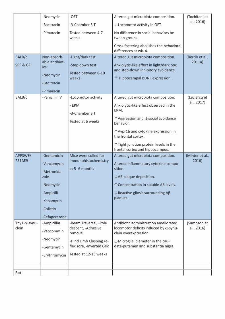
-Neomycin
-Bacitracin
-Pimaracin
-OFT
-3-Chamber SIT
Tested between 4-7 weeks
Altered gut microbiota composition.
↓Locomotor activity in OFT.
No difference in social behaviors be-tween groups.
Cross-fostering abolishes the behavioral differences at wk. 4.
(Tochitani et al., 2016)
BALB/c
SPF & GF
Non-absorb-able antibiot-ics:
-Neomycin
-Bacitracin
-Pimaracin
-Light/dark test
-Step down test
Tested between 8-10 weeks
Altered gut microbiota composition.
Anxiolytic-like effect in light/dark box and step-down inhibitory avoidance.
↑ Hippocampal BDNF expression.
(Bercik et al., 2011a)
BALB/c -Penicillin V -Locomotor activity
- EPM
-3-Chamber SIT
Tested at 6 weeks
Altered gut microbiota composition.
Anxiolytic-like effect observed in the EPM.
↑Aggression and ↓social avoidance behavior.
↑Avpr1b and cytokine expression in the frontal cortex.
↑Tight junction protein levels in the frontal cortex and hippocampus.
(Leclercq et al., 2017)
APPSWE/PS1ΔE9
-Gentamicin
-Vancomycin
-Metronida-zole
-Neomycin
-Ampicilli
-Kanamycin
-Colistin
-Cefaperazone
Mice were culled for immunohistochemistry
at 5- 6 months
Altered gut microbiota composition.
Altered inflammatory cytokine compo-sition.
↓Aβ plaque deposition.
↑Concentration in soluble Aβ levels.
↓Reactive gliosis surrounding Aβ plaques.
(Minter et al., 2016)
Thy1-a-synu-clein
-Ampicillin
-Vancomycin
-Neomycin
-Gentamycin
-Erythromycin
-Beam Traversal, -Pole descent, -Adhesive removal
-Hind Limb Clasping re-flex sore, -Inverted Grid
Tested at 12-13 weeks
Antibiotic administration ameliorated locomotor deficits induced by a-synu-clein overexpression.
↓Microglial diameter in the cau-date-putamen and substantia nigra.
(Sampson et al., 2016)
Rat

Sprague Dawley
-Ampicillin
-Vancomycin
-Ciprofloxacin
-Imipenem
-Metronida-zole
- None Altered expression of miRNAs in the amygdala and PFC.
Amygdala: ↓ miR-206-3p & miR-219a-2-3p, ↑ miR-369-3p.
PFC: ↓ miR219a-5p.
(Hoban et al., 2017)
-Ampicillin
-Vancomycin
-Ciprofloxacin HCL
-Imipenem
-Metronida-zole
- OFT, EPM, FST
- MWM, CRD
-Hotplate
Tested at 17-22 weeks
↓ Spatial memory observed in the MWM.
↑ Visceral sensitivity observed in CRD.
Depressive-like behavior observed in the FST.
Alterations in CNS serotonergic turn-over.
↓ Hippocampal CRHR1 expression, ↑in amygdala BDNF expression.
(Hoban et al., 2016a)
-Vancomycin in three con-centrations
Also assessed an antibiotic cocktail:
-Pimaricin, bacitracin,
neomycin
- NOR
- OFT
- MWM
- CRD
Tested at 8-11 weeks
Neonatal vancomycin significantly al-tered gut microbiota composition.
↑ Visceral sensitivity observed in CRD.
Behavior in adulthood was not affected by early-life antibiotic administration.
Early-life antibiotic cocktail also had no effect on behavior.
(O’Mahony et al., 2014)
-Ampicillin
-Vancomycin
-Ciprofloxacin
-Imipenem
-Metronida-zole
- Assessment of cardio-respiratory measures
4 weeks
Blunts the ventilatory response to hy-percapnia due to decreased respiratory frequency.
Blunts respiratory frequency during the peak hypoxic ventilatory response.
Respiratory timing variability unaltered.
↓Systolic blood pressure
Cardiorespiratory responsiveness to vagal afferent nerve stimulation is unaf-fected.
Altered brainstem monoamine and monoamine metabolite concentrations.
↑ Distal ileum permeability.
(O’Connor et al., 2019)
Wistar Dams were fed either control diet, or diet with 1% SST
- OFT, SIT, EPM, PPI
- Marble burying
Tested at 6-7 weeks
↓ Social interactions.
Anxiety-like behavior observed in the EPM.
Altered sensorimotor gating.
(Degroote et al., 2016)
Non-rodent
species

Zebrafish
Danio rerio
Ofloxacin
Ciprofloxacin
Enrofloxacin
Doxycycline
Chlortetracy-cline
Oxytetracy-cline
Social cohesion
Tested at 3 months
↓ Social cohesion behavior.
↑ Anxiety-like behavior observed as a ↓in shoaling.
Alterations in the expression of genes associated with locomotion.
(Wang et al., 2016)
Fruit fly
Drosophila melanogaster
Tetracycline
Rifampicin
Streptomycin
Multiple choice mating tests
♂ and ♀ mating preference was abol-ished.
(Sharon et al., 2013)

Table 4: Prebiotic studies of the microbiota-gut-brain axis, categorized by model organism.
Species/Sex Prebiotic Treat-ment
Effect Ref.
(time)Human
Healthy
♂
Inulin-propi-onate ester
Acute ↓ Striatal anticipatory reward responses to high-energy foods.
(Byrne et al., 2016)
Healthy RPS, RMS and inulin
2 weeks ↑Total SCFA with RPS and inulin.
↑Butyrate and acetate only with RPS.
No changes seen with RMS.
(Baxter et al., 2019)
Type 2 diabe-tes ♀
Resistant dextrin (Nu-triose®06)
8 weeks ↑ Depression, anxiety and stress (DASS). ↓ Cortisol, KYN, KYN/TRP ratio. Altered peripheral immune markers.
(Farhangi et al., 2018)
IBS ♂ ♀ Short-chain FOS
4 weeks ↓ Anxiety scores. (Azpiroz et al., 2017)
Autism ♂ ♀ B-GOS® mixture
6 weeks Improvement in social behavior symptoms and sleep in ASD subjects with B-GOS®.
(Grimaldi et al., 2018)
Mouse
C57BL/6
♂
FOS, GOS or both FOS and GOS
3 weeks ↓ Anxiety- and depressive-like behavior, stress-respon-siveness, hypothalamic Nr3C1 and hippocampal Crhr1 expression. ↑ Pro-social behavior, hippocampal, 5-HT in PFC, BDNF, GABAR-B1 and -B2 expression.
(Burokas et al., 2017)
FOS and GOS
6 weeks ↓ Chronic stress-induced social avoidance, cognitive dys-function, anhedonia,
HPA-axis hyper responsiveness anxiety- and depressive-like behavior.
(Burokas et al., 2017)
3′Sialyllac-tose
and
6′Sialyllac-tose
3 weeks ↓ Stressor-induced anxiety-like behavior and ↑ stress-in-duced ↓DCX+ immature neurons.
(Tarr et al., 2015)
2´-fucosyl-lactose
12 weeks
↑ LTP, spatial learning, working memory and operant conditioning.
(Vazquez et al., 2015)
Resistant starch
8 weeks ↓ Neuronal signaling in the ventromedial hypothalamus and PVN.
(So et al., 2007)
β- glucan 8 weeks ↓ Neuronal signaling in the arcuate nucleus, ventromedial hypothalamus, PVN,
periventricular nucleus and the NTS.
(Arora et al., 2012)
Diet-induced obese
Oligofruc-tose-en-riched inulin
9 weeks ↑ Neuronal signaling in the arcuate nucleus. (Anastasovs-ka et al.,
2012)CD1 B-GOS® 3 weeks ↓ LPS-induced anxiety-like behavior and HT2AR expres-
sion in the frontal cortex.(Savignac et
al., 2016)BALB/cJ FOS 7 weeks ↓ Aβ deposition and BAC levels. ↑ Hippocampal-depen-
dent learning.(Yen et al.,
2017)

SOD1G93A
♂ ♀
GOS 74 days ↓ Motor neuron death and spinal cord inflammatory markers.
(Song et al., 2013)
Rat
Sprague
Dawley
♂
2´-fucosyl-lactose
From PND3 and
weaning
↑ LTP (Oliveros et al., 2016)
2´-fucosyl-lactose
5 weeks ↑ Operant Conditioning and LTP, PSD-95 protein levels in hippocampus and frontal cortex, as well as CaMKII and BDNF in the hippocampus.
(Vazquez et al., 2015)
2´-fucosyl-lactose
5 weeks ↑ Operant Conditioning and long-term potentiation. (Vazquez et al., 2016)
Resistant Starch
65 Days ↑ Hypothalamic POMC expression. (Shen et al., 2009)
FOS or GOS 5 weeks ↑ Hippocampal BDNF and NR1 subunit expression. ↑ Hip-pocampal NR2A subunit and frontal cortex NR1 expression and d-serine levels by GOS.
(Savignac et al., 2013)
B-GOS® 3 weeks ↑ Cortical GluN2A subunit. (Kao et al., 2018)
B-GOS® 3 weeks ↑ Cortical neuronal responses to NMDA and improved attentional set-shifting performance.
(Gronier et al., 2018)
Chitosan oligosaccha-rides
10 Days ↑ Hippocampal-dependent memory.
↓ Hippocampal neuronal apoptosis, 8-OHdG, TNF-α and IL-1β levels.
(Jia et al., 2016)
♂ ♀ B-GOS® 3 weeks ↑ Hippocampal NR2A, SYN and BDNF levels PND22 and PND56
(Williams et al., 2016b)
Lister Hooded
♂
2´-fucosyl-lactose
From PND3 and
weaning
↑ Cognition (Oliveros et al., 2016)
Fischer 344
♂
GOS, PLWC 9 weeks ↑ REM sleep rebound following stress exposure. (Thompson et al., 2016)
GOS, polydex-trose, lacto-ferrin.
4 weeks ↓ Stress-induced learned helplessness and cFOS expres-sion in the DRN. ↑ Basal BDNF in prefrontal cortex by GOS, polydextrose and lactoferrin.
(Mika et al., 2017)
GOS, PLWC 40 Days ↑ Dendritic spine density of rat hippocampal neurons. (Waworuntu et al., 2016)
Pig♂ Sialyllactose 3 weeks No effect on recognition Memory or diurnal Activity. (Fleming et
al., 2018)Lactoferrin 5 weeks ↑ Hippocampal BDNF and cognitive function. (Chen et al.,
2015)GOS, PLWC 2 weeks ↑ Spatial learning. ↓ Cortical grey/white matter. (Mudd et al.,
2016)GOS and polydex-trose
3 weeks ↑ Exploratory behavior and recognition memory, ↓ Hip-pocampal 5-HT.
(Fleming et al., 2017)

Table 5: Probiotic studies of the microbiota-gut-brain axis, categorized by model organism.
Species Param-eter
Probiotic Treat-ment
(Time)
Effect Ref.
HumanHealthy
♂
B. longum 1714 4 weeks ↑ Neurocognitive performance (paired asso-ciative learning).
↑ Fz mobility.
↓ Cz theta power after end of probiotic ad-ministration.
(Allen et al., 2016)
L. rhamnosus (JB-1) ™
4 weeks Does not significantly impact HPA axis, stress, and cognition.
(Kelly et al., 2017)
♀ FMP:
B. animalis subsp. lactis
Classic yogurt starters:
S. thermophiles, L. bulgaricus,
L. lactis subsp lactis
4 weeks ↓ Reactivity in widely distributed network during emotional attention task.
↓ BOLD signal in amygdala, mid insula cortex, primary somatosensory cortex in emotional attention task.
Altered PAG resting state network.
(Tillisch et al., 2013)
♂ ♀ Ecologic®825: L. casei, L. paracasei,
B. lactis, L. sali-varius, B. lactis, L. plantarum
B. bifidum, L. aci-dophilus, L. lactis
4 weeks Change in gut microbiota profile.
↓ Some anxiety and depressive measures.
(Bagga et al., 2018b)
Ecologic®825: see above for compo-sition
4 weeks Changes in functional connectivity but no changes in structural connectivity.
(Bagga et al., 2018a)
Ecologic®Barrier:
B. bifidum, B. lactis, L. acidophilus,
L. brevis, L. casei, L. salivarius, L. lactis
4 weeks ↓ Aggression and rumination in response to depressive thoughts (LEIDS-r test).
(Steenbergen et al., 2015)
L. casei Shirota (Yakult)
3 weeks Only improvement in depression in POMS scale for people at the lowest end of the mood scale.
(Benton et al., 2007)

Group 1: Probiotic yogurt
L. acidophilus LA5, B. lactis BB12
Group 2: Capsule
L. casei, L. acidophilus , L. rhamnosus , L. bulgaricus, B. breve, B. long-um, S. thermo-philus, FOS
6 weeks Improvement in GHQ scale with either probiot-ic treatment.
(Mohammadi et al., 2016)
L. helveticus R0052 and B. longum R0175
30 days ↓ Anxiety via HADS score. (Messaoudi et al., 2011a)
B. subtilis: contain-ing 75% in spore form and 25% in vegetative form
4 weeks Only impacted microbiota composition.
No effect on GI symptoms or general wellness.
(Hanifi et al., 2015)
Aging (<60)
♂ ♀
L. reuteri 12 weeks No persisting effects on depression, anxiety or perceived stress.
(Ostlund-La-gerstrom et
al., 2016)Aging (60-75)
♂ ♀
L. helveticus IDCC3801
12 weeks ↑ Cognitive performance in RVIP and Stroop Color-Word task (cognitively demanding tasks).
(Chung et al., 2014)
Alzheimer’s Disease ♂ ♀
Capsule:
L. acidophilus, L. casei, B. bifidum, L. fermentum
12 weeks ↑ MMSE score. Change in blood lipid profile and carbohydrate metabolism factors.
(Akbari et al., 2016)
Chronic fatigue ♂♀
syndrome
L. casei strain Shi-rota
8 weeks ↓ Anxiety symptoms. (Rao et al., 2009)
Urinary-free cortisol 10-50ng/ml (low) ♂ ♀
L. helveticus R0052 and B. longum R0175
30 days ↑Perceived stress score, hospital anxiety, de-pression scale score.
↑HSCL-90 subscores for obsessive-compulsive, anxiety, and depression.
↑↑ HSCL-90 in Factor 1 (anxiety and depres-sion).
(Messaoudi et al., 2011b)
HIV-1+
♂
Vivomixx®:
L. plantarum, S. thermophiles, B. breve, L. paraca-sei, L.delbrueckii subsp. Bulgaricus, L. acidophilus, B. longum, B. infantis
6 months ↑ Neurocognitive performance. (Ceccarelli et al., 2017a)
♂ ♀ Vivomix®: (See composition above)
6 months ↑ Verbal and visual memory. (Ceccarelli et al., 2017b)

IBS ♂ ♀
B. longum NCC3001 6 weeks ↓ HAD-A and D scores indicating ↓ in anxiety/depression.
↑ Quality of life.
(Pinto-Sanchez et al., 2017b)
L. paracasei, ssp. paracasei F19
L. acidophilus La5 B. lactis Bb12
8 weeks No significant psychological changes. (Simren et al., 2010)
MDD ♂ ♀
Capsule:
L. acidophilus, L. ca-sei, and B. bifidum
8 weeks ↓ BDI score. (Akkasheh et al., 2016)
L. helveticus R0052 and B. longum R0175 (CNCM strain I-3470)
8 weeks ↓ BDI score (depressive index).
↓ Kynurenine: Tryptophan ratio.
(Kazemi et al., 2019)
L. Plantarum 299v 8 weeks ↑ APT and CVLT total recall of trials 1–5
↓ Kynurenine concentration
↑ 3-hydroxykynurenine: kynurenine ratio
(Rudzki et al., 2019)
MS ♂ ♀
Capsule: L. aci-dophilus, L. casei, B. bifidum
L. fermen-tum
12 weeks ↑BDI, EDSS, DHQ scores (depression, diet and multiple sclerosis scores).
(Kouchaki et al., 2017)
Normal weight obese syn-drome and obesity
♀
1 “bag”: S. thermo-philes, B. animalis subsp. Lactis, S. thermophilus, B. bifidum
L. delbrueckii spp. Bulgaricus , L. lactis subsp. Lactis, L. ac-idophilus, L. planta-rum , L. reuteri
3 weeks ↓ positivity to BUT.
↓ EDI-2 responses.
(De Lorenzo et al., 2017)
Obesity ♂ ♀
L. rhamnosus CG-MCC1.3724
24 weeks ↑ Body esteem in ♀s, ↓ Depression score in ♀s.
(Sanchez et al., 2017)
Pregnancy L. rhamnosus HN001
<6 months
↓ Postpartum depression and anxiety scores. (Slykerman et al., 2017)
Schizophrenia
♂ ♀
Bifiform Balance: L. rhamnosus strain GG B. animalis sub-sp. lactis Bb12
14 weeks ↓ C. albicans antibodies in ♂ serum,
associated with ↓ psychiatric symptom occur-rence.
(Severance et al., 2017)
L. rhamnosus strain GG
B. animalis subsp. lactis strain Bb12
14 weeks No changes in frequency of psychotic symp-toms.
↓ Incidence of severe bowel difficulties.
↓ Acute von Willebrand factor, ↑ MCP-1, BDNF, RANTES, MIP-1.
(Tomasik et al., 2015)
Bifiform Balance: L. rhamnosus strain GG B. animalis sub-sp. lactis Bb12
2 weeks No differences in PANSS total symptom score.
↓ Incidence of severe bowel difficulties.
(Dickerson et al., 2014)

Pregnancy L. rhamnosus GG, L. rhamnosus LC705, B. breve Bb99, and Propionibacterium freudenreichii- sub-species shermanii
From 36 weeks’
gestation until the birth of
the infant
Analyzed 81 randomly selected colostrum sam-ples from a bank of 500.
↑ Human milk oligosaccharides: 3-fucosyllac-tose and 3′-sialyllactose in colostrum
↓ Human milk oligosaccharides: difucosyllac-to-N-hexaose, lacto-N-tetraose, lacto-N-fuco-pentaose I, and 6′-sialyllactose.
(Seppo et al., 2019)
Stress
♂ ♀
Probio-Stick:
L. acidophilus Rosell-52
B. longum Rosell-175
3 weeks ↓ Stress-induced abdominal pain and nausea/vomiting.
↓ Flatulence, gas production.
No effect on other symptoms (physical, psy-chological, sleep).
(Diop et al., 2008)
Stress/ ♂ ♀
Exhaustion
Multiobionta: Pro-biotic culture blend
L. acidophilus, B. bifidum, B. longum
6 months ↑ in fatigue and stress. (Gruenwald et al., 2002)
Moderate stress
♂ ♀
L. plantarum DR7 4, 8 and 12 weeks
↓ Stress and anxiety using DASS-42, but not PSS-10, questionnaires.
No effect on depression.
↑ Anti-inflammatory cytokines and ↓pro-in-flammatory cytokines in young adults.
↓ Pro-inflammatory cytokines in normal adults over 12 weeks.
↑ Verbal learning and memory in young adults
↑ Basic attention, social emotional cognition and associate learning in normal adults over 12 weeks.
↓ IDO/TDO, kynurenine, cortisol, IFN-γ and TNF-α.
↑ TPH, Serotonin, IL-4, IL-10.
(Chong et al., 2019)
Student ♂ ♀
exam stress
L. casei strain Shiro-ta YIT 9029
8 weeks ↓ Physical symptoms of stress/anxiety (Takada et al., 2016)
B. bifidum R0071 or L. helveticus or B. infantis
6 weeks ↑ Healthy days (i.e. no flu/cold) only from B. bifidum.
↓ Reports of symptoms lasting more than a day in participants receiving B. bifidum.
Proportion of participants reporting cold/flu between weeks 2 and 3 lower in those receiv-ing B. bifidum or B. infantis.
(Lang-kamp-Henken et al., 2015)
L. plantarum 299v 14 days No difference in perceived stress.
↑ L. plantarum 299v and Lactobaccili on Day 14 in saliva.
(Andersson et al., 2016)

L. casei strain Shiro-ta (YIT 9029)
Formulated in 100mL of milk
8 weeks No change in psychological parameters (anxi-ety, depression scales). ↓ Physical symptoms i.e. runny nose, cold.
(Kato-Kataoka et al., 2016)
TBI
♂
240 ml of fer-mented milk with the
L. johnsonii
Given along with 30g glutamine
6-14 days via enter-
al tube
↓ Infection rate, length of stay in ICU, days on mechanical ventilator.
(Falcao de Arruda and de Aguilar-Nasci-mento, 2004)
Type 2 Diabetes Capsule: L. aci-dophilus, L. casei, B. bifidum
L. fermentum, vita-min D3
12 weeks Improved BDI, BAI, GHQ scores (↓anxiety and depression).
(Raygan et al., 2018)
Low mood ♂♀
L. helveticus, B. longum
8 weeks No effect. (Romijn et al., 2017)
Mouse Stress
♀
Pediococcus acidi-lactici
36 days No effect on tonic immobility induced by fear.
No effect in OFT, ↑ Memory (not due to emo-tional reactivity).
(Parois et al., 2017)
AKR – DSS
♂
B. longum NCC3001 2 to 3 weeks
↓ Anxiety-like behavior in DSS. (Bercik et al., 2011b)
B. longum NCC3001 ↓ AH neuron excitability,
resistance and magnitude of cationic current.
(Khoshdel et al., 2013)
Balb/c
♂
B. longum 1714 or B. breve 1205
6 weeks ↓ Bodyweight gain from B. longum.
↓ Body temperature ↑during stress-induced hyperthermia.
↓ Anxiety in marble burying with either strain.
↓ Anxiety via EPM in B. breve but no differ-ence in locomotion.
↓ Latency in OFT in B. longum group.
↓ Immobility time in FST for B. longum group.
↑ Spleen weight in B. breve.
(Savignac et al., 2014)
L. rhamnosus 29 days ↓ Stress-induced hypothermia, anxiety (EPM).
↓ GABAB1b mRNA in basolateral and central amygdala, locus coeruleus, DG, CA3, CA1.
↓ GABAAα2 mRNA in CG1, PrL and IL cortical areas, and BLA, CeA.
↑ GABAAα2 mRNA in the DG. Effects abol-ished through vagotomy.
(Bravo et al., 2011)

Alzheimer’s Disease
♂
SLAB51: S. thermo-philes, B. longum, B. breve, B. infan-tis, L. acidophilus, L. plantarum, L. paracasei, L. delbrueckii subsp. bulgaricus, L. brevis
4 months ↓ Oxidative stress in brain.
↓ Cognitive decline, brain damage, Aβ peptide accumulation.
(Bonfili et al., 2018)
ICR: Vascular ♂ dementia model
C. butyricum 6 weeks ↑ Spatial memory, ↓ Neuronal apoptosis, ↑ Butyrate in brain.
(Liu et al., 2015)
ICR: ♂
Ischemia Injury
C. butyricum 2 weeks ↓ Neurological deficit score,↓ Hippocampal neuron karyopyknosis. ↓ Apoptosis in CA1 via TUNEL.
(Sun et al., 2016a)
Shank3 KO (Jackson Labs)
♂ ♀
L. reuteri 3 weeks GABAR expression correlated with L. reuteri in mice. Probiotic modifies social (in ♂s) and repetitive behavior (♂s and ♀s), ↑GABAR (regional and sex-dependent magnitudes) and oxytocin expression.
(Tabouy et al., 2018)
Swiss Albino
SD & CUMS
♂
L. plantarum MTCC 9510
28 days ↓ SD-induced anxiety and depression (FST, EPM).
↑ Learning and memory (MWM), ↓ BBB permeability.
Abolished Bdnf decrease in hippocampus.
Prevented some stress-induced microbiota alterations
(Dhaliwal et al., 2018)
Shank3 KO (Jackson Labs)
VPA Mouse Model of ASD
BTBR
♂
L. reuteri 4 weeks In all three strains, rescued impaired sociability as measured by three-chamber sociability, re-ciprocal social interaction and/or social novelty tests.
(Sgritta et al., 2019)
C57BI/6J Maternal Sepa-ration
♂
B. CECT 7765 21 days Partially attenuated exaggerated HPA stress response.
↓ Vulnerability to stress in adulthood.
↓ Anxiety, catecholaminergic hyperactivity in adulthood.
Protected against stress-induced microbiota alterations.
(Moya-Perez et al., 2017b)
TBI
♂
C. butyricum 14 days before
and after TBI
Ameliorated neurological deficits and brain edema size.
↓ Degenerating neurons, ↑ Occludin and ZO-1 expression at BBB to ameliorate ↑per-meability from TBI.
↑ P-Akt/Akt, Bcl-2 along with ↓ Bax indicat-ing less neuronal. apoptosis, ↑ GLP1 in colon, GLP1-R in brain.
(Li et al., 2018a)

C. rodentium infection ♀
Lacidofil:
L rhamnosus and L helveticus
10 days Attenuates WAS non-spatial memory deficit.
Ameliorates BDNF and c-Fos in hippocampus after WAS.
(Gareau et al., 2011)
TNBS-induced colitis and memory ♂
impairment
L. plantarum colitis:
3 days
memory:
5 days
Reversed memory impairments (spontaneous alteration in Y-Maze). Restored some microbio-ta perturbations.
↑ Bdnf and↓ NFKB activation in hippocampus and LPS in blood.
(Lee et al., 2018a)
Chronic social defeat stress ♂
L. rhamnosus
(JB-1) ™
28 days ↓ Stress-induced anxiety.
Prevented deficits in social interaction with conspecifics.
(Bharwani et al., 2017)
Maternal HFD ♂
L. reuteri 4 weeks Rescues impairments in LTP in DAergic VTA neurons.
(Buffington et al., 2016)
Subchronic social defeat stress ♂
L. helveticus MCC1848
24 days restored normal sucrose consumption in the sucrose preference test
↓ Anxiety-like behavior in the SIT.
No effect on polydipsia-like behavior.
Rescued Drd3, Htr1a sCSDS-induced deficit in NAc.
(Maehata et al., 2019)
DSS- colitis ♂ ♀
L. rhamnosus and L. helveticus
20 days Rescued some decreases in NOR, anxiety and c-fos in CA1.
(Emge et al., 2016)
Chronic stress
♂
L. helveticus R0052 or /and B. longum R0175
14 days Combination of both probiotics reversed dis-tension pain hypersensitivity at larger volumes.
Prevented GR decrease in hypothalamus and hippocampus if pretreated with both probiot-ics or B. longum.
Only both probiotics prevented this ↑in the PFC.
(Ait-Belgnaoui et al., 2018)
Maternal sepa-ration –
early life stress
♂
L. plantarum PS128 28 days Restores sucrose preference (↓in ELS/no probiotic).
↑ Locomotor activity, ↓ anxiety in EPM in naïve mice.
↑ DA and DAergic metabolites in PFC in early life stress.
↓ HVA: DA ratio. No change in DAergic activity in striatum.
(Liu et al., 2016b)
6JNarl: GF ♂
L. plantarum PS128 16 days ↓ Anxiety-like behavior, ↑ Locomotor activity. (Liu et al., 2016a)
6N: MIA B. fragilis 6 days Corrected microbiota changes in MIA offspring.
Prevented/↓MIA-induced anxiety (OFT, mar-ble burying), ↓deficits in sensorimotor gating.
Did not improve social deficits.
(Hsiao et al., 2013)

WAS
♂
L. helveticus R0052 and B. longum R0175
2 weeks ↓ cFos+ neurons responding to WAS in PVN, CA3, and amygdaloid nucleus (↑in non-WAS/probiotic compared to other control animals).
↑ Bdnf in hypothalamus in response to WAS.
↓ Expression of cytoskeleton organization, microglia activation, synaptogenesis and cell adhesion markers in hypothalamus in response to WAS.
(Ait-Belgnaoui et al., 2014)
Mouse & RatSwiss-Webster
Ex-vivo record-ings
L. reuteri DSM 17938
9 days ↓ Spontaneous mesenteric nerve firing activ-ity.
Effect not inhibited by smooth muscle relax-ation or vagotomy.
Partial evoked antagonism of TRPV1.
↓ Distension evoked firing via TRPV1 antago-nism.
Supernatants also able to exhibit antagonism.
↓ Calcium rise and ionic current in dorsal root ganglia primary culture.
(Perez-Burgos et al., 2015)
Rat Sprague-Dawley
♂
Faecalibacterium prausnitzii (ATCC 27766)
Prevented and treated depressive-like or anxi-olytic-like behaviors caused by CUMS. Rectified CUMS-induced weight loss.
↑ Cecal acetate, propionate and n-butyrate levels.
↓ CUMS-induced circulating corticosterone and CRP increase.
↑ IL-10 level, and ↓IL-6 induced by CUMS.
(Hao et al., 2019)
Swiss-Webster Ex-vivo electro-physiology
♂
L. rhamnosus
(JB-1) ™
L. salivarius
N/A ↑Firing rate of mesenteric afferent bundles.
70% of single unit afferent nerves also had an ↑ firing rate.
Administration does not reduce firing rate after distension.
Effects abolished by vagotomy.
(Perez-Burgos et al., 2013)
Brown Norway Water avoid-ance stress (WAS) ♂
Lacidofil Powder: 95% L. rhamnosus R0011
5% L. helveticus R0052
7 days Prevents stress induced bacterial adherence to enterocytes.
Prevents bacterial translocation to mesenteric lymph nodes after WAS. No apparent effect on intestinal histology.
Inhibit chronic stress elevation of ion secretion in intestines.
No impact on permeability in ileum or colon.
(Zareie et al., 2006)

FSL and FRL:
HFD
♂
Ecologic Barrier:
B. bifidum, B. lactis, L. acidophilus, L. brevis, L. casei, L. salivarius, L. lactis
12 weeks Protected against depressant effects of HFD in FSL (FST).
No effect on locomotor activity.
(Abildgaard et al., 2017a)
Aging Probiotic Mixture KF (1:1 ratio):
L. curvatus HY7601
L. plantarum KY1032
Reversed age-dependent ↓in spontaneous alteration (Y Maze).
No significant effect on escape latency (MWM).
↑Hippocampal DCX, BDNF and phosphorylat-ed CREB relative to control aged mice, ↓acti-vation of mTOR pathway.
(Jeong et al., 2015)
Maternal sepa-ration ♂
B. breve 6330 42 days ↑ BDNF IV mRNA in control rats given probiot-ic (not affected by maternal separation).
(O’Sullivan et al., 2011)
Depression – maternal sepa-ration ♂
B. infantis 35624 45 days Normalized effects of maternal separation in FST.
↓ 5-HIAA in amygdaloid cortex.
(Desbonnet et al., 2010)
Chronic re-straint stress model
♂
L. helveticus NS8 ~27 days ↓ Anxiety-like and depressive-like behavior.
↑ Recognition memory.
↑ Hippocampal bdnf, NE, 5-HT.
(Liang et al., 2015a)
Maternal sepa-ration stress
Lacidofil Powder: 95% L. rhamnosus R0011
5% L. helveticus R0052
P4-P20 (17 days)
Transient L. colonization.
Restored ion transport and permeability after WAS in adult life (persisting effect). Abolished MS-induced HPA overactivation.
(Gareau et al., 2007)
♂ Lacidofil Powder: 95% L. rhamnosus R0011
5% L. helveticus R0052
Admin-istered
to dams from P2-P14 (13
days)
Restored age-appropriate forgetting (infantile amnesia) and relapse-resistant extinction of aversive memories during infancy (P17).
No effect on anxiety or locomotor activity in pups or dams.
No effect on maternal care behavior (pup retrieval).
(Cowan et al., 2016)
♂ Lacidofil Powder: 95% L. rhamnosus R0011
5% L. helveticus R0052
To dams from
P2-P14
(13 days)
Restored age-appropriate neural activity in the pre-frontal cortex following fear expression and inhibition during infancy (P17).
(Cowan et al., 2019)
♂ ♀ Lacidofil Powder: 95% L. rhamnosus R0011
5% L. helveticus R0052
Admin-istered
to dams from P2-P14 (13
days)
Restored normative timing of physical pu-berty onset in both sexes (reversed stress-in-duced delay in preputial separation for males, stress-induced acceleration of vaginal opening in females).
(Cowan and Richardson,
2018)

♂ ♀ L. rhamnosus GG ~4 weeks Ameliorate ↓ in exploration and ↑ in anxiety from stress.
Altered gene expression in hippocampus (no behavior in ♀s).
(McVey Neufeld et al.,
2017)
♂ B. infantis 14 days No effect in FST. ↓ 5-HIAA in frontal cortex.
↓ DOPAC in amygdaloid cortex.
No effect on CRF mRNA in hypothalamus.
(Desbonnet et al., 2008)
Ecologic Barrier:
B. bifidum, B. lactis, L. acidophilus, L. brevis, L. casei, L. salivarius, Lactococcus lactis
11 weeks ↓ Depression (FST).
No locomotor effect (OFT).
↓ Expression of HPA axis genes in hippocam-pus.
↑ Expression of neuroprotective genes (Trek2, Traak).
(Abildgaard et al., 2017b)
L. reuteri 9 ↓ Firing threshold of afterhyperpolarization myenteric neuron.
↓ Slow after-hyperpolarization potential of sensory neurons.
↓ Potassium-dependent calcium channel opening.
(Kunze et al., 2009)
L. rhamnosus and B. longum
12 days ↓ Depression-like behavior.
↑ Expression of GABA-A Receptor subunits in hippocampus.
(Liang et al., 2017)
L. casei 54-2-33 14 days ↑ Anxiety (OFT).
↓ Expression of 5-HT1A mRNA in hippocampus.
(Barrera-Bu-gueno et al.,
2017)Response to CRD after par-tial ♀ restraint stress
L. farciminis 14 days Probiotic prevented ↑ in c-Fos expression in sacral spinal cord, PVN and MeA.
(Ait-Belgnaoui et al., 2009)
Antibiotic microbiota depletion
♂
L. fermentum NS9 41 days (behavior tested at day 31)
↓ Myeloperoxidase activity in colon.
Alleviated ampicillin-induced anxiety (EPM).
Alleviated memory deficits seen in MWM in ampicillin-treated animals. Prevented ampi-cillin-induced MR and NMDA-R decrease in hippocampus.
(Wang et al., 2015)
HA induced neuro-inflam-mation model ♂
L. helveticus NS8 2 weeks ↓ Anxiety-like behavior (EPM).
↑ Learning and memory (MWM). ↓ 5-HT in cerebellum and hippocampus but did not restore regional. ↑ 5-HIAA in HA brain.
(Luo et al., 2014)
Myocardial infarction
♂
L. helveticus
B. longum
4 weeks ↓ Bax/Bcl-2 and Caspase-3 levels in DG, medi-al amygdala and lateral amygdala but not CA1 or CA3 ↓ apoptosis.
Differences in phopsho-Akt:Akt ratios in these regions.
(Girard et al., 2009)

CRD
♂
L. reuteri 9 days Inhibition of ↑ heart rate in response to CRD.
↓ Single unit discharge of PNS neurons in response to distention.
No effect on pain via tail flick or paw pressure tests.
(Kamiya et al., 2006)
Colitis induced by zymosan
♂
L. rhamnosus GG 40 days Measurements taken in adulthood after ear-ly-life colitis induction.
↓ Visceromotor response to distension.
No change in DA or its metabolites in brain-stem.
↑ DA and DOPAC in frontal cortex.
↑ NA and 5-HIAA in cerebellum.
↓ His in brainstem ↓ Glu and Lys in subcortex.
↓ Glu, Lys, His, Taurine in frontal cortex.
↓ GABA in frontal cortex, subcortex and cere-bellum.
(Kannampalli et al., 2014)
Trans-genera-tional paternal stress ♂
Lacidofil Powder: 95% L. rhamnosus R0011
5% L. helveticus R0052
Mater-nally P2-P14 (13
days)
Restored age-appropriate forgetting and re-lapse-resistant extinction of aversive memories in F1 offspring during infancy (P17).
(Callaghan et al., 2016)
Sprague-Dawley & WKY:
CRD
L. salivarius UCC118, B. infantis 35624, or B. breve UCC2003
14 days ↑ Pressure threshold in Sprague-Dawley rats in response to distension if given B. infantis or B. breve.
↑ Pressure threshold in WKY rats given B. infantis.
↓ Pain behavior in both strains when given B. infantis.
(McKernan et al., 2010)
Wistar ♂
L. helveticus, B. longum
7 days ↓ Anxiety in marble burying. (Messaoudi et al., 2011a)
L. helveticus, B. longum
7 days No rewarding properties of probiotic in condi-tioned place preference relative to morphine.
No learning and memory deficit in passive avoidance paradigm.
(Messaoudi et al., 2011b)
VSL #3 6 weeks Effect on brain gene expression (>300 genes).
Attenuate age-related decrease in LTP.
↓ Microglial activation in hippocampus.
(Distrutti et al., 2014)
HFD ♂ L. paracasei 12 weeks ↓ Microglial activation, ↓ Cognitive dysfunc-tion.
(Chunchai et al., 2018)
AD L. plantarum 60 days ↑ Performance in MWM in AD+Probiotic group.
(Nimgampal-le and Kuna,
2017)

Diabetes
♂
Mix: L. acidophilus, B. lactis, L. fermen-tum
8 weeks ↑ Performance on MWM in diabetes probiot-ic.
↑ Synaptic transmission (electrophysiology).
(Davari et al., 2013)
Zebrafish
♂ ♀
L. rhamnosus 4 weeks ↑ Exploration, attention.
↑ Brain bdnf, serotoninergic system genes.
(Borrelli et al., 2016)
Drosophila
♂ ♀
L. plantarum L168 2-3 days ↑ Social behavior in kdm5-deficient flies.
↓ Intestinal permeability
↓ Number of intestines with defects in kdm5-deficient flies.
↓ 5-HT levels in kdm5-deficient flies.
Restored intestinal barrier integrity.
KDM5 demethylase affects social behavior through the gut-microbiome-brain axis.
3.5-fold increase in median survival of kdm5-deficient flies.
Only GF flies with kdm5-deficiency were viable.
(Chen et al., 2019)

Table 6: Tools used in the analysis of the gut microbiome.
Tool Function Based on: Platform Advantages Disadvantages
Biomarker Pro-filing Microbiota Composition DNA NGS
- Somewhat Cost Effective
- Semi-quantitative
Lacks Functional Information
Metagenomics Microbiota Functional Gene Capacity DNA NGS
Can Achieve
Strain-level Resolu-tion
- Expensive
- Computation-ally intensive
Metabolomics Metabolic Productivity Metabo-lites
LC/GC-MS
- Targeted or Untar-geted
- Semi-quantitative
Origin of Metab-olite is Unclear
Metatranscrip-tomics
Microbial Functional Gene Ex-pression
RNANGS Host and Microbial
Gene Transcripts
- Requires RNA Preservation
- Host Genes may Dominate
Metaproteom-ics Protein Expression Protein LC/GC-
MS Semi-quantitative Protein Origin not Clear

XVIII. Tables Abbreviations
↓ - decrease/ reduction/ downregulation
↑ - increase/ enhancement/ upregulation
5-HIAA - 5-Hydroxyindoleacetic acid
5-HT – 5-hydroxytryptamine or serotonin
5-HT1A/2AR - Serotonin 1A/2A receptor
8-OHdG - 8-Oxo-2’-deoxyguanosine
Aβ - Amyloid β
AH – Afterhyperpolarization
APT - Attention and perceptivity test
ASD – Autism spectrum disorder
ASF - Altered schaedler flora
B. acidophilus– Bifidobacterium acidophilus
B. adolescentis – Bifidobacterium adolescentis
B. animalis – Bifidobacterium animalis
B. bifidum – Bifidobacterium bifidum
B. breve – Bifidobacterium breve
B. catenulaturm– Bifidobacterium catenulaturm
B. dentium– Bifidobacterium dentium
B. fragilis – Bifidobacterium fragilis
B. infantis– Bifidobacterium infantis
B. lactis – Bifidobacterium lactis
B. longum – Bifidobacterium longum
B. subtilis – Bifidobacterium subtilis
B-GOS® - Bimuno-galactooligosaccharide
BACHD - Bacterial artificial chromosome model of HD
BAI - Beck anxiety inventory
BBB – Blood-brain barrier
BDF1 - Bromodomain-containing factor 1
BDI - Beck depression inventory
BDNF – Brain-derived neurotrophic factor
BLA – Basolateral amygdala
BOLD - Blood-oxygen-level dependent
BrdU – Bromodeoxyuridine
BUT - Body uneasiness test

C. rodentium – Citrobacter rodentium
C. butyricum - Clostridium butyricum
C. perfringens - Clostridium perfringens
C. difficile - Clostridium difficile
CA1/3 - Cornu Ammonis 1/3
CaMKII – Calcium calmodulin-dependent protein kinase
CC - Corpus callosum
CeA - Central nucleus of the amygdala
CNS – Central nervous system
Cog – Cognition
CRD – Colorectal distension
CREB - cAMP response element-binding protein
CRF - Corticotropin-releasing factor
CRHR1 - Corticotropin-releasing hormone receptor 1
CRP - C-reaction protein
CUMS - chronic unpredictable mild stress
CVLT - California verbal learning test
DA – Dopamine
DAergic – Dopaminergic
DASS - Depression anxiety stress scales
DCX – Doublecortin
DG – Dentate gyrus
DHQ - Diet history questionnaire
DNA - Deoxyribonucleic acid
DOPAC - Dihydroxyphenylacetic acid
Drd3 - Dopamine receptor D3
DREADD - Designer receptors exclusively activated by designer drugs
DRN - Dorsal raphe nucleus
DSS - Dextran sulphate sodium
EAE - Experimental autoimmune encephalomyelitis
EDI - Eating disorders inventory

EDSS - Expanded disability status scale
ELS - Early life stress
FOS – Fructooligosaccharide
EPM – Elevated plus maze
FMP - Fermented milk product
FMT - Fecal microbiota transplant
FRL - Flinders resistant line
FSL - Flinders sensitive line
FST - Forced-swim test
GABA - γ-Aminobutyric acid
Gcg – Glucagon
GF – Germ-free
GHQ - General health questionnaire
GI - Gastrointestinal
GOS – Galactooligosaccharide
HA - Hyperammonemia
HADS - Hospital anxiety and depression scale
HCL – Hydrochloric acid
HD - Huntington disease
HFD – High-fat diet
HIV - Human immunodeficiency virus
HPA - Hypothalamic-pituitary-adrenal
HSCL - Hopkins symptom checklist
Htr1a - Serotonin 1A receptor
Ht2ar – Serotonin 2A receptor
HVA - Homovanillic acid
Hyperammonemia (HA)
IBS - Irritable bowel syndrome
IC – Intracerebral
ICR - Institute of Cancer Research
IDO - Indoleamine 2,3-dioxygenase
IEG – Immediate early gene

IFNγ - Interferon gamma
IL (Immunology) – Interleukin
IL (Cortex) - Infralimbic cortex
KDM5 - Histone demethylase kdm5
KM - Chinese kun ming mouse
KYN - Kynurenine
L. acidophilus – Lactobacillus acidophilus
L. brevis– Lactobacillus brevis
L. bulgaricus – Lactobacillus bulgaricus
L. casei – Lactobacillus casei
L. curvatus – Lactobacillus curvatus
L. delbrueckii – Lactobacillus delbrueckii
L. farciminis– Lactobacillus farciminis
L. fermentum– Lactobacillus fermentum
L. helveticus – Lactobacillus helveticus
L. johnsonii– Lactobacillus johnsonii
L. paracasei – Lactobacillus paracasei
L. plantarum – Lactobacillus plantarum
L. reuteri – Lactobacillus reuteri
L. rhamnosus – Lactobacillus rhamnosus
L. salivarius– Lactobacillus salivarius
LA – Lateral amygdala
LC/GC-MS - Liquid chromatography/ gas-chromatography-mass spectrometry
LDB – Light/dark box
LD - Light/dark preference
LEIDS-r - Leiden index of depression sensitivity-revised
LPS – Lipopolysaccharides
LTP – Long-term potentiation
MB – Marble burying
MCP-1 - Monocyte chemoattractant protein-1
MDD - Major depressive disorder
MeA - Medial amygdala

MGBA – Microbiota-gut-brain axis
MMSE - Mini mental state exam
MIA – Maternal immune activation
MIP-1 - Macrophage inflammatory protein-1
MiR/ miRNA - microRNA
mRNA - Messenger ribonucleic acid
MS - Multiple Sclerosis
MR - Mineralocorticoid receptor
mTOR - Mammalian target of rapamycin
MWM – Morris water maze
NA – Noradrenaline
NAc – Nucleus accumbens
NE – Norepinephrine
NFKB - Nuclear factor kappa-light-chain-enhancer of activated B cells
Ngfi-a - Nerve growth factor inducible A
NGS – Next-generation Sequencing
NIH - National Institutes of Health
NMRAR - N-methyl-D-aspartate receptor
NMRI - Naval medical research institute
NOR – Novel object recognition
NPY - Neuropeptide Y
NR1/2 – NMRAR 1/2
Nr3C1 - Nuclear Receptor Subfamily 3 Group C Member 1
NSF - Novelty suppressed feeding
NTS - Nucleus tractus solitaris (the nucleus of the solitary tract)
OFT - Open-field test
PAG - Periaqueductal gray
PANSS - Positive and negative syndrome scale
PD - Parkinson’s disease
PFC - Prefrontal cortex
PLWC - Polydextrose, lactoferrin and whey protein concentrate
PND – Postnatal day

PNS - Peripheral nervous system
POMS - Profile of mood states scale
PPI – Prepulse inhibition
PrL - Prelimbic cortex
PS1 – Presenilin 1
PSD95 - Postsynaptic density protein 95
PSS - Perceived stress scale
PVN - Paraventricular nucleus
RANTES - Regulated on activation, normal T cell expressed and secreted
REM – Rapid eye movement
RMS - Resistant maize starch
RNA - Ribonucleic acid
RPS - Resistant potatoes starch
RVIP - Rapid visual information processing
SCFA – Short-chain fatty acid
sCSDS - Subchronic social defeat stress
SD - Sleep deprivation
SGZ - Subgranular zone
SIT - Social interaction test
SPF – Specific pathogen free
SPT – Social preference test
SYN – Synuclein
TBI – Traumatic brain injury
TDO - tryptophan 2,3-dioxygenase
Thy1 - Thymidylate synthase complementing protein 1
TNBS - 2,4,6-trinitrobenzene sulfonic acid
TNF-α/β - Tumor necrosis factor alpha/beta
TPH - Tryptophan hydroxylase
TRP – Tryptophan
TrkB - Tropomyosin receptor kinase B

TRPV1 - Transient receptor potential vanilloid 1
TST – Tail suspension test
TUNEL - Terminal deoxynucleotidyl transferase dUTP nick end labeling
VTA - Ventral tegmental area
WAS - Water avoidance stress
WKY - Wistar Kyoto

Call out box
“This is a comprehensive review of current knowledge of the influence that the microbiota has on brain and be-haviour. In particular we focus on the pathways involved and the models used in the field. Moreover, we highlight what still remains to be understood in order to fully realize the potential for the development of microbiota-based therapeutic strategies for brain disorders.”
Graphical abstract