Equine Protozoal Myeloencephalitis - VTechWorks
163
Equine Protozoal Myeloencephalitis: Investigating Immunopathogenesis and Treatment Efficacy in Mouse Models and Clinically Affected Horses Alayna N. Hay Dissertation submitted to the faculty of the Virginia Polytechnic Institute and State University in partial fulfillment of the requirements for the degree of Doctor of Philosophy In Animal and Poultry Sciences Caroline M. Leeth Sally Johnson David S. Lindsay Sharon G. Witonsky December 12 th , 2019 Blacksburg, VA Keywords: Equine Protzoal Myloencephalitis, interferon gamma, EPM treatment Copyright (Optional)
-
Upload
khangminh22 -
Category
Documents
-
view
2 -
download
0
Transcript of Equine Protozoal Myeloencephalitis - VTechWorks
Equine Protozoal Myeloencephalitis: Investigating Immunopathogenesis and Treatment
Efficacy in Mouse Models and Clinically Affected Horses
Alayna N. Hay
Dissertation submitted to the faculty of the Virginia Polytechnic Institute and State
University in partial fulfillment of the requirements for the degree of
Doctor of Philosophy
In
Animal and Poultry Sciences
Caroline M. Leeth
Sally Johnson
David S. Lindsay
Sharon G. Witonsky
December 12th, 2019
Blacksburg, VA
Keywords: Equine Protzoal Myloencephalitis, interferon gamma, EPM treatment
Copyright (Optional)

Equine Protozoal Myeloencephalitis: Investigating Immunopathogenesis and Treatment
Efficacy in Mouse Models and Clinically Affected Horses
Alayna Hay
ABSTRACT
Equine protozoal myeloencephalitis (EPM), predominantly caused by the protozoa
Saracocystis neurona, is a common neurologic disease in horses from North America.
Equine exposure to the parasite occurs frequently as the protozoa is excreted in opossum
(Didelphis virginiana) feces and contaminates the horse’s environment. However,
clinical neurologic disease only emerges in a small fraction of exposed horses. The
seemingly protective immune response that develops in some exposed horses but not all
is not fully defined. Previous reports utilizing horse EPM models and immune
compromised mouse models, which develop disease simulating EPM after infection with
S. neurona, have reported a role of T-lymphocytes and the cytokine interferon gamma, in
disease protection. As part of this dissertation, the role of T-lymphocytes and IFNγ was
further elucidated. It was determined that IFNγ production is essential for T-lymphocytes
to offer protection against S. neurona induced encephalitis, in immune compromised
mice. Another factor hindering prognosis of EPM affected horses is treatment failure.
The efficacy of the antiprotozoal decoquinate, was tested and found to be ineffective at
preventing S. neurona encephalitis, in immune compromised mice. However, the
antiprotozoal, diclazuril, was found to be effective at preventing S. neurona encephalitis
in immunocompromised mice but once treatment was terminated, infection persisted, and
neurologic disease developed. In-situ methods were employed to extensively evaluate the
immunopathology of spinal cord tissue samples collected from EPM affected horses. A
novel in-situ hybridization technique was successfully utilized to identify S. neurona in

tissue samples collected from horses with EPM. This technique will create new
opportunities for investigating the immunopathology of EPM. Overall results from the
studies conducted in this dissertation suggest that IFNγ production from T lymphocytes is
essential for them to offer protection against S. neurona encephalitis. Additionally,
further insight on FDA approved and non-FDA approved treatment options for S.
neurona infection was gained through the use of the B6Ifnγ -/- mouse model.
Collectively, these studies expanded on the knowledge of an understudied equine
neurologic disease.
General Public Abstract
Horses are susceptible to the neurologic disease Equine Protozoal Myeloencephalitis,
more commonly referred to as EPM by equine enthusiasts. The disease results from
ingestion of the parasite, Saracocystis neurona, which contaminates the horse’s natural
environment; therefore, horses are likely to come in contact with the parasite while eating
or drinking. Not all horses that encounter S. neurona develop neurologic disease, some
will be protected by their immune system with the only evidence of exposure being
serological antibodies. In efforts to not experimentally induce EPM in horses, an
immunocompromised mouse model is often used instead. Through the use of the
immunocompromised mouse model, researchers have discovered that the immune cell, T
lymphocytes, and signaling molecule, interferon gamma, are important for protection
against S. neurona infection. In one study conducted for this dissertation it was found that
T lymphocytes need to be able to produce interferon gamma in order to provide
protection. Another issue that the immunocompromised mouse model has helped address,
is EPM treatment efficacy. The inability of antiprotozoal drugs that are utilized for EPM
treatment to fully eliminate the parasite from the horse’s body is thought to cause
reoccurring disease in some horses. One non-FDA approved treatment was evaluated
here and determined not to be effective in the immunocompromised mouse model. One
FDA approved treatment option, which is commonly used to treat EPM, was evaluated as
well. This drug was proven to be effective at preventing disease while mice were being
treated but termination of treatment led to development of neurologic disease,
exemplifying treatment failure. One final study was conducted to examine the different
types of immune cells and signaling molecules in spinal cord tissue samples collected,

from horses which had to be euthanized due to poor prognosis related to EPM. In this
study a novel experimental technique was successfully used which will help progress
EPM research. Overall results of these studies offered more explanation on the immune
response that protects against neurologic disease from S. neurona infection and
demonstrated that not all treatments are effective and reoccurring disease may be a result
of treatment failure.

Acknowledgements
The research performed in this dissertation could not have been accomplished without the
support of my mentor Caroline Leeth and faithful lab mate Jing Zhu. I’d also like to
acknowledge all the contributions made by undergraduate laboratory member Ashley
Potter and previous Leeth lab member Leah Kasmark. My committee helped me greatly
throughout my graduate student career. My graduate student accomplishments would
have been so much more difficult to achieve without the support of my husband and
family.
Table of Contents
Abstract……………………………………………………………………………….
General Public Abstract………….………………………………………………….
Acknowledgements………………………………………………………………….....
Table Contents………………………………………………………………………
Attribution……………………………………………………………………………
Chapter I: Introduction……………………………………………………………….1
Chapter II: Review of Literature
Introduction ……………………………………………………………………7
Antemortem, post-mortem and differential diagnosis of EPM………………..8
Diagnostic Assays….…………………………………………………..8
Differential Diagnosis………………………………………………...10
Post-Mortem Diagnosis……………………………………………... 13
Treatment Options……………………………………………………………14
Sulfadiazine/pyrimethamine………………………………………………….14
Ponazuril and Diclazuril……………………………………………...14
Pyrantel Tartrate……………………………………………………. ..17
Decoquinate ………………………………………………………….17
Bump kinase Inhibitors……………………………………………….18
Conclusions…………………………………………………………..18
The Experimental EPM Model: Horse……………………………………….19
Immunopathogenesis in the horse model…………………………………… 23
Mouse Model………………………………………………………………....27
Mouse Model Immunopathogenesis………………………………………… 28
Conclusions…………………………………………………………………. 29
References…………………………………………………………… ……...31
Chapter III: Saracocystis neurona induced myeloencephalitis relapse following
anticoccidial treatment
Abstract………………………………………………………………………47
Introduction………………………………………………………………… 48
Materials and Methods……………………………………………………... 50
Results………………………………………………………………………..55
Discussion…………………………………………………………………... 59
References…………………………………………………………………... 63
Chapter IV: The efficacy of decoquinate in preventing the development of neurologic
disease in Saracocystis neurona infected C57Bl/6 interferon gamma knockout mice
Abstract………………………………………………………………………68
Introduction…………………………………………………………………..68
Materials and Methods……………………………………………………… 70
Results………………………………………………………………………. 75
Discussion…………………………………………………………………… 78
References…………………………………………………………………... 81
Chapter V: What the immunocompromised models tell us about the
immunopathogenesis of S. neurona induce protozoal encephalitis.
Abstract………………………………………………………………………85
Introduction………………………………………………………………….85
Establishing Immunodeficient mouse model………………………………..86
The role of lymphocytes and MHC haplotype in the S. neurona disease…..90
The role of T lymphocytes in S. neurona infection………………………..94
The role of B lymphocytes in S. neurona infection………………………. 96
Investigating the role of interferon gamma in S. neurona encephaliti….... 98
Conclusions………………………………………………………………100
References………………………………………………………………..102
Chapter VI: Interferon gamma producing T- lymphocytes help prevent Saracocystis
neurona induced myeloencephalitis in mouse model
Abstract…………………………………………………………………….105
Introduction………………………………………………………………...105
Materials and Methods………………………………………………….... 108
Results……………………………………………………………………. 113
Discussion……………………………………………………………….... 118
References………………………………………………………………… 122
Chapter VII: Exploration of methods for investigating the immunophenotype of EPM
affected horses
Abstract……………………………………………………………………..127
Introduction…………………………………………………………………127
Materials and Methods………………………………………………….......129
Results………………………………………………………………………135
Discussion……………………………………………………………….......141
References……………………………………………………………….......144
Chapter VIII: Conclusions and Future Directions
Conclusions and Future Directions…………………………………………..148
References…………………………………………………………………152
Attribution
The co-authors Jing Zhu, Leah Kasmark, David Lindsay and Caroline Leeth contributed
to the experimental work involved in the study in Chapter II. Caroline Leeth also
contributed knowledge and guidance for the study in Chapter II.
Tanya LeRoith is a board-certified pathologist and reviewed cerebellum slides for
encephalitis in the published study conducted in Chapter II.
Sharon Witonsky contributed knowledge and guidance for the study conducted in
Chapter II.
The manuscript in Chapter II is published and permission for use in this dissertation was
granted.
Dr. Siobhan Ellison contributed one of the medications tested in Chapter III.
1
Chapter I: Introduction
Reports of horses developing idiopathic neurological disease began in the late
1960s (Pricket et al, 1968 and Rooney et al,1970) and postmortem examination revealed
protozoa and inflammatory lesions. The protozoan was originally expected to be
Toxoplasma gondii and later determined to be another apicomplexan species, Sarcocystis
sp., thus the neurologic disease became known as equine protozoal myeloencephalitis
(EPM) (Beech, 1974 and Mayhew, 1977). Over a decade had elapsed before the
etiological protozoa was isolated from an EPM affected horse and continuously
propagated in culture (Dubey, 1991). It was officially identified as a member of the
Sarcocystis species and was suitably named Saracocystis neurona, given the frequent
finding of the protozoa in the central nervous system (CNS) of the affected host (Dubey,
1991). In subsequent studies S. neurona was successfully isolated from CNS tissue
collected from EPM affected horses (Davis et al, 1991, Bowman et al, 1992). Disease
simulating EPM occurred in horses or immunodeficient mice after infection with S.
neurona, which provided proof of concept that protozoal myeloencephalitis arose from S.
neurona infection. The opossum was identified as the definitive host of S. neurona
(Dubey and Lindsay, 1998), excreting S. neurona sporocysts in its feces, contaminating
the horse’s environment and exposing them to ingestion of S. neurona. The intermediate
host include,raccoons (Stanek et al, 1998 and Dubey, 2001), armadillos (Cheadle et al,
2001a), cats (Dubey, 2000) and skunks (Cheadle et al, 2001b), and harbor sarcocysts in
muscle tissue. The horse is a dead-end host as only asexual protozoal life stages,
schizonts and merozoites, and no sarcocysts or sporocysts are found in the horse (Dubey
et al, 1991, Davis et al, 1991, Bowman et al, 1992).
2
Approximately 1% of horses which encounter S. neurona will develop neurologic
disease but the seroprevalence of S. neurona antibodies ranges from 15-89% of tested
horses (Reed et al, 2016). The seroprevalence variance is influenced by geographic
region and presences of sera antibodies merely indicates exposure to S. neurona and not
clinical neurologic disease (Reed et al, 2016). The high exposure rate but relatively low
incidence of neurologic disease, most likely indicates that most exposed horses elicit the
required immune response to prevent clinical neurologic disease. The
immunopathogenesis of EPM is not completely understood and a better understanding
would improve diagnostic tests and treatment options of EPM. The current EPM
immunopathogenesis knowledge has mostly been acquired by employing
immunocompromised mouse models.
Marsh et al (1997) was of the first investigators to report that immune
compromised mice lacking T lymphocytes (C57Bl/6 Foxn1nu), succumbed to EPM like
disease after infection with S. neurona merozoites. Following this investigation Dubey
and Lindsay (1998) discovered robust S. neurona protozoal encephalitis in Balb/c
interferon gamma (Ifnγ-/-) mice, initiating the common use of this mouse as a model for
S. neurona infection and neurologic disease. Both the Balb/c and C57Bl/6 mouse strains
with the Ifnγ gene knockout develop neurologic abnormalities on average within 30 days
post infection, and commonly serve as positive control mice in investigations employing
other immune compromised mice (Rosypal et al, 2002, Witonsky et al, 2003, Witonsky et
al, 2005a, Witonsky et al, 2005b, Hay et al, 2019). The B6.129P2- β2m tm1/unc knockout
mouse, void of MHC I expression and consequently lacking IFNγ producing CD8 + T
lymphocytes, developed encephalitis as a result of S. neurona infection (Witonsky et al,

3
2005a). The B6 Ifnγ -/- mouse is most commonly utilized for investigating EPM and was
used in the current dissertation for multiple studies.
The B6 Ifnγ -/- mouse was used in this dissertation to investigate the potential of
treatment failure being a result of disease relapse which occurs in some EPM affected
horses (MacKay et al, 2006, Hay et al, 2019). This mouse model was also used in this
dissertation to test the efficacy of antiprotozoal drug decoquinate against S. neurona
infection in the B6 Ifnγ -/- mouse. Additional study objectives of this dissertation
included the use immunocompromised mouse models which had not previously been
infected with S. neurona to further investigate the immunopathogenesis of EPM - like
disease in the mouse. This dissertation also included a study which explored different in-
situ methods to investigate pathological changes in spinal cord tissue samples collected
from clinically EPM affected horses and neurologically normal horses. The overall goal
of this dissertation was to gain further knowledge on immunopathogenesis and treatment
options to improve prognosis for horses affected with clinical EPM.
References
Beech, J. and Dodd, D.C., 1974. Toxoplasma-like encephalomyelitis in the horse. Veterinary
Pathology. I l: 87-96.
Bowman, D.D., Cummings, J.F., Davis, S.W., Delahunta, A., Dubey, J.P., Suter, M.M., Rowland,
P.H., and Conner, D.L. 1991. Characterization of Saracocystis neurona from a
thoroughbred with equine protozoal myeloencephalitis. Cornell Vet. 82; 41-52.
4
Cheadle, C. A. Yowell,D.C. Sellon, M. Hines, P.E. Ginn, A. E. Marsh, J.B. Dame, and E. C.
Greiner. 2001a. The striped skunk (Mephitis mephitis) is an intermediate host for
Saracocystis neurona. International Journal for Parasitology. 31; 843–849.
Cheadle, S. M. Tanhauser, J.B. Dame, D.C. Sellon, Hines, D., Ginn, P. E., Mackay, R.J., and
Greiner, E. C. 2001b. The nine-banded armadillo (Dasypus novemcinctus) is an
intermediate host for Sarcocystis neurona. International Journal for Parasitology. 31; 330–
335.
Davis S.W., Daft B.M., Dubey J.P. 1991. Saracocystis neurona cultured in vitro from a horse with
equine protozoal myelitis. Equine Veterinary Journal. 23; 315–317.
Dubey, J. P., and D. S. Lindsay. 1998. Isolation in immunodeficient mice of Saracocystis
neurona from opossum (Didelphis virginiana) faeces and its differentiation from
Sarcocystis falcatula. International Journal Parasitology 28: 1823-1828.
Dubey, J. P., Saville, W. J., Lindsay, D. S., Stich, R. W., Stanek, J. F., Speert, C. a, … Reed, S. M.
(2000). Completion of the life cycle of Saracocystis neurona. The Journal of Parasitology,
86; 1276–1280.
Dubey, J.P., Davis, S.W., Speer, C.A., Bowman, D.D., De Lahunta, A., Granstrom, D.E., Topper,
M.J., Hamir, A.N., Cummings, J.F. and Suter, M.M., 1991. Saracocystis neurona n. sp.
(Protozoa:Apicomplexa); the etiologic agent of equine protozoal myeloencephalitis.
Journal of Parasitology. 77; 212-218.
5
Hay, A.N. Witonsky, S.G., Lindsay, D.S., LeRoith, T. Zhu, J., Kasmark, L., Leeth C.M. 2019.
Saracocystis neurona- Induced Myeloencephalitis Relapse Following Anticoccidial
Treatment. Journal of Parasitology. 105; 371-378.
Marsh, A. E., Barr, B. C., Lakritz, J., Nordhausen, R., Madigan, J. E., & Conrad, P. A. 1997.
Experimental infection of nude mice as a model for Saracocystis neurona- associated
encephalitis. Parasitology Research, 83; 706–711.
Reed, S. M., Furr, M., Howe, D. K., Johnson, A. L., MacKay, R. J., Morrow, J. K., and Witonsky,
S. 2016. Equine protozoal myeloencephalitis: An updated consensus statement with a
focus on parasite biology, diagnosis, treatment, and prevention. Journal of Veterinary
Internal Medicine 30; 491–502.
Rosypal, A. C., Lindsay, D. S., Duncan, R., Ahmed, S. A., Zajac, A. M., & Dubey, J. P. (2002).
Mice lacking the gene for inducible or endothelial nitric oxide are resistant to sporocyst
induced Saracocystis neurona infections. 103; 315–321.
Witonsky, S. G., Gogal Jr., R. M., Duncan Jr., R. B., Norton, H., Ward, D., Yang, J., & Lindsay,
D. S. 2005b. Humoral Immunity Is Not Critical for Protection Against Experimental
Infection with Saracocystis neurona in B-Cell–Deficient Mice. Journal of Parasitology. 91;
830–837.
Witonsky, S. G., Gogal, R. M., Duncan, R. B., & David, S. 2005. Protective Immune Response to
Experimental Infection with Saracocystis neurona in C57BL / 6. Journal of Parasitology.
89; 924–931.
6
Witonsky, S. G., Gogal, R. M., Duncan, R. B., and Lindsay, D. S. 2003. Immunopathologic
Effects Associated with Saracocystis neurona–Infected Interferon-Gamma Knockout
Mice. Journal of Parasitology. 89; 932–940.
Witonsky, S. G., Gogal, R. M., Duncan, R. B., Norton, H., Ward, D., & Lindsay, D. S. 2005a.
Prevention of meningo / encephalomyelitis due to Saracocystis neurona infection in mice
is mediated by CD8 cells. 35; 113–123.
7
Chapter II: Review of Literature
Introduction
Reports of horses developing idiopathic neurological disease began in the late 1960s
(Pricket, 1968 and Rooney,1970) and postmortem examination revealed protozoa and
inflammatory lesions. The protozoan was originally expected to be Toxoplasma gondii
and later determined to be another apicomplexan species, Sarcocystis sp., thus the
neurologic disease became known as equine protozoal myeloencephalitis (EPM) (Beech,
1974 and Mayhew, 1977). Over a decade had elapsed before the etiological protozoa was
isolated from an EPM affected horse and continuously propagated in culture (Dubey,
1991). It was officially identified as a member of the Sarcocystis species and was suitably
named Saracocystis neurona, given the protozoa’s preference for replication in the
central nervous system (CNS) of the affected host (Dubey, 1991). In subsequent studies
S. neurona was successfully isolated from CNS tissue collected from EPM affected
horses (Davis et al, 1991, Bowman et al, 1992). Disease simulating EPM occurred in
horses or immunodeficient mice after infection with S. neurona, which provided proof of
concept that protozoal myeloencephalitis arose from S. neurona infection. The opossum
was identified as the definitive host of S. neurona (Dubey and Lindsay, 1998), excreting
sporocysts in its feces, contaminating the horse’s environment and exposing them to
ingestion of S. neurona. The transmission of S. neurona sarcocysts, which are found in
the muscle of a slew of intermediate hosts, including racoons (Stanek et al, 1998 and
Dubey, 2001), armadillos (Cheadle et al, 2001a), cats (Dubey, 2000) and skunks
(Cheadle et al, 2001b), results in the production of sporocysts in the opossums intestines
indicating that the opossum is the definitive host of S. neurona. The horse is a dead-end

8
host as only asexual protozoal life stages, schizonts and merozoites, and no sarcocysts or
sporocysts are found (Dubey et al, 1991, Davis et al, 1991, Bowman et al, 1992).
Molecular characterization has differentiated S. neurona from other sarcocystis species
found in opossums including S. falcatula, S. lindsayi and S. speeri (Marsh et al, 1999,
Tanhauser et al, 1999, Rosenthal et al, 2001). After identification of S. neurona as the
etiological agent of EPM, it was determined that infection with Neospora hughesi also
result in neurologic disease in susceptible horses (Marsh et al, 1996). However, the
predominant cause of EPM remains as S. neurona, as occurrences of N. hughesi
histologically is rare and prevalence of N. hughesi antibodies in EPM affected horses is
less common than S. neurona antibodies (James et al, 2017). The seroprevalence of S.
neurona antibodies ranges from 15-89% of horses depending on geographic region and
presences of sera antibodies merely indicates exposure to S. neurona and not clinical
neurologic disease (Reed et al, 2016). There seems to be no conclusive risk factors with
reports of disease occurring in various breeds and age of horses, immunosuppression
related to excessive transport or performance may increase risk (Cohen et al, 2007 and
Morley et al, 2008). The high exposure rate but relatively low incidence of neurologic
disease, most likely indicates that most exposed horses elicit the required immune
response to prevent clinical neurologic disease. The immunopathogenesis of EPM is not
completely understood and a better understanding would improve diagnostic tests and
treatment options of EPM. The current knowledge of diagnostic tests, treatments and
immunopathogenesis of S. neurona in both the horse and experimental mouse models is
covered in this review.
Antemortem, post-mortem and differential diagnosis of EPM

9
Diagnostic assays
Antemortem diagnosis of EPM is dependent on the presences of at least serum S.
neurona antibodies, response to anti-protozoal treatment and clinical neurologic signs
which vary from muscle atrophy, ataxia, proprioception loss to more severe signs such as
seizures, depending on the location of the pathological lesion (Dubey et al, 2015).
Initially positively identifying S. neurona antibodies was limited by available assays but
knowledge of parasite biology improved and expanded on detection assays. This helped
to positively identify EPM affected horses and advance the knowledge of EPM.
Originally western blot analysis, which reportedly demonstrated no cross reactivity with
other Saraocystis species including, S. cruzi, S. fayeri and S. muris, was utilized for
detection of sera and CSF antibodies against S. neurona (Granstrom et al, 1993). The
specificity of the western blot turned out to be less than ideal and was improved by
Rosano et al (2000), by introducing the use of bovine serum containing S. cruzi
antibodies as part of the blocking solution for the western blot. Direct agglutination tests
were developed in the form of sera agglutination test which was successfully utilized
experimentally by Lindsay and Dubey (2001) and indirect fluorescent antibody test
(IFAT) was utilized as a clinical diagnostic tool, demonstrating greater sensitivity and
specificity than the western blot (Durate et al, 2003). After generation of recombinant S.
neurona surface antigen (SAG) protein-1, SnSAG-1 (Ellison et al, 2002), an enzyme
linked immunosorbent assay (ELISA) was generated for the specific detection of
antibodies against the SnSAG-1 (Ellison et al, 2003). Commercially available diagnostic
assays for S. neurona antibody testing include; western blot, IFAT and various SAG
ELISAs (Reed et al, 2016).
10
Surface antigens are present on other apicomplexan parasites such as Toxoplasma
gondii, and have presumably conserved function, relating to host cell adhesion and
invasion, contributing to the parasite’s virulence (Lekutis et al, 2001). The presences of
SAGs were originally suggested by Liang, et al (1998) when two proteins, Sn14 and
Sn16, were identified by western blot. Neutralization of these proteins by antibodies from
sera and CSF collected from EPM affected horses, impeded replication of S. neurona
schizonts in-vitro. In addition to SnSAG-1, Howe et al (2005) named three other SnSAGs
referred to as SnSAG 2,3 and 4. Recognition of SnSAGs 2,3 and 4, expanded the
ELISAs available for EPM diagnostics and greatest sensitivity and specificity was noted
with the SnSAG 2 ELISA and the least sensitive being SnSAG-1 ELISA (Hoane et al,
2005). Discovery that not all S. neurona isolates expressed SnSAG-1 but frequently
expressed SnSAG 2,3 and 4 (Howe et al, 2008), explained the lack of sensitivity of the
SnSAG-1 ELISA observed by Hoane et al (2005). Saracocystis neurona isolates lacking
SnSAG-1 expressed a paralogue referred to as either SnSAG 5 or SnSAG6 (Crowdus et
al, 2008 and Wendte et al, 2010). The discovered variation in surface antigen expression
amongst different S. neurona isolates meant diagnostics needed to be further improved,
which led to the development of polyvalent ELISA. The engineered ELISA with
chimeric recombinant protein SnSAG 4/3 utilized along with SnSAG 2 ELISA was
identified as the optimal ELISA assays for S. neurona antibody detection in sera and CSF
samples (Yeargan et al, 2011). To improve testing efficiency without sacrificing
accuracy, a trivalent ELISA was later engineered to detect antibodies against SnSAG 2,3
and 4 (Yeargan et al, 2015). Reed et al (2013), found that a serum: CSF antibody ratio
provides the most accurate antemortem diagnostic test for EPM, as it accounts for passive
11
transfer of sera antibodies across the blood brain barrier (BBB), decreasing false positives
related to CSF antibody detection. With the SAG 2,3,4 ELISA, the serum: CSF ratio
indicative of EPM is ≤ 100 (Reed et al, 2013). Optimal antemortem diagnostic assays are
essential for any non-terminal immunopathogenesis studies in horses naturally affected
by EPM and additional biomarkers for confirming EPM and differentiating it from other
neurologic diseases would help progress the understanding of EPM.
Differential Diagnosis
Due to the assortment of neurologic signs that can present in EPM affected horses, EPM
can be mistaken for other neurologic diseases or lameness. Other neurologic diseases
affecting the horse include infectious diseases such as; equine herpes
myeloencephalopathy, west nile viral encephalomyelitis, eastern equine
encephalomyelitis and rabies. The mentioned infectious diseases often occur in
unvaccinated horses, especially rabies, and most are accompanied by fever, whereas EPM
affected horses rarely develop a fever. Non-infectious diseases of the spinal cord such as
cervical vertebral stenotic myelopathy (CVSM) primarily manifests as neurologic
abnormalities in the hind limbs and radiographs often reveal vertebrae column
abnormalities, which cause compression on the spinal column (Zacchary et al, 2012).
Another common neurologic disease in horses is neuroaxonal dystrophy which
progresses to equine degenerative myeloencephalopathy (EDM) and is a result of chronic
degeneration of the cell body and axons of neurons. This degeneration hinders neuronal
signaling, generally causing hypermetric gait and symmetric ataxia (Finno, 2011). There
are no definitive diagnostic assays for NAD/EDM, but improvement can be seen in some

12
horses supplemented with Vitamin E. Typically NAD/EDM is observed in horses 4 years
of age or younger and the disease is thought to be heritable (Finno, 2011). Since it is not
currently possible to definitely diagnosis EPM (antemortem), it’s hard to determine if
neurologic horses which do not have a S. neurona serum:CSF antibody ratio indicative of
EPM and/or do not respond to appropriate treatment, are afflicted by another neurologic
disease such as EDM, or simply just may not fall within the typical criteria used for EPM
diagnosis. Therefore, in efforts to differentiate EPM from other neurologic diseases,
further diagnostic studies investigating other potential biomarkers such as phosphorylated
neurofilament H (pNF-H), C reactive protein and serum amyloid A have been conducted.
The acute phase proteins, C-reactive protein and serum amyloid A were not
reported to be elevated in a small study examining this protein concentrations in serum
and CSF of equids with EPM or CVSM (Mittelamn et al, 2018). However, it was
confirmed that pNF-H can be detected in both the serum and CSF of horses and horses
with EPM and had greater serum concentrations of pNF-H than horses suffering from
CVSM (Intan-Shameha et al, 2017). The neurofilament (pNF-H) accumulates in the
serum and CSF of other mammalian species as a result of neuronal and axonal
degeneration, which can result from neuroinflammation (Lyman et al, 2014) and
neuroinflammation pathologically in EPM horses. Reports of elevated serum pNF-H
concentrations in horses with EMD have been reported (Gomez et al, 2019) but how
levels differed from EPM affected horses was not described. The use of pNF-H as a
biomarker for EPM deserves further investigation and it may also help explore disease
progression or status in horses with chronic or relapsing EPM. Any additional biomarkers
that could be utilized to confirm EPM diagnosis would help improve prognosis of

13
affected horses by offering further explanation on disease pathogenesis which could help
broaden treatment options and supportive therapies.
Post Mortem Diagnosis
Currently the only definitive means of diagnosing EPM is findings of pathological
lesions consistent with EPM. Histologic changes associated with EPM consist of focal or
multi focal areas of immune and neural cell infiltrates, which typically include a
combination of lymphocytes, neutrophils, eosinophils, multinucleated giant cells, gitter
cells, foci of perivascular cuffing and gliosis, and necrotic tissue (Dubey et al, 2001b). In
order to pathologically characterize EPM lesions, S. neurona needed to be identified
within the lesion site (Boy et al, 1990, Dubey et al, 1974, Granstrom et al, 1992). It
proves difficult to find within the equine CNS even when characteristic lesions are
identified and S. neurona antibody titers correlate with disease. When S. neurona
merozoites or schizonts are identified in the CNS they are most commonly found in the
cytoplasm of neurons and monocytes. The addition of immunohistochemistry staining for
S. neurona in collected tissue samples significantly enhances chances of finding S.
neurona (Dubey et al, 1999, Dubey and Hamir, 2000). Multi focal or focal lesions can
appear throughout the spinal cord and at times are found in the brain, particularly the
brainstem (Dubey et al, 2015). Given the inflammatory nature of EPM lesions within the
CNS, it is reasonable to hypothesize that the immune response against S. neurona within
the CNS is majorly responsible for the CNS damage and subsequent neurologic signs.
Whether the infrequent finding of S. neurona stages in horses with EPM, even in horses
seemingly acutely affected prior to euthanasia and necropsy, is due to lack of parasite
14
presences or the difficulty of locating a microscopic parasite in a larger CNS, remains
unknown.
Treatment options
Sulfadiazine/pyrimethamine
Historically, treatment plans for EPM affected horses consisted of sulfonamide
(sulfadiazine) and pyrimethamine drugs which are traditionally used to treat other
Apicomplexan infections. The drugs have a synergistic effect which causes a disruption
of tetrahydrofolate synthesis which in turn interrupts nucleic acid synthesis affecting the
replication of S. neurona merozoites (Lindsay and Dubey, 1999). The
sulfadiazine/pyrimethamine mixture is commercially labeled as ReBalance (PRN,
Pharmacal) and is an FDA approved treatment. The recommended treatment duration for
EPM with ReBalance is 90 days which is longer than other treatment options due to the
short half-life of the drug which influences CNS concentrations. This prolonged
treatment period can lead to complications such as gastrointestinal upset and reproductive
complications (Pusterla and Tobin, 2017). While relapse of neurologic deficits after
treatment cessation can be an issue related to all EPM treatments the greatest percentage
of relapse (25%) seems to be associated with sulfadiazine/ pyrimethamine treatment
(Fenger et al, 1998). The usage of sulfadiazine/pyrimethamine treatment for EPM is
declining and more frequently treatment plans encompass the Benzeneacetonitrile agents:
diclazuril and toltrazuril sulfone.
Ponazuril and Diclazuril
In both diclazuril and toltrazuril sulfone (ponazuril) the mechanism of action
against S. neurona is related to direct action on the apicoplast, a chloroplast like structure

15
present in S. neurona but not in the horse (or other mammals). Both diclazuril (Dirikolu
et al, 1999) and ponazuril (Furr and Kennedy, 2001) have more favorable
pharmacokinetics than sulfadiazine/pyrimethamine and therapeutic CNS concentrations
are easily maintained with a recommended standard treatment plan of 28 days. In
addition to being, two FDA approved treatment options for EPM, both drugs especially
diclazuril, have decreased relapse rates and demonstrate promising prophylactic effects.
The activity of ponazuril against S. neurona merozoite replication was proven in
culture (Lindsay et al, 2000) and its efficacy as a treatment for clinically affected EPM
horses was investigated and established (Furr, 2001). Furr et al (2001) found that
ponazuril (10mg/kg) successfully improved neurologic deficits by one neurologic grade
or more and/or horses became negative for CSF S. neurona antibodies after treatment.
When a single dose of ponazuril is given to mice at 4-14 days post infection (DPI) with S.
neurona, the onset of neurologic signs is delayed and the number of parasite in the brain
is decreased, suggesting that even a single dose of ponazuril improved prognosis in the
immune compromised state (Franklin et al, 2003). Likewise, weekly administration of
ponazuril in experimentally infected horses greatly reduced seroconversion and clinical
signs suggesting weekly doses of ponazuril may have preventive effects (MacKay et al,
2008). Additionally, in experimentally S. neurona infected young horses, the
administration of ponazuril at a dose of 2.5 mg/kg or 5 mg/kg for one week prior to
infection and throughout the study period, decreased the occurrence of neurologic signs,
71 or 40%, respectively. This was a significant improvement from infected untreated
horses which all developed clinical disease (Furr et al, 2006). The reported relapse rate
16
within 4 months post treatment with ponazuril in naturally affected horses, treated daily
for 28 days, was 8% (Furr et al, 2001).
The suspected relapse rate of diclazuril in naturally affected EPM horses is
estimated to be 5% (Bentz et al, 2000) with relapse being define by the recurrence of
neurologic signs within 6 months of terminating treatment, and no experimental horse
model studies have been conducted to investigate relapse after diclazuril treatment.
However, mouse model studies demonstrate that diclazuril effectively inhibits S. neurona
replication in treated infected immune compromised mice (Dubey et al, 2001c) but within
60 days of cessation of treatment mice develop neurologic disease, exemplifying the
cooperation of treatment and the host’s immune system for eliminating the infection (Hay
et al, 2019). This is supported by in-vitro work which diclazuril inhibited the activity of
S. neurona merozoites and limited replication but did not eliminate it (Lindsay and
Dubey, 2000). Additionally, reports of prophylactic effects of diclazuril are demonstrated
in foals treated with a low dose of diclazuril had significantly less sera S. neurona
antibodies compared to untreated foals, despite residing in an EPM endemic area
(Pusterla et al, 2015). Likewise, a recent pharmacokinetic study has determined that a low
dose of diclazuril administrated every 3-4 days achieves plasma concentrations which are
known to inhibit S. neurona replication, highlighting the use of diclazuril for prophylactic
treatment in EPM endemic areas or horses with reoccurring disease (Hunyadi et al,
2018). Additional pharmacokinetic studies had previously been conducted evaluating the
suggested treatment dose (1mg/kg) compared to the prophylactic dose of 0.5mg/kg
(Hunyadi et al, 2015). The previously discussed medications,
17
sulfadiazine/pyrimethamine, diclazuril and ponazuril, are the only FDA approved EPM
treatment options, the efficacy of other drugs has been investigated as well.
Pyrantel Tartrate
The anthelminthic drug, pyrantel tartrate, demonstrated activity against S.
neurona merozoites in culture (Kruttlin et al, 2001) but when prophylactically given to
interferon gamma knockout mice no activity against S. neurona sporocyst infection was
observed (Lindsay and Dubey, 2001). In an experimental horse model study, which dosed
weanling foals with a low dose of S. neurona sporocysts daily for 118 days, to
supposedly mimic natural exposure, no treatment effect was found. In the pyrantel study,
most horses (10 of 12) in each group seroconverted regardless of treatment status and
only one horse displayed clinical neurologic signs (Rossano et al, 2005a).
Decoquinate
The quinolone anticoccidial drug, decoquinate, compounded with
immunomodulator, levamisole, has been investigated as therapeutic treatment option as
well. In a clinical field trial horses, which were presumably diagnosed with EPM based
on clinical neurologic signs and positive sera titers, were treated with the decoquinate/
levamisole compounded medication and improvement in neurologic abnormalities were
observed (Ellison et al, 2012). Lindsay et al (2013) demonstrated that in-vitro
decoquinate inhibited merozoite replication of two isolates of S. neurona merozoites, one
opossum (SnOP-15) and one equine (Sn6) derived, and at high enough concentrations
eliminated developing schizonts. While these two studies promisingly evaluate the
potential of decoquinate as EPM therapeutic, additional studies investigating the
18
pharmacokinetics of the drug and efficacy in the horse without levamisole should be
conducted.
Bump Kinase Inhibitors
A class of drugs known as bump kinases inhibitors, which work against the
calcium dependent protein kinase -1 in S. neurona, have also been investigated as a
treatment option (Ojo et al, 2016). Specifically, the bump kinase inhibitor-1553,
prevented neurologic disease in S. neurona infected gamma interferon knockout mice,
while all infected untreated mice developed disease. No histological signs of S. neurona
or lesions were observed in the brains of treated mice and only 10% of mice developed
sera antibodies against S. neurona (Ojo et al, 2016). This study introduced a novel class
of drugs into the repertoire of potential EPM treatment options and should be further
investigated in the horse.
Conclusions
While current treatment plans employing diclazuril or ponazuril drastically
improve prognosis for affected horses there still are a number of horses, which relapse
within six months of treatment cessation most likely due to treatment failure and
therefore additional treatment strategies need to continue to be investigated. A high-
throughput drug screen identified 18 drugs which disrupted S. neurona growth such as
the compound, dantrolene (Bowden et al, 2018). Novel medications for treatment of EPM
should continue to be explored, as well as further elucidating the immunopathogenesis of
EPM in order to target immunomodulators to support current treatments which seem to
be most effective with the assistance of the host’s immune system. Several models are
currently used to investigating the protective immune response against S. neurona,

19
experimental mouse models, experimental infection in horses and use of samples
collected from naturally affected horses.
The experimental EPM model: Horse
One of the first attempts to experimentally reproduce EPM was conducted by Fenger et al
(1997), when Sarcocystis sporocysts were orally inoculated into seven S. neurona naïve
foals, 3 of which received repeated (2-3 doses) inoculation of 1-to 10 million sporocyst
over a time span of 42 days. Two foals received single doses of 20-40 million sporocysts
and two additional foals served as controls and did not receive sporocysts. While all foals
developed neurologic deficits, sera and CSF antibodies against S. neurona and
histological inflammatory lesions were observed in the CNS of 3 of 5 infected foals, no
parasite was detected or retrieved in cultured CNS tissue samples. These results help
support the hypothesis the opossum was the definitive hose of S. neurona (Fenger, 1997).
Cutler et al (2001) infected naïve Canadian yearling horses which were either subjected
to immunosuppression by dexamethasone or not, with S. neurona sporocyst collected
from wild opossums. The horses were given 5 x10 5 sporocysts daily for seven days
which resulted in an immune response against S. neurona as measured by serum and CSF
antibodies in all horses. The horses treated with dexamethasone throughout developed
other infections such as pneumonia which complicated interpretation of the study results.
The neurologic signs exhibited by foals in this study were mild and, in some cases,
improved throughout the course of the study. The investigators were not able to find S.
neurona in collected CNS tissue samples, blood or CSF in any of the infected horses.
Although EPM like disease was observed in the horses of this study it was not compatible
in severity to clinical EPM despite the repetitive dosing and immunosuppression (Cutler

20
et al, 2001). Lindsay et al (2000) inoculated culture derived S. neurona merozoites,
which were originally harvested from an EPM affected horse, directly into the CNS. This
attempt to induce clinical EPM did not result in S. neurona induced encephalitis; all
horses demonstrated CSF antibodies to S. neurona which waned with time and sera
antibodies were produced as well, but neurologic abnormalities were not observed nor
were histological lesions. Proceeding investigations attempted to induce
immunosuppression in horses with corticosteroids or lengthy transport, in order to induce
more severe S. neurona infection in efforts to recover the parasite from the CNS. Saville
et al (2001) subjected 12 Canadian S. neurona naïve foals to a lengthy transportation
session and inoculated foals directly after arrival to the housing facility or 14 days after
arrival of which some foals were treated with dexamethasone in hopes of achieving
further immune suppression. The foals in this study were orally infected with sporocysts
harvested from intestines of opossums and the exact number of viable S. neurona
sporocysts was unknown. The inoculum utilized in the study was also given to interferon
gamma knockout mice, which demonstrated neurologic disease as a result of S. neurona
induced encephalitis, proving pathogenicity of the inoculum. All foals demonstrated
antibodies against S. neurona in circulation (sera) and in CSF and mild neurologic
deficits were observed. The neurologic deficits were not compatible to what is typically
observed in naturally affected horses and some affected horses were demonstrating
neurologic improvement by the end of study without treatment. Necropsy of all foals
revealed inflammatory lesions but S. neurona was not observed in the lesion sites, the
most severe lesions and clinical signs were observed in the foals that did not receive
dexamethasone, suggesting that the dexamethasone hampered the inflammatory response
21
improving pathology (Saville et al, 2001). Once the life cycle of S. neurona was fully
elucidated (Dubey, 2000) and confirmed, the usage of laboratory raised racoons and
opossums (Stanek et al, 2002) to harvest known quantities of S. neurona sporcocysts
opened new opportunities for establishing a horse model which recapitulated clinical
EPM.
Utilizing a two-phase transport induced immunosuppression method, Saville et al
(2002) infected S. neurona naïve foals with one dose of 1.5 x 10⁶ S. neurona sporocyst
from laboratory opossums. All infected foals developed moderate neurologic signs 5- 7
days post infection (DPI) and inflammatory lesions were found at necropsy in some foals
but S. neurona was not seen. The results of the study were not altered by the second
transport as there were no notable differences between single transport and double
transport groups. Another study utilizing Canadian foals subjected to transport induced
stress were inoculated with doses of S. neurona sporocysts which varied from 102 to 106
collected from laboratory raised opossums. In general, as the dose of S. neurona
sporocyst increased, the occurrence of sera and CSF antibodies against S. neurona
increased, and incidence and severity of neurologic signs also increased and the largest
dose of sporocysts resulted most consistently in histological inflammatory lesions but no
S. neurona was found. The changes observed between each group were not consistent
amongst all horses (n= 4/group) in the group suggesting differences in individual immune
response (Sofaly et al, 2002). Elitsur et al, (2007) attempted to map out the migration
pattern of S. neurona in horses by infecting six ponies with one large dose of sporocysts
(250 x106 ) from the laboratory Racoon-Opossum and one pony was necropsied
1,2,3,5,7,and 9 DPI. Initially, S. neurona schizonts were found in the mesenteric lymph

22
node followed by the liver, then the lung and by 7 and 9 DPI inflammatory lesions were
noted in the CNS of necropsied ponies but no parasite was found. The ponies in this
study remained clinically normal and whether neurologic deficits would have developed
over time or the immune system would have resolved the infection within the CNS is
unknown.
In most of the previously discussed studies young horses of weanling or yearling
age were utilized as study subjects which in most cases created issues relating to
respiratory ailments or exacerbated helminth infections due to immune suppression and
the young and immature immune system of the young horse (Perkins and Wagner, 2015).
While young horses provided naïve subjects for developing the EPM model, it added in
additional complications that often skewed study results. Likewise, it is unknown
whether horses that present with clinical EPM were previously exposed or not, given the
high seroprevalence it is likely that horses with clinical EPM were previously exposed to
S. neurona.
While the previously discussed attempts to create an EPM- horse model did
successfully induce neurologic disease, results were inconsistent and required immune
suppression making the models not ideal for investigation of immunopathogenesis and
vaccine studies. Proceeding these attempts to experimentally induce clinical EPM, it was
investigated whether intravenous inoculation of autologous leukocytes which had been
cultured with S. neurona merozoites, would result in more robust neurologic signs that
more closely paralleled clinical EPM (Ellison et al, 2004). This methodology did result in
the onset of neurologic signs more consistent with clinically affected horses and S.
neurona was recovered from the CNS tissue samples of infected horses (n=4) (Ellison et

23
al, 2004). Although this model does not mimic natural oral exposure to S. neurona in
horses and intravenous inoculation bypasses the barrier of the gastrointestinal tract, it did
offer reproducible infection without immunosuppression which hinders the ability to
study immunopathogenesis.
Immunopathogenesis in the horse model
The reintroduction of autologous lymphocytes infected with S. neurona merozoites
model (Ellison et al, 2004) was utilized by Witonsky et al (2008) to investigate immune
responses in infected horses. All horses in the study developed moderate neurologic signs
by 15 DPI and were sera and CSF positive for SnSAG1, S. neurona antibodies by the end
of the study at 55 DPI, indicating an immune response as these horses did not have S.
neurona CSF antibodies prior to infection. Furthermore, at various time points
throughout the study investigators found that peripheral blood leukocytes (PBLs)
collected from the S. neurona infected horses had increased antigen specific CD8+ T
lymphocyte responses after 24 hour in-vitro stimulation with S. neurona merozoites
compared to control horses. The increase in antigen specific response was determined by
increased number of CD8+ T lymphocytes and increased interferon gamma production.
Additionally, PBL collected from S. neurona infected horses demonstrated decreased
cellular proliferation after in vitro stimulation with PMA/I compared to control horses.
The decreased proliferative immune response to PMA/I was also observed in PBLs
collected from horses with clinical EPM, from natural infection which was defined by S.
neurona antibodies in the CSF and neurologic signs (Yang et al, 2006). Yang et al
(2006) found significant increase in CD4+ T lymphocytes in circulation in the EPM
affected horses compared to the control horses which were serologically positive for S.
24
neurona antibodies but neurologically normal. Lewis et al (2014) utilized the EPM
induction model described by Ellison et al (2004) and found that infected horses
progressed in severity of neurologic disease over the course of the 70-day study period.
There were some immune response changes relating to S. neurona infection such as
apoptotic changes, increased PBL proliferative response to PMA/I which was opposite of
what was previously observed (Yang et al, 2006 and Witonsky et al, 2008) and a
decreased proliferative response to S. neurona in vitro stimulation (Lewis et al, 2014).
The method described by Ellison et al (2004), does consistently result in moderately
severe neurologic signs and collectively the discussed results of studies utilizing this
model, suggest an altered antigen specific response in S. neurona infected horses. Other
researchers have utilized tissue samples collected from naturally affected EPM horses to
evaluate immunopathology.
The finding of S. neurona in the CNS tissue during postmortem examination can
be difficult but this allows for in situ examination which has provided opportunities for
investigation of the immune response within the CNS in EPM affected horses. Scott et al
(2005) evaluated lymphocyte population in EPM identified lesion sites, in formalin fixed
paraffin embedded CNS tissue samples collected from EPM affected horses (n=17). The
examined sections were replicates of those which S. neurona was found. In the analyzed
sections there were significantly greater percentages of lymphocytes in the samples from
EPM affected horses compared to control horses, consisting primarily of CD3+ T
lymphocytes. Although S. neurona could not be identified directly in the examined
sections due to limited experimental capabilities, results of this in-situ studied are
insightful.

25
Investigations comparing immune cells and immune mediators circulating in the
CSF and blood of EPM affected horses to neurologically normal healthy horses have also
provided insight into the immune response which is thought to be protective against S.
neurona myeloencephalitis. In a small study examining the different T-lymphocyte
subsets in the CSF of EPM affected horses (n=4) and healthy horses (n=7) found a
significant increase in CD8+ T-lymphocytes in EPM horses (Furr, 2001). While the small
population size in this study is potentially skewing the findings, they are supported by
other studies which noted differences in cell mediated immune response influenced by S.
neurona infection (Scott et al, 2005, Witonsky et al, 2003, 2008, Yang et al, 2006). A
brief study examining the gene expression profile of several cytokines, including IFNγ,
found that lymphocytes isolated from clinically affected EPM horses and stimulated in-
vitro with SnSAG1 protein, had a delayed expression of IFNγ gene expression compared
to healthy horses (Spencer et al, 2005). Given the inhibitory role of immune mediator
TGFβ, on the effects of cytokine IFNγ, which plays a crucial role in protecting against S.
neurona infection in mice, and resolving other intracellular infections, it was thought that
EPM affected horses potentially expressed more TGFβ than normal healthy horses. It was
found that EPM affected horses (n=9) had decreased concentration of CSF TGFβ
compared to healthy horses (n=9) but there was great variance in sample concentrations
potentially a consequence of duration EPM affected horses had been clinically affected
(Furr and Pontzer, 2001). In-vitro experiments demonstrated that CSF collected from
horses does have immune regulatory properties as treatment with anti- TGFβ antibody
did result in an overall increase in IFNγ production in both control and EPM horses (Furr
and Pontzer, 2001). When comparing neurologic and non-neurologic horses with S.
26
neurona CSF antibodies, Njoku et al, (2001) discovered that non-neurologic horses had
greater CSF concentration of nitric oxide metabolites compared to neurologic horses.
These results suggest that nitric oxide metabolites may play role in resolving S. neurona
infection in exposed horses. Conversely, in mouse models which are either deficient for
inducible nitric oxide synthetase (iNOS) or endothelial nitric oxide synthetase (eNOS) S.
neurona encephalitis is not inducible (Rosypal et al, 2002) but the disease susceptibility
in mice lacking the neuronal isoform of nitric oxide synthase (nNOS) was not
investigated. Therefore, it is possible that nNOS plays a role in S. neurona infection
resolution as suggested by Njoku et al (2001).
While more is known about the immunopathogenesis of EPM than when
investigations began decades ago, much remains unknown. It is still not well established
how S. neurona is able to migrate into the CNS after clinical EPM horses are exposed to
S. neurona. Likewise, it is unknown if exposed horses which remain subclinical mount
the appropriate immune response necessary for clearance of the infection without
insignificant damage to the CNS or, if S. neurona never enters the CNS in this population
of horses. The original experimental horse model studies utilizing dexamethasone and
transport to induce immune suppression reported that neurologic abnormalities are mild
and in some cases are beginning to resolve by the end of the study period. Likewise,
establishment of replication outside of the CNS seems to be necessary for the
development of clinical neurologic disease as direct inoculation of S. neurona merozoites
into the CNS did not result in neurologic disease. However, the horses in this study which
had neither sera nor CSF antibodies against S. neurona prior to infection did develop
antibodies against the parasite demonstrating an immune response (Lindsay, 2000).

27
While the experimental model developed by Ellison et al (2004) eliminated the immune
suppression component of other models, which hampered the ability to accurately
investigate the immune response, the model bypasses the natural infection route of gut
which also proposes an issue.
Mouse model
In the time paralleling the investigations of researchers working to establish an
experimental horse model to study EPM, researchers sought a smaller model to
recapitulate EPM like disease. Marsh et al (1997) was of the first investigators to report
that immune compromised mice lacking T lymphocytes (C57Bl/6 nude), succumbed to
EPM like disease after infection with S. neurona merozoites. Following this investigation
Dubey and Lindsay (1998) discovered robust S. neurona protozoal encephalitis in Balb/c
interferon gamma (Ifnγ-/-) mice, initiating the common use of this mouse as a model for
S. neurona infection and neurologic disease. Both the Balb/c and C57Bl/6 mouse strains
with the Ifnγ gene knockout develop neurologic abnormalities on average within 30 days
post infection, and commonly serve as positive control mice in investigations employing
other immune compromised mice (Rosypal et al, 2002, Witonsky et al, 2003, Witonsky et
al, 2005a, Witonsky et al, 2005b, Hay et al, 2019). Infection of Balb/c Ifnγ -/- mice with
S. neurona sporocysts provided awareness on the migration and replication pattern, in a
neurologic disease susceptible host. Initially, replication occurs in the small intestine,
followed by the mesenteric lymph node, limited replication occurs in the visceral tissues
and by the 3rd week post infection replication occurs almost strictly in the CNS, although
S. neurona is found in the brain 11 days after infection (Dubey, 2001d). The Ifny -/-
mouse has served as a proof of concept model for effective treatment options for S.

28
neurona infection (Lindsay and Dubey, 2001, Dubey, 2001, Franklin et al, 2003) and the
potential of treatment failure related to disease relapse in EPM affected horses (Hay et al,
2019). Additionally, neurologic disease will result in Ifny -/- mice that are infected with
both S. neurona sporocysts by oral or subcutaneous inoculation and merozoites by
subcutaneous inoculation (Dubey, 2001). At least 1000 merozoites are needed for
consistent infection and antibody production, and at least 1000 sporocysts provides the
most consistent and robust infection (Dubey, 2001). While many S. neurona isolates are
infective to Ifny gene knockout mice, viability of sporocysts and merozoites in inoculums
(Cheadle et al, 2001c) and for some isolates, and extensive passage negatively alters
virulence and pathogenicity (Dubey, 2001).
Mouse model Immunopathogenesis
The robust susceptibility of the Ifnγ -/- mouse to S. neurona highlights the essential role
of this cytokine in disease protection but exactly how it exerts protection is not easily
answered, as this pleiotropic cytokine has an array of functions and is produced by a
variety of both innate and adaptive immune cells. The B6.129P2- β2m tm1/unc knockout
mouse, void of MHC I expression and consequently lacking IFNγ producing CD8 + T
lymphocytes, developed encephalitis as a result of S. neurona infection (Witonsky et al,
2005a). These mice didn’t develop encephalitis until 150 DPI which was at a
significantly later time point that B6 Ifnγ -/- positive control mice (Witonsky et al,
2005a). These results may indicate that the cytokine IFNγ may be more essential for
protection against S. neurona induced encephalitis than CD8+ T lymphocytes.
Interferon gamma induces nitric oxide synthetase (NOS) as defense mechanism
against invading intracellular pathogens but genetic depletion of inducible NOS or

29
endothelial NOS in mice, did not yield encephalitis in S. neurona infected mice (Rosypal
et al, 2002). Interestingly, S. neurona encephalitis is only observed in lymphocyte null
ICR scid mice (Marsh et al, 1997) when IFNγ is depleted (Sellon et al, 2004), despite
them lacking the seemingly protective role of T lymphocytes. This was refuted by
Ahlgrim et al (unpublished work) who reported that Balb/c mice with the scid mutation
displayed resistance to S. neurona infection but C57Bl/6 scid mice were not. These
discrepancies suggest a potential genetic predisposition to S. neurona protozoal
encephalitis, potentially relating to MHC haplotype, which differs amongst the ICR,
Balb/c and C57Bl/6 mouse and further investigation on this topic matter is warranted.
Additionally, as it is known that the majority of S. neurona exposed horses do not
develop clinical neurologic disease but elicit an immune response resulting in antibodies
against S. neurona, the role of B lymphocytes was investigated in the mouse model. It
was found that mice lacking B lymphocytes (B6.129S2-Igh-6tmlcgn/J) remain
neurologically normal for as long as 240 DPI with S. neurona (Witonsky et al, 2005b).
The immune compromised mouse has served as a reliable and informative model for
investigating EPM but translation of results to the horse should be done cautiously.
Conclusions
The results from the investigations pertaining to S. neurona biology, EPM
immunopathogenesis, mouse and horse experimental models, which have been conducted
over the past several decades, have improved the understanding about EPM and treatment
of the disease. However, much of the clinical issues that still exist relating to EPM
concern poor response to treatment resulting in disease progression to the point of

30
euthanasia being the most humane option for the horse. These issues could be addressed
by further understanding of the immune response or further exploration of treatments
mentioned in this literature such as bump kinase inhibitors, that are not regularly used for
treatment now, or new therapies such as immune modulators. An immunomodulator such
as Zylexis (inactivated parapoxovis virus) has demostrated to increase INFγ (Horohov et
al, 2008) which is involved in the protective immune response against S. neurona. Future
studies that investigate the immunopathology or immunopathogenesis of EPM would
ideally have large sample populations in order to investigate the diversity associated with
different disease states, acute, chronic and reoccurring and treatment response. A study
involving a large sample population would need to be a collaborative effort of researchers
to gather enough samples from naturally affected EPM horses and healthy control horses,
which would minimize issues associated with expenses and ethics of experimental EPM
study models. Future mouse studies could include infection models which evaluate
disease susceptibility based on MHC haplotype to further investigate the role of a genetic
predisposition. Likewise, a study evaluating the MHC haplotype in EPM affected horses
could also provide valuable information as it is probable that there is a genetic
predisposition in horses. A genetic mutation of the interferon gamma gene, interferon
gamma receptor gene or T-bet gene could also explain EPM susceptibility. A T-bet
mutation could decrease IFNγ production by CD8 + T lymphocytes, impair T lymphocyte
antigen priming, and decrease the cytotoxic function of natural killer cells. These
immunological impairments have been observed in EPM affected horses. Additionally,
further studies investigating the role of resident CNS immune cells such as astrocytes and
microglia in S. neurona infection would also be beneficial. These resident CNS immune

31
cells play important roles in both infectious and degenerative disease pathogenesis in
humans and therefore may also be important in horses (Lyman et al, 2014).
References
Beech, J. and Dodd, D.C., 1974. Toxoplasma-like encephalomyelitis in the horse. Veterinary
Pathology. I l: 87-96.
Bentz BG, Dirikolu L, Carter WG, et al. 2000. Diclazuril and equine protozoal myeloencephalitis
(EPM): a clinical report. Equine Vet Education. 2000; 2:258–63.
Bowden, G. D., Land, K. M., O’Connor, R. M., & Fritz, H. M. 2018. High-throughput screen of
drug repurposing library identifies inhibitors of Saracocystis neurona growth. International
Journal for Parasitology: Drugs and Drug Resistance, 8; 137–144.
Bowman, D.D., Cummings, J.F., Davis, S.W., Delahunta, A., Dubey, J.P., Suter, M.M., Rowland,
P.H., and Conner, D.L. 1991. Characterization of Saracocystis neurona from a
thoroughbred with equine protozoal myeloencephalitis. Cornell Vet. 82; 41-52.
Boy, M.G., Galligan, D.T., Drivers, T.J., 1990. Protozoal encephalomyelitis in horses; 82 cases
(1972-1986). Journal American Veterinarian Medicine Association. 77; 212-218.
Cheadle, C. A. Yowell,D.C. Sellon, M. Hines, P.E. Ginn, A. E. Marsh, J.B. Dame, and E. C.
Greiner. 2001a. The striped skunk (Mephitis mephitis) is an intermediate host for
Saracocystis neurona. International Journal for Parasitology. 31; 843–849.
Cheadle, S. M. Tanhauser, J.B. Dame, D.C. Sellon, Hines, D., Ginn, P. E., Mackay, R.J., and
Greiner, E. C. 2001b. The nine-banded armadillo (Dasypus novemcinctus) is an
32
intermediate host for Sarcocystis neurona. International Journal for Parasitology. 31; 330–
335.
Cheadle, M. A., Tanhauser, S. M., Scase, T. J., Dame, J. B., Mackay, R. J., Ginn, P. E., Greiner, E.
C. 2001c. Viability of Saracocystis neurona sporocysts and dose titration in gamma-
interferon knockout mice. Veterinary Parasitology, 95; 223–231.
Cohen, ND., Mackay, R.J., Toby, E., Andrews, F.M., Barr, B.S., Beech, J., Bernard, W.V., Clark,
C.K., Drivers, T.J. et al. 2007. A multicenter case-control study of risk factors for equine
protozoal myeloencephalitis. Journal of American Veterinary Medicine Association. 15;
1857-1863.
Crowdus C.A., Marsh A.E., Saville W.J., Lindsay D.S., Dubey J.P., Granstrom, D.E., Howe D.K.,
2008. SnSAG5 is an alternative surface antigen of Saracocystis neurona strains that is
mutually exclusive to SnSAG1. Veterinary Parasitology. 158:36–43.
Cutler, T. J., MacKay, R. J., Ginn, P. E., Gillis, K., Tanhauser, S. M., LeRay, E. V., Greiner, E. C.
2001. Immunoconversion against Saracocystis neurona in normal and dexamethasone-
treated horses challenged with S. neurona sporocysts. Veterinary Parasitology, 95; 197–
210.
Davis S.W., Daft B.M., Dubey J.P. 1991. Saracocystis neurona cultured in vitro from a horse with
equine protozoal myelitis. Equine Veterinary Journal. 23; 315–317.
Dirikolu L, Karpiesiuk W, Lehner A.F., Hughes C, Woods W.E., Harkins J.D., Boyles J, Atkinson
A, Granstrom, D.E., Tobin T. 2006. New therapeutic approaches for equine protozoal

33
myeloencephalitis: pharmacokinetics of diclazuril sodium salts in horses. Veterinary
Therapeutics. 7:52–63.
Dubey, J.P., Davis, S.W., Speer, C.A., Bowman, D.D., De Lahunta, A., Granstrom, D.E., Topper,
M.J., Hamir, A.N., Cummings, J.F. and Suter, M.M., 1991. Saracocystis neurona n. sp.
(Protozoa:Apicomplexa); the etiologic agent of equine protozoal myeloencephalitis.
Journal of Parasitology. 77; 212-218.
Dubey, J. P., and Lindsay, D. S. (1998). Isolation in immunodeficient mice of Saracocystis
neurona from opossum (Didelphis virginiana) faeces, and its differentiation from
Sarcocystis falcatula. International Journal for Parasitology. 28; 1823-1828.
Dubey JP, Mattson DE, Speer CA, Baker RJ, Mulrooney DM, Tornquist SJ, Hamir AN, Gerros
TC. 1999. Characterization of Saracocystis neurona isolate (SN6) from a naturally infected
horse from Oregon. Journal Eukaryote Microbiology. 46:500–506.
Dubey JP, Hamir AN. 2000. Immunohistochemical confirmation of Saracocystis neurona
infections in raccoons, mink, cat, skunk and pony. Journal of Parasitology. 86:1150–1152.
Dubey, J. P., Saville, W. J., Lindsay, D. S., Stich, R. W., Stanek, J. F., Speert, C. a, … Reed, S. M.
(2000). Completion of the life cycle of Saracocystis neurona. The Journal of Parasitology,
86; 1276–1280.
Dubey, J. P., W. J. Saville, J. F. Stanek, D. S. Lindsay, B. M. Rosenthal, M. J. Oglesbee, A. C.
Rosypal, C. J. Njoku, R. W. Stich, O. C. Kwok, S. K. Shen, A. N. Hamir, and S. M. Reed.
2001a. Saracocystis neurona infections in raccoons (Procyon lotor): evidence for natural

34
infection with sarcocysts, transmission of infection to opossums (Didelphis virginiana),
and experimental induction of neurologic disease in raccoons. Veterinary Parasitology.
100;117-129.
Dubey, J. P., D. S. Lindsay, W. J. Saville, S. M. Reed, D. E. Granstrom, and C. A. Speer. 2001b.
A review of Saracocystis neurona and equine protozoal myeloencephalitis (EPM).
Veterinary Parasitology. 95; 89-131.
Dubey, J.P., D. Fritz, D.S. Lindsay, S.K. Shen, O.C.H. Kwok, and K.C. Thompson. 2001c.
Diclazuril preventive therapy of gamma interferon knockout mice fed Saracocystis
neurona sporocysts. Veterinary Parasitology. 94; 257– 263.
Dubey, J. P. 2001d. Migration and development of Saracocystis neurona in tissues of interferon
gamma knockout mice fed sporocysts from a naturally infected opossum. Veterinary
Parasitology. 95; 341–351.
Dubey, J. P., Howe, D. K., Furr, M., Saville, W. J., Marsh, A. E., Reed, S. M., & Grigg, M. E.
2015. An update on Saracocystis neurona infections in animals and equine protozoal
myeloencephalitis (EPM). Veterinary Parasitology, 209; 1–42.
Dubey, J.P., Davis, G.W., Koestner, A. and Kiryu, K., 1974. Equine encephalomyelitis due to a
protozoan parasite resembling Toxoplasma gondii. Journal American Veterinary Medicine
Association. 165; 249-255.
Durate, P.C., Daft, B.M., Conrad, P.A., Packham, A.E., Gardner, I.A. 2003. Comparison of a
serum indirect fluorescent antibody test with two western blot tests for the diagnosis of
equine protozoal myeloencephalitis. Veterinary Diagnostic Investigation 15; 8-13.
35
Elitsur, A. E., Marsh, A. E., Reed, S. M., Dubey, J. P., Oglesbee, M. J., Murphy, J. E., W.J.A.,
Saville. 2007. Early Migration of Saracocystis neurona in Ponies Fed Sporocysts. Journal
of Parasitology. 93; 1222–1225.
Ellison, S. P., A. L. Omara-Opyene, C. A. Yowell, A. E. Marsh, and J. B. Dame. 2002. Molecular
characterization of a major 29 kDa surface antigen of Saracocystis neurona. International
Journal of Parasitology. 32:217-225.
Ellison SP, Kennedy T, Brown KK. Development of an ELISA to detect antibodies to rSAG1 in
the horse. 2003. International Journal Applied Research Veterinary Medicine. 1:318–327
Ellison, E., Greiner, E., Brown, K., and Kennedy, T. 2004. Experimental infection of horses with
culture-derived Saracocystis neurona merozoites as a model for equine protozoal
myeloencephalitis, International Journal of Applied Research in Veterinary Medicine. 2;
79–89.
Ellison, S.P. and Lindsay, D.S. 2012. Decoquinate combined with levamisole reduce the clinical
signs and serum SAG 1,5,6 antibodies in horses with suspected Equine Protozoal
Myeloencephalitis. International Journal Applied Research Veterinary Medicine. 10
Fenger, C. K., Granstrom, D. E., Gajadhar, A. A., Williams, N. M., McCrillis, S. A., Stamper, S.,
Dubey, J. P. 1997. Experimental induction of equine protozoal myeloencephalitis in horses
using Sarcocystis sp. sporocysts from the opossum (Didelphis virginiana). Veterinary
Parasitology. 68; 199–213.
Fenger, C. K. 1998. Treatment of equine protozoal myeloencephalitis. Compendium of
Continuing Education for the Practicing Veterinarian. 10; 1154–1157.
36
Franklin, R.P., MacKay R.J., Gillis, K.D, Tanhauser, S.M., Ginn, P.E., Kennedy, T.J. 2003. Effect
of a single dose of ponazuril on neural infection and clinical disease in Saracocystis
neurona-challenged interferon-gamma knockout mice. Veterinary Parasitology. 114; 123-
130.
Furr, M., and Kennedy, T. 2001. Cerebrospinal fluid and serum concentrations of ponazuril in
horses. Veterinary Therapeutics: research in applied veterinary medicine. 2: 232-237.
Furr, M and Pontzer, C. 2001. Transforming growth factor beta concentrations and interferon
gamma response in cerebrospinal fluid of horses with equine protozoal myeloencephalitis.
Equine Veterinary Journal. 33; 721-725.
Furr, M., T. Kennedy, R. MacKay, S. Reed, F. Andrews, B. Bernard, F. Bain, and D. Byars. 2001.
Efficacy of ponazuril 15% oral paste as a treatment for equine protozoal
myeloencephalitis. Veterinary Therapeutics: Research in Applied Veterinary Medicine 2:
215-222.
Furr, M., H. McKenzie, W.J.A. Saville, J.P. Dubey, S.M. Reed, and W. Davis. 2006. Prophylactic
administration of ponazuril reduces clinical signs and delays seroconversion in horses
challenged with Saracocystis neurona. Journal of Parasitology. 92; 637-643.
Granstrom, D. E., O. Alvarez, Jr., J. P. Dubey, P. F. Comer, and N. M. Williams. 1992. Equine
protozoal myelitis in Panamanian horses and isolation of Saracocystis neurona. Journal of
Parasitology. 78: 909-912.
Granstrom, D.E., Dubey, J.P., Davis, S.W., Fayer, R., Fox, J.C., Poonacha, K.B., Giles, R.C. and
Comer, P.F., 1993. Equine protozoal myeloencephalitis: antigen analysis of cultured
Saracocystis neurona merozoites. Journal Veterinary Diagnostics Investigation. 5: 88-90.
37
Hay, A.N. Witonsky, S.G., Lindsay, D.S., LeRoith, T. Zhu, J., Kasmark, L., Leeth C.M. 2019.
Saracocystis neurona- Induced Myeloencephalitis Relapse Following Anticoccidial
Treatment. Journal of Parasitology. 105; 371-378.
Hoane, J.S., Morrow, J.K., Saville W.J., Dubey J.P., Granstrom, D.E., Howe D.K. 2005. Enzyme-
linked immunosorbent assays for the detection of equine antibodies specific to
Saracocystis neurona surface antigens. Clinical Diagnostic Laboratory Immunology.
12:1050–1056.
Horohov, D. W., Breathnach, C. C., Sturgill, T. L., Rashid, C., Stiltner, J. L., Strong, D., Holland,
R. E. 2008. In vitro and in vivo modulation of the equine immune response by
parapoxvirus ovis. Equine Veterinary Journal. 40; 468–472.
Howe, D.K., Gaij, R.Y., Mroz-Barrett, M., Gubbels, M.J., Striepen, B., Stamper, S. 2005.
Saracocystis neurona merozoites express a family of immunogenic surface antigens that
are orthologues of the Toxoplasma gondii surface antigens (SAGs) and SAG-related
sequences. Infection and immunity. 73;1023-33.
Howe, D. K., Gaji, R. Y., Marsh, A. E., Patil, B. A., Saville, W. J., Lindsay, D. S., Granstrom, D.
E. (2008). Strains of Saracocystis neurona exhibit differences in their surface antigens,
including the absence of the major surface antigen SnSAG1. International Journal for
Parasitology, 38; 623–631.
Hunyadi, L., Papich, M.G., Pusterla, N. 2015. Pharmacokinectics of a low dose and FDA-labeled
dose of diclazuril administered orally as a pelleted topdressing in adult horses. Journal
Veterinary Pharmacology and therapeutics. 38; 243-248.

38
Hunyadi, L. Papich, M. G. Pusterla, N. 2018. Diclazuril nonlinear mixed-effects pharmacokinetic
modelling of plasma concentrations after oral administration to adult horses every 3–4
days. Veterinary Journal. 242, 74–76.
Hyun, C., Gupta, G. D., & Marsh, A. E. 2003. Sequence comparison of Saracocystis neurona
surface antigen from multiple isolates. Veterinary Parasitology. 112; 11–20.
Intan-Shameha, A. R., Divers, T. J., Morrow, J. K., Graves, A., Olsen, E., Johnson, A. L., &
Mohammed, H. O. 2017. Phosphorylated neurofilament H (pNF-H) as a potential
diagnostic marker for neurological disorders in horses. Research in Veterinary Science.
114; 401–405.
Kruttlin, E.A., Rossano, M.G., Murphy, A.J., Vrable, R.A., Kaneene, J.B., Schott, H.C.,
Mansfield, L.S. 2001. The effects of pyrantel tartrate on Saracocystis neurona merozoite
viability. Veterinary therapeutics: research in applied veterinary medicine. 2: 268-276.
Lekutis, C., D. J. Ferguson, and J. C. Boothroyd. 2000. Toxoplasma gondii: identification of a
developmentally regulated family of genes related to SAG2. Experimental Parasitology.
96:89-96.
Lewis, S. R., Ellison, S. P., Dascanio, J. J., Lindsay, D. S., Gogal, R. M., Werre, S. R., Witonsky,
S. G. 2014. Effects of Experimental Saracocystis neurona -Induced Infection on Immunity
in an Equine Model. Journal of Veterinary Medicine. 1–16.

39
Liang, F. T., Granstrom, D. E., Zhao, X. M., and Timoney, J. F. 1998. Evidence that surface
proteins Sn14 and Sn16 of Saracocystis neurona merozoites are involved in infection and
immunity. Infection and Immunity. 66;1834-1838.
Lindsay D. S, Dubey JP. 1999. Determination of activity of pyrimethamine, trimethoprim,
sulfonamides, and combinations of pyrimethamine and sulfonamides against Saracocystis
neurona in cell cultures. Veterinary Parasitology. 82:205–10.
Lindsay, D. S., Dykstra, C. C., Williams, A., Spencer, J. A., Lenz, S. D., Palma, K., Blagburn, B.
L. 2000. Inoculation of Saracocystis neurona merozoites into the central nervous system of
horses. Veterinary Parasitology, 92; 157–163.
Lindsay, D.S., Dubey, J.P., Kennedy, T.J. 2000. Determination of the activity of ponazuril against
Saracocystis neurona in cell cultures. Veterinary Parasitology. 92;165-169.
Lindsay, D. S., and J. P. Dubey. 2000. Determination of the activity of diclazuril against
Saracocystis neurona and Sarcocystis falcatula in cell cultures. Journal of Parasitology. 86:
164–166.
Lindsay, D.S., and Dubey, J.P., 2001. Determination of the activity of pyrantel tartrate against
Saracocystis neurona in gamma-interferon gene knockout mice. Veterinary Parasitology.
2; 141-144.
Lindsay, D.S., M.M. Nazir, A. Maqbool, S.P. Ellison, and J.S. Strobl, 2013. Efficacy of
decoquinate against Saracocystis neurona in cell cultures. Veterinary Parasitology. 196:
21–23.
40
Lyman, M., Lloyd, D. G., Ji, X., Vizcaychipi, M. P., & Ma, D. (2014). Neuroinflammation: The
role and consequences. Neuroscience Research. 79, 1–12.
Mackay R.J., Tanhauser, S.T., Gillis, K.D., et al. 2008. Effect of intermittent oral administration of
ponazuril on experimental Saracocystis neurona infection of horses. American Journal of
Veterinary Research. 69:396–402.
Marsh, A.E., Barr, B.C., Madigan, J. Lakritz, J., Nordhausen, R., Conrad, P.A. 1996. Neosporosis
as a cause of equine protozoal myeloencephalitis. Journal American Veterinary Medicine
Association. 209; 1907-1913.
Marsh, A. E., Barr, B. C., Lakritz, J., Nordhausen, R., Madigan, J. E., & Conrad, P. A. 1997.
Experimental infection of nude mice as a model for Saracocystis neurona- associated
encephalitis. Parasitology Research, 83; 706–711.
Marsh A.E., Barr, B.C., Tell, L, Bowman, D.D., Conrad, P.A., Ketcherside, C, Green, T. 1999.
Comparison of the internal transcribed spacer, ITS-1, from Sarcocystis falcatula isolates
and Saracocystis neurona. Journal of Parasitology. 85:750–757.
Marsh, A. E., Barr, B. C., Lakritz, J., Nordhausen, R., Madigan, J. E., & Conrad, P. A. 1997.
Experimental infection of nude mice as a model for Saracocystis neurona- associated
encephalitis. Parasitology Research. 83; 706–711.
Mittelman, N. S., Stefanovski, D., & Johnson, A. L. 2018. Utility of C-reactive protein and serum
amyloid A in the diagnosis of equine protozoal myeloencephalitis. Journal of veterinary
internal medicine, 32; 1726–1730.

41
Morales Gómez, A. M., Zhu, S., Palmer, S., Olsen, E., Ness, S. L., Divers, T. J., Mohammed, H.
O. 2019. Analysis of neurofilament concentration in healthy adult horses and utility in the
diagnosis of equine protozoal myeloencephalitis and equine motor neuron disease.
Research in Veterinary Science, 125, 1–6.
Morley, P.S., Traub-Dargatz, J.L., Benedict, K.M., Saville, W.J.A., Voelker, L.D., Wagner, B.A.
2008. Risk factors for owner reported occurrence of equine protozoal myeloencephalitis in
the US equine population. Journal of Veterinary Internal Medicine. 22;619-629.
Njoku, C. J., Saville, W. J. A., Reed, S. M., Oglesbee, M. J., Rajala-Schultz, P. J., & Stich, R. W.
2002. Reduced levels of nitric oxide metabolites in cerebrospinal fluid are associated with
equine protozoal myeloencephalitis. Clinical and Diagnostic Laboratory Immunology. 9;
605–610.
Ojo, K.K, Dangoudoubiyam, S., Verma SK, Scheele S, DeRocher AE, Yeargan M, Choi R, Smith
TR, Rivas KL, Hulverson MA, Barrett LK, Fan E, Maly DJ, Parsons M, Dubey JP, Howe
DK, Van Voorhis WC. 2016. Selective inhibition of Saracocystis neurona calcium-
dependent protein kinase 1 for equine protozoal myeloencephalitis therapy. International
Journal of Parasitology. 46; 871-880.
Ellison, S.P., Greiner, E., Brown, K. Kennedy, T. 2004. Experimental Infection of Horses with
Culture-Derived Saracocystis neurona Merozoites as a Model for Equine Protozoal
Myeloencephalitis. International Journal of Applied Research Veterinary Medicine, 2; 79–
89.

42
Perkins, G.A., Wagner, B. 2015. The development of equine immunity: current knowledge on
immunology in the young horse. Equine Veterinary Journal. 47; 267-274.
Pusterla, N., Packham, A., Mackie, S., Kass, P. H., Hunyadi, L., Conrad P. A. 2015. Daily feeding
of diclazuril top dress pellets in foals reduces seroconversion to Saracocystis neurona.
Veterinary Parasitology Journal 206: 236–238.
Pusterla, N., and Tobin, T. 2017. Therapeutics for Equine Protozoal Myeloencephalitis. Veterinary
Clinics of North America - Equine Practice, 33, 87–97.
Reed S.M., Howe, D.K., Morrow, J.K., Graves, A., Yeargan, M.R., Johnson, A.L., MacKay, R.J.,
Furr, M, Saville, W.J., Williams, N.M. 2013. Accurate antemortem diagnosis of equine
protozoal myeloencephalitis (EPM) based on detecting intrathecal antibodies against
Saracocystis neurona using the SnSAG2 and SnSAG4/3 ELISAs. Journal of Veterinary
Internal Medicine. 27:1193–1200.
Reed, S. M., Furr, M., Howe, D. K., Johnson, A. L., MacKay, R. J., Morrow, J. K., and Witonsky,
S. 2016. Equine protozoal myeloencephalitis: An updated consensus statement with a
focus on parasite biology, diagnosis, treatment, and prevention. Journal of Veterinary
Internal Medicine 30; 491–502.
Rooney, J.R., Prickett, M.E. and Delaney, F.M., 1970. Focal myelitis-encephalitis in horses.
Cornell Vet., 60: 494-501.
Rosenthal, B. M., Lindsay, D. S., & Dubey, J. P. 2001. Relationships among Sarcocystis species
transmitted by New World opossums (Didelphis spp.). Veterinary Parasitology, 95; 133–
142.

43
Rossano, M. G., Mansfield, L. S., Kaneene, J. B., Murphy, A. J., Brown, C. M., Schott, H. C., &
Fox, J. C. 2000. Improvement of western blot test specificity for detecting equine serum
antibodies to Saracocystis neurona. Journal of Veterinary Diagnostic Investigation. 12;
28–32.
Rossano, M.G., Schott II, H.C., Kaneene, J.B., Murphy, A.J., Kruttlin, E.A., Hines, M.T., Sellon,
D.C., Patterson, J.S., Elshei- kha, H.M., Dubey, J.P., Mansfield, L.S. 2005a. Effect of daily
pyrantel tartrate in preventing Saracocystis neurona infection in experimentally challenged
horses. American. Journal of Veterinary Research 66; 846-852.
Rossano, M. G., Schott, H. C., Murphy, A. J., Kaneene, J. B., Sellon, D. C., Hines, M. T.,
Mansfield, L. S. 2005b. Parasitemia in an immunocompetent horse experimentally
challenged with Saracocystis neurona sporocysts. Veterinary Parasitology, 127; 3–8.
Rosypal, A. C., Lindsay, D. S., Duncan, R., Ahmed, S. A., Zajac, A. M., & Dubey, J. P. (2002).
Mice lacking the gene for inducible or endothelial nitric oxide are resistant to sporocyst
induced Saracocystis neurona infections. 103; 315–321.
Saville, W.J., Stitch, R.W., Reed, S.M, Njoku, C.J, Oglesebee, M.J, Wunschmann, A., Grover, D.
L, Larew- Naugle, A.L, Stanek, J. F, Granstrom, D.E, Dubey, J.P. 2001. Utilization of
stress in the development of an equine model for equine protozoal myeloencephalitis.
Veterinary Parasitology 95: 211-222.
Saville, W. J. A., Sofaly, C. D., Reed, S. M., Dubey, J. P., Oglesbee, M. J., Lacombe, V. A., Ng,
T. 2004. An Equine Protozoal Myeloencephalitis Challenge Model Testing A Second
44
Transport After Inoculation with Saracocystis neurona Sporocysts. International Journal of
Parasitology. 90; 1406-1410.
Scott, P., Witonsky, S., Robertson, J., and Draft, B. 2005. Increased presences of T lymphocytes in
central nervous system of EPM affected horses. Journal of Parasitology 91: 1499-1502.
Sellon, D. C., Knowles, D. P., Greiner, E. C., Long, M. T., Melissa, T., Hochstatter, T., Damet, J.
B. 2004. Depletion of Natural Killer Cells Does Not Result in Neurologic Disease Due to
Saracocystis neurona in Mice with Severe Combined Immunodeficiency. Journal of
Parasitology, 90; 782–788.
Sofaly, C. D., Reed, S. M., Gordon, J. C., Dubey, J. P., Oglesbee, M. J., Njoku, C. J., Saville, W.
J. A. 2002. Experimental Induction of Equine Protozoan Myeloencephalitis (Epm) In the
Horse: Effect of Saracocystis neurona Sporocyst Inoculation Dose On The Development
Of Clinical Neurologic Disease. International Journal of Parasitology. 88; 1164-1170.
Spencer, J.A., S.E. Ellison, A.J. Guarino, and B.L. Blagburn. 2004. Cell-mediated immune
responses in horses with equine protozoal myeloencephalitis. Journal of Parasitology. 90:
428-430.
Stanek, J. F., Dubey, J. P., Oglesbee, M. J., Reed, S. M., Lindsay, D. S., Capitini, L. A., Saville,
W. J. A. (2002). Life Cycle of Saracocystis neurona in Its Natural Intermediate Host, the
Raccoon, Procyon lotor. The Journal of Parasitology, 88; 1151.
45
Tanhauser SM, Yowell CA, Cutler TJ, Greiner EC, MacKay RJ, Dame JB. 1999. Multiple DNA
markers differentiate Saracocystis neurona and Sarcocystis falcatula. Journal of
Parasitology. 85:221–228
Wendte J.M., Miller, M.A., Nandra, A.K., Peat, S.M., Crosbie, P.R., Conrad, P.A., Grigg, M.E.
2010. Limited genetic diversity among Saracocystis neurona strains infecting southern sea
otters precludes distinction between marine and terrestrial isolates. Veterinary
Parasitology.169:37–44.
Witonsky, S. G., Gogal, R. M., Duncan, R. B., and Lindsay, D. S. 2003. Immunopathologic
Effects Associated with Saracocystis neurona–Infected Interferon-Gamma Knockout
Mice. Journal of Parasitology. 89; 932–940.
Witonsky, S. G., Gogal, R. M., Duncan, R. B., Norton, H., Ward, D., & Lindsay, D. S. 2005a.
Prevention of meningo / encephalomyelitis due to Saracocystis neurona infection in mice
is mediated by CD8 cells. 35; 113–123.
Witonsky, S. G., Gogal, R. M., Duncan, R. B., & David, S. 2005. Protective Immune Response to
Experimental Infection with Saracocystis neurona in C57BL / 6. Journal of Parasitology.
89; 924–931.
Witonsky, S. G., Gogal Jr., R. M., Duncan Jr., R. B., Norton, H., Ward, D., Yang, J., & Lindsay,
D. S. 2005b. Humoral Immunity Is Not Critical for Protection Against Experimental
Infection with Saracocystis neurona in B-Cell–Deficient Mice. Journal of Parasitology. 91;
830–837.

46
Witonsky, S.G., S. Elliston, J. Yang, R.M. Gogal, H. Lawler, Y. Suzuki, N. Sriranganathan, F.
Andrews, D. Ward, D.S. Lindsay. 2008. Horses experimentally infected with Saracocystis
neurona develop altered immune responses in vitro. Journal of Parasitology. 94; 1047-
1055.
Yang, J., Ellison, S., Gogal, R., Norton, H., Lindsay, D. S., Andrews, F., Witonsky, S. 2006.
Immune response to Saracocystis neurona infection in naturally infected horses with
equine protozoal myeloencephalitis. Veterinary Parasitology. 138; 200–210.
Zacchary. J.F. and McGavin, M.D. Pathologic basis of veterinary disease (5th edition), Elsevier,
St Louis (MO) (2012), pp. 833-835.
47
Chapter III : Saracocystis neurona induced myeloencephalitis relapse following
anticoccidial treatment
HAY ET AL. – S. NEURONA INFECTION RELAPSE POST TREATMENT
SARACOCYSTIS NEURONA- INDUCED MYELOENCEPHALITIS RELAPSE
FOLLOWING ANTICOCCIDICAL TREATMENT
Alayna N. Hay1, Sharon G. Witonsky2, David S. Lindsay3, Tanya LeRoith3, Jing
Zhu1, Leah Kasmark1, and Caroline M. Leeth1
1 Department of Animal and Poultry Sciences, Virginia Tech, Blacksburg, Virginia
24061.
2 Department of Large Animal Clinical Sciences, Virginia-Maryland College of
Veterinary Medicine, Virginia Tech, Blacksburg, Virginia 24061.
3 Department of Biomedical Sciences and Pathobiology, Virginia-Maryland College of
Veterinary Medicine, Virginia Tech, Blacksburg, Virginia 24061.
Correspondence should be sent to Caroline M. Leeth at: [email protected]
ABSTRACT
Saracocystis neurona is a ubiquitous parasite in the eastern United States, which
is the principal causative agent in the neurologic disorder, equine protozoal
myeloencephalitis (EPM). While much is known about this protozoa’s life cycle in its
natural host, the opossum (Didelphis virginiana), little is known of how it acts in the
aberrant equine host which displays a high incidence of exposure with a relatively low
rate of morbidity. For this study, we employed the popular interferon gamma knock out
mouse model to determine the potential for recrudescence of S. neurona infection after
treatment with the anticoccidial drug, diclazuril. Mice were infected with S. neurona
merozoites and 7 days post infection (DPI) they were treated with diclazuril for 30 or 60
48
days or not treated at all. All infected non-treated mice developed neurologic signs
consistent with S. neurona infection within 30 DPI. All diclazuril treated infected mice
remained clinically normal while on treatment but developed neurologic signs within 60
days of treatment cessation. Histological examination of cerebellums from all infected
mice demonstrated characteristic lesions of S. neurona infection, regardless of treatment
status. Cerebellar samples collected from infected treated mice, displaying neurologic
signs, produced viable S. neurona in culture. However, cerebellar samples collected from
infected and neurologically normal mice at the end of a 30 day treatment period, did not
produce viable S. neurona in culture. Analysis of the humoral immune response in
infected mice showed that during treatment IgM antibody production decreased
suggesting the organism was hidden from immune surveillance. The cessation of
treatment and subsequent development of neurologic disease resulted in the increase of
IgM antibody production suggesting re-recognition by the immune system. Based on the
study results the authors propose that diclazuril was able to inhibit the replication and
migration of S. neurona but not fully eliminate the parasite suggesting recrudescence of
infection after treatment is possible.
KEY WORDS
Diclazuril, Saracocystis neurona, Equine Protozoal Myeloencephalitis, Relapse, Horse,
Opossum, Didelphis virginiana
Equine protozoal myeloencephalitis (EPM) is the most prevalent protozoal
disease of horses in North America with the predominant cause being ingestion of
Saracocystis neurona sporocysts deposited by opossums (Didelphis virginiana) (Dubey

49
and Lindsay, 1999, Dubey et al. 1999, Dubey et al. 2000). The resulting neurological
disorder affects approximately 14 of every 10,000 horses older than 6 mo of age in the
United States (NAHMS, 2001). The reason for high exposure rates but relatively low
morbidity remains unclear as does the life cycle of the parasite in the aberrant horse host.
Previous studies indicate a potential aberrant immune response to S. neurona in
susceptible horses (Spencer et al., 2004, Scott et al., 2005, Yang et al., 2006, Lewis,
2014), indicating EPM susceptibility may persist for the life of the animal and is not a
onetime occurrence. Clinical treatment consists of the use of anti-protozoal drugs such as
ponazuril, diclazuril and sulfa drugs as well as corticosteroids to combat associated
inflammation (Reed et al, 2016). Based on Saville et al. (2000) study, which included all
stages of disease as well as all different treatments, the average equine patient is expected
to improve only one neurological grade. Approximately 10-25% of EPM affected horses
that respond positively to appropriate treatment are at risk for developing recurrent or
novel neurological signs after discontinuing therapy. Whether the horses responding to
treatment, are relapsing from their initial infection (i.e. persistent infection) or from re-
exposure to the environmental stages of S. neurona is not fully understood (MacKay et
al., 2006). Challenges of using the horse to study EPM are numerous and include the
complexity of experimental infection, the terminal end point nature of many studies, and
the difficulty in controlling environmental factors. (Saville, 2001, Witonsky, 2008,
Lewis, 2014) Alternatively, immune compromised, C57bl/6J or BALB/c interferon
gamma gene knockout (Ifnγ -/-) mice offer a regularly utilized model for addressing
certain aspects of S. neurona pathology (Dubey and Lindsay, 1998; Dubey., 2001, 2013;
Witonsky et al., 2003). The goal of the current study was to determine the potential for

50
persistent infection by S. neurona in mice after receiving diclazuril treatment previously
shown to inhibit disease in this model (Dubey et al., 2001).
MATERIALS and METHODS
Saracocystis neurona culture and preparation of inoculum
Merozoites of the sixth passage of the Sn-15OP isolate were grown in African
green monkey (Cercopithecus
aethiops) kidney cells (CV-1(ATTC CCL-70))American Type Culture Collection,
Manassas, Virginia), separated from host cells, collected and enumerated for inoculations
as previously described (Lindsay et al., 2013). The Sn-15OP isolate was isolated from the
brain of an Ifnγ -/- mouse fed sporocysts obtained from the feces of a naturally infected
opossum from Virginia (Dubey, 2000). This isolate was originally a gift from Dr. J. P.
Dubey, United States Department of Agriculture, Beltsville, Maryland and has been
maintained cryopreserved in liquid nitrogen.
Mice
Male and female B6.129S7-Ifngtm1Ts/J (B6.Ifnγ -/-) mice, age 8-10 wk old, were obtained
from Jackson Laboratory (JR2287, Bar Harbor, Maine). Mice were maintained at
Virginia Tech in accordance with an Institutional Animal Care and Use Committee
(IACUC) breeding protocol and experimental studies were conducted under other
IACUC approved protocols. There were three experimental mouse groups that were
infected with S. neurona merozoites: infected untreated (n=13), 30 day treatment (n=15)
and 60 day treatment (n=12). Mice in the treatment groups were fed rodent diet
compounded with diclazuril for the duration of 30 or 60 days beginning 7 days post
infection (DPI). At the end of the 30 or 60 day treatment period, mice were changed to
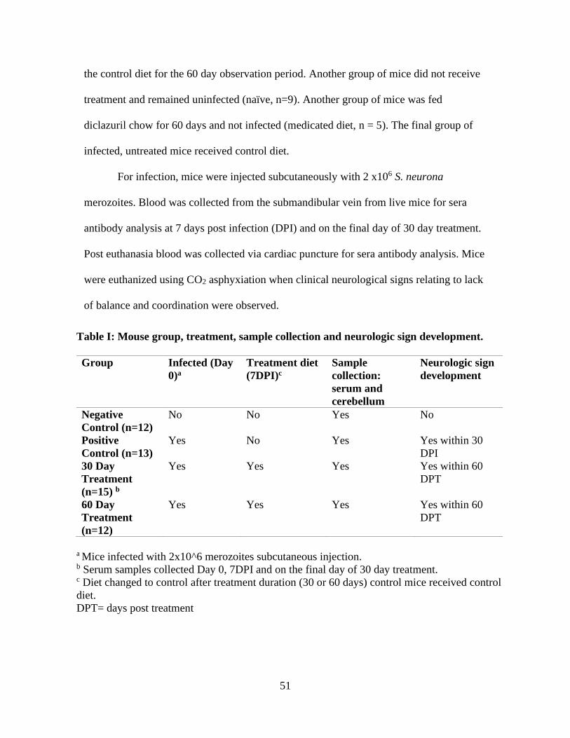
51
the control diet for the 60 day observation period. Another group of mice did not receive
treatment and remained uninfected (naïve, n=9). Another group of mice was fed
diclazuril chow for 60 days and not infected (medicated diet, n = 5). The final group of
infected, untreated mice received control diet.
For infection, mice were injected subcutaneously with 2 x10⁶ S. neurona
merozoites. Blood was collected from the submandibular vein from live mice for sera
antibody analysis at 7 days post infection (DPI) and on the final day of 30 day treatment.
Post euthanasia blood was collected via cardiac puncture for sera antibody analysis. Mice
were euthanized using CO2 asphyxiation when clinical neurological signs relating to lack
of balance and coordination were observed.
Table I: Mouse group, treatment, sample collection and neurologic sign development.
Group Infected (Day
0)a
Treatment diet
(7DPI)c
Sample
collection:
serum and
cerebellum
Neurologic sign
development
Negative
Control (n=12)
No No Yes No
Positive
Control (n=13)
Yes No Yes Yes within 30
DPI
30 Day
Treatment
(n=15) b
Yes Yes Yes Yes within 60
DPT
60 Day
Treatment
(n=12)
Yes Yes Yes Yes within 60
DPT
a Mice infected with 2x10^6 merozoites subcutaneous injection. b Serum samples collected Day 0, 7DPI and on the final day of 30 day treatment. c Diet changed to control after treatment duration (30 or 60 days) control mice received control
diet.
DPT= days post treatment

52
Diclazuril treatment
Diclazuril (Sigma- Aldrich, St. Louis, Missouri and LKT Laboratories, St. Paul,
Minnesota) was compounded into 5LG4 rat and mouse chow (Test Diet, St. Louis,
Missouri) at a concentration of 50 ppm. Medicated chow provided 10mg of diclazuril/kg
of body weight to each mouse, a dose previously shown to prevent the development of S.
neurona associated disease in Ifnγ -/- mice (Dubey et al., 2001). The rat and mouse
chow(5LG4), without diclazuril served as the control diet.
Cerebellum preparation and histology
At the time of euthanasia, cerebellums were collected and those intended for the
use of histology, were fixed in 10% buffered formalin solution. The cerebellum was then
embedded in paraffin and processed for hematoxylin and eosin (H&E) staining. Triplicate
sections were cut from paraffin blocks and used for immunohistochemical staining. One
sample section was assessed for the total number of perivascular cuffs, gliosis foci and
number of organisms for each cerebellum collected and given a score of 1 (mild) - 6
(severe). The total number of perivascular cuffs and gliosis foci increased with score
where a score of 1 = 0-10 cuffs or foci and 6 = 55-70 cuffs or foci. The total number of
organisms was scored in a similar manner, score 1 = 0-20 organisms and 6 = number
organisms >100. Sections of cerebellum were stained with polyclonal rabbit anti-S.
neurona anti-sera as described by Gerhold et al. (2005) with the exception that sections
were digested with perxo-block (Novex , Life Technologies, Carlsbad, California) prior
to incubation with anti-sera at 1:500 dilution.
Sera immunoglobulins

53
Blood was collected from infected untreated (n = 10), naïve (n = 9) and 30 day (n
= 10) and 60 day (n = 10) treatment groups at the time of euthanasia, for sera IgM, total
IgG and S. neurona immunoglobulin analysis by enzyme linked immunoabsorbent assay
(ELISA). Serum was collected from whole blood, following centrifugation and stored at -
20ᵒ C until utilized for ELISA. Immunoglobulin ELISAs were performed as described
(Christianson et al., 1997) using IgG and IgM isotypes (Southern Biotech, Birmingham,
Alabama). Plates were read on an Infinite M200 Pro plate reader with Magellan 7.0
software (Tecan, Mannedorf, Switzerland).
For S. neurona specific ELISA, a S. neurona merozoite protein lysate was created
as a plate coating agent for antibody capture. For the lysate, frozen Sn-15OP merozoites
were thawed and centrifuged at 2,500 g for 5 min at 4ᵒ C, merozoites were washed twice
in ice cold 1x PBS, re-suspended to a concentration of 1x 10⁷/mL in 1x PBS, 10 mLs of
radioimmunoprecipitation assay (RIPA) buffer (Thermo Fisher Scientific, Waltham,
Massachusetts) was added per 1mL of merozoite suspension. The merozoite suspension
was incubated on ice for 15 min with intermittent manual shaking and then centrifuged at
1,400 g for 15 min at 4ᵒ C. After centrifugation, the supernatant was collected and 100
uL was used to coat the ELISA plate wells (ThermoFisher, Ref # 9018) incubating
overnight at 4ᵒ C. The protocol then proceeded as described in Christianson et al. (1997).
The detection antibody was goat anti mouse-Ig kappa- alkaline phosphatase (Southern
Biotech) and optical density (OD) values were reported for data analysis.
Cerebellum culture and observations
At the time of euthanasia cerebellums intended for the use of culture, were
collected from infected 30 day treated mice at the following time points: on the final day
54
of treatment (n=3), 12 days post cessation of treatment (n=3) and at the time of
neurologic sign development post treatment (n=2). Additionally, cerebellums were
collected from infected untreated mice (n=2). Immediately after collection cerebellums
were placed in ice cold 1xPBS and then homogenized in 1 ml Hanks balanced salt
solution (HBSS) using a sterile Teflon-coated tissue grinder and 12 ml grinding tube. The
homogenized brain-HBSS solution was inoculated on to 2 (25 cm2) tissue culture flasks
containing monolayers of CV-1 cells. The homogenate was incubated with CV-1 cells for
2 hr at 37ᵒ C. After 2 hr, the inoculum was removed, the cell monolayers were washed
with HBSS, and 5 ml of maintenance medium was added for long term culture. Cultures
were examined twice weekly and the medium replaced twice weekly for a month and
then once weekly thereafter until S. neurona was detected or for 2 mo when the study
was ended, and cultures were considered negative.
Statistical analysis
Neurologic disease incidence was determined using a log rank Mantel-Cox test
with a significant level set at P < 0.05 and Bonferroni correction to account for multiple
comparisons with a significance threshold at P < 0.017. Significance for immunoglobulin
changes during treatment was determined using ANOVA accounting for repeated
measures and multiple comparisons with Tukey’s T-test. To assess the immunoglobulin
changes that occurred between the naïve mouse group and throughout treatment ANOVA
was used with Dunnetts test to account for multiple comparisons. All other statistical
analysis was calculated using Kruskal- Wallis test and Dunn’s test for post hoc
comparisons. The significant level was set at P < 0.05. All analyses were performed using
GraphPad Prism software version 6 for Mac (GraphPad Software, La Jolla, California).

55
RESULTS
Saracocystis neurona induced
myeloencephalitis
All infected untreated mice
developed neurologic signs 23
days of starting the non-
medicated diet. Saracocystis
neurona-infected diclazuril-
treated mice did not develop
clinical signs while on the
medicated diet. Both the infected
30 and 60 day treated mice
remained clinically normal on the
non-medicated diet for significantly longer than the infected untreated mice. Neurologic
disease did occur within 60 days of ceasing diclazuril treatment in all infected treated
mice (Table I). There was no statistically significant difference in the incidence of
neurologic disease between the 30 day treatment and 60 day treatment groups after
switching to the control diet (Fig. 1).
Figure 1. The graph depicts the percent of neurologically normal mice
versus the number of days mice have been consuming control diet. Time
point day 0 represents the day mice were placed on the control diet. The
infected untreated mice started consumption of non-medicated diet 7 DPI,
and 30 and 60 day treated mice started at the end of the treatment periods.
Closed circles represent number of neurologically normal mice at that time
point within each group. **** P = <0.0001.

56
Histopathological analysis
Histology results confirm S. neurona induced encephalitis. When assessed at the time of
neurologic sign onset, the cerebellums of infected untreated mice possessed significantly
more gliosis foci by number (Fig. 2A) and perivascular cuffs (Fig. 2B) compared to
infected 30 and 60 day treated mice. Saracocystis neurona organisms were found in
significantly greater numbers in cerebellum sections from infected untreated mice
compared to mice in the 60 day treatment group at the time of necropsy. There were no
Figure 2. Scores of histopathologic changes associated with infection and treatment (trt) in
cerebellums collected from mice displaying neurologic signs. (A) perivascular cuffing foci in
cerebellum H&E stained sections. (B) Gliosis foci in cerebellum H&E stained sections. (C)
Saracocystis neurona organisms in H&E and IHC stained cerebellum sections. Male B6.Ifng-/-
mice, age 8-10 wk at start of experiment. * P = <0.05, ** P = <0.005

57
significant differences in number of gliosis foci (Fig. 2A), perivascular cuffing (Fig. 2B
Figure 3. Cerebellar immunohistochemistry and H&E images. All images are of cerebellum
samples collected when neurologic signs were displayed by infected untreated mice (A, B) 30
day treated mice (C, D) and 60 day treated mice (E, F). (A) H&E image where the asterisk (*)
indicates eosinophilic inflammation and arrows indicate Saracocystis neurona asexual stages.
(B) H&E image where asterisks (*) indicate eosinophilic inflammation and arrows gliosis +
eosinophilic inflammation. (C) IHC image arrows and dark brown staining indicate S. neurona
asexual life stages. (D) H&E stained asterisks (*) indicate perivascular cuffing and arrows
indicate gliosis. (E) IHC image arrows and dark brown staining are S. neurona asexual life
stages. (F) H&E staining asterisks (*) indicate gliosis and eosinophilic inflammation and
arrows S. neurona asexual life stages.
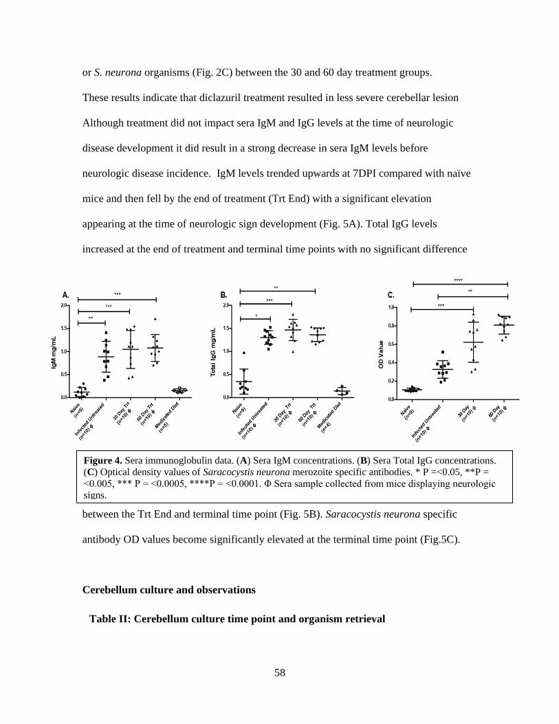
58
or S. neurona organisms (Fig. 2C) between the 30 and 60 day treatment groups.
These results indicate that diclazuril treatment resulted in less severe cerebellar lesion
Although treatment did not impact sera IgM and IgG levels at the time of neurologic
disease development it did result in a strong decrease in sera IgM levels before
neurologic disease incidence. IgM levels trended upwards at 7DPI compared with naïve
mice and then fell by the end of treatment (Trt End) with a significant elevation
appearing at the time of neurologic sign development (Fig. 5A). Total IgG levels
increased at the end of treatment and terminal time points with no significant difference
between the Trt End and terminal time point (Fig. 5B). Saracocystis neurona specific
antibody OD values become significantly elevated at the terminal time point (Fig.5C).
Cerebellum culture and observations
Table II: Cerebellum culture time point and organism retrieval
Figure 4. Sera immunoglobulin data. (A) Sera IgM concentrations. (B) Sera Total IgG concentrations.
(C) Optical density values of Saracocystis neurona merozoite specific antibodies. * P =<0.05, **P =
<0.005, *** P = <0.0005, ****P = <0.0001. Φ Sera sample collected from mice displaying neurologic
signs.

59
Of 3 cerebellum samples collected on the final day of a 30 day treatment period
and of three collected 12 days post treatment, no viable S. neurona was recovered after
culture. However, of 2 cerebellum samples harvested from mice demonstrating
neurologic signs within 60 days after a 30 day treatment period, one produced viable S.
neurona that replicated in culture (Table II). Our results in these immunodeficient mice
show that viable parasites can remain in the mice even after appropriate therapy with
diclazuril.
DISCUSSION
Using the immunocompromised Ifnγ -/- mouse, the current study demonstrates that
continuous diclazuril treatment for 30 or 60 days prevents the development of S. neurona
Cerebellum
culture time
point
End of 30
day trt
12 days post
30 day trt
Infected 30
day treated a
Infected
untreated a
Live Organism
Retrieval
0 /3 0/3 1/2 2/2
Figure 5. Sera immunoglobulin data during treatment and time points during treatment compared to
naïve mice. 7 DPI = 7 days post infection. Trt End = At the end of 30 days of diclazuril treatment (A)
Sera IgM concentrations. (B) Sera total IgG concentrations. (C) Optical density values of
Saracocystis neurona merozoite specific antibodies. * P = <0.05, **P = <0.005, *** P = <0.0005,
****P = <0.0001. Φ Sera sample collected at time of symptom development.
a Cerebellums collected from mice displaying neurologic signs

60
induced protozoal myeloencephalitis and associated neurologic signs. However, within
60 days after cessation of diclazuril treatment mice develop neurologic signs. This
suggests that treatment prevents replication and/or migration of S. neurona but cessation
of treatment leads to resumption of pathogenic activity. Previous in- vitro cell culture
studies demonstrated that diclazuril limited the development of S. neurona merozoites,
but parasite replication was observed after the removal of diclazuril from cell culture
media. This also suggests that diclazuril only has an inhibitory effect on parasite activity
(Lindsay and Dubey, 2000). In the current study, cerebellum samples collected on the
final day of a 30 day diclazuril treatment period yielded no viable S. neurona. However, a
cerebellum sample collected from a neurologic mouse post treatment yielded viable S.
neurona capable of replicating in culture. The results of our histology data reveal positive
S. neurona antibody staining in the cerebellum regardless of treatment duration. The
decrease in the number of gliosis foci and perivascular cuffs in the treated mice compared
to untreated mice suggests that treatment decreases the severity of S. neurona associated
lesions. It should be acknowledged that Dubey et al. (2001), administered diclazuril
compounded rodent diet for 39 days, to Ifnγ -/- mice 7 days after infection with 1000 S.
neurona sporocysts from the isolate Sn-15OP and sacrificed mice 8 days after treatment.
They found that 8 days after the cessation of treatment, cerebellums subpassaged to naïve
Ifnγ -/- mice were not pathogenic nor did cerebellum histology results show positive S.
neurona antibody staining (Dubey et al., 2001). In the current study, cerebellum samples
collected 12 days after 30 days of treatment from infected mice did not yield S. neurona
in culture suggesting that the brains were not yet parasitized at this point after treatment.
We hypothesize that if treated infected mice in Dubey’s study were left alive for 60 days

61
post treatment, clinical disease may have developed, as in our study. Collectively, these
results indicate S. neurona can resume its activity after cessation of diclazuril in vivo
barring an adequate immunological response from the host.
While the immunologic response to S. neurona in Infγ-/- mice is not directly
translatable to equine patients, quantifying the humoral response gives clues to immune
awareness of parasitic infection. Extraneural S. neurona replication occurs 7 DPI
(Dubey, 2001) which allows for antigen recognition by naïve B- lymphocytes resulting in
the modest elevation of sera IgM concentrations 7 DPI compared to the naïve mouse
group. At the end of 30 days of diclazuril treatment, sera IgM concentrations declined
from the beginning of treatment (7 DPI) similar to those observed in the naïve mouse
group suggesting that few new naïve B-lymphocytes were encountering parasite during
treatment. The resumption of active S. neurona infection and parasite replication and
migration is probable after treatment was discontinued as indicated by the significant
increase in sera IgM concentrations at the time of neurologic disease development. The
significant increase in total IgG sera concentration throughout the course of treatment can
be attributed to maturation of the initial immune response, with IgM B- lymphocytes
switching isotypes to IgG through the process of class switch recombination. The
significant increase in S. neurona antibody OD values at the time of neurologic sign also
supports maturation of an immune response to S. neurona and relates the humoral
immune response to S. neurona infection. Pusterla et al. (2015) demonstrated that foals
residing in a high risk EPM area treated with a low dose of diclazuril (0.5 mg/kg) had
significantly less sera conversion of antibodies against S. neurona compared to untreated
foals following the decline of maternal antibodies. Furr et al. (2006) also demonstrated

62
the prophylactic nature of the coccidiostat Ponazuril in decreasing the incidence and
severity of experimentally induced EPM, assessed by sera and CSF antibodies and
clinical neurologic signs. Murphy et al (2006), experimentally infected horses with S.
neurona and demonstrated significant increases in S. neurona- IgM concentrations in
serum and CSF samples collected from experimentally infected horses. The horses in the
mentioned study developed clinical neurologic disease as a result of the experimental S.
neurona infection. While our study was performed in the immunocompromised mouse
model and cannot be directly correlated to disease in the horse, the previously mentioned
equine model studies by Furr et al. (2006), Murphy et al (2006), Pusterla et al. (2015),
also support the efficacy of humoral immune response analysis. The sera
immunoglobulin results from the current study, also highlight the potential for evaluating
S. neurona specific IgM antibodies clinically in equine patients, before the start of anti-
protozoal treatment, at the time of treatment discontinuation and periodically after to
monitor for the development of recurrent infection.
Recurrent disease in horses may be a result of persistent infection due to treatment
failure as opposed to re-exposure to parasite. Other EPM treatment efficacy studies
demonstrate potential treatment failure that could lead to disease relapse. Furr et al (2001)
demonstrated that within 118 days after appropriate ponazuril treatment only 62% of
horses in the study improved neurologically 1 grade and 10% of treated horses had
cerebral spinal fluid negative for S. neurona antibodies by 90 days post treatment. In the
same study 8% of horses that displayed improvement after treatment demonstrated
recurrent disease by 118 days post treatment. Horses in the study were not housed in a
manner to determine if recurrent disease could be attributed to re-exposure to S. neurona
63
or persistent infection due to treatment failure but it was hypothesized to be a result of
treatment failure (Furr et al., 2001). Additionally, horses treated with
sulfadiazine/pyrimethamine have a disease relapse rate of 25% (Fenger, 1998). To the
best of the authors knowledge, no studies have been done in horses that evaluate the
disease relapse rate related to diclazuril treatment. In many cases, the discussed anti-
protozoal drug coupled with an appropriate immune response in an otherwise normal
horse is a successful treatment strategy for clinically affected patients but for those
patients that present with recurrent signs, other treatment strategies should be considered.
The results of this study imply that diclazuril can inhibit S. neurona activity but not
eliminate the parasite, providing evidence that recurrent disease could be a result of
persistent infection and treatment failure and not simply reinfection.
ACKNOWLEDGMENT
Authors would like to thank Dr. Heather Bradford for her contribution to the
statistical analysis of the manuscript.
LITERATURE CITED
Christianson, G. J., W. Brooks, S. Vekasi, E. A. Manolfi, J. Niles, S. L. Roopenian, J. B.
Roths, R. Rothlein, and D. C. Roopenian. 1997. Beta 2-microglobulin-deficient
mice are protected from hypergammaglobulinemia and have defective antibody
responses because of increased IgG catabolism. Journal of Immunology 159:
4781–4792.
Dubey, J.P. 2000. Prevalence of Sarcocystis species sporocysts in wild caught opossums
(Didelphis virginiana). Journal of Parasitology 86: 705–710.
64
Dubey, J.P. 2001. Migration and development of Saracocystis neurona in tissues in
interferon-gamma knockout mice fed sporocysts from a naturally infected
opossum. Veterinary Parasitology 95: 341-351.
Dubey, J.P., D. Fritz, D.S. Lindsay, S.K. Shen, O.C.H. Kwok, and K.C. Thompson.
2001. Diclazuril preventive therapy of gamma interferon knockout mice fed
Saracocystis neurona sporocysts. Veterinary Parasitology 94: 257– 263.
Dubey, J. P., and D. S. Lindsay. 1998. Isolation in immunodeficient mice of Saracocystis
neurona from opossum (Didelphis virginiana) faeces and its differentiation from
Sarcocystis falcatula. International Journal Parasitology 28: 1823-1828.
Dubey, J. P, and D. S. Lindsay. 1999. Sarcocystis speeri n. sp. (Protozoa: Sarcocystidae)
from the opossum (Didelphis virginiana). Journal of Parasitology 85: 903-909.
Dubey, J.P., D. E. Mattison, C. A. Speer, R. J. Baker, D. M. Mulrooney, S. J. Tornquist,
A. N. Hamir, and T. C. Gerros, 1999. Characterization of a Saracocystis neurona
isolate (SN6) from a naturally infected horse from Oregon. Journal of Eukaryotic
Microbiology 46: 500-506.
Dubey, J.P., W.J.A. Saville, D.S. Lindsay, R.W. Stich, J.F. Stanek, C.A. Speer, B.M.
Rosenthal, C.J. Njoku, O.C.H. Kwok, S.K. Shen, et al. 2000. Completion of the
life cycle of Saracocystis neurona. Journal of Parasitology 86: 1276-1280.
Furr, M., T. Kennedy, R. MacKay, S. Reed, F. Andrews, B. Bernard, F. Bain, and D.
Byars. 2001. Efficacy of ponazuril 15% oral paste as a treatment for equine
protozoal myeloencephalitis. Veterinary Therapeutics: Research in Applied
Veterinary Medicine 2: 215-222.

65
Furr, M., H. McKenzie, W.J.A. Saville, J.P. Dubey, S.M. Reed, and W. Davis. 2006.
Prophylactic administration of ponazuril reduces clinical signs and delays
seroconversion in horses challenged with Saracocystis neurona. Journal of
Parasitology 92: 637-643.
Gerhold, R.W., E.W. Howerth, and D.S. Lindsay. 2005. Saracocystis neurona-associated
Meningoencephalitis and Description of Intramuscular Sarcocysts in a Fisher
(Martes pennanti). Journal of Wildlife Diseases. 41: 224-230.
Lindsay, D.S., and J. P. Dubey. 2000. Determination of the activity of diclazuril against
Saracocystis neurona and Sarcocystis falcatula in cell cultures. Journal of
Parasitology. 86: 164–166.
Lindsay, D.S., M.M. Nazir, A. Maqbool, S.P. Ellison, and J.S. Strobl, 2013. Efficacy of
decoquinate against Saracocystis neurona in cell cultures. Veterinary Parasitology.
196: 21–23.
MacKay, R. J. 2006. Equine protozoal myeloencephalitis: Treatment, prognosis, and
prevention. Clinical Techniques in Equine Practice 5: 9-16.
Mitchell, S. M., A. M. Zajac, W. L. Davis, T. J. Kennedy, and D. S. Lindsay. 2005. The
effects of ponazuril on development of apicomplexans in vitro. Journal of
Eukaryote Microbiology 52: 231-235.
Murphy, J.E., Marsh, A.E., Reed, S.M., Meadows, C., Bolten, K., 2006. Development
and Evaluation of a Saracocystis neurona -Specific IgM Capture Enzyme-Linked
Immunosorbent Assay 322–328.
66
NAHMS (National Animal Health Monitoring System). 2001. Equine protozoal
myeloencephalitis (EPM) in the U.S. Centers for Epidemiology and Animal
Health – USDA APHIS, Fort Collins, Colorado, USA.
Pusterla, N., A. Packham, S. Mackie, P.H. Kass, L. Hunyadi, and P.A., Conrad. 2015.
Daily feeding of diclazuril top dress pellets in foals reduces seroconversion to
Saracocystis neurona. Veterinary Parasitology Journal 206: 236–238.
Reed, S. M., M. Furr, D. K. Howe, A. L. Johnson, R. J. MacKay, J. K. Morrow and S.
Witonsky. 2016. Equine protozoal myeloencephalitis: An updated consensus
statement with a focus on parasite biology, diagnosis, treatment, and
prevention. Journal of Veterinary Internal Medicine 30: 491–502.
Saville W.J., P.S Morley, S.M. Reed, D.E. Granstrom, C.W. Kohn, K.W. Hinchcliff, and
T.E. Wittum. 2000. Evaluation of risk factors associated with clinical
improvement and survival of horses with equine protozoal myeloencephalitis.
Journal of American Veterinary Medicine Association 217: 1181-1185.
Saville, W.J., R.W. Stitch, S.M Reed, C.J Njoku, M.J Oglesebee, A Wunschmann, D. L
Grover, A.L Larew- Naugle, J. F Stanek, D.E Granstrom, J.P Dubey. 2001.
Ulitization of stress in the development of an equine model for equine protozoal
myeloencephalitis. Veterinary Parasitology 95: 211-222.
Scott, P., S. Witonsky, J. Robertson, and B. Draft. 2005. Increased presences of T
lymphocytes in central nervous system of EPM affected horses. Journal of
Parasitology 91: 1499-1502.
67
Spencer, J.A., S.E. Ellison, A.J. Guarino, and B.L. Blagburn. 2004. Cell-mediated
immune responses in horses with equine protozoal myeloencephalitis. Journal of
Parasitology 90: 428-430.
Witonsky, S.G., S. Elliston, J. Yang, R.M. Gogal, H. Lawler, Y. Suzuki, N.
Sriranganathan, F. Andrews, D. Ward, D.S. Lindsay. 2008. Horses experimentally
infected with Saracocystis neurona develop altered immune responses in vitro.
Journal of Parasitology 94: 1047-1055.
Witonsky, S.G., R.M. Gogal, R.B. Duncan, and D.S. Lindsay. 2003. Immunopathologic
effects associated with Saracocystis neurona–infected interferon-gamma
knockout mice. Journal of Parasitology 89: 932–940.
Yang, J. S, Ellison. R, Gogal, H. Norton, D.S. Lindsay, F. Andrews, D. Ward, and S.
Witonsky. 2006. Immune response to S. neurona infection in naturally infected
horses with equine protozoal myeloencephalitis. Veterinary Parasitology 138:
200-210.

68
Chapter IV: The efficacy of decoquinate in preventing the development of neurologic
disease in Saracocystis neurona infected C57Bl/6 interferon gamma knockout mice
Abstract
Equine protozoal myeloencephalitis (EPM), predominantly caused by the protozoa
Saracocystis neurona, is a common neurologic disease of horses from North America.
Exposure to the parasite occurs frequently but clinical neurologic disease presents in a
small fraction of exposed horses. Treatment for EPM affected horses consists of anti-
protozoal drugs along with anti-inflammatories to combat associated inflammation in the
central nervous system (CNS). Recently, the anti-protozoal drug decoquinate has been
evaluated for its efficacy aganist S. neurona infection. In the current study, we sought to
determine if decoquinate could retard neurologic disease in S. neurona infected C57Bl/6
interferon gamma knockout (B6Ifnγ -/-) mice, an EPM mouse model. Decoquinate was
administered to mice 7 days post infection, in the form of either medicated chow
compounded with 30 or 60 ppm of decoquinate or by orally dosing mice daily with
20mg/kg of decoquinate. All treated mice developed neurologic signs within 30 days post
infection (DPI) at a rate that did not significantly differ from infected untreated controls
and S. neurona was found in the cerebellums of all infected mice regardless of treatment
status. Additionally, mass spectrometry results indicate the greatest sera decoquinate
concentration was found in mice from the group that received medicated chow with
60ppm of decoquinate opposed to the group that was orally dosed daily. Results of the
current study indicate that decoquinate was not able to prevent neurologic disease in S.
neurona infected B6Ifnγ -/-mice.
Introduction
Equine protozoal myeloencephalitis (EPM) is one of the most common neurologic
diseases of horses in North America, with the principal basis for disease being ingestion

69
of Saracocystis neurona sporocysts. The pathogenic sporocysts are found in opossums
(Didelphis virginiana) feces which contaminate the horse’s feed and water sources
(Dubey and Lindsay, 1999, Dubey et al. 1999, Dubey et al. 2000). The prevalence of
exposure to S. neurona is high but there is not a direct correlation between exposure
incidence and clinical neurological disease incidence. Many neurologically normal horses
test positive for S. neurona sera antibodies indicating exposure to the parasite. The reason
for high exposure rates but relatively low morbidity remains unclear but previous studies
indicate a potential aberrant immune response to S. neurona in susceptible horses
(Spencer et al., 2004, Scott et al., 2005, Yang et al., 2006, Lewis et al, 2014). Clinical
EPM treatment consists of the use of anti-protozoal drugs such as ponazuril, diclazuril
and Sulfadiazine/pyrimethamine, as well as anti-inflammatory medications to combat
associated inflammation in the CNS (Pusterla and Tobin, 2017). A study examining all
stages of disease as well as all different treatment options, revealed that the average
equine patient is expected to improve only one neurological grade with treatment. In the
same study, approximately 10-25% of EPM affected horses that respond positively to
treatment are at risk for developing recurrent or novel neurological signs after
discontinuing therapy (Saville et al, 2000). This warrants investigation into novel
treatment strategies to improve the prognosis for EPM affected horses, and recently, the
anti-protozoal decoquinate has been examined for its efficacy as an EPM treatment.
A clinical field trial treating horses suspected to be affected by EPM with a
compounded formulation of decoquinate and immunomodulator levamisole, found that
clinical neurologic signs decreased after 10 days of treatment in treated horses (Ellison et
al, 2012). Lindsay et al (2013) demonstrated that in-vitro decoquinate treatment inhibited
70
merozoite replication of two isolates of S. neurona merozoites, one opossum (SnOP-15)
and one equine (Sn6) derived, and, at high enough concentrations, eliminated developing
schizonts. While these two studies show promise for the use of decoquinate as an EPM
treatment further studies with more defined and controlled study population, need to be
conducted.
In the current study, we sought to determine the efficacy of decoquinate in
preventing S. neurona induced encephalitis in the C57Bl/6 interferon gamma gene
knockout (Ifnγ-/-), mouse model (Dubey and Lindsay, 1998; Dubey., 2001, 2013;
Witonsky et al., 2003, Hay et al, 2019). Decoquinate is commercially available as a feed
additive for livestock for treatment of coccidiosis in cattle, goats and poultry (Deccox,
Zoetis). Currently it is compounded with immunomodulator levamisole and administrated
as an oral paste once daily, for 10-day treatment period in EPM affected horses (Ellison
et al, 2012 and Pathogenes, Reddick, Florida). In order to mimic commercially utilized
decoquinate administration methods, two administration methods were tested in the
current study; decoquinate medicated chow fed ad libitum and once daily oral
administration by gavage. The mice were infected with S. neurona and treatment began 7
days post infection (DPI) with mice monitored for the development of neurologic signs to
assess the ability of decoquinate to prevent persistent infection.
Materials and Methods
Saracocystis neurona culture and preparation of inoculum
Merozoites of the Sn-15OP isolate were grown in African green monkey (Cercopithecus
aethiops) kidney cells (CV-1(ATTC CCL-70)) American Type Culture Collection,
Manassas, Virginia), separated from host cells, collected and enumerated for inoculations

71
as previously described (Lindsay et al, 2013). The Sn-15OP isolate was isolated from the
brain of an Ifnγ -/- mouse fed sporocysts obtained from the feces of a naturally infected
opossum from Virginia (Dubey, 2000). This isolate was originally a gift from Dr. J. P.
Dubey, United States Department of Agriculture, Beltsville, Maryland and has been
maintained cryopreserved in liquid nitrogen.
Mice
Male and female B6.129S7-Ifngtm1Ts/J (B6.Ifnγ -/-) mice, age 8-10 weeks old, were
originally obtained from Jackson Laboratory (JR2287, Bar Harbor, Maine). A colony of
B6. Ifnγ -/- mice was maintained through inbreeding with a brother sister mating strategy
and mice used in the current study were from this colony. Mice were maintained at
Virginia Tech in accordance with an Institutional Animal Care and Use Committee
(IACUC) breeding protocol and experimental studies were conducted under other
IACUC approved protocols. For Sarcoystis neurona infection, mice were injected
subcutaneously with 2 x10⁶ S. neurona merozoites. Mice were euthanized using CO2
asphyxiation when clinical neurological signs relating to lack of balance and coordination
were observed. Blood was collected post euthanasia via cardiac puncture from infected
mice and sera was collected after centrifugation and stored at -20 C for further analysis.
Cerebellum samples were also collected for histological analysis from all infected mice,
post euthanasia. The body weight of all mice was monitored throughout the study and
percent body weight loss was calculated after euthanasia.
Decoquinate Treatment
Medicated Diet
72
Decoquinate (Sigma-Aldrich, St. Louis, Missouri) was compounded into 5LG4 rat and
mouse chow (Test Diet, St. Louis, Missouri) at a concentration of 30 and 60 ppm. The
5LG4) without decoquinate served as the control diet. The S. neurona infected mice were
divided in to three different groups: 30ppm medicated diet (n=6), 60ppm medicated diet
(n=8) or control diet (n=4 per treatment). The consumption of the appropriate treatment
or control diet began 7 days post infection (DPI) for 30 days. At the end of the 30- day
treatment feeding period, mice were switched to the control diet. The time point of 7 DPI
was selected for commencement of treatment, as primarily extraneural replication of S.
neurona is occurring at this time point (Dubey, 2001).
Oral gavage decoquinate treatment
Decoquinate was administered in the form of an 8% decoquinate formulation contributed
by Dr. Siobhan Ellison (Pathogenes Inc, Reddick, Florida). This formulation of
decoquinate is most soluble at a pH 9 and optimal solubility was necessary to achieve for
oral gavage administration. Decoquinate was suspended in sucralose sweetened sterile
water at pH 9. Treatment began 7 DPI and was administered every 24 hours at a dose of
20 mg/kg (n=10) and sucralose sweetened sterile water was given to the control mice
(n=9) for 30 days or until the development of neurologic disease.
Cerebellum preparation and histology
At the time of euthanasia, cerebellums were collected from mice treated by oral gavage
(n=5), 60ppm decoquinate chow (n=5) and control mice (n=5) from both treatment trials
(Table I). Cerebellum samples from mice treated with 30ppm of medicated chow were
not processed since neither treatment option yielded protection against development of
neurologic disease. It was opted to only select 60ppm medicated chow samples for

73
processing. Cerebellums were fixed in 10% buffered formalin solution, paraffin
embedded and sectioned for immunohistochemistry (IHC) staining. Sections of
cerebellum were stained with polyclonal rabbit anti-S. neurona anti-sera as described by
Gerhold et al. (2005) with the exception that sections were digested with perxo-block
(Novex, Life Technologies, Carlsbad, California) prior to incubation with anti-sera at
1:500 dilution. The sections were reviewed for positive S. neurona staining with a Primo
Star Upright Light Microscope (Carl Zeiss) to confirm S. neurona presences in the
cerebellum.
Table I: Sample collection and processing for each group
Group
Neurologic
signs
Cerebelluma
Processing
IHC Positiveb Mass
Spectrometryc
Infected
Untreated –
30ppm
medicated diet
(n=4)
Yes
(4/4)
N/A
N/A
N/A
Infected
Untreated –
60ppm
medicated diet
(n=4)
Yes
(4/4)
Yes
(n=2)
2/2 n= 2
Infected
Untreated –
oral gavage
(n=9)
Yes
(9/9)
Yes
(n=3)
3/3 n=3
30ppm
medicated Diet
(n=6)
Yes
(6/6)
N/A N/A N/A
60 ppm
medicated diet
(n=8)
Yes
(8/8)
Yes
(n=5)
5/5 n=5

74
Oral gavage
(n=10)
Yes
(10/10)
Yes
(n=5)
5/5 n=5
Mass spectrometry To ensure that decoquinate was delivered by both means of
administration, mass spectrometry was preformed using a LC/MS system and the
protocol previously described (Wang, 2017) to detect and quantitate decoquinate in
mouse sera. The sample size was 50 uL of sera and Decoquinate D5 (Sigma Aldrich, St.
Louis, Missouri) was used for the internal standard. Sera samples collected from mice
treated with 30ppm of medicated chow were not processed since neither treatment option
yielded protection against development of neurologic disease. It was opted to select only
60ppm medicated chow samples for processing. The negative control samples were from
mice which received control diet (n=2) or mice orally dosed with sucralose sweetened
sterile water (n=3).
Statistical analysis
For analysis of the neurologic incidence curves, a log rank (Mantel-Cox) test was
performed. For analysis of the mass spectrometry results, one- way ANOVA was
conducted, and Tukey’s T-test accounted for post hoc multiple comparisons. A Pearson
correlation test was conducted to assess correlation of weight loss and sera concentrations
of decoquinate. All statistical analysis was done in Graph Pad Prism software and
statistical significance was set at P<0.05.
a Number of collected cerebellum samples processed for each group.
b Number of processed cerebellum samples which were IHC positive for S. neurona.
c Number of sera samples processed for decoquinate Mass Spectrometry

75
Results
Neurologic incidence
Neurologic signs developed
in all infected mice
regardless of treatment status
within 30 DPI and 23 days
post treatment (DPT).
Neither decoquinate
compounded chow (30 or
60ppm) or oral gavage
administration of
decoquinate were able to
avert neurologic
abnormalities and neurologic
incidence was 100% in all
infected B6Ifnγ-/- mice.
There were no significant
differences in occurrence of
neurologic sign development
between the infected
untreated control groups and
30ppm chow, 60ppm chow or
0 10 20 30
0
50
100
Days Post Infection
% N
eu
rolo
gic
ally N
orm
al M
ice Infected Untreated
(n=4)
30ppm(n= 6)
0 10 20 30
0
50
100
Days Post Infection
% N
eu
rolo
gic
ally N
orm
al M
ice
Infected Untreated(n=4)
60ppm(n= 6)
0 10 20 30
0
50
100
Days Post Infection
% N
eu
rolo
gic
ally N
orm
al M
ice Infected Untreated
(n=9)
Gavage(n=10)
B
C
A
Figure 1: Neurologic incidence curves for all treatments
compared to infected untreated controls for each treatment.
All treatments began 7 DPI. A &B. Infected B6 Ifnγ -/-
received 30 ppm compounded chow (A) or 60 ppm (B). C.
Infected treated B6 Ifnγ -/- received oral administration
(gavage) of decoquinate.

76
daily oral dose, conclusively implying no treatment effect (Figure 1A-C).
Immunohistochemistry
For each processed sample (Table I), two replicate cerebellar sections were examined for
the presences of S. neurona. In all examined samples, positive IHC staining was found
regardless of treatment status (Figure 2). The positive IHC results from the cerebellum
samples collected from mice displaying neurologic abnormalities further support the
inability of decoquinate treatment to prevent neuroinvasion and S. neurona replication in
the CNS.
Figure 2: Representative immunohistochemistry images of cerebellum samples collected from A) infected untreated B6
Ifnγ-/- mouse B) oral gavage treatment and C) 60ppm medicated chow treatment. All cerebellums had positive S.
neurona staining indicated by dark brown staining in images.
200um 200um 200um
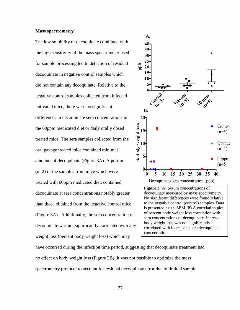
77
Mass spectrometry
The low solubility of decoquinate combined with
the high sensitivity of the mass spectrometer used
for sample processing led to detection of residual
decoquinate in negative control samples which
did not contain any decoquinate. Relative to the
negative control samples collected from infected
untreated mice, there were no significant
differences in decoquinate sera concentrations in
the 60ppm medicated diet or daily orally dosed
treated mice. The sera samples collected from the
oral gavage treated mice contained minimal
amounts of decoquinate (Figure 3A). A portion
(n=2) of the samples from mice which were
treated with 60ppm medicated diet, contained
decoquinate at sera concentrations notably greater
than those obtained from the negative control mice
(Figure 3A). Additionally, the sera concentration of
decoquinate was not significantly correlated with any
weight loss (percent body weight loss) which may
have occurred during the infection time period, suggesting that decoquinate treatment had
no effect on body weight loss (Figure 3B). It was not feasible to optimize the mass
spectrometry protocol to account for residual decoquinate error due to limited sample
Figure 3: A) Serum concentrations of
decoquinate measured by mass spectrometry.
No significant differences were found relative
to the negative control (control) samples. Data
is presented as +/- SEM. B) A correlation plot
of percent body weight loss correlation with
sera concentrations of decoquinate. Increase
body weight loss was not significantly
correlated with increase in sera decoquinate
concentration.
A.
B.

78
amount. Even though the majority of analyzed sera samples contained little to no
decoquinate, there were several samples that contained concentrations above those from
the negative control indicating that treated mice did receive decoquinate during the
treatment periods.
Discussion
In the current study we found that when S. neurona infected B6 Ifnγ-/- mice were treated
7DPI with mouse chow compounded with either 30ppm or 60ppm of the antiprotozoal
drug, decoquinate, neurologic disease was not prevented in the mice. Likewise, oral
administration of decoquinate at a dose of 20mg/kg once daily did not retard neurologic
disease. All treated, infected mice developed neurologic signs similar to infected
untreated mice within 30 DPI and S. neurona was identified by IHC in collected
cerebellum samples, regardless of treatment status. Hay et al (2019) previously
demonstrated that mouse chow compounded with 50ppm of the antiprotozoal diclazuril,
an agent commonly used for EPM treatment, delayed the occurrence of neurologic
disease in S. neurona infected B6 Ifnγ -/- compared to infected untreated controls. If the
efficacy of decoquinate was to be consider comparable or superior to that of other EPM
treatment options such as diclazuril, similar or better treatment efficacy would have been
expected in the current study. It should be noted that the lipophilic properties of
decoquinate do make it a difficult drug to pharmacologically compound, unlike
diclazuril, proposing a concern that it was not effectively compound into the medicated
diet used in the current study. This concern was addressed by performing mass
spectrometry in efforts to assess administration success of decoquinate. Decoquinate was
found in concentrations well above negative control mice concentrations in 2 of 5 mice
79
fed decoquinate compound chow at 60ppm, which ensured ingestion of decoquinate from
compounded chow. As to why decoquinate was not found in high concentrations of all
analyzed mice in the 60ppm treatment group, this may be explained by the rapid
clearance of decoquinate. Initial studies investigating the clearance of decoquinate from
blood found the clearance to be rapid with nearly all drug cleared within one hour of
dosing in investigated species which included, sheep, chickens, quail and cattle (Mitchell,
1988). It is unknown when the last time chow consumption (decoquinate intake)
occurred in neurologic mice prior to euthanasia and blood collection. In the current study
the two mice with the highest decoquinate sera concentrations experienced no weight
loss, suggesting that decoquinate intake was not affected by neurologic deficits and
perhaps intake of decoquinate occurred shortly before blood collection in these mice.
Furthermore, the three mice from the 60ppm chow group, which experienced the greatest
percent of body weight loss, had sera decoquinate concentrations very similar to those of
negative controls. In these three mice, chow intake (decoquinate) may have been impeded
by progression of S. neurona infection and this offers potential explanation for the low
sera decoquinate concentrations.
To help combat the bioavailability issues of standard decoquinate, a specially
formulated 8% decoquinate compound contributed by Dr. Ellison (Pathogenes Inc), was
also tested in the current study. The formulation of decoquinate is similar to the one
described by Wang et al (2013) which was formulated in a manner to reduce decoquinate
particle size and increase solubility in order to increase bioavailability to improve its
potential as an anti-malarial drug. In the current study, the 8% decoquinate formulation
was orally administrated at a dose of 20mg/kg of decoquinate once daily by oral gavage
80
technique. Our mass spectrometry results indicate that there were only slight,
insignificant differences in the amount of decoquinate in the sera from the mice treated
by oral gavage compared to the negative control samples. As previously mentioned,
decoquinate is rapidly cleared after administration and this provides a plausible
explanation as to why minimal amounts of decoquinate were detected in sera samples
collected from the gavage mice. The last decoquinate dose the mice received was 24
hours prior to sera sample collection. The quick metabolism of decoquinate makes
toxicity a low risk even when given frequently also resulting in little systemic
accumulation supporting the need for frequent dosing. The overall results of this study
question the efficacy of decoquinate as an EPM treatment.
Currently, there are no published decoquinate pharmacokinetic studies in horses
and whether the drug is able to pass the blood brain barrier and enter into the CNS,
remains unknown. In Ellison et al (2012), clinical field trial suggested efficacy of
compounded decoquinate and levamisole as successful treatment for presumptive EPM
affected horses, the two compounds were not evaluated individually and in-vivo effects
of each compound in the horse is unknown. In-vitro synergestic effects of decoquinate
and levamisole were not observed (Lindsay et al, 2013) but this may differ in vivo. The
results of the current S. neurona infection mouse model study demonstrates that
decoquinate does not have the same properties in vivo as other available treatments such
as diclazuril, despite promising in-vitro studies (Lindsay et al, 2013). This is most likely
due to unfavorable biological properties of decoquinate, which result in decreased
bioavailability. Based on the results of the current study and the gap in knowledge about

81
decoquinate pharmokinetics in the horse, further studies need to be conducted to ensure
validity of decoquinate as a treatment option for EPM.
Acknowledgments
Dr. Siobhan Ellison is thanked for her contribution of the decoquinate formulation used
in the oral gavage experiments in this study.
References
Dubey, J. P, and D. S. Lindsay. 1999. Sarcocystis speeri n. sp. (Protozoa: Sarcocystidae)
from the opossum (Didelphis virginiana). Journal of Parasitology 85: 903-909.
Dubey, J. P., and D. S. Lindsay. 1998. Isolation in immunodeficient mice of Saracocystis
neurona from opossum (Didelphis virginiana) faeces and its differentiation from
Sarcocystis falcatula. International Journal Parasitology 28: 1823-1828.
Dubey, J.P. 2000. Prevalence of Sarcocystis species sporocysts in wild caught opossums
(Didelphis virginiana). Journal of Parasitology 86: 705–710.
Dubey, J.P. 2001. Migration and development of Saracocystis neurona in tissues in
interferon-gamma knockout mice fed sporocysts from a naturally infected
opossum. Veterinary Parasitology 95: 341-351.
Dubey, J.P., D. E. Mattison, C. A. Speer, R. J. Baker, D. M. Mulrooney, S. J. Tornquist,
A. N. Hamir, and T. C. Gerros, 1999. Characterization of a Saracocystis neurona
isolate (SN6) from a naturally infected horse from Oregon. Journal of Eukaryotic
Microbiology 46: 500-506.
Dubey, J.P., W.J.A. Saville, D.S. Lindsay, R.W. Stich, J.F. Stanek, C.A. Speer, B.M.
Rosenthal, C.J. Njoku, O.C.H. Kwok, S.K. Shen, et al. 2000. Completion of the
life cycle of Saracocystis neurona. Journal of Parasitology 86: 1276-1280.

82
Ellison, S.P. and Lindsay, D.S. 2012. Decoquinate combined with levamisole reduce the
clinical signs and serum SAG 1,5,6 antibodies in horses with suspected Equine
Protozoal Myeloencephalitis. International Journal Applied Research Veterinary
Medicine. 10
Gerhold, R.W., E.W. Howerth, and D.S. Lindsay. 2005. Saracocystis neurona-associated
Meningoencephalitis and Description of Intramuscular Sarcocysts in a Fisher
(Martes pennanti). Journal of Wildlife Diseases. 41: 224-230.
Hay, A.N. Witonsky, S.G., Lindsay, D.S., LeRoith, T. Zhu, J., Kasmark, L., Leeth C.M.
2019. Saracocystis neurona- Induced Myeloencephalitis Relapse Following
Anticoccidial Treatment. Journal of Parasitology. 105: 371-378.
Lewis, S. R., Ellison, S. P., Dascanio, J. J., Lindsay, D. S., Gogal, R. M., Werre, S. R., …
Witonsky, S. G. (2014). Effects of Experimental Saracocystis neurona -Induced
Infection on Immunity in an Equine Model . Journal of Veterinary Medicine, 2014,
1–16.
Lindsay, D.S., M.M. Nazir, A. Maqbool, S.P. Ellison, and J.S. Strobl, 2013. Efficacy of
decoquinate against Saracocystis neurona in cell cultures. Veterinary Parasitology.
196: 21–23.
Reed, S. M., M. Furr, D. K. Howe, A. L. Johnson, R. J. MacKay, J. K. Morrow and S.
Witonsky. 2016. Equine protozoal myeloencephalitis: An updated consensus
statement with a focus on parasite biology, diagnosis, treatment, and
prevention. Journal of Veterinary Internal Medicine 30: 491–502.

83
Saville W.J., P.S Morley, S.M. Reed, D.E. Granstrom, C.W. Kohn, K.W. Hinchcliff, and
T.E. Wittum. 2000. Evaluation of risk factors associated with clinical
improvement and survival of horses with equine protozoal myeloencephalitis.
Journal of American Veterinary Medicine Association 217: 1181-1185.
Scott, P., S. Witonsky, J. Robertson, and B. Draft. 2005. Increased presences of T
lymphocytes in central nervous system of EPM affected horses. Journal of
Parasitology 91: 1499-1502.
Spencer, J.A., S.E. Ellison, A.J. Guarino, and B.L. Blagburn. 2004. Cell-mediated
immune responses in horses with equine protozoal myeloencephalitis. Journal of
Parasitology 90: 428-430.
Wang, H., Li, Q., Reyes, S., Zhang, J., Xie, L., Melendez, V., … Kozar, M. P. (2013).
Formulation and Particle Size Reduction Improve Bioavailability of Poorly Water-
Soluble Compounds with Antimalarial Activity. Malaria Research and Treatment,
2013, 1–10. https://doi.org/10.1155/2013/769234
Witonsky, S.G., R.M. Gogal, R.B. Duncan, and D.S. Lindsay. 2003. Immunopathologic
effects associated with Saracocystis neurona–infected interferon-gamma
knockout mice. Journal of Parasitology 89: 932–940.
Witonsky, S.G., S. Elliston, J. Yang, R.M. Gogal, H. Lawler, Y. Suzuki, N.
Sriranganathan, F. Andrews, D. Ward, D.S. Lindsay. 2008. Horses experimentally
infected with Saracocystis neurona develop altered immune responses in vitro.
Journal of Parasitology 94: 1047-1055.

84
Yang, J. S, Ellison. R, Gogal, H. Norton, D.S. Lindsay, F. Andrews, D. Ward, and S.
Witonsky. 2006. Immune response to S. neurona infection in naturally infected
horses with equine protozoal myeloencephalitis. Veterinary Parasitology 138:
200-210.
85
Chapter V: What the immunocompromised models tell us about the
immunopathogenesis of S. neurona induce protozoal encephalitis.
Abstract
The equine neurologic disease, equine protozoal myeloencephalitis (EPM), is difficult to
study in the horse and often mouse models are employed to investigate the disease
instead. Throughout the late 1990’s to the present, immune compromised mouse models
have been utilized to investigate the immunopathogenesis of S. neurona infection. The
results of the studies reviewed here indicated that an IFNγ driven immune response,
especially as it pertains to MHC expression, and T-lymphocyte mediated immunity,
seems be of utmost importance, to prevent onset of clinical neurologic disease in S.
neurona infected mice. The genetically engineered immune compromised mouse models
which have been used to attempt to decipher the immune response which protects against
S. neurona induced encephalitis are reviewed here.
Introduction
Equine protozoal myeloencephalitis (EPM) is caused by the apicomplexan protozoa
Sarcocystsis neurona which is deposited into the horse’s environment by opossum feces.
Intake of contaminated feed or water exposes horses to S. neurona but most exposed
horses never develop clinical neurologic disease associated with S. neurona infection in
the CNS. However, 1% of the equine population in the United States (NAHMS, 2001)
does progress to neurologic disease after S. neurona exposure and the seemingly aberrant
immune response in this population is not fully characterized, nor is the immune response
that negates neurologic disease after exposure in healthy horses. Throughout the late
1990’s to the present immune compromised mouse models have been used to investigate
86
the immunopathogenesis of S. neurona infection. The immunocompetent C57Bl/6 mouse
does not develop neurologic disease upon infection with S. neurona but they do develop
an immune response against the parasite, generating sera antibodies and a robust cell
mediated immune response that seems to be driven by CD8 + T lymphocytes (Witonsky
et al, 2003a). The utilization of these mouse models has provided insight into a protective
immune response against S. neurona infection being mediated by interferon gamma
(IFNγ) and T lymphocytes. The use of the mouse model has allowed for controlled and
mechanistic studies and offered an alternative study model to help avoid challenges
associated with the horse model, such as previous exposure and disease status of the
horse, expenses and ethical concerns. In this review the contribution of the mouse model
to elucidating the immunopathogenesis of S. neurona infection are highlighted.

87
Table I: Neurologic disease susceptibility
Mouse
strain
Immune
Dysfunction
Neurologic
Signsa
Study
Durationc
S.neurona
Isolate
Reference
Balb/c Immune Competent No 50 SnOP-15
Sporocysts
Rosypal,
2002
Balb/c
Ifnγ -/-
No IFNγ Yes 30 SnOP-15
Sporocysts
SN-37R
Merozoites,
sporozoites,
bradyzoites
Dubey and
Lindsay,
1998
Dubey,
2013
Balb/c
scid
Lymphocyte null Nob 60 SnOP-15
Merozoites
Unknown
Hay,
unpublished
Ahlgrim,
unpublished
C57Bl/6
(B6)
Immune competent No 240 SnOP-15
Sporocysts
Merozoites
Rosypal,
2002
Witonsky,
2003a
B6 Ifnγ-
/-
No IFNγ Yes 30d SnOP-15
merozoites
SN-37R
Merozoites,
sporozoites
and
bradyzoites
Witonsky,
2003b, Hay,
2019
Dubey,
2013
B6 nude No T-
lymphocytes
Yes 50 UCD3
Merozoites
SnOP-15
sporocysts
Marsh, 1997
Dubey and
Lindsay,
1998
B6 scid Lymphocyte null Yes 35 SnOP-15
merozoites
Unknown
Hay,
Unpublished
Ahlgrim,
Unpublished
B6 Rag
1 -/-
Lymphocyte null Yes 35 SnOP-15
Sporocysts
Hay,
Unpublished
B6 iNos
-/-
No inducible NOS
or
No
64 SnOP-15
sporocyst
Rosypal,
2002
B6eNos -
/-
No endothelial
NOS
No 64 SnOP-15
sporocysts
Rosypal,
2002
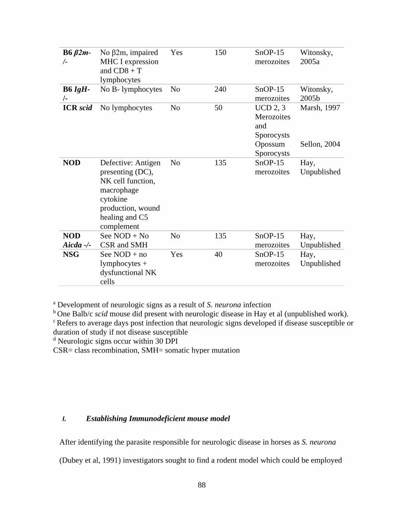
88
I. Establishing Immunodeficient mouse model
After identifying the parasite responsible for neurologic disease in horses as S. neurona
(Dubey et al, 1991) investigators sought to find a rodent model which could be employed
B6 β2m-
/-
No β2m, impaired
MHC I expression
and CD8 + T
lymphocytes
Yes 150 SnOP-15
merozoites
Witonsky,
2005a
B6 IgH-
/-
No B- lymphocytes No
240 SnOP-15
merozoites
Witonsky,
2005b
ICR scid
No lymphocytes No
50 UCD 2, 3
Merozoites
and
Sporocysts
Opossum
Sporocysts
Marsh, 1997
Sellon, 2004
NOD Defective: Antigen
presenting (DC),
NK cell function,
macrophage
cytokine
production, wound
healing and C5
complement
No
135 SnOP-15
merozoites
Hay,
Unpublished
NOD
Aicda -/-
See NOD + No
CSR and SMH
No
135 SnOP-15
merozoites
Hay,
Unpublished
NSG See NOD + no
lymphocytes +
dysfunctional NK
cells
Yes 40 SnOP-15
merozoites
Hay,
Unpublished
a Development of neurologic signs as a result of S. neurona infection b One Balb/c scid mouse did present with neurologic disease in Hay et al (unpublished work). c Refers to average days post infection that neurologic signs developed if disease susceptible or
duration of study if not disease susceptible d Neurologic signs occur within 30 DPI
CSR= class recombination, SMH= somatic hyper mutation
89
to investigate the immunopathogenesis of S. neurona infection and potential treatment
options. Marsh et al (1997) was of the first investigators to infect immunodeficient mice
with culture derived S. neurona merozoites from the UCD 2 and 3 S. neurona strains.
Three immunodeficient mouse models were utilized in this investigation, C57Bl/6
Foxn1nu Nu/J (B6 nude) which lack T-lymphocytes, ICR- Prkdcscid (ICR scid) and
immunocompetent C57Bl/6 (B6) mice which received methylprednisolone acetate for
immunosuppression. The ICR scid mice are lymphocyte deficient due to the protein
kinase DNA activated catalytic polypeptide (Prkdc) mutation, which disrupts the
maturation processes in lymphocytes. The B6 nude mice developed neurologic disease on
average 50 days post intraperitoneal infection with 2x10 5 S. neurona merozoites derived
from the equine S. neurona isolate UCD3. Histological analysis of the brains collected
from these mice revealed encephalitis and presences of S. neurona and S. neurona was
successfully isolated from cultured brain homogenates of infected B6 nude mice. In
addition to the brain, other visceral tissue samples were collected from the B6 nude mice
and the only other site of inflammation was in the liver. The ICR scid and
immunosuppressed B6 mice did not develop neurologic disease following infection
(Marsh, 1997). In this study mice lacking solely T-lymphocytes but intriguingly not those
lacking both T and B-lymphocytes developed S. neurona induced encephalitis. This
perhaps suggests that B-lymphocytes play a role in the pathogenesis or as suggested by
Marsh et al, discrepancy in disease susceptibility may be a result of genetic differences.
Nonetheless, a rodent model for EPM had begun to be established. Following this
investigation Dubey and Lindsay (1998) infected B6 nude and Balb/c interferon gamma
gene knockout (Ifnγ -/-) mice with opossum derived S. neurona sporocysts and
90
merozoites. It was reported, both B6 nude and Balb/c Ifnγ-/- mice developed neurologic
abnormalities attributed to S. neurona induced encephalitis, confirming disease
susceptibility in B6 nude mice and proposing both another mouse model and the role of
IFNγ, in the protective immune response against S. neurona infection. The Balb/c Ifnγ -/-
mouse presented a more economical mouse model to study EPM which also developed
more severe S. neurona induced encephalitis compared to B6 nude mice and thus became
the commonly used rodent model for studying EPM-like disease. The discovery of
disease susceptibility in the Balb/ c Ifnγ-/- or C57BL/6 Ifnγ-/- mice, paved the way for
rodent model investigations of S. neurona biology, treatments and immunopathogenesis.
The repeatable and quick onset of neurologic disease in the Ifnγ -/- mouse allowed it to be
used as a positive control mouse to ensure pathogenicity of S. neurona sporocysts or
merozoites of different isolates in the mouse model. They were also utilized as positive
controls in investigations which assessed disease susceptibility in other genetically
engineered, immune compromised mice.
II. The role of lymphocytes and MHC haplotype in the S. neurona disease
susceptibility
In addition to Marsh et al (1997) findings of ICR scid mice being resistant to S.
neurona induced encephalitis, Sellon et al (2004) also reported disease resistance in the
ICR scid mouse unless IFNγ was depleted (see section: V). An unpublished (Ahlgrim et
al) investigation reported when B6. CB17- Prkdc scid /SzJ (B6 scid) and CBySmn. CB17 -
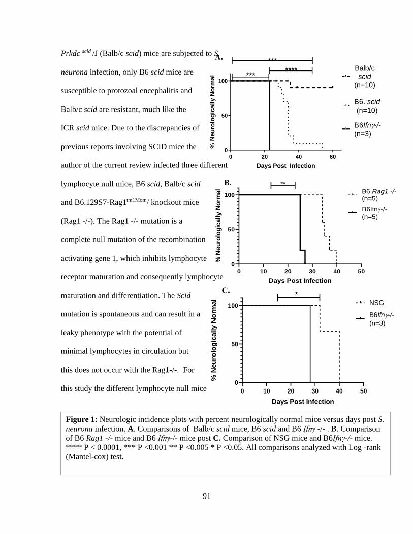
91
Prkdc scid /J (Balb/c scid) mice are subjected to S.
neurona infection, only B6 scid mice are
susceptible to protozoal encephalitis and
Balb/c scid are resistant, much like the
ICR scid mice. Due to the discrepancies of
previous reports involving SCID mice the
author of the current review infected three different
lymphocyte null mice, B6 scid, Balb/c scid
and B6.129S7-Rag1tm1Mom/ knockout mice
(Rag1 -/-). The Rag1 -/- mutation is a
complete null mutation of the recombination
activating gene 1, which inhibits lymphocyte
receptor maturation and consequently lymphocyte
maturation and differentiation. The Scid
mutation is spontaneous and can result in a
leaky phenotype with the potential of
minimal lymphocytes in circulation but
this does not occur with the Rag1-/-. For
this study the different lymphocyte null mice
Figure 1: Neurologic incidence plots with percent neurologically normal mice versus days post S.
neurona infection. A. Comparisons of Balb/c scid mice, B6 scid and B6 Ifnγ -/- . B. Comparison
of B6 Rag1 -/- mice and B6 Ifnγ-/- mice post C. Comparison of NSG mice and B6Ifnγ-/- mice.
**** P < 0.0001, *** P <0.001 ** P <0.005 * P <0.05. All comparisons analyzed with Log -rank
(Mantel-cox) test.
0 20 40 600
50
100
Days Post Infection
% N
eu
rolo
gic
ally N
orm
al
Balb/cscid
(n=10)
B6. scid(n=10)
B6Ifn-/-(n=3)
*******
***
A.
0 10 20 30 40 50
0
50
100
Days Post Infection
% N
eu
rolo
gic
ally N
orm
al
B6Ifn-/-(n=5)
B6 Rag1 -/-(n=5)
**B.
0 10 20 30 40 50
0
50
100
Days Post Infection
% N
eu
rolo
gic
ally N
orm
al
NSG
B6Ifn-/-(n=3)
*C.
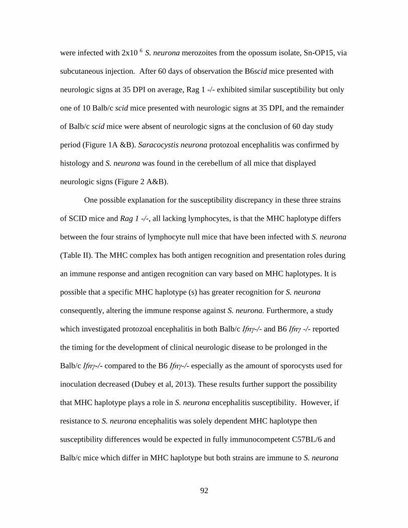
92
were infected with 2x10 6 S. neurona merozoites from the opossum isolate, Sn-OP15, via
subcutaneous injection. After 60 days of observation the B6scid mice presented with
neurologic signs at 35 DPI on average, Rag 1 -/- exhibited similar susceptibility but only
one of 10 Balb/c scid mice presented with neurologic signs at 35 DPI, and the remainder
of Balb/c scid mice were absent of neurologic signs at the conclusion of 60 day study
period (Figure 1A &B). Saracocystis neurona protozoal encephalitis was confirmed by
histology and S. neurona was found in the cerebellum of all mice that displayed
neurologic signs (Figure 2 A&B).
One possible explanation for the susceptibility discrepancy in these three strains
of SCID mice and Rag 1 -/-, all lacking lymphocytes, is that the MHC haplotype differs
between the four strains of lymphocyte null mice that have been infected with S. neurona
(Table II). The MHC complex has both antigen recognition and presentation roles during
an immune response and antigen recognition can vary based on MHC haplotypes. It is
possible that a specific MHC haplotype (s) has greater recognition for S. neurona
consequently, altering the immune response against S. neurona. Furthermore, a study
which investigated protozoal encephalitis in both Balb/c Ifnγ-/- and B6 Ifnγ -/- reported
the timing for the development of clinical neurologic disease to be prolonged in the
Balb/c Ifnγ-/- compared to the B6 Ifnγ-/- especially as the amount of sporocysts used for
inoculation decreased (Dubey et al, 2013). These results further support the possibility
that MHC haplotype plays a role in S. neurona encephalitis susceptibility. However, if
resistance to S. neurona encephalitis was solely dependent MHC haplotype then
susceptibility differences would be expected in fully immunocompetent C57BL/6 and
Balb/c mice which differ in MHC haplotype but both strains are immune to S. neurona
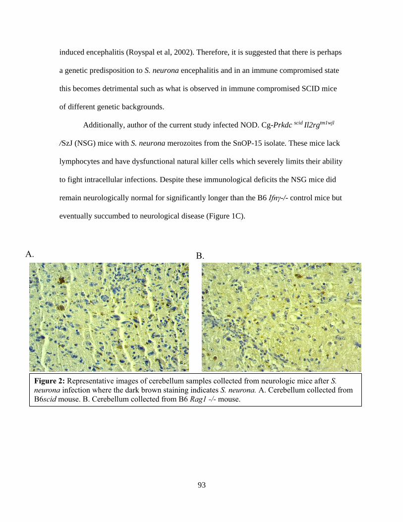
93
induced encephalitis (Royspal et al, 2002). Therefore, it is suggested that there is perhaps
a genetic predisposition to S. neurona encephalitis and in an immune compromised state
this becomes detrimental such as what is observed in immune compromised SCID mice
of different genetic backgrounds.
Additionally, author of the current study infected NOD. Cg-Prkdc scid Il2rgtm1wjl
/SzJ (NSG) mice with S. neurona merozoites from the SnOP-15 isolate. These mice lack
lymphocytes and have dysfunctional natural killer cells which severely limits their ability
to fight intracellular infections. Despite these immunological deficits the NSG mice did
remain neurologically normal for significantly longer than the B6 Ifnγ-/- control mice but
eventually succumbed to neurological disease (Figure 1C).
Figure 2: Representative images of cerebellum samples collected from neurologic mice after S.
neurona infection where the dark brown staining indicates S. neurona. A. Cerebellum collected from
B6scid mouse. B. Cerebellum collected from B6 Rag1 -/- mouse.
A. B.

94
Mouse strain S. neurona Susceptibility MHC haplotype
Balb/c No H2d
Balb/c Ifnγ -/- Yes H2d
Balb/c scid No H2d
C57Bl/6 (B6) No H2b
B6 Ifnγ-/- Yes H2b
B6 nude Yes H2b
B6 scid Yes H2b
B6 Rag 1 -/- Yes H2b
B6 iNos -/- No
H2b
B6eNos -/- No H2b
B6 β2m-/- Yes H2b
B6 IgH-/- No
H2b
ICR scid
No
H2g7
NOD No
H2g7
NOD Aida -/- No
H2g7
NSG Yes H2g7
III. The role of T lymphocytes in S. neurona infection
Marsh et al (1997) was the first to highlight the importance of T lymphocytes in the
protective immune response against S. neurona infection by demonstrating encephalitis
in B6 nude mice. This was further confirmed by Dubey and Lindsay (1998) and later on
Witonsky et al (2005a) exemplified the more specific role of CD8+ T-lymphocytes in the
protective cell mediated immune response. The B6.129P2- β2m tm1/unc knockout mouse
(B6 β2m-/-), which lack CD8+ T lymphocytes succumbed to protozoal encephalitis after
infection with 1x10 7 S. neurona merozoites derived from the SnOP-15 isolate (Witonsky
Table II: MHC haplotype of investigated mouse models

95
et al, 2005a). Authors of the current review sought to investigate the role of T-
lymphocyte derived IFNγ in fulfillment of neurologic disease, by adoptively transferring
splenic T-lymphocytes from wild type C57BL/6 (B6) mice and B6 Ifnγ -/- , into disease
susceptible B6 scid mice. After infection with 2x10⁶ S. neurona merozoites derived from
the SnOP-15 opossum isolate, neurologic signs occurred on average 25 DPI, in 100% of
mice which received B6Ifnγ -/- T-lymphocytes and positive control B6Ifnγ -/- mice.
While the group of B6scid mice which received B6 T- lymphocytes remained
neurologically normal until the conclusion of the study at 37 DPI (Figure 3A). After
histological analysis of cerebellums, S. neurona protozoal encephalitis was confirmed in
B6Ifnγ -/- T-lymphocytes and positive control B6Ifnγ -/- mice and both encephalitis and
S. neurona were absent in B6 T- lymphocytes recipient mice (Figure 3B). These results
imply a need for IFNγ producing T lymphocytes in the protective cell mediated immune
response against S. neurona infection in the B6 scid mouse, highlight the importance of
both T lymphocytes and IFNγ. The discussed studies establish importance of T-
lymphocytes and IFNγ in conferring immunity against S. neurona infection. The role of
B lymphocytes in immunopathogenesis has also been investigated.

96
IV. The role of B lymphocytes in S. neurona infection
Activated B lymphocytes generate antibodies against the pathogen responsible for
initiating an immune response and subsequent B lymphocyte activation. It is known that
both EPM resistant and susceptible horses generate antibodies against S. neurona after
exposure and to provide insight on whether antibodies are necessary for disease
protection or just a consequence of the activated immune response, B- lymphocyte
deficient mice (B6.129S2-Igh-6tmlcgn/J) were infected with 1x10⁷ S. neurona
merozoites from the SnOP-15 isolate (Witonsky et al, 2005b). After as long as 240 days
post infection, the infected B lymphocyte deficient mice did not develop neurologic
disease. This suggest that antibodies are not an essential component of the protective
immune response (Witonsky et al, 2005b). The author of the current review more
specifically assessed the importance of the role of antibody affinity and antibody isotype
in the immune response aganist S. neurona encephalitis. The NOD/ShiLt-Aicdaem1Cml
0 10 20 30 40
0
50
100
Days Post Infection
% N
eu
rolo
gic
ally N
orm
al TIFN
(n=3)
TIFN-/-
(n=7)
B6Ifn -/-
(n=3)
**
**
200um
A. B.
Figure 3: A. Neurologic incidence plot with percent neurologically normal mice versus days
post S. neurona infection. B. Representative IHC image of cerebellum collected from recipient
of non-IFNγ producing T lymphocytes. Dark brown staining is S. neurona.
97
/DvsJ (NOD Aicda -/- ) mouse was infected with 2x10 6 S. neurona merozoites from the
SnOP-15 isolate. The knockout of the gene Aicda (activation-induced cytidine
deaminase) disrupts the processes of class switch recombination and somatic hyper
mutation, which are necessary for generation of antibody isotypes other than IgM and
antibody affinity maturation, respectively. The NOD Aicda -/- mice were observed for
165 DPI and no neurologic abnormalities presented in these mice nor were any
abnormalities observed in NOD mice without the Aicda deletion, but positive control B6
Ifnγ-/- did develop encephalitis (Figure 4). The results of this study confer with Witonsky
et al (2005b) that the humoral immune response doesn’t play an essential role in the
protective immune response against S. neurona in naïve mouse models with impaired
humoral immunity. However, different antibody isotypes may play a role in monitoring
disease progression after S. neurona inoculation and/or antiprotozoal treatment as
indicated by Hay et al (2019) and therefore the importance of S. neurona antibodies
should not be undermined. As previously mentioned, encephalitis can be readily induced
in the Balb/c Ifnγ -/- or B6 Ifnγ -/- mice but the precise mechanism(s) that IFNγ acts upon
to infer protection is unknown but what has been investigated on this topic is discussed
below.

98
0 50 100 150 200
0
50
100
Days Post Infection
% N
eu
rolo
gic
ally N
orm
al
NOD(n=5)
NOD Aicda -/-(n=10)
B6Ifn-/-(n=3)
******
V. Investigating the role of interferon gamma in S. neurona encephalitis
The pleiotropic immune mediator, IFNγ, protects against intracellular infections such as
the parasitic pathogen, S. neurona. This interferon is produced by both innate immune
cells such as natural killer cells and dendritic cells and also the adaptive immune cells,
cytotoxic killer T-lymphocytes (CD8+ T- lymphocytes) and by the TH1 subset of T-
helper lymphocytes. The cytokine increases MHC expression for antigen presentation,
increases the cytotoxic nature of CD8+ T-lymphocytes and skews the phenotype of T
helper lymphocytes to the pro-inflammatory TH 1 phenotype, all of which are needed to
fight an intracellular infection. To delve further into the role of IFNγ in disease protection
investigators infected a variety of mice with genetic mutations relating to IFNγ
production or effects. The immune mediator inducible nitric oxide synthetase (iNOS) is
induced by cytokines such as IFNγ and constitutively expressed endothelial nitric oxide
synthetase (eNOS) function is upregulated by IFNγ. Likewise, it has been reported that
Figure 4: Neurologic incidence plot with percent neurologically normal mice versus days
post S. neurona infection. *** P <.0001. All comparisons analyzed with Log -rank
(Mantel-cox) test.
99
NOS plays a role in the protective immune response against other parasitic infections
such as Toxoplasma gondii which can lead to cerebral encephalitis in
immunocompromised individuals (Schluter et al, 1999). This would suggest that NOS
may also play a role in resolving S. neurona infection but Rosypal et al (2002), reported
that both the C57BL/6 iNOS knockout mouse and C57BL/6 eNOS knockout mouse were
resistant to neurologic disease induced by oral inoculation of 4x10⁴ S. neurona sporocysts
from the Sn-OP15 isolate. The B6 iNOS and eNOS knockout mice were observed for 64
DPI and while the positive control Balb/c Ifnγ-/- mice succumbed to S. neurona induced
encephalitis, both NOS knockout strains of mice did not (Rosypal, 2002). While NOS is
IFNγ mediated and offers protection during other intracellular infections it seems that it is
not crucial for conferring immunity against S. neurona infection and encephalitis. In
addition to mediating iNOS function during infection, IFNγ increases the expression of
MHC complex and consequently antigen presentation.
The β2m molecule is one of the two main protein chains that compose the MHC I
molecule and without β2m MHC I is dysfunctional and consequently a variety of
immunological functions are as well. One of those immunological functions is CD8+ T
lymphocytes which are predominant producers of IFNγ and depend on MHC I expression
for full maturation and are therefore deficient in β2m null mice. As mentioned, Witonsky
et al (2005a) investigated disease susceptibility in B6 β2m -/- mice and reported that after
infection with S. neurona merozoites B6 β2m -/- mice developed protozoal encephalitis.
Evolvement of neurologic disease as a result of S. neurona infection occurred 150-180
DPI in the β2m -/- mice, which was notably slower than positive control B6 Ifnγ-/- mice,
who became neurologic by 30 DPI. The results of this study insightfully demonstrate the

100
necessity for expression of the β2m molecule for complete resolvent of S. neurona
infection. However, if the interplay of IFNγ and CD8+ T-lymphocytes was the sole
component to the protective immune response against S. neurona infection, it would be
expected that neurologic disease would have occurred at a rate that more closely
paralleled the B6 Ifnγ-/- positive control mice. Lastly the literature reports that ICR scid
mice are not susceptible to S. neurona infection and depletion of IFNγ producing natural
killer cells does not warrant encephalitis either but when IFNγ is neutralized in vivo the
ICR scid mouse succumbs to protozoal encephalitis after infection with S. neurona
sporocyst (Sellon et al, 2004). In the context of Sellon et al (2004) study, these results
indicate that innate immune cells other than natural killer cells can produce enough IFNγ
to control S. neurona infection. The exact mechanism by which IFNγ acts upon the
immune system to generate a protective response and eliminate S. neurona infection
remains to be fully elucidated. The literature strongly suggests that the interaction of
IFNγ, MHC expression and T lymphocyte mediated immunity is involved in the
protective immune response, at least in the EPM mouse model.
Conclusions/Discussion
While beneficial studies have been conducted over the past decades there remains a gap
in the knowledge of the immunopathogenesis of EPM. The results of the studies reviewed
here indicated that the IFNγ driven immune response seems be of utmost importance to
prevent onset of clinical neurologic disease in S. neurona infected mice. Also, study
results allude to a strong potential of genetic predisposition since immune compromised
mice with certain MHC haplotypes seem to be disease resistant after S. neurona
infection. Future mouse model studies could include S. neurona infection of NOD scid

101
mice which have the same MHC haplotype as disease resistant ICR scid mice. If disease
resistance in the lymphocyte null mouse was dependent on MHC haplotype then
presumably the NOD scid would be resistant as well. Also, a B6 scid mouse with the
Balb/c MHC haplotype (H2d) could be infected with S. neurona to further investigate the
MHC haplotype role in immunopathogenesis. The disease resistant mouse models
discussed in the current review display no histological evidence of S. neurona within the
CNS or observable neurologic signs. This implies that either the immune response
supported elimination of the parasite prior to neural invasion or the immune response
within the CNS was able to eliminate the parasite without causing extensive pathological
damage. It has been demonstrated in T. gondii mouse models that IFNγ production by
microglia cells, resident CNS immune cells, is essential for containment of latent T.
gondii infection (Sa et al, 2015). Future studies utilizing mice with specific mutations that
effect only IFNγ production in the CNS would help determine if disease protection is a
result of the integrated role of peripheral immune response and the CNS immune
response or not. There are many different S. neurona isolates which can differ
genetically, potentially altering pathogenicity (Asmundsson et al, 2006), and sporocyst
viability and merozoite culture passage number can also potentially impact virulence
(Cheadle et al, 2001). The studies reviewed here utilized Ifny-/- mice as positive controls
to ensure S. neurona virulence but whether these factors impact disease outcome in other
immune compromised mouse strains, is unknown and this should be considered in future
studies. Likewise, it has been shown that either oral or subcutaneous inoculation of
opossum derived sporocysts or subcutaneous inoculation of merozoites results in
encephalitis in B6Ifny -/- mice (Dubey et al, 2001). Oral inoculation of sporocysts most
102
closely resembles infection route in the horse and both sporocysts and merozoite
infection results in similar disease outcome in the mouse, but inoculation method should
be considered when interoperating results and translating them to the horse. Overall,
mouse models have helped narrow the gap in the knowledge of EPM
immunopathogenesis, but further studies are still warranted.
References
Asmundsson I.M., Dubey J.P., Rosenthal B.M., 2006. A genetically diverse but distinct North
American population of Saracocystis neurona includes an overrepresented clone
described by 12 microsatellite alleles. Infection Genetic Evolution. 6;352–360.
Cheadle, M. A., Tanhauser, S. M., Scase, T. J., Dame, J. B., Mackay, R. J., Ginn, P. E., &
Greiner, E. C. 2001. Viability of Saracocystis neurona sporocysts and dose titration in
gamma-interferon knockout mice. Veterinary Parasitology, 95; 223–231.
Dubey, J.P., Davis, S.W., Speer, C.A., Bowman, D.D., de Lahunta, A., Granstrom, D.E., Topper,
M.J., Hamir, A.N., Cummings, J.F. and Suter, M.M., 1991. Saracocystis neurona n. sp.
(Protozoa:Apicomplexa); the etiologic agent of equine protozoal myeloencephalitis.
Journal of Parasitology. 77: 212-218.
Dubey, J. P., & Lindsay, D. S. 1998. Isolation in immunodeficient mice of Saracocystis neurona
from opossum (Didelphis virginiana) faeces, and its differentiation from Sarcocystis
falcatula. International Journal for Parasitology.

103
Dubey, J.P., Lindsay, D.S., Kwok, O.C., Shen, S.K. 2001. The gamma interferon knockout
mouse model for Saracocystis neurona: comparison of infectivity of sporocysts and
merozoites and routes of inoculation. Journal of Parasitology. 87; 1171-1173.
Dubey, J. P., Sundar, N., Kwok, O. C. H., & Saville, W. J. A. 2013. Saracocystis neurona
infection in gamma interferon gene knockout (KO) mice: Comparative infectivity of
sporocysts in two strains of KO mice, effect of trypsin digestion on merozoite viability,
and infectivity of bradyzoites to KO mice and cell culture. Veterinary Parasitology, 196;
212–215.
Hay, A.N. Witonsky, S.G., Lindsay, D.S., LeRoith, T. Zhu, J., Kasmark, L., Leeth C.M. 2019.
Saracocystis neurona- Induced Myeloencephalitis Relapse Following Anticoccidial
Treatment. Journal of Parasitology. 105: 371-378.
Marsh, A. E., Barr, B. C., Lakritz, J., Nordhausen, R., Madigan, J. E., & Conrad, P. A. 1997.
Experimental infection of nude mice as a model for Saracocystis neurona- associated
encephalitis. Parasitology Research, 83; 706–711.
NAHMS (National Animal Health Monitoring System). 2001. Equine protozoal
myeloencephalitis (EPM) in the U.S. Centers for Epidemiology and Animal Health –
USDA APHIS, Fort Collins, Colorado, USA.
Rosypal, A. C., Lindsay, D. S., Duncan, R., Ahmed, S. A., Zajac, A. M., & Dubey, J. P. 2002.
Mice lacking the gene for inducible or endothelial nitric oxide are resistant to sporocyst
induced Saracocystis neurona infections. Veterinary Parasitology. 103; 315–321.
104
Schluter, D., Deckert-Schluter, M., Lorenz, E., Meyer, T., Rollinghoff, M., Bogdan, C., 1999.
Inhibition of inducible nitric oxide synthetase exacerbates chronic cerebral toxoplasmosis
in Toxoxplasma gondii- susceptible C57BL/6 mice but does not reactive the latent
disease in T. gondii-resistant BALB/c mice. Journal of Immunology. 162, 3512-3518.
Sellon, D. C., Knowles, D. P., Greiner, E. C., Long, M. T., Melissa, T., Hochstatter, T., Damet, J.
B. 2004. Depletion of Natural Killer Cells Does Not Result in Neurologic Disease Due to
Saracocystis neurona in Mice with Severe Combined Immunodeficiency. Journal of
Parasitology. 90; 782–788.
Witonsky, S.G., Gongal, R.M., Duncan, R.B., Lindsay, D.S. 2003a. Protective immune response
to experimental infection with Saracocystis neurona in C57Bl/6 mice. Journal of
Parasitology. 89; 924-931.
Witonsky, S. G., Gogal Jr., R. M., Duncan, R. B., & Lindsay, D. S. 2003b. Immunopathologic
Effects Associated With Saracocystis neurona–Infected Interferon-Gamma Knockout Mice.
Journal of Parasitology. 89; 932–940.
Witonsky, S. G., Gogal, R. M., Duncan, R. B., Norton, H., Ward, D., & Lindsay, D. S. 2005a.
Prevention of meningo / encephalomyelitis due to Saracocystis neurona infection in mice
is mediated by CD8 cells. Journal of Parasitology. 35; 113–123.
Witonsky, S. G., Gogal, R.M., Duncan, R.B., Norton, H., Ward, D., Y, Jibing and Lindsay, D. S.
2005b. Humoral Immunity Is Not Critical for Protection against Experimental Infection
with Saracocystis neurona in B-Cell-Deficient Mice. Journal of Parasitology. 91; 830–
837.

105
Chapter VI: Interferon gamma producing T- lymphocytes help prevent Saracocystis
neurona induced myeloencephalitis in mouse model
Abstract
Equine protozoal myeloencephalitis (EPM), is predominantly caused by the prozotoa
Saracocystis neurona and is a prevalent neurologic disease affecting horses in North
America. Mouse models simulating EPM are commonly used to study the disease due to
numerous challenges associated with experimentally inducing EPM in horses. In the
current study, the C57Bl/6 scid mouse was found to be susceptible to S. neruona
encephalitis but the transfer of interferon gamma producing T lymphocytes into B6scid
mice offered protection against encephalitis. The transfer of IFNγ deficient T
lymphocytes did not offer protection and recipient mice succumbed to disease after
infection with S. neruona. The results of this study exemplify the need for IFNγ
production by T lymphocytes for T lymphocytes to offer protection against S. neurona
encephalitis.
Introduction
Equine Protozoal Myeloencephalitis (EPM), predominantly caused by the
protozoa Saracocystis neurona (Dubey et al, 1991) is a widespread, neurologic disease of
horses in the United States. Horses ingest S. neurona sporocyst when feed and water
sources become contaminated by opossum feces, which harbor infective life stages of the
parasite. The presences of S. neurona specific antibodies in equine sera indicates
exposure to S. neurona and the incidence of exposure is far greater than clinical disease
incidence. Sera antibody prevalence and clinical disease development are not directly
correlated, and it’s estimated that only 1% of exposed horses develop clinical neurologic
disease (Reed et al, 2016). The mechanism defining the immune response which protects

106
most horses exposed to S. neurona from developing neurologic disease remains to be
fully elucidated. Investigations have been in both experimentally and naturally affected
horses to help define the immunopathogenesis of EPM (Saville et al, 2001, Ellison et al,
2004, Sellon et al, 2004a, Spencer, 2005, Yang et al, 2006, Witonsky et al, 2008, Lewis,
2014). However, it is difficult to use the horses as model to study EPM due to
physiological and genetic differences amongst individual horses, ethical concerns and
expenses related to using the horse as an experimental and therefore mouse models are
more frequently used.
Mouse model studies have highlighted the importance of the cytokine interferon
gamma (IFNγ) and the T-lymphocyte mediated immune response for protection against S.
neuonra infection. T-lymphocytes of the TH1 and cytotoxic (CTL) phenotypes are
predominant producers of IFNγ, along with innate immune cells such as natural killer
cells and macrophages. Initial mouse model studies utilized immune compromised mice
to investigate immunopathogenesis in S. neurona induced encephalitis in the mouse
model. The C57Bl/6 Foxn1nu Nu/J (B6 nude) mice which lack T-lymphocytes (Marsh et
al, 1997), interferon gamma knockout (Ifnγ -/-) mice (Dubey and Lindsay 1998, Dubey
2001, Witonsky et al, 2003a, Hay et al, 2019) and C57Bl/6. β2m knockout mice which
lack MHC I expression and consequently, functional CD8+ T-lymphocytes (Witonsky et
al, 2005a) were utilized to demonstrate the importance of T lymphocytes and cytokine
IFNγ in protection against S. neurona induced encephalitis. Upon infection with S.
neurona all previoudly mentioned mouse strains demonstrate pathological evidence of S.
neurona induced encephalitis and apparent neurologic signs. Mice with T-lymphocytes
but lacking functional B-lymphocytes (mµMT -/-) are disease resistant (Witonsky et al,
107
2005b). Interestingly, discrepancies exist in previous reports of the incidence of
neurologic disease in severe combined immunodeficiency (SCID) mice. The SCID mice
are lymphocyte deficient due to the protein kinase DNA activated catalytic polypeptide
(Prkdc) mutation, which disrupts the maturation processes in lymphocytes. The SCID
mouse has an intact and functional innate immune system and produces some IFNγ but
lack functional T and B lymphocytes. Therefore, the potential protective role of T-
lymphocytes and associated IFNγ production is also lacking in the SCID mouse. Marsh et
al (1997) reported that ICR- Prkdcscid (ICR scid) mice do not develop neurologic disease
associated with S. neruona infection but Ahlgrim et al (unpublished work) reported that
disease occurs in B6. CB17- Prkdc scid /SzJ (B6 scid) and CBySmn. CB17 - Prkdc scid /J
(Balb/c scid) are disease resistant. These discrepancies potentially relate to genetic
differences in the different mouse strains and IFNγ production as splenocytes collected
from Balb/c mice have been reported to have increased IFNγ production in-vitro (Koo et
al, 2006). Additionally, Sellon et al (2004b) reported that depletion of IFNγ results in
neurologic disease in S. neurona infected ICR scid mice further supporting the role of
this cytokine in disease prevention.
These conflicting results suggest that SCID mice may be susceptible to disease
under certain circumstances and therefore further investigation is warranted as
characterizing these differences may yield valuable information. In the current study we
determined the neurologic disease susceptibility related to S. neurona infection in
lymphocyte null B6scid and Balb/c scid mice. Furthermore, we sought out to further
define the role of T-lymphocytes and T-lymphocyte derived IFNγ in protection against S.
108
neurona induced encephalitis through adoptive transfer of IFNγ competent of deficient T-
lymphocytes.
Materials and Methods
Mice
Male mice age 8-10 weeks old of the following strains; B6. 129S7- Ifngtm1Ts/J (B6.Ifnγ -
/-) (JR 2287), B6.CB17-Prkdc scid /SzJ (B6.scid) (JR1913), CBySmn.CB17-Prkdc scid / J
(Balb/c scid) (JR1803) and C57Bl/6 (B6) (JR 664) were obtained from Jackson
Laboratory (Bar Harbor, Maine). The B6.Ifnγ -/- mice were maintained in accordance
with an Institutional Animal Care and Use Committee (IACUC) breeding protocol and
for these mice and all others mentioned, experimental studies were conducted under other
IACUC approved protocols.
For the neurologic disease susceptibility experiments in lymphocyte null mice,
Balb/c scid (n=10) and B6. scid (n=10) mice (Table I), were infected with 2 x 10 6 S.
neurona merozoites via subcutaneous injection on Day 0. Blood was collected via
submandibular venipuncture from a cohort of both Balb/c. scid (n=5) and B6. scid (n=5)
mice on Day 0 before S. neurona infection and then on a weekly basis for 3 weeks; 7
days post infection (DPI), 14 DPI and 21 DPI and upon the development of neurologic
signs or 39 DPI when mice were euthanized and blood was collected post euthanasia via
cardiac puncture. The mice used for weekly blood samples were ear notched for
identification. Blood was collected from the remainder of the mice post euthanasia which
occurred upon the development of neurologic signs or 60 DPI via cardiac puncture (B6.
scid n=5) or (Balb/c scid n=5). Sera was collected after centrifugation and stored at -20 C
for further analysis. B6. Ifnγ -/- mice (n=5) were used as positive controls to test S.
neurona virulence. All mice were euthanized using CO 2 asphyxiation and development

109
of neurologic disease was characterized by clinical neurological signs relating to lack of
balance and coordination. At the time of euthanasia cerebellum samples were collected
for histological analysis.
For the T-lymphocyte transfer experiments the B6 and B6 Ifnγ -/- mice intended
for the use as T-lymphocyte donors, were euthanized and spleens were collected for cell
isolation. For the transfer experiments 3x10 6 T-lymphocytes were intraperitoneally
injected into B6. scid recipient mice (n=10). The mice were divided into two recipient
groups; those which received IFNγ producing T-lymphocytes, TIFNγ (n=3) and those
which received IFNγ deficient T-lymphocytes, T IFNγ -/- (n=7) (Table II). Blood was
collected via submandibular venipuncture from all these mice 3 weeks post T-
lymphocyte transfer for analysis of circulating T-lymphocytes via flow cytometry. At 6
weeks post cell transfer mice were infected with 2x10 6 S. neurona merozoites via
subcutaneous injection. All mice were euthanized using CO 2 asphyxiation when clinical
neurological signs relating to lack of balance and coordination were observed or at 37
DPI. Post euthanasia blood was collected via cardiac puncture for flow cytometry
analysis of circulating T-lymphocytes and sera analysis for IFNγ, spleens were collected
for further immune cell analysis via flow cytometry and cerebellum samples were
collected for histological analysis.
Saracocystis neurona culture and preparation of inoculum
Merozoites of the sixth passage of the SN-15OP isolate were grown in African green
monkey (Cercopithecus aethiops) kidney cells (CV-1, ATTC CCL-70, Manassas, VA,
USA), separated from host cells, collected and enumerated for inoculations as previously
described (Lindsay et al, 2013). The SN-15OP isolate was isolated from the brain of an

110
Balb/c Ifnγ -/- mouse fed sporocysts obtained from the feces of a naturally infected
opossum from Virginia (Dubey, 2000). This isolate was originally a gift from Dr. J. P.
Dubey, United States Department of Agriculture, Beltsville, Maryland USA and has been
maintained cryopreserved in liquid nitrogen.
Cerebellum histological analysis
For the neurologic disease susceptibility trial there were five cerebellar samples collected
post euthanasia from each the Balb/c scid and B6scid groups and two from the positive
control B6Ifnγ-/- mice (Table I). For the T-lymphocyte transfer trial all cerebellums were
collected from all transfer recipient mice (Table II). All collected cerebellums were fixed
in 10% formalin, embedded in paraffin sectioned for hematoxylin and eosin (H&E) and
immunohistochemistry (IHC) staining. The H&E stained samples were assessed for total
number of perivascular cuffs, gliosis foci and IHC for number of S. neurona organisms.
For each assessed parameter a score was assigned 1 (nothing present) to 8 (greatest
number of cuffs, foci or organism). Sections of cerebellum were stained with polyclonal
Mouse
Strain
Weekly
Blood
samples
Neurologic
Signs
IHC
Positive
Encephalitis
B6. Scid
n=10
Yes
5/10
Yes
10/10
Positive
5/5
Yes
5/5
Balb/c. scid
n=10
Yes
5/10
Yes 1/10a
Positive
1/5b
Yes
1/5c
B6 Ifnγ -/-
No Yes
5/5
Positive
2/2
Yes
2/2
a All other mice (n=8) were euthanized at 39 DPI (n=4) or 60 DPI (n=5) b All other mice (n=4) were negative for S. neurona staining c All other mice (n=4) did not show signs of encephalitis
Table I: Neurologic disease susceptibility
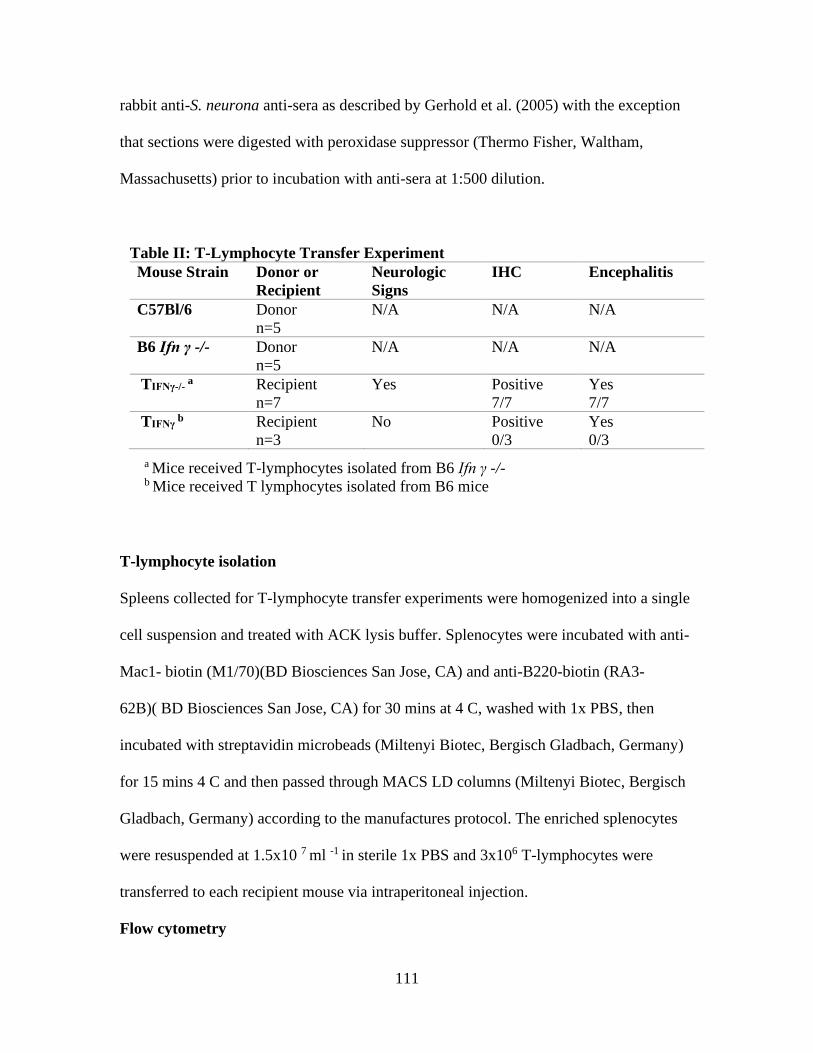
111
rabbit anti-S. neurona anti-sera as described by Gerhold et al. (2005) with the exception
that sections were digested with peroxidase suppressor (Thermo Fisher, Waltham,
Massachusetts) prior to incubation with anti-sera at 1:500 dilution.
T-lymphocyte isolation
Spleens collected for T-lymphocyte transfer experiments were homogenized into a single
cell suspension and treated with ACK lysis buffer. Splenocytes were incubated with anti-
Mac1- biotin (M1/70)(BD Biosciences San Jose, CA) and anti-B220-biotin (RA3-
62B)( BD Biosciences San Jose, CA) for 30 mins at 4 C, washed with 1x PBS, then
incubated with streptavidin microbeads (Miltenyi Biotec, Bergisch Gladbach, Germany)
for 15 mins 4 C and then passed through MACS LD columns (Miltenyi Biotec, Bergisch
Gladbach, Germany) according to the manufactures protocol. The enriched splenocytes
were resuspended at 1.5x10 7 ml -1 in sterile 1x PBS and 3x10⁶ T-lymphocytes were
transferred to each recipient mouse via intraperitoneal injection.
Flow cytometry
Table II: T-Lymphocyte Transfer Experiment
Mouse Strain Donor or
Recipient
Neurologic
Signs
IHC Encephalitis
C57Bl/6 Donor
n=5
N/A N/A N/A
B6 Ifn γ -/- Donor
n=5
N/A N/A N/A
TIFNγ-/- a Recipient
n=7
Yes Positive
7/7
Yes
7/7
TIFNγ b Recipient
n=3
No Positive
0/3
Yes
0/3
a Mice received T-lymphocytes isolated from B6 Ifn γ -/- b Mice received T lymphocytes isolated from B6 mice
112
For the T-lymphocyte transfer experiments all blood samples collected were treated with
sodium EDTA directly after collection. The collected spleen samples were homogenized
into a single cell suspension. Both blood and spleen samples were then treated with ACK
lysis buffer and then cells were washed twice in 1x PBS twice. Both peripheral blood
leukocytes (PBL) and splenocytes were counted on Nexcelom cell counter and
resuspended at 2x 10⁷ cells/mL. Cells were stained with fluorochrome conjugated mouse
antibodies against CD3 (145-2C11), CD4(GK15), CD8(53-6.7) and B220(RA3-6B2)
(Biolegend, SanDiego, CA) and incubated at 4 C for 30 mins. Directly before conducting
flow cytometry propidium iodide (PI) viability dye was added to each sample to
differentiate between live and dead cells. All samples were run on Attune NxT cytometer
(Thermo Fisher Scientific) with Attune NxT software v 2.4 (Life Technologies, Thermo
Fisher, Waltham, MA). Live Cells were gated for single cell populations and then for
lymphocytes using FSC vs SSC. All data analysis was completed utilizing FlowJo
software (FlowJo, LLC, Ashland, OR).
Serum Interferon gamma ELISA
Sera IFNγ concentrations were measured using standardized ELISA kit, Mouse IFNγ
ELISA kit (Invitrogen, ThermoFisher, Ref # 88-7314). The manufacturers protocol was
followed. Plates were read on an Infinite M200 Pro plate reader using Magellan 7.0
software (Tecan).
Statistical Analysis
For determining significance in the neurologic incidence data, a log-rank Mantel-Cox test
with a significant level set at P < 0.05 and Bonferroni correction to account for multiple
comparisons with a significance threshold at P <0.017. Significance for measured
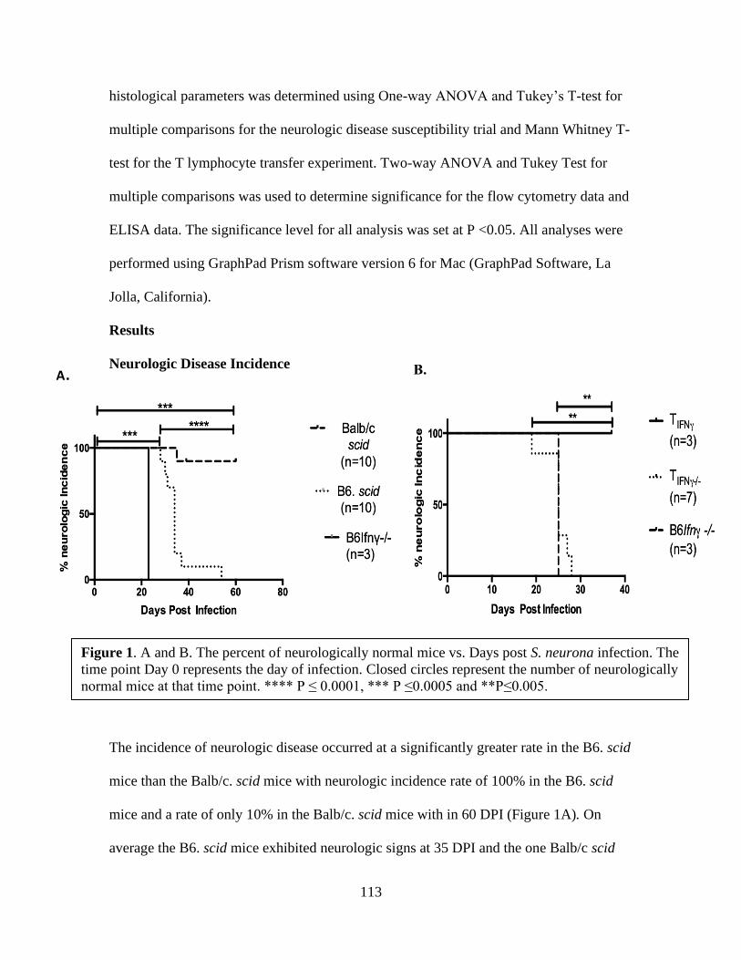
113
histological parameters was determined using One-way ANOVA and Tukey’s T-test for
multiple comparisons for the neurologic disease susceptibility trial and Mann Whitney T-
test for the T lymphocyte transfer experiment. Two-way ANOVA and Tukey Test for
multiple comparisons was used to determine significance for the flow cytometry data and
ELISA data. The significance level for all analysis was set at P <0.05. All analyses were
performed using GraphPad Prism software version 6 for Mac (GraphPad Software, La
Jolla, California).
Results
Neurologic Disease Incidence
The incidence of neurologic disease occurred at a significantly greater rate in the B6. scid
mice than the Balb/c. scid mice with neurologic incidence rate of 100% in the B6. scid
mice and a rate of only 10% in the Balb/c. scid mice with in 60 DPI (Figure 1A). On
average the B6. scid mice exhibited neurologic signs at 35 DPI and the one Balb/c scid
A. B.
Figure 1. A and B. The percent of neurologically normal mice vs. Days post S. neurona infection. The
time point Day 0 represents the day of infection. Closed circles represent the number of neurologically
normal mice at that time point. **** P ≤ 0.0001, *** P ≤0.0005 and **P≤0.005.

114
mouse demonstrated signs at 35 DPI. All positive control Ifnγ -/- mice developed
neurologic signs within 30 DPI (Figure 1A). The onset of neurologic signs relating to
lack of balance and coordination did occur in both strains of SCID mice but at a
significantly lower rate in the Balb/c scid mice, suggesting that these mice do mount an
immune response against S. neurona which is more protective than the response of the
B6 scid. Based on these results the B6. scid mouse was selected as the recipient mouse
for the T-lymphocyte transfer experiments conducted in the current study.
For the T-lymphocyte transfer experiment, there was a 100% neurologic incidence in the
group of mice that received IFNγ deficient T-lymphocytes (TIFNγ-/-) with an average rate
of 25 DPI. All positive control Ifnγ -/- mice displayed neurologic signs within 30 DPI.
The transfer of the IFNγ producing T-lymphocytes (TIFNγ) decreased the rate of
neurologic incidence, as all mice in this group did not develop neurologic signs by the
conclusion of the observation period at 37 DPI. The study was concluded at 37DPI
because this time period exceeded the average mortality rate (26 DPI) of B6 Ifnγ -/-
positive controls. These results indicate that the transfer of IFNγ producing T-
lymphocytes positively influenced the rate of neurologic incidence by decreasing the
development of neurologic signs. However, the transfer of the IFNγ deficient T-
lymphocytes did not (Figure 1B).
Histopathological analysis
For both the neurologic disease susceptibility (Table I) and T-lymphocyte transfer (Table
II) experiments the histology results confirmed S. neurona induced encephalitis in all
mice that displayed clinical neurologic signs and those that did not display clinical signs
115
did not develop encephalitis. Sarcocysitis neurona induced encephalitis was characterized
in the collected cerebellar samples by the presences of both gliosis and perivascular
cuffing and the finding of S. neurona by IHC staining. For the neurologic disease
susceptibility trial there was significantly more perivascular cuffing present in the B6 scid
mice than Balb/c scid mice and the B6. Ifnγ -/- had significantly greater amounts of
perivascular cuffing than both groups of SCID mice (Figure 3A). There was a
significantly greater amount of gliosis foci in the B6scid cerebellum samples compared to
the Balb/c scid and the positive control cohort, B6 Ifnγ -/- , had significantly greater
number of gliosis foci than both B6 scid and Balb/c scid mice (Figure 3B). Saracocystis
neurona was only found in one of the cerebellum samples collected from the Balb/c scid
mice and therefore there was significantly less S. neurona in this group compared to the
B6 scid and B6 Ifnγ-/- mice. The B6 Ifnγ-/- mice had largest number of S. neurona and
significantly more than mice in both SCID groups (Figure 3C).
The transfer of IFNγ producing T- lymphocytes did not significantly alter the presences
of perivascular cuffing between the two recipient groups (Figure 3D) and there was an
insignificant increase in the TIFNγ-/- group compared to the TIFNγ . There were no gliosis
foci identified in the TIFNγ group and therefore there was significantly greater number of
gliosis foci in the TIFNγ-/- group (Figure 3E). Likewise, no S. neurona was identified by
IHC in the TIFNγ group and therefore there was significantly greater amount of positive S.
neurona IHC staining in the TIFNγ-/- group (Figure 3F). The transfer of IFNγ producing T
lymphocytes resulted in improved pathology and no encephalitis compared to the transfer

116
of IFNγ deficient T lymphocytes highlighting the importance of the cytokine IFNγ not
just T-lymphocytes.
B. C.
D.
.E. F.
A.
Figure 3. Scores of the measured histological changes associated with S. neurona infection for both the
neurologic disease susceptibility trial (A-C) and the T-lymphocyte transfer trial (D-F). A,D. Perivascular
cuffing foci in H&E stained cerebellum sections. B,E. Gliosis Foci in H&E stained cerebellum sections.
C,F. Saracocystis neurona organisms in IHC stained cerebellum sections. Data is present as ± SEM. *
P≤ 0.05, **P ≤ 0.005, *** P ≤0.0005, **** P ≤ 0.0001.

117
Flow Cytometry
The peripheral blood samples collected at 3 weeks post T-lymphocyte transfer exhibited
CD3+ T-lymphocytes in circulation for both recipient groups with no significant
differences between the two groups (data not shown), confirming successful T-
lymphocyte transfer. Splenocytes and PBL samples collected from transfer mice post
euthanasia were analyzed for populations of CD4+ and CD8+ T- lymphocytes. In the
TIFNγ group there was a trending increase (p = 0.05) in CD8+ T-lymphocytes in the spleen
compared to the TIFNγ-/- group but no differences in the CD4+ T-lymphocyte population
(Figure 4A). The results from the PBL samples demonstrate a slight but insignificant
increase in both CD4+ and CD8 + T lymphocytes in circulation in the TIFNγ-/- group
compared to the TIFNγ group (Figure 4B). The increase in T-lymphocyte populations in
circulation in the TIFNγ-/- may be due to persistent S. neurona infection and a resolution of
infection in the TIFNγ group.
A. B.
Figure 4. Flow cytometry data analyzing differences in CD4+ T lymphocyte and CD8 + T
lymphocyte populations in splenic (A) and peripheral blood leukocytes (B). Spleen and blood
samples were collected at the time of neurologic sign development for TIFNγ -/- group and 37 DPI
for the TIFNγ group. No significant differences occurred but there was a trending increase (P= 0.05)
in splenic CD8+ T-lymphocyte population (A). Data is presented as ± SEM.
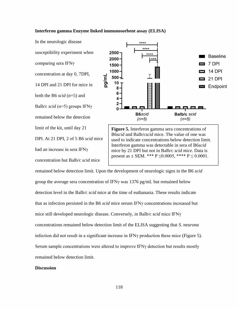
118
Interferon gamma Enzyme linked immunosorbent assay (ELISA)
In the neurologic disease
susceptibility experiment when
comparing sera IFNγ
concentration at day 0, 7DPI,
14 DPI and 21 DPI for mice in
both the B6 scid (n=5) and
Balb/c scid (n=5) groups IFNγ
remained below the detection
limit of the kit, until day 21
DPI. At 21 DPI, 2 of 5 B6 scid mice
had an increase in sera IFNγ
concentration but Balb/c scid mice
remained below detection limit. Upon the development of neurologic signs in the B6 scid
group the average sera concentration of IFNγ was 1376 pg/mL but remained below
detection level in the Balb/c scid mice at the time of euthanasia. These results indicate
that as infection persisted in the B6 scid mice serum IFNγ concentrations increased but
mice still developed neurologic disease. Conversely, in Balb/c scid mice IFNγ
concentrations remained below detection limit of the ELISA suggesting that S. neurona
infection did not result in a significant increase in IFNγ production these mice (Figure 5).
Serum sample concentrations were altered to improve IFNγ detection but results mostly
remained below detection limit.
Discussion
Figure 5. Interferon gamma sera concentrations of
B6scid and Balb/cscid mice. The value of one was
used to indicate concentrations below detection limit.
Interferon gamma was detectable in sera of B6scid
mice by 21 DPI but not in Balb/c scid mice. Data is
present as ± SEM. *** P ≤0.0005, **** P ≤ 0.0001.
119
Conversely to previous investigations our results from this study indicate that
when B6scid and Balb/c scid mice are infected with opossum derived SN-15OP S.
neurona merozoites both strains of mice develop neurologic disease but at altered rates.
The B6scid mice demonstrated fulminant neurologic disease with all mice demonstrating
neurologic signs on average 35 DPI and cerebellar histology associated with S. neurona
encephalitis. The B6scid mice developed neurologic signs at a significantly slower rate
than positive control B6Ifnγ-/- mice, which on average developed signs 23 DPI. Given
that B6scid mice still have a functional innate immune system and therefore some IFNγ
production it is likely that this helped control S. neurona infection and prolong
development of neurologic signs but was evidently not enough to entirely prevent
disease. The Balb/c scid mice were nearly disease resistant with only one mouse
demonstrating neurologic signs at 35 DPI and cerebellar histology associated with S.
neurona infection. The remaining Balb/c scid mice remained neurologically normal
throughout the study and presented with normal cerebellar pathology at 39 DPI or 60
DPI. Based on results from previous investigations we hypothesized that if disease
susceptibility differed between the B6 scid and Balb/c scid strains this was potentially
due to an increase production of innate immune cell derived IFNγ in response to S.
neurona infection. Sellon et al (2004) demonstrated that ICR scid mice are resistant to
disease when infected with S. neurona but when IFNγ was depleted in ICR scid mice
they developed neurologic disease. Koo et al (2006) demonstrated that there is a greater
production of predominantly natural killer cell derived IFNγ when Balb/c splenocytes
were cultured with Burkholderia pseudomallei compared to B6 splenocytes. However,
we were not able to detect an increase in circulating IFNγ concentrations in sera samples

120
collected weekly for three weeks post S. neurona infection in the Balb/c scid mice
compared to the B6scid mice. It is possible that a peak of IFNγ production occurred less
than 7DPI and the optimal sampling time point to distinguish significant differences
between the two groups was overlooked. Another possibility is that the differences in
IFNγ production were in the central nervous system influencing neurovirulence which is
the case in Toxoplasma gondii infection. Sa et al (2015) demonstrated the importance of
microglia derived IFNγ in controlling T. gondii growth and infection in Balb/c mice. It is
also possible that the differences in disease resistance is irrelevant to IFNγ production
and may be related to other genetic differences that exist between the two strains of mice.
Genetic differences may also explain discrepancies in previous reports as both Marsh et
al (1997) and Sellon et al (2004) conducted their studies with ICR scid mice and not
Balb/c scid or B6 scid. In the current study we demonstrated that there is a significant
difference in neurologic disease susceptibility related to S. neurona infection in Balb/c
scid and B6 scid mouse strains but the immune response protecting Balb/c scid mice
needs to be further investigated. While the current study was conducted in mice, it should
be mentioned that S. neurona infection and disease susceptibility studies have been
conducted in SCID equine foals.
When equine foals with the scid mutation were inoculated with S. neurona
sporocysts developed parasitemia and infection in visceral tissues (liver, spleen, skeletal
muscle, cardiac muscle, kidney, tongue and heart) but not clinical neurologic disease.
Immunocompetent control horses (4 of 6 horses) in this study did not demonstrate
parasitemia or infection of visceral tissues but did develop neurologic signs relating to S.
neurona infection by the end of the study period (Sellon et al, 2004). In the mentioned

121
study two of three SCID foals had to be euthanized 21-32 days post infection due to
illness unrelated to S. neurona infection resulting in small sample population. Authors of
the current study feel that if this study included more foals and a healthier a health status
could be maintained for a longer timeframe than SCID foals may succumb to disease
relating to S. neurona infection.
To further investigate the role of T-lymphocyte derived IFNγ in protection against
S. neurona induced encephalitis we selected the B6scid mice as adoptive transfer
recipients due to their increased disease susceptibility compared to Balb/c scid mice. We
found that the transfer of IFNγ producing T-lymphocytes exerted a protective effect on
neurologic disease after infection with S. neurona. The recipient group of IFNγ producing
T-lymphocytes did not exhibit neurologic abnormalities or characteristic cerebellar
lesions associated with S. neurona infection. However, 100% of recipients of Ifnγ-/- T-
lymphocytes demonstrated neurologic signs, pathological cerebellar lesions and S.
neurona was identified by immunohistochemistry like B6scid mice. Results from the
flow cytometry data collected in the current study demonstrate a significant increase in
splenic CD8+ T-lymphocytes from T IFNγ compared to the T IFNγ-/- but not CD4+ T-
lymphocytes. Previous studies have revealed the importance of CD8+ T-lymphocytes in
the protection against S. neurona induced encephalitis as this population of cells
increases after infection in immunocompetent C57Bl/6 mice (Witonsky, 2003b) and
without functional CD8+ T-lymphocytes mice are susceptible to S. neurona induced
myeloencephalitis (Witonsky et al, 2005a). While there was a significant increase in
splenic CD8+ T lymphocytes there was not a significant difference in splenic CD4+ T
lymphocyte population between the two groups. This suggest and supports previous

122
reports that CD8+ T-lymphocytes play a more essential role in the protective immune
response compared to CD4+ T-lymphocytes in S. neurona infection. Cell mediated
immune response specifically CD8+ T-lymphocytes and IFNγ is important for protection
against other equine pathogens such as equine herpesvirus-1 (Paillot et al, 2005) and
other intracellular protozoan infections such as T. gondii (Gigley, 2011). The results from
the current study exemplifies the need for T-lymphocytes to be capable of producing
IFNγ in order to be protective, and suggest a potential role of IFNγ deficient T-
lymphocytes in accelerating disease progression because the average rate of neurologic
sign development in the TIFNγ-/- group was considerably earlier than B6 scid mice. While
both B6scid and TIFNγ-/- groups of mice were infected with the same strain of S. neurona
and virulence was confirmed with positive control B6Ifnγ-/- mice, the infection time
points of these two groups of mice occurred at different timepoints and therefore to make
direct comparisons, further investigation is warranted. Collectively these results indicate
the importance of IFNγ producing T-lymphocytes in protection against disease
development but also that dysfunctional T- lymphocytes may play an essential role for
disease development. While this study was conducted with murine models it is likely that
if an immunomodulator that increase T-lymphocyte IFNγ production in horses could be
identified this would greatly improve prognosis for EPM affected patients. Studies
investigating the immunological properties of available immunomodulators for horses
have been conducted (Horohov, 2008, and Witonsky, 2019) but their effects on S.
neurona infection remain to be investigated and further investigation is warranted.
References

123
Dubey, J.P., Davis, S.W., Speer, C.A., Bowman, D.D., de Lahunta, A., Granstrom, D.E., Topper,
M.J., Hamir, A.N., Cummings, J.F. and Suter, M.M., 1991. Saracocystis neurona n. sp.
(Protozoa:Apicomplexa); the etiologic agent of equine protozoal myeloencephalitis.
Journal of Parasitology. 77; 212-218.
Dubey, J. P., & Lindsay, D. S. 1998. Isolation in immunodeficient mice of Saracocystis neurona
from opossum (Didelphis virginiana) faeces, and its differentiation from Sarcocystis
falcatula. International Journal for Parasitology. 28; 1823-1828.
Dubey, J. P., Saville, W. J., Lindsay, D. S., Stich, R. W., Stanek, J. F., Speert, C.A., Reed, S. M.
2000. Completion of the life cycle of Saracocystis neurona. The Journal of Parasitology,
86; 1276–1280.
Dubey, J.P., D. Fritz, D.S. Lindsay, S.K. Shen, O.C.H. Kwok, and K.C. Thompson. 2001.
Diclazuril preventive therapy of gamma interferon knockout mice fed Saracocystis
neurona sporocysts. Veterinary Parasitology 94: 257– 263.
Ellison, SP., Greiner, E., Brown, K.K., Kennedy, T. 2004. Experimental infection of horses with
culture-derived Saracocystis neurona merozoites as a model for equine protozoal
myeloencephalitis. International Journal of Applied Research in Veterinary Medicine. 2;
79-89.
Gerhold, R.W., E.W. Howerth, and D.S. Lindsay. 2005. Saracocystis neurona-associated
Meningoencephalitis and Description of Intramuscular Sarcocysts in a Fisher (Martes
pennanti). Journal of Wildlife Diseases. 41: 224-230.
Gigley, J. P., Bhadra, R., & Khan, I. A. 2011. CD8 T Cells and Toxoplasma gondii: A New
Paradigm. Journal of parasitology research, 2011; 243796.
124
Hay, A.N. Witonsky, S.G., Lindsay, D.S., LeRoith, T. Zhu, J., Kasmark, L., Leeth C.M. 2019.
Saracocystis neurona- Induced Myeloencephalitis Relapse Following Anticoccidial
Treatment. Journal of Parasitology. 105: 371-378.
Horohov, D. W., Breathnach, C. C., Sturgill, T. L., Rashid, C., Stiltner, J. L., Strong, D., …
Holland, R. E. 2008. In vitro and in vivo modulation of the equine immune response by
parapoxvirus ovis. Equine Veterinary Journal, 40; 468–472.
Koo, G. C., & Gan, Y. H. (2006). The innate interferon gamma response of BALB/c and
C57BL/6 mice to in vitro Burkholderia pseudomallei infection. BMC Immunology, 7.
Lewis, R.S. et al. 2014. Effects of Experimental Saracocystis neurona Induced infection on
immunity in an Equine Model. Journal of Vet Med.
Lindsay, D.S., M.M. Nazir, A. Maqbool, S.P. Ellison, and J.S. Strobl, 2013. Efficacy of
decoquinate against Saracocystis neurona in cell cultures. Veterinary Parasitology. 196:
21–23.
Marsh, A. E., Barr, B. C., Lakritz, J., Nordhausen, R., Madigan, J. E., Conrad, P. A. 1997.
Experimental infection of nude mice as a model for Saracocystis neurona- associated
encephalitis. Parasitology Research. 83; 706–711.
Paillot, R. Daly, J.M., Juillard, V., Minke, J.M., Hannant, D., Kydd, J.H., 2005. Equine
interferon gamma synthesis in lymphocytes after in vivo infection and in vitro stimulation
with EHV-1. Vaccine. 23; 4541-4551.

125
Reed, S. M., M. Furr, D. K. Howe, A. L. Johnson, R. J. MacKay, J. K. Morrow and S. Witonsky.
2016. Equine protozoal myeloencephalitis: An updated consensus statement with a focus
on parasite biology, diagnosis, treatment, and prevention. Journal of Veterinary Internal
Medicine 30: 491–502.
Saville, W.J., R.W. Stitch, S.M Reed, C.J Njoku, M.J Oglesebee, A Wunschmann, D. L Grover,
A.L Larew- Naugle, J. F Stanek, D.E Granstrom, J.P Dubey. 2001. Ulitization of stress in
the development of an equine model for equine protozoal myeloencephalitis. Veterinary
Parasitology 95: 211-222.
Sellon, D. et al. 2004a. Infection of Immunodeficient horses with Saracocystis neurona does not
result in neurologic disease. Clinical and Diagnostic Lab Immunology. 11(6) 1134-39.
Sellon, D. et al. 2004b. Depletion of Natural Killer Cells Does not result in neurologic disease
due to Saracocystis neurona in mice with severe combined immunodeficiency. Journal of
Parasitology. 90 (4); 782-788.
Spencer, J. Deinnocentes, P. Moyana, E., Guarino, A., Ellison, S., Bird, R., and Blagburn, B.
2005. Cytokine gene expression in response to SnSAG1 in horses with equine protozoal
myeloencephalitis. Clinical and Diagnostic laboratory immunology. 12; 644-646.
Witonsky, S. G., Gogal Jr., R. M., Duncan, R. B., & Lindsay, D. S. 2003a. Immunopathologic
Effects Associated With Saracocystis neurona–Infected Interferon-Gamma Knockout
Mice. Journal of Parasitology, 89; 932–940.
Witonsky, S.G., Gongal, R.M., Duncan, R.B., Lindsay, D.S. 2003b. Protective immune response
to experimental infection with Saracocystis neurona in C57Bl/6 mice. Journal of
Parasitology. 89; 924-931.

126
Witonsky, S.G. et al. 2005a. Prevention of meningo/encephalomyelitis due to Saracocystis
neurona infected in mice is mediated by CD8 cells. International Journal for Parasitology
35: 113-123.
Witonsky, S.G. et al. 2005b. Humoral immunity is not critical for protection against
experimental infection with Saracocystis neurona in B-cell deficient mice. Journal of
Parasitology. 91; 830-7.
Witonsky, S.G., S. Elliston, J. Yang, R.M. Gogal, H. Lawler, Y. Suzuki, N. Sriranganathan, F.
Andrews, D. Ward, D.S. Lindsay. 2008. Horses experimentally infected with Saracocystis
neurona develop altered immune responses in vitro. Journal of Parasitology 94: 1047-
1055.
Witonsky, S., Buechner-Maxwell, V., Santonastasto, A., Pleasant, R., Werre, S., Wagner, B.,
Lindsay, D. 2019. Can levamisole upregulate the equine cell-mediated macrophage (M1)
dendritic cell (DC1) T-helper 1 (CD4 Th1) T-cytotoxic (CD8) immune response in vitro?
Journal of veterinary internal medicine. 33; 889–896.
Yang, J. Ellison, S. Gogal, R. Norton, H. Lindsay D.S., Andrews, F., Ward, D. and Witonsky,S.
2006. Immune response to Sarcocysitis neurona infection in naturally infected horses
with equine protozoal myeloencephalitis. Veterinary Parasitology. 138; 200-210.

127
Chapter VI: Exploration of methods for investigating the immunophenotype of EPM
affected horses
Abstract
Equine protozoal myeloencephalitis (EPM) predominantly caused by the protozoa
Saracocystis neurona, is a common neurologic disease in horses from North America.
Exposure to the parasite occurs frequently but clinical neurologic disease emerges in a
small fraction of exposed horses. Histological evaluation of spinal cord samples collected
from EPM affected horses has indicated that the immune response following S. neurona
neuroinvasion is of inflammatory nature, subsequently resulting in CNS damage and
clinical neurologic deficits. It is unknown whether an aberrant immune response in the
CNS is responsible for neurologic disease or if disease is due to failure of the peripheral
immune response to control infection or the fault of both the CNS and peripheral immune
response. The goal of this study was to explore histological methods which could be used
to characterize an EPM immune phenotype. Characterizing the EPM immune phenotype
which could further be used to decipher the immune response to S. neurona infection.
The results of this study include the use of in-situ hybridization technique RNAscope for
identification of S. neurona in spinal cord tissue samples collected from EPM affected
horses, proposing a new method for immunopathology analysis for EPM.
Introduction
Horses are predisposed to ingestion of pathogens due to environmental
contamination, such as the protozoa Saracocystis neurona carried in opossum (Didelphis
virginiana) feces (Dubey and Lindsay, 1998, Dubey et al. 2000, Dubey, 2000). Many
horses encounter S. neurona and exposure merely results in antibody production against
the parasite and no adverse disease (Reed et al, 2016). However, for a select population
of horses’ exposure(s) to S. neurona, results in the development of clinical neurologic
128
disease known as equine protozoal myeloencephalitis (EPM). EPM is the most common
neurologic disease that affects approximately 14 of every 10,000 horses older than 6
months of age in the United States (NAHMS, 2001).
Histological evaluation of the spinal cord samples collected from EPM affected
horses has revealed inflammatory lesions which result in CNS damage and neurologic
deficits (Dubey, 2001 and Dubey, 2015). Focal or multifocal pathological lesions can
occur anywhere within the CNS of the horse, but typically are found in the cervical and
lumbar regions of the spinal cord and the brainstem in the brain. The nonspecific origin
of pathological lesions means neurologic signs correlated with EPM range vastly, from
mild stumbling to proprioception loss to complete recumbency in more severe cases. The
multitude of neurologic signs makes the disease difficult to clinically differentiate from
other neurologic diseases and clinicians rely heavily on antibody titers and response to
treatment for antemortem diagnosis. Additional biomarkers which could be used to
differentiate EPM from other neurologic diseases would improve diagnostic capabilities,
but this is limited partly by the knowledge of the immunopathogenesis of EPM. It is not
known whether an aberrant immune response in the CNS is responsible for neurologic
disease in susceptible horses or if disease is due to failure of the peripheral immune
response to control the infection allowing for CNS invasion, or a fault of both the neural
and peripheral immune responses. Mouse model studies simulating EPM have
demonstrated the necessity of the cytokine IFNγ for disease protection and suggest an
importance of pro-inflammatory cell mediated immune response (Marsh et al, 1997 and
Witonsky et al, 2005). Altered cell mediated immune response in naturally affected EPM
horses and in experimentally infected horses have also been reported (Scott et al, 2005,
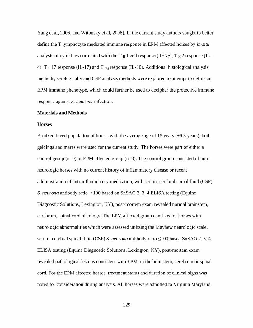
129
Yang et al, 2006, and Witonsky et al, 2008). In the current study authors sought to better
define the T lymphocyte mediated immune response in EPM affected horses by in-situ
analysis of cytokines correlated with the T H 1 cell response ( IFNγ), T H 2 response (IL-
4), T H 17 response (IL-17) and T reg response (IL-10). Additional histological analysis
methods, serologically and CSF analysis methods were explored to attempt to define an
EPM immune phenotype, which could further be used to decipher the protective immune
response against S. neurona infection.
Materials and Methods
Horses
A mixed breed population of horses with the average age of 15 years (±6.8 years), both
geldings and mares were used for the current study. The horses were part of either a
control group (n=9) or EPM affected group (n=9). The control group consisted of non-
neurologic horses with no current history of inflammatory disease or recent
administration of anti-inflammatory medication, with serum: cerebral spinal fluid (CSF)
S. neurona antibody ratio >100 based on SnSAG 2, 3, 4 ELISA testing (Equine
Diagnostic Solutions, Lexington, KY), post-mortem exam revealed normal brainstem,
cerebrum, spinal cord histology. The EPM affected group consisted of horses with
neurologic abnormalities which were assessed utilizing the Mayhew neurologic scale,
serum: cerebral spinal fluid (CSF) S. neurona antibody ratio ≤100 based SnSAG 2, 3, 4
ELISA testing (Equine Diagnostic Solutions, Lexington, KY), post-mortem exam
revealed pathological lesions consistent with EPM, in the brainstem, cerebrum or spinal
cord. For the EPM affected horses, treatment status and duration of clinical signs was
noted for consideration during analysis. All horses were admitted to Virginia Maryland
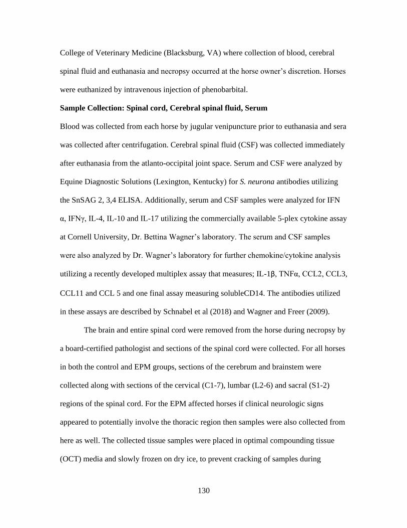
130
College of Veterinary Medicine (Blacksburg, VA) where collection of blood, cerebral
spinal fluid and euthanasia and necropsy occurred at the horse owner’s discretion. Horses
were euthanized by intravenous injection of phenobarbital.
Sample Collection: Spinal cord, Cerebral spinal fluid, Serum
Blood was collected from each horse by jugular venipuncture prior to euthanasia and sera
was collected after centrifugation. Cerebral spinal fluid (CSF) was collected immediately
after euthanasia from the atlanto-occipital joint space. Serum and CSF were analyzed by
Equine Diagnostic Solutions (Lexington, Kentucky) for S. neurona antibodies utilizing
the SnSAG 2, 3,4 ELISA. Additionally, serum and CSF samples were analyzed for IFN
α, IFNγ, IL-4, IL-10 and IL-17 utilizing the commercially available 5-plex cytokine assay
at Cornell University, Dr. Bettina Wagner’s laboratory. The serum and CSF samples
were also analyzed by Dr. Wagner’s laboratory for further chemokine/cytokine analysis
utilizing a recently developed multiplex assay that measures; IL-1β, TNFα, CCL2, CCL3,
CCL11 and CCL 5 and one final assay measuring solubleCD14. The antibodies utilized
in these assays are described by Schnabel et al (2018) and Wagner and Freer (2009).
The brain and entire spinal cord were removed from the horse during necropsy by
a board-certified pathologist and sections of the spinal cord were collected. For all horses
in both the control and EPM groups, sections of the cerebrum and brainstem were
collected along with sections of the cervical (C1-7), lumbar (L2-6) and sacral (S1-2)
regions of the spinal cord. For the EPM affected horses if clinical neurologic signs
appeared to potentially involve the thoracic region then samples were also collected from
here as well. The collected tissue samples were placed in optimal compounding tissue
(OCT) media and slowly frozen on dry ice, to prevent cracking of samples during

131
freezing and stored at -80 C for future analysis. Additionally, approximately 3 mm tissue
samples were collected and snap frozen on liquid nitrogen for future DNA and RNA
extraction. The remainder of the spinal cord was fixed in 10% buffered formalin and
tissue adjacent to those collected for frozen tissue analysis was collected for histological
analysis.
Histological analysis
The formalin fixed sections were paraffin embedded and section and stained with
hematoxylin and eosin (H&E) and immunohistochemistry (IHC) staining. The H&E
stained sections were reviewed by a veterinarian board certified pathologist for
histological changes related to EPM which included; inflammatory changes (lymphocytic
infiltration, eosinophilic inflammation, gliosis, perivascular cuffing), degenerative
changes and S. neurona itself. For IHC analysis replicate sections to those that the EPM
lesion was identified in were stained with rabbit anti-S. neurona sera as described by
Gerhold et al (2005) with the exception that the anti-S. neurona antibody was utilized at a
concentration of 1:500 and peroxidase inhibitor (ThermoFisher Scientific) was used for
inhibition of endogenous peroxidases. The collected samples from the control horses
were also reviewed by a board-certified pathologist to confirm normal CNS pathology
and sections were also stained with rabbit anti- S. neurona sera to serve as experimental
negative controls.
Immunofluorescent slide staining
The OCT frozen tissue samples collected from the region where pathological lesions
were identified in H&E stained samples, were sectioned on a cryostat in 10µm sections
and stained with one of the combinations listed in table I, in order to visualize cytokine

132
and cell localization. The antibodies used to identify astrocytes were tight junction
protein, connexin 43 (Cx43- PE), Glial fibrillary acidic protein (GFAP-FitC) and the
antibody for microglia cells ionized calcium binding adaptor molecule 1 (Iba-1-FitC), all
antibodies were purchased from SantaCruz BioTech, (Dallas, TX) and used a working
concentration of 1:100. The T-lymphocyte antibodies CD4-FitC (concentration 1:100)
and CD8-PE (concentration 1:250) were purchased from Bio-Rad (Hercules, CA). The
cytokine antibodies (IFNγ, IL-17a, IL-10 and IL-4) were all conjugated with AF-647 and
were purchased from Dr. Bettina Wagner Laboratory (Cornell University, Ithaca, NY).
Briefly, the staining protocol utilized is as follows, slides were incubated in histological
grade acetone 10 mins, 4 C, washed 3x in 1x PBS, tissue sections were encircled with
ImmunoPap pen, 3% FBS blocking buffer was added to each section, incubated for 1 hr,
room temperature (RT) in a moist chamber, washed 3x in 1 x PBS, incubated with the
appropriately diluted antibodies 1 hr RT in a moist chamber for T-lymphocyte markers
and cytokines or overnight at 4 C for microglia and astrocyte +cytokines. Following
antibody incubation, slides were washed 3x with 1x PBS, stained with nuclear
counterstain DAPI for 10 mins RT, washed 3x with 1x PBS and cover slipped and left to
dry at RT and stored at 4 C till imaged. Each spinal cord section (n=12 sections/horse)
was imaged using an TiE- Nikon fluorescent light microscope utilizing the same light
exposure sections for imaging consistency within 48 hours of staining. All sections were
scanned for cytokine (IFNγ, IL-17a, IL-4 or IL-10) staining and 4-5, 20x HPF images of
cytokine staining were captured to represent the cytokine staining pattern of a particular
section. The total number of cytokines in each image was then manually enumerated
along with Microglia cells and T-lymphocytes when present in the captured image.

133
Astrocytes could not accurately be manually enumerated due to the complexity of the
cells and therefore fluorescent intensity of the GFAP marker was measured which has
previously been described by (Lemos, 2008). A scoring system of 0 (no cells present) to
8 (the greatest number of cells present) was utilized for quantification analysis of cell
types in each captured image.
Table I: Antibody combinations for immunofluorescent staining of CNS tissue
samples
Cell Type Cell Type
Antibody
Antibody combinations: Cell Type + Cytokine
Astrocytes a Cx43
+
GFAP
Astrocyte
+
IFNγ
Astrocyte
+
IL17a
Astrocyte
+
IL4
Astrocyte
+
IL10
Microglia a Iba-1 Microglia
+
IFNγ
Microglia
+
IL17a
Microglia
+
IL4
Microglia
+
IL10
T-
lymphocytes b
CD4
+
CD8
T-lymphocytes
+
IFNγ
T-lymphocytes
+
IL17a
T-lymphocytes
+
IL4
T-lymphocytes
+
IL10 a Overnight incubation period at 4 C b One-hour incubation period at room temperature
RNAscope
The In-situ hybridization technique, RNAscope, was utilized to investigate another
method to detect S. neurona in the CNS of EPM affected horses. The assay allows
visualization and quantification of gene expression in formalin fixed paraffin embedded
(FFPE) tissue samples. For this study a SnSAG2 probe was designed for S. neurona
detection and IL-10 and IFNγ probes were also designed (Table II). All probe design was
conducted by the ACD Biotech (Newark, California) bioinformatics team but the general
structure of a probe resembles a Z and consist of 18-25 base region which complements
the RNA sequence of interest, followed by a spacer region and then a tail sequence which

134
serves as a binding site for the pre-amplifer molecule used in the assay when two probes
hybridize together. For the EPM group the FFPE samples sections were replicate of those
used for IHC staining and close to where pathological lesion was identified. The
manufactuers protocol for the multiplex fluorescent assay was followed. Briefly, samples
were deparaffinized in xylene, dehydrated in 100% ethanol, endogenous peroxidases
were inhibited with RNAscope kit specific reagent, antigen retrieval step consisted of
submerging slides in boiling Antigen reterival reagent (ACD Biotech), followed by
treatment with protease plus reagent provided in kit, slides were incubate at 40 C for 30
minutes with protease plus, washed with 1x RNAscope wash buffer. The probes were
diluted appropriately in dilution buffer (included in kit). and incubated at 40 C for 2 hrs
and then washed and incubated with pre amplifier 1, 2, and 3 in three separate 30 min (
pre-amplifier 1 and 2) and 15 mins (pre-amplifier 3) incubations at 40 C, then amplifiers
incubation for 15 mins followed by probe label ( TSA dye 520, 570 or 650) for 30 mins,
this was repeated twice for amplifiers 2 and 3. In between each incubation period slides
were washed 2x with RNAscope wash buffer. The final step was nuclear counter stain
DAPI and cover slipping, air dry in the dark at room temperature for 30 mins and stored a
4 C. All slides were imaged with TiE- Nikon fluorescent light microscope utilizing the
same light exposure sections for imaging consistency within 48 hours of staining.
Table II: RNAscope probe sequence reference numbers
Probe Sequence Ref number
SnSAG2 AY191006.1
IFNy NC_009149.3
IL-10 NC_009148.3
Statistical Analysis
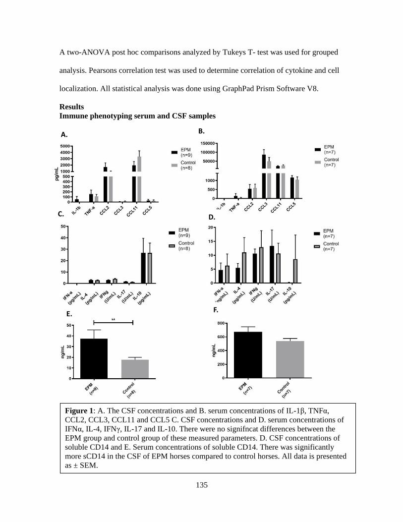
135
A two-ANOVA post hoc comparisons analyzed by Tukeys T- test was used for grouped
analysis. Pearsons correlation test was used to determine correlation of cytokine and cell
localization. All statistical analysis was done using GraphPad Prism Software V8.
Results
Immune phenotyping serum and CSF samples
A.
D.
E.
C.
F.
B.
Figure 1: A. The CSF concentrations and B. serum concentrations of IL-1β, TNFα,
CCL2, CCL3, CCL11 and CCL5 C. CSF concentrations and D. serum concentrations of
IFNα, IL-4, IFNγ, IL-17 and IL-10. There were no signifncat differences between the
EPM group and control group of these measured parameters. D. CSF concentrations of
soluble CD14 and E. Serum concentrations of soluble CD14. There was significantly
more sCD14 in the CSF of EPM horses compared to control horses. All data is presented
as ± SEM.
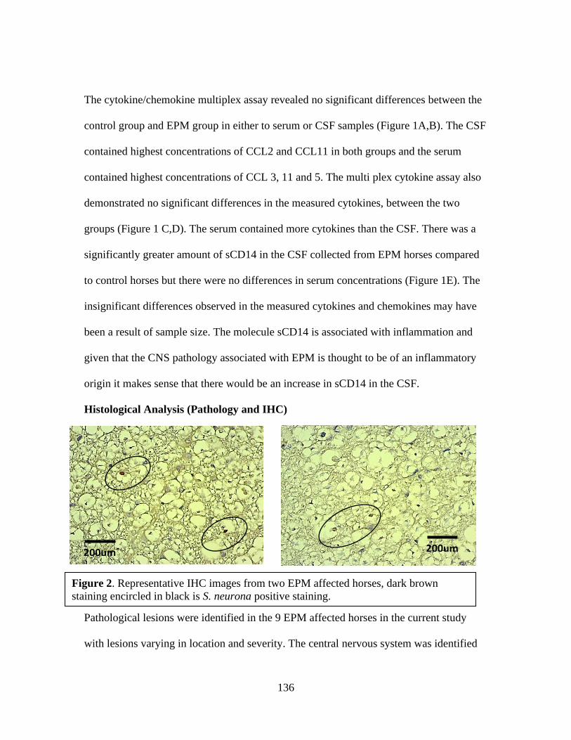
136
The cytokine/chemokine multiplex assay revealed no significant differences between the
control group and EPM group in either to serum or CSF samples (Figure 1A,B). The CSF
contained highest concentrations of CCL2 and CCL11 in both groups and the serum
contained highest concentrations of CCL 3, 11 and 5. The multi plex cytokine assay also
demonstrated no significant differences in the measured cytokines, between the two
groups (Figure 1 C,D). The serum contained more cytokines than the CSF. There was a
significantly greater amount of sCD14 in the CSF collected from EPM horses compared
to control horses but there were no differences in serum concentrations (Figure 1E). The
insignificant differences observed in the measured cytokines and chemokines may have
been a result of sample size. The molecule sCD14 is associated with inflammation and
given that the CNS pathology associated with EPM is thought to be of an inflammatory
origin it makes sense that there would be an increase in sCD14 in the CSF.
Histological Analysis (Pathology and IHC)
Pathological lesions were identified in the 9 EPM affected horses in the current study
with lesions varying in location and severity. The central nervous system was identified
Figure 2. Representative IHC images from two EPM affected horses, dark brown
staining encircled in black is S. neurona positive staining.
200um 200um

137
to be pathologically normal in 9 control horses entered into this study. IHC results
demonstrated S. neurona in 5 of 9 EPM affected horses. It is not always the case that S.
neurona is found at lesion sites so it was not expected that all IHC results would be
positive. Likewise, it may be that if additional IHC staining were completed S. neurona
may be found.
Immunofluorescence slide staining
The quantitated cytokine values obtained from each captured 20x HPF image for all cell
marker types and cytokine combinations analyzed (Table I) were plotted to determine
correlation between
cytokine and cell type
localization in analyzed
spinal cord samples
(Figures 4 and 5). The
only significant
correlations observed
were in the control group,
there were significant
positive correlation
between the localization
of astrocytes and IFNγ
and IL-4 (figure 5 A & C).
For both the control group
and EPM group the most
A. B.
C
. D.
Figure 3: For all immunofluorescent slide imaging all slides were imaged
using a Nikon Ti-E fluorescent light microscope. All images were
captured at 20x magnification and light exposure settings were kept
consistent for all slides analyzed. For all images blue is nuclear staining
(DAPI), A. Red is CD8 and pink is IFNγ. B. Green is CD4 and red is
IFNγ. C. Green is glial fibrillary acidic protein (GFAP), red is connexin
43 (Cx43) which were both used to identify astrocytes and pink is IFNγ.
D. Green is ionized calcium binding adaptor molecule-1 (Iba-1) which
was used to identify microglia cells and pink is IL-17a.

138
prevalent cell types were astrocytes followed by microglia which is not surprising as
these are resident CNS immune cells and therefore found in greater abundance than T-
lymphocytes under normal physiological conditions. It was hypothesized that more T
lymphocytes would be observed in cytokine images collected from EPM affected horses,
but this was not observed. While all antibody markers stained positively as depicted in
figure 3A-D, we were not able to detect S. neurona in the frozen tissue samples and
therefore we cannot say conclusively that our results reflect S. neurona induced
pathology changes.

139
Figure 5: Correlation plot of localization of cell types and cytokines analyzed in the
control group. The Y axis depicts the score and the X axis the total number of cytokine
in each image captured. There was significant positive correlation between astrocytes
and A. IFNγ (P=0.0007) and C. IL-4 (P=0.04).
Figure 4: Correlation plot of localization of cell types and cytokines analyzed
in EPM group. The Y axis depicts the score and the X axis the total number of
cytokine in each image captured. There were no significant correlations
observed.

140
RNAscope
Utilizing samples collected for the current study we were able to validate the SnSAG2
probe to provide additional in-situ techniques for further studies. The SAG2 probe
positively identified S. neurona in a cerebellum collected from an S. neurona infected
mouse from another study. The cerebellum sample contained an abundant amount of S.
neurona which was identified by IHC and then RNAscope. Likewise, S. neurona
was found by RNAscope in four EPM horses which also demonstrated positive IHC
staining. The IFNγ and IL-10 probes were used in combination with the SAG2 probe and
in some cases IFNγ was visualized with the SAG2 probe but IL-10 was not observed with
the SAG2 probe. Samples from the control group of horses which were identified by IHC
to be negative for S. neurona were also negative by RNAscope for the SAG2 probe.
A.
Figure 6: RNAscope image of SnSAG2 staining (pink), IFNγ (green), cell nucleus
(blue). A. positive control mouse cerebellum. B. Spinal cord tissue from EPM affected
horse.
B.

141
These results help to validate the SAG2 probe adding to the techniques which can be
utilized to examine the pathology of EPM affected horses.
Discussion
In the current study a variety of methods were utilized to explore methodology which
could be used to investigate the immune response in the EPM affected horse. One of the
main factors that limits EPM prognosis in affected horses is the lack of understanding of
the immune response against the etiological agent S. neurona. This lack of knowledge
impedes treatment and supportive therapy options. The current study investigated both
the peripheral immune response and the immune response within the CNS, with a more
detailed focus on the CNS, in a small population of EPM affected horses. The
immunofluorescent analysis of IFNγ, IL-17a, IL-10 and IL-4 and cell markers for
astrocytes, microglia and T lymphocytes were conducted in frozen tissue samples as
opposed to FFPE tissue to negate the antibody interference/specificity issues that
sometimes result from the cross linking of formalin fixed proteins (Delcambre et al,
2016). All selected antibodies for this study stained appropriately in the frozen tissue
samples. However, because the frozen tissue sample sections were only from the same
anatomical region (ex. C1) of histologically identified EPM lesions and not direct
replicate sections, it is difficult to directly correlate these results with identified lesions.
Since it was the goal of this study to evaluate the immunopathology in EPM affected
horses this was considered a major pitfall. On the other hand, it is unknown how the
immunopathology is altered in tissue surrounding pathological lesions and knowledge of
this pathological aspect was gained in the current study. In the examined frozen tissue
samples, we didn’t observe any significant differences in the cells which were present in

142
tissue samples collected from EPM affected horses compared to control horses. The
cytokine’s analyzed were found in relatively the same abundance in both groups as well.
The cytokine IFNγ was found most frequently in captured images of frozen tissue
samples and IL-17a was found least frequently. There was a significant correlation
between the localization of astrocytes and IFNγ and IL-4 in the control group but when
interrupting these results, it should be considered that astrocytes were most frequently
found in all tissue samples analyzed. In normal pathology astrocytes and microglia cells
are found in higher abundance than T lymphocytes within the CNS because they are
resident immune cells. Given that results of the current study did not significantly differ
between control and EPM horses it is suggested that pathology in tissue surrounding
pathological lesion site, may not be affected and therefore is not the optimal tissue
sample for EPM immunopathology investigation. The ability to more closely correlate
immunopathology analysis such as what was conducted in this study, with S. neurona
itself or histological inflammatory lesions consistent with EPM would be ideal. To help
expand on the tools available for immunopathology analysis we employed a novel in-situ
hybridization technique, RNAscope. We successfully utilized a target probe for SnSAG2
to identify S. neurona in FFPE tissue samples collected from the EPM group which also
positively stained for S. neurona by IHC analysis. The SnSAG2 sequence was selected
for S. neurona probe design due to it being found on 7 different S. neurona isolates from
horses (Howe et al, 2008) and its high specificity and sensitivity when used as a target
protein for S. neurona antibody detection in ELISA (Yeagan et al, 2011). RNAscope
allows visualization of gene expression instead of protein expression. The design of
target probes is dependent on available gene sequence knowledge and is not limited by
143
available equine antibodies like traditional immunohistochemistry, which immensely
expands the immune phenotyping possibilities in the horse. The use of FFPE tissues as
opposed to frozen tissue samples allows for more extensive collaboration efforts of EPM
researchers because FFPE tissue is routinely collected in postmortem examination
whereas frozen tissue is not.
There were no significant differences observed in sera or CSF concentrations of
measured cytokines and chemokines but there was significantly greater amount of sCD14
in CSF samples collected from EPM horses compared to control horses. The molecule
sCD14 is increased during systemic inflammatory conditions which involve monocyte
recruitment and macrophage activation in both the horse and human (Nockher et al, 1999
and Wagner, 2013). It has also been reported to be increased in inflammatory conditions
which affect the CNS such as bacterial mengitis in humans (Nockher et al, 1999). Other
biomarkers related to CNS damage such as phosphorylated neurofilament H (pNF-H)
have been investigated as potential biomarkers for EPM (Intan-Shameha et al, 2017 and
Gomez et al, 2019). The sCD14 results of the current study suggest that it too could
potentially be used a biomarker for EPM diagnosis and future studies involving larger
sample populations are warranted. Equine protozoal myeloencephalitis can acutely or
chronically affect horses and treatment failure can result in disease relapse (Mackay et al,
2006). A biomarker such as sCD14 may also aid in monitoring disease status after
treatment to help decrease potential for relapse. While there were no significant
differences in analyzed chemokines and cytokines in the current study, this may have
been a result of the small sample population and future studies utilizing larger study
populations to examine changes in these cytokines and chemokines should be conducted.

144
If differences could be detected in these immune mediators, valuable information of
immune cell recruitment and immune response (inflammatory or anti-inflammatory)
could be gained. Overall the results of the current study suggest that the novel in-situ
technique RNAscope may be optimal for studying the immunopathology of EPM. The
use of this assay allows for S. neurona detection and the analysis of three other markers
which could consist of cell markers, immune mediators or cell structural components, in
FFPE tissue samples. The results of the current study and previous studies investigating
EPM immunopathogenesis are limited by sample size, making it difficult to assess
immunological changes due disease status (acute, chronic or relapsing) or treatment
status, leaving many unanswered questions. An assay which allows for an extensive
immunopathology analysis, in tissue samples routinely collected during postmortem
examination in EPM suspected horses, eases difficulties associated with collaborative
efforts, creating new opportunities for EPM research.
References
Delcambre, G. H., Liu, J., Herrington, J. M., Vallario, K., & Long, M. T. 2016.
Immunohistochemistry for the detection of neural and inflammatory cells in equine brain
tissue. PeerJ. 1; 1–15.
Dubey, J. P., and D. S. Lindsay. 1998. Isolation in immunodeficient mice of Saracocystis
neurona from opossum (Didelphis virginiana) faeces and its differentiation from
Sarcocystis falcatula. International Journal Parasitology 28: 1823-1828.
Dubey, J.P. 2000. Prevalence of Sarcocystis species sporocysts in wild caught opossums
(Didelphis virginiana). Journal of Parasitology 86: 705–710.

145
Dubey, J.P., Saville W.J.A., Lindsay D.S., Stich R.W., Stanek J.F, Speer, C.A., Rosenthal, B.M.,
Njoku, C.J., Kwok, O.C.H., Shen S.K., et al. 2000. Completion of the life cycle of
Saracocystis neurona. Journal of Parasitology 86: 1276-1280.
Howe, D. K., Gaji, R. Y., Marsh, A. E., Patil, B. A., Saville, W. J., Lindsay, D. S., Granstrom, D.
E. 2008. Strains of Saracocystis neurona exhibit differences in their surface antigens,
including the absence of the major surface antigen SnSAG1. International Journal for
Parasitology. 38; 623–631.
Intan-Shameha, A. R., Divers, T. J., Morrow, J. K., Graves, A., Olsen, E., Johnson, A. L., &
Mohammed, H. O. (2017). Phosphorylated neurofilament H (pNF-H) as a potential
diagnostic marker for neurological disorders in horses. Research in Veterinary Science. 114,
401–405.
Lemos, K. R., Marques, L. C., Aquino, L. P. C. T., Alessi, A. C., & Zacarias, R. Z. (2008).
Astrocytic and microglial response and histopathological changes in the brain of horses
with experimental chronic Trypanosoma evansi infection. Revista Do Instituto de
Medicina Tropical de Sao Paulo, 50; 243–249.
Mackay, R. J. 2006. Equine protozoal myeloencephalitis: Treatment, prognosis, and prevention.
Clinical Techniques In Equine Practice 5: 9–16.
NAHMS (NATIONAL ANIMAL HEALTH MONITORING SYSTEM). 2001. Equine Protozoal
myeloencephalitis (EPM) in the U.S. Centers for Epidemiology and Animal Health–
USDA APHIS, Fort Collins, Colorado, 46 p.

146
Nockher, W. A., Wick, M., & Pfister, H.-W. 1999. Cerebrospinal fluid levels of soluble CD14 in
inflammatory and non-inflammatory diseases of the CNS: upregulation during bacterial
infections and viral meningitis. In Journal of Neuroimmunology. 101; 161-169.
Reed, S. M., M. Furr, D. K. Howe, A. L. Johnson, R. J. Mackay, J. K. Morrow, and S. Witonsky.
2016. Equine protozoal myeloencephalitis: An updated consensus statement with a focus
on parasite biology, diagnosis, treatment, and prevention. Journal of Veterinary Internal
Medicine 30: 491–502.
Schnabel CL, Wemette M, Babasyan S, Freer H, Baldwin C, Wagner B. 2018. C-C motif
chemokine ligand (CCL) production in equine peripheral blood mononuclear cells
identified by newly generated monoclonal antibodies. Veterinary Immunology and
Immunopathology. 204:28-39.
Scott. P., Witonsky, S., Robertson, J., Draft,B., 2005. Increased prescence of T lymphocytes in
central nervous system of EPM affected horses. Journal of parasitology. 91, 1499-1502.
Spencer, J. A., Ellison, S. E., Guarino, A. J., & Blagburn, B. L. (2004). Cell-Mediated Immune
Responses in Horses with Equine Protozoal Myeloencephalitis. Journal of Parasitology,
90(2), 428–430.
Wagner, B. Feer ,H., 2009. Development of a bead-based multiplex assay for simultaneous
quantification of cytokines in horse. Veterinary immunology and immunopathology. 127;
242-248.
147
Wagner, B., Ainsworth, D. M., & Freer, H. 2013. Analysis of soluble CD14 and its use as a
biomarker in neonatal foals with septicemia and horses with recurrent airway obstruction.
Veterinary Immunology and Immunopathology. (1–2), 124.
Witonsky, S. G., Ellison, S., Yang, J., Gogal, R. M., Lawler, H., Suzuki, Y., Lindsay, D. S. 2008.
Horse experimentally infected with Saracocystis neurona develop altered immune
responses in vitro. International Journal of Parasitology. 94.
Yang, J., Ellison, S., Gogal, R., Norton, H., Lindsay, D. S., Andrews, F., Witonsky, S. 2006.
Immune response to Saracocystis neurona infection in naturally infected horses with
equine protozoal myeloencephalitis. Veterinary Parasitology, 138; 200–210.
Yeargan, M.R. and Howe, D.K. 2011. Improved detection of equine antibodies against
Saracocystis neurona using polyvalent ELISAs based on the parasite SnSAG surface
antigens. Veterinary Parasitology. 176; 16-22.

148
Chapter VIII: Conclusions Future Directions
Equine Protozoal Myeloencephalitis (EPM), is a neurologic disease in horses considered
to be the most common neurologic disease of horses, yet the knowledge of the disease remains
fairly limited. The work conducted here in mouse models, combined with the work in the
previous literature, implicates an essential role of cytokine, interferon gamma (IFNγ), and IFNγ
competent T lymphocytes, in the protective immune response against S. neurona induced
encephalitis. The protective immune response results in control of S. neurona replication and
elimination of the parasite before causing significant central nervous system (CNS) damage and
subsequent clinical neurologic disease. In the immunocompromised mouse, this immune
response is not limited to the role of T lymphocytes, as Balb/cscid mice which are void of T-
lymphocytes, do not consistently succumb to neurologic disease after S. neurona infection. In
has been reported that in-vitro bacterial stimulation of Balb/c splenocytes results in an increased
production of innate immune cell derived IFNγ, compared to C57Bl/6 (B6) splenocytes (Koo,
2006). However, an increase in IFNγ production was not detected in the sera of Balb/c scid mice
compared to C57Bl/6 (B6) scid mice 7 days post infection with S. neurona. If robust IFNγ
production did occur in the Balb/c scid mice during this study, it may have occurred prior to the
7DPI sampling point or IFNγ may not have been the explanation for the resistance observed in
these mice. Another immunological factor which differs between these two mice strains is their
MHC haplotype. The MHC complex (I and II) are responsible for antigen presentation and are
integral part of activating than robust immune response against an invading pathogen and
adaptive immune response activation. The genetic variation between the B6 and Balb/c MHC
haplotype is one contributing factor to Toxoplasmosis resistance in the Balb/c mouse compared
to susceptible B6 mouse. There have also been reported differences in disease susceptibility

149
differences in B6 Ifnγ -/- and Balb/c Ifnγ-/- mice, with the Balb/c strain remaining neurologically
normal for slightly longer than B6 strain, especially as the infection dose of S. neurona decreased
(Dubey, 2013). Future mouse model studies to investigate the MHC haplotype role in S. neurona
disease susceptibility could include S. neurona infection of NOD scid mice which have the same
MHC haplotype as disease resistant ICR scid mice (Marsh et al, 1997 and Sellon et al, 2004).
Therefore, it’s hypothesized that NOD scid mice would be resistant to S. neurona encephalitis, if
disease resistance in an immune compromised state is dependent on MHC haplotype. Also, a B6
scid mouse with the Balb/c MHC haplotype (H2d) could be infected with S. neurona and disease
resistance would be expected, if the hypothesized role of the MHC haplotype is correct. The
protective immune response against S.neurona encephalitis is not limited to MHC haplotype as
both Balb/c and B6 mice are resistant to S. neurona infection (Rosypal et al, 2002) but as
mentioned it could potentially be influenced by MHC haplotype. Additionally, a role of MHC
haplotype in EPM susceptibility is one explanation for not being able to readily induce EPM in
experimental infection studies.
If there is a specific MHC haplotype that is associated with clinical EPM susceptibility in
horses, disease susceptibility may also be further influenced by strain of S. neurona which is
ingested. There is a considerable amount of genetic variation noted in S. neurona (Asmundsson
et al, 2006). The amount of S. neurona ingested, either in a single exposure or multiple
exposures, may also influence susceptibly. Another mouse model study which could be
conducted is to test disease susceptibility in a mouse with just a CD8 knockout mutation to
determine if the susceptibility reported in β2m knockout mice was strictly due to lack of
functional CD8+ T lymphocytes or combined role of lack of β2m and MHC I expression and
CD8+ T lymphocytes. If CD8 knockout mice are disease resistant, then susceptibility can be

150
attributed to β2m deficiency and immunologic impairment related to antigen presentation by
MHC I complex. It is likely that CD8 knockout mice would be susceptible to disease as these
cells are a main source of IFNγ production. Transfer of IFNγ producing CD3+ T lymphocytes
offered protection against S. neurona infection in susceptible B6 scid mice. It would also be
interesting to investigate disease outcome in a Balb/c β2m knockout mouse and determine if
disease progression paralleled B6 β2m knockout mouse to further investigate the role of MHC
complex in disease susceptibility. In addition to these studies further treatment studies should be
conducted.
The B6 Ifnγ -/- mouse model was employed to investigate the treatment efficacy of
antiprotozoal drugs diclazuril and decoquinate. Decoquinate was not proven to be effective at
preventing infection despite different administration methods and confirmation that the mice
received decoquinate during treatment periods. Decoquinate is a “sticky” compound with a short
half -life and poor bioavailability, making it difficult to administer and potentially,
pharmacologically unfavorable. A clinical field trial did report improvement in horses potentially
affected by EPM after treatment with decoquinate, but the compound used for treatment was a
combination of levamisole and decoquinate and individual drug testing was not completed
(Ellison, 2012). Therefore, further studies investigating the efficacy of decoquinate and
levamisole individually need to be conducted, as it could be the synergistic effect of these two
compounds that resulted in neurologic improvement in Ellison et al (2012) study. Likewise,
pharmacokinetic studies should be conducted to further test the bioavailability of the drug in the
horse and determine if effective CNS concentrations can be obtained.
Further studies investigating immunomodulators which could serve as supportive therapy
to help prevent relapses potentially associated with treatment failure should be conducted as

151
well. One immunomodulator that has been reported to increase IFNγ production is Zylexis
(inactivated parapoxvirus) (Horohov, 2008). The common EPM treatment diclazuril did prevent
S. neurona encephalitis while B6 Ifnγ -/- mice were being treated but cessation of treatment led
to disease development. This suggest that the medication inhibited S. neurona replication but an
IFNγ dependent immune response was necessary for complete parasite elimination. This also
supports the idea that some horse’s relapse after response to EPM treatment as a result of
treatment failure, expressing a need for treatment options which fully eliminate S. neurona.
Lastly, studies investigating the immune response within the CNS and associated neuropathology
are limited. This is partly due to difficultly to obtain funding for studies with a population large
enough to investigate differences in disease states. Some EPM affected horses which develop
neurologic signs and are treated and appropriately respond to treatment are able to return their
job. However, some horses don’t respond to treatment and disease progresses to the point where
they need to be retired from work or euthanized due to poor quality of life. Why these
differences occur could be unrelated to neuropathology, but a pre-existing condition could also
make some horses more susceptible to EPM infection or explain differences in EPM affected
horses. Concurrent equine motor neuron disease and EPM (due to neospora hughesi infection)
has been reported (Finno, 2010). Also, it has been reported that workload of horses is positively
correlated with serum concentrations of phosphorylated neurofilament – H which accumulates
due to neuron axonal degeneration (Morales Gomez, 2019). This suggest that working (riding)
horses may experience axonal degeneration as result of neuron damage that might occur as a
result of riding. It could as be that the neuroinflammation associated with EPM pathology could
trigger another neurologic disorder such as equine motor neuron disease. Investigating these
potential phenomena could help determine prognosis of EPM horses. Overall future studies

152
investigating the potential for genetic predisposition to EPM and improving treatment options
would be valuable.
References
Asmundsson IM, Dubey JP, Rosenthal BM. A genetically diverse but distinct North American
population of Saracocystis neurona includes an overrepresented clone described by 12
microsatellite alleles. Infectious Genetic Evolution. 6:352–360.
Dubey, J. P., Sundar, N., Kwok, O. C. H., & Saville, W. J. A. (2013). Saracocystis neurona
infection in gamma interferon gene knockout (KO) mice: Comparative infectivity of
sporocysts in two strains of KO mice, effect of trypsin digestion on merozoite viability,
and infectivity of bradyzoites to KO mice and cell culture. Veterinary Parasitology. (1–
2), 212.
Ellison, S.P. and Lindsay, D.S. 2012. Decoquinate combined with levamisole reduce the clinical
signs and serum SAG 1,5,6 antibodies in horses with suspected Equine Protozoal
Myeloencephalitis. International Journal Applied Research Veterinary Medicine. 10.
Finno, C.J., Eaton, J.S., Aleman, M., Hollingsworth, S.R., 2010. Equine protozoal
myeloencephalitis due to Neospora hughesi and equine motor neuron disease in a mule.
Veterinary Ophthalmology. 13(4), 259–265.
Horohov, D. W., Breathnach, C. C., Sturgill, T. L., Rashid, C., Stiltner, J. L., Strong, D.,
Holland, R. E. 2008. In vitro and in vivo modulation of the equine immune response by
parapoxvirus ovis. Equine Veterinary Journal, 40(5), 468–472.
Koo, G. C., & Gan, Y. H. (2006). The innate interferon gamma response of BALB/c and
C57BL/6 mice to in vitro Burkholderia pseudomallei infection. BMC Immunology, 7.

153
Marsh, A. E., Barr, B. C., Lakritz, J., Nordhausen, R., Madigan, J. E., & Conrad, P. A. (1997).
Experimental infection of nude mice as a model for Saracocystis neurona- associated
encephalitis. Parasitology Research, 83; 706–711.
Morales Gómez, A. M., Zhu, S., Palmer, S., Olsen, E., Ness, S. L., Divers, T. J., Mohammed, H.
O. 2019. Analysis of neurofilament concentration in healthy adult horses and utility in the
diagnosis of equine protozoal myeloencephalitis and equine motor neuron disease.
Research in Veterinary Science. 125; 1–6.
Rosypal, A. C., Lindsay, D. S., Duncan, R., Ahmed, S. A., Zajac, A. M., & Dubey, J. P. (2002).
Mice lacking the gene for inducible or endothelial nitric oxide are resistant to sporocyst
induced Saracocystis neurona infections. 103, 315–321.
Sellon, D. C., Knowles, D. P., Greiner, E. C., Long, M. T., Melissa, T., Hochstatter, T., Damet, J.
B. (2004). Depletion of Natural Killer Cells Does Not Result in Neurologic Disease Due
to Saracocystis neurona in Mice with Severe Combined Immunodeficiency. Journal of
Parasitology. 90(4), 782–788.

154