EQUINE PROTOZOAL MYELOENCEPHALITIS - CiteSeerX
118
University of Kentucky UKnowledge eses and Dissertations--Veterinary Science Veterinary Science 2013 EQUINE PROTOZOAL MYELOENCEPHALITIS: INVESTIGATION OF GENETIC SUSCEPTIBILITY AND ASSESSMENT OF AN EQUINE INFECTION METHOD Breanna M. Gaubatz University of Kentucky, [email protected] is Master's esis is brought to you for free and open access by the Veterinary Science at UKnowledge. It has been accepted for inclusion in eses and Dissertations--Veterinary Science by an authorized administrator of UKnowledge. For more information, please contact [email protected]. Recommended Citation Gaubatz, Breanna M., "EQUINE PROTOZOAL MYELOENCEPHALITIS: INVESTIGATION OF GENETIC SUSCEPTIBILITY AND ASSESSMENT OF AN EQUINE INFECTION METHOD" (2013). eses and Dissertations--Veterinary Science. Paper 8. hp://uknowledge.uky.edu/gluck_etds/8
-
Upload
khangminh22 -
Category
Documents
-
view
1 -
download
0
Transcript of EQUINE PROTOZOAL MYELOENCEPHALITIS - CiteSeerX
University of KentuckyUKnowledge
Theses and Dissertations--Veterinary Science Veterinary Science
2013
EQUINE PROTOZOALMYELOENCEPHALITIS: INVESTIGATIONOF GENETIC SUSCEPTIBILITY ANDASSESSMENT OF AN EQUINE INFECTIONMETHODBreanna M. GaubatzUniversity of Kentucky, [email protected]
This Master's Thesis is brought to you for free and open access by the Veterinary Science at UKnowledge. It has been accepted for inclusion in Thesesand Dissertations--Veterinary Science by an authorized administrator of UKnowledge. For more information, please contact [email protected].
Recommended CitationGaubatz, Breanna M., "EQUINE PROTOZOAL MYELOENCEPHALITIS: INVESTIGATION OF GENETIC SUSCEPTIBILITYAND ASSESSMENT OF AN EQUINE INFECTION METHOD" (2013). Theses and Dissertations--Veterinary Science. Paper 8.http://uknowledge.uky.edu/gluck_etds/8

STUDENT AGREEMENT:
I represent that my thesis or dissertation and abstract are my original work. Proper attribution has beengiven to all outside sources. I understand that I am solely responsible for obtaining any needed copyrightpermissions. I have obtained and attached hereto needed written permission statements(s) from theowner(s) of each third-party copyrighted matter to be included in my work, allowing electronicdistribution (if such use is not permitted by the fair use doctrine).
I hereby grant to The University of Kentucky and its agents the non-exclusive license to archive and makeaccessible my work in whole or in part in all forms of media, now or hereafter known. I agree that thedocument mentioned above may be made available immediately for worldwide access unless apreapproved embargo applies.
I retain all other ownership rights to the copyright of my work. I also retain the right to use in futureworks (such as articles or books) all or part of my work. I understand that I am free to register thecopyright to my work.
REVIEW, APPROVAL AND ACCEPTANCE
The document mentioned above has been reviewed and accepted by the student’s advisor, on behalf ofthe advisory committee, and by the Director of Graduate Studies (DGS), on behalf of the program; weverify that this is the final, approved version of the student’s dissertation including all changes requiredby the advisory committee. The undersigned agree to abide by the statements above.
Breanna M. Gaubatz, Student
Dr. Daniel K. Howe, Major Professor
Dr. Daniel K. Howe, Director of Graduate Studies
EQUINE PROTOZOAL MYELOENCEPHALITIS: INVESTIGATION OF GENETIC SUSCEPTIBILITY AND ASSESSMENT OF AN EQUINE INFECTION METHOD
_____________________________________
THESIS _____________________________________
A thesis submitted in partial fulfillment of the
requirements for the degree of Master of Science in the College of Agriculture
at the University of Kentucky
By
Breanna Marie Gaubatz
Lexington, KY
Director: Dr. Daniel K. Howe, Professor of Veterinary Science
Lexington, Kentucky
2013
Copyright © Breanna Marie Gaubatz 2013
ABSTRACT OF THESIS
EQUINE PROTOZOAL MYELOENCEPHALITIS: INVESTIGATION OF GENETIC SUSCEPTIBILITY AND ASSESSMENT OF AN EQUINE INFECTION METHOD
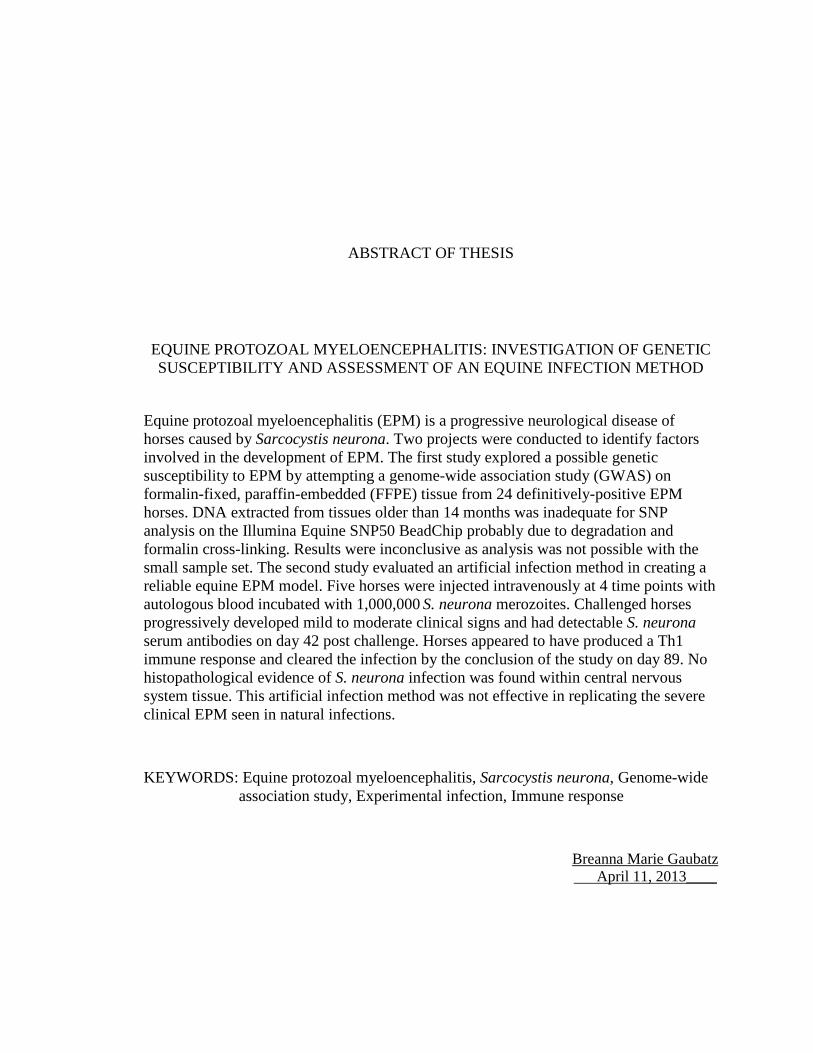
Equine protozoal myeloencephalitis (EPM) is a progressive neurological disease of horses caused by Sarcocystis neurona. Two projects were conducted to identify factors involved in the development of EPM. The first study explored a possible genetic susceptibility to EPM by attempting a genome-wide association study (GWAS) on formalin-fixed, paraffin-embedded (FFPE) tissue from 24 definitively-positive EPM horses. DNA extracted from tissues older than 14 months was inadequate for SNP analysis on the Illumina Equine SNP50 BeadChip probably due to degradation and formalin cross-linking. Results were inconclusive as analysis was not possible with the small sample set. The second study evaluated an artificial infection method in creating a reliable equine EPM model. Five horses were injected intravenously at 4 time points with autologous blood incubated with 1,000,000 S. neurona merozoites. Challenged horses progressively developed mild to moderate clinical signs and had detectable S. neurona serum antibodies on day 42 post challenge. Horses appeared to have produced a Th1 immune response and cleared the infection by the conclusion of the study on day 89. No histopathological evidence of S. neurona infection was found within central nervous system tissue. This artificial infection method was not effective in replicating the severe clinical EPM seen in natural infections.
KEYWORDS: Equine protozoal myeloencephalitis, Sarcocystis neurona, Genome-wide association study, Experimental infection, Immune response
Breanna Marie Gaubatz April 11, 2013____
EQUINE PROTOZOAL MYELOENCEPHALITIS: INVESTIGATION OF GENETIC SUSCEPTIBILITY AND ASSESSMENT OF AN EQUINE INFECTION METHOD
By
Breanna Marie Gaubatz
___ Dr. Daniel K. Howe_________ Director of Thesis
______Dr. Daniel K. Howe_________
Director of Graduate Studies
__________April 11, 2013__________ Date
iii
ACKNOWLEDGEMENTS
The successful completion of this thesis is due to the support and encouragement of many
individuals. I would like to take this opportunity to express my gratitude to the following:
To Dr. Dan Howe, my advisor, for his patience, support, and guidance throughout my
graduate studies. To Dr. Uneeda Bryant, my committee member, who was pivotal in
finding the parasites in tissue, and for her patience in helping me to identify them as well.
To Dr. Amanda Adams, my committee member, for her help with the cytokine assays,
and for instructing me on the immunology of the horse. To Dr. David Horohov, my
committee member, for his expertise and insight in equine immunology.
To the members of the EPM lab, Michelle Yeargan, Ablesh Gautam, and Dr. Sriveny
Dangoudoubiyam, for sharing their knowledge and helping me with aspects of these
studies. To Dr. Ernie Bailey for his help with the genome-wide association study, to Dr.
Kirsten Scoggin for her assistance with the immunohistochemical staining, and to Dr.
Steve Reed for his expertise with the neurological examinations.
To the University of Kentucky Veterinary Diagnostic Laboratory Histology Department
for locating the archived samples, and recutting many slides for me. In addition, my
thanks goes to the staff at the Eastern Tennessee Clinical Research, especially Dr. Craig
Reinemeyer and Dr. Julio Prado, for their assistance with the challenge infection study.
I would like to greatly acknowledge the Dan C. Hutson Graduate Enrichment Fellowship
for their financial support during my graduate studies.

iv
Most importantly, I am thankful for the encouragement and support that I received from
my friends and family. In particular, my parents for their never-ending support and for
instilling in me a drive to further my education and be the best that I can be.
v
TABLE OF CONTENTS
Acknowledgements ......................................................................................................... iii
List of Tables .................................................................................................................. vii
List of Figures ................................................................................................................ viii
Chapter One: Literature Review ....................................................................................... 1
1.1. Introduction ........................................................................................................ 1
1.2. History ................................................................................................................ 2
1.3. Phylogeny and Life Cycle of Sarcocystis neurona ............................................ 2
1.4. Pathogenesis ....................................................................................................... 6
1.5. Clinical Signs ..................................................................................................... 7
1.6. Pathology ............................................................................................................ 8
1.7. Diagnosis ............................................................................................................ 9 1.7.1. Differential Diagnosis ........................................................................... 9 1.7.2. Antemortem Diagnosis......................................................................... 10 1.7.3. Postmortem Diagnosis ......................................................................... 11
1.8. Treatment ......................................................................................................... 12 1.8.1. Anti-protozoal Treatment ..................................................................... 12 1.8.2. Ancillary Treatment ............................................................................. 13
1.9. Prognosis .......................................................................................................... 14
1.10. Epidemiology ................................................................................................. 15
1.11. Prevention ....................................................................................................... 17
1.12. Cell-mediated Immunity ................................................................................ 18
1.13. Infectious Disease Susceptibility ................................................................... 19
1.14. Genome-wide Association Study ................................................................... 21
1.15. Equine EPM Model ........................................................................................ 24 1.15.1. Intragastric Introduction ................................................................... 24 1.15.2. Intrathecal Introduction ..................................................................... 27 1.15.3. Intravenous Introduction ................................................................... 27
1.16. Research Objectives ....................................................................................... 29
Chapter Two: Genome-wide association study to investigate genetic susceptibility of equine protozoal myeloencephalitis ............................................................................... 30
2.1. Introduction ...................................................................................................... 30
2.2. Materials and Methods ..................................................................................... 31 2.2.1. Case Selection ...................................................................................... 31
vi
2.2.2. Immunohistochemistry ......................................................................... 31 2.2.3. Genotyping ........................................................................................... 32
2.3. Results .............................................................................................................. 33 2.3.1. Case Selection ...................................................................................... 33 2.3.2. Confirmation of Sarcocystis neurona .................................................. 35 2.3.3. Genotyping ........................................................................................... 38
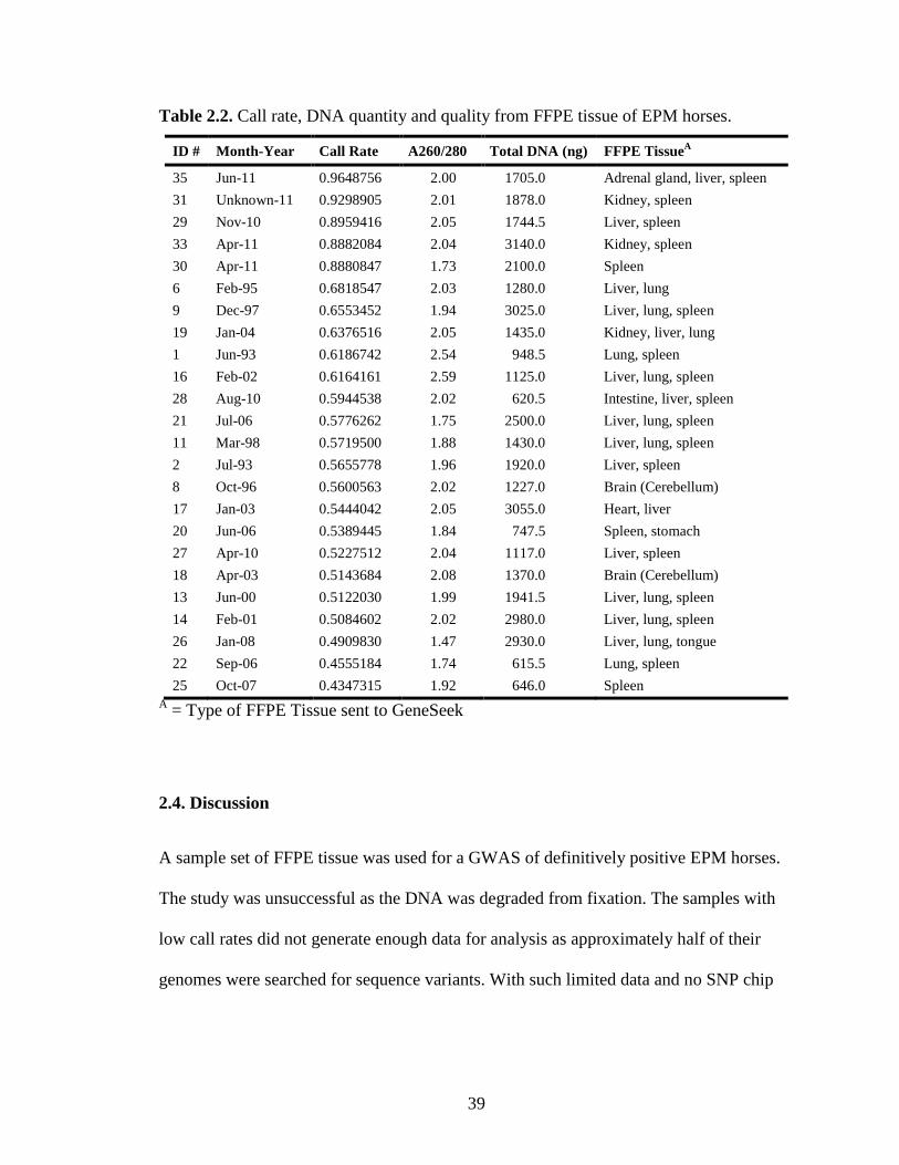
2.4. Discussion ........................................................................................................ 39
Chapter Three: Assessment of an equine protozoal myeloencephalitis model using an artificial infection method in horses ............................................................................... 42
3.1. Introduction ...................................................................................................... 42
3.2. Materials and Methods ..................................................................................... 43 3.2.1. Experimental Animals .......................................................................... 43 3.2.2. Experimental Infection of Horses ........................................................ 44 3.2.3. Sample Collection and Preparation .................................................... 47 3.2.4. Examinations ....................................................................................... 47 3.2.5. Euthanasia and Necropsy .................................................................... 49 3.2.6. Histological Examination .................................................................... 49 3.2.7. Immunohistochemistry ......................................................................... 49 3.2.8. Enzyme-Linked Immunosorbent Assay ................................................ 51 3.2.9. Relative Quantification (RQ) of Cytokine mRNA Expression by Real-time PCR ........................................................................................................ 53 3.2.10. Statistics ............................................................................................. 55 3.2.11. Sarcocystis neurona Merozoite Invasion Assay ................................ 56
3.3. Results .............................................................................................................. 57 3.3.1. Clinical Findings ................................................................................. 57 3.3.2. Postmortem Examination..................................................................... 58 3.3.3. Antibody Response ............................................................................... 62 3.3.4. Cell-mediated Immune Response ......................................................... 72 3.3.5. Merozoite Invasion Assay .................................................................... 76
3.4. Discussion ........................................................................................................ 77
Chapter Four: Conclusion ............................................................................................... 81
Appendices ..................................................................................................................... 83
Appendix A: List of Abbreviations ......................................................................... 83
Appendix B: Neurologic Examination Results ....................................................... 86
References ...................................................................................................................... 90
Vita ............................................................................................................................... 106
vii
LIST OF TABLES
Table 2.1. Horses used in the study…………………………………………………34
Table 2.2. Call rate, DNA quantity and quality from FFPE tissue of EPM horses…39
Table 3.1. Description of ataxia grades……………………………………………...48
Table 3.2. Neurology examination score sheet……………………………………...48
Table 3.3. Primer probe sets used for Real-time PCR………………………..……..55

viii
LIST OF FIGURES
Figure 1.1. Phylogenic tree of Sporozoea (Apicomplexa/Alveolata)…………………3
Figure 1.2. Life cycle of Sarcocystis neurona…………………………………………5
Figure 2.1. CNS lesions of EPM horses……………………………………………...36
Figure 2.2. Sarcocystis neurona within CNS lesions of EPM horses………………..37
Figure 3.1. Timeline for inoculation, sample collection, and examination of horses..46
Figure 3.2. Neurology examination scores…………………………………………...58
Figure 3.3. Histological evaluation of the control horse……………………………..60
Figure 3.4. Histological evaluation of Horse 467……………………………………60
Figure 3.5. Histological evaluation of Horse 468……………………………………61
Figure 3.6. Anti-Sarcocystis neurona total IgG antibody titers of sera by ELISA
analysis…………………………………………………………………...64
Figure 3.7. Percent positivity of anti-Sarcocystis neurona total IgG antibodies of CSF
by ELISA analysis……………………………….………………………66
Figure 3.8. Percent positivity of anti-Sarcocystis neurona SAG1 IgG isotype
antibodies of sera by ELISA analysis……………………………………69
Figure 3.9. Percent positivity of anti-Sarcocystis neurona SAG5 IgG isotype
antibodies of sera by ELISA analysis……………………………………71
ix
Figure 3.10. Relative quantification (RQ) of proinflammatory cytokine mRNA
expression by Real-time PCR after Sarcocystis neurona stimulation…...73
Figure 3.11. Relative quantification (RQ) of mRNA expression by Real-time PCR after
Sarcocystis neurona stimulation…………………………………………74
Figure 3.12. Relative quantification (RQ) of anti-inflammatory cytokine mRNA
expression by Real-time PCR after Sarcocystis neurona stimulation…...75
Figure 3.13. Percent invasion of Sarcocystis neurona F9F merozoites……………….76

1
CHAPTER ONE
Literature Review
1.1. Introduction
Equine protozoal myeloencephalitis (EPM) is an equine neurological disease. The disease
was first described in 1970 (Rooney et al., 1970), and protozoa were identified within
lesions from EPM horses in 1974 (Beech and Dodd, 1974; Cusick et al., 1974; Dubey et
al., 1974). The apicomplexan parasite Sarcocystis neurona is the etiologic agent of EPM
(Dubey et al., 1991). Following the range of the definitive host, the opossum (Didelphis
virginiana, Didelphis albiventris) (Dubey and Lindsay, 1998; Dubey et al., 2001a), EPM
is only found in the western hemisphere. Horses are aberrant hosts, and become infected
by ingesting feed or water that is contaminated with opossum feces (Dubey and Lindsay,
1998). The disease displays clinical signs ranging from mild lameness to recumbency,
and even death. Classic EPM clinical signs are asymmetrical ataxia with focal muscle
atrophy (Dubey et al., 2001b).
Currently, the most accurate antemortem diagnosis of EPM is based on neurological
signs consistent with EPM and a positive ELISA test for S. neurona antibodies in the
serum and CSF (Furr et al., 2002; Yeargan and Howe, 2011). A definitive diagnosis can
only be made during a postmortem examination when S. neurona is located histologically
within the CNS (Furr et al., 2002). Over half of the horses in the United States are
seropositive for S. neurona (MacKay, 1997b), while only 0.5-1% actually develop the
disease (Dubey et al., 2001b).
2
1.2. History
A neurologic disease, referred to as “focal myelitis-encephalitis,” was first described in
52 Kentucky and Pennsylvania horses in 1970 (Rooney et al., 1970). Protozoa were first
witnessed within central nervous system (CNS) lesions of affected horses in 1974 (Beech
and Dodd, 1974; Cusick et al., 1974; Dubey et al., 1974). Initially, the disease-causing
protozoa was misidentified as Toxoplasma gondii (Cusick et al., 1974), but subsequent
evidence determined it was a Sarcocystis spp. due to its morphology and antigenic
properties (Simpson and Mayhew, 1980). In 1976, Mayhew et al. were the first to name
the disease, equine protozoal myeloencephalitis (EPM) (Mayhew et al., 1976). Studies of
previous EPM cases indicated that a single parasitic agent was located within the lesions
(Dubey et al., 1991). In 1991, an organism was isolated from a naturally infected horse
with EPM (Davis et al., 1991b), given the name Sarcocystis neurona based on the
location of the parasite within the horse and identified as the causative agent of EPM
(Dubey et al., 1991). An immunohistochemical study of past EPM cases determined that
most cases were caused by S. neurona (Hamir et al., 1993). There have been some reports
of rare EPM cases caused by Neospora spp (Hamir et al., 1998; Marsh et al., 1996). This
organism was isolated from a diseased horse in 1998 and established as a new species,
Neospora hughesi (Marsh et al., 1998).
1.3. Phylogeny and Life Cycle of Sarcocystis neurona
Sarcocystis neurona is a parasite belonging to the phylum Apicomplexa and family
Sarcocystidae (Beck et al., 2009), along with other cyst-forming coccidians, such as T.
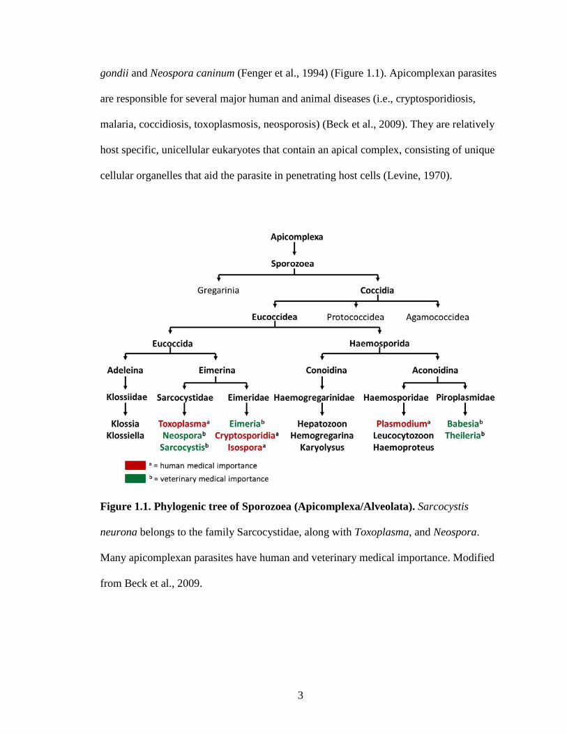
3
gondii and Neospora caninum (Fenger et al., 1994) (Figure 1.1). Apicomplexan parasites
are responsible for several major human and animal diseases (i.e., cryptosporidiosis,
malaria, coccidiosis, toxoplasmosis, neosporosis) (Beck et al., 2009). They are relatively
host specific, unicellular eukaryotes that contain an apical complex, consisting of unique
cellular organelles that aid the parasite in penetrating host cells (Levine, 1970).
Figure 1.1. Phylogenic tree of Sporozoea (Apicomplexa/Alveolata). Sarcocystis
neurona belongs to the family Sarcocystidae, along with Toxoplasma, and Neospora.
Many apicomplexan parasites have human and veterinary medical importance. Modified
from Beck et al., 2009.
4
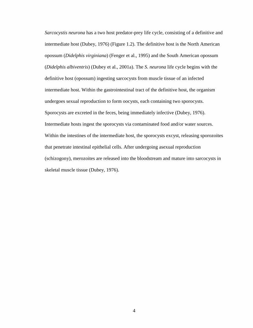
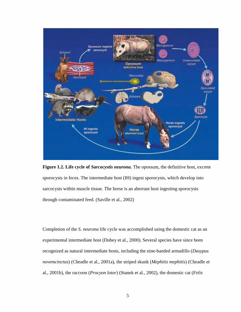
Sarcocystis neurona has a two host predator-prey life cycle, consisting of a definitive and
intermediate host (Dubey, 1976) (Figure 1.2). The definitive host is the North American
opossum (Didelphis virginiana) (Fenger et al., 1995) and the South American opossum
(Didelphis albiventris) (Dubey et al., 2001a). The S. neurona life cycle begins with the
definitive host (opossum) ingesting sarcocysts from muscle tissue of an infected
intermediate host. Within the gastrointestinal tract of the definitive host, the organism
undergoes sexual reproduction to form oocysts, each containing two sporocysts.
Sporocysts are excreted in the feces, being immediately infective (Dubey, 1976).
Intermediate hosts ingest the sporocysts via contaminated food and/or water sources.
Within the intestines of the intermediate host, the sporocysts excyst, releasing sporozoites
that penetrate intestinal epithelial cells. After undergoing asexual reproduction
(schizogony), merozoites are released into the bloodstream and mature into sarcocysts in
skeletal muscle tissue (Dubey, 1976).
5
Figure 1.2. Life cycle of Sarcocystis neurona. The opossum, the definitive host, excrete
sporocysts in feces. The intermediate host (IH) ingest sporocysts, which develop into
sarcocysts within muscle tissue. The horse is an aberrant host ingesting sporocysts
through contaminated feed. (Saville et al., 2002)
Completion of the S. neurona life cycle was accomplished using the domestic cat as an
experimental intermediate host (Dubey et al., 2000). Several species have since been
recognized as natural intermediate hosts, including the nine-banded armadillo (Dasypus
novemcinctus) (Cheadle et al., 2001a), the striped skunk (Mephitis mephitis) (Cheadle et
al., 2001b), the raccoon (Procyon lotor) (Stanek et al., 2002), the domestic cat (Felis
6
domesticus) (Stanek et al., 2003), and the sea otter (Enhydra lutris) (Dubey et al., 2001c).
The horse is considered an aberrant or dead-end host, since S. neurona does not mature
into sarcocysts in the muscles of infected horses (Dubey et al., 2001b). Therefore, horses
are unable to infect a definitive host, resulting in an incomplete life cycle (MacKay,
1997a).
1.4. Pathogenesis
The pathogenesis of EPM in the horse is not thoroughly understood. Horses ingest food
and/or water sources contaminated with S. neurona sporocysts, which travel to the
intestine. The sporocysts excyst, releasing sporozoites, which invade the intestinal
epithelium, progressively moving into the endothelium of blood vessels. The organisms
asexually reproduce to form meronts, causing the endothelial cell to rupture, releasing
merozoites into the bloodstream (Furr, 2006). The merozoites migrate to the CNS
(MacKay et al., 2000), although the actual mechanism of transport into the CNS has yet
to be determined. It is proposed that S. neurona passes through the blood-brain barrier via
leukocytes or the cytoplasm of endothelial cells (Furr, 2006). By invading the leukocyte,
S. neurona is not only provided access to the CNS, but protection from antibodies
(Lindsay et al., 2006). Once inside the CNS, S. neurona invades neurons and microglial
cells, and slowly undergoes additional asexual reproduction to form schizonts.
Eventually, the structures rupture, releasing merozoites, each of which can repeat the
reproductive process (Simpson and Mayhew, 1980).
7
Several factors likely affect the progression of the disease, including the number of
organisms (Sofaly et al., 2002), length of time before treatment (Saville et al., 2000a),
location of lesion (Dubey et al., 2001b), and stressful events during infection (Saville et
al., 2001).
1.5. Clinical Signs
The clinical signs of EPM are extremely variable due to the possibility of S. neurona
causing lesions anywhere in the CNS. Some horses present with clinical signs that appear
gradually over time, while others display a much faster progression (Reed, 2008). Early
signs of EPM are frequent stumbling and unexplainable lameness of the thoracic or
pelvic limbs. The classic EPM clinical signs are asymmetrical ataxia (incoordination)
with focal muscle atrophy (Dubey et al., 2001b).
Horses with an affected spinal cord exhibit gait abnormalities, manifested as ataxia of the
limbs, generally affecting one side more than the other. This leads to asymmetrical
muscle atrophy of the shoulder or rump (Reed, 2008). Damage to the grey matter results
in focal muscle atrophy and severe muscle weakness; white matter damage manifests as
ataxia and weakness in limbs caudal to the site of damage (MacKay et al., 2000).
Additional signs may include dragging of the hoof especially while turning, hypometria
of forelimbs, and reluctance to back up. Some clinical signs may only be witnessed while
the horse is in training, such as head tossing, inability to maintain a lead, and a sore back
(MacKay, 1997a). In rarer EPM cases involving an affected brain, clinical signs may
include behavioral changes, blindness, seizures, drooping lip or ear, head tilt, and atrophy

8
of the tongue and the muscle of mastication (Furr et al., 2002). The disease can progress
to the point of recumbency (MacKay, 1997a).
1.6. Pathology
Lesions associated with EPM are limited to CNS tissue (Beech and Dodd, 1974; Cusick
et al., 1974; Dubey et al., 1974). The most commonly affected area of the brain is the
brain stem, however, the majority of affected tissue is found in the spinal cord (Dubey et
al., 2001b). Gross lesions are often visible in the grey matter of the spinal cord (MacKay
et al., 2000). Histologically, characteristic lesions are easily recognizable (Beech and
Dodd, 1974). Lesions are multifocal asymmetrical areas of hemorrhage, nonsuppurative
inflammation, and necrosis (Dubey et al., 2001b). Inflammation of tissue is extremely
variable with a mixture of cell types present, including lymphocytes, neutrophils,
eosinophils, multinucleated giant cells, and gitter cells (Dubey et al., 2001b). Other
common findings include perivascular cuffing of blood vessels with mononuclear cells,
phagocytosis of axons with gitter cell formation, and astrocyte proliferation (MacKay et
al., 2000). The white matter of diseased spinal cord tissue is often vacuolated due to the
swelling and degeneration of axons (Beech and Dodd, 1974).
Histologically, S. neurona merozoites are difficult to distinguish from nuclear debris
within areas of necrotic tissue with the use of hematoxylin and eosin (H&E) staining
(Beech and Dodd, 1974). Immunohistochemical (IHC) staining, however, has been
shown to aid in locating and positively identifying S. neurona (Hamir et al., 1993).
Schizonts and merozoites can be located in neurons, giant cells, neutrophils, and
9
macrophages, with some merozoites found free in the tissue (Dubey et al., 2001b;
MacKay et al., 2000).
1.7. Diagnosis
Diagnosis of EPM is often challenging. Not only do clinical signs mimic those of other
diseases, but antemortem diagnostic tests are not always conclusive. Positive serum tests
indicate that a horse has been exposed to S. neurona. A positive cerebrospinal fluid (CSF)
test implies that the parasite has crossed the blood-brain barrier and is suggestive of an
active infection (Furr, 2006).
1.7.1. Differential Diagnosis
Due to the wide range of potential clinical signs, EPM can present as other neurological
diseases (MacKay et al., 2000). A thorough clinical and neurologic exam is necessary to
rule out other neurological conditions (Furr et al., 2002). Cervical vertebral malformation
(CVM) and Equine herpesvirus-1 myeloencephalopathy often result in symmetrical
neurological signs (Mayhew, 1999). Ancillary testing can provide additional information
to rule out other diseases. For example, if cervical spinal cord damage is suspected,
cervical radiographs can aid in diagnosing other possible abnormalities, such as CVM,
cervical fracture, or cervical osteoarthritis (Mayhew, 1999). If cervical spinal cord
compression is suggested, a myelogram would definitively rule out CVM (Furr et al.,
2002). Analysis of CSF is useful in distinguishing between viral and bacterial
meningoencephalitis, as well as CNS trauma (Furr, 2006). The CSF can be tested for red
10
blood cell concentration, as well as for cytologic evaluation (Furr, 2006). An EPM horse
has a normal CSF cytology, while West Nile Virus infected horses have abnormal CSF
cytologies (Furr et al., 2002).
1.7.2. Antemortem Diagnosis
Several antemortem diagnostic tests recognize antibodies against S. neurona. The
immunoblot (Western Blot) was the first test developed for detecting S. neurona-specific
antibodies in the serum and CSF (Granstrom et al., 1993). In 2000, the Western blot was
modified, increasing the sensitivity and specificity by using a bovine serum to block
proteins nonspecific for S. neurona (Rossano et al., 2000). Another diagnostic test, the
Indirect Fluorescent Antibody Test (IFAT) (Duarte et al., 2003; Duarte et al., 2004), is
unable to differentiate between S. neurona and Sarcocystis fayeri infections. This is not
problematic for diagnosis if clinical signs are taken into account, as S. fayeri is
nonpathogenic in the horse (Saville et al., 2004a). Another diagnostic tool is the enzyme-
linked immunosorbent assay (ELISA) for detection of antibodies to the S. neurona
surface antigen-1 protein (SnSAG1) (Ellison et al., 2003). Studies show that the SnSAG1
ELISA has low sensitivity and is not a useful diagnostic tool (Hoane et al., 2005; Johnson
et al., 2010). This is understandable because not all strains of S. neurona express
SnSAG1 (Howe et al., 2008). The development of an ELISA for SnSAG2, SnSAG3, and
SnSAG4 are much more promising and have increased sensitivity and specificity
(Yeargan and Howe, 2011). While the Western blot only detects the presence of
antibodies, the IFAT and ELISA have increased diagnostic value since they are able to
11
quantify the amount of antibody present in samples with end-point titers (Duarte et al.,
2003; Hoane et al., 2005).
Testing paired samples of serum and CSF is useful in the diagnosis of EPM (Furr, 2006).
Passive transfer of antibodies from the blood through the blood-brain barrier results in the
presence of antibodies in the CSF (Furr et al., 2011). Normally, the amount of antibody in
the CSF is proportionate to the amount in the serum (Furr, 2002). However, during an
active CNS infection, antibodies will be produced resulting in a higher amount of
antibody in the CSF than would be proportionally expected (Furr et al., 2011). However,
blood contamination of the CSF can produce a false-positive test result (Finno et al.,
2007; Miller et al., 1999). Two CSF indices used to determine the CSF/serum proportions
are the Goldman-Witmer coefficient (C-value) and the antigen-specific antibody index
(AI) (Furr et al., 2011).
An additional tool for EPM diagnosis is the horse’s response to anti-protozoal treatment.
If the horse responds favorably to treatment, it potentially suffered from EPM. With this
said, even in the event of diagnosis of EPM it is inconclusive as other diseases may
respond to this type of treatment (Bentz et al., 1999).
1.7.3. Postmortem Diagnosis
The “gold standard” (Duarte et al., 2003) and only definitive diagnosis of EPM is a
postmortem microscopic identification of S. neurona in the brain or spinal cord (Furr et
al., 2002). Unfortunately, organisms can be easily overlooked, as usually only a small
number of organisms are located in areas of inflammation and tissue necrosis (Dubey et
12
al., 2001b). The use of IHC staining with S. neurona antibody has been shown to aid in
the detection of S. neurona by increasing the identification of organisms by 31% (20%
identification with H&E staining; 51% with IHC staining) (Hamir et al., 1993). Prior
treatment for EPM with anti-protozoal drugs decreases the likelihood of identifying S.
neurona organisms in tissues (Boy et al., 1990). Due to these complicating issues, an
EPM diagnosis is often based on the presence of characteristic lesions indicative of EPM,
regardless of identification of S. neurona (MacKay et al., 2000).
1.8. Treatment
Time is the most crucial factor in the treatment of EPM. Earlier treatment will reduce the
amount of CNS damage, which can be permanent (Dubey et al., 2001b). Medication will
kill the protozoa, but will not repair the damage to the nervous tissue. If too severely
damaged, the horse may need to be euthanized (Morley et al., 2001). Treatment is fairly
expensive (Saville et al., 2000a), but usually leads to recovery in 70-75% of horses
(Dubey et al., 2001b). There are several treatment options available for EPM.
1.8.1. Anti-protozoal Treatment
The Food and Drug Administration (FDA) has approved several treatments for EPM.
Beginning in the 1970s, sulfadiazine/pyrimethamine was the traditional treatment
(Mayhew et al., 1976). The drug, ReBalance, a combination of sulfadiazine and
pyrimethamine (25%:1.25% suspension), has since been approved by the FDA (MacKay,
2006). ReBalance requires a prolonged administration of 90 to 270 days (Animal Health
Pharmaceuticals, 2004). The drug limits the protozoan’s ability to synthesize folic acid,
13
unfortunately, reducing the host animal to do so as well (MacKay et al., 2000). Long term
treatment may cause anemia. Supplementation with folic acid is rarely necessary if the
horse has access to high quality green forage (i.e., alfalfa hay) (MacKay, 1997a). The
drug should not be administered to pregnant mares, as it has been known to cause
abortions (MacKay, 2006).
The anticoccidial drug, Marquis, a 15% ponazuril paste, is another drug option. This drug
targets S. neurona, while leaving the host tissue unaffected (Bayer Corporation, 2001).
Marquis is administered orally, once daily, for 28 days and appears to be a safe course of
treatment when used at the recommended dosage (Bayer Corporation, 2001). A third
drug, Navigator, a 32% nitazoxanide paste, is administered orally for 28 days (IDEXX
Pharmaceuticals, 2003). Navigator is FDA-approved, but the manufacturer, IDEXX
Pharmaceuticals, stopped production of this drug in 2009. This drug has a broad spectrum
of activity against parasites, bacteria and viruses, including the natural bacterial flora of
the horse’s gastrointestinal system (MacKay, 2006). Consequently, a horse on Navigator
needs to be closely monitored for signs of toxicity, such as colic and diarrhea (MacKay et
al., 2000). The newest drug option is Protazil, a 1.56% pellet of diclazuril (Schering-
Plough Animal Health, 2007). This drug is an anticoccidial and considered relatively safe
for the host tissue (MacKay, 2006). These top-dress pellets are administered for 28 days
(Schering-Plough Animal Health, 2007).
1.8.2. Ancillary Treatment
Additional therapies are sometimes recommended to reduce inflammation and minimize
further CNS damage (MacKay, 1997a). As S. neurona merozoites rapidly die in response
14
to anti-protozoal therapy, an inflammatory response can occur within the CNS.
Nonsteroidal anti-inflammatory drugs (NSAIDs) (i.e., flunixin meglumate) and dimethyl
sulfoxide (DMSO) are recommended during the first 1-2 weeks of treatment (MacKay,
2006). Corticosteroids are usually avoided, as they may increase neurologic symptoms
(Cutler et al., 2001). However, some horses exhibit increasingly severe clinical signs at
the onset of treatment, and the use of a corticosteroid may help control the inflammatory
response initiated by the anti-protozoal drug (MacKay et al., 2000). Vitamin E can be
given throughout the course of anti-protozoal therapy to prevent further oxidative damage
of the CNS and to promote healing (MacKay, 1997a). Previous research suggests that
EPM horses exhibit a decreased cell-mediated immunity, in particular a Th-1 response
(Tornquist et al., 2001). Thus, non-specific immune stimulants, such as levamisole, killed
Propionibacterium acnes, and Mycobacterium wall extract are sometimes used in
addition to anti-protozoal therapy (MacKay, 2006).
1.9. Prognosis
The prognosis of a horse diagnosed with EPM is dependent upon the severity of clinical
symptoms and the response to treatment (Saville et al., 2000a). Sixty percent of
moderately to severely affected horses usually improve after treatment, with 10-20%
recovering completely and are highly prone to relapse. Eighty percent of mildly affected
horses will improve, with 50% recovering completely and being less prone to relapse
(MacKay, 2006).

15
Horses treated earlier are more likely to recover completely (Saville et al., 2000a).
Treated horses are 10 times more likely to improve than are horses left untreated
(MacKay et al., 2000). A relapse, or the reoccurrence of clinical signs once treatment has
ended, is estimated to occur in 10% of horses within 3 years of discontinuing treatment
(MacKay, 2008).
1.10. Epidemiology
Prevalence of EPM is dependent upon the geographic distribution of the opossum (D.
virginiana and D. albiventris), which is limited to North, Central, and South America
(Dubey et al., 2001a; Fenger et al., 1995). Reports of EPM cases in the eastern
hemisphere are occasionally identified in horses that originated from the Americas (Lam
et al., 1999; Mayhew and Greiner, 1986; Ronen, 1992). The United States Department of
Agriculture (USDA) reports that only 14 of 10,000 horses are actually diagnosed with
EPM each year in the United States (US) (National Animal Health Monitoring System
(NAHMS), 2001). This number is significantly smaller than the estimated 50% exposure
rate of horses in the US (MacKay, 1997b). Seroprevalence of S. neurona varies
throughout the country: 89.2% in Oklahoma (Bentz et al., 2003), 53.6% in Ohio (Saville
et al., 1997), 45.3% in Chester County, Pennsylvania (Bentz et al., 1997), 45% in Oregon
(Blythe et al., 1997), and 33.6% in Colorado (Tillotson et al., 1999). Variation of
seroprevalence within each region is affected by different climatic factors. Oregon
exhibited a higher seroprevalence in wetter coastal regions than in more arid regions
16
(Blythe et al., 1997). Ohio showed an association between a decrease in seroprevalence
and the number of days the temperature was below freezing (Saville et al., 1997).
Several risk factors have been associated with the development of EPM. While
seroprevalence increases with the age of the horse (Bentz et al., 2003; Bentz et al., 1997;
Blythe et al., 1997; Saville et al., 1997; Tillotson et al., 1999), younger horses (1-5 years
old) are at a higher risk for actually developing EPM (Boy et al., 1990; Cohen et al.,
2007; Saville et al., 2000b). Certain breeds, such as Standardbred and Thoroughbred,
appear to have a higher incidence of EPM (Boy et al., 1990; Rooney et al., 1970). These
breeds are often associated with intense training at an early age, which could compromise
their immune system. Horses involved in racing or competitions have an increased risk
for EPM (Cohen et al., 2007; Saville et al., 2000b). Other stressful events that may be
associated with EPM include injury/accident, surgery, and parturition (Saville et al.,
2000b). The occurrence of EPM increases during the spring, summer and fall (Saville et
al., 2000b), which may be due to a climatic effect (Saville et al., 1997) or an increased
amount of travel and competitions during these months. The presence of the opossum
directly correlates with EPM cases, as the incidence of EPM is greater in areas that
contain higher populations of opossums (MacKay et al., 2000). The risk of EPM is
greater if wooded areas surround the farm and lower if a creek or river is close to the
property (Saville et al., 2000b).

17
1.11. Prevention
Certain management practices can be followed to decrease horse exposure to S. neurona.
Trapping or keeping opossums away with specialized fencing (MacKay, 1997a) will
prevent them from accessing the food and water sources of the horse (Saville et al.,
2000b). Keeping grain in securely closed containers and cleaning up any spilled grain,
fallen fruit, or bird seed will eliminate the opossums’ food sources (MacKay, 2006), as
well as storing forage (hay) in a facility that excludes wildlife (MacKay et al., 2000).
Providing water sources for horses, such as water troughs, that are separate from ponds or
creeks removes them from access to wildlife. Removing carcasses from the property and
disposing of them properly prevents scavenging by opossums (Saville et al., 2002).
The use of an anti-protozoal drug as a daily preventative has been proposed (Saville et
al., 2002). Protazil (diclazuril) appears promising, as it easily dispenses as a top-dress to
horse feed (Schering-Plough Animal Health, 2007). However, it is unknown how a daily
preventative affects the horse’s natural immune response to S. neurona. Ultimately, this
option is not cost-effective (MacKay, 2006). Another preventative option is the use of the
drug, Marquis (ponazuril), intermittedly or during times of stress (Furr et al., 2006).
Fort Dodge Animal Health manufactured a vaccine from killed, cultured S. neurona
merozoites in a MetaStim® adjuvant (MacKay, 2006). The USDA gave the vaccine a
conditional license in 2000. The efficacy of the vaccine remained to be determined, as
there was little data to support the effectiveness of the vaccine in preventing EPM (Marsh
et al., 2004). As a result, the vaccine was removed from the market in 2009.
18
1.12. Cell-mediated Immunity
Cell-mediated immunity (CMI) is important for the elimination of intracellular parasites
(Gazzinelli et al., 1994; Khan et al., 1997). To understand the role of the immune system
in S. neurona infection, studies were initially conducted on mice. When injected
subcutaneously with S. neurona merozoites, immunocompetent mice did not develop
neurologic disease, nor were there any lesions in CNS tissue (Witonsky et al., 2003b).
While there was an initial increase in the percentage of CD8+ peripheral blood
lymphocytes and CD8+ splenocytes, levels returned to normal by the end of the 28 day
study. This suggests that the immune response from a healthy immune system can
prevent disease. CD8+ T cells seem to be involved in preventing disease in mice, as CD8
cell knockout (KO) mice inoculated with S. neurona merozoites developed
meningo/encephalomyelitis (Witonsky et al., 2005).
Interferon-gamma (IFN-γ), a cytokine secreted by T cells (CD8+ and CD4+), natural
killer (NK) cells, and IFN-γ producing non-T cells (Suzuki, 2002), has been shown to
play a role in preventing neurologic infection. When infected with S. neurona, IFN-γ KO
mice quickly developed meningo/encephalitis (Dubey and Lindsay, 1998; Witonsky et
al., 2003a). Neurologic disease did not occur when severe combined immunodeficiency
(SCID) mice were infected with S. neurona (Marsh et al., 1997). SCID mice lack
adaptive immune responses from specific B and T cells, but have functioning NK cells. It
is thought that the NK cells in SCID mice produce enough IFN-γ to protect them from
neurologic disease (Sellon et al., 2004a). SCID mice treated with anti-IFN-γ antibody
rapidly developed severe neurologic disease, highlighting the importance of this cytokine
(Sellon et al., 2004a).
19
Limited research has been conducted on the CMI response in EPM horses. Studies used
both naturally and experimentally infected horses. Initial research has shown that EPM
horses have reduced cell-mediated responses to antigen-specific mitogens (Spencer et al.,
2005; Spencer et al., 2004; Tornquist et al., 2001). It is difficult to determine if the
immunosuppression is a result of the parasite suppressing the horse’s immune response,
or rather if the condition existed prior to the infection and is essential for the development
of the disease (Tornquist et al., 2001). Both naturally and experimentally infected horses
showed a suppressed response to the non-antigen specific mitogen, phorbol myristate
acetate/ionomycin (PMA/I), in vitro (Witonsky et al., 2008; Yang et al., 2006). Compared
to normal horses, EPM horses have increased interleukin (IL)-4 expression (Spencer et
al., 2005) and suppressed IFN-γ mRNA expression in lymphocytes (Spencer et al., 2004).
Studies show contrasting results regarding immune cell subsets. When compared to non-
EPM horses, Tornquist et al. (2001) saw a decrease in CD4+ cells, while Yang et al.
(2006) documented an increase in CD4+ cells. Witonsky et al. (2008) was the only study
to note a decreased percentage of CD8+ cells. As these studies were completed in vitro,
they likely do not represent the actual immune response within the horse.
1.13. Infectious Disease Susceptibility
The interaction between environmental and genetic factors is responsible for the
development of infectious disease (Casanova and Abel, 2005). Exposure to infectious
pathogens is critical for the progression of disease, while genetic factors affect an
individual’s susceptibility to disease (Kwiatkowski, 2000). Genetic predisposition to

20
infectious disease is considered to be either monogenic or polygenic (Alcais et al., 2009).
Monogenic, or primary immunodeficiencies (PID), predisposition results from a single
gene mutation causing susceptibility to a single disease (Casanova and Abel, 2007).
Monogenic diseases have rare susceptibility alleles, display a high penetrance with a
severe phenotype, and follow a Mendelian inheritance pattern (Casanova and Abel,
2005). Examples of PIDs include Mendelian susceptibility to Mycobacterial disease
(MSMD) and pyogenic bacterial infections. Children suffering from MSMD are
susceptible to weakly virulent Mycobacteria, but show resistance to most other infectious
agents (Alcais et al., 2005). These children have a mutation in the IL-12-IFN-γ pathway
(Dorman and Holland, 2000; Newport et al., 1996). Pyogenic bacterial infections result
from a susceptibility to Streptococcus pneumoniae due to an interleukin-1 receptor-
associated kinase (IRAK)-4 deficiency (Picard et al., 2003).
The majority of infectious diseases are complex. A combination of multiple genes are
involved in polygenic predisposition with each gene contributing slightly to the overall
susceptibility (Alcais et al., 2009). Complex diseases have common susceptibility alleles
and do not follow the Mendelian pattern of inheritance (Casanova and Abel, 2005).
Schistosomiasis was the first infectious disease to have mapped a susceptibility locus.
This locus controls the level of infection with the parasite Schistosoma mansoni (Marquet
et al., 1996). Diseases, such as leprosy (Abel and Demenais, 1988) and pulmonary
tuberculosis (Alcais et al., 2005), display a polygenic predisposition. Variants in the
regulatory region of PARK2 and PACRG have been associated with leprosy
susceptibility (Mira et al., 2004). Pulmonary tuberculosis susceptibility has been linked to
the chromosomal region 8q12-q13 (Baghdadi et al., 2006), as well as a mutation in the

21
promoter region of the monocyte chemoattractant protein-1 (MCP1) (Flores-Villanueva
et al., 2005).
The genetic background of mouse strains affects their susceptibility to disease. Thus,
Balb/c mice are resistant to the coccidium Eimeria ferrisi, while C57BL/6 mice are
susceptible to this infection (Klesius and Hinds, 1979). Different susceptibility of mice
strains was also evidenced with T. gondii infection. At a high dose (1 x 10⁵ T. gondii
trophozoites), Balb/c and DBA/2 mice were most susceptible to infection, while DBA/1
and white SW/SIM strains were the most resistant. However, a lower dose (1 x 10³ T.
gondii trophozoites), Balb/c mice became one of the more resistant strains (Araujo et al.,
1976). The variation of survival between the mice strains seems to be dependent upon
genes of the major histocompatibility complex (MHC), as well as non-MHC genes
(Deckert-Schluter et al., 1994; Williams et al., 1978).
1.14. Genome-wide Association Study
Genome-wide association studies (GWAS) have become a powerful approach in
identifying genes involved with monogenic and complex traits (McCarthy et al., 2008).
Genome-wide association studies scan the entire genome for common variants, and
usually compare a group that has a certain trait (i.e., disease) with a control group that
does not exhibit the trait (Pearson and Manolio, 2008).
Single nucleotide polymorphisms (SNP) are the most common sequence variant (Collins
et al., 1998), and are used as markers throughout the genome (Pearson and Manolio,
2008). These variants create diversity in populations, as well as being responsible for an

22
individual’s disease susceptibility. Most SNPs do not affect the function of the gene,
instead they aid in locating other mutation(s) that are impacting gene function. SNPs that
do modify functionality of the gene are usually located in the regulatory or coding
regions of the genome (Collins et al., 1998). SNP chips are tools that can rapidly analyze
the genome for SNPs and map them to specific regions on chromosomes (Chowdhary et
al., 2008). These arrays have probes that target thousands of SNPs, which uniformly span
the entire genome. This high-throughput genotyping technology is now commercially
available (Chowdhary and Raudsepp, 2008).
A GWAS typically employs four steps. First, a sample set is chosen, consisting of
individuals displaying a specific trait or disease. A comparison group is necessary that
does not exhibit the trait or disease of interest (Wellcome Trust Case Control Consortium,
2007). For all individuals, DNA is extracted and genotyped. Statistical tests are used to
assess for any association between SNPs and the trait or disease of interest (Pearson and
Manolio, 2008). Finally, a technical validation is necessary, as well as a replication of the
study in an independent population, to ensure that the results represent valid associations
(McCarthy et al., 2008).
Through the use of GWAS, loci have been identified that implicate predisposition to
disease (McCarthy et al., 2008). Such diseases include type 1 (Hakonarson et al., 2007)
and type 2 diabetes (Steinthorsdottir et al., 2007), inflammatory bowel disease or Crohn’s
disease (Hampe et al., 2007), rheumatoid arthritis (Begovich et al., 2004), and behavioral
disorders, such as schizophrenia and bipolar disorder (Craddock et al., 2005).
Susceptibility to infectious diseases is usually polygenic and considered a complex trait,
as several genes are generally involved (Bellamy, 2006). One of the first infectious

23
disease GWAS was performed in 2007, discovering genetic determinants that affect the
viral load during asymptomatic stages of HIV (Fellay et al., 2007). Another early
infectious disease GWAS was performed on Kawasaki disease, finding several variants in
genes that are involved with the pathogenesis (Burgner et al., 2009). Knowledge of
affected genes can help identify pathways involved with disease susceptibility, increase
understanding of the pathogenesis, and aid in the development of potential therapeutics
(Davila and Hibberd, 2009).
The technology used in conducting GWAS in humans is now being applied in other
species, such as the cow and horse. Sequencing the genome of these animals was
instrumental in the development of tools specific for analyzing their genomes, making the
study of complex traits and diseases possible (Chowdhary and Raudsepp, 2008). In cattle,
GWAS have been used heavily for improvement of production, conformation, and
fitness. In the dairy industry, GWAS has been instrumental in breeding animals that have
increased milk production (Wiggans et al., 2011). In the past, equine researchers have
used gene maps and comparative genomics to identify monogenic traits, such as
metabolic disorders (Dranchak et al., 2007), hereditary skin abnormalities (Tryon et al.,
2007), and immune system disorders (Shin et al., 1997). The equine SNP chip has been
implemented in numerous studies of more complex traits and diseases, including
recurrent laryngeal neuropathy (Dupuis et al., 2011), osteochondrosis (Lykkjen et al.,
2010), dwarfism (Orr et al., 2010), and equine viral arteritis (Go et al., 2011). This
technology has also been used to develop optimum racing distance for Thoroughbreds
(Hill et al., 2010) and desired coat colors (Reissmann et al., 2007).
24
1.15. Equine EPM Model
The development of an equine model of EPM would significantly enhance knowledge of
the disease. An equine model would aid in understanding the pathogenesis of EPM and
lead to potential improvements in the diagnosis, treatment, and prevention of the disease.
Several studies have attempted to induce EPM with varying levels of success, utilizing
three different methods to introduce S. neurona into the horse.
1.15.1. Intragastric Introduction
Intragastric introduction was the first method used to create an EPM model. Two
approaches were used in the preparation of S. neurona sporocysts inoculums. In one
approach, the sporocysts were collected from intestinal scrapings of naturally infected
opossums (Fenger et al., 1997). Since the opossum is the definitive host for three
Sarcocystis spp (S. neurona, Sarcocystis falcatula, Sarcocystis speeri) (Rosenthal et al.,
2001), the inoculums were not pure S. neurona, but a mixture of Sarcocystis spp (Fenger
et al., 1997). Bioassay or polymerase chain reaction (PCR) characterized the S. neurona
inoculums from opossum’s intestinal scrapings (Cutler et al., 2001; Saville et al., 2001).
In a second approach, sporocysts were collected from laboratory-raised opossums. In this
approach, tongues of naturally infected raccoons were fed to laboratory-raised opossums
whose intestines were scraped for sporocysts, which were then fed to laboratory-raised
raccoons. The raccoon muscle was subsequently fed to laboratory-raised opossums, from
which the sporocysts were finally collected (Sofaly et al., 2002).
The initial study by Fenger et al. (1997) introduced an inoculum of mixed Sarcocystis spp
sporocysts from the intestines of naturally infected feral opossums directly into the foals’
25
stomachs using a nasogastric tube. One to three doses of at least 2 x 10⁶ sporocysts were
administered to the foals. All foals seroconverted between 24 and 42 days post
inoculation (dpi) and had CSF antibodies present between 24 and 42 dpi. Foals displayed
mild neurologic symptoms, with 3 of the 5 foals exhibiting histopathologic lesions in the
brainstem and/or spinal cord. However, no organisms were observed in the tissues
(Fenger et al., 1997).
In a second study, Cutler et al. (2001) corticosteroids were used as an immunomodulator
to induce EPM. An inoculum of PCR-characterized S. neurona sporocysts, collected from
the intestines of naturally infected feral opossums, was administered via a nasogastric
tube to horses for 7 consecutive days at a dose of 5 x 10⁵ S. neurona sporocysts. A
treatment group was additionally administered 0.1 mg/kg of the corticosteroid,
dexamethasone, daily beginning 7 days prior to the first inoculation through the
termination of the study. All horses seroconverted in the blood and CSF, but the
dexamethasone-treated horses immunoconverted in less time and showed increased
clinical signs. Unfortunately, it was difficult to determine if the horses displayed
neurologic signs from the S. neurona infection or signs of weakness from systemic
disease caused by the immunosuppression (Cutler et al., 2001).
Saville et al. (2001) examined the effect of stress, specifically corticosteroids and
transportation, on generating EPM in the horse. Bioassay characterized S. neurona
sporocysts from the intestines of naturally infected feral opossums were administered in a
single dose of 8 x 10⁴ sporocysts via nasogastric tube to horses in 2 treatment groups.
Horses were then transported for 55 hours to create stress. Upon arrival at the facility,
one treatment group was immediately administered S. neurona sporocysts. A second
26
treatment group was allowed 14 days to acclimate, and then given 0.5 mg/kg dose of
dexamethasone prior to being inoculated with 8 x 10⁴ S. neurona sporocysts. This group
continued to receive 0.2 mg/kg of dexamethasone twice a week for the remainder of the
study. While all horses immunoconverted in both serum and CSF, transportation resulted
in a quicker seroconversion time (Saville et al., 2001). Additionally, dexamethasone-
treated horses presented milder clinical signs, which contradicts previous study results
from Cutler et al. (2001). Since transportation stress seemed to be effective in inducing
EPM, Saville et al. (2004b) conducted another study to test the effect of a second
transportation. The inoculums of S. neurona sporocysts were taken from laboratory-
raised opossums. Horses underwent prolonged transportation and then were inoculated
with a single dose of 1.5 x 10⁶ S. neurona sporocysts. One treatment group was not
transported again, while a second treatment group was transported again at either 4, 11,
or 18 dpi. All horses seroconverted between 12 and 21 dpi. The treatment group with the
single transportation exhibited more severe clinical signs than the horses with the second
transport (Saville et al., 2004b).
Sofaly et al. (2002) studied the effect of inoculation doses on the induction of EPM.
Inoculua of S. neurona sporocysts, derived from laboratory-raised opossums, were
administered to horses in doses varying from 102 - 106. There was a dose-dependent
relationship, as increased doses resulted in earlier seroconversion time. Results showed
that a dose of at least 106 was necessary to consistently induce S. neurona infection
(Sofaly et al., 2002).
The method of inducing EPM via S. neurona sporocysts to the stomach of the horse
resulted in fairly consistent findings between studies. Horses had both serum and CSF
27
immunoconversion, mild to moderate clinical signs, and occasional lesions present in the
brain/spinal cord tissue. However, this method was unable to reproduce severe clinical
signs (i.e., asymmetrical signs, inability to rise) and S. neurona organisms were never
recovered nor seen histopathologically.
1.15.2. Intrathecal Introduction
Intrathecal introduction was the second method used to create an EPM model. In a study
by Lindsay et al. (2000), S. neurona merozoites were directly introduced into the CNS of
the horse. In an effort to maintain a consistent CSF volume, 10mL of CSF were removed
from the horses prior to injecting 5 x 10⁶ culture derived S. neurona merozoites
suspended in 10mL plasmalyte into the subarachnoid space. Horses did seroconvert and
antibodies were present in the CSF, but no clinical signs of EPM were evident (Lindsay
et al., 2000). Intrathecal introduction of S. neurona merozoites, therefore, did not prove to
be a successful method to establish EPM.
1.15.3. Intravenous Introduction
The final method used to create an EPM model was through an artificial parenteral
introduction of S. neurona. While investigating S. neurona parasitemia in SCID horses,
Sellon et al. (2004b) injected intravenously 5 x 10⁸ S. neurona culture derived merozoites
into both SCID and immunocompetent horses. Neurological symptoms were present in 2
of the 3 immunocompetent horses. Immunocompetent horses were able to control
parasitemia. Using PCR, S. neurona was not detected in visceral tissues, but was present
in neural tissue. Conversely, none of the SCID horses developed any neurologic signs.
28
However, SCID horses were unable to control parasitemia, and S. neurona was detected
in visceral tissues (Sellon et al., 2004b).
Ellison et al. (2004) developed a procedure to infect horses with host lymphocytes
containing intracellular S. neurona. Blood was collected from the horse, and the buffy
coat was isolated and incubated with S. neurona merozoites for 5 hours. The infected
cells were added to 6mL of collected blood and injected intravenously back into the
horse. A single horse was given 100,000 merozoites at four different times, each a week
apart. Three other horses were dosed with varying amounts of merozoites (100, 1,000,
10,000) daily for 15 consecutive days. All horses developed moderate clinical signs, with
blood and CSF immunoconversion by day 7. One of the challenged horses was a
pregnant mare. Within the lung tissue of the fetus, the author states that an organism or
artifact was identified with H&E staining and there was antibody binding using IHC
staining (Ellison et al., 2004). It seems more likely that what the author viewed was
actually an artifact, as S. neurona is not known to be present in the lung tissue.
A second study by Ellison and Witonsky (2009) modified this procedure. Blood from
each horse was collected in an ethylenediaminetetraacetic acid (EDTA) tube, and 6,000
S. neurona merozoites were directly inoculated into each of the blood tubes. Blood tubes
were incubated at 37°C overnight. The blood was then injected intravenously back into
the horse, and the process was repeated for 14 consecutive days. This resulted in horses
developing clinical signs with serum and CSF immunoconversion (Ellison and Witonsky,
2009).
29
The intravenous method of induction of EPM resulted in moderate clinical signs and
earlier seroconversion times than the intragastric or intrathecal methods. Unfortunately,
S. neurona has yet to be identified histologically within lesions of the CNS tissue.
1.16. Research Objectives
Although extensive EPM research has been conducted over the past two decades, there is
still much to learn about this disease. The objective of the two studies in this thesis was to
further understand factors involved in the development of EPM within the horse. The first
study explored a possible genetic susceptibility to EPM, as only a small number of horses
exposed to S. neurona actually develop the disease. The susceptibility was investigated
by performing a genome-wide association study (GWAS) on formalin-fixed, paraffin-
embedded (FFPE) tissues from definitively positive EPM horses. The second study tested
the viability of a previously described artificial infection method to create a reliable
equine EPM model. The development of a working EPM model would be useful in
understanding the pathogenesis of the disease, and aid in the diagnosis, treatment, and
prevention of EPM. Additionally, the immune response to the challenge infection was
examined in the second study.
30
CHAPTER TWO
Genome-wide association study to investigate genetic susceptibility of
equine protozoal myeloencephalitis
2.1. Introduction
Equine protozoal myeloencephalitis (EPM) is the most commonly diagnosed
neurological disease of horses in the United States (Dubey et al., 2001b). The disease is
caused by the protozoan parasite, Sarcocystis neurona (Dubey et al., 1991). Horses are an
aberrant host of S. neurona, and are infected by ingesting feed or water contaminated
with sporocysts. Within the horse, the organism infiltrates the central nervous system and
initiates an immune response, which leads to damaged tissue and neurological disease
(MacKay et al., 2000). Diagnosis of EPM is challenging, with microscopic identification
of S. neurona in the brain or spinal cord during postmortem examination the only
definitive diagnosis (Furr et al., 2002).
Seroprevalence studies show an exposure rate to S. neurona of approximately 50% in
horses of the United States (MacKay, 1997b). However, only 0.5-1% of all horses are
actually diagnosed with EPM (Dubey et al., 2001b). The factors involved in facilitating
disease development are still unknown. Some breeds, such as the Thoroughbred and
Standardbred, seem to have higher incidences of EPM (Boy et al., 1990; Rooney et al.,
1970). Researchers have proposed a genetic basis for the susceptibility to EPM, but no
studies have been performed to date. Investigating a potential genetic predisposition is
now possible due to the availability of the equine genome sequence and technologies that
31
scan the genome to identify variants associated with susceptibility to disease (Wade et al.,
2009).
The goal of this study was to perform a genome-wide association study (GWAS) of
definitively positive EPM horses. This GWAS used archived formalin-fixed, paraffin-
embedded (FFPE) tissues to identify potential genetic variants associated with disease
susceptibility.
2.2. Materials and Methods
2.2.1. Case Selection
The case report database of the University of Kentucky Veterinary Diagnostic Laboratory
(UK VDL) was searched to identify cases from 1993-2011 with a diagnosis of EPM.
Each case report was reviewed for a statement of identification of protozoal organisms in
CNS tissue. Archived hematoxylin and eosin (H&E) stained slides of the brain and spinal
cord tissues from these cases were examined by light microscopy to confirm the presence
of parasites.
2.2.2. Immunohistochemistry
Immunohistochemical (IHC) staining was completed using an automated staining system
(Bond-maX, Leica Biosystems, Newcastle Upon Tyne, UK) following the manufacturer’s
IHC Protocol F and the Bond Polymer Refine detection system (Leica Biosystems). This
procedure involved an automated dewaxing and rehydration of the tissue, continuing with
32
a heat-induced antigen retrieval using a ready-to-use citrate based buffer (pH 6.0) and
surfactant (Leica Biosystems) at 100°C for 20 minutes. Slides were then incubated with
3% hydrogen peroxide (H2O2) for 5 minutes, followed by application of anti-S. neurona
rabbit serum diluted 1:2500 with Bond Primary Antibody Diluent (Leica Biosystems),
and incubated for 15 minutes. Slides were treated with postprimary blocking reagent for 8
minutes and then horseradish peroxidase-labeled IgG polymer for 8 minutes.
Diaminobenzidine tetrahydrochloride (DAB) substrate was added, and the slides were
incubated for 10 minutes. Finally, the slides were counterstained with hematoxylin for 5
minutes. Between each incubation step, slides were washed using Bond Wash Solution
10x Concentrate (Leica Biosystems), diluted with distilled water to a 1x working
concentration, to remove any unbound material. Brain tissue from a clinical EPM horse
with a large number of S. neurona organisms was stained as a control. A negative control
consisted of only Bond Primary Antibody Diluent (Leica Biosystems).
Slides were removed from the instrument and washed under distilled water. Tissue was
dehydrated using a graded series of alcohol washes (2-85% ethanol, 2-90% ethanol, 2-
95% ethanol, 2-100% ethanol) and cleared with 4 xylene washes. Slides were covered
using mounting medium and glass coverslips, and viewed with light microscopy to
identify S. neurona organisms.
2.2.3. Genotyping
For each confirmed EPM case, 5 µm serial sections were cut from FFPE spleen/liver or
cerebellum tissue. Five tissue scrolls from serial sections of each horse were placed in
labeled Eppendorf tubes. Samples were sent to GeneSeek, Inc. (Lincoln, Nebraska) for
33
genotyping. DNA was isolated from tissues using a phenol/chloroform extraction
method. Samples were run on a single Equine SNP50 BeadChip (Illumina, San Diego,
California) using the manufacturer’s protocols. This array contains 54,602 single
nucleotide polymorphisms (SNPs) evenly distributed across all 31 autosomes with an
average probe spacing of 43.2kb, derived from the EquCab2.0 SNP assembly of the horse
genome (http://www.broadinstitute.org/mammals/horse). Average call rate was used for
quality control.
2.3. Results
2.3.1. Case Selection
The UK VDL database contained 194 cases with a diagnosis of EPM, or suspected EPM,
between 1993 and 2011. In 36 case reports, pathologists stated that organisms were
observed in the CNS by histopathologic examination.
Signalment was obtained from each of the 36 case reports (Table 2.1). A majority of the
horses were Thoroughbreds (n=29), other breeds included Quarter Horses (n=3),
Tennessee Walking Horse (n=1), Percheron (n=1), and mixed breeds (n=1). There was
minimal difference between the number of males (n=15) and females (n=19). Of the
horses with a known age, about half (n=16) were 1-5 years old, while the remainder
(n=15) were 6-30 years of age.
34
Table 2.1. Horses used in the study.
ID # Year Breed Sex Age Parasites
Identified – H&E
Parasites Identified -
IHC
1 1993 Thoroughbred Female 5 Yes Yes 2 1993 Thoroughbred Female 4 Yes Yes 3 1994 Quarter Horse Male 9 Yes Yes 4 1994 Tennessee Walking Horse Female 9 Yes Yes 5 1994 Thoroughbred Female 16 Yes N/AB 6 1995 Thoroughbred Male 2 Yes Yes 7 1995 Thoroughbred Female 3 N/AA N/AB 8 1996 Thoroughbred Unknown Unknown Yes Yes 9 1997 Thoroughbred Female 19 Yes Yes 10 1998 Mixed Breed Male Adult Yes Yes 11 1998 Thoroughbred Male Unknown Yes Yes 12 1999 Thoroughbred Female Adult Yes No 13 2000 Thoroughbred Female 6 Yes Yes 14 2001 Thoroughbred Male 15 Yes Yes 15 2001 Quarter Horse Female 3 Yes Yes 16 2002 Thoroughbred Male 2 Yes Yes 17 2003 Thoroughbred Female 3 Yes Yes 18 2003 Thoroughbred Female 5 Yes Yes 19 2004 Thoroughbred Female 3 Yes Yes 20 2006 Thoroughbred Male 1 Yes Yes 21 2006 Thoroughbred Female 13 Yes Yes 22 2006 Thoroughbred Male 3 Yes Yes 23 2006 Thoroughbred Male 12 Yes No 24 2007 Quarter Horse Male 8 Yes Yes 25 2007 Thoroughbred Male 3 Yes Yes 26 2008 Thoroughbred Male 18 Yes Yes 27 2010 Thoroughbred Unknown 1 Yes Yes 28 2010 Thoroughbred Male 1 Yes Yes 29 2010 Thoroughbred Female 30 Yes Yes 30 2011 Thoroughbred Female 7 Yes Yes 31 2011 Thoroughbred Female Adult Yes Yes 32 2011 Percheron Male 17 Yes Yes 33 2011 Thoroughbred Female 2 Yes Yes 34 2011 Unknown Female 14 Yes Yes 35 2011 Thoroughbred Female 11 Yes Yes 36 2011 Thoroughbred Male 2 Yes No
A = No slides available at UK VDL B = No tissue blocks available at UK VDL
35
2.3.2. Confirmation of Sarcocystis neurona
For all cases in which archived slides were available (n=35), parasites were visualized on
H&E stained slides (Table 2.1). The infection presented differently across the cases.
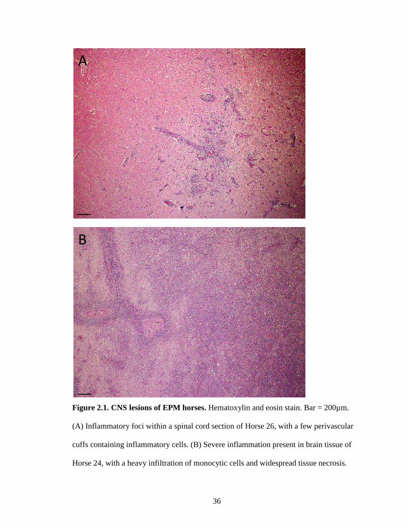
Lesions varied from focal areas of inflammation to severe inflammation with widespread
necrosis (Figures 2.1A & 2.1B). Parasites were generally located within areas of
inflammation (Figure 2.2A). The number of parasites present varied, but was usually
associated with the degree of inflammation/necrosis. Thus, the more severe the
inflammation, typically the higher number of parasites observed.
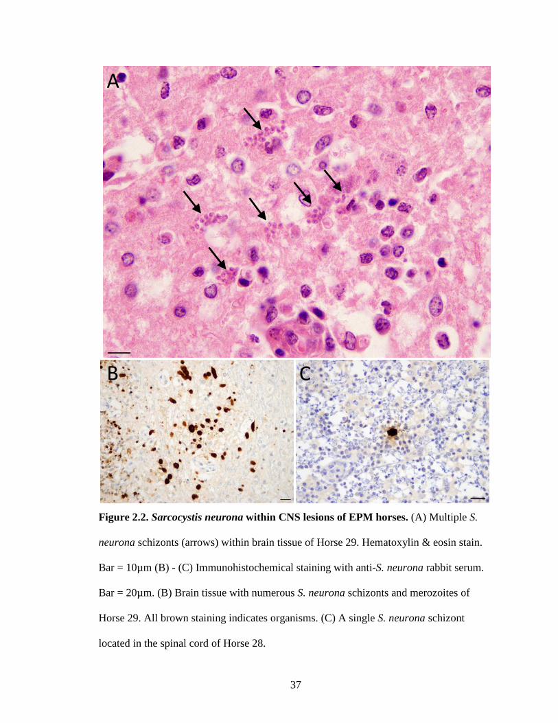
Immunohistochemical staining was used for further confirmation that the parasites
present in the tissues were S. neurona, as IHC staining allows for identification of
parasites, especially merozoites. The number of parasites located with IHC staining
varied across cases. Within lesions, only a single S. neurona parasite was observed in
some horses, while multiple S. neurona parasites in various stages of growth were seen in
other horses (Figures 2.2B & 2.2C). Of the cases where tissue blocks were available
(n=34), S. neurona parasites were identified in 31 cases (Table 2.1). Only a single
parasite was observed in the corresponding H&E slide of the 3 cases that were not
positive by IHC staining.
36
Figure 2.1. CNS lesions of EPM horses. Hematoxylin and eosin stain. Bar = 200µm.
(A) Inflammatory foci within a spinal cord section of Horse 26, with a few perivascular
cuffs containing inflammatory cells. (B) Severe inflammation present in brain tissue of
Horse 24, with a heavy infiltration of monocytic cells and widespread tissue necrosis.
A
B
37
Figure 2.2. Sarcocystis neurona within CNS lesions of EPM horses. (A) Multiple S.
neurona schizonts (arrows) within brain tissue of Horse 29. Hematoxylin & eosin stain.
Bar = 10µm (B) - (C) Immunohistochemical staining with anti-S. neurona rabbit serum.
Bar = 20µm. (B) Brain tissue with numerous S. neurona schizonts and merozoites of
Horse 29. All brown staining indicates organisms. (C) A single S. neurona schizont
located in the spinal cord of Horse 28.
A
C B

38
2.3.3. Genotyping
Twenty-four definitive EPM cases of Thoroughbreds were selected to be genotyped and
sent to GeneSeek, Inc. After purification from FFPE tissues, the majority of DNA
samples were within optimal range (1.8-2.1) for purity (A260/280) (Table 2.2). The
quantity of total DNA extracted from all 24 FFPE tissues was sufficient to run the SNP
chip assay (>200ng) (Table 2.2). The range in the amount of DNA extracted was
dependent on the size of the tissue present in the tissue block.
Only 5 of the samples genotyped had a call rate above 88% (Table 2.2) signifying that
over 88% of the possible 54,602 SNPs were read (i.e. successful genotype identification).
The tissues of these 5 cases were fixed in formalin for the shortest time period of all 24
cases analyzed. Call rates of the remaining 19 samples were only 43-68%. Any tissue
fixed in formalin for more than 14 months resulted in a drop in call rate. The various
tissue types from which DNA was extracted (i.e., adrenal gland, brain, heart, intestine,
kidney, liver, lung, spleen, stomach, tongue) did not appear to effect the call rate.
39
Table 2.2. Call rate, DNA quantity and quality from FFPE tissue of EPM horses.
ID # Month-Year Call Rate A260/280 Total DNA (ng) FFPE TissueA
35 Jun-11 0.9648756 2.00 1705.0 Adrenal gland, liver, spleen 31 Unknown-11 0.9298905 2.01 1878.0 Kidney, spleen 29 Nov-10 0.8959416 2.05 1744.5 Liver, spleen 33 Apr-11 0.8882084 2.04 3140.0 Kidney, spleen 30 Apr-11 0.8880847 1.73 2100.0 Spleen 6 Feb-95 0.6818547 2.03 1280.0 Liver, lung 9 Dec-97 0.6553452 1.94 3025.0 Liver, lung, spleen 19 Jan-04 0.6376516 2.05 1435.0 Kidney, liver, lung 1 Jun-93 0.6186742 2.54 948.5 Lung, spleen 16 Feb-02 0.6164161 2.59 1125.0 Liver, lung, spleen 28 Aug-10 0.5944538 2.02 620.5 Intestine, liver, spleen 21 Jul-06 0.5776262 1.75 2500.0 Liver, lung, spleen 11 Mar-98 0.5719500 1.88 1430.0 Liver, lung, spleen 2 Jul-93 0.5655778 1.96 1920.0 Liver, spleen 8 Oct-96 0.5600563 2.02 1227.0 Brain (Cerebellum) 17 Jan-03 0.5444042 2.05 3055.0 Heart, liver 20 Jun-06 0.5389445 1.84 747.5 Spleen, stomach 27 Apr-10 0.5227512 2.04 1117.0 Liver, spleen 18 Apr-03 0.5143684 2.08 1370.0 Brain (Cerebellum) 13 Jun-00 0.5122030 1.99 1941.5 Liver, lung, spleen 14 Feb-01 0.5084602 2.02 2980.0 Liver, lung, spleen 26 Jan-08 0.4909830 1.47 2930.0 Liver, lung, tongue 22 Sep-06 0.4555184 1.74 615.5 Lung, spleen 25 Oct-07 0.4347315 1.92 646.0 Spleen
A = Type of FFPE Tissue sent to GeneSeek
2.4. Discussion
A sample set of FFPE tissue was used for a GWAS of definitively positive EPM horses.
The study was unsuccessful as the DNA was degraded from fixation. The samples with
low call rates did not generate enough data for analysis as approximately half of their
genomes were searched for sequence variants. With such limited data and no SNP chip
40
analysis possible, results were inconclusive if horses have a genetic susceptibility to
EPM.
Archived FFPE tissues are a large resource for disease studies. Unfortunately, the
formalin fixation used to preserve tissue morphology degrades DNA, producing DNA-
interstrand and DNA-protein cross-links (Crisan and Mattson, 1993). Formalin can also
cause nucleotide substitutions or deletions (De Giorgi et al., 1994). Obtaining high
quality DNA from FFPE samples is limited by the duration of fixation and the length of
storage time (Crisan and Mattson, 1993). Over time, formalin acidifies into formic acid
further deteriorating the DNA (Ferrer et al., 2007). The DNA extraction method also
limits the size of DNA fragments. It has been shown that the phenol-chloroform
extraction method further damages the fragile formalin-fixed DNA (Wang et al., 1994).
The use of commercial DNA extraction kits has been shown to yield higher quality DNA
(Ferrer et al., 2007; Jacobs et al., 2007). Since this study used formalin fixative and
phenol-chloroform extraction method, the limited number of samples (n=5) with an
appropriate call rate is understandable.
The high number of Thoroughbreds in the sample set diagnosed with EPM may be
misleading. Thoroughbreds may not be more susceptible to EPM than other breeds, as the
data analysis needs to consider the demographics of Lexington, Kentucky from which the
sample set was pulled. Due to the demographics of the horses used in the study
Thoroughbred horses were overrepresented. To be conclusive, the sample set would need
to contain horses from other geographic areas. For the purpose of this study, however,
Thoroughbred horses were desired for the SNP analysis, as they have less diverse genetic
markers than other breeds.

41
Based on our findings, the use of FFPE tissues for GWAS might be possible if samples
were less than a year old. It is very likely that a GWAS of EPM susceptibility will have
to be done prospectively, as there are not enough EPM cases reviewed at the UK VDL in
a single year to obtain a significant sample set. However, if hair was collected during
necropsy, and the definitive EPM diagnosis was obtained by the presence of S. neurona
in the CNS, the DNA from the hair bulbs could be used. This would provide quality DNA
to be run on the SNP chip. Additionally, different extraction methods could be utilized,
such as a commercial DNA extraction kit, to obtain higher quality DNA. This
methodology may be useful in studying more prevalent diseases that allow for adequate
collection within one year.
42
CHAPTER THREE
Assessment of an equine protozoal myeloencephalitis model using an
artificial infection method in horses
3.1. Introduction
The development of an experimental horse equine protozoal myeloencephalitis (EPM)
model would enhance the understanding of the pathogenesis and improve diagnostics and
treatment therapies of the disease. Several previous attempts have been made to create
models using various strategies, each resulting in varied success. In one model,
Sarcocystis neurona sporozoites collected from the intestines of opossums were given
intragastrically to horses (Fenger et al., 1997). A dose dependent relationship was seen
with horses given a larger inoculum, 106 sporozoites, developing more severe clinical
signs (Sofaly et al., 2002). Corticosteroids (Cutler et al., 2001) and transport stress
(Saville et al., 2004b; Saville et al., 2001) caused horses to seroconvert and display mild
to moderate clinical signs of EPM. However, S. neurona were not identified in neurologic
tissue histologically. In a second model, intrathecal placement of S. neurona merozoites
caused horses to develop antibodies in the blood and CSF, but not develop clinical
neurologic EPM signs (Lindsay et al., 2000).
An EPM model by Ellison et al. (2004) incubated lymphocytes with S. neurona
merozoites overnight before intravenously injecting them back into the horse. This
process was repeated for 15 consecutive days. All challenged horses developed moderate
neurologic disease with antibodies present in the serum and CSF. A modified process by
Ellison and Witonsky (2009) incubated S. neurona merozoites in whole blood overnight
43
before injecting back into the horse. This resulted in progressive neurological signs with
antibodies present in the serum and CSF.
There is limited knowledge of the horse cell-mediated immune (CMI) response to S.
neurona. Studies, using natural and experimentally infected horses, have provided
evidence that EPM horses have an increased IL-4 (Spencer et al., 2005) and suppressed
IFN-γ expression in lymphocytes (Spencer et al., 2004). It is difficult to determine,
however, if the development of EPM requires immunosuppression prior to infection or if
S. neurona has the ability to suppress the horses’ immune response (Tornquist et al.,
2001).
The object of this study was to evaluate whether an artificial infection model described
previously (Ellison and Witonsky, 2009; Ellison et al., 2004) could be replicated to
produce clinical EPM. Horses were injected intravenously with autologous blood
incubated with S. neurona merozoites. Additionally, the immune response of horses to S.
neurona was examined during the challenge infection.
3.2. Materials and Methods
3.2.1. Experimental Animals
Six Quarter Horse yearling horses (12 – 18 months of age; 3 male, 3 female) were
acquired from a commercial source. Horses were screened for antibodies to S. neurona
surface antigens SnSAG2 and SnSAG4/3 (Equine Diagnostic Solutions, Lexington,
Kentucky). One horse (464) had an antibody titer of 1:250 and was removed from
44
consideration as the uninoculated control. The uninoculated control was randomly
selected from the remaining group of 5 horses, all of which had an antibody titer for
SnSAG2 and SnSAG4/3 of <1:250. Neurologic examinations were performed to ensure
that all horses were neurologically normal prior to challenge/inoculation.
Horses were housed at East Tennessee Clinical Research, Inc. (ETCR) (Rockwood,
Tennessee) in individual 12 x 12 ft. stalls within a wooden-trussed, metal-roofed
building. Stalls consisted of 2 metal panels and 2 solid wooden walls, bedded with
hardwood shavings and sawdust over packed clay and limestone. Horses were fed hay
and grain concentrates daily in amounts adequate for proper growth and maintenance.
Water was provided ad libitum. Horses were acclimated to study conditions for 2 days
prior to first inoculation. Handling of horses was in compliance with local regulations,
facility standard operating procedures, and the facility Institutional Animal Care and Use
Committee (IACUC). Daily health observations of each horse were conducted by staff at
ETCR, paying attention to general appearance, behavior, appetite, fecal consistency, and
particularly the horses’ neurologic function.
3.2.2. Experimental Infection of Horses
Sarcocystis neurona merozoites were cultured at the University of Kentucky, as
described previously (Howe et al., 2005). Cultured parasites were harvested and
transported at 37°C in a styrofoam container for approximately two hours to a meeting
location in Jellico, Tennessee. At ETCR, 5mL peripheral blood was collected from the
jugular vein of each horse into EDTA vacutainer tubes (BD Vacutainer, Franklin Lakes,
45
New Jersey). Blood tubes were car transported in an incubator at 37°C for two hours to
the meeting location in Jellico, Tennessee. Each blood tube was inoculated with a million
(1 x 10⁶) merozoites consisting of equal parts S. neurona strain SN3 (Granstrom et al.,
1992) and strain SN4 (Davis et al., 1991a). The control horse, 465, received only cell
culture medium. The blood tubes were incubated at 37°C while traveling back to ETCR.
Upon arrival, tubes were incubated at 37°C on a rocking platform for an additional two
hours. Finally, autologous blood cells were injected back into each horse via intravenous
injection of the jugular vein. Inoculations occurred on Days 0, 5, 12, and 20 (Figure 3.1).
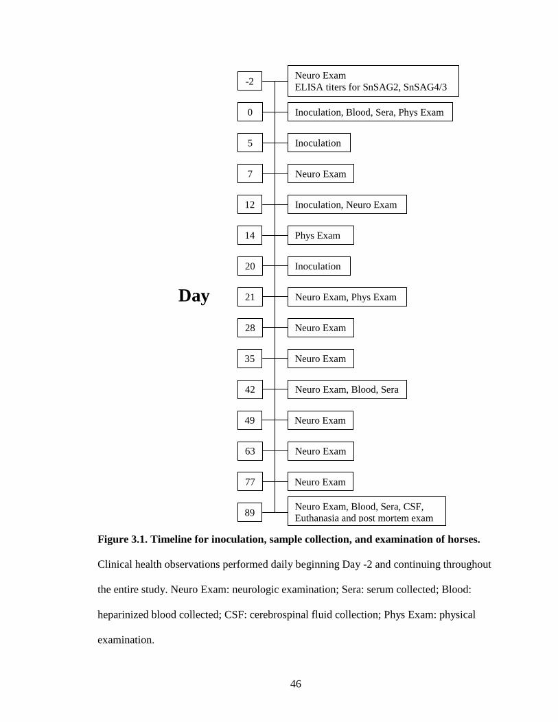
46
Figure 3.1. Timeline for inoculation, sample collection, and examination of horses.
Clinical health observations performed daily beginning Day -2 and continuing throughout
the entire study. Neuro Exam: neurologic examination; Sera: serum collected; Blood:
heparinized blood collected; CSF: cerebrospinal fluid collection; Phys Exam: physical
examination.
Day
42 Neuro Exam, Blood, Sera
21 Neuro Exam, Phys Exam
12 Inoculation, Neuro Exam
0 Inoculation, Blood, Sera, Phys Exam
Neuro Exam ELISA titers for SnSAG2, SnSAG4/3 -2
Inoculation 5
14 Phys Exam
Inoculation 20
Neuro Exam 28
Neuro Exam 35
Neuro Exam 49
Neuro Exam
7
Neuro Exam 63
Neuro Exam 77
Neuro Exam, Blood, Sera, CSF, Euthanasia and post mortem exam 89

47
3.2.3. Sample Collection and Preparation
The timeline for sample collections is summarized in Figure 3.1. Serum samples were
collected and sent to Equine Diagnostic Solutions on Day -2 to test for antibody titers
against S. neurona. Additional serum samples were obtained on Day 0, 42, and 89 by
collecting peripheral blood from the jugular vein in a 6mL serum separator vacutainer
tube (BD Vacutainer). Serum tubes were allowed to clot for at least 30 minutes and then
centrifuged at 3,000g for 10 minutes. Peripheral blood samples were collected from the
jugular vein in two 4mL sodium heparinized vacutainer tubes (BD Vacutainer) on Day 0,
42, and 89. Immediately following euthanasia on Day 89, 3mL CSF was collected from
the foramen magnum of the atlanto-occipital joint in 4mL red top tubes (BD Vacutainer)
using aseptic technique.
Staff members of the ETCR performed all sample collections. Tubes were transported by
car at 37°C to the University of Kentucky. Serum was removed from the tubes. Serum
and CSF were stored at 4°C until assayed. Heparinized blood was immediately stimulated
as described in section 3.2.9.
3.2.4. Examinations
Physical examinations were performed on Days 0, 14 and 21 by ETCR staff. Neurologic
examinations were completed on Days -2, 7, 12, 21, 28, 35, 42, 49, 63, 77, and 89. Exams
were conducted by Dr. Steve Reed (Rood and Riddle Equine Hospital, Lexington,
Kentucky) on days -2, 12, and 42. Dr. Julio Prado (ETCR) performed the remaining
neurologic examinations. The examination schedule is summarized in Figure 3.1.

48
Neurologic examinations evaluated each of the horses’ limbs (left front, right front, left
hind, and right hind) at the walk, trot, circling, and backing. Each movement was
assessed for ataxia (incoordination), paresis (weakness), spasticity (stiffness), and
dysmetria (range of movement), and assigned a grade of 0-5 (Table 3.1), resulting in a
total of 64 scores per horse (Table 3.2).
Table 3.1. Description of ataxia grades. (Rose, 2000)
Grade Description 0 No deficits; normal 1 Just detected at normal gait 2 Deficit easily detected and exaggerated by backing, turning, swaying, loin pressure or
neck extension 3 Deficit very prominent on walking, turning, loin pressure or neck extension 4 Stumbling, tripping, falling down spontaneously 5 Recumbent, unable to rise
Table 3.2. Neurology examination score sheet. At the walk, trot, circling, and backing,
horses received a score of 0-5 for each limb based on ataxia, paresis, spasticity, and
dysmetria.
Walk Trot Circling Backing
LF LR RF RR LF LR RF RR LF LR RF RR LF LR RF RR
Ataxia Paresis Spasticity Dysmetria LF: left front; LR: left rear; RF: right front; RR: right rear
49
3.2.5. Euthanasia and Necropsy
All six horses were euthanized and necropsied on Day 89. Horses were sedated
intravenously with xylazine and then injected intravenously with a lethal dose of sodium
pentobarbital. The euthanasia method was consistent with specification of the American
Veterinary Medical Association (AVMA) Guidelines on Euthanasia (2007). During
necropsy, sections of the brain and spinal cord were collected. Specifically, the dura
mater was opened and 0.5cm sections of the spinal cord were removed from each of the
following sections: Cervical(C)1-2, C3-4, C7-Thoracic(T)1, T4-5, T8-9, T12-13, Lumbar(L)1-2,
L3-4, L7-Sacral(S)2. Tissue samples were placed in containers of buffered neutral
formalin, and later sectioned, mounted, and stained for histological evaluation.
Euthanasia and necropsies were performed by staff at ETCR.
3.2.6. Histological Examination
Histological examination was performed by board-eligible veterinary pathologist Dr.
Uneeda Bryant at the University of Kentucky Veterinary Diagnostic Laboratory (UK
VDL). Tissue sections were stained with H&E and evaluated under light microscopy for
abnormalities.
3.2.7. Immunohistochemistry
Immunohistochemical (IHC) staining was completed using an automated staining system
(Bond-maX, Leica Biosystems, Newcastle Upon Tyne, UK) following the manufacturer’s
IHC Protocol F and the Bond Polymer Refine detection system (Leica Biosystems). This
50
procedure involved an automated dewaxing and rehydration of the tissue, continuing with
a heat-induced antigen retrieval using a ready-to-use citrate based (pH 6.0) buffer and
surfactant (Leica Biosystems) at 100°C for 20 minutes. Slides were then incubated with
3% hydrogen peroxide (H2O2) for 5 minutes, followed by application of anti-S. neurona
rabbit serum diluted 1:2500 with Bond Primary Antibody Diluent (Leica Biosystems),
and incubated for 15 minutes. Slides were treated with postprimary blocking reagent for 8
minutes and then a horseradish peroxidase-labeled IgG polymer for 8 minutes.
Diaminobenzidine tetrahydrochloride (DAB) substrate was added, and the slides were
incubated for 10 minutes. Finally, the slides were counterstained with hematoxylin for 5
minutes. Between each incubation step, slides were washed using Bond Wash Solution
10x Concentrate (Leica Biosystems), diluted with distilled water to a 1x working
concentration, to remove any unbound material. Brain tissue from a clinical EPM horse
with a large number of S. neurona organisms was stained as a control. A negative control
consisted of only Bond Primary Antibody Diluent (Leica Biosystems).
Slides were removed from the instrument and washed under distilled water. Tissue was
dehydrated using a graded series of alcohol washes (2-85% ethanol, 2-90% ethanol, 2-
95% ethanol, 2-100% ethanol) and cleared with 4 xylene washes. Slides were covered
using mounting medium and glass coverslips, and viewed with light microscopy to
identify S. neurona organisms.
51
3.2.8. Enzyme-Linked Immunosorbent Assay
Enzyme-linked immunosorbent assays (ELISAs) were conducted essentially as described
previously (Hoane et al., 2005; Yeargan and Howe, 2011). Each rSnSAG was diluted in
phosphate buffered saline (PBS) at predetermined optimal concentrations (SnSAG1
0.2µg/mL, SnSAG2 1.0µg/mL, SnSAG4/3 0.5µg/mL, SnSAG5 0.5µg/mL), and then
75µL of the antigen was dispensed into each well of high binding 96-well plates
(Corning, Corning, New York). Plates were incubated overnight at 4°C. Antigen was
removed and plates rinsed three times using PBS with 0.05% Tween 20 (PBST). Wells
were blocked with 200µL blocking solution (PBS containing 5% normal goat serum, 1%
Tween 20, 1% nonfat dry milk powder) and incubated at room temperature (RT) for 1.5
hours. Wells were rinsed a single time with PBST. Primary antibody was diluted (serum
1:250, CSF 1:2.5) with antibody diluent solution (1:10 blocking solution with PBST),
75µL was added to duplicate wells, and incubated at 37°C for 1 hour. Plates were washed
5 times with PBST. Secondary antibody, horseradish peroxidase (HRP)-conjugated goat
anti-horse immunoglobulin (Ig)G (Jackson ImmunoResearch Laboratories Inc., West
Grove, Pennsylvania) diluted 1:10,000 in antibody diluent, was added to wells in 75µL
aliquots and incubated at 37°C for 1 hour. The plates were washed 5 times with PBST,
then 75µL of RT chromagenic substrate TMB (3,3’,5,5’-tetramethylbenzidine; Pierce,
Thermo Fisher Scientific, Inc., Waltham, Massachusetts) was added to each well. Plates
sat at RT for 20-30 minutes for color to develop. Reactions were stopped with 75µL of
2M sulfuric acid. Optical density (OD) was measured at 450nm (OD450) in an Emax
microplate reader (Molecular Devices, Sunnyvale, California).

52
To account for inter-plate variability, percent positivity values were calculated for each
sample relative to the reference standards [(OD(sample) – OD(negative control)) /
(OD(positive control) – OD(negative control))] X 100 (Wright et al., 1993). The positive
controls were serum samples from symptomatic horses with histologically-confirmed
EPM. The negative control was a serum sample from a pre-infection weanling used in a
previous S. neurona challenge study (Fenger et al., 1997). At a dilution of 1:250 (serum)
or 1:2.5 (CSF), a sample is positive if the percent positivity is above the cutoff value of
15% for SnSAG1, SnSAG2 and SnSAG5, and 10% for SnSAG4/3 (Hoane et al., 2005;
Yeargan and Howe, 2011). End-point titers, the last dilution with a percent positivity
above a set cutoff value, were established with serial 2-fold dilutions of serum and CSF
samples beginning with 1:250 (serum) or 1:2.5 (CSF). A positive serum sample has at
least an end-point titer of 1:250. Samples below the cutoff value at a 1:250 dilution are
negative and given the antibody titer of <1:250.
ELISAs for IgG isotypes were essentially conducted as stated above, except utilizing
different secondary antibodies. Monoclonal antibodies (mAb) specific for IgGa (CVS 48;
1:100), IgGb (CVS39; 1:10), and IgG(T) (CVS40; 1:10) (Lunn et al., 1991) were diluted
to optimal concentrations with antibody diluent solution. Wells were incubated 1 hour at
37°C with 75µL mAb diluent solution. Plates were rinsed 5 times with PBST, and
incubated 1 hour at 37°C with 75µL of HRP-goat anti-mouse IgG antibody (Jackson
ImmunoResearch Laboratories Inc.) diluted 1:10,000 in antibody diluent solution.

53
3.2.9. Relative Quantification (RQ) of Cytokine mRNA Expression by Real-time PCR
Upon arrival at the University of Kentucky on Days 0, 42, and 89, paired heparinized
blood tubes were measured to ensure each tube contained 2.5mL of blood. A 108 pellet of
frozen SN3 S. neurona merozoites was resuspended in 1mL of RPMI 1640 medium
(HyClone, Thermo Fisher Scientific, Inc.), and 100µL of the suspension was added to
one blood tube from each of the six horses. Blood tubes were incubated at 37°C for 24
hours. Each blood tube was pipetted into a PAXGENE blood RNA tube (PreAnalytiX,
Valencia, California), incubated at RT for 24 hours, and then stored at -20°C.
Total RNA was extracted using the PAXGENE blood RNA extraction kit (Qiagen,
Valencia, California) following manufacturer’s protocol. PAXGENE tubes (PreAnalytiX)
were thawed, mixed thoroughly, and centrifuged 10 minutes at 2,800g. The supernatant
was poured off, and pellets were resuspended in 4mL of RNAse-free water (Qiagen).
Tubes were centrifuged for 10 minutes at 2,800g. The supernatant was discarded. Pellets
were resuspended in 350µL Buffer BR1 (Qiagen) and transferred to 1.5mL Eppendorf
tubes. To each tube, 300µL Buffer BR2 (Qiagen) and 80µL Proteinase K (Qiagen) was
added, with samples incubated 5 minutes at RT, then 10 minutes at 55°C. Tubes were
thoroughly mixed and centrifuged 10 minutes at 20,000g. Pellets were removed and
discarded. The supernatants were mixed with 350µL of 100% ethanol. Spin columns,
placed in 2mL collection tubes, were centrifuged 1 minute at 8,000g with 700µL of
sample. After each centrifugation, the spin columns were transferred to new 2mL
collection tubes, and filled with the remaining sample volume, and centrifuged 1 minute
at 8,000g. This process was repeated three times for 1 minute at 8,000g using 700µL

54
Buffer BR3 (Qiagen), for 1 minute at 8,000g using 500µL Buffer BR4 (Qiagen), and for
3 minutes at 20,000g using 500µL Buffer BR4 (Qiagen). Spin columns were then
centrifuged again for 1 minute at 20,000g with no buffer added. Spin columns were
placed in 1.5mL Eppendorf tubes with 40 µL Buffer BR5 (Qiagen) added to the columns,
and centrifuged for 1 minute at 8,000g. The process was repeated a second time. Spin
columns were discarded after second centrifugation with Buffer BR5 (Qiagen). The tubes
were incubated 5 minutes at 65°C, then immediately placed on ice. The RNA
concentration was quantified at OD260 using an Eppendorf Biophotometer (Hauppauge,
New York).
In PCR tubes, 0.5µg of each RNA sample was brought up to a volume of 41.5µL with
RNAse-free water (Qiagen), and mixed with 38.5µL of reverse transcription master mix.
Reverse transcription master mix contained 0.5µL avian myelobastosis virus (AMV)
reverse transcriptase (20U/µL; Promega, Madison, Wisconsin), 1µL oligo dT primer
(0.5µg/µL; Promega), 1µL RNAsin (40U/µL; Promega), 4µL dNTP (10mM; Promega),
16µL MgCl2 (25mM; Promega), and 16µL AMV buffer 5x (Promega). Reactions were
incubated at 42°C for 15 minutes, then 95°C for 5 minutes in a thermocycler. The cDNA
samples were stored at -20°C until further analysis.
Gene expression utilized equine specific intron-spanning beta-glucuronidase (β-GUS),
Granzyme B (GrzB), IFN-γ, IL-2, IL-4, and T-bet primer/probe sets (Applied
Biosystems, Foster City, California) (Table 3.3). The cDNA was diluted 1:1 with RNAse-
free water (Qiagen), and reactions were setup using an automated PCR setup machine
(Corbett). Each reaction consisted of 4.5µL diluted cDNA, 5µL TaqMan® Gene

55
Expression Master Mix (Applied Biosystems), and 0.5µL primer/probe set (Applied
Biosystems). Samples were run in duplicate using the 7900HT Fast Real-Time PCR
System (Applied Biosystems). Reactions were incubated for 10 minutes at 95°C, and
then underwent 40 cycles of 95°C for 15 seconds, followed by 1 minute at 65°C. The
PCR efficiencies were determined using LinRegPCR (Ramakers et al., 2003). Gene
expression levels were calculated using the ΔΔCT method (Livak and Schmittgen, 2001),
where ΔΔCT = [(Mean gene of interest CT – Mean β-GUS CT)Horse – (Mean gene of
interest CT – Mean β-GUS CT)Calibrator]. The calibrator for each horse was the non-
stimulated media control of the pre-challenge sample. The reference gene β-GUS was
used as an endogenous control (Breathnach et al., 2006). Results were calculated as 2-
ΔΔCT and denoted as relative quantification (RQ), measuring fold changes of expression
levels.
Table 3.3. Primer probe sets used for Real-time PCR.
Gene Reference β-GUS Liu et al., 2011 GrzB Liu et al., 2011 IFN-γ Adams et al., 2011 IL-2 Adams et al., 2011 IL-4 Adams et al., 2011 T-bet Ainsworth et al., 2003
3.2.10. Statistics
Statistical analyses were run on SIGMASTAT™ (Systat Inc. Richmond, California).
Two-way repeated measured analysis of variance (ANOVA) was used to detect
significant differences between the gene expression and the ELISA end-point titers, and

56
IgG isotype percent positivity values for the three sample days of the challenged horses.
Data was log10 transformed, if not distributed normally, prior to analysis. Differences
were considered to be significant at P < 0.05. For the CMI assay, comparisons were made
from the mean difference between S. neurona stimulated and non-stimulated blood at
different time points for the challenged horses. As there was only a single control horse,
statistical comparisons could not be made between the challenged and control groups.
3.2.11. Sarcocystis neurona Merozoite Invasion Assay
A confluent T25 flask of bovine turbinate (BT) cells was washed twice with 37°C PBS.
Cells were incubated with 0.5mL warm trypsin-versene solution. Once cells sloughed off,
they were resuspended in 15mL 4% FBS RPMI 1640, and aliquotted 0.5mL into each
well of a 24-well plate. Plate was incubated at 37°C for 5 days.
As previously described (Howe et al., 2005), S. neurona strain F9F merozoites that
express yellow fluorescent protein (YFP) were propagated by serial passages in BT cells.
Merozoites were harvested from infected BT monolayer cultures that had fully lysed.
Merozoites were pelleted by centrifugation at 3,000g for 10 minutes, then resuspended in
4mL 4% FBS RPMI 1640. Parasites were passed through 22 and 25 gauge needles, and
then through a 3.0µm pore sized membrane filter. The merozoite suspension was placed
in a styrofoam container filled with 37°C water bottles to replicate conditions during the
car transport. The styrofoam container was kept at room temperature. The BT cell
monolayer in the 24-well plate received fresh media, 0.5mL 4% FBS RPMI 1640. At
different time points (0, 1, 2, 3, 4, 6 hours), 20µL of the S. neurona merozoite suspension
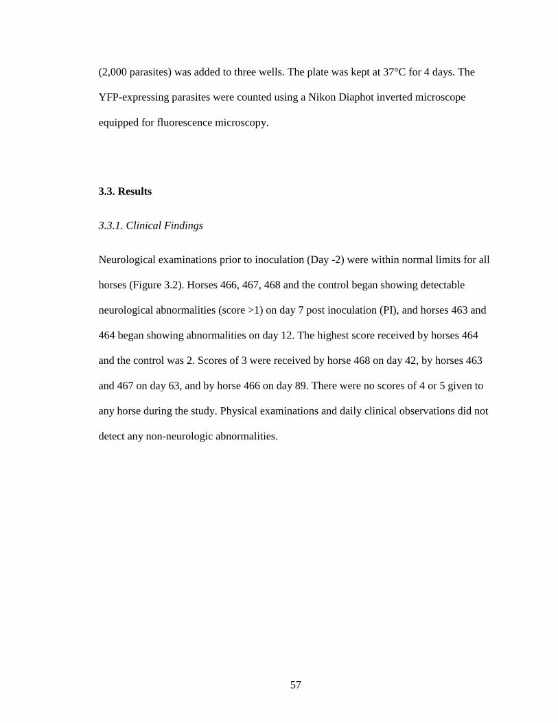
57
(2,000 parasites) was added to three wells. The plate was kept at 37°C for 4 days. The
YFP-expressing parasites were counted using a Nikon Diaphot inverted microscope
equipped for fluorescence microscopy.
3.3. Results
3.3.1. Clinical Findings
Neurological examinations prior to inoculation (Day -2) were within normal limits for all
horses (Figure 3.2). Horses 466, 467, 468 and the control began showing detectable
neurological abnormalities (score >1) on day 7 post inoculation (PI), and horses 463 and
464 began showing abnormalities on day 12. The highest score received by horses 464
and the control was 2. Scores of 3 were received by horse 468 on day 42, by horses 463
and 467 on day 63, and by horse 466 on day 89. There were no scores of 4 or 5 given to
any horse during the study. Physical examinations and daily clinical observations did not
detect any non-neurologic abnormalities.
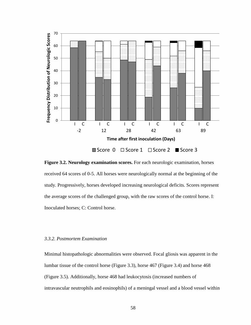
58
Figure 3.2. Neurology examination scores. For each neurologic examination, horses
received 64 scores of 0-5. All horses were neurologically normal at the beginning of the
study. Progressively, horses developed increasing neurological deficits. Scores represent
the average scores of the challenged group, with the raw scores of the control horse. I:
Inoculated horses; C: Control horse.
3.3.2. Postmortem Examination
Minimal histopathologic abnormalities were observed. Focal gliosis was apparent in the
lumbar tissue of the control horse (Figure 3.3), horse 467 (Figure 3.4) and horse 468
(Figure 3.5). Additionally, horse 468 had leukocytosis (increased numbers of
intravascular neutrophils and eosinophils) of a meningal vessel and a blood vessel within
0
10
20
30
40
50
60
70
Freq
uenc
y Di
strib
utio
n of
Neu
rolo
gic S
core
s
Time after first inoculation (Days)
Score 0 Score 1 Score 2 Score 3
I C I I I I I C C C C C -2 12 28 89 63 42
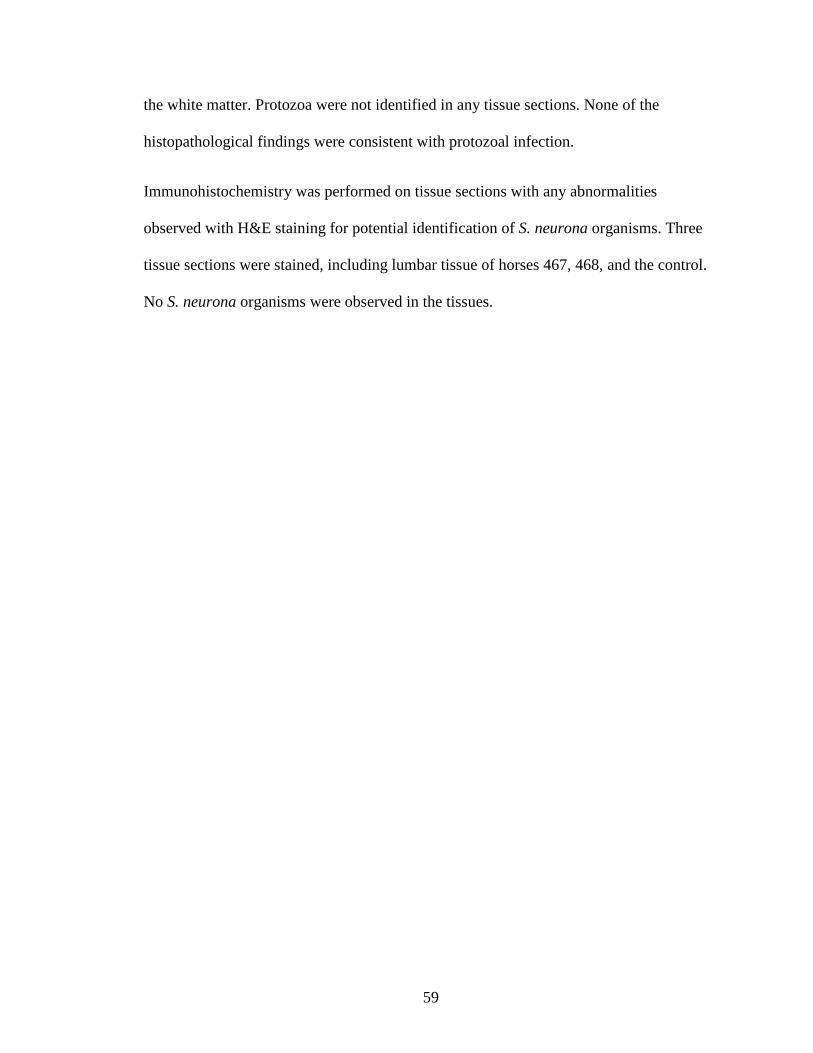
59
the white matter. Protozoa were not identified in any tissue sections. None of the
histopathological findings were consistent with protozoal infection.
Immunohistochemistry was performed on tissue sections with any abnormalities
observed with H&E staining for potential identification of S. neurona organisms. Three
tissue sections were stained, including lumbar tissue of horses 467, 468, and the control.
No S. neurona organisms were observed in the tissues.
60
Figure 3.3. Histological evaluation of the control horse. Hematoxylin and eosin stain.
(A) Focal gliosis located within the lumbar region of spinal cord. Bar = 100µm.
(B) Higher magnification of focal gliosis in Figure 3.3A. Bar = 20µm.
Figure 3.4. Histological evaluation of Horse 467. Hematoxylin and eosin stain.
(A) Focal gliosis located within the lumbar region of the spinal cord. Bar = 100µm.
(B) Higher magnification of focal gliosis in Figure 3.4A. Bar = 20µm.
A
B
A
B
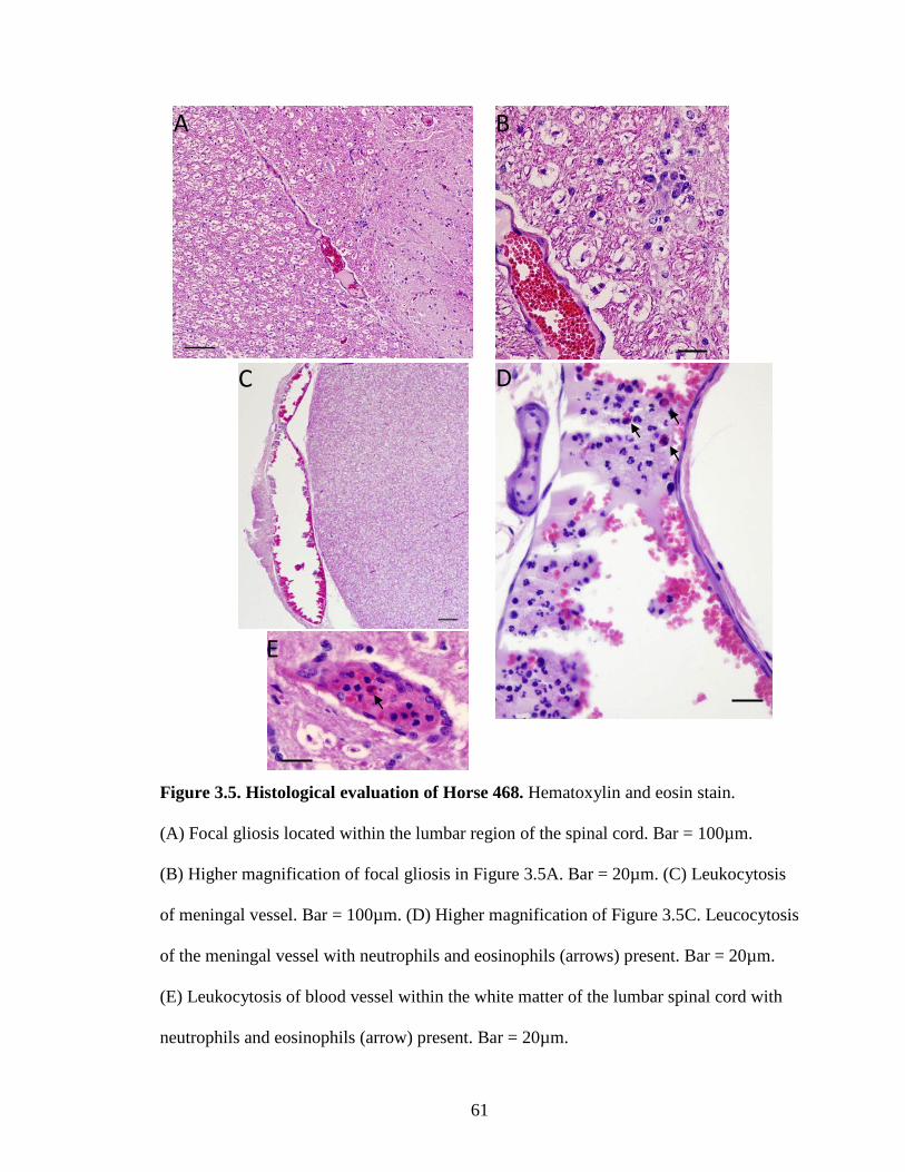
61
Figure 3.5. Histological evaluation of Horse 468. Hematoxylin and eosin stain.
(A) Focal gliosis located within the lumbar region of the spinal cord. Bar = 100µm.
(B) Higher magnification of focal gliosis in Figure 3.5A. Bar = 20µm. (C) Leukocytosis
of meningal vessel. Bar = 100µm. (D) Higher magnification of Figure 3.5C. Leucocytosis
of the meningal vessel with neutrophils and eosinophils (arrows) present. Bar = 20µm.
(E) Leukocytosis of blood vessel within the white matter of the lumbar spinal cord with
neutrophils and eosinophils (arrow) present. Bar = 20µm.
A B
C D
E
62
3.3.3. Antibody Response
3.3.3.1. Total IgG in Serum
Serum (pre-challenge, days 42 and 89) from all six horses was analyzed by ELISA to
assess the total IgG antibody response against the parasite surface antigens SnSAG1,
SnSAG2, SnSAG3, SnSAG4, and SnSAG5. End-point antibody titers were determined
on all serum samples as the lowest dilution at which the percent positivity was greater
than the cutoff value for each SnSAG. The control horse had sera end-point titers of
<1:250 on all sample days for SnSAG1, SnSAG2, SnSAG4/3 and SnSAG5 (Figure 3.6).
For SnSAG1, all challenged horses had serum end-point titers of <1:250 pre-challenge
(Figure 3.6A). On day 42, all samples were positive with end-point titers of 1:1000 for
horse 466, 1:500 for horses 463 and 464, and 1:250 for horses 467 and 468. On day 89,
horses 463, 464, 466 remained positive with end-point titers of 1:250, while horses 467
and 468 were negative with a titer of <1:250. The SnSAG1 serum end-point titers of the
challenged horses were statistically significant between the different time points (P =
0.004). Day 42 serum titers were statistically significant from the pre-challenge and day
89 serum titers (P < 0.05).
Serum end-point antibody titers for SnSAG2 were <1:250 for all challenged horses in the
pre-challenge and day 89 samples (Figure 3.6B). On day 42, horses 464 and 468 had
titers of 1:500, and horses 463 and 466 a titer of 1:250. Horse 467 was negative with a
titer of <1:250. The SnSAG2 serum end-point titers of the challenged horses were
statistically significant between the different time points (P = 0.006). Day 42 serum titers
were statistically significant from the pre-challenge and day 89 serum titers (P < 0.05).

63
All challenge horses on all sample days had serum end-point antibody titers of <1:250 for
SnSAG4/3 (Figure 3.6C).
Challenged horses all had serum end-point titers of <1:250 in the pre-challenge serum for
SnSAG5 (Figure 3.6D). End-point titers on day 42 were 1:1000 for horses 463 and 464,
1:500 for horse 466, 1:250 for horse 467, and <1:250 for horse 468. Day 89 antibody
titers were 1:250 for horses 463 and 464, and <1:250 for horses 466, 467, and 468. The
SnSAG5 serum end-point titers of the challenged horses were statistically significant
between the different time points (P = 0.013). Day 42 serum titers were statistically
significant from the pre-challenge and day 89 serum titers (P < 0.05).
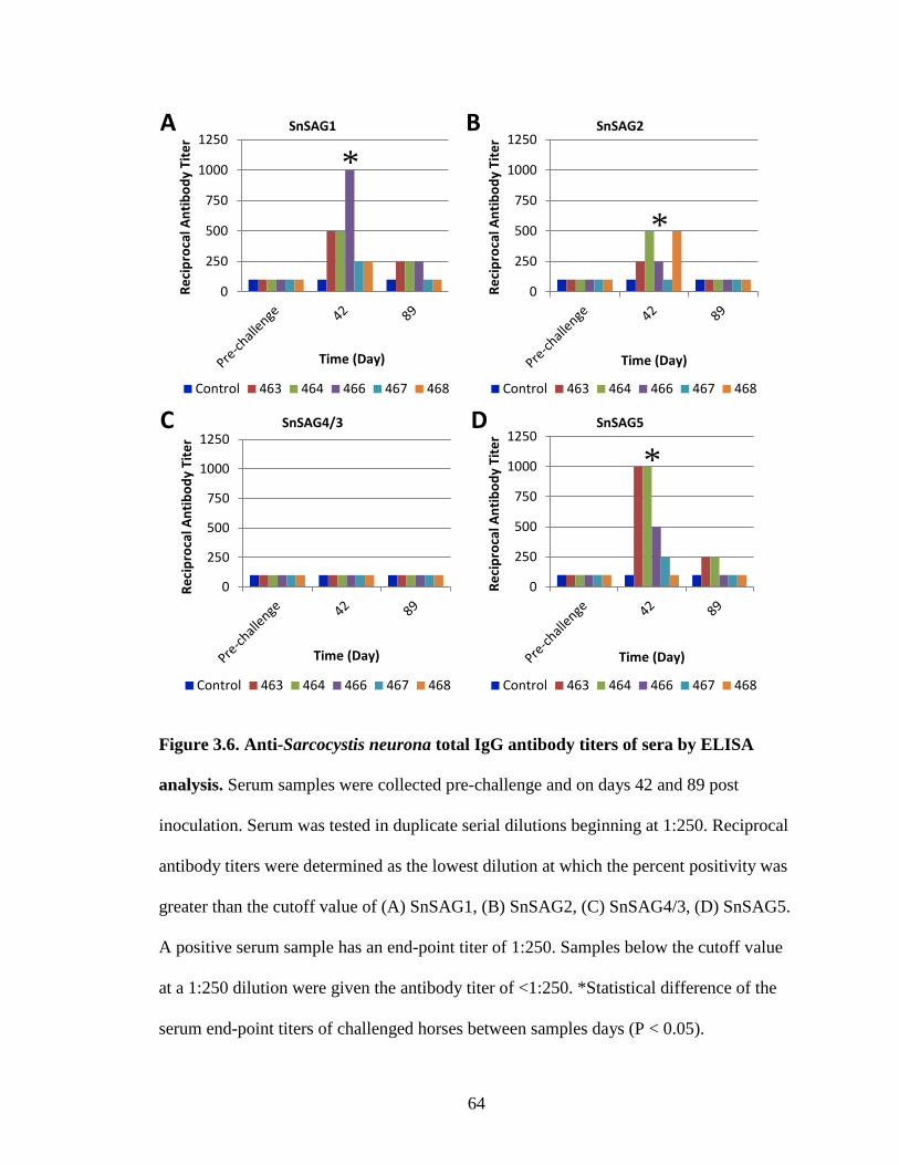
64
Figure 3.6. Anti-Sarcocystis neurona total IgG antibody titers of sera by ELISA
analysis. Serum samples were collected pre-challenge and on days 42 and 89 post
inoculation. Serum was tested in duplicate serial dilutions beginning at 1:250. Reciprocal
antibody titers were determined as the lowest dilution at which the percent positivity was
greater than the cutoff value of (A) SnSAG1, (B) SnSAG2, (C) SnSAG4/3, (D) SnSAG5.
A positive serum sample has an end-point titer of 1:250. Samples below the cutoff value
at a 1:250 dilution were given the antibody titer of <1:250. *Statistical difference of the
serum end-point titers of challenged horses between samples days (P < 0.05).
0
250
500
750
1000
1250
Reci
proc
al A
ntib
ody
Tite
r
Time (Day)
SnSAG1
Control 463 464 466 467 468
0
250
500
750
1000
1250
Reci
proc
al A
ntib
ody
Tite
r
Time (Day)
SnSAG2
Control 463 464 466 467 468
0
250
500
750
1000
1250
Reci
proc
al A
ntib
ody
Tite
r
Time (Day)
SnSAG4/3
Control 463 464 466 467 468
0
250
500
750
1000
1250
Reci
proc
al A
ntib
ody
Tite
r
Time (Day)
SnSAG5
Control 463 464 466 467 468
A
C D
B
*
*
*
65
3.3.3.2. Total IgG in CSF
CSF collected from horses on day 89 was analyzed by ELISA to assess total IgG
antibody against the surface antigens. The control horse was negative for all SnSAGs
(Figure 3.7). Horse 464 was the only challenged horse with antibodies against SnSAG1
in CSF (Figure 3.7A). The CSF from all other challenged horses was negative for all
SnSAGs (Figure 3.7A-D).
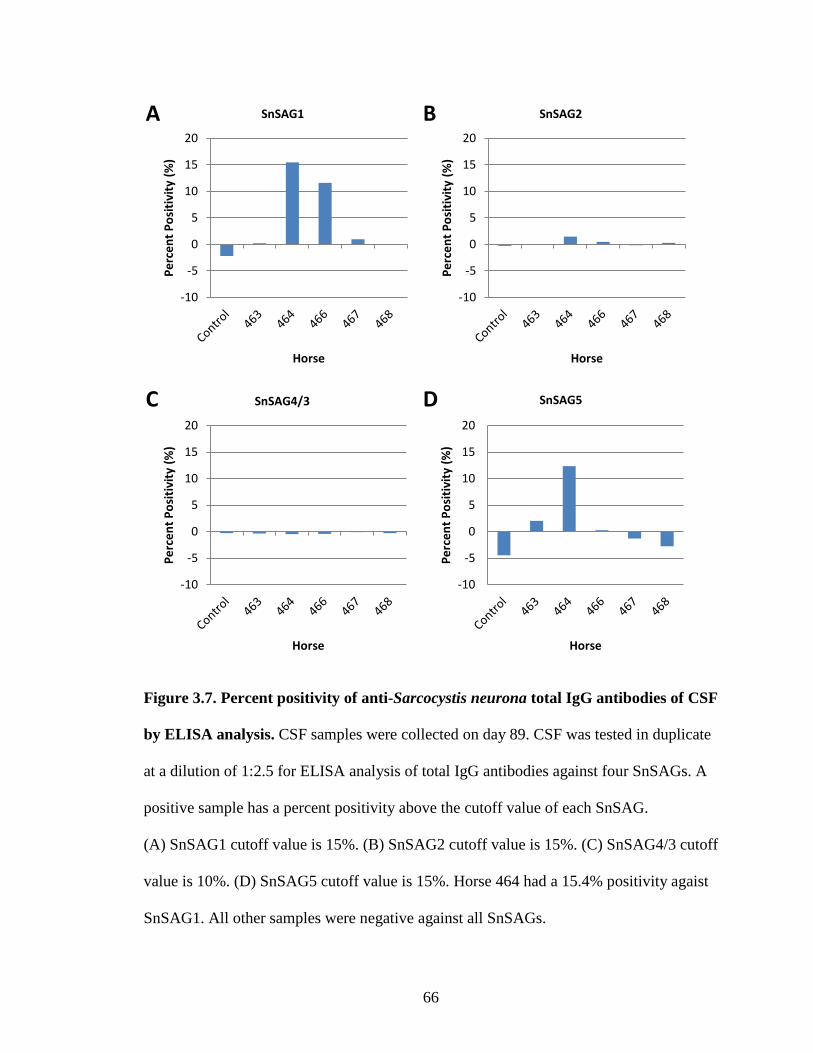
66
Figure 3.7. Percent positivity of anti-Sarcocystis neurona total IgG antibodies of CSF
by ELISA analysis. CSF samples were collected on day 89. CSF was tested in duplicate
at a dilution of 1:2.5 for ELISA analysis of total IgG antibodies against four SnSAGs. A
positive sample has a percent positivity above the cutoff value of each SnSAG.
(A) SnSAG1 cutoff value is 15%. (B) SnSAG2 cutoff value is 15%. (C) SnSAG4/3 cutoff
value is 10%. (D) SnSAG5 cutoff value is 15%. Horse 464 had a 15.4% positivity agaist
SnSAG1. All other samples were negative against all SnSAGs.
-10
-5
0
5
10
15
20
Perc
ent P
ositi
vity
(%)
Horse
SnSAG1
-10
-5
0
5
10
15
20
Perc
ent P
ositi
vity
(%)
Horse
SnSAG2
-10
-5
0
5
10
15
20
Perc
ent P
ositi
vity
(%)
Horse
SnSAG4/3
-10
-5
0
5
10
15
20Pe
rcen
t Pos
itivi
ty (%
)
Horse
SnSAG5
A B
C D
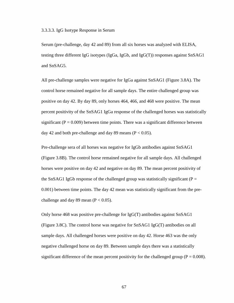
67
3.3.3.3. IgG Isotype Response in Serum
Serum (pre-challenge, day 42 and 89) from all six horses was analyzed with ELISA,
testing three different IgG isotypes (IgGa, IgGb, and IgG(T)) responses against SnSAG1
and SnSAG5.
All pre-challenge samples were negative for IgGa against SnSAG1 (Figure 3.8A). The
control horse remained negative for all sample days. The entire challenged group was
positive on day 42. By day 89, only horses 464, 466, and 468 were positive. The mean
percent positivity of the SnSAG1 IgGa response of the challenged horses was statistically
significant (P = 0.009) between time points. There was a significant difference between
day 42 and both pre-challenge and day 89 means (P < 0.05).
Pre-challenge sera of all horses was negative for IgGb antibodies against SnSAG1
(Figure 3.8B). The control horse remained negative for all sample days. All challenged
horses were positive on day 42 and negative on day 89. The mean percent positivity of
the SnSAG1 IgGb response of the challenged group was statistically significant (P =
0.001) between time points. The day 42 mean was statistically significant from the pre-
challenge and day 89 mean (P < 0.05).
Only horse 468 was positive pre-challenge for IgG(T) antibodies against SnSAG1
(Figure 3.8C). The control horse was negative for SnSAG1 IgG(T) antibodies on all
sample days. All challenged horses were positive on day 42. Horse 463 was the only
negative challenged horse on day 89. Between sample days there was a statistically
significant difference of the mean percent positivity for the challenged group (P = 0.008).

68
The mean percent positivity of day 42 was statistically significant from both pre-
challenge and day 89 means (P < 0.05).
69
Figure 3.8. Percent positivity of anti-Sarcocystis neurona SAG1 IgG isotype
antibodies of sera by ELISA analysis. Serum samples were collected pre-challenge and
on days 42 and 89 post inoculation. Serum was tested in duplicate at dilution of 1:250 for
ELISA analysis of (A) IgGa, (B) IgGb, (C) IgG(T) antibody levels against SnSAG1. A
positive sample has a percent positivity above the cutoff value of 15%. The control horse
was negative on all sample dates. *Statistical difference of the mean percent positivity of
challenged horses between samples days (P < 0.05).
0
20
40
60
80
100
Perc
ent P
ositi
vity
(%)
Time (Day)
SnSAG1 IgGa
Control 463 464 466 467 468
0
20
40
60
80
100
Perc
ent P
ositi
vity
(%)
Time (Day)
SnSAG1 IgGb
Control 463 464 466 467 468
0
20
40
60
80
100
Perc
ent P
ositi
vity
(%)
Time (Day)
SnSAG1 IgG(T)
Control 463 464 466 467 468
A B
C
*
* *
70
The control horse was positive for IgGa antibodies against SnSAG5 at pre-challenge and
negative on day 42 and 89 (Figure 3.9A). Horses 463 and 466 were positive for IgGa
antibodies pre-challenge. All challenged horses were positive on day 42. Only horses 467
and 468 were negative on day 89. The mean percent positivity was statistically significant
between sample days for the challenged horses (P < 0.001). The mean percent positivity
on day 42 was statistically significant from the pre-challenge and day 89 means (P <
0.05).
Horse 463 and the control were positive for SnSAG5 IgGb antibodies at pre-challenge
(Figure 3.9B). The control horse was negative on day 42 and 89. All challenged horses
were positive on day 42. Only horses 463, 464, and 466 were positive on day 89. The
mean percent positivity between time points was statistically significant for the
challenged group (P < 0.001). Day 42 mean percent positivity was statistically significant
from pre-challenge and day 89 means (P < 0.05).
The control and horses 463 and 466 were positive for IgG(T) antibodies against SnSAG5
in the pre-challenge sample (Figure 3.9C). The control horse was negative on day 42 and
89. All challenged horses were positive on day 42. Of the challenged horses, only 466
and 467 were negative on day 89. The mean percent positivity was statistically significant
between sample days (P < 0.001) for the challenged horses. Day 42 mean percent
positivity was statistically significant from pre-challenge and day 89 means (P < 0.05).
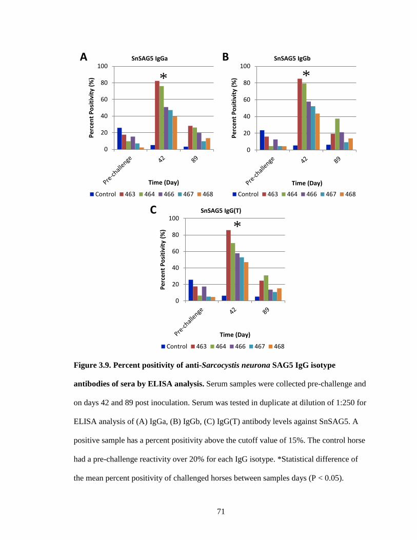
71
Figure 3.9. Percent positivity of anti-Sarcocystis neurona SAG5 IgG isotype
antibodies of sera by ELISA analysis. Serum samples were collected pre-challenge and
on days 42 and 89 post inoculation. Serum was tested in duplicate at dilution of 1:250 for
ELISA analysis of (A) IgGa, (B) IgGb, (C) IgG(T) antibody levels against SnSAG5. A
positive sample has a percent positivity above the cutoff value of 15%. The control horse
had a pre-challenge reactivity over 20% for each IgG isotype. *Statistical difference of
the mean percent positivity of challenged horses between samples days (P < 0.05).
0
20
40
60
80
100
Perc
ent P
ositi
vity
(%)
Time (Day)
SnSAG5 IgGa
Control 463 464 466 467 468
0
20
40
60
80
100
Perc
ent P
ositi
vity
(%)
Time (Day)
SnSAG5 IgGb
Control 463 464 466 467 468
0
20
40
60
80
100
Perc
ent P
ositi
vity
(%)
Time (Day)
SnSAG5 IgG(T)
Control 463 464 466 467 468
A B
C
*
*
*
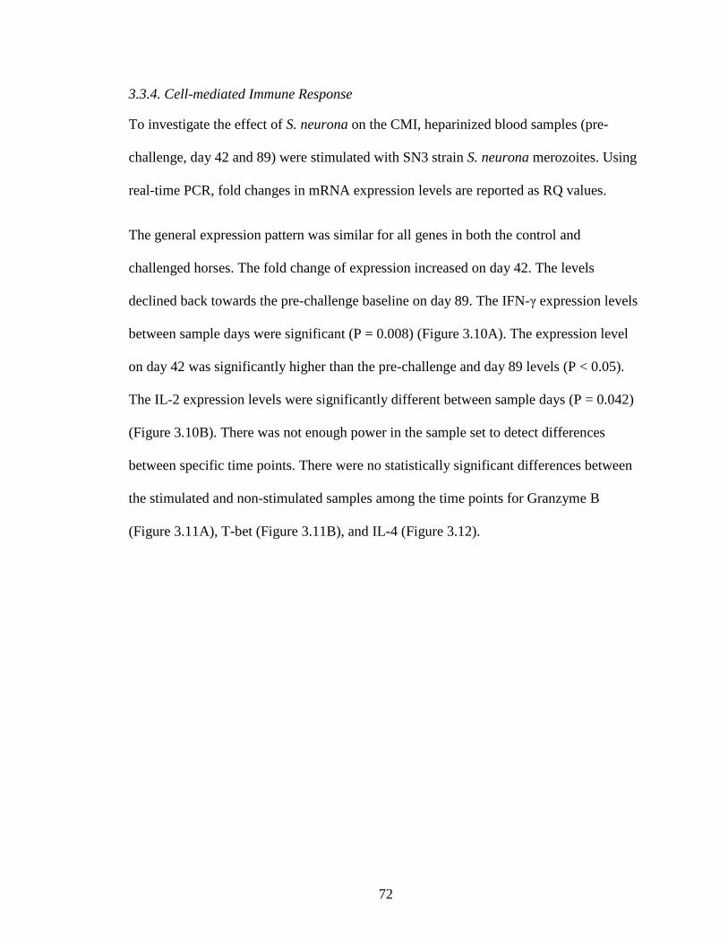
72
3.3.4. Cell-mediated Immune Response
To investigate the effect of S. neurona on the CMI, heparinized blood samples (pre-
challenge, day 42 and 89) were stimulated with SN3 strain S. neurona merozoites. Using
real-time PCR, fold changes in mRNA expression levels are reported as RQ values.
The general expression pattern was similar for all genes in both the control and
challenged horses. The fold change of expression increased on day 42. The levels
declined back towards the pre-challenge baseline on day 89. The IFN-γ expression levels
between sample days were significant (P = 0.008) (Figure 3.10A). The expression level
on day 42 was significantly higher than the pre-challenge and day 89 levels (P < 0.05).
The IL-2 expression levels were significantly different between sample days (P = 0.042)
(Figure 3.10B). There was not enough power in the sample set to detect differences
between specific time points. There were no statistically significant differences between
the stimulated and non-stimulated samples among the time points for Granzyme B
(Figure 3.11A), T-bet (Figure 3.11B), and IL-4 (Figure 3.12).
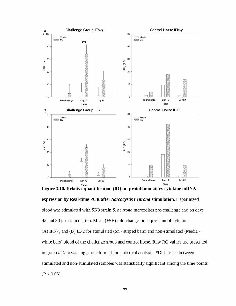
73
Figure 3.10. Relative quantification (RQ) of proinflammatory cytokine mRNA
expression by Real-time PCR after Sarcocystis neurona stimulation. Heparinized
blood was stimulated with SN3 strain S. neurona merozoites pre-challenge and on days
42 and 89 post inoculation. Mean (±SE) fold changes in expression of cytokines
(A) IFN-γ and (B) IL-2 for stimulated (Sn - striped bars) and non-stimulated (Media -
white bars) blood of the challenge group and control horse. Raw RQ values are presented
in graphs. Data was log10 transformed for statistical analysis. *Difference between
stimulated and non-stimulated samples was statistically significant among the time points
(P < 0.05).
A
B Challenge Group IL-2
Challenge Group IFN-γ Control Horse IFN-γ
*
Control Horse IL-2

74
Figure 3.11. Relative quantification (RQ) of mRNA expression by Real-time PCR
after Sarcocystis neurona stimulation. Heparinized blood was stimulated with SN3
strain S. neurona merozoites pre-challenge and on days 42 and 89 post inoculation. Mean
(±SE) fold changes in expression of (A) Granzyme B and (B) T-bet for stimulated (Sn -
striped bars) and non-stimulated (Media - white bars) blood of the challenge group and
control horse. Raw RQ values are presented in graphs.
A
B
Control Horse Granzyme B
Challenge Group T-bet Control Horse T-bet
Challenge Group Granzyme B

75
Figure 3.12. Relative quantification (RQ) of anti-inflammatory cytokine mRNA
expression by Real-time PCR after Sarcocystis neurona stimulation. Heparinized
blood was stimulated with SN3 strain S. neurona merozoites pre-challenge and on days
42 and 89 post inoculation. Mean (±SE) fold changes in expression of cytokine IL-4 for
stimulated (Sn - striped bars) and non-stimulated (Media - white bars) blood of the
challenge group and control horse. Raw RQ values are presented in graphs.
Control Horse IL-4 Challenge Group IL-4
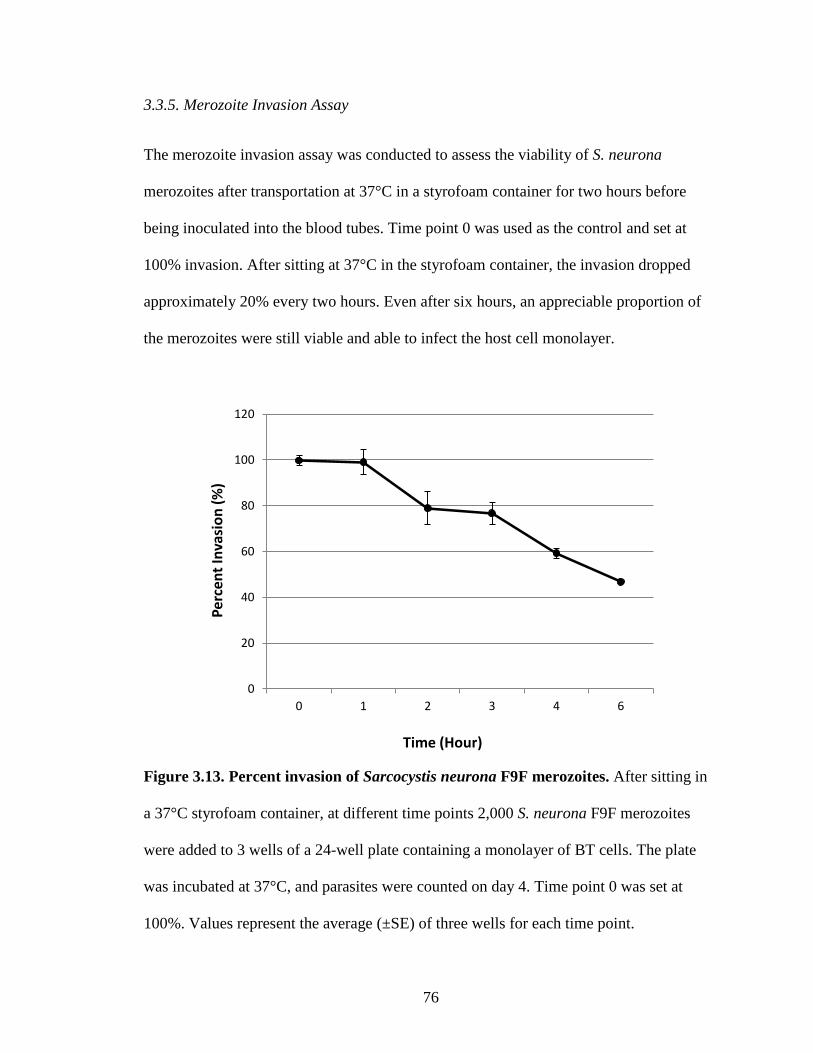
76
3.3.5. Merozoite Invasion Assay
The merozoite invasion assay was conducted to assess the viability of S. neurona
merozoites after transportation at 37°C in a styrofoam container for two hours before
being inoculated into the blood tubes. Time point 0 was used as the control and set at
100% invasion. After sitting at 37°C in the styrofoam container, the invasion dropped
approximately 20% every two hours. Even after six hours, an appreciable proportion of
the merozoites were still viable and able to infect the host cell monolayer.
Figure 3.13. Percent invasion of Sarcocystis neurona F9F merozoites. After sitting in
a 37°C styrofoam container, at different time points 2,000 S. neurona F9F merozoites
were added to 3 wells of a 24-well plate containing a monolayer of BT cells. The plate
was incubated at 37°C, and parasites were counted on day 4. Time point 0 was set at
100%. Values represent the average (±SE) of three wells for each time point.
0
20
40
60
80
100
120
0 1 2 3 4 6
Perc
ent I
nvas
ion
(%)
Time (Hour)
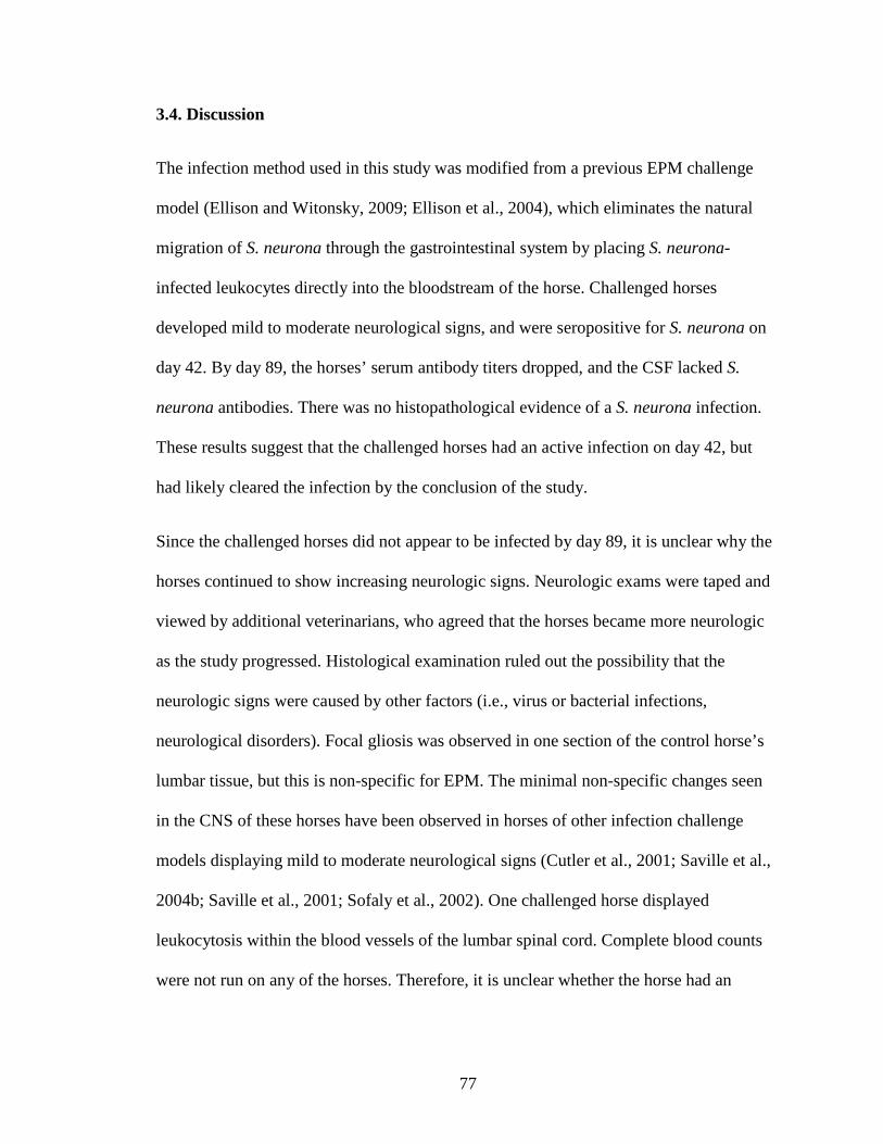
77
3.4. Discussion
The infection method used in this study was modified from a previous EPM challenge
model (Ellison and Witonsky, 2009; Ellison et al., 2004), which eliminates the natural
migration of S. neurona through the gastrointestinal system by placing S. neurona-
infected leukocytes directly into the bloodstream of the horse. Challenged horses
developed mild to moderate neurological signs, and were seropositive for S. neurona on
day 42. By day 89, the horses’ serum antibody titers dropped, and the CSF lacked S.
neurona antibodies. There was no histopathological evidence of a S. neurona infection.
These results suggest that the challenged horses had an active infection on day 42, but
had likely cleared the infection by the conclusion of the study.
Since the challenged horses did not appear to be infected by day 89, it is unclear why the
horses continued to show increasing neurologic signs. Neurologic exams were taped and
viewed by additional veterinarians, who agreed that the horses became more neurologic
as the study progressed. Histological examination ruled out the possibility that the
neurologic signs were caused by other factors (i.e., virus or bacterial infections,
neurological disorders). Focal gliosis was observed in one section of the control horse’s
lumbar tissue, but this is non-specific for EPM. The minimal non-specific changes seen
in the CNS of these horses have been observed in horses of other infection challenge
models displaying mild to moderate neurological signs (Cutler et al., 2001; Saville et al.,
2004b; Saville et al., 2001; Sofaly et al., 2002). One challenged horse displayed
leukocytosis within the blood vessels of the lumbar spinal cord. Complete blood counts
were not run on any of the horses. Therefore, it is unclear whether the horse had an
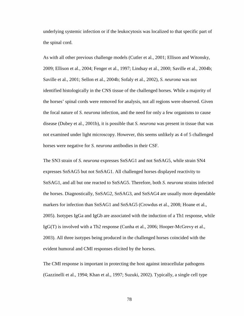
78
underlying systemic infection or if the leukocytosis was localized to that specific part of
the spinal cord.
As with all other previous challenge models (Cutler et al., 2001; Ellison and Witonsky,
2009; Ellison et al., 2004; Fenger et al., 1997; Lindsay et al., 2000; Saville et al., 2004b;
Saville et al., 2001; Sellon et al., 2004b; Sofaly et al., 2002), S. neurona was not
identified histologically in the CNS tissue of the challenged horses. While a majority of
the horses’ spinal cords were removed for analysis, not all regions were observed. Given
the focal nature of S. neurona infection, and the need for only a few organisms to cause
disease (Dubey et al., 2001b), it is possible that S. neurona was present in tissue that was
not examined under light microscopy. However, this seems unlikely as 4 of 5 challenged
horses were negative for S. neurona antibodies in their CSF.
The SN3 strain of S. neurona expresses SnSAG1 and not SnSAG5, while strain SN4
expresses SnSAG5 but not SnSAG1. All challenged horses displayed reactivity to
SnSAG1, and all but one reacted to SnSAG5. Therefore, both S. neurona strains infected
the horses. Diagnostically, SnSAG2, SnSAG3, and SnSAG4 are usually more dependable
markers for infection than SnSAG1 and SnSAG5 (Crowdus et al., 2008; Hoane et al.,
2005). Isotypes IgGa and IgGb are associated with the induction of a Th1 response, while
IgG(T) is involved with a Th2 response (Cunha et al., 2006; Hooper-McGrevy et al.,
2003). All three isotypes being produced in the challenged horses coincided with the
evident humoral and CMI responses elicited by the horses.
The CMI response is important in protecting the host against intracellular pathogens
(Gazzinelli et al., 1994; Khan et al., 1997; Suzuki, 2002). Typically, a single cell type
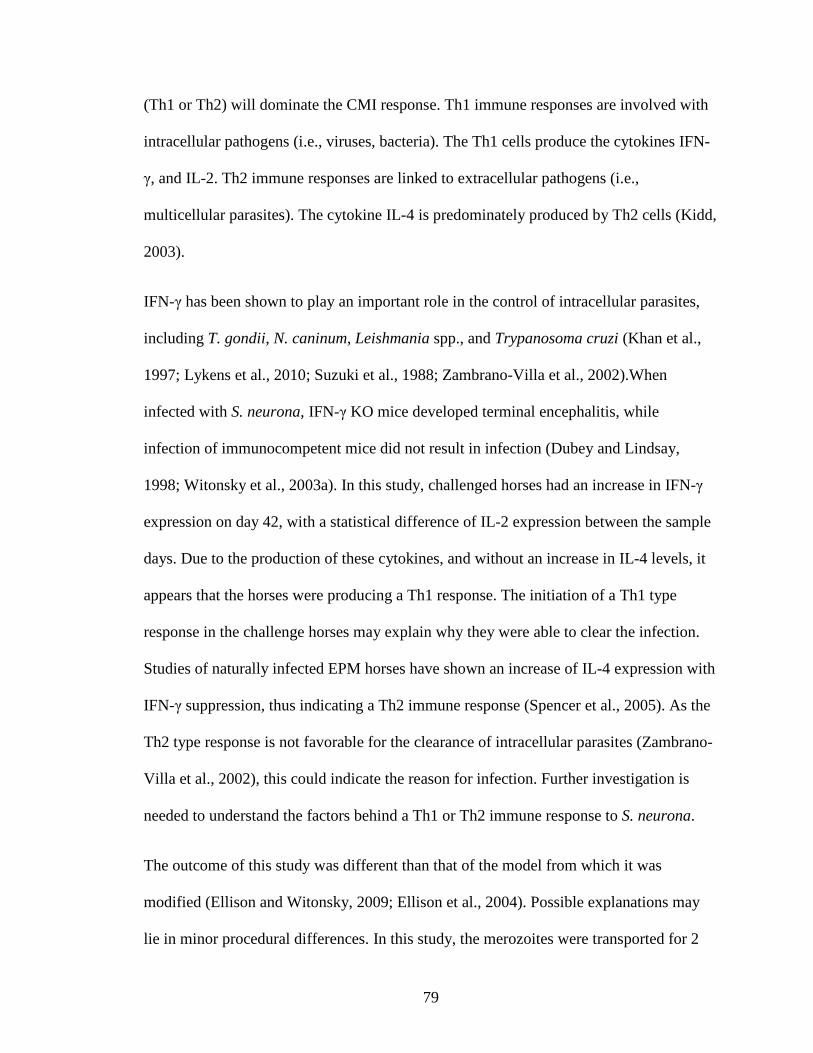
79
(Th1 or Th2) will dominate the CMI response. Th1 immune responses are involved with
intracellular pathogens (i.e., viruses, bacteria). The Th1 cells produce the cytokines IFN-
γ, and IL-2. Th2 immune responses are linked to extracellular pathogens (i.e.,
multicellular parasites). The cytokine IL-4 is predominately produced by Th2 cells (Kidd,
2003).
IFN-γ has been shown to play an important role in the control of intracellular parasites,
including T. gondii, N. caninum, Leishmania spp., and Trypanosoma cruzi (Khan et al.,
1997; Lykens et al., 2010; Suzuki et al., 1988; Zambrano-Villa et al., 2002).When
infected with S. neurona, IFN-γ KO mice developed terminal encephalitis, while
infection of immunocompetent mice did not result in infection (Dubey and Lindsay,
1998; Witonsky et al., 2003a). In this study, challenged horses had an increase in IFN-γ
expression on day 42, with a statistical difference of IL-2 expression between the sample
days. Due to the production of these cytokines, and without an increase in IL-4 levels, it
appears that the horses were producing a Th1 response. The initiation of a Th1 type
response in the challenge horses may explain why they were able to clear the infection.
Studies of naturally infected EPM horses have shown an increase of IL-4 expression with
IFN-γ suppression, thus indicating a Th2 immune response (Spencer et al., 2005). As the
Th2 type response is not favorable for the clearance of intracellular parasites (Zambrano-
Villa et al., 2002), this could indicate the reason for infection. Further investigation is
needed to understand the factors behind a Th1 or Th2 immune response to S. neurona.
The outcome of this study was different than that of the model from which it was
modified (Ellison and Witonsky, 2009; Ellison et al., 2004). Possible explanations may
lie in minor procedural differences. In this study, the merozoites were transported for 2
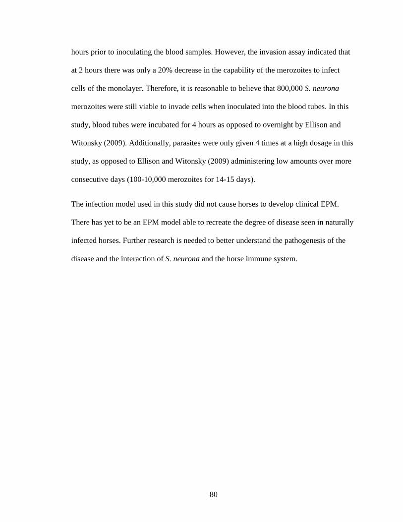
80
hours prior to inoculating the blood samples. However, the invasion assay indicated that
at 2 hours there was only a 20% decrease in the capability of the merozoites to infect
cells of the monolayer. Therefore, it is reasonable to believe that 800,000 S. neurona
merozoites were still viable to invade cells when inoculated into the blood tubes. In this
study, blood tubes were incubated for 4 hours as opposed to overnight by Ellison and
Witonsky (2009). Additionally, parasites were only given 4 times at a high dosage in this
study, as opposed to Ellison and Witonsky (2009) administering low amounts over more
consecutive days (100-10,000 merozoites for 14-15 days).
The infection model used in this study did not cause horses to develop clinical EPM.
There has yet to be an EPM model able to recreate the degree of disease seen in naturally
infected horses. Further research is needed to better understand the pathogenesis of the
disease and the interaction of S. neurona and the horse immune system.
81
CHAPTER FOUR
Conclusion
The studies described in this thesis investigated factors contributing to the development
of EPM in the horse. In the first study, an attempt to look at potential susceptibility of
horses to EPM was performed using a GWAS on FFPE tissue. The DNA extracted from
the FFPE tissues was not adequate for SNP analysis, however, only yielding usable
results in 5 of 24 horses. Tissue age appeared to be a factor in the viability of the DNA.
Tissue that was older than 14 months showed a dramatic drop in call rate. It seems likely
that use of FFPE tissues for GWAS will not be a practical method for an EPM study, as
the UK VDL does not process enough cases annually to develop a reasonable sample
size. Collaboration with other veterinary diagnostic laboratories would be needed to
create an appropriate sample size of FFPE samples. Additionally, samples could be
processed soon after case is received, and DNA stored until a sufficient sample size is
collected.
Illumina has developed a restoration solution to be used on DNA damaged in FFPE tissue
(Pokholok et al., 2010). Through the use of polymerases, degraded DNA is repaired,
leaving strands of DNA that do not contain the damaged bases. Strands can then be
ligated together and run on an Infinium® HD Assay (Illumina). To date this technology
has only been optimized for use on human SNP chips. In the future, it might be possible
to use the restoration solution on damaged DNA extracted from equine FFPE samples
before running them on an equine SNP chip. This may result in increased call rates,
making the archived FFPE samples usable in a GWAS on EPM susceptibility.
82
The second study tested the use of a horse EPM model that involves injecting whole
blood infected with S. neurona merozoites directly into the bloodstream of horses.
Neurological examinations along with humoral and CMI responses were evaluated. At
the conclusion of the study, horses displayed mild to moderate neurological signs and
appeared to have cleared the S. neurona infection. Challenged horses had an increased
expression of IFN-γ and IL-2, indicating a Th1 immune response. The severe clinical
signs seen with natural infections were not observed in these challenged horses.
Therefore, the infection model used in this experiment was not effective in replicating
clinical EPM.
Multiple attempts have been made to develop a working equine EPM model, but each has
been unable to replicate the disease seen in naturally infected horses. There appears to be
an underlying component needed to cause the disease that has not been identified with
these challenge models. Questions regarding the interaction between S. neurona and the
horse immune response should be addressed prior to attempting another EPM model.
Additional studies are needed to confirm the relationship between IFN-γ and IL-4
expression in horses infected with S. neurona. Other factors that may be involved include
the horses’ genetics, stress level, and concurrent infections. As well, the S. neurona strain
virulence and mechanisms of immunosuppression may play a role. Greater understanding
of the pathogenesis of EPM could lead to improved diagnostics, treatment, identification
of potential susceptible horses, and, ultimately, prevention of this devastating disease.
83
APPENDIX A
List of Abbreviations
AMV Avian myelobastosis virus
ANOVA Analysis of variance
AVMA American Veterinary Medical Association
β-GUS Beta-glucuronidase
BT Bovine turbinate
C Cervical
cDNA Complementary DNA
CMI Cell-mediated immunity
CNS Central nervous system
CSF Cerebrospinal fluid
CVM Cervical vertebral malformation
DAB Diaminobenzidine tetrahydrochloride
DMSO Dimethyl sulfoxide
DNA Deoxyribonucleic acid
Dpi Days post inoculation
EDTA Ethylenediaminetetraacetic acid
ELISA Enzyme-linked immunosorbent assay
EPM Equine protozoal myeloencephalitis
ETCR East Tennessee Clinical Research, Inc.
FBS Fetal bovine serum
FDA Food and Drug Administration
84
FFPE Formalin-fixed, paraffin-embedded
GrzB Granzyme B
GWAS Genome-wide association studies
H&E Hematoxylin and eosin
HRP Horseradish peroxidase
IACUC Institutional Animal Care and Use Committee
IFAT Indirect fluorescent antibody test
IFN-γ Interferon-gamma
Ig Immunoglobulin
IH Intermediate host
IHC Immunohistochemical
IL Interleukin
IRAK Interleukin-1 receptor-associated kinase
KO Knockout
L Lumbar
mAb Monoclonal antibody
MCP1 Monocyte chemoattractant protein-1
MHC Major histocompatibility complex
mRNA Messenger RNA
MSMD Mendelian susceptibility to mycobacterial disease
NK Natural killer
NSAID Nonsteroidal anti-inflammatory drug
OD Optical density
PBS Phosphate buffered saline
PBST PBS with 0.05% Tween 20
85
PCR Polymerase chain reaction
PI Post inoculation
PID Primary immunodeficiencies
PMA/I Phorbol myristate acetate/ionomycin
RNA Ribonucleic acid
RQ Relative quantification
RT Room temperature
S Sacral
SCID Severe combined immunodeficiency
SE Standard error
SNP Single nucleotide polymorphism
SnSAG Sarcocystis neurona surface antigen
T Thoracic
UK VDL University of Kentucky Veterinary Diagnostic Laboratory
US United States
USDA United States Department of Agriculture
YFP Yellow fluorescent protein
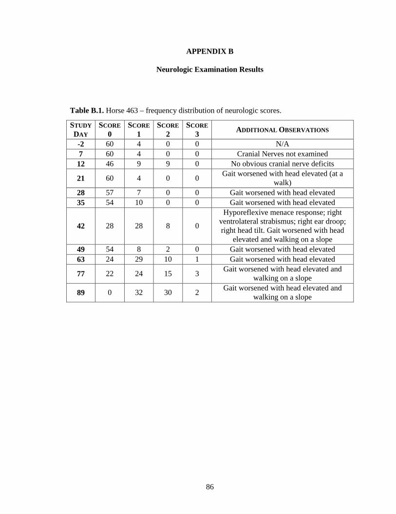
86
APPENDIX B
Neurologic Examination Results
Table B.1. Horse 463 – frequency distribution of neurologic scores.
STUDY DAY
SCORE 0
SCORE 1
SCORE 2
SCORE 3 ADDITIONAL OBSERVATIONS
-2 60 4 0 0 N/A 7 60 4 0 0 Cranial Nerves not examined 12 46 9 9 0 No obvious cranial nerve deficits
21 60 4 0 0 Gait worsened with head elevated (at a walk)
28 57 7 0 0 Gait worsened with head elevated 35 54 10 0 0 Gait worsened with head elevated
42 28 28 8 0
Hyporeflexive menace response; right ventrolateral strabismus; right ear droop; right head tilt. Gait worsened with head
elevated and walking on a slope 49 54 8 2 0 Gait worsened with head elevated 63 24 29 10 1 Gait worsened with head elevated
77 22 24 15 3 Gait worsened with head elevated and walking on a slope
89 0 32 30 2 Gait worsened with head elevated and walking on a slope
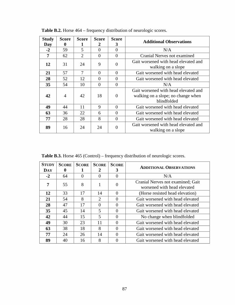
87
Table B.2. Horse 464 – frequency distribution of neurologic scores.
Study Day
Score 0
Score 1
Score 2
Score 3 Additional Observations
-2 59 5 0 0 N/A 7 62 2 0 0 Cranial Nerves not examined
12 31 24 9 0 Gait worsened with head elevated and walking on a slope
21 57 7 0 0 Gait worsened with head elevated 28 52 12 0 0 Gait worsened with head elevated 35 54 10 0 0 N/A
42 4 42 18 0 Gait worsened with head elevated and walking on a slope; no change when
blindfolded 49 44 11 9 0 Gait worsened with head elevated 63 36 22 6 0 Gait worsened with head elevated 77 28 28 8 0 Gait worsened with head elevated
89 16 24 24 0 Gait worsened with head elevated and walking on a slope
Table B.3. Horse 465 (Control) – frequency distribution of neurologic scores.
STUDY DAY
SCORE 0
SCORE 1
SCORE 2
SCORE 3 ADDITIONAL OBSERVATIONS
-2 64 0 0 0 N/A
7 55 8 1 0 Cranial Nerves not examined; Gait worsened with head elevated
12 33 17 14 0 (Horse resisted head elevation) 21 54 8 2 0 Gait worsened with head elevated 28 47 17 0 0 Gait worsened with head elevated 35 45 14 5 0 Gait worsened with head elevated 42 44 15 5 0 No change when blindfolded 49 30 23 11 0 Gait worsened with head elevated 63 38 18 8 0 Gait worsened with head elevated 77 24 26 14 0 Gait worsened with head elevated 89 40 16 8 0 Gait worsened with head elevated

88
Table B.4. Horse 466 – frequency distribution of neurologic scores.
STUDY DAY
SCORE 0
SCORE 1
SCORE 2
SCORE 3 ADDITIONAL OBSERVATIONS
-2 63 1 0 0 Moves very well 7 53 10 1 0 Cranial Nerves not examined 12 28 14 21 1 Gait worsened with head elevated 21 50 14 0 0 Gait worsened with head elevated
28 50 9 5 0 Gait worsened with head elevated and walking on a slope
35 39 22 3 Gait worsened with head elevated 42 44 17 3 0 No change when blindfolded
49 28 22 14 0 Gait worsened with head elevated and walking on a slope
63 35 17 12 0 Gait worsened with head elevated 77 44 15 5 0 Gait worsened with head elevated
89 30 6 24 4 Gait worsened with head elevated and walking on a slope
Table B.5. Horse 467 – frequency distribution of neurologic scores.
STUDY DAY
SCORE 0
SCORE 1
SCORE 2
SCORE 3 ADDITIONAL OBSERVATIONS
-2 52 12 0 0 Mild ataxia in circles- trots ok but weak at stopping
7 58 5 1 0 Cranial Nerves not examined
12 36 28 0 0 Gait worsened with head elevated (pacing). Muzzle deviated while eating.
21 51 13 0 0 Gait worsened with head elevated
28 42 17 5 0 Gait worsened with head elevated and walking on a slope
35 45 16 3 0 Gait worsened with head elevated
42 18 36 10 0 Gait worsened with head elevated and walking on a slope; no change when
blindfolded
49 30 23 11 0 Gait worsened with head elevated and walking on a slope
63 22 26 14 2 Gait worsened with head elevated 77 38 14 11 1 Gait worsened with head elevated
89 0 4 50 10 Gait worsened with head elevated and walking on a slope
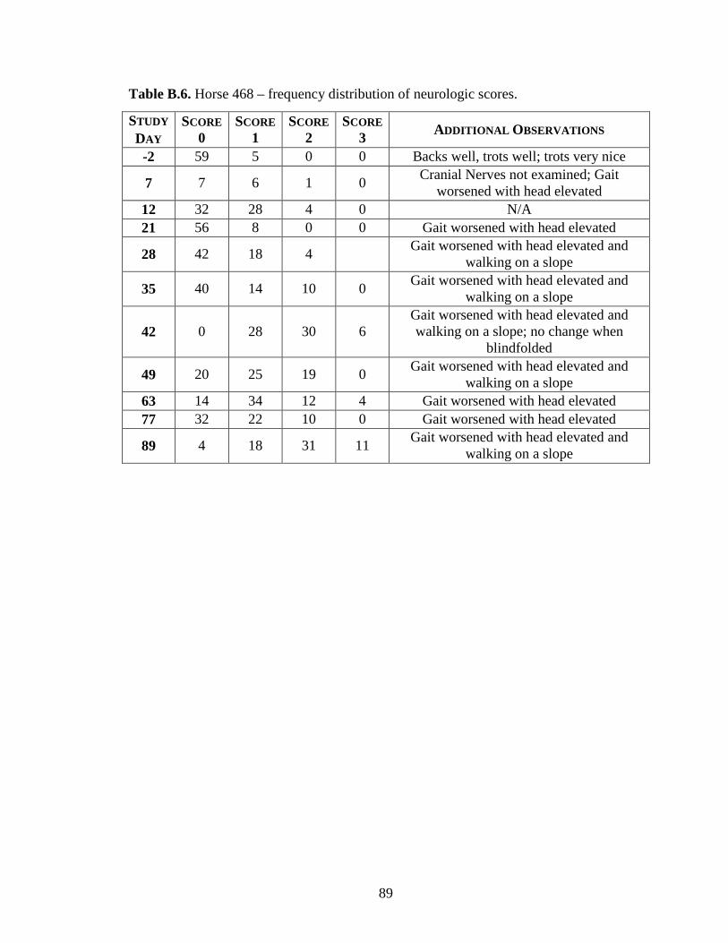
89
Table B.6. Horse 468 – frequency distribution of neurologic scores.
STUDY DAY
SCORE 0
SCORE 1
SCORE 2
SCORE 3 ADDITIONAL OBSERVATIONS
-2 59 5 0 0 Backs well, trots well; trots very nice
7 7 6 1 0 Cranial Nerves not examined; Gait worsened with head elevated
12 32 28 4 0 N/A 21 56 8 0 0 Gait worsened with head elevated
28 42 18 4 Gait worsened with head elevated and walking on a slope
35 40 14 10 0 Gait worsened with head elevated and walking on a slope
42 0 28 30 6 Gait worsened with head elevated and walking on a slope; no change when
blindfolded
49 20 25 19 0 Gait worsened with head elevated and walking on a slope
63 14 34 12 4 Gait worsened with head elevated 77 32 22 10 0 Gait worsened with head elevated
89 4 18 31 11 Gait worsened with head elevated and walking on a slope
90
REFERENCES
Abel, L., Demenais, F., (1988). "Detection of major genes for susceptibility to leprosy and its subtypes in a Caribbean island: Desirade island." American Journal of Human Genetics 42(2): 256-266.
Adams, A.A., Sturgill, T.L., Breathnach, C.C., Chambers, T.M., Siger, L., Minke, J.M., Horohov, D.W., (2011). "Humoral and cell-mediated immune responses of old horses following recombinant canarypox virus vaccination and subsequent challenge infection." Veterinary Immunology and Immunopathology 139(2-4): 128-140.
Ainsworth, D.M., Grunig, G., Matychak, M.B., Young, J., Wagner, B., Erb, H.N., Antczak, D.F., (2003). "Recurrent airway obstruction (RAO) in horses is characterized by IFN-gamma and IL-8 production in bronchoalveolar lavage cells." Veterinary Immunology and Immunopathology 96(1-2): 83-91.
Alcais, A., Abel, L., Casanova, J.L., (2009). "Human genetics of infectious diseases: between proof of principle and paradigm." Journal of Clinical Investigation 119(9): 2506-2514.
Alcais, A., Fieschi, C., Abel, L., Casanova, J.L., (2005). "Tuberculosis in children and adults: two distinct genetic diseases." Journal of Experimental Medicine 202(12): 1617-1621.
American Veterinary Medical Association. "AVMA Guidelines on Euthanasia."11/1/12, from http://www.avma.org/issues/animal_welfare/euthanasia.pdf.
Animal Health Pharmaceuticals, (2004). "Freedom of information summary. NADA 141-240. REBALANCE Antiprotozoal oral suspension (sulfadiazine and pyrimethamine) for the treatment of horses with equine protozoal myeloencephalitis (EPM) caused by Sarcocystis neurona."
Araujo, F.G., Williams, D.M., Grumet, F.C., Remington, J.S., (1976). "Strain-dependent differences in murine susceptibility to toxoplasma." Infection and Immunity 13(5): 1528-1530.
Baghdadi, J.E., Orlova, M., Alter, A., Ranque, B., Chentoufi, M., Lazrak, F., Archane, M.I., Casanova, J.L., Benslimane, A., Schurr, E., Abel, L., (2006). "An autosomal dominant major gene confers predisposition to pulmonary tuberculosis in adults." Journal of Experimental Medicine 203(7): 1679-1684.
Bayer Corporation, (2001). "Freedom of information summary. NADA 141-188. Marquis Sarcocystis neurona.".
Beck, H.P., Blake, D., Darde, M.L., Felger, I., Pedraza-Diaz, S., Regidor-Cerrillo, J., Gomez-Bautista, M., Ortega-Mora, L.M., Putignani, L., Shiels, B., Tait, A., Weir,
91
W., (2009). "Molecular approaches to diversity of populations of apicomplexan parasites." International Journal for Parasitology 39(2): 175-189.
Beech, J., Dodd, D.C., (1974). "Toxoplasma-like encephalomyelitis in the horse." Veterinary Pathology 11(1): 87-96.
Begovich, A.B., Carlton, V.E., Honigberg, L.A., Schrodi, S.J., Chokkalingam, A.P., Alexander, H.C., Ardlie, K.G., Huang, Q., Smith, A.M., Spoerke, J.M., Conn, M.T., Chang, M., Chang, S.P., Saiki, R.K., Catanese, J.J., Leong, D.U., Garcia, V.E., McAllister, L.B., Jeffery, D.A., Lee, A.T., Batliwalla, F., Remmers, E., Criswell, L.A., Seldin, M.F., Kastner, D.L., Amos, C.I., Sninsky, J.J., Gregersen, P.K., (2004). "A missense single-nucleotide polymorphism in a gene encoding a protein tyrosine phosphatase (PTPN22) is associated with rheumatoid arthritis." American Journal of Human Genetics 75(2): 330-337.
Bellamy, R., (2006). "Genome-wide approaches to identifying genetic factors in host susceptibility to tuberculosis." Microbes and Infection 8(4): 1119-1123.
Bentz, B.G., Carter, W.G., Tobin, T., (1999). "Diagnosing equine protozoal myeloencephalitis: complicating factors." Compendium on Continuing Education for the Practicing Veterinarian 21(10): 975-981.
Bentz, B.G., Ealey, K.A., Morrow, J., Claypool, P.L., Saliki, J.T., (2003). "Seroprevalence of antibodies to Sarcocystis neurona in equids residing in Oklahoma." Journal of Veterinary Diagnostic Investigation 15(6): 597-600.
Bentz, B.G., Granstrom, D.E., Stamper, S., (1997). "Seroprevalence of antibodies to Sarcocystis neurona in horses residing in a county of southeastern Pennsylvania." Journal of the American Veterinary Medical Association 210(4): 517-518.
Blythe, L.L., Granstrom, D.E., Hansen, D.E., Walker, L.L., Bartlett, J., Stamper, S., (1997). "Seroprevalence of antibodies to Sarcocystis neurona in horses residing in Oregon." Journal of the American Veterinary Medical Association 210(4): 525-527.
Boy, M.G., Galligan, D.T., Divers, T.J., (1990). "Protozoal encephalomyelitis in horses: 82 cases (1972-1986)." Journal of the American Veterinary Medical Association 196(4): 632-634.
Breathnach, C.C., Sturgill-Wright, T., Stiltner, J.L., Adams, A.A., Lunn, D.P., Horohov, D.W., (2006). "Foals are interferon gamma-deficient at birth." Veterinary Immunology and Immunopathology 112(3-4): 199-209.
Broad Institute of MIT and Harvard. "Horse Genome Project."11/1/12, from http://www.broadinstitute.org/mammals/horse.
Burgner, D., Davila, S., Breunis, W.B., Ng, S.B., Li, Y., Bonnard, C., Ling, L., Wright, V.J., Thalamuthu, A., Odam, M., Shimizu, C., Burns, J.C., Levin, M., Kuijpers,
92
T.W., Hibberd, M.L., International Kawasaki Disease Genetics Consortium, (2009). "A genome-wide association study identifies novel and functionally related susceptibility Loci for Kawasaki disease." PLoS Genetics 5(1): e1000319.
Casanova, J.L., Abel, L., (2005). "Inborn errors of immunity to infection: the rule rather than the exception." Journal of Experimental Medicine 202(2): 197-201.
Casanova, J.L., Abel, L., (2007). "Human genetics of infectious diseases: a unified theory." EMBO Journal 26(4): 915-922.
Cheadle, M.A., Tanhauser, S.M., Dame, J.B., Sellon, D.C., Hines, M., Ginn, P.E., MacKay, R.J., Greiner, E.C., (2001a). "The nine-banded armadillo (Dasypus novemcinctus) is an intermediate host for Sarcocystis neurona " International Journal for Parasitology 31(4): 330-335.
Cheadle, M.A., Yowell, C.A., Sellon, D.C., Hines, M., Ginn, P.E., Marsh, A.E., Dame, J.B., Greiner, E.C., (2001b). "The striped skunk (Mephitis mephitis) is an intermediate host for Sarcocystis neurona." International Journal for Parasitology 31(8): 843-849.
Chowdhary, B.P., Paria, N., Raudsepp, T., (2008). "Potential applications of equine genomics in dissecting diseases and fertility." Animal Reproduction Science 107(3-4): 208-218.
Chowdhary, B.P., Raudsepp, T., (2008). "The horse genome derby: racing from map to whole genome sequence." Chromosome Research 16(1): 109-127.
Cohen, N.D., Mackay, R.J., Toby, E., Andrews, F.M., Barr, B.S., Beech, J., Bernard, W.V., Clark, C.K., Divers, T.J., Furr, M.O., Kohn, C.W., Levy, M., Reed, S.M., Seahorn, T.L., Slovis, N.M., (2007). "A multicenter case-control study of risk factors for equine protozoal myeloencephalitis." Journal of the American Veterinary Medical Association 231(12): 1857-1863.
Collins, F.S., Brooks, L.D., Chakravarti, A., (1998). "A DNA polymorphism discovery resource for research on human genetic variation." Genome Research 8(12): 1229-1231.
Craddock, N., O'Donovan, M.C., Owen, M.J., (2005). "The genetics of schizophrenia and bipolar disorder: dissecting psychosis." Journal of Medical Genetics 42(3): 193-204.
Crisan, D., Mattson, J.C., (1993). "Retrospective DNA analysis using fixed tissue specimens." DNA and Cell Biology 12(5): 455-464.
Crowdus, C.A., Marsh, A.E., Saville, W.J., Lindsay, D.S., Dubey, J.P., Granstrom, D.E., Howe, D.K., (2008). "SnSAG5 is an alternative surface antigen of Sarcocystis neurona strains that is mutually exclusive to SnSAG1." Veterinary Parasitology 158(1-2): 36-43.

93
Cunha, C.W., McGuire, T.C., Kappmeyer, L.S., Hines, S.A., Lopez, A.M., Dellagostin, O.A., Knowles, D.P., (2006). "Development of specific immunoglobulin Ga (IgGa) and IgGb antibodies correlates with control of parasitemia in Babesia equi infection." Clinical and Vaccine Immunology 13(2): 297-300.
Cusick, P.K., Sells, D.M., Hamilton, D.P., Hardenbrook, H.J., (1974). "Toxoplasmosis in two horses." Journal of the American Veterinary Medical Association 164(1): 77-80.
Cutler, T.J., MacKay, R.J., Ginn, P.E., Gillis, K., Tanhauser, S.M., LeRay, E.V., Dame, J.B., Greiner, E.C., (2001). "Immunoconversion against Sarcocystis neurona in normal and dexamethasone-treated horses challenged with S. neurona sporocysts." Veterinary Parasitology 95(2-4): 197-210.
Davila, S., Hibberd, M.L., (2009). "Genome-wide association studies are coming for human infectious diseases." Genome Medicine 1(2): 19.
Davis, S.W., Daft, B.N., Dubey, J.P., (1991a). "Sarcocystis neurona cultured in vitro from a horse with equine protozoal myelitis." Equine Veterinary Journal 23(4): 315-317.
Davis, S.W., Speer, C.A., Dubey, J.P., (1991b). "In vitro cultivation of Sarcocystis neurona from the spinal cord of a horse with equine protozoal myelitis." Journal of Parasitology 77(5): 789-792.
De Giorgi, C., Finetti Sialer, M., Lamberti, F., (1994). "Formalin-induced infidelity in PCR-amplified DNA fragments." Molecular and Cellular Probes 8(6): 459-462.
Deckert-Schluter, M., Schluter, D., Schmidt, D., Schwendemann, G., Wiestler, O.D., Hof, H., (1994). "Toxoplasma encephalitis in congenic B10 and BALB mice: impact of genetic factors on the immune response." Infection and Immunity 62(1): 221-228.
Dorman, S.E., Holland, S.M., (2000). "Interferon-gamma and interleukin-12 pathway defects and human disease." Cytokine & Growth Factor Reviews 11(4): 321-333.
Dranchak, P.K., Leiper, F.C., Valberg, S.J., Piercy, R.J., Carling, D., McCue, M.E., Mickelson, J.R., (2007). "Biochemical and genetic evaluation of the role of AMP-activated protein kinase in polysaccharide storage myopathy in Quarter Horses." American Journal of Veterinary Research 68(10): 1079-1084.
Duarte, P.C., Daft, B.M., Conrad, P.A., Packham, A.E., Gardner, I.A., (2003). "Comparison of a serum indirect fluorescent antibody test with two western blot tests for the diagnosis of equine protozoal myeloencephalitis." Journal of Veterinary Diagnostic Investigation 15(1): 8-13.
Duarte, P.C., Daft, B.M., Conrad, P.A., Packham, A.E., Saville, W.J., MacKay, R.J., Barr, B.C., Wilson, W.D., Ng, T., Reed, S.M., Gardner, I.A., (2004). "Evaluation

94
and comparison of an indirect fluorescent antibody test for detection of antibodies to Sarcocystis neurona, using serum and cerebrospinal fluid of naturally and experimentally infected, and vaccinated horses." Journal of Parasitology 90(2): 379-386.
Dubey, J.P., (1976). "A review of Sarcocystis of domestic animals and of other coccidia of cats and dogs." Journal of the American Veterinary Medical Association 169(10): 1061-1078.
Dubey, J.P., Davis, G.W., Koestner, A., Kiryu, K., (1974). "Equine encephalomyelitis due to a protozoan parasite resembling Toxoplasma gondii." Journal of the American Veterinary Medical Association 165(3): 249-255.
Dubey, J.P., Davis, S.W., Speer, C.A., Bowman, D.D., de Lahunta, A., Granstrom, D.E., Topper, M.J., Hamir, A.N., Cummings, J.F., Suter, M.M., (1991). "Sarcocystis neurona n. sp. (Protozoa: Apicomplexa), the etiologic agent of equine protozoal myeloencephalitis." Journal of Parasitology 77(2): 212-218.
Dubey, J.P., Lindsay, D.S., (1998). "Isolation in immunodeficient mice of Sarcocystis neurona from opossum (Didelphis virginiana) faeces, and its differentiation from Sarcocystis falcatula." International Journal for Parasitology 28(12): 1823-1828.
Dubey, J.P., Lindsay, D.S., Kerber, C.E., Kasai, N., Pena, H.F., Gennari, S.M., Kwok, O.C., Shen, S.K., Rosenthal, B.M., (2001a). "First isolation of Sarcocystis neurona from the South American opossum, Didelphis albiventris, from Brazil." Veterinary Parasitology 95(2-4): 295-304.
Dubey, J.P., Lindsay, D.S., Saville, W.J., Reed, S.M., Granstrom, D.E., Speer, C.A., (2001b). "A review of Sarcocystis neurona and equine protozoal myeloencephalitis (EPM)." Veterinary Parasitology 95(2-4): 89-131.
Dubey, J.P., Saville, W.J., Lindsay, D.S., Stich, R.W., Stanek, J.F., Speer, C.A., Rosenthal, B.M., Njoku, C.J., Kwok, O.C., Shen, S.K., Reed, S.M., (2000). "Completion of the life cycle of Sarcocystis neurona." Journal of Parasitology 86(6): 1276-1280.
Dubey, J.R., Rosypal, A.C., Rosenthal, B.M., Thomas, N.J., Lindsay, D.S., Stanek, J.F., Reed, S.M., Saville, W.J., (2001c). "Sarcocystis neurona infections in sea otter (Enhydra lutris): evidence for the natural infections with sarcocysts and transmission of infection to oposums (Didelphis virginiana)." Journal of Parasitology 87(6): 1387-1393.
Dupuis, M.C., Zhang, Z., Druet, T., Denoix, J.M., Charlier, C., Lekeux, P., Georges, M., (2011). "Results of a haplotype-based GWAS for recurrent laryngeal neuropathy in the horse." Mammalian Genome 22(9-10): 613-620.
Ellison, S., Witonsky, S., (2009). "Evidence that antibodies against recombinant SnSAG1 of Sarcocystis neurona merozoites are involved in infection and immunity in
95
equine protozoal myeloencephalitis." Canadian Journal of Veterinary Research 73(3): 176-183.
Ellison, S.P., Greiner, E., Brown, K.K., Kennedy, T., (2004). "Experimental infection of horses with culture-derived Sarcocystis neurona merozoites as a model for equine protozoal myeloencephalitis." International Journal of Applied Research in Veterinary Medicine 2(2): 79-89.
Ellison, S.P., Kennedy, T.J., Brown, K.K., (2003). "Development of an ELISA to detect antibodies to rSAG1 in the horse." International Journal of Applied Research in Veterinary Medicine 1(4): 318-327.
Fellay, J., Shianna, K.V., Ge, D., Colombo, S., Ledergerber, B., Weale, M., Zhang, K., Gumbs, C., Castagna, A., Cossarizza, A., Cozzi-Lepri, A., De Luca, A., Easterbrook, P., Francioli, P., Mallal, S., Martinez-Picado, J., Miro, J.M., Obel, N., Smith, J.P., Wyniger, J., Descombes, P., Antonarakis, S.E., Letvin, N.L., McMichael, A.J., Haynes, B.F., Telenti, A., Goldstein, D.B., (2007). "A whole-genome association study of major determinants for host control of HIV-1." Science 317(5840): 944-947.
Fenger, C.K., Granstrom, D.E., Gajadhar, A.A., Williams, N.M., McCrillis, S.A., Stamper, S., Langemeier, J.L., Dubey, J.P., (1997). "Experimental induction of equine protozoal myeloencephalitis in horses using Sarcocystis sp. sporocysts from the opossum (Didelphis virginiana)." Veterinary Parasitology 68(3): 199-213.
Fenger, C.K., Granstrom, D.E., Langemeier, J.L., Gajadhar, A., Cothran, G., Tramontin, R.R., Stamper, S., Dubey, J.P., (1994). "Phylogenetic relationship of Sarcocystis neurona to other members of the family Sarcocystidae based on small subunit ribosomal RNA gene sequence." Journal of Parasitology 80(6): 966-975.
Fenger, C.K., Granstrom, D.E., Langemeier, J.L., Stamper, S., Donahue, J.M., Patterson, J.S., Gajadhar, A.A., Marteniuk, J.V., Xiaomin, Z., Dubey, J.P., (1995). "Identification of opossums (Didelphis virginiana) as the putative definitive host of Sarcocystis neurona." Journal of Parasitology 81(6): 916-919.
Ferrer, I., Armstrong, J., Capellari, S., Parchi, P., Arzberger, T., Bell, J., Budka, H., Strobel, T., Giaccone, G., Rossi, G., Bogdanovic, N., Fakai, P., Schmitt, A., Riederers, P., Al-Sarraj, S., Ravid, R., Kretzschmar, H., (2007). "Effects of formalin fixation, paraffin embedding, and time of storage on DNA preservation in brain tissue: a BrainNet Europe study." Brain Pathology 17(3): 297-303.
Finno, C.J., Packham, A.E., Wilson, W.D., Gardner, I.A., Conrad, P.A., Pusterla, N., (2007). "Effects of blood contamination of cerebrospinal fluid on results of indirect fluorescent antibody tests for detection of antibodies against Sarcocystis neurona and Neospora hughesi." Journal of Veterinary Diagnostic Investigation 19(3): 286-289.

96
Flores-Villanueva, P.O., Ruiz-Morales, J.A., Song, C.H., Flores, L.M., Jo, E.K., Montaño, M., Barnes, P.F., Selman, M., Granados, J., (2005). "A functional promoter polymorphism in monocyte chemoattractant protein–1 is associated with increased susceptibility to pulmonary tuberculosis." Journal of Experimental Medicine 202(12): 1649-1658.
Furr, M., (2002). "Antigen-specific antibodies in cerebrospinal fluid after intramuscular injection of ovalbumin in horses." Journal of Veterinary Internal Medicine 16(5): 588-592.
Furr, M., (2006). "Immunity, pathophysiology, and diagnosis of equine protozoal myeloencephalitis." Clinical Techniques in Equine Practice 5(1): 3-8.
Furr, M., Howe, D., Reed, S., Yeargan, M., (2011). "Antibody coefficients for the diagnosis of equine protozoal myeloencephalitis." Journal of Veterinary Internal Medicine 25(1): 138-142.
Furr, M., MacKay, R., Granstrom, D., Schott, H., Andrews, F., (2002). "Clinical diagnosis of equine protozoal myeloencephalitis (EPM)." Journal of Veterinary Internal Medicine 16(5): 618-621.
Furr, M., McKenzie, H., Saville, W.J., Dubey, J.P., Reed, S.M., Davis, W., (2006). "Prophylactic administration of ponazuril reduces clinical signs and delays seroconversion in horses challenged with Sarcocystis neurona." Journal of Parasitology 92(3): 637-643.
Gazzinelli, R.T., Wysocka, M., Hayashi, S., Denkers, E.Y., Hieny, S., Caspar, P., Trinchieri, G., Sher, A., (1994). "Parasite-induced IL-12 stimulates early IFN-gamma synthesis and resistance during acute infection with Toxoplasma gondii." Journal of Immunology 153(6): 2533-2543.
Go, Y.Y., Bailey, E., Cook, D.G., Coleman, S.J., Macleod, J.N., Chen, K.C., Timoney, P.J., Balasuriya, U.B., (2011). "Genome-wide association study among four horse breeds identifies a common haplotype associated with in vitro CD3+ T cell susceptibility/resistance to equine arteritis virus infection." Journal of Virology 85(24): 13174-13184.
Granstrom, D.E., Alvarez, O., Jr., Dubey, J.P., Comer, P.F., Williams, N.M., (1992). "Equine protozoal myelitis in Panamanian horses and isolation of Sarcocystis neurona." Journal of Parasitology 78(5): 909-912.
Granstrom, D.E., Dubey, J.P., Davis, S.W., Fayer, R., Fox, J.C., Poonacha, K.B., Giles, R.C., Comer, P.F., (1993). "Equine protozoal myeloencephalitis: antigen analysis of cultured Sarcocystis neurona merozoites." Journal of Veterinary Diagnostic Investigation 5(1): 88-90.
Hakonarson, H., Grant, S.F., Bradfield, J.P., Marchand, L., Kim, C.E., Glessner, J.T., Grabs, R., Casalunovo, T., Taback, S.P., Frackelton, E.C., Lawson, M.L.,
97
Robinson, L.J., Skraban, R., Lu, Y., Chiavacci, R.M., Stanley, C.A., Kirsch, S.E., Rappaport, E.F., Orange, J.S., Monos, D.S., Devoto, M., Qu, H.Q., Polychronakos, C., (2007). "A genome-wide association study identifies KIAA0350 as a type 1 diabetes gene." Nature 448(7153): 591-594.
Hamir, A.N., Moser, G., Galligan, D.T., Davis, S.W., Granstrom, D.E., Dubey, J.P., (1993). "Immunohistochemical study to demonstrate Sarcocystis neurona in equine protozoal myeloencephalitis." Journal of Veterinary Diagnostic Investigation 5(3): 418-422.
Hamir, A.N., Tornquist, S.J., Gerros, T.C., Topper, M.J., Dubey, J.P., (1998). "Neospora caninum-associated equine protozoal myeloencephalitis." Veterinary Parasitology 79(4): 269-274.
Hampe, J., Franke, A., Rosenstiel, P., Till, A., Teuber, M., Huse, K., Albrecht, M., Mayr, G., De La Vega, F.M., Briggs, J., Gunther, S., Prescott, N.J., Onnie, C.M., Hasler, R., Sipos, B., Folsch, U.R., Lengauer, T., Platzer, M., Mathew, C.G., Krawczak, M., Schreiber, S., (2007). "A genome-wide association scan of nonsynonymous SNPs identifies a susceptibility variant for Crohn disease in ATG16L1." Nature Genetics 39(2): 207-211.
Hill, E.W., McGivney, B.A., Gu, J., Whiston, R., Machugh, D.E., (2010). "A genome-wide SNP-association study confirms a sequence variant (g.66493737C>T) in the equine myostatin (MSTN) gene as the most powerful predictor of optimum racing distance for Thoroughbred racehorses." BMC Genomics 11: 552.
Hoane, J.S., Morrow, J.K., Saville, W.J., Dubey, J.P., Granstrom, D.E., Howe, D.K., (2005). "Enzyme-linked immunosorbent assays for detection of equine antibodies specific to Sarcocystis neurona surface antigens." Clinical and Diagnostic Laboratory Immunology 12(9): 1050-1056.
Hooper-McGrevy, K.E., Wilkie, B.N., Prescott, J.F., (2003). "Immunoglobulin G subisotype responses of pneumonic and healthy, exposed foals and adult horses to Rhodococcus equi virulence-associated proteins." Clinical and Diagnostic Laboratory Immunology 10(3): 345-351.
Howe, D.K., Gaji, R.Y., Marsh, A.E., Patil, B.A., Saville, W.J., Lindsay, D.S., Dubey, J.P., Granstrom, D.E., (2008). "Strains of Sarcocystis neurona exhibit differences in their surface antigens, including the absence of the major surface antigen SnSAG1." International Journal for Parasitology 38(6): 623-631.
Howe, D.K., Gaji, R.Y., Mroz-Barrett, M., Gubbels, M.J., Striepen, B., Stamper, S., (2005). "Sarcocystis neurona merozoites express a family of immunogenic surface antigens that are orthologues of the Toxoplasma gondii surface antigens (SAGs) and SAG-related sequences." Infection and Immunity 73(2): 1023-1033.
IDEXX Pharmaceuticals, (2003). "Freedom of information summary. NADA 141-178. NAVIGATOR (32% nitazoxanide). Antiprotozoal paste for the treatment of

98
horses with equine protozoal myeloencephalitis (EPM) caused by Sarcocystis neurona.".
Jacobs, S., Thompson, E.R., Nannya, Y., Yamamoto, G., Pillai, R., Ogawa, S., Bailey, D.K., Campbell, I.G., (2007). "Genome-wide, high-resolution detection of copy number, loss of heterozygosity, and genotypes from formalin-fixed, paraffin-embedded tumor tissue using microarrays." Cancer Research 67(6): 2544-2551.
Johnson, A.L., Burton, A.J., Sweeney, R.W., (2010). "Utility of 2 immunological tests for antemortem diagnosis of equine protozoal myeloencephalitis (Sarcocystis neurona Infection) in naturally occurring cases." Journal of Veterinary Internal Medicine 24(5): 1184-1189.
Khan, I.A., Schwartzman, J.D., Fonseka, S., Kasper, L.H., (1997). "Neospora caninum: role for immune cytokines in host immunity." Experimental Parasitology 85(1): 24-34.
Kidd, P., (2003). "Th1/Th2 balance: the hypothesis, its limitations, and implications for health and disease." Alternative Medicine Review 8(3): 223-246.
Klesius, P.H., Hinds, S.E., (1979). "Strain-dependent differences in murine susceptibility to coccidia." Infection and Immunity 26(3): 1111-1115.
Kwiatkowski, D., (2000). "Science, medicine, and the future: susceptibility to infection." BMJ 321(7268): 1061-1065.
Lam, K.K., Watkins, K.L., Chan, C.W., (1999). "First report of equine protozoal myeloencephalitis in Hong Kong." Equine Veterinary Education 11(1): 54-56.
Levine, N.D., (1970). "Taxonomy of the sporozoa." Journal of Parasitology 56(sec. II): 208-209.
Lindsay, D.S., Dykstra, C.C., Williams, A., Spencer, J.A., Lenz, S.D., Palma, K., Dubey, J.P., Blagburn, B.L., (2000). "Inoculation of Sarcocystis neurona merozoites into the central nervous system of horses." Veterinary Parasitology 92(2): 157-163.
Lindsay, D.S., Mitchell, S.M., Yang, J., Dubey, J.P., Gogal, R.M., Jr., Witonsky, S.G., (2006). "Penetration of equine leukocytes by merozoites of Sarcocystis neurona." Veterinary Parasitology 138(3-4): 371-376.
Liu, C., Betancourt, A., Cohen, D.A., Adams, A.A., Sun, L., Horohov, D.W., (2011). "Granzyme B-mRNA expression by equine lymphokine activated killer (LAK) cells is associated with the induction of apoptosis in target cells." Veterinary Immunology and Immunopathology 143(1-2): 108-115.
Livak, K.J., Schmittgen, T.D., (2001). "Analysis of relative gene expression data using real-time quantitative PCR and the 2-ΔΔCT Method." Methods 25(4): 402-408.
99
Lunn, D.P., Holmes, M.A., Duffus, W.P., (1991). "Three monoclonal antibodies identifying antigens on all equine T lymphocytes, and two mutually exclusive T-lymphocyte subsets." Immunology 74(2): 251-257.
Lykens, J.E., Terrell, C.E., Zoller, E.E., Divanovic, S., Trompette, A., Karp, C.L., Aliberti, J., Flick, M.J., Jordan, M.B., (2010). "Mice with a selective impairment of IFN-gamma signaling in macrophage lineage cells demonstrate the critical role of IFN-gamma activated macrophages for the control of protozoan parasitic infections in vivo." Journal of Immunology 184(2): 877-885.
Lykkjen, S., Dolvik, N.I., McCue, M.E., Rendahl, A.K., Mickelson, J.R., Roed, K.H., (2010). "Genome-wide association analysis of osteochondrosis of the tibiotarsal joint in Norwegian Standardbred trotters." Animal Genetics 41(Suppl 2): 111-120.
MacKay, R.J., (1997a). "Equine protozoal myeloencephalitis." Veterinary Clinics of North America: Equine Practice 13(1): 79-96.
MacKay, R.J., (1997b). "Serum antibodies to Sarcocystis neurona - half the horses in the United States have them!" Journal of the American Veterinary Medical Association 210(4): 482-483.
MacKay, R.J., (2006). "Equine protozoal myeloencephalitis: treatment, prognosis, and prevention." Clinical Techniques in Equine Practice 5(1): 9-16.
MacKay, R.J., (2008). "Equine protozoal myeloencephalitis: managing relapses." Compendium Equine January/February: 24-27.
MacKay, R.J., Granstrom, D.E., Saville, W.J., Reed, S.M., (2000). "Equine protozoal myeloencephalitis." Veterinary Clinics of North America: Equine Practice 16(3): 405-425.
Marquet, S., Abel, L., Hillaire, D., Dessein, H., Kalil, J., Feingold, J., Weissenbach, J., Dessein, A.J., (1996). "Genetic localization of a locus controlling the intensity of infection by Schistosoma mansoni on chromosome 5q31-q33." Nature Genetics 14(2): 181-184.
Marsh, A.E., Barr, B.C., Lakritz, J., Nordhausen, R., Madigan, J.E., Conrad, P.A., (1997). "Experimental infection of nude mice as a model for Sarcocystis neurona-associated encephalitis." Parasitology Research 83(7): 706-711.
Marsh, A.E., Barr, B.C., Madigan, J., Lakritz, J., Nordhausen, R., Conrad, P.A., (1996). "Neosporosis as a cause of equine protozoal myeloencephalitis." Journal of the American Veterinary Medical Association 209(11): 1907-1913.
Marsh, A.E., Barr, B.C., Packham, A.E., Conrad, P.A., (1998). "Description of a new Neospora species (Protozoa: Apicomplexa: Sarcocystidae)." Journal of Parasitology 84(5): 983-991.

100
Marsh, A.E., Lakritz, J., Johnson, P.J., Miller, M.A., Chiang, Y.W., Chu, H.J., (2004). "Evaluation of immune responses in horses immunized using a killed Sarcocystis neurona vaccine." Veterinary Therapeutics 5(1): 34-42.
Mayhew, I.G., 1999. The Diseased Spinal Cord. In: Proceedings of the 45th Annual Convention of the American Association of Equine Practitioners, Albuquerque, New Mexico, pp. 67-84.
Mayhew, I.G., de Lahunta, A., Whitlock, R.H., Pollock, R.V.H., 1976. Equine Protozoal Myeloencephalitis. In: Proceedings of the 2nd Annual Convention of the American Association of Equine Practitioners, Dallas, Texas, pp. 107-114.
Mayhew, I.G., Greiner, E.C., (1986). "Protozoal diseases." Veterinary Clinics of North America: Equine Practice 2(2): 439-459.
McCarthy, M.I., Abecasis, G.R., Cardon, L.R., Goldstein, D.B., Little, J., Ioannidis, J.P., Hirschhorn, J.N., (2008). "Genome-wide association studies for complex traits: consensus, uncertainty and challenges." Nature Reviews Genetics 9(5): 356-369.
Miller, M.M., Sweeney, C.R., Russell, G.E., Sheetz, R.M., Morrow, J.K., (1999). "Effects of blood contamination of cerebrospinal fluid on western blot analysis for detection of antibodies against Sarcocystis neurona and on albumin quotient and immunoglobulin G index in horses." Journal of the American Veterinary Medical Association 215(1): 67-71.
Mira, M.T., Alcais, A., Van Thuc, N., Moraes, M.O., Di Flumeri, C., Hong Thai, V., Chi Phuong, M., Thu Huong, N., Ngoc Ba, N., Xuan Khoa, P., Sarno, E.N., Alter, A., Montpetit, A., Moraes, M.E., Moraes, J.R., Dore, C., Gallant, C.J., Lepage, P., Verner, A., van de Vosse, E., Hudson, T.J., Abel, L., Schurr, E., (2004). "Susceptibility to leprosy is associated with PARK2 and PACRG." Nature 427(6975): 636-640.
Morley, S., Traub-Dargatz, J., Saville, W., Wagner, B., Garber, L., Hillberg-Seitzinger, A., (2001). "Equine protozoal myeloencephalitis." Journal of Equine Veterinary Science 21(6): 262-270.
National Animal Health Monitoring System (NAHMS), (2001). "NAHMS Equine '98: Equine Protozoal Myeloencephalitis (EPM) in the U.S. Department of Agriculture: Animal and Plant Health Inspection Service: Veterinary Service. Fort Collins, Co. No. N312.0501."
Newport, M.J., Huxley, C.M., Huston, S., Hawrylowicz, C.M., Oostra, B.A., Williamson, R., Levin, M., (1996). "A mutation in the interferon-gamma-receptor gene and susceptibility to mycobacterial infection." New England Journal of Medicine 335(26): 1941-1949.
Orr, N., Back, W., Gu, J., Leegwater, P., Govindarajan, P., Conroy, J., Ducro, B., Van Arendonk, J.A., MacHugh, D.E., Ennis, S., Hill, E.W., Brama, P.A., (2010).

101
"Genome-wide SNP association-based localization of a dwarfism gene in Friesian dwarf horses." Animal Genetics 41(Suppl 2): 2-7.
Pearson, T.A., Manolio, T.A., (2008). "How to interpret a genome-wide association study." Journal of the American Medical Association 299(11): 1335-1344.
Picard, C., Puel, A., Bonnet, M., Ku, C.L., Bustamante, J., Yang, K., Soudais, C., Dupuis, S., Feinberg, J., Fieschi, C., Elbim, C., Hitchcock, R., Lammas, D., Davies, G., Al-Ghonaium, A., Al-Rayes, H., Al-Jumaah, S., Al-Hajjar, S., Al-Mohsen, I.Z., Frayha, H.H., Rucker, R., Hawn, T.R., Aderem, A., Tufenkeji, H., Haraguchi, S., Day, N.K., Good, R.A., Gougerot-Pocidalo, M.A., Ozinsky, A., Casanova, J.L., (2003). "Pyogenic bacterial infections in humans with IRAK-4 deficiency." Science 299(5615): 2076-2079.
Pokholok, D.K., Le, J.M., Steemers, F.J., Ronaghi, M., Gunderson, K.L., 2010. Analysis of restored FFPE samples on high-density SNP arrays. In: Proceedings of the 101st Annual Meeting of the American Association for Cancer Research, Washington D.C., pp. LB-134.
Ramakers, C., Ruijter, J.M., Deprez, R.H., Moorman, A.F., (2003). "Assumption-free analysis of quantitative real-time polymerase chain reaction (PCR) data." Neuroscience Letters 339(1): 62-66.
Reed, S.M., 2008. Neurology is not a euphemism for necropsy: a review of selected neurological diseases affecting horses. In: Proceedings of the 54th Annual Convention of the American Association of Equine Practitioners, San Diego, California, pp. 78-109.
Reissmann, M., Bierwolf, J., Brockmann, G.A., (2007). "Two SNPs in the SILV gene are associated with silver coat colour in ponies." Animal Genetics 38(1): 1-6.
Ronen, N., (1992). "Putative equine protozoal myeloencephalitis in an imported Arabian filly." Journal of the South African Veterinary Assoc 63(2): 78-79.
Rooney, J.R., Prickett, M.E., Delaney, F.M., Crowe, M.W., (1970). "Focal myelitis-encephalitis in horses." Cornell Veterinarian 60(3): 494-501.
Rose, R.J., Hodgson, D. R., 2000. Manual of Equine Practice, 2nd Edition. W.B. Saunders Company, Philadelphia.
Rosenthal, B.M., Lindsay, D.S., Dubey, J.P., (2001). "Relationships among Sarcocystis species transmitted by New World opossums (Didelphis spp.)." Veterinary Parasitology 95: 133-142.
Rossano, M.G., Mansfield, L.S., Kaneene, J.B., Murphy, A.J., Brown, C.M., Schott, H.C., Fox, J.C., (2000). "Improvement of western blot test specificity for detecting equine serum antibodies to Sarcocystis neurona." Journal of Veterinary Diagnostic Investigation 12(1): 28-32.
102
Saville, W.J., Dubey, J.P., Oglesbee, M.J., Sofaly, C.D., Marsh, A.E., Elitsur, E., Vianna, M.C., Lindsay, D.S., Reed, S.M., (2004a). "Experimental infection of ponies with Sarcocystis fayeri and differentiation from Sarcocystis neurona infections in horses." Journal of Parasitology 90(6): 1487-1491.
Saville, W.J., Morley, P.S., Reed, S.M., Granstrom, D.E., Kohn, C.W., Hinchcliff, K.W., Wittum, T.E., (2000a). "Evaluation of risk factors associated with clinical improvement and survival of horses with equine protozoal myeloencephalitis." Journal of the American Veterinary Medical Association 217(8): 1181-1185.
Saville, W.J., Reed, S.M., Granstrom, D.E., Hinchcliff, K.W., Kohn, C.W., Wittum, T.E., Stamper, S., (1997). "Seroprevalence of antibodies to Sarcocystis neurona in horses residing in Ohio." Journal of the American Veterinary Medical Association 210(4): 519-524.
Saville, W.J., Reed, S.M., Morley, P.S., Granstrom, D.E., Kohn, C.W., Hinchcliff, K.W., Wittum, T.E., (2000b). "Analysis of risk factors for the development of equine protozoal myeloencephalitis in horses." Journal of the American Veterinary Medical Association 217(8): 1174-1180.
Saville, W.J., Sofaly, C.D., Reed, S.M., Dubey, J.P., Oglesbee, M.J., Lacombe, V.A., Keene, R.O., Gugisberg, K.M., Swensen, S.W., Shipley, R.D., Chiang, Y.W., Chu, H.J., Ng, T., (2004b). "An equine protozoal myeloencephalitis challenge model testing a second transport after inoculation with Sarcocystis neurona sporocysts." Journal of Parasitology 90(6): 1406-1410.
Saville, W.J., Stich, R.W., Reed, S.M., Njoku, C.J., Oglesbee, M.J., Wunschmann, A., Grover, D.L., Larew-Naugle, A.L., Stanek, J.F., Granstrom, D.E., Dubey, J.P., (2001). "Utilization of stress in the development of an equine model for equine protozoal myeloencephalitis." Veterinary Parasitology 95(2-4): 211-222.
Saville, W.J.A., Reed, S.M., Dubey, J.P., 2002. Prevention of equine protozoal myeloencephalitis (EPM). In: Proceedings of the 48th Annual Convention of the American Association of Equine Practitioners, Orlando, Florida, pp. 181-185.
Schering-Plough Animal Health, (2007). "Freedom of information summary. NADA 141-268. PROTAZIL Antiprotozoal pellets. 1.56% diclazuril oral pellets for the treatment of equine protozoal myeloencephalitis (EPM) caused by Sarcocystis neurona in horses. ."
Sellon, D.C., Knowles, D.P., Greiner, E.C., Long, M.T., Hines, M.T., Hochstatter, T., Hasel, K.M., Ueti, M., Gillis, K., Dame, J.B., (2004a). "Depletion of natural killer cells does not result in neurologic disease due to Sarcocystis neurona in mice with severe combined immunodeficiency." Journal of Parasitology 90(4): 782-788.
Sellon, D.C., Knowles, D.P., Greiner, E.C., Long, M.T., Hines, M.T., Hochstatter, T., Tibary, A., Dame, J.B., (2004b). "Infection of immunodeficient horses with
103
Sarcocystis neurona does not result in neurologic disease." Clinical and Diagnostic Laboratory Immunology 11(6): 1134-1139.
Shin, E.K., Perryman, L.E., Meek, K., (1997). "A kinase-negative mutation of DNA-PKCS in equine SCID results in defective coding and signal joint formation." Journal of Immunology 158(8): 3565-3569.
Simpson, C.F., Mayhew, I.G., (1980). "Evidence for Sarcocystis as the etiologic agent of equine protozoal myeloencephalitis." Journal of Protozoology 27(3): 288-292.
Sofaly, C.D., Reed, S.M., Gordon, J.C., Dubey, J.P., Ogleebee, M.J., Njoku, C.J., Grover, D.L., Saville, W.J., (2002). "Experimental induction of equine protozoan myeloencephalitis (EPM) in the horse: effect of Sarcocystis neurona sporocyst inoculation dose on the development of clinical neurologic disease." Journal of Parasitology 88(6): 1164-1170.
Spencer, J.A., Deinnocentes, P., Moyana, E.M., Guarino, A.J., Ellison, S.E., Bird, R.C., Blagburn, B.L., (2005). "Cytokine gene expression in response to SnSAG1 in horses with equine protozoal myeloencephalitis." Clinical and Diagnostic Laboratory Immunology 12(5): 644-646.
Spencer, J.A., Ellison, S.E., Guarino, A.J., Blagburn, B.L., (2004). "Cell-mediated immune responses in horses with equine protozoal myeloencephalitis." Journal of Parasitology 90(2): 428-430.
Stanek, J.F., Dubey, J.P., Oglesbee, M.J., Reed, S.M., Lindsay, D.S., Capitini, L.A., Njoku, C.J., Vittitow, K.L., Saville, W.J., (2002). "Life cycle of Sarcocystis neurona in its intermediate host, the raccoon, Procyon lotor." Journal of Parasitology 88(6): 1151-1158.
Stanek, J.F., Stich, R.W., Dubey, J.P., Reed, S.M., Njoku, C.J., Lindsay, D.S., Schmall, L.M., Johnson, G.K., LaFave, B.M., Saville, W.J., (2003). "Epidemiology of Sarcocystis neurona infections in domestic cats (Felis domesticus) and its association with equine protozoal myeloencephalitis (EPM) case farms and feral cats from a mobile spay and neuter clinic." Veterinary Parasitology 117(4): 239-249.
Steinthorsdottir, V., Thorleifsson, G., Reynisdottir, I., Benediktsson, R., Jonsdottir, T., Walters, G.B., Styrkarsdottir, U., Gretarsdottir, S., Emilsson, V., Ghosh, S., Baker, A., Snorradottir, S., Bjarnason, H., Ng, M.C., Hansen, T., Bagger, Y., Wilensky, R.L., Reilly, M.P., Adeyemo, A., Chen, Y., Zhou, J., Gudnason, V., Chen, G., Huang, H., Lashley, K., Doumatey, A., So, W.Y., Ma, R.C., Andersen, G., Borch-Johnsen, K., Jorgensen, T., van Vliet-Ostaptchouk, J.V., Hofker, M.H., Wijmenga, C., Christiansen, C., Rader, D.J., Rotimi, C., Gurney, M., Chan, J.C., Pedersen, O., Sigurdsson, G., Gulcher, J.R., Thorsteinsdottir, U., Kong, A., Stefansson, K., (2007). "A variant in CDKAL1 influences insulin response and risk of type 2 diabetes." Nature Genetics 39(6): 770-775.
104
Suzuki, Y., (2002). "Host resistance in the brain against Toxoplasma gondii." Journal of Infectious Diseases 185(Suppl 1): S58-65.
Suzuki, Y., Orellana, M.A., Schreiber, R.D., Remington, J.S., (1988). "Interferon-gamma: the major mediator of resistance against Toxoplasma gondii." Science 240(4851): 516-518.
Tillotson, K., McCue, P.M., Granstrom, D.E., Dargatz, D.A., Smith, M.O., Traub-Dargatz, J.L., (1999). "Seroprevalence of antibodies to Sarcocystis neurona in horses residing in northern Colorado." Journal of Equine Veterinary Science 19(2): 122-126.
Tornquist, S.J., Boeder, L.J., Mattson, D.E., Cebra, C.K., Bildfell, R.J., Hamir, A.N., (2001). "Lymphocyte responses and immunophenotypes in horses with Sarcocystis neurona infection." Equine Veterinary Journal 33(7): 726-729.
Tryon, R.C., White, S.D., Bannasch, D.L., (2007). "Homozygosity mapping approach identifies a missense mutation in equine cyclophilin B (PPIB) associated with HERDA in the American Quarter Horse." Genomics 90(1): 93-102.
Wade, C.M., Giulotto, E., Sigurdsson, S., Zoli, M., Gnerre, S., Imsland, F., Lear, T.L., Adelson, D.L., Bailey, E., Bellone, R.R., Blocker, H., Distl, O., Edgar, R.C., Garber, M., Leeb, T., Mauceli, E., MacLeod, J.N., Penedo, M.C., Raison, J.M., Sharpe, T., Vogel, J., Andersson, L., Antczak, D.F., Biagi, T., Binns, M.M., Chowdhary, B.P., Coleman, S.J., Della Valle, G., Fryc, S., Guerin, G., Hasegawa, T., Hill, E.W., Jurka, J., Kiialainen, A., Lindgren, G., Liu, J., Magnani, E., Mickelson, J.R., Murray, J., Nergadze, S.G., Onofrio, R., Pedroni, S., Piras, M.F., Raudsepp, T., Rocchi, M., Roed, K.H., Ryder, O.A., Searle, S., Skow, L., Swinburne, J.E., Syvanen, A.C., Tozaki, T., Valberg, S.J., Vaudin, M., White, J.R., Zody, M.C., Lander, E.S., Lindblad-Toh, K., (2009). "Genome sequence, comparative analysis, and population genetics of the domestic horse." Science 326(5954): 865-867.
Wang, W., Kumar, P., Schwarz, M., Malone, G., Haworth, A., Kumar, S., (1994). "PCR amplification of 40-year-old paraffin-embedded tumor-tissues - comparison of 4 different DNA extraction and purification methods." International Journal of Oncology 5(3): 453-457.
Wellcome Trust Case Control Consortium, (2007). "Genome-wide association study of 14,000 cases of seven common diseases and 3,000 shared controls." Nature 447(7145): 661-678.
Wiggans, G.R., VanRaden, P.M., Cooper, T.A., (2011). "The genomic evaluation system in the United States: past, present, future." Journal of Dairy Science 94(6): 3202-3211.
Williams, D.M., Grumet, F.C., Remington, J.S., (1978). "Genetic control of murine resistance to Toxoplasma gondii." Infection and Immunity 19(2): 416-420.
105
Witonsky, S.G., Ellison, S., Yang, J., Gogal, R.M., Lawler, H., Suzuki, Y., Sriranganathan, N., Andrews, F., Ward, D., Lindsay, D.S., (2008). "Horses experimentally infected with Sarcocystis neurona develop altered immune responses in vitro." Journal of Parasitology 94(5): 1047-1054.
Witonsky, S.G., Gogal, R.M., Jr., Duncan, R.B., Jr., Norton, H., Ward, D., Lindsay, D.S., (2005). "Prevention of meningo/encephalomyelitis due to Sarcocystis neurona infection in mice is mediated by CD8 cells." International Journal for Parasitology 35(1): 113-123.
Witonsky, S.G., Gogal, R.M., Jr., Duncan, R.B., Lindsay, D.S., (2003a). "Immunopathologic effects associated with Sarcocystis neurona-infected interferon-gamma knockout mice." Journal of Parasitology 89(5): 932-940.
Witonsky, S.G., Gogal, R.M., Jr., Duncan, R.B., Lindsay, D.S., (2003b). "Protective immune response to experimental infection with Sarcocystis neurona in C57BL/6 mice." Journal of Parasitology 89(5): 924-931.
Wright, P.F., Nilsson, E., Van Rooij, E.M., Lelenta, M., Jeggo, M.H., (1993). "Standardisation and validation of enzyme-linked immunosorbent assay techniques for the detection of antibody in infectious disease diagnosis." Revue Scientifique et Technique 12(2): 435-450.
Yang, J., Ellison, S., Gogal, R., Norton, H., Lindsay, D.S., Andrews, F., Ward, D., Witonsky, S., (2006). "Immune response to Sarcocystis neurona infection in naturally infected horses with equine protozoal myeloencephalitis." Veterinary Parasitology 138(3-4): 200-210.
Yeargan, M.R., Howe, D.K., (2011). "Improved detection of equine antibodies against Sarcocystis neurona using polyvalent ELISAs based on the parasite SnSAG surface antigens." Veterinary Parasitology 176(1): 16-22.
Zambrano-Villa, S., Rosales-Borjas, D., Carrero, J.C., Ortiz-Ortiz, L., (2002). "How protozoan parasites evade the immune response." Trends in Parasitology 18(6): 272-278.
106
VITA
Breanna Marie Gaubatz
Education
University of Kentucky 2010-present Lexington, Kentucky Graduate Research Assistant College of Agriculture Department of Veterinary Science
Murray State University 2010 Murray, Kentucky Bachelor of Science Major: Animal Health Technology Minor: Equine Science
Awards and Honors
• Dan C. Hutson Graduate Enrichment Fellowship, University of Kentucky (2010-2012)
• Outstanding Senior in Equine Science, Murray State University (2010) • Presidential Scholarship, Murray State University (2006 – 2010) • Dean’s List, Murray State University (2006 – 2010)
Presentations
November 2011, Gluck Equine Research Center University of Kentucky Departmental Seminar: “The Genetic Basis for EPM Susceptibility.” University of Kentucky, Lexington, Kentucky.
July 2009, Miller Summer Research Internship, Summer Forum – Poster: “Construction of a Floxed Positive/Negative Selectable Marker.” University of Missouri, Columbia, Missouri.