Management of Anaemia of Chronic Disease: Beyond Iron ...
10
nutrients Article Management of Anaemia of Chronic Disease: Beyond Iron-Only Supplementation Evasio Pasini 1 , Giovanni Corsetti 2, * , Claudia Romano 2 , Roberto Aquilani 3 , Tiziano Scarabelli 4 , Carol Chen-Scarabelli 5 and Francesco S. Dioguardi 6 Citation: Pasini, E.; Corsetti, G.; Romano, C.; Aquilani, R.; Scarabelli, T.; Chen-Scarabelli, C.; Dioguardi, F.S. Management of Anaemia of Chronic Disease: Beyond Iron-Only Supplementation. Nutrients 2021, 13, 237. https://doi.org/10.3390/nu 13010237 Received: 7 December 2020 Accepted: 12 January 2021 Published: 15 January 2021 Publisher’s Note: MDPI stays neu- tral with regard to jurisdictional clai- ms in published maps and institutio- nal affiliations. Copyright: © 2021 by the authors. Li- censee MDPI, Basel, Switzerland. This article is an open access article distributed under the terms and con- ditions of the Creative Commons At- tribution (CC BY) license (https:// creativecommons.org/licenses/by/ 4.0/). 1 Cardiac Rehabilitation Division, Scientific Clinical Institutes Maugeri, IRCCS Lumezzane, Lumezzane, 25065 Brescia, Italy; [email protected] 2 Division of Human Anatomy and Physiopathology, Department of Clinical and Experimental Sciences, University of Brescia, 25065 Brescia, Italy; [email protected] 3 Department of Biology and Biotechnology, University of Pavia, 27100 Pavia, Italy; [email protected] 4 Center for Heart and Vessel Preclinical Studies, St. John Hospital and Medical Center, Wayne State University, Detroit, MI 48202, USA; [email protected] 5 Division of Cardiology, Richmond Veterans Affairs Medical Center (VAMC), Richmond, VA 23249, USA; [email protected] 6 Department of Internal Medicine, University of Cagliari, 9128 Cagliari, Italy; [email protected] * Correspondence: [email protected]; Fax: +39-030-3717486 Abstract: Chronic diseases are characterised by altered autophagy and protein metabolism dis- arrangement, resulting in sarcopenia, hypoalbuminemia and hypo-haemoglobinaemia. Hypo- haemoglobinaemia is linked to a worse prognosis independent of the target organ affected by the disease. Currently, the cornerstone of the therapy of anaemia is iron supplementation, with or without erythropoietin for the stimulation of haematopoiesis. However, treatment strategies should incorporate the promotion of the synthesis of heme, the principal constituent of haemoglobin (Hb) and of many other fundamental enzymes for human metabolism. Heme synthesis is controlled by a complex biochemical pathway. The limiting step of heme synthesis is D-amino-levulinic acid (D- ALA), whose availability and synthesis require glycine and succinil-coenzyme A (CoA) as precursor substrates. Consequently, the treatment of anaemia should not be based only on the sufficiency of iron but, also, on the availability of all precursor molecules fundamental for heme synthesis. Therefore, an adequate clinical therapeutic strategy should integrate a standard iron infusion and a supply of essential amino acids and vitamins involved in heme synthesis. We reported preliminary data in a select population of aged anaemic patients affected by congestive heart failure (CHF) and catabolic disarrangement, who, in addition to the standard iron therapy, were treated by reinforced therapeutic schedules also providing essential animo acids (AAs) and vitamins involved in the maintenance of heme. Notably, such individualised therapy resulted in a significantly faster increase in the blood concentration of haemoglobin after 30 days of treatment when compared to the nonsupplemented standard iron therapy. Keywords: chronic diseases; iron deficiency; haemoglobin; anaemia; aminoacids; rehabilitation 1. Introduction Noncommunicable diseases account for 38 million deaths per year, according to the World Health Organization [1]. Of these deaths, chronic diseases (CD) constitute a major cause of mortality. The most common CD include congestive heart failure (CHF), senes- cence, cancer, chronic obstructive pulmonary disease (COPD), diabetes, arthritis, asthma and some viral diseases such as hepatitis C and acquired immunodeficiency syndrome [2]. All CD are characterised by a hypercatabolic syndrome due to low-grade inflammation (caused by specific molecules such as cytokine, hormones, etc.), which induces metabolic alterations and muscular and globular protein disarray. An unmatched autophagy activity Nutrients 2021, 13, 237. https://doi.org/10.3390/nu13010237 https://www.mdpi.com/journal/nutrients
-
Upload
khangminh22 -
Category
Documents
-
view
2 -
download
0
Transcript of Management of Anaemia of Chronic Disease: Beyond Iron ...
nutrients
Article
Management of Anaemia of Chronic Disease:Beyond Iron-Only Supplementation
Evasio Pasini 1, Giovanni Corsetti 2,* , Claudia Romano 2, Roberto Aquilani 3, Tiziano Scarabelli 4,Carol Chen-Scarabelli 5 and Francesco S. Dioguardi 6
�����������������
Citation: Pasini, E.; Corsetti, G.;
Romano, C.; Aquilani, R.; Scarabelli,
T.; Chen-Scarabelli, C.; Dioguardi, F.S.
Management of Anaemia of Chronic
Disease: Beyond Iron-Only
Supplementation. Nutrients 2021, 13,
237. https://doi.org/10.3390/nu
13010237
Received: 7 December 2020
Accepted: 12 January 2021
Published: 15 January 2021
Publisher’s Note: MDPI stays neu-
tral with regard to jurisdictional clai-
ms in published maps and institutio-
nal affiliations.
Copyright: © 2021 by the authors. Li-
censee MDPI, Basel, Switzerland.
This article is an open access article
distributed under the terms and con-
ditions of the Creative Commons At-
tribution (CC BY) license (https://
creativecommons.org/licenses/by/
4.0/).
1 Cardiac Rehabilitation Division, Scientific Clinical Institutes Maugeri, IRCCS Lumezzane, Lumezzane,25065 Brescia, Italy; [email protected]
2 Division of Human Anatomy and Physiopathology, Department of Clinical and Experimental Sciences,University of Brescia, 25065 Brescia, Italy; [email protected]
3 Department of Biology and Biotechnology, University of Pavia, 27100 Pavia, Italy;[email protected]
4 Center for Heart and Vessel Preclinical Studies, St. John Hospital and Medical Center, Wayne State University,Detroit, MI 48202, USA; [email protected]
5 Division of Cardiology, Richmond Veterans Affairs Medical Center (VAMC), Richmond, VA 23249, USA;[email protected]
6 Department of Internal Medicine, University of Cagliari, 9128 Cagliari, Italy; [email protected]* Correspondence: [email protected]; Fax: +39-030-3717486
Abstract: Chronic diseases are characterised by altered autophagy and protein metabolism dis-arrangement, resulting in sarcopenia, hypoalbuminemia and hypo-haemoglobinaemia. Hypo-haemoglobinaemia is linked to a worse prognosis independent of the target organ affected bythe disease. Currently, the cornerstone of the therapy of anaemia is iron supplementation, with orwithout erythropoietin for the stimulation of haematopoiesis. However, treatment strategies shouldincorporate the promotion of the synthesis of heme, the principal constituent of haemoglobin (Hb)and of many other fundamental enzymes for human metabolism. Heme synthesis is controlled bya complex biochemical pathway. The limiting step of heme synthesis is D-amino-levulinic acid (D-ALA), whose availability and synthesis require glycine and succinil-coenzyme A (CoA) as precursorsubstrates. Consequently, the treatment of anaemia should not be based only on the sufficiency of ironbut, also, on the availability of all precursor molecules fundamental for heme synthesis. Therefore,an adequate clinical therapeutic strategy should integrate a standard iron infusion and a supply ofessential amino acids and vitamins involved in heme synthesis. We reported preliminary data in aselect population of aged anaemic patients affected by congestive heart failure (CHF) and catabolicdisarrangement, who, in addition to the standard iron therapy, were treated by reinforced therapeuticschedules also providing essential animo acids (AAs) and vitamins involved in the maintenance ofheme. Notably, such individualised therapy resulted in a significantly faster increase in the bloodconcentration of haemoglobin after 30 days of treatment when compared to the nonsupplementedstandard iron therapy.
Keywords: chronic diseases; iron deficiency; haemoglobin; anaemia; aminoacids; rehabilitation
1. Introduction
Noncommunicable diseases account for 38 million deaths per year, according to theWorld Health Organization [1]. Of these deaths, chronic diseases (CD) constitute a majorcause of mortality. The most common CD include congestive heart failure (CHF), senes-cence, cancer, chronic obstructive pulmonary disease (COPD), diabetes, arthritis, asthmaand some viral diseases such as hepatitis C and acquired immunodeficiency syndrome [2].
All CD are characterised by a hypercatabolic syndrome due to low-grade inflammation(caused by specific molecules such as cytokine, hormones, etc.), which induces metabolicalterations and muscular and globular protein disarray. An unmatched autophagy activity
Nutrients 2021, 13, 237. https://doi.org/10.3390/nu13010237 https://www.mdpi.com/journal/nutrients

Nutrients 2021, 13, 237 2 of 10
ensues, clinically resulting in sarcopenia, hypoalbuminemia and hypo-haemoglobinemia(otherwise known as anaemia) [3,4]. Among the globular proteins, haemoglobin (Hb) isone of the most readily measureable in the blood.
Mounting experimental and clinical evidence has demonstrated that both anaemia andiron deficiency (ID) are present in patients with CD, resulting in the significant limitationof therapeutic rehabilitative strategies such as rehabilitation programs, thereby worseningthe prognosis of these patients [5–7]. Studies suggest concomitant roles for inflammationand autophagy bridged to the subsequent protein disarrangement [8].
Gut dysbiosis, nutritional imbalance (malnutrition) with dysgeusia and, most impor-tantly, ID with or without renal dysfunction capable of reduced erythropoietin-mediatederythropoiesis, are responsible for anaemia in CHF patients [5,9]. Consequently, ironsupplementation, with or without the addition of erythropoietin, is the most commonlyrecommended therapeutical approach to CHF-mediated anaemia [10].
Based on the current biochemical knowledge, the pathogenesis of anaemia in CD(including CHF) should be considered in its entirety [11]. Although heme is the principalbiochemical constituent of haemoglobin, with ID contributing partially to the anaemia inCD, one of the additional contributory factors in anaemia includes the impaired synthesisof the tetrapyrrolic rings to which iron binds, thereby facilitating the metabolic function ofheme, a hemoprotein. The production of D-amino-levulinic acid (D-ALA), the limiting stepin the synthesis of the heme ring, warrants significant consideration. D-amino-levulinic acid(D-ALA) is derived from one amino acid, glycine, and Kreb’s cycle intermediate succinyl-coenzyme A (CoA) [12]. Therefore, an adequate treatment of anaemia in CD necessitatesthe incorporation of a standard iron infusion, along with the supplementation of essentialamino acids (EAAs) and vitamins involved as precursors or cofactors in heme synthesis.
In order to demonstrate the importance of the molecules involved in the synthesisof haemoglobin, we first provide a review of the main events necessary for the synthesisand the functions of heme and the roles of the molecules centrally involved in hemesynthesis. Following the review, we present a clinical study designed to evaluate the effectsof an integrated supplementation with iron and the molecules involved in the synthesis ofhaemoglobin in anemic patients with evident deficiencies of these molecules.
1.1. Iron and Heme
Iron (Fe), a requisite metal in almost all biological systems, is necessary for numerouscritical processes, such as DNA synthesis, heme and iron-sulfur cluster synthesis, etc.Therefore, the cellular regulation of the iron concentration is essential for the maintenanceof normal physiology [13].
About 70% of the body’s iron is found in the red blood cells as a component ofhaemoglobin and in the muscle cells as myoglobin. Iron is also a crucial component of avery large class of metalloproteins containing heme—hence the name, hemoproteins.
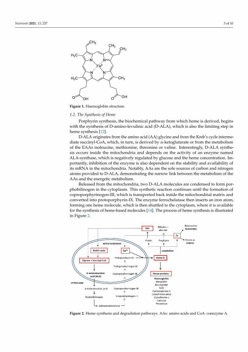
Heme is an organic, ring-shaped molecule consisting of an iron ion coordinated tofour pyrroles, which are small, pentagon-shaped molecules with four carbons and onenitrogen, which, together, form an iron-binding tetrapyrrole, called a porphyrin (Figure 1).Thus, heme is an iron-binding porphyrin [11]. Interestingly, iron plays a balanced attractiveforce interacting with the nitrogen molecules of heme; thus, the electrons stay balanced,and the global molecule remains stable.
There are four different forms of heme in nature: heme-A, -B, -C and -O; they influencethe functions of the molecules in which heme is present. Although heme-B is the mostcommon form, heme-A and -C are present in many molecules. The biochemical behaviorsof the most common heme groups are regulated by differences of the functional groups inthe side chains bound to carbons 3, 8 and 18 [11].
Nutrients 2021, 13, 237 3 of 10Nutrients 2021, 13, x FOR PEER REVIEW 3 of 11
Figure 1. Haemoglobin structure.
There are four different forms of heme in nature: heme-A, -B, -C and -O; they influ-ence the functions of the molecules in which heme is present. Although heme-B is the most common form, heme-A and -C are present in many molecules. The biochemical behaviors of the most common heme groups are regulated by differences of the func-tional groups in the side chains bound to carbons 3, 8 and 18 [11].
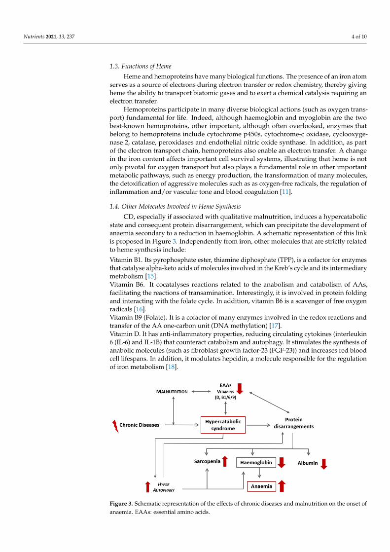
1.2. The Synthesis of Heme Porphyrin synthesis, the biochemical pathway from which heme is derived, begins
with the synthesis of D-amino-levulinic acid (D-ALA), which is also the limiting step in heme synthesis [12].
D-ALA originates from the amino acid (AA) glycine and from the Kreb’s cycle in-termediate succinyl-CoA, which, in turn, is derived by α-ketoglutarate or from the me-tabolism of the EAAs isoleucine, methionine, threonine or valine. Interestingly, D-ALA synthesis occurs inside the mitochondria and depends on the activity of an enzyme named ALA-synthase, which is negatively regulated by glucose and the heme concen-tration. Importantly, inhibition of the enzyme is also dependent on the stability and availability of its mRNA in the mitochondria. Notably, AAs are the sole sources of car-bon and nitrogen atoms provided to D-ALA, demonstrating the narrow link between the metabolism of the AAs and the energetic metabolism.
Released from the mitochondria, two D-ALA molecules are condensed to form porphobilinogen in the cytoplasm. This synthetic reaction continues until the formation of coproporphyrinogen-III, which is transported back inside the mitochondrial matrix and converted into protoporphyrin-IX. The enzyme ferrochelatase then inserts an iron atom, forming one heme molecule, which is then shuttled to the cytoplasm, where it is available for the synthesis of heme-based molecules [14]. The process of heme synthesis is illustrated in Figure 2.
Figure 1. Haemoglobin structure.
1.2. The Synthesis of Heme
Porphyrin synthesis, the biochemical pathway from which heme is derived, beginswith the synthesis of D-amino-levulinic acid (D-ALA), which is also the limiting step inheme synthesis [12].
D-ALA originates from the amino acid (AA) glycine and from the Kreb’s cycle interme-diate succinyl-CoA, which, in turn, is derived by α-ketoglutarate or from the metabolismof the EAAs isoleucine, methionine, threonine or valine. Interestingly, D-ALA synthe-sis occurs inside the mitochondria and depends on the activity of an enzyme namedALA-synthase, which is negatively regulated by glucose and the heme concentration. Im-portantly, inhibition of the enzyme is also dependent on the stability and availability ofits mRNA in the mitochondria. Notably, AAs are the sole sources of carbon and nitrogenatoms provided to D-ALA, demonstrating the narrow link between the metabolism of theAAs and the energetic metabolism.
Released from the mitochondria, two D-ALA molecules are condensed to form por-phobilinogen in the cytoplasm. This synthetic reaction continues until the formation ofcoproporphyrinogen-III, which is transported back inside the mitochondrial matrix andconverted into protoporphyrin-IX. The enzyme ferrochelatase then inserts an iron atom,forming one heme molecule, which is then shuttled to the cytoplasm, where it is availablefor the synthesis of heme-based molecules [14]. The process of heme synthesis is illustratedin Figure 2.
Nutrients 2021, 13, x FOR PEER REVIEW 4 of 11
Figure 2. Heme synthesis and degradation pathways. AAs: amino acids and CoA: coenzyme A.
1.3. Functions of Heme Heme and hemoproteins have many biological functions. The presence of an iron
atom serves as a source of electrons during electron transfer or redox chemistry, thereby giving heme the ability to transport biatomic gases and to exert a chemical catalysis re-quiring an electron transfer.
Hemoproteins participate in many diverse biological actions (such as oxygen transport) fundamental for life. Indeed, although haemoglobin and myoglobin are the two best-known hemoproteins, other important, although often overlooked, enzymes that belong to hemoproteins include cytochrome p450s, cytochrome-c oxidase, cycloox-ygenase 2, catalase, peroxidases and endothelial nitric oxide synthase. In addition, as part of the electron transport chain, hemoproteins also enable an electron transfer. A change in the iron content affects important cell survival systems, illustrating that heme is not only pivotal for oxygen transport but also plays a fundamental role in other im-portant metabolic pathways, such as energy production, the transformation of many molecules, the detoxification of aggressive molecules such as as oxygen-free radicals, the regulation of inflammation and/or vascular tone and blood coagulation [11].
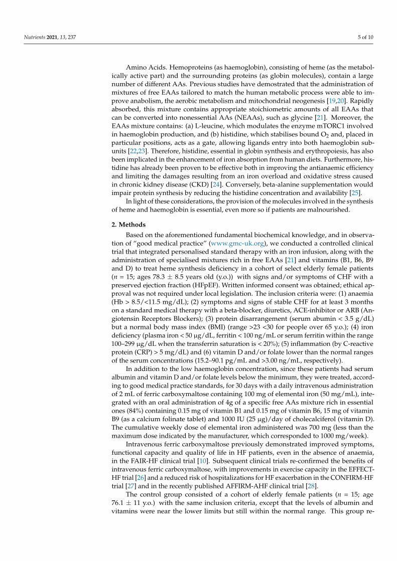
1.4. Other Molecules Involved in Heme Synthesis CD, especially if associated with qualitative malnutrition, induces a hypercatabolic
state and consequent protein disarrangement, which can precipitate the development of anaemia secondary to a reduction in haemoglobin. A schematic representation of this link is proposed in Figure 3. Independently from iron, other molecules that are strictly related to heme synthesis include:
Vitamin B1. Its pyrophosphate ester, thiamine diphosphate (TPP), is a cofactor for enzymes that catalyse alpha-keto acids of molecules involved in the Kreb’s cycle and its intermediary metabolism [15].
Vitamin B6. It cocatalyses reactions related to the anabolism and catabolism of AAs, facilitating the reactions of transamination. Interestingly, it is involved in protein folding and interacting with the folate cycle. In addition, vitamin B6 is a scavenger of free oxy-gen radicals [16].
Figure 2. Heme synthesis and degradation pathways. AAs: amino acids and CoA: coenzyme A.
Nutrients 2021, 13, 237 4 of 10
1.3. Functions of Heme
Heme and hemoproteins have many biological functions. The presence of an iron atomserves as a source of electrons during electron transfer or redox chemistry, thereby givingheme the ability to transport biatomic gases and to exert a chemical catalysis requiring anelectron transfer.
Hemoproteins participate in many diverse biological actions (such as oxygen trans-port) fundamental for life. Indeed, although haemoglobin and myoglobin are the twobest-known hemoproteins, other important, although often overlooked, enzymes thatbelong to hemoproteins include cytochrome p450s, cytochrome-c oxidase, cyclooxyge-nase 2, catalase, peroxidases and endothelial nitric oxide synthase. In addition, as partof the electron transport chain, hemoproteins also enable an electron transfer. A changein the iron content affects important cell survival systems, illustrating that heme is notonly pivotal for oxygen transport but also plays a fundamental role in other importantmetabolic pathways, such as energy production, the transformation of many molecules,the detoxification of aggressive molecules such as as oxygen-free radicals, the regulation ofinflammation and/or vascular tone and blood coagulation [11].
1.4. Other Molecules Involved in Heme Synthesis
CD, especially if associated with qualitative malnutrition, induces a hypercatabolicstate and consequent protein disarrangement, which can precipitate the development ofanaemia secondary to a reduction in haemoglobin. A schematic representation of this linkis proposed in Figure 3. Independently from iron, other molecules that are strictly relatedto heme synthesis include:
Vitamin B1. Its pyrophosphate ester, thiamine diphosphate (TPP), is a cofactor for enzymesthat catalyse alpha-keto acids of molecules involved in the Kreb’s cycle and its intermediarymetabolism [15].Vitamin B6. It cocatalyses reactions related to the anabolism and catabolism of AAs,facilitating the reactions of transamination. Interestingly, it is involved in protein foldingand interacting with the folate cycle. In addition, vitamin B6 is a scavenger of free oxygenradicals [16].Vitamin B9 (Folate). It is a cofactor of many enzymes involved in the redox reactions andtransfer of the AA one-carbon unit (DNA methylation) [17].Vitamin D. It has anti-inflammatory properties, reducing circulating cytokines (interleukin6 (IL-6) and IL-1B) that counteract catabolism and autophagy. It stimulates the synthesis ofanabolic molecules (such as fibroblast growth factor-23 (FGF-23)) and increases red bloodcell lifespans. In addition, it modulates hepcidin, a molecule responsible for the regulationof iron metabolism [18].
Nutrients 2021, 13, x FOR PEER REVIEW 5 of 11
Vitamin B9 (Folate). It is a cofactor of many enzymes involved in the redox reac-tions and transfer of the AA one-carbon unit (DNA methylation) [17].
Vitamin D. It has anti-inflammatory properties, reducing circulating cytokines (in-terleukin 6 (IL-6) and IL-1B) that counteract catabolism and autophagy. It stimulates the synthesis of anabolic molecules (such as fibroblast growth factor-23 (FGF-23)) and in-creases red blood cell lifespans. In addition, it modulates hepcidin, a molecule responsi-ble for the regulation of iron metabolism [18].
Amino Acids. Hemoproteins (as haemoglobin), consisting of heme (as the metaboli-cally active part) and the surrounding proteins (as globin molecules), contain a large number of different AAs. Previous studies have demostrated that the administration of mixtures of free EAAs tailored to match the human metabolic process were able to im-prove anabolism, the aerobic metabolism and mitochondrial neogenesis [19,20]. Rapidly absorbed, this mixture contains appropriate stoichiometric amounts of all EAAs that can be converted into nonessential AAs (NEAAs), such as glycine [21]. Moreover, the EAAs mixture contains: (a) L-leucine, which modulates the enzyme mTORC1 involved in haemoglobin production, and (b) histidine, which stabilises bound O2 and, placed in particular positions, acts as a gate, allowing ligands entry into both haemoglobin subu-nits [22,23]. Therefore, histidine, essential in globin synthesis and erythropoiesis, has al-so been implicated in the enhancement of iron absorption from human diets. Further-more, histidine has already been proven to be effective both in improving the antianae-mic efficiency and limiting the damages resulting from an iron overload and oxidative stress caused in chronic kidney disease (CKD) [24]. Conversely, beta-alanine supplemen-tation would impair protein synthesis by reducing the histidine concentration and avail-ability [25].
In light of these considerations, the provision of the molecules involved in the syn-thesis of heme and haemoglobin is essential, even more so if patients are malnourished.
Figure 3. Schematic representation of the effects of chronic diseases and malnutrition on the onset of anaemia. EAAs: essential amino acids.
2. Methods Based on the aforementioned fundamental biochemical knowledge, and in observa-
tion of “good medical practice” (www.gmc-uk.org), we conducted a controlled clinical trial that integrated personalised standard therapy with an iron infusion, along with the administration of specialised mixtures rich in free EAAs [21] and vitamins (B1, B6, B9 and D) to treat heme synthesis deficiency in a cohort of select elderly female patients (n = 15; ages 78.3 ± 8.5 years old (y.o.)) with signs and/or symptoms of CHF with a preserved ejection fraction (HFpEF). Written informed consent was obtained; ethical approval was not required under local legislation. The inclusion criteria were: (1) anaemia (Hb >
Figure 3. Schematic representation of the effects of chronic diseases and malnutrition on the onset ofanaemia. EAAs: essential amino acids.
Nutrients 2021, 13, 237 5 of 10
Amino Acids. Hemoproteins (as haemoglobin), consisting of heme (as the metabol-ically active part) and the surrounding proteins (as globin molecules), contain a largenumber of different AAs. Previous studies have demostrated that the administration ofmixtures of free EAAs tailored to match the human metabolic process were able to im-prove anabolism, the aerobic metabolism and mitochondrial neogenesis [19,20]. Rapidlyabsorbed, this mixture contains appropriate stoichiometric amounts of all EAAs thatcan be converted into nonessential AAs (NEAAs), such as glycine [21]. Moreover, theEAAs mixture contains: (a) L-leucine, which modulates the enzyme mTORC1 involvedin haemoglobin production, and (b) histidine, which stabilises bound O2 and, placed inparticular positions, acts as a gate, allowing ligands entry into both haemoglobin sub-units [22,23]. Therefore, histidine, essential in globin synthesis and erythropoiesis, has alsobeen implicated in the enhancement of iron absorption from human diets. Furthermore, his-tidine has already been proven to be effective both in improving the antianaemic efficiencyand limiting the damages resulting from an iron overload and oxidative stress causedin chronic kidney disease (CKD) [24]. Conversely, beta-alanine supplementation wouldimpair protein synthesis by reducing the histidine concentration and availability [25].
In light of these considerations, the provision of the molecules involved in the synthesisof heme and haemoglobin is essential, even more so if patients are malnourished.
2. Methods
Based on the aforementioned fundamental biochemical knowledge, and in observa-tion of “good medical practice” (www.gmc-uk.org), we conducted a controlled clinicaltrial that integrated personalised standard therapy with an iron infusion, along with theadministration of specialised mixtures rich in free EAAs [21] and vitamins (B1, B6, B9and D) to treat heme synthesis deficiency in a cohort of select elderly female patients(n = 15; ages 78.3 ± 8.5 years old (y.o.)) with signs and/or symptoms of CHF with apreserved ejection fraction (HFpEF). Written informed consent was obtained; ethical ap-proval was not required under local legislation. The inclusion criteria were: (1) anaemia(Hb > 8.5/<11.5 mg/dL); (2) symptoms and signs of stable CHF for at least 3 monthson a standard medical therapy with a beta-blocker, diuretics, ACE-inhibitor or ARB (An-giotensin Receptors Blockers); (3) protein disarrangement (serum abumin < 3.5 g/dL)but a normal body mass index (BMI) (range >23 <30 for people over 65 y.o.); (4) irondeficiency (plasma iron < 50 µg/dL, ferritin < 100 ng/mL or serum ferritin within the range100–299 µg/dL when the transferrin saturation is < 20%); (5) inflammation (by C-reactiveprotein (CRP) > 5 mg/dL) and (6) vitamin D and/or folate lower than the normal rangesof the serum concentrations (15.2–90.1 pg/mL and >3.00 ng/mL, respectively).
In addition to the low haemoglobin concentration, since these patients had serumalbumin and vitamin D and/or folate levels below the minimum, they were treated, accord-ing to good medical practice standards, for 30 days with a daily intravenous administrationof 2 mL of ferric carboxymaltose containing 100 mg of elemental iron (50 mg/mL), inte-grated with an oral administration of 4g of a specific free AAs mixture rich in essentialones (84%) containing 0.15 mg of vitamin B1 and 0.15 mg of vitamin B6, 15 mg of vitaminB9 (as a calcium folinate tablet) and 1000 IU (25 µg)/day of cholecalciferol (vitamin D).The cumulative weekly dose of elemental iron administered was 700 mg (less than themaximum dose indicated by the manufacturer, which corresponded to 1000 mg/week).
Intravenous ferric carboxymaltose previously demonstrated improved symptoms,functional capacity and quality of life in HF patients, even in the absence of anaemia,in the FAIR-HF clinical trial [10]. Subsequent clinical trials re-confirmed the benefits ofintravenous ferric carboxymaltose, with improvements in exercise capacity in the EFFECT-HF trial [26] and a reduced risk of hospitalizations for HF exacerbation in the CONFIRM-HFtrial [27] and in the recently published AFFIRM-AHF clinical trial [28].
The control group consisted of a cohort of elderly female patients (n = 15; age76.1 ± 11 y.o.) with the same inclusion criteria, except that the levels of albumin andvitamins were near the lower limits but still within the normal range. This group re-

Nutrients 2021, 13, 237 6 of 10
ceived only standard iron therapy without supplementation. The baseline mean clinicalbiochemical data from two cohorts are summarised in Table 1.
Table 1. Comparison of the baseline antropometric and clinical biochemical data from patients who received integratedtherapy and iron standard therapy (control). Note that patients that received the iron standard therapy had nutritionalparameters (albumin and vitamins) close to the lower limit of the normal range. TIBC, Total Iron Binding Capacity; CRP,C-reactive protein; NT-proBNP, B-type natriuretic peptide; LVEF, Left Ventricular Ejection Fraction; BMI, body mass indexand CVP, Central venous pressure.
Integrated Therapy(n = 15)
Standard Therapy(n = 15) Normal Value
Age (y.o) 78.3 ± 8.5 76.1 ± 11 —BMI 26.9 ± 1.85 25.5 ± 1.91 <25
Haemoglobin (g/dL) 10.2 ± 0.8 10.37 ± 0.91 >11.5Creatinine (mg/dL) 1.06 ± 0.25 1.02 ± 0.19 0,5–1,1
Albumin (g/dL) 3.31 ± 0.37 3.55 ± 0.08 >3.5Ferritin (ng/mL) 73.73 ± 38.81 94.80 ± 40.76 15–200
Sideraemia (serum iron) (µg/dL) 36.07 ± 6.85 33.72 ± 9.33 50–150Transferrin saturated (%) 14.33 ± 4.24 12.88 ± 2.18 20–45
Transferrin total (TIBC) (µg/dL) 228.53 ± 48.09 224.33 ± 50.0 180–380Vitamin B9 (ng/mL) 2.32 ± 0.35 3.13 ± 0.20 3
1,25-OH Vitamin D (pg/mL) 17.73 ± 4.23 21.87 ± 2.25 21–100CRP (mg/dL) 10.67 ± 2.43 10.82 ± 2.70 <5
NT-proBNP (pg/mL) 2449.2 ± 2048.69 2650.27 ± 2177.65 <450LVEF (%) 54 ± 6 56 ± 5 >50
CVP (mmHg) 5.5 ± 1.08 5.73 ± 1.05 <8
Statistics
Data are expressed as the mean ± standard deviation. A two-tailed paired Student’st-test was used to compare the data before (T0) and after (T30) the therapy within eachgroup. Increments by treatments between the two groups were evaluated by the unpairedtwo-tailed Welch’s t-test. The p-value < 0.05 was considered significant.
3. Results
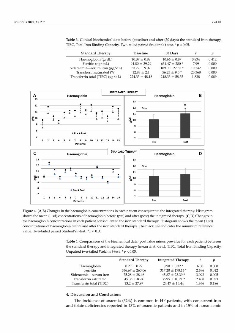
The baseline and postintervention clinical biochemical data of the patients recivingintegrated therapy are summarised in Table 2. Increased levels of sideraemia (serum iron),ferritin and saturated transferrin were observed in both groups (Tables 2 and 3). However,when compared to the basal levels, only the experimental group receiving intravenous irontherapy plus integrated therapy demonstrated a significant increase in the haemoglobinconcentration (Figure 4A,B), while no modification was observed in the group receivingthe standard iron therapy only (see Table 3 and Figure 4C,D). The comparisons of themodifications of the biochemical data for the two groups of patients (postvalue minusprevalue) are summarised in Table 4.
Table 2. Clinical biochemical data before (baseline) and after (30 days) integrated therapy. TIBC,Total Iron Binding Capacity. Two-tailed paired Student’s t-test. * p < 0.05.
Integrated Therapy Baseline 30 Days t p
Haemoglobin (g/dL) 10.1 ± 0.76 11.06 ± 0.83 * 10.94 0.000Ferritin (ng/mL) 73.73 ± 38.81 390.93 ± 196.4 * 6.89 0.000
Sideraemia—serum iron (µg/dL) 36.07 ± 6.85 81.93 ± 16.84 * 7.59 0.000Transferrin saturated (%) 14.33 ± 4.24 51.28 ± 10.24 * 13.35 0.000
Transferrin total (TIBC) (µg/dL) 228.53 ± 48.09 253.0 ± 50.37 * 6.14 0.000
Nutrients 2021, 13, 237 7 of 10
Table 3. Clinical biochemical data before (baseline) and after (30 days) the standard iron therapy.TIBC, Total Iron Binding Capacity. Two-tailed paired Student’s t-test. * p < 0.05.
Standard Therapy Baseline 30 Days t p
Haemoglobin (g/dL) 10.37 ± 0.88 10.66 ± 0.87 0.834 0.412Ferritin (ng/mL) 94.80 ± 39.29 631.47 ± 280 * 7.99 0.000
Sideraemia—serum iron (µg/dL) 33.72 ± 9.07 109.0 ± 27.62 * 10.242 0.000Transferrin saturated (%) 12.88 ± 2.1 56.23 ± 9.5 * 20.368 0.000
Transferrin total (TIBC) (µg/dL) 224.33 ± 48.18 218.33 ± 58.35 1.828 0.089
Nutrients 2021, 13, x FOR PEER REVIEW 8 of 11
Figure 4. (A,B) Changes in the haemoglobin concentrations in each patient consequent to the integrated therapy. Histo-gram shows the mean (±sd) concentrations of haemoglobin before (pre) and after (post) the integrated therapy. (C,D) Changes in the haemoglobin concentrations in each patient consequent to the iron standard therapy. Histogram shows the mean (±sd) concentrations of haemoglobin before and after the iron standard therapy. The black line indicates the minimum reference value. Two-tailed paired Student’s t-test. * p < 0.05.
4. Discussion and Conclusions The incidence of anaemia (32%) is common in HF patients, with concurrent iron
and folate deficiencies reported in 43% of anaemic patients and in 15% of nonanaemic patients [29]. Currently, the standard therapy of anaemia is primarily based on the sup-plementation of iron, with or without erythropoietin for hematopoiesis stimulation. Pre-vious randomised, controlled studies with intravenous iron in HF patients reported that haemoglobin increased after four or six months of treatment [7,10,30,31]. Indeed, if a de-ficiency of fundamental molecules (such as amino acids and vitamins) results in the lack of heme synthesis, iron supplementation alone will not lead to a proportional hemopro-tein increase and of Hb in primis. In addition, an isolated increase in soluble iron ions, without any accompanying augmentation in heme, could favor the persistence of oxida-tive stress (via Fenton/Haber-Weiss reactions), chronic inflammation and autophagy [32]. The mode of iron supplementation also appears to be important, as oral supple-mentation was ineffective in improving the exercise capacity in HF patients with re-duced ejection fraction and iron deficiency [33].
After the integrated therapy, we observed improvements of ferritin in both groups, but this was more elevated in patients who received the standard therapy. Iron, alt-hough it is an essential micronutrient, is potentially toxic for biological systems, since it generates free radicals by interconversions between ferrous (Fe2+) and ferric (Fe3+) forms. However, existing regulatory processes in the body are efficacious in reducing the toxici-ty of iron, even in response to an overload [34]. Interestingly, patients receiving the inte-grated therapy had lower ferritin concentrations than those receiving the standard ther-apy. This suggests that the use of iron for the synthesis of haemoglobin was rapidly
Figure 4. (A,B) Changes in the haemoglobin concentrations in each patient consequent to the integrated therapy. Histogramshows the mean (±sd) concentrations of haemoglobin before (pre) and after (post) the integrated therapy. (C,D) Changes inthe haemoglobin concentrations in each patient consequent to the iron standard therapy. Histogram shows the mean (±sd)concentrations of haemoglobin before and after the iron standard therapy. The black line indicates the minimum referencevalue. Two-tailed paired Student’s t-test. * p < 0.05.
Table 4. Comparisons of the biochemical data (postvalue minus prevalue for each patient) betweenthe standard therapy and integrated therapy (mean ± st. dev.). TIBC, Total Iron Binding Capacity.Unpaired two-tailed Welch’s t-test. * p < 0.05.
Standard Therapy Integrated Therapy t p
Haemoglobin 0.29 ± 0.22 0.90 ± 0.32 * 6.08 0.000Ferritin 536.67 ± 260.06 317.20 ± 178.16 * 2.696 0.012
Sideraemia—serum iron 75.28 ± 28.46 45.87 ± 23.39 * 3.092 0.005Transferrin saturated 45.35 ± 8.24 36.95 ± 10.71 * 2.408 0.023
Transferrin total (TIBC) 13.2 ± 27.97 24.47 ± 15.44 1.366 0.186
4. Discussion and Conclusions
The incidence of anaemia (32%) is common in HF patients, with concurrent ironand folate deficiencies reported in 43% of anaemic patients and in 15% of nonanaemic

Nutrients 2021, 13, 237 8 of 10
patients [29]. Currently, the standard therapy of anaemia is primarily based on the sup-plementation of iron, with or without erythropoietin for hematopoiesis stimulation. Pre-vious randomised, controlled studies with intravenous iron in HF patients reported thathaemoglobin increased after four or six months of treatment [7,10,30,31]. Indeed, if adeficiency of fundamental molecules (such as amino acids and vitamins) results in the lackof heme synthesis, iron supplementation alone will not lead to a proportional hemoproteinincrease and of Hb in primis. In addition, an isolated increase in soluble iron ions, withoutany accompanying augmentation in heme, could favor the persistence of oxidative stress(via Fenton/Haber-Weiss reactions), chronic inflammation and autophagy [32]. The modeof iron supplementation also appears to be important, as oral supplementation was ineffec-tive in improving the exercise capacity in HF patients with reduced ejection fraction andiron deficiency [33].
After the integrated therapy, we observed improvements of ferritin in both groups,but this was more elevated in patients who received the standard therapy. Iron, although itis an essential micronutrient, is potentially toxic for biological systems, since it generatesfree radicals by interconversions between ferrous (Fe2+) and ferric (Fe3+) forms. However,existing regulatory processes in the body are efficacious in reducing the toxicity of iron,even in response to an overload [34]. Interestingly, patients receiving the integrated therapyhad lower ferritin concentrations than those receiving the standard therapy. This suggeststhat the use of iron for the synthesis of haemoglobin was rapidly promoted and muchmore effective in the integrated therapy, thus also limiting the possible toxic effects ofiron deposits.
Based on these preliminary data showing a rapid escalation in the haemoglobin level(within 30 days after the interventions aimed at increasing both iron and heme), we proposethat a more effective approach to treating heme synthesis (including anaemia) in CD mustconsider not only the iron availability but, also, integrate a therapeutic strategy thatcounteracts the catabolic drives and promotes protein syntheses. Therefore, the standardintravenous or oral iron supplementation should incorporate also a supply of specificmixtures of EAAs and vitamins involved in the biochemical pathway of heme synthesis,as illustrated in Figure 5. Consequently, the careful evaluation of either the nutritionalstatus of patients and the presence of catabolism or impairement of the availability ofmolecules involved in heme synthesis, as well as their integration, must therefore be thefirst step of an effective individualised therapeutic intervention aimed at correcting thestate of anaemia in patients with CD such as CHF. Our therapeutical approach based onbiochemical data should be confirmed in a large-scale clinical trial.
Nutrients 2021, 13, x FOR PEER REVIEW 9 of 11
promoted and much more effective in the integrated therapy, thus also limiting the pos-sible toxic effects of iron deposits.
Based on these preliminary data showing a rapid escalation in the haemoglobin level (within 30 days after the interventions aimed at increasing both iron and heme), we propose that a more effective approach to treating heme synthesis (including anaemia) in CD must consider not only the iron availability but, also, integrate a therapeutic strat-egy that counteracts the catabolic drives and promotes protein syntheses. Therefore, the standard intravenous or oral iron supplementation should incorporate also a supply of specific mixtures of EAAs and vitamins involved in the biochemical pathway of heme synthesis, as illustrated in Figure 5. Consequently, the careful evaluation of either the nutritional status of patients and the presence of catabolism or impairement of the avail-ability of molecules involved in heme synthesis, as well as their integration, must there-fore be the first step of an effective individualised therapeutic intervention aimed at cor-recting the state of anaemia in patients with CD such as CHF. Our therapeutical ap-proach based on biochemical data should be confirmed in a large-scale clinical trial.
Figure 5. Schematic representation of the effects of integrated therapy on the containment of anaemia.
The main messages: • In chronic hypercatabolic diseases (such as CHF), the metabolism of iron and the
heme protein is markedly impaired, inducing anaemia and the likely impairment of many other hemoproteins involved in the essential metabolic pathways.
• Heme is the metabolically active part of haemoglobin. It is characterised by the presence of iron atoms linked to tetrapyrrole groups.
• Many other important biologically active molecules, named hemoproteins (includ-ing Hb), contain heme as the metabolically active part, with the surrounding pro-teins (as globin molecules) containing a large number of different amino acids.
• The maintenance of an adequate blood concentration of both iron and heme is fun-damental for proper function of the heme-containing enzymes.
• In patients with anaemia and chronic hypercatabolic diseases, the correction of de-ficiencies in iron, as well as the metabolic substrates required for all hemoproteins, is essential for proper treatment.
Author Contributions: E.P. and G.C. wrote the manuscript; these authors contributed equally to this work. G.C. and C.R. arranged the text, the figures and the references. F.S.D., R.A., T.S. and C.C.-S. reviewed the text. T.S. and C.C.-S. revised the English language. All authors conceptual-ised the topic and discussed the literature data. All authors approved the final version of the man-uscript and ensure the accuracy of the work and intellectual content. All authors have read and agreed to the published version of the manuscript.
Funding: This study was supported by local funds to CG.
Institutional Review Board Statement: Not applicable.
Informed Consent Statement: Informed consent was obtained from all subjects involved in the study. Data Availability Statement: The data presented in this study are available on request from the corresponding author. The data are not publicly available due to privacy reason.
Figure 5. Schematic representation of the effects of integrated therapy on the containment of anaemia.
The main messages:
• In chronic hypercatabolic diseases (such as CHF), the metabolism of iron and the hemeprotein is markedly impaired, inducing anaemia and the likely impairment of manyother hemoproteins involved in the essential metabolic pathways.
• Heme is the metabolically active part of haemoglobin. It is characterised by thepresence of iron atoms linked to tetrapyrrole groups.
• Many other important biologically active molecules, named hemoproteins (includingHb), contain heme as the metabolically active part, with the surrounding proteins (asglobin molecules) containing a large number of different amino acids.

Nutrients 2021, 13, 237 9 of 10
• The maintenance of an adequate blood concentration of both iron and heme is funda-mental for proper function of the heme-containing enzymes.
• In patients with anaemia and chronic hypercatabolic diseases, the correction of defi-ciencies in iron, as well as the metabolic substrates required for all hemoproteins, isessential for proper treatment.
Author Contributions: E.P. and G.C. wrote the manuscript; these authors contributed equally to thiswork. G.C. and C.R. arranged the text, the figures and the references. F.S.D., R.A., T.S. and C.C.-S.reviewed the text. T.S. and C.C.-S. revised the English language. All authors conceptualised thetopic and discussed the literature data. All authors approved the final version of the manuscript andensure the accuracy of the work and intellectual content. All authors have read and agreed to thepublished version of the manuscript.
Funding: This study was supported by local funds to CG.
Institutional Review Board Statement: Not applicable.
Informed Consent Statement: Informed consent was obtained from all subjects involved in the study.
Data Availability Statement: The data presented in this study are available on request from thecorresponding author. The data are not publicly available due to privacy reason.
Conflicts of Interest: The authors declare that the research was conducted in the absence of anycommercial or financial relationships that could be construed as a potential conflict of interests.
References1. World Health Organization. Noncommunicable diseases. Fact Sheet. January 2015. Available online: https://www.who.int/
topics/noncommunicable_diseases/factsheets/en/ (accessed on 5 April 2016).2. Bernell, S.; Howard, S.W. Use your words carefully: What is a chronic disease? Front. Public Health 2016, 4, 159. [CrossRef]
[PubMed]3. Pasini, E.; Aquilani, R.; Dioguardi, F.S.; D’Antona, G.; Gheorghiade, M.; Taegtmeyer, H. Hypercatabolic syndrome: Molecular
basis and effects of nutritional supplements with amino acids. Am. J. Cardiol. 2008, 101, 11E–15E. [CrossRef] [PubMed]4. Aquilani, R.; Maestri, R.; Boselli, M.; Achilli, M.P.; Arrigoni, N.; Bruni, M.; Dossena, M.; Verri, M.; Buonocore, D.; Pasini, E.; et al.
The relationship between plasma amino acids and circulating albumin and haemoglobin in postabsorptive stroke patients. PLoSONE 2019, 14, e0219756. [CrossRef] [PubMed]
5. Comín-Colet, J.; Enjuanes, C.; González, G.; Torrens, A.; Cladellas, M.; Meroño, O.; Ribas, N.; Ruiz, S.; Gómez, M.; Verdú, J.M.;et al. Iron deficiency is a key determinant of health-related quality of life in patients with chronic heart failure regardless ofanaemia status. Eur. J. Heart Fail. 2013, 15, 1164–1172. [CrossRef] [PubMed]
6. Klip, I.T.; Comin-Colet, J.; Voors, A.A.; Ponikowski, P.; Enjuanes, C.; Banasiak, W.; Lok, D.J.; Rosentryt, P.; Torrens, A.; Polonski,L.; et al. Iron deficiency in chronic heart failure: An international pooled analysis. Am. Heart J. 2013, 165, 575–582.e3. [CrossRef][PubMed]
7. van Veldhuisen, D.; Anker, S.; Ponikowski, P.; Macdougall, I.C. Anemia and iron deficiency in heart failure: Mechanisms andtherapeutic approaches. Nat. Rev. Cardiol. 2011, 8, 485–493. [CrossRef]
8. Corsetti, G.; Chen-Scarabelli, C.; Romano, C.; Pasini, E.; Dioguardi, F.S.; Onorati, F.; Knight, R.; Patel, H.; Saravolatz, L.; Faggian,G.; et al. Autophagy and oncosis/necroptosis are enhanced in cardiomyocytes from heart failure patients. Med. Sci. Monit. BasicRes. 2019, 25, 33–44. [CrossRef]
9. Sîrbu, O.; Floria, M.; Dascalita, P.; Stoica, A.; Adascalitei, P.; Sorodoc, V.; Sorodoc, L. Anemia in heart failure—From guidelines tocontroversies and challenges. Anatol. J. Cardiol. 2018, 20, 52–59. [CrossRef]
10. Anker, S.D.; Colet, J.C.; Filippatos, G.; Willenheimer, R.; Dickstein, K.; Drexler, H.; Lüscher, T.F.; Bart, B.; Banasiak, W.; Niegowska,J.; et al. Ferric carboxymaltose in patients with heart failure and iron deficiency. N. Engl. J. Med. 2009, 361, 2436–2448. [CrossRef]
11. Poulos, T.L. Heme enzyme structure and function. Chem. Rev. 2014, 114, 3919–3962. [CrossRef]12. Pasini, E.; Dioguardi, F.S. Iron supplementation in the cardiorenal anaemia syndrome: A global metabolic approach. Eur. J. Heart
Fail. 2012, 14, 1429. [CrossRef] [PubMed]13. Richardson, D.R.; Ponka, P. The molecular mechanisms of the metabolism and transport of iron in normal. and neoplastic cells.
Biochim. Biophys. Acta 1997, 1331, 1–40. [CrossRef]14. Layer, G.; Reichelt, J.; Jahn, D.; Heinz, D.W. Structure and function of enzymes in heme biosynthesis. Protein Sci. 2010, 19,
1137–1161. [CrossRef] [PubMed]15. Lonsdale, D. A review of the biochemistry, metabolism and clinical benefits of thiamin(e) and its derivatives. Evid. Based
Complement Alternat. Med. 2006, 3, 49–59. [CrossRef] [PubMed]16. Parra, M.; Stahl, S.; Hellmann, H. Vitamin B6 and its role in cell metabolism and physiology. Cells 2018, 7, 84. [CrossRef]17. Bailey, L.B.; Gregory, J.F. Folate metabolism and requirements. J. Nutr. 1999, 129, 779–782. [CrossRef]
Nutrients 2021, 13, 237 10 of 10
18. Smith, E.M.; Tangpricha, V. Vitamin D and anemia: Insights into an emerging association. Curr. Opin. Endocrinol. Diabetes Obes.2015, 22, 432–438. [CrossRef]
19. D’Antona, G.; Ragni, M.; Cardile, A.; Tedesco, L.; Dossena, M.; Bruttini, F.; Caliaro, F.; Corsetti, G.; Bottinelli, R.; Carruba, M.O.;et al. Branched-chain amino acid supplementation promotes survival and supports cardiac and skeletal muscle mitochondrialbiogenesis in middle-aged mice. Cell Metab. 2010, 12, 362–372. [CrossRef]
20. Corsetti, G.; Pasini, E.; D’Antona, G.; Nisoli, E.; Flati, V.; Assanelli, D.; Dioguardi, F.S.; Bianchi, R. Morphometric changes inducedby amino acid supplementation in skeletal and cardiac muscles of old mice. Am. J. Cardiol. 2008, 101, 26E–34E. [CrossRef]
21. Rondanelli, M.; Aquilani, R.; Verri, M.; Boschi, F.; Pasini, E.; Perna, S.; Faliva, A.; Condino, A.M. Plasma kinetics of essentialamino acids following their ingestion as free formula or as dietary protein components. Aging Clin. Exp. Res. 2017, 29, 801–805.[CrossRef]
22. Chung, J.; Bauer, D.E.; Ghamari, A.; Nizzi, C.P.; Deck, K.M.; Kingsley, P.D.; Yie, Y.Y. The mTORC1/4E-BP pathway coordinateshemoglobin production wit leucin availability. Sci. Signal. 2015, 8, ra34. [CrossRef] [PubMed]
23. Birukou, I.; Schweers, R.L.; Olson, J.S. Distal histidine stabilizes bound O2 and acts as a gate for ligand entry in both subunits ofadult human hemoglobin. J. Biol. Chem. 2010, 285, 8840–8854. [CrossRef] [PubMed]
24. Vera-Aviles, M.; Vantana, E.; Kardinasari, E.; Koh, N.L.; Latunde-Dada, G.O. Protective role of histidine supplementation againstoxidative stress damage in the management of anemia of chronic kidney disease. Pharmaceuticals 2018, 11, 111. [CrossRef][PubMed]
25. Blancquaert, L.; Everaert, I.; Missinne, M.; Baguet, A.; Stegen, S.; Volkaert, A.; Petrovic, M.; Vervaet, C.; Achten, E.; DE Maeyer,M.; et al. Effects of histidine and a-alanine supplementation on human muscle carnosine storage. Med. Sci. Sports Exerc. 2017, 49,602–609. [CrossRef] [PubMed]
26. van Veldhuisen, D.J.; Ponikowski, P.; van der Meer, P.; Metra, M.; Böhm, M.; Doletsky, A.; Voors, A.A.; Macdougall, I.C.; Anker,S.D.; Roubert, B.; et al. Effect of ferric carboxymaltose on exercise capacity in patients with chronic heart failure and iron deficiency.Circulation 2017, 136, 1374–1383. [CrossRef] [PubMed]
27. Ponikowski, P.; van Veldhuisen, D.J.; Comin-Colet, J.; Ertl, G.; Komajda, M.; Mareev, V.; McDonagh, T.; Parkhomenko, A.; Tavazzi,L.; Levesque, V.; et al. Beneficial effects of long-term intravenous iron therapy with ferric carboxymaltose in patients withsymptomatic heart failure and iron deficiency. Eur. Heart J. 2015, 36, 657–668. [CrossRef]
28. Ponikowski, P.; Kirwan, B.A.; Anker, S.D.; McDonagh, T.; Dorobantu, M.; Drozdz, J.; Fabien, V.; Filippatos, G.; Göhring, U.M.;Keren, A.; et al. Ferric carboxymaltose for iron deficiency at discharge after acute heart failure: A multicentre, double-blind,randomised, controlled trial. Lancet 2020, 396, 1895–1904. [CrossRef]
29. de Silva, R.; Rigby, A.S.; Witte, K.K.A.; Nikitin, N.P.; Tin, L.; Goode, K.; Bhandari, S.; Clark, A.L.; Cleland, J.G.F. Anemia, renaldysfunction, and their interaction in patients with chronic heart failure. Am. J. Cardiol. 2006, 98, 391–398. [CrossRef]
30. Toblli, J.E.; Lombraña, A.; Duarte, P.; Di Genarro, F. Intravenous iron reduces NT-pro-brain natriuretic peptide in anemia patientswith chronic heart failure and renal insufficiency. J. Am. Coll. Cardiol. 2007, 50, 1657–1665. [CrossRef]
31. Okonko, D.O.; Grzeslo, A.; Witkowski, T.; Mandal, A.K.J.; Slater, R.M.; Roughton, M.; Foldes, G.; Thum, T.; Majda, J.; Banasiak,W.; et al. Effect of intravenous iron sucrose on exercise tolerance in anemic and nonanemic patients with chronic heart failure andiron deficiency. FERRIC-HF: A randomized, controlled, observer-blinded trial. J. Am. Coll. Cardiol. 2008, 51, 103–312. [CrossRef]
32. Stohs, S.; Bagchi, D. Oxidative mechanisms in the toxicity of metal ions. Free Radic. Biol. Med. 1995, 18, 321–336. [CrossRef]33. Lewis, G.D.; Malhotra, R.; Hernandez, A.F.; McNulty, S.E.; Smith, A.; Felker, G.M.; Tang, W.H.W.; LaRue, S.J.; Redfield, M.M.;
Semigran, M.J.; et al. Effect of oral iron repletion on exercise capacity in patients with heart failure with reduced ejection fractionand iron deficiency: The IRONOUT HF randomized clinical trial. JAMA 2017, 317, 1958–1966. [CrossRef] [PubMed]
34. Eid, R.; Arab, T.T.N.; Greenwood, M.T. Iron mediated toxicity and programmed cell death: A review and a re-examination ofexisting paradigms. Biochim. Biophys. Acta Mol. Cell Res. 2017, 1864, 399–430. [CrossRef] [PubMed]